Uncovering regularities: On Bare and Evaluated Controllers in Tigrinya
Upload
independentCategory
view
2download
0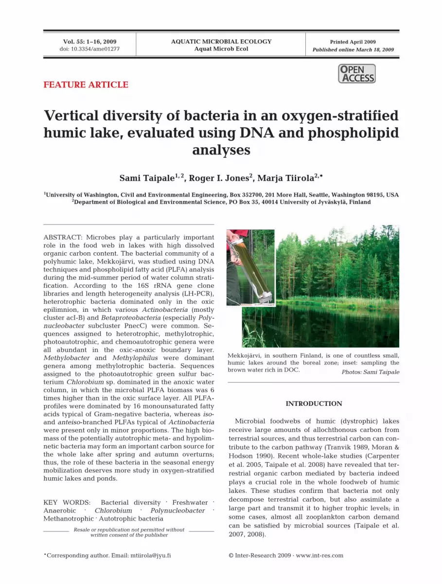
AQUATIC MICROBIAL ECOLOGYAquat Microb Ecol
Vol. 55: 1–16, 2009doi: 10.3354/ame01277
Printed April 2009
Published online March 18, 2009
INTRODUCTION
Microbial foodwebs of humic (dystrophic) lakesreceive large amounts of allochthonous carbon fromterrestrial sources, and thus terrestrial carbon can con-tribute to the carbon pathway (Tranvik 1989, Moran &Hodson 1990). Recent whole-lake studies (Carpenteret al. 2005, Taipale et al. 2008) have revealed that ter-restrial organic carbon mediated by bacteria indeedplays a crucial role in the whole foodweb of humiclakes. These studies confirm that bacteria not onlydecompose terrestrial carbon, but also assimilate alarge part and transmit it to higher trophic levels; insome cases, almost all zooplankton carbon demandcan be satisfied by microbial sources (Taipale et al.2007, 2008).
© Inter-Research 2009 · www.int-res.com*Corresponding author. Email: [email protected]
FEATURE ARTICLE
Vertical diversity of bacteria in an oxygen-stratifiedhumic lake, evaluated using DNA and phospholipid
analyses
Sami Taipale1, 2, Roger I. Jones2, Marja Tiirola2,*
1University of Washington, Civil and Environmental Engineering, Box 352700, 201 More Hall, Seattle, Washington 98195, USA2Department of Biological and Environmental Science, PO Box 35, 40014 University of Jyväskylä, Finland
ABSTRACT: Microbes play a particularly importantrole in the food web in lakes with high dissolvedorganic carbon content. The bacterial community of apolyhumic lake, Mekkojärvi, was studied using DNAtechniques and phospholipid fatty acid (PLFA) analysisduring the mid-summer period of water column strati-fication. According to the 16S rRNA gene clonelibraries and length heterogeneity analysis (LH-PCR),heterotrophic bacteria dominated only in the oxicepilimnion, in which various Actinobacteria (mostlycluster acI-B) and Betaproteobacteria (especially Poly-nucleobacter subcluster PnecC) were common. Se-quences assigned to heterotrophic, methylotrophic,photoautotrophic, and chemoautotrophic genera wereall abundant in the oxic-anoxic boundary layer.Methylobacter and Methylophilus were dominantgenera among methylotrophic bacteria. Sequencesassigned to the photoautotrophic green sulfur bac-terium Chlorobium sp. dominated in the anoxic watercolumn, in which the microbial PLFA biomass was 6times higher than in the oxic surface layer. All PLFA-profiles were dominated by 16 monounsaturated fattyacids typical of Gram-negative bacteria, whereas iso-and anteiso-branched PLFAs typical of Actinobacteriawere present only in minor proportions. The high bio-mass of the potentially autotrophic meta- and hypolim-netic bacteria may form an important carbon source forthe whole lake after spring and autumn overturns;thus, the role of these bacteria in the seasonal energymobilization deserves more study in oxygen-stratifiedhumic lakes and ponds.
KEY WORDS: Bacterial diversity . Freshwater .
Anaerobic . Chlorobium . Polynucleobacter .
Methanotrophic . Autotrophic bacteria
Resale or republication not permitted without written consent of the publisher
Mekkojärvi, in southern Finland, is one of countless small,humic lakes around the boreal zone; inset: sampling thebrown water rich in DOC. Photos: Sami Taipale
OPENPEN ACCESSCCESS
Aquat Microb Ecol 55: 1–16, 2009
Boreal humic lakes are usually steeply stratified insummer with respect to temperature and oxygen. Insheltered lakes and ponds, the anoxic hypolimnion canbe many times thicker than the oxic epilimnion, andthus the anoxic decomposition of dissolved organiccarbon (DOC), including allochthonous DOC, can havean important role in the carbon cycle. Methanogenesisby Archaea usually dominates anaerobic carbon min-eralization in anoxic freshwaters (Canfield et al. 2005),providing both a carbon and energy source formethane-oxidizing bacteria (MOB, methanotrophs) inthe water column (Sundh et al. 2005). In addition tomethane, hydrogen sulfide (H2S) and reduced iron(Fe2+) are formed under anoxic conditions. Thesereduced elements are oxidized by different microor-ganisms, like chemolithoautotrophic bacteria (e.g. Gal-lionella sp.) and photolithoautotrophic green sulfurbacteria (GSB, e.g. Chlorobium sp.; Heising et al. 1999,Hadas et al. 2001). The species diversity and ecologicalsignificance of responsible MOB and chemolithoau-totrophic bacteria is of special interest in humic lakes,where the strong light attenuation can severely restrictphotoautotrophic production. In view of this widerange of available substrates and environmental het-erogeneity in humic lakes, their bacterial communitiesmight be particularly diverse.
The main bacterial clusters specific for freshwaterecosystems have been revealed by 16S rRNA genesequencing (Glöckner et al. 2000, Zwart et al. 2002).Several studies have shown that the bacterial commu-nity composition (BCC) of surface waters of humiclakes is often dominated by Actinobacteria and partic-ularly by Betaproteobacteria (Glöckner et al. 2000,Burkert et al. 2003, Eiler et al. 2003, Grossart et al.2008, Percent et al. 2008), but the vertical diversity ofthe BCC in humic lakes with oxygen stratificationremains largely unstudied. Furthermore, 16S rRNAgene libraries cannot alone reveal the abundance ofdifferent bacterial groups, and thus 16S rRNA geneidentification must be related to quantitative informa-tion. Phospholipid fatty acid (PLFA) analysis has beenused for characterization and quantification of themicrobial community structure in various terrestrialand aquatic systems (White et al. 1979, Mancuso et al.1990). Because phospholipids are readily metabolizedand are thus relatively short-lived after the death of thecell, they are considered as quantitative biomarkers forliving organisms (White et al. 1979). However, whenusing PLFA profiles of field samples, microbial groupscan only be crudely separated, e.g. as Gram-negativebacteria, Gram-positive bacteria, and phytoplankton.By combining PLFA profiles with DNA analyses, amore comprehensive picture can be achieved, andthe biomasses of different taxonomic groups can beestimated.
The aim of this study was to investigate the verticaldistribution of bacteria in the water column of a humiclake with high DOC content by combining DNA andPLFA techniques. Four 16S rRNA gene libraries withaltogether over 400 sequences were analyzed fromthe oxic (epilimnion) and the anoxic (hypolimnion)water layers, as well as from the anoxic-oxic bound-ary layer (metalimnion) during summer stratification.Sequence data were combined with the informationon closest bacterial genera to predict potentialfunctions of bacteria operating in the different waterlayers, such as heterotrophy, photoautotrophy, chemo-autotrophy, and methylotrophy. Although this classi-fication is only tentative, it provides preliminaryinsights into the possible processes fuelling the car-bon cycle in an oxygen-stratified lake ecosystem.Sequence data were linked with length heterogeneityanalysis of PCR-amplified 16S rRNA genes (LH-PCR;Suzuki et al. 1998) and PLFA analysis to identify bio-markers that could be useful for characterization ofmicrobial communities in high DOC content lakes andto estimate the overall composition of bacterial bio-mass.
MATERIALS AND METHODS
Study site. Mekkojärvi (61° 13’ N, 25° 8’ E) is locatedin the Evo forest area in southern Finland. The lake issmall (area: 0.35 ha), shallow (mean depth: 3 m), andreceives a high loading of terrestrial carbon from thesurrounding catchment area. The lake water is darkbrown (color generally between 300 and 800 mg Pt l–1)and naturally acidic (pH generally from 4 to 6). Duringthe study period, the water color was 411 ± 50 and442 ± 24 mg Pt l–1 in the 0 to 0.6 and 0.6 to 3 m waterlayers, respectively, and pH varied between 5.8 and6.1 in the whole water column. All means are givenwith ± SD. Dissolved inorganic carbon concentrationvaried from 3.5 ± 0.8 to 14.0 ± 0.8 mg C l–1 in the 0 to0.6 and 0.6 to 3 m water layers, respectively. Moredetailed physical and chemical properties of the lakewere presented by Kuuppo-Leinikki & Salonen (1992).After ice-break, the surface water warms rapidly,resulting in steep temperature, oxygen, and nutrientgradients. Spring mixing is often incomplete, butautumn overturn happens regularly. Greater bacterialdensity has been measured in the oxic-anoxic bound-ary zone of the metalimnion (20 to 45 × 106 cells ml–1)than in the oxic epilimnion (2 to 7 × 10⁶ cells ml–1;Arvola et al. 1992). The high concentration of bacteri-ochlorophyll d in the hypolimnion reflects a high den-sity of photosynthethic bacteria, especially duringspring and summer at 1.8 to 3.0 m from where thehighest bacteriochlorophyll d concentration (>200 µg
2
Taipale et al.: Bacterial diversity in a humic lake
l–1) was measured (Taipale et al. 2008). Methane con-centration is highest at 1.8 to 3.0 m, where it is between100 and 150 µmol l–1 throughout the open water sea-son. Methane oxidation rate is highest at the depth of 0to 1.2 m throughout all seasons, although duringautumnal mixing the methane-oxidation rate is high inthe whole water column (Taipale et al. 2008).
Sampling procedure. Samples were collected forPLFA analysis and LH-PCR on 11 and 25 July and 1August, and for 16S rRNA gene sequencing andcloning on 1 August during summer stratification in2005. Water samples were taken from 4 different lay-ers — 0 to 0.6 m (oxic), 0.6 to 1.2 m (oxic-anoxicboundary layer), 1.2 to 1.8 m (anoxic), and 1.8 to3.0 m (anoxic) — using a Limnos tube sampler (height60 cm, volume 4.25 l). Water was filtered through a100 µm mesh net in the field, and in the laboratory1.05 to 3.35 l of lake water from each layer were fil-tered through Whatman GF/F filters for PLFA analy-sis. About 50 ml of lake water were filtered througha Filtropur acetylacetate filter unit (0.2 µm pore size;Sarstedt) for the DNA analyses. Water temperatureand oxygen concentration were measured in situwith a YSI 550A combined temperature and dis-solved oxygen meter (Yellow Springs Instruments)and redox potential with a WTW Multiline P3 pH/conductivity meter equipped with a Schott redox-combination electrode at 0.5 m intervals from surfaceto bottom. The accuracy was ±0.3 mg O2 l–1 foroxygen and ±0.3°C for temperature measurements.Ammonium, iron (total concentration by atomic ab-sorption spectrometry), and sulfate were analyzedfrom each 60 cm water layer on 25 July using thevalidated routine methods of the Finnish StandardAssociation (www.sfs.fi/en/). Methane samples (30ml) were collected in 60 ml polypropylene syringesdirectly from the Limnos tube sampler and analyzedas described by Taipale et al. (2007).
Cell counts, particulate organic carbon, and DOC.Prokaryotic cell numbers and volumes were deter-mined from acriflavine-stained (Bergström et al. 1986)samples using epifluorescence microscopy and Analy-SIS 3.1 Soft Imaging System (www.soft-imaging.net).The total bacterial cell volume was converted to car-bon using a factor of 0.36 pg C µm–3 (Tulonen 1993).Particulate organic carbon (POC) was measured fromsamples filtered on pre-ignited glass-fiber filters(Whatman GF/F) according to Salonen (1979), andDOC was measured from the filtrate using a Shimadzutotal organic carbon analyzer TOC–5000A.
DNA extraction. The DNA extraction procedure wasmodified from protocols described by Wilson (1990)and Griffiths et al. (2000). Briefly, acetylacetate filterswere opened and dissolved in 400 µl of phenol-chloro-form-isoamylalcohol (25:24:1), after which the samples
were homogenized with 0.4 g of 0.1 mm diameter glassbeads and 400 µl of TE buffer (10 mM Tris [pH 8.0],1 mM EDTA) in microcentrifuge tubes in a FastPrepbead-milling instrument (speed 5.5 for 30 s). The sam-ples were then re-extracted with chloroform-isoamyl-alcohol (24:1), precipitated with ethanol, and dissolvedin 100 µl of sterile water.
LH-PCR analysis and sequencing. 16S rRNA gene-specific PCR was performed as described by Tiirola etal. (2003), except that the polymerase was providedfrom Biotools and the reaction mixture included 1 mgml–1 bovine serum albumin. The broad-range bacter-ial PCR primers were forward primer 27F (5’-AGAGTT TGA TCM TGG CTC AG-3’; Lane 1991) andIRD700 labeled reverse primer PRUN518r (5’-ATTACC GCG GCT GCT GG-3’; Muyzer et al. 1993),which amplify naturally varying 465 to 565 bp longfragments of the 16S rRNA gene (Tiirola et al. 2003)corresponding the Escherichia coli region 8-534. LH-PCR gel electrophoresis was performed with an auto-mated LI-COR 4200 sequencer (LI-COR BioTech)using 6% Long Ranger denaturing polyacrylamidegel (FMC Bioproducts). The molecular size markerconsisted of LH-PCR products of known sequencelengths. The data were analyzed using QuantityOnesoftware (Bio-Rad Laboratories). Cloning of the same16S rRNA gene PCR products (without IRD-labeling)was performed using the TOPO TA Cloning Kit forSequencing (Invitrogen) according to the manufac-turer’s instructions. The sequencing of the clonelibrary was performed using the BigDye Terminatorv3.1 Cycle Sequencing Kit (Applied Biosystems) andvector primers T7 and T3. The samples were runin the ABI3100 capillary sequencing instrument(Applied Biosystems). Exact lengths (bp) of the insertswere calculated from the sequence data, and theinformation was used to link the phylogenetic classifi-cation of the sequences to the corresponding LH-PCRpeaks.
Sequence analysis. DNA sequences were editedusing Contig Express software (Invitrogen) and clus-tered to operational taxonomic units (OTUs) at 95(OTU0.95) or 99% (OTU0.99) sequence similarity levelusing the CD-HIT program (www.bioinformatics.org/cd-hit/). Sequences (~450 bp in length) were comparedto those in databases using BLAST software and theRibosomal Database Project II programs Seqmatch andClassifier (http://rdp.cme.msu.edu). ClustalX was usedfor sequence alignment and for constructing a neigh-bor-joining tree for selected Mekkojärvi clones repre-senting major phylotypes (OTU0.95 having more than2 similar clones). The alignment was corrected byhand-editing using the program Bioedit (www.mbio.ncsu.edu). Nucleotide sites with gaps in any of thesequences were excluded from the analysis, and the
3
Aquat Microb Ecol 55: 1–16, 2009
final tree was based on the analysis of 367 alignedpositions. In addition, actinobacterial sequences werecompared to freshwater clusters presented by Glöck-ner et al. (2000), Urbach et al. (2001), Zwart et al.(2002), and Warnecke et al. (2004). Betaproteobacterialsequences were compared to the clustering of Zwart etal. (2002), except Polynucleobacter sequences, whichwere compared in more detail to the 4 P. necessariusclusters presented by Hahn (2003). Chlorobi se-quences were compared to the sequences of the closesttype strains of the genera Chlorobium and Chlorobac-ulum (Imhoff 2003). Confidence values for the group-ings of the trees were derived by bootstrap analysiswith 100 iterations. The sequence data are depositedin the EMBL database under accession numbersAM949036 to AM949471.
Functional classification of bacterial sequences.Potential functions of bacterial groups in differentwater layers of Mekkojärvi were estimated using the16S rRNA gene sequence results. All bacterial se-quences were assigned to distinct functional groupsaccording to the primary carbon and energy source ofthe closest matching cultivated and classified organ-isms, following descriptions of distinct species byBalows et al. (1992). Bacteria were classified as photo-trophic or chemotrophic according to their energysource. All bacteria that use an organic carbon sourcewere considered heterotrophic. Methylotrophic bac-teria were separated from autotrophic bacteria to agroup including MOB and other bacteria using C-1compounds. Bacteria that have an anaerobic respira-tion pathway were classified as anaerobic bacteria.Sequences that did not have matches to cultivatedorganisms but matched sequences of the thus faruncultivated candidate phyla TM7 or OP11 were clas-sified as chemoheterotrophic.
Phospholipid analysis. Immediately after filtration,Whatman GF/F filters were placed in extraction tubescontaining 28.5 ml of chloroform–methanol–50 mMphosphate buffer, pH 7.4 (1:2:0.8 [vol:vol:vol]; Bligh &Dyer 1959). PLFAs were extracted, fractionated,saponified, and methylated using the protocol of Bligh& Dyer (1959) as modified by White et al. (1979),Keinänen et al. (2003), and Taipale et al. (2008).For quantification, dipentadecanoylphostatidylcholine(c15:0) was added as an internal standard. Lipidswere fractionated to neutral, glyco-, and phospho-lipids using 10, 20, and 10 ml of chloroform, acetone,and methanol, respectively. The phospholipid fractionwas evaporated to dryness under nitrogen flow. Inmild alkaline methanolysis, the internal standardstridecanoic and nonadecanoic acid methyl esters wereadded, and fatty acids were saponified, methylated,and extracted as methylesters. The phospholipid fattyacids were run by GC-HRMS (VG AutoSpec). The
modified timer program (Virtue et al. 1996) was usedto separate fatty acid methyl esters from each other.First, the column was kept at 50°C for 1 min, afterwhich the temperature was raised first by 30°C min–1
to 110°C, then by 1°C min–1 to 220°C, and finally by10°C min–1 to 300°C. The program time was then140 min. The temperature of the injector was 290°C,and the temperature of the source was 250°C. Thecolumn used was Rtx-5MS, which was 30 m long with0.25 mm ID 25 µm phase. The positions of doublebonds in monounsaturated phospholipid fatty acids(MUFAs) were determined by capillary GC-MS oftheir dimethyl disulfide (DMDS) adducts (Nichols etal. 1986).
PLFA biomarkers. Generally PLFAs were divided astypical for Gram-positive bacteria, for Gram-negativebacteria, and for phytoplankton. Iso- and anteiso-branched PLFAs, a14:0, i15:0, a15:0, i16:0, i17:0, a17:0,and i18:0, are typical for Gram-positive bacteria(O’Leary & Wilkinson 1988). According to the 16SrRNA gene sequencing, Tetrasphaera sp. and Micro-coccus luteus were closest to the dominant Actinobac-teria, and thus iso- and anteiso-branched PLFAs wereused as biomarkers for Actinobacteria in the oxic epil-imnion. To estimate the contribution of Actinobacteria,it was assumed that iso- and anteiso-branched PLFAscontributed 80% (Wieser et al. 2002, McKenzie et al.2006) of all PLFAs of Actinobacteria.
The 16 and 18 MUFAs, especially 16:1ω9c, 16:1ω7c,16:1ω5c, 18:1ω7, and 18:1ω5c, are common PLFAs inGram-negative bacteria. 18:1ω7 was used as a bio-marker for Proteobacteria (Ratledge & Wilkinson1988). Additionally, 16:1ω8c, 16:1ω6c, and 16:1ω5twere considered specific PLFAs for MOB type I (Bow-man et al. 1991). Correspondingly, 18:1ω8c PLFA hasbeen used as a specific biomarker for MOB type II(Guckert et al. 1991). To estimate the contribution ofMOB, it was assumed that 16:1ω8c, 16:1ω6c, and16:1ω5t PLFAs constituted 64% (Wartiainen et al.2006) of all MOB type I PLFAs. Br17:1, 10me16:0, and17:1 are PLFA biomarkers for the sulfate-reducinggenera Desulfovibrio, Desulfobacter, and Desulfobul-bus, respectively (Macalady et al. 2000).
The typical PLFAs for Chlorobium sp. are 14:0, 16:0,and 16:1, which constitute 90% of all of their PLFAs(Kenyon 1972, Imhoff 2003). According to 16S rRNAgene sequencing, GSB belonged to Chlorobium sp.group 3a and thus it was assumed that 14:0 PLFAs con-stituted 16% (Imhoff 2003) of all GSB PLFAs. Whenestimating the biomass of Chlorobium sp., it wasassumed that there are 2 fatty acids per 1 phosphate(P) molecule and 200 µg PLFA per mg organic carbon;for Actinobacteria and Methylobacter sp., we assumed2 fatty acids per 1 P molecule and 380 µg PLFA per mgorganic carbon (Findlay 1996).
4

Taipale et al.: Bacterial diversity in a humic lake
RESULTS
Physical and chemical parameters
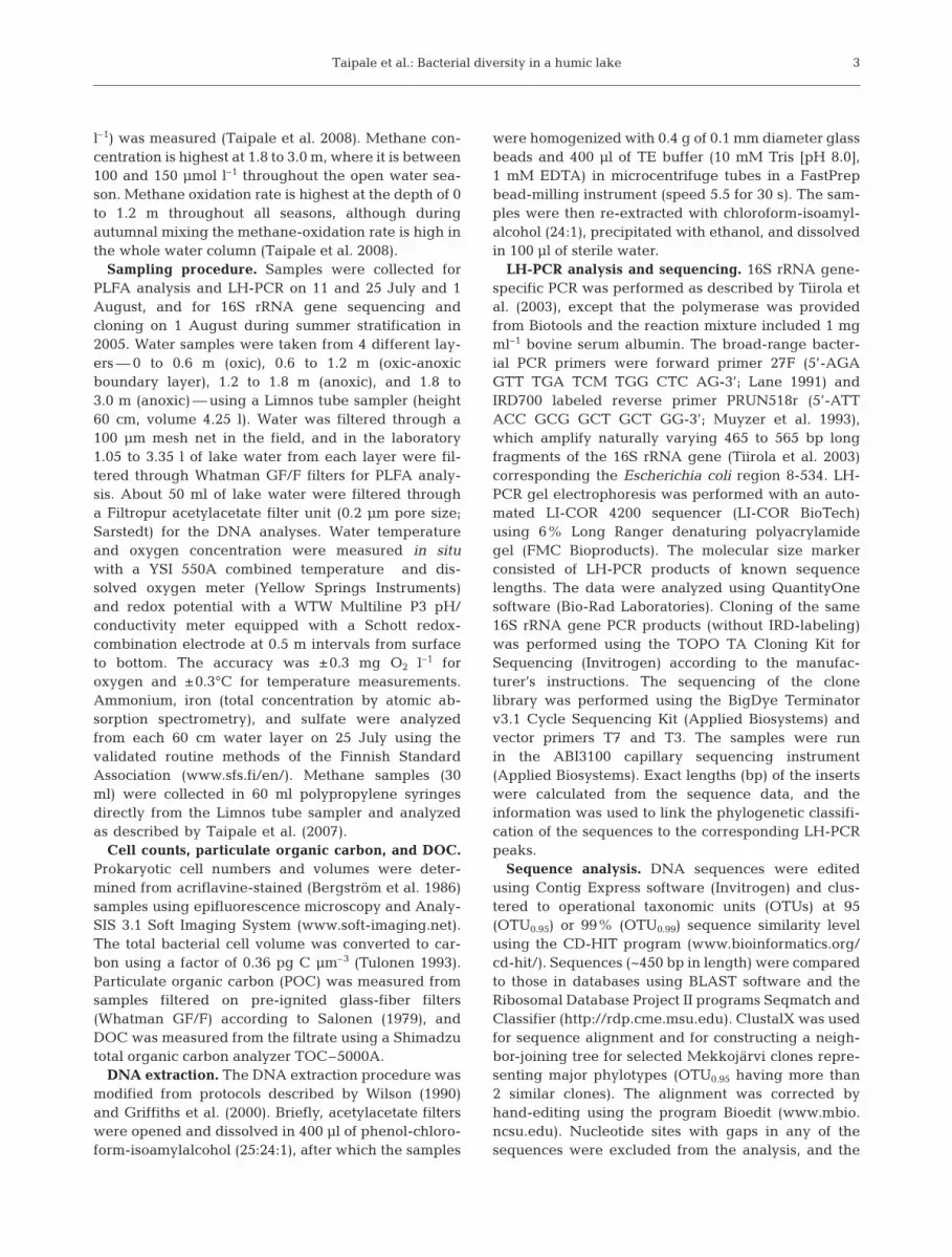
During summer stratification, only the uppermostmeter of the epilimnion contained measurable oxygen,and, according to the decrease in redox potentials, theanoxic-oxic boundary layer was between 1.2 and 1.8 m(Fig. 1). Ammonium, iron, and methane concentrationsincreased down the water column, whereas sulfateconcentration decreased slightly (Table 1). Methaneconcentrations were stable in the anoxic layers butdecreased rapidly towards the surface (Table 1).
Bacterial community composition
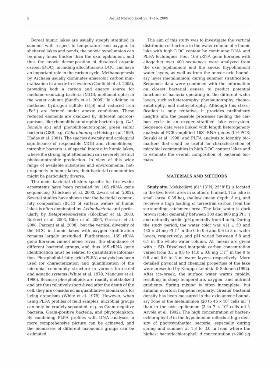
Altogether, 435 clones of the 16S rRNA gene weresequenced from the different water layers. When allclones were pooled, most clones were assigned to thephyla Chlorobi (149 clones, 34%) and Proteobacteria(142 clones, 33%), of which most were Betaproteobac-teria (103 clones, 23%), Actinobacteria (43 clones,10%), Planctomycetes (35 clones, 8%), and Bac-teroidetes (18 clones, 4%; Fig. 2). Other phyla detectedin minor proportions were TM7 (10 clones), Firmicutes(3 clones), Verrucomicrobia (3 clones), and Chloroflexi(3 clones). In addition, 1 or 2 clones were obtained fromAcidobacteria, Chlamydia, Cyanobacteria, and OP11,and the rest were not classified. The number of differ-ent OTUs of sequences that shared ≥95% sequencesimilarity (OTU0.95; Table 2) was equally high (42 to 49)in all water layers.
On the basis of 16S rRNA gene sequencing, therelative abundance of dominant bacterial groups(Fig. 3A) varied vertically in the oxic and anoxic watercolumns. At the oxygenic surface (0 to 0.6 m),sequences assigned to Actinobacteria (29%) and Pro-
teobacteria (43% of the 16S rRNA gene clones)including the class Betaproteobacteria (30%) werehighly abundant, but the number of actinobacterialsequences decreased sharply together with the de-creasing oxygen concentration. Both type I and IIMOB were found at 0 to 0.6 m. MOB type I sequencesmatched most closely with the type strains of Methy-lobacter psychrophilus (98.3% similarity) and M. tun-dripaludum (98.9% similarity), and the 2 obtainedMOB type II sequences matched best with Methylo-cella palustris (97%). In addition to MOB, a sequencematching the methylotrophic Methylophilus sp. (95 to96% similarity) was also found at 0 to 0.6 m, but wasespecially abundant (4% of all sequences) at 0.6 to1.2 m, where Chlorobi (39% of sequences) and Beta-proteobacteria (31% of sequences) were the mostabundant groups. Chlorobi was also the dominantmicrobial phylum at 1.2 to 1.8 m (47%) and at 1.8 to3.0 m (43%). Methylobacter luteus was the only MOBtype I sequence found at 0.6 to 1.2 m. Gallionella fer-ruginea (98.7% similarity with the type species) was afrequent species in the oxic layer at 0 to 0.6 m. G. fer-ruginea and Rhodoferax sp. were the most frequentBetaproteobacteria at 1.2 to 1.8 m. Planctomycetesand the thus far uncultivated phylum TM7 were alsoabundant bacterial phyla at 1.2 to 1.8 m. Sequences ofDeltaproteobacteria were abundant at 1.8 to 3.0 mclose to the bottom, where the Fe(III)-reducing bac-terium Geobacter psychrophilus (7% of all sequencesfrom the hypolimnion) was the closest cultivatedorganism (with 94 to 99.75% sequence identity) ofmost of the sequences except 1 sequence that wasclosest to the sulfate-reducing bacterium STO23.However, at other depths, single deltaproteobacterialsequences were obtained that were affiliated toPelobacter propionicus, Desulfocapsa sp. Cad626,Desulforomonadales sp. TC37, and to the unidentifiedbacterium ROMEm4sh208, which are all chemolitho-trophic bacteria that gain their sole electron acceptorsfrom sulfur or iron compounds.
5
Fig. 1. Temperature, oxygen, and redox potential during summer stratification of Mekkojärvi on 1 August 2005
Depth NH4-N Fe SO4 CH4
(m) (µg l–1) (mg l–1) (mg l–1) (µmol l–1)
0–0.6 33 1.0 8.0 11.30.6–1.2 25 1.9 8.9 84.21.2–1.8 85 2.7 8.4 138.41.8–2.4 250 2.9 7.8 138.32.4–3.0 330 2.9 6.6 135.13.0–3.6 940 4.7 3.9 nd
Table 1. Ammonium-nitrogen (NH4-N), iron (Fe), sulfate(SO4), and methane (CH4) concentrations in successive watercolumn layers of Mekkojärvi on 25 July 2005. nd: not
determined

Aquat Microb Ecol 55: 1–16, 20096
Fig. 2. Phylogenetic relationships of abundant Mekkojärvi phylotypes and their closest relatives based on partial 16S rRNA genesequences. The phylogenetic tree was constructed from 367 aligned nucleotide positions using the neighbor-joining method withKimura correction for multiple substitutions. A representative of each major OTU0.95 group with 3 or more clones has beenselected for the figure. The number of clones in each group is indicated after the clone name. The LH-PCR length (bp) of relatedsequences is indicated after the organism name or EMBL accession number. The clones from epi-, upper meta-, lower meta-, andhypolimnion are indicated by the following prefixes: MekkoE, MekkoM1, MekkoM2, and MekkoH. Nodes supported bybootstrap values between 70 to 90% and 90 to 100% are shown by gray and black circles, respectively. The scale bar indicates
0.1 changes per nucleotide position
Taipale et al.: Bacterial diversity in a humic lake
Diversity of sequences of the phylum Chlorobi
More detailed comparative phylogenetic clusteringwas done for the sequences belonging to the mostabundant bacterial phyla Chlorobi and Actinobacteriaand the class Betaproteobacteria. All 16S rRNA genesequences of the phylum Chlorobi were assigned toChlorobium group 3a (Alexander et al. 2002) sharing96 to 99% similarity to the sequences of the typestrains of Chlorobium phaeobacteroides, C. clathrati-forme, and C. ferrooxidans. In the neighbor-joininganalysis (Appendix 1A, available as AME Supple-mentary Material at www.int-res.com/articles/suppl/a055p001_app.pdf), all 149 Chlorobi sequences clus-tered with the aforementioned Chlorobium typestrains and not with the Chlorobaculum type strains(Imhoff et al. 2003). Of the Chlorobi sequences, 77%clustered with C. phaeobacteroides, 16% with C. fer-rooxidans, and 8% with C. clathratiforme, but the
branching of the tree was not supported by high boot-strapping values. The sequences clustering with C. fer-rooxidans were obtained from the hypolimnion orlower metalimnion, but C. phaeobacteroides and C.clathratiforme sequences were obtained from differentwater depths including the epilimnion, showing novertical specialization.
Diversity of sequences of the classBetaproteobacteria
Betaproteobacteria was the largest class in the phy-lum Proteobacteria in Mekkojärvi. The detailed phylo-genetic analysis (Appendix 1B,C) showed that thePolynucleobacter necessarius cluster (Zwart et al. 2002)was very common in the oxic epilimnion of Mekkojärvi,and also in lower water phases (0.6 to 3.0 m), showingadaptation to microaerobic and anaerobic conditions.All P. necessarius cluster sequences were inside the P.necessarius subcluster PnecC (Hahn 2003), except asingle sequence of the subcluster PnecA. These identi-cal or nearly identical sequences formed a coherentgroup with, e.g. isolate MWH-MekkB1 previously iso-lated from Mekkojärvi (Vannini et al. 2007) and cloneFuku35 obtained from the clone library of the acidifiedbog lake Grosse Fuchskuhle (Glöckner et al. 2000).Most other betaproteobacterial sequences were outsidethe other typical freshwater clusters Rhodoferax sp.BAL47, GKS16, GKS 98, Ralstonia pickettii, and LD28.Although not close to Rhodoferax sp. BAL47 or GKS16,
a large number of different Rhodoferax-affiliated but still not closely related phy-lotypes were detected. A diverse clusterof Methylophilus-affiliated sequenceswas observed in the metalimnetic oxic-anoxic boundary layer. All sequencesthat were classified to Nitrosomonadaleswere related to Gallionella (family Gal-lionellaceae), thus showing a lack ofbetaproteobacterial ammonia oxidizers ofthe family Nitrosomonadaceae. Sequen-ces belonging to known ammonia-oxidizing gammaproteobacterial bacteria(genus Nitrosococcus) were not detectedeither, but most of the gammaproteo-bacterial sequences (12/18) were derivedfrom MOB (family Methylococcaceae).
Diversity of sequences of the phylumActinobacteria
The similarity value of most actinobac-terial sequences and sequences of clos-
7
Layer Depth (m) Clones OTU0.95 OTU0.99
Epilimnion 0–0.6 92 47 69Metalimnion 1 0.6–1.2 124 49 68Metalimnion 2 1.2–1.8 113 42 56Hypolimnion 1.8–3.0 106 49 72Total 435 132 225
Table 2. Number of different 16S rRNA gene clones sharingless than 95% and 99% sequence similarity in 4 depth zones
of Mekkojärvi. OTU: operational taxonomic unit
Fig. 3. Percentage contribution of 16S rRNA gene sequences in the water col-umn of Mekkojärvi assigned to (A) major bacterial phyla and (B) different bac-terial functional groups: heterotrophic bacteria (HB), photoheterotrophicbacteria (PHB), methylotrophic bacteria including methanotrophs and otherC1-compound-utilizing bacteria (MB), photoautotrophic bacteria (PAB),
chemoautotrophic bacteria (CAB), and other anaerobic bacteria

Aquat Microb Ecol 55: 1–16, 2009
est cultivated organisms ranged from 94 to 96%. Thesequences could be grouped into 4 coherent clusters,which were clearly separated by OTU0.95 grouping(Fig. 2) as well as in the more detailed trees (Appen-dix 1D, E). The largest groups of actinobacterialsequences were obtained from the typical freshwaterbacteria group hgcI clustered by Glöckner et al.(2000), of which most (22) sequences (represented byMekkoE-21 and MekkoE69) clustered with subgroupSTA2-30, designated by Zwart et al. (2002), and only 3sequences (represented by Mekko M1-15 in Fig. 2)clustered with the other hgcI-group ACK-M1 (Zwart etal. 2002). In the other tree with the sequences collectedand clustered by Warnecke et al. (2004) (Appen-dix 1E), all of these sequences were affiliated into thelineage acI (acI-B and acI-A, repectively). Six closelyrelated actinobacterial sequences obtained from theoxic 0 to 0.6 m (represented by OTU MekkoE-89)matched best with the unidentified actinobacterial iso-late MWH-Ta8 and clones belonging to cluster acII(Warnecke et al. (2004). Another small group of se-quences clustered with actinobacterial isolate MC19 aswell as Grosse Fuchskuhle clone FukuS94 (accessionnumber AJ290019; Glöckner et al. 2000). The assigna-tion of this group of sequences to specific freshwaterclusters earlier described for Actinobacteria remainsindistinct, but was most closely related to clusteracIV by Warnecke et al. (2004), which correspondsto the group C111 after the classification by Urbachet al. (2001) including subgroups MED0-06, CL500-29,or URK0-14 clustered by Zwart et al. (2002; Appen-dix 1D,E).
Functional affiliation of the Mekkojärvi 16S rRNAgene sequences
Tentative functions of bacteria behind the 16S rRNAsequences were predicted using information of closestmatching classified organisms. In candidate phyla withno cultivated representatives and in cases when thesequences could not be classified, the sequences wereconsidered heterotrophic. According to the 16S rRNAgene library data, sequences affiliated with heterotro-phic bacteria (HB) dominated at 0 to 0.6 m (68% of allbacterial sequences in the epilimnion), but contributedonly 16 to 27% of all bacterial sequences at 0.6 to 3.0 m(Fig. 3B). Methylotrophic bacteria (MB), includingsequences assigned to Methylophilus, Methylobacter,and Methylocapsa, were more abundant at 0 to 0.6 mand 0.6 to 1.2 m (11%) than at 1.2 to 1.8 m (4%) or at1.8 to 3.0 m (7%). Sequences assigned to chemoau-totrophic bacteria were equally dispersed (14 to 17%)in the water column over 0.6 to 3.0 m, in which layersof photoautotrophic bacteria (PAB) formed the most
8
Alphaproteobacteria
Epsilonproteobacteria
Chloroflexi and Deltaproteobacteria
Bacteroidetes, Verru
comicrobium, etc.
Actinobacteria
Chlorobi
Betaproteobacteria
Deltaproteobacteria
Planctomycetes
Delta- and Betaproteobacteria
Bacteroidetes
047
047
247
547
748
749
549
850
050
250
450
651
451
652
252
553
653
955
051
1 to
512
518
to51
9
20406080
Percentage of all fragments
0 to
0.6
m0.
6 to
1.2
m1.
2 to
1.8
m1.
8 to
3.0
m
Fig
. 4.M
ean
(±S
D) m
agn
itu
des
(%) o
f LH
-PC
R fr
agm
ents
in th
e ep
ilim
nio
n (0
to 0
.6 m
), u
pp
er m
etal
imn
ion
(0.6
to 1
.2 m
), lo
wer
met
alim
nio
n (1
.2 to
1.8
m),
an
d h
ypol
imn
ion
(1.8
to
3.0
m)
of M
ekk
ojär
vi i
n m
id-s
um
mer
sam
pli
ng
s. F
rag
men
t si
ze c
lass
es (
nu
mb
er o
f b
ase
pai
rs)
are
show
n o
n t
he
x-ax
is a
nd
th
e p
hyl
ogen
etic
aff
ilia
tion
s of
th
ese
are
ind
icat
ed, b
ased
wh
ere
pos
sib
le o
n t
he
info
rmat
ion
from
the
16S
rR
NA
gen
e cl
one
lib
rari
es. H
oriz
onta
l lin
es in
dic
ate
dif
fere
nt f
rag
men
t siz
e cl
asse
s th
at a
re a
ffil
iate
d to
the
sam
e p
hyl
ogen
etic
gro
up
; wh
ere
gro
up
con
trib
uti
ons
are
un
clea
r, t
his
is s
how
n b
y a
das
hed
lin
e or
‘etc
’
Taipale et al.: Bacterial diversity in a humic lake
abundant (38 to 45%) functional bacterial group towhich our sequences were assigned. Anaerobic meta-bolism was naturally more common in the deeper partsof the water column, and sequences of such types con-tributed 8% and 12% of all sequences at 1.2 to 1.8 mand 1.8 to 3.0 m, respectively.
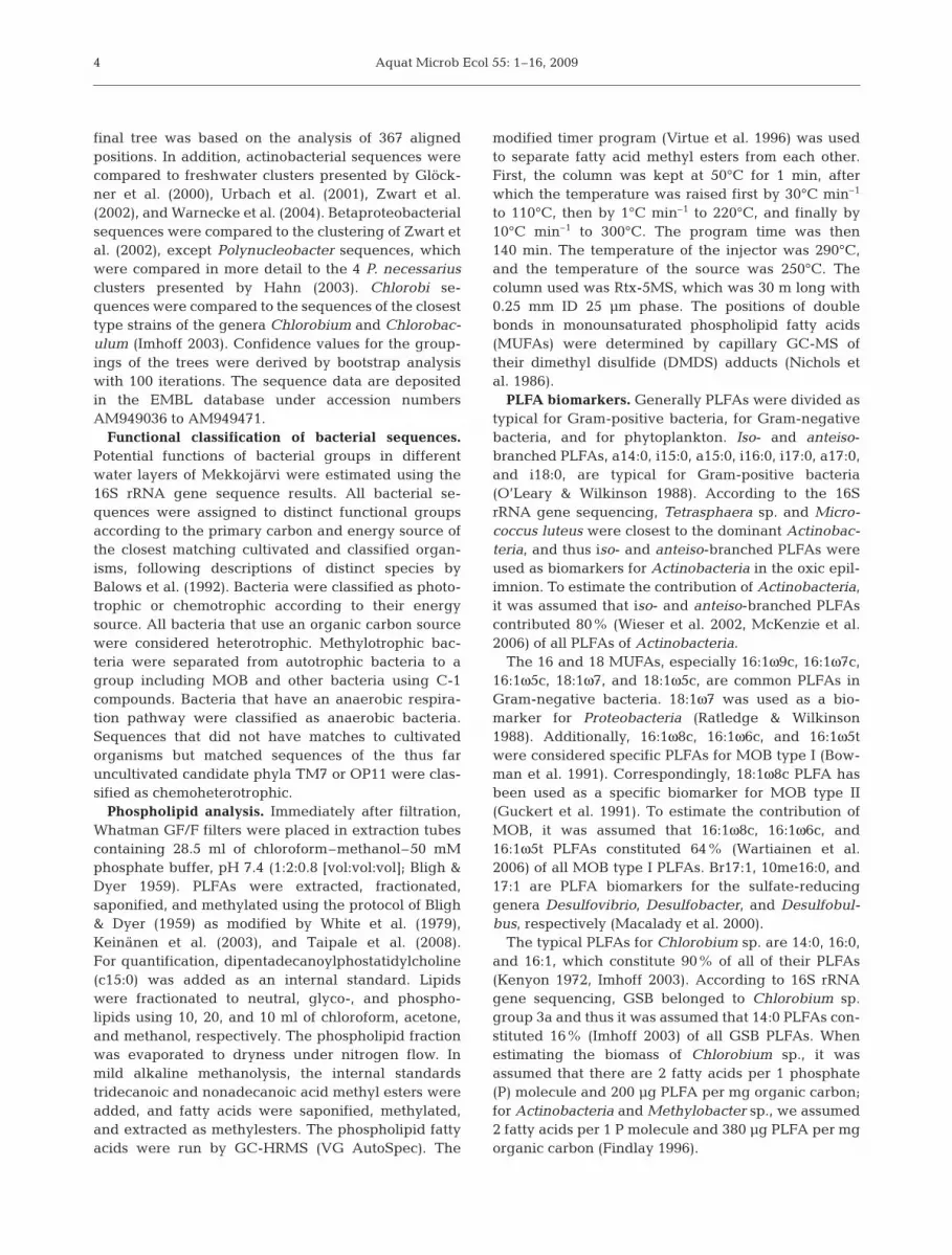
Linking LH-PCR profiles and 16S rRNA genesequences
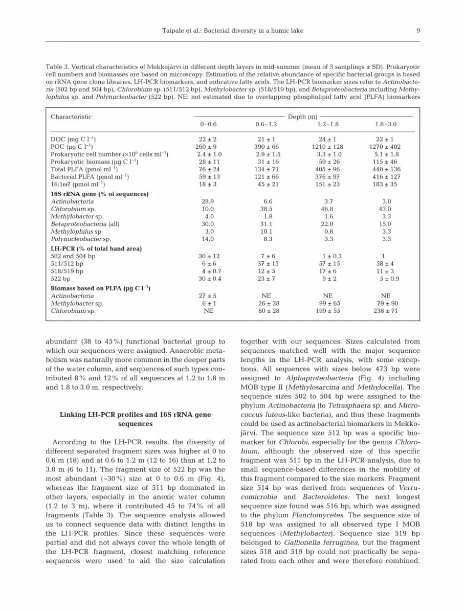
According to the LH-PCR results, the diversity ofdifferent separated fragment sizes was higher at 0 to0.6 m (18) and at 0.6 to 1.2 m (12 to 16) than at 1.2 to3.0 m (6 to 11). The fragment size of 522 bp was themost abundant (~30%) size at 0 to 0.6 m (Fig. 4),whereas the fragment size of 511 bp dominated inother layers, especially in the anoxic water column(1.2 to 3 m), where it contributed 45 to 74% of allfragments (Table 3). The sequence analysis allowedus to connect sequence data with distinct lengths inthe LH-PCR profiles. Since these sequences werepartial and did not always cover the whole length ofthe LH-PCR fragment, closest matching referencesequences were used to aid the size calculation
together with our sequences. Sizes calculated fromsequences matched well with the major sequencelengths in the LH-PCR analysis, with some excep-tions. All sequences with sizes below 473 bp wereassigned to Alphaproteobacteria (Fig. 4) includingMOB type II (Methylosarcina and Methylocella). Thesequence sizes 502 to 504 bp were assigned to thephylum Actinobacteria (to Tetrasphaera sp. and Micro-coccus luteus-like bacteria), and thus these fragmentscould be used as actinobacterial biomarkers in Mekko-järvi. The sequence size 512 bp was a specific bio-marker for Chlorobi, especially for the genus Chloro-bium, although the observed size of this specificfragment was 511 bp in the LH-PCR analysis, due tosmall sequence-based differences in the mobility ofthis fragment compared to the size markers. Fragmentsize 514 bp was derived from sequences of Verru-comicrobia and Bacteroidetes. The next longestsequence size found was 516 bp, which was assignedto the phylum Planctomycetes. The sequence size of518 bp was assigned to all observed type I MOBsequences (Methylobacter). Sequence size 519 bpbelonged to Gallionella ferruginea, but the fragmentsizes 518 and 519 bp could not practically be sepa-rated from each other and were therefore combined.
9
Characteristic Depth (m)0–0.6 0.6–1.2 1.2–1.8 1.8–3.0
DOC (mg C l–1) 22 ± 2 21 ± 1 24 ± 1 22 ± 1POC (µg C l–1) 260 ± 9 390 ± 66 1210 ± 128 1270 ± 402Prokaryotic cell number (×106 cells ml–1) 2.4 ± 1.0 2.9 ± 1.5 3.3 ± 1.0 5.1 ± 1.8Prokaryotic biomass (µg C l–1) 28 ± 11 31 ± 16 59 ± 26 115 ± 46Total PLFA (pmol ml–1) 76 ± 24 134 ± 71 405 ± 96 440 ± 136Bacterial PLFA (pmol ml–1) 59 ± 13 121 ± 66 376 ± 97 416 ± 12716:1ω7 (pmol ml–1) 18 ± 3 45 ± 21 151 ± 23 183 ± 35
16S rRNA gene (% of sequences)Actinobacteria 28.9 6.6 3.7 3.0Chlorobium sp. 10.0 38.5 46.8 43.0Methylobacter sp. 4.0 1.8 1.6 3.3Betaproteobacteria (all) 30.0 31.1 22.0 15.0Methylophilus sp. 3.0 10.1 0.8 3.3Polynucleobacter sp. 14.0 8.3 3.3 3.3
LH-PCR (% of total band area)502 and 504 bp 30 ± 12 7 ± 6 1 ± 0.3 1511/512 bp 6 ± 6 37 ± 15 57 ± 15 58 ± 4518/519 bp 4 ± 0.7 12 ± 5 17 ± 6 11 ± 3522 bp 30 ± 0.4 23 ± 7 9 ± 2 5 ± 0.9
Biomass based on PLFA (µg C l–1) Actinobacteria 27 ± 5 NE NE NEMethylobacter sp. 6 ± 1 26 ± 28 99 ± 65 79 ± 90Chlorobium sp. NE 80 ± 28 199 ± 55 238 ± 71
Table 3. Vertical characteristics of Mekkojärvi in different depth layers in mid-summer (mean of 3 samplings ± SD). Prokaryoticcell numbers and biomasses are based on microscopy. Estimation of the relative abundance of specific bacterial groups is basedon rRNA gene clone libraries, LH-PCR biomarkers, and indicative fatty acids. The LH-PCR biomarker sizes refer to Actinobacte-ria (502 bp and 504 bp), Chlorobium sp. (511/512 bp), Methylobacter sp. (518/519 bp), and Betaproteobacteria including Methy-lophilus sp. and Polynucleobacter (522 bp). NE: not estimated due to overlapping phospholipid fatty acid (PLFA) biomarkers
Aquat Microb Ecol 55: 1–16, 2009
The sequence size 522 bp was assigned to many gen-era among Betaproteobacteria, e.g. Polynucleobactersp., Methylophilus sp. (also size 536 bp), and Rhodo-ferax sp., and thus this fragment cannot be used as aspecific biomarker of any genus but only as a bio-marker of the Betaproteobacteria. Deltaproteobacter-ial sequences closely resembling Geobacter psychro-philusT were assigned to the LH-PCR length 539 bp.According to 16S rRNA gene sequencing of the 4Mekkojärvi clone libraries, the fragment size 567 bpwould have matched sequences classified to candi-date phyla TM7 and OP11 (but only with low identi-ties of <90%); however, such sequence size wasexcluded from the LH-PCR data analysis, since thesize exceeded the expected bacterial partial 16SrRNA sequences size (465 to 565 bp) using primers27F and PRUN518, based on a previous analysis ofcultivated organisms (Tiirola et al. 2003).
Contribution of bacterial and phytoplankton PLFAs
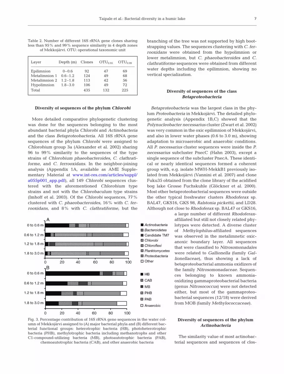
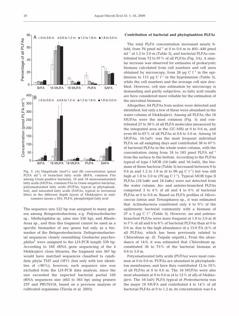
The total PLFA concentration increased nearly 6-fold, from 76 pmol ml–1 at 0 to 0.6 m to 405–440 pmolml–1 at 1.2 to 3.0 m (Table 3), and bacterial PLFAs con-tributed from 72 to 97% of all PLFAs (Fig. 5A). A simi-lar increase was observed for estimates of prokaryoticbiomass calculated from cell numbers and cell sizesobtained by microscopy, from 28 µg C l–1 in the epi-limnion to 115 µg C l–1 in the hypolimnion (Table 3),while the cell numbers and the average cell size dou-bled. However, cell size estimation by microscopy isdemanding and partly subjective, so fatty acid resultsare here considered more reliable for the estimation ofthe microbial biomass.
Altogether, 64 PLFAs from seston were detected andidentified, but only a few of these were abundant in thewater column of Mekkojärvi. Among all PLFAs, the 16MUFAs were the most common (Fig. 5) and con-tributed 27 to 38% of all PLFA molecules (measured bythe integrated area in the GC-MS) at 0 to 0.6 m, andeven 60 to 67% of all PLFAs at 0.6 to 3.0 m. Among 16MUFAs, 16:1ω7c was the most frequent individualPLFA on all sampling days and contributed 38 to 67%of bacterial PLFAs in the whole water column, with theconcentration rising from 18 to 183 pmol PLFA ml–1
from the surface to the bottom. According to the PLFAstypical of type I MOB (16:1ω8c and 16:1ω5t), the bio-mass of these bacteria (Table 3) increased between 0 to0.6 m and 1.2 to 1.8 m (6 to 99 µg C l–1) but was stillhigh at 1.8 to 3.0 m (79 µg C l–1). Typical MOB type IIPLFAs (18:1ω8c and 18:1ω6c) were not detected fromthe water column. Iso- and anteiso-branched PLFAscomprised 2 to 4% of all and 4 to 6% of bacterialPLFAs at 0 to 0.6 m. Based on PLFA profiles of Micro-coccus luteus and Tetrasphaera sp., it was estimatedthat Actinobacteria constituted only 4 to 9% of theepilimnetic bacterial community with a biomass of27 ± 5 µg C l–1 (Table 3). However, iso and anteiso-branched PLFAs were more frequent at 1.8 to 3.0 m (6to 7% of all and 6 to 8% of bacterial PLFAs) than at 0 to0.6 m, due to the high abundance of a 15:0 FA (4% ofall PLFAs), which has been previously related toChlorobium sp. (S. Taipale unpubl.). From the abun-dance of 14:0, it was estimated that Chlorobium sp.constituted 36 to 73% of the bacterial biomass at0.6 to 3.0 m.
Polyunsaturated fatty acids (PUFAs) were most com-mon at 0 to 0.6 m. PUFAs are abundant in phytoplank-ton membranes, and here they contributed 12 to 19%of all PLFAs at 0 to 0.6 m. The 18 MUFAs were alsomost abundant at 0 to 0.6 m (4 to 12% of all) of Mekko-järvi. The 18:1ω7c PLFA typical of Proteobacteria wasthe major 18 MUFA and contributed 4 to 14% of allbacterial PLFAs at 0 to 1.2 m; its concentration was 6 ±
10
0
20
40
60
80
Per
cen
tag
e o
f al
l PL
FA
s 0 to 0.6 m 0.6 to 1.2 m 1.2 to 1.8 m 1.8 to 3.0 m
0BrFA 16 MUFA 18 MUFA PUFA SAFA
BrFA 16 MUFA 18 MUFA PUFA SAFA
50
100
150
200
250
300
350
400
Co
nc
en
tra
tio
n (
pm
ol P
LF
A m
l–1) 0 to 0.6 m 0.6 to 1.2 m 1.2 to 1.8 m 1.8 to 3.0 m
B
A
Fig. 5. (A) Magnitude (mol%) and (B) concentration (pmolPLFA ml–1) of branched fatty acids (BrFA, common FAsamong Gram-positive bacteria), 16 and 18 monounsaturatedfatty acids (MUFAs, common FAs in Gram-negative bacteria),polyunsaturated fatty acids (PUFAs, typical in phytoplank-ton), and saturated fatty acids (SAFAs, typical in terrestriallitter) in the different depth layers of Mekkojärvi in mid-
summer (mean ± SD). PLFA: phospholipid fatty acid
Taipale et al.: Bacterial diversity in a humic lake
3 pmol PLFA ml–1 at 0 to 1.2 m and 7 ± 5 pmol PLFAml–1 at 1.2 to 3.0 m. A minor amount of 18:1ω9c, atypical phytoplankton PLFA, was also detected.
The contribution of saturated fatty acids (SAFAs)was highest at 0 to 0.6 m (29 to 37%), but contributedless than 27% of all PLFAs at 0.6 to 3.0 m. The mostabundant SAFA was palmitic acid (16:0, 8 to 17%),which is a frequent PLFA in bacteria and phytoplank-ton and thus not specific for any group. Additionally,14:0 FA had the highest contribution in both the oxiceplilimnion (9 to 17% of all) and the anoxichypolimnion (6 to 7% of all). Other SAFAs contributedless than 3.5% of all PLFAs.
PLFAs typical of sulfate-reducing bacteria were alsodetected. A minor contribution (<1% of all) of Desul-fobacteria-typical 10Me16:0 was detected at alldepths, but especially in the anoxic water column.Traces of br17:1 and 17:1 PLFAs were detected andthus possibly reflected the presence of Desulfovibrioand Desulfobulbus species in the anoxic water column(1.2 to 3.0 m). Minor contributions (<2% of all PLFAs)of 15 MUFAs, 15:1ω6 and 15:1ω7, were also detected,especially in the anoxic water column.
DISCUSSION
In this study, Chlorobi, Betaproteobacteria, and Acti-nobacteria were the major contributors to the microbialcommunity of an oxygen-stratified polyhumic lake,according to 16S rRNA gene sequencing, LH-PCR, andPLFA analysis. The relative number of Actinobacteriadecreased and Chlorobi increased in the anoxic waterlayers. Affiliation of the 16S rRNA gene sequences totheir closest cultivated neighbor organisms providedentry to the possible traits of bacteria, and showed thatbacteria assigned to classical heterotrophic generadominated only in the oxic epilimnion of Mekkojärvi.However, it is important to remember that small differ-ences at the 16S rRNA level may hide considerable dif-ferences at the levels of genome and phenotype.Another consideration is that the diversity of se-quences in clone libraries may not quantitativelyreflect the diversity of the sequence types present inthe original sample, due to limited size of the clonelibraries and well-known sources for biases generatedin the DNA extraction process, PCR amplification, andcloning steps (for a review see von Wintzingerode et al.1997). In this study, the data from gene libraries werestrengthened by the length heterogeneity analysis ofthe PCR-amplified 16S rRNA gene (LH-PCR; Suzuki etal. 1998). This provided a more accurate tool for theanalysis of the relative abundance of distinct bacterialgroups, as sequencing covers usually only hundreds ofmolecules, whereas profiling covers the major diver-
sity of millions of molecules. According to our LH-PCRand 16S rRNA gene sequencing results, many of theLH-PCR fragment sizes were specific biomarkers forsome distinct bacterial groups in this lake. All frag-ments less than 473 bp in length belonged to Alpha-proteobacteria (including MOB type II); 502 to 504 bpfragments belonged to Actinobacteria, and those of511/512 bp to Chlorobium sp. In addition to these bio-markers, fragments of 518/519 bp belonged to Methy-lobacter or Gallionella genera and fragments of 522 bpto various Betaproteobacteria, especially the generaPolynucleobacter and Methylophilus. This kind of bio-marker technique can be useful when studying sea-sonal changes in the microbial communities in humiclakes.
Habitat-specific clustering has shown that most bac-teria inhabiting freshwater systems are indigenous tofreshwater and distinct from soil or other ecosystems,and ‘typical freshwater bacteria’ included 34 phyloge-netic clusters that appeared to exhibit a global distrib-ution (Zwart et al. 2002). In this study, HB consistedmainly of Actinobacteria and Betaproteobacteria,which is consistent with earlier results from freshwaters (Glöckner et al. 2000, Zwart et al. 2002, van derGucht et al. 2005), including humic lakes (Burkert et al.2003, Grossart et al. 2008). Actinobacteria were typicalbacteria of the oxic surface water in Mekkojärvi; theirclone sequences and specific LH-PCR fragments con-tributed 29% and 21 to 43% (variation from the 3 sam-pling times) of all epilimnion sequences, respectively,but their relative contribution in the 16S rRNA genepool decreased rapidly in the anoxic water layers, inwhich the total bacterial PLFA biomass was 6 timeshigher than in the epilimnion. Most actinobacterialsequences were not close to cultivated organisms butclustered with the sequences of the cluster acI-B (War-necke et al. 2004), also known as hgcI subclusterSTA2-30 (Zwart et al. 2002). Among Betaproteobacte-ria, Polynucleobacter sp. was the most common genus(11% of all 16S rRNA gene sequences in the epil-imnion) inhabiting especially the oxic epilimnion, butsome sequences were also obtained from the anoxiclayers, although fewer in relative numbers. Polynucle-obacter sequences of Mekkojärvi belonged to the sub-cluster PnecC (Hahn 2003), which seems to be a groupcharacteristic of humic and acidic environments(Grossart et al. 2008). Isolates of Polynucleobacter nec-essarius have previously been obtained from Mekko-järvi and from lakes, ponds, and rivers in centralEurope, China, and East Africa by the filtration-acclimatization method separating these extremelysmall bacteria using 0.2 µm pore size filters (Hahn2003, Vannini et al. 2007). There are indications that P.necessarius cluster bacteria efficiently use allochtho-nous humic substances (Burkert et al. 2003, Grossart et
11
Aquat Microb Ecol 55: 1–16, 2009
al. 2008). In the LH-PCR analysis of Mekkojärvi, thesequence size 522 bp dominated (~30% of all frag-ments) in the epilimnion, reflecting the importance ofPolynucleobacter and Methylophilus.
According to the PLFA profiles, Actinobacteriawould have constituted only 4 to 8% of epilim-netic bacterioplankton, whereas Gram-negative typi-cal PLFAs were richly abundant. The higher contribu-tion of 18:1ω7 PLFA than of iso- and anteiso-branchedPLFAs in the epilimnion implies a dominance of Pro-teobacteria over Actinobacteria among the HB com-munity. DNA results indicated a much higher impor-tance of Actinobacteria than did PLFA results. Thiscould arise if in the PLFA analysis some small Acti-nobacteria cells passed through GF/F filters, whichhave a nominal pore size of about 0.7 µm. The smallsize could also lead to higher 16S rRNA gene:biomassratios, thus leading to overestimations when the 16SrRNA gene is the sole base of the community composi-tion analysis. Terrestrial inputs might also includeinactive or dead cells, in which PLFAs are alreadydecomposed but DNA remains. Slow degradation ofDNA might also explain why some Chlorobium sp.sequences were derived from the oxic epilimnion,although this genus is considered strictly anaerobic.Several reasons might therefore lead to slightly differ-ing results when molecular and chemotaxonomicaldata are compared, and the results might depend onwhether BCC is expressed as biomass, cell numbers, or16S rRNA gene copies.
Methylotrophic bacteria in Mekkojärvi were as-signed to the methanol-utilizing genus Methylophilussp. and type I MOB belonging to the genus Methy-lobacter previously found in many other freshwatersystems (Zwart et al. 2002, Eller et al. 2005a). The pre-cise contribution of Methylobacter sp. was difficult toestimate using LH-PCR fragments due to the closelysimilar fragment size of Gallionella ferruginea, whichinterfered with the specific LH-PCR fragment indicat-ing MOB in the metalimnion. However, according tofluorescence in situ hybridization (FISH) results (G.Eller unpubl.), the contribution of MOB I to all DAPI-stained cells was higher in the epilimnion and uppermetalimnion (3.4 and 8.2%, respectively) than in thelower metalimnion and hypolimnion (2.6 and 3.1%),which is in good accord with our clone libraries, butmuch less than predicted from the fatty acid analysis.Although fatty acids specific for MOB type II were notfound from the water column, a single sequenceresembling Methylocella palustris was found in theepilimnion, which shows that MOB types I and II wereboth present, although not dominant in the water col-umn. Additionally, sulfate concentration was high inthe anoxic water column, so an interesting questionlies in the role of anaerobic methane oxidation coupled
with sulfate reduction, which has been detected insome anoxic fresh waters (Eller et al. 2005b). SpecificDeltaproteobacteria living in partnership with theArchaea capable of reverse methanogenesis often fallinto the Desulfosarcina/ Desulfococcus cluster in theorder Desulfobacterales. Fatty acids referring to thedeltaproteobacterial sulfate reducers were detected,but only 2 of the 10 deltaproteobacterial sequenceswere classified to the order Desulfobacterales.
The study showed the dominance of photoautrophicGSB in all water layers of Mekkojärvi, except in thefully aerobic epilimnion. Although this finding differsfrom earlier 16S rRNA gene libraries of other lakeecosystems (e.g. Glöckner et al. 2000, Percent et al.2008), pigment analysis has shown that these bacteriacommonly thrive in the metalimnia of humic lakes(Vila et al. 1998). The lack of Chlorobi from the list oftypical freshwater bacteria (Zwart et al. 2002) is mostprobably connected to the fact that earlier studies con-centrated on aerobic systems and did not compare thespatial distribution of bacteria in lakes with oxygenstratification. The dominance of Chlorobium sp. is inaccordance with the microscopic and bacteriochloro-phyll d observations by Arvola et al. (1992) andKuuppo-Leinikki & Salonen (1992). The sequences ofGSB were assigned to Chlorobium group 3a (Alexan-der et al. 2002) and were close to Chlorobium ferroox-idans, C. phaeobacteroides, or Chlorobium clathrati-forme. The 16S rRNA genes of these species are verysimilar (Imhoff 2003), which makes exact differentia-tion using this gene difficult.
Development of phototrophic sulfur bacteria re-quires the presence of both light and anaerobic condi-tions, and these 2 requirements can coincide in thesteeply stratified, high DOC content lake, despite thedark color and hence very low irradiances in thehypolimnion. GSB are usually found in deeper layersthan the purple sulfur bacteria, since they toleratehigher H2S concentrations and require less light. Thehigh biomass of photosynthetic bacteria in the meta-and hypolimnion of Mekkojärvi is remarkable in viewof the shallow penetration of light into this lake (1%level of surface light at 0.75 to 1.0 m; Kuuppo-Leinikki& Salonen 1992). Chlorobiaceae have modest radiationrequirements, which is a consequence of their unique,highly effective light-harvesting chlorosomes (Trüper& Pfennig 1992), but low in situ doubling times (3.1 to26 yr) of Chlorobiaceae have still been calculated forthe Black Sea chemocline (Manske et al. 2005). Thehigh ammonium concentration found in Mekkojärvi inthe anoxic water layers (Kuuppo-Leinikki & Salonen1992) would also facilitate the growth of Chlorobium,which prefer ammonium as a nitrogen source (Trüper& Pfennig 1992). GSB can create high biomass in a thinwater layer (Kuuppo-Leinikki & Salonen 1992), but our
12
Taipale et al.: Bacterial diversity in a humic lake
results suggest that their biomass was high in thewhole anoxic water column in this shallow lake withespecially high biomasses (corresponding to up to238 µg C l–1) measured at 1.8 to 3.0 m. In freshwaterhabitats, some Chlorobiaceae bacteria can grow insymbiotic associations with motile chemotrophic bac-teria (Trüper & Pfennig 1992, Glaeser & Overmann2004), but such cell aggregation was never seen dur-ing intensive microscopic examination of Mekkojärvisamples (K. Salonen pers. comm.).
Chlorobi are considered obligate photolithoauto-trophs, even when utilizing organic acids such asacetate or pyruvate, showing a strict dependenceupon a simultaneous supply of bicarbonate and sul-fide or thiosulfate (Trüper & Pfennig 1992) or ferrousiron (Heising et al. 1999). Early studies of GSB physi-ology (Hoare & Gibson 1964) showed that in the pro-cess of photoassimilation, acetate is assimilated di-rectly into the amino acids and not oxidized to carbondioxide to generate reducing power for biosyntheticreactions leading to cell growth. Photoheterotrophyand polysaccharide storage within cells may indeedexplain the relatively low difference between carbondioxide fixation activities in light and dark (Kuuppo-Leinikki & Salonen 1992) when considering the highbiomass of GSB microscopically detected in Mekko-järvi (Kuuppo-Leinikki & Salonen 1992) and re-exam-ined in this study by molecular and fatty acid analysis.
Microbial interactions may also be significant duringmicrobial transformations other than the sulfur cycle innatural ecosystems. Due to the high concentration ofiron in the water column, iron oxidation–reductionreactions probably have an important role in the meta-and hypolimnion of Mekkojärvi. Chlorobium ferro-oxidans oxidizes ferrous iron. Sequences assigned toFe(III)-reducing and acetate-oxidizing Geobacter psy-chrophilus were common in the anoxic hypolimnion,and sequences close to Rhodoferax ferrireducens weredetected in all water layers except the oxic epilimnion.Sulfate- and Fe(III)-reducing organisms that can useacetate as an electron donor are of particular interestbecause of their possible role in outcompetingmethanogens and thus inhibiting biogenic methaneproduction (Schimel 2004). Among other chemo-lithotrophic bacteria, the iron-oxidizing Gallionellaferruginea was abundant in the upper and lowermetalimnon. In the anoxic hypolimnion, sequencesbelonging to the phylum Planctomycetes were abun-dant, and this includes many facultative aerobicchemoorganotrophic species growing by fermentationor respiration, but also some strict anaerobic auto-trophs that carry out the anaerobic oxidation of ammo-nia using nitrite (anammox; Strous et al. 1999). On theother hand, sequences of known aerobic ammonia-oxi-dizing bacteria were not detected in the Mekkojärvi
clone libraries, although ammonium oxidation shouldbe favored by the steep oxygen gradient and highammonium concentration in the lower water layers.
Mekkojärvi has been the focus of numerous studies(e.g. Kankaala 1988, Taipale et al. 2008) confirmingthat high bacterial biomass is a typical feature of humiclakes when compared to phytoplankton primary pro-duction and biomass. Phagotrophic nanoflagellateswere determined as major consumers of bacteria, dailyclearing ca. 22% of the epilimnetic water column(Salonen & Jokinen 1988), but direct grazing by clado-cerans can also account for 3 to 48% of daily removalof the bacterial biomass (Kankaala 1988). However,grazing does not only happen in the oxic epilimnion,since diel vertical migration of zooplankton has beenreported. Kuuppo-Leinikki & Salonen (1992) notedthat the maximum values of inorganic carbon uptakein Mekkojärvi were similar or sometimes even higherin the anaerobic hypolimnion (up to 90 mg C m–3 d–1)than in the aerobic epilimnion during mid-summerstratification. Since the PLFAs assigned to the phyto-plankton are low in the hypolimnion, this inorganiccarbon uptake must be bacterial. The growth rate ofChlorobi has been considered slow (Manske et al.2005); therefore, it is interesting that high abundanceof GSB can be encountered between periods of watercolumn overturn. In Mekkojärvi, the complete wateroverturn happens at least once per year, thus creatingan unstable environment with fluctuating increasesand complete die-off of anaerobic organisms, such asGSB. Decaying bacterial biomass would contributereserves of labile carbon and nutrients for the use ofheterotrophic bacterioplankton and consumers in thenext period of stratification.
Further studies are needed to show whether GSB arehighly abundant in other oxygen-stratified humic lakesand ponds, and what role they might have in the totalcarbon budget of these lakes. High autotrophic bacter-ial production (either chemo- or photoautotrophic) inthe hypo- and metalimnion may shift the whole-yeartrophic status of those lakes towards theoretical net au-totrophy, although the ultimate source of carbon diox-ide for autotrophic bacteria as well as of methane forMOB is most likely derived from allochthonous carboncompounds. Efficient photoassimilation of acetate orother end products of anaerobic fermentation wouldalso turn the net balance of the lake towards(photo)heterotrophy. Furthermore, it has been pro-posed that sulfate-reducing bacteria are important inoutcompeting methanogens for substrates (acetate),thus inhibiting methane production (Schimel 2004). Ifphotoassimilation of acetate by GSB is significant, GSBmight also have the same crucial role as sulfate-reduc-ing bacteria in the inhibition of acetoclastic methano-genesis. As in many forest lakes, methane production in
13

Aquat Microb Ecol 55: 1–16, 2009
Mekkojärvi is high, and the relatively low carbon stableisotope values of the methane (δ13C, –77 to –81‰,Kankaala et al. 2007) indicates hydrogenoclasticmethane production from H2/CO2 (cf. Whiticar 1999).Indeed, the methane-producing groups in the Archaeaclone library derived from the sediment of similarnearby lakes belonged mostly to Methanomicrobiales(Jurgens et al. 2000), which includes hydrogenoclasticmethanogens. However, the extent and rate of acetatephotoassimilation by Chlorobi remains largely specula-tive until it has been properly studied in situ under en-vironmental conditions.
Acknowledgements. The study was supported by Academy ofFinland grants 104438, 120089, and 105860. We thank thestaff of Lammi Biological Station who contributed to fieldwork, G. Eller for kindly providing the FISH results, and E. L.Porkka for helping with the laboratory analyses.
LITERATURE CITED
Alexander B, Andersen JH, Cox RP, Imhoff JF (2002) Phy-logeny of green sulfur bacteria on the basis of genesequences of 16S rRNA and of the Fenna-Matthews-Olsonprotein. Arch Microbiol 178:131–140
Arvola L, Salonen K, Kankaala P, Lehtovaara A (1992) Verti-cal distributions of bacteria and algae in a steeply strati-fied humic lake under high grazing pressure from Daph-nia longispina. Hydrobiologia 229:253–269
Balows A, Trüper HG, Dworkin M, Harder W, Schleifer K(eds) (1992) The prokaryotes — a handbook on the biologyof bacteria: ecophysiology, isolation, identification, appli-cations. Springer-Verlag, New York
Bergström I, Heinänen A, Salonen K (1986) Comparison ofacridine orange, acriflavine, and bisbenzimide stains forenumeration of bacteria in clear and humic waters. ApplEnviron Microbiol 51:664–667
Bligh EG, Dyer WJ (1959) A rapid method of total lipid extrac-tion and purification. Can J Biochem Physiol 37:911–917
Bowman JP, Skerratt JH, Nichols PD, Sly LI (1991) Phospho-lipid fatty-acid and lipopolysaccharide fatty-acid signa-ture lipids in methane-utilizing bacteria. FEMS MicrobiolEcol 85:15–22
Burkert U, Warnecke F, Babenzien HD, Zwirnmann E, Pern-thaler J (2003) Members of a readily enriched beta-proteobacterial clade are common in surface waters of ahumic lake. Appl Environ Microbiol 69:6550–6559
Canfield DE, Kristensen E, Thamdrup B (2005) Aquaticgeomicrobiology. Adv Mar Biol 48. Elsevier AcademicPress, San Diego, CA
Carpenter SR, Cole JJ, Pace ML, Van de Bogert M and others(2005) Ecosystem subsidies: terrestrial support of aquaticfood webs from 13C addition to contrasting lakes. Ecology86:2737–2750
Eiler A, Langenheder S, Bertilsson S, Tranvik LJ (2003) Het-erotrophic bacterial growth efficiency and communitystructure at different natural organic carbon concentra-tions. Appl Environ Microbiol 69:3701–3709
Eller G, Deines P, Grey J, Richnow HH, Kruger M (2005a)Methane cycling in lake sediments and its influence onchironomid larval partial derivative C-13. FEMS MicrobiolEcol 54:339–350
Eller G, Kanel LK, Krüger M (2005b) Cooccurrence of aerobicand anaerobic methane oxidation in the water column ofLake Plußsee. Appl Environ Microbiol 71:8925–8928
Findlay RH (1996) The use of phospholipid fatty acids todetermine microbial community structure. In: AkkermansADL, Elsas JD, Bruijn FJ (eds) Molecular microbial ecol-ogy manual. Kluwer Academic Publishers, Norwell, MA,p 4.1.4/1–17
Glaeser J, Overmann J (2004) Biogeography, evolution, anddiversity of epibionts in phototrophic consortia. ApplEnviron Microbiol 70:4821–4830
Glöckner FO, Zaichikov E, Belkova N, Denissova L, Pern-thaler J, Pernthaler A, Amann R (2000) Comparative 16SrRNA analysis of lake bacterioplankton reveals globallydistributed phylogenetic clusters including an abundantgroup of actinobacteria. Appl Environ Microbiol 66:5053–5065
Griffiths RI, Whiteley AS, O’Donnell AG, Bailey MJ (2000)Rapid method for coextraction of DNA and RNA from nat-ural environments for analysis of ribosomal DNA- andrRNA-based microbial community composition. ApplEnviron Microbiol 66:5488–5491
Grossart HP, Jezbera J, Hornák K, Hutalle KM, Buck U,2imek K (2008) Top-down and bottom-up induced shifts inbacterial abundance, production and community composi-tion in an experimentally divided humic lake. EnvironMicrobiol 10:635–652
Guckert JB, Ringelberg DB, White DC, Hanson RS, Bratina BJ(1991) Membrane fatty-acids as phenotypic markers inthe polyphasic taxonomy of methylotrophs within theProteobacteria. J Gen Microbiol 137:2631–2641
Hadas O, Pinkas R, Erez J (2001) High chemoautotrophicprimary production in Lake Kinneret, Israel: a neglectedlink in the carbon cycle of the lake. Limnol Oceanogr46:1968–1976
Hahn MW (2003) Isolation of strains to the cosmopolitanPolynucleobacter necessarius clusters from freshwaterhabitats located in three climatic zones. Appl EnvironMicrobiol 69:5248–5254
Heising S, Richter L, Ludwig W, Schink B (1999) Chlorobiumferrooxidans sp. nov., a phototrophic green sulfur bac-terium that oxidizes ferrous iron in coculture with a‘Geospirillum’ sp. strain. Arch Microbiol 172:116–124
Hoare DS, Gibson J (1964) Photoassimilation of acetate andthe biosynthesis of amino acids by Chlorobium thiosul-phatophilum. Biochem J 91:546–559
Imhoff JF (2003) Phylogenetic taxonomy of the family Chloro-biaceae on the basis of 16S rRNA and fmo (FennaMatthews-Olson protein) gene sequences. Int J Syst EvolMicrobiol 53:941–951
Jurgens G, Glöckner F, Amann R, Saano A, Montonen L,Likolammi M, Münster U (2000) Identification of novelArchaea in bacterioplankton of a boreal forest lake byphylogenetic analysis and fluorescent in situ hybridiza-tion. FEMS Microbiol Ecol 34:45–56
Kankaala P (1988) The relative importance of algae and bac-teria as food for Daphnia longispina (Cladocera) in a poly-humic lake. Freshw Biol 19:285–296
Kankaala P, Taipale S, Nykänen H, Jones RI (2007) Oxidation,efflux and isotopic fractionation of methane during autum-nal turnover in a polyhumic, boreal lake. J Geophys Res112, G02033, doi:10.1029/2006JG000336
Keinänen MM, Korhonen LK, Martikainen PJ, Vartiainen Tand others (2003) Gas chromatographic–mass spectromet-ric detection of 2- and 3-hydroxy fatty acids as methylesters from soil, sediment and biofilm. J Chromatogr B783:443–451
14
Taipale et al.: Bacterial diversity in a humic lake
Kenyon CN (1972) Fatty-acid composition of unicellularstrains of blue-green-algae. J Bacteriol 109:827–834
Kuuppo-Leinikki P, Salonen K (1992) Bacterioplanktonin a small polyhumic lake with an anoxic hypolimnion.Hydrobiologia 229:159–168
Lane DJ (1991) 16S/23S rRNA sequencing. In: StackebrandtE, Goodfellow M (eds) Nucleic acid techniques in bacter-ial systematics. John Wiley & Sons, Chichester, p 115–175
Macalady JL, Mack EE, Nelson DC, Scow KM (2000) Sedi-ment microbial community structure and mercury methy-lation in mercury-polluted Clear Lake, California. ApplEnviron Microbiol 66:1479–1488
Mancuso CA, Franzmann PD, Burton HR, Nichols PD (1990)Microbial community structure and biomass estimates of amethanogenic antarctic lake ecosystem as determined byphospholipid analyses. Microb Ecol 19:73–95
Manske AK, Glaeser J, Kuypers MM, Overmann J (2005)Physiology and phylogeny of green sulfur bacteria form-ing a monospecific phototrophic assemblage at a depth of100 meters in the Black Sea. Appl Environ Microbiol 71:8049–8060
McKenzie CM, Seviour EM, Schumann P, Maszenan AM andothers (2006) Isolates of ‘Candidatus Nostocoida limicola’Blackall et al. 2000 should be described as three novelspecies of the genus Tetrasphaera, as Tetrasphaera jenk-insii sp. nov., Tetrasphaera vanveenii sp. nov. and Tetras-phaera veronensis sp. nov. Int J Syst Evol Microbiol 56:2279-2290
Moran MA, Hodson RE (1990) Bacterial production on humicand nonhumic components of dissolved organic carbon.Limnol Oceanogr 35:1744–1756
Muyzer G, Dewaal EC, Uitterlinden AG (1993) Profiling ofcomplex microbial-populations by denaturing gradientgel-electrophoresis analysis of polymerase chain reaction-amplified genes coding for 16S ribosomal-RNA. ApplEnviron Microbiol 59:695–700
Nichols PD, Guckert JB, White DC (1986) Determination ofmonounsaturated fatty-acid double-bond position andgeometry for microbial monocultures and complex consor-tia by capillary GC-MS of their dimethyl disulfide adducts.J Microbiol Methods 5:49–55
O’Leary WM, Wilkinson SG (1988) Gram-positive bacteria. In:Ratledge C, Wilkinson SG (eds) Microbial lipids, Vol 1.Academic Press, London, p 188–202
Percent SF, Frischer ME, Vescio PA, Duffy EB and others(2008) Bacterial community structure of acid-impactedlakes: What controls diversity? Appl Environ Microbiol 74:1856–1868
Ratledge C, Wilkinson SG (eds) (1988) Microbial lipids, Vol 1.Academic Press, London
Salonen K (1979) A versatile method for the rapid and accu-rate determination of carbon by high temperature com-bustion. Limnol Oceanogr 23:337–348
Salonen K, Jokinen S (1988) Flagellate grazing on bacteria ina small dystrophic lake. Hydrobiologia 161:203–209
Schimel J (2004) Playing scales in the methane cycle: frommicrobial ecology to the globe. Proc Natl Acad Sci USA101:12400–12401
Strous M, Fuerst JA, Kramer EHM, Logemann S and others(1999) Missing lithotroph identified as new plancto-mycete. Nature 400:446–449
Sundh I, Bastviken D, Tranvik LJ (2005) Abundance, activity,and community structure of pelagic methane-oxidizingbacteria in temperate lakes. Appl Environ Microbiol 71:6746–6752
Suzuki M, Rappe MS, Giovannoni SJ (1998) Kinetic bias inestimates of coastal picoplankton community structure
obtained by measurements of small-subunit rRNAgene PCR amplicon length heterogeneity. Appl EnvironMicrobiol 64:4522–4529
Taipale S, Kankaala P, Jones RI (2007) Contributions of differ-ent organic carbon sources to Daphnia in the pelagic food-web of a small polyhumic lake: results from mesocosmDI13C-additions. Ecosystems 10:757–772
Taipale S, Kankaala P, Tiirola M, Jones RI (2008) Whole-lakedissolved inorganic 13C additions reveal seasonal shifts inzooplankton diet. Ecology 89:463–474
Tiirola MA, Suvilampi JE, Kulomaa MS, Rintala JA (2003)Microbial diversity in a thermophilic aerobic biofilm pro-cess: analysis by length heterogeneity PCR (LH-PCR).Water Res 37:2259–2268
Tranvik LJ (1989) Bacterioplankton growth, grazing mortalityand quantitative relationship to primary production.J Plankton Res 11:985–1000
Trüper HG, Pfennig N (1992) The family Chlorobiaceae. In:Balows A, Trüper HG, Dworkin M, Harder W, SchleiferKH (eds) The prokaryotes — a handbook on the biology ofbacteria: ecophysiology, isolation. Springer-Verlag, NewYork, p 3583–3592
Tulonen T (1993) Bacterial production in a mesohumic lakeestimated from [14C]leucine incorporation rate. MicrobEcol 26:201–217
Urbach E, Vergin KL, Young L, Morse A, Larson GL, Giovan-noni SJ (2001) Unusual bacterioplankton communitystructure in ultra-oligotrophic Crater Lake. LimnolOceanogr 46:557–572
Van der Gucht K, Vandekerckhove T, Vloemans N, CousinS and others (2005) Characterization of bacterial com-munities in four freshwater lakes differing in nutrientload and food web structure. FEMS Microbiol Ecol 53:205–220
Vannini C, Pockl M, Petroni G, Wu QL and others (2007)Endosymbiosis in statu nascendi: close phylogenetic rela-tionship between obligately endosymbiotic and obligatelyfree-living Polynucleobacter strains (Betaproteobacteria).Environ Microbiol 9:347–359
Vila X, Abella CA, Figueras JB, Hurley JP (1998) Verticalmodels of phototrophic bacterial distribution in the meta-limnetic microbial communities of several freshwaterNorth American kettle lakes. FEMS Microbiol Ecol 25:287–299
Virtue P, Nichols PD, Boon PI (1996) Simultaneous estimationof microbial phospholipid fatty acids and diether lipids bycapillary gas chromatography. J Microbiol Methods 25:177–185
Von Wintzingerode F, Göbel UB, Stackebrandt E (1997)Determination of microbial diversity in environmentalsamples: pitfalls of PCR-based rRNA analysis. FEMSMicrobiol Rev 21:213–229
Warnecke F, Amann R, Pernthaler J (2004) Actinobacterial16S rRNA genes from freshwater habitats cluster in fourdistinct lineages. Environ Microbiol 6:242–253
Wartiainen I, Hestnes AG, McDonald IR, Svenning MM(2006) Methylobacter tundripaludum sp. nov., a methane-oxidizing bacterium from Arctic wetland soil on the Sval-bard islands, Norway (78 degrees N). Int J Syst EvolMicrobiol 56:109–113
White DC, Davis WM, Nickels JS, King JD, Bobbie RJ (1979)Determination of the sedimentary microbial biomass byextractable lipid phosphate. Oecologia 40:51–62
Whiticar MJ (1999) Carbon and hydrogen isotope systematicsof bacterial formation and oxidation of methane. ChemGeol 161:291–314
Wieser M, Denner EBM, Kämpfer P, Schumann P and oth-
15

Aquat Microb Ecol 55: 1–16, 2009
ers (2002) Emended descriptions of the genus Micrococ-cus, Micrococcus luteus (Cohn 1872) and Micrococcuslylae (Kloos et al. 1974). Int J Syst Evol Microbiol 52:629–637
Wilson K (1990) Preparation of genomic DNA from bacteria.In: Ausubel FM, Brent R, Kingston RE, Moore DD and
others (eds) Current protocols in molecular biology. Wiley,New York, p 2.4.1–2.4.2
Zwart G, Crump BC, Kamst-van Agterveld MP, Hagen F, HanSK (2002) Typical freshwater bacteria: an analysis of avail-able 16S rRNA gene sequences from plankton of lakesand rivers. Aquat Microb Ecol 28:141–155
16
Editorial responsibility: Klaus Jürgens,Rostock, Germany
Submitted: April 21, 2008; Accepted: December 29, 2008Proofs received from author(s): February 24, 2009
Copyright © 2022 FDOKUMEN