Autopsy Study Defines Composition and Dynamics of ... - MDPI
Upload
kemri-wellcomeCategory
view
5download
0
Medicine for Global Health
Ndila et al. BMC Medicine 2014, 12:65http://www.biomedcentral.com/1741-7015/12/65
RESEARCH ARTICLE Open Access
Verbal autopsy as a tool for identifying childrendying of sickle cell disease: a validation studyconducted in Kilifi district, KenyaCarolyne Ndila1,2*, Evasius Bauni1,2, Vysaul Nyirongo3, George Mochamah1, Alex Makazi1, Patrick Kosgei1,Gideon Nyutu1, Alex Macharia1, Sailoki Kapesa1, Peter Byass2,4,5 and Thomas N Williams1,2,6
Abstract
Background: Sickle cell disease (SCD) is common in many parts of sub-Saharan Africa (SSA), where it is associatedwith high early mortality. In the absence of newborn screening, most deaths among children with SCD gounrecognized and unrecorded. As a result, SCD does not receive the attention it deserves as a leading cause ofdeath among children in SSA. In the current study, we explored the potential utility of verbal autopsy (VA) as a toolfor attributing underlying cause of death (COD) in children to SCD.
Methods: We used the 2007 WHO Sample Vital Registration with Verbal Autopsy (SAVVY) VA tool to determineCOD among child residents of the Kilifi Health and Demographic Surveillance System (KHDSS), Kenya, who diedbetween January 2008 and April 2011. VAs were coded both by physician review (physician coded verbal autopsy,PCVA) using COD categories based on the WHO International Classification of Diseases 10th Edition (ICD-10) and byusing the InterVA-4 probabilistic model after extracting data according to the 2012 WHO VA standard. Both of thesemethods were validated against one of two gold standards: hospital ICD-10 physician-assigned COD for childrenwho died in Kilifi District Hospital (KDH) and, where available, laboratory confirmed SCD status for those who diedin the community.
Results: Overall, 6% and 5% of deaths were attributed to SCD on the basis of PCVA and the InterVA-4 model,respectively. Of the total deaths, 22% occurred in hospital, where the agreement coefficient (AC1) for SCD betweenPCVA and hospital physician diagnosis was 95.5%, and agreement between InterVA-4 and hospital physician diagnosiswas 96.9%. Confirmatory laboratory evidence of SCD status was available for 15% of deaths, in which the AC1 againstPCVA was 87.5%.
Conclusions: Other recent studies and provisional data from this study, outlining the importance of SCD as a cause ofdeath in children in many parts of the developing world, contributed to the inclusion of specific SCD questions inthe 2012 version of the WHO VA instruments, and a specific code for SCD has now been included in the WHO andInterVA-4 COD listings. With these modifications, VA may provide a useful approach to quantifying the contributionof SCD to childhood mortality in rural African communities. Further studies will be needed to evaluate thegeneralizability of our findings beyond our local context.
Keywords: Sickle cell disease, Verbal autopsy, Agreement coefficient, Child mortality, Kenya
* Correspondence: [email protected] Medical Research Institute (KEMRI)/Wellcome Trust Programme,Centre for Geographic Medicine Research-Coast, P.O Box 230, Kilifi, Kenya2INDEPTH Network of Demographic Surveillance Sites, Accra, GhanaFull list of author information is available at the end of the article
© 2014 Ndila et al.; licensee BioMed Central Ltd. This is an Open Access article distributed under the terms of the CreativeCommons Attribution License (http://creativecommons.org/licenses/by/2.0), which permits unrestricted use, distribution, andreproduction in any medium, provided the original work is properly credited. The Creative Commons Public DomainDedication waiver (http://creativecommons.org/publicdomain/zero/1.0/) applies to the data made available in this article,unless otherwise stated.
Ndila et al. BMC Medicine 2014, 12:65 Page 2 of 9http://www.biomedcentral.com/1741-7015/12/65
BackgroundSickle cell disease (SCD) [1,2] represents a growing healthproblem both in Africa and in populations of African ori-gin [3]. In a recent study, we estimated that, currently,more than 300,000 children are born with SCD worldwideevery year [3]. Approximately three-quarters of thesebirths occur in sub-Saharan Africa (SSA), where facilitiesfor the diagnosis and treatment of SCD are few. As aresult, between 50 and 90% of children born with SCD onthe continent die undiagnosed in the first 5 years of life[4]. The combination of these high birth rate and mortal-ity figures mean that SCD currently accounts for morethan 6% of all deaths among children younger than 5 yearsin SSA [5].Poor awareness of SCD among health leaders, com-
bined with a lack of facilities for proper diagnosis andcare, means that few data are available regarding suchbasic questions as the modes and age profile of deathsamong children with SCD throughout much of SSA.Recognizing this knowledge gap, we investigated whetherverbal autopsy (VA), in which a structured questionnaireis administered to the relatives of deceased people, with aview to ascertaining the probable causes of death, mightbe one potential approach to investigating the contribu-tion of SCD to childhood deaths in areas where facilitiesfor diagnosis are suboptimal. Here we present data from astudy in which we investigated the utility of VA as a toolfor diagnosing SCD among children dying in Kilifi Countyon the coast of Kenya.
MethodsEthics approvalEthics permission was granted by the KEMRI/NationalEthics Review Committee (ERC) in Nairobi and VAs wereonly administered after obtaining written informed consentfrom potential respondents.
Study areaThe study was conducted within the area served bythe Kilifi Health and Demographic Surveillance System(KHDSS) on the coast of Kenya [6]. Established in October2000, the KHDSS serves as a framework for population-based epidemiological studies of diseases of local import-ance, monitors mortality trends, and is used to test andevaluate the impact of public health interventions. TheKHDSS covers an area of 891 km2 and includes a residentpopulation that currently numbers approximately 260,000.Between 1,200 and 1,500 deaths are recorded each year, ofwhich 240 to 300 are among children older than 28 daysand younger than 14 years of age. More than 60% of thesedeaths occur outside hospital, and their causes are rarelyrecorded.Kilifi District Hospital (KDH) provides primary care
for the residents of the KHDSS, serving a population of
approximately 125,000 children younger than 14 years, andacting as a first-referral hospital for healthcare facilitiesthroughout Kilifi District [7]. The KHDSS forms one com-ponent of an integrated health surveillance system: resi-dents of the area are identified at the point of admissionto KDH, where their clinical data are immediately enteredonto a computerized database, along with the results of arange of routine laboratory investigations [6]. KDH is theonly health facility in Kilifi District that provides specialistcare to children with SCD.
Study population and cause of death assignment usingverbal autopsyIn collaboration with the Kenyan Ministry of Health(MOH), the KHDSS began collecting VA data in 2008with the aim of documenting the pattern of underlyingcause of death (COD) in the community [8]. The currentanalysis focuses on residents of the KHDSS area aged28 days to 14 years who died between January 2008 andApril 2011. VAs were administered for these childrenusing the standard 2007 WHO Sample Vital Registrationwith Verbal Autopsy instrument [8]. VAs were reviewedand coded to provide a maximum of two underlyingCODs by two separate methods. First, we used conven-tional physician coded verbal autopsy (PCVA) in whichtwo independent clinicians reviewed each questionnaireand indicated the underlying COD. As described previously[8], to facilitate comparisons between COD assignment byPCVA and COD assignment by physicians on the hospitalwards, the PCVA coding in the current study followedthe longer COD list described in the WHO Inter-national Classification of Diseases and Related HealthProblems 10th revision (ICD-10) [9] rather than themore restricted list that is normally used with theWHO 2007 instruments. Importantly, for the purposesof the current study, unlike the restricted WHO 2007COD list, the ICD-10 list included a code for SCD.Where the two clinicians disagreed or where one couldnot make a diagnosis, a third clinician was consulted. ACOD was assigned when two clinicians agreed on theCOD. In instances where there was no agreementbetween the three clinicians, a consensus COD wasreached through arbitration. None of the clinicians hadaccess to data regarding the name or SCD status(affected or unaffected) of deceased and no predeter-mined diagnostic algorithms were used to attributeCODs to SCD. For the purpose of this analysis, assess-ments were specifically categorized according towhether or not SCD was mentioned among the under-lying CODs. Second, we also used the freely availableInterVA-4 computer-based probabilistic model [10] toassign CODs. The 2012 WHO SCD indicator did notmap directly to any specific question contained inour questionnaires, which were based on the 2007

Ndila et al. BMC Medicine 2014, 12:65 Page 3 of 9http://www.biomedcentral.com/1741-7015/12/65
WHO instrument. However, these data were partiallycaptured in the free-text sections of the VA forms, forwhich we developed an automated search for 'sickle ORscd' and mapped it onto the SCD indicator.
COD assignment in the pediatric ward at KDHHigh-quality clinical and laboratory data were availablefor a subset of children who died in KDH [8]. In orderto facilitate comparisons between these different methodsof COD assignment, we mapped the ICD-10 COD codesgenerated by both PCVA and by the hospital physiciansto conform to the 2012 WHO VA COD categories thatare used by the InterVA-4 software [11] (see Additionalfile 1).
Confirmatory laboratory evidence for SCDWorldwide, a number of genotypes manifest phenotypicallyas SCD. The principal genotypes include homozygosityfor the βs mutation of the HBB gene (HbSS), sickle cell-hemoglobin C disease (HbSC) and sickle cell-β-thalassemia.HbSS is the only significant cause of SCD in Kenya [12].Results of blood tests for HbS typing, conducted by ei-ther cellulose acetate hemoglobin electrophoresis (HelenaLaboratories, Beaumont, TX, USA) or by high perform-ance liquid chromatography (Variant Analyzer, BioRad,Hercules, CA, USA), were available for a subset of chil-dren who were either tested during the course of their ad-mission to KDH or who were members of two prospectivecohort studies: (i) the Kilifi Sickle Cell Disease (KSCD)study [13] or the Kilifi Genetic Birth Cohort (KGBC)study [14]. We quantified the contribution of SCD tochildhood mortality according to VA, and validated theresults in the subset of deaths in children who had beeninvolved in any of these studies, and for whom therewas therefore laboratory data that confirmed or refuteda diagnosis of SCD HbSS.
Data handlingFirst, we compared data on the underlying CODs recordedby the two VA clinicians to determine inter-reviewer agree-ment. Second, we compared the underlying CODs reachedby PCVA consensus against the InterVA-4 model. Third,for those who had died in KDH, we validated the CODsassigned by PCVA and the InterVA-4.02 model againstthe COD recorded through the KDH pediatric wardsurveillance system. Where more than one COD wasgiven, we selected the underlying COD as our unit ofcomparison between the VA and pediatric ward COD.Finally, we validated the VA CODs in the subset of deathswith available laboratory data.
Statistical analysisWe used the agreement coefficient of Gwet (AC1) [15,16]to measure levels of agreement (see Additional file 2).
Sensitivity and specificity were also calculated to measurediagnostic validity of the VA tool and InterVA-4 model inidentifying deaths due to SCD.Data were transformed using SAS® v.9.2 (SAS Institute
Inc., Cary, NC, USA) software and all analyses carriedout using R v3.0.0 [17].
ResultsTotal deaths recorded and VAs administeredDuring the period of the study, a total of 750 deathsamong children aged between 28 days and 14 years wererecorded through the KHDSS. VAs were administeredand PCVA and InterVA-4.02 applied to 610 (81.3%) ofthese deaths. VAs were omitted for the following rea-sons: movement of the family from the study area (86;11.5%), no appropriate respondent identified (30; 4.0%)and refusal of relatives to be interviewed (8; 1.1%). Noreason was given for 16 (2.1%). Of those with com-pleted VAs, the median age at death was 2.0 years; 452(74%) were children aged under 5 years, of whom 262(58%) were infants. Of the 610 children, 334 (55%)were male. Regarding place of death, 45% of childrendied in their homes, 43% died in hospital and 12% diedelsewhere.
Agreement between PCVA codersThe cause-specific mortality fractions (CSMFs) assignedby the two primary PCVA coders are summarized inFigure 1 and Additional file 3. The most common diagno-ses assigned by both clinicians were malaria, malnutrition,bacterial meningitis, pneumonia, congenital malformations,and SCD. Agreement between the two PCVA coders variedby diagnosis. For example, PCVA coder 1 attributed 84(14%) and 33 (5%) of the total 610 deaths to malaria andHIV respectively, whereas PCVA coder 2 attributed 96(16%) deaths to malaria and 21 (3%) to HIV. In addition,PCVA coder 1 attributed 65 (11%) deaths to malnutri-tion, while PCVA coder 2 attributed only 34 (6%). Theoverall inter-coder agreement (AC1) for all deaths was54.6% (Table 1), corresponding only to a moderate tohigh level of agreement [15]. Conversely, however, theAC1 for deaths due to SCD was very high [15], at 97.3%(Table 1). Coders did not include SCD in their list ofunderlying causes of death on the basis of any fixedalgorithms, but treated each VA on a case-by-case basis.In some cases, SCD was included under questions 511and 512, which enquired about pre-existing conditions,in others, the coders appeared to have formed their viewon the basis of the free-text elements at the end of theform, which sometimes described characteristic featuresof SCD, or on the basis of specific responses to ques-tions within the form. Two such questions that ap-peared to have particular influence were questions 869
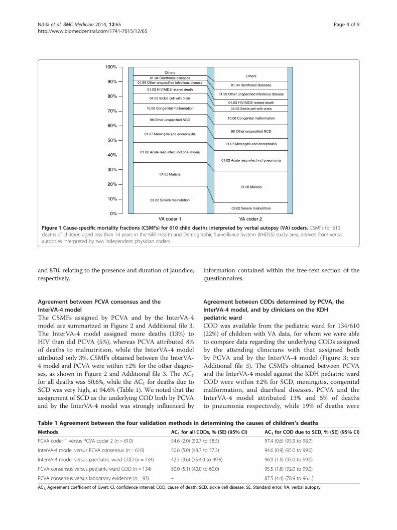
0%
10%
20%
30%
40%
50%
60%
70%
80%
90%
100%
VA coder 1 VA coder 2
03.02 Severe malnutrition
03.02 Severe malnutrition
01.05 Malaria
01.05 Malaria
01.02 Acute resp infect incl pneumonia
01.02 Acute resp infect incl pneumonia
01.07 Meningitis and encephalitis
01.07 Meningitis and encephalitis
98 Other unspecified NCD
98 Other unspecified NCD
10.06 Congenital malformation
10.06 Congenital malformation
04.03 Sickle cell with crisis
04.03 Sickle cell with crisis
01.03 HIV/AIDS related death
01.03 HIV/AIDS related death
01.99 Other unspecified infectious disease
01.99 Other unspecified infectious disease
01.04 Diarrhoeal diseases
01.04 Diarrhoeal diseases
OthersOthers
Figure 1 Cause-specific mortality fractions (CSMFs) for 610 child deaths interpreted by verbal autopsy (VA) coders. CSMFs for 610deaths of children aged less than 14 years in the Kilifi Health and Demographic Surveillance System (KHDSS) study area, derived from verbalautopsies interpreted by two independent physician coders.
Ndila et al. BMC Medicine 2014, 12:65 Page 4 of 9http://www.biomedcentral.com/1741-7015/12/65
and 870, relating to the presence and duration of jaundice,respectively.
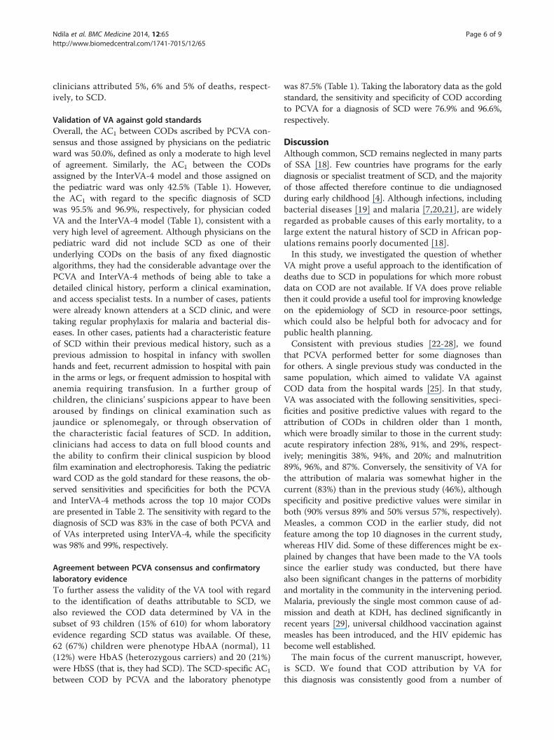
Agreement between PCVA consensus and theInterVA-4 modelThe CSMFs assigned by PCVA and by the InterVA-4model are summarized in Figure 2 and Additional file 3.The InterVA-4 model assigned more deaths (13%) toHIV than did PCVA (5%), whereas PCVA attributed 8%of deaths to malnutrition, while the InterVA-4 modelattributed only 3%. CSMFs obtained between the InterVA-4 model and PCVA were within ±2% for the other diagno-ses, as shown in Figure 2 and Additional file 3. The AC1
for all deaths was 50.6%, while the AC1 for deaths due toSCD was very high, at 94.6% (Table 1). We noted that theassignment of SCD as the underlying COD both by PCVAand by the InterVA-4 model was strongly influenced by
Table 1 Agreement between the four validation methods in d
Methods AC1 for all CO
PCVA coder 1 versus PCVA coder 2 (n = 610) 54.6 (2.0) (50.7
InterVA-4 model versus PCVA consensus (n = 610) 50.6 (5.0) (48.7
InterVA-4 model versus paediatric ward COD (n = 134) 42.5 (3.6) (35.4.
PCVA consensus versus pediatric ward COD (n = 134) 50.0 (5.1) (40.0
PCVA consensus versus laboratory evidence (n = 93) –
AC1, Agreement coefficient of Gwet; CI, confidence interval; COD, cause of death; SC
information contained within the free-text section of thequestionnaires.
Agreement between CODs determined by PCVA, theInterVA-4 model, and by clinicians on the KDHpediatric wardCOD was available from the pediatric ward for 134/610(22%) of children with VA data, for whom we were ableto compare data regarding the underlying CODs assignedby the attending clinicians with that assigned bothby PCVA and by the InterVA-4 model (Figure 3; seeAdditional file 3). The CSMFs obtained between PCVAand the InterVA-4 model against the KDH pediatric wardCOD were within ±2% for SCD, meningitis, congenitalmalformation, and diarrheal diseases. PCVA and theInterVA-4 model attributed 13% and 5% of deathsto pneumonia respectively, while 19% of deaths were
etermining the causes of children’s deaths
Ds, % (SE) (95% CI) AC1 for COD due to SCD, % (SE) (95% CI)
to 58.5) 97.4 (0.6) (95.9 to 98.7)
to 57.2) 94.6 (0.9) (95.0 to 99.0)
0 to 49.6) 96.9 (1.5) (95.0 to 99.0)
to 60.0) 95.5 (1.8) (92.0 to 99.0)
87.5 (4.4) (78.9 to 96.1.)
D, sickle cell disease. SE, Standard error; VA, verbal autopsy.

0%
10%
20%
30%
40%
50%
60%
70%
80%
90%
100%
VA COD InterVA−4
01.05 Malaria 01.05 Malaria
01.03 HIV/AIDS related death
01.03 HIV/AIDS related death01.07 Meningitis and encephalitis
01.07 Meningitis and encephalitis10.06 Congenital malformation
10.06 Congenital malformation01.02 Acute resp infect incl pneumonia
01.02 Acute resp infect incl pneumonia01.04 Diarrhoeal diseases
01.04 Diarrhoeal diseases04.03 Sickle cell with crisis
04.03 Sickle cell with crisis
03.02 Severe malnutrition 03.02 Severe malnutrition
01.99 Other unspecified infectious disease01.99 Other unspecified infectious disease
Others Others
Figure 2 Cause-specific mortality fractions (CSMFs) for 610 child deaths interpreted by physician coded verbal autopsy (PCVA) and theInterVA-4 model. CSMFs for 610 deaths of children aged less than 14 years in the Kilifi Health and Demographic Surveillance System (KHDSS)study area, derived from verbal autopsies interpreted by PCVA and the InterVA-4 model.
Ndila et al. BMC Medicine 2014, 12:65 Page 5 of 9http://www.biomedcentral.com/1741-7015/12/65
attributed to pneumonia on the pediatric ward. Similarly,PCVA and the InterVA-4 model assigned almost twiceas many deaths to malaria as did clinicians on thepediatric ward. The InterVA-4 model attributed 12% ofdeaths to HIV/AIDS, whereas the PCVA and the
0%
10%
20%
30%
40%
50%
60%
70%
80%
90%
100%
VA Coders In
01.05 Malaria 0
01.02 Acute resp infect incl pneumonia01.02 Acute r
03.02 Severe malnutrition
03.02 S
98 Other unspecified NCD
98 Oth
10.06 Congenital malformation
10.06 Co
01.07 Meningitis and encephalitis
01.07 Men
01.03 HIV/AIDS related death
01.03 HI
04.03 Sickle cell with crisis
04.03 S
01.04 Diarrhoeal diseases
01.04 D
Others
Figure 3 Cause-specific mortality fractions (CSMFs) for 134 child deathsmodel and pediatric hospital cause of death (COD). The figure shows CSMInterVA-4 model were compared against the COD given by physicians for tho
pediatric ward clinicians attributed 5% and 6% of deathsrespectively. By contrast, the InterVA-4 model attrib-uted 2% of deaths to malnutrition, while both thePCVA and the pediatric ward clinicians attributed 13%.The InterVA-4 model, PCVA, and pediatric ward
terVA−4 Paediatric ward COD
1.05 Malaria
01.05 Malaria
esp infect incl pneumonia
01.02 Acute resp infect incl pneumonia
evere malnutrition
03.02 Severe malnutrition
er unspecified NCD
98 Other unspecified NCD
ngenital malformation
10.06 Congenital malformation
ingitis and encephalitis
01.07 Meningitis and encephalitis
V/AIDS related death
01.03 HIV/AIDS related death
ickle cell with crisis
04.03 Sickle cell with crisis
iarrhoeal diseases
01.04 Diarrhoeal diseases
Others
Others
assigned by physician coded verbal autopsy (PCVA), the InterVA-4Fs for 134 child deaths. Underlying CODs determined by PCVA and these who died on the pediatric ward at Kilifi District Hospital (KDH).
Ndila et al. BMC Medicine 2014, 12:65 Page 6 of 9http://www.biomedcentral.com/1741-7015/12/65
clinicians attributed 5%, 6% and 5% of deaths, respect-ively, to SCD.
Validation of VA against gold standardsOverall, the AC1 between CODs ascribed by PCVA con-sensus and those assigned by physicians on the pediatricward was 50.0%, defined as only a moderate to high levelof agreement. Similarly, the AC1 between the CODsassigned by the InterVA-4 model and those assigned onthe pediatric ward was only 42.5% (Table 1). However,the AC1 with regard to the specific diagnosis of SCDwas 95.5% and 96.9%, respectively, for physician codedVA and the InterVA-4 model (Table 1), consistent with avery high level of agreement. Although physicians on thepediatric ward did not include SCD as one of theirunderlying CODs on the basis of any fixed diagnosticalgorithms, they had the considerable advantage over thePCVA and InterVA-4 methods of being able to take adetailed clinical history, perform a clinical examination,and access specialist tests. In a number of cases, patientswere already known attenders at a SCD clinic, and weretaking regular prophylaxis for malaria and bacterial dis-eases. In other cases, patients had a characteristic featureof SCD within their previous medical history, such as aprevious admission to hospital in infancy with swollenhands and feet, recurrent admission to hospital with painin the arms or legs, or frequent admission to hospital withanemia requiring transfusion. In a further group ofchildren, the clinicians’ suspicions appear to have beenaroused by findings on clinical examination such asjaundice or splenomegaly, or through observation ofthe characteristic facial features of SCD. In addition,clinicians had access to data on full blood counts andthe ability to confirm their clinical suspicion by bloodfilm examination and electrophoresis. Taking the pediatricward COD as the gold standard for these reasons, the ob-served sensitivities and specificities for both the PCVAand InterVA-4 methods across the top 10 major CODsare presented in Table 2. The sensitivity with regard to thediagnosis of SCD was 83% in the case of both PCVA andof VAs interpreted using InterVA-4, while the specificitywas 98% and 99%, respectively.
Agreement between PCVA consensus and confirmatorylaboratory evidenceTo further assess the validity of the VA tool with regardto the identification of deaths attributable to SCD, wealso reviewed the COD data determined by VA in thesubset of 93 children (15% of 610) for whom laboratoryevidence regarding SCD status was available. Of these,62 (67%) children were phenotype HbAA (normal), 11(12%) were HbAS (heterozygous carriers) and 20 (21%)were HbSS (that is, they had SCD). The SCD-specific AC1
between COD by PCVA and the laboratory phenotype
was 87.5% (Table 1). Taking the laboratory data as the goldstandard, the sensitivity and specificity of COD accordingto PCVA for a diagnosis of SCD were 76.9% and 96.6%,respectively.
DiscussionAlthough common, SCD remains neglected in many partsof SSA [18]. Few countries have programs for the earlydiagnosis or specialist treatment of SCD, and the majorityof those affected therefore continue to die undiagnosedduring early childhood [4]. Although infections, includingbacterial diseases [19] and malaria [7,20,21], are widelyregarded as probable causes of this early mortality, to alarge extent the natural history of SCD in African pop-ulations remains poorly documented [18].In this study, we investigated the question of whether
VA might prove a useful approach to the identification ofdeaths due to SCD in populations for which more robustdata on COD are not available. If VA does prove reliablethen it could provide a useful tool for improving knowledgeon the epidemiology of SCD in resource-poor settings,which could also be helpful both for advocacy and forpublic health planning.Consistent with previous studies [22-28], we found
that PCVA performed better for some diagnoses thanfor others. A single previous study was conducted in thesame population, which aimed to validate VA againstCOD data from the hospital wards [25]. In that study,VA was associated with the following sensitivities, speci-ficities and positive predictive values with regard to theattribution of CODs in children older than 1 month,which were broadly similar to those in the current study:acute respiratory infection 28%, 91%, and 29%, respect-ively; meningitis 38%, 94%, and 20%; and malnutrition89%, 96%, and 87%. Conversely, the sensitivity of VA forthe attribution of malaria was somewhat higher in thecurrent (83%) than in the previous study (46%), althoughspecificity and positive predictive values were similar inboth (90% versus 89% and 50% versus 57%, respectively).Measles, a common COD in the earlier study, did notfeature among the top 10 diagnoses in the current study,whereas HIV did. Some of these differences might be ex-plained by changes that have been made to the VA toolssince the earlier study was conducted, but there havealso been significant changes in the patterns of morbidityand mortality in the community in the intervening period.Malaria, previously the single most common cause of ad-mission and death at KDH, has declined significantly inrecent years [29], universal childhood vaccination againstmeasles has been introduced, and the HIV epidemic hasbecome well established.The main focus of the current manuscript, however,
is SCD. We found that COD attribution by VA forthis diagnosis was consistently good from a number of
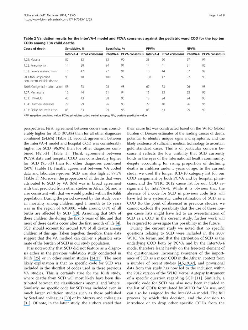
Table 2 Validation results for the InterVA-4 model and PCVA consensus against the pediatric ward COD for the top tenCODs among 134 child deaths
Cause of death Sensitivity, % Specificity, % PPV% NPV%
InterVA-4 PCVA consensus InterVA-4 PCVA consensus InterVA-4 PCVA consensus InterVA-4 PCVA consensus
1.05: Malaria 80 83 83 90 38 50 97 97
1.02: Pneumonia 14 28 94 91 14 41 81 85
3.02: Severe malnutrition 10 47 97 91 10 44 87 92
98: Other unspecifiednon-communicable disease
9 18 100 92 100 17 92 93
10.06: Congenital malformation 55 73 98 98 67 73 96 98
1.07: Meningitis 12 44 91 94 15 33 93 96
1.03: HIV/AIDS 40 43 88 95 18 24 94 93
1.04: Diarrheal diseases 29 29 96 98 29 40 96 96
4.03: Sickle cell with crisis 83 83 99 98 83 63 99 99
NPV, negative predicted value; PCVA, physician coded verbal autopsy; PPV, positive predictive value.
Ndila et al. BMC Medicine 2014, 12:65 Page 7 of 9http://www.biomedcentral.com/1741-7015/12/65
perspectives. First, agreement between coders was consid-erably higher for SCD (97.3%) than for all other diagnosescombined (54.6%) (Table 1). Second, agreement betweenthe InterVA-4 model and hospital COD was considerablyhigher for SCD (96.9%) than for other diagnoses com-bined (42.5%) (Table 1). Third, agreement betweenPCVA data and hospital COD was considerably higherfor SCD (95.5%) than for other diagnoses combined(50%) (Table 1). Finally, agreement between VA codeddata and laboratory-proven SCD was also high at 87.5%(Table 1). Moreover, the proportion of all deaths that wereattributed to SCD by VA (6%) was in broad agreementwith that predicted from other studies in Africa [5], and isalso consistent with that we would predict within the Kilifipopulation. During the period covered by this study, over-all mortality among children aged 1 month to 15 yearswas in the region of 40/1000, while around 0.8% of allbirths are affected by SCD [19]. Assuming that 50% ofthese children die during the first 5 years of life, and thatmost of these deaths occur after the first month of life [4],SCD should account for around 10% of all deaths amongchildren of this age. Taken together, therefore, these datasuggest that the VA method can deliver a plausible esti-mate of the burden of SCD in our study population.It is noteworthy that SCD did not feature as a diagno-
sis either in the previous validation study conducted inKilifi [25] or in other similar studies [24,27]. The mostlikely explanation is that no specific code for SCD wasincluded in the shortlist of codes used in these previousVA studies. This is certainly true for the Kilifi study,where deaths from SCD will most likely have been dis-tributed between the classifications 'anemia' and 'others'.Similarly, no specific code for SCD was included even inmuch larger validation studies such as those publishedby Setel and colleagues [30] or by Murray and colleagues[31]. Of note, in the latter study, the authors stated that
their cause list was constructed based on the WHO GlobalBurden of Disease estimates of the leading causes of death,potential to identify unique signs and symptoms, and thelikely existence of sufficient medical technology to ascertaingold standard cases. This is of particular concern be-cause it reflects the low visibility that SCD currentlyholds in the eyes of the international health community,despite accounting for rising proportion of decliningdeaths in children under 5 years of age. In the currentstudy, we used the longer ICD-10 category list for ourCOD assignment by both PCVA and by hospital physi-cians, and the WHO 2012 cause list for our COD as-signment by InterVA-4. While it is obvious that theabsence of a code for SCD in previous code lists willhave led to a systematic underestimation of SCD as aCOD (to the point of absence) in previous studies, wecannot exclude the possibility that the use of these lon-ger cause lists might have led to an overestimation ofSCD as a COD in the current study; further work willbe required to investigate this possibility in the future.During the current study we noted that no specific
questions relating to SCD were included in the 2007WHO VA forms, and that the attribution of SCD as theunderlying COD both by PCVA and by the InterVA-4model therefore leant heavily on the free-text element ofthe questionnaires. Increasing awareness of the import-ance of SCD as a major COD in the African context froma number of recent studies [4,5,19,32], and provisionaldata from this study has now led to the inclusion withinthe 2012 version of the WHO Verbal Autopsy Instrumentof a specific question regarding SCD [11]. Similarly, aspecific code for SCD has also now been included inthe list of CODs formulated by WHO for VA use, andcan also be assigned by the InterVA-4 model. The fullprocess by which this decision, and the decision tointroduce or to drop other specific CODs from the
Ndila et al. BMC Medicine 2014, 12:65 Page 8 of 9http://www.biomedcentral.com/1741-7015/12/65
2012 list, were made have been described recently byLeitao and colleagues [33].Although our study provides new evidence that VA
has potential as a tool for identifying deaths due to SCD,it is open to a number of criticisms. Perhaps most im-portantly, SCD constitutes a major research interest atour program [7,13,19,20] and, as a result, it is possiblethat our coding clinicians are unusually well versed inthe signs and symptoms of SCD. We aimed to mitigateagainst this bias by applying the InterVA-4 model, whichat least guarantees consistency and comparability withpossible future studies elsewhere. Similarly, in order tovalidate our COD assignment by VA, we used as ourgold standard COD assignment by a clinician for thesubset of deaths that occurred in hospital. We cannotbe sure that the distribution of CODs among deaths thatoccur in hospital is fully representative of CODs overall,and this could potentially lead to an unrecognized andunmeasurable bias. Nevertheless, we suggest that ourstudy would benefit from replication and validation atother sites.We have previously estimated that approximately 240,000
children are born with SCD annually in Africa alone [3]and that 50-90% of these children die undiagnosed in theirfirst few years of life [4]. Others have estimated that thismeans that SCD accounts for around 6% of all childdeaths in SSA [5], placing SCD firmly on the list of leadingcauses of death, at least in countries where the genefrequency is high. This is recognized by the welcomeinclusion of the hemoglobinopathies in the latest round ofthe Global Burden of Disease Study [34]. Nevertheless,much more remains to be done to raise the profile of SCDon the global health agenda.
ConclusionIn summary, we have presented data suggesting that VAmight provide a useful approach to investigating thecontribution of SCD to childhood deaths in one part ofSSA. We hope that our study will prompt similar workin other parts of Africa, including the further explorationof automated approaches to VA coding that might begeneralized to other settings.
Additional files
Additional file 1: Spreadsheet showing mapping of physiciancoded verbal autopsy (PCVA) and hospital causes of death to the2012 WHO verbal autopsy (VA) cause of death (COD) categories.The spreadsheet shows how the PCVA and hospital causes of death weremapped to the 2012 WHO VA categories in order to facilitatecomparisons between the three methods.
Additional file 2: Calculation of the agreement coefficient of Gwet.
Additional file 3: Cause-specific mortality fractions as assignedby the three different methods of cause of death (COD)assignment.
AbbreviationsAC1: Agreement coefficient; COD: Cause of Death; CSMF: Cause SpecificMortality Fraction; ERC: Ethics Review Committee; HbSC: Sickle Cell/HbCDisease; HbSS: Sickle Cell Anemia; HIV: Human Immunodeficiency Syndrome;ICD-10: International Classification of Diseases 10th Edition; KDH: Kilifi DistrictHospital; KGBC: Kilifi Genetic Birth Cohort; KHDSS: Kilifi Health and DemographicSurveillance System; KSCD: Kilifi Sickle Cell Disease; MOH: Ministry of Health;PCVA: Physician Coded Verbal Autopsy; SAVVY: Sample Vital Registration withVerbal Autopsy; SCD: Sickle Cell Disease; SSA: Sub-Saharan Africa; VA: VerbalAutopsy; WHO: World Health Organization.
Competing interestsAll authors declare that they have no conflicts of interest.
Author contributionsCN reviewed the literature, analyzed and interpreted data, and wrote the paper.EB contributed to the study design and helped to edit the final version of thepaper. VN helped in interpreting the data and editing of the paper. GM helpedto code the VAs, and helped in interpretation of the results and editing of thepaper. AMA, PK and SK helped with VA data coding and editing of the paper.GN helped with data management and editing of the paper. AM helped withlaboratory data management and editing of the paper. PB helped withinterpreting the data and editing the final version of the paper. TNW conceivedthe study and edited the final version of the paper. All authors read andapproved the final version of the manuscript.
AcknowledgmentsWe thank the study respondents, the field workers, the MOH Kilifi, HerbertOpi, Sophie Uyoga, Emily Orori, Adan Mohammed, Metrine Tendwa, JohnsonMakale, Prophet Ingosi, Samuel Geji, and all of the KEMRI-Wellcome Trustcollaborators for their help with this study. We also thank the INDEPTH Networkwho supported the first presentation of these data through a travel scholarshipawarded to CN. This paper is published with permission of the Director ofKEMRI. The study was funded by a grant from the USAID National M&E SupportProgramme (sub-grant no: 631548-10S-1524) and through a Senior ResearchFellowship awarded to TNW by the Wellcome Trust, UK (091758). PB anddevelopment of the InterVA-4 model were funded by the Umeå Centre forGlobal Health Research, with support from FAS, the Swedish Council forWorking Life and Social Research (grant no. 2006–1512).
Author details1Kenya Medical Research Institute (KEMRI)/Wellcome Trust Programme,Centre for Geographic Medicine Research-Coast, P.O Box 230, Kilifi, Kenya.2INDEPTH Network of Demographic Surveillance Sites, Accra, Ghana. 3UnitedNation Statistics Division, New York, USA. 4Umeå Centre for Global HealthResearch, Department of Public Health and Clinical Medicine, UmeåUniversity, 90187 Umeå, Sweden. 5WHO Collaborating Centre for VerbalAutopsy, Umeå University, 90187 Umeå, Sweden. 6Department of Medicine,Imperial College, London W21NY, UK.
Received: 17 January 2014 Accepted: 31 March 2014Published: 22 April 2014
References1. Rees DC, Williams TN, Gladwin MT: Sickle-cell disease. Lancet 2010,
376:2018–2031.2. Perutz MF, Rosa J, Schechter A: Therapeutic agents for sickle cell disease.
Nature 1978, 275:369–370.3. Piel FB, Patil AP, Howes RE, Nyangiri OA, Gething PW, Dewi M, Temperley
WH, Williams TN, Weatherall DJ, Hay SI: Global epidemiology of sicklehaemoglobin in neonates: a contemporary geostatistical model-basedmap and population estimates. Lancet 2013, 381:142–151.
4. Grosse SD, Odame I, Atrash HK, Amendah DD, Piel FB, Williams TN: Sicklecell disease in Africa: a neglected cause of early childhood mortality.Am J Prev Med 2011, 41:S398–S405.
5. Modell B, Darlison M: Global epidemiology of haemoglobin disorders andderived service indicators. Bull World Health Organ 2008, 86:480–487.
6. Scott JA, Bauni E, Moisi JC, Ojal J, Gatakaa H, Nyundo C, Molyneux CS,Kombe F, Tsofa B, Marsh K, Peshu N, Williams TN: Profile: The Kilifi Healthand Demographic Surveillance System (KHDSS). Int J Epidemiol 2012,41:650–657.
Ndila et al. BMC Medicine 2014, 12:65 Page 9 of 9http://www.biomedcentral.com/1741-7015/12/65
7. Komba AN, Makani J, Sadarangani M, Ajala-Agbo T, Berkley JA, Newton CR,Marsh K, Williams TN: Malaria as a cause of morbidity and mortality inchildren with homozygous sickle cell disease on the coast of Kenya.Clin Infect Dis 2009, 49:216–222.
8. Bauni E, Ndila C, Mochamah G, Nyutu G, Matata L, Ondieki C, Mambo B,Mutinda M, Tsofa B, Maitha E, Etyang A, Williams TN: Validatingphysician-certified verbal autopsy and probabilistic modeling(InterVA) approaches to verbal autopsy interpretation using hospitalcauses of adult deaths. Popul Health Metr 2011, 9:49.
9. World Health O: International Statistical Classification of Diseases and RelatedHealth Problems: ICD-10. Geneva: WHO; 1993.
10. Byass P, Chandramohan D, Clark SJ, D'Ambruoso L, Fottrell E, Graham WJ,Herbst AJ, Hodgson A, Hounton S, Kahn K, Krishnan A, Leitao J, OdhiamboF, Sankoh OA, Tollman SM: Strengthening standardised interpretation ofverbal autopsy data: the new InterVA-4 tool. Glob Health Action 2012,5:1–8.
11. Ascertaining and attributing causes of death. [http://www.who.int/healthinfo/statistics/verbalautopsystandards/en/]
12. Ojwang PJ, Ogada T, Beris P, Hattori Y, Lanclos KD, Kutlar A, Kutlar F,Huisman TH: Haplotypes and alpha globin gene analyses in sickle cellanaemia patients from Kenya. Br J Haematol 1987, 65:211–215.
13. Sadarangani M, Makani J, Komba AN, Ajala-Agbo T, Newton CR, Marsh K,Williams TN: An observational study of children with sickle cell disease inKilifi, Kenya. Br J Haematol 2009, 146:675–682.
14. Scott JA, Berkley JA, Mwangi I, Ochola L, Uyoga S, Macharia A, Ndila C,Lowe BS, Mwarumba S, Bauni E, Marsh K, Williams TN: Relationbetween falciparum malaria and bacteraemia in Kenyan children: apopulation-based, case–control study and a longitudinal study.Lancet 2011, 378:1316–1323.
15. Gwet KL: Computing inter-rater reliability and its variance in the presenceof high agreement. Br J Math Stat Psychol 2008, 61:29–48.
16. Gwet KL: Handbook of Inter-Rater Reliability. 2nd edition. Gaithersburg, USA:Advanced Analytics LLC.
17. The Comprehensive R Archive Network. [http://cran.r-project.org/]18. Makani J, Williams TN, Marsh K: Sickle cell disease in Africa: burden and
research priorities. Ann Trop Med Parasitol 2007, 101:3–14.19. Williams TN, Uyoga S, Macharia A, Ndila C, McAuley CF, Opi DH, Mwarumba
S, Makani J, Komba A, Ndiritu MN, Sharif SK, Marsh K, Berkley JA, Scott JAG:Bacteraemia in Kenyan children with sickle-cell anaemia: a retrospectivecohort and case–control study. Lancet 2009, 374:1364–1370.
20. McAuley CF, Webb C, Makani J, Macharia A, Uyoga S, Opi DH, Ndila C,Ngatia A, Scott JA, Marsh K, Williams TN: High mortality from Plasmodiumfalciparum malaria in children living with sickle cell anemia on the coastof Kenya. Blood 2010, 116:1663–1668.
21. Makani J, Komba AN, Cox SE, Oruo J, Mwamtemi K, Kitundu J, Magesa P,Rwezaula S, Meda E, Mgaya J, Pallangyo K, Okiro E, Muturi D, Newton CR,Fegan G, Marsh K, Williams TN: Malaria in patients with sickle cell anemia:burden, risk factors, and outcome at the outpatient clinic and duringhospitalization. Blood 2010, 115:215–220.
22. Snow B, Marsh K: How useful are verbal autopsies to estimate childhoodcauses of death. Health Policy Planning 1992, 7:22–29.
23. Snow RW, Armstrong JR, Forster D, Winstanley MT, Marsh VM, Newton CR,Waruiru C, Mwangi I, Winstanley PA, Marsh K: Childhood deaths in Africa:uses and limitations of verbal autopsies. Lancet 1992, 340:351–355.
24. Mobley CC, Boerma JT, Titus S, Lohrke B, Shangula K, Black RE: Validationstudy of a verbal autopsy method for causes of childhood mortality inNamibia. J Trop Pediatr 1996, 42:365–369.
25. Quigley MA, Armstrong Schellenberg JR, Snow RW: Algorithms for verbalautopsies: a validation study in Kenyan children. Bull World Health Organ1996, 74:147–154.
26. Rodriguez L, Reyes H, Tome P, Ridaura C, Flores S, Guiscafre H: Validation ofthe verbal autopsy method to ascertain acute respiratory infection ascause of death. Indian J Pediatr 1998, 65:579–584.
27. Kahn K, Tollman SM, Garenne M, Gear JS: Validation and application ofverbal autopsies in a rural area of South Africa. Trop Med Int Health 2000,5:824–831.
28. Datta N, Mand M, Kumar V: Validation of causes of infant death in thecommunity by verbal autopsy. Indian J Pediatr 1988, 55:599–604.
29. O'Meara WP, Bejon P, Mwangi TW, Okiro EA, Peshu N, Snow RW, NewtonCR, Marsh K: Effect of a fall in malaria transmission on morbidity andmortality in Kilifi, Kenya. Lancet 2008, 372:1555–1562.
30. Setel PW, Rao C, Hemed Y, Whiting DR, Yang G, Chandramohan D, AlbertiKG, Lopez AD: Core verbal autopsy procedures with comparativevalidation results from two countries. PLoS Med 2006, 3:e268.
31. Murray CJ, Lopez AD, Black R, Ahuja R, Ali SM, Baqui A, Dandona L, DantzerE, Das V, Dhingra U, Dutta A, Fawzi W, Flaxman AD, Gómez S, Hernández B,Joshi R, Kalter H, Kumar A, Kumar V, Lozano R, Lucero M, Mehta S, Neal B,Ohno SL, Prasad R, Praveen D, Premji Z, Ramírez-Villalobos D, Remolador H,Riley I, et al: Population Health Metrics Research Consortium goldstandard verbal autopsy validation study: design, implementation, anddevelopment of analysis datasets. Popul Health Metr 2011, 9:27.
32. Christianson AL, Howson CP, Modell B: March of Dimes Global Report onBirth Defects: The Hidden Toll of Dying and Disabled Children. White Plains,New York: March of Dimes Birth Defects Foundation; 2006.
33. Leitao J, Chandramohan D, Byass P, Jakob R, Bundhamcharoen K,Choprapawon C, de Savigny D, Fottrell E, Franca E, Froen F, Gewaifel G,Hodgson A, Hounton S, Kahn K, Krishnan A, Kumar V, Masanja H, Nichols E,Notzon F, Rasooly MH, Sankoh O, Spiegel P, AbouZahr C, Amexo M, KebedeD, Alley WS, Marinho F, Ali M, Loyola E, Chikersal J, et al: Revising the WHOVerbal Autopsy instrument to facilitate routine cause-of-death monitoring.Global Health Action 2013, 6:21518.
34. Murray CJ, Vos T, Lozano R, Naghavi M, Flaxman AD, Michaud C, Ezzati M,Shibuya K, Salomon JA, Abdalla S, Aboyans V, Abraham J, Ackerman I,Aggarwal R, Ahn SY, Ali MK, Alvarado M, Anderson HR, Anderson LM, AndrewsKG, Atkinson C, Baddour LM, Bahalim AN, Barker-Collo S, Barrero LH, Bartels DH,Basanez M-G, Baxter A, Bell ML, Benjamin EJ, et al: Disability-adjusted life years(DALYs) for 291 diseases and injuries in 21 regions, 1990–2010: asystematic analysis for the Global Burden of Disease Study 2010.Lancet 2012, 380:2197–2223.
doi:10.1186/1741-7015-12-65Cite this article as: Ndila et al.: Verbal autopsy as a tool for identifyingchildren dying of sickle cell disease: a validation study conducted inKilifi district, Kenya. BMC Medicine 2014 12:65.
Submit your next manuscript to BioMed Centraland take full advantage of:
• Convenient online submission
• Thorough peer review
• No space constraints or color figure charges
• Immediate publication on acceptance
• Inclusion in PubMed, CAS, Scopus and Google Scholar
• Research which is freely available for redistribution
Submit your manuscript at www.biomedcentral.com/submit
Copyright © 2022 FDOKUMEN