Selective photothermolysis of spider veins and reticular ...
Upload
independentCategory
view
3download
0
(This is a sample cover image for this issue. The actual cover is not yet available at this time.)
This article appeared in a journal published by Elsevier. The attachedcopy is furnished to the author for internal non-commercial researchand education use, including for instruction at the authors institution
and sharing with colleagues.
Other uses, including reproduction and distribution, or selling orlicensing copies, or posting to personal, institutional or third party
websites are prohibited.
In most cases authors are permitted to post their version of thearticle (e.g. in Word or Tex form) to their personal website orinstitutional repository. Authors requiring further information
regarding Elsevier’s archiving and manuscript policies areencouraged to visit:
http://www.elsevier.com/copyright
Author's personal copy
Venomic and pharmacological activity of Acanthoscurria paulensis(Theraphosidae) spider venom
Caroline Barbosa F. Mourão a,1, Fagner Neves Oliveira a,1, Andréa C. e Carvalho a,Claudia J. Arenas a, Harry Morales Duque a, Jacqueline C. Gonçalves a,Jéssica K.A. Macêdo a, Priscilla Galante a, Carlos A. Schwartz a, Márcia R. Mortari a,Maria de Fátima M. Almeida Santos b, Elisabeth F. Schwartz a,*
a Laboratório de Toxinologia, Departamento de Ciências Fisiológicas, Universidade de Brasília ,Instituto de Ciências Biológicas, Brasília 70910-900, DF, BrazilbDepartamento de Genética e Morfologia, Universidade de Brasília, Brasília 70910-900, DF, Brazil
a r t i c l e i n f o
Article history:Received 17 September 2012Received in revised form 5 November 2012Accepted 8 November 2012Available online 21 November 2012
Keywords:Acanthoscurria paulensisTarantulaEdematogenicCardiotoxicToxicityToxin
a b s t r a c t
In the present study we conducted proteomic and pharmacological characterizations ofthe venom extracted from the Brazilian tarantula Acanthoscurria paulensis, and evaluatedthe cardiotoxicity of its two main fractions. The molecular masses of the venom compo-nents were identified by mass spectrometry (MALDI-TOF-MS) after chromatographicseparation (HPLC). The lethal dose (LD50) was determined in mice. Nociceptive behaviorwas evaluated by intradermal injection in mice and the edematogenic activity by the rathind-paw assay. Cardiotoxic activity was evaluated on in situ frog heart and on isolated frogventricle strip. From 60 chromatographic fractions, 97 distinct components were identi-fied, with molecular masses between 601.4 and 21,932.3 Da. A trimodal molecular massdistribution was observed: 30% of the components within 500-1999 Da, 38% within 3500–5999 Da and 21% within 6500–7999 Da. The LD50 in mice was 25.4 � 2.4 mg/g and theeffects observed were hypoactivity, anuria, constipation, dyspnea and prostration untildeath, which occurred at higher doses. Despite presenting a dose-dependent edemato-genic activity in the rat hind-paw assay, the venom had no nociceptive activity in mice.Additionally, the venom induced a rapid blockage of electrical activity and subsequentdiastolic arrest on in situ frog heart preparation, which was inhibited by pretreatment withatropine. In the electrically driven frog ventricle strip, the whole venom and its lowmolecular mass fraction, but not the proteic one, induced a negative inotropic effect thatwas also inhibited by atropine. These results suggest that despite low toxicity, A. paulensisvenom can induce severe physiological disturbances in mice.
� 2012 Elsevier Ltd. All rights reserved.
1. Introduction
According to the effects of venom in humans, accidentscaused by spiders can be categorized in, at least, twodistinct groups: those producing necrotic ulceration, and
the ones that do not. Arachnidism produced by widowspiders (Theridiidae) will result in systemic symptoms butwith minimal tissue damage. Envenomation by Agelenidaefamily (araneomorph funnel-web spiders, including hoboand grass spiders) results in severe tissue damage(Mattiello-Sverzut et al., 1998; Elston et al., 2000) and, ina minority of accidents, also systemic symptoms. Localnecrosis and systemic symptoms are observed in the eventsincited by Sicariidae (Loxosceles; recluse and fiddleheadspiders) (Madrigal et al., 1972; Barbaro et al., 1992).
* Corresponding author. Tel.: þ55 61 31073106; fax: þ55 61 31073107.E-mail address: [email protected] (E.F. Schwartz).
1 These authors contributed equally to the manuscript.
Contents lists available at SciVerse ScienceDirect
Toxicon
journal homepage: www.elsevier .com/locate/ toxicon
0041-0101/$ – see front matter � 2012 Elsevier Ltd. All rights reserved.http://dx.doi.org/10.1016/j.toxicon.2012.11.008
Toxicon 61 (2013) 129–138
Author's personal copy
Tarantulas (Theraphosidae, Mygalomorphae) bites areconsidered to be painful, but do not induce local necrosis orsystemic effects (Saucier, 2004). With exception of bites byspiders of the genus Atrax (Hexathelidae), there are norecords of serious accidents caused by Mygalomorphaespiders in human probably due to several factors such asvenom low toxicity to humans, insufficient amount ofvenom injected, greater difficulty of Orthognatans’ chelic-erae in piercing the skin, and also because some species livein low frequented local by man (Lucas, 2003). Unlessinfection occurs, a slight inflammation at the puncture sitecan arise. Spiders of the family Theraphosidae have urti-cating hairs covering their bodies, which are brushed off bythe spider as a mechanism of defense to deter predators.These hairs were found to induce local dermatitis invertebrates, including humans (Shrum et al., 1999). Thepuncture wounds from the spider’s fangs require localwound care, follow-up for signs of infection, short-termanalgesia and a tetanus booster (Kelley and Wasserman,1998; Shrum et al., 1999).
The spider venom is a diverse mixture of low molecularmass compounds (16% of all compounds), acylpolyamines(11%), linear peptides (6%), cysteine-knotted mini-proteins(60%), neurotoxic proteins (1%) and enzymes (6%) (Jacksonand Parks, 1989; Kuhn-Nentwig et al., 2011). It is mainlyused to paralyze prey and for defense, and contains toxinsthat affect the central or peripheral nervous systems. Theseneurotoxins have been identified mostly as acylpolyaminesand peptides or proteins that act onmembrane receptors orion channels (see review Estrada et al., 2007).
The acylpolyamine toxins are low molecular masscompounds (<1 kDa) that appear to have evolved tospecifically provoke rapid paralysis. Their complex struc-tures are composed by a polyamine chain with a primaryamino or a guanidine group at one end and an aromaticring at the other. These compounds interact with multipletargets in the central and peripheral nervous systems ofinsects, and also in the CNS of mammals, whereas the maintargets are ionotropic glutamate and nicotinic acetylcho-line receptors (Kawai et al., 1982; Herold and Yaksh, 1992;Bixel et al., 2001).
Eight hundred curated sequences of protein toxins havebeen described for spider venom to date, among themapproximately 20% corresponds to Theraphosidae spiders(available at ArachnoServer 2.0; Herzig et al., 2011). Most ofthese 200 peptides has 30–40 amino acid residues, threedisulfide bridges and basic character (Escoubas and Rash,2004), and are modulators of ion-channels, such ascalcium, sodium, and potassium.
In this communication we report the results of proteo-mic and pharmacological characterizations of the venomextracted from the Brazilian spider Acanthoscurriapaulensis.
2. Materials and methods
2.1. Spider collection
A. paulensis (Theraphosidae, Mygalomorphae) is a darkbrown colored spider widely distributed in three Brazilianregions: South, Southeast and Midwest (Mello-Leitão,
1923; Lucas et al., 2010). The specimens used in thepresent work were collected in Brasília (Distrito Federal,Brazil) under the “Instituto Chico Mendes de Conservaçãoda Biodiversidade” (ICMBio) license number 24227-1. Onlyadult male specimens were used in this study due to theiravailability in field at the time. The spiders were identifiedby Dr Paulo César Motta from the Laboratory of Arachnids(University of Brasília, Brasília, DF, Brazil) based onmorphological characteristics.
2.2. Venom extraction and purification
The venom of eight adult male specimens of A. paulensisspiders was monthly obtained by electrical stimulation,solubilized in deionized water containing 0.12% trifluoro-acetic acid (TFA) and centrifuged at 10,000 � g for 10 min.The soluble supernatant was immediately frozen, lyophi-lized and stored at �20 �C. The venom dry weight wasdetermined in a high precision analytic balance. Aliquots of5 mg of dried venom were solubilized in deionized water,centrifuged at 10,000 � g for 10 min and the supernatantwas submitted to high performance liquid chromatography(HPLC), using a C18 reversed-phase semipreparativecolumn (Jupiter 5 mm, 300 �A, 250 � 10 mm, Phenomenex)using a linear gradient from solution A (0.12% TFA) to 60%solution B (0.10% TFA in acetonitrile – ACN) run for 60 minafter 10 initialminutes at 0% solution Bwith detection at 216and 230 nm. The fractions eluted at aflow rate of 1.5mL/minwere individually and manually collected, vacuum driedand stored at �20 �C until use. In order to obtain the lowmolecular mass fraction (LMMF) and protein fraction (PF)for the evaluation of cardiotoxic activity, the fractionseluting from 0 to 35% solution B and from35 to 74% solutionBwere separately collected. After removal of solvent, LMMFand PF were quantified by dry weight in a high precisionanalytic balance and stored at �20 �C until use.
2.3. Mass spectrometry
The molecular masses of the chromatographic fractionsof A. paulensis venom were performed on an UltraFlexIIIMALDI-TOF/TOF mass spectrometer (Bruker Daltonics,Germany). The samples were reconstituted in deionizedwater at variable concentrations and dissolved (1:3, v:v)in an a-cyano-4-hydroxycinnamic acid matrix solution(a-cyano-4-hydroxycinnamic acid at 5 mg/mL dissolved onacetonitrile, water, trifluoroacetic acid, 5:4:1, v:v:v) spottedin triplicate onto a sample plate and allowed to dry at roomtemperature. The MS spectra were acquired in both re-flected and linear positive modes. Calibration of the systemwas performed using a mixture of the Peptide CalibrationStandard and Protein Calibration Standard I for massspectrometry (Bruker Daltonics, Germany). Spectra wereprocessed with MassLynx� 3.5 (Manchester, UK) andFlexAnalysis 3.3 (Bruker Daltonics, Germany).
2.4. Animals
Animals were contained in accordance with the ethicalguidelines of the Brazilian Society for Neuroscience andBehavior, which follows the guidelines for animal care
C.B.F. Mourão et al. / Toxicon 61 (2013) 129–138130
Author's personal copy
prepared by the Committee on Care and Use of LaboratoryAnimal Resources, National Research Council, U.S.A. Thisstudy was approved by the Committee for Ethics in AnimalUse of the Institute of Biological Sciences, University ofBrasília, under license numbers 73742/2008 and 127103/2011. Likewise, every effort was made to avoid unnecessarystress and pain to the experimental animals. The number ofanimals was kept to the minimum necessary to test theconcept. The experimental animals used in this study wereSwiss albino mice (Mus musculus) of approximately 26–30 g, Wistar rats (Rattus norvegicus) of 150–200 g, and frog(Lithobates catesbeianus).
2.5. Lethality assay (LD50)
The toxicity of A. paulensis venom was evaluated bydetermining the 50% lethal dose (LD50) in mice, the doseable to kill 50% of animals tested. Four experimental groupswere tested with different doses of venom: 20, 25, 30 and40 mg/g of mice (n ¼ 5/group). The venomwas dissolved in100 mL saline solution (150 mM NaCl) and injected by i.p.route. The control group (n ¼ 5) was injected with saline.The lethality rate of animals was observed 48 h afterinoculation of venom or saline. At the end of the experi-ment, the surviving animals were euthanized with anoverdose of sodium pentobarbital (about 75 mg/kg). Thevalues for the lethality assay and their confidence limitswere calculated by Probit analysis (Finney, 1971), using thesoftware BioStat 5.8.4 version 2009.
2.6. Determination of venom effects
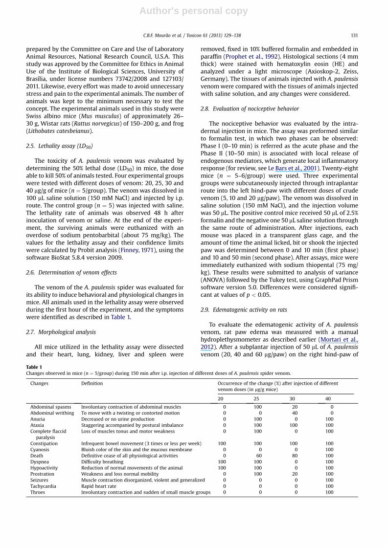
The venom of the A. paulensis spider was evaluated forits ability to induce behavioral and physiological changes inmice. All animals used in the lethality assay were observedduring the first hour of the experiment, and the symptomswere identified as described in Table 1.
2.7. Morphological analysis
All mice utilized in the lethality assay were dissectedand their heart, lung, kidney, liver and spleen were
removed, fixed in 10% buffered formalin and embedded inparaffin (Prophet et al., 1992). Histological sections (4 mmthick) were stained with hematoxylin eosin (HE) andanalyzed under a light microscope (Axioskop-2, Zeiss,Germany). The tissues of animals injected with A. paulensisvenomwere compared with the tissues of animals injectedwith saline solution, and any changes were considered.
2.8. Evaluation of nociceptive behavior
The nociceptive behavior was evaluated by the intra-dermal injection in mice. The assay was preformed similarto formalin test, in which two phases can be observed:Phase I (0–10 min) is referred as the acute phase and thePhase II (10–50 min) is associated with local release ofendogenous mediators, which generate local inflammatoryresponse (for review, see Le Bars et al., 2001). Twenty-eightmice (n ¼ 5–6/group) were used. Three experimentalgroups were subcutaneously injected through intraplantarroute into the left hind-paw with different doses of crudevenom (5, 10 and 20 mg/paw). The venom was dissolved insaline solution (150 mM NaCl), and the injection volumewas 50 mL. The positive control mice received 50 mL of 2.5%formalin and the negative one 50 mL saline solution throughthe same route of administration. After injections, eachmouse was placed in a transparent glass cage, and theamount of time the animal licked, bit or shook the injectedpaw was determined between 0 and 10 min (first phase)and 10 and 50 min (second phase). After assays, mice wereimmediately euthanized with sodium thiopental (75 mg/kg). These results were submitted to analysis of variance(ANOVA) followed by the Tukey test, using GraphPad Prismsoftware version 5.0. Differences were considered signifi-cant at values of p < 0.05.
2.9. Edematogenic activity on rats
To evaluate the edematogenic activity of A. paulensisvenom, rat paw edema was measured with a manualhydroplethysmometer as described earlier (Mortari et al.,2012). After a subplantar injection of 50 mL of A. paulensisvenom (20, 40 and 60 mg/paw) on the right hind-paw of
Table 1Changes observed in mice (n ¼ 5/group) during 150 min after i.p. injection of different doses of A. paulensis spider venom.
Changes Definition Occurrence of the change (%) after injection of differentvenom doses (in mg/g mice)
20 25 30 40
Abdominal spasms Involuntary contraction of abdominal muscles 0 100 20 0Abdominal writhing To move with a twisting or contorted motion 0 0 40 0Anuria Decreased or no urine production 0 100 0 100Ataxia Staggering accompanied by postural imbalance 0 100 100 100Complete flaccid
paralysisLoss of muscles tonus and motor weakness 0 100 0 100
Constipation Infrequent bowel movement (3 times or less per week) 100 100 100 100Cyanosis Bluish color of the skin and the mucous membrane 0 0 0 100Death Definitive cease of all physiological activities 0 60 80 100Dyspnea Difficulty breathing 100 100 0 100Hypoactivity Reduction of normal movements of the animal 100 100 0 100Prostration Weakness and loss normal mobility 0 100 20 100Seizures Muscle contraction disorganized, violent and generalized 0 0 0 100Tachycardia Rapid heart rate 0 0 0 100Throes Involuntary contraction and sudden of small muscle groups 0 0 0 100
C.B.F. Mourão et al. / Toxicon 61 (2013) 129–138 131

Author's personal copy
sodium thiopental anesthetized rats (n ¼ 6/group), the ratpaw edema was determined every 10 min in the first hourand every 30min in the second hour. The left hind-pawwasinjected with 150 mM NaCl to serve as control. Data wastested by two-way ANOVA followed by the Bonferroni post-test (p < 0.05 and p < 0.001).
2.10. Cardiotoxic activity on in situ frog heart
Frogs (L. catesbeianus) were initially anesthetized with2% lidocaine chloride through the foramen magnum andthen decerebrated by transection of the brain at the level ofmid-diencephalon. The scapulae were excised unilaterallyto approach the vagus nerve, which was, when required,stimulated (6 V, 10 Hz, 0.5 ms) by a pair of electrodesconnected to the stimulator (S48 Stimulator, Grass Instru-ment Division). The abdominal cavity was opened, thedorsal vena cava cannulated, and the apex of the ventricleof the exposed heart was attached by ametal hook to a F-60myograph (Narco Bio-Systems). Both mechanic and electricresponses of the spontaneously beating heart were recor-ded simultaneously as described (Schwartz et al., 1999).The responses were recorded for 3 min after vagal stimu-lation and after crude venom (500 mg) administrationthrough the cannula implanted in the posterior vena cava.The potential blocking action of atropine upon vagal stim-ulation or crude venom administration (500 mg) was testedby previous injection of the muscarinic blocker (2 mg) anddata recorded for 3 min. Compounds were injected in thevena cava in a total volume of 200 mL Ringer solution(in mM: 111 NaCl, 1.9 KCl, 1.1 CaCl2, 2.4 NaHCO3, 10 glucose,pH 7.2).
2.11. Isolated frog ventricle strip
The frog was immobilized as described above. The heartwas removed and the ventricle was dissected from isolatedheart in aerated glucose added Ringer at room tempera-ture. The ventricle strips (about 3 mm) were individuallytransferred to a chamber containing 2.0 mL Ringer solution,
and were electrically driven with square pulses of 2.0 msduration, 0.15 Hz frequency and the lowest voltage thatinduced maximum contractions (20 V) (S48 Stimulator,Grass Instrument Division). The rate and strength ofcontraction were registered with the F-60 transducer anda recorder (Narco Bio-Systems). Acetylcholine (0.25 mg),atropine (2 mg), crude venom (50 mg), PF (50 mg) and LMMF(12.5 mg) were removed from the bath through washing it10 times between the experiments. Data was analyzed byANOVA and Tukey post-test (p < 0.05).
3. Results
3.1. Venom composition
The fractionation of A. paulensis venom by RP-HPLCyielded a total of 60 chromatographic fractions (Fig. 1).From these, 97 distinct components were identified byMALDI-TOF MS analysis, with molecular masses varyingfrom m/z 601.4 to 21,932.3. Analysis of the molecularmasses obtained by mass spectrometry mapping of A.paulensis venom reveals the presence of three main groupsof molecular mass components (Fig. 2), with 30% of thecomponents within the range of 500 and 1999 Da and 38%within the range of 3500 and 5999 Da. A third groupdistributes from 6500 to 7999 Da, with about 21%. Theelution profile (% acetonitrile) vs. the molecular massesfound in the venom is presented in Fig. 3. Low molecularmass compounds (<1 kDa) are present in most of theanalyzed fractions. The ions m/z 601.4 and 729.6 weredetected in abundance in the most hydrophilic fractionsbut were also found spread over many elution fractions.Peptides withmolecular masses greater than 2000 Dawereobserved only from the 34th fraction analyzed (37% ACN),both in reflected (500–6000 Da) and linear (3.5–15 kDa and10–40 kDa) modes. Considering that, for cardiotoxicityevaluation, the fractions eluting from 0 to 35% ACN andfrom 35 to 74% ACN were separately collected and named,respectively, low molecular mass fraction (LMMF) andprotein fraction (PF).
0 80 60 40 20 0
750
1500
50
100
0
Time (min)
mA
bs
2
16
nm
% A
ce
to
nitrile
1
2 5
6 7
11
9
15
14
16
13 17
20
22 23
25
55
39
28
44
35
31 34
48
46
40
49
58
59
53
60
56
10 07 0530
Fig. 1. Chromatographic profile of 5.0 mg of Acanthoscurria paulensis soluble venom. Fractionation by RP-HPLC in a C18 semipreparative column (Phenomenex)using a gradient of acetonitrile (ACN) that is represented by the dashed line, with a flow rate of 1.5 mL/min and absorbance at 216 nm. The main fractions arenumbered.
C.B.F. Mourão et al. / Toxicon 61 (2013) 129–138132

Author's personal copy
3.2. Venom toxicity, lethality assay (LD50) and morphologicalanalysis
The lowest venomdose (20mg/g ofmice) didnot produceany mortality, but caused hypoactivity, prostration,writhing, dyspnea, ataxia and constipation. At intermediarydoses (25 and 30 mg/g), besides the effects already observedat the lowest dose, abdominal spasms, anuria, and generalflaccid paralysis were also noticed, leading to death 60% and80% of animals, respectively. In the highest dose (40 mg/g ofmice), all animals presented also spasms, cyanosis, tachy-cardia, seizures (5 min after injection) and death in about90min after the beginning of the experiment. The LD50 of A.paulensis venom (25.4 � 2.4 mg/g or 763.5 mg/mice of 30 g)was estimated by the Probit analysis method (Fig. 4).
The behavioral and physiological effects observed inmice during the first 150 min after i.p. injection of A. pau-lensis venomwere abdominal spasms, abdominal writhing,anuria, ataxia, complete flaccid paralysis, cyanosis, con-stipation, dyspnea, hypoactivity, prostration, seizure,tachycardia, throes and death as specified in Table 1.
No morphological alterations were observed in tissues(heart, lung, kidney, liver and spleen) from mice injectedwith any dose of A. paulensis venom (20, 25, 30 and 40 mg/gof mice) (data not shown).
3.3. Evaluation of nociceptive behavior
In both phases of the nociception test, at the dosestested (5, 10 and 20 mg/mice hind-paw), A. paulensis venomdid not induce nociceptive behavior in mice whencompared to the control (saline). In addition, all experi-mental groups and saline control were significantlydifferent from the formalin group in the first and secondphases [F(4,23) ¼ 189.30 and F(4,23) ¼ 16.95, p < 0.0001,respectively] (Fig. 5).
3.4. Edematogenic activity
Subplantar injection of A. paulensis crude venom causeda dose-dependent rat hind-paw edema compared to thatobserved in the saline-injected contralateral paw (Fig. 6). In
0
2
4
6
8
10
12
500-
999
1000
-149
9
1500
-199
9
2000
-249
9
2500
-299
9
3000
-349
9
3500
-399
9
4000
-449
9
4500
-499
9
5000
-549
9
5500
-599
9
6000
-649
9
6500
-699
9
7000
-749
9
7500
-799
9
8000
-849
9
8500
-899
9
9000
-949
9
9500
-999
9
> 10
000
Ab
so
lu
te freq
uen
cy
MM range (Da)
Fig. 2. Frequency of molecular masses of chromatographic fractions obtained from A. paulensis venom. The 97 different molecular masses were distributed in500 Da classes, with an interval from 500 to 25,000 Da. Data obtained from analysis in MALDI-TOF/TOF MS, operating in positive reflected and linear modes, witha-cyano-4-hydroxycinnamic acid as matrix solution.
0.0
5,000.0
10,000.0
15,000.0
20,000.0
25,000.0
0 10 20 30 40 50 60 70 80
Mo
lec
ua
lr m
as
s
(D
a)
% Acetonitrile
FPFMML
Fig. 3. Molecular mass distribution of the compounds present in A. paulensis venom depending on their HPLC elution profile. Chromatographic fractions wereobtained by RP-HPLC as cited before. Data acquired by MALDI-TOF/TOF MS analysis operating in positive reflected and linear modes, from 500 to 25,000 Da, witha-cyano-4-hydroxycinnamic acid as matrix solution. The bars refer to the low molecular mass fraction (LMMF) and protein fraction (PF) compounds, being bothfractions separately collected and used in the ventricle strip assay.
C.B.F. Mourão et al. / Toxicon 61 (2013) 129–138 133

Author's personal copy
a two-way ANOVA test, significant differences in the effectof treatment [F(3,35) ¼ 41.06; P < 0.0001], time[F(7,35) ¼ 6.46; P < 0.0001] and treatment-vs.-time inter-action [F(21,245) ¼ 1.679; P < 0.001] were observed. Posthoc analysis indicated that highest doses of A. paulensiscrude venom (60 and 40 mg/paw) induced an edematogeniceffect that was observed during thewhole experimentationperiod. Moreover, at the periods of 40, 90 and 120 min aftervenom injection, significant differences between thehighest doses and the lowest (20 mg/paw) were observed.
3.5. Cardiotoxic activity
The evaluation of the records obtained in the in situ frogheart showed transient cardiac arrest produced by vagalstimulation and after administration of venom (500 mg)(Fig. 7). The vagal stimulation led to a reduction on thecontraction force (negative inotropic effect) and heart rate(negative chronotropic effect), well-known effects medi-ated by the release of acetylcholine (ACh) from para-sympathetic autonomic nerve terminals. These effects werereversible within 30 s. The crude venom also producednegative chronotropic and inotropic effects, causing cardiac
arrest, which was reversible in about 2 min. The vagalstimulation effect was completely blocked in the presenceof atropine (2 mg), a muscarinic receptor blocker. By addingcrude venom (500 mg) in the heart pretreated with atro-pine, no changes in the electrical register were observed,indicating the blockade by atropine. The brief destabiliza-tion in the mechanical register could be explained by theFrank–Starling mechanism, which illustrates the ability ofthe vertebrate heart to intrinsically modulate the rate andstrength of cardiac muscle contractility in response tochanges in atrial pressure driven by changes in venousreturn (da Silva et al., 2011).
The assay with the isolated frog ventricle stripsconfirmed the results obtained in the heart in situ (Fig. 8).The crude venom (50 mg) caused a reduction in the strengthof ventricle slice contraction (negative inotropic effect)similar to that produced by acetylcholine (0.25 mg). Thesame effect was reproduced with the “low molecular massfraction – LMMF” (12.5 mg), but not with the “proteinfraction – PF” (50 mg). The administration of atropine (2 mg)to the bath caused a mild positive inotropic effect, whichremained after administration of ACh, crude venom orLMMF. In the presence of atropine, the effects of these wereno longer observed. The records of ACh (0.25 mg) and LMMF(12.5 mg) in presence of atropine (2 mg) were equal to that of“atropine plus venom”, shown in Fig. 8A, and therefore arenot illustrated. Fig. 8B shows the reduction rate of musclecontraction obtained for each treatment. Statistically, thecrude venom, LMMF and acetylcholine had similar negativeinotropic effects. The protein fraction (PF) conversely didnot show this effect.
4. Discussion
Commonly, during the chromatographic separation ofspider venom by RP-HPLC, the acylpolyamines and otherlow molecular mass compounds elute in the first morehydrophilic chromatographic steps (Kitaguchi and Swartz,2005; Estrada et al., 2007). Most venom peptides thatmodulate the activity of ion channels elute between 35 and
Fig. 4. Dose-response curve obtained from i.p. injection of A. paulensisspider venom in mice. R2 ¼ 0.9618.
Fig. 5. Nociceptive behavior in mice after A. paulensis spider venom injec-tion. The points represent mean � SEM (n ¼ 6/group) at three differentvenom doses (20, 10 and 5 mg/paw), vehicle and formalin groups, injected ina final volume of 50 mL in the left mice hind-paw. Nociceptive behavior inthe first phase (0–10 min after the injection) and the second phase (10–50 min after the injection) was scored as the amount of time spent lickingthe injected hind paw. Data was analyzed by ANOVA followed by the Tukeypost-test. *p < 0.05 compared to vehicle, þp < 0.05 compared to 5 mg/pawdose, #p < 0.05 compared to 10 mg/paw dose, $ p < 0.05 compared to 20 mg/paw dose.
Fig. 6. Volume of the rat paw edema obtained by subplantar injection ofA. paulensis venom. Results are expressed as percentages of rat paw volumeincrease in relation to baseline data obtained before injection. The pointsrepresent mean � SEM (n ¼ 6/group) at three different venom doses (20, 40and 60 mg/paw) and control group (saline), injected in a final volume of50 mL in the right and left rat hind-paw, respectively. Data was analyzed byANOVA followed by the Bonferroni post-test. *p < 0.05 compared to saline,#p < 0.05 compared to 20 mg/paw dose, þp < 0.05 compared to 40 mg/pawdose.
C.B.F. Mourão et al. / Toxicon 61 (2013) 129–138134

Author's personal copy
45% acetonitrile, while higher molecular mass proteins,including enzymes, elute at higher acetonitrile concentra-tions (Horni et al., 2001; Guette et al., 2006; Estrada et al.,2007). For this reason, and based on the present massspectrometry mapping, two main venom componentgroups were separately collected: the fraction of lowmolecular mass (LMMF) from 10 to 45 min (0–35% ACN),and protein fraction (PF) from 45 to 74 min (35–74% ACN).The molecular mass distribution profile of the A. paulensisvenom components was trimodal, differently from thebimodal profile obtained for the venom of 55 spiders’species by Escoubas and Rash (2004), in which most of thecompounds (57.8%) had between 3500 and 4500 Da, anda lower amount (6.9%) was in a secondary distribution,6500–7000 Da.
The ions m/z 601.4 and 729.6, found in abundance in A.paulensis venom, correspond to those observed in thevenom of Lasiodora parahybana (m/z 601.38 and 729.35), in
juveniles (4 years old), adults (8 years) and older spiders(14 years), being considered as biomarkers (Guette et al.,2006). Skinner et al. (1990) partially characterized thesetwo acylpolyamines, named Apc600 and Apc728, from thevenom of the tarantula Aphonopelma chalcodes.
Despite the reputation of spiders, there are few reportsof bites by these animals, most of them result in mild tosevere pain, itching and increased sensitivity, which maypersist for several hours after the bite, swelling, redness,joint stiffness and swelling of limbs, burning and cramping.In more severe cases, strong cramping and muscle spasmswhich can last several hours can also be observed. The painafter the bite may be due to a combination of mechanicaldamage due to large chelicerae, low pH venom (usuallyclose to 5) and effect of biogenic amines (serotonin andhistamine), adenosine and ATP (Escoubas and Rash, 2004).These spiders are traded as pets and world widely kept andbred in captivity (de Haro and Jouglard, 1998), so accidents
Fig. 7. Effect of A. paulensis venom on the isolated frog heart in situ. Force generation and electrical activity (EA) were recorded during control perfusion withRinger solution and after vagal stimulus (6 V, 10 Hz, 0.5 ms), crude venom administration (500 mg) or these same treatments after atropine administration (2 mg).The compounds were administered in the posterior vena cava in a total volume of 200 mL. Arrows indicate the start of treatment.
Fig. 8. Effect of A. paulensis venom and venom fractions on frog heart ventricle strips. (A) Force generation was recorded during control perfusion with Ringersolution and after treatment with acetylcholine (ACh, 0.25 mg), crude venom (Venom, 50 mg), protein fraction (PF, 50 mg) and low molecular mass fraction (LMMF,12.5 mg). All treatments were repeated in the presence of atropine (2 mg), being equal to “atropine þ venom” shown above. The ventricle strips received electricalstimulus of 20 V, 0.15 Hz and 2.0 ms duration. Each treatment was performed at least twice. Arrows indicate the treatment time. (B) Reduction rate of musclecontraction obtained after treatments. Bars represent the mean � SEM (n � 2/treatment). Data was analyzed by ANOVA followed by Tukey post-test. *p < 0.05compared to ACh, #p < 0.05 compared to venom, þp < 0.05 compared to LMMF.
C.B.F. Mourão et al. / Toxicon 61 (2013) 129–138 135
Author's personal copy
can potentially occur with some frequency. As far as weknow, there are no reports of human deaths caused byaccidents with Theraphosidae spiders worldwide. On theother hand, some studies have demonstrated significanttoxicity of Theraphosidae venoms in various animals,including rats, mice, cats, birds and dogs (Bucherl, 1971;Bettini and Brignoli, 1978; Atkinson, 1993). There areseveral reports of canine and feline fatalities provoked byAustralian theraphosid spiders, such as Selenocosmia spp.(Robinson and Griffin, 1985; Raven, 2000).
There are few quantitative studies reporting the toxicityof the spider venom. A comparative study in mice using thecrude venom of 55 spiders’ species revealed considerabletoxicity differences, with time to death ranging from 3 minto more than 2 h and symptoms involving rigid or flaccidparalysis, piloerection, lacrimation and excessive salivation,hyperextension of the tail and seizure (Escoubas and Rash,2004). The venom toxicity was higher for Asian and Africanspecies, and for arboreal ones such as Heteroscodra, Stro-matopelma, and Poecilotheria species. Among the 20 SouthAmerican species, 12 of them, including Grammostolaspatulata and Acanthoscurria sp., showed higher venomtoxicity leading to death in less than 30 min (Escoubas andRash, 2004).
The LD50 value of A. paulensis venom, when injectedintraperitoneally in 30 g mice, was 25.4 � 2.4 mg/kg, witha clear dose-dependence, and death occurred in approxi-mately 2 h.
It has been reported for Acanthoscurria musculosa a LD50of 7.5 mg/kg when intravenously inject into mice (Bucherl,1971). The LD50 calculated after intravenous injection of thevenom of the tarantula Stromatopelma griseipeswas 8.1 and9.5 mg/kg for young female and adult male spiders,respectively (Célérier et al., 1993). Quantitative comparisonwith previous studies is challenging, since the toxicityvaries with the route of venom administration and the timeconsidered. The A. paulensis venom seems to be less toxicthan other tarantula venoms (Bucherl, 1971; Célérier et al.,1993). However, it is important to consider the adminis-tration route used to determine the LD50 value. This venomtoxicity might be higher, with maybe different observedsymptoms, if it was administered by intravenous or intra-cerebroventricular route, and not by intraperitoneal route,as it was done.
Even so, considering the LD50 obtained for A. paulensisvenom and the absence of serious accidents reported withits bite, it is assumed that this spider is not of clinicalimportance for humans. Even though the trials using miceand other nonhuman animals are essential tools for toxicitystudies, differential toxicity in mammals can lead toincorrect extrapolations. The venom of Australian Atraxrobustus is highly toxic and potentially lethal to humans,but has almost no effect in most non-primates, includingrodents and domestic animals such as dogs (Sutherlandand Tibballs, 2001). In contrast, Phlogiellus spp. and Sele-nocosmia spp. venoms are fatal to dogs, but have little effectin humans (Isbister et al., 2003).
Among the symptoms described for Theraphosidaespider bites the most common is the severe pain. Theintradermal injection in mice was used to investigate thenociceptive response induced by A. paulensis venom effects
on pain. In the formalin test, the first phase is thought toresult from direct activation of primary afferent sensoryneurons, whereas the second phase has been proposed toreflect the combined effects of afferent input and centralsensitization in the dorsal horn (for review see Le Bars et al.,2001). The first and second phases of this response areassociated with direct activation of transient receptorpotential ankyrin (TRPA)-1 receptors present in nociceptors(McNamara et al., 2007). The second phase is also associ-ated with the development of an inflammatory responsetriggered by many mediators such as IL-1b, IL-6, IL-8 andTNF-a (Chichorro et al., 2004), eicosanoids and NO(Hunskaar and Hole, 1987; Moore et al., 1991), whichprolongs the pain for the measurement remainder time(Shin et al., 2011).
At the doses tested (5, 10 and 20 mg venom/paw), thevenom did not produce a significantly effect on nociceptiontest. Although inflammatory responses have been observedin the paw edema test, it is worth speculating that thevenom in larger doses (>20 mg venom/paw) could inducepainful response. In fact, 20 mg venom/paw was able toinduce hind-paw edema after 10 min of administration.Phoneutria nigriventer (Costa et al., 2001; Zanchet and Cury,2003) and Loxosceles gaucho (Barbaro et al., 2010) venomsand peptides isolated from Scaptocosa raptoria venom(Ferreira et al., 1998) also produced edema in rodents.These studies showed that pain and swelling caused bythese spider venoms are related, as they involve the samemolecular cascades.
The cardiotoxic activity of A. paulensis venom and its twochromatographic fractions, LMMF and PF, was evaluated bytwo assays: in situ frog heart and frog heart ventricularslices. In both, the venom induced cardiac arrest inhibitedby atropine, suggesting the dependence of acetylcholinereceptor activation. Only the LMMF was able to producesimilar response, indicating the venom peptides are notresponsible for it. The venom of the tarantula spider Lasio-dora sp. caused a dose-dependent bradycardia, a transientcardiac arrest and rhythmdisturbanceson isolated rat heart,effects that were enhanced by anticholinesterase drugs,abolished by atropine, inhibited by an inhibitor of AChvesicular transport, and not modified by TTX, leading thesuggestion that this venom induces the release of ACh fromparasympathetic nerve terminals by activating TTX-resistant Naþ-channels (Kalapothakis et al., 2003). Thedialyzed P. nigriventer venom produced positive inotropicand chronotropic effects in isolated rat heart that wereinhibited by b-adrenergic antagonists and potentiated byatropine (Costa et al., 1998). In a previous study, P. nig-riventer whole venom induced negative chronotropic andinotropic effects on isolated guinea pig atria, these effectsbeing abolished by atropine (Vital-Brazil et al., 1988). Whilethe sympathetic effect is explained by the presence ofvenom neurotoxins able to modulate Naþ-channel activity,the parasympathetic response is probably mediated by thepresence of biogenic amines in the venom, which wereexcluded by venom dialysis (Costa et al., 1998). In thepresent study, it was shown that the parasympathetic-likeresponse produced by A. paulensis venom is also due tolow molecular mass compounds, possibly biogenic aminesalso or polyamines. Alternatively, LMMF component(s),
C.B.F. Mourão et al. / Toxicon 61 (2013) 129–138136

Author's personal copy
eluting in more hydrophilic fractions, might induce therelease of acetylcholine from parasympathetic nerveterminals, resulting in the negative inotropic effectobserved.
In conclusion, the A. paulensis venom proteomic andpharmacological profiling was presented for the first time.By means of chromatography and mass spectrometry thevenom compounds variability was showed, which featured60 chromatographic fractions and 97 different compo-nents. Noteworthy are the lowmolecular mass compounds,such as 601.4 and 729.6 Da which are putative acylpoly-amines, in addition to many peptide components, amongwhich 60% are between 3500 and 7999 Da. LD50 wasdefined and is in accordance to the values reported fortarantula spiders, which generally do not provoke severeenvenoming. Despite that, A. paulensis venom inducedmany behavioral and physiological changes in mice, andedematogenic activity in rats. An inotropic effect producedon frog heart is probably due to the low molecular masscompounds present in the more hydrophilic fractions ofvenom that may act either by inducting the release ofacetylcholine from parasympathetic terminals or bydirectly acting as a cholinergic agonist.
Acknowledgments
Financial support: CNPq (303003/2009-0, 490068/2009-0, 564223/2010-7). CBFM and ACEC receive scholar-ship from CNPq, and CJA, HMD, JCG, JKAM and PG fromCAPES. The authors acknowledge Rafael D.Melani and KarlaG. Moreira for their assistance on some bioassays, Dr PauloCésar Motta for identifying the spiders, and Dr Carlos Blochfrom Mass Spectrometry Laboratory, EMBRAPA, Brazil.
Conflict of interest
There is no conflict of interest.
References
Atkinson, R.K., 1993. A comparison of the toxicity of the venoms of twelvecommon Australian spider species on rodent vital organ systems.Comp. Biochem. Physiol. C. 106, 639–642.
Barbaro, K.C., Cardoso, J.L., Eickstedt, V.R., Mota, I., 1992. Dermonecroticand lethal components of Loxosceles gaucho spider venom. Toxicon 30(3), 331–338.
Barbaro, K.C., Lira, M.S., Araújo, C.A., Pareja-Santos, A., Távora, B.C., Pre-zotto-Neto, J.P., Kimura, L.F., Lima, C., Lopes-Ferreira, M., Santoro, M.L.,2010. Inflammatory mediators generated at the site of inoculation ofLoxosceles gaucho spider venom. Toxicon 56, 972–979.
Bettini, S., Brignoli, P.M., 1978. Review of the spider families, with noteson the lesser-known poisonous forms. In: Bettini, S. (Ed.), ArthropodVenoms. Springer-Verlag, Berlin, pp. 103–118.
Bixel, M.G., Krauss, M., Weise, C., Bolognesi, M.L., Rosini, M.,Usherwood, P.N., Melchiorre, C., Hucho, F., 2001. Binding ofpolyamine-containing toxins in the vestibule of the nicotinic acetyl-choline receptor ion channel. Farmaco 56 (1–2), 133–135.
Bucherl, W., 1971. Spiders. In: Bucherl, W., Buckley, E.E. (Eds.), VenomousAnimals and Their Venoms. Academic Press, London, pp. 197–277.
Célérier, M.L., Paris, C., Lange, C., 1993. Venom of an aggressive AfricanTheraphosidae (Scodra griseipes): milking the venom, a study of itstoxicity and its characterization. Toxicon 31, 577–590.
Chichorro, J.G., Lorenzetti, B.B., Zampronio, A.R., 2004. Involvement ofbradykinin, cytokines, sympathetic amines and prostaglandins informalin-induced orofacial nociception in rats. Br. J. Pharmacol. 141,1175–1184.
Costa, S.K., Hyslop, S., Nathan, L.P., Zanesco, A., Brain, S.D., de Nucci, G.,Antunes, E., 1998. Activation by Phoneutria nigriventer spider venomof autonomic nerve fibers in the isolated rat heart. Eur. J. Pharmacol.18, 139–146.
Costa, S.K., Esquisatto, L.C., Camargo, E., Gambero, A., Brain, S.D., DeNucci, G., 2001. Comparative effect of Phoneutria nigriventer spidervenom and capsaicin on the rat paw oedema. Life Sci. 69, 1573–1585.
da Silva, S.R., da Silva, R., Lange, A.B., 2011. Effects of crustacean car-dioactive peptide on the hearts of two Orthopteran insects, and thedemonstration of a Frank-Starling-like effect. Gen. Comp. Endocrinol.171 (2), 218–224.
de Haro, L., Jouglard, J., 1998. The dangers of pet tarantulas: experience ofthe Marseilles Poison Centre. J. Toxicol. Clin. Toxicol. 36, 51–53.
Elston, D.M., Eggers, J.S., Schmidt, W.E., Storrow, A.B., Doe, R.H.,McGlasson, D., Fischer, J.R., 2000. Histological findings after brownrecluse spider envenomation. Am. J. Dermatopathol. 22 (3), 242–246.
Escoubas, P., Rash, L., 2004. Tarantulas: eight-legged pharmacists andcombinatorial chemists. Toxicon 43, 555–574.
Estrada, G., Villegas, E., Corzo, G., 2007. Spider venoms: a rich source ofacylpolyamines and peptides as new leads for CNS drugs. Nat. Prod.Rep. 24, 145–161.
Ferreira, L.A., Lucas, S.M., Alves, E.W., Hermann, V.V., Reichl, A.P.,Habermehl, G., Zingali, R.B., 1998. Isolation, characterization andbiological properties of two kinin-like peptides (peptide-S andpeptide-r) from Scaptocosa raptoria venom. Toxicon 36, 31–39.
Finney, D.J., 1971. Probit Analysis, third ed. Cambridge University Press.Guette, C., Legros, C., Tournois, G., Goyffon, M., Célérier, M.L., 2006.
Peptide profiling by matrix-assisted laser desorption/ionization time-of-flight mass spectrometry of the Lasiodora parahybana tarantulavenom gland. Toxicon 47, 640–649.
Herold, E.E., Yaksh, T.L., 1992. Anesthesia and muscle relaxation withintrathecal injections of AR636 and AG489, two acylpolyamine spidertoxins, in rat. Anesthesiology 77, 507–512.
Herzig, V., Wood, D.L., Newell, F., Chaumeil, P.A., Kaas, Q., Binford, G.J.,Nicholson, G.M., Gorse, D., King, G.F., 2011. ArachnoServer 2.0, anupdated online resource for spider toxin sequences and structures.Nucleic Acids Res. 39, 653–657.
Horni, A., Weickmann, D., Hesse, M., 2001. The main products of the lowmolecular mass fraction in the venom of the spider Latrodectusmenavodi. Toxicon 39, 425–428.
Hunskaar, S., Hole, K., 1987. The formalin test in mice: dissociationbetween inflammatory and non-inflammatory pain. Pain 30, 103–114.
Isbister, G.K., Seymour, J.E., Gray, M.R., Raven, R.J., 2003. Bites by spiders ofthe family Theraphosidae in humans and canines. Toxicon 41 (4),519–524.
Jackson, H., Parks, T.N., 1989. Spider toxins: recent applications inneurobiology. Annu. Rev. Neurosci. 12, 405–414.
Kalapothakis, E., Kushmerick, C., Gusmão, D.R., Favaron, G.O.C.,Ferreira, A.J., Gomez, M.V., Pinto de Almeida, A., 2003. Effects of thevenom of a Mygalomorph spider (Lasiodora sp.) on the isolated ratheart. Toxicon 41, 23–28.
Kawai, N., Niwa, A., Abe, T., 1982. Spider venom contains specific receptorblocker of glutaminergic synapses. Brain Res. 247, 169–171.
Kelley, T., Wasserman, G., 1998. The dangers of pet tarantulas: experienceof the Marseilles Poison Centre. J. Toxicol. Clin. Toxicol. 36, 55–56.
Kitaguchi, T., Swartz, K.J., 2005. An inhibitor of TRPV1 channels isolatedfrom funnel web spider venom. Biochemistry 44, 15544–15549.
Kuhn-Nentwig, L., Stocklin, R., Nentwig, W., 2011. Venom compositionand strategies in spiders: is everything possible? Adv. Insect Physiol.40, 1–86.
Le Bars, D., Gozariu, M., Cadden, S.W., 2001. Animal models of noci-ception. Pharmacol. Rev. 53, 597–652.
Lucas, S.M., 2003. Aranhas de interesse médico no Brasil. In:Cardoso, J.L.C., França, F.O., Wen, F.H., Sant’Ana Málaque, C.M.,Haddad, V. (Eds.), Animais Peçonhentos no Brasil: biologia,clínica e terapêutica dos acidentes. Sarvier Editora, São Paulo,pp. 141–149.
Lucas, S.M., Paula, F.D.S., Gonzalez Filho, H.M.O., Brescovit, A.D., 2010.Redescription and newdistribution records of Acanthoscurria paulensis(Araneae: Mygalomorphae:Theraphosidae). Zoologia 27, 563–568.
Madrigal, G.C., Ercolani, R.L., Wenzl, J.E., 1972. Toxicity from a bite of thebrown spider (Loxosceles reclusus). Skin necrosis, hemolytic anemia,and hemoglobinuria in a nine-year-old child. Clin. Pediatr. (Phila) 11(11), 641–644.
Mattiello-Sverzut, A.C., Fontana, M.D., Diniz, C.R., da Cruz-Höfling, M.A.,1998. Pathological changes induced by PhTx1 from Phoneutria nig-riventer spider venom in mouse skeletal muscle in vitro. Toxicon 36(10), 1349–1361.
C.B.F. Mourão et al. / Toxicon 61 (2013) 129–138 137

Author's personal copy
McNamara, C.R., Mandel-Brehm, J., Bautista, D.M., Siemens, J.,Deranian, K.L., Zhao, M., Hayward, N.J., Chong, J.A., Julius, D.,Moran, M.M., Fanger, C.M., 2007. TRPA1 mediates formalin-inducedpain. Proc. Natl. Acad. Sci. U. S. A. 104, 13525–13530.
Mello-Leitão, C., 1923. Theraphosoideas do Brasil. Rev. Mus. Paulista 13,431–438.
Moore, P.K., Oluyomi, A.O., Babbedge, R.C., Wallace, P., Hart, S.L., 1991. L-NG-nitro arginine methyl ester exhibits antinociceptive activity in themouse. Br. J. Pharmacol. 102, 198–202.
Mortari, M.R., do Couto, L.L., dos Anjos, L.C., Mourão, C.B.F., Camargos, T.S.,Vargas, J.A., Oliveira, F.N., Gati Cdel, C., Schwartz, C.A., Schwartz, E.F.,2012. Pharmacological characterization of Synoeca cyanea venom: anaggressive social wasp widely distributed in the Neotropical region.Toxicon 59, 163–170.
Prophet, E.B., Mills, B., Arrington, J.B., Sobin, L.H. (Eds.), 1992. ArmedForces Institute of Pathology: Laboratory Methods in Histotechnology.American Registry of Pathology, Armed Forces Institute of Pathology,Washington DC.
Raven, R.J., 2000. Spiders (other arachnids and myriapods). In: Ryan, M.,Burwell, C. (Eds.), Wildlife of Tropical North Queensland. QueenslandMuseum, Brisbane, pp. 21–41.
Robinson, G., Griffin, G., 1985. Effects of a bite from a barking spider(Selenocosmia stirlingi Hogg). Vic. Nat. 100, 116–117.
Saucier, J.R., 2004. Arachnid envenomation. Emerg. Med. Clin. N. Am. 22,405–422.
Schwartz, E.N., Schwartz, C.A., Sebben, A., Largura, S.W., Mendes, E.G.,1999. Indirect cardiotoxic activity of the caecilian Siphonops paulensis(Gymnophiona, Amphibia) skin secretion. Toxicon 37, 47–54.
Shin, D.J., Jeong, C.W., Lee, S.H., Yoon, M.H., 2011. Receptors involved inthe antinociception of intrathecal melatonin in formalin test of rats.Neurosci. Lett. 494, 207–210.
Shrum, K., Robertson, D., Baratz, K.H., Casperson, T.J., Rostvold, J.A., 1999.Keratitis and retinitis secondary to tarantula hair. Arch. Ophthalmol.117, 1096.
Skinner, W.S., Dennis, P.A., Lui, A., Carney, R.L., Quistad, G.B., 1990.Chemical characterization of acylpolyamine toxins from venom ofa trap-door spider and two tarantulas. Toxicon 28, 541–546.
Sutherland, S.K., Tibballs, J., 2001. Australian Animal Toxins: the Creatures,Their Toxins and Care of Poisoned Patient, second ed. OxfordUniversity Press, Melbourne.
Vital-Brazil, O., Leite, G.B., Fontana, M.D., 1988. Modo de ação da peçonhada aranha armadeira, Phoneutria nigriventer (Keyserling, 1891), nasaurículas isoladas de cobaia. Ciênc Cult 40, 181–185.
Zanchet, E.M., Cury, Y., 2003. Peripheral tackykinin and excitatory aminoacid receptors mediate hyperalgesia induced by Phoneutria nigriventervenom. Eur. J. Pharmacol. 467, 111–118.
C.B.F. Mourão et al. / Toxicon 61 (2013) 129–138138
Copyright © 2022 FDOKUMEN