Vegetation drives the structure of active microbial communities ...
33
Submitted 5 March 2020 Accepted 16 September 2020 Published 21 October 2020 Corresponding author Sébastien Roy, [email protected] Academic editor Sanket Joshi Additional Information and Declarations can be found on page 24 DOI 10.7717/peerj.10109 Copyright 2020 Gagnon et al. Distributed under Creative Commons CC-BY 4.0 OPEN ACCESS Vegetation drives the structure of active microbial communities on an acidogenic mine tailings deposit Vanessa Gagnon 1 ,2 , Michaël Rodrigue-Morin 1 , Julien Tremblay 2 , Jessica Wasserscheid 2 , Julie Champagne 2 , Jean-Philippe Bellenger 3 , Charles W. Greer 2 and Sébastien Roy 1 1 Centre SÈVE, Département de biologie, Faculté des Sciences, Université de Sherbrooke, Sherbrooke, Québec, Canada 2 National Research Council Canada, Energy, Mining and Environment, Montréal, Québec, Canada 3 Centre SÈVE, Département de chimie, Faculté des Sciences, Université de Sherbrooke, Sherbrooke, Québec, Canada ABSTRACT Plant-microbe associations are increasingly recognized as an inextricable part of plant biology and biogeochemistry. Microbes play an essential role in the survival and devel- opment of plants, allowing them to thrive in diverse environments. The composition of the rhizosphere soil microbial communities is largely influenced by edaphic conditions and plant species. In order to decipher how environmental conditions on a mine site can influence the dynamics of microbial communities, we characterized the rhizosphere soil microbial communities associated with paper birch, speckled alder, and spruce that had naturally colonized an acidogenic mine tailings deposit containing heavy metals. The study site, which had been largely undisturbed for five decades, had highly variable vegetation density; with some areas remaining almost barren, and others having a few stands or large thickets of mature trees. Using Illumina sequencing and ordination analyses (redundancy analysis and principal coordinate analysis), our study showed that soil bacterial and fungal community structures correlated mainly with vegetation density, and plant species. Tailings without any vegetation were the most different in bacterial community structure, compared to all other areas on the mine site, as well as an adjacent natural forest (comparison plot). The bacterial genera Acidiferrobacter and Leptospirillum were more abundant in tailings without vegetation than in any of the other sites, while Bradyrhizobium sp. were more abundant in areas of the tailings deposit having higher vegetation density. Frankia sp. is equally represented in each of the vegetation densities and Pseudomonas sp. present a greater relative abundance in boreal forest. Furthermore, alder rhizosphere showed a greater relative abundance of Bradyrhizobium sp. (in comparison with birch and spruce) as well as Haliangium sp. (in comparison with birch). In contrast, fungal community structures were similar across the tailings deposit regardless of vegetation density, showing a greater relative abundance of Hypocrea sp. Tailings deposit fungal communities were distinct from those found in boreal forest soils. Alder rhizosphere had greater relative abundances of Hypocrea sp. and Thelephora sp., while birch rhizosphere were more often associated with Mollisia sp. Our results indicate that, with increasing vegetation density on the mine site, the bacterial communities associated with the individual deciduous or coniferous species studied were increasingly similar to the bacterial communities found How to cite this article Gagnon V, Rodrigue-Morin M, Tremblay J, Wasserscheid J, Champagne J, Bellenger J-P, Greer CW, Roy S. 2020. Vegetation drives the structure of active microbial communities on an acidogenic mine tailings deposit. PeerJ 8:e10109 http://doi.org/10.7717/peerj.10109
-
Upload
khangminh22 -
Category
Documents
-
view
3 -
download
0
Transcript of Vegetation drives the structure of active microbial communities ...

Submitted 5 March 2020Accepted 16 September 2020Published 21 October 2020
Corresponding authorSébastien Roy,[email protected]
Academic editorSanket Joshi
Additional Information andDeclarations can be found onpage 24
DOI 10.7717/peerj.10109
Copyright2020 Gagnon et al.
Distributed underCreative Commons CC-BY 4.0
OPEN ACCESS
Vegetation drives the structure of activemicrobial communities on an acidogenicmine tailings depositVanessa Gagnon1,2, Michaël Rodrigue-Morin1, Julien Tremblay2,Jessica Wasserscheid2, Julie Champagne2, Jean-Philippe Bellenger3,Charles W. Greer2 and Sébastien Roy1
1Centre SÈVE, Département de biologie, Faculté des Sciences, Université de Sherbrooke, Sherbrooke, Québec,Canada
2National Research Council Canada, Energy, Mining and Environment, Montréal, Québec, Canada3Centre SÈVE, Département de chimie, Faculté des Sciences, Université de Sherbrooke, Sherbrooke, Québec,Canada
ABSTRACTPlant-microbe associations are increasingly recognized as an inextricable part of plantbiology and biogeochemistry. Microbes play an essential role in the survival and devel-opment of plants, allowing them to thrive in diverse environments. The composition ofthe rhizosphere soil microbial communities is largely influenced by edaphic conditionsand plant species. In order to decipher how environmental conditions on a mine sitecan influence the dynamics ofmicrobial communities, we characterized the rhizospheresoil microbial communities associated with paper birch, speckled alder, and spruce thathad naturally colonized an acidogenic mine tailings deposit containing heavy metals.The study site, which had been largely undisturbed for five decades, had highly variablevegetation density; with some areas remaining almost barren, and others having a fewstands or large thickets of mature trees. Using Illumina sequencing and ordinationanalyses (redundancy analysis and principal coordinate analysis), our study showedthat soil bacterial and fungal community structures correlated mainly with vegetationdensity, and plant species. Tailings without any vegetation were the most different inbacterial community structure, compared to all other areas on the mine site, as wellas an adjacent natural forest (comparison plot). The bacterial genera Acidiferrobacterand Leptospirillum were more abundant in tailings without vegetation than in any ofthe other sites, while Bradyrhizobium sp. were more abundant in areas of the tailingsdeposit having higher vegetation density. Frankia sp. is equally represented in eachof the vegetation densities and Pseudomonas sp. present a greater relative abundancein boreal forest. Furthermore, alder rhizosphere showed a greater relative abundanceof Bradyrhizobium sp. (in comparison with birch and spruce) as well as Haliangiumsp. (in comparison with birch). In contrast, fungal community structures were similaracross the tailings deposit regardless of vegetation density, showing a greater relativeabundance of Hypocrea sp. Tailings deposit fungal communities were distinct fromthose found in boreal forest soils. Alder rhizosphere had greater relative abundances ofHypocrea sp. and Thelephora sp., while birch rhizosphere were more often associatedwith Mollisia sp. Our results indicate that, with increasing vegetation density on themine site, the bacterial communities associated with the individual deciduous orconiferous species studied were increasingly similar to the bacterial communities found
How to cite this article Gagnon V, Rodrigue-Morin M, Tremblay J, Wasserscheid J, Champagne J, Bellenger J-P, Greer CW,Roy S. 2020. Vegetation drives the structure of active microbial communities on an acidogenic mine tailings deposit. PeerJ 8:e10109http://doi.org/10.7717/peerj.10109
in the adjacent forest. In order to properly assess and restore disturbed sites, it isimportant to characterize and understand the plant-microbe associations that occursince they likely improve plant fitness in these harsh environments.
Subjects Microbiology, Biogeochemistry, Environmental Contamination and Remediation,Environmental ImpactsKeywords Mine tailings, Heavy metals, Primary succession, Vegetation density classes, PGPRmicroorganisms
INTRODUCTIONIn Québec, the first gold-mining operation began in the 1840s (Ministère de l’Énergie et desRessources naturelles, 2018). At the time, regulations concerning mine site restoration didnot exist, and once mining operations were completed, sites were generally abandoned. Itis only recently that legislation concerning the obligation to restore mine sites has been putin place. In the meantime, 499 mining sites are considered abandoned in Québec (Ministèrede l’Énergie et des Ressources naturelles, 2019). The abandoned sites have several commonenvironmental problems, such as acidification and general soil degradation through theoxidation of sulfide minerals (such as iron pyrite (FeS2)) in the presence of water, oxygenand microorganisms and eventually through the production of acid mine drainage (AMD)(Kefeni, Msagati & Mamba, 2017; Simate & Ndlovu, 2014).
The nature of acid mine drainage (acidic pH and high metal(loid) concentration) alterthe physical, chemical and biological structure of the ecosystem. This, combined with loworganic matter content (top soil removed by mining activities), hinders plant colonization(Fellet et al., 2011; Wang et al., 2017). Moreover, the acidic pH that characterizes minetailings, in addition to low plant density, result in low clay-humic complexes in these soils,leading to weak or almost no absorption, aggregation and sedimentation of metal(loid)s(Agbenyeku, Muzenda & Msibi, 2016;Akcil & Koldas, 2006;Pandey, Pandey & Misra, 2000).Thus, free metals in soils are able to migrate with runoff to streams and nearby forestsunaffected by mining activities and this transfer of heavy metals into plants, water andthe food chain can cause a public health problem (Ali, Khan & Sajad, 2013; Baalousha,Motelica-Heino & Le, 2006; Simate & Ndlovu, 2014).
Several remediation techniques including mechanical separation, pyrometallurgicalseparation, and chemical soil treatments are possible, but these techniques are costly inaddition to being slow to implement (Mulligan, Yong & Gibbs, 2001). The use of plants toremedy mining sites is an increasingly popular measure, due to its low cost and ease ofimplementation. One such plant remedy is phytostabilization, which consists of reducingthe mobility of soil contaminants using plants and their associated microorganisms (Ali,Khan & Sajad, 2013;Ma et al., 2011). In a mining context, the soils make the establishmentof plants very difficult, since the plants are lacking in macroelements (i.e., C, N, P)essential for growth and they are confronted with constant oxidative stress caused bythe excessive concentration of heavy metals (Sheoran, Sheoran & Poonia, 2010; Simate &Ndlovu, 2014). The critical period of establishment of a plant could be greatly facilitated
Gagnon et al. (2020), PeerJ, DOI 10.7717/peerj.10109 2/33

by the inoculation of roots by microorganisms that can increase the adaptability andresilience of the plant and therefore enhance the success of phytostabilization (Grossnickel,2005; Phieler, Voit & Kothe, 2013; Zhuang et al., 2007). Certain microorganisms in therhizosphere, including PGPR bacteria (Plant Growth-Promoting Rhizobacteria) andmycorrhizal fungi, promote plant growth and allow the plant to adapt more easily to harshenvironments (Bulgarelli et al., 2013; Sasse, Martinoia & Northen, 2018; Thijs et al., 2016).As an example, PGPR organisms can fix atmospheric nitrogen or solubilize phosphates,making these limiting macronutrients available to the plant. PGPR organisms could alsosecrete phytohormones (mostly auxins), that could enhance the germination and growthof the plant, and siderophores that promote metal mobility as they increase solubility(Berendsen, Pieterse & Bakker, 2012; Rajkumar et al., 2012).
Moreover, mycorrhizal fungi in association with plant roots extend their mycelialnetworks in the rhizosphere soil which leads to higher nutrient (i.e., P, N) and watercapture than plant roots themselves (Jung et al., 2012). Indeed, Gamalero et al. (2009)reported that arbuscular mycorrhizal fungi could modify the root architecture of plants(depending on the host) thus resulting inmore branching roots and eventually by increasingthe nutrient uptake by the plant. Some arbuscular fungi (AM) and ectomycorrhizal (ECM)fungi act as buffers between the soil and the plant roots which could decrease heavy metalbioavailability by chelating them in their vacuoles, improving plant resistance to heavymetal stress in soil (Colpaert et al., 2011; Khan, 2005;Miransari, 2010; Thijs et al., 2016).
The mine site studied was characterized as being naturally colonized by trees, shrubs,and grasses. The vegetation density was quite variable on site, from a lack of plants to densegroves composed of several species (Tardif et al., 2019). The mine tailings had a pH rangingfrom 3 to 9, and arsenic concentrations ranging from 1 to 1951 ppm. The objectives of thisstudy were to determine the main environmental parameters that structured the microbialcommunities in soil and the rhizosphere, and begin to decipher how environmentalconditions on a mine site can influence the dynamics of microbial communities, which area key factor in plant survival and development in the natural environment. Understandinghow plant-microbial associations developed in amining context could help develop tailoredmicrobial inocula to prepare seedlings used in mine site restoration and facilitate theirestablishment.
MATERIALS & METHODSStudy areaThe studied mine site is located in Abitibi-Témiscamingue, Québec (Canada), at theSouthern limit of the boreal forest belonging to the bioclimatic domain Abies balsamea-Betula papyrifera. The forest is mainly composed of balsam fir (Abies balsamea), blackspruce (Picea marinara), white spruce (Picea glauca), paper birch (Betula papyrifera) andJack pine (Pinus banksiana) (Bergeron et al., 2004; Kneeshaw & Bergeron, 1998). The minesite is home to several plant species, which grow naturally on tailings and mainly includealder (Alnus incana ssp. rugosa), paper birch and spruce (Picea sp.) (Gagnon et al., 2020;Tardif et al., 2019). For this study, the mine site was organized into four classes constituting
Gagnon et al. (2020), PeerJ, DOI 10.7717/peerj.10109 3/33
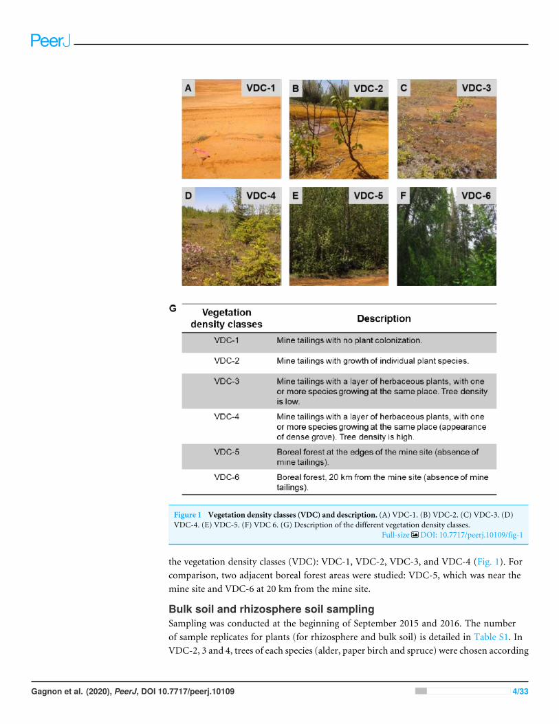
Figure 1 Vegetation density classes (VDC) and description. (A) VDC-1. (B) VDC-2. (C) VDC-3. (D)VDC-4. (E) VDC-5. (F) VDC 6. (G) Description of the different vegetation density classes.
Full-size DOI: 10.7717/peerj.10109/fig-1
the vegetation density classes (VDC): VDC-1, VDC-2, VDC-3, and VDC-4 (Fig. 1). Forcomparison, two adjacent boreal forest areas were studied: VDC-5, which was near themine site and VDC-6 at 20 km from the mine site.
Bulk soil and rhizosphere soil samplingSampling was conducted at the beginning of September 2015 and 2016. The numberof sample replicates for plants (for rhizosphere and bulk soil) is detailed in Table S1. InVDC-2, 3 and 4, trees of each species (alder, paper birch and spruce) were chosen according
Gagnon et al. (2020), PeerJ, DOI 10.7717/peerj.10109 4/33

to their availability and size, selecting primarily only small plants (approximately 0.5 min height) (Table S1). These provided plant samples, and soil/tailings samples. To collectfifty gram soils/tailings samples in areas without plants (VDC-1), in the forest adjacentto the mine site (VDC-5) and in the distant far boreal forest (VDC-6), samples wererandomly collected under the litter (if present) in the first 30 cm of soil. More specifically,bulk soil (and not rhizosphere soil) was collected in VDC-1, 5 and 6, due to the lack ofplants (VDC-1) or the difficulty in VDC-5 and VDC-6 of harvesting the rhizosphere soilof mature trees in the forest. All plant and soil/tailings samples were kept at 4 ◦C until theyarrived at the laboratory. Then, the roots were shaken in a beaker to collect the rhizospheresoil (approximately 20 g). The rhizosphere and bulk soil were frozen at −20 ◦C until RNAextraction.
Soil physico-chemical analysisSoil pH, water content and elemental analyses by inductively coupled plasma massspectrometry (ICP-MS) (except for total nitrogen and organic content) were performedpreviously (Gagnon et al., 2020). For elemental analysis, samples were digested using nitricacid (HNO3). For plant tissues, this digestion resulted in complete mineralization of thesamples. Digestion of soil samples with this strong acid (HNO3) is an established methodknown to extract all metals that can become environmentally available, but does not extractmetal included in the mineral fraction of recalcitrant minerals (e.g., silicates) (Osher etal., 2006; Tuovinen et al., 2015, U.S. EPA, 1996; Zhi Dang, Liu & Haigh, 2002). Digestionswere carried out on a heating plate (DigiPREP Mini, SCP SCIENCE, Québec, Canada).Briefly, samples were crushed using a mortar and pestle in liquid nitrogen. Twenty-fivemg of powder were transferred to 15 ml digiTUBES R© (SCP SCIENCE) to which 1 ml ofconcentrated nitric acid (HNO3 OptimaTM, trace metal grade, Fisherbrand, Fair Lawn, NJ,USA) was added. Tubes were heated at 45 ◦C for 30 min. Then, another 1 ml of nitric acidwas added, and the tubes were heated at 65 ◦C for 2 h. Digestates were brought to 10 mlwith milliQ R© water (Millipore, Québec, Canada). Elemental quantitation was performedusing a Thermo Fisher XSERIES 2 ICP-MS (Thermo Fisher Scientific, Bremen, Germany)and PlasmaLab software (Thermo Electron Corp. 2004. version 2.6.1.335. [PlasmaLab].Germany: PC). Rhodium was used as an internal standard. The SLR5 certified waterstandard was used as an internal control to calibrate the ICP-MS. The studied metals were:sodium (23Na), magnesium (24Mg), aluminum (27Al), phosphorus (31P), potassium (39K),calcium (44Ca), titanium (47Ti), vanadium (51V), chromium (52Cr), manganese (55Mn),iron (57Fe), cobalt (59Co), nickel (60Ni), copper (65Cu), zinc (66Zn), arsenic (75As),selenium (82Se), molybdenum (95Mo), silver (107Ag), cadmium (111Cd), antimony (121Sb),barium (137Ba), tungsten (182W), thallium (205Tl) and lead (206Pb). Total nitrogen wasdetermined by the combustionmethod (AOAC 990.03) and organic content was quantifiedby the calcination method (AGDEX 522, method PR-2) in an external laboratory (GroupeEnvironeX, https://www.labenvironex.com/en/). The results are presented in Table S2.
Gagnon et al. (2020), PeerJ, DOI 10.7717/peerj.10109 5/33
RNA extraction, library preparation and sequencingTotal RNA was extracted from environmental samples and then separated from DNA.RNA-based analysis was preferred overDNA-based analysis, as it provides data onmicrobialpopulations that are present and active, and not simply present (DNA).
Two grams of frozen bulk soil or rhizosphere was weighed into tubes containing 1.5g of silica beads. Total RNA was extracted, purified and eluted as described in the RNAPowerSoil R© Total RNA Isolation kit (MO BIO Laboratories Inc., #12866-25, CA, USA).For removal of residual DNA, the RNA extracts were subjected to DNase treatment usingthe DNase I kit (Thermo Fisher Scientific, #18068015, Canada). Efficiency of DNA removalwas verified through universal PCR amplification of the bacterial 16S rRNA genes andvisualization of PCR product on 2% agarose gels to confirm the absence of DNA.
The 16S rRNA gene sequences (eubacteria and archaea) and the fungal nuclear ribosomalinternal transcribed spacer (ITS) region were first amplified by RT-PCR using the OneStepRT-PCR kit (Qiagen, #220211, Canada). The specific primer sequences (Integrated DNATechnologies, Canada) for these regions (16S rRNA gene and ITS) are described in Table S3.These were chosen to identify bacterial and fungal classes and genera, as reported in thecurrent literature (Kaiser et al., 2016; Sun et al., 2017; Urbanovà, Šnajdr & Baldrian, 2015;Žifčáková et al., 2016). For each region (16S rRNA gene and ITS), four pairs of primerswith additional bases inserted between the sequencing primer overhang and the genespecific sequence (staggered pad) were used proportionally among the samples to creatediversity in the sequence reads, thereby improving the quality of sequencing data. Foramplification of the 16S rRNA region, a peptide nucleic acid (PNA) clamp was usedto prevent amplification of eukaryotic RNA. PNA chloroplast and PNA mitochondrialblockers were used as proposed in Hong, Yang & Zhuang (2016).
Amplicons were visualized on a 2% agarose gel. The samples were purified usingAMPure XP magnetic beads (Beckman Coulter Genomics, #A63881, CA, USA), and asecond short amplification of 8 cycles was carried out with the Nextera XT index kit(Illumina R© #FC-131-1001, set A, B, C and D, CA, USA) to insert different index primerson the amplicons, according to the protocol in the Illumina ‘‘16SMetagenomic SequencingLibrary Preparation’’ guide (Part #15044223 Rev. B). Unique codes were added to eachsample by amplifying 5 µl of the purified PCR product with 25 µl of KAPA HIFI HotStartReady Mix, 300 nM each Nextera XT Index Primer (Illumina R© Inc., CA, USA) and 10µl UltraPureTM DNase/RNase-Free Distilled Water for a total volume of 50 µl. Thermalcycling conditions were as follows: 3 min at 98 ◦C, 8 cycles of 30 s at 98 ◦C, 30 s at 55 ◦C,30 s at 72 ◦C, and a final elongation step of 5 min at 72 ◦C. Indexed amplicons werepurified with magnetic beads as described above. Amplicon products were quantified usingthe Quant-it PicoGreenTM dsDNA Assay Kit according to the manufacturer’s instruction(Thermo Fisher Scientific, MA, USA) and combined in an equimolar ratio. The ampliconpool was verified with a Bioanalyzer 2100 (Agilent Technologies, CA, USA) to confirmthe size of the amplicons and to visualize the potential presence of primer dimers oradapters, in which case an additional purification step of the amplicon pools with SelectSPRI (Beckman Coulter, #B23319, CA, USA) beads was performed. Finally, the pool wasdenatured with 0.2 N NaOH, and the internal control PhiX was added at 5% (Illumina R©
Gagnon et al. (2020), PeerJ, DOI 10.7717/peerj.10109 6/33

Inc., #FC-110-3001, CA, USA). The samples were sequenced on the MiSeq instrument(Illumina R© Inc., #SY-410-1003, CA, USA) using the MiSeq v2 500 cycle kit (Illumina R©
Inc., #MS-102-2003, CA, USA) at the National Research Council Canada in Montréal,Québec (Canada).
Bioinformatics analysisSequencing results were analyzed using a previously described methodology (Tremblay etal., 2015; Yergeau et al., 2015). Briefly, both the 16S rRNA gene and ITS amplicons wereseparately filtered, assembled, trimmed and controlled for quality. Sequencing data wasanalyzed using AmpliconTagger (Tremblay & Yergeau, 2019). Raw reads were scanned forsequencing adapters and PhiX spike-in sequences and remaining reads were merged usingtheir common overlapping part with FLASH (Magoč & Salzberg, 2011). Primer sequenceswere removed from merged sequences and remaining sequences were filtered for qualitysuch that sequences having an average quality (Phred) score lower than 30 or one or moreundefined bases (N) or more than 5 bases lower than a quality score 15 were discarded.
The generation of OTU tables was done with a three round clustering strategy. Qualitycontrolled sequences were dereplicated at 100% identity. These 100% identity clusterswere denoised at 99% identity using dnaclust v3 (Ghodsi, Liu & Pop, 2011). Clusters havingan abundance of 3 or more reads were scanned for chimeras with UCHIME denovo andUCHIME reference-based algorithms, using the Broad Institute 16S rRNA gene Goldreference (http://microbiomeutil.sourceforge.net). The remaining clusters were clusteredat 97% identity (dnaclust) to generate OTUs.
The taxonomic assignment of bacterial and fungal OTU results was performed with theRDP classifier (Bayesian classifier) with a training model constructed from the Greengenesdatabase (version 13.5) (DeSantis et al., 2006; Wang et al., 2007). Fungal OTUs wereclassified using a training model constructed with the United Database (Kõljalg et al.,2013).
The RDP classifier gives a score (0 to 1) to each taxonomic depth of each OTU. Eachtaxonomic depth having a score≥ 0.5 was kept to reconstruct the final lineage. Taxonomiclineages were combinedwith the cluster abundancematrix obtained above to generate a rawOTU table. From that rawOTU table, anOTU table containing bacteria only was generated.This latter OTU table was normalized using the edgeR R packages (Robinson, McCarthy &Smyth, 2010) to obtain a Counts Per Million (CPM) normalized OTU table. A multiplesequence alignment was then obtained by aligning OTU sequences on a Greengenes corereference alignment (DeSantis et al., 2006) using the PyNAST v1.2.2 aligner (Caporaso etal., 2010). Alignments were filtered to keep only the hypervariable region of the alignment.Alpha (observed species (Chao1 index)) and beta (Bray Curtis distances) diversity metricsand taxonomic summaries were then computed using the QIIME v1.9.1 software suite(Caporaso et al., 2010; Kuczynski et al., 2011) using the normalized OTU table.
Sequencing data are available on the NCBI Sequence Read Archive (SRA) portal underaccession number PRJNA634113.
Gagnon et al. (2020), PeerJ, DOI 10.7717/peerj.10109 7/33

Statistical analysisA matrix of environmental properties needed for the redundancy analysis (RDA) andthe MANOVA was performed as described in Gagnon et al. (2020). Briefly, the speciesdata matrix (y) was transformed by a centered log ratio since OTU data are consideredcompositional data (Macklaim, 2014). Since there were 174 bacterial classes and 1906bacterial genera and 31 fungal classes and 889 fungal genera, for uniformity we chose tokeep the 30 most abundant bacterial and fungal classes and genera for RDA. The RDA wasperformed using the ‘‘rda’’ function with the significant explanatory variables selected bythe ‘‘ordiR2step’’ function. A second RDA was performed on the significant explanatoryvariables. The significance of the explanatory variable (red arrows) was verified using the‘‘anova.cca’’ function (Table S4). The RDA are presented in type II scaling to allow a betterinterpretation of the correlation between matrices (x) and (y) (Legendre & Legendre, 2009).
Principal coordinate analysis (PCoA) was performed on normalized OTU tables. Adistance matrix was created via the ‘‘vegdist’’ function, and PCoA was performed usingthe ‘‘pcoa’’ function. MANOVA was conducted to determine if significant differencesin active bacterial populations and active fungal populations were present across VDC,or between plant species (terHorst, Lennon & Lau, 2014; Liu et al., 2015b; Wu et al., 2017;Warton & Hudson, 2004). Table 1 presents the p-values indicating the significance ofdifferences observed between the active microbial populations between the various VDCs,and between plant species.
The microbial community profiles of the most abundant bacterial and fungal classesand genera were determined using the Shiny software (In-house R script) that allows theillustration of 20 classes.
To characterize the degree of change, and possible trends in microbial diversity acrossVDC, Chao1 boxplots were performed (GraphPad Prism 8 software for OS X, v8.1.2). Weused the ‘‘Identify outlier’’ (method: ROUT, Q = 1%) function in GraphPad Prism toremove outliers from Chao1 datasets.
Statistical differences between vegetation density classes and plant species were analyzedin R using the ‘‘Kruskal.test’’, ‘‘Dunn.test’’, ‘‘lm’’ and ‘‘Anova’’ functions. Significantdifferences in bacterial richness between VDC and plant species were investigated usingnon-parametric Kruskal–Wallis followed by Dunn’s multicomparison test as the datasetdid not meet the normality requirement after transformation. Significant differences infungal richness between VDCwere investigated using unbalanced ANOVA (sum of squaresof type III) analysis followed by Tukey’s post-hoc test.
To determine what microbial populations were shared (vs unique) amongst VDC orplant species, the following online software was used: http://bioinformatics.psb.ugent.be/webtools/Venn/. The software sorted our metadata and OTU table (at the genus level),producing a list of most to least abundant populations. The 100 most abundant bacterialOTUs, and 100 most abundant fungal OTUs were then categorized into Venn tables thatlist which OTUs were shared or unique amongst VDC or plant species (Tables S5 and S6).
Gagnon et al. (2020), PeerJ, DOI 10.7717/peerj.10109 8/33
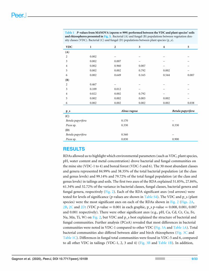
Table 1 P-values fromMANOVA (nperm= 999) performed between the VDC and plant species’ soilsand rhizospheres presented in Fig. 3. Bacterial (A) and fungal (B) populations between vegetation den-sity classes (VDC). Bacterial (C) and fungal (D) populations between plant species (p_s).
VDC 1 2 3 4 5
(A)2 0.002 – – – –3 0.002 0.007 – – –4 0.002 0.960 0.007 – –5 0.002 0.002 0.792 0.002 –6 0.002 0.649 0.165 0.544 0.007
(B)2 0.467 – – – –3 0.109 0.012 – – –4 0.022 0.002 0.792 – –5 0.002 0.002 0.003 0.002 –6 0.002 0.002 0.002 0.002 0.038
p_s Alnus rugosa Betula papyrifera
(C)Betula papyrifera 0.170 –Picea sp. 0.330 0.330
(D)Betula papyrifera 0.560 –Picea sp. 0.830 0.900
RESULTSRDAs allowed us to highlight which environmental parameters (such as VDC, plant species,pH, water content and metal concentration) drove bacterial and fungal communities onthe mine site (VDC-1 to 4) and boreal forest (VDC-5 and 6). The 30 most abundant classesand genera represented 84.99% and 38.35% of the total bacterial population (at the classand genus levels) and 99.14% and 79.72% of the total fungal population (at the class andgenus levels) in tailings and soils. The first two axes of the RDA explained 51.85%, 27.84%,61.34% and 32.72% of the variance in bacterial classes, fungal classes, bacterial genera andfungal genera, respectively (Fig. 2). Each of the RDA significant axes (red arrows) weretested for levels of significance (p-values are shown in Table S4). The VDC and p_s (plantspecies) were the most significant axes on each of the RDAs shown in Fig. 2 (Figs. 2A,2B, 2C and 2D: (VDC p-value = 0.001 in each graphic, p_s p-value = 0.008, 0.001, 0.007and 0.001 respectively). There were other significant axes (e.g., pH, Ca, Cd, Cr, Cu, Fe,Na, Mn, Ti, W) on Fig. 2, but VDC and p_s best explained the structure of bacterial andfungal communities. Further analysis (PCoA) revealed that most differences in bacterialcommunities were noted in VDC-1 compared to other VDC (Fig. 3A and Table 1A). Totalbacterial communities also differed between alder and birch rhizospheres (Fig. 3C andTable 1C). Differences in fungal total communities were found in VDC-5 and 6, comparedto all other VDC in tailings (VDC-1, 2, 3 and 4) (Fig. 3B and Table 1B). In addition,
Gagnon et al. (2020), PeerJ, DOI 10.7717/peerj.10109 9/33
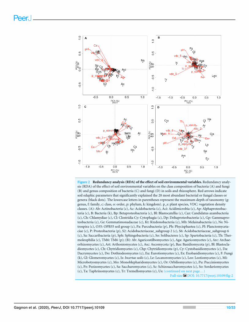
Figure 2 Redundancy analysis (RDA) of the effect of soil environmental variables. Redundancy analy-sis (RDA) of the effect of soil environmental variables on the class composition of bacteria (A) and fungi(B) and genus composition of bacteria (C) and fungi (D) in soils and rhizosphere. Red arrows indicatesoil edaphic parameters that significantly explained the 20 most abundant bacterial or fungal classes orgenera (black dots). The lowercase letters in parentheses represent the maximum depth of taxonomy (g:genus, f: family, c: class, o: order, p: phylum, k: kingdom). p_s: plant species, VDC: vegetation densityclasses. (A): Ab: Actinobacteria (c), Ac: Acidobacteria (c), Aci: Acidimicrobia (c), Ap: Alphaproteobac-teria (c), B: Bacteria (k), Bp: Betaproteobacteria (c), Bl: Blastocatellia (c), Caz: Candidatus azambacteria(c), Ch: Chlamydiae (c), Cl: Clostridia Cy: Cytophagia (c), Dp: Deltaproteobacteria (c), Gp: Gammapro-teobacteria (c), Ge: Gemmatimonadaceae (c), Kt: Ktedonobacteria (c), Mb: Melainabacteria (c), Ns: Ni-trospira (c), O35: OPB35 soil group (c), Pa: Parcubacteria (p), Ph: Phycisphaetea (c), Pl: Planctomyceta-ciae (c), P: Proteobacteria (p), S2: Acidobacteriaceae_subgroup 2 (c), S6: Acidobacteriaceae_subgroup 6(c), Sa: Saccaribacteria (p), Sph: Sphingobacteria (c), So: Solibacteres (c), Sp: Spartobacteria (c), Th: Ther-moleophilia (c), TM6: TM6 (p); (B): Ab: Agaricostilbomycetes (c), Aga: Agaricomycetes (c), Arc: Archae-orhizomycetes (c), Art: Arthoniomycetes (c), Asc: Ascomycota (p), Bas: Basidiomycota (p), Bl: Blastocla-diomycetes (c), Ch: Chytridiomycetes (c), Chp: Chytridiomycota (p), Cy: Cystobasidiomycetes (c), Da:Dacrymycetes (c), Do: Dothiodeomycetes (c), Eu: Eurotiomycetes (c), Ex: Exobasidiomycetes (c), F: Fungi(k), Gl: Glomeromycetes (c), Is: Incertae sedis (c), Le: Lecanoromycetes (c), Leo: Leotiomycetes (c), Mi:Microbotyomycetes (c), Mo: Monoblepharidomycetes (c), Or: Orbiliomycetes (c), Pu: Pucciniomycetes(c), Pz: Pezizomycetes (c), Sa: Saccharomycetes (c), Sc: Schizosaccharomycetes (c), So: Sordariomycetes(c), Ta: Taphrinomycetes (c), Tr: Tremellomycetes (c), Us: (continued on next page. . . )
Full-size DOI: 10.7717/peerj.10109/fig-2
Gagnon et al. (2020), PeerJ, DOI 10.7717/peerj.10109 10/33

Figure 2 (. . .continued)Ustilaginomycetes (c); (C): 03: 0319-6G20 (f), Ac: Acidobacteraceae (c), Ab: Acidibacter (g), Aq: Aquicella(g), At: Acidothermus (g), B: Bacteria (k), Bd: Bdellovibrio (g), Bl: Blrii4 (f), Br: Bryobacter (g), Caz:Candidatus azambacteria (c), Cs: Candidatus solibacter (c), Ch: Chthniobacter (g), Ge: Gemmata(g), Gr: Granulicella (g), H16: H16 (g), Ha: Haliangium (g), Le: Legionella (g), O35: OPB35 soilgroup (c), Pa: Parcubacteria (p), P: Proteobacteria (p), UAc: Uncultured-Acidobacteraceae (c), UPl:Uncultured-Planctomycetaciae (c), Rh: Rhizomicrobium (g), S2: Acidobactericaceae _subgroup 2 (c),S6: Acidobactericaceae_subgroup 6 (c), Sa: Saccharibacteria (p), SM: SM2D12 (f), So: Sorangium (g),Te: Tepidisphaeraceae (c), TM6: TM6 (p); (D): Ac: Archaeorhizomyces (g), Aga: Agarocimycetes (c),Agl: Agaricales (o), Am: Amphinema (g), Ar: Articulospora (g), Asc: Ascomycota (p), Bas: Basidiomycota(p), Cl: Cladophialospora (g), Cla: Claussenomyces (g), Co: Cortinarius (g), Cry: Cryptococcus (g), Eu:Eurotiomycetes (c), F: Fungi (k), He: Helotiales (o), Hy: Hyaloscypha (g), In: Inocybe (g), Is: Incertea sedis(c), La: Lactarius (g), Leo: Leotiomycetes (c), Mol:Mollisia (g), Mo:Mortierella (g), Rh: Rhizoscyphus (g),Ru: Russula (g), Sc: Schizangiella (g), Se: Serendipita (g), So: Sordariomycetes (c), Th: Thelephora (g), To:Tomentella (g), Ty: Tylospora (g).
differences were noted between alder and spruce rhizosphere fungal communities (Fig. 3Dand Table 1D).
The six most abundant classes of bacteria for each VDC and p_s were Gammaproteobac-teria (7.9%–11.5%), Deltaproteobacteria (9.5%–10.8%), Alphaproteobacteria (8.3%–10.4%), Planctomycetacia (7.0%–9.3%), Acidobacteria (3.9%–6.4%) and Actinobacteria(2.8%–5.7%) (Fig. 4 and Table S7 A). For fungi, the classes ‘‘others (with no phylum)’’,Agaricomycetes (11.3%–26.3%), Leotiomycetes (5.7%–18.7%), ‘‘others (Ascomycotaphyla)’’ (9.3%–14.5%), Incertae sedis (3.6%–19.7%) and Sordariomycetes (2.3%–8.3%)were the six most abundant (Fig. 5 and Table S7B). The 100 most abundant bacterial andfungal genera in each of the VDC as well as p_s are listed in Tables S5 and S6.
Acidiferrobacter sp. (median abundance: 0.0002%) and Leptospirillum sp. (medianabundance: 0.0007%) had a greater relative abundance in VDC-1 (Figs. 6A and 6B).Bradyrhizobium showed a greater relative abundance in the rhizosphere on the vegetatedtailings and the boreal forest (median abundances: 0.000216% to 0.000218%) (VDC-2to 6) (Fig. 6C). Frankia sp. showed an equal relative abundance (median abundances:0.0009%–0.0013%) in each of the vegetation densities (Fig. 6D). Pseudomonas sp. showedthe highest relative abundance in the boreal forest (VDC-5 and 6) (median abundances:0.0013% and 0.0035%, respectively) in comparison to all other VDC (Fig. 6E) (medianabundances: 0.001158% to 0.001163%).
The fungal genus Hypocrea had higher relative abundances (median abundances:0.0024% to 0.0029%) in tailings with low to high vegetation density (VDC-2, 3 and 4)compared to barren tailings of VDC-1 (Fig. 7A). In the presence of no or low vegetation(VDC-1 and 2)Mollisia showed a greater relative abundance (median abundances: 0.0028%and 0.0041%, respectively) compared to other VDC (Fig. 7D) (median abundances:0.0007% to 0.0008%). Dissophora sp., and Mortierella sp. showed the highest relativeabundance in VDC 5 and 6 (Figs. 7B and 7C) (Dissophora sp. median abundances: 0.0021%and 0.0066%, Mortierella sp. median abundances: 0.0648% and 0.1846%). Other fungalgenus tended to be found in higher abundance in forest soils (VDC-5 and/or VDC-6)compared to tailings: Cortinarius sp. (VDC-5, median abundance 0.0042%),Neonectria sp.(VDC-5 and 6, median abundances 0.0003% and 0.0331%, respectively) and Rhinocladiella
Gagnon et al. (2020), PeerJ, DOI 10.7717/peerj.10109 11/33
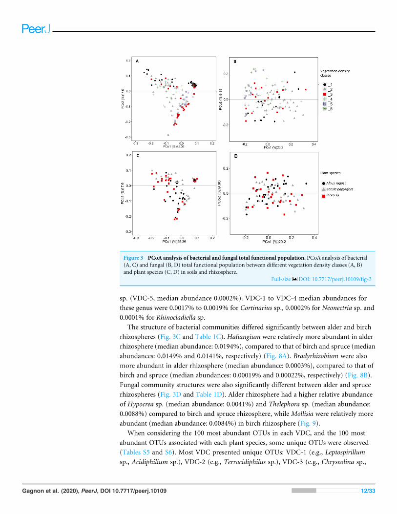
Figure 3 PCoA analysis of bacterial and fungal total functional population. PCoA analysis of bacterial(A, C) and fungal (B, D) total functional population between different vegetation density classes (A, B)and plant species (C, D) in soils and rhizosphere.
Full-size DOI: 10.7717/peerj.10109/fig-3
sp. (VDC-5, median abundance 0.0002%). VDC-1 to VDC-4 median abundances forthese genus were 0.0017% to 0.0019% for Cortinarius sp., 0.0002% for Neonectria sp. and0.0001% for Rhinocladiella sp.
The structure of bacterial communities differed significantly between alder and birchrhizospheres (Fig. 3C and Table 1C). Haliangium were relatively more abundant in alderrhizosphere (median abundance: 0.0194%), compared to that of birch and spruce (medianabundances: 0.0149% and 0.0141%, respectively) (Fig. 8A). Bradyrhizobium were alsomore abundant in alder rhizosphere (median abundance: 0.0003%), compared to that ofbirch and spruce (median abundances: 0.00019% and 0.00022%, respectively) (Fig. 8B).Fungal community structures were also significantly different between alder and sprucerhizospheres (Fig. 3D and Table 1D). Alder rhizosphere had a higher relative abundanceof Hypocrea sp. (median abundance: 0.0041%) and Thelephora sp. (median abundance:0.0088%) compared to birch and spruce rhizosphere, while Mollisia were relatively moreabundant (median abundance: 0.0084%) in birch rhizosphere (Fig. 9).
When considering the 100 most abundant OTUs in each VDC, and the 100 mostabundant OTUs associated with each plant species, some unique OTUs were observed(Tables S5 and S6). Most VDC presented unique OTUs: VDC-1 (e.g., Leptospirillumsp., Acidiphilium sp.), VDC-2 (e.g., Terracidiphilus sp.), VDC-3 (e.g., Chryseolina sp.,
Gagnon et al. (2020), PeerJ, DOI 10.7717/peerj.10109 12/33

Figure 4 Bacterial community profile (relative abundance of transcripts) of the 20 most abundantclasses in soils and the rhizospheres of different vegetation density classes (A) and plant species (B).Letters in parentheses indicate phyla (Ac: Acidobacteria, Ab: Actinobacteria, B: Bacteroidetes, C: Chlamy-diae, Ch: Chloroflexi, F: Firmicutes, P: Proteobacteria, Pl: Planctomycetes, S: Saccharibacteria, V: Verru-comicrobia). Other bacteria designates bacteria that were not identified to phylum level.
Full-size DOI: 10.7717/peerj.10109/fig-4
Sphingomonas sp.), and VDC-6 (e.g., Rhizobium sp., Rhodoplanes sp.) (Table S5A).Fungal communities showed more unique OTUs in each VDC, compared to bacterialcommunities, specifically: VDC-1 (e.g., Acidomyces sp., Sclerotinia sp.), VDC-2 (e.g., Pezizasp., Gymnopilus sp.), VDC-3 (e.g., Lanzia sp., Podospora sp.), VDC-4 (e.g., Clitopilus sp.,Hyphodontia sp.), VDC-5 (e.g., Kalaharituber sp., Phacidium sp.), and VDC-6 (e.g., Apodussp., Entoloma sp.) (Table S6A). In plant species, some unique bacterial OTUs also werenoted in Alnus rugosa (e.g., Chryseolinea sp, Phaselicystis sp.), and Betula papyrifera (e.g.,Pseudomonas sp., Terracidiphilus sp.) (Table S5A). Fungal communities showed moreunique OTUs in each of the plant species, compared to bacterial communities. Uniquefungal OTUs included the following in plant species: Alnus rugosa (e.g., Apodus sp., Pezizasp.), Betula papyrifera (e.g., Gymnopilus sp., Clitopilus sp.), and Picea sp. (e.g., Podosporasp., Schizosaccharomyces sp.) (Table S6B).
The Chao1 index of richness in microbial population biodiversity showed that bacterialrichness in tailings increased significantly in the presence of vegetation, compared to thebarren VDC-1 (Fig. 10A). Indeed, bacterial richness in VDC-2 to VDC-6 was 2.5-foldto 3.5 fold higher compared to VDC-1 (VDC-1: 525.7, VDC-2: 1327.1, VDC-3: 1601.9,VDC-4: 1399.5, VDC-5: 1843.8 and VDC-6: 1524.8). We also observed that the level ofbacterial diversity in tailings from VDC-2 to VDC-4 was comparable to that of the forestsoils of VDC-5 and VDC-6. Fungal richness in diversity did not differ significantly between
Gagnon et al. (2020), PeerJ, DOI 10.7717/peerj.10109 13/33
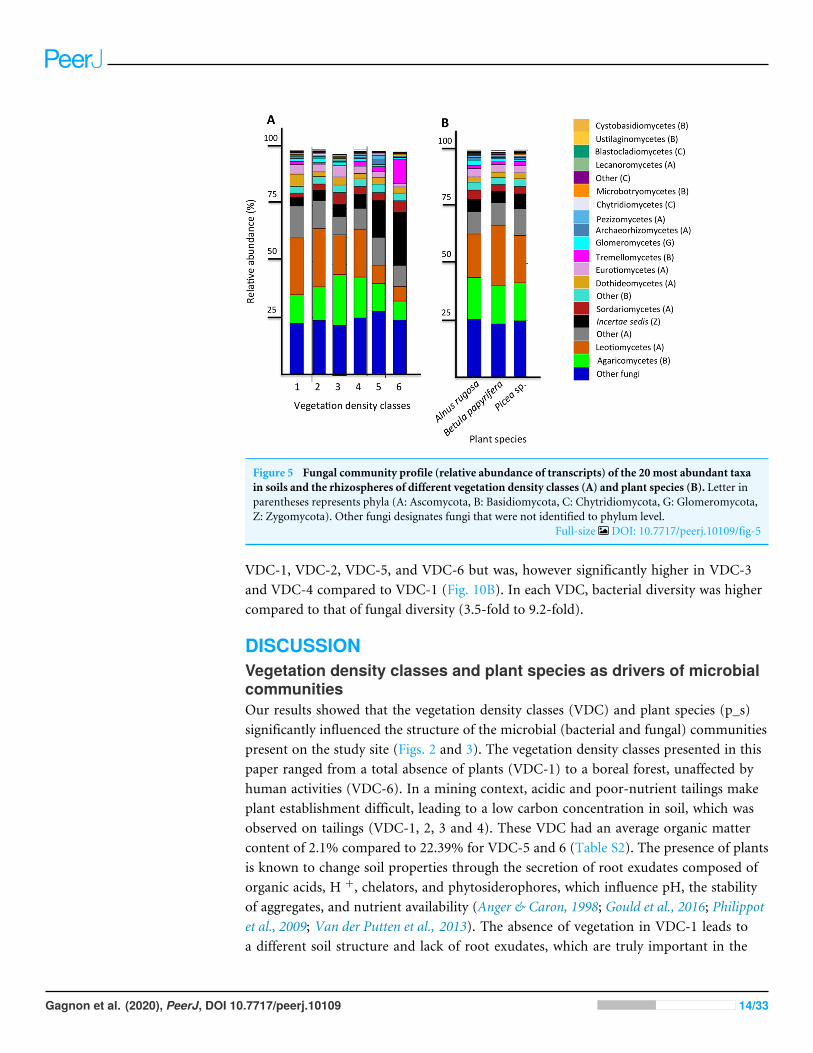
Figure 5 Fungal community profile (relative abundance of transcripts) of the 20 most abundant taxain soils and the rhizospheres of different vegetation density classes (A) and plant species (B). Letter inparentheses represents phyla (A: Ascomycota, B: Basidiomycota, C: Chytridiomycota, G: Glomeromycota,Z: Zygomycota). Other fungi designates fungi that were not identified to phylum level.
Full-size DOI: 10.7717/peerj.10109/fig-5
VDC-1, VDC-2, VDC-5, and VDC-6 but was, however significantly higher in VDC-3and VDC-4 compared to VDC-1 (Fig. 10B). In each VDC, bacterial diversity was highercompared to that of fungal diversity (3.5-fold to 9.2-fold).
DISCUSSIONVegetation density classes and plant species as drivers of microbialcommunitiesOur results showed that the vegetation density classes (VDC) and plant species (p_s)significantly influenced the structure of the microbial (bacterial and fungal) communitiespresent on the study site (Figs. 2 and 3). The vegetation density classes presented in thispaper ranged from a total absence of plants (VDC-1) to a boreal forest, unaffected byhuman activities (VDC-6). In a mining context, acidic and poor-nutrient tailings makeplant establishment difficult, leading to a low carbon concentration in soil, which wasobserved on tailings (VDC-1, 2, 3 and 4). These VDC had an average organic mattercontent of 2.1% compared to 22.39% for VDC-5 and 6 (Table S2). The presence of plantsis known to change soil properties through the secretion of root exudates composed oforganic acids, H +, chelators, and phytosiderophores, which influence pH, the stabilityof aggregates, and nutrient availability (Anger & Caron, 1998; Gould et al., 2016; Philippotet al., 2009; Van der Putten et al., 2013). The absence of vegetation in VDC-1 leads toa different soil structure and lack of root exudates, which are truly important in the
Gagnon et al. (2020), PeerJ, DOI 10.7717/peerj.10109 14/33
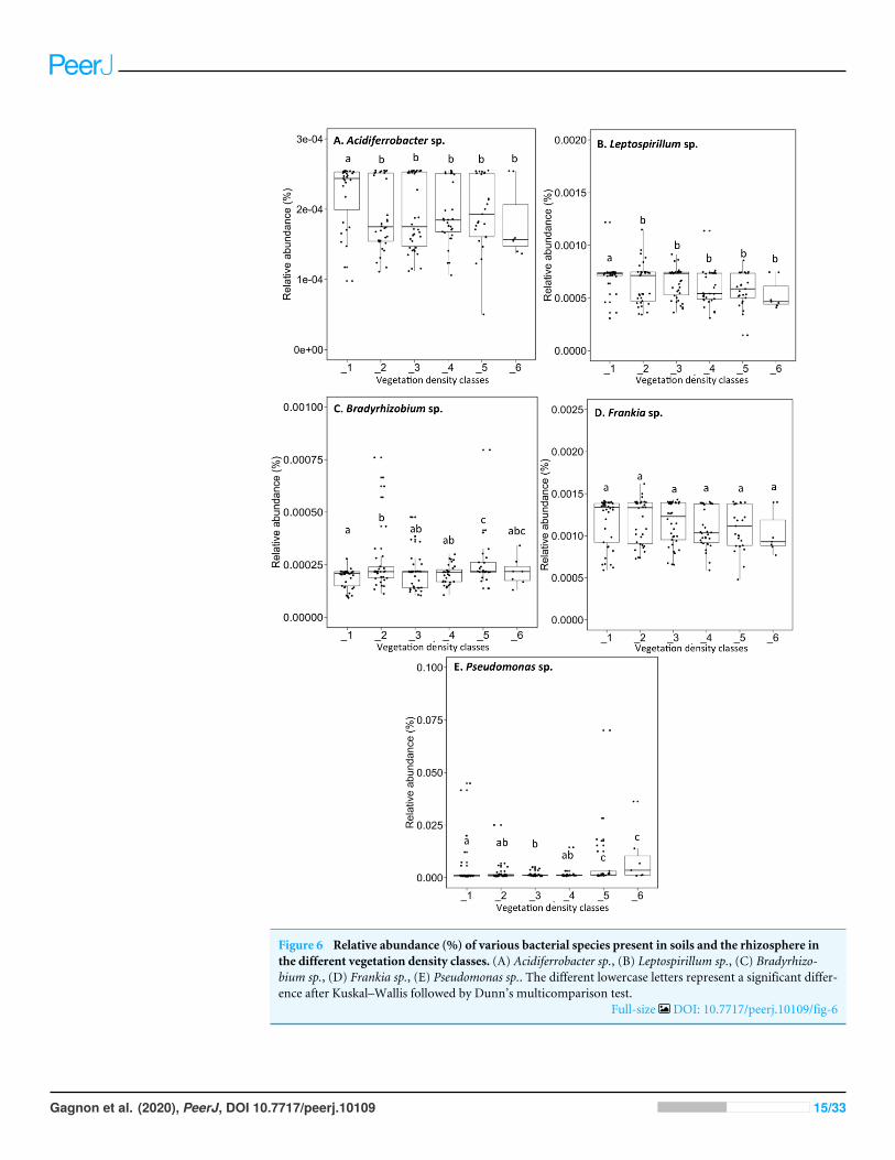
Figure 6 Relative abundance (%) of various bacterial species present in soils and the rhizosphere inthe different vegetation density classes. (A) Acidiferrobacter sp., (B) Leptospirillum sp., (C) Bradyrhizo-bium sp., (D) Frankia sp., (E) Pseudomonas sp.. The different lowercase letters represent a significant differ-ence after Kuskal–Wallis followed by Dunn’s multicomparison test.
Full-size DOI: 10.7717/peerj.10109/fig-6
Gagnon et al. (2020), PeerJ, DOI 10.7717/peerj.10109 15/33
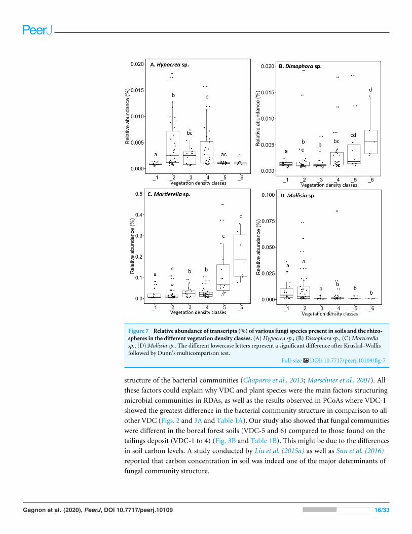
Figure 7 Relative abundance of transcripts (%) of various fungi species present in soils and the rhizo-spheres in the different vegetation density classes. (A) Hypocrea sp., (B) Dissophora sp., (C)Mortierellasp., (D)Molissia sp.. The different lowercase letters represent a significant difference after Kruskal–Wallisfollowed by Dunn’s multicomparison test.
Full-size DOI: 10.7717/peerj.10109/fig-7
structure of the bacterial communities (Chaparro et al., 2013; Marschner et al., 2001). Allthese factors could explain why VDC and plant species were the main factors structuringmicrobial communities in RDAs, as well as the results observed in PCoAs where VDC-1showed the greatest difference in the bacterial community structure in comparison to allother VDC (Figs. 2 and 3A and Table 1A). Our study also showed that fungal communitieswere different in the boreal forest soils (VDC-5 and 6) compared to those found on thetailings deposit (VDC-1 to 4) (Fig. 3B and Table 1B). This might be due to the differencesin soil carbon levels. A study conducted by Liu et al. (2015a) as well as Sun et al. (2016)reported that carbon concentration in soil was indeed one of the major determinants offungal community structure.
Gagnon et al. (2020), PeerJ, DOI 10.7717/peerj.10109 16/33
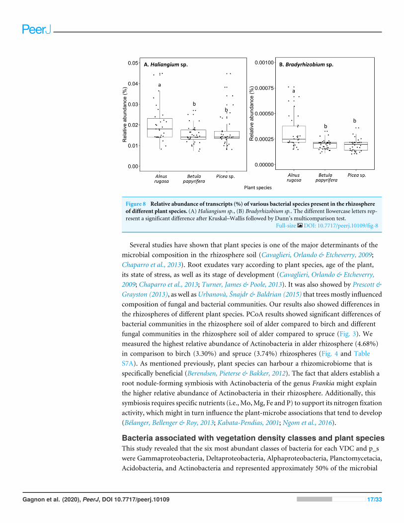
Figure 8 Relative abundance of transcripts (%) of various bacterial species present in the rhizosphereof different plant species. (A) Haliangium sp., (B) Bradyrhizobium sp.. The different llowercase letters rep-resent a significant difference after Kruskal–Wallis followed by Dunn’s multicomparison test.
Full-size DOI: 10.7717/peerj.10109/fig-8
Several studies have shown that plant species is one of the major determinants of themicrobial composition in the rhizosphere soil (Cavaglieri, Orlando & Etcheverry, 2009;Chaparro et al., 2013). Root exudates vary according to plant species, age of the plant,its state of stress, as well as its stage of development (Cavaglieri, Orlando & Etcheverry,2009; Chaparro et al., 2013; Turner, James & Poole, 2013). It was also showed by Prescott &Grayston (2013), as well asUrbanovà, Šnajdr & Baldrian (2015) that trees mostly influencedcomposition of fungal and bacterial communities. Our results also showed differences inthe rhizospheres of different plant species. PCoA results showed significant differences ofbacterial communities in the rhizosphere soil of alder compared to birch and differentfungal communities in the rhizosphere soil of alder compared to spruce (Fig. 3). Wemeasured the highest relative abundance of Actinobacteria in alder rhizosphere (4.68%)in comparison to birch (3.30%) and spruce (3.74%) rhizospheres (Fig. 4 and TableS7A). As mentioned previously, plant species can harbour a rhizomicrobiome that isspecifically beneficial (Berendsen, Pieterse & Bakker, 2012). The fact that alders establish aroot nodule-forming symbiosis with Actinobacteria of the genus Frankia might explainthe higher relative abundance of Actinobacteria in their rhizosphere. Additionally, thissymbiosis requires specific nutrients (i.e., Mo,Mg, Fe and P) to support its nitrogen fixationactivity, which might in turn influence the plant-microbe associations that tend to develop(Bélanger, Bellenger & Roy, 2013; Kabata-Pendias, 2001; Ngom et al., 2016).
Bacteria associated with vegetation density classes and plant speciesThis study revealed that the six most abundant classes of bacteria for each VDC and p_swere Gammaproteobacteria, Deltaproteobacteria, Alphaproteobacteria, Planctomycetacia,Acidobacteria, and Actinobacteria and represented approximately 50% of the microbial
Gagnon et al. (2020), PeerJ, DOI 10.7717/peerj.10109 17/33
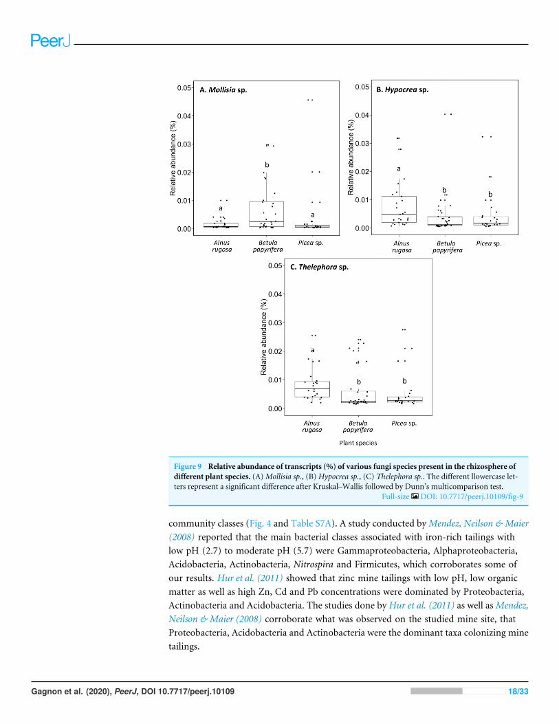
Figure 9 Relative abundance of transcripts (%) of various fungi species present in the rhizosphere ofdifferent plant species. (A)Mollisia sp., (B) Hypocrea sp., (C) Thelephora sp.. The different llowercase let-ters represent a significant difference after Kruskal–Wallis followed by Dunn’s multicomparison test.
Full-size DOI: 10.7717/peerj.10109/fig-9
community classes (Fig. 4 and Table S7A). A study conducted byMendez, Neilson & Maier(2008) reported that the main bacterial classes associated with iron-rich tailings withlow pH (2.7) to moderate pH (5.7) were Gammaproteobacteria, Alphaproteobacteria,Acidobacteria, Actinobacteria, Nitrospira and Firmicutes, which corroborates some ofour results. Hur et al. (2011) showed that zinc mine tailings with low pH, low organicmatter as well as high Zn, Cd and Pb concentrations were dominated by Proteobacteria,Actinobacteria and Acidobacteria. The studies done by Hur et al. (2011) as well asMendez,Neilson & Maier (2008) corroborate what was observed on the studied mine site, thatProteobacteria, Acidobacteria and Actinobacteria were the dominant taxa colonizing minetailings.
Gagnon et al. (2020), PeerJ, DOI 10.7717/peerj.10109 18/33
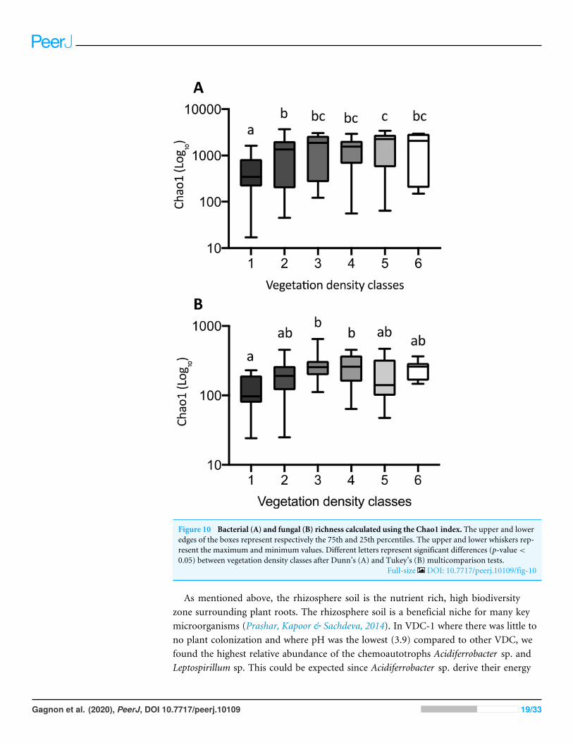
Figure 10 Bacterial (A) and fungal (B) richness calculated using the Chao1 index. The upper and loweredges of the boxes represent respectively the 75th and 25th percentiles. The upper and lower whiskers rep-resent the maximum and minimum values. Different letters represent significant differences (p-value <
0.05) between vegetation density classes after Dunn’s (A) and Tukey’s (B) multicomparison tests.Full-size DOI: 10.7717/peerj.10109/fig-10
As mentioned above, the rhizosphere soil is the nutrient rich, high biodiversityzone surrounding plant roots. The rhizosphere soil is a beneficial niche for many keymicroorganisms (Prashar, Kapoor & Sachdeva, 2014). In VDC-1 where there was little tono plant colonization and where pH was the lowest (3.9) compared to other VDC, wefound the highest relative abundance of the chemoautotrophs Acidiferrobacter sp. andLeptospirillum sp. This could be expected since Acidiferrobacter sp. derive their energy
Gagnon et al. (2020), PeerJ, DOI 10.7717/peerj.10109 19/33
through the oxidation of various inorganic compounds and Leptospirillum sp. does sothrough the oxidation of iron, which is present in high concentrations (31,000 ppm) inVDC-1 (Table S2) (Tyson et al., 2000). The low organic matter content of the tailings(average of 2.1% in (VDC1 to 4) (Gagnon et al., 2020) likely limited the development ofheterotrophic microbial communities in tailings. This could have also contributed to therelative abundance of chemoautotrophs in VDC-1. Such chemoautotrophic organismstend to be less active in the presence of plants because the latter secrete many carbon-richmolecules, which allows heterotrophs to dominate in their presence (Bais et al., 2006;Turner, James & Poole, 2013).
It is known that PGPR organisms facilitate plant growth in abiotic-stressed environments(Grandlic et al., 2008; Sasse, Martinoia & Northen, 2018; Thijs et al., 2016; Weyens et al.,2009; Zhuang et al., 2007). Berendsen, Pieterse & Bakker (2012), as well as Santoyo et al.(2016), showed that plants could select and shape their rhizomicrobiome as a function oftheir homeostatic states and for meeting their needs. Plants growing inmetal-contaminatedsoils have different rhizomicrobiomes compared to plants growing in non-contaminatedsoils; they recruit beneficial microorganisms to support their growth and limit theirmetal uptake (Sasse, Martinoia & Northen, 2018; Thijs et al., 2016). Microorganisms in therhizosphere soil can stimulate plant growth by providing nitrogen, solubilizing phosphorus,producing phytohormones (i.e., auxins, cytokinins), synthesizing siderophores, andprotecting plants from pathogens (Glick, 2010; Prashar, Kapoor & Sachdeva, 2014).
We observed the presence of nitrogen-fixing, symbiotic organisms such asBradyrhizobium sp., Frankia sp., and Rhizobium sp. in VDC-2, 3, and 4. These nitrogenfixers may have improved the competitiveness of plants harbored in these nitrogen-poor substrates (Bais et al., 2006; Ma et al., 2011; Mus et al., 2016; Wani, Khan & Zaidi,2007) as VDC-2 to 4 contained less than 0.25 ppm N (Table S2). Bradyrhizobium sp.have been reported to benefit plants growing in metal-contaminated soil. For example,Bradyrhizodium sp. (vigna) was shown to improve growth, nodulation, nitrogen content,and reduce the uptake of Ni and Zn in green gram (Vigna radiata). This occurred throughthe production of indole-3-acetic acid, siderophores and ammonia (Ma et al., 2011;Wani,Khan & Zaidi, 2007).
Despite many PGPR traits reported in Pseudomonas sp. in contaminated soils (i.e.,mine tailings), our study showed (Fig. 6E) that the relative abundance of Pseudomonassp. was higher in boreal forest soils (VDC-5 and 6) compared to that of the tailingsstorage area (VDC-1 to 4) (Glick, 2010; Ma et al., 2011). Pseudomonas sp. are known fortheir production of organic acids that solubilize mineral phosphorus, and the productionof phosphatase/phytase that hydrolyzes phosphate-organic compounds, increasing theavailability of this limiting nutrient in soils (Hunter, Teakle & Bending, 2014; Richardson etal., 2009; Vacheron et al., 2013).
In our study, average phosphate concentrations were not systematically higher in borealforest soils (VDC-5 and VDC-6; 90.8 and 57.9 ppm, respectively) compared to the tailings(VDC-1 to VDC-4; 18.7, 25.8, 62.2, and 35.1 ppm, respectively) however, higher phosphateconcentrations did tend to occur in denser vegetation (VDC-3 to VDC-6) (Table S2). Soilphosphate concentrations in VDC-6 might be influenced by the rapid uptake of phosphate
Gagnon et al. (2020), PeerJ, DOI 10.7717/peerj.10109 20/33
by mature trees, as the nutrient becomes available. Our observations are therefore notcontradictory; the likely higher demand for phosphorus in the boreal forest vegetationcould explain the higher abundance of Pseudomonas sp. in these soil (VDC-5 and 6),compared to mine tailings (VDC-1 to 4).
The nitrogen demand of alders is higher than that of other plant species. Despite othertree species, the absorption of nutrient by alder (N and P) is approximately in the sameproportion, which could explain the high nitrogen demands by this species to supportgrowth. Tomeet this high nitrogendemand, plant recruitment of beneficialmicroorganismsin the rhizosphere soil is primary (Lõhmus et al., 2006). Bradyrhizobium sp. were knownfor their symbiotic association with legume plant, and to promote nodulation in theseplants. In addition, like other rhizobia, Bradyrhizobium sp. also have the capacity to fixatmospheric nitrogen and to make this nutrient available for other organisms (Saharan &Nehra, 2011). There is little literature on recruitment of Bradyrhizobium sp. by alder, butit is possible that this microorganism could be favorably recruited by alder (in comparisonto birch and spruce) to improve its nitrogen acquisition. Furthermore, few authors havereported the possible role of Haliangium sp. as PGPR organisms. However, Kundim etal. (2003) as well as Ma et al. (2018) do mention that Haliangium sp. secrete antifungalmolecules that limit the development of phytopathogens. The highest relative abundance ofBradyrhizobium sp. andHaliangium sp. in alder rhizosphere soil could also be explained bythe different bacterial selection between different plant species (Lei et al., 2018; Urbanovà,Šnajdr & Baldrian, 2015).
In essence, our results indicate that bacterial richness was significantly lower in VDC-1compared to all other VDC, including those of the natural environment (VDC-5 andVDC-6). Considering the increase of vegetation density as an indicator of environmentalrecovery, these results strongly suggest that bacterial diversity promptly recovers to naturallevels following the establishment of vegetation.
Fungi associated with vegetation density classes and plant speciesThe ectomycorrhizal (ECM) fungi, arbuscular fungi (AM) and dark septate endophytes(DSE) are reported to enhance the phytoremediation potential of plants through elementalcycling and fungal-metal interactions (Deng & Cao, 2017; Pirttilä & Frank, 2011). ECMfungi include an estimated 6000 species (mostly Basidiomycetes), and establish symbiosiswith only 5% of terrestrial plants, in few woody plant families and genera (i.e., Pinaceae,Fagaceae, Betula sp., Populus sp., and Alnus sp.), AM fungi comprise approximately 150species of Zygomycetes (Deng & Cao, 2017). They are associated with herbaceous plants,and various woody plant families (Deng & Cao, 2017; Fortin, Plenchette & Piché, 2015;Landeweert et al., 2001; Mandyam & Jumpponen, 2005). The DSE fungi are reported toestablish symbiosis with over 600 plant species, including plants that were not reportedto be mycorrhizal. They are present in the rhizosphere soil and roots of plants colonizingmetal-contaminated sites, and they are characterised as conidial and sterile fungalendophytes, which form melanised inter- and intra-hyphal structures (Mandyam &Jumpponen, 2005; Upson et al., 2009). Many fungal taxa are reported to establish symbiosiswith plants colonizing metal-laden sites. For example, strains of genera Trichoderma sp.,
Gagnon et al. (2020), PeerJ, DOI 10.7717/peerj.10109 21/33

Fusarium sp., Aspergillus sp., and Cladosporium sp., colonized Portulaca plant, a heavymetal hyperaccumulator (Deng et al., 2014). Penicillum spp. and Trichoderma spp. are alsothe most frequently isolated fungi that attenuate heavy metal stress in plants (Babu et al.,2014a; Babu et al., 2014b; Deng & Cao, 2017; Khan et al., 2014).
ECM fungi can play a role in metal housekeeping through mechanisms such asprecipitation, chelation, cell-wall binding, and the binding of metals by organic acids,polyphosphates, peptides and their transport through intracellular compartments (Colpaertet al., 2011). Moreover, glomalin (a glycoprotein) produced by some AM fungi increaseheavy metal binding, reducing the uptake of heavy metals by the host plants (Bano &Ashfaq, 2013). In addition, ECM and AM fungi are involved in contaminant detoxificationandmediate the nutritional status of heavy metals in plants (Barea, Azcon & Azcon-Aguilar,2002; Harms, Schlosser & Wick, 2011; Luo et al., 2014; Thijs et al., 2016).
DSE are known to enhance mineral uptake of host plants, increase the utilization ofvarious organic pools andmodification of host water uptake. Indeed, the high melanisationof DSE allows them to resist severe drought and heat and increase plant growth undersuch conditions (Kilvin, Emery & Rudgers, 2013; Mandyam & Jumpponen, 2005; Pirttilä &Frank, 2011; Zhang et al., 2008).
Liu et al. (2015a) reported a shift in fungal communities as a function of organic carboncontent in soils: the abundance of Agaricomycetes decreased when more organic carbonwas present, and when there was an increase of Incertae sedis. Our results corroborate this;the relative abundance of Agaricomycetes in mine tailings (VDC-1 to 4) (average ∼20%)is higher than its relative abundance in boreal forest soil (VDC-5 and 6) (average ∼15%).We also observed, as Liu et al. (2015a), a higher relative abundance of Incertae sedis inboreal forest soils (VDC-5 and 6) (∼16%) compared to that in mine tailings (VDC-1 to 4)(∼5%) (Fig. 4 and Table S7B) . The higher organic matter content in boreal forest couldalso explain our PCoA results that showed a difference in fungal communities in VDC-5and 6 in comparison to those in mine tailings soils (VDC-1 to 4) (Fig. 3B and Table 1B).
In our study, ECM Hypocrea sp. were associated more with plant rhizospheres fromVDC-2 to 4, and could potentially be beneficial to plants on such sites. As an example,Morales-Barrera & Cristiani-Urbina (2015) found that Hypocrea tawa could be useful todetoxify Cr(VI)-contaminated wastewaters because of its capacity to reduce hexavalentchromium (Cr(VI)) to a much less toxic trivalent chromium form (Cr(III)). Hypocrea cansolubilizemetals (i.e., Cr,Ni, Cu andPb) and,with othermicroorganisms, could immobilizethese same heavy metals following their solubilisation (Congeevaram et al., 2007; Kumariet al., 2015). Based on the previous studies, Hypocrea sp. could have alleviated heavy metalstress, helping host plants grow on the tailings storage site.
Our study revealed a higher relative abundance of Mollisia sp. in VDC-1 and 2. Thiscould be explained by the fact that Mollisia sp. is known for its high metal tolerance(Pirttilä & Frank, 2011), and that the low organic matter content in these VDC likelyprovided conditions allowing high bioavailability of metals (Table S2).
Our study also found a greater relative abundance of Zygomycota fungi (Dissophora sp.andMortierella sp.) in boreal forest soil (VDC-5 and 6) compared to tailings (VDC-1 to 4).These observations corroborate those of Liu et al. (2015a) and Sun et al. (2016) according
Gagnon et al. (2020), PeerJ, DOI 10.7717/peerj.10109 22/33
to which the relative abundance of Zygomycota (and specificallyMortierella sp.) increasedwith soil carbon content. The organic matter content of VDC-5 and 6 were on average22.4%, while they were 2.1% on the tailings storage area VDC (Fig. 7 and Table S2).
The capability of plants species to shape their rhizosphere soil microbiome to adaptto various environmental conditions can lead to significant differences in the relativeabundance of diverse fungi and bacteria (Berendsen, Pieterse & Bakker, 2012; Sasse,Martinoia & Northen, 2018). Prescott & Grayston (2013) and Urbanovà, Šnajdr & Baldrian(2015) reported that fungal communities in soil were largely explained by dominant trees.Urbanovà, Šnajdr & Baldrian (2015) reported that different proportions of arbuscular andectomycorrhizal fungi were found under different tree species. In our study, we showedthat birch rhizosphere present a higher relative abundance of the DSE fungus Mollisia sp.compared to the rhizosphere soil of alder and spruce (Fig. 9A). This finding is similar tothat reported by Fernández-Miranda Cagigal (2017), where DSE were the dominant fungiof healthy fine root of, amongst other species, Betula pubescens.
Globally, we observed that fungal richness was not as dependent upon the presence ordensity of vegetation as was bacterial diversity. Fungal diversity did not differ significantlybetween VDC-1 to VDC-4, compared to that of boreal forest soils (VDC-5 and VDC-6).While bacterial richness in diversity increased following vegetation establishment, that offungal communities remained relatively stable. These observations corroborate those ofSun et al. (2017) which found that bacterial diversity was 7.0- to 7.5-fold greater comparedto fungal diversity in a recovering forest ecosystem.
CONCLUSIONSOur results indicate that vegetation density classes (VDC) and plant species best explainedthe bacterial and fungal community structures. Indeed, PCoA showed significant differencesin total microbial communities between the various VDC as well as plant species. Thesetwo parameters are intimately related, since the plants, by their root exudates, are able toselect the type of microorganisms composing their rhizospheres. Moreover, the plants thatcolonize these soils change them differently, from one VDC to another, thus producingdifferent physical and chemical properties. These may or may not stimulate plants tostructure their rhizosphere soil microbial communities in different ways.
The capability of complex plant assemblages to colonize a tailings storage area with theseproperties is a demonstration of autonomous recovery following human disturbance. Weobserved that as vegetation density increased and began in some sectors to resemble thatof the adjacent natural forest, microbial communities associated with alder, spruce, andbirch varied. Indeed, while plant species is a known determinant of microbial communitycomposition in the rhizosphere, this study revealed that the parameter of vegetation densityalso explained community structure. This is an important observation since the capabilityof nutrient-poor substrates to sustain plant growth can be improved by the presence ofbacterial and fungal communities capable of weathering minerals and increase water andnutrient acquisition in plants. Microbial communities and more microbial diversity canimprove survival and development of plants. They can contribute directly and indirectly
Gagnon et al. (2020), PeerJ, DOI 10.7717/peerj.10109 23/33
to resilience of these recovering environments. By extension, it is worthwhile to considerplanting biodiverse plant thickets in reclamation efforts. While our study begins to shedlight into the associations, and specific fungal and bacterial taxa, that were found tothrive in this particular environment, subsequent studies will be required to deepen ourunderstanding of the specific plant-microbe associations we observed. Ultimately, thiswill help us better harness plant-microbe and plant-plant associations to improve andaccelerate the ecological restoration of anthropized environments.
ACKNOWLEDGEMENTSThe authors would like to thank the National Research Council Canada (NRC- Energy,Mining and Environment, Montréal) for its contribution to the work (facilities andtechnical support). The authors also thank Francois Rousseu for help with statisticalanalysis, Phillipe Venne for ICP-MS analysis, Christine Maynard for sequencingpreparation, and Julie Beaudin for manuscript preparation and revision.
ADDITIONAL INFORMATION AND DECLARATIONS
FundingThis work was supported by the Fonds de recherche du Québec–Nature et technologies(FRQNT) in the Programme de recherche en partenariat sur le développement durable dusecteur minier under Grant number 2015-MI-193427. The funders had no role in studydesign, data collection and analysis, decision to publish, or preparation of the manuscript.
Grant DisclosuresThe following grant information was disclosed by the authors:Fonds de recherche du Québec–Nature et technologies (FRQNT): 2015-MI-193427.
Competing InterestsThe authors declare there are no competing interests.
Author Contributions• Vanessa Gagnon conceived and designed the experiments, performed the experiments,analyzed the data, prepared figures and/or tables, authored or reviewed drafts of thepaper, and approved the final draft.• Michaël Rodrigue-Morin, Jessica Wasserscheid and Julie Champagne performed theexperiments, authored or reviewed drafts of the paper, and approved the final draft.• Julien Tremblay performed the experiments, analyzed the data, authored or revieweddrafts of the paper, and approved the final draft.• Jean-Philippe Bellenger conceived and designed the experiments, analyzed the data,authored or reviewed drafts of the paper, and approved the final draft.• Charles W. Greer and Sébastien Roy conceived and designed the experiments, analyzedthe data, authored or reviewed drafts of the paper, supervision of the research, andapproved the final draft.
Gagnon et al. (2020), PeerJ, DOI 10.7717/peerj.10109 24/33
Field Study PermissionsThe following information was supplied relating to field study approvals (i.e., approvingbody and any reference numbers):
The owners of the mine site granted access to our research team through an agreementbetween the Université de Sherbrooke and the mine owners which allows the scientificstudy of the area surrounding the mine.
DNA DepositionThe following information was supplied regarding the deposition of DNA sequences:
Sequencing data are available at the NCBI Sequence Read Archive (SRA): PRJNA634113.
Data AvailabilityThe following information was supplied regarding data availability:
The data are available at Figshare: Gagnon, Vanessa; Wasserscheid, Jessica; Rodrigue-Morin,Michaël; Tremblay, Julien; Champagne, Julie; Bellenger, Jean-Philippe; et al. (2020):code_dataset. figshare. Dataset. https://doi.org/10.6084/m9.figshare.12530534.v1.
Supplemental InformationSupplemental information for this article can be found online at http://dx.doi.org/10.7717/peerj.10109#supplemental-information.
REFERENCESAgbenyeku E-OE, Muzenda E, Msibi MI. 2016. Chemical alteration in three clayey soils
from percolation and interaction with acid mine drainage (AMD). South AfricanJournal of Chemical Engineering 21:28–36 DOI 10.1016/j.sajce.2016.04.003.
Akcil A, Koldas S. 2006. Acid mine drainage (AMD): causes, treatment and case studies.Journal of Cleaner Production 14:1139–1145 DOI 10.1016/S0734-9750(03)00099-5.
Ali H, Khan E, SajadMA. 2013. Phytoremediation of heavy metal-concepts and applica-tions. Chemosphere 91:869–881 DOI 10.1016/j.chemosphere.2013.01.075.
Anger DA, Caron J. 1998. Plant-induced changes in soil structure: processes andfeedbacks. Biogeochemistry 42:55–72 DOI 10.1023/A:1005944025343.
BaaloushaM,Motelica-HeinoM, Le P. 2006. Conformation and size of humic sub-stances: effects of major cation concentration and type, pH, salinity, and residencetime. Colloids and Surfaces A: Physicochemical and Engineering Aspects 272:48–55DOI 10.1179/0371955042250046.
Babu AG, Shim J, Bang KS, Shea PJ, Oh BT. 2014a. Trichoderma virens PDR-28: a heavymetal-tolerant and plant growth-promoting fungus for remediation and bioenergycrop production on mine tailing soil. Journal of Environmental Management132:129–134 DOI 10.1016/j.jenvman.2013.10.009.
Babu AG, Shim J, Shea PJ, Oh BT. 2014b. Penicillium aculeatum PDR-4 and Tri-choderma sp. PDR-16 promote phytoremediation of mine tailing soil and bio-energy production with sorghum sudangrass. Ecological Engineering 69:186–191DOI 10.1016/j.ecoleng.2014.03.055.
Gagnon et al. (2020), PeerJ, DOI 10.7717/peerj.10109 25/33
Bais HP,Weir TL, Perry LG, Gilroy S, Vivanco JM. 2006. The role of root exudates inrhizosphere interactions with plants and other organisms. Annual Review of PlantBiology 57:233–266 DOI 10.1146/annurev.arplant.57.032905.105159.
Bano SA, Ashfaq D. 2013. Role of mycorrhiza to reduce heavy metal stress. NaturalScience 2(12A):16–20 DOI 10.4236/ns.2013.512A003.
Barea JM, Azcon R, Azcon-Aguilar C. 2002.Mycorrhizosphere interactions toimprove plant fitness and soil quality. Antonie Leeuwenhoek 81:343–351DOI 10.1023/A:1020588701325.
Bélanger P-A, Bellenger J-P, Roy S. 2013. Strong modulation of nutrient distribution inAlnus glutinosa as a function of the actinorhizal symbiosis. Botany 91(4):218–224DOI 10.1139/cjb-2012-0184.
Berendsen RL, Pieterse CMJ, Bakker PAHM. 2012. The rhizosphere microbiome andplant health. Trends in Plant Science 17(8):478–486 DOI 10.1016/j.tplants.2012.04.001.
Bergeron Y, Gauthier S, FlanniganM, Kafka V. 2004. Fire regimes at the transitionbetween mixed wood and coniferous boreal forest in Northwestern Quebec. Ecology85:1916–1932 DOI 10.1890/02-0716.
Bulgarelli D, Schlaeppi K, Spaepen S, Ver Lorenvan Themaat E, Schulze-Lefert P. 2013.Structure and functions of the bacterial microbiota of plants. Annual Review of PlantBiology 64:807–838 DOI 10.1146/annurev-arplant-050312-120106.
Caporaso JG, Kuczynski J, Stombaugh J, Bittinger K, Bushman FD, Costello EK,Fierer N, Gonzalez Peña A, Goodrich JK, Gordon JI. 2010. QIIME allows analysisof high-throughput community sequencing data. Nature Methods 7:335–336DOI 10.1038/nmeth.f.303.
Cavaglieri L, Orlando J, Etcheverry M. 2009. Rhizosphere microbial communitystructure at different maize plant growth stages and root locations.MicrobiologicalResearch 164:391–399 DOI 10.1016/j.micres.2007.03.006.
Chaparro JM, Badri DV, Bakker MG, Sugiyama A, Manter DK, Vivanco JM. 2013.Root exudation of phytochemicals in Arabidopsis follows specific patterns that aredevelopmentally programmed and correlate with soil microbial functions. PLOSONE 8:1–10 DOI 10.1016/j.micres.2007.03.006.
Colpaert JV,Wevers JHL, Krznaric E, Adriaensen K. 2011.How metal-tolerant ecotypeof ectomycorrhizal fungi protect plants from heavy metal pollution. Annals of ForestScience 68:17–24 DOI 10.1007/s13595-010-0003-9.
Congeevaram S, Dhanarani S, Park J, Dexilin M, Thamaraiselvi K. 2007. Biosorption ofchromium and nickel by heavy metal resistant fungal and bacterial isolates. Journal ofHazardous Materials 146(1-2):270–277 DOI 10.1016/j.jhazmat.2006.12.017é.
Deng Z, Cao L. 2017. Fungal endophytes and their interactions with plants in phytoreme-diation: a review. Chemosphere 168:1100–1106DOI 10.1016/j.chemosphere.2016.10.097.
Deng Z, Zhang R, Shi Y, Hu L, Tan H, Cao L. 2014. Characterization of Cd-Pb-Zn-resistant endophytic Lasiodiploda sp. MXSF31 from metal accumulating
Gagnon et al. (2020), PeerJ, DOI 10.7717/peerj.10109 26/33
Portulaca oleracea and its potential in promoting the growth of rape in metal-contaminated soils. Environmental Science and Pollution Research 21:2346–2357DOI 10.1007/s11356-013-2163-2.
DeSantis TZ, Hugenholtz P, Larsen N, Rojas M, Brodie EL, Keller K, Huber T, DaleviD, Hu P, Andersen GL. 2006. Greengenes, a chimera-checked 16S rRNA genedatabase and workbench compatible with ARB. Applied and Environmental Micro-biology 72(7):5069–5072 DOI 10.1128/AEM.03006-05.
Fellet G, Marchiol L, Vedove D, Peressotti A. 2011. Application of biochar on mine tail-ings: effects and perspectives for land reclamation. Chemosphere 83(9):1262–1267DOI 10.1016/j.chemosphere.2011.03.053.
Fernández-Miranda Cagigal E. 2017. Dark septate endophytes (DES) in polluted areas.In: Hughes E, ed. Endophytic fungi. New York: Nova Publisher 125–146.
Fortin JA, Plenchette C, Piché Y. 2015. Les mycorhizes l’essor de la nouvelle révolutionverte. Second edition. Montréal: Multimonde.
Gagnon V, Rodrigue-MorinM, Tardif A, Beaudin J, Greer CW, Shipley B, BellengerJ-P, Roy S. 2020. Differences in elemental composition of tailings, soils, and planttissues following five decades of native plant colonization on a gold mine site inNorthwestern Québec. Chemosphere 250 DOI 10.1016/j.chemosphere.2020.126243.
Gamalero E, Lingua G, Berta G, Glick BR. 2009. Beneficial role of plant growth pro-moting bacteria and arbuscular mycorrhizal fungi on plant responses to heavy metalstress. Canadian Journal of Microbiology 55:501–514 DOI 10.1139/W09-010.
Ghodsi M, Liu B, PopM. 2011. DNACLUST: accurate and efficient clustering ofphylogenetic marker genes. BCM Bioinformatics 12(271):1–11DOI 10.1186/1471-2105-12-271.
Glick BR. 2010. Using soil bacteria to facilitate phytoremediation. Biotechnology Advances28:367–374 DOI 10.1016/j.biotechadv.2010.02.001.
Gould IJ, Quinton JN,Weigelt A, De Deyn GB, Bardgett RD. 2016. Plant diversity androot traits benefit physical properties key to soil function in grasslands. EcologyLetters 19:1140–1149 DOI 10.1111/ele.12652.
Grandlic CJ, MendezMO, Chorover J, Machado B, Maier RM. 2008. Plant growth-promoting bacteria for phytostabilization of mine tailings. Environmental Science andTechnology 42:2–5 DOI 10.1021/es072013j.
Grossnickel SC. 2005. Importance of root growth in overcoming planting stress. NewForest 30(2–3):273–294 DOI 10.1007/s11056-004-8303-2.
Harms H, Schlosser D,Wick LY. 2011. Untapped potential: exploiting fungi inbioremediation of hazardous chemical. Nature Review Microbiology 9:177–192DOI 10.1038/nrmicro2519.
Hong SC, Yang C, Zhuang Z. 2016. Application of peptide nucleic acid–based assaystoward detection of somatic mosaicism.Molecular Therapy - Nucleic Acids 5:1–5DOI 10.1038/mtna.2016.22.
Hunter PJ, Teakle GR, Bending GD. 2014. Root traits and microbial communityinteractions in relation to phosphorus availability and acquisition, with particularreference to Brassica. Frontiers in Plant Science 5:1–18 DOI 10.3389/fpls.2014.00027.
Gagnon et al. (2020), PeerJ, DOI 10.7717/peerj.10109 27/33
HurM, Kim Y, Song H-R, Min Kim J, Im Choi Y, Yi H. 2011. Effect of geneticallymodified poplars on soil microbial communities during the phytoremediation ofwaste mine tailings. Applied and Environmental Microbiology 77(21):7611–7619DOI 10.1128/AEM.06102-11.
Jung SC, Martinez-Medina A, Lopez-Raez JA, PozoMJ. 2012.Mycorrhiza-inducedresistance and priming of plant defenses. Journal of Chemical Ecology 38:651–664DOI 10.1007/s10886-012-0134-6.
Kõljalg U, Nilsson RH, Abarenkov K, Tedersoo L, Taylor AFS, BahramM, Bates ST,Bruns TD, Bengtsson-Palme J, Callaghan TM, Douglas B, Drenkhan T, EberhardtU, Dueñas M, Grebenc T, Griffith GW, HartmannM, Kirk PM, Kohout P, LarssonE, Lindahl BD, Lücking R, Martín MP, Matheny PB, Nguyen NH, Niskaren T,Oja J, Peay KG, Peintrer U, PetersonM, Põldmaa K, Saag L, Saar I, Schüßler A,Scott JA, Senés C, SmithME, Suija A, Taylor DL, Telleria MT,Weiss M, LarssonK-H. 2013. Towards a unified paradigm for sequence-based identification of fungi.Molecular Ecology 22(21):5271–5277.
Kabata-Pendias A. 2001. Trace elements in soils and plants. Third edition. New York:CRC press.
Kaiser K,Wemheuer B, Korolkow V,Wemheuer F, Nacke H, Schöning I, SchrumpfM, Daniel R. 2016. Driving forces of soil bacterial community structure, diversity,and function in temperate grasslands and forests. Scientific Reports 6:33696DOI 10.1038/srep33696.
Kefeni KK, Msagati TAM,Mamba BB. 2017. Acid mine drainage: prevention, treatmentoptions, and resource recovery: a review. Journal of Cleaner Production 151:475–493DOI 10.1016/j.jclepro.2017.03.082.
Khan AG. 2005. Role of soil microbes in the rhizospheres of plants growing on tracemetal contaminated soils in phytoremediation. Journal of Trace Elements in MedicineBiology 18:355–364 DOI 10.1016/j.jtemb.2005.02.006.
Khan AL,Waqas M, Hussain J, Al-Harrasi A, Lee I-J. 2014. Fungal endophyte Peni-cillium janthinellum LK5 can reduce cadmium toxicity in Solanum lyco-persicum(Sitiens and Rhe). Biology and Fertility of Soils 50:75–85DOI 10.1007/s00374-013-0833-3.
Kilvin SN, Emery SM, Rudgers JA. 2013. Fungal symbionts alter plant responses toglobal change. American Journal of Botany 100(7):1445–1457DOI 10.3732/ajb.1200558.
KneeshawDD, Bergeron Y. 1998. Canopy gap characteristics and tree replacement in theSoutheastern boreal forest. Ecology 79(3):783–794 DOI 10.1890/0012-9658(1998)079.
Kuczynski J, Stombaugh J, WaltersWA, González A, Caporaso JG, Knight R. 2011.Using QIIME to analyze 16S rRNA gene sequences from microbial communities.Current Protocols in Bioinformatics Chapter 10:Unit 10.7DOI 10.1002/0471250953.bi1007s36.
Kumari D, Pan X, Achal V, Zhang D, Al-Misned F, MortuzaMG. 2015.Multiple metal-resistant bacteria and fungi from acidic copper mine tailings of Xinjiang, China.
Gagnon et al. (2020), PeerJ, DOI 10.7717/peerj.10109 28/33
International Journal of Systematic and Evolutionary Microbiology 74(4):3113–3121DOI 10.1007/s12665-015-4349-z.
Kundim BA, Itou Y, Sakagami Y, Fudou R, Iizuka T, Yamanaska S, Ojika M. 2003. Newhaliangicin isomers, potent antifungal metabolites produced by a marine myxobac-terium. The Journal of Antibiotics 56:630–638 DOI 10.7164/antibiotics.56.630.
Lõhmus K, TruuM, Truu J, Ostonen I, Kaar E, Vares A, Uri V, Alama S, Kanal A. 2006.Functional diversity of culturable bacteria communities in the rhizosphere in relationto fine-root and soil parameters in alder stands on forest, abandoned agricultural,and oil-shale mining area. Plant and Soil 283:1–10 DOI 10.1007/s11104-005-2509-8.
Landeweert R, Hoffland E, Finlay RD, Kuyper TW, Breemen NV, Finlay RD. 2001.Linking plants to rocks: ectomycorrhizal fungi mobilize nutrients from minerals.Trends in Ecology and Evolution 16(5):248–254 DOI 10.1016/S0169-5347(01)02122-X.
Legendre P, Legendre L. 2009.Numerical ecology. Third edition. Oxford: Elsevier.Lei S, Xu X, Chen Z, Xiong J, Ma R, Zhang L, Yang X, Zhu Y, Zhang B, Tian B.
2018. Analysis of the community composition and bacterial diversity of therhizosphere microbiome across different plant taxa.Microbiology e762:1–10DOI 10.1002/mbo3.762.
Liu J, Sui Y, Yu Z, Shi Y, Chu H, Jin J, Liu X,Wang G. 2015a. Soil carbon con-tent drives the biogeographical distribution of fungal communities in theblack soil zone of Northeast China. Soil Biology and Biochemistry 83:29–39DOI 10.1016/j.soilbio.2015.01.009.
LiuW, Xi X, Sudu Q, Kwok L, Guo Z, Hou Q, Menhe B, Su T, Zhang H. 2015b.High-throughput sequencing reveals microbial community diversity of Ti-betan naturally fermented yak milk. Annals of Microbiology 65:1741–1751DOI 10.1007/s13213-014-1013-x.
Luo Z-B,Wu C, Zhang C, Li H, Lipka U, Polle A. 2014. The role of ectomycorrhizasin heavy metal stress tolerance of host plants. Environmental Experimental Botany108:47–62 DOI 10.1016/j.envexpbot.2013.10.018.
MaM, Jiang X,Wang Q, Guan D, Li L, OngenaM, Li J. 2018. Isolation and identi-fication of PGPR strain and its effect on soybean growth and soil bacterial com-munity composition. International Journal of Agriculture Biology 20:1289–1297DOI 10.17957/IJAB/15.0627.
Ma Y, PrasadMNV, RajkumarM, Freitas H. 2011. Plant growth promoting rhizobacte-ria and endophytes accelerate phytoremediation of metalliferous soils. BiotechnologyAdvances 29:248–258 DOI 10.1016/j.biotechadv.2010.12.001.
Macklaim JM. 2014. Unifying the analysis of high-throughput sequencing datasets: char-acterizing RNA-seq, 16S rRNA gene sequencing and selective growth experiments bycompositional data analysis.Microbiome 2:1–13 DOI 10.1186/2049-2618-2-15.
Magoč T, Salzberg SL. 2011. FLASH: fast length adjustment of short reads to improvegenome assemblies. Bioinformatics 27:2957–2963 DOI 10.1093/bioinformatics/btr507.
MandyamK, Jumpponen A. 2005. Seeking the elusive function of the root-colonizingdark septate endophytic fungi. Studies in Mycology 53:173–189DOI 10.3114/sim.53.1.173.
Gagnon et al. (2020), PeerJ, DOI 10.7717/peerj.10109 29/33
Marschner P, Yang C-H, Lieberei R, Crowley DE. 2001. Soil and plant effects onbacterial community composition in the rhizosphere. Soil Biology and Biochemistry33:1437–1445 DOI 10.1016/S0038-0717(01)00052-9.
MendezMO, Neilson JW,Maier RM. 2008. Characterization of a bacterial communityin an abandoned semiarid lead-zinc mine tailings site. Applied and EnvironmentalMicrobiology 74(12):3899–3907 DOI 10.1128/AEM.02883-07.
Ministère de l’Énergie et des Ressources naturelles (MERN). 2018.History of themining industry. Available at https://mern.gouv.qc.ca/ en/mines/mining-industry/history-mining-industry/ (accessed on 20 February 2020).
Ministère de l’Énergie et des Ressources naturelles (MERN). 2019. Restauration dessites miniers abandonnés. Available at http://mern.gouv.qc.ca/mines/ restauration-miniere/ restauration-des-sites-miniers-abandonnes/ (accessed on 20 February 2020).
Miransari M. 2010. Contribution of arbuscular mycorrhizal symbiosis to plant growthunder different types of soil stress. Plant Biology 12:563–569DOI 10.1111/j.1438-8677.2009.00308.x.
Morales-Barrera L, Cristiani-Urbina E. 2015. Bioreduction of hexavalent chromium byHypocrea tawa in a concentric draft-tube airlift bioreactor. Journal of Environmentaland Biotechnology Research 1(1):37–44.
Mulligan C, Yong RN, Gibbs BF. 2001. Remediation technologies for metal- contam-inated soils and groundwater: an evaluation. Engineering Geology 60:193–207DOI 10.1016/S0013-7952(00)00101-0.
Mus F, CrookMB, Garcia K, Garcia Costas A, Geddes BA, Kouri ED, ParamasivanP, RyuM-H, Oldroyd GED, Poole PS, Udvardi MK, Voigt CA, Ané J-M, Pe-ters JW. 2016. Symbiotic nitrogen fixation and the challenges to its extensionto non-legumes. Applied and Environmental Microbiology 82(13):3698–3710DOI 10.1128/AEM.01055-16.
NgomM, Oshone R, Diagne N, CissokoM, Svistoonoff S, Tisa LS, Laplaze L, OurèyeS, Champion A. 2016. Tolerance to environmental stress by the nitrogen-fixingactinobacterium Frankia and its role in actinorhizal plants adaptation. Symbiosis70(1-3):17–29 DOI 10.1007/s13199-016-0396-9.
Osher LJ, Leclerc L, Wiersma GB, Hess CT, Guiseppe VE. 2006.Heavy metal con-tamination from historic mining in upland soil and estuarine sediments ofEgypt Bay, Maine, USA. Estuarine, Coastal and Shelf Science 70(1–2):169–179DOI 10.1016/j.ecss.2006.06.009.
Pandey AK, Pandey SD, Misra V. 2000. Stability constants of metal humic acid com-plexes and its role in environmental detoxification. Ecotoxicology and EnvironmentalSafety 47:195–200 DOI 10.1006/eesa.2000.1947.
Phieler R, Voit A, Kothe E. 2013.Microbially supported phytoremediation of heavymetal contaminated soils: strategies and applications. Advances in BiochemicalEngineering/Biotechnology 141:211–235 DOI 10.1007/10_2013_200.
Philippot L, Hallin S, Börjesson G, Baggs EM. 2009. Biochemical cycling in therhizosphere having an impact on global change. Plant and Soil 321:61–81DOI 10.1007/s11104-008-9796-9.
Gagnon et al. (2020), PeerJ, DOI 10.7717/peerj.10109 30/33

Pirttilä AM, Frank CA. 2011. Endophytes of forest trees. New York: Springer.Prashar P, Kapoor N, Sachdeva S. 2014. Rhizosphere: its structure,bacterial diversity
and significance. Reviews in Environmental Science and Bio/Technology 13:63–77DOI 10.1007/s11157-013-9317-z.
Prescott CE, Grayston SJ. 2013. Tree species influence on microbial communities inlitter and soil: current knowledge and research needs. Forest Ecology Management309:19–27 DOI 10.1016/j.foreco.2013.02.034.
RajkumarM, Sandhya S, PrasadMNV, Freitas H. 2012. Perspectives of plant-associated microbes in heavy metal phytoremediation. Biotechnology Advances30(6):1562–1574 DOI 10.1016/j.biotechadv.2012.04.011.
Richardson AE, Barea J-M, McNeill AM, Prigent-Combaret C. 2009. Acquisitionof phosphorus and nitrogen in the rhizosphere and plant growth promotion bymicroorganisms. Plant and Soil 321:305–339 DOI 10.1007/s11104-009-9895-2.
RobinsonMD,McCarthy DJ, Smyth GK. 2010. edgeR: a bioconductor package fordifferential expression analysis of digital gene expression data. Bioinformatics26(1):139–140 DOI 10.1093/bioinformatics/btp616.
Saharan BS, Nehra V. 2011. Plant growth promoting rhizobacteria: a critical review. LifeSciences and Medicine Research 21:1–30.
Santoyo G, Moreno-Hagelsied G, Orozco-MosquedaMDC, Glick BR. 2016. Plantgrowth-promoting bacterial endophytes.Microbiological Research 183:92–99DOI 10.1016/j.micres.2015.11.008.
Sasse J, Martinoia E, Northen T. 2018. Feed your friends: do plant exudates shape theroot microbiome. Trends in Plant Science 23(1):25–41DOI 10.1016/j.tplants.2017.09.003.
Sheoran V, Sheoran AS, Poonia P. 2010. Soil reclamation of abandoned mine land byrevegetation: a review. International Journal of Soil, Sediment and Water 3(2):1–21.
Simate GS, Ndlovu S. 2014. Acid mine drainage: challenge and opportunities. Journal ofEnvironmental Chemical Engineering 2(3):1785–1803 DOI 10.1016/j.jece.2014.07.021.
Sun R, DsouzaM, Gilbert JA, Gui X,Wang D, Guo Z, Ni Y, Chu H. 2016. Fungalcommunity composition in soils subjected to long-term chemical fertilizationis most influenced by the type of organic matter. Environmental Microbiology18(12):5137–5150 DOI 10.1111/1462-2920.13512.
Sun S, Li S, Avera BN, Strahm BD, Badgley BD. 2017. Soil bacterial and fungal commu-nities show distinct recovery pattern during forest ecosystem restoration. Applied andEnvironmental Microbiology 83(14):e00966-17 DOI 10.1128/AEM.00966-17.
Tardif A, Rodrigue-MorinM, Gagnon V, Shipley B, Roy S, Bellenger J-P. 2019. Therelative importance of abiotic conditions and subsequent land use on the borealprimary succession of acidogenic mine tailings. Ecological Engineering 127:66–74DOI 10.1016/j.ecoleng.2018.11.003.
terHorst CP, Lennon JT, Lau JA. 2014. The relative importance of rapid evolution forplant-microbe interactions depends on ecological cotext. Proceedings of the RoyalSociety B. Biological Sciences 281:20140028 DOI 10.1098/rspb.2014.0028.
Gagnon et al. (2020), PeerJ, DOI 10.7717/peerj.10109 31/33
Thijs S, SillenW, Rineau F,Weyens N, Vangronsveld J. 2016. Toward an en-hanced understanding of plant-microbiome interactions to improve phytore-mediation: engineering the metaorganism. Frontiers in Microbiology 7:1–15DOI 10.3389/fmicb.2016.00341.
Tremblay J, Singh K, Fern A, Kirton ES, He S, Woyke T, Lee J, Chen F, Dangl JL, TringeSG. 2015. Primer and platform effects on 16S rRNA tag sequencing. Frontiers inMicrobiology 6:1–15 DOI 10.3389/fmicb.2015.00771.
Tremblay J, Yergeau E. 2019. Systematic processing of ribosomal RNA gene ampliconsequencing data. Gigascience 8(12):1–14.
Tuovinen H, Vesterbacka D, Pohjolainen E, Read D, Solatie D, Lehto J. 2015.A comparison of analytical methods for determining uranium and thoriumin ores and mill tailings. Journal of Geochemical Exploration 148:174–180DOI 10.1016/j.gexplo.2014.09.004.
Turner TR, James EK, Poole PS. 2013. The plant microbiome. Genome Biology 14:209DOI 10.1186/gb-2013-14-6-209.
Tyson GW, Chapman J, Hugenholtz P, Allen EE, Ram RJ, Richardson PM, SolovyevVV, Rubin EM, Rokhsar DS, Banfield JF. 2000. Community structure andmetabolism through reconstruction of microbial genomes from the environment.Nature 428:1–7 DOI 10.1038/nature02340.
Upson R, NewshamKK, Bridge PD, Pearce DA, Read DJ. 2009. Taxonomic affinitiesof dark septate endophytes of Colobanthus quitensis and Deschampsia antarc-tica, the two native antarctic vascular plant species. Fungal Ecology 2:184–196DOI 10.1016/j.funeco.2009.02.004.
UrbanovàM, Šnajdr J, Baldrian P. 2015. Composition of fungal and bacterial communi-ties in forest litter and soil is largely determined by dominant trees. Soil Biology andBiochemistry 84:53–64 DOI 10.1016/j.soilbio.2015.02.011.
U.S. EPA. 1996.Method 3050B: acid digestion of sediments, sludges, and soils. Revision2. Washington, D.C. Available at https://www.epa.gov/ sites/production/ files/ 2015-06/documents/ epa-3050b.pdf .
Vacheron J, Desbrosses G, BouffaudM-L, Touraine B, Moënne-Loccoz Y, MullerD, Legendre L, Wisniewski-Dyé F, Prigent-Combaret C. 2013. Plant growth-promoting rhizobacteria and root system functioning. Frontiers in Plant Science4:1–19 DOI 10.3389/fpls.2013.00356.
Van der PuttenWH, Bardgett RD, Bever JG, Bezemer TM, Casper BB, Fukami T,Kardol P, Klironomos JN, Kulmatiski A, Schweitzer JA, Suding KN, Vande VoordeTFJ, Wardle DA. 2013. Plant-soil feedbacks: the past, the present and the futurechallenges. Journal of Ecology 101:265–276 DOI 10.1111/1365-2745.12054.
Wang Q, Garrity GM, Tiedje JM, Cole JR. 2007. Naïve bayesian classifier for rapidassignment of rRNA sequences into the new bacterial taxonomy. Applied andEnvironmental Microbiology 73(16):5261–5267 DOI 10.1128/AEM.00062-07.
Wang L, Ji B, Runqing L, SunW. 2017. A review on in situ phytoremediation of minetailings. Chemosphere 184:594–600 DOI 10.1016/j.chemosphere.2017.06.025.
Gagnon et al. (2020), PeerJ, DOI 10.7717/peerj.10109 32/33
Wani PA, KhanMS, Zaidi A. 2007. Effect of metal tolerant plant growth promotingBradyrhizobium sp. (vigna) on growth, symbiosis, seed yield and metal uptake bygreengram plants. Chemosphere 70(1):36–45 DOI 10.1016/j.chemosphere.2007.07.028.
Warton DI, Hudson HM. 2004. AMANOVA statistic is just as powerful as distance-based statistics for multivariate abundances. Ecology 85:858–874 DOI 10.1890/02-0419.
Weyens N, Van der Lelie D, Taghavi S, Vangronsveld J. 2009. Phytoremediation:plant-endophyte partnerships take the challenge. Current Opinion in Biotechnology20(2):248–254 DOI 10.1016/j.copbio.2009.02.012.
Wu L, Yang Y, Chen S, Shi ZJ, ZhaoM, Zhu Z, Yang S, Qu Y, Ma Q, He Z, Zhou J,He Q. 2017.Microbial functional trait of rRNA operon copy numbers increaseswith organic levels in anaerobic digesters. The ISME Journal 11:2874–2878DOI 10.1038/ismej.2017.135.
Yergeau E, Bell TH, Champagne J, Maynard C, Tardif S, Tremblay J, Greer CW.2015. Transplanting soil microbiomes leads to lasting effects on willow growth,but not on the rhizosphere microbiome. Frontiers in Microbiology 6:1–14DOI 10.3389/fmicb.2015.01436.
Zhang Y, Zhang Y, LiuM, Shi X, Zhao Z. 2008. Dark septate endophyte (DSE) fungiisolated from metal polluted soils: their taxonomic position, tolerance, andaccumulation of heavy metal in vitro. Journal of Microbiology 46(6):624–632DOI 10.1007/s12275-008-0163-6.
Zhi Dang Z, Liu C, HaighMJ. 2002.Mobility of heavy metals associated with thenatural weathering of coal mine spoils. Environmental Pollution 118(3):419–426DOI 10.1016/S0269-7491(01)00285-8.
Zhuang X, Chen J, ShimH, Bai Z. 2007. New advances in plant growth-promotingrhizobacteria for bioremediation. Environment International 33:406–413DOI 10.1016/j.envint.2006.12.005.
Žifčáková L, Větrovský T, Howe A, Baldrian P. 2016.Microbial activity in forestsoil reflects the changes in ecosystem properties between summer and winter.Environmental Microbiology 18(1):288–301 DOI 10.1111/1462-2920.13026.
Gagnon et al. (2020), PeerJ, DOI 10.7717/peerj.10109 33/33