Varki and Gagneux AN NY ACAD SCI2012
21
Ann. N.Y. Acad. Sci. ISSN 0077-8923 ANNALS OF THE NEW YORK ACADEMY OF SCIENCES Issue: Glycobiology of the Immune Response Multifarious roles of sialic acids in immunity Ajit Varki and Pascal Gagneux Glycobiology Research and Training Center, Departments of Medicine, and Cellular and Molecular Medicine, University of California at San Diego, La Jolla, California Address for correspondence:Ajit Varki or Pascal Gagneux, Glycobiology Research and Training Center, Departments of Medicine, and Cellular and Molecular Medicine, UC San Diego, La Jolla, CA 92093-0687, [email protected] or [email protected] Sialic acids are a diverse family of monosaccharides widely expressed on all cell surfaces of vertebrates and so-called “higher” invertebrates, and on certain bacteria that interact with vertebrates. This overview surveys examples of biological roles of sialic acids in immunity, with emphasis on an evolutionary perspective. Given the breadth of the subject, the treatment of individual topics is brief. Subjects discussed include biophysical effects regulation of factor H; modulation of leukocyte trafficking via selectins; Siglecs in immune cell activation; sialic acids as ligands for microbes; impact of microbial and endogenous sialidases on immune cell responses; pathogen molecular mimicry of host sialic acids; Siglec recognition of sialylated pathogens; bacteriophage recognition of microbial sialic acids; polysialic acid modulation of immune cells; sialic acids as pathogen decoys or biological masks; modulation of immunity by sialic acid O-acetylation; sialic acids as antigens and xeno-autoantigens; antisialoglycan antibodies in reproductive incompatibility; and sialic-acid–based blood groups. Keywords: sialic acids; immunity; evolution; selectins; Siglecs; sialidases Sialic acids (Sias) are unusual sugars with a shared nine-carbon backbone that are widely expressed on the surfaces of all cells in all animals of the deuteros- tome lineage (vertebrates and so-called “higher” invertebrates), and also in certain pathogenic or symbiotic bacteria that associate with them (Refs. 1–7; Fig. 1A). Given their remarkable diversity in structure, glycosidic linkage, and underlying gly- can chains, as well their exposed location, it is not surprising that Sias have numerous roles in many aspects of immunity (by “immunity,” we here mean immunology, as well as aspects of microbiology that are relevant to symbiosis and pathogenesis). Details regarding the occurrence, biosynthesis, structural diversity, cellular expression patterns, rapid evolu- tion, and species variations of sialic acids have been extensively reviewed elsewhere 1–7 and will not be re- peated here. This overview surveys the multifarious roles of sialic acids in selected aspects of immu- nity. Given the vast breadth of the subject under consideration, the treatment of the selected topics is necessarily brief, and references to the primary literature are not comprehensive. The emphasis is also on topics with which the authors are more familiar. Biophysical effects of sialic acids Given their ubiquitous presence and abundance at the surface of all cell types (including those of the immune system), Sias have major biophysical ef- fects. 8,9 The typical cell displays tens of millions of Sia molecules, and it is estimated that the lo- cal concentrations on the cell surface glycocalyx can approach 100 mM. 10 Sialic acids thus pro- vide a large component of negative charge repul- sion between cells, which could alter the biophysical properties of cellular interactions (Fig. 1B). Many earlier studies removed Sias from immune cell sur- faces using sialidases and showed marked changes in behavior of such cells. 11 However, such studies are often confusing, because wholesale removal of cell surface Sias has many potentially pleiotropic effects. First, removal reduces the net charge and hydrophilicity of the cell surface. Second, it can reduce the charge repulsion between adjacent cell surface molecules. Third, it eliminates ligands for doi: 10.1111/j.1749-6632.2012.06517.x 16 Ann. N.Y. Acad. Sci. 1253 (2012) 16–36 c 2012 New York Academy of Sciences.
Transcript of Varki and Gagneux AN NY ACAD SCI2012

Ann. N.Y. Acad. Sci. ISSN 0077-8923
ANNALS OF THE NEW YORK ACADEMY OF SCIENCESIssue: Glycobiology of the Immune Response
Multifarious roles of sialic acids in immunity
Ajit Varki and Pascal GagneuxGlycobiology Research and Training Center, Departments of Medicine, and Cellular and Molecular Medicine, University ofCalifornia at San Diego, La Jolla, California
Address for correspondence: Ajit Varki or Pascal Gagneux, Glycobiology Research and Training Center, Departments ofMedicine, and Cellular and Molecular Medicine, UC San Diego, La Jolla, CA 92093-0687, [email protected] [email protected]
Sialic acids are a diverse family of monosaccharides widely expressed on all cell surfaces of vertebrates and so-called“higher” invertebrates, and on certain bacteria that interact with vertebrates. This overview surveys examples ofbiological roles of sialic acids in immunity, with emphasis on an evolutionary perspective. Given the breadth of thesubject, the treatment of individual topics is brief. Subjects discussed include biophysical effects regulation of factorH; modulation of leukocyte trafficking via selectins; Siglecs in immune cell activation; sialic acids as ligands formicrobes; impact of microbial and endogenous sialidases on immune cell responses; pathogen molecular mimicryof host sialic acids; Siglec recognition of sialylated pathogens; bacteriophage recognition of microbial sialic acids;polysialic acid modulation of immune cells; sialic acids as pathogen decoys or biological masks; modulation ofimmunity by sialic acid O-acetylation; sialic acids as antigens and xeno-autoantigens; antisialoglycan antibodies inreproductive incompatibility; and sialic-acid–based blood groups.
Keywords: sialic acids; immunity; evolution; selectins; Siglecs; sialidases
Sialic acids (Sias) are unusual sugars with a sharednine-carbon backbone that are widely expressed onthe surfaces of all cells in all animals of the deuteros-tome lineage (vertebrates and so-called “higher”invertebrates), and also in certain pathogenic orsymbiotic bacteria that associate with them (Refs.1–7; Fig. 1A). Given their remarkable diversity instructure, glycosidic linkage, and underlying gly-can chains, as well their exposed location, it is notsurprising that Sias have numerous roles in manyaspects of immunity (by “immunity,” we here meanimmunology, as well as aspects of microbiology thatare relevant to symbiosis and pathogenesis). Detailsregarding the occurrence, biosynthesis, structuraldiversity, cellular expression patterns, rapid evolu-tion, and species variations of sialic acids have beenextensively reviewed elsewhere1–7 and will not be re-peated here. This overview surveys the multifariousroles of sialic acids in selected aspects of immu-nity. Given the vast breadth of the subject underconsideration, the treatment of the selected topicsis necessarily brief, and references to the primaryliterature are not comprehensive. The emphasis is
also on topics with which the authors are morefamiliar.
Biophysical effects of sialic acids
Given their ubiquitous presence and abundance atthe surface of all cell types (including those of theimmune system), Sias have major biophysical ef-fects.8,9 The typical cell displays tens of millionsof Sia molecules, and it is estimated that the lo-cal concentrations on the cell surface glycocalyxcan approach 100 mM.10 Sialic acids thus pro-vide a large component of negative charge repul-sion between cells, which could alter the biophysicalproperties of cellular interactions (Fig. 1B). Manyearlier studies removed Sias from immune cell sur-faces using sialidases and showed marked changesin behavior of such cells.11 However, such studiesare often confusing, because wholesale removal ofcell surface Sias has many potentially pleiotropiceffects. First, removal reduces the net charge andhydrophilicity of the cell surface. Second, it canreduce the charge repulsion between adjacent cellsurface molecules. Third, it eliminates ligands for
doi: 10.1111/j.1749-6632.2012.06517.x16 Ann. N.Y. Acad. Sci. 1253 (2012) 16–36 c© 2012 New York Academy of Sciences.

Varki & Gagneux Roles of sialic acids in immunity
Figure 1. Examples of roles of sialic acids in immunity. Sialic acids are shown as pink diamonds. See the text for details.(A) Neu5Ac, the most common sialic acid in mammals. These acidic sugars share a nine-carbon backbone and can be modifiedin many ways. (B) The high density of terminal sialic acids on the glycocalyx of vertebrate cells imparts negative charge andhydrophilicity to cell surfaces, altering biophysical properties. (C) Factor H binds cell surface Sias, protecting cell surfaces fromthe alternative complement pathway. (D) Intrinsic Sia-binding molecules such as selectins on endothelia, leukocytes, and plateletsinitiate leukocyte rolling on endothelial surfaces, a key initial step for leukocyte extravasation. (E) Intrinsic Sia-binding Siglecmolecules on immune cells detect sialylated ligands and can inhibit immune cell activation. There are also activatory Siglecs. (F)Host Sias are frequently exploited as attachment sites (“receptors”) by pathogens including protozoa, viruses, bacteria, and toxins.(G) Microbial sialidases can help pathogens to expose underlying glycan-binding sites, to avoid sialylated decoys (see below), and/orprovide Sias as food sources. The loss of SAMPs from cells may then be used by host immune cells to react to pathogens, and/orto clear away desialylated cells or glycoproteins. (H) Endogenous sialidases such as Neu1 can modulate immune cell function bymodulating receptor clustering, possibly by exposing underlying galactose residues and facilitating galectin-mediated cross-linkingof surface molecules. (I) Microbial mimicry of host Sias allows manipulation of host immune response by engaging inhibitorySiglecs, inhibiting complement via factor H binding, and reducing the opportunity of the host to form antibodies. (J) Microbialsynthesis of Sia-like molecules, such as legionaminic acid and pseudaminic acid stabilizes fimbriae.
endogenous receptors like Siglecs and selectins (seelater). Fourth, sialic acid removal exposes underly-ing glycans (mostly galactose residues), which canbe recognized by other endogenous receptors, suchas galectins and the galactose-binding proteins ofmacrophages. Finally, there is potential for sialidasetreatment to enhance cell surface interactions andlattices of galectins with the uncapped N-glycans ofvarious surface receptors (see later).
Thus, more subtle alterations in cell surface Siasare needed to investigate their specific functions.In this regard, the use of mild periodate oxidationto eliminate only the C9 and C8 side chain carbonatoms of Sias is a remarkably specific manipulation,which leaves the rest of the sialic acid molecule andits negative charge intact, and is not known to af-fect other surface structures.12 However, periodateoxidation also generates a C7-aldehyde on the sialic
Ann. N.Y. Acad. Sci. 1253 (2012) 16–36 c© 2012 New York Academy of Sciences. 17

Roles of sialic acids in immunity Varki & Gagneux
acid side chain, which can potentially react withlysine residues on adjacent proteins, cross-linkingcell surface glycoproteins. This may help explainsome reported dramatic effects of periodate oxida-tion on lymphocytes.13 A less risky option would beto modify the type of sialic acid on the surface. Oneapproach is to feed unnatural (bio-orthogonal) pre-cursors to generate unnatural Sias.14–16 If high levelsof incorporation can be achieved on the cell surface,the altered biophysical properties can then be fur-ther studied. However, Sia recognition phenomenacould also be altered. In this regard, it would also beworth asking whether the single oxygen atom dif-ference between the common Sia (Neu5Ac) and thenonhuman one (Neu5Gc)17 can alter immune cellbehavior.
Meanwhile, the high densities of Sias found onsurface polysaccharides of various pathogenic bac-teria (see later) can also markedly alter the bio-physical properties of these organisms.3–7 But again,simply eliminating these Sias by genetic or enzy-matic means can have secondary effects, due to ex-posure of underlying glycans. Overall, while it isclear that the biophysical properties of Sias modu-late many cellular and microbial interactions in theimmune system, these effects are not easy to study,because the experimental approaches may perturbthe very thing that is being explored in a pleiotropicfashion. This is an important and challenging areafor future studies.
Sialic acid regulation of fluid phase innateimmunity
Classic studies showed a role for Sias in regulat-ing the alternative pathway of the complement ac-tivation.18,19 The mechanism involves the majorserum protein factor H, which recognizes Sias as“self,” gets recruited to native cell surfaces, andso helps to downregulate the constant “tick-over”of the complement pathway on all surfaces (Refs.18–21; Fig. 1C). Details of this mechanism havebeen elucidated, including accelerated dissociationof the C3bBb C3 convertase and acting as a cofac-tor for factor I–mediated cleavage of C3b.21 WhileSias may thus act as “self-associated molecular pat-terns” (SAMPs)22 for recognition by factor H, thisdoes not fully explain the relative specificity of fac-tor H for sialoglycan structural variants. There arealso complexities involving the type of glycosidiclinkage of Sias to the underlying glycan (its presen-
tation in space), which may alter recognition.23,24
Furthermore, studies have shown that this factorH self-recognition is mediated by certain anion-binding sites, which can also recognize sulfated gly-cosaminoglycans as “self.” The factor H domainsinvolved in these recognition phenomena are largelydomains 19–20 (Refs. 25–28), and this mechanismis also hijacked by bacteria that express Sias on theirsurface polysaccharides21,23 (see later). Interestingly,mutations in some of these domains were found bygenome-wide association studies to correlate withincreased risk of complement-mediated inflamma-tory processes, such as hemolytic-uremic syndrome,membranoproliferative glomerulonephritis,29 andage-dependent macular degeneration.27,30 These ex-periments of nature provide evidence of the func-tional significance of Sias as SAMPs for recognitionby factor H.
Variation in sialic acid side chain O-acetylationcan also affect factor H binding. Classic studies sug-gested that the amount of sialic acid on red bloodcells on erythrocytes of different strains of micemight restrict the extent of control of the alter-nate complement pathway activation.31 It was thenshown that the difference was not the amount ofsialic acid, but in the extent of sialic acid side-chainO-acetylation, that is, such modified Sias are notgood targets for factor H binding.32 These olderobservations need to be revisited in the light ofmodern evidence regarding the mechanisms thatcontrol Sia O-acetylation,1,33 as well as genetic andgenomic sequences of these strains. Also of noteis that ficolins (circulating soluble activators of thelectin pathway of complement activation) can rec-ognize sialic acids, particularly on the surfaces ofsialylated bacteria.34–36 This appears to be a hostresponse to molecular mimicry by bacteria (seelater).
Modulation of leukocyte trafficking viasialylated selectin ligands
Until the 1980s, factor H was the only known in-trinsic vertebrate sialic acid–binding protein. A clas-sic study37 then noted that pretreatment of lymphnode sections with a sialidase abolished the inter-action of lymphocytes with the high-endothelialvenules, which normally provide exit sites forlymphocytes. These and other observations even-tually led to recognition of the selectin fam-ily of cell adhesion molecules and their role in
18 Ann. N.Y. Acad. Sci. 1253 (2012) 16–36 c© 2012 New York Academy of Sciences.

Varki & Gagneux Roles of sialic acids in immunity
leukocyte trafficking (Refs. 38–42; Fig. 1D). Dif-ferent isoforms of these endogenous lectins werefound expressed on leukocytes (L-selectin), platelets(P-selectin), and endothelium (P- and E-selectin).It then became apparent that Sias in the glycan se-quence Sia�2–3Gal�1–3/4(Fuc�1–3/4)GlcNAc�1-R (Sialyl Lewis X/A) are critical components of thenatural ligands for these selectins.38,39,43–50 In someinstances, Sialyl Lewis X/A motifs combine withother features such as sulfation on the Gal or Glc-NAc residues (L-selectin ligands), and/or sulfationof adjacent tyrosine residues (P-selectin) and con-tribute toward specific recognition sites on specificproteins, particularly mucin-like glycoproteins.51 Aparticularly striking example was the elucidationof a defined amino-terminal sulfoglycopeptide mo-tif on P-selectin glycoprotein ligand-1 (PSGL-1),which serves as a specific high affinity ligand of P-selectin.52
Whereas Sias form a critical part of most lig-ands for selectins, recognition does not seem tobe affected by other structural details of the Siasthemselves, except that the �2–3 linkage to theunderlying galactose residue is critical. In keepingwith this, some selectin ligands can function witha sulfate ester at the three position of galactose, in-stead of a sialic acid.53 Overall, it appears that Siasare primarily acting as conveyers of a necessary neg-ative charge for selectin interactions, and the de-tails of Sia diversity do not matter. In keeping withthis, some 6-O-sulfated glycosaminoglycans, suchas heparan sulfate, can act as alternate selectin lig-ands.54,55 However, if one oxidizes the side chain ofsialic acid with periodate and generates an aldehyde,this reactive group can be cross-linked into the bind-ing pocket of the selectin via a covalent interaction.56
Overall it is evident that �2–3 linkage-specific sia-lyltransferases play a key role in generating selectinligands, along with �1–3/4 fucosyltransferases,and GlcNAc sulfotransferases and/or tyrosinesulfotransferases.57
Siglecs in the control of immune cellactivation
In the mid-1980s, some macrophage types werefound to form rosettes with sheep erythrocytesin vitro, and that binding could be abolished bysialidase pretreatment of the erythrocytes.58 Thissialic acid–dependent receptor was purified andshown to be a very large protein called siaload-
hesin, which was then demonstrated to bind sialicacid–containing ligands in vitro.59 However giventhe size of the sialoadhesin molecule and the era inwhich this work occurred, cloning proved difficult.Meanwhile, expression cloning of the presumed lig-and for a B cell “adhesion molecule” called CD22had surprisingly yielded a sialyltransferase.60 In fact,it turned out that CD22 was a sialic acid–bindinglectin, with recombinant soluble CD22 shown tobind Sias through its extracellular domain, andnot to the sialyltransferase identified through ex-pression cloning (the transferase is not at the cellsurface but rather generates sialylated ligands ofCD22 in the Golgi).61 Moreover, recognition byCD22 was specific for the �2–6 linkage, with nobinding to �2–3–linked Sia.62 Additional studiesdefined the highly conserved preference of CD22for this linkage and characterized the interactionsfurther.63 Soon thereafter, the cloning of siaload-hesin revealed that its amino-terminal domains hada homology with CD22 and to similar domains oftwo other previously known proteins, CD33 andmyelin-associated glycoprotein (MAG), suggestingthat these molecules might belong to a single fam-ily of sialic acid–binding proteins.64 Further stud-ies showed that this was indeed the case, resultingin recognition of a new family of sialic acid–binding proteins.65 It was initially suggested thatthese molecules be called sialoadhesins.66–68 How-ever, besides the confusing relationship to the firstmolecule already with this name, some of these pro-teins did not seem to mediate cell–cell adhesion.The alternate term suggested was Siglec, to standfor sialic acid–recognizing Ig-superfamily lectins, asa subset of I-type lectins (Ig-like lectins).69 Discus-sions among those working in the field eventually ledto general acceptance of this term, with the found-ing members sialoadhesin, CD22, CD33, and MAGbeing designated Siglecs-1–4 (Ref. 70). Studies ofexpressed sequence tags and mining of genomic se-quence data then led to extension of this family ofintrinsic vertebrate lectins, which now comprise atleast 16 members in primates.71–79
Interestingly, a subset of Siglecs seems rela-tively conserved in mammals (CD22/Siglec-2 andsialoadhesin/Siglec-1), and even among vertebrates(MAG/Siglec-4 and Siglec-15). In contrast, anothersubset found in one large syntenic cluster (onchromosome 19q in humans) shows the high-est amount of variation between species.71,72 This
Ann. N.Y. Acad. Sci. 1253 (2012) 16–36 c© 2012 New York Academy of Sciences. 19

Roles of sialic acids in immunity Varki & Gagneux
subfamily was named CD33-related Siglecs (orCD33rSiglecs) and shown to have a variety of Sia-binding properties. CD33rSiglecs can be furthersubdivided into two categories. Most have cytoso-lic domains containing immune receptor tyrosine-based inhibitory motifs (ITIMs) that can be tyrosinephosphorylated, resulting in recruitment of tyrosinephosphates like SHP-1 and SHP-2.80–84 This in turnresults in dephosphorylation of tyrosine residues onvarious kinases associated with other receptors, ef-fectively downregulating their functions (Fig. 1E).Thus, these inhibitory CD33rSiglecs likely serveas innate immune detectors for SAMPs, therebydownregulating unwanted inflammation, particu-larly that occurring in response to tissue damage.85
Notably several of these CD33rSiglecs also have asecond cytosolic ITIM-like motif whose functionsare much less clear. Moreover, there is evidence thatsome of the inhibitory effects of such Siglecs do notrequire either of these tyrosine-based motifs.86,87
This suggests that more attention should be paid tothe better-conserved extracellular C2-set domains,as mediators of additional and/or complementaryfunctions.
In contrast to the inhibitory Siglecs, a positivelycharged amino acid in the transmembrane do-main of some activating CD33rSiglecs allows themto engage immune receptor tyrosine-based activa-tory motif (ITAM)-containing adapter moleculeslike DAP12, which in turn recruits the tyrosine ki-nase Syk and mediates tyrosine phosphorylationof various receptors and kinases.88–91 In some in-stances, CD33rSiglecs with inhibitory and activa-tory properties have undergone gene conversionevents that maintain their amino-terminal iden-tity, suggesting that they may be paired receptors,sending opposite signals on binding of the same lig-and(s). In this context, it seems likely that the acti-vatory Siglecs represent an evolutionary response tobacteria that are “hijacking” inhibitory Siglecs (seelater).88,92
The general subject of Siglecs and their biol-ogy and evolution has been extensively discussedelsewhere73,74,76,77,79,84,93–98 and details will not berepeated here. However, certain features are wor-thy of special note. First, most Siglecs are typicallybound by so-called “cis ligands,” that is, sialylatedglycans on the same cell surface.99,100 However, an-other cell surface or a soluble ligand with a highenough density of sialylated ligands can compete
out the cis ligands and cause engagement.76,101,102
Second, the amino-terminal V-set Ig-like domaincontains the sialic acid recognition site, including acanonical conserved arginine residue that is criticalfor interaction with the carboxylate of sialylated lig-ands.103–107 Interestingly, this arginine residue canbe naturally mutated, affecting one or more Siglecsunique to a given species or taxon.71,88,107 One pos-sibility is that these events occur randomly becausethe arginine codon (CGN) is highly mutable. How-ever, there are instances where the arginine appearsto mutate and then reappears in one phylogeneticbranch, for example, for Siglecs-5 and Siglecs-14 inhumans versus great apes.88 Taken together with thehigh frequency of such events, it is more likely thatthese mutations are an evolutionary mechanism forrapidly “retiring” a Siglec, that is, curtailing its in-teractions with sialic acid–containing ligands with-out losing the entire molecule, leaving the optionto “resurrect” it later. By convention, such Siglecsare referred to by a Roman numeral (e.g., Siglec-XIIin humans and Siglec-V in chimpanzees). Both thearginine mutations and the paired receptors men-tioned above are likely to be evolutionarily related tothe interactions of sialylated microbes with Siglecs(see later).
Sialic acids as host ligands (receptors)for microbes
Given the location and abundance of Sia on cellsurfaces, it is not surprising that numerous virusesand some bacteria use host-sialylated structures astargets for binding and recognition (Refs. 108–112;Fig. 1F). The same is also true of several importantbacterial toxins.112 In the case of viruses that bindSia via a hemagglutinin, most also often express asialidase (neuraminidase) that cleaves the same re-ceptor.113 This dualistic recognition and removal ofsialic acid is best studied for influenza viruses.114 Thetraditional term neuraminidase is being replaced bysialidase, since neuraminic acid (with a free aminogroup) is not only vanishingly rare in nature but isalso actually resistant to the neuraminidases studiedto date.
Unfortunately, given the history of virology,where viruses were originally characterized by theirhemagglutinin (H) or neuraminidase (N), by anti-genicity/serology, and now by their RNA genotypes,it would be difficult to ask this particular field to
20 Ann. N.Y. Acad. Sci. 1253 (2012) 16–36 c© 2012 New York Academy of Sciences.

Varki & Gagneux Roles of sialic acids in immunity
change nomenclature, for example, from H1N1 toH1S1.
When it comes to natural sialic acid modificationsas pathogen ligands, further subtleties abound. Forexample, some viruses recognize O-acetyl-Sias andhave a receptor-destroying enzyme that removes theO-acetyl group.115–125 Several eukaryotic pathogensalso employ sialic acid recognition as part of inter-actions with hosts (the falciparum malarial mero-zoite).126–129 Meanwhile, a bacterial SubAb toxinselectively recognizes ligands bearing the Neu5Gcsialic acid.130 Examples of such binding phenom-ena are numerous and have been reported in detailelsewhere.108,109,112
Impact of microbial sialidaseson the immune system
We have already mentioned the striking effects onimmune cell function of adding exogenous siali-dases. Given the marked instability of vertebratesialidases in extracellular fluids, the only sialidasesthat could have been used for such studies havebeen of microbial origin, particularly soluble bacte-rial sialidases, which are easily found in nature.113,131
Why would so many bacteria express sialidases? Themost obvious answer is that the first structure en-countered by them on and around most cell surfacesis likely to be a sialic acid. Thus, without a mech-anism to bind to sialic acid (as is the case with amajority of bacteria), it is useful to bacteria removethis negatively charged sugar. This may help in thebreakdown of both soluble mucins (sialic acid–richglycoproteins secreted by epithelia) and cell surfaceglycoconjugates on the way to cellular entry or in-teractions (Fig. 1G). Some bacteria, for example,Haemophilus influenzae also use the free Sia as asource of energy by “browsing” on host Sia.132,133
Such free Sia can be broken down to the useful en-ergy sources pyruvate and ManNAc (the latter afterit is converted into GlcNAc).132,133
Other functions for bacterial sialidases are nowbecoming apparent. For example, released Sia maybe taken up by some bacteria and used to decoratetheir surfaces (see the section "Modulation of im-mune cell responses by intrinsic sialidases" on theexpression of Sia by certain bacteria using exogenoussources). There is also evidence that free Sia can actas a signal to certain bacteria, for example, Pneumo-coccus,134 directing them toward biofilm formationand/or colonization. Perhaps free sialic acid is a way
for the bacterium to recognize that it has arrived ina vertebrate environment suitable for colonization.In most of the situations mentioned above, the rolesof different sialic acid types and glycosidic linkagetypes have not been considered. However, in someinstances it is clear that modifications of Sias, suchas the N-glycolyl group at the five position or O-acetyl groups on the side chain, can limit the actionof bacterial sialidases (by “masking the mask”).135
Further studies are needed to understand the signif-icance of this inhibitory effect. Finally, given therole of Sias as a SAMP recognized by moleculessuch as Siglecs,22 bacterial desialylation could alsoperturb natural self-recognition phenomena, per-haps increasing inflammatory responses by ex-posing desialylated danger-associated molecularpatterns (DAMPS).136,137 This concept requires fur-ther study.
Modulation of immune cell responsesby intrinsic sialidases
As mentioned earlier, active vertebrate sialidases arenot reported in extracellular fluids, as they are un-stable. While there are four sialidases in vertebratecells (Neu1–4), the major one in most cells is Neu-1.131,138–140 The fact that the Neu1 gene is locatedwithin the major histocompatibility locus, and thatit has altered activity in some mouse strains, isof great interest from the immunological perspec-tive.141,142 Although this enzyme is primarily in thelysosome, it is now known to also exist at the cellsurface. In both instances, Neu1 is very unstableunless it is in a complex with two other proteins,beta galactosidase and protective protein/cathepsinA (PPCA).143 A selective advantage for this insta-bility can be considered. It is reasonable to sug-gest that a vertebrate organism with Sias coveringall its cell surfaces and terminating the glycans onextracellular glycoproteins would not benefit fromhaving constitutive extracellular sialidase activity,risking damage to its SAMPs, and exposing under-lying glycans. Following this logic, it may be thatmaintaining the extracellular fluid in a sialidase-freestate also allows exploitation of the sudden appear-ance of a sialidase as a “danger signal,” indicatingthe presence of a bacterial or viral organism—thatis, giving a potential higher fidelity to the “desia-lylation signal.” Regardless of these speculations, itappears that endogenous Neu1 is capable of being
Ann. N.Y. Acad. Sci. 1253 (2012) 16–36 c© 2012 New York Academy of Sciences. 21
Roles of sialic acids in immunity Varki & Gagneux
translocated to the cell surface and desialylating cer-tain surface molecules, such as TLRs, TCRs, and in-tegrins (Refs. 144–146; Fig. 1H), and modifying sig-naling147,148 and phagocytosis.149 The resulting al-teration of receptor functions is poorly understood,and candidate mechanisms include the loss of chargerepulsion and/or altered galectin-mediated cluster-ing.150 Meanwhile, Neu3 is also found on the cellmembrane, but has been shown to act specificallyon the sialic acids of gangliosides.151,152
Microbial molecular mimicry of host sialicacids
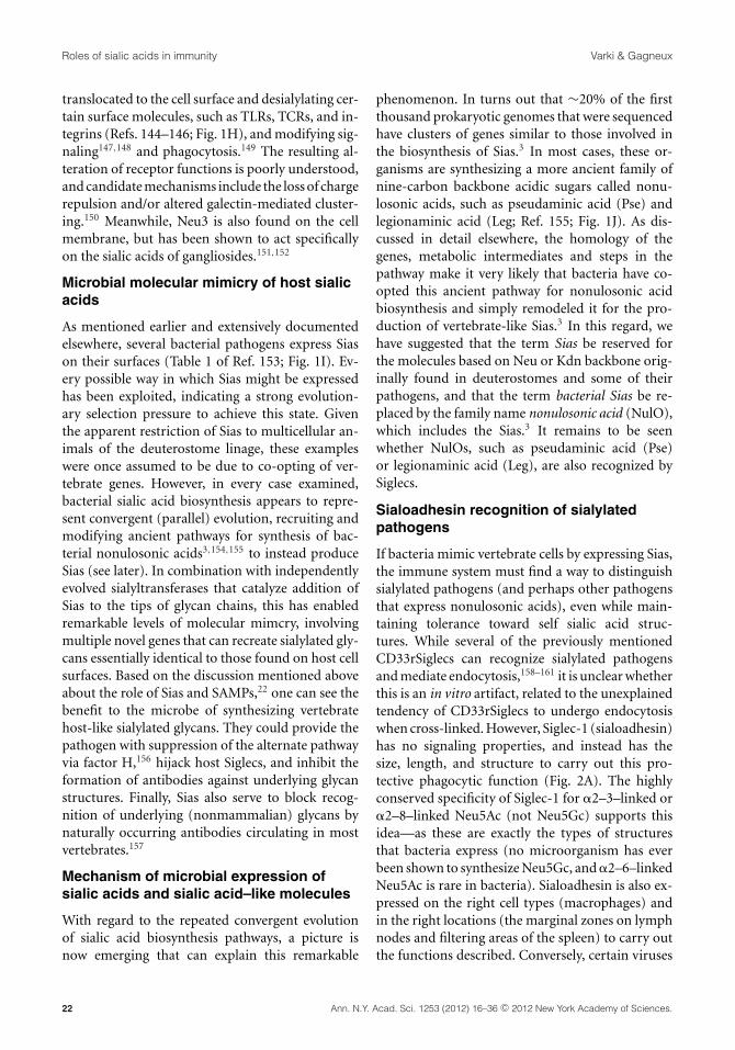
As mentioned earlier and extensively documentedelsewhere, several bacterial pathogens express Siason their surfaces (Table 1 of Ref. 153; Fig. 1I). Ev-ery possible way in which Sias might be expressedhas been exploited, indicating a strong evolution-ary selection pressure to achieve this state. Giventhe apparent restriction of Sias to multicellular an-imals of the deuterostome linage, these exampleswere once assumed to be due to co-opting of ver-tebrate genes. However, in every case examined,bacterial sialic acid biosynthesis appears to repre-sent convergent (parallel) evolution, recruiting andmodifying ancient pathways for synthesis of bac-terial nonulosonic acids3,154,155 to instead produceSias (see later). In combination with independentlyevolved sialyltransferases that catalyze addition ofSias to the tips of glycan chains, this has enabledremarkable levels of molecular mimcry, involvingmultiple novel genes that can recreate sialylated gly-cans essentially identical to those found on host cellsurfaces. Based on the discussion mentioned aboveabout the role of Sias and SAMPs,22 one can see thebenefit to the microbe of synthesizing vertebratehost-like sialylated glycans. They could provide thepathogen with suppression of the alternate pathwayvia factor H,156 hijack host Siglecs, and inhibit theformation of antibodies against underlying glycanstructures. Finally, Sias also serve to block recog-nition of underlying (nonmammalian) glycans bynaturally occurring antibodies circulating in mostvertebrates.157
Mechanism of microbial expression ofsialic acids and sialic acid–like molecules
With regard to the repeated convergent evolutionof sialic acid biosynthesis pathways, a picture isnow emerging that can explain this remarkable
phenomenon. In turns out that ∼20% of the firstthousand prokaryotic genomes that were sequencedhave clusters of genes similar to those involved inthe biosynthesis of Sias.3 In most cases, these or-ganisms are synthesizing a more ancient family ofnine-carbon backbone acidic sugars called nonu-losonic acids, such as pseudaminic acid (Pse) andlegionaminic acid (Leg; Ref. 155; Fig. 1J). As dis-cussed in detail elsewhere, the homology of thegenes, metabolic intermediates and steps in thepathway make it very likely that bacteria have co-opted this ancient pathway for nonulosonic acidbiosynthesis and simply remodeled it for the pro-duction of vertebrate-like Sias.3 In this regard, wehave suggested that the term Sias be reserved forthe molecules based on Neu or Kdn backbone orig-inally found in deuterostomes and some of theirpathogens, and that the term bacterial Sias be re-placed by the family name nonulosonic acid (NulO),which includes the Sias.3 It remains to be seenwhether NulOs, such as pseudaminic acid (Pse)or legionaminic acid (Leg), are also recognized bySiglecs.
Sialoadhesin recognition of sialylatedpathogens
If bacteria mimic vertebrate cells by expressing Sias,the immune system must find a way to distinguishsialylated pathogens (and perhaps other pathogensthat express nonulosonic acids), even while main-taining tolerance toward self sialic acid struc-tures. While several of the previously mentionedCD33rSiglecs can recognize sialylated pathogensand mediate endocytosis,158–161 it is unclear whetherthis is an in vitro artifact, related to the unexplainedtendency of CD33rSiglecs to undergo endocytosiswhen cross-linked. However, Siglec-1 (sialoadhesin)has no signaling properties, and instead has thesize, length, and structure to carry out this pro-tective phagocytic function (Fig. 2A). The highlyconserved specificity of Siglec-1 for �2–3–linked or�2–8–linked Neu5Ac (not Neu5Gc) supports thisidea—as these are exactly the types of structuresthat bacteria express (no microorganism has everbeen shown to synthesize Neu5Gc, and �2–6–linkedNeu5Ac is rare in bacteria). Sialoadhesin is also ex-pressed on the right cell types (macrophages) andin the right locations (the marginal zones on lymphnodes and filtering areas of the spleen) to carry outthe functions described. Conversely, certain viruses
22 Ann. N.Y. Acad. Sci. 1253 (2012) 16–36 c© 2012 New York Academy of Sciences.

Varki & Gagneux Roles of sialic acids in immunity
Figure 2. More examples of roles of sialic acids in immunity. Sialic acids are shown as pink diamonds. See key in Figure 1, and thetext for details. (A) Siglec-1 (sialoadhesin) expressed on macrophages recognizes Sias in patterns commonly found on microbialpathogens and facilitates phagocytosis. Siglec-1 may also mediate immune cell interactions with one another. Some viruses exploitSiglec-1 binding to gain access to host cells. (B) Certain bacteriophages use Sias on their microbial hosts as “receptors” for invasion.(C) Polysialic acid on immune molecules such as neuropilin on dendritic cells modulates interactions with T cells. (D) Sia-richsecretions on host epithelia can act as decoys for Sia-binding microbes. (E) Sia-covered erythrocytes and Sia-rich plasma proteinscan act as “viral traps.” (F) Sias act as biological masks by blocking interactions between intrinsic receptors and underlyingglycan structures. (G) Sias on potentially antigenic glycoconjugates prevent the formation of antibodies to “cryptoantigens.”Less commonly, Sias can be autoantigens. (H) Nonself Sias can be metabolically incorporated from dietary sources and become“xeno-autoantigens,” targeted by intrinsic anti-Sia antibodies. (I) Female genital tract reactions to nonself Sia on sperm canlead to reproductive incompatibility. (J) Some mammals, such as cats, have blood groups defined by Sia-containing glycolipids.(K) O-acetylation of Sias can block Sia recognition by intrinsic lectins like Siglecs, and modulate microbial lectin interactions, ina positive or negative fashion. (L) Alpha-2–6 sialylation of IgG-Fc region N -glycans can change the effects of IgG antibodies fromactivating to inhibitory.
that emerge from host cells with a coating of sialicacids can “hijack” sialoadhesin for use as a modeof entry into macrophages.162,163 Sialoadhesin alsocontributes to intrinsic immune functions, and im-mune changes found in sialoadhesin-deficient miceindicate a role in regulation of the adaptive immunesystem.164–167
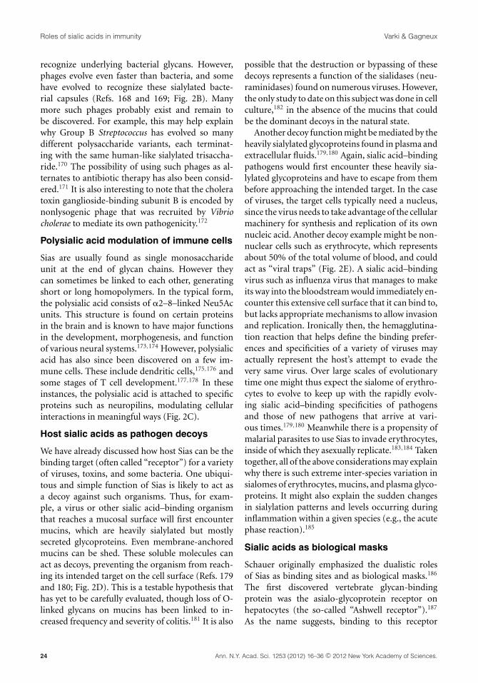
Bacteriophage recognition of microbialsialic acids
When bacteria mimic vertebrate cells by ex-pressing similar sialoglycans, they are also likelyusing variations of this mechanism to escaperecognition by bacteriophages that normally
Ann. N.Y. Acad. Sci. 1253 (2012) 16–36 c© 2012 New York Academy of Sciences. 23
Roles of sialic acids in immunity Varki & Gagneux
recognize underlying bacterial glycans. However,phages evolve even faster than bacteria, and somehave evolved to recognize these sialylated bacte-rial capsules (Refs. 168 and 169; Fig. 2B). Manymore such phages probably exist and remain tobe discovered. For example, this may help explainwhy Group B Streptococcus has evolved so manydifferent polysaccharide variants, each terminat-ing with the same human-like sialylated trisaccha-ride.170 The possibility of using such phages as al-ternates to antibiotic therapy has also been consid-ered.171 It is also interesting to note that the choleratoxin ganglioside-binding subunit B is encoded bynonlysogenic phage that was recruited by Vibriocholerae to mediate its own pathogenicity.172
Polysialic acid modulation of immune cells
Sias are usually found as single monosaccharideunit at the end of glycan chains. However theycan sometimes be linked to each other, generatingshort or long homopolymers. In the typical form,the polysialic acid consists of �2–8–linked Neu5Acunits. This structure is found on certain proteinsin the brain and is known to have major functionsin the development, morphogenesis, and functionof various neural systems.173,174 However, polysialicacid has also since been discovered on a few im-mune cells. These include dendritic cells,175,176 andsome stages of T cell development.177,178 In theseinstances, the polysialic acid is attached to specificproteins such as neuropilins, modulating cellularinteractions in meaningful ways (Fig. 2C).
Host sialic acids as pathogen decoys
We have already discussed how host Sias can be thebinding target (often called “receptor”) for a varietyof viruses, toxins, and some bacteria. One ubiqui-tous and simple function of Sias is likely to act asa decoy against such organisms. Thus, for exam-ple, a virus or other sialic acid–binding organismthat reaches a mucosal surface will first encountermucins, which are heavily sialylated but mostlysecreted glycoproteins. Even membrane-anchoredmucins can be shed. These soluble molecules canact as decoys, preventing the organism from reach-ing its intended target on the cell surface (Refs. 179and 180; Fig. 2D). This is a testable hypothesis thathas yet to be carefully evaluated, though loss of O-linked glycans on mucins has been linked to in-creased frequency and severity of colitis.181 It is also
possible that the destruction or bypassing of thesedecoys represents a function of the sialidases (neu-raminidases) found on numerous viruses. However,the only study to date on this subject was done in cellculture,182 in the absence of the mucins that couldbe the dominant decoys in the natural state.
Another decoy function might be mediated by theheavily sialylated glycoproteins found in plasma andextracellular fluids.179,180 Again, sialic acid–bindingpathogens would first encounter these heavily sia-lylated glycoproteins and have to escape from thembefore approaching the intended target. In the caseof viruses, the target cells typically need a nucleus,since the virus needs to take advantage of the cellularmachinery for synthesis and replication of its ownnucleic acid. Another decoy example might be non-nuclear cells such as erythrocyte, which representsabout 50% of the total volume of blood, and couldact as “viral traps” (Fig. 2E). A sialic acid–bindingvirus such as influenza virus that manages to makeits way into the bloodstream would immediately en-counter this extensive cell surface that it can bind to,but lacks appropriate mechanisms to allow invasionand replication. Ironically then, the hemagglutina-tion reaction that helps define the binding prefer-ences and specificities of a variety of viruses mayactually represent the host’s attempt to evade thevery same virus. Over large scales of evolutionarytime one might thus expect the sialome of erythro-cytes to evolve to keep up with the rapidly evolv-ing sialic acid–binding specificities of pathogensand those of new pathogens that arrive at vari-ous times.179,180 Meanwhile there is a propensity ofmalarial parasites to use Sias to invade erythrocytes,inside of which they asexually replicate.183,184 Takentogether, all of the above considerations may explainwhy there is such extreme inter-species variation insialomes of erythrocytes, mucins, and plasma glyco-proteins. It might also explain the sudden changesin sialylation patterns and levels occurring duringinflammation within a given species (e.g., the acutephase reaction).185
Sialic acids as biological masks
Schauer originally emphasized the dualistic rolesof Sias as binding sites and as biological masks.186
The first discovered vertebrate glycan-bindingprotein was the asialo-glycoprotein receptor onhepatocytes (the so-called “Ashwell receptor”).187
As the name suggests, binding to this receptor
24 Ann. N.Y. Acad. Sci. 1253 (2012) 16–36 c© 2012 New York Academy of Sciences.

Varki & Gagneux Roles of sialic acids in immunity
occurs when one removes Sias from a glycoproteinand exposes the underlying beta-linked galactoseresidues (Fig. 2F). Since this discovery, additionalbeta-galactose–binding receptors in macrophageshave been discovered. However, in most instancesgene knockouts of these proteins failed to uncovera clear-cut natural function in intrinsic systems.188
On the other hand, when a sialidase of bacterialorigin enters the circulation, there can be extensivedesialylation of cells and proteins, and these recep-tors become relevant. This was recently shown as ahost mechanism to clear away the excess of plateletsthat might result in increased coagulopathy that isassociated with microbial sepsis.189 The removal ofSias could also generate “eat me” signals that al-low macrophages to recognize and eliminate dyingor apoptotic cells.190 It is important to recognizeand differentiate the galactose-binding receptors in-volved in such phenomena from soluble galectins,which can bind terminal or subterminal galactoseresidues on cell surfaces.191,192 Galectins may ac-tually function in the opposite direction, acting toreduce endocytosis of the cell surface proteins byforming lattices, and may thus be more importantin regulation of signaling, as shown by others.193–195
The numerous other functions of galectins in theimmune system192,195 will not be discussed here,except to say that they can be modulated by thepresence or absence of terminal Sias, particularly�2–6–linked ones.196 Also, unlike most galectinsthat prefer N-acetyllactosamine ligands with non-sialylated terminal beta-Gal residues, galectin-8 andgalectin-9 have domains that preferentially recog-nize �2–3–sialylated N-acetyllactosamines.197,198
Antibodies against intrinsic sialic acids
Not surprisingly, it is uncommon to find antibodiesagainst sialic acid–containing glycans, if the sialicacid in question is already intrinsic within the host(Fig. 2G). Presumably, this is because B cells thathappen to express a B cell receptor (sIgM) thatcan recognize sialylated glycans are tolerized andeliminated before they leave the bone marrow.Indeed, as discussed earlier, this might be one of theselective advantages to pathogens that express Sias.In mammals, most of these comments reflect uponthe common Neu5Ac sialic acid. On the other hand,it is possible to induce mice to generate monoclonalantibodies that detect Neu5Ac-containing gly-cans,199,200 and the addition of an O-acetyl group to
the sialic acid can increase the probability of gettingsuch an antibody.201 Overall, while host-intrinsicSias can be generally considered “immunosuppres-sive” for the host organism, exceptions can be found.
Sialic acids as xenoautoantigens
The above comments do not apply if a partic-ular sialic acid is missing in a species. This ap-pears to be the case both in humans17 and in thesauropsid lineage of animals (birds and reptiles),202
which appear unable to synthesize the commonmammalian sialic acid Neu5Gc from its precursorNeu5Ac. In the case of humans, the basis for thisphenotype is a fixed loss-of function mutation of thecytidine monophosphate N-acetylneuraminic acidhydroxylase (CMAH) gene,203,204 which remainsintact in our closest evolutionary relatives the chim-panzees.204 The significance of the independent lossin the sauropsid lineage is unclear, though it doesmake for convenient source of anti-Neu5Gc anti-bodies by immunizing chickens, which generate arobust response.205 Unexplained also is the fact thatsimilar antibodies appear when chickens that be-come infected with the Marek’s disease lymphomavirus.206
In the case of humans, more information is avail-able. It appears that Neu5Gc from dietary sourcescan be metabolically incorporated either into ourtissues207 or into commensal bacteria such as H.influenzae, which specialize in taking up low quan-tities of Sias present in the upper oropharynx.208
One or both mechanisms appear to be the cause ofmoderate-to-high levels of anti-Neu5Gc antibod-ies in humans (Fig. 2H). Current studies suggestthat these antibodies may be interacting with themetabolically incorporated Neu5Gc of dietary ori-gin to generate chronic inflammation.17 This mayhelp explain the propensity of red meat (beef, pork,and lamb, the richest sources of dietary Neu5Gc)to increase the risk of inflammation-associated dis-eases such as carcinomas, cardiovascular disease,and macular degeneration. These findings also relateto the classic reports of the Hanganutziu-Deicher“heterophile” antibodies, which reacted with ani-mal red blood cells.209,210
Antisialyl antibodies can affectreproductive incompatibility
The female reproductive tract has levels of IgG anti-bodies and complement levels similar to that found
Ann. N.Y. Acad. Sci. 1253 (2012) 16–36 c© 2012 New York Academy of Sciences. 25

Roles of sialic acids in immunity Varki & Gagneux
in the serum.211 Thus a sperm that enters the uterusmust negotiate this immunological gauntlet beforeit can reach the ovum and fertilize it, further upin the fallopian tube. There may well be multipleantiglycan systems that can affect sperm, but theone so far documented involves antibodies againstNeu5Gc, which can enter the uterine fluid and af-fect both sperm and embryos that happen to haveNeu5Gc on them (Ref. 211; Fig. 2I). In this regard,it is suggested that this could even be a mechanismof speciation in the genus Homo, due to the loss ofNeu5Gc in ancestors, after it initially became poly-morphic.
Sialic-acid–based blood groupsin mammals
The above-mentioned considerations about the lackof immunogenicity of Neu5Ac do not apply if theNeu5Ac is attached to a specific polypeptide that isforeign to the host. An example is the MN bloodgroups in humans, where individual variations inthe amino acid sequence of the red cell protein gly-cophorin result in differential presentation of smallO-linked sialylated chains at the aminoterminus ofthe protein.212 While dictated by the underlyingpolypeptide, these antigenic variations also requirethe sialylated glycans, generating the antibodies thatinteract between humans and affect blood transfu-sion occasionally. Similar considerations apply tosome other blood group antibodies.213
Neu5Gc-based blood groups in cats
As with humans, there is one major antibody sys-tem that appears to restrict blood transfusion withincats. However, the two major blood groups in cats(A and B) were shown to be due to antibodies in Bcats against a sialylated glycolipid on the red bloodcells of A cats (Fig. 2J). The difference appears to bethe presence or absence of Neu5Gc in the ganglio-side GD3.214 We do not as yet know if the differentialexpression of Neu5Gc in the red blood cells of thesecats extends to other tissues of the animal. Thereis evidence that changes in the promoter regionof the CMAH gene might explain the differentialexpression in red blood cells.215 There are similarerythrocyte Neu5Gc polymorphisms in dogs,216 butevidence of anti-Neu5Gc antibodies has not been re-ported. In both instances, strain differences wouldbe worth studying further. Thus, it is possible thatSias can exist as alloantigens within populations of
the same species as well as xenoantigens within dif-ferent species.
Modulation of immunity by sialic acidO-acetylation
Given variable expression of O-acetyl groups onSias and their diverse effects in immunity, a sep-arate section on this modification seems justified.We have already mentioned the impact on factorH recognition, the relative resistance to bacterialsialidases to Sias with this modification, the block-ade of binding of some virus hemagglutinins, andthe facilitation of binding of others. Sias on cer-tain bacterial polysaccharides can be O-acetylated.Surprisingly, this modification is actually detrimen-tal to the bacterium in the host–pathogen interac-tion, either reducing recognition by CD33rSiglecsand/or enhancing immunogenicity.217 The logicalexplanation is that these modifications assist thebacteria in surviving in other situations, such asprotection from other microbial sialidases, and/orbacteriophage-binding proteins. The exception isO-acetyl blockade of recognition by sialoadhesin,218
which could be beneficial to the bacterium, avoidingphagocytosis.32,217
Unlike the case with the selectins, sialic acid–binding by Siglecs almost invariably requires recog-nition of the C7–C9 side-chain of the molecule.This is exemplified by the loss of recognition uponmild periodate oxidation of this side-chain.62,63,99
In view of this, it is not surprising that additionof an O-acetyl group to the side-chain blocks thebinding of all Siglecs studied to date.218,219 A Siglecselectively recognizing O-acetylated Sias has yet tobe found. Thus sialic acid O-acetylation seemed alogical candidate for regulation of Siglec function(Fig. 2K). This was indeed shown to be the case inmice with a defect in a sialic acid–specific esterase(SIAE), which normally downregulates sialic acidO-acetylation on B cells.220 The mutant mice thushave overreactive B cells, apparently due to lack ofproper SAMP ligands for CD22 and Siglec-G.221 Inkeeping with this, humans with autoimmune dis-eases have a higher frequency of harboring muta-tions in the SIAE gene.79,222
In another setting, O-acetylation of the outersialic acid of the ganglioside GD3 was first reportedas a melanoma-specific antigen not found in othernormal tissues.201 However, it later turned out thatnormal T cells can also express this structure.223–225
26 Ann. N.Y. Acad. Sci. 1253 (2012) 16–36 c© 2012 New York Academy of Sciences.

Varki & Gagneux Roles of sialic acids in immunity
Table 1. Cluster of differentiation (CD) numbers related to sialic acid biology
CD number Common name(s) Roles in immunity
CD15s Sialyl Lewis (sLeX) Key component of sialylated selectin ligands and of
preferred ligands for some Siglecs.
CD22 Siglec-2 (sialic acid–binding Ig-like
lectin 2)
Dampens B cell reactivity via selective recognition of
�2–6–linked Sias.
CD24 Heat stable antigen Heavily glycosylated, sialylated GPI-anchored molecule;
some glycoforms may be ligands for Siglec-10 or
P-selectin.
CD33 Siglec-3 (sialic acid–binding Ig-like
lectin 3)
Myeloid lineage marker; can downregulate reactivity of
innate immune cells.
CD34 Hematopoietic progenitor cell
antigen
Heavily glycosylated and sialylated cell surface
mucin-like protein; stem cell marker; some
glycoforms can be L-selectin ligands.
CD43 Leukosialin (leucocyte
sialoglycoprotein) (sialophorin)
Heavily sialylated major cell surface mucin-like protein;
modulates immune cell responses.
CD45 CD45 leukocyte common antigen Differing glycoforms in different immune cell types due
to alternate splicing; tyrosine phosphatase; possible
CD22 ligand.
CD52 CAMPATH-1 Heavily glycosylated and sialylated
glycosylphosphoinositol (GPI)-anchored cell surface
molecule.
CD56 Neural cell adhesion molecule 1 NK cell marker. Can carry polysialic acid, which can be
recognized by Siglec-7 and Siglec-11.
CD60a GD3 ganglioside Glycolipid with two sialic acids; human T cell marker;
promotes apoptosis?; Siglec-7 ligand?
CD60b 9-O-acetyl-GD3 ganglioside Presence of 9-O-acetyl group on outer sialic acid of GD3;
protects from apoptosis?
CD60c 7-O-acetyl-GD3 ganglioside Converted to 9-O-acetyl-GD3 over time due to
nonenzymatic migration of the O-acetyl group.
CD62E E-selectin (endothelial leukocyte
adhesion molecule 1)
Mediates leukocyte adhesion and rolling on
endothelium; expression upregulated by
inflammatory cytokines.
CD62L L-selectin (lymph node homing
receptor)
Mediates leukocyte adhesion and rolling on
endothelium, including lymphocyte trafficking.
CD62P P-selectin (granule membrane
protein 140, GMP-140)
Mediates platelet and endothelial interactions with
leukocytes, via correctly modified PSGL-1.
CD68 Macrosialin (Gp110) Marker of macrophages and few other cells; receptor for
oxidized low-density lipoprotein?
CD75s �2–6–sialylated lactosamines Cluster of Sia�2–6Gal�1–4GlcNAc�1-R units produced
by ST6Gal-I, mainly on N-glycans.
CD169 Sialoadhesin (sialic acid–binding
Ig-like lectin 1) (Siglec-1)
Macrophage subset marker; recognizes sialic acids on
endogenous ligands and on microbes.
CD170 Sialic acid–binding Ig-like lectin 5
(Siglec-5)
Inhibitory Siglecs on human innate immune cells, and
also on lymphocytes in “great apes.”
CD175s Sialyl-Tn Sia�2–6GalNAc�1-Ser/Thr; truncated O-glycan mostly
found on malignant cells.
Continued
Ann. N.Y. Acad. Sci. 1253 (2012) 16–36 c© 2012 New York Academy of Sciences. 27

Roles of sialic acids in immunity Varki & Gagneux
Table 1. Continued
CD number Common name(s) Roles in immunity
CD176s Sialylated form of Thomsen–
Friedenreich (T) antigen
Sia�2–3Gal1–3GalNAc�1-Ser/Thr; common O-glycan
on many cell types.
CD227 MUC-1, polymorphic epithelial
mucin, episialin
Major cell surface mucin on epithelial cells; on activated
T cells, can send inhibitory signals.
CD235a Glycophorin-A (PAS-2) (MN
sialoglycoprotein)
Major sialic acid carrier on RBCs; target for binding by
malarial merozoite via EBA-175.
CD235b Glycophorin-B (PAS-3)
(sialoglycoprotein delta)
Major sialic acid carrier on RBCs; target for binding by
malarial merozoite via EBL-1.
CD236 Glycophorin-C (PAS-2′)(glycoprotein beta)
Major sialic acid carrier on RBCs. Target for binding by
malarial merozoite via EBA-140.
CD327 Sialic acid–binding Ig-like lectin 6
(Siglec-6)
Inhibitory Siglecs on B cells in primates, and also on
placental trophoblast (in humans only).
CD328 Sialic acid–binding Ig-like lectin 7
(Siglec-7); AIRM1
Inhibitory Siglecs on human NK cells; lower levels on
monocytes and macrophages.
CD329 Sialic acid–binding Ig-like lectin 9
(Siglec-9);
Inhibitory Siglecs found on human neutrophils;
monocytes and macrophages.
For more information, see http://www.hcdm.org/MoleculeInformation/tabid/54/Default.aspx.
Indeed, this is the basis of the CD60 group of anti-gens (see Table 1). Interestingly, while GD3 is pro-apoptotic, O-acetyl-GD3 has opposite effects.226,227
The claimed mechanisms for these effects are fas-cinating, involving mitochondrial and other apop-totic pathways. However, there are topological issuesthat remain unresolved.228
Alteration of IgG Fc functionby α2–6–sialylation
Recent studies have shown that the minor subset ofcirculating IgG that has �2–6–linked Sias terminat-ing its N-glycan has an inhibitory potential, workingthrough the human DC-SIGN receptor on a regula-tory macrophage population to upregulate FcR�-IIB on other macrophages, and thereby dampenimmune responses (Refs. 229–231; Fig. 2L). This isalso suggested to be the mechanism of action of in-travenous pooled human IgG (IVIG) that is used forimmune suppression in the clinic. While the data areconsistent and compelling, the work mostly involvesa single model system for autoimmune disease. Italso assumes that the other suggested mechanismsof IVIG action (e.g., scavenging of activated com-plement,232 and anti-Siglec antibodies233) do notcontribute significantly. This is a very interestingavenue for future research, especially given than the
relevant sialyltransferase (ST6Gal-I) is highly reg-ulated in response to inflammation.185 and alterscellular activation and proliferation.196,234
Sialylated molecules or sialic acid–bindingproteins as cluster of differentiation (CD)markers
It would be incomplete to discuss the immune sys-tem without mentioning CD markers. There is along list of CD molecules that are either sialylatedor are involved in sialic acid recognition. Space doesnot allow a full discussion of each of these antigens,but a brief summary can be found in Table 1.
Conclusions and perspectives
As the outermost “onion layer” on all vertebratecells types, Sias were predestined to play roles asthe “molecular frontier” in ongoing evolutionaryarms races, both as targets for attack and as SAMPs.Sialome patterns evolve rapidly, likely because theyare prone to being exploited by rapidly evolvingpathogens and parasites. Meanwhile, changes in theintrinsic “landscape” of self-Sias have to be closelytracked by the intrinsic lectins such as Siglecs inorder to maintain homeostasis, even while theythemselves are being exploited by pathogens ex-pressing Sias. On the background of such ongoing
28 Ann. N.Y. Acad. Sci. 1253 (2012) 16–36 c© 2012 New York Academy of Sciences.

Varki & Gagneux Roles of sialic acids in immunity
“molecular dialectics” (revolution and counterrev-olution), major sialome changes at the level of aspecies, such as the wholesale loss of Neu5Gc in hu-mans likely required major system-wide accommo-dations. Meanwhile, vertebrate hosts are “locked in”to maintaining the numerous intrinsic roles of Siasin reproduction, development, and normal physi-ology. Vertebrate host species have evolved a pre-carious compromise of using the presence of selfSias as “self-associated patterns,” even while dis-criminating against close mimics, as well as againstmolecular surfaces lacking self Sias. Several othercell surface glycans such as galactose, fucose, andglycosaminoglycans also mediate immunity-relatedfunctions, including some of the ones discussedhere. However, given their shear abundance, struc-tural diversity, and vertebrate lineage-defining na-ture, Sias have been recruited for multifarious rolesin immunity, only some of which we have addressedhere.
Acknowledgments
We thank Shiv Pillai and Takashi Angata for criti-cal comments and suggestions. This work was sup-ported by grants from the NIH and by the MathersFoundation of New York.
Conflicts of interest
The authors declare no conflicts of interest.
References
1. Schauer, R., G.V. Srinivasan, D. Wipfler, et al. 2011. O-Acetylated sialic acids and their role in immune defense.Adv. Exp. Med. Biol. 705: 525–548.
2. Schauer, R. 2009. Sialic acids as regulators of molecular andcellular interactions. Curr. Opin. Struct. Biol. 19: 507–514.
3. Lewis, A.L., N. Desa, E.E. Hansen, et al. 2009. Innovations inhost and microbial sialic acid biosynthesis revealed by phy-logenomic prediction of nonulosonic acid structure. Proc.Natl. Acad. Sci. USA 106: 13552–13557.
4. Varki, A. & R. Schauer. 2009. Sialic Acids. In Essentials ofGlycobiology. 2nd ed. A. Varki, R.D. Cummings, J.D. Esko,H.H. Freeze, P. Stanley, C.R. Bertozzi, G.W. Hart & M.E.Etzler, Eds.: 199–218. Cold Spring Harbor Laboratory Press.Cold Spring Harbor, NY.
5. Severi, E., D.W. Hood & G.H. Thomas. 2007. Sialic acidutilization by bacterial pathogens. Microbiology 153: 2817–2822.
6. Troy, F.A. 1992. Polysialylation: from bacteria to brains.Glycobiology 2: 5–23.
7. Vimr, E.R., K.A. Kalivoda, E.L. Deszo & S.M. Steenbergen.2004. Diversity of microbial sialic acid metabolism. Micro-biol. Mol. Biol. Rev. 68: 132–153.
8. Byrne, B., G.G. Donohoe & R. O’Kennedy. 2007. Sialicacids: carbohydrate moieties that influence the biologicaland physical properties of biopharmaceutical proteins andliving cells. Drug Discov. Today 12: 319–326.
9. Rutishauser, U. 1998. Polysialic acid at the cell surface: bio-physics in service of cell interactions and tissue plasticity. J.Cell. Biochem. 70: 304–312.
10. Collins, B.E., O. Blixt, A.R. DeSieno, et al. 2004. Maskingof CD22 by cis ligands does not prevent redistribution ofCD22 to sites of cell contact. Proc. Natl. Acad. Sci. USA 101:6104–6109.
11. Pilatte, Y., J. Bignon & C.R. Lambre. 1993. Sialic acids as im-portant molecules in the regulation of the immune system:pathophysiological implications of sialidases in immunity.Glycobiology 3: 201–218.
12. Gahmberg, C.G. & L.C. Andersson. 1977. Selective radioac-tive labeling of cell surface sialoglycoproteins by periodate-tritiated borohydride. J. Biol. Chem. 252: 5888–5894.
13. Dehoux-zenou, S.M., M. Guenounou, H. Zinbi, et al. 1987.Behavior of aldehyde moieties involved in the activationof suppressor cells by sodium periodate. J. Immunol. 138:1157–1163.
14. Jacobs, C.L., K.J. Yarema, L.K. Mahal, et al. 2000. Metaboliclabeling of glycoproteins with chemical tags through unnat-ural sialic acid biosynthesis. Methods Enzymol. 327: 260–275.
15. Du, J., M.A. Meledeo, Z. Wang, et al. 2009. Metabolic glyco-engineering: sialic acid and beyond. Glycobiology 19: 1382–1401.
16. Prescher, J.A. & C.R. Bertozzi. 2006. Chemical technologiesfor probing glycans. Cell 126: 851–854.
17. Varki, A. 2010. Colloquium paper: uniquely human evolu-tion of sialic acid genetics and biology. Proc. Natl. Acad. Sci.USA 107(Suppl. 2): 8939–8946.
18. Fearon, D.T. 1978. Regulation by membrane sialic acid ofbeta1H-dependent decay-dissociation of amplification C3convertase of the alternative complement pathway. Proc.Natl. Acad. Sci. USA 75: 1971–1975.
19. Pangburn, M.K. & H.J. Muller-Eberhard. 1978. Comple-ment C3 convertase: cell surface restriction of beta1Hcontrol and generation of restriction on neuraminidase-treated cells. Proc. Natl. Acad. Sci. USA 75: 2416–2420.
20. Meri, S. & M.K. Pangburn. 1990. Discrimination betweenactivators and nonactivators of the alternative pathway ofcomplement: regulation via a sialic acid /polyanion bindingsite on factor H. Proc. Natl. Acad. Sci. USA 87: 3982–3986.
21. Ram, S., A.K. Sharma, S.D. Simpson, et al. 1998. A novelsialic acid-binding site on factor H mediates serum resis-tance of sialylated Neisseria gonorrhoeae. J. Exp. Med. 187:743–752.
22. Varki, A. 2011. Since there are PAMPs and DAMPs, theremust be SAMPs? Glycan “self-associated molecular pat-terns” dampen innate immunity, but pathogens can mimicthem. Glycobiology 21: 1121–1124.
23. Johnston, J.W., N.P. Coussens, S. Allen, et al. 2008. Char-acterization of the N-acetyl-5-neuraminic acid-binding siteof the extracytoplasmic solute receptor (SiaP) of nonty-peable haemophilus influenzae strain 2019. J. Biol. Chem.283: 855–865.
Ann. N.Y. Acad. Sci. 1253 (2012) 16–36 c© 2012 New York Academy of Sciences. 29

Roles of sialic acids in immunity Varki & Gagneux
24. Ram, S., L.A. Lewis & S. Agarwal. 2011. Meningococcalgroup w-135 and y capsular polysaccharides paradoxicallyenhance activation of the alternative pathway of comple-ment. J. Biol. Chem. 286: 8297–8307.
25. Zipfel, P.F., C. Skerka, J. Hellwage, et al. 2002. Factor H fam-ily proteins: on complement, microbes and human diseases.Biochem. Soc. Trans. 30: 971–978.
26. Kajander, T., M.J. Lehtinen, S. Hyvarinen, et al. 2011. Dualinteraction of factor H with C3d and glycosaminoglycansin host-nonhost discrimination by complement. Proc. Natl.Acad. Sci. USA 108: 2897–2902.
27. Morgan, H.P., J. Jiang, A.P. Herbert, et al. 2011. Crystal-lographic determination of the disease-associated T1184Rvariant of complement regulator factor H. Acta. Crystallogr.D Biol. Crystallogr. 67: 593–600.
28. Shaughnessy, J., S. Ram, A. Bhattacharjee, et al. 2011. Molec-ular characterization of the interaction between sialylatedNeisseria gonorrhoeae and factor H. J. Biol. Chem. 286:22235–22242.
29. Atkinson, J.P. & T.H. Goodship. 2007. Complement factorH and the hemolytic uremic syndrome. J. Exp. Med. 204:1245–1248.
30. Donoso, L.A., T. Vrabec & H. Kuivaniemi. 2010. Therole of complement Factor H in age-related macu-lar degeneration: a review. Surv. Ophthalmol. 55: 227–246.
31. Nydegger, U.E., D.T. Fearon & K.F. Austen. 1978. Autosomallocus regulates inverse relationship between sialic acid con-tent and capacity of mouse erythrocytes to activate humanalternative complement pathway. Proc. Natl. Acad. Sci. USA75: 6078–6082.
32. Shi, W.X., R. Chammas, N.M. Varki, et al. 1996. Sialicacid 9-O-acetylation on murine erythroleukemia cells af-fects complement activation, binding to I-type lectins, andtissue homing. J. Biol. Chem. 271: 31526–31532.
33. Arming, S., D. Wipfler, J. Mayr, et al. 2011. The human Cas1protein: a sialic acid-specific O-acetyltransferase? Glycobi-ology 21: 553–564.
34. Kjaer, T.R., A.G. Hansen, U.B. Sorensen, et al. 2011. In-vestigations on the pattern recognition molecule M-ficolin:quantitative aspects of bacterial binding and leukocyte as-sociation. J. Leukoc. Biol. 90: 425–437.
35. Honore, C., S. Rorvig, T. Hummelshoj, et al. 2010. Tetheringof Ficolin-1 to cell surfaces through recognition of sialic acidby the fibrinogen-like domain. J. Leukoc. Biol. 88: 145–158.
36. Gout, E., V. Garlatti, D.F. Smith, et al. 2010. Carbohy-drate recognition properties of human ficolins: glycan arrayscreening reveals the sialic acid-binding specificity of M-ficolin. J. Biol. Chem. 285: 6612–6622.
37. Rosen, S.D., M.S. Singer, T.A. Yednock & L.M. Stoolman.1985. Involvement of sialic acid on endothelial cells inorgan-specific lymphocyte recirculation. Science 228: 1005–1007.
38. McEver, R.P. 1991. Selectins: novel receptors that me-diate leukocyte adhesion during inflammation. Thromb.Haemost. 65: 223–228.
39. Cummings, R.D. & D.F. Smith. 1992. The selectin family ofcarbohydrate-binding proteins: structure and importanceof carbohydrate ligands for cell adhesion. BioEssays 14: 849–856.
40. Rosen, S.D. 1993. L-selectin and its biological ligands. His-tochemistry 100: 185–191.
41. Varki, A. 1994. Selectin ligands. Proc. Natl. Acad. Sci. USA91: 7390–7397.
42. Lasky, L.A. 1995. Selectin-carbohydrate interactions andthe initiation of the inflammatory response. Annu. Rev.Biochem. 64: 113–139.
43. Picker, L.J., R.A. Warnock, A.R. Burns, et al. 1991. Theneutrophil selectin LECAM-1 presents carbohydrate ligandsto the vascular selectins ELAM-1 and GMP-140. Cell 66:921–933.
44. Zhou, Q., K.L. Moore, D.F. Smith, et al. 1991. The selectinGMP-140 binds to sialylated, fucosylated lactosaminogly-cans on both myeloid and nonmyeloid cells. J. Cell Biol.115: 557–564.
45. Tyrrell, D., P. James, N. Rao, et al. 1991. Structural require-ments for the carbohydrate ligand of E-selectin. Proc. Natl.Acad. Sci. USA 88: 10372–10376.
46. Handa, K., E.D. Nudelman, M.R. Stroud, et al. 1991. Se-lectin GMP-140 (CD62; PADGEM) binds to sialosyl-Leaand sialosyl-Lex, and sulfated glycans modulate this bind-ing. Biochem. Biophys. Res. Commun. 181: 1223–1230.
47. Berg, E.L., J. Magnani, R.A. Warnock, et al. 1992. Com-parison of L-selectin and E-selectin ligand specificities: theL-selectin can bind the E-selectin ligands sialyl Lex andsialyl Lea. Biochem. Biophys. Res. Commun. 184: 1048–1055.
48. Foxall, C., S.R. Watson, D. Dowbenko, et al. 1992. The threemembers of the selectin receptor family recognize a com-mon carbohydrate epitope, the sialyl Lewisx oligosaccha-ride. J. Cell Biol. 117: 895–902.
49. Larsen, G.R., D. Sako, T.J. Ahern, et al. 1992. P-selectinand E-selectin. Distinct but overlapping leukocyte ligandspecificities. J. Biol. Chem. 267: 11104–11110.
50. Moore, K.L., N.L. Stults, S. Diaz, et al. 1992. Identificationof a specific glycoprotein ligand for P-selectin (CD62) onmyeloid cells. J. Cell Biol. 118: 445–456.
51. Yeh, J.C., N. Hiraoka, B. Petryniak, et al. 2001. Novel sul-fated lymphocyte homing receptors and their control bya core1 extension beta1,3-N-acetylglucosaminyltransferase.Cell 105: 957–969.
52. Leppanen, A., P. Mehta, Y.B. Ouyang, et al. 1999. A novelglycosulfopeptide binds to P-selectin and inhibits leuko-cyte adhesion to P-selectin. J. Biol. Chem. 274: 24838–24848.
53. Larkin, M., T.J. Ahern, M.S. Stoll, et al. 1992. Spectrum ofsialylated and nonsialylated fuco-oligosaccharides boundby the endothelial-leukocyte adhesion molecule E-selectin.Dependence of the carbohydrate binding activity on E-selectin density. J. Biol. Chem. 267: 13661–13668.
54. Norgard-Sumnicht, K.E., N.M. Varki & A. Varki. 1993.Calcium-dependent heparin-like ligands for L-selectin innonlymphoid endothelial cells. Science 261: 480–483.
55. Nelson, R.M., O. Cecconi, W.G. Roberts, et al. 1993. Heparinoligosaccharides bind L- and P-selectin and inhibit acuteinflammation. Blood 82: 3253–3258.
56. Norgard, K.E., H. Han, L. Powell, et al. 1993. Enhancedinteraction of L-selectin with the high endothelial venuleligand via selectively oxidized sialic acids. Proc. Natl. Acad.Sci. USA 90: 1068–1072.
30 Ann. N.Y. Acad. Sci. 1253 (2012) 16–36 c© 2012 New York Academy of Sciences.

Varki & Gagneux Roles of sialic acids in immunity
57. Rosen, S.D. 2004. Ligands for L-selectin: homing, inflam-mation, and beyond. Annu. Rev. Immunol. 22: 129–156.
58. Crocker, P.R. & S. Gordon. 1986. Properties and distributionof a lectin-like hemagglutinin differentially expressed bymurine stromal tissue macrophages. J. Exp. Med. 164: 1862–1875.
59. Crocker, P.R., S. Kelm, C. Dubois, et al. 1991. Purificationand properties of sialoadhesin, a sialic acid-binding receptorof murine tissue macrophages. EMBO J 10: 1661–1669.
60. Stamenkovic, I., D. Sgroi, A. Aruffo, et al. 1991. The B lym-phocyte adhesion molecule CD22 interacts with leukocytecommon antigen CD45RO on T cells and alpha 2–6 sialyl-transferase, CD75, on B cells. Cell 66: 1133–1144.
61. Sgroi, D., A. Varki, S. Braesch-Andersen & I. Stamenkovic.1993. CD22, a B cell-specific immunoglobulin superfamilymember, is a sialic acid-binding lectin. J. Biol. Chem. 268:7011–7018.
62. Powell, L.D., D. Sgroi, E.R. Sjoberg, et al. 1993. Naturalligands of the B cell adhesion molecule CD22beta carryN-linked oligosaccharides with alpha-2,6-linked sialic acidsthat are required for recognition. J. Biol. Chem. 268: 7019–7027.
63. Powell, L.D. & A. Varki. 1994. The oligosaccharide bindingspecificities of CD22beta, a sialic acid-specific lectin of Bcells. J. Biol. Chem. 269: 10628–10636.
64. Crocker, P.R., S. Mucklow, V. Bouckson, et al. 1994.Sialoadhesin, a macrophage sialic acid-binding receptor forhaemopoietic cells with 17 immunoglobulin-like domains.EMBO J. 13: 4490–4503.
65. Kelm, S., A. Pelz, R. Schauer, et al. 1994. Sialoadhesin,myelin-associated glycoprotein and CD22 define a new fam-ily of sialic acid-dependent adhesion molecules of the im-munoglobulin superfamily. Curr. Biol. 4: 965–972.
66. Freeman, S.D., S. Kelm, E.K. Barber & P.R. Crocker. 1995.Characterization of CD33 as a new member of the siaload-hesin family of cellular interaction molecules. Blood 85:2005–2012.
67. Kelm, S., R. Schauer & P.R. Crocker. 1996. The siaload-hesins: a family of sialic acid-dependent cellular recognitionmolecules within the immunoglobulin superfamily. Glyco-conjugate J. 13: 913–926.
68. Collins, B.E., M. Kiso, A. Hasegawa, et al. 1997. Bindingspecificities of the sialoadhesin family of I-type lectins:sialic acid linkage and substructure requirements for bind-ing of myelin-associated glycoprotein, Schwann cell myelinprotein, and sialoadhesin. J. Biol. Chem. 272: 16889–16895.
69. Powell, L.D. & A. Varki. 1995. I-type lectins. J. Biol. Chem.270: 14243–14246.
70. Crocker, P.R., E.A. Clark, M. Filbin, et al. 1998. Siglecs: afamily of sialic-acid binding lectins [letter]. Glycobiology 8:v.
71. Angata, T., E.H. Margulies, E.D. Green & A. Varki. 2004.Large-scale sequencing of the CD33-related Siglec genecluster in five mammalian species reveals rapid evolutionby multiple mechanisms. Proc. Natl. Acad. Sci. USA 101:13251–13256.
72. Angata, T. 2006. Molecular diversity and evolution of theSiglec family of cell-surface lectins. Mol. Divers. 10: 555–566.
73. Crocker, P.R., J.C. Paulson & A. Varki. 2007. Siglecs andtheir roles in the immune system. Nat. Rev. Immunol. 7:255–266.
74. von Gunten, S. & B.S. Bochner. 2008. Basic and clinicalimmunology of Siglecs. Ann. N. Y. Acad. Sci. 1143: 61–82.
75. Cao, H., B. de Bono, K. Belov, et al. 2009. Compara-tive genomics indicates the mammalian CD33rSiglec locusevolved by an ancient large-scale inverse duplication andsuggests all Siglecs share a common ancestral region. Im-munogenetics 61: 401–417.
76. O’Reilly, M.K. & J.C. Paulson. 2010. Multivalent ligands forSiglecs. Methods Enzymol. 478: 343–363.
77. Cao, H. & P.R. Crocker. 2011. Evolution of CD33-relatedSiglecs: regulating host immune functions and escapingpathogen exploitation? Immunology 132: 18–26.
78. Park, C.S. & B.S. Bochner. 2011. Potential targeting ofSiglecs, mast cell inhibitory receptors, in interstitial cystitis.Int. Neurourol. J. 15: 61–63.
79. Pillai, S., I.A. Netravali, A. Cariappa & H. Mattoo. 2011.Siglecs and immune regulation. Annu. Rev. Immunol. 30.[Epub ahead of print.] PMID: 22224769.
80. Yu, Z.B., M. Maoui, L.T. Wu, et al. 2001. mSiglec-E,a novel mouse CD33-related Siglec (sialic acid-bindingimmunoglobulin-like lectin) that recruits Src homology2 (SH2)-domain-containing protein tyrosine phosphatasesSHP-1 and SHP-2. Biochem. J. 353: 483–492.
81. Whitney, G., S.L. Wang, H. Chang, et al. 2001. A new Siglecfamily member, Siglec-10, is expressed in cells of the im-mune system and has signaling properties similar to CD33.Eur. J. Biochem. 268: 6083–6096.
82. Angata, T., S.C. Kerr, D.R. Greaves, et al. 2002. Cloningand characterization of human Siglec-11. A recently evolvedsignaling molecule that can interact with SHP-1 and SHP-2 and is expressed by tissue macrophages, including brainmicroglia. J. Biol. Chem. 277: 24466–24474.
83. Avril, T., H. Floyd, F. Lopez, et al. 2004. The membrane-proximal immunoreceptor tyrosine-based inhibitory motifis critical for the inhibitory signaling mediated by Siglecs-7and -9, CD33-related Siglecs expressed on human mono-cytes and NK cells. J. Immunol. 173: 6841–6849.
84. Nitschke, L. 2009. CD22 and Siglec-G: B-cell inhibitoryreceptors with distinct functions. Immunol. Rev. 230: 128–143.
85. Liu, Y., G.Y. Chen & P. Zheng. 2009. CD24-Siglec G/10discriminates danger- from pathogen-associated molecularpatterns. Trends Immunol. 30: 557–561.
86. Avril, T., S.D. Freeman, H. Attrill, et al. 2005. Siglec-5(CD170) can mediate inhibitory signalling in the absenceof immunoreceptor tyrosine-based inhibitory motif phos-phorylation. J. Biol. Chem. 280: 19843–19851.
87. Mitsuki, M., K. Nara, T. Yamaji, et al. 2010. Siglec-7 mediatesnonapoptotic cell death independently of its immunorecep-tor tyrosine-based inhibitory motifs in monocytic cell lineU937. Glycobiology 20: 395–402.
88. Angata, T., T. Hayakawa, M. Yamanaka, et al. 2006. Dis-covery of Siglec-14, a novel sialic acid receptor undergoingconcerted evolution with Siglec-5 in primates. FASEB J. 20:1964–1973.
Ann. N.Y. Acad. Sci. 1253 (2012) 16–36 c© 2012 New York Academy of Sciences. 31

Roles of sialic acids in immunity Varki & Gagneux
89. Angata, T., Y. Tabuchi, K. Nakamura & M. Nakamura. 2007.Siglec-15: an immune system Siglec conserved throughoutvertebrate evolution. Glycobiology 17: 838–846.
90. Yasui, K., T. Angata, N. Matsuyama, et al. 2011. Detec-tion of anti-Siglec-14 alloantibodies in blood componentsimplicated in nonhaemolytic transfusion reactions. Br. J.Haematol. 153: 794–796.
91. Hiruma, Y., T. Hirai & E. Tsuda. 2011. Siglec-15, a memberof the sialic acid-binding lectin, is a novel regulator forosteoclast differentiation. Biochem. Biophys. Res. Commun.409: 424–429.
92. Cao, H., U. Lakner, B. de Bono, et al. 2008. SIGLEC16encodes a DAP12-associated receptor expressed inmacrophages that evolved from its inhibitory counterpartSIGLEC11 and has functional and non-functional alleles inhumans. Eur. J. Immunol. 38: 2303–2315.
93. Varki, A. & T. Angata. 2006. Siglecs–the major subfamily ofI-type lectins. Glycobiology 16: 1R-27R.
94. Crocker, P.R. & P. Redelinghuys. 2008. Siglecs as positiveand negative regulators of the immune system. Biochem.Soc. Trans. 36: 1467–1471.
95. O’Reilly, M.K. & J.C. Paulson. 2009. Siglecs as targets fortherapy in immune-cell-mediated disease. Trends Pharma-col. Sci. 30: 240–248.
96. Varki, A. 2009. Natural ligands for CD33-related Siglecs?Glycobiology 19: 810–812.
97. Jandus, C., H.U. Simon & S. von Gunten. 2011. TargetingSiglecs: a novel pharmacological strategy for immuno- andglycotherapy. Biochem. Pharmacol. 82: 323–332.
98. Magesh, S., H. Ando, T. Tsubata, et al. 2011. High-affinityligands of Siglec receptors and their therapeutic potentials.Curr. Med. Chem. 18: 3537–3550.
99. Razi, N. & A. Varki. 1998. Masking and unmasking of thesialic acid-binding lectin activity of CD22 (Siglec-2) on Blymphocytes. Proc. Natl. Acad. Sci. USA 95: 7469–7474.
100. Kawasaki, Y., A. Ito, D.A. Withers, et al. 2010. GangliosideDSGb5, preferred ligand for Siglec-7, inhibits NK cell cyto-toxicity against renal cell carcinoma cells. Glycobiology 20:1373–1379.
101. Blixt, O., S. Han, L. Liao, et al. 2008. Sialoside analoguearrays for rapid identification of high affinity Siglec ligands.J. Am. Chem. Soc. 130: 6680–6681.
102. Cui, L., P.I. Kitov, G.C. Completo, et al. 2011. Supramolec-ular complexing of membane Siglec CD22 mediated by apolyvalent heterobifunctional ligand that templates on IgM.Bioconjug. Chem. 22: 546–550.
103. Vinson, M., P.A. Van der Merwe, S. Kelm, et al. 1996. Char-acterization of the sialic acid-binding site in sialoadhesin bysite-directed mutagenesis. J. Biol. Chem. 271: 9267–9272.
104. Van der Merwe, P.A., P.R. Crocker, M. Vinson, et al. 1996.Localization of the putative sialic acid-binding site on theimmunoglobulin superfamily cell-surface molecule CD22.J. Biol. Chem. 271: 9273–9280.
105. Crocker, P.R., M. Vinson, S. Kelm & K. Drickamer. 1999.Molecular analysis of sialoside binding to sialoadhesin byNMR and site-directed mutagenesis. Biochem. J. 341: 355–361.
106. Angata, T. & A. Varki. 2000. Cloning, characterization, andphylogenetic analysis of Siglec-9, a new member of the
CD33-related group of Siglecs: evidence for co-evolutionwith sialic acid synthesis pathways. J. Biol. Chem. 275:22127–22135.
107. Angata, T., N.M. Varki & A. Varki. 2001. A second uniquelyhuman mutation affecting sialic acid biology. J. Biol. Chem.276: 40282–40287.
108. Mandal, C. 1990. Sialic acid-binding lectins. Experientia 46:433–441.
109. Traving, C. & R. Schauer. 1998. Structure, function andmetabolism of sialic acids. Cell Mol Life Sci 54: 1330–1349.
110. Isa, P., C.F. Arias & S. Lopez. 2006. Role of sialic acids inrotavirus infection. Glycoconj. J. 23: 27–37.
111. Gee, G.V., A.S. Dugan, N. Tsomaia, et al. 2006. The role ofsialic acid in human polyomavirus infections. Glycoconj. J.23: 19–26.
112. Lehmann, F., E. Tiralongo & J. Tiralongo. 2006. Sialic acid-specific lectins: occurrence, specificity and function. CellMol. Life Sci. 63: 1331–1354.
113. Taylor, G. 1996. Sialidases: structures, biological signifi-cance and therapeutic potential. Curr. Opin. Struct. Biol.6: 830–837.
114. von Itzstein, M. 2007. The war against influenza: discov-ery and development of sialidase inhibitors. Nat. Rev. DrugDiscov. 6: 967–974.
115. Herrler, G., R. Rott, H.D. Klenk, et al. 1985. The receptor-destroying enzyme of influenza C virus is neuraminate-O-acetylesterase. EMBO J 4: 1503–1506.
116. Rogers, G.N., G. Herrler, J.C. Paulson & H.D. Klenk. 1986.Influenza C virus uses 9-O-acetyl-N-acetylneuraminic acidas a high affinity receptor determinant for attachment tocells. J. Biol. Chem. 261: 5947–5951.
117. Vlasak, R., M. Krystal, M. Nacht & P. Palese. 1987. The in-fluenza C virus glycoprotein (HE) exhibits receptor-binding(hemagglutinin) and receptor-destroying (esterase) activi-ties. Virology 160: 419–425.
118. Vlasak, R., W. Luytjes, J. Leider, et al. 1988. The E3 proteinof bovine coronavirus is a receptor-destroying enzyme withacetylesterase activity. J. Virol. 62: 4686–4690.
119. Herrler, G. & H.-D. Klenk. 1991. Structure and function ofthe HEF glycoprotein of influenza C virus. Adv. Virus Res.40: 213–234.
120. Pleschka, S., H.D. Klenk & G. Herrler. 1995. The catalytictriad of the influenza C virus glycoprotein HEF esterase:characterization by site-directed mutagenesis and func-tional analysis. J. Gen. Virol. 76: 2529–2537.
121. Cornelissen, L.A.H.M., C.M.H. Wierda, d. M.F.J. van, et al.1997. Hemagglutinin-esterase: a novel structural protein oftorovirus. J. Virol. 71: 5277–5286.
122. Wurzer, W.J., K. Obojes & R. Vlasak. 2002. The sialate-4-O-acetylesterases of coronaviruses related to mouse hepatitisvirus: a proposal to reorganize group 2 Coronaviridae. J.Gen. Virol. 83: 395–402.
123. de Groot, R.J. 2006. Structure, function and evolution of thehemagglutinin-esterase proteins of corona- and toroviruses.Glycoconj. J. 23: 59–72.
124. Schwegmann-Wessels, C. & G. Herrler. 2006. Sialic acids asreceptor determinants for coronaviruses. Glycoconj. J. 23:51–58.
32 Ann. N.Y. Acad. Sci. 1253 (2012) 16–36 c© 2012 New York Academy of Sciences.

Varki & Gagneux Roles of sialic acids in immunity
125. Langereis, M.A., A.L. van Vliet, W. Boot & R.J. de Groot.2010. Attachment of mouse hepatitis virus to O-acetylatedsialic acid is mediated by hemagglutinin-esterase and notby the spike protein. J. Virol. 84: 8970–8974.
126. Orlandi, P.A., F.W. Klotz & J.D. Haynes. 1992. A malariainvasion receptor, the 175-kilodalton erythrocyte bindingantigen of Plasmodium falciparum recognizes the terminalNeu5Ac(alpha 2–3)Gal- sequences of glycophorin A. J. CellBiol. 116: 901–909.
127. Dolan, S.A., J.L. Proctor, D.W. Alling, et al. 1994. Gly-cophorin B as an EBA-175 independent Plasmodium fal-ciparum receptor of human erythrocytes. Mol. Biochem.Parasitol. 64: 55–63.
128. DeLuca, G.M., M.E. Donnell, D.J. Carrigan & D.P. Blackall.1996. Plasmodium falciparum merozoite adhesion is me-diated by sialic acid. Biochem. Biophys. Res. Commun. 225:726–732.
129. Baum, J., R.H. Ward & D.J. Conway. 2002. Natural selectionon the erythrocyte surface. Mol. Biol. Evol. 19: 223–229.
130. Byres, E., A.W. Paton, J.C. Paton, et al. 2008. Incorporationof a non-human glycan mediates human susceptibility to abacterial toxin. Nature 456: 648–652.
131. Roggentin, P., R. Schauer, L.L. Hoyer & E.R. Vimr. 1993.The sialidase superfamily and its spread by horizontal genetransfer. Mol. Microbiol. 9: 915–921.
132. Vimr, E., C. Lichtensteiger & S. Steenbergen. 2000. Sialicacid metabolism’s dual function in Haemophilus influen-zae. Mol. Microbiol. 36: 1113–1123.
133. Johnston, J.W., A. Zaleski, S. Allen, et al. 2007. Regulationof sialic acid transport and catabolism in Haemophilus in-fluenzae. Mol. Microbiol. 66: 26–39.
134. Trappetti, C., A. Kadioglu, M. Carter, et al. 2009. Sialic acid:a preventable signal for pneumococcal biofilm formation,colonization, and invasion of the host. J. Infect. Dis. 199:1497–1505.
135. Corfield, A.P., W.M. Sander, R.W. Veh, et al. 1986. Theaction of sialidases on substrates containing O-acetylsialicacids. Biol. Chem. Hoppe Seyler 367: 433–439.
136. Matzinger, P. 2002. The danger model: a renewed sense ofself. Science 296: 301–305.
137. Chen, G.Y. & G. Nunez. 2010. Sterile inflammation: sens-ing and reacting to damage. Nat. Rev. Immunol. 10: 826–837.
138. Miyagi, T., T. Wada, K. Yamaguchi & K. Hata. 2004. Sialidaseand malignancy: a minireview. Glycoconj. J. 20: 189–198.
139. Magesh, S., V. Savita, S. Moriya, et al. 2009. Human siali-dase inhibitors: design, synthesis, and biological evaluationof 4-acetamido-5-acylamido-2-fluoro benzoic acids. Bioorg.Med. Chem. 17: 4595–4603.
140. Monti, E., E. Bonten, A. D’Azzo, et al. 2010. Sialidases invertebrates a family of enzymes tailored for several cell func-tions. Adv. Carbohydr. Chem. Biochem. 64: 403–479.
141. Carrillo, M.B., C.M. Milner, S.T. Ball, et al. 1997. Cloningand characterization of a sialidase from the murinehistocompatibility-2 complex: low levels of mRNA and asingle amino acid mutation are responsible for reduced sial-idase activity in mice carrying the Neu1a allele. Glycobiology7: 975–986.
142. Rottier, R.J., E. Bonten & A. D’Azzo. 1998. A point mutation
in the neu-1 locus causes the neuraminidase defect in theSM/J mouse. Hum. Mol. Genet. 7: 313–321.
143. Van, d.S.A., E. Bonten & A. D’Azzo. 1998. Transport ofhuman lysosomal neuraminidase to mature lysosomes re-quires protective protein cathepsin A. EMBO J. 17: 1588–1597.
144. Amith, S.R., P. Jayanth, S. Franchuk, et al. 2009. Dependenceof pathogen molecule-induced toll-like receptor activationand cell function on Neu1 sialidase. Glycoconj. J. 26: 1197–1212.
145. Hinek, A., T.D. Bodnaruk, S. Bunda, et al. 2008.Neuraminidase-1, a subunit of the cell surface elastin re-ceptor, desialylates and functionally inactivates adjacentreceptors interacting with the mitogenic growth factorsPDGF-BB and IGF-2. Am. J. Pathol. 173: 1042–1056.
146. Feng, C., L. Zhang, L. Almulki, et al. 2011. Endoge-nous PMN sialidase activity exposes activation epitope onCD11b/CD18 which enhances its binding interaction withICAM-1. J. Leukoc. Biol. 90: 313–321.
147. Amith, S.R., P. Jayanth, S. Franchuk, et al. 2010. Neu1 desia-lylation of sialyl alpha-2,3-linked beta-galactosyl residues ofTOLL-like receptor 4 is essential for receptor activation andcellular signaling. Cell. Signal. 22: 314–324.
148. Nan, X., I. Carubelli & N.M. Stamatos. 2007. Sialidase ex-pression in activated human T lymphocytes influences pro-duction of IFN-{gamma}. J. Leukoc. Biol. 81: 284–296.
149. Seyrantepe, V., A. Iannello, F. Liang, et al. 2010. Regulationof phagocytosis in macrophages by neuraminidase 1. J. Biol.Chem. 285: 206–215.
150. Toscano, M.A., G.A. Bianco, J.M. Ilarregui, et al. 2007. Dif-ferential glycosylation of TH1, TH2 and TH-17 effectorcells selectively regulates susceptibility to cell death. Nat.Immunol. 8: 825–834.
151. Monti, E., M.T. Bassi, N. Papini, et al. 2000. Identificationand expression of NEU3, a novel human sialidase associatedto the plasma membrane. Biochem. J. 349: 343–351.
152. Wang, Y., K. Yamaguchi, T. Wada, et al. 2002. A close associa-tion of the ganglioside-specific sialidase Neu3 with caveolinin membrane microdomains. J. Biol. Chem. 277: 26252–26259.
153. Vimr, E. & C. Lichtensteiger. 2002. To sialylate, or not tosialylate: that is the question. Trends Microbiol. 10: 254–257.
154. Schoenhofen, I.C., D.J. McNally, J.R. Brisson & S.M. Lo-gan. 2006. Elucidation of the CMP-pseudaminic acidpathway in Helicobacter pylori: synthesis from UDP-N-acetylglucosamine by a single enzymatic reaction. Glyco-biology 16: 8C-14C.
155. Schoenhofen, I.C., E. Vinogradov, D.M. Whitfield, et al.2009. The CMP-legionaminic acid pathway in Campylobac-ter: biosynthesis involving novel GDP-linked precursors.Glycobiology 19: 715–725.
156. Marques, M.B., D.L. Kasper, M.K. Pangburn & M.R. Wes-sels. 1992. Prevention of C3 deposition by capsular polysac-charide is a virulence mechanism of type III group Bstreptococci. Infect. Immun. 60: 3986–3993.
157. Campagnari, A.A., M.R. Gupta, K.C. Dudas, et al. 1987.Antigenic diversity of lipooligosaccharides of nontypableHaemophilus influenzae. Infect. Immun. 55: 882–887.
Ann. N.Y. Acad. Sci. 1253 (2012) 16–36 c© 2012 New York Academy of Sciences. 33

Roles of sialic acids in immunity Varki & Gagneux
158. Jones, C., M. Virji & P.R. Crocker. 2003. Recognition ofsialylated meningococcal lipopolysaccharide by Siglecs ex-pressed on myeloid cells leads to enhanced bacterial uptake.Mol. Microbiol. 49: 1213–1225.
159. Avril, T., E.R. Wagner, H.J. Willison & P.R. Crocker. 2006.Sialic acid-binding immunoglobulin-like lectin 7 medi-ates selective recognition of sialylated glycans expressed onCampylobacter jejuni lipooligosaccharides. Infect. Immun.74: 4133–4141.
160. Khatua, B., A. Ghoshal, K. Bhattacharya, et al. 2010. Sialicacids acquired by Pseudomonas aeruginosa are involved inreduced complement deposition and Siglec mediated host-cell recognition. FEBS Lett. 584: 555–561.
161. Carlin, A.F., S. Uchiyama, Y.C. Chang, et al. 2009. Molec-ular mimicry of host sialylated glycans allows a bacterialpathogen to engage neutrophil Siglec-9 and dampen theinnate immune response. Blood 113: 3333–3336.
162. Vanderheijden, N., P.L. Delputte, H.W. Favoreel, et al. 2003.Involvement of sialoadhesin in entry of porcine reproduc-tive and respiratory syndrome virus into porcine alveolarmacrophages. J. Virol. 77: 8207–8215.
163. Rempel, H., C. Calosing, B. Sun & L. Pulliam. 2008. Siaload-hesin expressed on IFN-induced monocytes binds HIV-1and enhances infectivity. PLoS ONE 3: e1967.
164. Jiang, H.R., L. Hwenda, K. Makinen, et al. 2006. Siaload-hesin promotes the inflammatory response in experimen-tal autoimmune uveoretinitis. J. Immunol. 177: 2258–2264.
165. Oetke, C., M.C. Vinson, C. Jones & P.R. Crocker. 2006.Sialoadhesin-deficient mice exhibit subtle changes in B- andT-cell populations and reduced immunoglobulin m levels.Mol. Cell. Biol. 26: 1549–1557.
166. Ip, C.W., A. Kroner, P.R. Crocker, et al. 2007. Siaload-hesin deficiency ameliorates myelin degeneration and ax-onopathic changes in the CNS of PLP overexpressing mice.Neurobiol. Dis. 25: 105–111.
167. Wu, C., U. Rauch, E. Korpos, et al. 2009. Sialoadhesin-positive macrophages bind regulatory T cells, negativelycontrolling their expansion and autoimmune disease pro-gression. J. Immunol. 182: 6508–6516.
168. Hallenbeck, P.C., E.R. Vimr, F. Yu, et al. 1987. Purifi-cation and properties of a bacteriophage-induced endo-N-acetylneuraminidase specific for poly-alpha-2,8-sialosylcarbohydrate units. J. Biol. Chem. 262: 3553–3561.
169. Gerardy-Schahn, R., A. Bethe, T. Brennecke, et al. 1995.Molecular cloning and functional expression of bacterio-phage PK1E-encoded endoneuraminidase Endo NE. Mol.Microbiol. 16: 441–450.
170. Cieslewicz, M.J., D. Chaffin, G. Glusman, et al. 2005. Struc-tural and genetic diversity of group B streptococcus capsularpolysaccharides. Infect. Immun. 73: 3096–3103.
171. Mushtaq, N., M.B. Redpath, J.P. Luzio & P.W. Taylor. 2004.Prevention and cure of systemic Escherichia coli K1 infec-tion by modification of the bacterial phenotype. Antimicrob.Agents Chemother. 48: 1503–1508.
172. Waldor, M.K. & D.I. Friedman. 2005. Phage regulatory cir-cuits and virulence gene expression. Curr Opin Microbiol 8:459–465.
173. Rutishauser, U. 2008. Polysialic acid in the plasticity of the
developing and adult vertebrate nervous system. Nat. Rev.Neurosci. 9: 26–35.
174. Weinhold, B., R. Seidenfaden, I. Rockle, et al. 2005. Geneticablation of polysialic acid causes severe neurodevelopmen-tal defects rescued by deletion of the neural cell adhesionmolecule. J. Biol. Chem. 280: 42971–42977.
175. Bax, M., S.J. van Vliet, M. Litjens, et al. 2009. Interaction ofpolysialic acid with CCL21 regulates the migratory capacityof human dendritic cells. PLoS ONE 4: e6987.
176. Rey-Gallardo, A., C. Delgado-Martin, R. Gerardy-Schahn,et al. 2011. Polysialic acid is required for neuropilin-2a/b-mediated control of CCL21-driven chemotaxis of maturedendritic cells and for their migration in vivo. Glycobiology21: 655–662.
177. Drake, P.M., J.K. Nathan, C.M. Stock, et al. 2008. Polysialicacid, a glycan with highly restricted expression, is foundon human and murine leukocytes and modulates immuneresponses. J. Immunol. 181: 6850–6858.
178. Drake, P.M., C.M. Stock, J.K. Nathan, et al. 2009. Polysialicacid governs T-cell development by regulating progenitoraccess to the thymus. Proc. Natl. Acad. Sci. USA 106: 11995–12000.
179. Gagneux, P. & A. Varki. 1999. Evolutionary considerationsin relating oligosaccharide diversity to biological function.Glycobiology 9: 747–755.
180. Bishop, J.R. & P. Gagneux. 2007. Evolution of carbohydrateantigens–microbial forces shaping host glycomes? Glycobi-ology 17: 23R–34R.
181. An, G., B. Wei, B. Xia, et al. 2007. Increased susceptibility tocolitis and colorectal tumors in mice lacking core 3-derivedO-glycans. J. Exp. Med. 204: 1417–1429.
182. Ghate, A.A. & G.M. Air. 1999. Influenza type B neu-raminidase can replace the function of type A neu-raminidase. Virology 264: 265–277.
183. Adams, J.H., B.K. Sim, S.A. Dolan, et al. 1992. A familyof erythrocyte binding proteins of malaria parasites. Proc.Natl. Acad. Sci. USA 89: 7085–7089.
184. Persson, K.E., F.J. McCallum, L. Reiling, et al. 2008. Varia-tion in use of erythrocyte invasion pathways by Plasmodiumfalciparum mediates evasion of human inhibitory antibod-ies. J. Clin. Invest. 118: 342–351.
185. Dalziel, M., S. Lemaire, J. Ewing, et al. 1999. Hepatic acutephase induction of murine beta-galactoside:alpha2,6 sialyl-transferase (ST6Gal I) is IL-6 dependent and mediated byelevation of Exon H-containing class of transcripts. Glyco-biology 9: 1003–1008.
186. Schauer, R. 1985. Sialic acids and their role as biologicalmasks. Trends Biochem. Sci. 10: 357–360.
187. Weiss, P. & G. Ashwell. 1989. The asialoglycoprotein recep-tor: properties and modulation by ligand. Prog. Clin. Biol.Res. 300: 169–184.
188. Braun, J.R., T.E. Willnow, S. Ishibashi, et al. 1996. Themajor subunit of the asialoglycoprotein receptor is ex-pressed on the hepatocellular surface in mice lackingthe minor receptor subunit. J. Biol. Chem. 271: 21160–21166.
189. Grewal, P.K., S. Uchiyama, D. Ditto, et al. 2008. The Ashwellreceptor mitigates the lethal coagulopathy of sepsis. Nat.Med. 14: 648–655.
34 Ann. N.Y. Acad. Sci. 1253 (2012) 16–36 c© 2012 New York Academy of Sciences.

Varki & Gagneux Roles of sialic acids in immunity
190. Elward, K. & P. Gasque. 2003. “Eat me” and “don’t eat me”signals govern the innate immune response and tissue repairin the CNS: emphasis on the critical role of the complementsystem. Mol. Immunol. 40: 85–94.
191. Liu, F.T. 2000. Galectins: a new family of regulators of in-flammation. Clin. Immunol. 97: 79–88.
192. Rabinovich, G.A., N. Rubinstein & M.A. Toscano. 2002.Role of galectins in inflammatory and immunomodulatoryprocesses. Biochim. Biophys. Acta. Gen. Subj. 1572: 274–284.
193. Lau, K.S., E.A. Partridge, A. Grigorian, et al. 2007. ComplexN-glycan number and degree of branching cooperate toregulate cell proliferation and differentiation. Cell 129: 123–134.
194. Mendelsohn, R., P. Cheung, L. Berger, et al. 2007. ComplexN-glycan and metabolic control in tumor cells. Cancer Res.67: 9771–9780.
195. Bi, S. & L.G. Baum. 2009. Sialic acids in T cell developmentand function. Biochim. Biophys. Acta 1790: 1599–1610.
196. Zhuo, Y. & S.L. Bellis. 2011. Emerging role of alpha2,6-sialicacid as a negative regulator of galectin binding and function.J. Biol. Chem. 286: 5935–5941.
197. Ideo, H., T. Matsuzaka, T. Nonaka, et al. 2011. Galectin-8-N-domain recognition mechanism for sialylated and sulfatedglycans. J. Biol. Chem. 286: 11346–11355.
198. Yoshida, H., M. Teraoka, N. Nishi, et al. 2010. X-ray struc-tures of human galectin-9 C-terminal domain in complexeswith a biantennary oligosaccharide and sialyllactose. J. Biol.Chem. 285: 36969–36976.
199. Fukushi, Y., E. Nudelman, S.B. Levery, et al. 1986. Anovel disialoganglioside (IV3NeuAcIII6NeuAcLc4) of hu-man adenocarcinoma and the monoclonal antibody (FH9)defining this disialosyl structure. Biochemistry 25: 2859–2866.
200. Song, Y., K. Kitajima & Y. Inoue. 1993. Mono-clonal antibody specific to alpha-2–>3-linked deaminatedneuraminyl beta-galactosyl sequence. Glycobiology 3: 31–36.
201. Cheresh, D.A., R.A. Reisfeld & A. Varki. 1984. O-acetylationof disialoganglioside GD3 by human melanoma cells createsa unique antigenic determinant. Science 225: 844–846.
202. Schauer, R., G.V. Srinivasan, B. Coddeville, et al. 2009. Lowincidence of N-glycolylneuraminic acid in birds and reptilesand its absence in the platypus. Carbohydr. Res. 344: 1494–1500.
203. Irie, A., S. Koyama, Y. Kozutsumi, et al. 1998. The molec-ular basis for the absence of N-glycolylneuraminic acid inhumans. J. Biol. Chem. 273: 15866–15871.
204. Chou, H.H., H. Takematsu, S. Diaz, et al. 1998. A mutationin human CMP-sialic acid hydroxylase occurred after theHomo-Pan divergence. Proc. Natl. Acad. Sci. USA 95: 11751–11756.
205. Fujii, Y., H. Higashi, K. Ikuta, et al. 1982. Specificities ofhuman heterophilic Hanganutziu and Deicher (H-D) anti-bodies and avian antisera against H-D antigen-active gly-cosphingolipids. Mol. Immunol. 19: 87–94.
206. Naiki, M., Y. Fujii, K. Ikuta, et al. 1982. Expression ofHanganutziu and Deicher type heterophile antigen on thecell surface of Marek’s disease lymphoma. Adv. Exp. Med.Biol. 152: 445–456.
207. Tangvoranuntakul, P., P. Gagneux, S. Diaz, et al. 2003. Hu-man uptake and incorporation of an immunogenic non-human dietary sialic acid. Proc. Natl. Acad. Sci. USA 100:12045–12050.
208. Taylor, R.E., C.J. Gregg, V. Padler-Karavani, et al.2010. Novel mechanism for the generation of humanxeno-autoantibodies against the nonhuman sialic acidN-glycolylneuraminic acid. J. Exp. Med. 207: 1637–1646.
209. Merrick, J.M., K. Zadarlik & F. Milgrom. 1978. Characteriza-tion of the Hanganutziu-Deicher (serum-sickness) antigenas gangliosides containing N-glycolylneuraminic acid. Int.Arch. Allergy Appl. Immunol. 57: 477–480.
210. Higashi, H., M. Naiki, S. Matuo & K. Okouchi. 1977.Antigen of “serum sickness” type of heterophile antibod-ies in human sera: indentification as gangliosides with N-glycolylneuraminic acid. Biochem. Biophys. Res. Commun.79: 388–395.
211. Ghaderi, D., S.A. Springer, F. Ma, et al. 2011. Sexual selectionby female immunity against paternal antigens can fix lossof function alleles. Proc. Natl. Acad. Sci. USA 108: 17743–17748.
212. Sadler, J.E., J.C. Paulson & R.L. Hill. 1979. The role of sialicacid in the expression of human MN blood group antigens.J. Biol. Chem. 254: 2112–2119.
213. Uemura, K., D. Roelcke, Y. Nagai & T. Feizi. 1984. Thereactivities of human erythrocyte autoantibodies anti-Pr2,anti-Gd, Fl and Sa with gangliosides in a chromatogrambinding assay. Biochem. J. 219: 865–874.
214. Andrews, G.A., P.S. Chavey, J.E. Smith & L. Rich. 1992.N-glycolylneuraminic acid and N-acetylneuraminic aciddefine feline blood group A and B antigens. Blood 79: 2485–2491.
215. Bighignoli, B., T. Niini, R.A. Grahn, et al. 2007. Cyti-dine monophospho-N-acetylneuraminic acid hydroxylase(CMAH) mutations associated with the domestic cat ABblood group. BMC Genet. 8: 27.
216. Yamakawa, T., A. Suzuki & Y. Hashimoto. 1986. Geneticcontrol of glycolipid expression. Chem. Phys. Lipids 42: 75–90.
217. Weiman, S., S. Uchiyama, F.Y. Lin, et al. 2010. O-acetylationof sialic acid on group B streptococcus inhibits neu-trophil suppression and virulence. Biochem. J. 428: 163–168.
218. Kelm, S., R. Schauer, J.C. Manuguerra, et al. 1994. Modi-fications of cell surface sialic acids modulate cell adhesionmediated by sialoadhesin and CD22. Glycoconjugate J. 11:576–585.
219. Sjoberg, E.R., L.D. Powell, A. Klein & A. Varki. 1994. Naturalligands of the B cell adhesion molecule CD22beta can bemasked by 9-O-acetylation of sialic acids. J. Cell Biol. 126:549–562.
220. Cariappa, A., H. Takematsu, H. Liu, et al. 2009. B cell antigenreceptor signal strength and peripheral B cell developmentare regulated by a 9-O-acetyl sialic acid esterase. J. Exp. Med.206: 125–138.
221. Pillai, S., A. Cariappa & S.P. Pirnie. 2009. Esterases andautoimmunity: the sialic acid acetylesterase pathway and theregulation of peripheral B cell tolerance. Trends Immunol.30: 488–493.
Ann. N.Y. Acad. Sci. 1253 (2012) 16–36 c© 2012 New York Academy of Sciences. 35
Roles of sialic acids in immunity Varki & Gagneux
222. Surolia, I., S.P. Pirnie, V. Chellappa, et al. 2010. Functionallydefective germline variants of sialic acid acetylesterase inautoimmunity. Nature 466: 243–247.
223. Kniep, B., W.A. Flegel, H. Northoff & E.P. Rieber. 1993.CDw60 glycolipid antigens of human leukocytes: structuralcharacterization and cellular distribution. Blood 82: 1776–1786.
224. Rieber, E.P. & G. Rank. 1994. CDw60: a marker for humanCD8+ T helper cells. J. Exp. Med. 179: 1385–1390.
225. Fox, D.A., X. He, A. Abe, T. Hollander, et al. 2001. The Tlymphocyte structure CD60 contains a sialylated carbohy-drate epitope that is expressed on both gangliosides andglycoproteins. Immunol. Invest. 30: 67–85.
226. Malisan, F., L. Franchi, B. Tomassini, et al. 2002. Acetylationsuppresses the proapoptotic activity of GD3 ganglioside. J.Exp. Med. 196: 1535–1541.
227. Birks, S.M., J.O. Danquah, L. King, et al. 2011. Targetingthe GD3 acetylation pathway selectively induces apoptosisin glioblastoma. Neuro. Oncol. 13: 950–960.
228. Chen, H.Y. & A. Varki. 2002. O-acetylation of GD3: an
enigmatic modification regulating apoptosis? J. Exp. Med.196: 1529–1533.
229. Kaneko, Y., F. Nimmerjahn & J.V. Ravetch. 2006. Anti-inflammatory activity of immunoglobulin G resulting fromFc sialylation. Science 313: 670–673.
230. Anthony, R.M. & J.V. Ravetch. 2010. A novel role for theIgG Fc glycan: the anti-inflammatory activity of sialylatedIgG Fcs. J. Clin. Immunol. 30(Suppl. 1): S9–S14.
231. Anthony, R.M., T. Kobayashi, F. Wermeling & J.V. Ravetch.2011. Intravenous gammaglobulin suppresses inflamma-tion through a novel T(H)2 pathway. Nature 475: 110–113.
232. Durandy, A., S.V. Kaveri, T.W. Kuijpers, M. Basta, et al. 2009.Intravenous immunoglobulins: understanding propertiesand mechanisms. Clin. Exp. Immunol. 158(Suppl. 1): 2–13.
233. von Gunten, S. & H.U. Simon. 2010. Cell death modu-lation by intravenous immunoglobulin. J. Clin. Immunol.30(Suppl. 1): S24–S30.
234. Jones, M.B., M. Nasirikenari, L. Feng, et al. 2010. Role forhepatic and circulatory ST6Gal-1 sialyltransferase in regu-lating myelopoiesis. J. Biol. Chem. 285: 25009–25017.
36 Ann. N.Y. Acad. Sci. 1253 (2012) 16–36 c© 2012 New York Academy of Sciences.