UVB effects on early developmental stages of commercially important macroalgae in southern Chile
10
UVB effects on early developmental stages of commercially important macroalgae in southern Chile N. P. Navarro & A. Mansilla & M. Palacios Received: 21 April 2007 / Revised and Accepted: 9 October 2007 # Springer Science + Business Media B.V. 2007 Abstract High levels of ultraviolet-B radiation (UVB) could represent a danger to seaweeds by affecting their physiological processes and development. The aim of this work was to study the effects of UVB radiation on early developmental stages of commercially and ecologically important marine algal species in southern Chile, consider- ing spores survival and embryos growth. Spores of Mazzaella laminarioides, Gigartina skottsbergii, Sarcotha- lia crispata and embryos of G. skottsbergii and Macrocystis pyrifera were submitted to treatments of a) photosynthet- ically active radiation (PAR: Control), b) PAR+UVA (PA) and c) PAR+UVA+UVB (PAB). UVradiation did not affect spore survival of M. laminarioides S. crispata and G. skottsbergii (P=0.55, P=0.6 & P=0.25 respectively), but did provoke differences in the growth rate of G. skottsbergii embryos (P=0.00). Differences in survival and growth of M. pyrifera embryos were also observed (P=0.001 & P= 0.007, respectively). Differences in growth of M. pyrifera embryos were observed only in the first five days, whereas changes in survival persisted until the end of the experi- ment. Additionally, UVB provoked morphological alter- ation in M. pyrifera embryos, as evidenced by progressive curling. These results suggest that the initial stages of the subtidal algae species G. skottsbergii and M. pyrifera cultivated in laboratory conditions were sensitive to UVA and UVB radiation, and their recruitment and development could be affected as well in natural conditions found in southern South America, where the ozone layer has thinned more than in other parts of the planet. Keywords Commercial seaweeds . Spores . UVB impact Introduction One of the most recognised atmospheric changes during the last few decades has been the thinning of the stratospheric ozone layer (Kirchhoff et al. 1997). This phenomenon has resulted in increasing levels of ultraviolet radiation B (UVBR: 280–320 nm) (Seckmeyer and Mckenzie 1992), reaching not only the Antarctic region, but also the southernmost part of South America (Chile and Argentina) (Kirchhoff et al. 1997; Bianciotto et al. 2003). During spring, when the area of the ozone hole increases and the stratospheric vortex elongates, the southern tip of South America is often under the influence of the ozone hole (Diaz et al. 2006). Since ultraviolet radiation also penetrates the water column (Smith et al. 1992; Figueroa 2002), marine organisms are exposed to its harmful effects as well. Benthic macroalgae, in contrast to phytoplankton, are fixed and restricted to their growth sites, thus lacking the ability to avoid solar radiation, especially during low tide (Franklin and Forster 1997). Sensitivity and tolerance of certain species to UVB radiation depends on their capacity to prevent and repair damage (van De Poll et al. 2001). When the levels of ultraviolet radiation increase, as they have in recent years, algal defense mechanisms may be insufficient, causing irreversible cellular damage. This sensitivity to UVB radiation might also vary according to developmental stages of species (Dring et al. 1996). Ultraviolet radiation has been found to affect marine macroalgae in several ways, including effects on photosyn- thesis, DNA lesions, nitrogen metabolism and growth, as shown in numerous publications (reviewed by Franklin and Forster 1997; Xue et al. 2005). However, most of these J Appl Phycol DOI 10.1007/s10811-007-9276-2 N. P. Navarro (*) : A. Mansilla : M. Palacios Facultad de Ciencias, Universidad de Magallanes, Casilla 113-D, Avenida Bulnes, 01855 Punta Arenas, Chile e-mail: [email protected]
Transcript of UVB effects on early developmental stages of commercially important macroalgae in southern Chile
UVB effects on early developmental stages of commerciallyimportant macroalgae in southern Chile
N. P. Navarro & A. Mansilla & M. Palacios
Received: 21 April 2007 /Revised and Accepted: 9 October 2007# Springer Science + Business Media B.V. 2007
Abstract High levels of ultraviolet-B radiation (UVB)could represent a danger to seaweeds by affecting theirphysiological processes and development. The aim of thiswork was to study the effects of UVB radiation on earlydevelopmental stages of commercially and ecologicallyimportant marine algal species in southern Chile, consider-ing spores survival and embryos growth. Spores ofMazzaella laminarioides, Gigartina skottsbergii, Sarcotha-lia crispata and embryos of G. skottsbergii and Macrocystispyrifera were submitted to treatments of a) photosynthet-ically active radiation (PAR: Control), b) PAR+UVA (PA)and c) PAR+UVA+UVB (PAB). UV radiation did not affectspore survival of M. laminarioides S. crispata and G.skottsbergii (P=0.55, P=0.6 & P=0.25 respectively), butdid provoke differences in the growth rate of G. skottsbergiiembryos (P=0.00). Differences in survival and growth ofM. pyrifera embryos were also observed (P=0.001 & P=0.007, respectively). Differences in growth of M. pyriferaembryos were observed only in the first five days, whereaschanges in survival persisted until the end of the experi-ment. Additionally, UVB provoked morphological alter-ation in M. pyrifera embryos, as evidenced by progressivecurling. These results suggest that the initial stages of thesubtidal algae species G. skottsbergii and M. pyriferacultivated in laboratory conditions were sensitive to UVAand UVB radiation, and their recruitment and developmentcould be affected as well in natural conditions found insouthern South America, where the ozone layer has thinnedmore than in other parts of the planet.
Keywords Commercial seaweeds . Spores . UVB impact
Introduction
One of the most recognised atmospheric changes during thelast few decades has been the thinning of the stratosphericozone layer (Kirchhoff et al. 1997). This phenomenon hasresulted in increasing levels of ultraviolet radiation B(UVBR: 280–320 nm) (Seckmeyer and Mckenzie 1992),reaching not only the Antarctic region, but also thesouthernmost part of South America (Chile and Argentina)(Kirchhoff et al. 1997; Bianciotto et al. 2003). Duringspring, when the area of the ozone hole increases and thestratospheric vortex elongates, the southern tip of SouthAmerica is often under the influence of the ozone hole(Diaz et al. 2006).
Since ultraviolet radiation also penetrates the watercolumn (Smith et al. 1992; Figueroa 2002), marineorganisms are exposed to its harmful effects as well.Benthic macroalgae, in contrast to phytoplankton, are fixedand restricted to their growth sites, thus lacking the abilityto avoid solar radiation, especially during low tide (Franklinand Forster 1997).
Sensitivity and tolerance of certain species to UVBradiation depends on their capacity to prevent and repairdamage (van De Poll et al. 2001). When the levels ofultraviolet radiation increase, as they have in recent years,algal defense mechanisms may be insufficient, causingirreversible cellular damage. This sensitivity to UVBradiation might also vary according to developmental stagesof species (Dring et al. 1996).
Ultraviolet radiation has been found to affect marinemacroalgae in several ways, including effects on photosyn-thesis, DNA lesions, nitrogen metabolism and growth, asshown in numerous publications (reviewed by Franklin andForster 1997; Xue et al. 2005). However, most of these
J Appl PhycolDOI 10.1007/s10811-007-9276-2
N. P. Navarro (*) :A. Mansilla :M. PalaciosFacultad de Ciencias, Universidad de Magallanes,Casilla 113-D, Avenida Bulnes,01855 Punta Arenas, Chilee-mail: [email protected]
studies have been focused primarily on the adult stages inthe life history of macroalgae (Cordi et al. 2001).
Research evaluating the effects of UVB radiation onjuvenile stages and spores is not frequent in literature, butearly growth forms are of great importance since reproduc-tive structures and the first stages of development areessential for recruitment, especially for those species ofgreat ecological and economic importance, as their recoveryover time depends almost entirely on reproductive ability.Considering the smaller size and greater structural simplic-ity of microscopic life stages, any kind of stress that affectsthe biology of a species may exert more evident effects ontheir early developmental stages, as has been shown forzoospores and germlings of various brown algae (Dring etal. 1996; Wiencke et al. 2000, 2006; Altamirano et al.2002), unicells of Ulvales (Cordi et al. 2001) and for earlylife stages of Gigartinales (Roleda et al. 2004a).
Knowledge of the sensitivity of these ontogenetic stagesis crucial, since recruitment of species depends on thesurvival of these stages, and conclusions obtained withmacroscopic stages should not be extrapolated to micro-scopic ones (Altamirano et al. 2002). However, informationregarding macroalgae, especially commercially importantspecies, is still limited. The aim of this work was toevaluate the effects of UVB radiation on early develop-mental stages of commercially and ecologically importantspecies in southern Chile, considering spore survival ofMazzaella laminarioides, Gigartina skottsbergii, Sarcotha-lia crispata and embryo growth of G. skottsbergii andMacrocystis pyrifera.
In Chile, Sarcothalia crispata (Bory) Leister, Gigartinaskottsbergii Setchell & Gardner, andMazzaella laminarioides(Bory) Fredericq are considered important carragenan-producing red alga. For this reason, all are harvested as asingle group to obtain this important phycocolloid, employedin the food and pharmaceutical industry (Marín et al. 2002).Macrocystis pyrifera (Linnaeus) C. Agardh is an economi-cally and ecologically important brown algae used as food inabalone farming and as an alginate resource. In the MagellanRegion is also an ecosystem engineer that serves as a refugeand reproductive site for a great diversity of organisms suchas sea urchins, king crabs, snow crabs and scallops.
Material and methods
Reproductive fronds of Mazzaella and Sarcothalia werecollected by hand from intertidal zones, while Gigartinaand Macrocystis were collected by SCUBA diving in thesubtidal zone of Punta Santa Ana (53 37′ S; 70 59′ W),Strait of Magellan (Chile) in autumn 2005 and transportedto the Center for Aquiculture and Subantarctic Marine
Resources Laboratory at the Department of Natural Sci-ences and Resources, University of Magallanes, whereunialgal cultures were established as described by Oliveiraet al. (1995). All cultures were maintained with Provasolienriched seawater (20 mL L−1; 31 psu salinity), in atemperature-controlled room at 9±1°C with artificialirradiation of 55 μmol photons m2 s−1 PAR (providedPhilips TLT 20W/54 daylight fluorescent tubes) on a 12–12 hlight-dark cycle.
Segments (2 cm2) from different reproductive frondswere cut and put in Petri dishes with sterile filteredseawater. These were kept in a culture chamber until therelease of spores. Three hundred spores from differentsspecies were cultivated in triplicate in plastic Petri dishes.In order to obtain embryos of Gigartina, tetraspores werecultivated. To obtain young sporophytes (embryos) ofMacrocystis, gametophytes were obtained from zoosporesin the laboratory. Afterwards, gametophytes were cultivatedfor 2 weeks to reach 3 mm long young sporophytes.Afterwards, the sporophytes were unfastened and collectedby Pasteur pipettes and put in plastic Petri dishesimmediately.
Experimental light conditions The UV radiation wassupplemented with UV-A (provided by UV-A-340 fluores-cent tubes, Q-Panel Lab Products, Cleveland, USA) andUV-B (TL20W/12RS, Philips, Netherlands). Petri disheswere covered with different radiation filters in order to setthree light/UV conditions: PAR only (λ≥400 nm, achievedby Ultraphan URUV, Digefra, Munich, Germany), PAR+UV-A (λ≥320 nm, Folanorm SF-AS, Folex, Cologne,Germany) and PAR+UV-A+UV-B (λ≥295 nm, UltraphanURT, Digefra, Munich, Germany). Radiation conditions in theexperimental treatments are summarised in Table 1.
PAR was kept low and constant during the entireexperimental period and the algae were exposed to aconstant UVB and UVA irradiance for only 3 h a day(from 11.00 to 14.00 h). Under these conditions, the sporesof red macroalgae were exposed for 4 subsequent days tocalculate survival, while Macrocystis and Gigartina em-bryos were exposed for 15 days.
Survival and growth measurements From each Petri dishthat contained red macroalgae spores, survival was calcu-lated by percentage alive after two and four days. In eachsample about 300 spores were counted and the percentageof live spores was determined using a light microscope. Thelive cells were easily distinguishable (pigmented anddividing cells) from the dead cells (no pigment andsometimes not settled).
In order to calculate the survival of Macrocystis andGigartina embryos, survival after 15 days was determined.Thirty 0.1 μm diameter and 1 week old gametophytes were
J Appl Phycol
obtained from tetraspores of G. skottsbergii, which werecultivated in Petri dishes in triplicate for growth rate andsurvival determination. Diameter was recorded for a periodof 15 days. Relative growth rates (RGR) were estimatedfrom the following equation:
RGR % per dayð Þ ¼ ln Df � ln Dið Þ*t�1� �
*100
(Lignel and Pedersén 1989), where Di is the initial and Df isthe final diameter of the young tetrasporophytes after t daysof culture under the different treatments. The diameter ofdiscoid embryos was measured in triplicate, using photo-
graphs, and analyzed by optical microscopy software ImagePro Plus 4.0.
Recovery After the experiment with UV radiation (PA andPAB), embryos of Gigartina, which had been cultivatedunder this radiation, were transferred to the controltreatment (P) for 14 days, to observe if there was recoveryof the growth rate after radiation.
Data analysis Arcsine transformation was applied topercentage data of spores and embryo survival. Data werecompared by one-way (UV irradiance treatment) analysis of
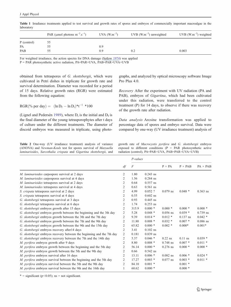
Table 2 One-way (UV irradiance treatment) analysis of variance (ANOVA) and Newman-Keuls test for spores survival of Mazzaellalaminarioides, Sarcothalia crispata and Gigartina skottsbergii, and growth rate of Macrocystis pyrifera and G. skottsbergii embryos exposed todifferent conditions (P = PAR photosynthetic active radiation (control), PA=PAR+UVA, PAB=PAR+UVA+UVB)
P-values
df F P × PA P × PAB PA × PAB
M. laminarioides carpospore survival at 2 days 2 1.80 0.243 nsM. laminarioides carpospores survival at 4 days 2 1.56 0.284 nsM. laminarioides tetraspores survival at 2 days 2 0.64 0.557 nsM. laminarioides tetraspores survival at 4 days 2 0.63 0.561 nsS. crispata tetraspores survival at 2 days 2 4.99 0.052 ? 0.079 ns 0.048 * 0.363 nsS. crispata tetraspores survival at 4 days 2 0.55 0.602 nsG. skottsbergii tetraspores survival at 3 days 2 0.93 0.445 nsG. skottsbergii tetraspores survival at 6 days 2 1.74 0.253 nsG. skottsbergii embryos growth after 15 days 2 315.9 0.000 * 0.000 * 0.000 * 0.000 *G. skottsbergii embryos growth between the beginning and the 3th day 2 5.28 0.048 * 0.056 ns 0.039 * 0.739 nsG. skottsbergii embryos growth between the 3th and the 7th day 2 9.39 0.014 * 0.012 * 0.137 ns 0.042 *G. skottsbergii embryos growth between the 7th and the 9th day 2 11.80 0.008 * 0.032 * 0.007 * 0.086 nsG. skottsbergii embryos growth between the 9th and the 15th day 2 65.82 0.000 * 0.002 * 0.000* 0.001*G. skottsbergii embryos recovery after14 days 2 3.41 0.102 nsG. skottsbergii embryos recovery between the beginning and the 7th day 2 0.181 0.839 nsG. skottsbergii embryos recovery between the 7th and the 14th day 2 5.37 0.046 * 0.22 ns 0.11 ns 0.039 *M. pyrifera embryos growth after 9 days 2 8.80 0.008 * 0.748 ns 0.007 * 0.011 *M. pyrifera embryos growth between the beginning and the 5th day 2 56.14 0.000 * 0.276 ns 0.000 * 0.000 *M. pyrifera embryos growth between the 5th and the 9th day 2 0.66 0.542 nsM. pyrifera embryos survival after 16 days 2 13.11 0.006 * 0.082 ns 0.006 * 0.024 *M. pyrifera embryos survival between the beginning and the 5th day 2 17.27 0.003 * 0.077 ns 0.003 * 0.011 *M. pyrifera embryos survival between the 5th and the 9th day 2 84.10 0.001 * 0.001 *M. pyrifera embryos survival between the 9th and the 16th day 1 60.62 0.000 * 0.000 *
* = significant (p<0.05); ns = not significant.
Table 2 One-way (UV irradiance treatment) analysis of variance(ANOVA) and Newman-Keuls test for spores survival of Mazzaellalaminarioides, Sarcothalia crispata and Gigartina skottsbergii, and
growth rate of Macrocystis pyrifera and G. skottsbergii embryosexposed to different conditions (P = PAR photosynthetic activeradiation (control), PA=PAR+UVA, PAB=PAR+UVA+UVB)
Table 1 Irradiance treatments applied to test survival and growth rates of spores and embryos of commercially important macroalgae in thelaboratory
PAR (μmol photons m−2.s−1) UVA (W.m−2) UVB (W.m−2) unweighted UVB (W.m−2) weighted
P (control) 55PA 55 0.9PAB 55 0.9 0.2 0.003
For weighted irradiance, the action spectra for DNA damage (Setlow 1974) was appliedP = PAR photosynthetic active radiation, PA=PAR+UVA, PAB=PAR+UVA+UVB
J Appl Phycol
variance (ANOVA). Newman-Keuls test was used to estab-lish statistical differences. In all cases the RGR mean valueof each Petri dish was considered as a replicate, thus for eachtreatment the number of replicate was three. All statisticaltests were performed in accordance with Zar (1999).
Results
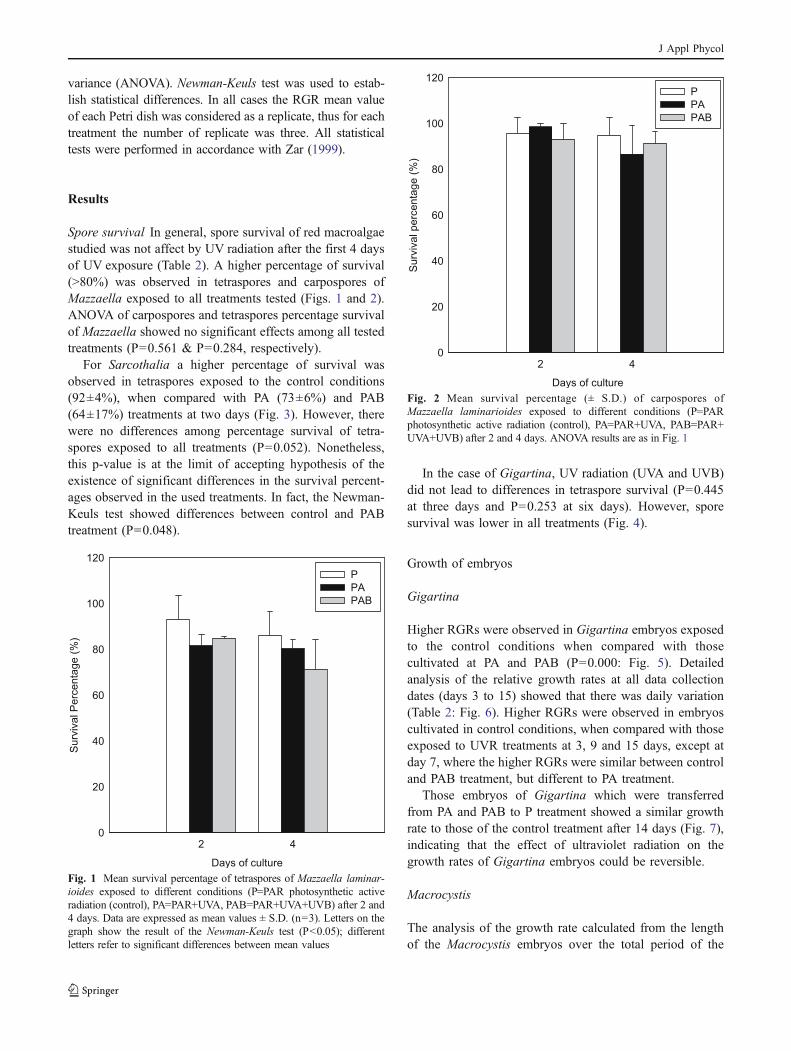
Spore survival In general, spore survival of red macroalgaestudied was not affect by UV radiation after the first 4 daysof UV exposure (Table 2). A higher percentage of survival(>80%) was observed in tetraspores and carpospores ofMazzaella exposed to all treatments tested (Figs. 1 and 2).ANOVA of carpospores and tetraspores percentage survivalof Mazzaella showed no significant effects among all testedtreatments (P=0.561 & P=0.284, respectively).
For Sarcothalia a higher percentage of survival wasobserved in tetraspores exposed to the control conditions(92±4%), when compared with PA (73±6%) and PAB(64±17%) treatments at two days (Fig. 3). However, therewere no differences among percentage survival of tetra-spores exposed to all treatments (P=0.052). Nonetheless,this p-value is at the limit of accepting hypothesis of theexistence of significant differences in the survival percent-ages observed in the used treatments. In fact, the Newman-Keuls test showed differences between control and PABtreatment (P=0.048).
In the case of Gigartina, UV radiation (UVA and UVB)did not lead to differences in tetraspore survival (P=0.445at three days and P=0.253 at six days). However, sporesurvival was lower in all treatments (Fig. 4).
Growth of embryos
Gigartina
Higher RGRs were observed in Gigartina embryos exposedto the control conditions when compared with thosecultivated at PA and PAB (P=0.000: Fig. 5). Detailedanalysis of the relative growth rates at all data collectiondates (days 3 to 15) showed that there was daily variation(Table 2: Fig. 6). Higher RGRs were observed in embryoscultivated in control conditions, when compared with thoseexposed to UVR treatments at 3, 9 and 15 days, except atday 7, where the higher RGRs were similar between controland PAB treatment, but different to PA treatment.
Those embryos of Gigartina which were transferredfrom PA and PAB to P treatment showed a similar growthrate to those of the control treatment after 14 days (Fig. 7),indicating that the effect of ultraviolet radiation on thegrowth rates of Gigartina embryos could be reversible.
Macrocystis
The analysis of the growth rate calculated from the lengthof the Macrocystis embryos over the total period of the
Days of culture
2 4
Su
rviv
al P
erc
en
tag
e (
%)
0
20
40
60
80
100
120
P
PA
PAB
Fig. 1 Mean survival percentage of tetraspores of Mazzaella laminar-ioides exposed to different conditions (P=PAR photosynthetic activeradiation (control), PA=PAR+UVA, PAB=PAR+UVA+UVB) after 2 and4 days. Data are expressed as mean values ± S.D. (n=3). Letters on thegraph show the result of the Newman-Keuls test (P<0.05); differentletters refer to significant differences between mean values
Days of culture
2 4
Su
rviv
al p
erc
en
tag
e (
%)
0
20
40
60
80
100
120
P
PA
PAB
Fig. 2 Mean survival percentage (± S.D.) of carpospores ofMazzaella laminarioides exposed to different conditions (P=PARphotosynthetic active radiation (control), PA=PAR+UVA, PAB=PAR+UVA+UVB) after 2 and 4 days. ANOVA results are as in Fig. 1
J Appl Phycol

experiment (9 days) showed differences between the treat-ments (P=0.007). The post-hoc test indicated that thedifferences were between the P treatment (control) and thePAB, with lower growth being observed under this last
treatment (Fig. 8). A more detailed analysis showed that themajor differences in the growth rates were observed in thefirst 5 days of cultivation (P=0.000), whereas in the periodbetween the 5th and the 9th day there were no significantdifferences (P=0.542) (Fig. 9).
Regarding survival, it was found that embryos cultivatedunder the PAB treatment showed a survival rate lower than
Days of culture
2 4
Su
rviv
al p
erc
en
tag
e (
%)
0
20
40
60
80
100
P
PA
PAB
Fig. 3 Mean survival percentage (± S.D.) of tetraspores of Sarcothaliacrispata exposed to different conditions (P=PAR photosynthetic activeradiation (control), PA=PAR+UVA, PAB=PAR+UVA+UVB) after 2 and4 days. ANOVA results are as in Fig. 1
Days of culture
2 4
Surv
ival P
erc
enta
ge (
%)
0
20
40
60
80
100
P
PA
PAB
Fig. 4 Survival percentage of tetraspores of Gigartina skottsbergiiexposed to different conditions (P=PAR Photosynthetic ActiveRadiation (control), PA=PAR+UVA, PAB=PAR+UVA+UVB) after 3and 6 days. Data are expressed as mean values ± S.D. (n=3). Letterson the graph show the result of the Newman-Keuls test (P<0.05);different letters refer to significant differences between mean values
P PA PAB
RG
R (
% d
ay
-1)
0
1
2
3
4
5
6
a
b
c
Fig. 5 Mean relative growth rate (RGR; % day-1) of Gigartinaskottsbergii embryos exposed to different conditions (P=PAR (con-trol), PA=PAR+UVA, PAB=PAR+UVA+UVB) after 15 days. Data areexpressed as mean values ± S.D. (n=4); different letters refer tosignificant differences between mean values with Newman-Keuls post-hoc test (P<0.05)
Days of culture
3 7 9 15
RG
R (
% d
ay -1
)
0
2
4
6
8
10
12
P
PA
PAB
a
a
a
a
b
a b
b
b
b
c
b
Fig. 6 Mean relative growth rate (RGR; % day-1) of Gigartinaskottsbergii embryos exposed to different conditions (P=PAR (con-trol), PA=PAR+UVA, PAB=PAR+UVA+UVB) of all data collectiondates (days 3 to 15). Data are expressed as mean values ± S.D. (n=4);different letters refer to significant differences between mean valueswith Newman-Keuls post-hoc test (P<0.05)
J Appl Phycol
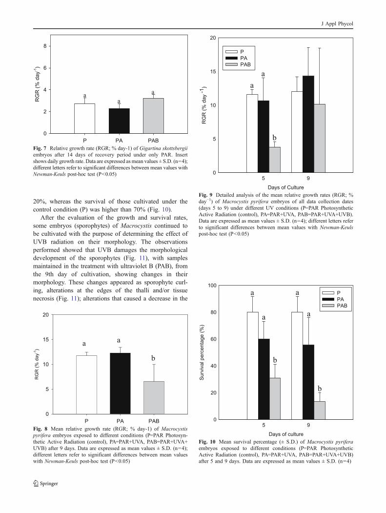
20%, whereas the survival of those cultivated under thecontrol condition (P) was higher than 70% (Fig. 10).
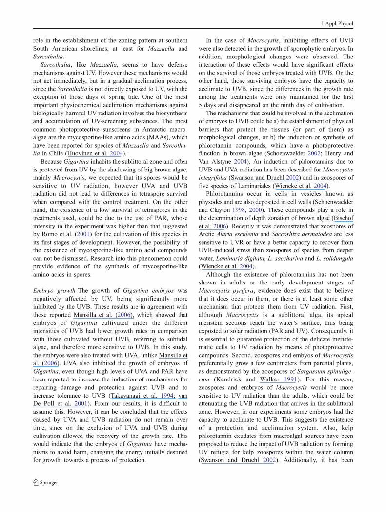
After the evaluation of the growth and survival rates,some embryos (sporophytes) of Macrocystis continued tobe cultivated with the purpose of determining the effect ofUVB radiation on their morphology. The observationsperformed showed that UVB damages the morphologicaldevelopment of the sporophytes (Fig. 11), with samplesmaintained in the treatment with ultraviolet B (PAB), fromthe 9th day of cultivation, showing changes in theirmorphology. These changes appeared as sporophyte curl-ing, alterations at the edges of the thalli and/or tissuenecrosis (Fig. 11); alterations that caused a decrease in the
P PA PAB
RG
R (
% d
ay
-1)
0
2
4
6
8
a a
a
Fig. 7 Relative growth rate (RGR; % day-1) of Gigartina skottsbergiiembryos after 14 days of recovery period under only PAR. Insertshows daily growth rate. Data are expressed asmean values ± S.D. (n=4);different letters refer to significant differences between mean values withNewman-Keuls post-hoc test (P<0.05)
P PA PAB
RG
R (
% d
ay
-1)
0
5
10
15
20
a a
b
Fig. 8 Mean relative growth rate (RGR; % day-1) of Macrocystispyrifera embryos exposed to different conditions (P=PAR Photosyn-thetic Active Radiation (control), PA=PAR+UVA, PAB=PAR+UVA+UVB) after 9 days. Data are expressed as mean values ± S.D. (n=4);different letters refer to significant differences between mean valueswith Newman-Keuls post-hoc test (P<0.05)
Days of Culture
5 9
RG
R (
% d
ay -1
)
0
5
10
15
20
P
PA
PAB
a
a
b
Fig. 9 Detailed analysis of the mean relative growth rates (RGR; %day−1) of Macrocystis pyrifera embryos of all data collection dates(days 5 to 9) under different UV conditions (P=PAR PhotosyntheticActive Radiation (control), PA=PAR+UVA, PAB=PAR+UVA+UVB).Data are expressed as mean values ± S.D. (n=4); different letters referto significant differences between mean values with Newman-Keulspost-hoc test (P<0.05)
Days of culture
5 9
Surv
ival perc
enta
ge (
%)
0
20
40
60
80
100
P
PA
PAB
a
a
b
a
a
b
Fig. 10 Mean survival percentage (± S.D.) of Macrocystis pyriferaembryos exposed to different conditions (P=PAR PhotosyntheticActive Radiation (control), PA=PAR+UVA, PAB=PAR+UVA+UVB)after 5 and 9 days. Data are expressed as mean values ± S.D. (n=4)
J Appl Phycol
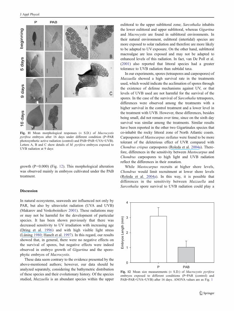
growth (P=0.000) (Fig. 12). This morphological alterationwas observed mainly in embryos cultivated under the PABtreatment.
Discussion
In natural ecosystems, seaweeds are influenced not only byPAR, but also by ultraviolet radiation (UVA and UVB)(Makarov and Voskoboinikov 2001). These radiations mayor may not be harmful for the development of particularspecies. It has been shown previously that there wasdecreased sensitivity to UV irradiation with increasing age(Dring et al. 1996) and with high visible light stress(Lüning 1980; Hanelt et al. 1997). In this regard, our resultsshowed that, in general, there were no negative effects onthe survival of spores, but negative effects were indeedobserved in embryo growth of Gigartina and the sporo-phytic embryos of Macrocystis.
These data seem contrary to the evidence presented by theabove-mentioned authors; however, our data should beanalyzed separately, considering the bathymetric distributionof these species and their evolutionary history. Of the speciesstudied, Mazzaella is an abundant species within the upper
eulittoral to the upper sublittoral zone; Sarcothalia inhabitsthe lower eulittoral and upper sublittoral, whereas Gigartinaand Macrocystis are found in sublittoral environments. Intheir natural environment, eulittoral (intertidal) species aremore exposed to solar radiation and therefore are more likelyto be adapted to UV exposure. On the other hand, sublittoralmacroalgae are less exposed and may not be adapted toenhanced levels of this radiation. In fact, van De Poll et al.(2001) also reported that littoral species had a greatertolerance to UVB radiation than subtidal taxa.
In our experiments, spores (tetraspores and carpospores) ofMazzaella showed a high survival rate in the treatmentsused, which would indicate the acclimation of spores throughthe existence of defense mechanisms against UV, or thatlevels of UVB used are not harmful for the survival of thespores. In the case of the survival of Sarcothalia tetraspores,differences were observed among the treatments with ahigher survival in the control treatment and a lower level inthe treatment with UVB. However, these differences, besidesbeing small, did not remain over time, since on the sixth daysurvival was similar among the treatments. Similar resultshave been reported in the other two Gigartinales species thatco-inhabit the rocky littoral zone of North Atlantic coasts.Carpospores of Mastocarpus stellatus were found to be moretolerant of the deleterious effect of UVR compared withChondrus crispus carpospores (Roleda et al. 2004a). There-fore, differences in the sensitivity between Mastocarpus andChondrus carpospores to high light and UVB radiationreflect the differences in their zonation.
While Mastocarpus recruits at higher shore levels,Chondrus would limit recruitment at lower shore levels(Roleda et al. 2004a). In this way, it is possible thatdifferences in the sensitivity between Mazzaella andSarcothalia spore survival to UVB radiation could play a
Fig. 11 Mean morphological responses (± S.D.) of Macrocystispyrifrea embryos after 16 days under different condition (P=PARphotosynthetic active radiation (control) and PAB=PAR+UVA+UVB).Letters A, B and C show details of M. pyrifera embryos exposed toUVB radiation at 9 days
P PAB
Em
bry
os L
en
gth
(m
m)
0
1
2
3
4
Fig. 12 Mean size measurements (± S.D.) of Macrocystis pyriferaembryos exposed to different conditions (P=PAR (control) andPAB=PAR+UVA+UVB) after 16 days. ANOVA values are as Fig. 1
J Appl Phycol
role in the establishment of the zoning pattern at southernSouth American shorelines, at least for Mazzaella andSarcothalia.
Sarcothalia, like Mazzaella, seems to have defensemechanisms against UV. However these mechanisms wouldnot act immediately, but in a gradual acclimation process,since the Sarcothalia is not directly exposed to UV, with theexception of those days of spring tide. One of the mostimportant physiochemical acclimation mechanisms againstbiologically harmful UV radiation involves the biosynthesisand accumulation of UV-screening substances. The mostcommon photoprotective sunscreens in Antarctic macro-algae are the mycosporine-like amino acids (MAAs), whichhave been reported for species of Mazzaella and Sarcotha-lia in Chile (Huovinen et al. 2004).
Because Gigartina inhabits the sublittoral zone and oftenis protected from UV by the shadowing of big brown algae,mainly Macrocystis, we expected that its spores would besensitive to UV radiation, however UVA and UVBradiation did not lead to differences in tetraspore survivalwhen compared with the control treatment. On the otherhand, the existence of a low survival of tetraspores in thetreatments used, could be due to the use of PAR, whoseintensity in the experiment was higher than that suggestedby Romo et al. (2001) for the cultivation of this species inits first stages of development. However, the possibility ofthe existence of mycosporine-like amino acid compoundscan not be dismissed. Research into this phenomenon couldprovide evidence of the synthesis of mycosporine-likeamino acids in spores.
Embryo growth The growth of Gigartina embryos wasnegatively affected by UV, being significantly moreinhibited by the UVB. These results are in agreement withthose reported Mansilla et al. (2006), which showed thatembryos of Gigartina cultivated under the differentintensities of UVB had lower growth rates in comparisonwith those cultivated without UVB, referring to subtidalalgae, and therefore more sensitive to UVB. In this study,the embryos were also treated with UVA, unlike Mansilla etal. (2006). UVA also inhibited the growth of embryos ofGigartina, even though high levels of UVA and PAR havebeen reported to increase the induction of mechanisms forrepairing damage and protection against UVB and toincrease tolerance to UVB (Takayanagi et al. 1994; vanDe Poll et al. 2001). From our results, it is difficult toassume this. However, it can be concluded that the effectscaused by UVA and UVB radiation do not remain overtime, since on the exclusion of UVA and UVB duringcultivation allowed the recovery of the growth rate. Thiswould indicate that the embryos of Gigartina have mecha-nisms to avoid harm, changing the energy initially destinedfor growth, towards a process of protection.
In the case of Macrocystis, inhibiting effects of UVBwere also detected in the growth of sporophytic embryos. Inaddition, morphological changes were observed. Theinteraction of these effects would have significant effectson the survival of those embryos treated with UVB. On theother hand, those surviving embryos have the capacity toacclimate to UVB, since the differences in the growth rateamong the treatments were only maintained for the first5 days and disappeared on the ninth day of cultivation.
The mechanisms that could be involved in the acclimationof embryos to UVB could be a) the establishment of physicalbarriers that protect the tissues (or part of them) asmorphological changes, or b) the induction or synthesis ofphlorotannin compounds, which have a photoprotectivefunction in brown algae (Schoenwaelder 2002; Henry andVan Alstyne 2004). An induction of phlorotannins due toUVB and UVA radiation has been described for Macrocystisintegrifolia (Swanson and Druehl 2002) and in zoospores offive species of Laminariales (Wiencke et al. 2004).
Phlorotannins occur in cells in vesicles known asphysodes and are also deposited in cell walls (Schoenwaelderand Clayton 1998, 2000). These compounds play a role inthe determination of depth zonation of brown algae (Bischofet al. 2006). Recently it was demonstrated that zoospores ofArctic Alaria esculenta and Saccorhiza dermatodea are lesssensitive to UVR or have a better capacity to recover fromUVR-induced stress than zoospores of species from deeperwater, Laminaria digitata, L. saccharina and L. solidungula(Wiencke et al. 2004).
Although the existence of phlorotannins has not beenshown in adults or the early development stages ofMacrocystis pyrifera, evidence does exist that to believethat it does occur in them, or there is at least some othermechanism that protects them from UV radiation. First,although Macrocystis is a sublittoral alga, its apicalmeristem sections reach the water’s surface, thus beingexposted to solar radiation (PAR and UV). Consequently, itis essential to guarantee protection of the delicate meriste-matic cells to UV radiation by means of photoprotectivecompounds. Second, zoospores and embyos of Macrocystispreferentially grow a few centimeters from parental plants,as demonstrated by the zoospores of Sargassum spinulige-rum (Kendrick and Walker 1991). For this reason,zoospores and embryos of Macrocystis would be moresensitive to UV radiation than the adults, which could beattenuating the UVB radiation that arrives in the sublittoralzone. However, in our experiments some embryos had thecapacity to acclimate to UVB. This suggests the existenceof a protection and acclimation system. Also, kelpphlorotannin exudates from macroalgal sources have beenproposed to reduce the impact of UVB radiation by formingUV refugia for kelp zoospores within the water column(Swanson and Druehl 2002). Additionally, it has been
J Appl Phycol
suggested that kelp spores and embryos are pre-adapted tothe UV conditions of the parent plant (Swanson and Druehl2000). Genetic adaptations to PAR light have beenpreviously demonstrated within the Laminariales (Gerard1988), and may also exist for potent environmental stressessuch as UV (Swanson and Druehl 2000).
On the other hand, embryos of Macrocystis showmorphological changes, seen as deformation in the thallusand the loss of cells, possibly from necrosis. These changeshave been reported for Laminaria ochroleuca (Roleda et al.2004b) and L. solidungula (Michler et al. 2002), and the redalgae Iridaea cordata (Navarro 2004). In this last case, thedeformation was manifest as curving of the thallus,diminishing in the area of exposure to UVB, and thusbeing a protective factor against radiation.
Implications Our results provide firm evidence of UVB radi-ation effects on the development of Macrocystis, Gigartina,Sarcothalia and Mazzaella, all of which are of greatcommercial and interest in southern Chile. Such effects havea potential to interfere with natural populations of thesemacroalgal species, as well.
In nature, the early developmental stages of macroalgaespecies could grow with the protection of nearby adults.Adult individuals could be attenuating the UVB radiation tothe young stages, reducing radiation levels which couldcause irreparable harm. The occurrence of microhabitatscould favour the survival of spores and juveniles stages(Brawley and Johnson 1991). This protection has beensuggested for other species (Ang 1991; Brawley andJohnson 1991; Johnson and Brawley 1998) and couldattenuate the UVB effect, preventing the irreparable harmcaused by high levels. Nonetheless, if the adults areextracted because of their economic importance, the resultwould be an increase of UVB levels reaching the intertidaland subtidal zone. In this scenario, the effects on growthand morphology may not be reversible, and the defensemechanisms might be insufficient, causing irreparablecellular damage and unbalances in early developmentalstages.
The importance of kelp forests of Macrocystis is beingincreasingly recognised in southern Chile for both its roleas a keystone species and ecosystem engineer. In additionMacrocystis has high commercial value and is underincreasing pressure for industrial exploitation. Within kelpforests we also find many other species of algae, includingGigartina (Zamorano and Westermeier 1996), which wouldbe affected by the future extraction of Macrocystis.Consequently, for both ecological and economic reasons,there is a great necessity to understand the role of UVB onnatural populations of algae and their long-term recruitmentsystems. In addition, complementary studies regarding theeffects of UVB on the carrageen and algine acid content of
these species will allow researchers to better understand thereal effects that this kind of radiation has on commercialspecies of macroalgae in Chile. A final aspect that shouldbe included in future work is the need to incorporate anoutlook of long-term recuperation of stratospheric ozonelevels, which are currently projected to not reach historiclevels until 2050 (WMO/UNEP 2002; Valkama et al. 2003).
Acknowledgement We gratefully acknowledge Susana Ovando,Clare Brown and Dr. Christopher Anderson for valuable suggestionsand improvement of this manuscript. This work was financed in partby Project Mineduc Acuicultura PR 334 (Chilean Ministry ofEducation), and by Project 26600 (University of Magallanes).
References
Altamirano M, Flores-Moya A, Figueroa F (2002) Effects of theradiation and temperature on growth of germling of three speciesof Fucus (Phaeophyta). Aquat Bot 1606:1–12
Ang PO Jr (1991) Natural dynamics of a Fucus distichus (Phaeophyta,Fucales) population: reproduction and recruitment. Mar EcolProg Ser 78:71–85
Bianciotto OA, Pinedo LB, San Roman NA, Blessio AY, CollantesMB (2003) The effects of natural UV-B radiation on a perennialSalicornia salt-marsh in Bahía San Sebastián, Tierra del Fuego,Argentina: a 3-year field study. J Photochem Photobiol B Biol70:177–185
Bischof K, Gómez I, Molis M, Hanelt D, Karsten U, Lüder U, RoledaMY, Zacher K, Wiencke C (2006) Ultraviolet radiation shapesseaweed communities. Rev Environmental Sci Biotechnol 5:141–166
Brawley SH, Johnson LE (1991) Survival of fucoid embryos in theintertidal zone depends upon developmental stage and microhab-itat. J Phycol 27:179–186
Cordi B, Donkin ME, Peloquin J, Price DN, Depledge MH (2001)The influence of UV–B radiation on the reproductive cells of theintertidal macroalga, Enteromorpha intestinalis. Aquat Toxicol56:1–11
Diaz S, Camilio C, Deferrari G, Fuenzalida H, Armstrong R, Booth C,Paladini A, Cabrera S, Casiccia C, Lovengreen C, Pedroni J,Rosales A, Zagarese H, Vernet M (2006) Symposium-in-Print:UV effects on aquatic and coastal ecosystems ozone and UVradiation over southern south America: climatology and anoma-lies. Photochem Photobiol 82:834–843
Dring M, Makarov V, Schoschina E, Lorenz M, Lüning K (1996)Influence of ultraviolet-radiation on chlorophyll fluorescence andgrowth in different life-history stages of three species ofLaminaria (Phaeophyta). Mar Biol 126:183–191
Figueroa FL (2002) Bio-optical characteristics of Gerlache andBransfield Strait waters during an Antarctic summer cruise. DeepSea Res II 49:675–691
Franklin L, Forster R (1997) The changing irradiance environment:consequences for marine macrophyte physiology, productivityand ecology. Eur J Phycol 32:207–237
Gerard VA (1988) Ecotypic differentiation in light-related traits of thekelp Laminaria saccharina. Mar Biol 97:25–36
Hanelt D, Wincke C, Karsten U, Nultsch W (1997) Photoinhibitionand recovery after high light stress in different developmentaland life-history stages of Laminaria saccharina (Phaeophyta). JPhycol 33:387–395
J Appl Phycol
Henry EB, Van Alstyne KL (2004) Effects of UV radiation on growthand phlorotannins in Fucus gardneri (Phaeophyceae) juvenilesand embryos. J Phycol 40:527–533
Huovinen P, Gómez I, Figueroa F, Ulloa N, Morales V, Lovengreen C(2004) Ultraviolet-absorbing mycosporine-like amino acids inred macroalgae from Chile. Bot Mar 47:21–29
Johnson LE, Brawley SH (1998) Dispersal and recruitment of canopy-forming intertidal alga: the relative roles of propagule availabilityin post-settlement processes. Oecologia 117:517–526
Kendrick GA, Walker D (1991) Dispersal distances for propagules ofSargassum spinuligerum (Sargassaceae, Phaeophyta) measureddirectly by vital staining and venturi suction sampling. Mar EcolProg Ser 79:133–138
Kirchhoff VWJH, Zamorano F, Casiccia C (1997) UVB enhancements atPunta Arenas, Chile. J Photochem Photobiol B Biol 38:174–177
Lignel A, Pedersén M (1989) Agar composition as a function ofmorphology and growth rate. Studies on some morphologicalstrains of Gracilaria secundata and Gracilaria verrucosa(Rhodophyta). Bot Mar 32:219–227
Lüning K (1980) Critical levels of light and temperature regulating thegametogenesis of three Laminaria species (Phaeophyceae). JPhycol 16:1–15
Makarov MV, Voskoboinikov GM (2001) The influence of ultraviolet-B radiation on spores release and growrh of kelp Laminariasaccharina. Bot Mar 44:89–94
Mansilla A, Werlinger C, Palacios M, Navarro NP, Cuadra P (2006)Effects of UVB radiation on the initial stages of growth ofGigartina skottsbergii, Sarcothalia crispata and Mazzaellalaminarioides (Gigartinales, Rhodophyta). J Appl Phycol18:451–459
Marín SL, Westermeier R, Melipillan J (2002) Simulation ofalternative management strategies for red algae, luga roja,(Gigartina skottsbergii Setchell and Gardner) in southern Chile.Ecol Mod 154:121–133
Michler T, Aguilera J, Hanelt D, Bischof K, Wiencke C (2002) Long-term effects of ultraviolet radiation on growth and photosyntheticperformance of polar and cold-temperature macroalgae. Mar Biol140:1117–1127
Navarro N (2004) Efeitos da radiação UVB em Iridaea cordata(Gigartinales, Rhodophyta). Dissertação Universidade de SãoPaulo
Oliveira EC, De Paula EJ, Plastino EM, Petti R (1995) Metodologíaspara el cultivo no axenico de macroalgas marinas in vitro. In:Alveal K, Ferrario M, Oliveira E, Sar E (eds) Manual de métodosficológicos. Universidad de Concepción, Chile, pp 429–447
Roleda M, van de Poll W, Hanelt D, Wiencke C (2004a) PAR and UVBReffects on photosynthesis, viability, growth andDNA in different lifestages of two coexisting Gigartinales: implications for recruitmentand zonation pattern. Mar Ecol Prog Ser 281:37–50
Roleda M, Hanelt D, Krabs G, Wiencke C (2004b) Morphology,growth, photosynthesis and pigment in Laminaria ochroleuca(Laminariales, Phaeophyta) under ultraviolet radiation. Phycologia43(5):603–613
Romo H, Avila M, Nuñez M (2001) Manual de técnicas de cultivo yrepoblación de “Luga Roja” (Gigartina skottsbergii) ProyectoFONDEF D97I1064. IFOP- Universidad de Concepción, Chile
Schoenwaelder M (2002) The occurrence and cellular significance ofphysodes in brown algae. Phycologia 41:125–139
Schoenwaelder M, Clayton M (1998) Secretion of phenolic substancesinto the zygote wall and cell plate in embryos of Hormosira andAcrocarpia (Fucales, Phaeophyceae). J Phycol 34:969–980
Schoenwaelder M, Clayton M (2000) Physode formation in embryosof Phyllospora comosa and Hormosira banksii (Phaeophyceae).Phycologia 39:1–9
Seckmeyer G, Mckenzie RL (1992) Elevated ultraviolet radiation inNew Zealand (45 S) contrasted with Germany (45 N). Nature359:135–137
Setlow RB (1974) The wavelengths in sunlight effective in producingskin cancer: a theoretical analysis. Proc Nat Acad Sci USA71:3363–3366
Smith RC, Prézelin BB, Baker KS, Bidigare R, Boucher N, Coley T,Karentz D, Macintyre S, Matlick H, Menzies D, Ondrusek M,Wan Z, Waters J (1992) Ozone depletion: ultraviolet radiationand phytoplankton biology in Antarctic waters. Science 255:952–959
Swanson A, Druehl L (2000) Differential meiospore size and toleranceof ultraviolet light stress within and among kelp species along adepth gradient. Mar Biol 136:657–664
Swanson A, Druehl L (2002) Induction, exudation and the UVprotective role of kelp phlorotannins. Aquat Bot 73:241–253
Takayanagi S, Trunk J, Sutherland JC, Sutherland BM (1994) Alfalfagrown outdoor are more resistant to UV-induced DNA damagethan plants grown in a UV-free environmental chamber. PhotochemPhotobiol 60:363–367
Valkama E, Kivimaenpaa M, Hartikainen H, Wulff A (2003) Thecombined effects of enhanced UV-B radiation and selenium ongrowth, chlorophyll fluorescence and ultrastructure in strawberry(Fragaria x ananassa) and barley (Hordeum vulgare) treated inthe field. Agric For Meteorol 120:267–278
van De Poll W, Eggert A, Buma A, Breeman A (2001) Effects of UV-induced DNA damage and photoinhibition on growth oftemperature marine red macrophytes: habitat - related differencesin UV-B tolerance. J Phycol 37:30–37
Wiencke C, Gómez I, Pakker H, Flores-Moya A, Altamirano M,Hanelt D, Bischof K, Figueroa FL (2000) Impact of UV-radiationon viability, photosynthetic characteristics and DNA of brownalgal zoospores: implications for depth zonation. Mar Ecol ProgSer 197:217–229
Wiencke C, Clayton M, Schoenwaelder M (2004) Sensitivity andacclimation to UV radiation of zoospores from five species ofLaminariales from the Arctic. Mar Biol 145:31–39
Wiencke C, Roleda M, Gruber A, Clayton MN, Bischof K (2006)Susceptibility of zoospores to UV radiation determines upperdepth distribution limit of Arctic kelps: evidence through fieldexperiments. J Ecol 94:455–463
Xue L, Zhang Y, Zhang T, An L, Wang X (2005) Effects of enhancedultraviolet-B radiation on algae and cianobacteria. Crit RevMicrobiol 31:79–89
WMO/UNEP (2002) Scientific assessment of ozone depletion: 2002.Available at http://www.wmo.ch/web/arep/reports/o3_assess_rep_2002_front_page.html
Zamorano J, Westermeier R (1996) Phenology of Gigartina skotts-bergii (Rhodophyta, Gigartinales) in Ancud Bay, southern Chile.Hydrobiologia 326/327:253–259
Zar JH (1999) Biostatistical analysis, 4th edn. Prentice-Hall, UpperSaddle River, NJ
J Appl Phycol