Use of the anterolateral thigh in cranio-orbitofacial reconstruction
6
Hindawi Publishing Corporation Plastic Surgery International Volume 2011, Article ID 941742, 6 pages doi:10.1155/2011/941742 Clinical Study Use of the Anterolateral Thigh in Cranio-Orbitofacial Reconstruction William J. Parkes, Howard Krein, Ryan Heffelfinger, and Joseph Curry Department of Otolaryngology-Head and Neck Surgery, Thomas Jefferson University Hospital, 925 Chestnut Street 6th Floor, Philadelphia, PA 19107, USA Correspondence should be addressed to William J. Parkes, [email protected] Received 15 June 2011; Accepted 25 August 2011 Academic Editor: Georg M. Huemer Copyright © 2011 William J. Parkes et al. This is an open access article distributed under the Creative Commons Attribution License, which permits unrestricted use, distribution, and reproduction in any medium, provided the original work is properly cited. Objective. To detail the clinical outcomes of a series of patients having undergone free flap reconstruction of the orbit and periorbita and highlight the anterolateral thigh (ALT) as a workhorse for addressing defects in this region. Methods. A review of 47 patients who underwent free flap reconstruction for orbital or periorbital defects between September 2006 and May 2011 was performed. Data reviewed included demographics, defect characteristics, free flap used, additional reconstructive techniques employed, length of stay, complications, and follow-up. The ALT subset of the case series was the focus of the data reviewed for this paper. Selected cases were described to highlight some of the advantages of employing the ALT for cranio-orbitofacial reconstruction. Results. 51 free flaps in 47 patients were reviewed. 38 cases required orbital exenteration. The ALT was used in 33 patients. Complications included 1 hematoma, 2 wound infections, 3 CSF leaks, and 3 flap failures. Conclusions. Free tissue transfer allows for the safe and effective reconstruction of complex defects of the orbit and periorbital structures. Reconstructive choice is dependent upon the extent of soft tissue loss, midfacial bone loss, and skullbase involvement. The ALT provides a versatile option to reconstruct the many cranio-orbitofacial defects encountered. 1. Introduction Extirpative surgery involving the orbit and periorbita often results in a complex defect involving multiple midfacial subsites. Depending upon the extent of resection, the recon- structive surgeon may need to address any combination of the following: periorbital skin (forehead, nose, and midface), orbital soft tissue, supra orbital or midface bone, paranasal sinuses, and anterior skull base. A multitude of reconstruc- tive options are available including skin grafting and regional flaps, such as the temporalis muscle or temporoparietal fascia. Yet, in some cases the volume of soft tissue loss or complexity of the defect may require the versatility afforded by free tissue transfer for optimal reconstruction. A variety of free flaps may be employed to this end, including the anterolateral thigh (ALT), radial forearm (RF), fibula, and latissimus dorsi. At our institution, we favor the use of the ALT. The ALT functions to allow a variety of soft tissue configurations very useful to periorbital reconstruction and is the subject of this work. First described by Song et al. [1] in 1984, the ALT has become a workhorse in head and neck reconstruction, allowing for a two-team approach and offering versatility, ample pedicle length, and low donor site morbidity. The ALT can be harvested as a fasciocutaneous, myocutaneous, subcu- taneous, or adipofascial flap. The perforators supplying the flap typically arise from the descending branch of the lateral circumflex femoral artery (LCFA), but anatomic variation is not infrequently encountered and must be recognized. Yu [2] described and defined 3 distinct types of ALT perforator systems. Type I perforators arose from the descending branch of the LCFA and accounted for 90% of perforators identified in their series. Type II perforators arose from the transverse branch of the LCFA and were encountered roughly 4% of the time. Lastly, Type III perforators arose directly from the profunda femoris artery and were too small for microvascular anastomosis. The perforators can be either septocutaneous or musculocutaneous. In their large series, Wei et al. [3] reported that 87.1 % of 504 fasciocutaneous or cutaneous ALT flaps were perfused by musculocutaneous
-
Upload
independent -
Category
Documents
-
view
2 -
download
0
Transcript of Use of the anterolateral thigh in cranio-orbitofacial reconstruction
Hindawi Publishing CorporationPlastic Surgery InternationalVolume 2011, Article ID 941742, 6 pagesdoi:10.1155/2011/941742
Clinical Study
Use of the Anterolateral Thigh inCranio-Orbitofacial Reconstruction
William J. Parkes, Howard Krein, Ryan Heffelfinger, and Joseph Curry
Department of Otolaryngology-Head and Neck Surgery, Thomas Jefferson University Hospital, 925 Chestnut Street 6th Floor,Philadelphia, PA 19107, USA
Correspondence should be addressed to William J. Parkes, [email protected]
Received 15 June 2011; Accepted 25 August 2011
Academic Editor: Georg M. Huemer
Copyright © 2011 William J. Parkes et al. This is an open access article distributed under the Creative Commons AttributionLicense, which permits unrestricted use, distribution, and reproduction in any medium, provided the original work is properlycited.
Objective. To detail the clinical outcomes of a series of patients having undergone free flap reconstruction of the orbit and periorbitaand highlight the anterolateral thigh (ALT) as a workhorse for addressing defects in this region. Methods. A review of 47 patientswho underwent free flap reconstruction for orbital or periorbital defects between September 2006 and May 2011 was performed.Data reviewed included demographics, defect characteristics, free flap used, additional reconstructive techniques employed, lengthof stay, complications, and follow-up. The ALT subset of the case series was the focus of the data reviewed for this paper. Selectedcases were described to highlight some of the advantages of employing the ALT for cranio-orbitofacial reconstruction. Results. 51free flaps in 47 patients were reviewed. 38 cases required orbital exenteration. The ALT was used in 33 patients. Complicationsincluded 1 hematoma, 2 wound infections, 3 CSF leaks, and 3 flap failures. Conclusions. Free tissue transfer allows for the safe andeffective reconstruction of complex defects of the orbit and periorbital structures. Reconstructive choice is dependent upon theextent of soft tissue loss, midfacial bone loss, and skullbase involvement. The ALT provides a versatile option to reconstruct themany cranio-orbitofacial defects encountered.
1. Introduction
Extirpative surgery involving the orbit and periorbita oftenresults in a complex defect involving multiple midfacialsubsites. Depending upon the extent of resection, the recon-structive surgeon may need to address any combination ofthe following: periorbital skin (forehead, nose, and midface),orbital soft tissue, supra orbital or midface bone, paranasalsinuses, and anterior skull base. A multitude of reconstruc-tive options are available including skin grafting and regionalflaps, such as the temporalis muscle or temporoparietalfascia. Yet, in some cases the volume of soft tissue loss orcomplexity of the defect may require the versatility affordedby free tissue transfer for optimal reconstruction. A varietyof free flaps may be employed to this end, including theanterolateral thigh (ALT), radial forearm (RF), fibula, andlatissimus dorsi. At our institution, we favor the use of theALT. The ALT functions to allow a variety of soft tissueconfigurations very useful to periorbital reconstruction andis the subject of this work.
First described by Song et al. [1] in 1984, the ALThas become a workhorse in head and neck reconstruction,allowing for a two-team approach and offering versatility,ample pedicle length, and low donor site morbidity. The ALTcan be harvested as a fasciocutaneous, myocutaneous, subcu-taneous, or adipofascial flap. The perforators supplying theflap typically arise from the descending branch of the lateralcircumflex femoral artery (LCFA), but anatomic variation isnot infrequently encountered and must be recognized. Yu[2] described and defined 3 distinct types of ALT perforatorsystems. Type I perforators arose from the descendingbranch of the LCFA and accounted for 90% of perforatorsidentified in their series. Type II perforators arose from thetransverse branch of the LCFA and were encountered roughly4% of the time. Lastly, Type III perforators arose directlyfrom the profunda femoris artery and were too small formicrovascular anastomosis. The perforators can be eitherseptocutaneous or musculocutaneous. In their large series,Wei et al. [3] reported that 87.1 % of 504 fasciocutaneousor cutaneous ALT flaps were perfused by musculocutaneous
2 Plastic Surgery International
perforators, while 12.9 % were perfused by septocutaneousperforators. The ALT has an average pedicle length of 7-8 cmwith a range in length up to 16 cm and a vessel diameter oflarger than 2 mm [4]. Donor defects with a medial to lateraldiameter less than 8-9 cm are typically closed primarily [5].
With respect to the orbit, the ALT provides sufficient softtissue and skin to fill the dead space within an exenterationcavity and to cover any implants or plates required foradjacent midface reconstruction. Multiple perforators andthe technique of de-epithelialization allow for the creationof separate islands within one flap so that each portion(i.e., skin, exenteration cavity, and maxillary sinus) of theoverall defect can be addressed separately. Most importantly,the ALT provides effective coverage for anterior skull basedefects. Both the vastus lateralis fascia and the adjacenttensor fascia lata can be harvested and used to reconstitutethe dural layer when necessary [6, 7].
This retrospective review details the clinical outcomes ofa series of patients having undergone free flap reconstructionof the periorbita and highlights the ALT as a workhorse foraddressing defects in this region.
2. Patients and Methods
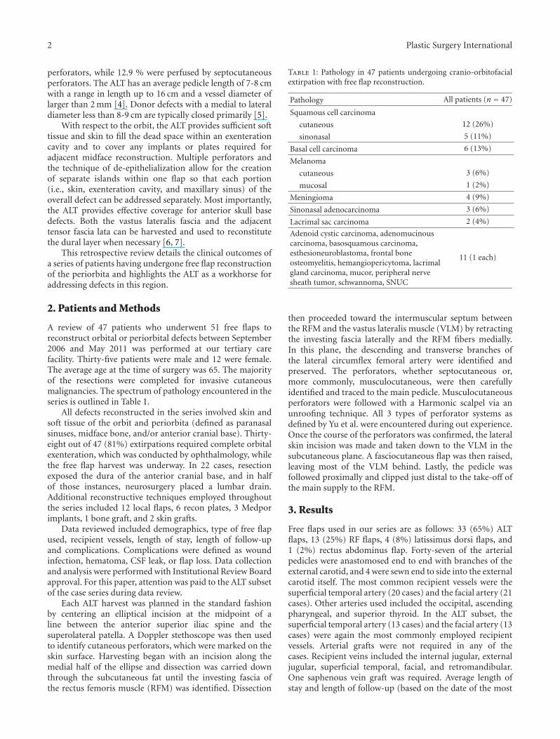
A review of 47 patients who underwent 51 free flaps toreconstruct orbital or periorbital defects between September2006 and May 2011 was performed at our tertiary carefacility. Thirty-five patients were male and 12 were female.The average age at the time of surgery was 65. The majorityof the resections were completed for invasive cutaneousmalignancies. The spectrum of pathology encountered in theseries is outlined in Table 1.
All defects reconstructed in the series involved skin andsoft tissue of the orbit and periorbita (defined as paranasalsinuses, midface bone, and/or anterior cranial base). Thirty-eight out of 47 (81%) extirpations required complete orbitalexenteration, which was conducted by ophthalmology, whilethe free flap harvest was underway. In 22 cases, resectionexposed the dura of the anterior cranial base, and in halfof those instances, neurosurgery placed a lumbar drain.Additional reconstructive techniques employed throughoutthe series included 12 local flaps, 6 recon plates, 3 Medporimplants, 1 bone graft, and 2 skin grafts.
Data reviewed included demographics, type of free flapused, recipient vessels, length of stay, length of follow-upand complications. Complications were defined as woundinfection, hematoma, CSF leak, or flap loss. Data collectionand analysis were performed with Institutional Review Boardapproval. For this paper, attention was paid to the ALT subsetof the case series during data review.
Each ALT harvest was planned in the standard fashionby centering an elliptical incision at the midpoint of aline between the anterior superior iliac spine and thesuperolateral patella. A Doppler stethoscope was then usedto identify cutaneous perforators, which were marked on theskin surface. Harvesting began with an incision along themedial half of the ellipse and dissection was carried downthrough the subcutaneous fat until the investing fascia ofthe rectus femoris muscle (RFM) was identified. Dissection
Table 1: Pathology in 47 patients undergoing cranio-orbitofacialextirpation with free flap reconstruction.
Pathology All patients (n = 47)
Squamous cell carcinoma
cutaneous 12 (26%)
sinonasal 5 (11%)
Basal cell carcinoma 6 (13%)
Melanoma
cutaneous 3 (6%)
mucosal 1 (2%)
Meningioma 4 (9%)
Sinonasal adenocarcinoma 3 (6%)
Lacrimal sac carcinoma 2 (4%)
Adenoid cystic carcinoma, adenomucinouscarcinoma, basosquamous carcinoma,esthesioneuroblastoma, frontal boneosteomyelitis, hemangiopericytoma, lacrimalgland carcinoma, mucor, peripheral nervesheath tumor, schwannoma, SNUC
11 (1 each)
then proceeded toward the intermuscular septum betweenthe RFM and the vastus lateralis muscle (VLM) by retractingthe investing fascia laterally and the RFM fibers medially.In this plane, the descending and transverse branches ofthe lateral circumflex femoral artery were identified andpreserved. The perforators, whether septocutaneous or,more commonly, musculocutaneous, were then carefullyidentified and traced to the main pedicle. Musculocutaneousperforators were followed with a Harmonic scalpel via anunroofing technique. All 3 types of perforator systems asdefined by Yu et al. were encountered during out experience.Once the course of the perforators was confirmed, the lateralskin incision was made and taken down to the VLM in thesubcutaneous plane. A fasciocutaneous flap was then raised,leaving most of the VLM behind. Lastly, the pedicle wasfollowed proximally and clipped just distal to the take-off ofthe main supply to the RFM.
3. Results
Free flaps used in our series are as follows: 33 (65%) ALTflaps, 13 (25%) RF flaps, 4 (8%) latissimus dorsi flaps, and1 (2%) rectus abdominus flap. Forty-seven of the arterialpedicles were anastomosed end to end with branches of theexternal carotid, and 4 were sewn end to side into the externalcarotid itself. The most common recipient vessels were thesuperficial temporal artery (20 cases) and the facial artery (21cases). Other arteries used included the occipital, ascendingpharyngeal, and superior thyroid. In the ALT subset, thesuperficial temporal artery (13 cases) and the facial artery (13cases) were again the most commonly employed recipientvessels. Arterial grafts were not required in any of thecases. Recipient veins included the internal jugular, externaljugular, superficial temporal, facial, and retromandibular.One saphenous vein graft was required. Average length ofstay and length of follow-up (based on the date of the most
Plastic Surgery International 3
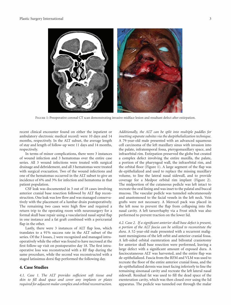
Figure 1: Preoperative coronal CT scan demonstrating invasive midface lesion and resultant defect after extirpation.
recent clinical encounter found on either the inpatient orambulatory electronic medical record) were 10 days and 14months, respectively. In the ALT subset, the average lengthof stay and length of follow-up were 11 days and 14 months,respectively.
In terms of minor complications, there were 3 instancesof wound infection and 3 hematomas over the entire caseseries. All 3 wound infections were treated with surgicaldrainage and debridement, and all 3 hematomas were treatedwith surgical evacuation. Two of the wound infections andone of the hematomas occurred in the ALT subset to give anincidence of 6% and 3% for infection and hematoma in thatpatient population.
CSF leak was documented in 3 out of 18 cases involvinganterior cranial base resection followed by ALT flap recon-struction. One leak was low flow and was managed conserva-tively with the placement of a lumbar drain postoperatively.The remaining two cases were high flow and required areturn trip to the operating room with neurosurgery for aformal skull base repair using a vascularized nasal septal flapin one instance and a fat graft combined with a pericranialflap in the other.
Lastly, there were 3 instances of ALT flap loss, whichtranslates to a 91% success rate in the ALT subset of theseries. Of the 3 losses, 2 were recognized and managed intra-operatively while the other was found to have necrosed at thefirst follow-up visit on postoperative day 18. The first intra-operative loss was reconstructed with a RF flap during thesame procedure, while the second was reconstructed with astaged latissimus dorsi flap performed the following day.
4. Case Studies
4.1. Case 1. The ALT provides sufficient soft tissue andskin to fill dead space and cover any implants or platesrequired for adjacent malar complex and orbital reconstruction.
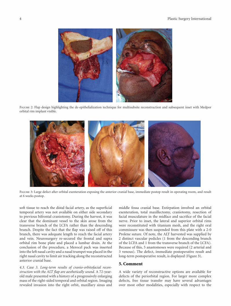
Additionally, the ALT can be split into multiple paddles forinsetting separate subsites via the deepithelialization technique.A 79-year-old male presented with an advanced squamouscell carcinoma of the left maxillary sinus with invasion intothe palate, infratemporal fossa, pterygomaxillary space, andinfraorbital rim. Extirpation preserved the globe but createda complex defect involving the entire maxilla, the palate,a portion of the pharyngeal wall, the infraorbital rim, andthe orbital floor (Figure 1). A large segment of the flap wasde-epithelialized and used to replace the missing maxillaryvolume, to line the lateral nasal sidewall, and to providecoverage for a Medpor orbital rim implant (Figure 2).The midportion of the cutaneous pedicle was left intact torecreate the oral lining and was inset to the palatal and buccalmucosa. The vascular pedicle was tunneled subcutaneouslyand anastomosed to the facial vessels in the left neck. Veingrafts were not necessary. A Merocel pack was placed inthe left nose to prevent the flap from collapsing into thenasal cavity. A left tarsorrhaphy via a Frost stitch was alsoperformed to prevent traction on the lower lid.
4.2. Case 2. If a significant anterior skull base defect is present,a portion of the ALT fascia can be utilized to reconstitute thedura. A 52-year-old male presented with a recurrent malig-nant meningioma of the left orbit and anterior cranial fossa.A left-sided orbital exenteration and bifrontal craniotomyfor anterior skull base resection were performed, leaving alarge defect with a significant amount of exposed dura. Afasciocutaneous ALT was harvested, and the entire flap wasde-epithelialized. Fascia from the RFM and VLM was used torecreate the floor of the entire anterior cranial fossa, and thede-epithelialized dermis was inset facing inferiorly to line theremaining sinonasal cavity and recreate the left lateral nasalsidewall. Residual fat was used to fill the dead space of theexenteration cavity, which was then closed over using the lidapparatus. The pedicle was tunneled out through the malar
4 Plastic Surgery International
Figure 2: Flap design highlighting the de-epithelialization technique for multisubsite reconstruction and subsequent inset with Medpororbital rim implant visible.
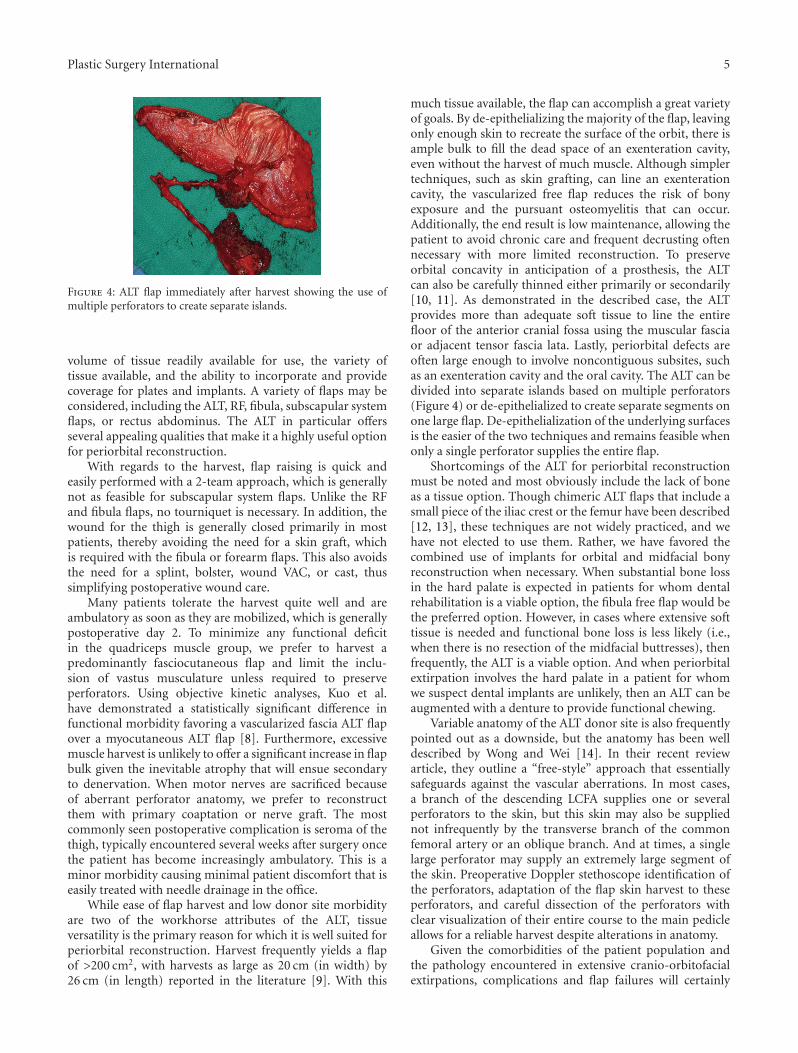
Figure 3: Large defect after orbital exenteration exposing the anterior cranial base, immediate postop result in operating room, and resultat 6 weeks postop.
soft tissue to reach the distal facial artery, as the superficialtemporal artery was not available on either side secondaryto previous bifrontal craniotomy. During the harvest, it wasclear that the dominant vessel to the skin arose from thetransverse branch of the LCFA rather than the descendingbranch. Despite the fact that the flap was raised off of thisbranch, there was adequate length to reach the facial arteryand vein. Neurosurgery re-secured the frontal and supraorbital rim bone plate and placed a lumbar drain. At theconclusion of the procedure, a Merocel pack was insertedinto the left nasal cavity and a nasal trumpet was placed in theright nasal cavity to limit air tracking along the reconstructedanterior cranial base.
4.3. Case 3. Long-term results of cranio-orbitofacial recon-struction with the ALT flap are aesthetically sound. A 72-year-old male presented with a history of a progressively enlargingmass of the right-sided temporal and orbital region. Imagingrevealed invasion into the right orbit, maxillary sinus and
middle fossa cranial base. Extirpation involved an orbitalexenteration, total maxillectomy, craniotomy, resection offacial musculature in the midface and sacrifice of the facialnerve. Prior to inset, the lateral and superior orbital rimswere reconstituted with titanium mesh, and the right oralcommissure was then suspended from this plate with a 2-0Prolene suture. Of note, the ALT harvested was supplied by2 distinct vascular pedicles (1 from the descending branchof the LCFA and 1 from the transverse branch of the LCFA).Because of this, 5 anastomoses were required (2 arterial and3 venous). The defect, immediate postoperative result andlong-term postoperative result, is displayed (Figure 3).
5. Comment
A wide variety of reconstructive options are available fordefects of the periorbital region. For larger more complexdefects, free tissue transfer may have several advantagesover most other modalities, especially with respect to the
Plastic Surgery International 5
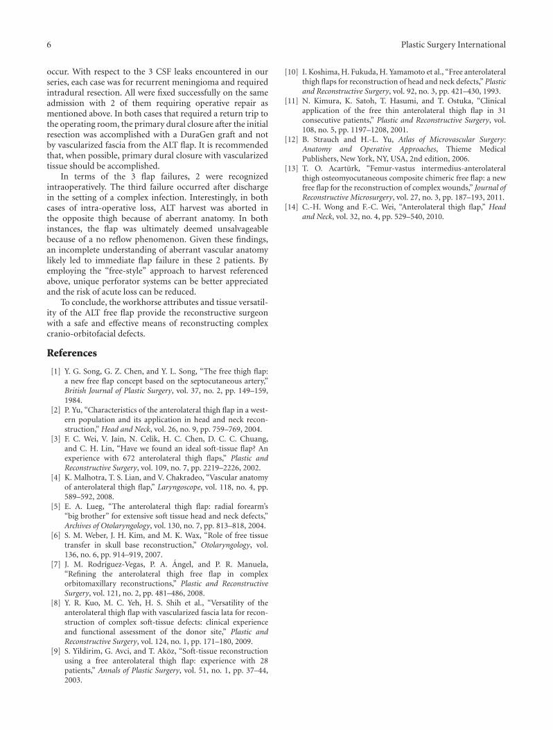
Figure 4: ALT flap immediately after harvest showing the use ofmultiple perforators to create separate islands.
volume of tissue readily available for use, the variety oftissue available, and the ability to incorporate and providecoverage for plates and implants. A variety of flaps may beconsidered, including the ALT, RF, fibula, subscapular systemflaps, or rectus abdominus. The ALT in particular offersseveral appealing qualities that make it a highly useful optionfor periorbital reconstruction.
With regards to the harvest, flap raising is quick andeasily performed with a 2-team approach, which is generallynot as feasible for subscapular system flaps. Unlike the RFand fibula flaps, no tourniquet is necessary. In addition, thewound for the thigh is generally closed primarily in mostpatients, thereby avoiding the need for a skin graft, whichis required with the fibula or forearm flaps. This also avoidsthe need for a splint, bolster, wound VAC, or cast, thussimplifying postoperative wound care.
Many patients tolerate the harvest quite well and areambulatory as soon as they are mobilized, which is generallypostoperative day 2. To minimize any functional deficitin the quadriceps muscle group, we prefer to harvest apredominantly fasciocutaneous flap and limit the inclu-sion of vastus musculature unless required to preserveperforators. Using objective kinetic analyses, Kuo et al.have demonstrated a statistically significant difference infunctional morbidity favoring a vascularized fascia ALT flapover a myocutaneous ALT flap [8]. Furthermore, excessivemuscle harvest is unlikely to offer a significant increase in flapbulk given the inevitable atrophy that will ensue secondaryto denervation. When motor nerves are sacrificed becauseof aberrant perforator anatomy, we prefer to reconstructthem with primary coaptation or nerve graft. The mostcommonly seen postoperative complication is seroma of thethigh, typically encountered several weeks after surgery oncethe patient has become increasingly ambulatory. This is aminor morbidity causing minimal patient discomfort that iseasily treated with needle drainage in the office.
While ease of flap harvest and low donor site morbidityare two of the workhorse attributes of the ALT, tissueversatility is the primary reason for which it is well suited forperiorbital reconstruction. Harvest frequently yields a flapof >200 cm2, with harvests as large as 20 cm (in width) by26 cm (in length) reported in the literature [9]. With this
much tissue available, the flap can accomplish a great varietyof goals. By de-epithelializing the majority of the flap, leavingonly enough skin to recreate the surface of the orbit, there isample bulk to fill the dead space of an exenteration cavity,even without the harvest of much muscle. Although simplertechniques, such as skin grafting, can line an exenterationcavity, the vascularized free flap reduces the risk of bonyexposure and the pursuant osteomyelitis that can occur.Additionally, the end result is low maintenance, allowing thepatient to avoid chronic care and frequent decrusting oftennecessary with more limited reconstruction. To preserveorbital concavity in anticipation of a prosthesis, the ALTcan also be carefully thinned either primarily or secondarily[10, 11]. As demonstrated in the described case, the ALTprovides more than adequate soft tissue to line the entirefloor of the anterior cranial fossa using the muscular fasciaor adjacent tensor fascia lata. Lastly, periorbital defects areoften large enough to involve noncontiguous subsites, suchas an exenteration cavity and the oral cavity. The ALT can bedivided into separate islands based on multiple perforators(Figure 4) or de-epithelialized to create separate segments onone large flap. De-epithelialization of the underlying surfacesis the easier of the two techniques and remains feasible whenonly a single perforator supplies the entire flap.
Shortcomings of the ALT for periorbital reconstructionmust be noted and most obviously include the lack of boneas a tissue option. Though chimeric ALT flaps that include asmall piece of the iliac crest or the femur have been described[12, 13], these techniques are not widely practiced, and wehave not elected to use them. Rather, we have favored thecombined use of implants for orbital and midfacial bonyreconstruction when necessary. When substantial bone lossin the hard palate is expected in patients for whom dentalrehabilitation is a viable option, the fibula free flap would bethe preferred option. However, in cases where extensive softtissue is needed and functional bone loss is less likely (i.e.,when there is no resection of the midfacial buttresses), thenfrequently, the ALT is a viable option. And when periorbitalextirpation involves the hard palate in a patient for whomwe suspect dental implants are unlikely, then an ALT can beaugmented with a denture to provide functional chewing.
Variable anatomy of the ALT donor site is also frequentlypointed out as a downside, but the anatomy has been welldescribed by Wong and Wei [14]. In their recent reviewarticle, they outline a “free-style” approach that essentiallysafeguards against the vascular aberrations. In most cases,a branch of the descending LCFA supplies one or severalperforators to the skin, but this skin may also be suppliednot infrequently by the transverse branch of the commonfemoral artery or an oblique branch. And at times, a singlelarge perforator may supply an extremely large segment ofthe skin. Preoperative Doppler stethoscope identification ofthe perforators, adaptation of the flap skin harvest to theseperforators, and careful dissection of the perforators withclear visualization of their entire course to the main pedicleallows for a reliable harvest despite alterations in anatomy.
Given the comorbidities of the patient population andthe pathology encountered in extensive cranio-orbitofacialextirpations, complications and flap failures will certainly
6 Plastic Surgery International
occur. With respect to the 3 CSF leaks encountered in ourseries, each case was for recurrent meningioma and requiredintradural resection. All were fixed successfully on the sameadmission with 2 of them requiring operative repair asmentioned above. In both cases that required a return trip tothe operating room, the primary dural closure after the initialresection was accomplished with a DuraGen graft and notby vascularized fascia from the ALT flap. It is recommendedthat, when possible, primary dural closure with vascularizedtissue should be accomplished.
In terms of the 3 flap failures, 2 were recognizedintraoperatively. The third failure occurred after dischargein the setting of a complex infection. Interestingly, in bothcases of intra-operative loss, ALT harvest was aborted inthe opposite thigh because of aberrant anatomy. In bothinstances, the flap was ultimately deemed unsalvageablebecause of a no reflow phenomenon. Given these findings,an incomplete understanding of aberrant vascular anatomylikely led to immediate flap failure in these 2 patients. Byemploying the “free-style” approach to harvest referencedabove, unique perforator systems can be better appreciatedand the risk of acute loss can be reduced.
To conclude, the workhorse attributes and tissue versatil-ity of the ALT free flap provide the reconstructive surgeonwith a safe and effective means of reconstructing complexcranio-orbitofacial defects.
References
[1] Y. G. Song, G. Z. Chen, and Y. L. Song, “The free thigh flap:a new free flap concept based on the septocutaneous artery,”British Journal of Plastic Surgery, vol. 37, no. 2, pp. 149–159,1984.
[2] P. Yu, “Characteristics of the anterolateral thigh flap in a west-ern population and its application in head and neck recon-struction,” Head and Neck, vol. 26, no. 9, pp. 759–769, 2004.
[3] F. C. Wei, V. Jain, N. Celik, H. C. Chen, D. C. C. Chuang,and C. H. Lin, “Have we found an ideal soft-tissue flap? Anexperience with 672 anterolateral thigh flaps,” Plastic andReconstructive Surgery, vol. 109, no. 7, pp. 2219–2226, 2002.
[4] K. Malhotra, T. S. Lian, and V. Chakradeo, “Vascular anatomyof anterolateral thigh flap,” Laryngoscope, vol. 118, no. 4, pp.589–592, 2008.
[5] E. A. Lueg, “The anterolateral thigh flap: radial forearm’s“big brother” for extensive soft tissue head and neck defects,”Archives of Otolaryngology, vol. 130, no. 7, pp. 813–818, 2004.
[6] S. M. Weber, J. H. Kim, and M. K. Wax, “Role of free tissuetransfer in skull base reconstruction,” Otolaryngology, vol.136, no. 6, pp. 914–919, 2007.
[7] J. M. Rodrıguez-Vegas, P. A. Angel, and P. R. Manuela,“Refining the anterolateral thigh free flap in complexorbitomaxillary reconstructions,” Plastic and ReconstructiveSurgery, vol. 121, no. 2, pp. 481–486, 2008.
[8] Y. R. Kuo, M. C. Yeh, H. S. Shih et al., “Versatility of theanterolateral thigh flap with vascularized fascia lata for recon-struction of complex soft-tissue defects: clinical experienceand functional assessment of the donor site,” Plastic andReconstructive Surgery, vol. 124, no. 1, pp. 171–180, 2009.
[9] S. Yildirim, G. Avci, and T. Akoz, “Soft-tissue reconstructionusing a free anterolateral thigh flap: experience with 28patients,” Annals of Plastic Surgery, vol. 51, no. 1, pp. 37–44,2003.
[10] I. Koshima, H. Fukuda, H. Yamamoto et al., “Free anterolateralthigh flaps for reconstruction of head and neck defects,” Plasticand Reconstructive Surgery, vol. 92, no. 3, pp. 421–430, 1993.
[11] N. Kimura, K. Satoh, T. Hasumi, and T. Ostuka, “Clinicalapplication of the free thin anterolateral thigh flap in 31consecutive patients,” Plastic and Reconstructive Surgery, vol.108, no. 5, pp. 1197–1208, 2001.
[12] B. Strauch and H.-L. Yu, Atlas of Microvascular Surgery:Anatomy and Operative Approaches, Thieme MedicalPublishers, New York, NY, USA, 2nd edition, 2006.
[13] T. O. Acarturk, “Femur-vastus intermedius-anterolateralthigh osteomyocutaneous composite chimeric free flap: a newfree flap for the reconstruction of complex wounds,” Journal ofReconstructive Microsurgery, vol. 27, no. 3, pp. 187–193, 2011.
[14] C.-H. Wong and F.-C. Wei, “Anterolateral thigh flap,” Headand Neck, vol. 32, no. 4, pp. 529–540, 2010.