A Fine-Grained Evaluation of SPARQL Endpoint Federation Systems
Upload
khangminh22Category
view
2download
0
Valve Academic Research Consortium 3:
updated endpoint definitions for aortic
valve clinical research
VARC-3 WRITING COMMITTEE: Philippe Genereux1, Nicolo Piazza 2,
Maria C. Alu 3, Tamim Nazif 3, Rebecca T. Hahn 3, Philippe Pibarot 4,
Jeroen J. Bax5, Jonathon A. Leipsic6, Philipp Blanke6, Eugene H. Blackstone 7,
Matthew T. Finn 3, Samir Kapadia8, Axel Linke9, Michael J. Mack10, Raj Makkar 11,
Roxana Mehran12, Jeffrey J. Popma13, Michael Reardon14, Josep Rodes-Cabau4,
Nicolas M. Van Mieghem15, John G. Webb16, David J. Cohen 17 and
Martin B. Leon3*
1Gagnon Cardiovascular Institute, Morristown Medical Center, Morristown, NJ, USA; 2McGill University Health Centre, Montreal, QC, Canada; 3Columbia University IrvingMedical Center/NewYork-Presbyterian Hospital and Cardiovascular Research Foundation, New York, NY, USA; 4Quebec Heart & Lung Institute, Laval University, Quebec, QC,Canada; 5Department of Cardiology, Leiden University Medical Center, Leiden, The Netherlands 6Department of Radiology, St. Paul’s Hospital and University of BritishColumbia, Vancouver, BC, Canada; 7Department of Thoracic and Cardiovascular Surgery, Cleveland Clinic and Department of Quantitative Health Sciences, Research Institute,Cleveland Clinic, Cleveland, OH, USA; 8Department of Cardiovascular Medicine, Cleveland Clinic, Cleveland, OH, USA; 9Herzzentrum Dresden, Dresden, Germany; 10BaylorScott & White Heart Hospital Plano, Plano, TX, USA; 11Smidt Heart Institute, Cedars-Sinai Medical Center, Los Angeles, CA, USA; 12The Zena and Michael A. WienerCardiovascular Institute, Icahn School of Medicine at Mount Sinai, New York, NY, USA; 13Beth Israel Deaconess Medical Center, Boston, MA, USA; 14Methodist DeBakey Heart& Vascular Center, Houston, TX, USA; 15Thoraxcenter, Erasmus University Medical Center, Rotterdam, The Netherlands; 16Department of Cardiology, St. Paul’s Hospital andUniversity of British Columbia, Vancouver, BC, Canada; and 17University of Missouri-Kansas City School of Medicine, Kansas City, MO, USA
Received 12 April 2020; revised 22 June 2020; editorial decision 11 September 2020; accepted 24 September 2020; online publish-ahead-of-print 19 April 2021
Aims The Valve Academic Research Consortium (VARC), founded in 2010, was intended to (i) identify appropriate clin-ical endpoints and (ii) standardize definitions of these endpoints for transcatheter and surgical aortic valve clinicaltrials. Rapid evolution of the field, including the emergence of new complications, expanding clinical indications, andnovel therapy strategies have mandated further refinement and expansion of these definitions to ensure clinicalrelevance. This document provides an update of the most appropriate clinical endpoint definitions to be used inthe conduct of transcatheter and surgical aortic valve clinical research.
...................................................................................................................................................................................................Methodsand results
Several years after the publication of the VARC-2 manuscript, an in-person meeting was held involving over 50 in-dependent clinical experts representing several professional societies, academic research organizations, the USFood and Drug Administration (FDA), and industry representatives to (i) evaluate utilization of VARC endpointdefinitions in clinical research, (ii) discuss the scope of this focused update, and (iii) review and revise specific clinic-al endpoint definitions. A writing committee of independent experts was convened and subsequently met to fur-ther address outstanding issues. There were ongoing discussions with FDA and many experts to develop a newclassification schema for bioprosthetic valve dysfunction and failure. Overall, this multi-disciplinary process hasresulted in important recommendations for data reporting, clinical research methods, and updated endpoint defini-tions. New definitions or modifications of existing definitions are being proposed for repeat hospitalizations, accesssite-related complications, bleeding events, conduction disturbances, cardiac structural complications, and
* Corresponding author. Tel: 212-305-7060, Fax: 212-342-3660, Email: [email protected] on behalf of the European Society of Cardiology. All rights reserved. VC The Author(s) 2021. For permissions, please email: [email protected].
European Heart Journal (2021) 42, 1825–1857 SPECIAL ARTICLEdoi:10.1093/eurheartj/ehaa799
Dow
nloaded from https://academ
ic.oup.com/eurheartj/article/42/19/1825/6237954 by Erasm
us Universiteit R
otterdam user on 03 M
ay 2022

bioprosthetic valve dysfunction and failure (including valve leaflet thickening and thrombosis). A more granular 5-class grading scheme for paravalvular regurgitation (PVR) is being proposed to help refine the assessment of PVR.Finally, more specific recommendations on quality-of-life assessments have been included, which have been tar-geted to specific clinical study designs.
...................................................................................................................................................................................................Conclusions Acknowledging the dynamic and evolving nature of less-invasive aortic valve therapies, further refinements of clinic-
al research processes are required. The adoption of these updated and newly proposed VARC-3 endpoints anddefinitions will ensure homogenous event reporting, accurate adjudication, and appropriate comparisons of clinicalresearch studies involving devices and new therapeutic strategies.
� � � � � � � � � � � � � � � � � � � � � � � � � � � � � � � � � � � � � � � � � � � � � � � � � � � � � � � � � � � � � � � � � � � � � � � � � � � � � � � � � � � � � � � � � � � � � � � � � � � � � � � � � � � � � � � � � � � � � � � � � � � � � � � � � � � � � � � � � � � � � � � � � � � � � � � � � � � � � � � � � � � � � � � � � � � � � � � � � � � � � � � � � � � � � � � � � � � � � � � � � � � � � � � � � � � �
Keywords Valve Academic Research Consortium • Transcatheter aortic valve replacement • Transcatheter aortic valveimplantation • Surgical aortic valve replacement • Endpoints • Definitions
Graphical Abstract
...................................................................................................................................................................................................
1826 P. Genereux et al.D
ownloaded from
https://academic.oup.com
/eurheartj/article/42/19/1825/6237954 by Erasmus U
niversiteit Rotterdam
user on 03 May 2022

..
..
..
..
..
..
..
..
..
..
..
..
..
..
..
..
..
..
..
..
..
..
..
..
..
..
..
..
..
..
..
..
..
..
..
..
..
..
..
..
..
..
..
..
..
..
..
..
..
..
..
..
..
..
.
Introduction
The Valve Academic Research Consortium (VARC) was organizedand founded in 2010 in the spirit of the Academic ResearchConsortium mission1–3 and included a diverse group of stakeholdersfrom international societies, academic research organizations, the USFood and Drug Administration, medical device manufacturers, and in-dependent clinician experts from interventional cardiology, cardiacimaging, cardiac surgery, heart failure, and targeted subspecialties(e.g. neurology) for the purpose of improving the processes, scientificrigour, and standardization of definitions related to clinical researchin valvular heart disease. The VARC initiative has been driven by therapid emergence of less-invasive transcatheter aortic valve replace-ment (TAVR) therapies for severe aortic stenosis (AS), although thisprocess has recently expanded to also include important transcath-eter mitral and tricuspid valve therapies.4–7 The first VARC consen-sus manuscript in January 2011 focused on selecting appropriateclinical endpoints and standardizing endpoint definitions for use inTAVR clinical trials.4 The VARC definitions for clinical endpointswere rapidly accepted and frequently utilized by the global TAVRclinical research community.8 However, <2 years later, evolution ofTAVR and the ambiguous nature of certain endpoint definitionsrequired a VARC-2 follow-up manuscript,5,9 which clarified specificdefinitions and expanded the understanding of patient risk stratifica-tion and case selection.
Worldwide, over 800 000 TAVR procedures have been per-formed in more than 65 countries. Concurrently, TAVR clinical re-search has matured and clinical research needs have changedthrough the incorporation of findings from key clinical trials, the rapiddevelopment of new clinical indications, and the introduction of newand iterative medical device technologies. In addition, new advancesin surgical aortic valve replacement (SAVR), and the growing overlapbetween interventional and surgical procedures, have mandated asimilar approach to clinical research for both fields. The improvementin clinical outcomes after TAVR10–14 combined with an emphasis onlower surgical risk patients in the future will direct greater attentionto important secondary endpoints such as all strokes, repeat hospital-ization, paravalvular regurgitation (PVR), and conduction disturban-ces. Similarly, new clinical trials will also rely heavily on carefullyconstructed composite safety and composite efficacy endpoints,many of which will be tailored to the device being studied and theanticipated risks and benefits (e.g. cerebral protection devices orlarge bore vascular closure devices). In the future, device safetyassessments will be facilitated by the more rigorous use of objectiveperformance criteria derived from contemporary clinical trials and/orvalidated national databases, like the ACC/STS Transcatheter ValveTherapy registry.15,16 Routinely, composite efficacy endpoints willcombine both ‘hard’ clinical outcomes (like death and stroke) withother ‘softer’ therapy benefit assessments (like a quality-of-life matrixor a functional assessment, e.g. 6-min walking distance). Finally, asclinical trials include younger patients (e.g. asymptomatic, ‘all-comer’,or bicuspid aortic valve studies), there is greater sensitivity to bothearly safety concerns and longer-term prosthetic valve function.
The main goal of this VARC-3 consensus manuscript is to providean update of these emerging clinical research issues in aortic valvetherapy. A clarification of existing endpoint definitions and a redirec-tion of endpoint selection for future clinical trials, registries or other
studies can enable clinicians, research scientists, and clinical eventcommittees to optimally conduct clinical research in the field of aor-tic valve disease. A detailed summary of important additions andchanges compared with VARC-2 definitions is presented in theSupplementary material online, Appendix.
Clinical endpointsVARC-3 recommends the use of clinically relevant endpoints withconsistent definitions, appropriate to the size and type of clinical stud-ies. Endpoints that VARC-3 considers to be essential to collect, adju-dicate, and report when performing large, randomized trials orrigorous observational studies are listed in Table 1. Clinical eventcommittees for large randomized trials or single-arm registry studiesshould include at least one cardiologist and one cardiovascular sur-geon (both knowledgeable in TAVR and SAVR), and when required,additional subspecialty physicians (especially a neurologist for studiesin which stroke is part of the primary endpoint). It is crucial to assigndevice or procedure-relatedness to the clinical endpoints and tocatalogue event timing relative to the index procedure. Under mostcircumstances, early events (especially in the first 30 days) should beattributed to the device or procedure, unless there is definitive evi-dence to the contrary.
MortalityDeath is the most objective and unbiased endpoint. All efforts shouldbe made to accurately determine the status (dead or alive) of allpatients at all time points during study follow-up, including comple-mentary interrogation of national registry and administrative data-bases. Establishing the exact cause of death may be difficult,17,18 soall-cause mortality should remain the preferred primary endpointmeasure. Nevertheless, death should furthermore be classified ascardiovascular or non-cardiovascular when possible and adjudicatedby a clinical events committee based on narrative summaries andsource documents (Table 2). Any deaths occurring during the pro-cedure should be considered cardiovascular. Death should be
Table 1 Valve Academic Research Consortium pro-posed clinical endpoints
Mortality
Neurologic events
Hospitalization (or re-hospitalization)
Bleeding and transfusions
Vascular and access-related complications
Cardiac structural complications
Other procedural or valve-related complications
New conduction disturbances and arrhythmias
Acute kidney injury
Myocardial infarction
Bioprosthetic valve dysfunction
Leaflet thickening and reduced motion
Clinically significant valve thrombosis
Patient-reported outcomes and health status
Composite endpoints
Valve Academic Research Consortium 3 1827D
ownloaded from
https://academic.oup.com
/eurheartj/article/42/19/1825/6237954 by Erasmus U
niversiteit Rotterdam
user on 03 May 2022

..
..
..
..
..
..
..
..
..
..
..
..
..
..
..
..
..
..
..
..
..
..
..
..
..
.
considered non-cardiovascular only if clearly related to anothercause. When doubt exists regarding the exact cause of death (i.e.sudden death, unexpected death), it should be consideredcardiovascular.
Death is further classified by the time of occurrence. WhileVARC-2 introduced immediate procedural mortality to evaluate dra-matic complications that occur within the first 72 h post-procedure,5
this endpoint occurs with a low incidence and has not been adoptedin the TAVR literature. Moreover, with patients now being dis-charged earlier post-procedure,19–24 the usefulness of this measurehas become questionable. Therefore, VARC-3 no longer recom-mends the use of immediate procedural mortality and recommendsinstead the use of periprocedural, early, and late mortality. Deathshould be classified as periprocedural if it occurs within 30 days of theindex procedure or beyond 30 days if the patient is still hospitalized(including transfer to another hospital for continuity of acute care,but excluding a rehabilitation facility or nursing home). Of particularimportance, the relationship between death and any potential majorperiprocedural complication, device failure, malfunction, or misuseshould be determined (Table 2). Besides periprocedural mortality,collection of early mortality, defined as mortality occurring between30 days and 1 year after the index procedure, and late mortality(1 year and beyond after the index procedure) will help to determinesafety and efficacy and to appropriately compare the impact of com-peting treatment strategies.25,26 Transcatheter aortic valve replace-ment is now being considered as an alternative therapy in low-riskpatients, many of whom are relatively younger than those at a highersurgical risk, placing a premium on the assessment of long-term out-comes >5 years after the index procedure. Longer-time intervalsfrom the procedure are associated with increased difficulty to accur-ately determine cardiovascular cause of death. Therefore, all-causemortality is a more reliable endpoint for late clinical assessments.However, to accommodate the need to quantify valve durability inlow-risk patients, VARC-3 also introduces the endpoint of ‘valve-related’ mortality, defined as cardiovascular mortality adjudicated tobe associated with bioprosthetic valve dysfunction (BVD; see belowbioprosthetic valve dysfunction).
Mortality should be reported as Kaplan–Meier cumulative failurerates to account for differential follow-up time. Corresponding sur-vival should be reported as Kaplan–Meier estimates and not asproportions.
Neurologic eventsThe occurrence of stroke is considered by patients, physicians, anddevice regulators to be one of the most important adverse events fol-lowing cardiovascular procedures. Periprocedural stroke in this con-text occurs primarily due to procedure-related central nervoussystem (CNS) embolization, while late events may be either device-related or spontaneous. Despite the substantial decrease in thereported rate of stroke after TAVR in recent trials,11,13,27–32 strokeclearly remains an important clinical outcome, and the prevention ofstroke and CNS injury has emerged as an important therapeutic tar-get with the introduction of cerebral embolic protection devices(CEPD).33–36 Recent studies have demonstrated that the detectionof overt and covert CNS injury is highly dependent on the intensityof surveillance, with systematic examination by neurologists and rou-tine CNS imaging yielding substantially higher event rates.37 This
underscores the importance of accurate ascertainment and standar-dized adjudication of neurological endpoints in cardiovascular trials.
VARC-3, like the Neurologic Academic Research Consortium(NeuroARC),38 recommends combining appropriate assessment ofneurologic symptoms with tissue-based criteria [pathology or neuroi-maging, ideally diffusion-weighted magnetic resonance imaging (DW-MRI)] for defining stroke and other CNS injury. Table 3 outlinesVARC-3 definitions for stroke and other overt CNS injury, covertCNS injury, and neurologic dysfunction without CNS injury (transi-ent ischaemic attack and delirium) in harmonization with recent con-sensus definitions.38–40 It also includes recommendations forreporting acute stroke severity and associated disability. Similar tomortality, neurological events should be defined as being periproce-dural if they occur within 30 days or during the index hospitalization,early if they occur within 1 year of the index procedure, or late if theyoccur beyond 1 year. Periprocedural neurological events could befurther sub-classified as acute (occurring within 24 h of the index
Table 2 Mortalitya
Causes of mortality
All-cause mortality
Cardiovascular mortality
Death meeting one of the following criteria:• Related to heart failure, cardiogenic shock, bioprosthetic valve
dysfunction, myocardial infarction, stroke, thromboembolism,
bleeding, tamponade, vascular complication, arrhythmia or
conduction system disturbances, cardiovascular infection (e.g.
mediastinitis, endocarditis), or other clear cardiovascular cause• Intraprocedural death• Sudden death• Death of unknown cause
Valve-related mortality
Death presumed to be related to bioprosthetic valve dysfunctionb
Non-cardiovascular mortality
Death clearly related to a non-cardiovascular cause: such as re-
spiratory failure not related to heart failure (e.g. pneumonia), renal
failure, liver failure, infection (e.g. urosepsis), cancer, trauma, and
suicide
Timing of mortality
Periprocedural mortality
Death meeting one of the following criteria:• Occurring <_30 days after the index procedure• Occurring >30 days but during the index hospitalizationc
Early mortality
Death occurring >30 days but <_1 year after the index
hospitalization
Late mortality
Death occurring >1 year after the index hospitalization
aMortality should be reported using Kaplan–Meier methods.bAs defined in Table 12 and Take home figure.cIncludes transfer to another hospital or rehabilitation facility for continuity ofacute care, but excludes chronic treatment at a rehabilitation facility or nursinghome.
1828 P. Genereux et al.D
ownloaded from
https://academic.oup.com
/eurheartj/article/42/19/1825/6237954 by Erasmus U
niversiteit Rotterdam
user on 03 May 2022

..
..
..
..
..
..
..
..
..
..
..
..
..
..
..
..
..
..
..
..
..
..
..
..
..
..
..
..
..
..
..
..
..
..
..
..
..
..
..
..
..
..
..
..
..
..
..
..
..
..
..
..
..
..
..
..
..
..
..
..
..
..
..
..
..
..
..
..
..
..
..
..
..
..
..
..
..
..
..
..
..
..
..
..
..
..
.procedure) or sub-acute (occurring between 24 h and 30 days followingthe index procedure). It is important to recognize that the occurrenceof neurologic events is also influenced by patient co-morbidities andother factors that should be clearly reported (baseline or new-onsetatrial fibrillation, oral anticoagulation or antiplatelet therapy, left atrial ap-pendage or left ventricle thrombus, carotid artery disease, etc.).
Stroke can be described both in terms of acute severity and subse-quent disability.40 Acute stroke severity, as assessed by the NationalInstitutes of Health Stroke Scale (NIHSS), may be reported in clinicaltrials, with an NIHSS of 0–5 considered to be a mild stroke, 6–14moderate, and >_15 severe.41 However, stroke-related disability,measured using the modified Rankin scale (mRS) continues to be thepreferred classification of stroke within clinical trials40 and should becollected routinely. Importantly, and conforming to the originalmRS,42 VARC-2, and NeuroARC, stroke should be classified as beingfatal, stroke with disability (mRS >_2 and increase of at least 1 frombaseline) or stroke without disability (mRS <2 or without increasefrom baseline). Although neurologic disability is best assessed at90 days post-event, such follow-up may not be included in some trialsor routinely performed in clinical practice. VARC-3 acknowledgesthese practical challenges and considers an assessment performed30–90 days after a neurologic event acceptable, although this maylead to an overestimation of the disability associated with stroke andthus represent a ‘worst-case scenario’. In low surgical risk andyounger patients, since activity, return-to-work, and longevityexpectations are greater, there has been a tendency to reduce thestroke disability threshold and include all strokes (with and withoutdisability) as a component of the primary endpoint.11,13
Valve Academic Research Consortium 3 has attempted to har-monize the above definitions and classifications with Neuro-ARC,while recognizing that Neuro-ARC definitions may be too detailedfor application in daily practice or within studies not primarily focusedon neurological events. Similarly, the routine use of DW-MRI is bothlogistically challenging and expensive, and thus, should be reservedfor dedicated studies related to neuroembolic protection. While theassessment of neurologic deficits will ideally be performed by a neur-ologist, assessment by a non-neurologist clinician may be acceptable,particularly when accompanied by brain imaging to confirm the clinic-al diagnosis.38,43 However, for CEPD trials, the assessment of neuro-logic deficits should be performed by a neurologist.
Despite the growing interest in periprocedural, clinically silentbrain infarction39 and neurocognitive impairment (detected by exten-sive neurocognitive testing),44,45 routine inclusion of these endpointsin clinical trials remains challenging for several reasons: (i) uncertaintyrelated to their association with hard clinical endpoints (e.g. mortal-ity) and quality of life (QOL); (ii) current lack of standardization ofdefinitions and assessment; (iii) variability in the cognitive domainascertained by different neuropsychological tests; and (iv) importantheterogeneity related to test execution.44 Indeed, abnormalities inneurocognitive testing used in SAVR and TAVR trials have not beenconsistently associated with the presence or severity of lesionsdetected by MRI.46–53 Nevertheless, given the weight of evidencesuggesting a potential association between silent infarct and cognitiveimpairment on longer-term follow-up,54–58 it may be reasonable fordedicated trials investigating different neuroprotection strategies toconsider including diffusion-weighted MRI and comprehensive neuro-cognitive testing, among the neurologic endpoints collected.34,35,59
Hospitalization or re-hospitalizationHospitalization or multiple re-hospitalizations after an index proced-ure are clinically and economically meaningful endpoints for patients,third-party payers, and health care systems in general. Recently, hos-pitalizations as an endpoint in cardiovascular clinical trials have beenelevated in importance, especially when hospitalizations for worsen-ing heart failure are a consequence of myocardial or valvular heartdisease.6,60–63 Hospitalizations due to worsening heart failure havebeen associated with increased early mortality and frequent repeathospitalizations.64–66 Using Mitral VARC (MVARC) as a starting plat-form,6 VARC-3 defines hospitalization (or re-hospitalization) as anyadmission to an inpatient unit or hospital ward for >_24 h, including anemergency department stay (Table 4). Visits to urgent care facilitiesor emergency departments for <24 h should also be noted (includingreasons and therapies) and they can be included in this endpoint, onlyif substantive intensification of therapy changes are enacted (e.g.intravenous diuretics, >_50% increase in drug therapy dosages, or add-ition of new pharmacotherapy agents). In recent heart failure trials,the association of intensification of medical therapy with all-cause andcardiovascular mortality was similar to heart failure hospitalizationsand emergency department visits.62 Valve Academic ResearchConsortium 3 places emphasis on hospitalizations which are eitherprocedure-related or valve-related (Table 4). Such hospitalizationsmay be due to (i) new complications such as strokes or conductiondisturbances, (ii) exacerbation or deterioration of previous in-hospital periprocedural complications (e.g. recurrent pleural effusion,worsening heart failure), (iii) BVD [e.g. PVR, valve thrombosis, endo-carditis, or structural valve deterioration (SVD)], and (iv) bleedingcomplications related to oral anticoagulation or anti-platelet therapyfor valve-related thromboembolic prevention or atrial fibrillation. Inspecific clinical trials comparing a strategy of either TAVR or SAVRvs. clinical surveillance of the diseased native aortic valve (e.g. earlyAVR vs. clinical surveillance for asymptomatic severe AS), the pro-gression of native aortic valve disease resulting in hospitalizations(due to heart failure, angina, syncope, or other valve-related reasons)can also be used as a worthwhile clinical endpoint.
Heart failure-related hospitalizations are of special interest andmay be considered as a powered primary endpoint or powered/hypothesis-driven secondary endpoint in some clinical trials. ValveAcademic Research Consortium 3 requires that new or worseningheart failure as the predominant reason for a hospital stay >_24 h isbased on symptoms and signs of heart failure with confirmation bydiagnostic tests and necessitating treatment using intravenous ormechanical heart failure therapies. Heart failure hospitalizations maybe associated with primary (cardiac related) causes or secondary(non-cardiac related) aetiologies, such as heart failure due to sepsisor fluid overload in renal failure patients.
Valve Academic Research Consortium 3 recommends dividingcardiovascular hospitalizations into those that are procedure-relatedor valve-related and a separate category of ‘other’ cardiovascularhospitalizations (Table 4). Examples of ‘other’ cardiovascular hospital-izations would include hospitalizations associated with acute myocar-dial infarction (MI) or hypertensive emergencies, which are clearlyunrelated to the valve therapies under investigation. Finally, thereshould be a category of non-cardiovascular hospitalizations (exam-ples in Table 4), which may be common in aortic valve clinical trials
Valve Academic Research Consortium 3 1829D
ownloaded from
https://academic.oup.com
/eurheartj/article/42/19/1825/6237954 by Erasmus U
niversiteit Rotterdam
user on 03 May 2022

Table 3 Neurologic events
Categories of neurologic events
Overt CNS injury (NeuroARC Type 1)
All strokea
• Ischaemic strokeb
Acute onset of focal neurological signs or symptoms conforming to a focal or multifocal vascular territory within the brain, spinal cord, or retina
(NeuroARC Type 1a or 1aH) and fulfilling one of the following criteria:
� Signs or symptoms lasting >_24 h or until death, with pathology or neuroimaging evidence of CNS infarction, or absence of other apparent
causes
� Symptoms lasting <24 h, with pathology or neuroimaging confirmation of CNS infarction in the corresponding vascular territoryc
• Haemorrhagic stroke
Acute onset of neurological signs or symptoms due to intracranial bleeding from intracerebral or subarachnoid haemorrhage not due to trauma
(NeuroARC Types 1b or 1c)• Stroke, not otherwise specified
Acute onset of neurological signs or symptoms persisting >_24 h or until death but without sufficient neuroimaging or pathology evidence to be
classified (NeuroARC Type 1d)
Symptomatic hypoxic-ischaemic injury
Non-focal (global) neurological signs or symptoms with diffuse brain, spinal cord, or retinal cell death confirmed by pathology or neuroimaging and
attributable to hypotension or hypoxia (NeuroARC Type 1e)
Covert CNS injury (NeuroARC Type 2)
Covert CNS infarctionc or haemorrhage
Neuroimaging or pathological evidence of CNS focal or multifocal ischaemia (NeuroARC Type 2a or 2aH) or haemorrhage (NeuroARC 2b) without
acute neurological symptoms consistent with the lesion or bleeding location
Neurologic dysfunction (acutely symptomatic) without CNS injury (NeuroARC Type 3)
TIA
Transient focal neurological signs or symptoms lasting <24 h presumed to be due to focal brain, spinal cord, or retinal ischaemia, but without evidence
of acute infarction by neuroimaging or pathology, or with no imaging performed (NeuroARC Type 3a or Type 3aH)
Delirium without CNS injury
Transient non-focal neurological signs or symptoms, typically of variable duration, without evidence of infarction on neuroimaging or pathology, or
with no imaging performed (NeuroARC Type 3b)
Stroke gradinga
Acute stroke severityd
• Mild neurological dysfunction: NIHSS 0-5• Moderate neurological dysfunction: NIHSS 6-14• Severe neurological dysfunction: NIHSS >_15
Stroke disabilitye
• Fatal Stroke: death resulting from a stroke• Stroke with disability: mRS score of >_2 at 90 dayse and increase of >_1 from pre-stroke baseline• Stroke without disability: mRS score of 0 (no symptoms) or 1 (able to carry out all usual duties and activities) at 90 dayse or no increase in mRS cat-
egory from pre-stroke baseline
Neurological events timing
• Periprocedural: Occurring <_30 days after the index procedure• Acute: Occurring <_24 h after the index procedure
• Sub-acute: Occurring >24 h and <_30 days after the index procedure• Early: Occurring >30 days and <_1 year after the index procedure• Late: Occurring >1 year after the index procedure
CNS, central nervous system; mRS, modified Rankin Scale; NIHSS, National Institute of Health Stroke Scale; TIA, transient ischaemic attack.aIn general, all studies should report at a minimum all stroke and stroke disability.bIncludes haemorrhagic conversions when ischaemic infarction is the primary mechanism.cWhen CNS infarction location does not match transient (<24 h) symptoms, the event should be classified as covert CNS infarction (NeuroARC Type 2a) and TIA(NeuroARC Type 3a), not as an ischaemic stroke.dSeverity assessment should be performed at the time of stroke diagnosis using the NIHSS.eDisability assessment using the mRS should be performed between 30 and 90 days with 90 days being optimal.
1830 P. Genereux et al.D
ownloaded from
https://academic.oup.com
/eurheartj/article/42/19/1825/6237954 by Erasmus U
niversiteit Rotterdam
user on 03 May 2022

..
..
..
..
..
..
..
..
..
..
..
..
..
..
..
..
..
..
..
..
..
..
..
..
.wherein patients are frequently elderly or have multiple co-morbidities.
To account for multiple re-hospitalizations, it is possible to alsoconsider the total number of hospitalizations rather than the time-to-first event, as demonstrated in the recent Cardiovascular OutcomesAssessment of the MitraClip Percutaneous Therapy for Heart FailurePatients With Functional Mitral Regurgitation (COAPT) trial.63 Allhospitalizations and re-hospitalizations must be carefully adjudicatedby a clinical events committee with available source documents.
Bleeding complications and bloodtransfusionsBleeding complications are frequent after TAVR and SAVR and areassociated with increased short- and long-term mortality.67–73
Besides the procedure, many other factors, including patient co-morbidities (e.g. renal insufficiency), associated conditions (e.g.
angiodysplasia), and concomitant therapies (e.g. oral anticoagulation,anti-platelet agents), predispose patients to bleeding.74,75 Therefore,it is essential to report periprocedural and long-term bleeding eventsand to identify relevant contributing factors.
Prior VARC consensus documents used the terms ‘minor’, ‘major’,and ‘life-threatening’ to characterize the severity of bleedingevents.5,76 While this classification offers an intuitively appealing gen-eral grading system for bleeding severity,77 the nomenclature maynot appropriately describe the true magnitude and clinical impact ofbleeding which occurs during surgical procedures. For example, sig-nificant bleeding occurring during an open SAVR that would havebeen classified as ‘life-threatening’ by VARC-2 criteria may be antici-pated and inherent to the SAVR procedure. Therefore, the formersubjective classifications have been modified into a more descriptiveclassification scheme, similar to the Bleeding Academic ResearchConsortium (BARC) bleeding classification3: Type 1 (minor), Type 2
Table 4 Hospitalization (or re-hospitalization)
Definition
Any admission after the index hospitalization or study enrolment to an inpatient unit or hospital ward for >_24 h, including an emergency department
stay. Hospitalizations planned for pre-existing conditions are excluded unless there is worsening of the baseline condition. Visits to urgent care centres
or emergency departments <24 h may also be included if substantive intensification of therapy changes (e.g. heart failure episodes) are enacted (e.g.
intravenous diuretics, significant increases in drug therapy dosages or addition of new pharmacotherapy agents)
Categories of hospitalization
Cardiovascular hospitalization
Procedure-related or valve-related hospitalization
• Hospitalization for new complications such as stroke, bleeding (e.g. haemothorax, retroperitoneal haematoma), pericardial effusion, vascular
or access-site complication (e.g. limb ischaemia, wound infection), new conduction disturbance or arrhythmia (e.g. atrioventricular block, atrial fib-
rillation), acute kidney injury, or any other procedure-related new complication, including periprocedural valve-related heart failure (e.g. paravalv-
ular leak, worsening LV function, worsening sub-valvular obstruction)• Exacerbation or deterioration of previous in-hospital periprocedural complication
(e.g. ventilator-induced pneumonia, recurrent pericardial or pleural effusion, recurrent haemothorax, valve-related heart failure)
• Bioprosthetic valve dysfunctiona such as valve thrombosis, endocarditis, structural valve deterioration, or non-structural valve dysfunction• Untreated diseased native aortic valveb or its related consequences such as heart failure, syncope, angina, new-onset arrhythmia, endocarditis,
or any other symptoms or consequences related to the untreated native aortic valve• Bleeding complications related to oral anticoagulation or antiplatelet therapy for valve-related thromboembolic prevention or atrial fibrillation• Heart failure-related hospitalizationsc requiring that new or worsening heart failure be the predominant reason for a hospital stay >_24 h on
the basis of symptoms and signs of heart failure with confirmation by diagnostic tests and necessitating treatment using intravenous or mechanical
heart failure therapies. Includes primary (cardiac related) and secondary (non-cardiac related)
Other cardiovascular hospitalization
• Cardiovascular hospitalization not directly related to the index procedure or the untreated native aortic valve
Including: acute myocardial infarction or chronic coronary artery disease, hypertension, arrhythmia (not related to the procedure or aortic valve),
heart failure from other specific and proven aetiologies (e.g. cardiomyopathies, concomitant untreated non-aortic valvular disease, severe right
ventricular dysfunction), peripheral vascular disease
Non-cardiovascular hospitalization
• Hospitalization not due to cardiovascular causes as defined above
Including: non-cardiovascular infection and sepsis (e.g. urosepsis), respiratory failure that is not related to heart failure (e.g. pneumonia), renal fail-
ure, liver failure, delirium or dementia, cancer, trauma, or psychiatric illness
aAs defined in Table 12 and Take home figure.bUntreated diseased native aortic valve in the context of a strategy trial comparing transcatheter or surgical aortic valve replacement to clinical surveillance with medical ther-apy, as appropriate.cSome trials may choose to focus on an endpoint of heart failure-related hospitalization.
Valve Academic Research Consortium 3 1831D
ownloaded from
https://academic.oup.com
/eurheartj/article/42/19/1825/6237954 by Erasmus U
niversiteit Rotterdam
user on 03 May 2022

..
..
..
..
..
..
..
..
..
..
..
..
..
..
..
..
..
..
..
..
..
..
..
..
..
..
..
..
..
..
..
..
..
..
..
.(major), Type 3 (life-threatening), and Type 4 (leading to death)bleeding (Table 5).
‘Overt’ bleeding is defined as any bleeding with a clinically obvioussource (e.g. neurologic, gastrointestinal, haemothorax, access-siterelated, any procedural-related bleeding) or with a source identifiedafter appropriate clinical investigation and diagnostic testing (mainlyimaging). Importantly, any procedural blood loss should be consid-ered overt bleeding.
Given the adverse prognostic implications of blood transfu-sions,67,72,78 the exact volume, time relative to the index procedure,and specific indication for each blood transfusion should be reported,whether or not it was associated with overt bleeding. The total num-ber of transfusions should be reported for the index procedure hos-pitalization and for any subsequent repeat hospitalization.Additionally, in order to better reflect the severity and acuity of peri-procedural bleeding events, the number of transfusions receivedwithin 48 h of the index procedure should be reported separately.Finally, VARC-3 acknowledges that many bleeding scales have beendeveloped, validated, and used in clinical trials.79–83 Given the uncer-tainty regarding which scale is the most optimal, the BARC bleedingclassification should also be prospectively recorded to complementthe VARC-3 bleeding scale, especially for non-periprocedural andlate (>1 year) bleeding events.3
Vascular and access-relatedcomplicationsWhile the frequency of vascular complications has decreased signifi-cantly with iterative improvements in TAVR device delivery systemprofile,84 the use of multiple alternative access approaches (sub-clavian, axillary, transcaval, transcarotid, direct aortic, suprasternalaortic, etc.) and novel percutaneous vascular closure device systemsreinforce the need to appropriately capture and report access site-related complications.85–95 VARC-3 now expands the classic defini-tions of major and minor vascular complications to better captureand classify vascular complications related to these emergingapproaches (Table 6). Valve Academic Research Consortium 3 alsointroduces a new sub-category of complications related to access butnot directly vascular in nature (access-related non-vascular complica-tions). These complications include injuries involving structures sur-rounding the access site [e.g. lung (pneumothorax), nerves], non-vascular infection of access sites, and also any complication related totrans-apical approach. Surgical complications related to opening orclosing the chest wall or sternum (e.g. sternum instability, wound de-hiscence, mediastinitis) should also be classified as access-relatednon-vascular complications.
Vascular and access-site-related complications include any com-plication occurring from the actual entry site (e.g. femoral artery
Table 5 Bleeding and transfusionsa
Overt bleedingb that fulfils one of the following criteria:
Type 1
• Overt bleeding that does not require surgical or percutaneous intervention, but does require medical intervention by a health care professional,
leading to hospitalization, an increased level of care, or medical evaluation (BARC 2)• Overt bleeding that requires a transfusion of 1 unit of whole blood/red blood cellsc (BARC 3a)
Type 2
• Overt bleeding that requires a transfusion of 2–4 units of whole blood/red blood cellsc (BARC 3a)• Overt bleeding associated with a haemoglobin drop of >3 g/dL (>1.86 mmol/L) but <5 g/d (<3.1 mmol/L) (BARC 3a)
Type 3
• Overt bleeding in a critical organ, such as intracranial, intraspinal, intraocular, pericardial (associated with haemodynamic compromise/tamponade
and necessitating intervention), or intramuscular with compartment syndrome (BARC 3b, BARC 3c)• Overt bleeding causing hypovolemic shock or severe hypotension (systolic blood pressure <90 mmHg lasting >30 min and not responding to vol-
ume resuscitation) or requiring vasopressors or surgery (BARC 3b)• Overt bleeding requiring reoperation, surgical exploration, or re-intervention for the purpose of controlling bleeding (BARC 3b, BARC 4)• Post-thoracotomy chest tube output >_2 L within a 24-h period (BARC 4)• Overt bleeding requiring a transfusion of >_5 units of whole blood/red blood cells (BARC 3a)c
• Overt bleeding associated with a haemoglobin drop >_5 g/dL (>_3.1 mmol/L) (BARC 3b).
Type 4
• Overt bleeding leading to death. Should be classified as:• Probable: Clinical suspicion (BARC 5a)
• Definite: Confirmed by autopsy or imaging (BARC 5b)
aThe timing, indication, and number of transfused blood products should be collected and reported specifically during the index procedure, during the entire index hospitaliza-tion, and during follow-up after discharge, whether or not overt bleeding is identified.bOvert bleeding is defined as any clinically obvious source of bleeding or bleeding source identified after appropriate investigation and diagnostic testing (e.g. imaging). Any pro-cedural blood loss should be considered overt bleeding.cTotal number of transfusions should be reported separately for (i) within 48 h of the index procedure, (ii) the total duration of the index procedure hospitalization, and (iii)during any subsequent repeat hospitalization.
1832 P. Genereux et al.D
ownloaded from
https://academic.oup.com
/eurheartj/article/42/19/1825/6237954 by Erasmus U
niversiteit Rotterdam
user on 03 May 2022

..
..
..
..
..
..
..
..
..
..
..
..
..
.or vein, subclavian or axillary artery, carotid artery, aorta, left ven-tricle apex, sternum, etc.), the insertion or removal of the deviceor any of its components/accessories (including needle, wire, dila-tor, sheath, and catheter), and the delivery process of the device,but exclude any complication associated with the actual deviceimplantation in the heart. Any complications involving cardiacstructures per se (e.g. aortic valve annulus, left ventricle outflowtract, left or right ventricle) should be reported specifically undercardiac structural complications and are not considered vascular
in nature (see Cardiac structural complications section below).The specific case of complications related to the transapical ap-proach, where the apex of the left ventricle is used as an entrypoint to deliver the device, should be classified as access-related non-vascular complications. On the other hand, leftventricle perforation originating from wire perforation from atransfemoral approach should be considered as a cardiac struc-tural complication. Vascular complications should include com-plications related to the primary vascular access site for a
Table 6 Vascular and access-related complicationsa
Vascular complicationsb
Major
One of the following:
• Aortic dissection or aortic rupture• Vascular (arterial or venous) injury (perforation, rupture, dissection, stenosis, ischaemia, arterial or venous thrombosis including pulmonary em-
bolism, arteriovenous fistula, pseudoaneurysm, haematoma, retroperitoneal haematoma, infection) or compartment syndrome resulting in death,
VARC type >_2 bleeding, limb or visceral ischaemia, or irreversible neurologic impairment• Distal embolization (non-cerebral) from a vascular source resulting in death, amputation, limb or visceral ischaemia, or irreversible end-organ
damage• Unplanned endovascular or surgical intervention resulting in death, VARC type >_2 bleeding, limb or visceral ischaemia, or irreversible neurologic
impairment• Closure device failurec resulting in death, VARC type >_2 bleeding, limb or visceral ischaemia, or irreversible neurologic impairment
Minor
One of the following:
• Vascular (arterial or venous) injury (perforation, rupture, dissection, stenosis, ischaemia, arterial or venous thrombosis including pulmonary em-
bolism, arteriovenous fistula, pseudoaneurysm, haematoma, retroperitoneal haematoma, infection) not resulting in death, VARC type >_2 bleeding,
limb or visceral ischaemia, or irreversible neurologic impairment• Distal embolization treated with embolectomy and/or thrombectomy, not resulting in death, amputation, limb or visceral ischaemia, or irreversible
end-organ damage• Any unplanned endovascular or surgical intervention, ultra-sound guided compression, or thrombin injection, not resulting in death, VARC type
>_2 bleeding, limb or visceral ischaemia, or irreversible neurologic impairment• Closure device failurec not resulting in death, VARC type >_2 bleeding, limb or visceral ischaemia, or irreversible neurologic impairment
Access-related non-vascular complications
Major
One of the following:
• Non-vascular structure, non-cardiac structured perforation, injury, or infection resulting in death, VARC type >_2 bleeding, irreversible nerve injury
or requiring unplanned surgery or percutaneous intervention• Non-vascular access site (e.g. trans-apical left ventricular) perforation, injury, or infection resulting in death, VARC type >_2 bleeding, irreversible
nerve injury or requiring unplanned surgery or percutaneous interventionMinor
One of the following:
• Non-vascular structure, non-cardiac structured perforation, injury, or infection not resulting in death, VARC type >_2, irreversible nerve injury, or
requiring unplanned surgery or percutaneous intervention• Non-vascular access site (e.g. trans-apical left ventricular) perforation, injury, or infection not resulting in death, VARC type >_2 bleeding, irrevers-
ible nerve injury or requiring unplanned surgery or percutaneous intervention
aAny complication related to the device insertion, delivery, and complete removal of all its components (delivery catheter, sheath, guide wire), excluding the actual implantationin the heart.bAny device-related vascular access site and any other accessory access sites (venous or arterial) used during procedure.cA failure to achieve haemostasis at the access site, resulting in alternative treatment (other than manual compression or planned adjunctive endovascular balloon inflation).dIncluding, but not limited to, the lung (e.g. pneumothorax), direct nerve injury, access site or wound infection, mediastinitis, sternal instability, wound dehiscence, and inabilityto close the chest.
Valve Academic Research Consortium 3 1833D
ownloaded from
https://academic.oup.com
/eurheartj/article/42/19/1825/6237954 by Erasmus U
niversiteit Rotterdam
user on 03 May 2022
..
..
..
..
transcatheter device, as well as any accessory vascular accesssites (venous or arterial) used during TAVR or SAVR (e.g.contralateral venous or arterial femoral access, radial access,surgical cannula, haemodynamic support).96 Vascular andaccess-site-related complications may include those occurringacutely during the procedure or at a delayed time (e.g. pseu-doaneurysm, fistula, access-site infection).
Closure device (sutures-based, collagen-based, patch-based, ormembrane based) failure is an important sub-category of vascularcomplications that should also be captured and reported as a distinctentity.91,97,98 Closure device failure is defined as failure to achievesuccessful haemostasis at the access site, leading to alternative treat-ment (other than manual compression or planned adjunctive endo-vascular balloon dilation).
Complications involving surgical access, including sternal wound in-fection, sternal dehiscence, sternal instability, or inability to close thechest, should be reported as access-related non-vascularcomplications.
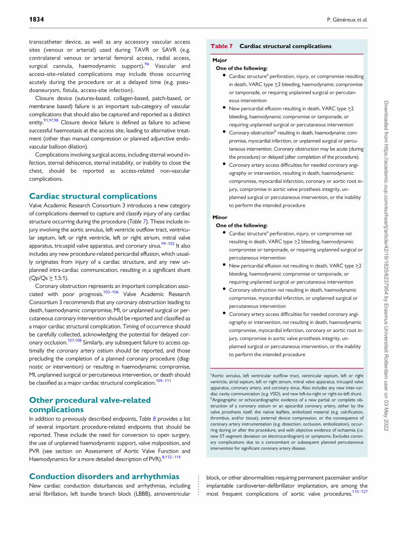
Cardiac structural complicationsValve Academic Research Consortium 3 introduces a new categoryof complications deemed to capture and classify injury of any cardiacstructure occurring during the procedure (Table 7). These include in-jury involving the aortic annulus, left ventricle outflow tract, ventricu-lar septum, left or right ventricle, left or right atrium, mitral valveapparatus, tricuspid valve apparatus, and coronary sinus.99–102 It alsoincludes any new procedure-related pericardial effusion, which usual-ly originates from injury of a cardiac structure, and any new un-planned intra-cardiac communication, resulting in a significant shunt(Qp/Qs >_ 1.5:1).
Coronary obstruction represents an important complication asso-ciated with poor prognosis.103–106 Valve Academic ResearchConsortium 3 recommends that any coronary obstruction leading todeath, haemodynamic compromise, MI, or unplanned surgical or per-cutaneous coronary intervention should be reported and classified asa major cardiac structural complication. Timing of occurrence shouldbe carefully collected, acknowledging the potential for delayed cor-onary occlusion.107,108 Similarly, any subsequent failure to access op-timally the coronary artery ostium should be reported, and thoseprecluding the completion of a planned coronary procedure (diag-nostic or intervention) or resulting in haemodynamic compromise,MI, unplanned surgical or percutaneous intervention, or death shouldbe classified as a major cardiac structural complication.109–111
Other procedural valve-relatedcomplicationsIn addition to previously described endpoints, Table 8 provides a listof several important procedure-related endpoints that should bereported. These include the need for conversion to open surgery,the use of unplanned haemodynamic support, valve malposition, andPVR (see section on Assessment of Aortic Valve Function andHaemodynamics for a more detailed description of PVR).8,112–114
Conduction disorders and arrhythmiasNew cardiac conduction disturbances and arrhythmias, includingatrial fibrillation, left bundle branch block (LBBB), atrioventricular
block, or other abnormalities requiring permanent pacemaker and/orimplantable cardioverter-defibrillator implantation, are among themost frequent complications of aortic valve procedures.115–127
Table 7 Cardiac structural complications
Major
One of the following:
• Cardiac structurea perforation, injury, or compromise resulting
in death, VARC type >_2 bleeding, haemodynamic compromise
or tamponade, or requiring unplanned surgical or percutan-
eous intervention• New pericardial effusion resulting in death, VARC type >_2
bleeding, haemodynamic compromise or tamponade, or
requiring unplanned surgical or percutaneous intervention• Coronary obstructionb resulting in death, haemodynamic com-
promise, myocardial infarction, or unplanned surgical or percu-
taneous intervention. Coronary obstruction may be acute (during
the procedure) or delayed (after completion of the procedure).• Coronary artery access difficulties for needed coronary angi-
ography or intervention, resulting in death, haemodynamic
compromise, myocardial infarction, coronary or aortic root in-
jury, compromise in aortic valve prosthesis integrity, un-
planned surgical or percutaneous intervention, or the inability
to perform the intended procedure
Minor
One of the following:
• Cardiac structurea perforation, injury, or compromise not
resulting in death, VARC type >_2 bleeding, haemodynamic
compromise or tamponade, or requiring unplanned surgical or
percutaneous intervention• New pericardial effusion not resulting in death, VARC type >_2
bleeding, haemodynamic compromise or tamponade, or
requiring unplanned surgical or percutaneous intervention• Coronary obstruction not resulting in death, haemodynamic
compromise, myocardial infarction, or unplanned surgical or
percutaneous intervention• Coronary artery access difficulties for needed coronary angi-
ography or intervention, not resulting in death, haemodynamic
compromise, myocardial infarction, coronary or aortic root in-
jury, compromise in aortic valve prosthesis integrity, un-
planned surgical or percutaneous intervention, or the inability
to perform the intended procedure
aAortic annulus, left ventricular outflow tract, ventricular septum, left or rightventricle, atrial septum, left or right atrium, mitral valve apparatus, tricuspid valveapparatus, coronary artery, and coronary sinus. Also includes any new inter-car-diac cavity communication (e.g. VSD), and new left-to-right or right-to-left shunt.bAngiographic or echocardiographic evidence of a new partial or complete ob-struction of a coronary ostium or an epicardial coronary artery, either by thevalve prosthesis itself, the native leaflets, embolized material (e.g. calcification,thrombus, and/or tissue), external device compression, or the consequence ofcoronary artery instrumentation (e.g. dissection, occlusion, embolization), occur-ring during or after the procedure, and with objective evidence of ischaemia (i.e.new ST-segment deviation on electrocardiogram) or symptoms. Excludes coron-ary complications due to a concomitant or subsequent planned percutaneousintervention for significant coronary artery disease.
1834 P. Genereux et al.D
ownloaded from
https://academic.oup.com
/eurheartj/article/42/19/1825/6237954 by Erasmus U
niversiteit Rotterdam
user on 03 May 2022

..
..
..
..
..
..
..
..
..
..
..
..
..
..
..
..
..
..
..
..
..
..
..
..
..
..
..
..
..
..
..
..
..
..
..
..
..
..
..
..
..
..
..
..
..
..
..
..
..
..
..
..
..
..
..
..
..
..
..
..
..
..
..
..
..
..
..
..
..
..
..
..
..
..
..
..
..
..
..
..
..
..
..
..
..
..
.
Studies have shown that both pre-existing and new-onset conductiondisturbances and arrhythmias may impact prognosis after AVR.128–133
Baseline conduction abnormalities, including 1st-degree atrioven-tricular block, right bundle branch block (RBBB), and LBBB have alsobeen shown to increase the risk of permanent pacemaker implant-ation after AVR.127,134 Moreover, a recent expert consensus docu-ment has proposed a stratification scheme based on the presence orabsence of baseline ECG findings (RBBB, PR interval) and the devel-opment of new conduction disturbances post-AVR (new LBBB, PR,or QRS prolongation, or new atrioventricular block).135 Given theseconsiderations, it is recommended that all studies at minimum reportthe baseline and post-procedure presence of the most importantconduction disturbances and arrhythmias, including those that havebeen shown to alter prognosis or predict permanent pacemaker im-plantation (Table 9). Studies specifically investigating conduction dis-turbances and arrhythmias may wish to collect and report moregranular data, collected at more frequent time-points. These studiesmay also collect additional information regarding therapies, includinganti-arrhythmic agents, chronotropic agents, temporary pacemakers,ablation, oral anticoagulants, or left atrial appendage occlusion.
Conduction disturbances and arrhythmias, particularly LBBB, high-degree atrioventricular block, and atrial fibrillation, can be transient orpersistent after AVR.121,137 Substantial variability exists across studiesin the rates of these complications, which may in part be due to
significant differences in the frequency of ascertainment and defini-tions used. Valve Academic Research Consortium 3 recommends thecollection of 12-lead electrocardiograms (ECGs) at a minimum, atbaseline, as early as feasible after the procedure, daily during hospital-ization, and at regular follow-up intervals (at least 30 days and yearly).It is also recommended that standardized consensus definitions forconduction disturbances be adopted. Specifically, the diagnosis ofLBBB should follow the American Heart Association/AmericanCollege of Cardiology Foundation/Heart Rhythm Society recommen-dation.138 Given the substantial resolution of new LBBB within thefirst 30 days after AVR,139 VARC-3 now proposes the following defin-ition to better characterize LBBB occurrence: transient LBBB(resolved before discharge or within 7 days post-AVR in case of pro-longed hospitalization), persistent LBBB (present at hospital dischargeor until Day 7 post-AVR in case of prolonged hospitalization), or per-manent LBBB (present at 30 days and beyond). Similarly, VARC-3proposes to categorize the timing of occurrence of important con-duction disorders as procedural (occurring <_24 h after the index pro-cedure) or delayed (occurring >24 h after the index procedure).
New-onset atrial fibrillation (or flutter) is defined as any arrhyth-mia during the index hospitalization that has the ECG characteristicsof atrial fibrillation (or flutter) and lasts sufficiently long to berecorded on a 12-lead ECG or at least 30 s on a rhythm strip. Its dur-ation (both pre- and post-index procedure) is characterized as beingparoxysmal, persistent, long-standing persistent, or permanent(Table 9). Valve Academic Research Consortium 3 endorses the defi-nitions provided by AHA/ACCF/HRS guidelines and recommenda-tions for standardization and interpretation of ECGs,138,140,141 butproposes a further classification regarding the timing of occurrenceof new-onset atrial fibrillation: periprocedural if it occurs within30 days of the index procedure and late/spontaneous, if it occurs be-yond 30 days of the index procedure.
Finally, it is problematic that many studies have reported the rateof new permanent pacemaker requirement as a percentage of the en-tire study population.142 Valve Academic Research Consortium 3now explicitly recommends that the calculation of the rate of newpermanent pacemaker implantation exclude from the denominatorpatients with prior permanent pacemaker, who are not at risk for theoutcome. This, in addition to reporting of the timing and indicationfor permanent pacemaker implantation, should help to facilitate com-parisons across studies. The same principle applies to the reportingof the rates of other conduction disturbances (LBBB) and arrhyth-mias (atrial fibrillation) that may pre-date the aortic valve procedure.
Acute kidney injuryAcute kidney injury after TAVR or SAVR is a complication associatedwith poor prognosis.143–147 Valve Academic Research Consortium 3recommends using the widely recognized Kidney Disease: ImprovingGlobal Outcomes (KDIGO) definition of acute kidney injury148
(Table 10). Acknowledging the challenges related to the use of urineoutput as a criterion in daily practice,149,150 serum creatinine criteriashould be the default criteria, and the urine output definition can beconsidered in the setting of dedicated acute kidney injury stud-ies.91,151 The need for new renal replacement therapy (temporary orpermanent) should now be reported as a separate entity (acute kid-ney injury stage 4). As described in the above section, the
Table 8 Other acute procedural and technical valve-related complicationsa
Conversion to open surgery
Conversion to open sternotomy or thoracotomy using cardiopul-
monary bypass secondary to any procedure-related complication or
failed intended transcatheter approach. Should be classified as:• Intraprocedural conversion: during the index procedure• Periprocedural conversion: <_30 days after the index procedure• Delayed conversion: >30 days after the index procedure
Unplanned use of mechanical circulatory supportb
Implantation of multiple (>1) transcatheter valves during
the index hospitalization
Valve malposition
Should be classified as:• Valve migration: After initial correct positioning, the valve pros-
thesis moves upward or downward, within the aortic annulus
from its initial position, without valve embolization• Valve embolization: The valve prosthesis moves either upward
or downward after final deployment such that it loses contact
with the aortic annulus• Ectopic valve deployment: Irretrievable deployment of a valve
prosthesis at a site other than the intended position because
of valve embolization or inability to deliver the prosthesis to
the desired location
Paravalvular regurgitation (see Table 16)
aIndividual events should be collected so that specific event rates can bedetermined.bMechanical circulatory support includes: cardiopulmonary bypass (CPB), extra-corporeal membrane oxygenation (ECMO), transcatheter pumps (e.g. Impella)or intra-aortic balloon pump (IABP).
Valve Academic Research Consortium 3 1835D
ownloaded from
https://academic.oup.com
/eurheartj/article/42/19/1825/6237954 by Erasmus U
niversiteit Rotterdam
user on 03 May 2022

..
..
..
..
..
..
..
.denominator for dialysis should exclude patients already on chronicdialysis prior to the aortic valve procedure.
While VARC-3 recognizes that eGFR is widely used clinically to clas-sify severity of renal dysfunction, the KDIGO guidelines have notadopted changes in eGFR for AKI classification, and as such, VARC-3
will follow the same classification. Valve Academic ResearchConsortium 3 also acknowledges the challenges in following creatininelevels beyond 48 h, especially in the context of early discharge.Creatinine levels should be measured at a minimum, at baselineand within 24 h post-procedure, and ideally daily up to 48 h
Table 9 Conduction disturbances and arrhythmias
Pre-index procedure
Conduction disturbances
� 1st-degree AV block
� 2nd-degree AV block
� Left bundle branch block
� Right bundle branch block
� IVCD with QRS >_120 ms
� Bradycardia (heart rate <60 b.p.m.) or SSS
Permanent pacemaker
� Type of permanent pacemaker should be recorded (e.g. single chamber, dual chamber, biventricular, defibrillator)
Atrial fibrillation (or flutter)
� Paroxysmal, persistent, long-standing persistent, or permanent
During or after index procedurea
Conduction disturbances
� 1st-, 2nd-, 3rd-degree AV block
� Left bundle branch block
� IVCD with QRS >_120 ms
� New-onset: defined as a new conduction disturbance relative to baseline
� Timing of occurrence:
� Procedural: <_24 h after the index procedure
� Delayed: >24 h after the index procedure
� Duration:
� Transient: resolved before discharge or <_7 days after the index procedure in case of prolonged hospitalization
� Persistent: present at hospital discharge or >7 days after the index procedure in case of prolonged hospitalization
� Permanent: present >30 days after the index procedure
Permanent pacemaker
� Type: single, dual, biventricular, defibrillator, leadless
� Timing: No. of days after the index procedure
� Indication: including AV Block, SSS
Atrial fibrillation (or flutter)
� New-onset: defined as any arrhythmia that was not present at baseline that has the ECG characteristics of atrial fibrillation (or flutter) and lasts
sufficiently long to be recorded on a 12-lead ECG or at least 30 s on a rhythm strip
� Timing of occurrenceb:
� Periprocedural: <_30 days after the index procedure
� Late/spontaneous: >30 days after the index procedure
� Durationb:
� Paroxysmal: atrial fibrillation that terminates spontaneously or with intervention <_7 days of onset.
� Persistent: Continuous atrial fibrillation that is sustained >7 days.
� Long-standing persistent: Continuous atrial fibrillation >12 months in duration.
� Permanent: Used when the patient and clinician make a joint decision to stop further attempts to restore and/or maintain sinus rhythm.
AF, atrial fibrillation or atrial flutter; AV, atrioventricular; ECG, electrocardiogram; IVCD, intraventricular conduction delay; SSS, sick sinus syndrome.aThe calculation of new pacemaker rates should exclude patients with pre-existing pacemaker. The same principle applies to reporting of rates of new conduction disturbancesand arrhythmias.bFrom January et al.136
1836 P. Genereux et al.D
ownloaded from
https://academic.oup.com
/eurheartj/article/42/19/1825/6237954 by Erasmus U
niversiteit Rotterdam
user on 03 May 2022

..
..
..
..
..
..
..
..
..
..
..
..
..
..
..
..
..
..
..
..
..
..
..
..
..
..
..
..
..
..
..
..
..
..
..
..
..
..
post-procedure. If post-procedure values are increased compared withbaseline, an additional value should be drawn, and serial measuresshould be assessed until the creatinine declines from its peak value.
Myocardial infarctionCharacterizing myocardial injury after SAVR or TAVR is importantand should be reported appropriately.152–157 Despite a growing bodyof evidence related to the potential clinical impact of differentdegrees of myocardial injury post-valve replacement,158 many chal-lenges remain regarding the diagnosis, adjudication, and comparisonof MI post-AVR procedures: (i) the different degrees of myocardialinjury inherent to different techniques and approaches (e.g. SAVR vs.alternative access TAVR vs. transfemoral TAVR), (ii) the use of differ-ent biomarkers (creatine kinase-MB, standard troponin, high-sensitivity troponin) with variable sensitivities and availability, (iii) thearbitrary (and evolving) nature of MI definitions used in cardiovascu-lar trials, and (iv) the lack of strong and conclusive evidence of associ-ation with hard clinical outcomes, especially among patientsundergoing AVR. In the absence of definitive data, and given the highincidence of concomitant coronary disease159–162 and potential needfor coronary revascularization,111,163–165 VARC-3 endorses the gen-eral classification of the Fourth Universal Myocardial Definition inregards to spontaneous MI (Type 1), imbalance between oxygen sup-ply and demand (Type 2), MI leading to death (Type 3), and MI relatedto coronary stent thrombosis (Type 4B) and coronary restenosis(Type 4C).166 However, for periprocedural MI post-percutaneouscoronary intervention (Type 4A) and post-coronary artery bypassgraft (Type 5), VARC-3 endorses the modified SCAI167 and ARC-2168 definition, which provide a common biomarker (troponin orCK-MB) threshold for both PCI and CABG, and proposes to use thesame definition for periprocedural MI post-SAVR and TAVR. Giventhat most current and future studies related to AVR strategies will in-volve long-term follow-up, with patients frequently suffering fromcoronary artery disease, VARC-3 believes that these definitions willallow the most appropriate characterization and classification oftypes of MI occurring in this population.6,166 Periprocedural biomark-er elevations not meeting the criteria for MI should be categorized as‘myocardial injury not meeting MI criteria’, and the implications ofthese lower levels of myonecrosis should be carefully examined.Importantly, biomarker elevations in the context of valve-relatedcomplications such as acute or delayed coronary occlusion, or failureto appropriately engage the coronary ostium, with subsequent com-plications during a coronary procedure, should also be classified ascardiac structural complications (Table 11).
Biomarkers of myocardial injury should be collected prior to the pro-cedure and be performed twice within the first 24 h post-procedure. Ifthe biomarker level at either time point is elevated by >_50% comparedwith baseline, serial measures should be drawn until the peak has beenreached and the levels begin to decline. All patients should also have abaseline 12-lead ECG, and this should be repeated as soon as feasibleafter the AVR procedure and daily until hospital discharge.
Mechanical aortic valve and autograftroot replacementEuropean and American guidelines currently recommend the use ofbioprosthetic valves in patients above the age of 65 and 70 years old,
respectively.170,171 Both guidelines also support the use of mechanicalvalves for patients below the age of 60 years old. Mechanical aorticprostheses have the advantage of prolonged durability, although theyrequire systemic oral anticoagulation and are thus associated withincreased bleeding risks over time.172,173 The decision-making pro-cess in the selection of prosthesis type includes factors such as: (i) lifeexpectancy and potential need for re-intervention, (ii) bleeding risk,(iii) patient lifestyle, (iv) concomitant co-morbidities requiring lifetimeoral anticoagulation or affecting bioprosthetic valve durability, and (v)patient preferences.174 Some mechanical heart valves require a lowerlevel of systemic anticoagulation, which is expected to lower the riskof long-term bleeding.175,176 Recently, a novel biopolymer-based leaf-let material has been developed, raising hopes for a heart valve im-plant with prolonged durability and no need for oralanticoagulation.177 Finally, in younger and middle-aged adults, auto-graft implantation (Ross Procedure) represents a viable option.178,179
While the VARC-3 criteria for valve degeneration and failure pre-sented below mainly focus on bioprosthetic valves, the modes of fail-ure are similar for mechanical implants and autograft replacements aswell (structural failure, non-structural failure, endocarditis, throm-bosis), though they also include re-intervention for recurrent/life-threatening bleeding or pulmonary valve insufficiency. The VARC-3classification characterizing mode of valve failure could also beapplied to other types of aortic valve implants, and the reasons forassociated re-intervention should be appropriately captured.
Table 10 Acute kidney injurya
Stage 1
AKI that fulfils at least one of the following criteria:• Increase in serum creatinine >_150–200% (>_1.5–2.0� increase)
within 7 days compared with baseline• Increase of >_ 0.3mg/dL (>_26.4 mmol/L) within 48 h of the
index procedure
Stage 2
AKI that fulfils the following criterion:• Increase in serum creatinine >200–300% (>2.0–3.0� increase)
within 7 days compared with baseline
Stage 3
AKI that fulfils at least one of the following criteria:• Increase in serum creatinine >300% (>3.0� increase) within 7
days compared with baseline• Serum creatinine >_4.0 mg/dL (>_354 mmol/L) with an acute in-
crease of >_0.5 mg/dL (>_44 mmol/L)
Stage 4
AKI requiring new temporary or permanent renal replacement
therapy
Adapted from Clinical Practice Guidelines for Acute Kidney Injury 2012. https://kdigo.org/guidelines/acute-kidney-injury/.AKI, acute kidney injury.aGiven practical challenges with the use of urine output criteria in daily practice,AKI should be solely defined based on serum creatinine values. Acute kidney in-jury defined by urine output using the following criteria might be used in the con-text of a dedicated AKI study: AKI Stage 1: Urine output <0.5 mL/kg/h for >_6 but<12 h; AKI stage 2: Urine output <0.5 mL/kg/h for >_12 but <24 h; AKI stage 3:Urine output <0.3 mL/kg/h for >_24 h or anuria for >_12 h.
Valve Academic Research Consortium 3 1837D
ownloaded from
https://academic.oup.com
/eurheartj/article/42/19/1825/6237954 by Erasmus U
niversiteit Rotterdam
user on 03 May 2022

..
..
..
..
..
..
..
..
..
..
.Bioprosthetic valve dysfunctionTake home figure summarizes endpoints for both structural andnon-structural bioprosthetic valve dysfunction (BVD) and depictsthe recommended decision tree for classification of aetiology andseverity of BVD, consistent with recently published consensusdocuments.180–183 In most instances, BVD is a progressive processthat requires serial longitudinal assessments of clinical status, as
well as valve morphology, function, and haemodynamics.Classification of BVD is further detailed in Table 12 and haemo-dynamic criteria for assessment of BVD severity in Table 13. Ofnote, due to the inherent variability of echocardiographic imagingand assessment, as well as fluctuations in blood flow which can re-sult in changes to Doppler measurements, a definite diagnosis ofSVD should not rely on the measurement of a single
Table 11 Myocardial infarction (adapted from 4th Universal, SCAI and ARC-2 definitions)
Type 1 (Spontaneous MI) (>48 h after the index procedure)a
• Detection of a rise and/or fall of cTn values with at least one value above the 99th percentile URL with at least one of the following:
� Symptoms of acute ischaemia
� New ischaemic ECG changes (new ST-segment or T-wave changes or new LBBB)
� New pathologic Q-waves in >_2 contiguous leads
� Imaging evidence of a new loss of viable myocardium or new wall motion abnormality in a pattern consistent with an ischaemic aetiology
� Identification of a coronary thrombus by angiography or autopsy• Post-mortem demonstration of an atherothrombus in the artery supplying the infarcted myocardium, or a macroscopically large circumscribed
area of necrosis with or without intramyocardial haemorrhage, meets the type 1 MI criteria regardless of cTn values
Type 2 (Imbalance between myocardial oxygen supply and demand)a
• Detection of a rise and/or fall of cTn values with at least one value above the 99th percentile URL, and evidence of an imbalance between myocar-
dial oxygen supply and demand unrelated to coronary thrombosis, requiring at least one of the following:
� Symptoms of ischaemia
� ECG changes indicative of new ischaemia (new ST-segment or T-wave changes or new LBBB)
� New pathologic Q-waves in >_2 contiguous leads
� Imaging evidence of a new loss of viable myocardium or new wall motion abnormality
Type 3 (MI associated with sudden cardiac death)a
• Patients who suffer cardiac death, with symptoms suggestive of myocardial ischaemia accompanied by presumed new ischaemic ECG changes or
ventricular fibrillation but die before blood samples for biomarkers can be obtained, or before increases in cardiac biomarkers can be identified,
or MI is detected by autopsy examination.
Type 4A (Criteria for PCI-related MI �48 h after the index procedure)b
• In patients with normal baseline CK-MB: The peak CK-MB measured within 48 h of the procedure >_10� the local laboratory ULN or CK-
MB >_5� ULN with one or more of the following:
� New pathologic Q-waves in >_2 contiguous leads
� New persistent LBBBc
� Flow-limiting angiographic complications in a major epicardial vessel or >1.5 mm diameter branch
� Substantial new loss of viable myocardium on imaging related to the procedure• In the absence of CK-MB measurements and a normal baseline cTn, a cTn (I or T) level measured within 48 h of the procedure rises to >_70� the
local laboratory ULN or >_35� ULN with one or more of the following:
� New pathologic Q-waves in >_2 contiguous leads
� New persistent LBBBc
� Flow-limiting angiographic complications in a major epicardial vessel or >1.5 mm diameter branch
� Substantial new loss of viable myocardium on imaging related to the procedure• In patients with elevated baseline CK-MB (or cTn): The CK-MB (or cTn) rises by an absolute increment equal to those levels recommended
above from the most recent pre-procedure level plus new ECG changes as described.
Type 4B (Stent thrombosis)a
• Stent thrombosis as documented by angiography or autopsy using the same criteria utilized for type 1 MI.• Acute: 0 to 24 h
• Subacute: >24 h to 30 days• Late: >30 days to 1 year• Very late: >1 year after stent implantation
Continued
1838 P. Genereux et al.D
ownloaded from
https://academic.oup.com
/eurheartj/article/42/19/1825/6237954 by Erasmus U
niversiteit Rotterdam
user on 03 May 2022

..
..
..
..
..
..
..
..
..
..
..
..
..
..
..
..
..
..
..
..
..
..
..
..
..
..
..
..
..
..
..
..
..
..
..
..
..
..
..
..
..
..
..
..
..
.haemodynamic parameter, and preferably should incorporate evi-dence from at least two serial echocardiograms. This is in contrastto prior definitions which considered only absolute threshold val-ues of aortic valve area and gradient. A baseline post-proceduralechocardiogram is essential to ensure adequate comparison duringfollow-up, especially if prosthesis-patient mismatch is present aftervalve implantation. Although echocardiography is a cornerstonefor the evaluation of valve function and haemodynamics, cardiaccomputed tomography (CT) is becoming increasingly used to bet-ter understand the pathology and mechanisms underlying BVD. Inparticular, CT has become central to the diagnosis of leaflet andvalve thrombosis, described in more detail below and summarizedin Tables 14 and 15. Bioprosthetic valve dysfunction may be relatedto several aetiologies (Take home figure): (i) SVD, which implies ir-reversible intrinsic changes to structural elements of the valve it-self; (ii) non-structural valve dysfunction, which includes PVR andprosthesis-patient mismatch; (iii) endocarditis; or (iv) thrombosis.The stages of SVD are described in Table 13: Stage 1: morphologic-al valve deterioration; Stage 2: moderate haemodynamic valve de-terioration; and Stage 3: severe haemodynamic valvedeterioration. When assessing the presence and severity ofhaemodynamic valve deterioration, it is important to differentiatetrue-haemodynamic changes vs. inter-echo variability in the meas-urement of gradient, effective orifice area, Doppler velocity index,or AR. Each case with potential haemodynamic valve deteriorationshould be individually adjudicated to confirm presence, stage, andaetiology. Haemodynamic valve deterioration may be caused bySVD but also by valve thrombosis or endocarditis. The assessmentof valve leaflet morphology and structure is key to make a differen-tial diagnosis between the different aetiologies of haemodynamic
valve deterioration. The definitions of SVD presented in Table 13allow one to differentiate haemodynamic deterioration that isrelated to SVD vs., for example, from high residual transprostheticgradients related to prosthesis-patient mismatch.
An important addition to VARC-3 is the incorporation of the def-inition for bioprosthetic valve failure (BVF). Bioprosthetic valve failureis a patient-oriented clinical endpoint that takes into account relevantand clinically meaningful consequences of BVD such as SVD-relatedStage 3 haemodynamic valve deterioration and irreversible changesin haemodynamics, as well as clinical symptoms or sequelae, includingvalve-related death and re-intervention (either surgical or transcath-eter; Take home figure). Thrombosis or endocarditis may also lead toirreversible Stage 3 haemodynamic valve deterioration and thus BVF.Bioprosthetic valve failure should be reported separately from sub-clinical BVD detected solely by haemodynamic findings. Given thecompeting risks between BVF and death, conventional Kaplan–Meierestimates (i.e. actuarial analysis) may overestimate the risk of BVF byassuming that patients without BVF, whether currently alive or dead,will have BVF in the future. This overestimation can have importantimplications for clinical trials involving elderly patients who will likelydie before experiencing BVF. To correctly understand the probabilityof BVF during the course of a patient’s lifetime, competing risks (cu-mulative probability) methods should be used.186,187 For mortalitycalculations alone, actuarial and competing risks methods would pro-vide identical curves. However, in estimating durability of a biopros-thesis (the device rather than the patient), death introducesinformative censoring that results in overestimating device durability;the competing risk function known as conditional probability gives amore accurate depiction of device durability than the Kaplan–Meiermethod. The exposure adjusted incidence rates of BVD, SVD, and
Table 11 Continued
Type 5 Periprocedural (post-SAVR, TAVR or CABG) MI (�48 h after the index procedure)b
• In patients with normal baseline CK-MB: The peak CK-MB measured within 48 h of the procedure >_10� the local laboratory ULN or CK-
MB >_5� ULN with one or more of the following:• New pathologic Q-waves in >_2 contiguous leads
• New persistent LBBBc
• Flow-limiting angiographic complications in a major epicardial vessel or >1.5 mm diameter branch• Substantial new loss of viable myocardium on imaging related to the procedure
• In the absence of CK-MB measurements and a normal baseline cTn, a cTn (I or T) level measured within 48 h of the procedure rises to >_70� the
local laboratory ULN or >_35� ULN with one or more of the following:• New pathologic Q-waves in >_2 contiguous leads
• New persistent LBBBc
• Flow-limiting angiographic complications in a major epicardial vessel or >1.5 mm diameter branch• Substantial new loss of viable myocardium on imaging related to the procedure• In patients with elevated baseline CK-MB (or cTn): The CK-MB (or cTn) rises by an absolute increment equal to those levels recommended
above from the most recent pre-procedure level plus new ECG changes as described.
The use of high-sensitivity (hs)-troponins is recommended for diagnosis of spontaneous MI, but has not been studied for assessment of periprocedural MI. Standard troponinassays are therefore recommended for evaluation of periprocedural MI. Periprocedural biomarker elevation >ULN not meeting the criteria for MI should be categorized as‘myocardial injury not meeting MI criteria’.CK-MB, creatine kinase-MB; cTn, cardiac troponin; ECG, electrocardiogram; LBBB, left bundle branch block; MI, myocardial infarction; SAVR, surgical aortic valve replacement;TAVR, transcatheter aortic valve replacement; ULN, upper limit of normal; URL, upper reference limit.aAdapted from Thygesen et al.169
bAdapted from Moussa et al.167 and Garcia-Garcia et al.168
cLBBB criteria to be used with caution after TAVR or SAVR given the relatively high rate of new LBBB after these procedures.
Valve Academic Research Consortium 3 1839D
ownloaded from
https://academic.oup.com
/eurheartj/article/42/19/1825/6237954 by Erasmus U
niversiteit Rotterdam
user on 03 May 2022

..
..
..
..
..
..
..
..
..
..
..
..
..
..
..
..
..
..
..
..
..
..
..
..
..
..
..
BVF can also be reported as exposure adjusted cumulative rate,which is defined as the number of subjects exposed to the biopros-thesis and experiencing an event (BVD, SVD, or BVF) divided by thetotal exposure time of all patients who are at risk of event, and it isexpressed per 100 patient-years.188
Hypo-attenuated leaflet thickening andreduced leaflet motionHypo-attenuated leaflet thickening (HALT) is visually identifiedincreased thickness of the bioprosthetic leaflet on contrast-enhancedcardiac CT. Hypo-attenuated leaflet thickening typically exhibits ameniscal-shaped configuration, starting and thickest at the insertionof the bioprosthetic leaflet at the stent frame or valve scaffold, and
gradually tapering towards the free edge of the leaflets. While the oc-currence of HALT has been described across all transcatheter aorticvalve platforms and surgical bioprostheses,189,190 its effect on patientoutcome and long-term valve function remains unclear191,192 Whenassessing the extent of HALT within a clinical study, a semi-quantitative grading scale should be used per leaflet as presented inTable 14 and Figure 1, describing the percentage leaflet involvementstarting at its basal insertion.193 The evaluation of HALT is performedusing multiplanar reformats with optional volume-rendered recon-structions. The strength of cardiac CT is its high spatial resolution.However, evaluation for the presence of HALT may be impaired bystreak artefacts caused by the stent frame, motion artefact or sub-optimal contrast attenuation, rendering CT studies at times inconclu-sive for HALT. Leaflet restriction caused by HALT can be describedas reduced leaflet motion (RLM). However, given the limited tem-poral resolution of cardiac CT, the strength of cardiac CT is in thediagnosis of HALT and findings of restricted leaflet motion shouldonly be pursued in the setting of HALT. Assessing leaflet motion in
Table 12 Aortic bioprosthetic valve dysfunction
Categories of BVD
Structural valve deterioration (SVD)
• Intrinsic permanent changes to the prosthetic valve, including
wear and tear, leaflet disruption, flail leaflet, leaflet fibrosis
and/or calcification, or strut fracture or deformation• See Table 13 for grading severity
Non-structural valve dysfunction (NSVD)
• Any abnormality, not intrinsic to the prosthetic valve, resulting
in valve dysfunction. Examples include residual intra- or para-
prosthetic aortic regurgitation; leaflet entrapment by pannus,
tissue, or suture; inappropriate positioning or sizing; dilatation
of the aortic root after stentless prostheses or aortic valve
sparing operations; prosthesis-patient mismatch; and
embolization
Thrombosis
• See Tables 13–15
Endocarditis
• Meeting at least one of the following criteria: (i) Fulfilment of
the Duke endocarditis criteria (ii) Evidence of abscess, pus, or
vegetation confirmed as secondary to infection by histological
or microbiological studies during re-operation; and (iii)
Evidence of abscess, pus, or vegetation confirmed on autopsy.
Clinical presentation
• Subclinical: Any bioprosthetic valve dysfunction associated
with absent or mild haemodynamic changes, AND absent
symptoms or sequelae• Bioprosthetic valve failure (BVF):
• Stage 1: Any bioprosthetic valve dysfunction associated
with clinically expressive criteria (new-onset or worsening
symptoms, LV dilation/hypertrophy/dysfunction, or pul-
monary hypertension) or irreversible Stage 3 haemo-
dynamic valve deterioration (HVD)
• Stage 2: Aortic valve reoperation or re-intervention• Stage 3: Valve-related deatha
BVD,bioprosthetic valve dysfunction.aValve-related death defined as in Table 2: cardiovascular mortality presumed tobe associated with bioprosthetic valve dysfunction.
Table 13 Stages of bioprosthetic valvedeteriorationa,b
Stage 1: Morphological valve deterioration
• Evidence of structural valve deterioration, non-structural valve
dysfunction (other than paravalvular regurgitation or pros-
thesis-patient mismatch), thrombosis, or endocarditis without
significant haemodynamic changes.
Stage 2: Moderate haemodynamic valve deterioration
• Increase in mean transvalvular gradient >_10 mmHg resulting in
mean gradient >_20 mmHgc with concomitant decrease in
EOA >_0.3 cm2 or >_25% and/or decrease in Doppler velocity
index >_0.1 or >_20% compared with echocardiographic assess-
ment performed 1–3 months post-procedure, OR new occur-
rence or increase of >_1 graded of intraprosthetic AR resulting
in >_ moderate AR.
Stage 3: Severe haemodynamic valve deterioration
• Increase in mean transvalvular gradient >_20 mmHg resulting in
mean gradient >_30 mmHgc with concomitant decrease in
EOA >_0.6 cm2 or >_50% and/or decrease in Doppler velocity
index >_0.2 or >_40% compared with echocardiographic assess-
ment performed 1–3 months post-procedure, OR new occur-
rence, or increase of >_2 grades,d of intraprosthetic AR
resulting in severe AR.
AR, aortic regurgitation; EOA, effective orifice area; DVI, Doppler velocity index.aAdapted from Capodanno et al.,180 Lancellotti et al.,184 and Dvir et al.185
bWhen assessing the presence and severity of haemodynamic valve deterioration,it is important to differentiate true-haemodynamic changes vs. inter-echo variabil-ity in the measurement of gradient, EOA, DVI, or AR. In particular, one shoulduse the same window for continuous-wave Doppler interrogation when compar-ing gradients in early (1–3 months) post-AVR echo vs. follow-up echo. Each casewith potential haemodynamic valve deterioration should be individually adjudi-cated to confirm presence, stage, and aetiology. Haemodynamic valve deterior-ation may be caused by structural valve deterioration but also by non-structuraldysfunction including valve thrombosis and endocarditis. The assessment of valveleaflet morphology and structure is key to make differential diagnosis betweenthe different aetiologies of haemodynamic valve deterioration.cThis criterion for haemodynamic dysfunction assumes normal flow.dThis criteria is assessed with the 3-class grading scheme (See Table 16).
1840 P. Genereux et al.D
ownloaded from
https://academic.oup.com
/eurheartj/article/42/19/1825/6237954 by Erasmus U
niversiteit Rotterdam
user on 03 May 2022
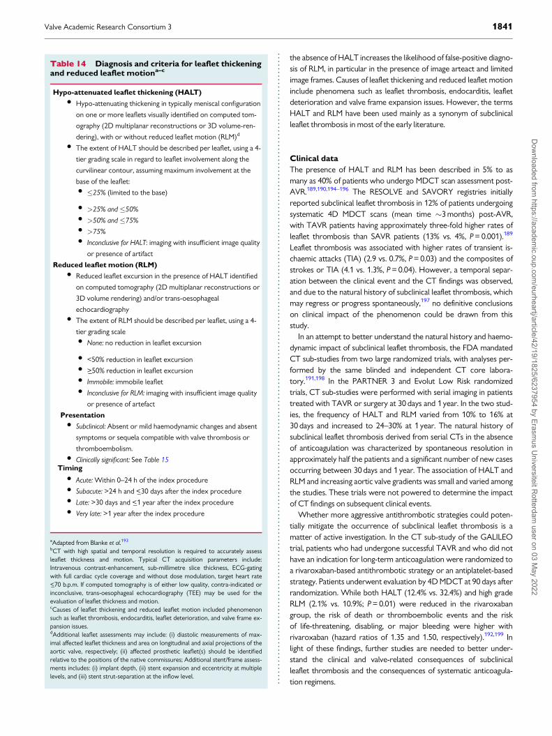
..
..
..
..
..
..
..
..
..
..
..
..
..
..
..
..
..
..
..
..
..
..
..
..
..
..
..
..
..
..
..
..
..
..
..
..
..
..
..
..
..
..
..
..
..
..
..
..
..
..
..
..
..
..
..
..
..
..
..
..
..
..
..
..
..
..
..
..
..
..
..
..
..
..
..
..
..
..
..
..
..
..
..
..
..
..
.the absence of HALT increases the likelihood of false-positive diagno-sis of RLM, in particular in the presence of image arteact and limitedimage frames. Causes of leaflet thickening and reduced leaflet motioninclude phenomena such as leaflet thrombosis, endocarditis, leafletdeterioration and valve frame expansion issues. However, the termsHALT and RLM have been used mainly as a synonym of subclinicalleaflet thrombosis in most of the early literature.
Clinical data
The presence of HALT and RLM has been described in 5% to asmany as 40% of patients who undergo MDCT scan assessment post-AVR.189,190,194–196 The RESOLVE and SAVORY registries initiallyreported subclinical leaflet thrombosis in 12% of patients undergoingsystematic 4D MDCT scans (mean time �3 months) post-AVR,with TAVR patients having approximately three-fold higher rates ofleaflet thrombosis than SAVR patients (13% vs. 4%, P = 0.001).189
Leaflet thrombosis was associated with higher rates of transient is-chaemic attacks (TIA) (2.9 vs. 0.7%, P = 0.03) and the composites ofstrokes or TIA (4.1 vs. 1.3%, P = 0.04). However, a temporal separ-ation between the clinical event and the CT findings was observed,and due to the natural history of subclinical leaflet thrombosis, whichmay regress or progress spontaneously,197 no definitive conclusionson clinical impact of the phenomenon could be drawn from thisstudy.
In an attempt to better understand the natural history and haemo-dynamic impact of subclinical leaflet thrombosis, the FDA mandatedCT sub-studies from two large randomized trials, with analyses per-formed by the same blinded and independent CT core labora-tory.191,198 In the PARTNER 3 and Evolut Low Risk randomizedtrials, CT sub-studies were performed with serial imaging in patientstreated with TAVR or surgery at 30 days and 1 year. In the two stud-ies, the frequency of HALT and RLM varied from 10% to 16% at30 days and increased to 24–30% at 1 year. The natural history ofsubclinical leaflet thrombosis derived from serial CTs in the absenceof anticoagulation was characterized by spontaneous resolution inapproximately half the patients and a significant number of new casesoccurring between 30 days and 1 year. The association of HALT andRLM and increasing aortic valve gradients was small and varied amongthe studies. These trials were not powered to determine the impactof CT findings on subsequent clinical events.
Whether more aggressive antithrombotic strategies could poten-tially mitigate the occurrence of subclinical leaflet thrombosis is amatter of active investigation. In the CT sub-study of the GALILEOtrial, patients who had undergone successful TAVR and who did nothave an indication for long-term anticoagulation were randomized toa rivaroxaban-based antithrombotic strategy or an antiplatelet-basedstrategy. Patients underwent evaluation by 4D MDCT at 90 days afterrandomization. While both HALT (12.4% vs. 32.4%) and high gradeRLM (2.1% vs. 10.9%; P = 0.01) were reduced in the rivaroxabangroup, the risk of death or thromboembolic events and the riskof life-threatening, disabling, or major bleeding were higher withrivaroxaban (hazard ratios of 1.35 and 1.50, respectively).192,199 Inlight of these findings, further studies are needed to better under-stand the clinical and valve-related consequences of subclinicalleaflet thrombosis and the consequences of systematic anticoagula-tion regimens.
Table 14 Diagnosis and criteria for leaflet thickeningand reduced leaflet motiona–c
Hypo-attenuated leaflet thickening (HALT)
• Hypo-attenuating thickening in typically meniscal configuration
on one or more leaflets visually identified on computed tom-
ography (2D multiplanar reconstructions or 3D volume-ren-
dering), with or without reduced leaflet motion (RLM)d
• The extent of HALT should be described per leaflet, using a 4-
tier grading scale in regard to leaflet involvement along the
curvilinear contour, assuming maximum involvement at the
base of the leaflet:• �25% (limited to the base)
• >25% and �50%• >50% and �75%• >75%• Inconclusive for HALT: imaging with insufficient image quality
or presence of artifact
Reduced leaflet motion (RLM)
• Reduced leaflet excursion in the presence of HALT identified
on computed tomography (2D multiplanar reconstructions or
3D volume rendering) and/or trans-oesophageal
echocardiography• The extent of RLM should be described per leaflet, using a 4-
tier grading scale• None: no reduction in leaflet excursion
• <50% reduction in leaflet excursion• >_50% reduction in leaflet excursion• Immobile: immobile leaflet• Inconclusive for RLM: imaging with insufficient image quality
or presence of artefact
Presentation
• Subclinical: Absent or mild haemodynamic changes and absent
symptoms or sequela compatible with valve thrombosis or
thromboembolism.• Clinically significant: See Table 15
Timing
• Acute: Within 0–24 h of the index procedure• Subacute: >24 h and <_30 days after the index procedure• Late: >30 days and <_1 year after the index procedure• Very late: >1 year after the index procedure
aAdapted from Blanke et al.193
bCT with high spatial and temporal resolution is required to accurately assessleaflet thickness and motion. Typical CT acquisition parameters include:Intravenous contrast-enhancement, sub-millimetre slice thickness, ECG-gatingwith full cardiac cycle coverage and without dose modulation, target heart rate<_70 b.p.m. If computed tomography is of either low quality, contra-indicated orinconclusive, trans-oesophageal echocardiography (TEE) may be used for theevaluation of leaflet thickness and motion.cCauses of leaflet thickening and reduced leaflet motion included phenomenonsuch as leaflet thrombosis, endocarditis, leaflet deterioration, and valve frame ex-pansion issues.dAdditional leaflet assessments may include: (i) diastolic measurements of max-imal affected leaflet thickness and area on longitudinal and axial projections of theaortic valve, respectively; (ii) affected prosthetic leaflet(s) should be identifiedrelative to the positions of the native commissures; Additional stent/frame assess-ments includes: (i) implant depth, (ii) stent expansion and eccentricity at multiplelevels, and (iii) stent strut-separation at the inflow level.
Valve Academic Research Consortium 3 1841D
ownloaded from
https://academic.oup.com
/eurheartj/article/42/19/1825/6237954 by Erasmus U
niversiteit Rotterdam
user on 03 May 2022

..
..
..
..
..
..
..
..
..
..
..
..
..
..
..
..
..
..
..
..
..
..
..
..
..
..
..
..
..
..
..
..
..
..
..
..
..
..
..
..
..
..
..
..
..
..
..
..
..
..
..
..
..
..
..
..
..
..
..
..
..
..
..
..
..
..
..
..
..
..
..
..
..
..
..
..
..
..
..
..
..
..
..
..
..
..
.Clinically significant valve thrombosisThe frequent, but commonly subclinical, occurrence of HALT, attimes described as subclinical leaflet thrombosis should be distin-guished from valve thrombosis with clinical manifestations. In trans-catheter heart valves in the aortic position, clinically significant valvethrombosis occurs in <1% of implants within 2 years of the indexprocedure, and is typically associated with rapid increases in trans-valvular gradients (mean AV gradient >40 mmHg).181,200–203 Patientswith valve thrombosis often present with worsening dyspnoea andheart failure symptoms, occasionally associated with thromboembol-ic complications. Valve thrombosis must be distinguished from rapidlyprogressive SVD and endocarditis, and typically responds to treat-ment with oral anticoagulation (vitamin K antagonists) for 2–4 months, with reduced gradients and improved symptoms.
Valve Academic Research Consortium 3 is updating the previouslyvague definition of valve thrombosis proposed in 2011 (Table 15).Clinically significant prosthetic valve thrombosis requires (i) clinicalsequelae of (a) a thromboembolic event (stroke, TIA, retinal occlu-sion, or other evidence of systemic thromboembolism) or (b) wor-sening valve stenosis/regurgitation (increasing dyspnoea or signs of
heart failure) AND either imaging evidence of valve-related throm-bus (CT or trans-oesophageal echocardiogram) or haemodynamicvalve deterioration stage 2 or 3, OR (ii) no clinical sequelae but imag-ing evidence of both valve-related thrombus (CT or trans-oesophageal echocardiogram) and haemodynamic valve deterior-ation stage 3, observed during routine interval imaging assessments.Importantly, the use and response to oral anticoagulant therapy mustbe carefully documented and provides corroboration of the valvethrombosis diagnosis.
If valve thrombosis is suspected, either by transthoracic echocar-diogram (increase in gradient or reduced leaflet motion) or becauseof a clinical event (e.g. thromboembolic event, heart failure), furtherinvestigation by CT or trans-oesophageal echocardiogram should beperformed to confirm the diagnosis, and alternative diagnoses such asendocarditis ruled out. Analogous to the ARC definition of stentthrombosis,1 valve thrombosis can be characterized according to thetiming of the event (acute, subacute, late, or very late), presentation(clinical, subclinical), and certainty (definite, probable, or possible).The method of acute and chronic antithrombotic treatment shouldbe specified (e.g. thrombolysis, intravenous unfractionated heparin,subcutaneous low-molecular-weight heparin, antiplatelet agent, ororal anticoagulant). If oral anticoagulant therapy is instituted, the fol-lowing information should be specified: (i) specific drug used; (ii) tar-get international normalized ratio (INR); (iii) average achieved INR;(iv) method of anticoagulation control (e.g. physician or nursedirected, patient home self-management); (v) duration of treatment;and (vi) adherence to medication. If the patient has a clinical eventrelated to valve thrombosis, the INR temporally associated with theevent should be reported together with any anticoagulant or antipla-telet therapy. The response to antithrombotic therapy should beassessed at 3 months and classified as resolved, persistent orrecurrent.
Assessment of aortic valve function andhaemodynamicsEchocardiography is the recommended imaging modality for the as-sessment of native AS as well as prosthetic valve function.204–207 Thesuggested time points for routine follow-up TTE following AVR with-in a large comparative randomized trial or new device approval studyare: baseline, within 30 days, 1 year, and yearly thereafter. A 6-monthechocardiogram is recommended for research and mechanistic stud-ies, but will be difficult to obtain routinely and can be omitted forpractical reasons.
Post-procedural valve assessment should include an evaluation ofstructure, function, and haemodynamics of both the prosthetic valveand ventricles. General recommendations for follow-up, outlined inprior prosthetic valve guidelines,204,207–210 imaging assessment rec-ommendations184,210,211 and reviews,212 include the acquisition ofpertinent patient information such as valve type, valve size, and im-plantation date, and the importance of blood pressure recording,given its potential impact on multiple parameters. Comparison withbaseline or follow-up studies is particularly useful in determiningvalvular dysfunction. Despite the recent developments in subclinicalleaflet thrombosis, VARC-3 does not recommend routine follow-upwith MDCT unless clinically indicated or required in the context of aclinical study.
Table 15 Clinically significant valve thrombosis
Clinical sequelae of a thromboembolic event (e.g. stroke, TIA, retinal
occlusion, other evidence of systemic thromboembolism) or worsening
valve stenosis/regurgitation (e.g. signs of heart failure, syncope) and
� Haemodynamic valve deterioration Stage 2 or 3a or
� Confirmatory imaging (CT evidence of HALTb or TEE findings)
In the absence of clinical sequelae, both
� Haemodynamic valve deterioration Stage 3a and
� Confirmatory imaging (CT evidence of HALTb or TEE findings)
Timing
� Acute: Within 0–24 h of the index procedure
� Subacute: >24 h and <_30 days after the index procedure
� Late: >30 days and <_1 year after the index procedure
� Very late: >1 year after the index procedure
Response to anticoagulant therapy (�3 months)
� Resolved: Partial or complete resolution of symptoms, imaging
findings, and HVD
� Persistent: No improvement in symptoms, imaging findings, or
HVD
� Recurrent: Recurrence of symptoms, imaging findings, or HVD
Certainty of diagnosis
� Definite: Histopathological confirmation
� Probable: Haemodynamic changes and imaging findings com-
patible with valve thrombosis, with resolution of haemodynamic
changes and imaging findings following anticoagulation therapy
� Possible: Imaging demonstrated findings compatible with leaflet
thrombosis formation, but either haemodynamic changes or
imaging findings persist following anticoagulation therapy or
anticoagulation therapy is not (yet) administered
CT, computed tomography; TEE, trans-oesophageal echocardiogram; HALT,hypo-attenuated leaflet thickening; HVD, haemodynamic valve deterioration.aAs defined in Table 13.bAs defined in Table 14.
1842 P. Genereux et al.D
ownloaded from
https://academic.oup.com
/eurheartj/article/42/19/1825/6237954 by Erasmus U
niversiteit Rotterdam
user on 03 May 2022

..
..
..
..
..
..
..
..
..
..
..
..
..
..
..
..
..
..Paravalvular regurgitation
Despite the recent description of several angiographic, haemo-dynamic catheter-based, CT-based, and MRI techniques to evaluatethe severity and/or the repercussions of post-TAVR AR,213–220
Doppler echocardiography remains the primary modality for assess-ing and comparing regurgitation after AVR. The technical difficultiesin evaluating prosthetic valve regurgitation by echocardiography arefully discussed in the American Society of Echocardiography (ASE)and European Association of Cardiovascular Imaging (EACVI) guide-lines and other recent studies or reviews advocating for the use ofspecific parameters for assessment of post-TAVR and post-SAVRpatients.184,204,210,211,221,222
Grading scheme for paravalvular regurgitation
post-AVR
Most recent guidelines have used a 3-class grading scheme (mild,moderate, severe) to report the severity of PVR,5,184,204,210 whereasangiographic grading and some echocardiographic grading schemesreported in the literature employ a 4-class grading scheme (Grade 1–4).217 In the 4-class scheme, there is often ambiguity or even frank dif-ferences in Grades 2 and 3; the Grade 2 class may be considered mildor moderate and Grade 3 may be considered moderate ormoderate-severe. In fact, a 5-class scheme is frequently used clinically.This divides mild PVR into two separate grades of mild and mild-to-moderate, and divides moderate PVR into two separate grades of
Take home figure Bioprosthetic valve dysfunction and Bioprosthetic valve failure. BMI, body mass index; BVD, bioprosthetic valve dysfunction;BVF, bioprosthetic valve failure; HALT, hypo-attenuated leaflet thickening; RLM, reduced leaflet motion.
Valve Academic Research Consortium 3 1843D
ownloaded from
https://academic.oup.com
/eurheartj/article/42/19/1825/6237954 by Erasmus U
niversiteit Rotterdam
user on 03 May 2022

..
..
..
..
..
..
..
..
..
..
..
..
..
..
..
..
..
..
..
..
..
..
..
..
..
..
..
..
..
..
..
..
..
..
..
..
..
..
..
..
..
..
..
..
..
..
..
..
..
..
..
..
..
..
..
..
..
..
..
..
..
..
..
..
..
..
..
..
..
..
..
..
..
..
..
..
..
..
..
..
..
..
..
..
..
..
.
moderate and moderate-to-severe.211 In this scheme, no PVR andtrace PVR could be combined into grade 0. Indeed, no studies haveshown that no or trace PVR has any impact on mortality.211,223,224
For research purposes, it is reasonable in some situations to capturetrace PVR separately from no PVR; however, trace PVR should neverbe combined with mild PVR, as this may dilute the impact of mildPVR on mortality. The 5-class grading scheme (mild, mild-moderate,moderate, moderate-severe, severe) can be easily collapsed andreported as the 3-class scheme (mild, moderate, severe) recom-mended by the ASE and European Association of CardiovascularImaging (EACVI) guidelines.184,204,210,225 Although more gradeswould initially produce greater variability, using the 5-class scheme,which assigns ‘in-between’ grades into a predetermined category, hasalready been shown to reduce variability between echocardiographycore laboratories.226 An analysis of inter-core laboratory variabilitywithin the Placement of Aortic Transcatheter Valve (PARTNER) IISAPIEN 3 registry, in which 3 core laboratories were used to assessPVR, has further supported this finding with an intra-class correlationcoefficient of 0.8 using the 5-grade scheme and 1.0 once collapsedinto the 3-grade scheme.211
Table 16 summarizes the unifying grading scheme with a suggestedcategorization of each qualitative, semi-quantitative, and quantitativeparameter.211,222,224 The proposed research-grading schemeattempts to synchronize multiple grading schemes with common
clinical practice. Valve Academic Research Consortium 3 believesthat the granular scheme provides a mechanism for systematic studyof outcomes and a means for correlating outcomes with prior gradingschemes.
Patient-reported outcomesQuality-of-life evaluation in aortic stenosis
Ideally, patient-reported health status measures such as theMinnesota Living with Heart Failure Questionnaire (MLHFQ) and theKansas City Cardiomyopathy Questionnaire (KCCQ) should be usedto assess disease-specific health status and QOL in patients withAS.227–230 The KCCQ has been used in several clinical trials ofpatients undergoing TAVR and SAVR231–235 and is collected as partof the Society of Thoracic Surgeons and American College ofCardiology Transcatheter Valve Therapy (STS/ACC TVT)registry.236
Recommended endpoints and timing of assessments
As previously described,5 a comprehensive QOL assessment thatincludes both disease-specific health status measures (such as theKCCQ or MLHFQ) and generic health status measures [such as theMedical Outcomes Study 36-item Short-Form Health Survey (SF-36),237 the 12-Item Short-Form Health Survey (SF-12),238 or theEuroQoL (EQ-5D)239] is recommended. However, for studies withlimited resources or for clinical practice, where brevity of surveytools is paramount, the KCCQ-12 may be useful. It is essential to en-sure complete ascertainment of health status measures at each timepoint, as missing data cannot be retrieved retrospectively, and statis-tical adjustment techniques may not be adequate. Acknowledging thematuration of TAVR and considering the many different trial designsthat are being considered depending on specific treatment goals,VARC-3 recommends that the selection of QOL measures and thetiming of assessments should be customized to the particular trial de-sign. For example, in a study involving the use of cerebral protectiondevices during TAVR, in addition to early and late assessments ofdisease-specific heath status measures (e.g. KCCQ), it may be appro-priate to also collect early (<30 days) and late (through 5 years) neu-rocognitive testing assessments.
Interpretation and reporting of quality-of-life results
For the KCCQ Overall Summary (OS) Score, previous studies havedemonstrated that a difference of 5 points corresponds with a smallbut clinically relevant difference, a 10-point difference represents amoderate difference, and a 20-point difference represents a large dif-ference.240 For the SF-12 physical and mental summary scales, a dif-ference of 2.5 points may be considered clinically relevant.241,242
However, no such reference standard is available for the EQ-5D,since its main role is to provide population-derived utility weights forthe purposes of cost-effectiveness analysis. Of note, these differencesapply to an individual patient; there are no similar standards for inter-pretation of mean differences between groups. To address this issue,investigators are encouraged to report the number (and percentage)of patients that reach the magnitude of these improvements either ascategories or in the form of a cumulative response distribution curve.
An example of this distinction may be seen in the results of theQOL sub-study of the PARTNER 3 trial.243 In that study, TAVR dem-onstrated a small but statistically significant health status benefit over
Figure 1 Multiplanar reconstruction alignment and semi-quanti-tative grading of hypo-attenuated leaflet thickening by computedtomography imaging. The dashed yellow line indicates the orienta-tion of the long-axis views in the lower row, aligned with the centreof the cusps. The extent of leaflet thickening can be graded on a sub-jective 4-tier grading scale along the curvilinear orientation of theleaflet. Typically, hypo-attenuated leaflet thickening appears menis-cal-shaped on long-axis reformats, with greater thickness at thebase than towards the centre of the leaflet. Reprinted with permis-sion from Blanke et al.193 Note—percentage ranges modified fromsource to eliminate ambiguity.
1844 P. Genereux et al.D
ownloaded from
https://academic.oup.com
/eurheartj/article/42/19/1825/6237954 by Erasmus U
niversiteit Rotterdam
user on 03 May 2022

..
..
..
..
..
..
..
..
..
..
..
..
..
..
..
..
..
..
..
..
..
..
..
..
..
..
..
..
..
..
..
..
..
..
..
..
..
..
..
..
..
..
..
..
..
..
..
..
..
..
..
..
..
..
..
..
..
..
..
..
..
..
..
..
..
..
..
..
..
..
..
..
..
..
..
..
..
..
..
..
..
..
..
..
..
..
.
SAVR at 1-year follow-up with a mean between-group difference of1.8 points on the KCCQ-OS score (95% CI 0.1–3.5). However,when the cumulative response to therapy was examined, there was a5% greater likelihood of achieving a large (i.e. >20 point) improve-ment in the KCCQ-OS score in TAVR patients, demonstrating thatthis small between-group difference was clinically relevant at the indi-vidual patient level (Figure 2).
Differential mortality between two treatments may complicate theinterpretation of QOL results, since QOL may appear to improveover time with less effective therapy owing to attrition of the sickestpatients. As such, use of endpoints that integrate survival and QOLmay provide more interpretable results. Definitions have been pro-posed to integrate these important outcomes—at least for patientsat extreme, high, or intermediate risk of complications withSAVR26,244,245 and have been adopted by VARC-3 (Table 17). Evenmore granular categorical analyses provide further perspectives onthe effect of these interventions over time and are also recom-mended by VARC-3.236 Ordinal categories based on previouslyestablished thresholds for clinically relevant changes in the KCCQ-OS scores have been defined as death, worsened (decrease frombaseline >5 points), no change (change between -5 and <5 points),mildly improved (increase between 5 and <10 points), moderatelyimproved (increase between 10 and <20 points), and substantiallyimproved (increase >_20 points). We believe that these integrateddefinitions better reflect the goals of treatment of patients sincepatients who have a reasonable QOL prior to treatment are mostlikely undergoing aortic valve replacement for its survival benefits;for such patients, a good outcome would be survival without wor-sening QOL. On the other hand, for patients who have a poorQOL, the main goal of aortic valve replacement is to improveQOL, and a good outcome would be survival with at least a rea-sonable QOL. These types of outcomes are particularly importantto elderly patients who are considering TAVR, as survival alone isunlikely to fully encompass their goals of treatment. In caseswhere there is a survival difference between treatments, we rec-ommend reporting both the composite categorical outcomes as
well as QOL among survivors in order to provide the most com-plete description of the results.
Composite endpointsProposed VARC composite endpoints were originally organized as(i) device success, (ii) early patient safety, and (iii) clinical efficacy. AsTAVR experience increased, time-related valve safety was added as afourth endpoint in VARC-2. In the modern era, with considerablymore TAVR experience, wherein younger and lower-risk patientswill also be treated selectively with TAVR, VARC-3 recognizes theneed to make adjustments in the required composite endpoints.
Similar to MVARC, an additional composite endpoint, technicalsuccess, has been introduced that captures the immediate success ofa procedure, which is measured at the time of leaving the procedureroom and encompasses the true technical safety of the device and itsdelivery (Table 18). Subsequently, the endpoint of device successaddresses short-term procedure- or valve-related issues that occurafter achieving technical success, and additionally includes the earlyperformance of the valve. Of note, coronary obstruction requiringunplanned intervention should be captured in both these compositeendpoints, while permanent pacemaker implantation or other con-duction disturbances should not be considered in this endpoint.
The composite of early safety measured at 30 days (see VARC-2),as traditionally used in the surgical literature, relates to the invasive-ness of the procedure and captures adverse events that significantlyimpact long-term prognosis. The need for a new permanent pace-maker has been added to the composite of early safety, acknowledg-ing the growing evidence of its negative impact after aortic valvereplacement.246–248 Recently, a negative long-term prognosis with anew LBBB after TAVR has been observed.118,132,133,249 At the pre-sent time, new LBBB was not included in the safety composite, butVARC-3 recognizes that this may become an important endpoint toconsider in the future. As a result of the frequency and relative benignnature of VARC-3 type 2 bleeding in the setting of surgery and its sig-nificant impact on prognosis after TAVR, early safety should includeVARC-3 type 3–4 bleeding in the setting of surgery but VARC-3 type2–4 bleeding in the setting of TAVR.
The composite endpoint of clinical efficacy in VARC-2 includedaggregated endpoints of disparate importance, variable reliability, andendpoint type (time-to-event or longitudinal status data). Moreover,health-related QOL and hospitalization have emerged as importantmetrics to assess the value of an intervention. The VARC-3 updatedcomposite endpoint of clinical efficacy therefore has a clearer focuson clinical endpoints, excluding echocardiographic results and sub-jective measures of functional status (NYHA classification).
Valve Academic Research Consortium 3 has also replaced thecomposite time-related valve safety endpoint with valve-relatedlong-term clinical efficacy which more appropriately directs atten-tion to the potential long-term clinical consequences and modesof failure of bioprosthetic heart valves. The new composite end-point (Table 18) includes: (i) BVF, defined as valve-related mortalityor aortic valve re-operation/re-intervention, or stage 3 haemo-dynamic valve deterioration (Take home figure), (ii) stroke or per-ipheral embolism (presumably valve-related, after ruling out othernon-valve aetiologies), and (iii) VARC-3 Type 2–4 bleeding second-ary to or exacerbated by antiplatelet or anticoagulant agents usedspecifically for valve-related concerns (e.g. clinically apparent leaflet
Figure 2 Ordinal analysis of Kansas City CardiomyopathyQuestionnaire Overall Summary Score (KCCQ-OS). Reprintedwith permission from Baron et al.244
Valve Academic Research Consortium 3 1845D
ownloaded from
https://academic.oup.com
/eurheartj/article/42/19/1825/6237954 by Erasmus U
niversiteit Rotterdam
user on 03 May 2022

....
....
....
....
....
....
....
....
....
....
....
....
....
....
....
....
....
....
....
....
....
....
....
....
....
....
....
....
....
....
....
....
...
....
....
....
....
....
....
....
....
....
....
....
....
....
....
....
....
....
....
....
....
....
....
....
....
....
....
....
....
....
....
....
....
....
....
....
....
....
....
....
....
....
....
....
....
....
....
....
....
....
....
....
....
....
....
....
....
....
....
....
....
....
....
....
....
....
....
....
....
....
....
....
....
Tab
le1
6C
rite
ria
for
pro
sth
eti
cao
rtic
valv
ere
gu
rgit
ati
on
a
Th
ree-c
lass
gra
din
gsc
hem
eN
on
e/T
race
Mild
Mo
dera
teS
evere
Fiv
e-c
lass
gra
din
gsc
hem
eN
on
e/T
race
Mild
Mild
-mo
dera
teM
od
era
teM
od
era
te-s
evere
Severe
Do
pp
ler
para
mete
rs(q
ualita
tive
or
sem
i-q
uan
tita
tive)
Jet
feat
ures
b
Exte
nsiv
e/w
ide
jet
orig
inA
bsen
tA
bsen
tA
bsen
tPr
esen
tPr
esen
tPr
esen
t
Mul
tiple
jets
Poss
ible
Poss
ible
Oft
enpr
esen
tO
ften
pres
ent
Usu
ally
pres
ent
Usu
ally
pres
ent
Jet
path
visi
ble
alon
gth
est
ent
Abs
ent
Abs
ent
Poss
ible
Oft
enpr
esen
tU
sual
lypr
esen
tPr
esen
t
Prox
imal
flow
conv
erge
nce
visi
ble
Abs
ent
Abs
ent
Abs
ent
Poss
ible
Oft
enpr
esen
tO
ften
pres
ent
E/A
ratio
c<
1.0
<1.
0<
1.0
>_1.
5>_
1.5
>_1.
5
Ven
aco
ntra
cta
wid
th(m
m)b
(col
our
Dop
pler
)
Not
quan
tifiab
le<
22
to<
44
to<
55
to<
6>_
6
Ven
aco
ntra
cta
area
(mm
2)d
(3D
colo
urD
oppl
er)
Not
quan
tifiab
le<
55
to<
1010
to<
2020
to<
30>_
30
Jet
wid
that
itsor
igin
(%LV
OT
diam
eter
)b
(col
our
Dop
pler
)
Nar
row
(<5)
Nar
row
(5to
<15
)
Inte
rmed
iate
(15
to<
30)
Inte
rmed
iate
(30
to<
45)
Larg
e
(45
to<
60)
Larg
e(>_
60)
Jet
dens
ity
(CW
Dop
pler
)
Inco
mpl
ete
orfa
int
Inco
mpl
ete
orfa
int
Var
iabl
eD
ense
Den
seD
ense
Jet
dece
lera
tion
rate
(PH
T,m
s)c,e
,f
(CW
Dop
pler
)
Slow
(>50
0)Sl
ow(>
500)
Var
iabl
e
(200
to<
500)
Var
iabl
e
(200
to<
500)
Var
iabl
e
(200
to<
500)
Stee
p(<
200)
Dia
stol
icflo
wre
vers
alin
prox
imal
desc
endi
ngao
rtae,
f
(PW
Dop
pler
)
Abs
ent
Abs
ent
orbr
iefe
arly
dias
tolic
Inte
rmed
iate
Inte
rmed
iate
Hol
odia
stol
ic(e
nd-d
iast
olic
velo
city
20to
<30
cm/s
)
Hol
odia
stol
ic
(end
-dia
stol
ic
velo
city
>_30
cm/s
)
Cir
cum
fere
ntia
lext
ent
ofPV
R(%
)(c
olou
rD
oppl
er)g
Not
quan
tifiab
le<
55
to<
1010
to<
2020
to<
30>_
30
Do
pp
ler
para
mete
rs(q
uan
tita
tive)
Reg
urgi
tant
volu
me
(mL/
beat
)h<
15<
1515
to<
3030
to<
4545
to<
60>_
60
Reg
urgi
tant
orifi
cear
ea(m
m2)h
<5
<5
5to
<10
10to
<20
20to
<30
>_30
Reg
urgi
tant
frac
tion
(%)h
<15
<15
15to
<30
30to
<40
40to
<50
>_50
CM
Rpa
ram
eter
s
Reg
urgi
tant
frac
tion
(%)h
<15
<15
15to
<30
30to
<40
40to
<50
>_50
CW
,con
tinuo
usw
ave;
LVO
T,l
eft
vent
ricu
lar
outfl
owtr
act;
PHT
,pre
ssur
eha
lf-tim
e;PV
R,p
arav
alvu
lar
regu
rgita
tion;
PW,p
ulse
dw
ave.
a Ada
pted
from
Piba
rot
etal
.,211
Rui
zet
al.,2
22
and
Zog
hbie
tal.2
10
bT
hese
para
met
ers
are
gene
rally
asse
ssed
visu
ally
.c T
his
para
met
eris
high
lyin
fluen
ced
bybo
thle
ftat
rial
and
left
vent
ricu
lar
load
ing
cond
ition
san
dco
ncom
itant
left
vent
ricu
lar
dias
tolic
dysf
unct
ion
whi
chis
high
lypr
eval
ent
inth
ispo
pula
tion.
Itm
aybe
usef
ulto
asse
ssse
veri
tyof
PVR
imm
edi-
atel
yaf
ter
proc
edur
eby
com
pari
ngw
ithpr
e-pr
oced
ure
E/A
ratio
.For
valu
esbe
twee
n1.
0an
d<
1.5,
the
resu
ltsar
eno
tin
terp
reta
ble.
dT
heve
naco
ntra
cta
area
ism
easu
red
bypl
anim
etry
ofth
eve
naco
ntra
cta
ofth
eje
t(s)
on2D
or3D
colo
urD
oppl
erim
ages
inth
esh
ort-
axis
view
.e A
pplie
sto
chro
nic
PVR
but
isle
ssre
liabl
efo
rpe
ripr
oced
ural
orea
rly
post
-pro
cedu
rala
sses
smen
t.f T
hese
para
met
ers
are
influ
ence
dby
LVan
dao
rtic
com
plia
nce.
Hen
ce,l
owtr
ansv
alvu
lar
end-
dias
tolic
aort
ato
LVpr
essu
regr
adie
ntdu
eto
conc
omita
ntm
oder
ate/
seve
reLV
dias
tolic
dysf
unct
ion
may
lead
tofa
lse-
posi
tive
resu
lts.T
hehi
ghde
pend
ency
ofao
rtic
flow
reve
rsal
onao
rtic
com
plia
nce
cons
ider
ably
limits
the
utili
tyof
this
para
met
erin
the
elde
rly
popu
latio
n.T
hese
para
met
ers
are
also
influ
ence
dby
chro
notr
opy.
g The
circ
umfe
rent
iale
xten
tof
PVR
ism
easu
red
asth
esu
mof
the
circ
umfe
rent
iall
engt
hsof
each
regu
rgita
ntje
tve
naco
ntra
cta
(not
incl
udin
gth
eno
n-re
gurg
itant
spac
ebe
twee
nth
ese
para
teje
ts)
divi
ded
byth
eci
rcum
fere
nce
ofth
eou
ter
edge
ofth
etr
ansc
athe
ter
valv
e.h T
here
gurg
itant
frac
tion
isca
lcul
ated
bydi
vidi
ngth
ere
gurg
itant
volu
me
byth
eLV
outfl
owtr
act
stro
kevo
lum
e.T
here
gurg
itant
volu
me
isca
lcul
ated
asth
edi
ffere
nce
ofst
roke
volu
me
mea
sure
din
the
LVou
tflow
trac
tm
inus
the
stro
kevo
lum
em
easu
red
inth
eri
ght
vent
ricu
lar
outfl
owtr
act.
The
effe
ctiv
ere
gurg
itant
orifi
cear
eais
calc
ulat
edby
divi
ding
the
regu
rgita
ntvo
lum
eby
the
time
velo
city
inte
gral
ofth
eA
Rflo
wby
CW
Dop
pler
.The
crite
ria
for
regu
rgita
ntvo
lum
ean
dor
ifice
area
are
deri
ved
from
stud
ies
onch
roni
cna
tive
AR
and
may
not
besu
itabl
e(i.
e.to
ohi
gh)
toth
eco
ntex
tor
pros
thet
icao
rtic
valv
eA
Ran
dm
ore
wei
ght
shou
ldbe
put
onR
Ffo
rqu
antit
atio
nof
pros
thet
icva
lve
AR
.
1846 P. Genereux et al.D
ownloaded from
https://academic.oup.com
/eurheartj/article/42/19/1825/6237954 by Erasmus U
niversiteit Rotterdam
user on 03 May 2022

thrombosis). Importantly, this endpoint is recorded at 5 years andthereafter (annually through 10 years or longer) and is intended tocompare treatment strategies with long-term surveillance of out-comes. This endpoint will help to differentiate iterative existingTAVR devices from new devices and will be particularly relevantas TAVR is expanded to younger patients who may over their life-time require multiple valve interventions.
Valve Academic Research Consortium 3criteria and aortic regurgitation-relatedresearchValve Academic Research Consortium originally was triggered by theemergence of a novel therapy and subsequent research related to thetreatment of severe AS. Recently, early experiences for treatment ofpure aortic regurgitation (AR) with dedicated transcatheter deviceshave been described,250–253 and larger feasibility and comparative stud-ies are to be expected (e.g. NCT02732704). Valve Academic ResearchConsortium 3 criteria and definitions can appropriately be used duringthe conduct of research related to AR, whether surgical, transcatheter,or medical treatments are being studied.
Valve Academic Research Consortium 3and the Covid-19 pandemicValve Academic Research Consortium 3 recommends rigorous end-point definitions with precise timing and frequency of follow-up,
Table 17 General outcome from a patient-reportedperspective
Favourable outcome
At 1 year, a patient:• Is alive; and
� Has a KCCQ Overall Summary score >_60 (roughly
equivalent to NYHA Class II or better)
� Has not had a decline of >10 points in the KCCQ
Overall Summary score from baseline
Acceptable outcome
At 1 year, a patient:• Is alive; and
� Has a KCCQ Overall Summary score >_45 (roughly
equivalent to NYHA Class III or better)
� Has not had a decline of >10 points in the KCCQ
Overall Summary score from baseline
Unfavourable outcomeAt 1 year, a patient:
• Is not alive; or• Is alive; and
� Has a KCCQ Overall Summary score <45 (roughly
equivalent to NYHA Class IV)
� Has had a decline of >10 points in the KCCQ Overall
Summary score from baseline
KCCQ, Kansas City Cardiomyopathy Questionnaire; NYHA, New York HeartAssociation.
Table 18 Composite endpoints
Technical success (at exit from procedure room)
• Freedom from mortality• Successful access, delivery of the device, and retrieval of the
delivery system• Correct positioning of a single prosthetic heart valve into the
proper anatomical location• Freedom from surgery or intervention related to the devicea
or to a major vascular or access-related, or cardiac structural
complication
Device success (at 30 daysb)
• Technical success• Freedom from mortality• Freedom from surgery or intervention related to the devicea
or to a major vascular or access-related or cardiac structural
complication• Intended performance of the valvec (mean gradient <20
mmHg, peak velocity <3 m/s, Doppler velocity index >_0.25,
and less than moderate aortic regurgitation)
Early safety (at 30 days)
• Freedom from all-cause mortality• Freedom from all stroke• Freedom from VARC type 2–4 bleeding (in trials where control
group is surgery, it is appropriate to include only Type 3 and 4
bleeding)• Freedom from major vascular, access-related, or cardiac struc-
tural complication• Freedom from acute kidney injury stage 3 or 4• Freedom from moderate or severe aortic regurgitation• Freedom from new permanent pacemaker due to procedure-
related conduction abnormalities• Freedom from surgery or intervention related to the device
Clinical efficacy (at 1 year and thereafter)
• Freedom from all-cause mortality• Freedom from all stroke• Freedom from hospitalization for procedure- or valve-related
causes• Freedom from KCCQ Overall Summary Score <45 or decline
from baseline of >10 point (i.e. Unfavourable Outcome)
Valve-related long-term clinical efficacy (at 5 years and
thereafter)• Freedom from bioprosthetic Valve Failure (defined as: Valve-
related mortality OR Aortic valve re-operation/re-intervention
OR Stage 3 haemodynamic valve deterioration—See Take
home figure)• Freedom from stroke or peripheral embolism (presumably
valve-related, after ruling out other non-valve aetiologies)• Freedom from VARC Type 2–4 bleeding secondary to or exa-
cerbated by antiplatelet or anticoagulant agents, used specific-
ally for valve-related concerns (e.g. clinically apparent leaflet
thrombosis)
aExcluding permanent pacemaker.bIn-hospital may be used if 30-day data are not available.cHaemodynamic valve performance standards may differ depending on the specif-ic valve sizes implanted.
Valve Academic Research Consortium 3 1847D
ownloaded from
https://academic.oup.com
/eurheartj/article/42/19/1825/6237954 by Erasmus U
niversiteit Rotterdam
user on 03 May 2022

..
..
..
..
..
..
..
..
..
..
..
..
..
..
..
..
..
..
..
..
..
..
..
..
..
..
..
..
..
..
..
..
..
..
..
..
..
..
..
..
..
..
..
..
..
..
..
..
..
..
..
..
..
..
..
..
..
..
..
..
..
..
..
..
..
..
..
..
..
..
..
..
..
..
..
..
..
..
..
..
..
..
..
..
..
..
.attempting to achieve optimal capture, reporting, and disseminationof clinical research. Most of these endpoints imply either physical as-sessment of patients or the performance of testing involving patientcontact. Recently, the Covid-19 pandemic resulted in major chal-lenges to the conduct of clinical research, and has caused a re-evaluation of methods for data acquisition and greater flexibility intime windows for outcome assessments. Valve Academic ResearchConsortium 3 acknowledges these challenges and supports alterna-tive and innovative ways to ensure appropriate follow-up and meas-urement of patient outcomes (e.g. telemedicine), withoutcompromising the safety of patients and healthcare workers.Similarly, stringent follow-up schedules should be adapted to avoidunreasonable burdens to the clinical sites. Finally, since Covid-19infections can have serious cardiovascular and other medical conse-quences, the competing risks of Covid-19-related clinical events mustbe recognized and considered as the causation of some clinical out-comes are adjudicated.
Conclusion
The striking evolution of TAVR over the past decade mandates anequally nimble and meticulous refinement in clinical research toolsand reference materials. The VARC-3 update is concordant with theinitial ARC initiative and the VARC mission to provide clinicallymeaningful and standardized definitions which would be useful acrossthe spectrum of clinical research related to aortic valve disease ther-apy. Acknowledging the dynamic and evolving nature of these defini-tions, the adoption of these VARC updated endpoints and criteriawill ensure homogenous reporting, adjudication, and comparison be-tween devices and therapeutic strategies.
Supplementary material
Supplementary material is available at European Heart Journal online.
AcknowledgementsThe authors wish to thank the following individuals, who made signifi-cant contributions to the development of this document: Stuart J.Head, MD, PhD (Medtronic, Inc.), A. Pieter Kappetein, MD, PhD(Medtronic), Neil E. Moat, MD (Abbott), and Bahira Shahim, MD,PhD (Cardiovascular Research Foundation).
Conflict of interest: P.G. has received consultant fees from AbbottVascular, Abiomed, Boston Scientific, Cardinal Health, CardiovascularSystem Inc., Edwards Lifesciences, Medtronic, Opsens, Siemens,SoundBite Medical Solutions, Sig.Num, Saranas, Teleflex, TrytonMedical, and has equity in Pi-Cardia, Sig.Num, SoundBite MedicalSolutions, Saranas, and Puzzle Medical. N.P. has received consultantfees from Medtronic, Peijia, and Microport. M.C.A.’s institutionreceives research funding from Edwards Lifesciences and Abbott.T.N. discloses consulting fees or honoraria from EdwardsLifeSciences, Medtronic, Boston Scientific, and Biotrace Medical andconsulting fees and equity from Keystone Heart. R.T.H. reportsspeaker fees from Boston Scientific Corporation and Baylis Medical;consulting for Abbott Structural, Edwards Lifesciences, Medtronic,Navigate, Philips Healthcare and Siemens Healthcare; non-financial
support from 3mensio; and is the Chief Scientific Officer for theEchocardiography Core Laboratory at the Cardiovascular ResearchFoundation for multiple industry-sponsored trials, for which shereceives no direct industry compensation. P.P. received researchgrants from Edwards Lifesciences and Medtronic for echo corelabanalyses in TAVR. J.B. reports personal fees from Abbott Vascularand Boehringer Ingelheim, and grants from Edwards, BostonScientific, General Electric, Biotronik, and Medtronic, all outside thesubmitted work. J.L. reports personal fees from Circle CVI, GEHealthcare, Philips and HeartFlow, as well as grants to his institutionfor core lab work from Medtronic, Edwards, Abbott, and GEHealthcare. E.H.B. reports grants to his institution from EdwardsLifesciences. A.L. reports grants from Novartis and EdwardsLifesciences, personal fees from Medtronic, Abbott, Edwards, BostonScientific, Astra Zeneca, Novartis, Pfizer, Abiomed, Bayer,Boehringer, and stock options from Picardia, Transverse Medical, andClaret Medical. M.M. reports grants from Abbott, Medtronic, andEdwards. R.M. reports grants from Abbott and Edwards Lifesciences,and personal fees from Cordis and Medtronic. R.M. reports grants,personal fees, and other from Abbott Laboratories, grants fromAstraZeneca, Bayer, and Beth Israel Deaconess, grants and otherfrom Bristol-Myers Squibb, grants from CERC, Chiesi, ConceptMedical, CSL Behring, DSI, Medtronic, Novartis Pharmaceuticals, andOrbusNeich, grants and other from Abiomed, other from theMedicines Company, personal fees from Boston Scientific, personalfees from Janssen Scientific Affairs, Medscape/WebMD, RoivantServices, Sanofi, Siemens Medical Solutions, and non-financial supportand other from Idorsia Pharmaceuticals, non-financial support andother from Regeneron Pharmaceuticals, other from Spectranetics/Philips/Volcano Corp, personal fees from Medtelligence (JanssenScientific Affairs), other from Watermark Research Partners, otherfrom Claret Medical, other from Elixir Medical, personal fees fromACC, personal fees from AMA, grants from Applied Therapeutics,other from Merck, outside the submitted work. J.P.P. reports grantsfrom Medtronic, grants and personal fees from Edwards, grants fromBoston Scientific, grants from Abbott, outside the submitted work.M.R. reports consulting for Medtronic, with fees to his institution.J.R.-C. has received institutional research grants from EdwardsLifesciences, Medtronic and Boston Scientific. Nicolas Van Mieghemhas received research grants from Abbott, Boston Scientific, Edwards,Medtronic and Essential Medical/Teleflex. He received advisory feesfrom Abbott, Boston Scientific, Ancora, Medtronic and EssentialMedical/Teleflex. J.G.W. has received consulting fees from EdwardsLifesciences. D.J.C. reports institutional research grants and personalfees from Edwards Lifesciences, grants and personal fees fromMedtronic, grants and personal fees from Boston Scientific, grantsand personal fees from Abbott, outside the submitted work. M.B.L.reports institutional research grants from Edwards Lifesciences,Medtronic, Boston Scientific and Abbott, and advisory board/consult-ing fees from Medtronic, Boston Scientific, Abbott, Gore Medical andMeril Lifescience, all outside the submitted work.
References1. Cutlip DE, Windecker S, Mehran R, Boam A, Cohen DJ, van Es GA, Steg PG,
Morel MA, Mauri L, Vranckx P, McFadden E, Lansky A, Hamon M, Krucoff MW,Serruys PW. Clinical end points in coronary stent trials: a case for standardizeddefinitions. Circulation 2007;115:2344–2351.
1848 P. Genereux et al.D
ownloaded from
https://academic.oup.com
/eurheartj/article/42/19/1825/6237954 by Erasmus U
niversiteit Rotterdam
user on 03 May 2022

..
..
..
..
..
..
..
..
..
..
..
..
..
..
..
..
..
..
..
..
..
..
..
..
..
..
..
..
..
..
..
..
..
..
..
..
..
..
..
..
..
..
..
..
..
..
..
..
..
..
..
..
..
..
..
..
..
..
..
..
..
..
..
..
..
..
..
..
..
..
..
..
..
..
..
..
..
..
..
..
..
..
..
..
..
..
.2. Krucoff MW, Mehran R, van Es GA, Boam AB, Cutlip DE. The academic re-
search consortium governance charter. JACC Cardiovasc Interv 2011;4:595–596.3. Mehran R, Rao SV, Bhatt DL, Gibson CM, Caixeta A, Eikelboom J, Kaul S,
Wiviott SD, Menon V, Nikolsky E, Serebruany V, Valgimigli M, Vranckx P,Taggart D, Sabik JF, Cutlip DE, Krucoff MW, Ohman EM, Steg PG, White H.Standardized bleeding definitions for cardiovascular clinical trials: a consensusreport from the Bleeding Academic Research Consortium. Circulation 2011;123:2736–2747.
4. Leon MB, Piazza N, Nikolsky E, Blackstone EH, Cutlip DE, Kappetein AP,Krucoff MW, Mack M, Mehran R, Miller C, Morel MA, Petersen J, Popma JJ,Takkenberg JJ, Vahanian A, van Es GA, Vranckx P, Webb JG, Windecker S,Serruys PW. Standardized endpoint definitions for Transcatheter Aortic ValveImplantation clinical trials: a consensus report from the Valve AcademicResearch Consortium. J Am Coll Cardiol 2011;57:253–269.
5. Kappetein AP, Head SJ, Genereux P, Piazza N, van Mieghem NM, BlackstoneEH, Brott TG, Cohen DJ, Cutlip DE, van Es GA, Hahn RT, Kirtane AJ, KrucoffMW, Kodali S, Mack MJ, Mehran R, Rodes-Cabau J, Vranckx P, Webb JG,Windecker S, Serruys PW, Leon MB. Updated standardized endpoint definitionsfor transcatheter aortic valve implantation: the Valve Academic ResearchConsortium-2 consensus document. J Am Coll Cardiol 2012;60:1438–1454.
6. Stone GW, Adams DH, Abraham WT, Kappetein AP, Genereux P, Vranckx P,Mehran R, Kuck KH, Leon MB, Piazza N, Head SJ, Filippatos G, Vahanian AS.Clinical trial design principles and endpoint definitions for transcatheter mitralvalve repair and replacement: part 2: endpoint definitions: a consensus docu-ment from the Mitral Valve Academic Research Consortium. Eur Heart J 2015;36:1878–1891.
7. Taramasso M. Standardizing tricuspid assessment and outcomes: towards T-VARC. Paper presented at: TVT 2017; June 14th 2017; Chicago, IL 199. J Am CollCardiol 2020;75:3003-3015. .
8. Genereux P, Head SJ, Van Mieghem NM, Kodali S, Kirtane AJ, Xu K, Smith C,Serruys PW, Kappetein AP, Leon MB. Clinical outcomes after transcatheteraortic valve replacement using Valve Academic Research Consortium defini-tions: a weighted meta-analysis of 3,519 patients from 16 studies. J Am CollCardiol 2012;59:2317–2326.
9. Erlebach M, Head SJ, Mylotte D, Leon MB, Serruys PW, Kappetein AP, MartucciG, Genereux P, Windecker S, Lange R, Piazza N. VARC endpoint definitioncompliance rates in contemporary transcatheter aortic valve implantation stud-ies. EuroIntervention 2016;12:375–380.
10. Thourani VH, Kodali S, Makkar RR, Herrmann HC, Williams M, Babaliaros V,Smalling R, Lim S, Malaisrie SC, Kapadia S, Szeto WY, Greason KL, Kereiakes D,Ailawadi G, Whisenant BK, Devireddy C, Leipsic J, Hahn RT, Pibarot P,Weissman NJ, Jaber WA, Cohen DJ, Suri R, Tuzcu EM, Svensson LG, Webb JG,Moses JW, Mack MJ, Miller DC, Smith CR, Alu MC, Parvataneni R, D’AgostinoRB Jr, Leon MB. Transcatheter aortic valve replacement versus surgical valve re-placement in intermediate-risk patients: a propensity score analysis. Lancet2016;387:2218–2225.
11. Mack MJ, Leon MB, Thourani VH, Makkar R, Kodali SK, Russo M, Kapadia SR,Malaisrie SC, Cohen DJ, Pibarot P, Leipsic J, Hahn RT, Blanke P, Williams MR,McCabe JM, Brown DL, Babaliaros V, Goldman S, Szeto WY, Genereux P,Pershad A, Pocock SJ, Alu MC, Webb JG, Smith CR, Investigators P.Transcatheter aortic-valve replacement with a balloon-expandable valve in low-risk patients. N Engl J Med 2019;380:1695–1705.
12. Leon MB, Smith CR, Mack MJ, Makkar RR, Svensson LG, Kodali SK, ThouraniVH, Tuzcu EM, Miller DC, Herrmann HC, Doshi D, Cohen DJ, Pichard AD,Kapadia S, Dewey T, Babaliaros V, Szeto WY, Williams MR, Kereiakes D,Zajarias A, Greason KL, Whisenant BK, Hodson RW, Moses JW, Trento A,Brown DL, Fearon WF, Pibarot P, Hahn RT, Jaber WA, Anderson WN, AluMC, Webb JG. Transcatheter or surgical aortic-valve replacement inintermediate-risk patients. N Engl J Med 2016;374:1609–1620.
13. Popma JJ, Deeb GM, Yakubov SJ, Mumtaz M, Gada H, O’Hair D, Bajwa T,Heiser JC, Merhi W, Kleiman NS, Askew J, Sorajja P, Rovin J, Chetcuti SJ,Adams DH, Teirstein PS, Zorn GL, Forrest JK, Tchetche D, Resar J, Walton A,Piazza N, Ramlawi B, Robinson N, Petrossian G, Gleason TG, Oh JK, BoulwareMJ, Qiao H, Mugglin AS, Reardon MJ; Evolut Low Risk Trial Investigators.Transcatheter aortic-valve replacement with a self-expanding valve in low-riskpatients. N Engl J Med 2019;380:1706–1715.
14. Reardon MJ, Van Mieghem NM, Popma JJ, Kleiman NS, Sondergaard L, MumtazM, Adams DH, Deeb GM, Maini B, Gada H, Chetcuti S, Gleason T, Heiser J,Lange R, Merhi W, Oh JK, Olsen PS, Piazza N, Williams M, Windecker S,Yakubov SJ, Grube E, Makkar R, Lee JS, Conte J, Vang E, Nguyen H, Chang Y,Mugglin AS, Serruys PW, Kappetein AP. Surgical or transcatheter aortic-valvereplacement in intermediate-risk patients. N Engl J Med 2017;376:1321–1331.
15. Head SJ, Mylotte D, Mack MJ, Piazza N, van Mieghem NM, Leon MB, KappeteinAP, Holmes DR Jr. Considerations and recommendations for the introductionof objective performance criteria for transcatheter aortic heart valve device ap-proval. Circulation 2016;133:2086–2093.
16. Vemulapalli S, Carroll JD, Mack MJ, Li Z, Dai D, Kosinski AS, Kumbhani DJ, RuizCE, Thourani VH, Hanzel G, Gleason TG, Herrmann HC, Brindis RG, BavariaJE. Procedural volume and outcomes for transcatheter aortic-valve replace-ment. N Engl J Med 2019;380:2541–2550.
17. Svensson LG, Blackstone EH, Rajeswaran J, Brozzi N, Leon MB, Smith CR, MackM, Miller DC, Moses JW, Tuzcu EM, Webb JG, Kapadia S, Fontana GP, MakkarRR, Brown DL, Block PC, Guyton RA, Thourani VH, Pichard AD, Bavaria JE,Herrmann HC, Williams MR, Babaliaros V, Genereux P, Akin JJ PARTNERTrial Investigators. Comprehensive analysis of mortality among patientsundergoing TAVR: results of the PARTNER trial. J Am Coll Cardiol 2014;64:158–168.
18. Urena M, Webb JG, Eltchaninoff H, Mu~noz-Garcıa AJ, Bouleti C, Tamburino C,Nombela-Franco L, Nietlispach F, Moris C, Ruel M, Dager AE, Serra V, CheemaAN, Amat-Santos IJ, de Brito FS, Lemos PA, Abizaid A, Sarmento-Leite R,Ribeiro HB, Dumont E, Barbanti M, Durand E, Alonso Briales JH, Himbert D,Vahanian A, Imme S, Garcia E, Maisano F, del Valle R, Benitez LM, Garcıa delBlanco B, Gutierrez H, Perin MA, Siqueira D, Bernardi G, Philippon F, Rodes-Cabau J. Late cardiac death in patients undergoing transcatheter aortic valve re-placement: incidence and predictors of advanced heart failure and sudden car-diac death. J Am Coll Cardiol 2015;65:437–448.
19. Genereux P, Demers P, Poulin F. Same day discharge after transcatheter aorticvalve replacement: are we there yet? Catheter Cardiovasc Interv 2016;87:980–982.
20. Barbanti M, Capranzano P, Ohno Y, Attizzani GF, Gulino S, Imme S, Cannata S,Aruta P, Bottari V, Patane M, Tamburino C, Di Stefano D, Deste W, GiannazzoD, Gargiulo G, Caruso G, Sgroi C, Todaro D, di Simone E, Capodanno D,Tamburino C. Early discharge after transfemoral transcatheter aortic valve im-plantation. Heart 2015;101:1485–1490.
21. Noad RL, Johnston N, McKinley A, Dougherty M, Nzewi OC, Jeganathan R,Manoharan G, Spence MS, A pathway to earlier discharge following TAVI: as-sessment of safety and resource utilization. Catheter Cardiovasc Interv 2016;87:134–142 .
22. Durand E, Eltchaninoff H, Canville A, Bouhzam N, Godin M, Tron C, RodriguezC, Litzler PY, Bauer F, Cribier A. Feasibility and safety of early discharge aftertransfemoral transcatheter aortic valve implantation with the Edwards SAPIEN-XT prosthesis. Am J Cardiol 2015;115:1116–1122.
23. Kotronias RA, Teitelbaum M, Webb JG, Mylotte D, Barbanti M, Wood DA,Ballantyne B, Osborne A, Solo K, Kwok CS, Mamas MA, Bagur R. Early versusstandard discharge after transcatheter aortic valve replacement: a systematic re-view and meta-analysis. JACC Cardiovasc Interv 2018;11:1759–1771.
24. Wood DA, Lauck SB, Cairns JA, Humphries KH, Cook R, Welsh R, Leipsic J,Genereux P, Moss R, Jue J, Blanke P, Cheung A, Ye J, Dvir D, Umedaly H, KleinR, Rondi K, Poulter R, Stub D, Barbanti M, Fahmy P, Htun N, Murdoch D,Prakash R, Barker M, Nickel K, Thakkar J, Sathananthan J, Tyrell B, Al-Qoofi F,Velianou JL, Natarajan MK, Wijeysundera HC, Radhakrishnan S, Horlick E,Osten M, Buller C, Peterson M, Asgar A, Palisaitis D, Masson JB, Kodali S, NazifT, Thourani V, Babaliaros VC, Cohen DJ, Park JE, Leon MB, Webb JG. TheVancouver 3M (multidisciplinary, multimodality, but minimalist) clinical pathwayfacilitates safe next-day discharge home at low-, medium-, and high-volumetransfemoral transcatheter aortic valve replacement centers: the 3M TAVRstudy. JACC Cardiovasc Interv 2019;12:459–469.
25. Arnold SV, Reynolds MR, Lei Y, Magnuson EA, Kirtane AJ, Kodali SK, Zajarias A,Thourani VH, Green P, Rodes-Cabau J, Beohar N, Mack MJ, Leon MB, CohenDJ; PARTNER Investigators. Predictors of poor outcomes after transcatheteraortic valve replacement: results from the PARTNER (Placement of AorticTranscatheter Valve) trial. Circulation 2014;129:2682–2690.
26. Arnold SV, Spertus JA, Lei Y, Green P, Kirtane AJ, Kapadia S, Thourani VH,Herrmann HC, Beohar N, Zajarias A, Mack MJ, Leon MB, Cohen DJ. Howto define a poor outcome after transcatheter aortic valve replacement: concep-tual framework and empirical observations from the placement of aortictranscatheter valve (PARTNER) trial. Circ Cardiovasc Qual Outcomes 2013;6:591–597.
27. Daneault B, Kirtane AJ, Kodali SK, Williams MR, Genereux P, Reiss GR, SmithCR, Moses JW, Leon MB. Stroke associated with surgical and transcathetertreatment of aortic stenosis: a comprehensive review. J Am Coll Cardiol 2011;58:2143–2150.
28. Binder RK, Rodes-Cabau J, Wood DA, Mok M, Leipsic J, De Larochelliere R,Toggweiler S, Dumont E, Freeman M, Willson AB, Webb JG. Transcatheteraortic valve replacement with the SAPIEN 3: a new balloon-expandable trans-catheter heart valve. JACC Cardiovasc Interv 2013;6:293–300.
29. Adams DH, Popma JJ, Reardon MJ, Yakubov SJ, Coselli JS, Deeb GM, GleasonTG, Buchbinder M, Hermiller J Jr, Kleiman NS, Chetcuti S, Heiser J, Merhi W,Zorn G, Tadros P, Robinson N, Petrossian G, Hughes GC, Harrison JK, ConteJ, Maini B, Mumtaz M, Chenoweth S, Oh JKU.S. CoreValve ClinicalInvestigators.Transcatheter aortic-valve replacement with a self-expanding pros-thesis. N Engl J Med 2014;370:1790–1798.
Valve Academic Research Consortium 3 1849D
ownloaded from
https://academic.oup.com
/eurheartj/article/42/19/1825/6237954 by Erasmus U
niversiteit Rotterdam
user on 03 May 2022

..
..
..
..
..
..
..
..
..
..
..
..
..
..
..
..
..
..
..
..
..
..
..
..
..
..
..
..
..
..
..
..
..
..
..
..
..
..
..
..
..
..
..
..
..
..
..
..
..
..
..
..
..
..
..
..
..
..
..
..
..
..
..
..
..
..
..
..
..
..
..
..
..
..
..
..
..
..
..
..
..
..
..
..
..
..
.30. Bosmans J, Bleiziffer S, Gerckens U, Wenaweser P, Brecker S, Tamburino C,
Linke AADVANCE Study Investigators. The incidence and predictors of early-and mid-term clinically relevant neurological events after transcatheter aorticvalve replacement in real-world patients. J Am Coll Cardiol 2015;66:209–217.
31. Herrmann HC, Thourani VH, Kodali SK, Makkar RR, Szeto WY, Anwaruddin S,Desai N, Lim S, Malaisrie SC, Kereiakes DJ, Ramee S, Greason KL, Kapadia S,Babaliaros V, Hahn RT, Pibarot P, Weissman NJ, Leipsic J, Whisenant BK,Webb JG, Mack MJ, Leon MB. One-year clinical outcomes with SAPIEN 3 trans-catheter aortic valve replacement in high-risk and inoperable patients with se-vere aortic stenosis. Circulation 2016;134:130–140.
32. Kodali S, Thourani VH, White J, Malaisrie SC, Lim S, Greason KL, Williams M,Guerrero M, Eisenhauer AC, Kapadia S, Kereiakes DJ, Herrmann HC,Babaliaros V, Szeto WY, Hahn RT, Pibarot P, Weissman NJ, Leipsic J, Blanke P,Whisenant BK, Suri RM, Makkar RR, Ayele GM, Svensson LG, Webb JG, MackMJ, Smith CR, Leon MB. Early clinical and echocardiographic outcomesafter SAPIEN 3 transcatheter aortic valve replacement in inoperable, high-riskand intermediate-risk patients with aortic stenosis. Eur Heart J 2016;37:2252–2262.
33. Kapadia SR, Kodali S, Makkar R, Mehran R, Lazar RM, Zivadinov R, Dwyer MG,Jilaihawi H, Virmani R, Anwaruddin S, Thourani VH, Nazif T, Mangner N,Woitek F, Krishnaswamy A, Mick S, Chakravarty T, Nakamura M, McCabe JM,Satler L, Zajarias A, Szeto WY, Svensson L, Alu MC, White RM, Kraemer C,Parhizgar A, Leon MB, Linke A; SENTINEL Trial Investigators. Protection againstcerebral embolism during transcatheter aortic valve replacement. J Am CollCardiol 2017;69:367–377.
34. Lansky AJ, Schofer J, Tchetche D, Stella P, Pietras CG, Parise H, Abrams K,Forrest JK, Cleman M, Reinohl J, Cuisset T, Blackman D, Bolotin G, Spitzer S,Kappert U, Gilard M, Modine T, Hildick-Smith D, Haude M, Margolis P,Brickman AM, Voros S, Baumbach A. A prospective randomized evaluation ofthe TriGuard HDH embolic DEFLECTion device during transcatheter aorticvalve implantation: results from the DEFLECT III trial. Eur Heart J 2015;36:2070–2078.
35. Rodes-Cabau J, Kahlert P, Neumann FJ, Schymik G, Webb JG, Amarenco P,Brott T, Garami Z, Gerosa G, Lefevre T, Plicht B, Pocock SJ, Schlamann M,Thomas M, Diamond B, Merioua I, Beyersdorf F, Vahanian A. Feasibility and ex-ploratory efficacy evaluation of the Embrella Embolic Deflector system for theprevention of cerebral emboli in patients undergoing transcatheter aortic valvereplacement: the PROTAVI-C pilot study. JACC Cardiovasc Interv 2014;7:1146–1155.
36. Wendt D, Kleinbongard P, Knipp S, Al-Rashid F, Gedik N, El Chilali K, SchweterS, Schlamann M, Kahlert P, Neuhauser M, Forsting M, Erbel R, Heusch G, JakobH, Thielmann M. Intraaortic protection from embolization in patients under-going transaortic transcatheter aortic valve implantation. Ann Thorac Surg 2015;100:686–691.
37. Messe SR, Acker MA, Kasner SE, Fanning M, Giovannetti T, Ratcliffe SJ, BilelloM, Szeto WY, Bavaria JE, Hargrove WC, Mohler ER, Floyd TF, Acker MA,Bavaria JE, Floyd TF, Giovanetti T, Hargrove WC, Kasner SE, Messe SR, MatthaiWH, Mohler ER, Morris RJ, Pochettino AA, Price CEC, Ratcliffe SJ, Selnes OA,Szeto WY, Woo YJ, Desai ND, Augostides JG, Cheung AT, Hanson CW,Horak J, Kohl BA, Kukafka JD, Levy WJ, Mickler TA, Milas BL, Savino JS, VernickWJ, Weiss SJ, The Determining Neurologic Outcomes from Valve Operations(DeNOVO) Investigators. Stroke after aortic valve surgery: results from a pro-spective cohort. Circulation 2014;129:2253–2261.
38. Lansky AJ, Messe SR, Brickman AM, Dwyer M, Bart van der WH, Lazar RM,Pietras CG, Abrams KJ, McFadden E, Petersen NH, Browndyke J, Prendergast B,Ng VG, Cutlip DE, Kapadia S, Krucoff MW, Linke A, Scala MC, Schofer J, van EsGA, Virmani R, Popma J, Parides MK, Kodali S, Bilello M, Zivadinov R, Akar J,Furie KL, Gress D, Voros S, Moses J, Greer D, Forrest JK, Holmes D, KappeteinAP, Mack M, Baumbach A. Proposed standardized neurological endpoints forcardiovascular clinical trials: an Academic Research Consortium initiative. EurHeart J 2018;39:1687–1697.
39. Sacco RL, Kasner SE, Broderick JP, Caplan LR, Connors JJ, Culebras A, ElkindMS, George MG, Hamdan AD, Higashida RT, Hoh BL, Janis LS, Kase CS,Kleindorfer DO, Lee JM, Moseley ME, Peterson ED, Turan TN, Valderrama AL,Vinters HV. American Heart Association Stroke Council, Council onCardiovascular Surgery and Anesthesia; Council on Cardiovascular Radiologyand Intervention; Council on Cardiovascular and Stroke Nursing; Council onEpidemiology and Prevention; Council on Peripheral Vascular Disease; Councilon Nutrition, Physical Activity and Metabolism. An updated definition of strokefor the 21st century: a statement for healthcare professionals from theAmerican Heart Association/American Stroke Association. Stroke 2013;44:2064–2089.
40. Hicks KA, Mahaffey KW, Mehran R, Nissen SE, Wiviott SD, Dunn B, SolomonSD, Marler JR, Teerlink JR, Farb A, Morrow DA, Targum SL, Sila CA, Hai MTT,Jaff MR, Joffe HV, Cutlip DE, Desai AS, Lewis EF, Gibson CM, Landray MJ,Lincoff AM, White CJ, Brooks SS, Rosenfield K, Domanski MJ, Lansky AJ,
McMurray JJV, Tcheng JE, Steinhubl SR, Burton P, Mauri L, O Urinub CM,Pfeffer MA, Hung HMJ, Stockbridge NL, Chaitman BR, Temple RJ. 2017 cardio-vascular and stroke endpoint definitions for clinical trials. Circulation 2018;137:961–972.
41. Brott T, Adams HP Jr, Olinger CP, Marler JR, Barsan WG, Biller J, Spilker J,Holleran R, Eberle R, Hertzberg V, Rorick M, Moomaw CJ, Walker M.Measurements of acute cerebral infarction: a clinical examination scale. Stroke1989;20:864–870.
42. Broderick JP, Adeoye O, Elm J. Evolution of the modified rankin scale and itsuse in future stroke trials. Stroke 2017;48:2007–2012.
43. Wintermark M, Albers GW, Alexandrov AV, Alger JR, Bammer R, Baron J-C,Davis S, Demaerschalk BM, Derdeyn CP, Donnan GA, Eastwood JD, Fiebach JB,Fisher M, Furie KL, Goldmakher GV, Hacke W, Kidwell CS, Kloska SP,KoHrmann M, Koroshetz W, Lee T-Y, Lees KR, Lev MH, Liebeskind DS,Ostergaard L, Powers WJ, Provenzale J, Schellinger P, Silbergleit R, SorensenAG, Wardlaw J, Wu O, Warach S. Acute stroke imaging research roadmap.AJNR Am J Neuroradiol 2008;39: e23.
44. Spaziano M, Francese DP, Leon MB, Genereux P. Imaging and functional testing to as-sess clinical and subclinical neurological events after transcatheter or surgical aorticvalve replacement: a comprehensive review. J Am Coll Cardiol 2014;64:1950–1963.
45. Abdul-Jawad Altisent O, Ferreira-Gonzalez I, Marsal JR, Ribera A, Auger C,Ortega G, Cascant P, Urena M, Del Blanco BG, Serra V, Sureda C, Igual A,Rovira A, Gonzalez-Alujas MT, Gonzalez A, Puri R, Cuellar H, Tornos P,Rodes-Cabau J, Garcia-Dorado D. Neurological damage after transcatheter aor-tic valve implantation compared with surgical aortic valve replacement in inter-mediate risk patients. Clin Res Cardiol 2016;105:508–517.
46. Knipp SC, Kahlert P, Jokisch D, Schlamann M, Wendt D, Weimar C, Jakob H,Thielmann M. Cognitive function after transapical aortic valve implantation: asingle-centre study with 3-month follow-up. Interact Cardiovasc Thorac Surg2013;16:116–122.
47. Kahlert P, Knipp SC, Schlamann M, Thielmann M, Al-Rashid F, Weber M,Johansson U, Wendt D, Jakob HG, Forsting M, Sack S, Erbel R, Eggebrecht H.Silent and apparent cerebral ischemia after percutaneous transfemoral aorticvalve implantation: a diffusion-weighted magnetic resonance imaging study.Circulation 2010;121:870–878.
48. Rodes-Cabau J, Dumont E, Boone RH, Larose E, Bagur R, Gurvitch R, Bedard F,Doyle D, De Larochelliere R, Jayasuria C, Villeneuve J, Marrero A, Cote M,Pibarot P, Webb JG. Cerebral embolism following transcatheter aortic valve im-plantation: comparison of transfemoral and transapical approaches. J Am CollCardiol 2011;57:18–28.
49. Fanning JP, Wesley AJ, Platts DG, Walters DL, Eeles EM, Seco M, Tronstad O,Strugnell W, Barnett AG, Clarke AJ, Bellapart J, Vallely MP, Tesar PJ, Fraser JF.The silent and apparent neurological injury in transcatheter aortic valve im-plantation study (SANITY): concept, design and rationale. BMC CardiovascDisord 2014;14:45.
50. Knipp SC, Matatko N, Wilhelm H, Schlamann M, Thielmann M, Losch C, DienerHC, Jakob H. Cognitive outcomes three years after coronary artery bypass sur-gery: relation to diffusion-weighted magnetic resonance imaging. Ann ThoracSurg 2008;85:872–879.
51. Arnold M, Schulz-Heise S, Achenbach S, Ott S, Dorfler A, Ropers D, Feyrer R,Einhaus F, Loders S, Mahmoud F, Roerick O, Daniel WG, Weyand M,Ensminger SM, Ludwig J. Embolic cerebral insults after transapical aortic valveimplantation detected by magnetic resonance imaging. JACC Cardiovasc Interv2010;3:1126–1132.
52. Ghanem A, Muller A, Nahle CP, Kocurek J, Werner N, Hammerstingl C, SchildHH, Schwab JO, Mellert F, Fimmers R, Nickenig G, Thomas D. Risk and fate ofcerebral embolism after transfemoral aortic valve implantation: a prospectivepilot study with diffusion-weighted magnetic resonance imaging. J Am CollCardiol 2010;55:1427–1432.
53. Lazar RM, Pavol MA, Bormann T, Dwyer MG, Kraemer C, White R, ZivadinovR, Wertheimer JC, Thone-Otto A, Ravdin LD, Naugle R, Mechanic-Hamilton D,Garmoe WS, Stringer AY, Bender HA, Kapadia SR, Kodali S, Ghanem A, LinkeA, Mehran R, Virmani R, Nazif T, Parhizgar A, Leon MB. Neurocognition andcerebral lesion burden in high-risk patients before undergoing transcatheteraortic valve replacement: insights from the SENTINEL Trial. JACC CardiovascInterv 2018;11:384–392.
54. Breteler MM. Vascular risk factors for Alzheimer’s disease: an epidemiologicperspective. Neurobiol Aging 2000;21:153–160.
55. Lopez OL, Jagust WJ, Dulberg C, Becker JT, DeKosky ST, Fitzpatrick A,Breitner J, Lyketsos C, Jones B, Kawas C, Carlson M, Kuller LH. Risk factors formild cognitive impairment in the Cardiovascular Health Study Cognition Study:part 2. Arch Neurol 2003;60:1394–1399.
56. Vermeer SE, Prins ND, den Heijer T, Hofman A, Koudstaal PJ, Breteler MM.Silent brain infarcts and the risk of dementia and cognitive decline. N Engl J Med2003;348:1215–1222.
1850 P. Genereux et al.D
ownloaded from
https://academic.oup.com
/eurheartj/article/42/19/1825/6237954 by Erasmus U
niversiteit Rotterdam
user on 03 May 2022

..
..
..
..
..
..
..
..
..
..
..
..
..
..
..
..
..
..
..
..
..
..
..
..
..
..
..
..
..
..
..
..
..
..
..
..
..
..
..
..
..
..
..
..
..
..
..
..
..
..
..
..
..
..
..
..
..
..
..
..
..
..
..
..
..
..
..
..
..
..
..
..
..
..
..
..
..
..
..
..
..
..
..
..
..
..
.57. Haglund M, Passant U, Sjobeck M, Ghebremedhin E, Englund E. Cerebral amyl-
oid angiopathy and cortical microinfarcts as putative substrates of vascular de-mentia. Int J Geriatr Psychiatry 2006;21:681–687.
58. Bernick C, Kuller L, Dulberg C, Longstreth WT Jr, Manolio T, Beauchamp N,Price T, Cardiovascular Health Study Collaborative Reseach Group. Silent MRIinfarcts and the risk of future stroke: the cardiovascular health study. Neurology2001;57:1222–1229.
59. Baumbach A, Mullen M, Brickman AM, Aggarwal SK, Pietras CG, Forrest JK,Hildick-Smith D, Meller SM, Gambone L, den Heijer P, Margolis P, Voros S,Lansky AJ. Safety and performance of a novel embolic deflection device inpatients undergoing transcatheter aortic valve replacement: results from theDEFLECT I study. EuroIntervention 2015;11:75–84.
60. McMurray JJ, Packer M, Desai AS, Gong J, Lefkowitz MP, Rizkala AR, Rouleau JL,Shi VC, Solomon SD, Swedberg K, Zile MR. Angiotensin-neprilysin inhibitionversus enalapril in heart failure. N Engl J Med 2014;371:993–1004.
61. Pitt B, Pfeffer MA, Assmann SF, Boineau R, Anand IS, Claggett B, Clausell N,Desai AS, Diaz R, Fleg JL, Gordeev I, Harty B, Heitner JF, Kenwood CT, LewisEF, O’Meara E, Probstfield JL, Shaburishvili T, Shah SJ, Solomon SD, SweitzerNK, Yang S, McKinlay SM. Spironolactone for heart failure with preserved ejec-tion fraction. N Engl J Med 2014;370:1383–1392.
62. Obadia JF, Messika-Zeitoun D, Leurent G, Iung B, Bonnet G, Piriou N, LefevreT, Piot C, Rouleau F, Carrie D, Nejjari M, Ohlmann P, Leclercq F, Saint EtienneC, Teiger E, Leroux L, Karam N, Michel N, Gilard M, Donal E, Trochu JN,Cormier B, Armoiry X, Boutitie F, Maucort-Boulch D, Barnel C, Samson G,Guerin P, Vahanian A, Mewton NMITRA-FR Investigators. Percutaneous repairor medical treatment for secondary mitral regurgitation. N Engl J Med 2018;379:2297–2306.
63. Stone GW, Lindenfeld J, Abraham WT, Kar S, Lim DS, Mishell JM, Whisenant B,Grayburn PA, Rinaldi M, Kapadia SR, Rajagopal V, Sarembock IJ, Brieke A, MarxSO, Cohen DJ, Weissman NJ, Mack MJCOAPT Investigators. Transcathetermitral-valve repair in patients with heart failure. N Engl J Med 2018;379:2307–2318.
64. Jong P, Vowinckel E, Liu PP, Gong Y, Tu JV. Prognosis and determinants of sur-vival in patients newly hospitalized for heart failure: a population-based study.Arch Intern Med 2002;162:1689–1694.
65. Alla F, Zannad F, Filippatos G. Epidemiology of acute heart failure syndromes.Heart Fail Rev 2007;12:91–95.
66. Fonarow GC, Adams KF Jr, Abraham WT, Yancy CW, Boscardin WJ. Riskstratification for in-hospital mortality in acutely decompensated heart failure:classification and regression tree analysis. JAMA 2005;293:572–580.
67. Murphy GJ, Reeves BC, Rogers CA, Rizvi SI, Culliford L, Angelini GD. Increasedmortality, postoperative morbidity, and cost after red blood cell transfusion inpatients having cardiac surgery. Circulation 2007;116:2544–2552.
68. Genereux P, Cohen DJ, Williams MR, Mack M, Kodali SK, Svensson LG, KirtaneAJ, Xu K, McAndrew TC, Makkar R, Smith CR, Leon MB. Bleeding complica-tions after surgical aortic valve replacement compared with transcatheter aorticvalve replacement: insights from the PARTNER I Trial (Placement of AorticTranscatheter Valve). J Am Coll Cardiol 2014;63:1100–1109.
69. Genereux P, Cohen DJ, Mack M, Rodes-Cabau J, Yadav M, Xu K, Parvataneni R,Hahn R, Kodali SK, Webb JG, Leon MB. Incidence, predictors, and prognosticimpact of late bleeding complications after transcatheter aortic valve replace-ment. J Am Coll Cardiol 2014;64:2605–2615.
70. Pilgrim T, Stortecky S, Luterbacher F, Windecker S, Wenaweser P.Transcatheter aortic valve implantation and bleeding: incidence, predictors andprognosis. J Thromb Thrombolysis 2013;35:456–462.
71. Borz B, Durand E, Godin M, Tron C, Canville A, Litzler PY, Bessou JP, CribierA, Eltchaninoff H. Incidence, predictors and impact of bleeding after transcath-eter aortic valve implantation using the balloon-expandable Edwards prosthesis.Heart 2013;99:860–865.
72. Tchetche D, Van der Boon RM, Dumonteil N, Chieffo A, Van Mieghem NM,Farah B, Buchanan GL, Saady R, Marcheix B, Serruys PW, Colombo A, CarrieD, De Jaegere PP, Fajadet J. Adverse impact of bleeding and transfusion on theoutcome post-transcatheter aortic valve implantation: insights from the Pooled-RotterdAm-Milano-Toulouse In Collaboration Plus (PRAGMATIC Plus) initia-tive. Am Heart J 2012;164:402–409.
73. Piccolo R, Pilgrim T, Franzone A, Valgimigli M, Haynes A, Asami M, Lanz J,Raber L, Praz F, Langhammer B, Roost E, Windecker S, Stortecky S. Frequency,timing, and impact of access-site and non-access-site bleeding on mortalityamong patients undergoing transcatheter aortic valve replacement. JACCCardiovasc Interv 2017;10:1436–1446.
74. Rodes-Cabau J, Masson J-B, Welsh RC, Garcia del Blanco B, Pelletier M, WebbJG, Al-Qoofi F, Genereux P, Maluenda G, Thoenes M, Paradis J-M, Chamandi C,Serra V, Dumont E, Cote M. Aspirin versus aspirin plus clopidogrel as antith-rombotic treatment following transcatheter aortic valve replacement with aballoon-expandable valve: the ARTE (Aspirin Versus Aspirin þ Clopidogrel
Following Transcatheter Aortic Valve Implantation) randomized clinical trial.JACC Cardiovasc Interv 2017;10:1357–1365.
75. Abdul-Jawad Altisent O, Durand E, Mu~noz-Garcıa AJ, Nombela-Franco L,Cheema A, Kefer J, Gutierrez E, Benıtez LM, Amat-Santos IJ, Serra V,Eltchaninoff H, Alnasser SM, Elızaga J, Dager A, Garcıa del Blanco B, Ortas-Nadal M. D R, Marsal JR, Campelo-Parada F, Regueiro A, del Trigo M, DumontE, Puri R, Rodes-Cabau J. Warfarin and antiplatelet therapy versus warfarinalone for treating patients with atrial fibrillation undergoing transcatheter aorticvalve replacement. JACC Cardiovasc Interv 2016;9:1706–1717.
76. Leon MB, Piazza N, Nikolsky E, Blackstone EH, Cutlip DE, Kappetein AP,Krucoff MW, Mack M, Mehran R, Miller C, Morel MA, Petersen J, Popma JJ,Takkenberg JJ, Vahanian A, van Es GA, Vranckx P, Webb JG, Windecker S,Serruys PW. Standardized endpoint definitions for transcatheter aortic valveimplantation clinical trials: a consensus report from the Valve AcademicResearch Consortium. Eur Heart J 2011;32:205–217.
77. Stortecky S, Stefanini GG, Pilgrim T, Heg D, Praz F, Luterbacher F, Piccolo R,Khattab AA, Raber L, Langhammer B, Huber C, Meier B, Juni P, Wenaweser P,Windecker S. Validation of the Valve Academic Research Consortium bleedingdefinition in patients with severe aortic stenosis undergoing transcatheter aorticvalve implantation. J Am Heart Assoc 2015;4:e002135.
78. Nuis RJ, Rodes-Cabau J, Sinning JM, van Garsse L, Kefer J, Bosmans J, Dager AE,van Mieghem N, Urena M, Nickenig G, Werner N, Maessen J, Astarci P, PerezS, Benitez LM, Dumont E, van Domburg RT, de Jaegere PP. Blood transfusionand the risk of acute kidney injury after transcatheter aortic valve implantation.Circ Cardiovasc Interv 2012;5:680–688.
79. Rao AK, Pratt C, Berke A, Jaffe A, Ockene I, Schreiber TL, Bell WR, KnatterudG, Robertson TL, Terrin ML. Thrombolysis in Myocardial Infarction (TIMI)TrialMismatchr transcatheter aortic valve implantationrtic stenosis undergoingtranscatheter aortic valve implantationboration Plus (PRAGMATIC Plus) initia-tiveion PA, Metabolismrevention. J Am Coll Cardiol 1988;11:1–11.
80. The GUSTO Investigators. An international randomized trial comparing fourthrombolytic strategies for acute myocardial infarction. N Engl J Med 1993;329:673–682.
81. Yusuf S, Zhao F, Mehta SR, Chrolavicius S, Tognoni G, Fox KK. Clopidogrel inunstable angina to prevent recurrent events trial I. Effects of clopidogrel in add-ition to aspirin in patients with acute coronary syndromes without ST-segmentelevation. N Engl J Med 2001;345:494–502.
82. Stone GW, McLaurin BT, Cox DA, Bertrand ME, Lincoff AM, Moses JW, WhiteHD, Pocock SJ, Ware JH, Feit F, Colombo A, Aylward PE, Cequier AR, DariusH, Desmet W, Ebrahimi R, Hamon M, Rasmussen LH, Rupprecht HJ, HoekstraJ, Mehran R, Ohman EMACUITY Investigators. Bivalirudin for patients withacute coronary syndromes. N Engl J Med 2006;355:2203–2216.
83. Stone GW, Witzenbichler B, Guagliumi G, Peruga JZ, Brodie BR, Dudek D,Kornowski R, Hartmann F, Gersh BJ, Pocock SJ, Dangas G, Wong SC, KirtaneAJ, Parise H, Mehran R, HORIZONS-AMI Trial Investigators. Bivalirudin duringprimary PCI in acute myocardial infarction. N Engl J Med 2008;358:2218–2230.
84. Ando T, Akintoye E, Telila T, Briasoulis A, Takagi H, Grines CL, Afonso L.Trends in vascular complications in high-risk patients following transcatheteraortic valve replacement in the United States. Am J Cardiol 2017;119:1433–1437.
85. Thourani VH, Gunter RL, Neravetla S, Block P, Guyton RA, Kilgo P, Lerakis S,Devireddy C, Leshnower B, Mavromatis K, Stewart J, Simone A, Keegan P,Nguyen TC, Merlino J, Babaliaros V. Use of transaortic, transapical, and trans-carotid transcatheter aortic valve replacement in inoperable patients. AnnThorac Surg 2013;96:1349–1357.
86. Guyton RA, Block PC, Thourani VH, Lerakis S, Babaliaros V. Carotid artery ac-cess for transcatheter aortic valve replacement. Catheter Cardiovasc Interv 2013;82:E583–E586.
87. Greenbaum AB, O’Neill WW, Paone G, Guerrero ME, Wyman JF, Cooper RL,Lederman RJ. Caval-aortic access to allow transcatheter aortic valve replace-ment in otherwise ineligible patients: initial human experience. J Am Coll Cardiol2014;63(Pt A):2795–2804.
88. Thourani VH, Jensen HA, Babaliaros V, Suri R, Vemulapalli S, Dai D, BrennanJM, Rumsfeld J, Edwards F, Tuzcu EM, Svensson L, Szeto WY, Herrmann H,Kirtane AJ, Kodali S, Cohen DJ, Lerakis S, Devireddy C, Sarin E, Carroll J,Holmes D, Grover FL, Williams M, Maniar H, Shahian D, Mack M. Transapicaland transaortic transcatheter aortic valve replacement in the United States. AnnThorac Surg 2015;100:1718–1726; discussion 1726–7.
89. Basir MB, Velez C, Fuller B, Wyman J, Paone G, Wang DD, Guerrero M,Greenbaum A, O’Neill W. Rates of vascular access use in transcatheter aorticvalve replacement: a look into the next generation. Catheter Cardiovasc Interv2016;87:E166-E171.
90. Thourani VH, Li C, Devireddy C, Jensen HA, Kilgo P, Leshnower BG,Mavromatis K, Sarin EL, Nguyen TC, Kanitkar M, Guyton RA, Block PC, MaasAL, Simone A, Keegan P, Merlino J, Stewart JP, Lerakis S, Babaliaros V. High-risk
Valve Academic Research Consortium 3 1851D
ownloaded from
https://academic.oup.com
/eurheartj/article/42/19/1825/6237954 by Erasmus U
niversiteit Rotterdam
user on 03 May 2022

..
..
..
..
..
..
..
..
..
..
..
..
..
..
..
..
..
..
..
..
..
..
..
..
..
..
..
..
..
..
..
..
..
..
..
..
..
..
..
..
..
..
..
..
..
..
..
..
..
..
..
..
..
..
..
..
..
..
..
..
..
..
..
..
..
..
..
..
..
..
..
..
..
..
..
..
..
..
..
..
..
..
..
..
..
..
.patients with inoperative aortic stenosis: use of transapical, transaortic, andtranscarotid techniques. Ann Thorac Surg 2015;99:817–823; discussion 823–5.
91. Barbash IM, Barbanti M, Webb J, Molina-Martin De Nicolas J, Abramowitz Y,Latib A, Nguyen C, Deuschl F, Segev A, Sideris K, Buccheri S, Simonato M, RosaFD, Tamburino C, Jilaihawi H, Miyazaki T, Himbert D, Schofer N, Guetta V,Bleiziffer S, Tchetche D, Imme S, Makkar RR, Vahanian A, Treede H, Lange R,Colombo A, Dvir D. Comparison of vascular closure devices for access siteclosure after transfemoral aortic valve implantation. Eur Heart J 2015;36:3370–3379.
92. Amat-Santos IJ, Rojas P, Gutierrez H, Vera S, Castrodeza J, Tobar J, Goncalves-Ramirez LR, Carrasco M, Catala P, San RJ. Transubclavian approach: a competi-tive access for transcatheter aortic valve implantation as compared to transfe-moral. Catheter Cardiovasc Interv 2018;92:935–944.
93. Greenbaum AB, Babaliaros VC, Chen MY, Stine AM, Rogers T, O’Neill WW,Paone G, Thourani VH, Muhammad KI, Leonardi RA, Ramee S, Troendle JF,Lederman RJ. Transcaval access and closure for transcatheter aortic valve re-placement: a prospective investigation. J Am Coll Cardiol 2017;69:511–521.
94. Capretti G, Urena M, Himbert D, Ghodbane W, Gardy Verdonk C, GoublaireC, Alkhoder S, Raffoul R, Carrasco JL, Laredo M, Nataf P, Vahanian A.Suprasternal brachiocephalic approach as an alternative route for transcatheteraortic valve implantation: a single-centre experience. EuroIntervention 2017;12:e1849–e1856.
95. Philipsen TE, Collas VM, Rodrigus IE, Salgado RA, Paelinck BP, Vrints CM,Bosmans JM. Brachiocephalic artery access in transcatheter aortic valve implant-ation: a valuable alternative: 3-year institutional experience. Interact CardiovascThorac Surg 2015;21:734–740.
96. Allende R, Urena M, Cordoba JG, Ribeiro HB, Amat-Santos I, DeLarochelliereR, Paradis J-M, Doyle D, Mohammadi S, Cote M, Abdul-Jawad O, del Trigo M,Ortas MR, Laflamme L, Laflamme J, DeLarochelliere H, Dumont E, Rodes-Cabau J. Impact of the use of transradial versus transfemoral approach as sec-ondary access in transcatheter aortic valve implantation procedures. Am JCardiol 2014;114:1729–1734.
97. Wood DA, Krajcer Z, Sathananthan J, Strickman N, Metzger C, Fearon W, AzizM, Satler LF, Waksman R, Eng M, Kapadia S, Greenbaum A, Szerlip M,Heimansohn D, Sampson A, Coady P, Rodriguez R, Krishnaswamy A, Lee JT,Ben-Dor I, Moainie S, Kodali S, Chhatriwalla AK, Yadav P, O’Neill B, Kozak M,Bacharach JM, Feldman T, Guerrero M, Nanjundappa A, Bersin R, Zhang M,Potluri S, Barker C, Bernardo N, Lumsden A, Barleben A, Campbell J, CohenDJ, Dake M, Brown D, Maor N, Nardone S, Lauck S, O’Neill WW, Webb JG;SAFE MANTA Study Investigators. Pivotal clinical study to evaluate the safetyand effectiveness of the MANTA percutaneous vascular closure device. CircCardiovasc Interv 2019;12:e007258.
98. Kambara AM, Bastos Metzger P, Ribamar Costa J Jr, Moreira SM, Penner A,Sousa A, Sousa JE, Grube E, Abizaid A. First-in-man assessment of the InSealVCD, a novel closure device for large puncture accesses. EuroIntervention 2015;10:1391–1395.
99. Barbanti M, Yang TH, Rodes Cabau J, Tamburino C, Wood DA, Jilaihawi H,Blanke P, Makkar RR, Latib A, Colombo A, Tarantini G, Raju R, Binder RK,Nguyen G, Freeman M, Ribeiro HB, Kapadia S, Min J, Feuchtner G, Gurtvich R,Alqoofi F, Pelletier M, Ussia GP, Napodano M, de Brito FS Jr, Kodali S,Norgaard BL, Hansson NC, Pache G, Canovas SJ, Zhang H, Leon MB, WebbJG, Leipsic J. Anatomical and procedural features associated with aortic rootrupture during balloon-expandable transcatheter aortic valve replacement.Circulation 2013;128:244–253.
100. Bax JJ, Delgado V, Bapat V, Baumgartner H, Collet JP, Erbel R, Hamm C,Kappetein AP, Leipsic J, Leon MB, MacCarthy P, Piazza N, Pibarot P, RobertsWC, Rodes-Cabau J, Serruys PW, Thomas M, Vahanian A, Webb J, ZamoranoJL, Windecker S. Open issues in transcatheter aortic valve implantation. Part 2:procedural issues and outcomes after transcatheter aortic valve implantation.Eur Heart J 2014;35:2639–2654.
101. Girdauskas E, Owais T, Fey B, Kuntze F, Lauer B, Borger MA, Conradi L,Reichenspurner H, Kuntze T. Subannular perforation of left ventricular outflowtract associated with transcatheter valve implantation: pathophysiological back-ground and clinical implications. Eur J Cardiothorac Surg 2017;51:91–96.
102. Pasic M, Unbehaun A, Buz S, Drews T, Hetzer R. Annular rupture during trans-catheter aortic valve replacement: classification, pathophysiology, diagnostics,treatment approaches, and prevention. JACC Cardiovasc Interv 2015;8:1–9.
103. Dvir D, Leipsic J, Blanke P, Ribeiro HB, Kornowski R, Pichard A, Rodes-Cabau J,Wood DA, Stub D, Ben-Dor I, Maluenda G, Makkar RR, Webb JG. Coronaryobstruction in transcatheter aortic valve-in-valve implantation: preproceduralevaluation, device selection, protection, and treatment. Circ Cardiovasc Interv2015;8:e002079.
104. Ribeiro HB, Nombela-Franco L, Urena M, Mok M, Pasian S, Doyle D,DeLarochelliere R, Cote M, Laflamme L, DeLarochelliere H, Allende R,Dumont E, Rodes-Cabau J. Coronary obstruction following transcatheter
aortic valve implantation: a systematic review. JACC Cardiovasc Interv 2013;6:452–461.
105. Ribeiro HB, Rodes-Cabau J, Blanke P, Leipsic J, Kwan Park J, Bapat V, Makkar R,Simonato M, Barbanti M, Schofer J, Bleiziffer S, Latib A, Hildick-Smith D,Presbitero P, Windecker S, Napodano M, Cerillo AG, Abdel-Wahab M,Tchetche D, Fiorina C, Sinning JM, Cohen MG, Guerrero ME, Whisenant B,Nietlispach F, Palma JH, Nombela-Franco L, de Weger A, Kass M, Sandoli deBrito F Jr, Lemos PA, Kornowski R, Webb J, Dvir D. Incidence, predictors, andclinical outcomes of coronary obstruction following transcatheter aortic valvereplacement for degenerative bioprosthetic surgical valves: insights from theVIVID registry. Eur Heart J 2018;39:687–695.
106. Ribeiro HB, Webb JG, Makkar RR, Cohen MG, Kapadia SR, Kodali S,Tamburino C, Barbanti M, Chakravarty T, Jilaihawi H, Paradis J-M, de Brito FS,Canovas SJ, Cheema AN, de Jaegere PP, del Valle R, Chiam PTL, Moreno R,Pradas G, Ruel M, Salgado-Fernandez J, Sarmento-Leite R, Toeg HD, VelianouJL, Zajarias A, Babaliaros V, Cura F, Dager AE, Manoharan G, Lerakis S, PichardAD, Radhakrishnan S, Perin MA, Dumont E, Larose E, Pasian SG, Nombela-Franco L, Urena M, Tuzcu EM, Leon MB, Amat-Santos IJ, Leipsic J, Rodes-CabauJ. Predictive factors, management, and clinical outcomes of coronary obstruc-tion following transcatheter aortic valve implantation: insights from a large mul-ticenter registry. J Am Coll Cardiol 2013;62:1552–1562.
107. Jabbour RJ, Tanaka A, Finkelstein A, Mack M, Tamburino C, Van Mieghem N, deBacker O, Testa L, Gatto P, Purita P, Rahhab Z, Veulemans V, Stundl A,Barbanti M, Nerla R, Sinning JM, Dvir D, Tarantini G, Szerlip M, Scholtz W,Scholtz S, Tchetche D, Castriota F, Butter C, Søndergaard L, Abdel-Wahab M,Sievert H, Alfieri O, Webb J, Rodes-Cabau J, Colombo A, Latib A. Delayed cor-onary obstruction after transcatheter aortic valve replacement. J Am Coll Cardiol2018;71:1513–1524.
108. Tang GHL, Zaid S, Ahmad H, Undemir C, Lansman SL. Transcatheter valveneo-commissural overlap with coronary orifices after transcatheter aortic valvereplacement. Circ Cardiovasc Interv 2018;11:e007263.
109. Harhash A, Ansari J, Mandel L, Kipperman R. STEMI after TAVR: proceduralchallenge and catastrophic outcome. JACC Cardiovasc Interv 2016;9:1412–1413.
110. Htun WW, Grines C, Schreiber T. Feasibility of coronary angiography and per-cutaneous coronary intervention after transcatheter aortic valve replacementusing a Medtronic self-expandable bioprosthetic valve. Catheter Cardiovasc Interv2018;91:1339–1344.
111. Yudi MB, Sharma SK, Tang GHL, Kini A. Coronary angiography and percutan-eous coronary intervention after transcatheter aortic valve replacement. J AmColl Cardiol 2018;71:1360–1378.
112. Makkar RR, Jilaihawi H, Chakravarty T, Fontana GP, Kapadia S, Babaliaros V,Cheng W, Thourani VH, Bavaria J, Svensson L, Kodali S, Shiota T, Siegel R,Tuzcu EM, Xu K, Hahn RT, Herrmann HC, Reisman M, Whisenant B, Lim S,Beohar N, Mack M, Teirstein P, Rihal C, Douglas PS, Blackstone E, Pichard A,Webb JG, Leon MB. Determinants and outcomes of acute transcatheter valve-in-valve therapy or embolization: a study of multiple valve implants in the U.S.PARTNER trial (Placement of AoRTic TraNscathetER Valve Trial EdwardsSAPIEN Transcatheter Heart Valve). J Am Coll Cardiol 2013;62:418–430.
113. Walther T, Hamm CW, Schuler G, Berkowitsch A, Kotting J, Mangner N,Mudra H, Beckmann A, Cremer J, Welz A, Lange R, Kuck KH, Mohr FW,Mollmann H, Board GE. Perioperative results and complications in 15,964transcatheter aortic valve replacements: prospective data from the GARYRegistry. J Am Coll Cardiol 2015;65:2173–2180.
114. Goldfarb M, Drudi L, Almohammadi M, Langlois Y, Noiseux N, Perrault L,Piazza N, Afilalo J. Outcome reporting in cardiac surgery trials: systematic re-view and critical appraisal. J Am Heart Assoc 2015;4:e002204.
115. Biviano AB, Nazif T, Dizon J, Garan H, Fleitman J, Hassan D, Kapadia S,Babaliaros V, Xu K, Parvataneni R, Rodes-Cabau J, Szeto WY, Fearon WF, DvirD, Dewey T, Williams M, Mack MJ, Webb JG, Miller DC, Smith CR, Leon MB,Kodali S. Atrial fibrillation is associated with increased mortality in patientsundergoing transcatheter aortic valve replacement: insights from the Placementof Aortic Transcatheter Valve (PARTNER) trial. Circ Cardiovasc Interv 2016;9:e002766.
116. Dizon JM, Nazif TM, Hess PL, Biviano A, Garan H, Douglas PS, Kapadia S,Babaliaros V, Herrmann HC, Szeto WY, Jilaihawi H, Fearon WF, Tuzcu EM,Pichard AD, Makkar R, Williams M, Hahn RT, Xu K, Smith CR, Leon MB, KodaliSK, PARTNER Publications Office. Chronic pacing and adverse outcomes aftertranscatheter aortic valve implantation. Heart 2015;101:1665–1671.
117. Nazif TM, Dizon JM, Hahn RT, Xu K, Babaliaros V, Douglas PS, El-Chami MF,Herrmann HC, Mack M, Makkar RR, Miller DC, Pichard A, Tuzcu EM, SzetoWY, Webb JG, Moses JW, Smith CR, Williams MR, Leon MB, KodaliSKPARTNER Publications Office. Predictors and clinical outcomes of perman-ent pacemaker implantation after transcatheter aortic valve replacement: thePARTNER (Placement of AoRtic TraNscathetER Valves) trial and registry. JACCCardiovasc Interv 2015;8(Pt A):60–69.
1852 P. Genereux et al.D
ownloaded from
https://academic.oup.com
/eurheartj/article/42/19/1825/6237954 by Erasmus U
niversiteit Rotterdam
user on 03 May 2022
..
..
..
..
..
..
..
..
..
..
..
..
..
..
..
..
..
..
..
..
..
..
..
..
..
..
..
..
..
..
..
..
..
..
..
..
..
..
..
..
..
..
..
..
..
..
..
..
..
..
..
..
..
..
..
..
..
..
..
..
..
..
..
..
..
..
..
..
..
..
..
..
..
..
..
..
..
..
..
..
..
..
..
..
..
..
.118. Nazif TM, Williams MR, Hahn RT, Kapadia S, Babaliaros V, Rodes-Cabau J,
Szeto WY, Jilaihawi H, Fearon WF, Dvir D, Dewey TM, Makkar RR, Xu K,Dizon JM, Smith CR, Leon MB, Kodali SK. Clinical implications of new-onset leftbundle branch block after transcatheter aortic valve replacement: analysis ofthe PARTNER experience. Eur Heart J 2014;35:1599–1607.
119. Urena M, Webb JG, Cheema A, Serra V, Toggweiler S, Barbanti M, Cheung A,Ye J, Dumont E, DeLarochelliere R, Doyle D, Al Lawati HA, Peterson M,Chisholm R, Igual A, Ribeiro HB, Nombela-Franco L, Philippon F, Garcia delBlanco B, Rodes-Cabau J. Impact of new-onset persistent left bundle branchblock on late clinical outcomes in patients undergoing transcatheter aortic valveimplantation with a balloon-expandable valve. JACC Cardiovasc Interv 2014;7:128–136.
120. Mok M, Urena M, Nombela-Franco L, Ribeiro HB, Allende R, DeLarochelliereR, Doyle D, Dumont E, Cote M, Rodes-Cabau J. Clinical and prognostic implica-tions of existing and new-onset atrial fibrillation in patients undergoing trans-catheter aortic valve implantation. J Thromb Thrombolysis 2013;35:450–455.
121. Urena M, Mok M, Serra V, Dumont E, Nombela-Franco L, DeLarochelliere R,Doyle D, Igual A, Larose E, Amat-Santos I, Cote M, Cuellar H, Pibarot P, deJaegere P, Philippon F, Garcia del Blanco B, Rodes-Cabau J. Predictive factorsand long-term clinical consequences of persistent left bundle branch block fol-lowing transcatheter aortic valve implantation with a balloon-expandable valve.J Am Coll Cardiol 2012;60:1743–1752.
122. Amat-Santos IJ, Rodes-Cabau J, Urena M, Delarochelliere R, Doyle D, Bagur R,Villeneuve J, Cote M, Nombela-Franco L, Philippon F, Pibarot P, Dumont E.Incidence, predictive factors, and prognostic value of new-onset atrial fibrillationfollowing transcatheter aortic valve implantation. J Am Coll Cardiol 2012;59:178–188.
123. Bagur R, Rodes-Cabau J, Gurvitch R, Dumont E, Velianou JL, Manazzoni J,Toggweiler S, Cheung A, Ye J, Natarajan MK, Bainey KR, DeLarochelliere R,Doyle D, Pibarot P, Voisine P, Cote M, Philippon F, Webb JG. Need for per-manent pacemaker as a complication of transcatheter aortic valve implantationand surgical aortic valve replacement in elderly patients with severe aortic sten-osis and similar baseline electrocardiographic findings. JACC Cardiovasc Interv2012;5:540–551.
124. Urena M, Hayek S, Cheema AN, Serra V, Amat-Santos IJ, Nombela-Franco L,Ribeiro HB, Allende R, Paradis J-M, Dumont E, Thourani VH, Babaliaros V,Francisco Pascual J, Cortes C, del Blanco BG, Philippon F, Lerakis S, Rodes-Cabau J. Arrhythmia burden in elderly patients with severe aortic stenosis asdetermined by continuous electrocardiographic recording: toward a betterunderstanding of arrhythmic events after transcatheter aortic valve replace-ment. Circulation 2015;131:469–477.
125. Buellesfeld L, Stortecky S, Heg D, Hausen S, Mueller R, Wenaweser P, PilgrimT, Gloekler S, Khattab AA, Huber C, Carrel T, Eberle B, Meier B, BoekstegersP, Juni P, Gerckens U, Grube E, Windecker S. Impact of permanent pacemakerimplantation on clinical outcome among patients undergoing transcatheter aor-tic valve implantation. J Am Coll Cardiol 2012;60:493–501.
126. Roten L, Stortecky S, Scarcia F, Kadner A, Tanner H, Delacretaz E, Meier B,Windecker S, Carrel T, Wenaweser P. Atrioventricular conduction after trans-catheter aortic valve implantation and surgical aortic valve replacement. JCardiovasc Electrophysiol 2012;23:1115–1122.
127. Siontis GC, Juni P, Pilgrim T, Stortecky S, Bullesfeld L, Meier B, Wenaweser P,Windecker S. Predictors of permanent pacemaker implantation in patients withsevere aortic stenosis undergoing TAVR: a meta-analysis. J Am Coll Cardiol 2014;64:129–140.
128. Houthuizen P, van der Boon RMA, Urena M, Van Mieghem N, Brueren GBR, T.Poels T, Van Garsse LAFM, Rodes-Cabau J, Prinzen FW, de Jaegere P.Occurrence, fate and consequences of ventricular conduction abnormalitiesafter transcatheter aortic valve implantation. EuroIntervention 2014;9:1142–1150.
129. Houthuizen P, Van Garsse LA, Poels TT, de Jaegere P, van der Boon RM,Swinkels BM, Ten Berg JM, van der Kley F, Schalij MJ, Baan J Jr, Cocchieri R,Brueren GR, van Straten AH, den Heijer P, Bentala M, van Ommen V, Kluin J,Stella PR, Prins MH, Maessen JG, Prinzen FW. Left bundle-branch block inducedby transcatheter aortic valve implantation increases risk of death. Circulation2012;126:720–728.
130. Prihadi EA, Leung M, Vollema EM, Ng ACT, Ajmone Marsan N, Bax JJ, DelgadoV. Prevalence and prognostic relevance of ventricular conduction disturbancesin patients with aortic stenosis. Am J Cardiol 2017;120:2226–2232.
131. Schymik G, Tzamalis P, Bramlage P, Heimeshoff M, Wurth A, Wondraschek R,Gonska BD, Posival H, Schmitt C, Schrofel H, Luik A. Clinical impact of a newleft bundle branch block following TAVI implantation: 1-year results of theTAVIK cohort. Clin Res Cardiol 2015;104:351–362.
132. Regueiro A, Abdul-Jawad Altisent O, Del Trigo M, Campelo-Parada F, Puri R,Urena M, Philippon F, Rodes-Cabau J. Impact of new-onset left bundle branchblock and periprocedural permanent pacemaker implantation on clinical out-comes in patients undergoing transcatheter aortic valve replacement: a system-atic review and meta-analysis. Circ Cardiovasc Interv 2016;9:e003635.
133. Faroux L, Chen S, Muntane-Carol G, Regueiro A, Philippon F, Sondergaard L,Jorgensen TH, Lopez-Aguilera J, Kodali S, Leon M, Nazif T, Rodes-Cabau J.Clinical impact of conduction disturbances in transcatheter aortic valve replace-ment recipients: a systematic review and meta-analysis. Eur Heart J 2020;41:2771–2781
134. van Rosendael PJ, Delgado V, Bax JJ. Pacemaker implantation rate after trans-catheter aortic valve implantation with early and new-generation devices: a sys-tematic review. Eur Heart J 2018;39:2003–2013.
135. Rodes-Cabau J, Ellenbogen KA, Krahn AD, Latib A, Mack M, Mittal S, Muntane-Carol G, Nazif TM, Sondergaard L, Urena M, Windecker S, Philippon F.Management of conduction disturbances associated with transcatheter aorticvalve replacement: JACC Scientific Expert Panel. J Am Coll Cardiol 2019;74:1086–1106.
136. January CT, Wann LS, Alpert JS, Calkins H, Cigarroa JE, Cleveland JC Jr, ContiJB, Ellinor PT, Ezekowitz MD, Field ME, Murray KT, Sacco RL, Stevenson WG,Tchou PJ, Tracy CM, Yancy CW; American College of Cardiology/AmericanHeart Association Task Force on Practice Guidelines. 2014 AHA/ACC/HRSguideline for the management of patients with atrial fibrillation: a report of theAmerican College of Cardiology/American Heart Association Task Force onPractice Guidelines and the Heart Rhythm Society. J Am Coll Cardiol2014;64:e1–e76.
137. Testa L, Latib A, De Marco F, De Carlo M, Agnifili M, Latini RA, Petronio AS,Ettori F, Poli A, De Servi S, Ramondo A, Napodano M, Klugmann S, Ussia GP,Tamburino C, Brambilla N, Colombo A, Bedogni F. Clinical impact of persistentleft bundle-branch block after transcatheter aortic valve implantation withCoreValve Revalving System. Circulation 2013;127:1300–1307.
138. Surawicz B, Childers R, Deal BJ, Gettes LS, Bailey JJ, Gorgels A, Hancock EW,Josephson M, Kligfield P, Kors JA, Macfarlane P, Mason JW, Mirvis DM, Okin P,Pahlm O, Rautaharju PM, van Herpen G, Wagner GS, Wellens H;AmericanHeart Association Electrocardiography and Arrhythmias Committee, Councilon Clinical Cardiology; American College of Cardiology Foundation; HeartRhythm Society. AHA/ACCF/HRS recommendations for the standardizationand interpretation of the electrocardiogram: part III: intraventricular conductiondisturbances: a scientific statement from the American Heart AssociationElectrocardiography and Arrhythmias Committee, Council on ClinicalCardiology; the American College of Cardiology Foundation; and the HeartRhythm Society. Endorsed by the International Society for ComputerizedElectrocardiology. J Am Coll Cardiol 2009;53:976–981.
139. Walther T, Manoharan G, Linke A, Mollmann H, Holzhey D, Worthley SG, KimWK, Schafer U. Incidence of new-onset left bundle branch block and predictorsof new permanent pacemaker following transcatheter aortic valve replacementwith the PorticoTM valve. Eur J Cardiothorac Surg 2018;54:467–474.
140. Tracy CM, Epstein AE, Darbar D, DiMarco JP, Dunbar SB, Estes NAM,Ferguson TB, Hammill SC, Karasik PE, Link MS, Marine JE, Schoenfeld MH,Shanker AJ, Silka MJ, Stevenson LW, Stevenson WG, Varosy PD. ACCF/AHA/HRS focused update incorporated into the ACCF/AHA/HRS 2008 guidelinesfor device-based therapy of cardiac rhythm abnormalities: a report of theAmerican College of Cardiology Foundation/American Heart Association TaskForce on Practice Guidelines and the Heart Rhythm Society. Circulation 2013;126:e283.
141. January CT, Wann LS, Calkins H, Chen LY, Cigarroa JE, Cleveland JC Jr, EllinorPT, Ezekowitz MD, Field ME, Furie KL, Heidenreich PA, Murray KT, Shea JB,Tracy CM, Yancy CW. 2019 AHA/ACC/HRS focused update of the 2014 AHA/ACC/HRS guideline for the management of patients with atrial fibrillation: aReport of the American College of Cardiology/American Heart AssociationTask Force on Clinical Practice Guidelines and the Heart Rhythm Society. HeartRhythm 2019;16:e66–e93.
142. Chamandi C, Regueiro A, Auffret V, Rodriguez-Gabella T, Chiche O, Barria A,Cote M, Philippon F, Puri R, Rodes-Cabau J. Reported versus “real” incidenceof new pacemaker implantation post-transcatheter aortic valve replacement. JAm Coll Cardiol 2016;68:2387–2389.
143. Bagur R, Webb JG, Nietlispach F, Dumont E, De Larochelliere R, Doyle D,Masson JB, Gutierrez MJ, Clavel MA, Bertrand OF, Pibarot P, Rodes-Cabau J.Acute kidney injury following transcatheter aortic valve implantation: predictivefactors, prognostic value, and comparison with surgical aortic valve replace-ment. Eur Heart J 2010;31:865–874.
144. Genereux P, Kodali SK, Green P, Paradis JM, Daneault B, Rene G, Hueter I,Georges I, Kirtane A, Hahn RT, Smith C, Leon MB, Williams MR. Incidence andeffect of acute kidney injury after transcatheter aortic valve replacement usingthe new Valve Academic Research Consortium criteria. Am J Cardiol 2013;111:100–105.
145. Gargiulo G, Sannino A, Capodanno D, Perrino C, Capranzano P, Barbanti M,Stabile E, Trimarco B, Tamburino C, Esposito G. Impact of postoperativeacute kidney injury on clinical outcomes after transcatheter aortic valve im-plantation: a meta-analysis of 5,971 patients. Catheter Cardiovasc Interv 2015;86:518–527.
Valve Academic Research Consortium 3 1853D
ownloaded from
https://academic.oup.com
/eurheartj/article/42/19/1825/6237954 by Erasmus U
niversiteit Rotterdam
user on 03 May 2022

..
..
..
..
..
..
..
..
..
..
..
..
..
..
..
..
..
..
..
..
..
..
..
..
..
..
..
..
..
..
..
..
..
..
..
..
..
..
..
..
..
..
..
..
..
..
..
..
..
..
..
..
..
..
..
..
..
..
..
..
..
..
..
..
..
..
..
..
..
..
..
..
..
..
..
..
..
..
..
..
..
..
..
..
..
..
.146. Thongprayoon C, Cheungpasitporn W, Srivali N, Harrison AM, Gunderson TM,
Kittanamongkolchai W, Greason KL, Kashani KB. AKI after transcatheter or sur-gical aortic valve replacement. J Am Soc Nephrol 2016;27:1854–1860.
147. Ferro CJ, Law JP, Doshi SN, de Belder M, Moat N, Mamas M, Hildick-Smith D,Ludman P, Townend JN. Dialysis following transcatheter aortic valvereplacement: risk factors and outcomes: an analysis from the UK TAVI(Transcatheter Aortic Valve Implantation) registry. JACC Cardiovasc Interv 2017;10:2040–2047.
148. Kidney Disease: Improving Global Outcomes (KDIGO) Acute Kidney InjuryWork Group. KDIGO clinical practice guideline for acute kidney injury. KidneyInt Suppl 2012;2:1–138.
149. Shacham Y, Rofe M, Leshem-Rubinow E, Gal-Oz A, Arbel Y, Keren G, Roth A,Ben-Assa E, Halkin A, Finkelstein A, Banai S, Steinvil A. Usefulness of urine out-put criteria for early detection of acute kidney injury after transcatheter aorticvalve implantation. Cardiorenal Med 2014;4:155–160.
150. Koifman E, Segev A, Fefer P, Barbash I, Sabbag A, Medvedovsky D, SpiegelsteinD, Hamdan A, Hay I, Raanani E, Goldenberg I, Guetta V. Comparison of acutekidney injury classifications in patients undergoing transcatheter aortic valve im-plantation: predictors and long-term outcomes. Catheter Cardiovasc Interv2016;87:523–531.
151. Arbel Y, Ben-Assa E, Halkin A, Keren G, Schwartz AL, Havakuk O, Leshem-Rubinow E, Konigstein M, Steinvil A, Abramowitz Y, Finkelstein A, Banai S.Forced diuresis with matched hydration in reducing acute kidney injury duringtranscatheter aortic valve implantation (Reduce-AKI): study protocol for arandomized sham-controlled trial. Trials 2014;15:262.
152. Rodes-Cabau J, Gutierrez M, Bagur R, De Larochelliere R, Doyle D, Cote M,Villeneuve J, Bertrand OF, Larose E, Manazzoni J, Pibarot P, Dumont E.Incidence, predictive factors, and prognostic value of myocardial injury followinguncomplicated transcatheter aortic valve implantation. J Am Coll Cardiol 2011;57:1988–1999.
153. Paradis JM, Maniar HS, Lasala JM, Kodali S, Williams M, Lindman BR, DamianoRJ Jr, Moon MR, Makkar RR, Thourani VH, Babaliaros V, Xu K, Ayele GM,Svensson L, Leon MB, Zajarias A. Clinical and functional outcomes associatedwith myocardial injury after transfemoral and transapical transcatheter aorticvalve replacement: a subanalysis from the PARTNER Trial (Placement of AorticTranscatheter Valves). JACC Cardiovasc Interv 2015;8:1468–1479.
154. Ribeiro HB, Dahou A, Urena M, Carrasco JL, Mohammadi S, Doyle D, Le VenF, Allende R, Amat-Santos I, Paradis J-M, DeLarochelliere R, Puri R, Abdul-Jawad Altisent O, del Trigo M, Campelo-Parada F, Pibarot P, Dumont E, Rodes-Cabau J. Myocardial injury after transaortic versus transapical transcatheter aor-tic valve replacement. Ann Thorac Surg 2015;99:2001–2009.
155. Ribeiro HB, Larose E, de la Paz Ricapito M, Le Ven F, Nombela-Franco L,Urena M, Allende R, Amat-Santos I, Dahou A, Capoulade R, Clavel M-A,Mohammadi S, Paradis J-M, De Larochelliere R, Doyle D, Dumont E, Pibarot P,Rodes-Cabau J. Myocardial injury following transcatheter aortic valve implant-ation: insights from delayed-enhancement cardiovascular magnetic resonance.EuroIntervention 2015;11:205–213.
156. Ribeiro HB, Nombela-Franco L, Mu~noz-Garcıa AJ, Lemos PA, Amat-Santos I,Serra V, de Brito FS, Abizaid A, Sarmento-Leite R, Puri R, Cheema AN, Ruel M,Nietlispach F, Maisano F, Moris C, del Valle R, Urena M, Abdul Jawad AltisentO, del Trigo M, Campelo-Parada F, Jimenez Quevedo P, Alonso-Briales JH,Gutierrez H, Garcıa del Blanco B, Perin MA, Siqueira D, Bernardi G, Dumont E,Cote M, Pibarot P, Rodes-Cabau J. Predictors and impact of myocardial injuryafter transcatheter aortic valve replacement: a multicenter registry. J Am CollCardiol 2015;66:2075–2088.
157. Koskinas KC, Stortecky S, Franzone A, O’Sullivan CJ, Praz F, Zuk K, Raber L,Pilgrim T, Moschovitis A, Fiedler GM, Juni P, Heg D, Wenaweser P, WindeckerS. Post-procedural troponin elevation and clinical outcomes following trans-catheter aortic valve implantation. J Am Heart Assoc 2016;5:e002430.
158. Kim W-K, Rolf A, Liebetrau C, Van Linden A, Blumenstein J, Kempfert J,Bachmann G, Nef H, Hamm C, Walther T, Mollmann H. Detection of myocar-dial injury by CMR after transcatheter aortic valve replacement. J Am CollCardiol 2014;64:349–357.
159. Paradis JM, Fried J, Nazif T, Kirtane A, Harjai K, Khalique O, Grubb K, George I,Hahn R, Williams M, Leon MB, Kodali S. Aortic stenosis and coronary arterydisease: what do we know? What don’t we know? A comprehensive review ofthe literature with proposed treatment algorithms. Eur Heart J 2014;35:2069–2082.
160. Sankaramangalam K, Banerjee K, Kandregula K, Mohananey D, Parashar A,Jones BM, Jobanputra Y, Mick S, Krishnaswamy A, Svensson LG, Kapadia SR.Impact of coronary artery disease on 30-day and 1-year mortality in patientsundergoing transcatheter aortic valve replacement: a meta-analysis. J Am HeartAssoc 2017;6:e006092.
161. Faroux L, Guimaraes L, Wintzer-Wehekind J, Junquera L, Ferreira-Neto AN,del Val D, Muntane-Carol G, Mohammadi S, Paradis J-M, Rodes-Cabau J.
Coronary artery disease and transcatheter aortic valve replacement: JACCstate-of-the-art review. J Am Coll Cardiol 2019;74:362–372.
162. Paradis JM, White JM, Genereux P, Urena M, Doshi D, Nazif T, Hahn R,George I, Khalique O, Harjai K, Lasalle L, Labbe BM, DeLarochelliere R, DoyleD, Dumont E, Mohammadi S, Leon MB, Rodes-Cabau J, Kodali S. Impact of cor-onary artery disease severity assessed with the syntax score on outcomes fol-lowing transcatheter aortic valve replacement. J Am Heart Assoc 2017;6:e005070.
163. Ramee S, Anwaruddin S, Kumar G, Piana RN, Babaliaros V, Rab T, Klein LW.The rationale for performance of coronary angiography and stenting beforetranscatheter aortic valve replacement: from the interventional section leader-ship council of the American College of Cardiology. JACC Cardiovasc Interv 2016;9:2371–2375.
164. Kotronias RA, Kwok CS, George S, Capodanno D, Ludman PF, Townend JN,Doshi SN, Khogali SS, Genereux P, Herrmann HC, Mamas MA, Bagur R.Transcatheter aortic valve implantation with or without percutaneous coronaryartery revascularization strategy: a systematic review and meta-analysis. J AmHeart Assoc 2017;6:e005960.
165. Vilalta V, Asmarats L, Ferreira-Neto AN, Maes F, de Freitas Campos Guimar~aesL, Couture T, Paradis J-M, Mohammadi S, Dumont E, Kalavrouziotis D,Delarochelliere R, Rodes-Cabau J. Incidence, clinical characteristics, and impactof acute coronary syndrome following transcatheter aortic valve replacement.JACC Cardiovasc Interv 2018;11:2523–2533.
166. Thygesen K, Alpert JS, Jaffe AS, Chaitman BR, Bax JJ, Morrow DA, White HD,ESC Scientific Document Group. Fourth universal definition of myocardial in-farction (2018). Eur Heart J 2019;40:237–269.
167. Moussa ID, Klein LW, Shah B, Mehran R, Mack MJ, Brilakis ES, Reilly JP, ZoghbiG, Holper E, Stone GW. Consideration of a new definition of clinically relevantmyocardial infarction after coronary revascularization: an expert consensusdocument from the Society for Cardiovascular Angiography and Interventions(SCAI). J Am Coll Cardiol 2013;62:1563–1570.
168. Garcia-Garcia HM, McFadden EP, Farb A, Mehran R, Stone GW, Spertus J,Onuma Y, Morel MA, van Es GA, Zuckerman B, Fearon WF, Taggart D,Kappetein AP, Krucoff MW, Vranckx P, Windecker S, Cutlip D, Serruys PW,Academic Research Consortium. Standardized end point definitions for coron-ary intervention trials: the Academic Research Consortium-2 consensus docu-ment. Eur Heart J 2018;39:2192–2207.
169. Thygesen K, Alpert JS, Jaffe AS, Chaitman BR, Bax JJ, Morrow DA, White HD,Executive Group on behalf of the Joint European Society of Cardiology (ESC)/American College of Cardiology (ACC)/American Heart Association (AHA)/World Heart Federation (WHF) Task Force for the University Definition ofMyocardial Infarction. Fourth Universal Definition of Myocardial Infarction(2018). J Am Coll Cardiol 2018;72:2231–2264.
170. Baumgartner H, Falk V, Bax JJ, De Bonis M, Hamm C, Holm PJ, Iung B,Lancellotti P, Lansac E, Munoz DR, Rosenhek R, Sjogren J, Mas PT, Vahanian A,Walther T, Wendler O, Windecker S, Zamorano JL. 2017 ESC/EACTS guide-lines for the management of valvular heart disease. Rev Esp Cardiol (Engl Ed)2018;71:1–62.
171. Nishimura RA, Otto CM, Bonow RO, Carabello BA, Erwin JP, Fleisher LA, JneidH, Mack MJ, McLeod CJ, O Leod PT, Rigolin VH, Sundt TM, Thompson A. 2017AHA/ACC focused update of the 2014 AHA/ACC guideline for the manage-ment of patients with valvular heart disease: a report of the American Collegeof Cardiology/American Heart Association Task Force on Clinical PracticeGuidelines. J Am Coll Cardiol 2017;70:252–289.
172. Hammermeister K, Sethi GK, Henderson WG, Grover FL, Oprian C,Rahimtoola SH. Outcomes 15 years after valve replacement with a mechanicalversus a bioprosthetic valve: final report of the Veterans Affairs randomizedtrial. J Am Coll Cardiol 2000;36:1152–1158.
173. Oxenham H, Bloomfield P, Wheatley DJ, Lee RJ, Cunningham J, Prescott RJ,Miller HC. Twenty year comparison of a Bjork-Shiley mechanical heart valvewith porcine bioprostheses. Heart 2003;89:715–721.
174. Head SJ, Celik M, Kappetein AP. Mechanical versus bioprosthetic aortic valvereplacement. Eur Heart J 2017;38:2183–2191.
175. Puskas J, Gerdisch M, Nichols D, Quinn R, Anderson C, Rhenman B, Fermin L,McGrath M, Kong B, Hughes C, Sethi G, Wait M, Martin T, Graeve A. Reducedanticoagulation after mechanical aortic valve replacement: interim results fromthe prospective randomized on-X valve anticoagulation clinical trial randomizedFood and Drug Administration investigational device exemption trial. J ThoracCardiovasc Surg 2014;147:1202–1210; discussion 1210–1.
176. Puskas JD, Gerdisch M, Nichols D, Fermin L, Rhenman B, Kapoor D, CopelandJ, Quinn R, Hughes GC, Azar H, McGrath M, Wait M, Kong B, Martin T,Douville EC, Meyer S, Ye J, Jamieson WRE, Landvater L, Hagberg R, Trotter T,Armitage J, Askew J, Accola K, Levy P, Duncan D, Yanagawa B, Ely J, Graeve A,Puskas J, Gerdisch M, Nichols D, Graeve A, Fermin L, Rhenman B, Kapoor D,Copeland J, Quinn R, Hughes GC, Azar H, McGrath M, Wait M, Kong B, MartinT, Douville EC, Meyer S, Jamieson WRE, Ye J, Landvater L, Trotter T, Armitage
1854 P. Genereux et al.D
ownloaded from
https://academic.oup.com
/eurheartj/article/42/19/1825/6237954 by Erasmus U
niversiteit Rotterdam
user on 03 May 2022

..
..
..
..
..
..
..
..
..
..
..
..
..
..
..
..
..
..
..
..
..
..
..
..
..
..
..
..
..
..
..
..
..
..
..
..
..
..
..
..
..
..
..
..
..
..
..
..
..
..
..
..
..
..
..
..
..
..
..
..
..
..
..
..
..
..
..
..
..
..
..
..
..
..
..
..
..
..
..
..
..
..
..
..J, Askew J, Accola K, Levy P, Duncan D, Sethi G, Razi A, Hagberg R, HammanB, Swistel D, Shoukfeh MF, Tutuska P, Sai-Sudhakar CB, Damiano R, PetterssonG, Campbell M, Gregoric I, Cameron D, Blackwell R, Allen K. Anticoagulationand antiplatelet strategies after on-X mechanical aortic valve replacement. J AmColl Cardiol 2018;71:2717–2726.
177. Rotman OM, Kovarovic B, Chiu WC, Bianchi M, Marom G, Slepian MJ,Bluestein D. Novel polymeric valve for transcatheter aortic valve replacementapplications: in vitro hemodynamic study. Ann Biomed Eng 2019;47:113–125.
178. El-Hamamsy I, Eryigit Z, Stevens LM, Sarang Z, George R, Clark L, Melina G,Takkenberg JJ, Yacoub MH. Long-term outcomes after autograft versus homo-graft aortic root replacement in adults with aortic valve disease: a randomisedcontrolled trial. Lancet 2010;376:524–531.
179. Mazine A, Rocha RV, El-Hamamsy I, Ouzounian M, Yanagawa B, Bhatt DL,Verma S, Friedrich JO. Ross procedure vs mechanical aortic valve replacementin adults: a systematic review and meta-analysis. JAMA Cardiol 2018;3:978–987.
180. Capodanno D, Petronio AS, Prendergast B, Eltchaninoff H, Vahanian A, ModineT, Lancellotti P, Sondergaard L, Ludman PF, Tamburino C, Piazza N, Hancock J,Mehilli J, Byrne RA, Baumbach A, Kappetein AP, Windecker S, Bax J, Haude M.Standardized definitions of structural deterioration and valve failure in assessinglong-term durability of transcatheter and surgical aortic bioprosthetic valves: aconsensus statement from the European Association of PercutaneousCardiovascular Interventions (EAPCI) endorsed by the European Society ofCardiology (ESC) and the European Association for Cardio-Thoracic Surgery(EACTS). Eur Heart J 2017;38:3382–3390.
181. Mylotte D, Andalib A, Theriault-Lauzier P, Dorfmeister M, Girgis M, Alharbi W,Chetrit M, Galatas C, Mamane S, Sebag I, Buithieu J, Bilodeau L, de Varennes B,Lachapelle K, Lange R, Martucci G, Virmani R, Piazza N. Transcatheter heartvalve failure: a systematic review. Eur Heart J 2015;36:1306–1327.
182. Del Trigo M, Mu~noz-Garcia AJ, Wijeysundera HC, Nombela-Franco L, CheemaAN, Gutierrez E, Serra V, Kefer J, Amat-Santos IJ, Benitez LM, Mewa J, Jimenez-Quevedo P, Alnasser S, Garcia del Blanco B, Dager A, Abdul-Jawad Altisent O,Puri R, Campelo-Parada F, Dahou A, Paradis J-M, Dumont E, Pibarot P, Rodes-Cabau J. Incidence, timing, and predictors of valve hemodynamic deteriorationafter transcatheter aortic valve replacement: multicenter registry. J Am CollCardiol 2016;67:644–655.
183. Ruel M, Kulik A, Rubens FD, Bedard P, Masters RG, Pipe AL, Mesana TG. Lateincidence and determinants of reoperation in patients with prosthetic heartvalves. Eur J Cardiothorac Surg 2004;25:364–370.
184. Lancellotti P, Pibarot P, Chambers J, Edvardsen T, Delgado V, Dulgheru R, PepiM, Cosyns B, Dweck MR, Garbi M, Magne J, Nieman K, Rosenhek R, Bernard A,Lowenstein J, Vieira ML, Rabischoffsky A, Vyhmeister RH, Zhou X, Zhang Y,Zamorano JL, Habib G. Recommendations for the imaging assessment of pros-thetic heart valves: a report from the European Association of CardiovascularImaging endorsed by the Chinese Society of Echocardiography, the Inter-American Society of Echocardiography, and the Brazilian Department ofCardiovascular Imaging. Eur Heart J Cardiovasc Imaging 2016;17:589–590.
185. Dvir D, Bourguignon T, Otto CM, Hahn RT, Rosenhek R, Webb JG, Treede H,Sarano ME, Feldman T, Wijeysundera HC, Topilsky Y, Aupart M, Reardon MJ,Mackensen GB, Szeto WY, Kornowski R, Gammie JS, Yoganathan AP, Arbel Y,Borger MA, Simonato M, Reisman M, Makkar RR, Abizaid A, McCabe JM, DahleG, Aldea GS, Leipsic J, Pibarot P, Moat NE, Mack MJ, Kappetein AP, Leon MB;VIVID (Valve in Valve International Data) Investigators. Standardized definitionof structural valve degeneration for surgical and transcatheter bioprostheticaortic valves. Circulation 2018;137:388–399.
186. Pepe MS, Mori M. Kaplan-Meier, marginal or conditional probability curves insummarizing competing risks failure time data? Stat Med 1993;12:737–751.
187. Rajeswaran J, Blackstone EH. Competing risks: competing questions. J ThoracCardiovasc Surg 2017;153:1432–1433.
188. He X, Chen X, Lei L, Xia HA, Lee MLT. A Simple method for estimating confi-dence intervals for exposure adjusted incidence rate and its applications to clin-ical trials. J Biom Biostat 2015;6:238.
189. Chakravarty T, Søndergaard L, Friedman J, De Backer O, Berman D, KofoedKF, Jilaihawi H, Shiota T, Abramowitz Y, Jørgensen TH, Rami T, Israr S, FontanaG, de Knegt M, Fuchs A, Lyden P, Trento A, Bhatt DL, Leon MB, Makkar RR,Ramzy D, Cheng W, Siegel RJ, Thomson LM, Mangat G, Hariri B, Sawaya FJ,Iversen HK. Subclinical leaflet thrombosis in surgical and transcatheter biopros-thetic aortic valves: an observational study. Lancet 2017;389:2383–2392.
190. Makkar RR, Fontana G, Jilaihawi H, Chakravarty T, Kofoed KF, De Backer O,Asch FM, Ruiz CE, Olsen NT, Trento A, Friedman J, Berman D, Cheng W,Kashif M, Jelnin V, Kliger CA, Guo H, Pichard AD, Weissman NJ, Kapadia S,Manasse E, Bhatt DL, Leon MB, Søndergaard L. Possible subclinical leafletthrombosis in bioprosthetic aortic valves. N Engl J Med 2015;373:2015–2024.
191. Blanke P, Leipsic JA, Popma JJ, Yakubov SJ, Deeb GM, Gada H, Mumtaz M,Ramlawi B, Kleiman NS, Sorajja P, Askew J, Meduri CU, Kauten J, Melnitchouk
S, Inglessis I, Huang J, Boulware M, Reardon MJ. Leaflet thickening or immobilityfollowing aortic valve replacement: results from the evolut low risk sub-study. JAm Coll Cardiol 2020;75:2430–2442.
192. De Backer O, Dangas GD, Jilaihawi H, Leipsic JA, Terkelsen CJ, Makkar R, KiniAS, Veien KT, Abdel-Wahab M, Kim W-K, Balan P, Van Mieghem N, MathiassenON, Jeger RV, Arnold M, Mehran R, Guimar~aes AHC, Nørgaard BL, Kofoed KF,Blanke P, Windecker S, Søndergaard L. Reduced leaflet motion after transcath-eter aortic-valve replacement. N Engl J Med 2020;382:130–139.
193. Blanke P, Weir-McCall JR, Achenbach S, Delgado V, Hausleiter J, Jilaihawi H,Marwan M, Norgaard BL, Piazza N, Schoenhagen P, Leipsic JA. Computed tom-ography imaging in the context of transcatheter aortic valve implantation(TAVI)/transcatheter aortic valve replacement (TAVR): an expert consensusdocument of the Society of Cardiovascular Computed Tomography. JACCCardiovasc Imaging 2019;12:1–24.
194. Hansson NC, Grove EL, Andersen HR, Leipsic J, Mathiassen ON, Jensen JM,Jensen KT, Blanke P, Leetmaa T, Tang M, Krusell LR, Klaaborg KE, ChristiansenEH, Terp K, Terkelsen CJ, Poulsen SH, Webb J, Botker HE, Norgaard BL.Transcatheter aortic valve thrombosis: incidence, predisposing factors, and clin-ical implications. J Am Coll Cardiol 2016;68:2059–2069.
195. Pache G, Schoechlin S, Blanke P, Dorfs S, Jander N, Arepalli CD, Gick M,Buettner HJ, Leipsic J, Langer M, Neumann FJ, Ruile P. Early hypo-attenuatedleaflet thickening in balloon-expandable transcatheter aortic heart valves. EurHeart J 2016;37:2263–2271.
196. Yanagisawa R, Hayashida K, Yamada Y, Tanaka M, Yashima F, Inohara T, Arai T,Kawakami T, Maekawa Y, Tsuruta H, Itabashi Y, Murata M, Sano M, OkamotoK, Yoshitake A, Shimizu H, Jinzaki M, Fukuda K. Incidence, predictors, and mid-term outcomes of possible leaflet thrombosis after TAVR. JACC CardiovascImaging 2016;10:1–11.
197. Sondergaard L, De Backer O, Kofoed KF, Jilaihawi H, Fuchs A, Chakravarty T,Kashif M, Kazuno Y, Kawamori H, Maeno Y, Bieliauskas G, Guo H, Stone GW,Makkar R. Natural history of subclinical leaflet thrombosis affecting motion inbioprosthetic aortic valves. Eur Heart J 2017;38:2201–2207.
198. Makkar R. PARTNER 3 low-risk computed tomography substudy: subclinicalleaflet thrombosis in transcatheter and surgical bioprosthetic valves. In: TCT.San Francisco, CA; 2019.
199. Dangas GD, Tijssen JGP, Wohrle J, Sondergaard L, Gilard M, Mollmann H,Makkar RR, Herrmann HC, Giustino G, Baldus S, De Backer O, GuimaraesAHC, Gullestad L, Kini A, von Lewinski D, Mack M, Moreno R, Schafer U,Seeger J, Tchetche D, Thomitzek K, Valgimigli M, Vranckx P, Welsh RC,Wildgoose P, Volkl AA, Zazula A, van Amsterdam RGM, Mehran R, WindeckerS. A controlled trial of rivaroxaban after transcatheter aortic-valve replacement.N Engl J Med 2020;382:120–129.
200. Latib A, Naganuma T, Abdel-Wahab M, Danenberg H, Cota L, Barbanti M,Baumgartner H, Finkelstein A, Legrand V, de Lezo JS, Kefer J, Messika-ZeitounD, Richardt G, Stabile E, Kaleschke G, Vahanian A, Laborde JC, Leon MB,Webb JG, Panoulas VF, Maisano F, Alfieri O, Colombo A. Treatment and clinic-al outcomes of transcatheter heart valve thrombosis. Circ Cardiovasc Interv 2015;8:e001779.
201. Mangieri A, Montalto C, Poletti E, Sticchi A, Crimi G, Giannini F, Latib A,Capodanno D, Colombo A. Thrombotic versus bleeding risk after transcatheteraortic valve replacement: JACC review topic of the week. J Am Coll Cardiol2019;74:2088–2101.
202. De Marchena E, Mesa J, Pomenti S, Marin y Kall C, Marincic X, Yahagi K, LadichE, Kutys R, Aga Y, Ragosta M, Chawla A, Ring ME, Virmani R. Thrombus forma-tion following transcatheter aortic valve replacement. JACC Cardiovasc Interv2015;8:728–739.
203. Abdel-Wahab M, Simonato M, Latib A, Goleski PJ, Allali A, Kaur J, Azadani AN,Horlick E, Testa L, Orvin K, Kornowski R, Kass M, Don CW, Richardt G, WebbJG, Dvir D. Clinical valve thrombosis after transcatheter aortic valve-in-valveimplantation. Circ Cardiovasc Interv 2018;11:e006730.
204. Zoghbi WA, Chambers JB, Dumesnil JG, Foster E, Gottdiener JS, Grayburn PA,Khandheria BK, Levine RA, Marx GR, Miller FA Jr, Nakatani S, Quinones MA,Rakowski H, Rodriguez LL, Swaminathan M, Waggoner AD, Weissman NJ,Zabalgoitia M. Recommendations for evaluation of prosthetic valves with echo-cardiography and doppler ultrasound: a report From the American Society ofEchocardiography’s Guidelines and Standards Committee and the Task Forceon Prosthetic Valves, developed in conjunction with the American College ofCardiology Cardiovascular Imaging Committee, Cardiac Imaging Committee ofthe American Heart Association, the European Association ofEchocardiography, a registered branch of the European Society of Cardiology,the Japanese Society of Echocardiography and the Canadian Society ofEchocardiography, endorsed by the American College of CardiologyFoundation, American Heart Association, European Association ofEchocardiography, a registered branch of the European Society of Cardiology,
Valve Academic Research Consortium 3 1855D
ownloaded from
https://academic.oup.com
/eurheartj/article/42/19/1825/6237954 by Erasmus U
niversiteit Rotterdam
user on 03 May 2022

..
..
..
..
..
..
..
..
..
..
..
..
..
..
..
..
..
..
..
..
..
..
..
..
..
..
..
..
..
..
..
..
..
..
..
..
..
..
..
..
..
..
..
..
..
..
..
..
..
..
..
..
..
..
..
..
..
..
..
..
..
..
..
..
..
..
..
..
..
..
..
..
..
..
..
..
..
..
..
..
..
..
..
..
..
..
.the Japanese Society of Echocardiography, and Canadian Society ofEchocardiography. J Am Soc Echocardiogr 2009;22:975–1014; quiz 1082–4.
205. Baumgartner H, Hung J, Bermejo J, Chambers JB, Evangelista A, Griffin BP, IungB, Otto CM, Pellikka PA, Quinones M. Echocardiographic assessment of valvestenosis: EAE/ASE recommendations for clinical practice. J Am Soc Echocardiogr2009;22:1–23. quiz 101–2.
206. Nishimura RA, Otto CM, Bonow RO, Carabello BA, Erwin JP, 3rd, Guyton RA,O’Gara PT, Ruiz CE, Skubas NJ, Sorajja P, Sundt TM 3rd, Thomas JD. 2014AHA/ACC guideline for the management of patients with valvular heart disease:executive summary: a report of the American College of Cardiology/AmericanHeart Association Task Force on Practice Guidelines. J Am Coll Cardiol 2014;63:2438–2488.
207. Hahn RT, Leipsic J, Douglas PS, Jaber WA, Weissman NJ, Pibarot P, Blanke P,Oh JK. Comprehensive echocardiographic assessment of normal transcathetervalve function. JACC Cardiovasc Imaging 2019;12:25–34.
208. Bonow RO, Carabello BA, Chatterjee K, de Leon AC Jr, Faxon DP, Freed MD,Gaasch WH, Lytle BW, Nishimura RA, O’Gara PT, O’Rourke RA, Otto CM,Shah PM, Shanewise JS, 2006 Writing Committee Members; American Collegeof Cardiology/American Heart Association Task Force. 2008 Focused updateincorporated into the ACC/AHA 2006 guidelines for the management ofpatients with valvular heart disease: a report of the American College ofCardiology/American Heart Association Task Force on Practice Guidelines(Writing Committee to Revise the 1998 Guidelines for the Management ofPatients With Valvular Heart Disease): endorsed by the Society ofCardiovascular Anesthesiologists, Society for Cardiovascular Angiography andInterventions, and Society of Thoracic Surgeons. Circulation 2008;118:e523.
209. Vahanian A, Alfieri O, Andreotti F, Antunes MJ, Baron-Esquivias G,Baumgartner H, Borger MA, Carrel TP, De Bonis M, Evangelista A, Falk V, IungB, Lancellotti P, Pierard L, Price S, Schafers H-J, Schuler G, Stepinska J,Swedberg K, Takkenberg J, Von Oppell UO, Windecker S, Zamorano JL,Zembala M, Bax JJ, Baumgartner H, Ceconi C, Dean V, Deaton C, Fagard R,Funck-Brentano C, Hasdai D, Hoes A, Kirchhof P, Knuuti J, Kolh P, McDonaghT, Moulin C, Popescu BA, Reiner �Z, Sechtem U, Sirnes PA, Tendera M,Torbicki A, Vahanian A, Windecker S, Popescu BA, Von Segesser L, Badano LP,Bunc M, Claeys MJ, Drinkovic N, Filippatos G, Habib G, Kappetein AP, KassabR, Lip GYH, Moat N, Nickenig G, Otto CM, Pepper J, Piazza N, Pieper PG,Rosenhek R, Shuka N, Schwammenthal E, Schwitter J, Mas PT, Trindade PT,Walther T, Authors/Task Force Members. Guidelines on the management ofvalvular heart disease (version 2012). The joint task force on the managementof valvular heart disease of the European Society of Cardiology (ESC) and theEuropean Association for Cardio-Thoracic Surgery (EACTS). Eur J CardiothoracSurg 2012;42:S1–S44.
210. Zoghbi WA, Asch FM, Bruce C, Gillam LD, Grayburn PA, Hahn RT, Inglessis I,Islam AM, Lerakis S, Little SH, Siegel RJ, Skubas N, Slesnick TC, Stewart WJ,Thavendiranathan P, Weissman NJ, Yasukochi S, Zimmerman KG. Guidelinesfor the evaluation of valvular regurgitation after percutaneous valve repair orreplacement: a report from the American Society of EchocardiographyDeveloped in Collaboration with the Society for Cardiovascular Angiographyand Interventions, Japanese Society of Echocardiography, and Society forCardiovascular Magnetic Resonance. J Am Soc Echocardiogr 2019;32:431–475.
211. Pibarot P, Hahn RT, Weissman NJ, Monaghan MJ. Assessment of paravalvularregurgitation following TAVR: a proposal of unifying grading scheme. JACCCardiovasc Imaging 2015;8:340–360.
212. Pibarot P, Dumesnil JG. Prosthetic heart valves: selection of the optimal pros-thesis and long-term management. Circulation 2009;119:1034–1048.
213. Michel PL, Vahanian A, Besnainou F, Acar J. Value of qualitative angiographicgrading in aortic regurgitation. Eur Heart J 1987;8:11–14.
214. Schultz CJ, Slots TL, Yong G, Aben JP, Van Mieghem N, Swaans M, Rahhab Z, ElFaquir N, van Geuns R, Mast G, Zijlstra F, de Jaegere PP. An objective and re-producible method for quantification of aortic regurgitation after TAVI.EuroIntervention 2014;10:355–363.
215. Wood DA, Tops LF, Mayo JR, Pasupati S, Schalij MJ, Humphries K, Lee M, Al AliA, Munt B, Moss R, Thompson CR, Bax JJ, Webb JG. Role of multislice com-puted tomography in transcatheter aortic valve replacement. Am J Cardiol 2009;103:1295–1301.
216. Leipsic J, Gurvitch R, LaBounty TM, Min JK, Wood D, Johnson M, Ajlan AM,Wijesinghe N, Webb JG. Multidetector computed tomography in transcatheteraortic valve implantation. J Am Coll Cardiol Img 2011;4:416–429.
217. Sellers RD, Levy MJ, Amplatz K, Lillehei CW. Left retrograde cardioangiographyin acquired cardiac disease: technic, indications and interpretations in 700 cases.Am J Cardiol 1964;14:437–447.
218. Croft CH, Lipscomb K, Mathis K, Firth BG, Nicod P, Tilton G, Winniford MD,Hillis LD. Limitations of qualitative angiographic grading in aortic or mitral re-gurgitation. Am J Cardiol 1984;53:1593–1598.
219. Ribeiro HB, Orwat S, Hayek SS, Larose E, Babaliaros V, Dahou A, Le Ven F,Pasian S, Puri R, Abdul-Jawad Altisent O, Campelo-Parada F, Clavel M-A,
Pibarot P, Lerakis S, Baumgartner H, Rodes-Cabau J. Cardiovascular magneticresonance to evaluate aortic regurgitation after transcatheter aortic valve re-placement. J Am Coll Cardiol 2016;68:577–585.
220. Ferreira-Neto AN, Merten C, Beurich H-W, Zachow D, Richardt G, Larose E,Guimaraes L, Pibarot P, Pelletier-Beaumont E, Rodes-Cabau J, Abdel-Wahab M.Effect of aortic regurgitation by cardiovascular magnetic resonance after trans-catheter aortic valve implantation. Am J Cardiol 2019;124:78–84.
221. Mihara H, Shibayama K, Jilaihawi H, Itabashi Y, Berdejo J, Utsunomiya H, SiegelRJ, Makkar RR, Shiota T. Assessment of post-procedural aortic regurgitationafter TAVR: an intraprocedural TEE study. JACC Cardiovascular Imaging 2015;8:993–1003.
222. Ruiz CE, Hahn RT, Berrebi A, Borer JS, Cutlip DE, Fontana G, Gerosa G,Ibrahim R, Jelnin V, Jilaihawi H, Jolicoeur EM, Kliger C, Kronzon I, Leipsic J,Maisano F, Millan X, Nataf P, O’Gara PT, Pibarot P, Ramee SR, Rihal CS, Rodes-Cabau J, Sorajja P, Suri R, Swain JA, Turi ZG, Tuzcu EM, Weissman NJ,Zamorano JL, Serruys PW, Leon MB. Clinical trial principles and endpoint defi-nitions for paravalvular leaks in surgical prosthesis: an expert statement. J AmColl Cardiol 2017;69:2067–2087.
223. Athappan G, Patvardhan E, Tuzcu EM, Svensson LG, Lemos PA, Fraccaro C,Tarantini G, Sinning JM, Nickenig G, Capodanno D, Tamburino C, Latib A,Colombo A, Kapadia SR. Incidence, predictors, and outcomes of aortic regurgi-tation after transcatheter aortic valve replacement: meta-analysis and systematicreview of literature. J Am Coll Cardiol 2013;61:1585–1595.
224. Pibarot P, Hahn RT, Weissman NJ, Arsenault M, Beaudoin J, Bernier M, DahouA, Khalique OK, Asch FM, Toubal O, Leipsic J, Blanke P, Zhang F, Parvataneni R,Alu M, Herrmann H, Makkar R, Mack M, Smalling R, Leon M, Thourani VH,Kodali S. Association of paravalvular regurgitation with 1-year outcomes aftertranscatheter aortic valve replacement with the SAPIEN 3 valve. JAMA Cardiol2017;2:1208–1216.
225. Lancellotti P, Tribouilloy C, Hagendorff A, Moura L, Popescu BA, Agricola E,Monin JL, Pierard LA, Badano L, Zamorano JL, Sicari R, Vahanian A, RoelandtJRTC, on behalf of the European Association of Echocardiography. EuropeanAssociation of Echocardiography recommendations for the assessment of valvu-lar regurgitation. Part 1: aortic and pulmonary regurgitation (native valve dis-ease). Eur J Echocardiogr 2010;11:223–244.
226. Hahn RT, Pibarot P, Weissman NJ, Rodriguez L, Jaber WA. Assessment of para-valvular aortic regurgitation after transcatheter aortic valve replacement: intra-core laboratory variability. J Am Soc Echocardiogr 2015;28:415–422.
227. Arnold SV, Spertus JA, Lei Y, Allen KB, Chhatriwalla AK, Leon MB, Smith CR,Reynolds MR, Webb JG, Svensson LG, Cohen DJ. Use of the Kansas CityCardiomyopathy Questionnaire for monitoring health status in patients withaortic stenosis. Circ Heart Fail 2013;6:61–67.
228. Supino PG, Borer JS, Franciosa JA, Preibisz JJ, Hochreiter C, Isom OW, KriegerKH, Girardi LN, Bouraad D, Forur L. Acceptability and psychometric propertiesof the Minnesota Living With Heart Failure Questionnaire among patientsundergoing heart valve surgery: validation and comparison with SF-36. J CardFail 2009;15:267–277.
229. Rector TS, Cohn JN. Assessment of patient outcome with the Minnesota Livingwith Heart Failure questionnaire: reliability and validity during a randomized,double-blind, placebo-controlled trial of pimobendan. Pimobendan MulticenterResearch Group. Am Heart J 1992;124:1017–1025.
230. Green CP, Porter CB, Bresnahan DR, Spertus JA. Development and evaluationof the Kansas City Cardiomyopathy Questionnaire: a new health status meas-ure for heart failure. J Am Coll Cardiol 2000;35:1245–1255.
231. Arnold SV, Reynolds MR, Wang K, Magnuson EA, Baron SJ, ChinnakondepalliKM, Reardon MJ, Tadros PN, Zorn GL, Maini B, Mumtaz MA, Brown JM,Kipperman RM, Adams DH, Popma JJ, Cohen DJ. Health status after transcath-eter or surgical aortic valve replacement in patients with severe aortic stenosisat increased surgical risk: results from the CoreValve US Pivotal Trial. JACCCardiovasc Interv 2015;8:1207–1217.
232. Baron SJ, Arnold SV, Reynolds MR, Wang K, Deeb M, Reardon MJ, Hermiller J,Yakubov SJ, Adams DH, Popma JJ, Cohen DJ. Durability of quality of life benefitsof transcatheter aortic valve replacement: long-term results from theCoreValve US extreme risk trial. Am Heart J 2017;194:39–48.
233. Baron SJ, Arnold SV, Wang K, Magnuson EA, Chinnakondepali K, Makkar R,Herrmann HC, Kodali S, Thourani VH, Kapadia S, Svensson L, Brown DL, MackMJ, Smith CR, Leon MB, Cohen DJ, for the PARTNER 2 Investigators. Healthstatus benefits of transcatheter vs surgical aortic valve replacement in patientswith severe aortic stenosis at intermediate surgical risk: results from thePARTNER 2 randomized clinical trial. JAMA Cardiol 2017;2:837–845.
234. Reynolds MR, Magnuson EA, Wang K, Lei Y, Vilain K, Walczak J, Kodali SK,Lasala JM, O’Neill WW, Davidson CJ, Smith CR, Leon MB, Cohen DJ. Cost-ef-fectiveness of transcatheter aortic valve replacement compared with standardcare among inoperable patients with severe aortic stenosis: results from theplacement of aortic transcatheter valves (PARTNER) trial (Cohort B).Circulation 2012; 125:1102–1109.
1856 P. Genereux et al.D
ownloaded from
https://academic.oup.com
/eurheartj/article/42/19/1825/6237954 by Erasmus U
niversiteit Rotterdam
user on 03 May 2022
..
..
..
..
..
..
..
..
..
..
..
..
..
..
..
..
..
..
..
..
..
..
..
..
..
..
..
..
..
..
..
..
..
..
..
..
..
..
..
..
..
..
..
..
..
.235. Reynolds MR, Magnuson EA, Wang K, Thourani VH, Williams M, Zajarias
A, Rihal CS, Brown DL, Smith CR, Leon MB, Cohen DJ. Health-relatedquality of life after transcatheter or surgical aortic valve replacement inhigh-risk patients with severe aortic stenosis: results from the PARTNER(Placement of AoRTic TraNscathetER Valve) Trial (Cohort A). J Am CollCardiol 2012;60:548–558.
236. Arnold SV, Spertus JA, Vemulapalli S, Li Z, Matsouaka RA, Baron SJ, Vora AN,Mack MJ, Reynolds MR, Rumsfeld JS, Cohen DJ. Quality-of-life outcomes aftertranscatheter aortic valve replacement in an unselected population: a reportfrom the STS/ACC transcatheter valve therapy registry. JAMA Cardiol 2017;2:409–416.
237. Ware JE Jr, Sherbourne CD. The MOS 36-item short-form health survey (SF-36). I. Conceptual framework and item selection. Med Care 1992;30:473–483.
238. Ware J Jr, Kosinski M, Keller SD. A 12-Item Short-Form Health Survey: con-struction of scales and preliminary tests of reliability and validity. Med Care1996;34:220–233.
239. Shaw JW, Johnson JA, Coons SJ. US valuation of the EQ-5D health states:development and testing of the D1 valuation model. Med Care 2005;43:203–220.
240. Spertus J, Peterson E, Conard MW, Heidenreich PA, Krumholz HM, Jones P,McCullough PA, Pina I, Tooley J, Weintraub WS, Rumsfeld JS, CardiovascularOutcomes Research Consortium. Monitoring clinical changes in patients withheart failure: a comparison of methods. Am Heart J 2005;150:707–715.
241. Wyrwich KW, Spertus JA, Kroenke K, Tierney WM, Babu AN, Wolinsky FD;Heart Disease Expert Panel. Clinically important differences in health status forpatients with heart disease: an expert consensus panel report. Am Heart J 2004;147:615–622.
242. Ware JE, Kosinski M, Bjorner JB, Turner-Bowker DM, Gandek B, Maruish ME.UserishManual for the SF-36v2 Health Survey. 2nd ed. Lincoln, RI: QualityMetricIncorporated; 2007.
243. Baron SJ, Magnuson EA, Lu M, Wang K, Chinnakondepalli K, Mack M, ThouraniVH, Kodali S, Makkar R, Herrmann HC, Kapadia S, Babaliaros V, Williams MR,Kereiakes D, Zajarias A, Alu MC, Webb JG, Smith CR, Leon MB, Cohen DJ.Health status after transcatheter vs. surgical aortic valve replacement in low-risk patients with aortic stenosis. J Am Coll Cardiol 2019;74:2833–2842.
244. Austin PC, Lee DS, Fine JP. Introduction to the analysis of survival data in thepresence of competing risks. Circulation 2016;133:601–609.
245. Wolbers M, Koller MT, Stel VS, Schaer B, Jager KJ, Leffondre K, Heinze G.Competing risks analyses: objectives and approaches. Eur Heart J 2014;35:2936–2941.
246. Dizon JM, Nazif TM, Hess PL, Biviano A, Garan H, Douglas PS, Kapadia S,Babaliaros V, Herrmann HC, Szeto WY, Jilaihawi H, Fearon WF, Tuzcu EM,Pichard AD, Makkar R, Williams M, Hahn RT, Xu K, Smith CR, Leon MB, KodaliSK. Chronic pacing and adverse outcomes after transcatheter aortic valve im-plantation. Heart 2015;101:1665–1671.
247. Nazif TM, Dizon JM, Hahn RT, Xu K, Babaliaros V, Douglas PS, El-Chami MF,Herrmann HC, Mack M, Makkar RR, Miller DC, Pichard A, Tuzcu EM, Szeto WY,Webb JG, Moses JW, Smith CR, Williams MR, Leon MB, Kodali SK. Predictorsand clinical outcomes of permanent pacemaker implantation after transcatheteraortic valve replacement: the PARTNER (Placement of AoRtic TraNscathetERValves) trial and registry. JACC Cardiovasc Interv 2015;8(Pt A):60–69.
248. Nazif TM, Chen S, George I, Dizon JM, Hahn RT, Crowley A, Alu MC,Babaliaros V, Thourani VH, Herrmann HC, Smalling RW, Brown DL, Mack MJ,Kapadia S, Makkar R, Webb JG, Leon MB, Kodali SK. New-onset left bundlebranch block after transcatheter aortic valve replacement is associated with ad-verse long-term clinical outcomes in intermediate-risk patients: an analysis fromthe PARTNER II trial. Eur Heart J 2019;40:2218–2227.
249. Chamandi C, Barbanti M, Munoz-Garcia A, Latib A, Nombela-Franco L,Gutierrez-Ibanez E, Veiga-Fernandez G, Cheema AN, Cruz-Gonzalez I, Serra V,Tamburino C, Mangieri A, Colombo A, Jimenez-Quevedo P, Elizaga J, Lee D-H,Garcia del Blanco B, Puri R, Cote M, Philippon F, Rodes-Cabau J. Long-termoutcomes in patients with new-onset persistent left bundle branch block fol-lowing TAVR. JACC Cardiovasc Interv 2019;12:1175–1184.
250. Silaschi M, Conradi L, Wendler O, Schlingloff F, Kappert U, Rastan AJ,Baumbach H, Holzhey D, Eichinger W, Bader R, Treede H. The JUPITER regis-try: one-year outcomes of transapical aortic valve implantation using a secondgeneration transcatheter heart valve for aortic regurgitation. Catheter CardiovascInterv 2018;91:1345–1351.
251. Zhu D, Hu J, Meng W, Guo Y. Successful transcatheter aortic valve implant-ation for pure aortic regurgitation using a new second generation self-expanding J-Valve(TM) system—the first in-man implantation. Heart Lung Circ2015;24:411–414.
252. Schafer U, Schirmer J, Niklas S, Harmel E, Deuschl F, Conradi L. First-in-humanimplantation of a novel transfemoral selfexpanding transcatheter heart valve totreat pure aortic regurgitation. EuroIntervention 2017;13:1296–1299.
253. Seiffert M, Bader R, Kappert U, Rastan A, Krapf S, Bleiziffer S, Hofmann S,Arnold M, Kallenbach K, Conradi L, Schlingloff F, Wilbring M, Schafer U,Diemert P, Treede H. Initial German experience with transapical implantationof a second-generation transcatheter heart valve for the treatment of aortic re-gurgitation. JACC Cardiovasc Interv 2014;7:1168–1174.
Valve Academic Research Consortium 3 1857D
ownloaded from
https://academic.oup.com
/eurheartj/article/42/19/1825/6237954 by Erasmus U
niversiteit Rotterdam
user on 03 May 2022
Copyright © 2022 FDOKUMEN