Untitled - RCIN
87
-
Upload
khangminh22 -
Category
Documents
-
view
0 -
download
0
Transcript of Untitled - RCIN


Acta Protozool. (2005) 44: 1 - 12
Changes in Testate Amoebae (Protists) Communities in a Small Raised Bog.A 40-year Study
Keiko KISHABA1 and Edward A. D. MITCHELL1, 2
1University of Alaska Anchorage, Department of Biological Sciences, Anchorage, Alaska, USA; 2Now at: Swiss Federal ResearchInstitute WSL, Antenne romande, and Laboratoire des ����è������� ���� -ECOS, Ecole Polytechnique Fédérale de Lausanne(EPFL), Lausanne-Ecublens, Switzerland
Summary. We analyzed the testate amoebae communities from two sets of moss samples taken forty years apart (1961 and 2001) in thesame locations of a peat bog of the Swiss Jura Mountains. Peat cutting and lateral drainage of Le Cachot bog have caused a clear increasein tree cover, especially near the edges. Changes affecting herbaceous plants, mosses, or soil organisms can be subtle, and may easily beoverlooked. We hypothesized that we would see changes in the dominant Sphagnum species and the structure of testate amoebae communitiesliving in the mosses. More specifically, we hypothesized that the frequency of bryophyte and testate amoebae species indicative for dryconditions would increase and that the frequency of species indicative for wet conditions would decrease. The mean testate amoebae speciesrichness per sample decreased from 11.9 to 9.6, but the overall species richness was identical (33 species) in both years. Three speciesincreased significantly in mean relative abundance: Nebela tincta s. l. (+97%), Bullinularia indica (+ 810%), and Cyclopyxis eurystoma(+ 100%; absent in 1961), while two species decreased significantly: Assulina muscorum (- 63%) and Euglypha compressa (-93%). Thetestate amoebae communities clearly differed among microhabitat types (hummocks, lawns, hollows), but no overall significant change inthe community was detectable between the two sampling dates (Mantel test). These results could signify that changes at the microscopiclevel had already taken place by 1961 and no further overall significant changes in micro-environmental conditions took place during the40-year period of this study. This would agree with the faster response time usually attributed to microorganisms and would also implythat the bushes and trees may be poor predictors of the response of microorganisms if they are themselves in a non-equilibrium stage. Otherpossible causes for the lack of overall differences are discussed.
Key words: autogenic succession, drainage, long-term changes, Protozoa, Sphagnum-dominated peatland, testaceans.
INTRODUCTION
In Central Europe many peatlands have been dam-aged or destroyed, mainly through peat extraction,drainage for cultivating crops or for forestry. Today, inmany European countries, the conservation of the re-maining peatlands has become a priority. The two main
aims of such conservation measures are the preserva-tion of biodiversity and of the carbon pool and sequestra-tion functions of peatlands (Grünig 1994, Raeymaekers2000, Joosten and Clarke 2002).
An important challenge for the management of theremaining peatlands is the effect human activities havehad and continue to have on their hydrology. The surfaceof most remaining peatlands is significantly smaller thanit was originally resulting in a lowering of the water tablestarting in the periphery and extending towards thecentral part of the mires (Freléchoux et al. 2000a). Sucheffects are especially dramatic in the case of small
Address for correspondence: Edward A. D. Mitchell, SwissFederal Research Institute WSL, Antenne romande; and Laboratoiredes Systèmes Écologiques - ECOS, Ecole Polytechnique Fédérale deLausanne (EPFL), Case postale 96, CH-1015 Lausanne-Ecublens,Switzerland; Fax: +41 21 693 39 13 ; E-mail: [email protected]

2 K. Kishaba and E. A. D. Mitchell
raised bogs, which are quite common in mountainousareas of Central Europe. The drainage of peatlandscauses changes in the vegetation and biogeochemicalfunctioning. Peatlands often become overgrown withtrees (pine, spruce, or birch) and from C sinks becomeC sources as decomposition rates increase. Althoughpeatlands in some cases are capable of recovering fromthe damage, this process takes long time (Chapman etal. 2003). In a conservation perspective, although smallwetlands can play a vital role in the persistence of localpopulation of wetland-associated organisms (Gibbs 1993),even preserving the remaining peatlands may not sufficeto stop the loss of unique species related to the wettesthabitats because of the gradual modification of smallraised bogs through autogenic vegetation succession dueto change in the hydrology (Freléchoux et al. 2000b).The challenge here is that such changes happen slowlyand can easily go unnoticed unless, as is rarely the case,a good monitoring program is established. Forprotistologists, a further challenge is that there is muchless available data than for other taxonomic groups,especially plants and animals.
A number of peatlands have been studied around themiddle of the 20th century. In some cases, especiallywhen the author of these early studies is still active, it ispossible to go back and collect new data to study thechanges that have taken place. Several such studieshave recently been published, but none of them haveincluded microorganisms. Gunnarsson and others ob-served changes in pH, conductivity and plant communi-ties in two Swedish mires. The observed changes wereinterpreted either as autogenic succession, or indirecthuman impact through acid precipitation and increasedN availability (Gunnarsson et al. 2000, 2002). Chapmanand Rose investigated changes in the vegetation of apeatland in the UK over a period of 28 years and foundimportant shifts in the vegetation that were interpretedas being due to afforestation in the periphery of the bogas well as changes in the use of the bog itself (Chapmanand Rose 1991).
We analyzed the changes in the testate amoebaecommunities from a small raised bog of the Swiss JuraMountains, Le Cachot bog, over a period of forty years,between 1961 and 2001. Generations of scientists fromthe Neuchâtel region have studied the ecology, paleo-ecology, vegetation, arthropod and protist communitiesof peatlands of the Jura Mountains since the mid 19thcentury (Lesquereux 1844). See Matthey and Borcard(1996) for a list of some of these studies. One of thesescientists, Dr Willy Matthey had kept Sphagnum samples
he had collected in 1961 and was able to lead the secondauthor through the mire to the same spots to sample asecond series of samples in 2001. This made it possiblefor us to assess how much testate amoebae communitieshave changed in this site over a period of forty years.
Like most peatlands of the region, as a consequenceof peat cutting, the surface of Le Cachot peatland hasbeen strongly reduced over the years and its edges areconstituted of more or less eroded peat extraction walls.The resulting lateral drainage causes changes in thevegetation on the bog with a strong increase in treecover, especially near the edges of the peatland. Vegeta-tion changes however are not instantaneous and, al-though the site has not suffered from peat extraction inrecent decades and is now protected by law, recentcomparative studies have shown a significant increase intree cover and reduction of small water bodies - manyof which were the result of peat extraction activities -over the second part of the 20th century (Matthey1998, 2000, 2001). Changes in tree cover are relativelyeasy to see and less likely to be overlooked than moresubtle changes such as those affecting herbaceousplants, mosses, or soil organisms. Sphagnum specieshave relatively well-defined preferences along the watertable depth gradient (Vitt and Slack 1984; Rydin andMcDonald 1985; Rydin 1986, 1993; Vitt 2000). Testateamoebae also respond well to this gradient (Tolonen1986; Charman and Warner 1992, 1997; Tolonen et al.1992; Warner and Chmielewski 1992; Charman 1997;Bobrov et al. 1999; Mitchell et al. 1999; Booth 2001,2002). Hence we hypothesized that we would seechanges in the dominant Sphagnum species and thestructure of testate amoebae communities living in themosses. More specifically, we hypothesized that thefrequency of bryophyte and testate amoebae speciesindicative for dry conditions would increase and that thefrequency of species indicative for wet conditions woulddecrease.
MATERIALS AND METHODS
Site description, site history, and field sampling
La Cachot bog is located in the Jura Mountains, northwest Swit-zerland (47.5°N, 6.4°E), at an altitude of 1050 m a. s. l. The climate ofthe region is favorable for peatland development: The annual meanprecipitation is1446 mm and 36% of the precipitation falls betweenJune and September. The mean annual temperature is 4.7°C andranges between 9.7 and 13.3°C between June and September althoughit may fall below 0°C at any time of the year. The relative humidity is

Changes of bog testate amoebae in 40-yrs 3
usually almost 100% in the evening so the area is often foggy in themorning. Winter temperatures can be extremely cold during windlessnights (the record is almost - 40°C) due to the frequent inversion of airmasses caused by the closed basin topography of the valley (karstlandscape).
Traditional peat harvest for heating was practiced until the end ofWorld War II and caused the loss of approximately 90% of the originalsurface of raised bogs in the region (Grünig 1994). Peat extraction hasnow stopped and in places natural regeneration is taking place(Matthey 1996). At present, Le Cachot bog is delimited in severalplaces by 1 to 2 meter-high vertical peat walls that are slowly eroding.The bog surface located near these walls is dry because of the lateraldrainage due to the modified topography. The peat extraction processhad also created some pools, many of which are now being re-colonized by the vegetation. As a result, their number and surface areahas been decreasing substantially (Matthey 1998, 2000, 2001).The lateral drainage has clearly been beneficial to trees, manly pine(Pinus rotundata) and birch (Betula pubescens), and to ericaceousshrubs (e.g. Vaccinium uliginosum, V. myrtillus, V. vitisidaea, andCalluna vulgaris). This site, like other comparable ones in the region,nevertheless has a high conservation value for the many rare speciesof plants and animals that still thrive on it, as well as for otheranimals, not necessarily linked to peatlands, but that may benefitfrom the food, water or protection it offers (Matthey and Borcard1996).
Dr Willy Matthey collected a number of Sphagnum samples in1961 in different microhabitats of Le Cachot bog. A total of23 samples were available for this study. In September 2001,Dr Willy Matthey lead the second author to the study site to collectSphagnum samples from, as close as possible to the same spotswhere the 1961 samples had been taken. The general characteristic ofthe sites were recorded at the same time (Table 1). All mosses wereair-dried and preserved in envelopes until extractions could be done.The relative proportion of different bryophytes was estimated ineach sample to the nearest 5%. All bryophytes were identified to thelowest taxonomic level. Taxonomy follows Tutin et al. (1964-1980)for vascular plants, Isoviita (1967) for Sphagnum, and Smith (1980)for other mosses.
Testate amoebae extraction and analysis
The top 3 cm of the mosses was used for testate amoebaeextraction. The mosses and about 75 ml of water were boiled for10 min to detach the tests from the mosses. The samples were filteredthrough a tea strainer first then through a 300 µm mesh, then througha 150 µm mesh and finally over a 20 µm filter. The fraction between150 µm and 20 µm was then back sieved and preserved in 5 ml vialswith glycerol. Microscopic slides were prepared and amoebae testswere identified and counted at 200× and 400× magnification. Thefollowing taxonomic references were used for identification (Deflandre1929, 1936; Grospietsch 1958; Ogden and Hedley 1980; Ogden1983; Lüftenegger et al. 1988; Charman et al. 2000). For each samplea minimum total of 150 individuals were counted. One or more entireslides were counted for each sample. The rotifer Habrotrochaangusticollis was also counted but treated separately in the numericalanalyses. Both living and dead amoebae are included in the counts.Given that only the top 3 cm were counted and that Sphagnummosses usually grow at least 1cm a year, we consider the counts to berepresentative of the mean testate amoebae community over a 3-yearperiod.
Data analyses
The following grouping of species were used: (1) Nebela tinctas. l., includes Nebela tincta, Nebela tincta var. major, N. collaris,N. bohemica and N. parvula; (2) Centropyxis aerophila s. l. includesCentropyxis aerophila and Centropyxis cassis; (3) Phryganellaacropodia s. l. includes P. acropodia and Pseudodifflgia gracilis;(4) Phryganella sp. includes Phryganella paradoxa and similar taxasuch as some small Difflugia species. In addition, Heleopera sylvaticawas excluded from the data because it appeared that this speciesmight in some cases have been mistaken for other similar species(at least in lateral view) such as some Difflugia species. For sixsamples from 1961 the available material was very limited and thetotal count was less than 100. These samples were therefore excludedfrom both the 1961 and 2001 data sets, leaving 17 samples for eachyear and 34 in total. As a consequence of this, three rare species,Centropyxis cassis var. spinifera, Trinema complanatum, and Difflugiabacillifera were lost from the data. Because entire slides were counted,in some 2001 samples the total number of individual counted wasmuch higher than the target of 150 to 200 individuals. As a result therewas a significant difference between the mean total count between the2001 and 1961 samples. Such a difference could cause a bias in thedata if very rare species were found in the samples with a highercount. As the limited amount of material available from 1961 did notallow us to increase the counts for these samples we randomlyreduced the count of four samples from 2001 (samples 8, 9, 14 and23) to bring the total below 300 individuals. This was sufficient toreduce the mean total count difference between the two sets ofsamples and make it no longer significant.
We compared the species richness, Shannon-Wiener diversity andevenness indices between the two data sets using t-tests after check-ing for normality of the data. The multivariate community data ofbryophytes and testate amoebae were analyzed using Mantel tests toassess if there was a significant difference in community structurebetween the 1961 and 2001 samples (Legendre and Legendre 1998).For each of these analyses we used two similarity matrices: (1)a species × samples matrix and (2) a binary variable represented thetwo sampling dates. A Steinhaus similarity coefficient was used forthe species percentage data and a simple matching coefficient wasused for the binary variable (Legendre and Legendre 1998). Thepossible influence of rare testate amoebae species on the result wasassessed by removing successively the species that occurred (1) onlyonce (5 species), (2) once or twice (9 species), or (3) up to three times(13 species) in the overall data set. These tests were performed usingthe R package free software (Casgrain and Legendre 2004). To illus-trate in reduced space the structure of the multidimensional testateamoebae data we performed a detrended correspondence analysis(DCA) using the software CANOCO (Ter Braak 1988-1992).
RESULTS
Micro-habitat types
A brief description sampling sites is given in Table 1.We identified four micro-habitat types: hummocks, lawns,hollows, and non-Sphagnum mosses (usually growingon very dry hummocks or in closed pine forests). At

4 K. Kishaba and E. A. D. Mitchell
most sites the microtopography either did not change orseemed to have evolved towards drier conditions be-tween 1961 and 2001. As all the sites sampled in1961were dominated by Sphagnum mosses, the total numberof possible transitions was 12. Of these 12 possibletransitions, only seven were actually observed (Fig. 1).The microhabitat status of seven sites did not change.Of those that changed, seven became drier (four hollowsbecame lawns and three hummock lost their Sphagnumcover) while only three became wetter (one lawn be-came a hollow and two hummocks became lawns).However, this trend towards drying was not significant(paired sign test, P = 0.3438).
Bryophytes
Overall our samples contained 14 species of bryo-phytes in 1961 and 12 species in 2001. There was nosignificant difference in the species richness of individualsamples between the two sampling periods (2.12 ± S.E.0.30 in 1961; 2.06 ± S.E. 0.23 in 2001). Three speciesincreased both in terms of frequency and relative abun-
Table 1. Description of the 17 sampling sites from Le Cachot bog used in the study.
Site descriptionsSample 1961 2001Nr.
1 Small hollow Lawn on the edge of a Pinus rotundata forest4 Hollow near a Sphagnum fuscum hummock Lawn approximately 20 m from the edge of a Pinus rotundata forest5 Hummock, not far from sample 4 Hummock in an open Pinus rotundata forest6 Hollow Pinus rotundata forest Small hollow in an open7 Lawn of an open Pinus rotundata forest Lawn with tussocks on the edge8 Small hollow not far from the Pinus rotundata forest edge Lawn with low bushes on the edge of an open Pinus rotundata forest9 Lawn with Andromeda polifolia on the side of a hollow Hollow in an open Pinus rotundata forest10 Lawn Wet lawn11 Small hollow in an open Pinus rotundata forest Low, wet hummock in a pool12 Hummock near a Pinus rotundata tree Lawn in open Pinus rotundata forest13 Small cushion with Polytrichum strictum Lawn, no Sphagnum
in heath vegetation14 Hummock in Vaccinium heath a Pinus rotundata Opening with low bushes in
forest, no Sphagnum15 Lawn Lawn in an open Pinus rotundata forest17 Hummock at the edge between the pine forest and Lawn with tussocks on the edge of an open Pinus rotundata forest
the open Sphagnetum magellanici open bog community21 Pinus rotundata forest Closed Pinus rotundata forest22 Pinus rotundata forest with Polytrichum strictum Closed Pinus rotundata forest23 Pinus rotundata forest Closed Pinus rotundata forest
Fig. 1. Changes in the dominant moss type and microtopography of17 sampling sites in Le Cachot bog between 1961 and 2001. Hollows,lawns and hummocks all were dominated by Sphagnum mosses.
Fig. 2. Relationship between the frequency of testate amoebae spe-cies and their overall mean relative abundance.

Changes of bog testate amoebae in 40-yrs 5
dance between 1961 and 2001: Pleurozium schreberi,Sphagnum angustifolium, and S. magellanicum. Inaddition to these, Polytrichum strictum, and S. fuscumincreased in relative abundance but either decreased ordid not change in frequency. Seven species decreased inrelative abundance between 1961 and 2001:S. capillifolium, S. cuspidatum, S. fallax, S. rubellum,S. subsecundum, S. quinquefarium and S. contortum,the latter two being totally absent from the 2001 samples(Table 2). There was no overall significant difference inbryophyte community structure between the two yearsas assessed by a Mantel test between a similarity matrixof a binary site variable and a similarity matrix ofbryophyte relative abundance (P = 0.491; 999 permuta-tions).
Testate amoebae
A total of 6795 testate amoebae were identified andcounted in this study (3641 in 2001 and 3154 in 1961).The mean count per sample was 200 (SE: 8.3). Overalla total of 37 taxa were recorded. The overall meanspecies richness per sample was 10.8 (SE: 0.5). Theoverall mean relative abundance of testate amoebaetaxa was positively correlated with their frequency in thesamples (Fig. 2). The four most abundant taxa Nebelatincta s. l., Hyalosphenia papilio, Amphitrema flavum,and Assulina muscorum together made up 64.5% of thetotal count. The eight most abundant taxa accounted for84.7% the total count (Table 3). The rotifer Habrotrochaangusticollis was observed in 18 samples and its mean
relative density was 1.2 % (SE: 0.3) of the total testateamoebae count (Table 3).
The total species richness of individual samplingperiods was identical (33 species) in 1961 and 2001,although four of the 37 taxa recorded were absent fromeach set of samples. The mean species richness persample however was significantly lower in 2001 than1961 (means and SE in 1961 and 2001 respectively11.9 ± 0.7 and 9.6 ± 0.7, paired t-test p = 0.042, unpairedt-test p = 0.022; Table 3). However, the diversity andevenness indices were not significantly different be-tween 1961 and 2001 (Table 4).
Overall the relative abundance of 21 taxa increasedbetween 1961 and 2001 while that of 16 taxa decreased.However, due to the variability in the data few ofthese differences were significant. Three taxa, Nebelatincta s. l. (+ 97%), Bullinularia indica (+ 810%), andCyclopyxis eurystoma (+ 100%; absent in 1961) in-creased significantly between 1961 and 2001, while twoother species significantly decreased: Assulina muscorum(- 63%) and Euglypha compressa (- 93%). Despitethese differences, neither the changes in relative abun-dance of the species nor the change in rank of relativeabundance could be related to their known preferencesfor water table depth or pH in the region (Mitchell et al.1999) (data not shown). The rotifer Habrotrochaangusticollis was more frequent in 2001 (n=13) than1961 (n=5). The relative abundance (% of the totaltestate amoebae count) of this species increased signifi-cantly from 0.43 (SE 0.20) in 1961 to 1.92 (SE 0.49) in
Table 2. Number of presences and average relative abundance of 14 bryophyte species in 17 samples from Le Cachot. Swiss Jura Mountains,in 1961 and 2001.
Number of presences Relative abundance [%]1961 2001 Change 1961 2001 Change 1961-2001
n n Average SE Average SE Absolute [%]
Aulacomnium palustre 3 2 -1 0.9 0.5 0.6 0.1 -0.3 -33Mylia anomala 1 1 0 0.3 0.3 0.3 0.1 0.0 0Pleurozium schreberi 2 5 3 0.6 0.4 9.4 2.3 8.8 1500Polytrichum strictum 5 3 -2 3.8 2.6 12.4 3.0 8.5 223S. angustifolium 1 5 4 4.1 4.1 22.9 5.6 18.8 457S. capillifolium s. l. 3 3 0 12.6 7.7 2.9 0.7 -9.7 -77S. contortum 2 0 -2 7.6 6.0 0.0 0.0 -7.6 -100S. cuspidatum 2 1 -1 11.8 8.1 4.7 1.1 -7.1 -60S. fallax 3 2 -1 10.9 7.3 9.7 2.4 -1.2 -11S. fuscum 3 3 0 10.9 6.5 13.5 3.3 2.6 24S. magellanicum 5 8 3 11.8 6.6 17.6 4.3 5.9 50S. quinquefarium 1 0 -1 5.6 5.6 0.0 0.0 -5.6 -100S. rubellum 2 1 -1 7.1 5.5 0.6 0.1 -6.5 -92S. subsecundum 3 1 -2 12.1 7.5 5.3 1.3 -6.8 -56

6 K. Kishaba and E. A. D. Mitchell
2001 (paired t-test p = 0.013, unpaired t-test p = 0.009;Table 3).
The position of samples in the first two axes of theDCA revealed which samples shared similar testateamoebae communities and how similar or dissimilar pairsof samples were between 1961 and 2001 (Fig. 3). Mostsamples were clustered into two major groups corre-sponding to (1) samples taken mostly in moist lawns and(2) samples taken mostly in hummocks, forests and non-Sphagnum mosses. In addition some samples wereprojected in between these two groups and the pair ofsamples no 6, representing a wet Sphagnum cuspidatumhollow in 1961 and 2001 stood out as being a group oftheir own. For each sampling sites arrows linking pair ofsamples illustrate the changes in testate amoebae com-munities over the 40-year period. In most cases the 1961and 2001 samples were quite similar but some pairs ofsamples underwent important changes (e.g. samples 4,7, 8, 9, 12, and 17). Interestingly, while some of thesesuggest drier conditions in 2001 than in 1961 (samples 4,7, and 8), others seem to have become wetter (samples9, 12, and 17).
The position of testate amoebae in the ordinationshows which species were associated with the differentgroups of samples and the known ecology of the speciesin the region (Mitchell et al. 1999) can be used tointerpret the ordination space. Species indicative for lowwater table, such as Corythion dubium, Nebela tincta,Nebela militaris, Trigonopyxis arcula, and Euglyphaciliata have high scores on the first axis of the ordina-tion. Species characteristic of bog pools such as Nebelacarinata, Nebela marginata, and Amphitremastenostoma have low scores on both axis 1 and 2, inagreement with the position of samples 6 (Sphagnumcuspidatum hollow). Other species, such as Amphitremaflavum, Amphitrema wrightianum, and Hyalospheniapapilio, usually found in bog hollows but also abundantin moist lawns had low scores on axis 1 but higher scoreson axis 2.
Despite the changes observed for some speciesbetween 1961 and 2001, no overall significant differencewas found between the two data sets: The Mantel testsdid not reveal any overall significant differencesbetween the two data sets (999 permutations
Fig. 3. Scatter diagram of a DCA of testate amoebae data with representation of samples (a) and species (b). Axes 1 and 2 explain 21.5 and12.8% of the total variation in the species data. Samples from 1961 are represented by open squares and samples from 2001 by black squares.Arrows connect each pair of samples. Groups of samples are outlined in dashed lines and the types of micro-habitats sampled are summarized.Rare species that occurred in a single sample, two and three samples are represented respectively by open squares, light gray squares and darkgray squares.

Changes of bog testate amoebae in 40-yrs 7Ta
ble
3. R
elat
ive
abun
danc
e of
tes
tate
am
oeba
e an
d th
e ro
tifer
Hab
rotr
ocho
a an
gust
icol
lis
in m
oss
sam
ples
fro
m L
e C
acho
t bo
g co
llect
ed i
n 19
61 a
nd 2
001.
Rel
ativ
e ab
unda
nce
n
Ove
rall
1961
2001
Cha
nge
t-te
sts
P-va
lue
Rel
ativ
e ab
unda
nce
rank
Ove
rall
1961
2001
Cha
nge
Ave
rage
SE
Ave
rage
SE
Ave
rage
SE
[%] *
Pai
red
Unp
aire
dO
vera
ll19
6120
01C
hang
eN
=34
N=
17N
=17
Am
phit
rem
a fl
avum
12.7
03.
1816
.08
4.79
9.31
4.18
-42
0.10
60.
294
33
30
22
13
9-4
Am
phit
rem
a st
enos
tom
a1.
670.
651.
130.
862.
210.
979
50.
322
0.41
41
11
39
-49
36
3
Am
phit
rem
a w
righ
tian
um1.
380.
952.
281.
880.
470.
33-8
00.
362
0.34
71
21
02
01
07
43
-1
Arc
ella
ar
enar
ia0.
680.
260.
230.
141.
130.
4938
10.
098
0.08
81
62
31
4-9
14
41
06
Arc
ella
ar
tocr
ea0.
010.
010.
000.
000.
020.
0210
00.
332
0.32
53
63
43
2-2
10
11
Arc
ella
di
scoi
des
0.04
0.03
0.00
0.00
0.09
0.06
100
0.20
30.
194
33
34
26
-82
02
2
Ass
ulin
a m
usco
rum
11.8
22.
3517
.25
3.84
6.39
2.07
-63
0.01
50.
018
42
53
31
17
14
-3
Ass
ulin
a sc
andi
navi
ca0.
130.
090.
220.
180.
040.
04-8
20.
342
0.32
72
72
43
17
32
1-1
Ass
ulin
a se
min
ulum
3.78
1.29
5.45
2.37
2.12
0.96
-61
0.13
10.
202
86
10
41
91
36
-7
Bul
linu
lari
a in
dica
0.47
0.18
0.09
0.07
0.85
0.34
810
0.05
00.
039
21
28
16
-12
92
75
Cen
trop
yxis
ac
ulea
ta0.
270.
120.
370.
210.
160.
14-5
70.
419
0.39
52
32
02
33
75
2-3
Cen
trop
yxis
aer
ophi
la s
. l.
1.15
0.70
0.48
0.22
1.83
1.39
283
0.36
00.
344
13
17
11
-61
05
50
Cen
trop
yxis
la
evig
ata
0.12
0.10
0.04
0.04
0.20
0.20
403
0.44
90.
438
29
30
21
-92
11
0
Cor
ythi
on
dubi
um4.
591.
183.
951.
435.
231.
913
20.
620
0.59
57
76
-12
61
61
0-6
Cyc
lopy
xis
eury
stom
a s.
l.
0.10
0.05
0.00
0.00
0.20
0.09
100
0.05
20.
044
30
34
22
-12
40
44
Dif
flug
ia
avel
lena
0.02
0.02
0.04
0.04
0.00
0.00
-100
0.33
20.
325
35
31
34
31
10
-1
Dif
flug
ia
eleg
ans
0.73
0.69
1.46
1.39
0.00
0.00
-100
0.30
90.
301
14
12
34
22
22
0-2
Dif
flug
ia
man
icat
a2.
061.
231.
911.
602.
211.
931
50.
507
0.90
71
01
18
-39
63
-3
Eug
lyph
a ci
liat
a5.
261.
133.
571.
036.
951.
979
50.
055
0.13
76
84
-42
51
31
2-1
Eug
lyph
a co
mpr
essa
0.38
0.15
0.71
0.27
0.05
0.03
-93
0.03
00.
020
22
14
30
16
10
82
-6
Eug
lyph
a ro
tund
a0.
510.
290.
370.
170.
650.
567
60.
644
0.63
42
02
11
9-2
85
3-2
Hel
eope
ra r
osea
0.70
0.43
0.57
0.30
0.83
0.83
45
0.78
30.
773
15
16
17
17
61
-5
Hel
eope
ra
spha
gni
0.61
0.27
0.27
0.24
0.94
0.49
249
0.25
00.
225
19
22
15
-78
26
4
Hya
losp
heni
a el
egan
s6.
502.
679.
604.
683.
402.
52-6
50.
287
0.25
25
57
21
79
8-1
Hya
losp
heni
a pa
pili
o14
.14
3.74
11.6
54.
4316
.64
6.10
43
0.28
20.
512
24
2-2
19
12
7-5
Neb
ela
cari
nata
0.06
0.05
0.02
0.02
0.11
0.11
360
0.46
10.
450
32
32
25
-72
11
0
Neb
ela
gris
eola
0.12
0.08
0.09
0.06
0.15
0.15
69
0.71
90.
708
28
29
24
-53
21
-1
Neb
ela
mar
gina
ta0.
650.
520.
130.
131.
171.
0280
50.
333
0.32
11
82
71
3-1
43
12
1
Neb
ela
mil
itar
is2.
080.
622.
751.
011.
410.
69-4
90.
338
0.28
29
91
23
18
10
8-2
Neb
ela
penn
adia
na0.
010.
010.
020.
020.
000.
00-1
000.
332
0.32
53
73
23
42
11
0-1
Neb
ela
tinc
ta s
. l.
25.8
84.
2717
.44
4.37
34.3
16.
889
70.
026
0.04
71
11
03
41
71
70
Phr
ygan
ella
acr
opod
ia s
. l.
0.65
0.14
0.60
0.16
0.71
0.24
18
0.70
30.
708
17
15
18
31
81
08
-2
Phr
ygan
ella
sp.
0.23
0.20
0.43
0.39
0.02
0.02
-95
0.31
80.
309
25
18
33
15
32
1-1
Pla
coci
sta
spin
osa
0.03
0.03
0.00
0.00
0.06
0.06
100
0.33
20.
325
34
34
29
-51
01
1
Sphe
node
ia
lent
a0.
080.
080.
150.
150.
000.
00-1
000.
332
0.32
53
12
63
48
11
0-1
Tri
gono
pyxi
s ar
cula
0.24
0.10
0.41
0.18
0.08
0.08
-81
0.13
30.
106
24
19
27
86
51
-4
Tri
nem
a li
near
e0.
140.
070.
210.
120.
070.
07-6
70.
356
0.32
82
62
52
83
54
1-3
Ave
rage
tot
al c
ount
200
8.3
186
8.0
214
14.0
15
0.10
30.
085
Tota
l sp
ecie
s ri
chne
ss3
73
33
30
Ave
rage
sp
rich
ness
10.8
0.5
11.9
0.7
9.6
0.7
-19
0.04
20.
022
Hab
rotr
ocho
a an
gust
icol
lis
#1.
20.
30.
430.
201.
920.
4935
00.
013
0.00
91
85
13
8
# -
per
cent
age
of th
e to
tal t
esta
te a
moe
bae
coun
t; *
- ca
lcul
ated
as
100
× a
/b (
whe
re a
= 2
001,
and
b =
196
1 re
lativ
e ab
unda
nce)
.

8 K. Kishaba and E. A. D. Mitchell
p-value = 0.096). Removing rare species occurringin a single, two or three samples did not influencethe result.
DISCUSSION
The most significant changes that can be observedat the landscape level are the growth of trees, mainlypines (Pinus rotundata) but also locally birch (Betulapubescens), at the periphery of the bog and the infillingthrough natural succession of many small water bodies.Indeed, Matthey (1998, 2000, 2001) recorded a decreasein the number of open water bodies from 43 to 24 anda reduction of the overall surface from 309 m2 to 176 m2
over the same time period. With such dramatic changesat the macroscopic level we expected to see majorchanges in the bryophyte communities and also, at themicroscopic level, in the testate amoebae communities,but only few significant changes were detected andtherefore our data provided little support to our hypoth-esis.
No overall significant change was found between1961 and 2001 in the bryophyte communities despite
some changes in absolute and relative abundance ofsome species. For example, the dramatic increase inPleurozium schreberi could be seen as a clear indica-tion that in several sampling sites conditions were drierin 2001 than in 1961. Furthermore, species such asSphagnum cuspidatum and S. subsecundum, typicallyassociated with wet hollows were among those thatdecreased most dramatically. One of the species thatdecreased, S. capillifolium, is usually found in dryconditions, but this species was replaced by Polytrichumstrictum, Pleurozium shreberi, or S. angustifoliumthree species that are often associated with equally ormore dry conditions. However, in some cases the changesmay suggest shift to wetter conditions, for example forsample number 22 where Polytrichum strictum disap-peared and was replaced by S. magellanicum.
Our results show a significant reduction in testateamoebae diversity, but no changes in the Shannon-Wiener diversity or equitability. Although the overallspecies richness was exactly the same (33 species inboth years), the mean value per sample decreasedsignificantly from 11.9 to 9.6. Furthermore five specieseither increased or decreased significantly. However noclear interpretation can be drawn from these effects.For example, while Nebela tincta s. l. increased Assulinamuscorum decreased, but as both species are indicatorsof dry conditions in peatland of the region (Mitchell et al.1999) these contradictory responses cannot be inter-preted as an indication of changes in hydrology. A bettertaxonomic resolution for the Nebela tincta s. l. speciescomplex might have allowed further interpretation. Fur-thermore, despite the significant response of some of thedominant species, the Mantel test revealed no overallsignificant difference was found between 1961 and 2001at the community level. Several tentative explanationscan be given for the overall low level of response oftestate amoebae.
The first possible explanation related to the samplingprotocol: As the sampling sites were not permanentlymarked, it may be that, for some sites at least, the 2001sampling spot did not correspond exactly to the one of1961. However, it must be pointed out that Dr WillyMatthey knows the Le Cachot bog very well, havingstudied it in detail continuously since the early 1960s.Nevertheless, although for many sampling sites he wasable to point to the exact spot where the moss samplehad been taken in 1961, in a few cases he was not ableto locate the spot within less than 1 m and, in these cases,a representative sampling site for the general area wasselected. For this reason, both paired and unpaired test
Fig. 4. Comparative response time of different plant strata and soilhorizons to changes in the environment. (Modified from “Le Solvivant”, Jean-Michel Gobat, Michel Aragno and Willy Matthey,Presses polytechniques et universitaires romandes, copyright 2003).By analogy, the response time of different soil organisms is expectedto increase with size.

Changes of bog testate amoebae in 40-yrs 9
results are given. As testate amoebae show heteroge-neous horizontal distribution patterns even in apparentlyhomogeneous surfaces in response to micro-environ-mental gradients (Mitchell et al. 2000), and any uncer-tainty in the location of the 1961 sampling sites can beexpected to have an important effect on the data. In thisrespect this study can be viewed as a test for the limitof using testate amoebae as biomonitors in Sphagnumpeatlands. Their micro-distribution patterns may make itimpossible to simply return to a given sampling spotyears or decades after an initial study unless the initial
sampling sites were marked permanently. A paleoeco-logical approach on a series of short cores taken indifferent parts of the peatland, or the same samplingspots would eliminate this problem and also offer theopportunity to see how testate amoebae communitieschanged between 1961 and 2001. However, one chal-lenge of this approach will be to obtain exact dates forthe different sampling levels.
The second possible explanation is that the climatechanged over the 40-year period in a way that counter-acted the effect of drainage and vegetation succession.
Table 5. Comparison of meteorological measurements from Neuchâtel, Switzerland between a three and a five year period preceeding the1961 and 2001 sampling of Sphagnum mosses in Le Cachot bog, detail for the main growing period (May to September).
Five year period Three year period5.1957-9.1961 5.1997-9.2001 t-test 5.1959-9.1961 5.1999-9.2001 t-test
Mean SD Mean SD P-value Mean SD Mean SD P-value
Minimum temperature [°C] 11.8 2.1 13.0 2.0 0.045 11.9 2.1 13.1 2.0 0.109Maximum temperature [°C] 21.5 2.5 22.0 2.5 0.487 21.8 2.6 22.1 2.4 0.719Precipitation [mm] 85 38 93 31 0.368 85 47 91 27 0.644Mid-day relative humidity [%] 69.6 4.5 69.8 4.6 0.882 68.3 4.7 69.8 3.1 0.296
Table 4. Diversity indices calculated from the testate amoebae data from Le Cachot bog.
Sample No Species richness Shannon-Wiener diversity Shannon-Wiener equitability1961 2001 Change 1961 2001 Change 1961 2001 Change
1 17 6 -11 0.71 0.18 -0.53 0.25 0.10 -0.154 14 12 -2 0.63 0.76 0.13 0.24 0.31 0.075 8 14 6 0.53 0.89 0.37 0.25 0.34 0.086 15 13 -2 0.91 0.87 -0.05 0.34 0.34 0.007 11 10 -1 0.56 0.48 -0.08 0.23 0.21 -0.028 15 12 -3 0.76 0.71 -0.04 0.28 0.29 0.019 12 8 -4 0.70 0.39 -0.31 0.28 0.19 -0.09
10 13 5 -8 0.69 0.37 -0.32 0.27 0.23 -0.0411 11 13 2 0.70 0.50 -0.20 0.29 0.20 -0.1012 9 11 2 0.48 0.61 0.13 0.22 0.25 0.0413 10 7 -3 0.76 0.65 -0.10 0.33 0.34 0.0114 13 7 -6 0.84 0.73 -0.11 0.33 0.38 0.0515 13 12 -1 0.61 0.48 -0.13 0.24 0.19 -0.0517 11 7 -4 0.79 0.58 -0.21 0.33 0.30 -0.0321 8 8 0 0.63 0.38 -0.25 0.30 0.18 -0.1222 8 11 3 0.48 0.78 0.30 0.23 0.33 0.1023 15 8 -7 0.82 0.61 -0.21 0.30 0.29 -0.01
Mean 11.9 9.6 -2.3 0.68 0.59 -0.09 0.28 0.26 -0.02SE 0.7 0.7 0.7 0.03 0.05 0.67 0.01 0.02 0.67
t-test P-valuesPaired 0.042 0.105 0.389
Unpaired 0.022 0.102 0.463

10 K. Kishaba and E. A. D. Mitchell
We used to nearest reliable meteorological survey sta-tion, in Neuchâtel (487 m a.s. l. - n. b. approximately600 m lower than the studied peatland which is locatedat 1050 m a.s. l.) to determine if the climate (parametersanalyzed: monthly average of minimum and maximumtemperatures, precipitation and relative humidity) hadchanged between a three and a five-year period preced-ing the two sampling periods. Three to five yearscorresponds to the approximate time covered by mossessampled for testate amoebae. The climate record sug-gests that for the growing period (May through Septem-ber) of the five-year period preceding the 1961 samplingthe average minimum temperature was warmer than forthe same period preceding the 2001 sampling. Theseobservations are in agreement with the long-term cli-matic observations for Switzerland where a clear gen-eral warming is due mostly to an increase in minimum(i.e. night) temperature while a slight but significantreduction in maximum temperatures is observed (Rebetez2001, Rebetez et al. 2003). However, the trend forwarmer minimum temperatures was not significant whenonly the three-year preceding the samplings were com-pared (Table 5). By contrast no significant differencewas observed for the same time periods for precipitationand relative humidity (Table 5). If we accept that thetrends in climatic data measured at Neuchâtel, at a muchlower elevation than Le Cachot bog (487 m vs. 1050 m),are representative for the studied site, we consider itunlikely that changes in climate could have compensatedfor the effects of drainage and vegetation changes at thebog surface.
The third possible explanation is that if indeed majorchanges in micro-environmental conditions did occur, thetestate amoebae did not respond to them. This would bein contradiction with (a) results from multiple studies onthe ecology of testate amoebae (Tolonen 1986; Warner1990; Charman and Warner 1992, 1997; Tolonen et al.1992, 1994; Warner and Chmielewski 1992; Charman1997; Mitchell et al. 1999; Booth 2001, 2002), (b) theirfine-scale distribution patterns in relation to micro-topog-raphy (Mitchell et al. 2000), and (c) the observed rapidchanges in testate amoebae communities during peatlandregeneration (Buttler et al. 1996).
The fourth possible explanation, and the one weconsider most likely, is that most of the changes at themicroscopic level had already taken place when the1961 samples were taken and no overall significantchanges in micro-environmental conditions took placeduring the 40-year period covered by this study. This
would agree with the faster response time usually attrib-uted to microorganisms (Foissner 1987, 1999). Thiswould also agree with the observed differential responsetimes of different plant strata and soil horizons to envi-ronmental change (Gobat et al. 2003) (Fig. 4). Thisinterpretation also implies that the bushes and trees arepoor predictors of the response of microorganisms ifthey are themselves in a non-equilibrium stage. A seriesof short peat cores in this or comparable bogs wouldprobably be the best way to test this hypothesis.
Despite the lack of overall clear change between thetwo sampling dates, the testate amoebae clearly differedamong the microhabitat types as attested by the positionof wet pool, lawn and hummock samples in the ordina-tion space (Fig. 3). Furthermore, most of the samplepairs for which marked differences between 1961 and2001 appeared in the DCA (i.e. samples 4, 7, 8, 12, and17) indeed corresponded to observed changes in themicrohabitat and Sphagnum species (Table 1). Theseobservations confirm the well-established responsive-ness of testate amoebae to the major ecological gradi-ents that exist in Sphagnum-dominated peatlands.
Opportunities to carry out studies such as this one arefew, owing to the small number of ecological studies thatwere done several decades ago. An invaluable asset toalleviate some of the uncertainties of such studies wouldbe to have permanently marked sampling sites, but thesedo not always exist.
Acknowledgements. We thank Professor Willy Matthey for pro-viding his samples from 1961, data and guiding E. Mitchell throughthe bog to collect samples in 2001. We also thank an undergraduatestudent from UAA, Joshua Johnson, for his assistance, MeteoSuissefor providing the climatic data from the station of Neuchâtel, andDr Martine Rebetez for extracting the climatic data from the databaseand for fruitful discussions on the subject. Finally we thank ProfessorJean-Michel Gobat and the editor “Presses polytechniques etuniversitaires romandes” for their consent to use a figure from thebook “Le sol vivant”.
REFERENCES
Bobrov A. A., Charman D. J., Warner B. G. (1999) Ecology of testateamoebae (Protozoa: Rhizopoda) on peatlands in western Russiawith special attention to niche separation in closely related taxa.Protist 150: 125-136
Booth R. K. (2001) Ecology of testate amoebae (Protozoa) in twolake superior coastal wetlands: Implications for paleoecology andenvironmental monitoring. Wetlands 21: 564-576
Booth R. K. (2002) Testate amoebae as paleoindicators of surface-moisture changes on Michigan peatlands: modern ecology andhydrological calibration. J. Paleolimnol. 28: 329-348
Buttler A., Warner B. G., Grosvernier P., Matthey Y. (1996) Verticalpatterns of testate amoebae (Protozoa: Rhizopoda) and peatforming vegetation on cutover bogs in the Jura, Switzerland. NewPhytol. 134: 371-382

Changes of bog testate amoebae in 40-yrs 11
Casgrain P., Legendre P. (2004) The R Package for Multivariateand Spatial Analysis, Version 4.0 (development release 8)Département de sciences biologiques, Université de Montréal,C.P. 6128, succursale Centre-ville, Montréal, Québec H3C 3J7,Canada, Montréal
Chapman S. B., Rose R. J. (1991) Changes in the vegetation at CoomRigg Moss national nature reserve within the period 1958-86.J. Appl. Ecol. 28: 140-153
Chapman S., Buttler A., Francez A.-J., Laggoun-Défarge F., VasanderH., M. S., Combe J., Grosvernier P., Harms H., Epron D., GilbertD., Mitchell E. A. D. (2003) Commercial exploitation of peatlandsand maintenance of biodiversity - A conflict between economyand ecology. Front. Ecol. Environ. 1: 525-532
Charman D. J. (1997) Modelling hydrological relationships of testateamoebae (Protozoa: Rhizopoda) on New Zealand peatlands.J. R. Soc. N. Z. 27: 465-483
Charman D. J., Warner B. G. (1992) Relationship between testateamoebae (Protozoa, Rhizopoda) and microenvironmental param-eters on a forested peatland in Northeastern Ontario. Can. J. Zool.70: 2474-2482
Charman D. J., Warner B. G. (1997) The ecology of testateamoebae (Protozoa: Rhizopoda) in oceanic peatlands inNewfoundland, Canada: Modelling hydrological relationshipsfor palaeoenvironmental reconstruction. Ecoscience 4: 555-562
Charman D. J., Hendon D., Woodland W. A. (2000) The Identifica-tion of Testate Amoebae (Protozoa: Rhizopoda) in Peats, Qua-ternary Research Association, London
Deflandre G. (1929) Le genre Centropyxis Stein. Arch. Protistenk.67: 322-375
Deflandre G. (1936) Etude monographique sur le genre Nebela Leidy.Ann. Protistol. 5: 201-286
Foissner W. (1987) Soil protozoa: Fundamental problems, ecologicalsignificance, adaptation in ciliates and testaceans, bioindicators,and guide to the literature. Progr. Protozool. 2: 69-212
Foissner W. (1999) Soil protozoa as bioindicators: pros and cons,methods, diversity, representative examples. Agric. Ecosyst.Environ. 74: 95-112
Freléchoux F., Buttler A., Gillet F. (2000a) Dynamics of bog-pine-dominated mires in the Jura Mountains, Switzerland: A tentativescheme based on synusial phytosociology. Folia Geobot. 35:273-288
Freléchoux F., Buttler A., Schweingruber F. H., Gobat J. M. (2000b)Stand structure, invasion, and growth dynamics of bog pine(Pinus uncinata var. rotundata) in relation to peat cutting anddrainage in the Jura Mountains, Switzerland. Can. J. For. Res. 30:1114-1126
Gibbs J. P. (1993) Importance of small wetlands for the persistenceof local populations of wetland-associated animals. Wetlands 13:25-31
Gobat J.-M., Aragno M., Matthey W. (2003) Le sol vivant.Bases de pédologie. Biologie des sols, Second Edition,Presses Polytechniques et Universitaires Romandes, Lausanne,Suisse
Grospietsch T. (1958) Wechseltierchen (Rhizopoden), Kosmos Verlag,Stuttgart
Grünig A. (1994) Mires and mire landscapes in Switzerland and theirneed for conservation. In: Mires and Man. Mire Conservation ina Densely Populated Country - the Swiss Experience. ExcursionGuide and Symposium Proceedings of the 5th Field Symposiumof the International Mire Conservation Group (IMCG) to Swit-zerland 1992 (Ed. A. Grünig), Swiss Federal Institute For Forest,Snow and Landscape Research, Birmensdorf, 83-85
Gunnarsson U., Rydin H., Sjors H. (2000) Diversity and pH changesafter 50 years on the boreal mire Skattlosbergs Stormosse, CentralSweden. J. Veg. Sci. 11: 277-286
Gunnarsson U., Malmer N., Rydin H. (2002) Dynamics of con-stancy in Sphagnum dominated mire ecosystems? A 40-yearstudy. Ecography 25: 685-704
Isoviita P. (1967) Studies in Sphagnum L. I. Nomenclatural revisionof the European taxa. Ann. Bot. Fennici 3: 199-264
Joosten H., Clarke D. (2002) Wise Use of Mires and Peatlands.Background and Principles Including a Framework for Decision-Making, International Mire Conservation Group and Interna-tional Peat Society, Finland
Legendre P., Legendre L. (1998) Numerical Ecology: Second EnglishEdition, Elsevier, Amsterdam, The Netherlands
Lesquereux L. (1844) Quelques Recherches sur les Marais Tourbeuxen Général, Henri Wolfrath, Neuchâtel
Lüftenegger G., Petz W., Berger H., Foissner W., Adam H. (1988)Morphologic and biometric characterization of 24 soil testateamebas (Protozoa, Rhizopoda). Arch. Protistenk. 136:153-189
Matthey W. (1998) Evolution des points d’eau dans la tourbière duCachot. I. Morphologie et végétation des gouilles. Bull. Soc.Neuch. Sci. Nat. 121: 111-125
Matthey W. (2000) Evolution des points d’eau dans la tourbière duCachot. II. Le creuses du pied des murs. Bull. Soc. Neuch. Sci. Nat.123: 65-79
Matthey W. (2001) Evolution des points d’eau dans la tourbière duCachot (Jura Neuchâtelois). III. Les creuses des parties ouest etsud. Bull. Soc. Neuch. Sci. Nat. 124: 93-106
Matthey W., Borcard D. (1996) La vie animale dans les tourbièresjurassiennes. Bull. Soc. Neuch. Sci. Nat. 119: 3-18
Matthey Y. (1996) Conditions écologiques de la régénérationspontannée du Sphagnion magellanici dans le Jura Suisse. Ph.D. Thesis, Université de Neuchâtel, Suisse
Mitchell E. A. D., Buttler A. J., Warner B. G., Gobat J. M. (1999)Ecology of testate amoebae (Protozoa : Rhizopoda) in Sphagnumpeatlands in the Jura mountains, Switzerland and France.Ecoscience 6: 565-576
Mitchell E. A. D., Borcard D., Buttler A. J., Grosvernier P., GilbertD., Gobat J. M. (2000) Horizontal distribution patterns of testateamoebae (Protozoa) in a Sphagnum magellanicum carpet. Microb.Ecol. 39: 290-300
Ogden C. G. (1983) Observations on the systematics of the genusDifflugia in Britain (Rhizopoda, Protozoa). Bull. Br. Mus. Nat.Hist. (Zool.) 44: 1-73
Ogden C. G., Hedley R. H. (1980) An Atlas to Freshwater TestateAmoebae, Oxford University Press, Oxford
Raeymaekers G. (2000) Conserving Mires in the European Union,Office for Official Publications of the European Community,Luxembourg
Rebetez M. (2001) Changes in daily and nightly day-to-day tempera-ture variability during the twentieth century for two stations inSwitzerland. Theor. Appl. Climatol. 69: 13-21
Rebetez M., Saurer M., Cherubini P. (2003) To what extent canoxygen isotopes in tree rings and precipitation be used toreconstruct past atmospheric temperature? A case study. ClimaticChange 61: 237-248
Rydin H. (1986) Competition and niche separation in Sphagnum.Can. J. Bot. 64: 1817-1824
Rydin H. (1993) Mechanisms of interaction among Sphagnummosses along water-level gradients. Adv. Bryol. 5: 153-185
Rydin H., McDonald A. J. S. (1985) Tolerance of Sphagnum to waterlevel. J. Bryol. 13: 571-578
Smith A. J. E. (1980) The Moss Flora of Britain and Ireland,Cambridge University Press, Cambridge
Ter Braak C. J. F. (1988-1992) CANOCO - a FORTRAN programfor Canonical community Ordination (version 2.1). Microcom-puter Power, Ithaca, New York
Tolonen K. (1986) Rhizopod analysis. In: Handbook of HolocenePalaeoecology and Palaeohydrology (Ed. B. E. Berglund), JohnWiley and Sons, Chichester, 645-666
Tolonen K., Warner B. G., Vasander H. (1992) Ecology of testaceans(Protozoa, Rhizopoda) in mires in Southern Finland. 1. Autecol-ogy. Arch. Protistenk. 142: 119-138
Tolonen K., Warner B. G., Vasander H. (1994) Ecology of Testaceans(Protozoa, Rhizopoda) in mires in Southern Finland. 2. Multi-variate-Analysis. Arch. Protistenk. 144: 97-112
Tutin T. G., Heywood V. H., Burges N. A., Valentine D. H., WaltersS. M., Webb D. A. (1964-1980) Flora Europaea, CambridgeUniverstity Press, Cambridge, UK

12 K. Kishaba and E. A. D. Mitchell
Vitt D. H. (2000) Peatlands: Ecosystems Dominated by Bryophytes.In: Bryophyte Biology (Eds. A. J. Shaw, B. Goffinet), CambridgeUniversity Press, Cambridge, UK 312-343
Vitt D. H., Slack N. G. (1984) Niche diversification of Sphagnumrelative to environmental factors in northern Minnesota peatlands.Can. J. Bot. 62: 1409-1430.
Warner B. G. (1990) Testate Amoebae (Protozoa). In: Methods inQuaternary Ecology (Ed. B. G. Warner), Geoscience Canada,Geological Association of Canada, St. John’s. Reprint Series 5:65-74
Warner B. G., Chmielewski J. G. (1992) Testate amoebae (Protozoa)as indicators of drainage in a forested mire, northern Ontario,Canada. Arch. Protistenk. 141: 179-183
Received on 19th May, 2004; revised version on 26th August, 2004;accepted on 2nd September, 2004

Acta Protozool. (2005) 44: 13 - 18
Cell Surface Glycoproteins in Crithidia deanei: Influence of the Endosym-biont
Benedito Prado DIAS FILHO, Tânia UEDA-NAKAMURA, Carlos Henrique LOPEZ1, Luiza TamieTSUNETO, Benício Alves ABREU FILHO and Celso Vataru NAKAMURA
Laboratório de Microbiologia, Departamento de Análises Clínicas, Universidade Estadual de Maringá, Maringá, Paraná, Brazil
Summary. Crithidia deanei, a protozoan of the family Trypanosomatidae harboring an endosymbiont bacterium in its cytoplasm and curedstrain (endosymbiont-free) were compared as to glycoprotein composition. The wild strain of C. deanei showed a double band with molecularmass of 70/74 kDa. These bands were absent from symbiont-free cells. Indirect immunofluorescence microscopy using specific antibodiesagainst the 70/74 kDa glycoprotein isolated from the symbiont-containing cells revealed intense labeling of the cell surface in the symbiont-harboring C. deanei. However, no such labeling was observed in symbiont-free cells. These observations suggest that the endosymbiontinfluences the composition of the glycoprotein on the cell surface of C. deanei.
Key words: Crithidia deanei, endosymbiont, glycoproteins, immunofluorescence.
INTRODUCTION
The large family Trypanosomatidae includes somespecies associated with human, animal, or plant dis-eases. Most of the studies involving this family havefocused on the human-pathogenic species, andfew biochemical analyses have been attempted onthe so-called non-pathogenic trypanosomatids. Sometrypanosomatids: Crithidia deanei, C. oncopelti,
C. desouzai, Blastocrithidia culicis, and Herpetomo-nas roitmani harbor endosymbiotic bacteria (De Souzaand Motta 1999). The ability of high doses of antibioticsto eliminate the endosymbiont has increased interest inthe study of endosymbiont-harboring species, becauseseveral bacteria-protozoa interactions can be analyzedby comparing “cured” (endosymbiont-free) and wildstrains. It is recognized that these intracellular symbiontsare considerably integrated into the physiology of thehost cell (McGhee and Cosgrove 1980). Furthermore,the presence of symbionts induces several morphologi-cal alterations in the host cells, such as the rearrange-ment of kinetoplast DNA fibers and the disappearanceof the paraxial rod structure observed in C. deanei,C. oncopelti, and Blastocrithidia culicis (Freymüllerand Camargo 1981). The endosymbiont causes addi-
Address for correspondence: Celso Vataru Nakamura,Universidade Estadual de Maringá, Av. Colombo, 5790, CEP 87020-900, Maringá, Paraná, Brazil; Fax: +55 (044) 261-4860; E-mail:[email protected]
1Present address: Universidade Paranaense, Praça Mascarenhasde Morais, s/n, Umuarama, Paraná, Brazil

14 C. V. Nakamura et al.
tional modifications, such as: a different carbohydratecomposition of glycocalyx in C. fasciculata, a reducedsurface charge and influence on the heme-synthesispathway in C. deanei, the urea cycle in B. culicis, theenzyme threonine deaminase, nutritional requirements inB. culicis, C. oncopelti, and C. deanei, and the secre-tion of proteinases in C. desouzai, C. deanei, andC. oncopelti (De Souza and Motta 1999; Esteves etal. 1982; Oda et al. 1984; d’Avila-Levy et al. 2001,2003). For all these reasons, endosymbiont-bearingtrypanosomatids constitute excellent models for studieson symbiosis, which may contribute to better under-standing of the origin of organelles in eukaryotic cells.
Carbohydrates are only a minor fraction of cellcomponents, but they play an important role in theregulation of cell growth, antigenicity, and cell recogni-tion (Nicolson 1974, 1976). Most of the carbohydratesexposed on the cell surface are bound to proteins, andtherefore many studies have focused on membraneglycoproteins. Previous studies have reported thattrypanosomatid protozoans do not contain cytoplasmicstorage polysaccharides, so that carbohydrates are asso-ciated with cell membranes (De Souza 1989). In general,C. deanei and other trypanosomatids such as species ofTrypanosoma, Leishmania, and Herpetomonas showsimilar profiles of cell-surface carbohydrates (Dwyer1977, Chiari et al. 1978, Sixel et al. 1978).
The present investigation demonstrated the influenceof the endosymbiont on the composition of the glycopro-teins present on the cell surface of C. deanei grown ina chemically defined medium, through the use of sodiumdodecyl sulfate-polyacrylamide gel electrophoresis (SDS-PAGE)/indirect immunofluorescence assays.
MATERIALS AND METHODS
Microorganism. Cultures of symbiont-bearing Crithidia deanei(ATCC 30255) were maintained by weekly transfers into a chemi-cally defined medium (Mundim et al. 1974), added in 5-ml volumes toscrew-capped tubes. The symbiont-free strain of C. deanei wasmaintained in the same defined medium with 0.030 g/l of nicotinamide(Sigma Chemical Company, St. Louis, USA) (Mundim and Roitman1977). Cells were grown at 28°C for 48 h and stored at 4°C.
Cells cultivated in 1.5 l of defined medium (7 × 10⁷ cells/ml) wereharvested at the exponential phase (48 h) by centrifugation for 10 minat 2,000 g at 4°C and washed 4 times with cold 0.01M phosphate-buffered saline (PBS) pH 7.2.
Triton X-114 extraction. Symbiont-containing and symbiont-free cells (1×10⁸ cells/ml) were solubilized in 2% Triton X-114(Sigma) pre-condensed in Tris-saline buffer (TSB) (10 mM Tris -Invitrogen Life Technologies, 150 mM NaCl, pH 7.4) containing
1 mM phenylmethylsulfonyl fluoride (PMSF) (Sigma) for 30-40 minat 0-4°C. Insoluble material was removed from the lysate by centrifu-gation at 20,000 g for 30 min at 0-4°C. The hydrophobic and hydro-philic phases were separated using a 6% (w/v) sucrose (Sigma)cushion (1:1.5, v/v) as described by Bordier (1981). Separation of thedetergent from the proteins was effected by addition of zinc chloride(Mallinckrodt) to the samples, to obtain a final concentration of0.05 M. The glycoproteins were precipitated by addition of 5 vol-umes of cold acetone (Merck). The pellet was collected by centrifuga-tion, and the supernatant solution containing Triton X-114 wasdiscarded. The proteins were resuspended in water and precipitatedagain with 5 volumes of cold acetone. After centrifugation, the pelletwas washed with 50% cold acetone, dried at room temperature, andstored at -20°C until use.
SDS-PAGE electrophoresis. Samples of proteins and glycopro-teins (hydrophobic and hydrophilic phases) precipitated with ac-etone were solubilized in the same volume of hot sample buffer(10 mM Tris-HCl, pH 8.0 1 mM EDTA, 1.0% SDS and0.5% β-mercaptoethanol) and further boiled for 3 min. Sodium dodecylsulphate polyacrylamide gel electrophoresis (12% SDS-PAGE - GibcoInvitrogen Corporation, New York, USA) was run in duplicate(Laemmli 1970). Proteins were revealed by soaking in 0.25% (w/v)Coomassie Brilliant Blue R-250 (Sigma), in 50% (v/v) methanol and10% (v/v) acetic acid. Destaining was achieved in the stain diluent.Glycoproteins were detected by staining the gel by the modifiedperiodic acid-Schiff (PAS) method proposed by Doerner and White(1990). Briefly, gels were fixed overnight in 7.5% (v/v) acetic acid(Vetec) for 30 min, followed by addition of periodic acid (Sigma) for1 h at 4°C and then incubated with Schiff’s reagent for 1 h at 4°C.Reddish-pink bands of stained glycoprotein would then be visible.Reduction with 2% (w/v) sodium metabisulfite (Mallinckrodt) in7.5% (v/v) acetic acid was performed overnight and subsequentlystored in water.
Extraction and purification of 70/74 kDa glycoprotein. Thecells were subjected to one cycle of freezing-thawing, cold water wasadded, and the mixture centrifuged at 3,000 g for 30 min. The resultingpellet was dispersed in cold water and the suspension once morecentrifuged. The combined supernatants were heated at 100°C andlyophilized. The pellet remaining after centrifugation was extractedwith aqueous phenol at 75°C (Mendonça-Previato et al. 1983), theaqueous layer dialyzed 3 times against 0.05 M phosphate buffer, pH7.0, for 48 h, and applied to a Sephadex G-100 (Pharmacia Biotech)equilibrated column and run (1 ml/min) with a 0.1 M phosphatebuffer, pH 7.0, at 4°C. The column was eluted with the same buffer ata flow rate of 1 ml/min, and 1 ml fractions were collected. The 70/74 kDa glycoprotein from endosymbiont-containing C. deanei wasobtained. The elution profile was calibrated with known standards(Blue-dextran = 2,000,000 Da, Yellow-dextran = 2,000 Da, and Vita-min B
12 = 125 Da) (Amersham Biosciences).
Production and purification of antibody. Approximately2 mg/ml of 70/74 kDa glycoprotein was mixed with an equal volumeof complete Freund’s adjuvant for the first injection, and with incom-plete Freund’s adjuvant for the subsequent injections. A rabbit wasimmunized once a week for six weeks, and once a month thereafter.Pooled sera from four different bleeds were used in the experiments(Dias Filho et al. 1999). Purification of the crude antibody wasaffected by adjusting to pH 8.0 by adding 1/10 volume of 1.0 M Tris(pH 8.0), and then passing through a Protein A bed column (AmershamBiosciences). After the bed had been washed with 10 column volumes

Cell surface glycoproteins in Crithidia deanei 15
of 100 mM Tris (pH 8.0) and 10 column volumes of 10 mM Tris(pH 8.0), the column was eluted with 100 mM glycine (Sigma) (pH3.0). The eluate was collected in 1.5-ml conical tubes containing 50 µlof 1 M Tris (pH 8.0). The tubes were then mixed gently to return thepH to neutral. The immunoglobulin-containing fractions were identi-fied by absorbance at 280 nm (Kessler 1975).
Immunofluorescence staining. Cells were fixed for 30 min atroom temperature in a solution containing 4% freshly preparedparaformaldehyde in PBS at pH 7.2, washed in same buffer, andallowed to adhere for 10 min to coverslips previously coated with0.1% poly-L-lysine. Subsequently, they were incubated for 30 min inthe presence of 50 mM NH
4Cl to block free aldehyde groups, washed
in PBS, and incubated for 60 min in the presence of the polyclonalantibody recognizing 70/74 kDa glycoprotein (1:100 dilution in PBS)of endosymbiont-bearing C. deanei. Afterward they were washed inPBS-3% bovine serum albumin and incubated in the presence offluorescein-labeled goat anti-rabbit IgG (EY Laboratories, San Mateo,California, USA) for 60 min (diluted 1:100 in PBS). Next, the speci-mens were mounted with N-propyl gallate and observed in a Zeissmicroscope equipped for fluorescence. Control preparations wereincubated without the primary antibody.
Analytical methods. Protein concentration was determined bythe method of Lowry et al. (1951) using bovine serum albumin as astandard. Neutral sugars were determined with the phenol-sulfuricmethod, with glucose as a standard (Dubois et al. 1956).
RESULTS
Symbiont-containing and symbiont-free strains ofC. deanei showed very similar SDS-PAGE proteinprofiles (Fig. 1A). Both strains displayed a large numberof bands ranging from 6 to 96 kDa stained with CoomassieBlue. Most of the intensely stained bands were presentin the hydrophilic phase. A double band with molecularmass of 40/44 kDa, one major band from 50 to 63 kDa,and one band from 90 to 95 kDa were more evident inendosymbiont-free cells (Fig. 1A, lane 3). Poorly stainedproteins were seen in the hydrophobic phases of sym-
Figs 1 A, B. Triton X-114 fractionation of endosymbiont-containing and endosymbiont-free Crithidia deanei analyzed by SDS-PAGE stainedby Coomassie brilliant blue R-250 (A) and stained by periodic acid-Schiff (PAS) (B). Lanes 1, 2, 5, and 6 endosymbiont-harboring C. deanei,Lanes 3, 4, 7, and 8 aposymbiotic strain of C. deanei, Lane P molecular mass markers, Lanes 1, 3, 5, and 7 hydrophilic phase, Lanes 2, 4,6, and 8 hydrophobic phase, arrows major bands as described in Results. Amount of protein loaded on the gel = 30 µg in each slot.

16 C. V. Nakamura et al.
biont-containing and symbiont-free strains (Fig. 1A, lanes2 and 4).
A double band with molecular mass of 70/74 kDa waspresent in the hydrophilic and hydrophobic phases ob-tained from symbiont-containing C. deanei (Fig. 1B,lanes 5 and 6), but absent from symbiont-free cells(Fig. 1B, lanes 7 and 8). Both symbiont-containing andsymbiont-free strains exhibited two distinct glycopro-teins with masses of approximately 97 and 20 kDa in thehydrophilic phase (Fig. 1B, lanes 5 and 7). Moreover,both strains showed a broad and intensely stained con-jugate (less than 20 kDa) near the bottom of the gel inthe hydrophobic phase (Fig. 1B, lanes 6 and 8).
Treatment of C. deanei with Triton X-114 non-ionicdetergent at 0°C, followed by low-speed centrifugation,generated a detergent-insoluble pellet and a detergent-soluble supernatant. The supernatant was further frac-tionated by phase separation at 37°C into a detergent-rich phase and a detergent-depleted or aqueous phase.The results showed that all the strains of Crithidiacontained proteins with molecular masses ranging be-tween 6 and 96 kDa. Between the hydrophilic (aqueous)and hydrophobic (detergent-rich) phases, only quantita-tive differences were observed.
Marked differences in the plasma-membrane glyco-proteins between the cured and the symbiont-containingstrains of C. deanei were observed in the present study,mainly the presence of the double band (70/74 kDa) inboth phases obtained from symbiont-harboring C. deanei,versus its absence from symbiont-free cells. On theother hand, a glycoconjugate with a low molecular mass(< 20 kDa) was detected in the hydrophobic phases ofboth symbiont-containing and symbiont-free cells, asshown in Fig. 1B (lanes 6 and 8).
In order to confirm this finding, glycoconjugates wereextracted from endosymbiont-bearing C. deanei as de-scribed in the Material and Methods. The supernatantwas lyophilized and the residue fractionated by column
Figs 3A, B. Immunofluorescence microscopy. Crithidia deanei wereincubated first in the presence of antibodies recognizing 70/74-kDaglycoprotein and subsequently in the presence of FITC-labeled goatanti-rabbit IgG and observed fluorescence microscopy; A - intenselabeling of the surface of wild strain of C. deanei (arrows); B - surfacelabeling was not observed when symbiont-free cells were used. Scalebar 10 µm.
Fig. 2. Gel filtration on a 2 × 120 cm column of Sephadex G-100 ofaqueous extracts of symbiont-containing Crithidia deanei. Peak I waseluted between 17 to 23 ml at a position of molecular mass from 70-80 kDa, and when examined by polyacrylamide gel electrophoresis(top) showed a double band with a molecular mass of 70/74 kDa,arrow major band as described in Results. Fraction size = 5.0 ml.

Cell surface glycoproteins in Crithidia deanei 17
chromatography on Sephadex G-100 (Fig. 2), resulting inthe separation of two major peaks monitored for carbo-hydrate and protein. Peak I was eluted between 17 to23 ml at a position of molecular mass from 70 to 80, andwhen examined by polyacrylamide-gel electrophoresisshowed a double band with a molecular mass of70/74 kDa (Fig. 2, top).
Cell localization of 70/74 kDa glycoprotein was per-formed by using polyclonal antibodies which were de-tected with FITC-labeled secondary antibodies. Intenselabeling of the cell surface of endosymbiont-bearingC. deanei was observed in previously fixed cells(Fig. 3A). Cell surface labeling was not observed insymbiont-free cells (Fig. 3B). No labeling was observedwhen purified glycoprotein was added to the incubationmedium, or when cells were incubated only in thepresence of FITC-labeled secondary antibodies (datanot shown).
DISCUSSION
Evaluation of the total protein profile of trypanosomatidstrains by SDS-PAGE is not a particularly useful method,because the strains showed a large number of proteinbands of different apparent molecular masses, and theyalso were very similar among different strains (data notshown). Protein extraction performed with Triton X-114is a more efficient method to analyze proteins andglycoconjugates in SDS-PAGE, because it requires smallamounts of sample and provides clearer profiles, allow-ing individualization of bands. C. deanei displayed alarge number of proteins ranging from 6 to 96 kDa.Proteins with molecular masses of 40 kDa, 50/63 kDa,and 90/95 kDa are more evident in endosymbiont-freecells than in endosymbiont-containing cells. d’Avila-Levy et al. (2001) demonstrated the absence of the cell-associated cysteine proteinase of 100 kDa and a two-fold enhancement of extracellular proteinases in thecured strain, suggesting that the prokaryote endosym-biont induces alteration in the proteolytic profile inC. deanei. Proteinases are enzymes that have beenimplicated in a number of aspects of host-parasiteinteractions, including tissue and cell invasion, parasitedifferentiation, inactivation of deleterious host proteins,and catabolism of exogenous proteins for nutrition pur-poses (McKerrow et al. 1993, Faria e Silva et al. 1994,Engel et al. 1998, Sajid and McKerrow 2002).
In order to identify and compare the major glycopro-teins, the two phases (hydrophobic and hydrophilic)
obtained from Triton X-114 extracts of endosymbiont-harboring C. deanei and cured with high doses ofchloramphenicol were analyzed by SDS-PAGE afterstaining with Schiff’s reagent. Only the wild strain ofC. deanei contained a glycoprotein with molecular massof 70/74 kDa. On the other hand, two different glycopro-teins with masses of approximately 97 and 20 kDa, wereshown by both symbiont-containing and symbiont-freestrains, in the hydrophilic phase. Moreover, bothstrains showed a broad and intensely stained conjugate(less than 20 kDa) near the bottom of the gel in thehydrophobic phase. These results concord with thoseof previous studies, in which, after analysis ofthe glycoconjugates of trypanosomatid genera such asHerpetomonas, Endotrypanum, Leishmania, Trypa-nosoma (Branquinha et al. 1995), and Phytomonas(Abreu Filho et al. 2001), glycoconjugates with molecu-lar masses below 20 kDa were observed.
The immunofluorescence assay demonstrated thatthe glycoproteins of 70/74 kDa, present only in theendosymbiont-harboring C. deanei, are located in theplasma membrane. It has been reported that there is adifference in the composition of carbohydrates exposedon the cell surface of endosymbiont-containing andendosymbiont-free strains of C. deanei and Herpeto-monas roitmani, suggesting that the presence of thesymbiont bacterium can induce alterations in the surfaceof the cells (Dwyer and Chang 1976, Esteves et al.1982, Faria e Silva et al. 1994). It is interesting that thesestudies suggest that endosymbiont-bearing Crithidiaspecies present fewer surface-exposed carbohydratesthan do other species such as C. fasciculata andC. lucillae, which naturally lack the symbiont. Recently,Fampa et al. (2003) showed that an endosymbiont-freestrain showed a significant decrease in the interactionwith cells and gut tissue from several insect species,compared with the endosymbiont-harboring strain. Thereported differences in the cell-surface carbohydratesbetween endosymbiont-bearing and endosymbiont-freestrains imply that these carbohydrates may be involvedin the interaction between monoxenous trypanosomatidsand cell lines or insect guts. It has been reported thatcarbohydrates present on the cell surface of Leishma-nia are responsible for the adhesion of the protozoan tothe midguts of sandflies (Sacks and Kamhawi 2001).In addition, mouse peritoneal macrophages engulf moreendosymbiont-containing than endosymbiont-free proto-zoans, and the presence of the endosymbiont confersresistance to the macrophage killing mechanisms(Rozental et al. 1987).

18 C. V. Nakamura et al.
The major result emerging from the present studywas the differences found in the plasma-membraneglycoproteins between the cured and the symbiont-containing strains of C. deanei. The presence of thedouble band (70/74 kDa) in both phases obtained fromsymbiont-containing C. deanei, and its absence fromsymbiont-free cells, suggests that the symbiont some-how influences the glycoconjugate composition of theplasma membrane in these cells. Further studies usingthe purified 70/74-kDa glycoprotein may clarify thebasic aspects of the role played by the glycoconjugate inthe process of the interactions of C. deanei with itsintracellular symbionts.
Acknowledgments. This research was supported by grants fromCNPq - “Conselho Nacional de Desenvolvimento Científico eTecnológico”.
REFERENCES
Abreu Filho B. A., Dias Filho B. P., Vermelho A. B., Jankevicius S.I., Jankevicius J. V., Santos R. L. (2001) Surface componentcharacterization as taxonomic tools for Phytomonas spp identi-fication. Parasitol. Res. 87: 138-144
Bordier C. (1981) Phase separation of integral membrane protein inTriton X-114 solution. J. Biol. Chem. 156: 1604-1607
Branquinha M. H., Meirelles M. N. L., Lopes A., Moreira C.,Vermelho A. B. (1995) Use of glycoconjugates for trypanosomatidtaxonomy. Curr. Microbiol. 30: 77-82
Chiari E., De Souza W., Romanha A. J., Chiari C. A., Brener Z. (1978)Concanavalin A receptors on the cell membrane of Trypanosomacruzi. Acta Trop. 35: 113-121
d’Avila-Levy C.M., Melo C.A.N., Vermelho A.B., Branquinha M. H.(2001) Differential expression of proteolytic enzymes in endo-symbiont-harboring Crithidia species. FEMS Microbiol. Letters202: 73-77
d’Avila-Levy C. M., Souza R. F., Gomes R. C., Vermelho A. B.,Branquinha M. H. (2003) A metalloproteinase extracellularlyreleased by Crithidia deanei. Can. J. Microbiol. 49: 625-632
De Souza W. (1989) Components of the cell surface oftrypanosomatids. Prog. Protistol. 3: 87-184
De Souza W., Motta M. C. M. (1999) Endosymbiosis in protozoaof the Trypanosomatids family. FEMS Microbiol. Letters 173:1-8
Dias Filho B. P., Benchimol M., Andrade A. F. B., Angluster J., DeSouza W. (1999) Purification and immunocytochemical localiza-tion of neuraminidase from Tritrichomonas foetus. Parasitology118: 17-25
Doerner K. C., White B. A. (1990) Detection of glycoproteinsseparated by nondenaturating polyacrylamide gel electrophoresisusing the periodic acid-Schiff stain. Anal. Biochem. 187: 147-150
Dubois M., Gilles K. A., Hamilton J. K., Rebers P. A., Smith F.(1956) Colorimetric method for determination of sugars andrelated substances. Anal. Chem. 28: 350-356
Dwyer D. M. (1977) Leishmania donovani: surface membranecarbohydrates of promastigotes. Expl Parasitol. 41: 341-358
Dwyer D. M., Chang K. P. (1976) Surface membrane carbohydratealterations of a flagellated protozoan mediated by bacterial endo-symbionts. Proc. Natl. Acad. Sci. USA 73: 852-856
Engel J. C., Doyle P. S., Hsieh I., McKerrow J. H. (1998) Cysteineprotease inhibitors cure na experimental Trypanosoma cruziinfection. J. Exp. Med. 188: 725-734
Esteves M. J. G., Andrade A. F. B., Angluster J., De Souza W.,Mundim M. H., Roitman I., Pereira M. E. A. (1982) Cell surfacecarbohydrates in Crithidia deanei: influence of the endosymbiont.Eur. J. Cell. Biol. 26: 244-248
Fampa P., Corrêa-Da-Silva M. S., Lima D. C., Oliveira S. M. P.,Motta M. C. M., Saraiva M. B. E. (2003) Interaction of insecttrypanosomatids with mosquitoes, sand fly and the respectiveinsect cell lines. Int. J. Parasitol. 33: 1019-1026
Faria e Silva P. M., Fiorini J. E., Soares M. J., Alviano C. S., De SouzaW., Angluster J. (1994) Membrane-associated polysaccharidescomposition, nutritional requirements and cell differentiation inHerpetomonas roitmani: influence of the endosymbiont. J. Euk.Microbiol. 41: 55-59
Freymüller E., Camargo E. P. (1981) Ultrastructural differencesbetween species of trypanosomatid with and withoutendosymbiontes. J. Protozool. 28: 175-182
Kessler S. R. (1975) Rapid isolation antigens from cells with astaphylococcal protein-A antibody absorvent: parameters of theinteraction of the antibody antigen complex with protein A.J. Immnunobiol. 115: 1617-1621
Laemmli U. K. (1970) Cleavage of structural proteins during theassembly of the head bacteriophage T4. Nature 227: 680-685
Lowry O. H., Rosebrough N. J., Farr A. L., Randall R. J. (1951)Protein measurement with the folin phenol reagent. J. Biol. Chem.193: 265-275
McGhee R. B., Cosgrove W. B. (1980) Biology and physiology ofthe lower Trypanosomatidae. Microbiol. Rev. 44: 140-173
McKerrow J. H., Sun E., Rosenthal P. J., Bouvier J. (1993) Theproteases and pathogenicity of parasitic protozoa. Annu. Rev.Microbiol. 47: 821-853
Mendonça-Previato L., Gorin P. A. J., Braga A. F., Scharfstein J.,Previato J. O. (1983) Chemical structure and antigenic aspects ofcomplexes obtained from epimastigotes of Trypanosoma cruzi.Biochemistry 22: 4980-4987
Mundim M. H., Roitman I. (1977) Extra nutritional requirements ofartificially aposymbiotic Crithidia deanei. J. Protozool. 24: 329-331
Mundim M. H., Roitman I., Herman M. A., Kitajima E. W. (1974)Simples nutrition of Crithidia deanei, a reduviid trypanosomatidwith a endosymbiont. J. Protozool. 21: 518-521
Nicolson G. L. (1974) The interaction of lectin with animal cellsurface. Int. Ver. Cytol. 39: 89-90
Nicolson G. L. (1976) Transmembrane control of the receptor onnormal and tumor cells. Cytoplasmic influence over cell surfacecomponents. Biochim. Biphys. Acta 457: 57-108
Oda L. M., Alviano C. S., Silva-Filho F. C., Angluster J., Roitman I.,De Souza W. (1984) Surface anionic groups in symbiont-bearingand symbionte-free Crithidia deanei. J. Protozool. 31: 131-134
Rozental S., Carvalho T. U., De Souza W. (1987) Influence of theendosymbiont on the interaction of Crithidia deanei with mac-rophages. Microsc. Electron. Biol. Celular 11: 167-179
Sacks D. L., Kamhawi S. (2001) Molecular aspects of parasite-vectorand vector-host interactions in leishmaniasis. Annu. Rev. Microbiol.55: 453-483
Sajid M., McKerrow J. H. (2002) Cysteine proteases of parasiticorganisms. Mol. Biochem. Parasitol. 120: 1-21
Sixel J. L., Esteves M. J. G., Angluster J., De Souza W. (1978)Localization of Concanavalin A binding sites on the cell membraneof Herpetomonas samuelpessoai: influence of growth conditions.Cytobiology 17: 421-432
Received on 16th June, 2004; revised version on 22nd October, 2004;accepted on 27th October, 2004

Acta Protozool. (2005) 44: 19 - 32
Updating the Ciliate Genus Chlamydodon Ehrenberg, 1835, withRedescriptions of Three Species (Ciliophora: Cyrtophorida)
Jun GONG1, Weibo SONG1 and Alan WARREN2
1Laboratory of Protozoology, KLM, Ocean University of China, Qingdao, P. R. China; 2Department of Zoology, Natural HistoryMuseum, Cromwell Road, London, UK
Summary. The morphology and infraciliature of three marine cyrtophorid ciliates, Chlamydodon obliquus Kahl, 1931, C. mnemosyneEhrenberg, 1835 and C. triquetrus (Müller, 1786), collected from coastal waters near Qingdao (Tsingtao), China, were investigated in vivoand using protargol impregnation. Based on the previous and current studies, the definition for the genus Chlamydodon is updated:chilodonellids with a cross-striated band around the periphery of the somatic field; rightmost kineties extending dorso-apically and bendingto the left, making a conspicuous suture with left kineties; one preoral and two circumoral kineties obliquely arranged; equatorial fragmentwith loosely spaced kinetosomes; several to many terminal fragments. A revised diagnosis for the little known species C. obliquus issuggested: large-sized Chlamydodon, about 120-180 × 50-120 µm in vivo, body ellipsoid to triangular in outline; cross-striated bandcontinuous, largest portion of which runs along cell margin, anterior region crossing onto dorsal surface; about 37 right, 4 postoral and27 left kineties; 11-14 nematodesmal rods; about 7 terminal fragments on dorsal side; ca. 10 contractile vacuoles irregularly distributed; marinehabitat. Improved diagnoses for C. mnemosyne and C. triquetrus are also supplied. Comparisons between congeners demonstrate thatC. exocellatus Ozaki et Yagiu, 1941 and C. kasymovi Aliev, 1987 are synonyms of C. obliquus and C. triquetrus, respectively; C. pedariusKaneda, 1953 and C. apsheronica Aliev, 1987, should be conspecific with the type species C. mnemosyne. Based on the data of morphologyand infraciliature, an updated key to 5 Chlamydodon species is supplied.
Key words: Chlamydodon, Cyrtophorida, infraciliature, key to Chlamydodon, marine ciliate, morphology.
INTRODUCTION
Species of the cyrtophorid genus Chlamydodon areusually large and often occur in biofilm or in theperiphyton of eutrophic biotopes (Sauerbrey 1928; Kahl1931; Kiesselbach 1936; Borror 1963, 1972; Katter
1970; Hartwig 1973, 1980; Jones 1974; Hartwig andParker 1977; Agamaliev 1978, 1983; Al-Rasheid 1996,1997). One of the most recognizable features in thisgenus is the cross-striated band (CSB), which is locatedat the perimeter of the flattened cell and separates thedorsal and ventral sides of the cell. Using TEM and SEMmethods, Kurth and Bardele (2001) gave a detailedobservation of the type species Chlamydodonmnemosyne; they examined the CSB and other or-ganelles such as the somatic cortex and oral structure.Morphogenetic processes in C. mnemosyne were also
Address for correspondence: Weibo Song, Laboratory ofProtozoology, KLM, Ocean University of China, Qingdao 266003,P. R. China, Fax [+86]-532-2032283. E-mail: [email protected]

20 J. Gong et al.
studied in detail by Fauré-Fremiet (1950) and Bardeleand Kurth (2001).
Although the ultrastructural and morphogenetic char-acters of the genus Chlamydodon are well understood,there are many problems of species identification andseparation in this genus. The confusion derived from thatspecies have been distinguished by some live features(e.g. the body shape and size) and appearance of CSB,number of nematodesmal rods, and presence of a pig-ment spot (Kahl 1931, Ozaki and Yagiu 1941), many ofwhich are likely variable or inconspicuous and thuscannot be used as diagnostic characters. Furthermore,only half of the 16 nominal species have been studiedusing silver impregnation methods (Fauré-Fremiet 1950;Kaneda 1960a; Borror 1963, 1972; Dragesco 1963,1965, 1966; Katter 1970; Agamaliev 1978; Dragescoand Dragesco-Kernéis 1986; Aliev 1987; Alekperov andAsadullayeva 1997; Kurth and Bardele 2001). Severalof these remain poorly described, which renders identi-fication difficult.
Three morphotypes of Chlamydodon were isolatedfrom coastal waters off Qingdao. Detailed investigationsindicated that one is the species Chlamydodon obliquusand the other two are the well-known forms,Chlamydodon mnemosyne and C. triquetrus. Improveddescriptions of these three species are here presentedand their synonymy is discussed.
MATERIALS AND METHODS
Chlamydodon obliquus. One population was collected on 15 June2002 from a rock pool of a tideland near Qingdao (Tsingtao, 36° 08' N;120°43' E), China, which contained large amounts of seaweed. Tem-perature was 15°C, salinity was about 28‰, pH was about 7.3.
Chlamydodon mnemosyne. Three isolations were made (10 Au-gust 2002; 25 August 2002; 2 July 2003) from shrimp-farming pondsand from a rock pool near Qingdao. Temperature was 17-19°C,salinity was about 25-34‰, pH was about 7.3-7.6.
Chlamydodon triquetrus. Four isolations were made (12 March2003; 10 April 2003; 10 May 2003; 17 May 2003) from an abalone-culture pond and from scallop-farming waters. Temperature was 13-16°C, salinity was about 26-31‰, pH was about 7.2-7.8.
Isolated specimens were maintained in Petri dishes with seawater(salinity ca 30‰) at 18°C. Some seaweeds and diatoms collectedfrom original sites were supplied as food.
Living cells were observed with differential interference micros-copy. The infraciliature was revealed by the protargol impregnationmethod according to Wilbert (1975). Living individuals were exam-ined and measured at 1000× magnification; drawings of stained speci-mens were performed at 1250× with the aid of a camera lucida.Protargol impregnated voucher slides of three species are deposited inthe Laboratory of Protozoology, OUC, China, with the following
registration numbers: Chlamydodon obliquus, G02071501;Chlamydodon mnemosyne, G02081001; Chlamydodon triquetrus,G03041001. Terminology is mainly according to Corliss (1979).
RESULTS
To our knowledge, the definition of the genusChlamydodon has not been revised following examina-tion using modern methods. Therefore, we provide animproved diagnosis based on the data obtained.
Improved diagnosis for the genus Chlamydodon:Chilodonellids with a cross-striated band around theperiphery of the somatic field; rightmost kineties extend-ing apical-dorsally and bending to left, making a con-spicuous suture with the left kineties; one preoral andtwo circumoral kineties obliquely arranged; equatorialfragment with loosely spaced kinetosomes; several tomany terminal fragments.
Remarks: Considering the general infraciliature,Chlamydodon is similar to Cyrtophoron Deroux, 1974in ciliary pattern (Deroux 1974, Aliev 1991). It differsfrom the latter, however, by possessing a CSB.
The CSB is a generic feature of another cyrtophoridtaxon, Coeloperix Deroux in Gong and Song 2004, butChlamydodon can be separated from Coeloperix by:(1) somatic kineties in three fields: right, postoral and left(vs. in two fields, preoral and postoral, in Coeloperix);(2) presence of anterior suture formed by right and leftsomatic kineties (vs. absent in Coeloperix); and (3)during morphogenesis, the oral kineties in the opisthederive from postoral somatic kineties (vs. from leftmostones in Coeloperix) (Deroux 1976, Gong and Song2004).
Chlamydodon obliquus Kahl, 1931 (Figs 1, 2; Tables1, 2)
Syn: Chlamydodon exocellatus Ozaki et Yagiu,1941
Improved diagnosis: About 120-180 × 50-120 µmin vivo, cell ellipsoid to triangular in outline; cross-striated band (CSB) continuous, anterior portion crossingto the dorsal surface, ~ 37 right, 4 postoral, and 27 leftkineties; 11-14 nematodesmal rods; ~ 7 terminal frag-ments on dorsal side; ~5-15 irregularly distributed con-tractile vacuoles; macronucleus ~ 20-40 µm in diameter;marine habitat.
Description of Qingdao population: Size 160 × 90µm in vivo, as calculated from some measurements oflive specimens and values shown in Table 1, assuming a

On three marine Chlamydodon ciliates 21
Figs 1A-F. Morphology and infraciliature of Chlamydodon obliquus from life (A, C) and after protargol impregnation (B, D-F). A - ventral viewof a typical individual; B - ventral view showing detailed infraciliature of anterior part; C - lateral view; D - cyrtos; E, F - ventral (E) and dorsal(F) views of infraciliature, arrow indicates the dorsal depression and double-arrowheads mark the long canal-like depression extendinganteriorly. CSB - cross-striated band; CV - contractile vacuoles; CVP - contractile vacuole pores; LK - left kineties; PoK - postoral kineties;RK - right kineties; Su - suture; TF - terminal fragments. Scale bar 50 µm.

22 J. Gong et al.
Figs 2A-J. Photomicrographs of Chlamydodon obliquus from life (A, B) and after protargol impregnation (C-J). A, B - ventral (A) and dorsal(B) views of a typical individual; C - detailed infraciliature of oral field, arrow indicates preoral kinety and double-arrowheads indicatecircumoral kineties; D - focusing on basal body-like granules (arrowheads) and postoral kineties (arrow); E - ventral view of infraciliature,showing the anterior suture (arrows); F - dorsal view, focusing on the part of cross-striated band (arrow) that extends onto dorsal surface;G - macronucleus; H - a close-up of a section of cross-striated band; I - dorsal view, showing that terminal fragments (arrows) are variable inlength; J - dorsal view of posterior portion of cell, to show the canal-like depressions. Cs - cytostome; CSB - cross-striated band;Ma - macronucleus. Scale bars 50 µm.

On three marine Chlamydodon ciliates 23
shrinkage of about 12% due to the preparation proce-dure. Ventral side flattened with a conspicuous canal-like depression, 3-4 µm in width, extending from postoralarea to subcaudal region of cell (Figs 1A, 2A); dorsalside evenly humped, dorso-ventrally flattened, width:thickness ratio ~ 2:1 (Fig. 1C). From ventral view, celloutline reniform; anterior end evenly rounded and projectsslightly to left (Figs 1A, 2A). Right ciliary rows cross toanterior region of dorsal surface. Cross-striated band 3-4 µm wide, continuous, with anterior portion lying acrossthe dorsal surface and posterior portion running alongcell margin (Figs 1A, C, F; 2B, F, I). Endoplasm withseveral to many granules (2-3 µm across) and 1-4 largefood vacuoles (~ 10 µm in diameter); often containing
ingested particles of seaweed. Cytostome oval, incon-spicuous in vivo, positioned half way between mid-bodyand anterior end of cell. Cyrtos extends posteriorly andrightward. 5-15 contractile vacuoles, each 2-4 µm indiameter, irregularly distributed beneath ventral cortex(Fig. 1A); contractile vacuole pores recognizable afterprotargol impregnation (Fig. 1E). Cilia about 8 µm longin vivo. Movement by gliding on substratum or swim-ming. Feeds mainly on seaweed debris and microalgae.
Somatic kineties densely arranged, ~ 66-72. Whenviewed dorsoventrally, somatic kineties mostly confinedto region bounded by cross-striated band. Somatic kinetiesare grouped: 3-5 postoral kineties (PoK) that terminateanteriorly below cytostome; 34-40 right kineties (RK)
Table 1. Morphometric characteristics of Chlamydodon mnemosyne (first line), C. triquetrus (second line) and C. obliquus (third line). Datafrom protargol impregnated specimens. All measurements in µm. Abbreviations: Max - maximum, Mean - arithmetic mean, Min - minimum,n - number of individuals examined, SD - standard deviation.
Characters Min Max Mean SD n
Body length 54.0 78.0 65.4 6.7 1769.0 96.0 84.6 8.5 19
120.0 164.0 139.6 15.2 11Body width 31.0 50.0 43.1 5.6 17
26.0 45.0 36.7 3.9 1950.0 84.0 60.2 9.6 11
Total number of somatic kineties 29.0 35.0 31.4 1.4 1736.0 48.0 40.4 3.0 1966.0 72.0 69.2 2.0 11
Number of right kineties 14.0 18.0 15.3 1.1 1716.0 24.0 19.9 2.3 1934.0 40.0 37.1 1.7 11
Number of left kineties 11.0 13.0 12.1 0.6 1715.0 21.0 16.7 1.8 1926.0 29.0 27.7 1.2 11
Number of postoral kineties 4.0 4.0 4.0 0.0 174.0 4.0 4.0 0.0 193.0 5.0 4.0 0.8 11
Number of terminal fragments 2.0 4.0 2.5 1.0 164.0 8.0 5.9 1.0 186.0 10.0 7.0 1.3 11
Number of nematodesmal rods 8.0 11.0 9.4 0.7 169.0 14.0 11.9 1.2 19
11.0 14.0 12.8 1.0 9Macronucleus length 22.0 30.0 25.2 2.2 17
17.0 30.0 23.7 3.3 1924.0 36.0 29.5 4.3 11
Macronucleus width 10.0 22.0 13.8 3.5 179.0 18.0 14.1 2.4 19
24.0 36.0 29.5 4.3 11Number of C-shaped structures in the whole CSB 80.0 110.0 104.0 - 4
88.0 151.0 122.6 19.5 19255.0 298.0 275.2 17.1 9
Contractile vacuole pores, number 4.0 12.0 10.0 - 43.0 15.0 7.2 3.5 186.0 15.0 12.0 - 3

24 J. Gong et al.
extend anteriorly beyond the level of cytostome andbend to left; 26-29 left kineties (LK) extend subapicallyand form a hook-like suture (Su) with the right kineties(Figs 1B, E; 2D, E; Table 1). Usually the outermost10-18 rows of the right kineties extend onto dorsalsurface; the innermost 2 are interrupted by oral kineties.~ 7 terminal fragments, each composed of 7-16 basalbodies, arranged in a row adjacent to CBS (Figs 1F, 2I);equatorial fragment difficult to discern. CSB comprising~ 280 C-shaped structures (Figs 1E, F; 2F arrows).Heteromerous macronucleus rounded to oval, ~ 30 µm indiameter/length after protargol, positioned in body cen-ter. Micronucleus not detected. Some canal-like depres-sions with one extending anteriorly always present indorsal side of cell after protargol impregnation (Figs 1F,2J).
Three oral kineties typical of genus: the anteriorpreoral kinety overlap the two circumoral kineties, whichare parallel and closely arranged; all three kinetiesobliquely oriented (Figs 1B, E; 2C). Cyrtos (Cy) rela-tively short (~ 30 µm in length), composed of ~ 13nematodesmal rods; one circle of kinetosome-like dotsalways observed around cyrtos in protargol impregnatedspecimens (Figs 1B, D, 2D).
Remarks: Chlamydodon obliquus was originallydescribed by Kahl (1931) with notes of charactersobserved in vivo. Subsequently, Chatton (1936) illus-trated the lateral aspect of this species, showing theoblique appearance of its CBS. No redescriptions werepublished until Borror (1963) described an Americanpopulation using the Chatton-Lwoff method (Figs 7H, I).Borror, however, did not report on the detailed structureof the somatic and oral kineties, the number and positions
of the contractile vacuoles. Despite these, according tothe original and the subsequent investigations, this spe-cies can be recognized by the combination of the follow-ing characters: (1) large size (130-180 µm in length);(2) continuous CSB; (3) ~10 contractile vacuoles; and(4) ~70 ciliary rows (Kahl 1931, Chatton 1936, Borror1963).
Considering the size, general live appearance, numberof kineties, continuous CBS positioned obliquely relativeto the body axis when viewed from the side (Kahl 1931,Borror 1963), the Qingdao population resembles previ-ous descriptions reasonably well except for having fewernematodesmal rods (11-14 vs. 15-16) (Borror 1963). Wesuppose this difference is minor and hence establishmentof a new species is not justified.
About 16 nominal species have been assigned to thegenus Chlamydodon. With reference to the generalmorphology, the features of CSB, and the marine habitat,at least two species (C. minutus Dragesco, 1965 andC. roseus Dragesco, 1966) should be compared withC. obliquus: C. minutus can be distinguished fromC. obliquus by its smaller size (50-60 vs. 120-180 µm inlength), fewer somatic ciliary rows (30-32 vs. 66-72),and fewer contractile vacuoles (3-4 vs. 5-15) (Figs 7A,B; Table 2). Likewise, C. roseus differs from C. obliquusin its smaller body size (80-100 vs. 120-180 µm in length),presence of pigment spots in the anterior-left andposterior regions of the cell (vs. none in C. obliquus),and in having fewer somatic kineties (54-58 vs. 66-72)and nematodesmal rods (8-9 vs. 11-14) (Figs 7C, D;Table 2).
Alekperov and Asadullayeva (1997) misidentified amorphotype of Chlamydodon obliquus from the Caspian
Table 2. Morphometrical comparison between Chlamydodon obliquus and related species.
Character C. obliquus C. minutus C. roseus C. exocellatus*
Body length in µm 120-180 50-60 80-100 215-230Body width in µm 50-117 - - 100-132Number of somatic kineties (total) 66-72 30-32 54-58 -Number of right kineties 34-40 14-15 c. 30 -Number of postoral kineties 4 (3-5) 4 c. 6 -Number of left kineties 26-29 12-13 c. 21 -Number of nematodesmal rods 11-16 16-18 8-9 15-16Pigment spot** absent present present absentNumber of contractile vacuoles 5-15 3-4 5-6 5Data source Original Dragesco (1965) Dragesco (1966) Ozaki and
Yagiu (1941)
- data not available; * misidentified; ** pigment spot: a conglomeration of tiny endoplasmic granules, usually positioned at the anterior-leftof cell.

On three marine Chlamydodon ciliates 25
Sea (Russia) with only 38 somatic kineties (see Fig. 7E).The Russia form most resembles C. rectus Ozaki etYagiu, 1941 in terms of numbers of kineties (ca 38 vs.ca 30) and nematodesmal rods (19 vs. 20-22), althoughit is somehow larger than the Japanese population ofC. rectus (vs. 70-84 × 42-49 µm). Possibly, the Russiaform represents a large-sized population of C. rectus.
Another nominal species, Chlamydodon exocellatusOzaki et Yagiu, 1941, the infraciliature of which remainsunknown, was isolated form Japanese coastal waters(Ozaki and Yagiu 1941). Besides the similarities in CSB,it also resembles C. obliquus in body shape, absence ofpigment spots and numbers of contractile vacuoles andnematodesmal rods (Fig. 7G; Table 2). The only differ-ence is its larger size (215-230 vs. 120-180 µm) which,however, could be variable among populations in differ-ent food conditions. Hence, we regard C. exocellatus asa junior synonym of C. obliquus.
Chlamydodon mnemosyne Ehrenberg, 1835 (Figs3, 4; Tables 1, 3)
Syn: Chlamydodon pedarius Kaneda, 1953Chlamydodon apsheronica Aliev, 1987Based on previous and the present studies, an im-
proved diagnosis is suggested:
Improved diagnosis: Size 30-150 × 15-70 µm invivo, body shape oval in outline; cross-striated bandcontinuous around cell margin; 9-20 left, 11-18 right, and3-4 postoral kineties; ~ 3 terminal fragments; 8-15nematodesmal rods; macronucleus about 25 × 14 µm insize; marine habitat.
Description: Size usually 70 × 40 µm in vivo, ascalculated from some measurements of live specimensand values shown in Table 1, assuming a shrinkage ofabout 7% due to the preparation procedure. From ven-tral view, cell oval in outline (Figs 3A; 4A, I). Ventral sideflattened and dorsal side humped, dorsoventrally flat-tened, width: thickness ratio ~ 2:1 (Figs 3B, 4B). Bothends evenly rounded; left margin straight, right marginconvex. Cross-striated band 2-3 µm wide, completelyencircles cell perimeter (Figs 3A, B; 4B-F); a loop of thecross-striated band is positioned equatorially in the earlystage of cell division (Fig. 4K). Cytoplasm colorless, withirregularly distributed food granules (3-4 µm across) andyellowish particles (~ 0.2 µm in diameter) which aredensely located in the extreme anterior-left of cell toform the pigment spot; note that pigment spot incon-spicuous and easily overlooked. Cytostome oval, ven-trally located in anterior 1/5 of cell. Nematodesmal rodsstraight, each about 24 µm long and tipped with one tooth
Figs 3A-D. Morphology and infraciliature of Chlamydodon mnemosyne from life (A, B) and after protargol impregnation (C, D). A - ventralview of a typical individual, arrow indicates pigment spot located at the anterior-left of cell; B - lateral view showing the cross-striated band(arrows) around the perimeter of cell; C, D - ventral (C) and dorsal (D) views of infraciliature. CSB - cross-striated band; EF - equatorialfragment; FS - fine stripes; TF - terminal fragments. Scale bars 40 µm.

26 J. Gong et al.
(Fig. 2H). ~ 5-12 contractile vacuoles, irregularly distrib-uted underneath ventral cortex (Fig. 3A). Cilia about8 µm long in vivo. Feed on cyanobacteria and particlesof seaweed.
Infraciliature as shown in Figs 3C, D, 4G, L-O. A totalof 29-35 somatic kineties, 14-18 rows on right, 11-13rows on left, and 4 postoral rows. In common with itscongeners, three postoral kineties are involved in oral
Figs 4A-O. Photomicrographs of Chlamydodon mnemosyne from life (A-F, H, I, K) and after protargol impregnation (G, J, L-O). A - ventralview of a typical individual; B - lateral view, arrow indicates cross-striated band; C - part of cross-striated band at magnification; D, E - ventralviews, focusing on sections of cross-striated band (arrows) in posterior (D) and anterior-left (E) portions of cell; F - dorsal view, arrow markspigment spot; G - ventral view of infraciliature; arrowheads indicate the equatorial fragment; H - deformed cyrtos in a squeezed individual,showing the tooth (arrowhead) and rod (arrow); I - ventral view of a individual containing many food vacuoles; J - to note the terminal fragments(arrowheads) positioned at the anterior-left margin of dorsal surface; K - dorsal view of a individual in binary fission; L - to note the multiplyingof basal bodies; M - anterior portion of infraciliature on ventral side, showing that the innermost right kinety (arrow) is interrupted by oralkineties; N - to show cross-striated band (arrows) encircling ciliated ventral surface; O - dorsal side showing fine stripes. Scale bars 40 µm.

On three marine Chlamydodon ciliates 27Ta
ble
3. C
ompa
riso
n am
ong
popu
latio
ns o
f C
hlam
ydod
on m
nem
osyn
e, f
or w
hich
inf
raci
liatu
re i
s de
scri
bed.
Cha
ract
erC
. mne
mos
yne
C. m
nem
osyn
eC
. mne
mos
yne
C. m
nem
osyn
eC
. mne
mos
yne
C. p
edar
ius*
C. a
pshe
roni
ca*
Qin
gdao
pop
.G
erm
an p
op.
Afr
ican
pop
.Fr
ench
pop
.R
ussi
an p
op.
Japa
nese
pop
.R
ussi
an p
op.
Bod
y le
ngth
in
µm50
-100
40-1
0030
-150
50-1
1060
-110
110-
138
60-7
0B
ody
wid
th i
n µm
30-5
023
-65
--
-50
-70
45-5
5N
umbe
r of
rig
ht k
inet
ies
14-1
812
-15
11-1
811
-16
1616
-18
17N
umbe
r of
pos
tora
l ki
netie
s4
44
4-
43
Num
ber
of l
eft
kine
ties
11-1
39-
1211
-20
11-1
414
16-1
813
Num
ber
of s
omat
ic k
inet
ies
(tot
al)
29-3
525
-31
26-4
026
-34
3635
-40
30-3
5N
umbe
r of
nem
atod
esm
al r
ods
8-11
9-13
10-
1011
-13
ca 1
3N
umbe
r of
con
trac
tile
vacu
oles
5-12
5-11
ca 5
ca 1
5-
5-12
20-2
6D
ata
sour
ceO
rigi
nal
Kur
th a
ndD
rage
sco
and
Faur
é-A
lekp
erov
and
Kan
eda
(195
3,A
liev
(198
7)B
arde
le (2
001)
;D
rage
sco-
Frem
iet
(195
0)A
sadu
llaye
va19
60a,
b)
Bar
dele
and
Ker
néis
(19
86)
(199
7)K
urth
(20
01)
* m
isid
entif
ied;
- d
ata
not a
vaila
ble.
Tabl
e 4.
Com
pari
son
amon
g po
pula
tions
of
Chl
amyd
odon
tri
quet
rus.
Cha
ract
erC
. tri
quet
rus
C. t
riqu
etru
sC
. tri
quet
rus
C. t
riqu
etru
sC
. tri
quet
rus
C.
kasy
mov
i *
Qin
gdao
pop
.B
razi
lian
pop.
Rus
sian
pop
.Fr
ench
pop
.A
fric
an p
op.
Rus
sian
pop
.
Bod
y le
ngth
in
µm60
-100
70-1
0012
0-14
050
-90
50-1
4022
0-26
0B
ody
wid
th i
n µm
26-4
528
-40
45-5
0-
-85
-95
Num
ber
of s
omat
ic k
inet
ies
(tot
al)
36-4
840
48-5
038
-40
30-5
040
-45
Num
ber
of r
ight
kin
etie
s16
-24
-26
-28
20-2
1-
c. 2
0N
umbe
r of
pos
tora
l ki
netie
s4
-5
55
4N
umbe
r of
lef
t ki
netie
s15
-21
-16
-18
14-1
9-
ca 1
9N
umbe
r of
nem
atod
esm
al r
ods
9-14
1513
-15
14-1
6ca
14
13-1
5D
ata
sour
ceO
rigi
nal
Kat
ter
Aga
mal
iev
Dra
gesc
oD
rage
sco
and
Alie
v (1
987)
(197
0)(1
978)
(196
3)D
rage
sco-
Ker
néis
(19
86)
* m
isid
entif
ied;
- d
ata
not a
vaila
ble.

28 J. Gong et al.
formation during morphogenesis (Fig. 4L). Anteriorportion of the innermost row of right kineties interruptedby oral kineties (Fig. 4M). Equatorial fragment (EF,Fig. 3C; arrowheads in Fig. 4G) composed of about25 loosely spaced basal bodies. Usually 3 terminalfragments (TF, Figs 3D, 4J), each comprising ~ 3
densely spaced basal bodies. Macronucleus ellipsoidal,heteromerous, ~ 25 × 14 µm after protargol impregna-tion. Micronucleus not detected.
Three oral kineties almost equal in length: two circu-moral kineties (outer and inner) anterior of cytostomeand one preoral kinety left of these. Cyrtos obliquely
Figs 5A-H. Morphology and infraciliature of Chlamydodon triquetrus from life (A, D-F) and after protargol impregnation (B, C, G, H).A - ventral view of a typical individual; B, C, H - ventral (B, H) and dorsal (C) views of infraciliature, note that the cross-striated band isposteriorly discontinued (arrows); D - dorsal view showing pigment spot (arrow); E - lateral view; F - to show body shapes of differentspecimens; G - showing the multiplying of basal bodies (arrow) in the mid-body field of an individual at an early stage of morphogenesis.CSB - cross-striated band; CVP - contractile vacuole pores; EF - equatorial fragment; Ma - macronucleus; TF - terminal fragments.Scale bars 40 µm.

On three marine Chlamydodon ciliates 29
Figs 6A-N. Photomicrographs of Chlamydodon triquetrus from life (A-F) and after protargol impregnation (G-N). A - ventral view of a slenderspecimens, arrows indicate pigment spot; B - dorsal view of a well-fed individual, arrow indicates pigment spot; C - anterior portion of ventralsurface; D - focusing on anterior portion of dorsal surface; E - pigment spot at high magnification; F - showing numerous yellowish granules inendoplasm (arrowheads); G - ventral view of infraciliature; arrow indicates the anterior suture; H, I - dorsal view, arrows marks two ends ofinterrupted cross-striated band; J - oral field, arrows indicate postoral kineties; K - ventral view of a individual in morphogenesis, arrow marksthe area where basal bodies of three kineties multiply; L - posterior portion of cell, arrowheads indicate sparsely-spaced basal bodies inequatorial fragment; M, N - dorsal view of anterior portions, arrowheads marks terminal fragments positioned near the margin of the ciliatedfield. Co - circumoral kineties; Cs - cytostome; CSB - cross-striated band; CV - contractile vacuole; Cy - cyrtos; Ma - macronucleus; Pr - preoralkinety. Scale bars 40 µm.

30 J. Gong et al.
oriented, composed of 8-11 nematodesmal rods andassociated fibrous membranes, extends posteriorly toabout 40% of cell length (Fig. 3C).
Remarks: The type species Chlamydodon mnemo-syne was originally described by Ehrenberg (1835) fromthe German Baltic Sea. The first description of its infra-ciliature was by Fauré-Fremiet (1950). Subsequent re-descriptions based on live and silver impregnated cellsindicate different populations possess overlapped/vari-able characters: cell size, numbers of somatic kineties,contractile vacuoles, and nematodesmal rods (Table 3,Dragesco 1960, Dragesco and Dragesco-Kernéis 1986,Alekperov and Asadullayeva 1997, Kurth and Bardele2001). The Qingdao populations is most similar to thosedescribed by Fauré-Fremiet (1950) and Kurth and Bardele(2001) in terms of morphology and ciliature patterns(Table 3).
Chlamydodon pedarius Kaneda, 1953 isolatedfrom Japanese coastal waters was later redescribedusing living observations and silver impregnation(Kaneda 1953, 1960a, b). However, C. pedarius doesnot differ from C. mnemosyne in any of the maindiagnostic features: cell size, configuration of CSB,
numbers of somatic kineties, nematodesmal rods, andcontractile vacuoles (Figs 7F, L; Table 3). Therefore,C. pedarius we regarded it as a junior synonym ofC. mnemosyne.
Another nominal species Chlamydodon apsheronicaAliev, 1987, isolated from hypersaline water reservoirs,Russia (Aliev 1987), is similar to C. mnemosyne in cellsize, configuration of the CSB, numbers of somatickineties, and nematodesmal rods (Fig. 7J; Table 3).Considering that some ciliates may exhibit variable livingfeatures in waters with different salinities (Esteban andFinlay 2003), we suppose that the only difference of thenumber of contractile vacuoles (20-26 vs. 5-15) is notsufficient to circumscribe a new species, but ratherrepresents a population variation. Chlamydodonapsheronica is therefore conspecific with, and a juniorsynonym of, C. mnemosyne.
Chlamydodon triquetrus (Müller, 1786) (Figs 5, 6;Tables 1, 4)
Syn: Chlamydodon kasymovi Aliev, 1987Since there is no definition following examination
using modern methods for Chlamydodon triquetrus,
Figs 7A-L. Ventral (A, C, E, G, H, J-L ), dorsal (B, D, I) and side (F) views of Chlamydodon species. A, B - C. minutus (from Dragesco 1965);C, D - C. roseus (from Dragesco 1966); E - C. obliquus sensu Alekperov and Asadullayeva, 1997; F, L - C. pedarius sensu Kaneda, 1953;G - C. exocellatus sensu Ozaki and Yagiu, 1941; H, I - C. obliquus (from Borror 1963); J - C. apsheronica sensu Aliev, 1987; K - C. kasymovi(from Aliev 1987). Scale bars 20 µm.

On three marine Chlamydodon ciliates 31
we provide an improved diagnosis based on the dataobtained.
Improved diagnosis: Yellowish Chlamydodon, size50-140 × 25-60 µm in vivo, body elongate ellipsoid totriangle in outline; CSB posteriorly interrupted; oneyellow pigment spot in anterior-left of cell; 14-21 left, 16-28 right, and 4-5 postoral kineties; ~ 6 terminal frag-ments; 9-16 nematodesmal rods; with 5-12 contractilevacuoles; macronucleus ~ 24 × 14 µm; marine habitat.
Description: Size 100 × 50 µm in vivo, as calculatedfrom some measurements of live specimens and valuesshown in Table 1, assuming a shrinkage of about 15%due to the preparation procedure. Cell shape variable,usually ellipsoid to triangular in outline, with anteriorprotrusion to left. Anterior end evenly rounded, posteri-orly tapering; left margin somewhat sigmoid, right mar-gin convex (Figs 5A, D, F; 6A). Ventral side flattened,dorsal side humped, dorso-ventrally flattened, width:thickness ratio ~ 2:1 (Fig. 5E). Cross-striated band(3 µm wide) generally inverted U-shaped, i.e. encirclesthe cell perimeter but with a conspicuous gap in thesubcaudal region (Figs 5A-D, H; 6A, B, D, F, H, I).Endoplasm colourless, with tiny yellow granules (0.2-0.3 µm across) that renders cell yellowish at lowermagnifications (40×); granules irregularly distributed inmost parts of the cell (Fig. 6F) except in anterior leftregion where they are relatively densely spaced formingan amorphous “pigment spot” (area ~ 14 × 7 µm; n = 30;Figs 5A, D, 6E); food vacuoles 4-10 µm across, usuallyfilled with ingested diatoms. Cytostome oval, located atanterior 1/3 of ventral surface. Cyrtos composed of 9-14nematodesmal rods. ~ 5-12 contractile vacuoles (CV),irregularly distributed underneath ventral cortex (Figs5A, D; 6B); 3-15 contractile vacuole pores (CVP)recognizable after protargol impregnation (Figs 5B, H).Macronucleus oval, positioned slightly right of bodycentre. Cilia about 5 µm long in vivo. Gliding on substra-tum or swimming; when swimming, rotates around bodyaxis. Feeds on seaweed debris and diatoms.
Infraciliature as shown in Figs 5B, C, 6J-N. In total30-50 somatic kineties with 16-28, 14-21 and 4 rows inright, left and postoral fields, respectively (Table 1).Right kinety not interrupted by oral kineties. Three of thepostoral kineties are involved in the formation of the oralstructure during morphogenesis (Figs 5G, 6K). Leftkineties progressively reduced at posterior ends. Equa-torial fragment long (EF in Fig. 5B; arrowheads inFig. 6L) composed of about 40 loosely spaced basalbodies. Usually 6 terminal fragments (TF) almost equalin length, each comprising ~ 5 basal bodies (Figs 5C;
6M, N). Macronucleus ellipsoidal, heteromerous, size~24 × 14 µm after protargol impregnation. Micronucleusnot detected.
Oral structure typical of genus: circumoral kineties(Co) almost equal in length, preoral kinety (Pr) relativelylonger, extending leftwards to about mid-point of sutureline. A circle of non-ciliated kinetosome always presentwith nematodesmal rods (Fig. 6J).
Remarks: The Qingdao population corresponds wellwith previous descriptions of Chlamydodon triquetrusin terms of living morphology (body size and shape,presence of yellowish pigment spot, posterior interrup-tion in cross-striated band, number of contractile vacu-oles, etc.) and features revealed by silver impregnation(number and pattern of somatic kineties, number ofnematodesmal rods, etc.), thus the identification is un-doubted (Dragesco 1963, Katter 1970, Hartwig 1973,Agamaliev 1978, Dragesco and Dragesco-Kerneis 1986;Table 4).
Chlamydodon kasymovi Aliev, 1987 is similar toC. triquetrus in every respect except it has a largerbody size (220-260 vs. 50-140 µm; Aliev 1987) (Fig. 7K;Table 4). Considering that cell size may considerablyvary between populations of Chlamydodon (e.g.C. mnemosyne and C. obliquus, this paper), we provi-sionally regard C. kasymovi as an extreme form ofC. triquetrus.
Based on both morphology and infraciliature, a key tothe species that have been defined by silver impregna-tion is supplied:
Key to Chlamydodon species whose infraciliature isknown
1 CSB without interruption...........................................21’ CSB posteriorly interrupted.....................C. triquetrus2 Sixteen to 18 nematodesmal rods.............C. minutus2’ Less than 16 nematodesmal rods...............................33 More than 50 somatic kineties..................................43’ Less than 50 somatic kineties................C. mnemosyne4 More than 60 somatic kineties...............C. obliquus4’ Less than 60 somatic kineties......................C. roseus
Acknowledgements. This work was supported by “the NationalScience Foundation of China” (Project No. 40376045) and a RoyalSociety Joint Project Programme (No. Q822).
REFERENCES
Agamaliev F. G. (1978) Morphology of some free-living ciliates ofthe Caspian Sea. Acta Protozool. 17: 419-444

32 J. Gong et al.
Agamaliev F. G. (1983) Ciliates of Caspian Sea: Systematics Ecology,Zoogeography. Nauka, Leningrad (in Russian)
Alekperov I. K., Asadullayeva E.-S. (1997) New and little-knownciliates (orders Nassulida-Oligotrichida) from the Caspian SeaApsheronian coast. Communication 2. Zool. Zh. 76: 1411-1417(in Russian)
Aliev A. R. (1987) New species of infusoria (Cyrtophorida,Chlamydodontidae) from hyperhaline water reservoirs of theApsheron Peninsula. Zool. Zh. 66: 1736-1738 (in Russian)
Aliev A. R. (1991) New infusorian species from the genus Cyrtophoron(Ciliata, Hypostomata) from hyperhaline and mineralized waterbodies of Azerbaijan. Zool. Zh. 70: 126-130 (in Russian)
Al-Rasheid K. A. S. (1996) Records of free-living ciliates in SaudiArabia. I. Marine interstitial ciliates of the Arabian Gulf island ofAl-Batinah and Abu Ali. Arab Gulf J. Scient. Res. 14: 747-765
Al-Rasheid K. A. S. (1997) Records of free-living ciliates in SaudiArabia. III. Marine interstitial ciliates of the Arabian Gulf islandof Tarut. Arab Gulf J. Scient. Res. 15: 733-766
Bardele C. F., Kurth T. (2001) Light and scanning electron micro-scopical study of stomatogenesis in the cyrtophorid ciliateChlamydodon mnemosyne Ehrenberg, 1837. Acta Protozool. 40:49-61
Borror A. C. (1963) Morphology and ecology of the benthic ciliatedprotozoa of Alligator Harbor, Florida. Arch. Protistenk. 106: 465-534
Borror A. C. (1972) Tidal marsh ciliates (Protozoa): morphology,ecology, systematics. Acta Protozool. 31: 29-71
Chatton E. (1936) Les migrateurs horizontalement polarizes decertains peritriches - de leur signification. Mém. Mus. Hist. Nat.Belg. 3: 913-940
Corliss J. O. (1979) The Ciliated Protozoa. Characterization, Clas-sification and Guide to the Literature. 2nd ed. Pergamon Press,Oxford
Deroux G. (1974) Les dispositifs adhésifes ciliaires chez lesCyrtophorida et la famille des Hypocomidae. Protistologica10: 379-396
Deroux G. (1976) Le plan cortical des Cyrtophorida unité d’expressionet marges de variabilité. II. Cyrtophorida a thigmotactisme ventralgénéralise. Protistologica 12: 483-500
Dragesco J. (1960) Ciliés mésopsammiques littoraux. Systématique,morphologie, écologie. Trav. Stn. boil. Roscoff (N. S.) 122: 1-356
Dragesco J. (1963) Complément a la connaissance des ciliésmésopsammiques de Roscoff. I. Holotriches. Cah. Biol. Mar.4: 91-119
Dragesco J. (1965) Ciliés mésopsammiques d’Afrique noire. Cah.Biol. Mar. 6: 357-399
Dragesco J. (1966) Observations sur quelques cilies libres. Arch.Protistenk. 109: 155-206
Dragesco J., Dragesco-Kernéis A. (1986) Ciliés libres de l’Afriqueintertropicale. Faune Tropicale 26: 1-559
Ehrenberg C. G. (1835) Zusätze zur Erkenntniss grosser organischerAusbildung in den kleinsten thierischen Organismen. Abh. Akad.Wiss. Berlin 1835: 151-180
Esteban G. F., Finlay B. J. (2003) Cryptic freshwater ciliates in ahypersaline lagoon. Protist 154: 411-418
Fauré-Fremiet E. (1950) Mécanismes de la morphogenèse chezquelques ciliés gymnostomes hypostomiens. Arch. Anat.Micros. 39: 1-14
Gong J., Song W. (2004) Re-establishment of the cyrtophorid genusCoeloperix Deroux, nov. gen., with a description of Coeloperixsleighi nov. spec. (Protozoa, Ciliophora, Cyrtophorida). Europ.J. Protistol. 40: 175-181
Hartwig E. (1973) Die Ciliaten des Gezeiten-Sandstrandes derNordseeinsel Sylt. I. Systematik. Akad. Wiss. Lit. Math-Naturwiss.Kl. Mikrofauna Meeresboden 18: 387-453
Hartwig E. (1980) The marine interstitial ciliates of Bermuda withnotes on their geographical distribution and habitat (1). Cah. Biol.Mar. 21: 409-441
Hartwig E., Parker J. G. (1977) On the systematics and ecology ofinterstitial ciliates of sandy beaches in North Yorkshire. J. Mar.Biol. Ass. U.K. 57: 735-760
Jones E. E. (1974) The protozoa of Mobile Bay, Alabama. Univ.South Alabama Monogr. 1: 1-113
Kahl A. (1931) Urtiere oder Protozoa I: Wimpertiere oder Ciliata(Infusoria) 2. Holotricha außer den im 1. Teil behandeltenProstomata. Tierwelt Dtl. 21: 181-398
Kaneda M. (1953) Chlamydodon pedarius n. sp. J. Sci. HiroshimaUniv. (Ser. B) 14: 51-55
Kaneda M. (1960a) The morphology and morphogenesis of thecortical structure during fission of Chlamydodon pedarius.J. Sci. Hiroshima Univ. (Ser. B) 18: 265-277
Kaneda M. (1960b) Phase contrast microscopy of cytoplasmicorganelles in the gymnostome ciliate Chlamydodon pedarius.J. Protozool. 7: 306-313
Katter M. R. (1970) Estudo dos protozoários ciliados psamófilos dolitoral Brasileiro. Zool. E. Biol. Marinha N. S. 27: 123-206
Kiesselbach A. (1936) Zur Ciliatenfauna der nördlichen Adria.Thalassia 2: 1-53
Kurth T., Bardele C. F. (2001) Fine structure of the cyrtophoridciliate Chlamydodon mnemosyne Ehrenberg, 1837. ActaProtozool. 40: 33-47
Ozaki Y., Yagiu R. (1941) Studies on the marine ciliates of Japan,mainly from the Setonaikai (the Inland Sea of Japan). I. J. Sci.Hiroshima Univ. (Ser. B) 8: 159-180
Sauerbrey E. (1928) Beobachtungen über einige neue oder wenigbekannte marine Ciliaten. Arch. Protistenk. 62: 355-407
Wilbert N. (1975) Eine verbesserte Technik der Protargolimprägnationfür Ciliaten. Mikrokosmos 64: 171-179
Received on 28 April, 2004; revised version on 16th August, 2004;accepted on 10th September, 2004

Acta Protozool. (2005) 44: 33 - 41
The Ultrastructural Study of Nosema artemiae (Codreanu, 1957) (Micro-sporidia: Nosematidae)
Mykola OVCHARENKO1,2 and Irena WITA1
1Witold Stefa�ski Institute of Parasitology, Polish Academy of Sciences, Warsaw, Poland; 2Schmalhausen Institute of ZoologyNAS of Ukraine, Kyiv, Ukraine
Summary. Ultrastructure of microsporidium Nosema artemiae from the solar saltern located on the Southern Ukraine was studied. Theparasite infects the musculature of brine shrimp Artemia salina (L.). All developmental stages of the parasite are diplokaryotic. Merogonyoccurs through binary division of the tetranucleate merogonial plasmodium. Sporogony is disporoblastic. Unfixed spores are broadly ovalmeasuring 3.0 ± 0.2 (2.7-3.5) × 4.9 ± 0.3 (4.5-5.4) µm. The layered exospore includes a coarse-grained basal layer, and an external coatresembling a double membrane. The polar filament is slightly anisofilar, making 13-16 coils (9-12 + 4). The polaroplast is composed ofnarrow anterior lamellae and posterior wide lamellae, tapered at the peripheral area. The obtained new data permits the elucidate of featureswhich were incompletely characterized in the previous reports.
Key words: Artemia salina, Crustacea, Microsporidia, Nosema artemiae, ultrastructure.
INTRODUCTION
Six species of Microsporidia infecting Artemia spp.were recorded (Table 1). The descriptions were basedon material collected in Romania (Codreanu 1957;Codreanu-Balcescu and Codreanu 1978, 1980), Spain(Martinez et al.1989, 1993, 1994) and Brazil (Martinezet al. 1992). The ultrastructural data of five of themwere obtained, but detailed morphological descriptionswere not given. Two species: Vavraia anostraca
Martinez et al., 1992 and Endoreticulatus durfortiMartinez, Vivares et Bouix, 1993 were completely stud-ied. The ultrastructural data of Pleistophora myotrophaCodreanu, 1957 include some information concerningexospore construction and the merogony of the parasite.The ultrastructure of Gurleya dispersa Codreanu, 1957is unknown. Three species with binary divided merogonialstages were noticed in A. salina. They are: Unikaryonexiguum (Codreanu, 1957), E. durforti and Nosemaartemiae (Codreanu, 1957). The ultrastructure ofU. exiguum was briefly observed, and only few datarelating to sporogony and numbers of polar filament coilswere presented (Codreanu-Balcescu and Codreanu1978). E. durforti was completely studied by Martinezet al. (1993). N. artemiae was moderately studied by
Address for correspondence: Mykola Ovcharenko,Witold Stefa�ski Institute of Parasitology, Polish Academy ofSciences, Twarda 51/55, 00-818 Warszawa, Poland; E-mail:[email protected]

34 M Ovcharenko and I. Wita
Codreanu-Balcescu and Codreanu (1978) in Romania,and more completely described by Martinez et al. (1994)in Spain, but the descriptions of the earliest stages ofmerogony were not presented.
The microsporidium identified as N. artemiae wasnewly found in South Ukraine. The peculiarities of theultrastructure of the parasite are completely described,and the new data relating to early merogony and thespore construction are obtained. The systematic positionof this microsporidium is discussed.
MATERIALS AND METHODS
The microsporidium was isolated from a brine shrimp, inhabitingthe solar saltern located on the territory of Chernomorski BiosphereReserve (46°35′ N, 32°16′ E). Infected tissues were prepared for lightand ultrastructural study. The live spores and Giemsa stained slideswere examined under a light microscope (phase contrast microscopywas used for observations of the live spores). For measurements, thesoftware „Analysis Pro 2.11” in combination with Olympus BX50F4microscope were used. One hundred spores were measured on everyslide. For transmission electron microscopy, the infected tissues werefixed in a 2.5 % (v/v) glutaraldehyde in a 0.2 M sodium cacodylatebuffer (pH 7.2) for 1-3 days. After washing and postfixation in 2.0 %(w/v) osmium tetroxide in cacodylate buffer for 1 h at 4°C, the pieceswere dehydrated and embedded in Epon-Araldite as reported inprevious papers (Ovcharenko and Wita 2001a, b). Ultrathin sectionswere stained with uranyl acetate and lead citrate and examined in aJEM 100B electron microscope.
RESULTS
One of the 112 examined specimens of A. salina wasfound to be infected. The primary site of infections wasthe muscle tissue. Heavily infected muscles were hyper-trophied and easily disrupted. All stages of the parasitewere in direct contact with the host cell cytoplasm.
The earliest observed stages of investigatedmicrosporidium were rounded and elongated merontsapproximately 2.6 × 4.4 µm in size (Figs 1, 2). Theypossessed two nuclei in diplokaryotic arrangement. Thediameter of each nucleus measured 1.5 µm. The cyto-plasm of the meronts was homogenously granular. Elec-tron-dense nucleoli were registered in the nucleoplasmof early merogonial stages (Fig. 1). Merogony occurs bybinary fission of tetranucleate rounded stages about4.6 µm in diameter (Fig. 3).
The beginning of the sporogony was marked by thestructural transformation of the envelope surrounding
the late meronts. The thickening of the late merontssurface was accompanied by appearance of electron-dense granules which was connected with vesiculo-tubular secrets, adhering to the plasma membrane(Figs 3-5). The vesiculo-tubular structures occur be-tween the developing parasite cells. It can be present asclusters of irregularly arranged vesicles to strands ofvesicles, covered with electron-dense granules (Fig. 5).Finely the sporont surface is covered with electron-dense granular material about 40 nm thick, connectingwith a tubular caverns and ridges (Fig. 6). Few mem-brane profiles and vacuoles were visible inside of thecytoplasm of the sporont. Diplokaryotic sporonts wererounded to broadly oval. Each sporont gives rise bybinary fission to two diplokaryotic sporoblasts. Therough episporontal coat developed into the exospore ofthe future spore wall (Figs 8, 9).
The elongated sporoblasts were diplokaryotic, mea-suring about 2.1 × 3.9 µm. Their cytoplasm containedcisternae of the endoplasmic reticulum, free ribosomesand the polar filament primordia (Figs 6, 8). The wall ofthe future spore consists of electron-transparent en-dospore about 30 nm thick and double-layered exospore(Figs 8, 9). The electron-dense internal layer of theexospore contains regular granular protrusions whichmay represent in tangential view as a circular rows(Fig. 9). The outer coat of the exospore resembleddouble membrane (Fig. 9). During spore maturation theendospore thickness increased, except the apical pole(Figs 12, 14).
Unfixed spores were broadly oval (Fig. 11) measuring3.0 ± 0.2 (2.7-3.5) × 4.9 ± 0.3 (4.5-5.4) µm. The Giemsastained spores were 2.3 ± 0.3 (2.0-3.2) × 3.5 ± 0.2(3.1-4.6) µm in size. The spore envelope was composedof the 150-200 nm thick electron-transparent endospore,and the electron-dense exospore 75-90 nm wide (Figs 7,10, 12, 14). The endospore was greatly thinned over theanchoring disc (Fig. 14). The exospore was layered witha coarse-grained basal layer 50-60 nm wide, and a thinexternal coat resembling a double membrane (Figs. 12,14). The polar filament was slightly anisofilar, making13-16 coils of 130-140 nm in diameter. The last four coilswere slightly narrower, 120-125 nm wide (Fig. 10). Thetransversal sectioned polar filament exposed classicultrastructural organization with 19-20 electron-trans-parent longitudinal threads (Fig. 7). The polaroplast wascomposed of two lamellar parts. The anterior polaroplastcontained densely packed narrow lamellae (Figs 12, 14).The posterior polaroplast had wide lamellae in central

Nosema artemiae ultrastructure 35
Figs 1-5. Merogony and early sporogony of Nosema artemiae. 1 - rounded early merogonial stage with the nucleoli (arrows) and diplokaryoticnuclei; 2 - elongated merogonial stage; 3 - meront/sporont transitional stage with two diplokarya. Patches of electron-dense granules (arrows)appear outside the membrane of parasite cell; 4 - structural transformation of the parasite cell surface. Electron-dense conglomerates located onthe cisternae (arrows), appearing in the host cytoplasm adjacent to parasite cells forms an outer layer; 5 - modification of the host cytoplasmadjoining to sporogonial stages of the parasite. Electron-dense secretions on the surface of vesiculo-tubular formations are visible. The hostendoplasmic reticulum is arrowed. N - diplokaryotic nuclei. Scale bars 0.5 µm (1-3); 0.2 µm (4, 5).

36 M Ovcharenko and I. Wita
Figs 6-10. Electron micrographs of the late sporogonial stages and polar filament structure of Nosema artemiae. 6 - sporont and sporoblastwith diplokaryotic nuclei. The late sporogonial stages are bounded by a rough coat which develops into the exospore of the future spore;7 - part of a longitudinal section of the spore. Endospore, plasma membrane and ultrastructure of transversal sectioned polar filament arevisible; 8 - the late sporoblast with diplokaryon and developed polar filament; 9 - electron micrographs of a part of longitudinal section ofimmature spore. The exospore contains basal layer with a regular protrusions and the double membrane like external coat. The narrow electron-transparent endospore and sixteen transversely sectioned polar filament coils are also visible; 10 - electron micrographs of a part of longitudinalsectioned mature spore. The spore wall and one row of polar filament coils are visible. The diameter of polar filament of four last coils isnarrower. The spore wall consists from the plasma membrane, endospore and two layered exospore. En - endospore, Ex - exospore,Ex1 - exospore with a regular protrusions, Ex2 - exospore with double membrane like external coat, F - polar filament, N - diplokaryotic nuclei,Pm - plasma membrane, Sb - sporoblast, Sp - sporonts. Scale bars 1.2 µm (6, 8); 0.1 µm (7, 9, 10).

Nosema artemiae ultrastructure 37
Figs 11-15. Light and electron micrographs of the spores of Nosema artemiae. 11 - phase contrast image of the live spores; 12 - diagonal sectionof the middle part of the mature spore. The structure of the spore wall, the construction of the posterior polaroplast and the rows of ribosomesare observable; 13 - electron micrograph of the peripheral polaroplast construction. The polaroplast cisternae are formed for the narrow andwide lamellae; 14 - ultrastructure of the apical part of the spore. The construction of the spore wall, anchoring disc, polar sac, anterior andposterior parts of polaroplast are shown. The endospore is hardly thinner over the anchoring disk. The anterior polaroplast was constructedof densely packed narrow lamellae. The posterior polaroplast has wide lamellae in the central parts, transforming into narrow lamellae andtubules where it has been sectioned peripherally; 15 - diagonal section of the posterior part of the spore. The cytoplasm of the spores containsnumerous ribosomes forming chains around the nuclei. AD - anchoring disc, AP - anterior polaroplast, En - endospore, N - nuclei, NL - narrowlamellae, PP - posterior polaroplast, PS - polar sac, R - ribosomes, WL - wide lamellae. Scale bars 5 µm (11); 0.5 µm (12); 0.2 µm (15);0.1 µm (13, 14).

38 M Ovcharenko and I. Wita
Tabl
e 1.
Com
para
tive
data
of
Mic
rosp
orid
ia d
escr
ibed
fro
m A
rtem
ia s
pp.
Prom
inen
t fea
ture
sN
osem
a ar
tem
iae
End
oret
icul
atus
Gur
leya
Ple
isto
phor
aU
nika
ryon
Vavr
aia
anos
trac
a(C
odre
anu,
195
7)du
rfor
tidi
sper
sam
yotr
opha
exi
guum
Mar
tinez
et a
l., 1
992
Mar
tine
zC
odre
anu,
(Cod
rean
u,(C
odre
anu,
195
7)V
ivar
es e
t19
5719
57)
Bou
ix, 1
993
12
34
56
7
Dis
trib
utio
nR
oman
ia, S
pain
, Bra
sil,
Ukr
aine
•Sp
ain
Rom
ania
Rom
ania
Rom
ania
Bra
sil
Site
of
infe
ctio
nM
uscu
latu
re,
haem
ocyt
es,
Inte
stin
alH
aem
ocel
Mus
cula
ture
Inte
stin
al e
pith
eliu
mM
uscu
latu
re, I
ntes
tinal
hypo
derm
epith
eliu
mep
ithel
ium
, hae
moc
elIn
fect
ed c
ells
Hyp
ertr
ophy
ed.
Gia
nt n
ucle
iH
yper
trop
hyed
Des
truc
ted
No
data
Not
hy
pert
roph
yed
Des
truc
ted
are
prod
uced
. C
ytop
lasm
icor
gane
lles
are
dest
ruct
ed•
Para
sito
phor
ous
vesi
cle
Abs
ent
App
ears
dur
ing
No
data
No
data
App
ears
dur
ing
Abs
ent
mer
ogon
ysp
orog
ony
Mer
ont
size
(µm
)3.
0-6.
0 (M
artin
ez e
t al
.2
in d
iam
eter
2.3-
3.0
No
data
2.0-
4.0
No
data
1994
); 2
.6 ×
4.4
•M
eron
t nu
clei
Dip
loka
ryot
icIs
olat
edPu
nctif
orm
No
data
Isol
ated
Isol
ated
Mer
ogon
ial p
lasm
odiu
m1-
2 nu
clea
te (
Cod
rean
u 19
57),
2-4
nucl
eate
No
data
No
data
No
data
2-11
nuc
leat
e, w
ithte
tran
ucle
ate•
fold
ed c
oat
Mod
e of
div
isio
nB
inar
y fi
ssio
n•B
inar
y fi
ssio
n,N
o da
taN
o da
taB
inar
y fi
ssio
nP
lasm
otom
yro
sette
-lik
efr
agm
enta
tion
Spor
ont
shap
eE
long
ate,
thic
k w
alle
d,R
ound
Rou
ndL
obul
ated
No
data
Rou
ndw
ith p
rotu
bera
nces
(ear
ly)
Spor
ont
size
(µm
)2.
1 ×
3.9
•U
p to
19
No
data
8.6-
11.6
µm
to
No
data
No
data
16.0
-20.
3 µm
Spor
ont
nucl
eiD
iplo
kary
otic
Isol
ated
No
data
No
data
No
data
Isol
ated
Mod
e of
div
isio
nB
inar
y fi
ssio
nR
oset
te-l
ike
No
data
No
data
Bin
ary
fiss
ion
Step
wis
e an
dfr
agm
enta
tion
rose
tte-l
ike
frag
men
tatio
nSp
orop
horo
us v
esic
leA
bsen
tS
ubpe
rsis
tent
Cov
ered
by
No
data
No
data
Mer
onto
gene
ticfi
ne m
embr
ane
Num
ber
of s
poro
blas
ts-
8-12
84,
2 (
rare
)4-
32,
mos
tlyN
o da
ta16
-128
, m
ostly
32,
8, 1
664
, exc
eptio
nally
8In
clus
ions
-N
o da
taN
o da
taFi
ne g
ranu
les
No
data
Tub
ules
and
tubu
les
Size
of
spor
opho
rous
-U
p to
19
7.5-
9.4
No
data
No
data
11-2
1ve
sicl
e (µ
m)

Nosema artemiae ultrastructure 39Ta
ble
1 (c
ontd
.)
Spor
e sh
ape
Ova
l (C
odre
anu
1957
),O
val
Pyr
ifor
mO
void
Ovo
idO
val
(sta
ined
)ov
al w
ith b
lunt
end
s(M
artin
ez e
t al
. 19
94),
broa
dly
oval
•S
pore
siz
e (µ
m)
4.2-
5.7
long
(C
odre
anu
1957
);0.
9 ±
0.1
2 ×
5.0-
5.9
long
5.3-
6.9
long
2.6-
3.2
long
Mac
rosp
ores
3.0
(2.8
-3.5
) ×
4.6
(3.
9-5.
1)1.
7 ±
0.1
52.
0-3.
0 ×
3.5
-5.0
;(M
artin
ez e
t al
. 19
94);
3.0
±m
icro
spor
es0.
2 (2
.7-3
.5)
× 4
.9 ±
0.3
1.5-
2.0
× 2
.8-3
.5(4
.5-5
.4)•
Spor
e nu
clei
Dip
loka
ryot
icSi
ngle
,with
Sing
leSi
ngle
Sing
leIs
olat
edad
ditio
nal e
nvel
ope
Spor
e w
all
wid
e (n
m)
156-
213
(Mar
tinez
et
al.
140-
150
No
data
No
data
No
data
94-9
719
94);
215
-290
•E
xosp
ore
Two
laye
red,
the
sur
face
lay
erU
nifo
rmN
o da
taN
o da
taN
o da
taW
ith a
den
sere
sem
ble
doub
le m
embr
ane.
surf
ace
coat
Coa
rse-
grai
ned
basa
l lay
er a
nddo
uble
mem
bran
e sh
aped
coa
t•E
xosp
ore
thic
knes
s (n
m)
75-9
0; 5
7 (M
artin
ez e
t al
. 19
94)
30N
o da
taN
o da
taN
o da
ta35
Pola
r sa
cU
mbr
ella
-sha
ped,
Exp
ande
dN
o da
taN
o da
taN
o da
taL
ater
ally
1/5
of t
he s
pore
len
gth
prol
onge
dP
olar
opla
stB
ipar
tite
Bip
arti
teN
o da
taV
olum
inou
sN
o da
taB
ipar
tite
lam
inat
eA
nter
ior
pola
ropl
ast
Tig
htly
pac
ked
lam
ella
eFe
w la
mel
lae
No
data
No
data
No
data
Tig
htly
pac
ked
lam
ella
ePo
ster
ior
pola
ropl
ast
Wid
e la
mel
lae
(Mar
tinez
et a
l.M
embr
aneo
usN
o da
taN
o da
taN
o da
taE
xpan
ded
1994
); w
ide
lam
ella
e, n
arro
wer
sacc
ules
lam
ella
ein
per
iphe
ral
part
•Po
lar
fila
men
tIs
ofila
r, o
r lig
htly
ani
sofi
lar
Isof
ilar
No
data
No
data
Isof
ilar
Ani
sofi
lar
(Mar
tinez
et
al.
1994
)Po
lar
fila
men
t coi
ls13
-16
with
mor
e na
rrow
fou
r8-
11N
o da
ta11
-12,
the
36
10-1
2+5–
6;la
st c
oils
; 15
-17
(Cod
rean
u-di
stal
bei
ng8-
9+3
Bal
cesc
u an
d C
odre
anu
1980
);co
nstr
icte
d11
-13
(Mar
tinez
et
al.
1994
);13
-16
with
mor
e na
rrow
four
las
t co
ils•
Pola
r fi
lam
ent
row
s1
2N
o da
taN
o da
taN
o da
ta2;
1D
iam
eter
of
pola
r11
0-13
0 (M
artin
ez e
tN
o da
taN
o da
taN
o da
ta12
0+92
;fi
lam
ent (
nm)
al.
1993
); 1
20-1
40•
90+
70
(• -
aut
hors
dat
a)

40 M Ovcharenko and I. Wita
part, transforming into narrow lamellae where it hasbeen sectioned peripherally (Figs 13, 14). The cytoplasmof the spores contained numerous ribosomes sometimesforming chains around the diplokaryon (Figs 12, 15).
DISCUSSION
Generally, the ultrastructure of the investigatedmicrosporidium was similar to these of Nosema andBrachiola genera. A few details of the cytology needcomments: the ultrathin organization of the exospore, theultrastructural features of the polar filament and vesiculo-tubular structures along the parasite cell surface.
The exospore construction is the important taxonomicattribute for determination of the generic position ofmicrosporidians (Larsson 1999). Usually the sporesof the species of the genus Nosema possessed uniformlystructured exospore (Sato et al. 1982, Sokolova andLange 2002). The type species N. bombycis has“exospore thin, endospore moderately thick” (Sprague etal. 1992). Uniform dense exospore has also been re-ported in N. granulosis, infecting Gammarus duebeni(Amphipoda) (Terry et al. 1999). The layered exosporewith a double outer coat is characteristic for the generaof the family Thelohaniidae (Larsson 1988), but anexospore with two distinct strata was also reportedamong several Nosema like microsporidians such asSchroederella plumatellae (Morris and Adams 2002)and N. omaniae (Diarra and Toguebaye 1995). Gener-ally the layered exospore is not peculiar for representa-tives of the genus Nosema. The first ultrastructural dataconcerning exospore construction of N. artemiae wereobtained on the material collected in Spain. The two-layered exospore with the surface layer resembling adouble membrane was demonstrated on most of pub-lished photographs (Martinez et al. 1994). Identicallystructured exospore has the microsporidium studied(Figs 9, 12, 14), and we are disposed to believe that theexospore with an external double layer is characteristicfeature of this species.
The first information about the number of polarfilament coils (15-17) of N. artemiae was documentedby Codreanu-Balcescu and Codreanu (1980). Morecomplete records were presented by Martinez et al.(1994). According of them, the coiled part of polarfilament of N. artemiae is arranged in 13-16 coils, wherethe last four coils were somewhat narrower, but thespores with 11-13 coils were also observed, mainly in the
species coming from South East of Spain. The four coilsof polar filament of the spores of microsporidium col-lected in South Ukraine and Spain were slightly nar-rower, than the others. The polar filament develops untilthe spore is perfectly mature, and immature coils arenormally slightly narrower than completely mature coils(Larsson 1986). It does not seem that the last coilsshould be immature, because the number of narrow coilsis always four (Fig. 10). A similarly constructed polarfilament was described in Nosema chaetocnemae, fromChaetocnema tibialis (Coleoptera), but the authorsdefined this polar filament as isofilar (Yaman and Radek2003).
The vesiculo-tubular secretions observed in the cur-rent study appear irregularly along the cell surface oflate merogonial and sporogonial stages (Figs 5, 6).Similar structures were described in Brachiola spp. andsome other microsporidians, but contrary to N. artemiaeit were observed mostly as a protuberance of the sporontwall but not the sphaerular dense secretions adhering tothe plasmalemma of late meront and forming the innerlayer od the future exospore. It may be treated rather asepisporontal inclusions than as appendages of the sporontwall. In case of N. artemiae as “episporontal space”should be defined a part of host cell cytoplasm adjoiningto the parasite cells.
A comparison of N. artemiae with Brachiolavesicularum, B. algerae and B. gambiae brings somesimilarities. No multinucleate plasmodia or ribbon-likestages containing more than two diplokarya occurin N. artemiae similar to B. vesicularum and unliketo other Nosema species. The short polar filamentswith the more narrower three (B. vesicularum), one(B. algerae) and four (N. artemiae) posterior coils arelikewise constructed (Canning and Sinden 1973, Caliet al. 1998, Weiser and ������2004). Brachiola relatedspecies are thermophilic, proliferating and sporulating attemperatures ≥30°C (Cali et al.1998). The same andeven higher temperatures are characteristic for southernsalters, inhabiting Artemia spp.
Based on obtained data we are disposed to believethat N. artemiae cannot be considered as Brachiola orclassic Nosema-belonging species. More precise defini-tion of taxonomic position of this microsporidium andother Nosema-related species with slightly anisofilarpolar filament and double-layered exospore becomespossible after the further molecular analysis of theseparasites. The achieved data confirms suggestion aboutheterogeneous character of the genus Nosema.
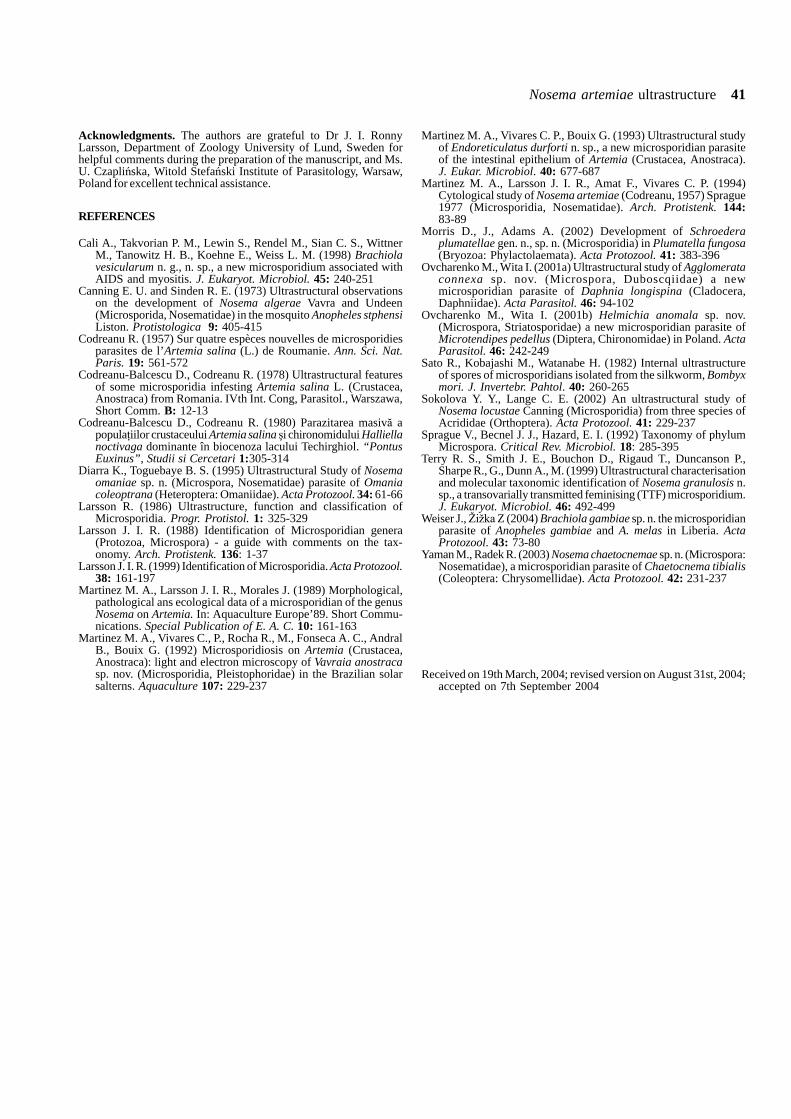
Nosema artemiae ultrastructure 41
Acknowledgments. The authors are grateful to Dr J. I. RonnyLarsson, Department of Zoology University of Lund, Sweden forhelpful comments during the preparation of the manuscript, and Ms.U. Czapli�ska, Witold Stefa�ski Institute of Parasitology, Warsaw,Poland for excellent technical assistance.
REFERENCES
Cali A., Takvorian P. M., Lewin S., Rendel M., Sian C. S., WittnerM., Tanowitz H. B., Koehne E., Weiss L. M. (1998) Brachiolavesicularum n. g., n. sp., a new microsporidium associated withAIDS and myositis. J. Eukaryot. Microbiol. 45: 240-251
Canning E. U. and Sinden R. E. (1973) Ultrastructural observationson the development of Nosema algerae Vavra and Undeen(Microsporida, Nosematidae) in the mosquito Anopheles stphensiListon. Protistologica 9: 405-415
Codreanu R. (1957) Sur quatre espèces nouvelles de microsporidiesparasites de l’Artemia salina (L.) de Roumanie. Ann. Sci. Nat.Paris. 19: 561-572
Codreanu-Balcescu D., Codreanu R. (1978) Ultrastructural featuresof some microsporidia infesting Artemia salina L. (Crustacea,Anostraca) from Romania. IVth Int. Cong, Parasitol., Warszawa,Short Comm. B: 12-13
Codreanu-Balcescu D., Codreanu R. (1980) Parazitarea masiv� apopulaiilor crustaceului Artemia salina i chironomidului Halliellanoctivaga dominante în biocenoza lacului Techirghiol. “PontusEuxinus”, Studii si Cercetari 1:305-314
Diarra K., Toguebaye B. S. (1995) Ultrastructural Study of Nosemaomaniae sp. n. (Microspora, Nosematidae) parasite of Omaniacoleoptrana (Heteroptera: Omaniidae). Acta Protozool. 34: 61-66
Larsson R. (1986) Ultrastructure, function and classification ofMicrosporidia. Progr. Protistol. 1: 325-329
Larsson J. I. R. (1988) Identification of Microsporidian genera(Protozoa, Microspora) - a guide with comments on the tax-onomy. Arch. Protistenk. 136: 1-37
Larsson J. I. R. (1999) Identification of Microsporidia. Acta Protozool.38: 161-197
Martinez M. A., Larsson J. I. R., Morales J. (1989) Morphological,pathological ans ecological data of a microsporidian of the genusNosema on Artemia. In: Aquaculture Europe’89. Short Commu-nications. Special Publication of E. A. C. 10: 161-163
Martinez M. A., Vivares C., P., Rocha R., M., Fonseca A. C., AndralB., Bouix G. (1992) Microsporidiosis on Artemia (Crustacea,Anostraca): light and electron microscopy of Vavraia anostracasp. nov. (Microsporidia, Pleistophoridae) in the Brazilian solarsalterns. Aquaculture 107: 229-237
Martinez M. A., Vivares C. P., Bouix G. (1993) Ultrastructural studyof Endoreticulatus durforti n. sp., a new microsporidian parasiteof the intestinal epithelium of Artemia (Crustacea, Anostraca).J. Eukar. Microbiol. 40: 677-687
Martinez M. A., Larsson J. I. R., Amat F., Vivares C. P. (1994)Cytological study of Nosema artemiae (Codreanu, 1957) Sprague1977 (Microsporidia, Nosematidae). Arch. Protistenk. 144:83-89
Morris D., J., Adams A. (2002) Development of Schroederaplumatellae gen. n., sp. n. (Microsporidia) in Plumatella fungosa(Bryozoa: Phylactolaemata). Acta Protozool. 41: 383-396
Ovcharenko M., Wita I. (2001a) Ultrastructural study of Agglomerataconnexa sp. nov. (Microspora, Duboscqiidae) a newmicrosporidian parasite of Daphnia longispina (Cladocera,Daphniidae). Acta Parasitol. 46: 94-102
Ovcharenko M., Wita I. (2001b) Helmichia anomala sp. nov.(Microspora, Striatosporidae) a new microsporidian parasite ofMicrotendipes pedellus (Diptera, Chironomidae) in Poland. ActaParasitol. 46: 242-249
Sato R., Kobajashi M., Watanabe H. (1982) Internal ultrastructureof spores of microsporidians isolated from the silkworm, Bombyxmori. J. Invertebr. Pahtol. 40: 260-265
Sokolova Y. Y., Lange C. E. (2002) An ultrastructural study ofNosema locustae Canning (Microsporidia) from three species ofAcrididae (Orthoptera). Acta Protozool. 41: 229-237
Sprague V., Becnel J. J., Hazard, E. I. (1992) Taxonomy of phylumMicrospora. Critical Rev. Microbiol. 18: 285-395
Terry R. S., Smith J. E., Bouchon D., Rigaud T., Duncanson P.,Sharpe R., G., Dunn A., M. (1999) Ultrastructural characterisationand molecular taxonomic identification of Nosema granulosis n.sp., a transovarially transmitted feminising (TTF) microsporidium.J. Eukaryot. Microbiol. 46: 492-499
Weiser J., ����� Z (2004) Brachiola gambiae sp. n. the microsporidianparasite of Anopheles gambiae and A. melas in Liberia. ActaProtozool. 43: 73-80
Yaman M., Radek R. (2003) Nosema chaetocnemae sp. n. (Microspora:Nosematidae), a microsporidian parasite of Chaetocnema tibialis(Coleoptera: Chrysomellidae). Acta Protozool. 42: 231-237
Received on 19th March, 2004; revised version on August 31st, 2004;accepted on 7th September 2004

Acta Protozool. (2005) 44: 43 - 49
Cross-infection Experiments Confirm the Host Specificity of Goussia spp.(Eimeriidae: Apicomplexa) Parasitizing Cyprinid Fish
Kálmán MOLNÁR1, Györgyi OSTOROS1 and Ferenc BASKA2
1Veterinary Medical Research Institute, Hungarian Academy of Sciences, Budapest; 2Szent István University, Faculty of Veteri-nary Science, Department of Pathology and Veterinary Forensic Medicine, Budapest, Hungary
Summary. The host specificity of the coccidian Goussia carpelli (Léger et Stankovitch, 1921) (Eimeriidae: Apicomplexa) was studied inaquarium experiments. Oocysts were obtained from the gut of 1- to 2-year-old common carp intensively infected with Goussia carpelli.These oocysts were mixed into mud containing infection-free oligochaetes (Tubifex tubifex and Limnodrilus hoffmeisteri). Laboratory-culturedfish demonstrated to be infection free were infected by feeding oligochaetes. The susceptibility of 3-5 cm long fingerlings of 8 fish species[common carp (Cyprinus carpio), goldfish (Carassius auratus), barbel (Barbus barbus), bleak (Alburnus alburnus), roach (Rutilus rutilus),bream (Abramis brama), white bream (Blicca bjoerkna) and vimba (Vimba vimba)] was experimentally evaluated. In the gut of commoncarp, intensive infection developed each of eight experiments, and oocysts were consistently detectable in the faeces and gut scrapings ondays 11-20 after feeding the fish with the oligochaetes. At the same time, oocyst formation could not be demonstrated in the gut of theother seven cyprinids. In another experiment of similar design, only the goldfish could be infected with oocysts obtained from naturallyinfected goldfish, and no infection was established in the common carp. The results of these experiments suggest that G. carpelli is strictlyspecific to common carp, while Carassius species most closely related to the common carp are parasitised by a distinct species of Goussiawith which it shares morphological similarity.
Key words: Apicomplexa, coccidia, cyprinid fishes, experimental infection, Goussia carpelli, Goussia sp., oligochaetes.
INTRODUCTION
Goussia carpelli, described by Léger andStankovitch (1921) from common carp (Cyprinuscarpio) and crucian carp (Carassius carassius), isone of the longest known and best studied fish coccidians.This parasite is a typical example of gut-parasitic fishcoccidia of small size, not exceeding 14 µm in diameter,and having a compact oocyst. Of the closely related
known species described from cyprinids, G. legeriStankovitch, 1920, G. cyprinorum (Stankovitch, 1921),G. cylindrospora (Stankovitch, 1920), G. cheni (Chen,1956), G. sinensis (Chen, 1956) and G. iroquoina(Molnár et Fernando, 1974) have similar intrapiscinelocation and structure. Although the majority of thesecoccidian species had been described under the nameEimeria, Dyková and Lom (1981) proposed that theyshould be classified in the genus Goussia created byLabbé (1896) because their sporocysts consist of twovalves connected by central sutures.
Stankovitch (1920, 1921) had previously suggestedthat oocysts isolated from different cyprinids repre-
Address for correspondence: Kálmán Molnár, Veterinary Medi-cal Research Institute, Hungarian Academy of Sciences, H-1581Budapest, PO Box 18, Hungary; Fax: 36 (1) 2521069; E-mail:[email protected]

44 K. Molnár et al.
sented distinct parasite species, a view subsequentlyadvocated by Belova and Krylov (2000) who suggestedthat the host-specificity of fish coccidia is as strong asthat of coccidia parasitising terrestrial vertebrates. Otherauthors (Musselius and Laptev 1967, Shulman 1984),however, were of the opinion that certain species, suchas G. carpelli, were able to develop in numerouscyprinid hosts. Moreover, Shulman and Zaika (1964)claimed to have found G. carpelli even in a fish speciesbelonging to the Cottidae family. Specificity experimentsusing G. iroquoina, a G. carpelli type coccidium spe-cies, have been performed by Paterson and Desser(1982). The most reliable experimental results wereobtained by Lukeš et al. (1991) who described that, withthe exception of the goldfish, no infection could beproduced in 9 other cyprinid fishes with G. carpellioriginating from the common carp.
The objective of this experimental work was todetermine which cyprinid fish species, other than thecommon carp, can be infected successfully withG. carpelli oocysts originating from common carp.
MATERIALS AND METHODS
Fish and oligochaetes used for experiments. Three- to 5-cm-long fingerlings of cyprinids, hatched in laboratory and reared inparasite-free environment, were used in the experiments. In additionto common carp (Cyprinus carpio), fingerlings of goldfish (Carassiusauratus), barbel (Barbus barbus), bleak (Alburnus alburnus), roach(Rutilus rutilus), bream (Abramis brama), white bream (Bliccabjoerkna) and vimba (Vimba vimba) were studied. For the transmis-sion of infection, laboratory-cultured, parasite-free Tubifex tubifexand Limnodrilus hoffmeisteri oligochaetes were used.
Coccidians used for experiments. Oocysts of G. carpelli wereobtained from the gut of common carp fingerlings and two-summercommon carp originating from fish farms. These were dissected a dayor two after their transfer to the laboratory, after their gut contentshad been excreted. Oocysts present in the mucus covering the intesti-nal mucosa were maintained in tap-water until the infection of oli-gochaetes. In each experiment, 10,000-20,000 oocysts were pouredinto plastic dishes containing 100 oligochaetes and sterilised mud.Similar procedures were used in one of the experiments employingG. carpelli-like oocysts obtained from goldfish.
Experimental design for infecting fish. A total of 9 experi-ments were performed. In the first 8 experiments, oligochaetes in-fected by G. carpelli oocysts were fed to fish. Experiments 1 and 2were designed to determine the interval, after ingestion, when maxi-mum oocyst excretion occurred. Experiments 4 and 5 were designedto learn how long the infected oligochaetes could transmit G. carpelliinfection to fish. The objective of experiments 3-7 was to learnwhether cyprinids other than the common carp could be infected withG. carpelli (Table 1). In experiment 8 the effect of repeated oligocha-ete feeding on the intensity of infection was studied (Table 2). In
experiment 9, oocysts intended for infection were collected from thegut of goldfish fingerlings, and these oocysts were mixed into the mudof the dish containing the oligochaetes (Table 3). Until the conclusionof the experiments, the oocysts collected from common carp andgoldfish were regarded as the spore stages of a single species,G. carpelli.
Infection of fish by oligochaetes. Oligochaetes were fed to thefish at different post-exposure intervals. The times of infection ofoligochaetes and fish are indicated in Tables 1-3. Oligochaetes cut intosmall pieces were used for infecting the fish. Two to three days beforethe infected oligochaetes were fed to them, individual fasted fish werein a plastic dish containing 0.5 l of water. These fish, sustained ongranulated dry fish food, had previously been acclimated to theconsumption of coccidia-free oligochaetes. Infected oligochaetes werefed for one to three days; the duration of feeding varied by experi-ment. The fish were kept in an individual dish throughout the experi-ment to limit potential sources of exposure. Throughout the experi-ment, fish were fed the granulated fish food. Water in the dishes waschanged daily and kept fresh and replenished with oxygen by aera-tion. Relatively few fish were assayed owing to the demands requiredto maintain fish individually in separate dishes. The experimentswere carried out at room temperature (21 to 25°C). The fish werekilled 10-21 days after infection, when peak oocyst excretion wasexpected. Feeding of fish food was stopped two days before the fishwere killed.
Examination of fish for infection. The gut of the fish wasremoved in its entirety, cut open lengthwise, and the mucus in whichthe oocysts were most easily detectable was lifted off the gut wall asfar as possible. In a negative case, besides the mucus the lumps offaeces present in the gut and mucosal scrapings taken from differentsegments of the intestinal wall were also placed on a slide, under acoverslip. The samples were examined under microscope at 200- to400-fold magnification. In the case of fish of smaller size, the entiregut was examined for the occurrence of oocysts or developmentalstages. For histological examination, segments from the intestine werefixed in Bouin’s solution and embedded in paraplast. The 5 µmsections were stained with haematoxylin and eosin solutions. Photo-micrographs of fresh oocysts in mucus under a coverslip were takenwith an Olympus DH-10 digital camera mounted on an OlympusBH2 microscope.
RESULTS
Experiments 1 and 2, in which parasite-free commoncarp fingerlings were infected with G. carpelli oocystscollected from common carp through tubificid vectors,have confirmed that G. carpelli infection can be suc-cessfully transmitted to the common carp. Oocystsenclosed in yellow bodies could be detected in themucus, faeces and gut epithelium at room temperatureby days 11-20 in these fish (Table 1; Figs 1, 3).
Equivalent experiments demonstrated that parasitesderived from the common carp infections did not resultin infections of either goldfish or other cyprinid fishes(Table 1).

Host specificity of Goussia parasitizing fish 45
Table1. Experimental infection of common carp and other cyprinids with Goussia carpelli through oligochaete vectors exposed to oocystsfor varying periods.
Fish species No. fish Oligochaetes exposed Time from Number of Intensity of No. exp.to oocysts before exposure to infected fish infectioninfection (days) killing (days)
Common carp 2 4 14 2 +/+++ 41 15-17 12 - - 51 15-17 17 1 + 52 26 14 2 +/++ 41 32 9 1 +++ 78 32 12 8 +/+++ 71 34 11 1 + 12 34 19 2 +++ 12 34 20 2 ++ 14 31-32 15-16 3 +/+++ 64 31-32 15-16 2 ++ 61 46-48 12 - - 51 46-48 17 1 + 51 68-70 12 - - 51 68-70 17 1 + 52 72 14 1 + 42 73-74 12 - - 22 73-74 13 1 +++ 22 73-74 14 1 +++ 22 73-74 15 1 +++ 24 73-74 16 4 ++/+++ 22 73-74 19 1 +++ 22 73-74 20 2 +/++ 22 73-74 21 2 +/++ 2
Goldfish 2 31-32 15-16 - - 62 32 9 - - 72 4-72* 14 - - 41 15-70* 12 - - 51 15-70* 17 - - 5
Common bream 2 32 12 - - 72 4-72* 14 - - 42 31-32 15-16 - - 61 15-70* 12 - - 51 15-70* 17 - - 5
Roach 2 32 12 - - 72 4-72* 14 - - 42 31-32 15-16 - - 61 15-70* 12 - - 51 15-70* 17 - - 5
Vimba 2 32 12 - - 72 31-32 15-16 - - 62 4-72* 14 - - 41 15-70* 12 - - 51 15-70* 17 - - 5
White bream 2 32 12 - - 72 31-32 15-16 - - 6
Bleak 2 32 12 - - 72 31-32 15-16 - - 6
Barbel 2 32 12 - - 72 31-32 15-16 - - 62 4-72* 14 - - 41 15-70* 12 - - 51 15-70* 17 - - 5
+ - a few oocysts in the gut; ++ - easily detectable coccidian infection; +++ - intensive infection; * - feeding by mixed oligochaete groupsexposed to oocysts of varying times

46 K. Molnár et al.
Fig. 1. Goussia carpelli oocysts located within a yellow body from the gut of an experimentally infected common carp fingerling. 3000×.Fig. 2. Goussia sp. oocysts located within a yellow body from the gut of an experimentally infected goldfish fingerling. 3000×.Fig. 3. Sporulated G. carpelli oocysts in the gut epithelium of the common carp. Histological section stained with haematoxylin and eosin. 500×.Fig. 4. Oocysts of a Goussia sp. in the gut epithelium of a goldfish. Histological section stained with haematoxylin and eosin. 500×.

Host specificity of Goussia parasitizing fish 47
The transmission of G. carpelli infection from com-mon carp to common carp was successful regardless ofwhether the oligochaetes had been infected 4, 26, 69 or72 days before the infection of fish. Oligochaetes in-fected for longer intervals resulted in less intensiveinfections in common carp (Table 1).
Oocyst excretion was observed, once, as early as9 days after infection but regularly from day 13 onwards(Tables 1, 2).
An only minimal increase in the intensity of infectionwas observed in common carp fed with the oligochaetesfor 8 days as compared to those fed oligochaetes for oneday (Table 2).
Fish could also be infected through tubificid vectorswith oocysts, morphologically appearing identical toG. carpelli, collected from goldfish. However, in thiscase infection developed only in goldfish, while commoncarp infected in the same way, and serving as controlsfor the goldfish, remained free of infection (Table 3;Figs 2, 4).
In goldfish, oocyst excretion could also be observedfrom day 13 post infection (Table 3). At that time themajority of oocysts were still in unsporulated or half-sporulated state in the gut lumen. However, after day 19an advanced stage of sporulation was observed (Fig. 4).
DISCUSSION
The primary objective of the experiments was tostudy the host specificity of G. carpelli and the possibil-ity to transmit parasites derived from common carp toother cyprinids. To maximize our ability to detect suchheterologous infections, we first sought to determine,without robust replication, other transmission character-istics such as the optimum time of infection with oli-gochaetes and the duration of oocyst formation andoocyst excretion.
As shown by the data of experiments presented inTables 1-2, G. carpelli infection could consistently be
Table 2. Common carp and goldfish fingerlings fed for 1-10 days on oligochaetes exposed to G. carpelli oocysts.
Fish species No. fish Oligochaetes Duration of Time from Number of Intensity of No. exp.exposed to oligochaete the first infected fish infectionoocysts before feeding (days) exposure Carp/Goldfish Carp/Goldfishinfection (days) to killing
(days)
Common carp/goldfish 1/1 36 1 15 1/- ++ / - 81/1 36-37 2 15 1/- ++ / - 81/1 36-38 3 15 1/- ++ / - 81/1 36-40 5 15 1/- ++ / - 81/1 36-41 6 15 1/- ++ / - 81/1 36-42 7 15 1/- +++ / - 81/1 36-43 8 15 1/- +++ / - 80/1 36-44 9 15 -/- / - 80/1 36-45 10 15 -/- / - 8
Table 3. Coccidian infection of goldfish and common carp fingerlings after feeding on tubificid oligochaetes exposed to oocysts of aGoussia sp. originating from goldfish.
Fish species No. fish Oligochaetes Time from Number of Intensity of No. exp.exposed to exposure infected fish infectionoocysts before to killing Goldfish/Carp Goldfish/Carpinfection (days) (days)
Goldfish/common carp 3/3 14 9 -/- -/- 93/3 14 14 3/- + to ++/- 92/2 14 20 2/- +++/- 92/2 14 23 2/- +++/- 9

48 K. Molnár et al.
established in infection-free common carp through thefeeding of tubificid (Tubifex, Limnodrilus) paratenichosts. Oocyst formation and excretion commenced ondays 9-12 post infection and continued at least forfurther eight days. In all the eight experiments, oocystscould be demonstrated only in common carp in the givenperiod, and other cyprinids remained infection free. Itwas especially striking that the goldfish did not becomeinfected either, although according to the original de-scription G. carpelli is a common parasite of the genusCyprinus and Carassius (Léger and Stankovitch 1921,Lukeš et al. 1991) could also produce a low-levelinfection in goldfish during their experiments. In contrastto these results, our experiments suggested that thecommon carp and the goldfish were infected by twodistinct species, and that the common carp could not beinfected successfully with oocysts collected from gold-fish (Table 3). Otherwise, the intraoligochaete andintrapiscine development of the latter species appearedto be similar to that of G. carpelli, and oocyst excretionwas recorded from day 13.
The results obtained in this study support the hypoth-esis that fish coccidia are characterized by relativelystrict host specificity (Stankovitch 1921, Léger andStankovitch 1921, Musselius et al. 1965). According toBelova and Krylov (2000), fish coccidia can developnormally only in closely related fish species, but weobserve an even greater degree of host specificity inG. carpelli, as it cannot cause infection even in thosespecies of Carassius most closely related to the com-mon carp, i.e. the goldfish (Carassius auratus) andpossibly the crucian carp (C. carassius) and the gibelcarp (C. auratus gibelio). The close relationship be-tween these fish species and the common carp isconfirmed by morphological characteristics as well as bymolecular evidence (Zardoya and Doadrio 1999). Simi-larly, the common carp cannot be infected with theGoussia species collected from goldfish, which, how-ever, can successfully be transmitted experimentally toinfection-free goldfish. These facts suggest that theG. carpelli-like species parasitising the goldfish might beregarded as a new, hitherto undescribed species. Waitingfor the definitive description, this species has beendesignated as Goussia sp.
In these experiments, infection was consistently at-tempted by the use of oligochaete paratenic hosts.Namely, both in our own experience and according to thefindings of Steinhagen and Körting (1990), this is the onlymethod by which G. carpelli infection can be repro-duced consistently. To date, few authors have attempted
to transmit coccidian infections experimentally in fish.Successful infection through direct transmission of oo-cysts has been reported by Musselius et al. (1965),Zmerzlaya (1966), Steinhagen and Körting (1988, 1990)and Steinhagen et al. (1989), who conducted theirexperiments with G. carpelli in common carp. Marincek(1973) and Steinhagen (1991) could also successfullyinfect the same fish species with oocysts ofG. subepithelialis. As regards to other fish species,Landsberg and Paperna (1985) successfully infectedTilapia by direct transmission of G. cichlidarum oo-cysts in tilapia. The paratenic host is necessary for thesuccessful transmission of infection was first suggestedby Molnár (1979), who could not induce G. carpelliinfection in common carp with oocysts mixed in the foodbut obtained intensive infection by feeding tubificidscollected from the natural environment. Similarly, thenecessity of mediator organisms has been reported bySolangi and Overstreet (1980) as well as Fournie andOverstreet (1983), who proved that a natural intermedi-ate host, the grass shrimp (Palaemonetes pugio), wasneeded for the development of Calyptospora funduli.Kent and Hedrick (1985), who studied G. carpelliinfection in goldfish (sic?!), successfully induced infec-tion by the use of vectors (tubificids, grass shrimp).However, Steinhagen and Körting (1990) could induceG. carpelli infection both directly and indirectly (bythe use of vectors), and they regarded tubificids asparatenic hosts only. Nevertheless, the above authorsemphasised that infection via vectors was more likelyto succeed. Steinhagen (1991) found G. carpelli andG. subepithelialis sporozoites within parasitophorousvacuoles in the gut epithelial cells of Tubifex and estab-lished that they maintained their viability for nine weeks.Fournie et al. (2000) detected sporozoites of C. funduliin the basal epithelial cells of the gut of shrimp, wherethey became infective in 5 days.
Cross-infection experiments were first reported byPaterson and Desser (1982), who successfully inducedG. iroquoina infection in the common shiner (Notropiscornutus) and fathead minnow (Pimephales promelas)with oocysts collected from the common shiner both bydirect infection and through Tubifex vectors. On thebasis of observations made in ponds, Musselius andLaptev (1967) came to the conclusion that if the com-mon carp, silver carp and bighead stocks are rearedtogether the common carp develops only G. carpelliinfection, whereas the oocysts of G. carpelli also can befound in the gut of silver carp and bighead where theyoccur together with G. sinensis and G. cheni which

Host specificity of Goussia parasitizing fish 49
typically infect these fish species. So far, only G. sinensisinfection has been recorded in silver carp and bighead inHungary (Molnár 1976), and the oocysts of G. carpelliwere not detectable from these fish species. The resultsof the present experiment suggest that it is unlikely thatG. carpelli infects other fish than the common carp. Wesuggest that in the experiment of Musselius and Laptev(1967) G. carpelli coccidia may have reached the gut ofsilver carp and bighead with the food owing to conditionsapplicable only when fish are reared together in aquaria.To rule out this potential source of error, fish in thepresent experiments were kept in individual dishes,isolated from fish of the same or other species. The onlyreliable results on the host specificity of G. carpelli wereobtained by Lukeš et al. (1991) who found that thisspecies was strictly host specific. However, in contrastwith our results, besides the common carp the aboveauthors could establish a slight infection in the goldfish aswell.
Outside the host oocysts from mammals and birdscan remain viable and infective for more than one year(Kheysin 1972). However, according to Musselius et al.(1965) oocysts remain viable in water only for 20-22days. According to our own observations (Molnár, un-published), the period of oocyst viability is actually muchshorter, and paratenic hosts play an important role alsoin the prolonged maintenance of infectivity. In addition tothe data reported by Steinhagen (1991), this is supportedalso by the results of experiments 3 and 4 of this study.In these experiments, besides relatively recently infectedoligochaetes, tubificid worms of an oligochaete stockexposed to oocysts four months earlier were also fed tofish. Both stocks established successful infection, thoughthe level of infection with earlier infected oligochaeteswas lower than when recently infected tubificid wormswere used.
Acknowledgements. The study was supported by the HungarianScientific Research Fund (OTKA, project no. T.031755) and by theprogramme entitled “The quality development of the biological andtechnological bases of the Hungarian fishing sector”, 4/039/2001. Theauthors thank Dr Csaba Székely for selecting the infected donor fishand for culturing uninfected cyprinid fingerlings and Dr BenjaminRosenthal for reviewing the manuscript.
REFERENCES
Belova L. M., Krylov M. V. (2000) Specificity of coccidia of fishes(Sporozoa, Coccidia). Trudy Zool. Inst. 286: 11-16 (in Russian)
Dyková J., Lom J. (1981) Fish coccidia: critical notes on life cycles,classification and pathogenicity. J. Fish Dis. 4: 487-505
Fournie J. W., Overstreet R. M. (1983) True intermediate hosts forEimeria funduli (Apicomplexa) from estuarine fishes. J. Protozool.30: 672-675
Fournie J. W., Vogelbein V. K., Overstreet R. M., Hawkins W. E.(2000) Life cycle of Calyptospora funduli (Apicomplexa:Calyptosporidae). J. Parasitol. 86: 501-505
Kent M. L., Hedrick R. P. (1985) The biology and associatedpathology of Goussia carpelli (Léger and Stankovitch) in gold fishCarassius auratus (Linnaeus). Fish Pathol. 20: 485-494
Kheysin Y. M. (1972) Life Cycles of Coccidia of Domestic Animals.University Park Press, London
Labbé A. (1896) Recherches zoologiques, cytologiques et biologiquessur les Coccidies. Arch. Zool. Exp. Gén. 4: 517-654
Landsberg J. H., Paperna I. (1985) Goussia cichlidarum n. sp.(Barrouxiidae, Apicomplexa), a coccidian parasite in theswimbladder of cichlid fish. Z. Parasitenk. 71: 199-201
Léger L., Stankovitch S. (1921) Sur la coccidiose des alevins de laCarpe. C. R. Acad. Sci. (Paris) 173: 742-744
Lukeš J., Steinhagen D., Körting W. (1991) Goussia carpelli(Apicomplexa, Eimeriorina) from cyprinid fish: Field observa-tions and infection experiments. Angew. Parasitol. 32: 149-153
Marincek M. (1973) Dévelopment d’ Eimeria subepithelialis (Sporo-zoa, Coccidia) parasite de la Carpe. Acta Protozool. 19: 197-215
Molnár K. (1976) Histological study of coccidiosis caused in thesilver carp and the bighead by Eimeria sinensis Chen (1956). ActaVet. Acad. Sci. Hung. 26: 303-312
Molnár K. (1979) Studies on coccidia of Hungarian pond fishes andfurther prospects of their control. Proceeding of the InternationalSymposium on Coccidia, Prague, 173-183
Musselius V. A., Laptev B. I. (1967) On the specificity of coccidiaof Hypophthalmichthys molitrix, Aristichthys nobilis and Cyprinuscarpio. Parazitologiya 1: 333-338 (in Russian)
Musselius V. A., Laptev B. I., Ivanova N. S. (1965) On the coccidiosisof the common carp (second part). Trudy VNIIPRH 13: 69-78(in Russian)
Paterson W. B., Desser S. S. (1982) The biology of two Eimeriaspecies (Protista: Apicomplexa) in their mutual fish hosts inOntario. Can. J. Zool. 60: 164-175
Shulman S. S. (1984) Parasitic protozoa. In: Key to Parasites ofFreshwater Fish of the USSR 1, (Ed. O. N. Bauer). Nauka,Leningrad (in Russian)
Shulman S. S., Zaika V. E. (1964) Coccidia of fishes of the LakeBaikal. Izv. Sib. Otd. Akad. Nauk SSSR, Ser. Biol. Med. Nauk. 2:126-130 (in Russian)
Solangi M. A., Overstreet R. M. (1980) Biology and pathogenesis ofthe coccidium Eimeria funduli infecting killifishes. J. Parasitol.66: 513-526
Stankovitch S. (1920) Sur deux nouvelles Coccidies parasites desPoisons Cyprinids. C. R. Soc. Biol. (Paris) 83: 833-835
Stankovitch S. (1921) Sur quelques Coccidies nouvelles des PoisonsCyprinids. C. R. Soc. Biol. (Paris) 85: 1128-1130
Steinhagen D. (1991) Ultrastructural observations on sporozoitestages of piscine Coccidia: Goussia carpelli and G. subepithelialisfrom the intestine of tubificid oligochaetes. Dis. Aquat. Org. 10:121-125
Steinhagen D., Körting W. (1988) Experimental transmission ofGoussia carpelli (Leger & Stankovitch, 1921) (Protista:Apicomplexa) to common carp, Cyprinus carpio L. Bull. Eur.Assoc. Fish Pathol. 8: 112-113
Steinhagen D., Körting, W. (1990) The role of tubificid oligochaetesin the transmission of Goussia carpelli. J. Parasitol. 76: 104-107
Steinhagen D., Körting W., van Muiswinkel W. B. (1989) Morphol-ogy and biology of Goussia carpelli (Protozoa: Apicomplexa)from the intestine of experimentally infected common carpCyprinus carpio. Dis. Aquat. Org. 6: 93-98
Zardoya R., Doadrio I. (1999) Molecular evidence on the evolution-ary and biogeographical patterns of European cyprinids. J. Molec.Evol. 9: 227-237
Zmerzlaya E. I. (1966) Temperature effect upon the infestation ofcarps with coccidia Eimeria carpelli Leger et Stankovitsch,1921. Zool. Zhurn. 45: 305-308 (in Russian)
Received on 28th June, 2004; revised version on 12th October, 2004;accepted on October 18th, 2004

Acta Protozool. (2005) 44: 51 - 60
Trichodinids (Ciliophora: Trichodinidae) from Native and Exotic AustralianFreshwater Fishes
Alistair D. M. DOVE1 and Peter J. O’DONOGHUE2
1Department of Microbiology and Immunology, College of Veterinary Medicine, Cornell University, C/- Marine SciencesResearch Center, Stony Brook University, Stony Brook NY, USA; 2Department of Microbiology and Parasitology, The Universityof Queensland, Queensland, Australia
Summary. There are very few data on trichodinids of freshwater fishes in Australia. 2003 fishes were surveyed across Eastern Australiato investigate the diversity of trichodinids present, to determine which species have been introduced with exotic fishes and to determinethe extent to which these species have crossed into native fish populations. Twenty-one putative trichodinid species were recovered fromthe 33 fish species examined. Trichodina heterodentata, T. mutabilis and T. reticulata were the exotic species recovered regularly; a singlespecimen matched a fourth exotic species, T. acuta. All four exotic species are redescribed from Australian material. Trichodina heterodentatawas recorded from 17 species of fishes, 15 of which were new host records; this species is identified as one of emerging importance in fishparasitology and a list of its known hosts is presented. Two new native species are also described based on silver stained specimens:T. cribbi sp. n. from Hypseleotris galii, H. klunzingeri, and Hypseleotris sp. 5; and T. bassonae sp. n. from Selenotoca multifasciata.Trichodina cribbi is characterised by a large circular central inclusion and approximately 28 denticles, which have a blade length slightlygreater than the ray length. Trichodina bassonae is characterised by a small, round, central inclusion and approximately 25 denticles, whichhave straight, non tapering rays that are in line with the leading edge of the denticle blade. It is estimated that the Australian trichodinidfauna may include up to 150 as yet undescribed species and represents a major source of unexplored biodiversity.
Key words: Australian, endemic, exotic, richness, Trichodina bassonae sp. n., Trichodina cribbi sp. n., trichodinids.
INTRODUCTION
Data on trichodinids of Australian freshwater fishesare sparse and fragmentary. No individual species havebeen described from either native or exotic freshwaterfishes in Australia. Lom and Dyková (1992) recordedan unspecified Trichodina species from the gills of
Gambusia holbrooki at Armidale, New South Wales.Langdon (1989) listed Trichodina spp. as “parasitic onnative and introduced fishes” but provided no data onwhich Trichodina species infected which host species,nor any data on shared Trichodina species. Rowlandand Ingram (1991) discussed the ectoparasitic diseasesof Murray cod (Maccullochella peeli), goldenperch (Macquaria ambigua) and silver perch(Bidyanus bidyanus). Although they identifiedChilodonella hexasticha, Ichthyophthirius multifilisand Ichthyobodo necator specifically, Trichodina spp.(and Tetrahymena spp.) were regarded as a singlegroup. These authors recognised that “the identity and
Address for correspondence: Alistair D. M. Dove, Departmentof Microbiology and Immunology, College of Veterinary Medicine,Cornell University. C/- Marine Sciences Research Center,Stony Brook University, Stony Brook NY 11794, USA;E-mail: [email protected]

52 A. D. M. Dove and P. J. O’Donoghue
number of species of Trichodina on freshwater fishesin Australia are not known”.
The aims of this study were: to identify which trichodinidspecies occur on Australian populations of exotic fresh-water fishes; to investigate the diversity of the Australiantrichodinid fauna; and to quantify the degree to whichtrichodinid species have been exchanged between nativeand exotic fishes. The null hypothesis that no exchangeof trichodinid species between native and exotic fishspecies has occurred was tested using survey data andcomparisons with known trichodinid records onAustralia’s exotic fishes in their original ranges.
MATERIALS AND METHODS
Collection and dissection protocol
Trichodinid ciliates were collected on an opportunistic basisduring a survey for other parasites that encompassed 58 sites through-out Victoria, New South Wales, the Australian Capital Territory andQueensland. In addition, a dedicated survey was conducted at 15 sitesin Queensland to obtain accurate data on prevalence, host and micro-habitat specificity. Site abbreviations are as follows: ALR - AlbertRiver; Queensland (Qld); ATH - Atherton, Qld; BCF - CrayfishFarm, Beenleigh Qld; BMP - Lake Burley-Griffin at Black MountainPeninsula, Australian Capital Territory (ACT); BOC - BoorolongCreek, New South Wales (NSW); BRB - Billabong at BangarangRoad, Echuca, Victoria (Vic); BRF - Brisbane River at Fernvale, Qld;CBL - Condamine River below Leslie Dam, Qld; CRH - CondamineRiver Headwaters, Qld; CRW - Condamine River at Warwick, Qld;EBK - Lake Burley-Griffin at East Basin Kingston, ACT; EGC -England Creek, Qld; ENC - Enoggerra Creek, Qld; ERS - EdwardRiver at Steven’s Weir, Deniliquin, NSW; GBL - Gum Bend Lake,Condobolin, NSW; KB216 - Kalinga Park Creek, Qld; LGN - LakeGinninderra, ACT; MNC - Monsildale Creek, Qld; MOC - Moody’sCreek, north Qld; NFC - Narranderra Fisheries Centre, NSW; NPR -North Pine River, Qld; OVR - Ovens River, Vic; RGR - MoggillCreek at Rafting Ground Road, Qld; SRW - Shoalhaven River atWarri Bridge, NSW; WCC - West Court Creek, north Qld; WKN -Walkamin Research Station, north Qld; WVN - Lake Wivenhoe, Qld.
Fishes were collected by cast-netting, dip-netting, electrofishingand line fishing, and were separated into species-specific plastic bagson site. In the laboratory, fishes were maintained in aerated 20 l tankswith water from the collection site. Dissections were made withinthree days to minimise the effect of collection stress on trichodinidprevalence.
Fish were killed by pithing the brain, then identified, measured(caudal fork length) and sexed (if possible), and immediately dis-sected in freshwater. Each fin and gill was removed and placed in aseparate Petri dish in aged water and the skin scraped into anotherdish of aged water. Each dish was then examined under dissectingmicroscope for the presence of trichodinids; any present were pipettedonto a clean or albuminised glass slide (depending on staining regi-men) and allowed to air dry.
Morphometrics
Stained preparations were made using two silver salt stainingprotocols: Foissner’s modification of Klein’s dry silver nitrate tech-nique (Foissner 1991), and Basson’s version of Lom’s dry silvernitrate technique (Lom 1958). Measurements presented here followthe protocols of Lom (1958). Three denticles were measured fromeach specimen and the means for denticle measurements are thereforebased on 3n denticles. Measurements were made using a systemsimilar to that described by Roff and Hopcroft (1986), with a digitisingtablet and a drawing tube attached to a compound microscope. Allmeasurements are given in micrometres. Type material was lodged inthe Queensland Museum.
Drawing and photography techniques
Representatives of each putative species from each host specieswere photographed using a 35 mm camera attached to a DIC com-pound microscope. These photographs were the basis for the draw-ings of each trichodinid species. Drawings were made by enlarging thephotographs by 200% on a photocopier, overlaying the resultingimage with architectural film and tracing the morphological features.The ink drawings were then scanned and the resulting files edited inAdobe Photoshop© 5.0.
RESULTS
A total of 2003 fishes was dissected, representing33 host species and 58 sites across Queensland, NewSouth Wales, Victoria and the Australian Capital Terri-tory. From these, 21 putative species of trichodinidswere recovered, 6 of which are discussed here. Threeexotic trichodinid species were recovered: Trichodinaheterodentata, T. mutabilis and T. reticulata. A singlespecimen matching a fourth exotic species, T. acuta,was also recovered. These four species of exotictrichodinids are redescribed here to confirm their identityand to provide data on their distribution among hosts inAustralia. In addition, two new species of Trichodinaare described from native fish species: T. cribbi sp. n.and T. bassonae sp. n. The remaining 15 trichodinidmorphotypes are not described here.
Descriptions
Trichodinidae Raabe, 1963Trichodina Ehrenberg, 1838
Trichodina heterodentata Duncan, 1977 (Fig. 1)
Hosts: [Host (number of infected hosts examined)]Ambassidae: Ambassis agassizi (4); Apogonidae:Glossamia aprion gilii (1); Cichlidae: Oreochromismossambicus (1); Cyprinidae: Cyprinus carpio (2);Eleotridae: Hypseleotris compressa (2); Hypseleotris

Australian trichodinids 53
galii (2); Hypseleotris klunzingeri (9); Philypnodongrandiceps (1); Galaxiidae: Galaxias maculatus (2);Galaxias olidus (1); Gerreidae: Gerres sp. (1);Percichthyidae: Macquaria ambigua (2); Poeciliidae:Gambusia holbrooki (5); Poecilia reticulata (6);Xiphophorus helleri (1); Xiphophorus maculatus (6);Pseudomugilidae: Pseudomugil signifer (1).
Localities: KB216, WKN, BRB, NPR, NFC, BCF,EBK, WVN, GBL, MNC, BOC, SRW.
Microhabitat: Skin, fins, occasionally gills.Description: Description based on 18 specimens:
[Host species, Autopsy number (number of specimens)]A. agassizi, AD334 (3); C. carpio, T141 (1);G. holbrooki, AD309 (1); G. aprion gilii, AD311 (1);H. compressa, NPR5 (1); H. galii, T137 (1), BCF5 (1),BCF6 (1); H. klunzingeri, T130 (1); O. mossambicus,NPR12 (1); P. signifer, AD252 (1); X. helleri, KB20/10/97_8 (1); X. maculatus, AD307, AD310, KB20/10/97_1 (1) KB20/10/97_3 (1). Medium to large disc-shaped cell; adhesive disc 56.9 (41.2-89.2) in diameter;denticle ring 31.2 (24.7-37.4) in diameter. Denticles 23(21-26); denticle span 17.2 (12.5-23.9). Denticle withstrongly curved blade ending in a sharp point, blade 5.2(4-7.4) long, 5.6 (4-8.3) wide. Central part elongate, tiprounded, 3 (2.3-4.6) long, 4.4 (3.1-6.4) wide. Projectionof central part not visible. Ray tapering evenly fromcentral part to pointed tip, 7.9 (5.5-11.2) long, 1.9 (1.1-3) wide at widest point. Central disc without inclusionsof any kind. Radial pins prominent, 11 (10-13) pins perdenticle. Nuclear apparatus not observed.
Taxonomic remarks: Trichodina heterodentata isa distinctive species characterised by the robust, stronglysickle-shaped denticle blades with pointed ends, theevenly tapering rays with pointed tips, the absence ofany central inclusions and the prominence of the radialpins. It was originally described from cultured cichlids,Oreochromis mossambicus from the Philippines, buthas subsequently been recorded from a large numberof fish species (see Table 1). Morphometric data forthe present material fall within the ranges forT. heterodentata presented in the original description byDuncan (1977) and subsequent redescriptions by Van Asand Basson (1989), Kruger et al. (1993) and Basson andVan As (1994). Van As and Basson (1989) and Bassonand Van As (1994) noted a degree of morphologicalvariability in isolates of T. heterodentata; a similardegree of variability was evident in the present material,especially with respect to the ray length and the degreeof curve in the distal blade edge. The distinctiveness ofthe denticle shape, however, made identification of thisspecies relatively easy.
Trichodina mutabilis Kazubski et Migala, 1968(Fig. 2)
Hosts: Cyprinidae: Cyprinus carpio (3); Poeciliidae:Gambusia holbrooki (1)
Localities: BCF.Microhabitat: Fins, gills.Description: Description based on seven individuals:
C. carpio BCF12/1/98_1 (4), BCF12/1/98_6 (1);
Fig. 1. Trichodina heterodentata Duncan, 1977 from the skin ofAmbassis agassizi. Scale bar 10 µm.
Fig. 2. Trichodina mutabilis Kazubski et Migala, 1968 from the skinof Cyprinus carpio. Scale bar 10 µm.

54 A. D. M. Dove and P. J. O’Donoghue
G. holbrooki BCF12/1/98_8 (2). Medium to large disc-shaped cell; adhesive disc 45.9 (40.5-55.7) in diameter,denticle ring 27.6 (24-37.5) in diameter. Denticles 24(22-26); denticle span 15.4 (11.5-19). Denticle bladeoblong, largely parallel to radial of disc, with squareddistal end, blade 5.6 (4.6-7) long, 4 (3.4-5.3) wide.Central part narrow, with oblong to rounded overlappingend; 1.9 (1.3-2.4) long, 3.5 (2.7-4.5). Projection ofcentral part not visible. Ray narrow with no taper, tipblunt to square; 7.3 (5.4-9) long, 1.4 (1-2) wide. Centraldisc without inclusions of any kind. Radial pins 11 (9-13)per denticle. Nuclear apparatus not observed.
Taxonomic remarks: Our material falls within theranges for this species in the description by Kazubskiand Migala (1968) and a subsequent redescription byand Basson and Van As (1994).
Trichodina reticulata Hirschmann et Partsch, 1955(Fig. 3)
Hosts: Cyprinidae: Carassius auratus (2), Cyprinuscarpio (1).
Localities: SRW, GBL, NFC.Microhabitat: Skin, fins.Description: Description based on five individuals
from C. auratus G372. Medium to large disc-shapedcell; adhesive disc 48.6 (40.9-54.2) in diameter, denticlering 30.4 (26-34.3) in diameter. Denticles 24 (23-25);denticle span 13.8 (11.8-14.9). Denticle blade curved oninside margin but roughly angular on outside margin, withsquared distal end, 5.6 (4.8-6.7) long, 4 (3.4-4.6) wide.Central part oblong, with oblong to rounded overlappingend; 2.5 (2.2-2.9) long, 3.7 (3.3-4.3) wide. Projection of
central part not visible. Ray with little taper, tip blunt tosquare; 5.2 (4.2-5.8) long, 1.9 (1.7-2.1) wide at widestpoint. Central disc with 12 (10-14) icosahedral inclusions(mean diameter 4 (2.5-6.3)); inclusions give impressionof a single large inclusion with reticulated markings.Radial pins prominent 10 (9-11) per denticle. Nuclearapparatus not observed.
Taxonomic remarks: Trichodina reticulata is ahighly distinctive species well-known as a pathogen ofCarassius spp. and other cyprinids in aquaculture andaquaria. The central inclusions are unlike those of almostany other Trichodina species, while the angular outeredge to the blade, prominent radial pins and large cellsize only serve to confirm the diagnosis. Our material isindistinguishable from the redescription in Basson et al.(1983) as well as photographs of this species in Lom(1961), Wellborn (1967) and Van As and Basson (1987).Basson and Van As (1994) observed that this species isprimarily a parasite of Carassius species and that it hasbeen widely distributed around the world with goldfish,C. auratus, and crucian carp, C. carassius and is nowpresent in the former USSR, Eastern Europe, Iran, NorthKorea, Japan, China, Indonesia, Israel and the USA.Neither C. auratus nor Cyprinus carpio is a new hostrecord for this parasite (see Lom and Dyková 1992).
Fig. 3. Trichodina reticulata Hirschmann et Partsch, 1955 from theskin of Carassius auratus. Scale bar 10 µm.
Fig. 4. Trichodina cf. acuta Lom, 1961 from the skin of Poeciliareticulata. Scale bar 10 µm.

Australian trichodinids 55
Trichodina cf. acuta Lom, 1961 (Fig. 4)
Hosts: Poeciliidae: Poecilia reticulata (1)Localities: WKN.Microhabitat: SkinDescription: Description based on the single speci-
men from T144. Medium disc-shaped cell; adhesive disc33.8 in diameter; denticle ring 20.7 in diameter. Denticles16; denticle span 12.4 (12.1-12.8). Denticle with curvedblade ending in a sharp point, blade 4 (3.9-4.1) long, 4.5(4.1-4.6) wide. Central part slightly elongate, tip rounded,2.4 (2.3-2.6) long, 3.8 (3.6-4) wide. Projection of centralpart not visible. Ray tapering evenly from central part topointed tip, 5.3 (4.8-5.7) long, 1.8 wide at widest point.Central disc with single, roughly circular inclusion 5.1 indiameter. 10 (9-11) radial pins per denticle. Nuclearapparatus not observed.
Taxonomic remarks: Trichodina cf. acuta is aproblematic specimen. It is certainly very similar tofigures of T. acuta from a number of sources (Lom1961; Kulemina 1968; Basson et al. 1983; Van As andBasson 1989; Basson and Van As 1993, 1994), more soin fact than T. compacta, which was only discriminatedfrom T. acuta by Van As and Basson (1989). Onesignificant difference concerns the number of denticles.Our specimen has 16 denticles, which is two fewer thanthe smallest number seen in any of the figures mentionedabove and three fewer than the mean in the originaldescription of Lom (1961). The absolute range given inLom (1961), however, is from 15-23, a range whichincorporates our specimen. The central disc of our
specimen is also rather small at 5.1, slightly more thanhalf the diameter of the disc measured in Van As andBasson (1989). It may be worth noting, though, that thespecimens in that paper were the largest and had thehighest number of denticles of any description we areaware of (20-23). Apparently the species shows consid-erable morphological variability. The characteristics ofthis specimen are sufficient to assign tentatively thisspecimen to T. acuta, but the examination of morespecimens is essential to confirm the diagnosis.
Trichodina cribbi sp. n. (Fig. 5)
Type host: Eleotridae: Hypseleotris klunzingeri (3)Other hosts: Eleotridae: Hypseleotris galii (1),
Hypseleotris sp. 5 (1)Type locality: Black Mountain Peninsula, Lake Burley
Griffin, Canberra, AustraliaLocalities: BCF, EC, NFC, ERS, CRW, GBL.Microhabitat: GillsEtymology: This species is named after Australian
parasitologist Thomas H. Cribb.Description: Description based on 12 specimenss
from H. klunzingeri: T121 (1), T126 (5), T136 (6).G464586-G464590 Medium to large disc-shaped cell;adhesive disc 51.7 (45.6-59.2) in diameter, denticle ring28.2 (23.7-31.9) in diameter. Denticles 29 (25-31); den-ticle span 13.7 (11.6-15.5). Denticle blade curved oninside margin but roughly angular on outside margin, withbluntly-squared distal end, 6.3 (5.1-7.7) long, 3.3 (2.4-4.3) wide. Central part oblong, with slightly-tapered,rounded overlapping end; 2.0 (1.4-2.7) long, 2.9 (2.3-3.5)wide. Projection of central part not visible. Ray with littletaper, sometimes curved anteriorly, tip blunt to rounded;4.6 (3.5-6) long, 1.2 (0.9-1.9) wide at widest point.Central disc with 1 large, roughly circular inclusion(mean diameter 14.3 (10.6-17.4)); sometimes with darkerargentophilic spots towards its centre. Radial pins indis-tinct, 10 (9-11) per denticle. Nuclear apparatus andadoral spiral not observed.
Taxonomic remarks:This species is somewhat simi-lar to the population of T. jadranica Raabe, 1958 asdescribed by Lom and Dyková (1992), but differs inhaving blunter denticle blades, thicker denticle raysand in having twice the diameter of the adhesive disc.In morphology, the denticles are very similar toT. porocephalusi Asmat, 2001, a species also describedfrom eleotrid fishes. There are several important differ-ences, however. The mean disc width of our material(51.7) is greater than the maximum reported forT. porocephalusi (50.5). We also recorded a mean
Fig. 5. Trichodina cribbi sp. n., from the skin of Hypseleotrisklunzingeri. Scale bar 10 µm.

56 A. D. M. Dove and P. J. O’Donoghue
denticle number (29) five greater than that of Asmat’smaterial (Asmat 2001). Finally, our mean number of pinsper denticle (10) is greater than the maximum number inT. porocephalusi (9). These differences serve to un-equivocally separate the species, though the similarity ofmorphology and hosts suggests a close relationship.
Trichodina bassonae sp. n. (Fig. 6)
Type host: Scatophagidae: Selenotoca multifasciata(1)
Type locality: Albert River, South-East Queensland,Australia.
Microhabitat: Fins, particularly the anal fin.Etymology: This species is named after South Afri-
can parasitologist Linda Basson, whose contribution tothe taxonomy and systematics of trichodinids has beenextensive.
Description: Description based on 12 specimensfrom T143. G464591-G464593. Small to medium-sizeddisc-shaped cell; adhesive disc 33.3 (30.2-37.9) in diam-eter, denticle ring 22.9 (19.7-26.4) in diameter. Denticles24 (23-26); denticle span 9.8 (8.7-10.8). Denticle bladestraight on leading edge, roughly rounded on trailingmargin, with bluntly-pointed distal end, 3.9 (3.4-4.4) long,2.9 (2.5-3.6) wide. Central part oblong, with slightly-tapered, rounded overlapping end; 1.4 (1.0-1.8) long, 2.8(2.2-3.5) wide. Projection of central part not visible.Denticle ray with no taper, tip blunt to rounded; 3.5 (3.0-4.4) long, 1.0 (0.5-1.4) wide at widest point. Central disc
with 1 small to medium, roughly circular inclusion ofmean diameter 9.0 (5.9-11.3). Radial pins 10 (9-11) perdenticle. Nuclear apparatus and adoral spiral not ob-served.
Taxonomic remarks:This material is characterisedby a unique configuration of having a denticle blade witha straight leading edge and a ray which is not tapered.This conformation, in combination with the circularcentral inclusion, is not similar to any published trichodinidspecies.
DISCUSSION
Exotic trichodinids
Of the 21 putative trichodinid species recovered, fourhave probably been introduced with exotic fishes.Trichodina mutabilis has almost certainly been intro-duced along with carp, Cyprinus carpio. Since itsoriginal description by Kazubski and Migala (1968), ithas become a well-known and characterised parasite ofcarp as well as a number of other cyprinid species(Basson et al. 1983). In addition to carp, it also infectedthe unrelated exotic mosquitofish, Gambusia holbrooki,at the sole site from which it was recovered in this study,a crayfish farm near Beenleigh, Queensland. Trichodinamutabilis may not be widespread in Australia, however,despite the extensive distribution of carp in inland drain-ages. It was not recovered from any carp or mosquitofish(combined sample size N > 400) collected in inland areasand, as such, appears to be restricted to the Albert Rivercarp population (the ultimate source of the crayfish farmfish). We have collected additional parasitological evi-dence that suggests that this population has a distinct andseparate origin to that of the inland, Boolarra strain ofcarp; carp at the crayfish farm harbour a dactylogyridmonogenean species (Dactylogyrus cf arcuatus) and abomolochid copepod which are not found on inland carp,which only harbour Dactylogyrus anchoratus (Doveand Ernst 1998).
Trichodina reticulata appears to have been intro-duced with its type host, the ornamental goldfishCarassius auratus. In the survey, it was restricted tothe two cyprinid fish species: carp, Cyprinus carpio,and goldfish, Carassius auratus. Based on these data itmay be premature, however, to conclude that this spe-cies is restricted to these two host species. Kazubski(1982a, 1988), for example, has documented infectionsof this parasite on tadpoles of the frog Rana temporaria
Fig. 6. Trichodina bassonae sp. n., from the fins of Selenotocamultifasciata. Scale bar 10 µm.

Australian trichodinids 57
Table 1. Hosts of Trichodina heterodentata Duncan, 1977.
Family Host species Locality Reference
Anura: Ranidae Kassina senegalensis experimental Kruger et al. 1993Anura: Ranidae Rana fuscigula experimental Kruger et al. 1993Anura: Xenopidae Xenopus laevis South Africa Kruger et al. 1993Ambassidae Ambassis agassizi Australia Present studyApogonidae Glossamia aprion gilii Australia Present studyBalitoridae Crossostoma lacustre Taiwan Basson and Van As 1994Characidae Hydrocynus forskali Egypt Al-Rasheid et al. 2000Characidae Micralestes actuidens South Africa Van As and Basson 1989Cichlidae Chetia flaviventris South Africa Van As and Basson 1989Cichlidae Oreochromis aureus Israel Van As and Basson 1989Cichlidae Oreochromis aureus × niloticus Israel Van As and Basson 1989Cichlidae Oreochromis mossambicus Philippines Duncan 1977
South Africa Van As and Basson 1989Venezuela Van As and Basson 1989Taiwan Van As and Basson 1989Australia Present study
Cichlidae Pseudocrenilabrus philander South Africa Van As and Basson 1989Cichlidae Sarotherodon galilaeus Israel Van As and Basson 1989Cichlidae Tilapia rendalli swierstrae South Africa Van As and Basson 1989Cichlidae Tilapia spearmanii South Africa Van As and Basson 1989Cichlidae Tilapia zillii Philippines Duncan 1977
Israel Van As and Basson 1989Cobitidae Misgurnus anguillicaudatus Taiwan Basson and Van As 1994Cyprinidae Acanthobrama sp. Israel Van As and Basson 1989Cyprinidae Aristichthys nobilis Taiwan Albaladejo and Arthur 1989Cyprinidae Barbus eutaenia South Africa Van As and Basson 1989Cyprinidae Barbus marequensis South Africa Van As and Basson 1989Cyprinidae Barbus paludinosus South Africa Van As and Basson 1989Cyprinidae Barbus trimaculatus South Africa Van As and Basson 1989Cyprinidae Candida barbata Taiwan Basson and Van As 1994Cyprinidae Carassius auratus Israel Van As and Basson 1989Cyprinidae Ctenopharyngodon idella Israel Van As and Basson 1989
Taiwan Albaladejo and Arthur 1989Cyprinidae Cyprinus carpio South Africa Van As and Basson 1989
Israel Van As and Basson 1989Indonesia Albaladejo and Arthur 1989Australia Present study
Cyprinidae Hypopthalmichthys molitrix Taiwan Basson and Van As 1994Cyprinidae Labeo cylindricus South Africa Van As and Basson 1989Cyprinidae Neobola brevianalis South Africa Van As and Basson 1989Cyprinidae Sarcocheilichthys nigripinnis Taiwan Basson and Van As 1994Eleotridae Hypseleotris compressa Australia Present studyEleotridae Hypseleotris galii Australia Present studyEleotridae Hypseleotris klunzingeri Australia Present studyEleotridae Philypnodon grandiceps Australia Present studyGalaxiidae Galaxias maculatus Australia Present studyGalaxiidae Galaxias olidus Australia Present studyGerreidae Gerres sp. Australia Present studyGobiidae Glossogobius giurus South Africa Van As and Basson 1989Gobiidae Rhinogobius brunneus Taiwan Basson and Van As 1994Mormyridae Marcusenius macrolepidotus South Africa Van As and Basson 1989Mormyridae Petrocephalus catostoma South Africa Van As and Basson 1992Percichthyidae Macquaria ambigua Australia Present studyPoeciliidae Aplocheilichthys johnstonii South Africa Van As and Basson 1989Poeciliidae Gambusia affinis Taiwan Basson and Van As 1994Poeciliidae Gambusia holbrooki Australia Present studyPoeciliidae Poecilia reticulata Australia Present studyPoeciliidae Xiphophorus helleri Australia Present studyPoeciliidae Xiphophorus maculatus Australia Present studyPseudomugilidae Pseudomugil signifer Australia Present studySynodontidae Synodontis zambezensis South Africa Van As and Basson 1989

58 A. D. M. Dove and P. J. O’Donoghue
in Poland. The frog species which are commonly sym-patric with carp and goldfish in Australia includeLimnodynastes tasmaniensis, L. fletcheri, L. interioris,Litoria peroni, L. leseuri and Crinia parinsignifera(pers. obs.) and a number of other species. Tadpoleswere not examined during the survey, but it is conceiv-able that T. reticulata may be using these hosts also.Further, the two sites from which infected fishes werecollected, the Shoalhaven River and Gum Bend Lake,are separated by more than 500 km and the GreatDividing Range, which suggests that the parasite iswidespread or, at least, occurs at multiple foci. Theimportance of T. reticulata may have been underesti-mated in this study.
The presence of Trichodina acuta in Australia,although equivocal because it is based on a singlespecimen, is a pattern consistent with published recordsof successful trichodinid invasions of alien systems.Kulemina (1968) recorded T. acuta from redfin, Percafluviatilis, in Poland, whereas Basson et al. (1983)recorded the species from fry of Tilapia sp. and BassonVan As (1993) recorded it from cage-cultured rainbowtrout, Oncorhynchus mykiss, in Qua-Qua, South Af-rica. Australia has populations of all three of these hostspecies, and therefore any one of them is the potentialsource for this parasite in Australia. The Barron Riverdrainage, however, harbours only one, Tilapia sp. (in thiscase T. mariae) as well as the closely-related cichlidOreochromis mossambicus, seemingly identifying thesource host species. Interestingly, however, T. acutawas not recovered from any of the T. mariae orO. mossambicus examined in this study. Until morespecimens can be examined, T. acuta will remain anenigma in Australia.
Undoubtedly the most significant exotic trichodinidrecovered in terms of host range and zoogeography wasTrichodina heterodentata. The origin of Australianpopulations of T. heterodentata is unclear. The simplestexplanation is that because it was originally describedfrom cultured cichlids, Oreochromis mossambicus, inthe Philippines by Duncan (1977), it has been importedto Australia with that species, which is now widespreadalong coastal drainages of Queensland (Mather andArthington 1991). However, some specimens were col-lected from sites more than 1000 km away and on theother side of the Great Dividing Range from the nearestwild O. mossambicus population. This suggests eitherthat the pattern of spread of this species in Australia issomewhat more complex or that T. heterodentata is, infact, a species native to Australia, giving it a Gondwanan
distribution. Assuming it is introduced, then a likelyculprit in the dissemination of T. heterodentata in Aus-tralian waterways is the mosquitofish, Gambusiaholbrooki. This species has spread to become perhapsthe most widely-distributed freshwater fish in Australia(Merrick and Schmida 1984) and, unlike any other fishin the study, has a range which incorporates the entireknown range of T. heterodentata. Mosquitofish maywell have acted as a vector for spreading this parasite tothe hosts and localities observed in the present study.The possibility that T. heterodentata is a native Austra-lian species cannot, however, be excluded.
Native trichodinids
The native fishes of Australia harbour a species-richfauna of native trichodinids, with at least 17 species ofundescribed trichodinids detected from 33 species ofnative fishes examined from all three survey sources.From the sampling regimes employed, it was apparentthat these species vary greatly in zoogeography, mi-crohabitat-specificity and host-specificity and should notbe regarded as a single homogenous assemblage. Un-doubtedly, a number of cryptic species lie within the 17native “species” discriminated in this study, also.
The richness of trichodinids recovered during thestudy can be used to estimate the biodiversity of Austra-lian trichodinids, using the technique of Cribb et al.(1994). Native fishes for which more than 9 specimenswere examined had a mean trichodinid richness of 2.4species. Cribb et al. (1994) considered that 9 specimenswas sufficient to conservatively estimate the parasiterichness on a host. Given the host range ofT. heterodentata, this can be assumed to represent 1.4native trichodinids, plus T. heterodentata. Given the lowsampling effort for most species (the most heavily-sampled species, Hypseleotris klunzingeri, harboured5 trichodinid species, the most of any fish species) itseems reasonable to round this figure up to 2 trichodinidspecies per fish species. The average number of hostspecies per native trichodinid species was also 2.4.Using the following formula, the species-richness of theAustralian trichodinid fauna can be calculated.
where R is the richness of the fauna, h is the numberof host species, ph is the number of parasite species perhost species and hp is the number of host species perparasite species. In this case:

Australian trichodinids 59
In other words, it may be expected that the Australiannative freshwater fish fauna will harbour about 150species of trichodinids. This number approaches the totalnumber of trichodinid species described from all hostfamilies to date!
Flux of trichodinids
We have shown that there have been host-switchingevents whereby parasites have crossed from nativefishes to exotic and vice versa. In this study there werethree records of host-switches involving trichodinids;Trichodina sp. J was recorded from Gambusiaholbrooki at the crayfish farm near Beenleigh,Queensland; Trichodina sp. P was recorded fromCyprinus carpio from the same site; and Trichodinaheterodentata was recorded from 11 native species and6 exotic species at 13 different sites in Eastern Australia.The first record is somewhat intriguing in that Trichodinasp. J was not recorded from any other host at thecrayfish farm near Beenleigh, however it’s only otherfocus of infection, the Albert River, is the water sourcefor the farm and the two water bodies may be consid-ered to be freely interacting (Ben Brookman, pers.comm.). The second record is not surprising and sup-ports the hypothesis proposed in the section above, thatTrichodina sp. P is one of the species most likely toundergo host-switching, in this case to the exotic fish,C. carpio. It is the third case of host-switching, how-ever, that provides the most informative data on parasiteflux between native and exotic hosts.
Trichodina heterodentata was the most widespreadand least host-specific trichodinid recovered. The factthat it is an exotic parasite makes this fact all the moreremarkable. We contend that T. heterodentata is aparasite of emerging importance in studies of parasitesof freshwater fishes, especially those which includespecies introductions. The fact that it was described aslate as 1977 (Duncan 1977) demonstrates the lack ofattention paid to the taxonomy of this group in the earlier20th century. However, enough data have now beencollected to indicate that T. heterodentata may be asimportant as any of the other, more prominent parasitespecies often associated with fish introductions: Argulusspp. (Branchiura), Myxobolus cerebralis (Myxozoa),Bothriocephalus acheilognathi (Cestoda), Camallanuscotti (Nematoda) and Lernaea spp. (Copepoda).
Van As and Basson (1989) discussed the biology ofT. heterodentata in some detail. They contended that,although described from the Philippines, the parasite isalmost certainly native to Eastern South Africa. Theybased their contention on the fact that they collectedT. heterodentata from sites in the Limpopo River sys-tem which have never been exposed to exotic fishintroductions, and that the host from which Duncan(1977) described the species, Oreochromis mossambicus,is native to that area. This contradicts the theory ofKazubski (1982b) that their material was not represen-tative of T. heterodentata sensu Duncan (1977). Van Asand Basson (1989) also recorded a number of new hostrecords for T. heterodentata. A revised host list for thisspecies is given in Table 1. The presence of this specieson at least 50 host species including fishes from at least16 diverse families and the tadpoles of three frog speciesindicates that it is a truly adaptable species.
The results of this study highlight the importance oftaxonomy for studies of exotic animal phenomena. It isconvenient to suggest that parasites are introduced withexotic fishes and that they are therefore directly respon-sible for declines in native fishes. That may be so insome cases, but it is far from a safe assumption. Thisstudy has clearly demonstrated that Australia has aunique and species-rich native trichodinid fauna, perhapsas many as 150 spp. and that species have crossed fromnative to exotic hosts and vice versa. These complexpatterns of species richness and flux require a morerigorous approach to taxonomy than that which has beenemployed in the past.
Acknowledgements. We thank Adam Fletcher, Ingo Ernst and MartyDeveney for providing invaluable assistance with the collection ofspecimens. Ben Brookman kindly allowed the collection of fishesfrom ponds at his crayfish farm. We thank Dr Linda Basson for herstaining protocol and for providing helpful input on the taxonomy oftrichodinids. AD was supported by an Australian Postgraduate Awardduring the study.
REFERENCES
Albaladejo J. D., Arthur J. R. (1989) Some trichodinids (Protozoa:Ciliophora: Peritrichida) from freshwater fishes imported into thePhilippines. Asian Fish. Sci. 3: 1-25
Al-Rasheid K. A. S., Ali M. A., Sakran T., Baki A. A. A., Ghaffar F.A. A. (2000) Trichodinid ectoparasites (Ciliophora: Peritrichida)of some river Nile fish, Egypt. Parasitol. Int. 49: 131-137
Asmat G. S. M. (2001) Trichodina porocephalusi sp. n. (Ciliophora:Trichodinidae) from an Indian flathead sleeper, Ophiocaraporocephalus (Valenciennes) (Eleotrididae). Acta Protozool.40: 297-301
Basson L., Van As J. G. (1993) First record of the Europeantrichodinids (Ciliophora: Peritrichida), Trichodina acuta Lom,1961 and T. reticulata Hirschmann and Partsch, 1955 in SouthAfrica. Acta Protozool. 32: 101-105

60 A. D. M. Dove and P. J. O’Donoghue
Basson L., Van As J. G. (1994) Trichodinid ectoparasites (Ciliophora:Peritrichida) of wild and cultured fishes in Taiwan, with notes ontheir origin. Sys. Parasitol. 28: 197-222
Basson L., Van As J. G., Paperna I. (1983) Trichodinid ectoparasitesof cichlid and cyprinid fishes in South Africa and Israel. Sys.Parasitol. 5: 245-257
Cribb T. H., Bray R. A., Barker S. C., Adlard R. D., Anderson G. R.(1994) Ecology and diversity of digenean trematodes of reef andinshore fishes of Queensland. Int. J. Parasitol. 24: 851-860
Dove A. D. M., Ernst I. (1998) Concurrent invaders - four exoticspecies of Monogenea now established on exotic freshwaterfishes in Australia. Int. J. Parasitol. 28: 1755-1764
Duncan B. L. (1977) Urceolariid ciliates, including three new species,from cultured Philippine fishes. Trans. Am. Microscop. Soc.96: 76-81
Foissner W. (1991) Basic light and scanning electron microscopicmethods for taxonomic studies of ciliated protozoa. Europ.J. Protistol. 27: 313-330
Kazubski S. L. (1982a) Morphological variability of Trichodinareticulata Hirschmann and Partsch, 1955 (Ciliata, Peritricha), aparasite of Carassius carassius (L.) from a small pond inKortowo (Olsztyn). Acta Protozool. 21: 1-6
Kazubski S. L. (1982b) Studies on interpopulational variability inTrichodinas (Ciliata). Acta Protozool. 21: 135-148
Kazubski S. L. (1988) Morphological variation in a ciliate, Trichodinareticulata Hirschmann and Partsch, 1955 (Peritrichida), in tad-poles from small ponds. Acta Protozool. 27: 259-269
Kazubski S. L., Migala K. (1968) Urceolariidae from breedingcarp - Cyprinus carpio L. in Zabeniec and remarks on the seasonalvariability of trichodinids. Acta Protozool. 6: 137-169
Kruger J., Van As J. G., Basson L. (1993) Trichodina heterodentataDuncan, 1977 (Ciliophora: Peritrichida), an ectoparasite on larvaeof the African clawed toad Xenopus laevis laevis (Daudin, 1802).Acta Protozool. 32: 255-259
Kulemina I. V. (1968) Parasitic ciliates (Peritricha, Urceolariidae)from the fry and young fishes of Lake Seliger. Acta Protozool. 6:185-209
Langdon J. S. (1989) Disease risks of fish introductions and trans-locations. Bur. Rur. Resour. Proc. 8: 98-107
Lom J. (1958) A contribution to the systematics and morphology ofendoparasitic ciliates from amphibians, with a proposal of uni-form specific characteristics. J Protozool. 5: 251-263
Lom J. (1961) Urceolariidae of Czechoslovakia. Wiad. Parazytol. 7:822-827
Lom J., Dyková I. (1992) Protozoan Parasites of Fishes. Elsevier,Amsterdam.
Mather P. B., Arthington A. H. (1991) An assessment of geneticdifferentiation among feral Australian tilapia populations. Aus.J. Mar. Freshwater Res. 42: 721-728
Merrick J. R., Schmida G. E. (1984) Australian Freshwater Fishes.Biology and Management, Griffin Press, Netley, SA
Roff J. C., Hopcroft R. R. (1986) High precision microcomputerbased measuring system for ecological research. Can. J. Fish.Aquat. Sci. 43: 2044-2048
Rowland S. J., Ingram B. A. (1991) Diseases of Australian nativefreshwater fishes with particular emphasis on the ectoparasiticand fungal diseases of Murray cod (Maccullochella peeli), goldenperch (Macquaria ambigua) and silver perch (Bidyanus bidyanus).NSW Fish. Bull. 4: 1-29
Van As J. G., Basson L. (1987) Host specificity of trichodinidectoparasites of freshwater fish. Parasitol. Today 3: 88-90
Van As J. G., Basson L. (1989) A further contribution to thetaxonomy of the Trichodinidae (Ciliophora: Peritrichia) and areview of the taxonomic status of some fish ectoparasitictrichodinids. Sys. Parasitol. 14: 157-179
Van As J. G., Basson L. (1992) Trichodinid ectoparasites (Ciliophora:Peritrichia) of freshwater fishes of the Zambesi River system,with a reappraisal of host-specificity. Sys. Parasitol. 22: 81-109
Wellborn T. L. (1967) Trichodina (Ciliata: Urceolariidae)) of fresh-water fishes of the Southeastern United States. J. Protozool. 14:399-412
Received on 16th March, 2004; revised version on 16th August,2004; accepted on 10th September, 2004

Acta Protozool. (2005) 44: 61 - 65
Description of a New Species with a Remarkable Cyst Structure in the GenusNaegleria: Naegleria angularis sp. n.
Johan F. DE JONCKHEERE1 and Susan BROWN2
1Scientific Institute of Public Health, Brussels, Belgium; 2CEH Dorset, Winfrith Technology Centre, Dorchester, Dorset, UK
Summary. A vahlkampfiid amoeba was isolated from a water sample collected in Peru. Ribosomal DNA sequence analysis showedthat the isolate belongs to the genus Naegleria, that it represents a novel species and that its closest relative is N. pussardi. Although thereis very little sequence difference between the new isolate and N. pussardi, and both species grow at 40°C, their cyst morphology isvery different. The cysts of the new species are angular to star-like in appearance and so do not resemble the cysts of any describedNaegleria species. The strain did not form flagellates in distilled water but did transform in Prescott’s and James’s solution. The nameN. angularis sp. n. is proposed for this isolate with angular cysts.
Key words: 5.8S rDNA, cyst morphology, Naegleria angularis sp. n.
INTRODUCTION
Over the last decade, ribosomal DNA (rDNA) se-quences of many strains of the family Vahlkampfiidaehave been compared and the evolutionary relationshipswhich have been inferred from these analyses are notcongruent with the phenotype-based classification ofvahlkampfiid species (De Jonckheere and Brown 1995,De Jonckheere 1997, De Jonckheere et al. 1997,Brown and De Jonckheere 1999). The least controver-
sial consequence was the reassignment of most speciesof the phenotypically-diverse and poorly-defined genusVahlkampfia to other genera (Brown and De Jonckheere1999). Several Vahlkampfia species were reassigned tothe genus Tetramitus, but these, and other vahlkampfiidspecies which cluster with the type species T. rostratusin rDNA phylogenetic trees, do not conform to themorphological definition of the genus (De Jonckheereand Brown 2003). Consequently the genus Tetramitusnow includes phenotypically-diverse species which may(or may not) have a flagellate stage with (or without) arostrum and with two (or four) flagella, and whose cystsmay (or may not) have pores. Despite the observedphenotypic variation, all Tetramitus species have verysimilar internal transcribed spacer (ITS) and 5.8S rDNAsequences.
Address for correspondence: Johan F. De Jonckheere, Researchunit for Tropical Diseases, Christian de Duve Institute for CellularPathology, Avenue Hippocrate, 74-75, B1200 Brussels, Belgium;Fax 32-2-7626853; E-mail: [email protected]

62 J. F. De Jonckheere and S. Brown
The genus Naegleria is also now defined by molecu-lar criteria (De Jonckheere 2002) but, in contrast withTetramitus, somewhat less morphological variation ex-ists within the genus, whilst ITS and 5.8S rDNA se-quences of Naegleria species are more divergent.The morphological variation in Naegleria is mostlyin the presence or absence of the flagellate stage(De Jonckheere et al. 2001), and whether or not theflagellate stage can divide. For example, N. minor wasoriginally described as Willaertia minor on the basis thatthe flagellates can divide, as is the case in W. magna(Dobson et al. 1993). However, small subunit (SSU)rDNA sequence analysis demonstrated that W. minor is,in fact, a Naegleria sp. Similarly, N. robinsoni has a5.8S rDNA sequence which clusters within the genusNaegleria, although also in this species the flagellates dodivide (De Jonckheere and Brown 1999). Within thegenus some slight variation is seen in the cyst morphol-ogy. Page (1975) attempted to differentiate N. gruberistrains on the basis of the cyst wall morphology, whichhe characterised as smooth, rough or angular. In fact, theuse of molecular methods established later that theN. gruberi strains he investigated represented severaldistinct species. These species are N. pringsheimi,N. pagei (De Jonckheere 2002), N. schusteri andN. americana (De Jonckheere 2004). Subsequently,Pussard and Pons (1979) tried to differentiate Naegleriaisolates on the basis of the number of pores in the cystwall and the shape of the plugs in these pores. In additionto those species which Page investigated, Pussard andPons included in their study the N. gruberi strains whichwere later differentiated by molecular techniques asN. gallica, N. americana (De Jonckheere 2004) andN. gruberi sensu stricto (De Jonckheere 2002), andtwo even turned out to belong to another genus, Willaertia(De Jonckheere et al. 1984). However, despite the factthat Page (1975) and Pussard and Pons (1979) observedsignificant differences in cyst morphology amongstNaegleria isolates which we now know are differentspecies, they failed to obtain morphological criteria thatcould be used in species identification, because cystmorphology varies according to culture age and condi-tions.
Here we present a new Naegleria sp. which has apronounced and consistent difference in cyst morphol-ogy from its closest relative, N. pussardi. These twospecies are very closely related: there is only a smalldifference in their ITS sequences (4 bp in the ITS1 and3 bp in the ITS2) and their 5.8S rDNA sequences areidentical.
MATERIALS AND METHODS
Strain T692 was isolated from a sample kindly provided byHumberto Guerra. The freshwater sample containing sediment wascollected in Iquitos, Peru, on June 24, 2003, and held at ambienttemperature until February, 2004, before it was processed for theisolation of amoebae. Sediment in the water sample was placed onnon-nutrient (NN) agar streaked with Escherichia coli and the plateswere incubated at 37°C. The vahlkampfiid strain which was isolatedwas later tested for growth at 40°C and 42°C. Light microscopicalobservations were made using Nikon Eclipse TS100 and OlympusBH2 microscopes.
DNA was isolated from pelleted trophozoites of strain T692using the UNSET method (Hugo et al. 1992). The ITS1, 5.8S andITS2 rDNA were PCR-amplified using the ITS forward primer andthe ITS reverse primer, corresponding to the 3’ end of the SSU rDNAand the 5’ end of the large subunit (LSU) rDNA, respectively(De Jonckheere 2004). Two pairs of ITS primers were employed, onepair designed for amplifying from Naegleria spp. specifically(De Jonckheere 1998) and a second less specific pair for amplifyingfrom vahlkampfiid species (De Jonckheere and Brown 2003).Employing the latter primer pair the PCR product was sequenced(both strands) without cloning using a Beckman CEQ2000 sequencerusing the CEQ Dye Terminator Cycle Sequencing kit (BeckmanCoulter Inc., Fullerton, CA, USA).
The 5.8S rDNA sequences were aligned using ClustalX (Thomp-son et al. 1997) and the alignment was manually adjusted in SepPub9(D. G. Gilbert, http://iubio.bio.indiana.edu). Because of long indels inthe ITS1 and ITS2 sequences, these sequences were not included inthe alignment. Phylogenetic trees were constructed from these align-ments of 177 nucleotides using the DNAPARS (parsimony),DNADIST (distance matrix), NEIGHBOR (Neighbor joining andUPGMA) and Drawgram programs of the PHYLIP (version 3.572c)package (Felsenstein 1989).
The ITS1, 5.8S rDNA and ITS2 sequence of strain T692 has beendeposited in EMBL with the accession number AJ785756.
RESULTS
Trophozoites of strain T692 (Fig. 1a) move veryrapidly across the substratum. During steady, unidirec-tional, locomotion the trophozoites are limax, with length28.8-40.0 µm (mean 33.5 µm) and breadth 4.8-8.0 µm(mean 6.9 µm); L/B ratio 3.8-5.8 (mean 4.9). Locomo-tory cells are not markedly eruptive, but the hyaloplasmdoes spill (from the front) to alternate sides, creating aslight left, then right, pattern of progression. The frontalhyaline zone is relatively-large; usually deeper thanbroad, often accounting for one third of the cell length.Trophozoites make frequent changes in direction and arevery eruptive when changing direction and when station-ary. All trophozoites observed had a perinuclear layer ofsmall granules. The nucleus is vesicular and round, oroval if squashed. The nucleolus has a diameter approx.

Naegleria angularis sp. n. 63
one third to a half the diameter of the nucleus. Alltrophozoites observed were uninucleate, with one, twoor three contractile vacuoles per cell. The posterior ofthe cell is rounded. Uroidal filaments were observed atthe rear of a small proportion of cells.
Cysts have two walls and are unevenly-round orirregular in shape (Fig. 1c). The two walls always meetat a point at excystment pores. The cyst shape is oftenangular, usually with three or four points each ending ata pore. In the latter cases the cysts have a star-likeappearance (Fig. 1b). The triangular and quadrangularcysts each account for approximately one third of cystsin a culture. Exact proportions cannot be provided asthey vary from culture to culture. The proximity of theouter and inner cyst walls varies greatly, even around theperimeter of a single cyst. The diameter of the outer wallvaries from 12.9-24.6 µm (mean 18.6 µm). All wereuninucleate with a perinuclear layer of small granules.
A temporary flagellate stage was not observed whenthe trophozoites were suspended in distilled water. How-ever, after suspension in Prescott’s and James’s solution(Page 1988) flagellates did appear. Strain T692 grows at40°C but not at 42°C.
A strong PCR product was obtained when using thegeneral vahlkampfiid ITS primers while only a faint bandwas obtained with the more specific Naegleria ITSprimers. Therefore, the PCR product using thevahlkampfiid primers was sequenced using the sameprimers. The sequence lengths of the ITS1, 5.8S andITS2 of strain T692 are 40, 176 and 91 bp, respectively.While the 5.8S rDNA sequence is identical to that ofN. pussardi (De Jonckheere 2004), the strain differsfrom the latter species in 4 bp in the ITS sequence(Table 1) and 3 bp in the ITS2 sequence (Table 2).Because the 5.8S rDNA sequence of strain T692 is thesame as that of N. pussardi both cluster in phylogeneticanalyses of the aligned 5.8S rDNA with other Naegleria(Fig. 2), whichever method used.
DISCUSSION
Prior to the DNA sequence analysis, our initial phe-notypic observations made us uncertain about whichvahlkampfiid genus strain T692 belongs to. The amoe-
Fig. 2. Phylogenetic tree inferred from comparisons of the 5.8SrDNA sequences using the Kimura two-parameter correction and theUPGMA method. Numbers at the right indicate the clusters asdefined previously (De Jonckheere 2004). Only cluster 3 (N. carteriand N. minor) does not form here.
Figs 1a-c. a - trophozoites of strain T692 showing the typicaleruptive pseudopodes during locomotion; b - angular cyst of strainT692 with three pores clearly visible; c - more irregularly shapedcyst. Bars 10 µm.

64 J. F. De Jonckheere and S. Brown
bae have a layer of refractile granules around thenucleus which seems to be typical for the generaNaegleria and Willaertia (Page 1988), but the morphol-ogy of the cysts, together with the absence of transfor-mation in distilled water, are not characteristic of thesegenera. Even so, the 5.8S rDNA sequence conclusivelyplaced the strain within the genus Naegleria, although abetter PCR product was obtained with the more generalvahlkampfiid primers than with the more specificNaegleria primers. This unexpected result prompted there-isolation, PCR-amplification and sequencing of DNAfrom strain T692 to check that the first result was not theconsequence of mislabelling. However, the same resultwas obtained and was confirmed by determining thesequence three times in both directions.
Phylogenetic analyses of the 5.8S rDNA sequenceswith those of other Naegleria species revealed thatstrain T692 is most closely related to N. pussardi(Fig. 2). These two species, which have a 5.8S rDNA of176 bp, constitute a separate branch 6 (De Jonckheere2004) with N. chilensis, which has a 5.8S rDNA of177 bp. The latter is the longest 5.8S rDNA lengthdetected in Naegleria, as deduced from an alignment ofNaegleria 5.8S rDNA sequences which is more accu-rate than the alignment published originally (De Jonckheere2002). All other Naegleria spp. have a 5.8S rDNAlength of 175 bp, except N. andersoni, N. jamiesoni(the two species in branch 4), and N. carteri (one of thetwo species in branch 3), which have a length of 174 bp.
Cysts of N. pussardi are typical in appearance forthe Naegleria genus: round in shape with a double wall,and with plugged pores (Pernin and De Jonckheere1996). The cysts of the new isolate T692 also have a
double wall with pores, but the cyst shape is usuallyangular, often with three or four points ending in a pore.Although Page (1975) described angular cysts inNaegleria spp. he did not depict or describe cysts thathad the star-like appearance which we observed instrain T692. When cysts have the star-like appearancethey bear a superficial resemblance to those ofAcanthamoeba strains, especially A. triangularis.Therefore the cyst also resembles the cyst ofVahlkampfia angularis (Robinson 1980). The speciesV. angularis is not a valid name, because it has neverbeen published in a regular journal but a description of itis in preparation using strains that were isolated morerecently (Robinson et al., in preparation). The absenceof the flagellate stage made Robinson assign his isolatesto the genus Vahlkampfia, despite the presence of poresin its cysts. Unpublished molecular results (JFDJ) indi-cate that the new strains of V. angularis do not belongto the genus Naegleria nor to the genus Vahlkampfia,but to the genus Tetramitus. Hence the strains ofB. Robinson will be described as T. angularis. Incontrast to T. angularis, which was reported to beunable to grow above 30°C, strain T692 does grow veryquickly at 37°C. It even grows at 40°C, but not at 42°C.In its temperature tolerance it resembles N. pussardi, itsphylogenetically closest relative.
Because of the morphological similarity of the cystswith those of V. angularis we decided to also use thespecies name angularis for strain T692 which, how-ever, belongs to the genus Naegleria. The specialmorphology of the cysts of this new species furtherincreases the degree of phenotypic diversity observedwithin the genus Naegleria.
Table 1. ITS1 sequence.
�� �� �� ��
���� ������ � ������� �� ���������������
��������� ������ � ������� �� ���������������
Table 2. ITS2 sequence.
�� �� �� ��
���� ����������� ����������� ��� � �� ������� ����
��������� ����������� ����������� ��� � �� ������� ����
� �� �� �� ��
������ � �� ���� ���� �� ���� �� �� ���
������ � �� ���� ���� �� ���� �� �� ���

Naegleria angularis sp. n. 65
Description of Naegleria angularis sp. n.
The trophozoites have the typical appearance ofNaegleria and they can be induced to transform intoflagellates. The cysts have an angular shape, with thepoints ending in a pore. Because of the angular shape ahigh proportion of the cysts have a star-like appearance,not seen in any other described Naegleria sp. The straingrows at 40°C, but not at 42°C.
Because of the morphological similarity of the cystswith those of T. angularis molecular identification isrequired, especially since flagellates might be difficult toobtain in N. angularis. The species can be identifiedfrom the ITS1 and ITS2 sequences, which differ by 4 bpand 3 bp, respectively, from those of N. pussardi. The5.8S rDNA sequence is identical to that of N. pussardi.
The type strain was isolated from a water/sedimentsample collected in Peru.
Acknowledgements. We would like to thank Humberto Guerra,Universidad Peruana Cayetano Heredia in Lima, Peru, for supplyingthe water sample.
REFERENCES
Brown S., De Jonckheere J. F. (1999) A reevaluation of the amoebagenus Vahlkampfia based on SSUrDNA sequences. Europ.J. Protistol. 35: 49-54
De Jonckheere J. F. (1997) The phylogenetic position of theamoeboflagellate Willaertia deduced from SSUrDNA sequences.Europ. J. Protistol. 33: 72-76
De Jonckheere J. F. (1998) Sequence variation in the ribosomalinternal transcribed spacer, including 5.8S, of Naegleria spp.Protist 149: 221-228
De Jonckheere J.F. (2002) A century of research on the amoeboflagellategenus Naegleria. Acta Protozool. 41: 309-342
De Jonckheere J. F. (2004) Molecular definition and the ubiquity ofspecies in the genus Naegleria. Protist 155: 89-103
De Jonckheere J. F., Brown S. (1995) Willaertia minor is a speciesof Naegleria. Evidence that Naegleria flagellates can divide.Europ. J. Protistol. 31: 58-62
De Jonckheere J. F., Brown S. (1999) SSUrDNA analysis reveals theexistence of another Naegleria sp. with dividing flagellates:N. robinsoni sp. nov. Europ. J. Protistol. 35: 264-266
De Jonckheere J.F., Brown S. (2003) The identification ofvahlkampfiids by ITS sequencing. Xth International Conferenceon the Biology and Pathogenicity of Free-Living Amoebae Pro-ceedings, ITSON-DIEP Publication Cientifica, (Eds. L. Lares-Villa, C. G. Booton and F. Marciano-Cabral), 105-106
De Jonckheere J. F., Dive D. G., Pussard M., Vickerman K. (1984)Willaertia magna gen. nov. sp. nov. (Vahlkampfiidae), a thermo-philic amoeba found in different habitats. Protistologica 20: 5-13
De Jonckheere J. F., Brown S., Robinson B. S. (1997) On the identityof the amoeboflagellates Didascalus thorntoni and Adelphamoebagaleacystis. J. Eukar. Microbiol. 44: 52-54
De Jonckheere J. F., Brown S., Dobson P. J., Robinson B. S., Pernin,P. (2001) The amoeba-to-flagellate transformation test is notreliable for the diagnosis of the genus Naegleria. Description ofthree new Naegleria spp. Protist 152: 115-121
Dobson P. J., Robinson B. S., Christy P., Hayes S. J. (1993) Lowgenetic diversity in Willaertia magna from wide geographicalsources, and characterisation of Willaertia minor n. sp.(Heterolobosea, Vahlkampfiidae). J. Eukar. Microbiol. 40: 298-304
Felsenstein J. (1989): PHYLIP: phylogenetic inference package(version 3.2). Cladistics 5: 164-166
Hugo E. R., Stewart V. J., Gast R. J., Byers T. J. (1992) Purificationof amoeba mtDNA using the UNSET procedure p. D-7. In:Protocols in Protozoology, (Eds. A. T. Soldo and J. J. Lee). AllenPress, Lawrence, Kans
Page F. C. (1975) Morphological variation in the cyst wall ofNaegleria gruberi (Amoebida, Vahlkampfiidae). Protistologica11: 195-204
Page F. C. (1988) A New Key to Freshwater and Soil Gymnamoebae.Freshwater Biol. Assoc., Ambleside
Pernin P., De Jonckheere J. F. (1996) Naegleria pussardi, a newNaegleria species phylogentically related to the high temperaturetolerant species at the molecular level. Europ. J. Protistol. 32:403-411
Pussard M., Pons R. (1979) Etude des pores kystiques de Naegleria(Valkampfiidae - Amoebida). Protistologica 15: 163-175
Robinson B. S. (1980) The distribution and biology of amoebae infreshwater in South Australia. MS thesis, University of Adelaide.
Thompson J. D., Gibson T. J., Plewniak F., Jeanmougin F., HigginsD. G. (1997) The ClustalX windows interface: flexible strategiesfor multiple sequence alignment aided by quality analysis tools.Nucleic Acids Res. 24: 4876-4882
Received on 29th July, 2004; revised version on 21st September,2004; accepted on 30th September, 2004
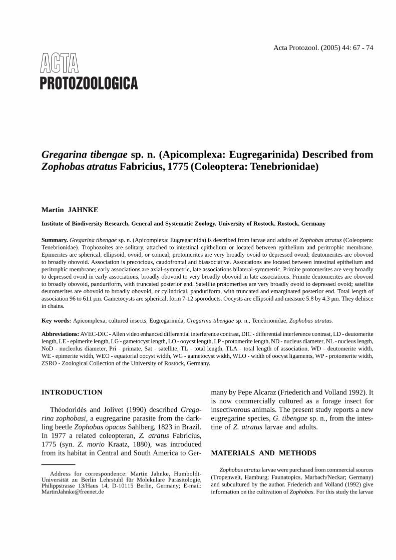
Acta Protozool. (2005) 44: 67 - 74
Gregarina tibengae sp. n. (Apicomplexa: Eugregarinida) Described fromZophobas atratus Fabricius, 1775 (Coleoptera: Tenebrionidae)
Martin JAHNKE
Institute of Biodiversity Research, General and Systematic Zoology, University of Rostock, Rostock, Germany
Summary. Gregarina tibengae sp. n. (Apicomplexa: Eugregarinida) is described from larvae and adults of Zophobas atratus (Coleoptera:Tenebrionidae). Trophozoites are solitary, attached to intestinal epithelium or located between epithelium and peritrophic membrane.Epimerites are spherical, ellipsoid, ovoid, or conical; protomerites are very broadly ovoid to depressed ovoid; deutomerites are obovoidto broadly obovoid. Association is precocious, caudofrontal and biassociative. Assocations are located between intestinal epithelium andperitrophic membrane; early associations are axial-symmetric, late associations bilateral-symmetric. Primite protomerites are very broadlyto depressed ovoid in early associations, broadly obovoid to very broadly obovoid in late associations. Primite deutomerites are obovoidto broadly obovoid, panduriform, with truncated posterior end. Satellite protomerites are very broadly ovoid to depressed ovoid; satellitedeutomerites are obovoid to broadly obovoid, or cylindrical, panduriform, with truncated and emarginated posterior end. Total length ofassociation 96 to 611 µm. Gametocysts are spherical, form 7-12 sporoducts. Oocysts are ellipsoid and measure 5.8 by 4.3 µm. They dehiscein chains.
Key words: Apicomplexa, cultured insects, Eugregarinida, Gregarina tibengae sp. n., Tenebrionidae, Zophobas atratus.
Abbreviations: AVEC-DIC - Allen video enhanced differential interference contrast, DIC - differential interference contrast, LD - deutomeritelength, LE - epimerite length, LG - gametocyst length, LO - ooycst length, LP - protomerite length, ND - nucleus diameter, NL - nucleus length,NoD - nucleolus diameter, Pri - primate, Sat - satellite, TL - total length, TLA - total length of association, WD - deutomerite width,WE - epimerite width, WEO - equatorial oocyst width, WG - gametocyst width, WLO - width of oocyst ligaments, WP - protomerite width,ZSRO - Zoological Collection of the University of Rostock, Germany.
INTRODUCTION
Théodoridès and Jolivet (1990) described Grega-rina zophobasi, a eugregarine parasite from the dark-ling beetle Zophobas opacus Sahlberg, 1823 in Brazil.In 1977 a related coleopteran, Z. atratus Fabricius,1775 (syn. Z. morio Kraatz, 1880), was introducedfrom its habitat in Central and South America to Ger-
many by Pepe Alcaraz (Friederich and Volland 1992). Itis now commercially cultured as a forage insect forinsectivorous animals. The present study reports a neweugregarine species, G. tibengae sp. n., from the intes-tine of Z. atratus larvae and adults.
MATERIALS AND METHODS
Zophobas atratus larvae were purchased from commercial sources(Tropenwelt, Hamburg; Faunatopics, Marbach/Neckar; Germany)and subcultured by the author. Friederich and Volland (1992) giveinformation on the cultivation of Zophobas. For this study the larvae
Address for correspondence: Martin Jahnke, Humboldt-Universität zu Berlin Lehrstuhl für Molekulare Parasitologie,Philippstrasse 13/Haus 14, D-10115 Berlin, Germany; E-mail:[email protected]

68 M. Jahnke
were kept at 23-27°C in the dark. Rolled oats and pieces of apple andcarrot were provided as food items.
Zophobas atratus larvae were eviscerated and intestines weredissected in Ringer’s solution for insects (Seifert 1995) under adissecting microscope. The gregarines were separated from the lumi-nal content and transferred on cover slips using a pipette. They werefixed (2% glutaraldehyde in 0.05% Na-cacodylate buffer, pH 7.4) andhanging drop preparations were made. Measurements were takenusing a micrometer; drawings were made with the aid of a drawingtube.
For permanent preparation wet smears were fixed in AFA (Seifert1995) for 10 min, stained with Masson’s haematoxyline according toRomais (1989) and counterstained in a 2% aqueous eosin solution,dehydrated in ascending ethanol concentrations, 100% n-propanol,Rotihistol (Carl Roth GmbH & Co, Karlsruhe, Germany) and mountedin neutral balsam. Gametocysts were collected from the intestine andthe feces, transferred in Ringer’s solution on cover slips and held inmoist chambers for maturation and dehiscence. The obtained oocystswere fixed in 2% picric acid in Ringer’s solution.
Photomicrographs of live gregarines in Ringer’s solution weretaken using a Zeiss Axiovert (bright-field, phase contrast) and aReichert-Jung Polyvar (DIC). AVEC-DIC photomicrographs of oo-cysts were made using a Nikon Diavert with Hamamatsu PhotonicMicroscopy System C1966.
For morphometric measurements only gregarines from larvae ofthe last and last but one instar were used. Measurements were takenat their widest points, except for deutomerite lengths that werecalculated as differences of total lengths and protomerite lengths.Measurements and indices in micrometers (µm) are given as rangevalues followed by means, standard deviations, and sample sizes inparentheses. For the most part they are those used by Clopton(1998). Terminology of shapes follows that suggested by theSystematics Association Committee for Descriptive Biological Ter-minology (1962).
RESULTS
Taxonomic position: Eugregarinida Léger, 1900sensu Levine et al. (1980); Septatina Lankester, 1885sensu Levine et al. (1980); Gregarinicae Chakaravarty,1960; Gregarinidae Labbé, 1889 sensu Levine (1988),with characters of the genus Gregarina Dufour, 1828sensu Clopton (2002): epimerite conical, button-shaped,globular, or cylindrical; association precocious; oocystsdoliform, navicular, or spherical; in intestine of insects.
Gregarina tibengae sp. n.
Trophozoite (Figs 1-5, 17): Cephalont (Figs 1-5;measurements and indices given in Table 1) attached tohost intestinal epithelium by epimerite, in posterior half ofmesenteron. Solitary, never in association. Earliestcephalonts circular to broadly ovoid (Fig. 1), minorconstriction at protomerite-deutomerite septum. Oldercephalonts more oblong. Epimerite simple, spherical
(Fig. 2), ellipsoid to broadly ovoid (Fig. 5), or cone-shaped (Figs 3, 4); without diamerite; surface smooth,without folds or impressions. Protomerite very broadly todepressed ovoid, tapered anteriorly to junction withepimerite; moderate posterior constriction at protomer-ite-deutomerite septum. Deutomerite shape obovoid tobroadly obovoid, with shallow sinuate margin at half tosecond third of length on each side, rounded to truncatedposteriorly, maximum width at anterior third; septumconcave. Nucleus spherical; typically with single spheri-cal nucleolus, eccentrical in location, young cephalontsoccasionally with 2-3 nucleoli. Sporont (Fig. 17) solitary;free, located between host intestinal epithelium andperitrophic membrane; morphology similar to latecephalonts but epimerite absent; epimerite residue ab-sent.
Association (Figs 6-13, 16, 18-20): Measure-ments and indices given in Table 1). Syzygy early(precocious), caudofrontal; multiple associations of oneprimite with two satellites occasionally observed(Fig. 13), linear multiple association seen only once(Fig. 19); located between host intestinal epithelium andperitrophic membrane, in second third of mesenteron ofhost. Early associations axial-symmetric (Figs 6, 7),morphology of gamonts similar to that of sporonts, someassociations with noticeable small satellites (Fig. 20).Late associations bilaterally symmetric, dorsoventrallyflattened, the side bearing the contact areas betweenprimite protomerite and primite deutomerite as well asbetween primite deutomerite and satellite protomerite is(hereby) defined as “ventral” (on the left in Fig. 9).
Primite: Protomerite very broadly to depressed ovoidin early associations; or broadly to very broadly obovoidin late associations, situated on ventral side; with moder-ate constriction at protomerite-deutomerite septum.Deutomerite obovoid to broadly obovoid; anterior endrounded; septum concave; with shallow to moderatesinuate margin at half to second third of length on eachside (panduriform); maximum width typically in anteriorthird, in some late associations anterior and posteriorthird with similar width, maximum width rarely in poste-rior third, close to the posterior end (never more than10 µm wider than anterior third); truncated posteriorly.Nucleus spherical; abaxial in location; in all parts ofdeutomerite, mostly in anterior half; concealed by granulain late associations; with one spherical nucleolus, eccen-trically located.
Satellite: Protomerite very broadly to depressedovoid; contact area with primite deutomerite ventral;rounded anteriorly; with minor constriction at protomer-

Gregarina tibengae sp. n. 69
Figs 1-15. Gregarina tibengae sp. n. 1-5 - cephalonts; 6-7 - early associations; 8, 10-12 - late associations, ventral view; 9 - late association,lateral view; 13 - multiple association; 14 - dehisced gametocyst with sporoducts; 15 - oocysts.

70 M. Jahnke
ite-deutomerite septum. Deutomerite of early associa-tions obconoid to obovoid; with shallow sinuate margin inmiddle to second third of length; rounded or truncatedposteriorly. Deutomerite of late associations obovoid to
broadly obovoid or cylindrical, often with square outline(Fig. 10); septum concave; with shallow to moderatesinuate margin in middle of length; truncated andemarginated posterior end, sinus elongated anteriorly
Figs 16-21. Gregarina tibengae sp. n. 16 - late association (bright-field, inverted); 17 - sporont (DIC); 18 - early association (DIC); 19 - linearmultiple association (DIC, inverted); 20 - early association (phase-contrast, inverted); 21 - oocysts (AVEC-DIC). Scale bars 5 µm (21); 20 µm(17, 18); 50 µm (19, 20); 100 µm (16).
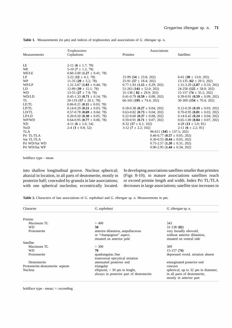
Gregarina tibengae sp. n. 71
into shallow longitudinal groove. Nucleus spherical;abaxial in location, in all parts of deutomerite, mostly inposterior half; concealed by granula in late associations;with one spherical nucleolus; eccentrically located.
In developing associations satellites smaller than primites(Figs 8-10); in mature associations satellites reachor exceed primite length and width. Index Pri TL/TLAdecreases in large associations; satellite size increases in
Table 1. Measurements (in µm) and indices of trophozoites and associations of G. tibengae sp. n.
Trophozoites AssociationsMeasurements Cephalonts Primites Satellites
LE 2-11 (6 ± 1.7; 78)WE 5-10 (7 ± 1.2; 78)WE/LE 0.60-3.00 (1.27 ± 0.41; 78)LP 3-22 (12 ± 4.1; 78) 15-99 (54 ± 23.8; 202) 6-61 (30 ± 13.0; 202)WP 11-35 (20 ± 5.2; 78) 25-91 (57 ± 18.4; 202) 13-135 (62 ± 29.5; 202)WP/LP 1.31-3.67 (1.83 ± 0.44; 78) 0.77-1.93 (1.15 ± 0.29; 202) 1.31-3.29 (2.07 ± 0.33; 202)LD 12-89 (39 ± 15.1; 78) 51-263 (141 ± 52.0; 202) 24-258 (125 ± 58.0; 202)WD 13-55 (27 ± 7.9; 78) 31-138 ( 82 ± 29.9; 202) 15-157 (74 ± 35.2; 202)WD/LD 0.45-1.33 (0.71 ± 0.14; 78) 0.41-0.79 (0.59 ± 0.08; 202) 0.39-0.91 (0.59 ± 0.09; 202)TL 20-119 (57 ± 20.1; 78) 66-343 (195 ± 74.4; 202) 30-309 (154 ± 70.4; 202)LE/TL 0.06-0.21 (0.11 ± 0.03; 78)LP/TL 0.14-0.29 (0.21 ± 0.03; 78) 0.18-0.38 (0.27 ± 0.04; 202) 0.12-0.30 (0.19 ± 0.03; 202)LD/TL 0.57-0.78 (0.69 ± 0.04; 78) 0.63-0.82 (0.73 ± 0.04; 202) 0.70-0.95 (0.81 ± 0.03; 202)LP/LD 0.20-0.50 (0.30 ± 0.05; 78) 0.22-0.60 (0.37 ± 0.08; 202) 0.14-0.42 (0.24 ± 0.04; 202)WP/WD 0.64-0.95 (0.77 ± 0.06; 78) 0.59-0.91 (0.71 ± 0.07; 202) 0.65-1.00 (0.84 ± 0.07; 202)ND 4-11 (6 ± 1.6; 54) 8-32 (17 ± 6.1; 102) 4-28 (13 ± 5.0; 81)NoD 2-4 (3 ± 0.8; 52) 3-12 (7 ± 2.2; 102) 2-11 (6 ± 2.2; 81)TLA 96-611 (345 ± 137.5; 202)Pri TL/TLA 0.46-0.77 (0.57 ± 0.05; 202)Sat TL/TLA 0.30-0.55 (0.44 ± 0.05; 202)Pri WD/Sat WD 0.75-2.57 (1.20 ± 0.31; 202)Pri WD/Sat WP 0.90-2.95 (1.44 ± 0.34; 202)
boldface type - mean
Table 2. Characters of late associations of G. zophobasi and G. tibengae sp. n. Measurements in µm.
Character G. zophobasi G. tibengae sp. n.
PrimiteMaximum TL > 400 343WD 50 31-138 (82)Protomerite anterior dilatation, ampullaceous very broadly obovoid,
or “champignon” aspect, without anterior dilatation,situated on anterior pole situated on ventral side
SatelliteMaximum TL > 300 309WD 70 15-157 (74)Protomerite quadrangular, fine depressed ovoid, striation absent
transversal epicytical striationDeutomerite attenuated posterior end emarginated posterior end
Protomerite-deutomerite septum triangular concaveNucleus ellipsoid, > 30 µm in lenght, spherical, up to 32 µm in diameter,
always in posterior part of deutomerite in all parts of deutomerite,mostly in anterior part
boldface type - mean; >: exceeding

72 M. Jahnke
Tabl
e 3.
Mea
sure
men
ts (
in µ
m)
and
char
acte
rs o
f sp
ecie
s si
mila
r to
G.
tibe
ngae
sp.
n. 1
- G
. am
oji,
2 -
G.
cero
pria
e, 3
- G
. de
cour
ti,
4 -
G.
drag
esco
i, 5
- G
. go
noce
phal
i,6
- G
. inc
lina
ta, 7
- G
. jol
ivet
i, 8
- G
. mac
ulat
a va
r. ba
nule
nsis
, 9 -
G. m
esom
orph
i, 10
- G
. nip
hand
rode
s, 1
1 -
G. s
tein
i, 12
- G
. tib
enga
e
Cha
ract
er1
23
45
67
89
1011
12
Ass
ocia
tion
sT
LA
--
->
100
0>
175
355-
470
--
--
-96
-611
(34
5)
Pri
TL
-?-
200
[> 3
00]
430-
550
78-9
116
0-20
316
5-30
011
5-16
525
0-30
014
3-37
7 (2
33)
108-
187
(147
)66
-343
(19
5)
Pri
WD
-4
5-
-36
-38
63-8
5>
300
-10
8-14
910
2-23
5 (1
46)
13-3
3 (2
0)31
-138
(82
)
Pri
LP/
TL
--
--
0.17
-0.2
20.
17-0
.30
--
0.17
0.17
-0.4
0 (0
.26)
0.10
-0.1
8 (0
.13)
0.18
-0.3
8 (0
.27)
Sat T
L-
?-30
0[>
350
]44
5-54
078
-91
181-
260
150-
400
115-
145
250-
300
102-
347
(218
)84
-141
(11
3)30
-309
(15
4)
Sat W
D-
45
--
33-4
052
-85
--
108-
149
61-2
14 (
114)
11-2
2 (1
5)15
-157
(74
)
Sat
LP/
TL
--
--
0.12
-0.1
80.
11-0
.17
--
0.13
0.15
-0.3
3 (0
.19)
0.10
-0.1
7 (0
.13)
0.12
-0.3
0 (0
.19)
Bila
tera
l sym
met
ryye
sye
sye
sye
sye
sye
sye
sye
sye
sye
sye
sye
s
Cur
ved
no
yes
yes
no
yes
yes
no
yes
yes
yes
no
yes
Mar
gine
of
sinu
ate
sinu
ate
conv
exsi
nuat
esi
nuat
esi
nuat
eco
nvex
sinu
ate
conv
exco
nvex
sinu
ate
sinu
ate
deut
omer
ites
or c
onve
x
Pand
urif
orm
yes
no
no
no
no
no
no
yes
no
no
yes
yes
deut
omer
ites
in
late
ass
ocia
tion
s
Em
agin
ated
pos
teri
orn
on
on
on
on
on
on
on
on
on
on
oye
s
end
of s
atel
lite
deut
omer
ite
Nuc
lei
sphe
rica
lel
lips
oid
sphe
rica
lel
lips
oid
sphe
rica
lsp
heri
cal
elli
psoi
del
lips
oid
sphe
rica
lsp
heri
cal
sphe
rica
lsp
heri
cal
Pri
NL
; Sa
t N
L-
30;
30-
-8-
16;
8-16
20;
2040
-50;
40-
50-
34;
3036
; 36
18;
1832
; 28
Gam
etoc
ysts
Sha
peel
lips
oid
elli
psoi
d-
--
sphe
rica
lsp
heri
cal
-sp
heri
cal
sphe
rica
lel
lips
oid
sph
eric
al
or e
llips
oid
Dia
met
er70
x 5
414
0-20
0-
--
200
150
-20
022
585
× 6
0 -
160
× 10
0214
6-19
1 (1
67)
Ooc
ysts
:
LO
× W
O4.
4 ×
2.3
4 ×
2-
--
6 ×
46
× 3
-5.
6 ×
4.2
8.8
× 5.
54.
9 ×
4.91 ,
5 ×
325.
8 ×
4.3
Bol
dfac
e -
mea
ns;
> -
exc
eedi
ng;
[ ]
- no
t gi
ven
by t
he a
utho
rs,
assu
med
fro
m i
llust
ratio
n; 1
from
Clo
pton
et
al.
(199
1);
2 fr
om G
eus
(196
9)

Gregarina tibengae sp. n. 73
large associations; indices Pri WD/Sat WD and Pri WD/Sat WP constantly decreasing during growth of associa-tion.
Cytology: Cytoplasm brownish; primite cytoplasmalways filled with numerous large vesicles; vesiclesabsent in satellite cytoplasm (Fig. 18); paraglycogeneglanula present in both cells.
Gametocysts (Fig. 14): Roughly spherical; withhyaline gametocyst coat, coat thickness (in Ringer’ssolution) 2-6 µm; located between host intestinal epithe-lium and peritrophic membrane, egested with feces.Gametocysts isolated from intestine: LG (including coat)139-176 (161 ± 9.1, 21), WG 129-173 (153 ± 11.1, 21),WG/LG 0.87-1.0 (0.95 ± 0.04, 21). Gametocysts col-lected from feces: LG 146-191 (167 ± 15.0, 15),WG 173-187 (159 ± 15.2, 15), WG/LG 0.90-1.0(0.96 ± 0.03, 15). Gametocysts collected from freshfeces mature and dehisce within 50-80 h. Oocystsextruded in chains through sporoducts. Sporoducts withwide basis, proximal bulbus and distal tube; length 25-50 µm; usually 7-12 per gametocyst, sometimes moreprimordial but not evaginated.
Oocysts (Figs 15, 21): Ellipsoid, connected in chainsby “ligaments”; uniform in size and shape; LO 5.7-6.2(5.8 ± 0.13, 64), WEO 4.0-4.5 (4.3 ± 0.11, 64), WLO1.7-2.3 (2.1 ± 0.1, 64), WEO/LO 0.68-0.77(0.74 ± 0.02, 64), WLO/LO 0.27-0.39 (0.36 ± 0.02, 64),WLO/WEO 0.38-0.54 (0.48 ± 0.03, 64); cross-sectionroughly circular; two, sometimes numerous, polar oocystresidua present.
Taxonomic summary
Type specimens: Eight syntype slides (19 March2002-17 October 2002) were deposited in the ZSRO(# ZSRO Pro 0001a - Pro 0001h; label: Gregarinatibengae (Apic., Eugregarinida) ex. Zophobas atratusFabricius, 1775 (Col., Tenebrionidae), [syntype], leg. &det. Martin Jahnke, Germany: Rostock, cultured host).
Type host: Zophobas atratus Fabricius, 1775(Tenebrionidae: Tenebrioninae: Tenebrionini); larvae andadults
Symbiotype: One adult host specimen was depos-ited in the ZSRO (# ZSRO Pro 0001i; label: Germany,Rostock, 2003-V-12, leg. & det. Martin Jahnke,Tenebrionidae, Zophobas atratus Fabricius, 1775,cultured, [symbiotype], type host of Gregarina tibengae).Two additional voucher specimens were labeledanalogous and have got collection numbers ZSRO Pro0001j - ZSRO Pro 0001k.
Infection site: Mesenteron.Prevalence: High; 83% (n=30).Type locality: Germany, RostockOther localities: Germany: Hamburg, Marbach
(Neckar), Tübingen; cultured.Etymology: The specific epithet refers to the Swabian
word “Tibenga” for Tübingen were the author found thisgregarine for the first time.
DISCUSSION
The present gregarine is the second eugregarinespecies described from Zophobas. A comparision of thecharacters of Gregarina zophobasi Théodoridès etJolivet, 1990 and G. tibengae is given in Table 2. In bothspecies the gamonts of late associations are alike in size.However, G. zophobasi can be distinguished fromG. tibengae based on its elongated gamonts, the “champi-gnon” view of primite protomerites, the attenuated pos-terior end of deutomerite, the triangular shape of theprotomerite-deutomerite septum, and the ellipsoid nucleuswhich is always situated in the posterior part of deu-tomerite. Of the 56 species of Gregarina known fromTenebrionid beetle hosts, the following 11 species exhibita similar primite protomerite: G. amoji Datta, Ghoseet Haldar, 1991, G. ceropriae Théodoridès et Desportes,1967, G. decourti Théodoridès, Desportes et Jolivet,1975, G. dragescoi Théodoridès et Desportes, 1966,G. gonocephali, Obata, 1953, G. inclinata Hoshide,1979, G. joliveti Théodoridès, 1958, G. maculata Léger,1904 var. banyulensis Théodoridès, 1955, G. mesomorphiDevdhar et Deshpande, 1971, G. niphandrodes Clopton,Percival et Janovy, 1991, and G. steini Berndt, 1902.A comparision of these species with G. tibengae is givenin Table 3. The associations of all species are bilateral-symmetric and most are slightly curved, similar tothose of G. tibengae. Gregarina decourti, G. joliveti,G. mesomorphi, and G. niphandrodes can bedestinguished from G. tibengae based on their convexmargins of deutomerites. In G. gonocephali only earlyassociations have deutomerites with sinuate marginswereas those of late associations are convex. Grega-rina amoji, G. ceropriae, G. dragescoi, G. inclinata,G. maculata var. banyulensis, and G. steini exhibitsinuate margins of deutomerites but only G. amoji,G. maculata var. banyulensis, and G. steini possesspanduriform deutomerites similar to those of G. tibengae.Gregarina amoji lack the emarginated posterior end

74 M. Jahnke
of the satellites, moreover, G. amoji has ellipsoidgametocysts. Gregarina ceropriae, G. maculata var.banyulensis, and G. steini can be distinguished from G.tibengae based on their attenuate deutomerites. Grega-rina inclinata possess characteristic unciform associa-tions different from G. tibengae. In comparision withG. tibengae the associations of G.dragescoi are plainlylarger (maximum TLA 611 µm versus >1000 µm). Thecombination of diagnostic characters of the describedgregarine, especially associations possessing bilateralsymmetry, panduriform margin of deutomerites, andemarginated posterior end of satellite deutomerite, seemto differentiate it from known Gregarina species ofTenebrionidae.
Acknowledgements. I thank Raimund Apfelbach (University ofTübingen), Willfried Kröger (University of Rostock) and Willi Maile(Freising) for their kind support in microscopy and Susanne Braun(Tübingen) and Axel Sonnemann (Hohenstein) for obtaining hostlarvae for this study.
REFERENCES
Berndt A. (1902) Beitrag zur Kenntnis der im Darme der Larven vonTenebrio molitor lebenden Gregarinen. Arch. Protistenk. 1: 375-420
Clopton R. E. (1998) Revision of the genus Torogregarina anddescription of Torogregarina sphinx n. sp. (Apicomplexa:Eugregarinida) from a Missouri River Bank Beetle, Bembidionlaevigatum (Coleoptera: Carabidae), in southeastern Nebrasca.J. Parasitol. 84: 823-827
Clopton R. E. (2002) Phylum Apicomplexa 1970: OrderEugregarinorida Léger, 1900. In: Illustrated Guide to the Protozoa(2 ed.), (Eds. J. J. Lee, G. Leedale and P. C. Bradbury). Societyof Protozoologists, Lawrence, Kansas, 205-288
Clopton R. E., Percival T. J., Janovy J. (1991) Gregarina niphandrodesn. sp. (Apicomplexa: Eugregarinorida) from adult Tenebrio molitor(L.) with oocyst descriptions of other Gregarine parasites of theYellow Mealworm. J. Protozool. 38: 472-479
Datta S. C., Ghose S., Haldar D. P. (1991) Studies in septategregarines (Apicomplexa: Sporozoea) from insects of West Ben-gal: six new species of Hirmocystis, Gregarina, Stenophora andHyalosporida. Rec. Zool. Surv. India 89: 219-231
Received on 12th June, 2003; revised version on 14 September, 2004;accepted on 15th September, 2004
Devdhar M. J., Deshpande S. B. (1971) On two new gregarines ofthe genus Gregarina from Mesomorphus velliger, Blanc(Coleoptera - Tenebrionidae) from Dharwar, India. Indian Zool.2: 27-34
Friederich U., Volland W. (1992) Futtertierzucht (2 ed). Ulmer,Stuttgart
Geus A. (1969) Sporentierchen, Sporozoa: die Gregarinida der land-und süßwasserbewohnenden Arthropoden Mitteleuropas. In: DieTierwelt Deutschlands und der angrenzenden Meeresteile nachihren Merkmalen und ihrer Lebensweise, (Ed. F. Dahl). GustavFischer, Jena
Hoshide K. (1979) Notes on the gregarines in Japan 10. B. Fac. Educ.Yamaguchi Univ. 29: 31-75
Levine N. D. (1988) The Protozoan Phylum Apicomplexa, Vol. 1.CRC Press, Boca Raton, Florida
Levine N. D., Corliss J., Cox F., Deroux G., Grain J., Honigberg B.,Leedale G., Loeblich J., Lom J., Lynn D., Merinfeld E., Page F.,Poljansky G., Sprague V., Vavra J., Wallace F. (1980) A newlyrevised classification of the protozoa. J. Protozool. 27: 37-58
Obata K. (1953) Reports on some gregarines from Japanese insects(1). J. Sci. Hiroshima Univ. (B. 1 Zool.) 14: 1-34
Romais B. (1989) Mikroskopische Technik (17 ed). (Ed. P. Böck).Urban & Schwarzenberg, München
Seifert G. (1995) Entomologisches Praktikum (3 ed). Thieme, StuttgartSystematics Association Committee for Descriptive Biological Ter-
minology (1962) II and IIa. Terminology of simple symmetricalplane shapes (Carts 1, la). Taxon 11: 145-156, 245-247
Théodoridès J. (1955) Contribution a l’étude des parasites etphorétiques de Coléptères terrestres. Vie Milieu 1 (Supp1. 4):1-310
Théodoridès J. (1958) Deux nouvelles Eugrégarines parasites desHegeter (Coleoptera Tenebrionidae) des ÎIes Canaries: Gregarinajoliveti n. sp. (Gregarinidae) et Lophocephalus fernandezi n. sp.(Stylocephalidae). Ann. Sci. Nat. Zool. (11e série) 20: 105-112
Théodoridès J., Desportes I (1966) Quelques Grégarines de Coléptèresdu Laos. Protistologica 2: 53-58
Théodoridès J., Desportes I (1967) Quatre nouvelles Grégarinesparasites de Coléptères Tenebrionides du Laos. Protistologica3: 147-153
Théodoridès J., Jolivet P. (1990) Eugrégarines de Co1éoptères duBrézil. Description de trois espéces nouvelles et notes biologiques.Bol. Mus. Para. Emilio Goeldi (sér. Zool.) 6: 17-58
Théodoridès J., Desportes I, Jolivet P. (1975) Grégarines de 1aThaï1ande. Ann. Parasit. Hum. Comp. 50: 145-159
Acta Protozool. (2005) 44: 75 - 80
Filamoeba sinensis sp. n., a second species of the genus Filamoeba Page,1967, isolated from gills of Carassius gibelio (Bloch, 1782)
Iva DYKOVÁ1,2, Hana PECKOVÁ1, Ivan FIALA1,2 and Helena DVO�ÁKOVÁ1
1Institute of Parasitology, Academy of Sciences of the Czech Republic; 2Faculty of Biological Sciences, University of SouthBohemia, ������ ��� ����, Czech Republic
Summary. Filamoeba sinensis, a new species of amphizoic amoeba isolated from the gills of Carassius gibelio (Bloch, 1782) sampled inHubei Province, China, is described. Both morphology and SSU rRNA gene sequences were employed to compare this species withFilamoeba nolandi Page, 1967, the type and only species of the genus. Trophozoites differ from F. nolandi in having robust multipolarlybranched pseudopodia. A less pronounced hyaloplasmic zone produces a lower number of filiform subpseudopodia. The greatest dimensionsof extended trophic forms reach 40-50 µm. Cysts, with a relatively thick and homogeneous wall, are smooth and free of pores. The diameterof rounded cysts ranges from 14 to 20 µm. Cristae of mitochondria are tubular. They are arranged in parallel or irregular mutual positions,their anastomoses or branches were not observed. Phylogenetic analysis revealed a close relationship to F. nolandi.
Key words: amphizoic amoebae, Filamoeba sinensis sp. n., morphology, SSU rDNA, phylogeny.
INTRODUCTION
Despite the widely recognised diversity of organismscurrently assigned to Amoebozoa Lühe, 1913 sensuCavalier-Smith (2003), diagnostic criteria are not welldefined within all groups of amoebae. Morphologicalfeatures that could possibly differentiate the taxa havenot been adequately described in “neglected” groups ofamoebae. It is most probably due to the exceptionalvariability of these protists, methods of observation
available in amoeba research as time progressed and,possibly, also due to the rare frequency of many findings.As a consequence of diagnostic difficulties, many spe-cies have not been reported again since their descrip-tions and some were never accommodated tosuprageneric taxons. Molecular characterisation of amoe-bae based mostly on SSU rRNA gene sequences hasbecome gradually an integral part of the description ofnew species. For synthesis of both morphological andmolecular approaches in amoeba studies, molecularcharacterisations of type strains are of a great value,since they facilitate comparisons and serve as a basis forstudies of intra-amoebozoan phylogeny.
A freshwater amoeba described as Filamoebanolandi Page, 1967 was isolated from shallow water on
Address for correspondence: Iva Dyková, Institute ofParasitology AS CR, Branišovská 31, 370 05 ���������� ���,Czech Republic; Fax: 0042-385310388; E-mail: [email protected]

76 I. Dyková et al.
the sandy shore of Little Deer Lake in Itasca County,Minnesota. Prior to its description in 1964, Page depos-ited the strain in the former Culture Collection of Algaeand Protozoa (CCAP), presently the UK National Cul-ture Collection (UKNCC). Page (1967) did not settle thesystematic position of this organism in the original de-scription. In his publications “An Illustrated Key toFreshwater and Soil Amoebae” and “A New Key toFreshwater and Soil Amoebae” Page (1976, 1988) placedthe genus Filamoeba in the family EchinamoebidaePage, 1975. Phylogenetic analyses published thus far(Amaral Zettler et al. 2000, Bolivar et al. 2001, Peglaret al. 2003) included the SSU rRNA gene sequence ofAmerican Type Culture Collection (ATCC 50430) strainof F. nolandi (NC-AS-23-1) and not of that depositedin UKNCC as a type strain of the species. Contrary tothe type strain, the ATCC strain denoted as F. nolandi(NC-AS-23-1) is of marine origin. It was isolated froma sediment core 23 nautical miles off coast of Wilmington,NC, in 1993.
This paper presents a new amoeba species, thesecond one of the genus Filamoeba Page, 1967, whichwas isolated from a freshwater fish host.
MATERIALS AND METHODS
The strain denoted as CH26, was isolated from gill tissue ofCarassius gibelio (Bloch, 1782) sampled in Hubei Province, China, inApril 2002. It was recovered from decomposing tissue stored for14 days in eppendorf tubes and washed repeatedly with sterile tapwater in order to separate spores of Myxobolus sp. detected in gilltissue of the opposite gill arch. A homogeneous population of tropho-zoites (passage No. 8), cultured on non-nutrient agar wascryopreserved in September 2002. A clone was derived in Novemberof the same year from passage No. 13.
Amoeba trophozoites were observed in hanging drop prepara-tions using an Olympus BX51 microscope equipped with theNomarski DIC system. For ultrastructural studies, trophozoites andcysts were fixed on agar plates with 2.5% cacodylate buffered glut-araldehyde, then pelleted via centrifugation, postfixed with cacody-late buffered 1% osmium tetroxide, dehydrated with series of ac-etones and embedded in Spurr’s resin. The same methods of culturing,observation and harvesting were applied to the strain of Filamoebanolandi. It was ordered for purposes of comparison from UKNCCwhere it is listed in the type culture collection as CCAP 1526/1 strain.
Our original clone used in this study was cryopreserved and isstored in the culture collection of the Institute of Parasitology, Acad-emy of Sciences of the Czech Republic, ���������� ���.
DNA isolation, amplification and sequencing. Morphologicalcharacterisation of clones derived from both strains (CH26/I andCCAP 1526/1) was completed with phylogenetic analysis of small-subunit ribosomal RNA gene sequences. DNA was extracted from the
cell pellets using the DNeasyTM Tissue kit, according to the protocolof the manufacturer (Qiagen). The SSU rRNA genes of both strainswere amplified by polymerase chain reaction (PCR) using universaleukaryotic primers ERIB 1 (5´ - ACCTGGTTGAT CCTGCCAG -3´) and ERIB 10 (5´ - CTTCCGCAGGTTCACCTACGG - 3´)(Barta et al. 1997). Each 25 µl reaction included 50 ng of genomicDNA, 10 pmol of each oligonucleotide, 250 ìM of each dNTP, 2.5 µl10 × PCR Buffer (Top-Bio, Czech Republic), and 1 unit of Taq-Purple polymerase (Top-Bio, Czech Republic). The mixture waspreheated 5 min at 95°C. Thermocycling conditions were as follows:5 cycles consisting of 94°C for 1 min, 44°C for 1.5 min and 72°C for2 min, 25 cycles consisting of 94°C for 1 min, 48°C for 1.5 min and72°C for 2 min, then a 10 min incubation at 72°C. The PCR productswere isolated from agarose and cloned into the pCR® 2.1 TOPOcloning vector (Invitrogen). Vectors with inserts were sequenced inboth directions using CEQ DTCS Dye Kit (Beckman Coulter) on theCEQ TM 2000 automatic sequencer (Beckman Coulter).
Alignments and phylogenetic analyses. The alignments weredone using the Clustal_X program (Thompson et al. 1997) withvarious alignment parameters. Ambiguously aligned regions wereexcluded. The SSU rRNA gene sequences from the newly sequencedFilamoeba strains were aligned against sequences of the ATCC strainof Filamoeba nolandi and 37 eukaryotic taxa available throughGenBank, including representatives of Amoebozoa sensu Bolivaret al. (2001). Hexamita inflata was selected as an outgroup. The finalalignment consisted of 1615 characters (1022 characters were parsi-mony informative).
The phylogenetic relationships between taxa were determinedusing maximum parsimony (MP) and maximum likelihood (ML)methods carried out in the program package PAUP*, version 4.0b8(Swofford 2001). The MP analysis was performed using a heuristicsearch with random addition of taxa. Gaps were treated as missingdata. Transition/transversion/ (Ts:Tv) ratio was 1:2. For ML, thelikelihood ratio test (LRT) implemented in the Modeltest v. 3.06(Posada and Crandall 1998) was used to determine the best model ofevolution. ML analyses were done with the TrN+G model of evolu-tion selected by LRT. Clade support was assessed by bootstraping(MP, 1000 replicates; ML, 500 replicates).
RESULTS
Description of Filamoeba strain CH26. Thestrain under study was characterised by an irregular,multipolarly branched, uninucleate trophozoites (Fig. 1).Robust pseudopodia were pointed, non-anastomosingsubpseudopodia were filiform and up to 12 µm long. Thegreatest dimensions of locomotive and extended formswere 40-50 µm. A rounded or lobe-shaped nucleus wasmostly difficult to distinguish in Nomarski DIC. Thecytoplasm usually contained about 10 contractile vacu-oles. Floating forms were rounded. Cysts (Fig. 3) weresmooth, rounded, ovoid, irregularly ovoid or reniformwith homogeneous and rather thick wall that lackedpores and closely applied to the amoeba plasmalemma.

New Filamoeba species 77
Figs 1-10. Morphology of strains of Filamoeba spp. 1 - trophozoites, representatives of Filamoeba sinensis sp. n., strain CH26.2 - trophozoites, representatives of Filamoeba nolandi Page, 1967, type strain from UKNCC (CCAP1526/1). 3 - cysts of F. sinensis formedfrom trophozoites in groups, note their irregular shape. 4, 5 - ultrastructure of F. sinensis trophozoites in two different levels of sectioningshow a lobular nucleus, rounded nucleolus, several vacuoles in the cytoplasm and mitochondria sectioned in various planes. 6 - the surface oftrophozoite of F. sinensis. 7 - mitochondria of F. sinensis in cross section. 8 - roughly longitudinal sections through mitochondria showing theshape and arrangement of their cristae. 9 - electron micrograph of mature cyst of F. sinensis. 10 - detail of cyst wall structure of F. sinensis. Scalebars: 20 µm (1-3); 1 µm (4, 5, 9); 500 nm (6, 8); 200 nm (7, 10).

78 I. Dyková et al.
The diameter of rounded cysts was 14-20 µm, thegreatest dimension of oval cysts was 37 µm. The outlineof cysts was evidently modified by close contact withneighbouring ones, i.e. by the space available for eachcysts, which was observed also in old cultures.
At the ultrastructural level (Figs 4-6), trophozoiteswere truly “naked”. The organic layer coating thesurface membrane was very thin, almost invisible(Fig. 6). The rounded as well as lobe-shaped nucleicontained rounded nucleoli (Figs 4, 5). Numerous mito-chondria, mostly of elongated shape were randomlydistributed within the cytoplasm of trophozoites. Theyhad tubular cristae (Fig. 7) situated irregularly or parallelto each other (Fig. 8). Neither cross-, nor longitudinalsections through mitochondria revealed anastomoses orbranches of cristae. The cyst wall was homogeneous(Figs 9, 10). The nucleus of cysts sectioned at variouslevels was mostly rounded, with the diameter about3 µm.
Based on comparison with the true type strain ofF. nolandi (CCAP 1526/1) (Fig. 2), which we hadcultured under identical conditions, the strain CH26differed morphologically having robust multipolarlybranched pseudopodia, less pronounced hyaloplasmiczone and a lower number of contractile vacuoles. Detailsof ultrastructure were not given in the original descrip-tion of F. nolandi. The arrangement of tubular cristae inmitochondria of F. sinensis showed no signs of ramifi-cation.
SSU rRNA data and phylogenetic analysis ofFilamoeba strains. The complete SSU rRNA gene se-quences obtained from strains CH26 and CCAP 1526/1have been deposited in the EMBL/GenBank databaseunder accession numbers AY714369 and AY714368respectively. They matched with the sequence ofF. nolandi (AF293896) available in GenBank, but stillcould not be identified with it. The sequence of SSUrRNA gene of our strain CH26 is 1839 bp. The GCcontent is 50.73%. A sequence of the same length butdiffering in the GC content (49.59%) was obtained forthe F. nolandi strain CCAP 1526/1. Similarities basedon comparisons of complete sequences of SSU rRNAgene were as follows: 96.3% for strain CH26 andF. nolandi type strain CCAP 1526/1; 97.1% whenCH26 and F. nolandi ATCC strain NS-AS-23-1wascompared, 97.7% for F. nolandi ATCC strain NS-AS-23-1 and F. nolandi CCAP strain 1526/1.
As revealed by phylogenetic (ML) analysis (Fig. 11),two Filamoeba strains sequenced in this study plus
ATCC strains of F. nolandi form monophyletic group(supported by 100% bootstrap) that is independent ofGymnamoebia sensu stricto according Bolivar et al.(2001) and of LH, V and PV lineages sensu Peglar et al.(2003). Gephyramoeba sp. clusters with Filamoebastrains with a bootstrap support lower than 50%. Withinthe group of Filamoeba strains, the true type strain ofF. nolandi (CCAP 1526/1) is closely related to theATCC strain of F. nolandi of marine origin, while ourCH26 strain branches out of these two strains. The MPanalysis resulted in three trees with the same length(8701 steps). The phylogenetic position of Filamoebastrains as well as relation to Gephyramoeba sp. was thesame as obtained in ML.
Tentative assignment of strain CH26 to the genusFilamoeba Page, 1967, araising from comparison ofmorphological data available on freshwater naked amoe-bae, was supported by results of phylogenetic analyses.The SSU rRNA gene sequence we have obtained forthe type strain of F. nolandi Page, 1976 complementedmolecular data on Filamoeba spp. represented to dateby the one sequence of the ATCC strain of F. nolandi,which contrary to the type species is of marine origin.
Details distinguishing the strain CH26 from the typestrain of F. nolandi Page, 1967 together with results ofphylogenetic analyses based on SSU rRNA gene se-quences allowed to establish the new species, F. sinensis,the second one within the genus.
Filamoeba sinensis sp. n.
Origin of type material: Gills of the Prussian carp,Carassius gibelio (Bloch, 1782) (Cypriniformes: Cyp-rinidae).
Type locality: Fish farm in Hubei Province, China.Type material: Photosyntypes (light micrographs),
nos. 13870-13891, transmission electron micrographs,nos. 17151-17165 and 17251-17275, and cryopreservedculture (clone CH26/I), deposited in the Institute ofParasitology, Academy of Sciences of the Czech Repub-lic, ���������� ���.
Etymology: The species name is given according tothe geographic origin of fish host. It was derived fromLatin adjective “Chinese”.
Diagnostic summary. Trophozoites uninucleate withnarrow peripheral hyaloplasmic zone, robust multipolarlybranched pseudopodia and numerous contractile vacu-oles; greatest dimensions not exceeding 50 µm; mito-chondria with tubular cristae arranged in parallel orirregular mutual positions. Cysts rounded (14-20 µm in
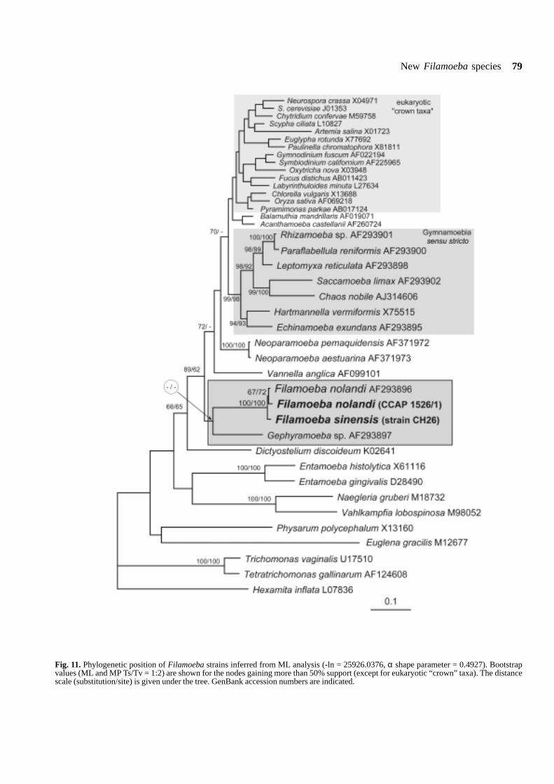
New Filamoeba species 79
Fig. 11. Phylogenetic position of Filamoeba strains inferred from ML analysis (-ln = 25926.0376, α shape parameter = 0.4927). Bootstrapvalues (ML and MP Ts/Tv = 1:2) are shown for the nodes gaining more than 50% support (except for eukaryotic “crown” taxa). The distancescale (substitution/site) is given under the tree. GenBank accession numbers are indicated.

80 I. Dyková et al.
diameter) or ovoid with homogeneous wall free of pores.F. sinensis differs from type species of the genus inmorphology of trophozoites and SSU rRNA gene se-quence.
DISCUSSION
Tentative generic diagnosis of amoeba under study,morphology of which was unique among amoebae wehave isolated from fish, was possible thanks to thoroughoriginal description of F. nolandi (Page 1967), supple-mented with several line drawings of good quality anddocumentation in monographs by Page (1988, 1991).Description of the type species of the genus FilamoebaPage, 1967 was based on light microscopy and someunpublished observations in TEM (Page 1967). Foralmost 40 years, F. nolandi was the unique speciesrepresenting the genus. The number of reported isolateswas surprisingly low and most of them were not docu-mented (Page 1976, Coutinho Salazar 1982, Ariza Astolfiet al. 1988). This seems to be the reason why inphylogenetic studies dealing with relationships amongamoebae, one only SSU rDNA sequence of Filamoebasp. (ATTC strain) was used.
Amaral Zettler et al. (2000), who discovered thatdistance and likelihood methods united Filamoebanolandi and Gephyramoeba sp., also discovered con-sistent sister relationship of Acanthamoeba - Balamuthiawith Gephyramoeba - Filamoeba. The bootstrap sup-port for common ancestry of both couples was marginalin their analyses. On the basis of an “unexpectedrelationship” between Gephyramoeba and Filamoeba,and on the basis of morphological data, Amaral Zettler etal. (2000) suggested reconsideration of the currentsystematic placement of both genera. The strong unionof Gephyramoeba and Filamoeba characterised byAmaral Zettler et al. (2000), based on distance andlikelihood methods, weakened in studies by Bolivar et al.(2001) and Peglar et al. (2003). Also in our study, thisunion is supported by low bootstrap only. In accordancewith Peglar et al. (2003), representatives of four lin-eages of gymnamoebae included in our analysis are notrelated to the cluster of Filamoeba strains.
In the overview of morphological features importantin amoeboid taxa subjected to phylogenetic analysis,tubular and branching cristae of mitochondria werementioned by Amaral Zettler et al. (2000) in F. nolandiand Gephyramoeba sp. To the best of our knowledge,
this feature was not documented in any ultrastructuralstudy. The branching pattern of mitochondrial cristae ofF. sinensis, if any, can hardly be identified with typicalbranching pattern as known, e.g. in Acanthamoeba spp.
Acknowledgements. This study formed part of the Project No. 206/00/0265 supported by the Grant Agency of the Czech Republic. Theauthors are grateful for financial support received also from theresearch project of the Institute of Parasitology AS CR (Z6022909)and Ministry of Education Youth and Sports (Project No. MSM12300003).
REFERENCES
Amaral Zettler L. A., Nerad T. A., O’Kelly C. J., Peglar M. T.,Gillevet P. M., Silberman J. D., Sogin M. L. (2000) A molecularreassessment of the leptomyxid amoebae. Protist 151: 275-282
Ariza Astolfi C., Guevara Benitez D. C., Ubeda Outiveros J. M.,Cutellas Barrios C. (1988) Aislamiento de Filamoeba nolandi(Page, 1967) en una piscina de Sevilla (España). Rev. Iber.Parasitol. 48: 127-131
Barta J. R., Martin D. S., Liberator P. A., Dashkewitz M., AndersonJ. W., Deighner S. D., Elbrecht A., Perkins-Barrow A., Jenkins M.C., Danforth H. D., Ruff M. D., Profous-Juchelka H. (1997)Phylogenetic relationships among eight Eimeria species infectingdomestic fowl inferred using complete small subunit ribosomalDNA sequences. J. Parasitol. 83: 262-271
Bolivar I., Fahrni J. F., Smirnov A., Pawlowski J. (2001) SSU rRNA-based phylogenetic position of the genera Amoeba and Chaos(Lobosea, Gymnamoebida): the origin of Gymnamoebae revis-ited. Mol. Biol. Evol. 18: 2306-2314
Cavalier-Smith T. (2003) Protist phylogeny and the high-level clas-sification of Protozoa. Eur. J. Protistol. 39: 338-348
Coutinho Salazar H., Moura H., Tramontani Ramos R. (1982)Isolamento de amebas de vida libre a partir de agua mineralengarrafada. Rev. Saude Publ. 16: 261-267
Page F. C. (1967) Filamoeba nolandi n. g., n. sp., a filose amoeba.Trans. Amer. Micr. Soc. 4: 405-411
Page F. C. (1976) An Illustrated Key to Freshwater and SoilAmoebae. Frashwater Biological Association, Ambleside, UK
Page F. C. (1988) A New Key to Freshwater and Soil Amoebae.Freshwater Biological Association, Ambleside, UK
Page F.C. (1991) Nackte Rhizopoda. Nackte Rhizopoda undHeliozoea, (Eds F. C. Page and F. J. Siemensma). Gustav FischerVerlag, Stuttgart, New York
Peglar M. T., Amaral Zettler L. A., Anderson O. R., Nerad T. A.,Gillevet P. M., Mullen T. E., Frasca Jr. S., Silberman J. D. O’KellyC. J., Sogin M. L. (2003) Two new small-subunit ribosomal RNAgene lineages within the subclass Gymnamoebia. J. Eukaryot.Microbiol. 50: 224-232
Posada D., Crandall K. A. (1998) Modeltest: testing the model ofDNA substitution. Bioinformatics 14: 817-818
Swofford D. L. (2001) PAUP*: Phylogenetic Analysis Using Parsi-mony. Version 4.0b8. Sinauer Associates, Sunderland, MA
Thompson J. D., Gibson T. J., Plewniak F., Jeanmougin F., HigginsD. G. (1997) The CLUSTAL_X windows interface: flexiblestrategies for multiple sequence alignment aided by qualityanalysis tools. Nucl. Acid Res. 25: 4876-4882
Received on 4th August, 2004; revised version on 16th September,2004; accepted on 19th September, 2004

Acta Protozool. (2005) 44: 81 - 84
Effect of Hormones on the Concentration of a Digoxin-like Material inTetrahymena
György CSABA1, Péter KOVÁCS1 and Éva PÁLLINGER2
1Department of Genetics, Cell and Immunobiology, Semmelweis University; 2Molecular Immunological Research Groupof the Hungarian Academy of Sciences, Budapest, Hungary
Summary. The unicellular Tetrahymena pyriformis contains digoxin-like materials (digoxin, digitoxin, digoxigenin). The effect of two amino-acid-type hormones (histamine and serotonin) and a peptide hormone (insulin) was studied on the level of the hormone-like steroid, digoxin,in the concentrations of 10-6 and 10-9 by using immunocytochemistry and flow cytometric and confocal microscopic analysis. Each hormoneelavated significantly the digoxin concentration at the end of the 30 min treatment. As Tetrahymena was maintained in a tryptone-yeastextract medium during the treatment, it seems to be likely that steroid synthesis was enhanced. The most effective was histamine, the effectof which was significantly more expressed than that of the others and the less effective (however significant), was serotonin. The resultscall attention to hormonal interactions inside the unicellular organism and on the possibility of mutual intercellular influences, consideringthe low effective hormone concentration which was needed for the striking effect.
Key words: digoxin, evolution, histamine, hormonal interactions, insulin, serotonin, Tetrahymena.
INTRODUCTION
Many members and characteristics of the hormonalsystem of higher ranked animals can be demonstratedin the unicellular Tetrahymena. It contains receptor-likestructures in the plasma membrane and cytosol(Csaba1980, 1985; Christopher and Sundermann 1995;Christensen et al. 2003), hormones similar to mamma-lian ones (LeRoith et al. 1980, 1983; Lenard 1992;
K�hidai and Csaba 1995; K�hidai et al. 2000, 2002a, b)and signal transduction pathways (Kuno et al. 1979;Muto et al. 1983; Kovács and Csaba 1990, 1997) usingthe same components which are working in mammals.Hormones, received by the receptors can influence a lotof physiological processes of the unicellular animal, e.g.cell division, phagocytosis, chemotaxis etc. (Wheatley etal. 1993, Csaba 2000), nevertheless our knowledge onthe interactions of these hormones inside the Tetrahy-mena is very scarce. Recently we studied the effect ofthree amino acid- or peptide-type hormones (which arepresent in Tetrahymena) on the concentration of epider-mal growth factor (EGF), a peptide hormone, alsopresent in Tetrahymena (Csaba et al. 2004). As this
Short communication
Address for correspondence: György Csaba, Departmentof Genetics, Cell and Immunobiology, Semmelweis University ofMedicine, Nagyvarad ter 4, 1445 Budapest, Hungary; E-mail:[email protected]

82 G. Csaba et al.
was demonstrated, in the present experiment the effectof the same hormones on digoxin, a hormone-like steroidmolecule, is studied.
MATERIALS AND METHODS
Populations of Tetrahymena pyriformis GL were grown axeni-cally in a 0.1% yeast extract containing 1% tryptone medium at 28°C.In the experiments cells in the mid-exponential growth phase (48hcultures; ~ 5 × 10⁵ cells/ml) were used. The cells were washed withfresh culture medium and harvested by centrifugation at ~ 400 g for3 min at room temperature, and were adjusted to ~ 5 × 10⁵ cells/mlwith fresh culture medium. Five samples were prepared for eachmeasurement. The samples contained Tetrahymena in fresh mediumand the hormones (except control) in 10-6 and 10-9 M concentrationsfor 30 min. The hormones were the following: histaminedihydrochloride (Sigma, St. Louis, USA); serotonin HCl (Sigma) orinsulin (Semilente MC, Novo, Copenhagen, Denmark). After treat-ment the cells were studied by flow cytometry and confocal micros-copy.
Flow cytometric analysis
Samples of cells were fixed with 4% paraformaldehyde solution(dissolved in pH 7.2 PBS) for 5 min, and then washed twice in washbuffer (0.1% BSA; 20 mM Tris-HCl; 0.9% NaCl; 0.05% NonidetNP-40; pH 8.2). To block nonspecific binding of antibodies the cellswere treated with blocking buffer (1% BSA in PBS) for 30 min atroom temperature. Aliquots from cell suspensions (50 ml) weretransferred into tubes, and 50 ml primary antibody (monoclonal anti-digoxin developed in mouse ascites; [Sigma, D-8156, Clone Di-22;dilution 1:200 in antibody buffer (1% BSA in wash buffer)] wasadded for 30 min at room temperature. Negative controls were carriedout with 50 ml PBS containing 10 mg/ml BSA instead of primaryantibody.
After washing four times with wash buffer to remove excessprimary antibody the cells were incubated with secondary antibody(FITC-labeled monoclonal anti-mouse IgG developed in goat; Sigma,St. Louis USA; dilution 1: 50 with antibody buffer) for 30 min atroom temperature.
For controlling the specificity, autofluorescence of the cells andaspecificity of the secondary antibodies were detected. The measure-ment was done in a FACSCalibur flow cytometer (Becton Dickinson,San Jose, USA), using 25,000 cells for each measurement. In the cellpopulations the digoxin content (concentration) had been compared.For the measurement and analysis CellQuest Pro program was used.The numerical comparison of detected values was done by the com-parison of percentual changes of geometric mean channel values to theappropriate control groups by using Origin program and Studentt-test.
Confocal microscopic analysis
After the flow cytometric analysis the cells were subjected toconfocal microscopic analysis in a BioRad MRC 1024 confocal laserscanning microscope, equipped with krypton-argon mixed gas-laseras a light source, at an excitation wavelength of 480 nm line. Three
independent series of experiments were carried out with identicalresults.
RESULTS AND DISCUSSION
Tetrahymena is able to synthesize steroid molecules.One of them is the tetrahymanol for the synthesis ofwhich squalene tetrahymanol synthase enzyme is re-sponsible (Saar et al. 1991). The unicellular eukaryotesalso contain 20a-hydroxysteroid dehydrogenase (Inazuet al. 1994)). In addition to the synthesized steroidhormones (as testosterone and estradiol: Csaba et al.1998) digoxin, a cardioactive glycoside can be found,which also has a steroid structure. In earlier experiments(Kovács et al. 1998) the presence of digoxin, digitoxinand digoxigenin was demonstrated in Tetrahymena. Themonoclonal antibody used in the present experimentswas produced against digoxin however, it had a highaffinity also to digoxigenin.
The flow-cytometric results unanimously show thatthe hormones used for treatment significantly elevatedthe concentration of the digoxin-like materials in Tet-rahymena (Table 1). The confocal microscopic picturessupport the flow cytometric quantitative results. How-ever, there is no difference between the control andhormone treated cells in the localization of the digoxin-like material (Fig. 1).
As simple tryptone-yeast extract medium was admin-istered for maintaining Tetrahymena it is not likely thatcomplete digoxin was taken up from it. This means thatthe (amino-acid and peptide) hormones which are alsophysiologically present in Tetrahymena, enhanced thesteroid synthesis. The most effective hormone washistamine which was similarly effective in the EGF-experiments done earlier (Csaba et al. 2004). However,in these earlier experiments serotonin was as effectiveas histamine and now serotonin was the less active, andin the present case the difference between the effects ofhistamine and the other two hormones is significant. Thisemphasizes the important role of histamine at this lowlevel of phylogeny, which is supported by the presenceof histamine and histamine synthesizing enzyme (HDC)genes already in bacteria (Ruby et al. 2002, Darvas andFalus 2004), and in Tetrahymena (Hegyesi et al. 1999).The fact, that there was no significant difference be-tween the two concentrations used shows the stronginfluence of histamine and the other two hormones in avery low (nanomolar) concentration. Considering this,
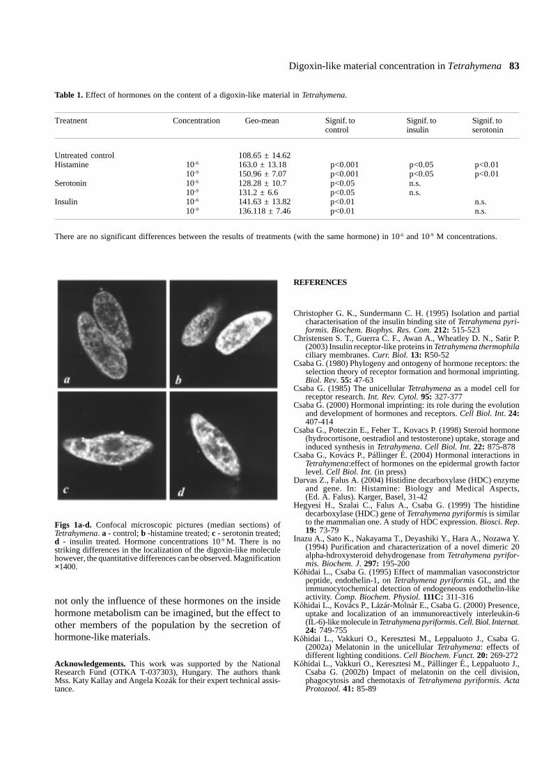
Digoxin-like material concentration in Tetrahymena 83
not only the influence of these hormones on the insidehormone metabolism can be imagined, but the effect toother members of the population by the secretion ofhormone-like materials.
Acknowledgements. This work was supported by the NationalResearch Fund (OTKA T-037303), Hungary. The authors thankMss. Katy Kallay and Angela Kozák for their expert technical assis-tance.
REFERENCES
Christopher G. K., Sundermann C. H. (1995) Isolation and partialcharacterisation of the insulin binding site of Tetrahymena pyri-formis. Biochem. Biophys. Res. Com. 212: 515-523
Christensen S. T., Guerra C. F., Awan A., Wheatley D. N., Satir P.(2003) Insulin receptor-like proteins in Tetrahymena thermophilaciliary membranes. Curr. Biol. 13: R50-52
Csaba G. (1980) Phylogeny and ontogeny of hormone receptors: theselection theory of receptor formation and hormonal imprinting.Biol. Rev. 55: 47-63
Csaba G. (1985) The unicellular Tetrahymena as a model cell forreceptor research. Int. Rev. Cytol. 95: 327-377
Csaba G. (2000) Hormonal imprinting: its role during the evolutionand development of hormones and receptors. Cell Biol. Int. 24:407-414
Csaba G., Poteczin E., Feher T., Kovacs P. (1998) Steroid hormone(hydrocortisone, oestradiol and testosterone) uptake, storage andinduced synthesis in Tetrahymena. Cell Biol. Int. 22: 875-878
Csaba G., Kovács P., Pállinger É. (2004) Hormonal interactions inTetrahymena:effect of hormones on the epidermal growth factorlevel. Cell Biol. Int. (in press)
Darvas Z., Falus A. (2004) Histidine decarboxylase (HDC) enzymeand gene. In: Histamine: Biology and Medical Aspects,(Ed. A. Falus). Karger, Basel, 31-42
Hegyesi H., Szalai C., Falus A., Csaba G. (1999) The histidinedecarboxylase (HDC) gene of Tetrahymena pyriformis is similarto the mammalian one. A study of HDC expression. Biosci. Rep.19: 73-79
Inazu A., Sato K., Nakayama T., Deyashiki Y., Hara A., Nozawa Y.(1994) Purification and characterization of a novel dimeric 20alpha-hdroxysteroid dehydrogenase from Tetrahymena pyrifor-mis. Biochem. J. 297: 195-200
K�hidai L., Csaba G. (1995) Effect of mammalian vasoconstrictorpeptide, endothelin-1, on Tetrahymena pyriformis GL, and theimmunocytochemical detection of endogeneous endothelin-likeactivity. Comp. Biochem. Physiol. 111C: 311-316
K�hidai L., Kovács P., Lázár-Molnár E., Csaba G. (2000) Presence,uptake and localization of an immunoreactively interleukin-6(IL-6)-like molecule in Tetrahymena pyriformis. Cell. Biol. Internat.24: 749-755
K�hidai L., Vakkuri O., Keresztesi M., Leppaluoto J., Csaba G.(2002a) Melatonin in the unicellular Tetrahymena: effects ofdifferent lighting conditions. Cell Biochem. Funct. 20: 269-272
K�hidai L., Vakkuri O., Keresztesi M., Pállinger É., Leppaluoto J.,Csaba G. (2002b) Impact of melatonin on the cell division,phagocytosis and chemotaxis of Tetrahymena pyriformis. ActaProtozool. 41: 85-89
Figs 1a-d. Confocal microscopic pictures (median sections) ofTetrahymena. a - control; b -histamine treated; c - serotonin treated;d - insulin treated. Hormone concentrations 10-9 M. There is nostriking differences in the localization of the digoxin-like moleculehowever, the quantitative differences can be observed. Magnification×1400.
Table 1. Effect of hormones on the content of a digoxin-like material in Tetrahymena.
Treatnent Concentration Geo-mean Signif. to Signif. to Signif. tocontrol insulin serotonin
Untreated control 108.65 ± 14.62Histamine 10-6 163.0 ± 13.18 p<0.001 p<0.05 p<0.01
10-9 150.96 ± 7.07 p<0.001 p<0.05 p<0.01Serotonin 10-6 128.28 ± 10.7 p<0.05 n.s.
10-9 131.2 ± 6.6 p<0.05 n.s.Insulin 10-6 141.63 ± 13.82 p<0.01 n.s.
10-9 136.118 ± 7.46 p<0.01 n.s.
There are no significant differences between the results of treatments (with the same hormone) in 10-6 and 10-9 M concentrations.
84 G. Csaba et al.
Kovács P., Csaba G. (1990) Influence of the phosphoinositol (PI)system in the mechanism of hormonal imprinting. Biochem.Biophys. Res. Com. 170: 119-126
Kovács P., Csaba G. (1997) PLA2 activity in Tetrahymena pyrifor-
mis. Effects of inhibitors and stimulators. Lipid Mediat. CellSignal 15: 233-247
Kovács P., Müller W. E. G. Csaba G. (1998) Induction of digoxin-likematerial production, and the digoxin binding in the unicellularorganism Tetrahymena by digitoxin. Cell Biol. Int. 22: 473-478
Kuno T., Yoshida N., Tanaka C. 1979) Immunocytochemical local-ization of cyclic AMP and cyclic GMP in synchronously dividingTetrahymena. Acta Histochem. Cytochem. 12: 563
Lenard J. (1992) Mammalian hormones in microbial cells. TrendsBiochem. Sci. 17: 147-150
LeRoith D., Schiloach J., Roth J., Lesniak M. A. (1980) Evolutionaryorigins of vertebrate hormones: substances similar to mammalianinsulin are native to unicellular eukaryotes. Proc. Natl. Acad. Sci.USA. 77: 6184-6186
LeRoith D., Schiloach J., Berelowitz M., Frohman L. A., Liotta A.S., Krieger B. T., Roth J. (1983) Are messenger molecules inmicrobes the ancestors of the vertebrate hormones and tissuefactors? Fed. Proc. 42: 2602-2607
Received on 21st July, 2004; revised version on 27th September,2004; accepted on October 4th, 2004
Muto Y., Kudo S., Nozawa Y. (1983) Effect of local anethetics oncalmodulin- dependent guanylate cyclase in the plasma membraneof Tetrahymena pyriformis. Biochem. Pharmacol. 32: 3559-3563
Ruby K. W., Griffith R. W., Kaeberle M. I. (2002) Histamineproduction in Haemophilus somnus. Comp. Immunol. Microbiol.Infect. Dis. 25: 13-20
Saar J., Kader J.C., Poralla K., Ourisson G. (1991) Purification andsome properties of the squalene-tetrahymanol cyclase fromTetrahymena pyriformis. Biochim. Biophys. Acta 1075: 93-101.
Wheatley D. N., Christensen S. T., Schousbue P., Rasmussen L.(1993) Signalling in cell growth and death: adequate nutrientsalone my not be sufficent for ciliates. Cell Biol. Int. 17: 817-823

Acta Protozool. (2005) 44: 85
Ghazi S. M. ASMAT
Department of Zoology, University of Chittagong, Chittagong, Bangladesh
Dear Editor,I have gone through an article of August issue, 2004, entitled “First Record of Trichodinella epizootica (Raabe,
1950) Šramek-Hušek, 1953, with description of Trichodina notopteridae sp. n. (Ciliophora: Peritrichida) fromFreshwater Fishes of India” by Amlan Kumar Mitra and Durga P. Haldar. Acta Protozool. 43: 269-274 (2004).
For your kind notice I am presenting some misdeeds about the presentation and information of the article asfollows.
a) The authors have given the latest references (e.g.,Mitra and Haldar 2003) but intentionally omitted thefollowing references (including the articles published inActa Protozool.). They have cited the papers in whichthe name of Haldar is present, why? Astonishingly, thereviewers also overlooked the matter, intentionally orunintentionally I don’t know, they might have satisfac-tory answer.
1) Ghazi S. M. Asmat (2000) Trichodina cuchiae sp. n. (Ciliophora:Trichodinidae) from Gangetic mudeel, Monopterus cuchia(Hamilton-Buchanan, 1822) (Synbranchiformes: Synbranchidae)in India. The Chittagong Univ. J. Sc. 24 (1): 55-61
2) Ghazi S. M. Asmat (2000) First record of Trichodina acuta Lom,1961 (Ciliophora: Trichodinidae) from India. The ChittagongUniv. J. Sc. 24 (1): 63-70
3) Ghazi S. M. Asmat (2001a) Trichodina cancilae sp. n. (Mobilina:Trichodinidae) from the gills of a freshwater gar, Xenentodoncancila (Hamilton) (Belonidae). Acta Protozool. 40: 141-146
4) Ghazi S. M. Asmat (2001b) Trichodina canningensis sp. n.(Ciliophora: Trichodinidae) from an Indian estuarine fish, Mystusgulio (Hamilton) (Bagridae). Acta Protozool. 40: 147-151
5) Ghazi S. M. Asmat (2001c) Trichodina porocephalusi sp. n.(Ciliophora: Trichodinidae) from an Indian flathead sleeper,Ophiocara porocephalus (Valenciennes) (Eleotrididae). ActaProtozool. 40: 297-301
6) Ghazi S. M. Asmat (2001d) Occurrence and morphology ofTrichodinella epizootica (Raabe, 1950) Šramek-Hušek, 1953 inIndia. The Chittagong Univ. J. Sc. 25 (2): 37-44
7) Ghazi S. M. Asmat (2002a) Trichodinid ciliates (Ciliophora:Trichodinidae) from Indian fishes with description of two newspecies. Bangladesh J. Zool. 30 (2): 87-100
8) Ghazi S. M. Asmat (2002b) Occurrence and morphology of someTripartiella sp. (Ciliophora: Trichodinidae) from freshwater andestuarine fishes in India. Univ. j. zool. Rajshahi Univ. 21: 49-55
9) Ghazi S. M. Asmat (2002c) Two new species of trichodinid ciliates(Ciliophora: Trichodinidae) from Indian fishes. Univ. j. zool.Rajshahi Univ. 21: 31-34
b) The Indian Trichodinella epizootica is alreadyreported in a Bangladeshi journal vide ref. 6. Howauthors could publish it again knowing the matter, it ishighly disgraceful, I think. You have to do somethingrelating this article and information.
c) According to Article 29.2 (International Code ofZoological Nomenclature, Fourth Edition, 1999, p. 32),we know the suffix - idae is used for a family name. Butthe new species name described in the said paper is“... notopteridae”. Would it be valid?
d) The description of the new species is not also valid,I think. I described the same kind of individuals asTrichodina nigra. If they disagree, they may comparetheir specimens with T. nigra of my paper (Asmat 2002a). There is no reason to compare these withT. luciopercae. The Indians never ever identifiedT. nigra properly. I wonder that the reviewers of thearticle also forgot the features of T. nigra?
It would be highly appreciated if you could kindly gothrough this matter seriously and let me know thedecision of the editorial board of Acta Protozoologicaregarding this article.
Thanking you.
Address for correspondence: Ghazi S. M. Asmat, Departmentof Zoology, University of Chittagong, Chittagong 4331, Bangladesh Received on 23rd October, 2004
Letter to the Editor

Acta Protozool. (2005) 44: 87
Amlan Kumar MITRA and Durga P. HALDAR
Protozoology Laboratory, Department of Zoology, University of Kalyani, Kalyani, West Bengal, India
Sir,In the August, 2004 issue, we published a paper
incorporating our first record of Trichodinella epizootica(Raabe, 1950) Šramek-Hušek, 1953 from a freshwaterfish in India. Recently, it was pointed out to us thatTrichodinella epizootica has already been reportedfrom an Indian fish by Asmat (2001) from the host fishPuntius serana in Kalyani, West Bengal, India. Assuch, our claim of first record of this species in India isnot tenable. However, the species obtained by us wasrecorded from Puntius gelius from a different locality.The matter was perhaps overlooked by the experts whoreviewed the paper, because the description of the firstreport of T. epizootica was published in a University
Address for correspondence: D. P. Haldar, B-1/438, P.O., Kalyani,Dist. Nadia, Pin 741235, West Bengal, India
journal in Bangladesh which is neither available here norin the Internet. In view of the fact that the particularspecies was obtained by several authors outside India,our “second” record of the species from this subconti-nent only has a great significance so far as the distribu-tion and host ranges of trichodinid ciliophorans areconcerned.
Yours etc.
Received on November 2nd, 2004
Letter to the Editor