University of Shendi
66
Republic of the Sudan Ministry of Higher Education and Scientific Research University of Shendi Faculty of Graduate Studies and Scientific Research Effect of Environmental Storage on Red Blood Cells Parameters at Elmak Nimer University Hospital Blood Bank A thesis submitted for partial fulfillment of the degree of M.Sc. in medical Laboratory Sciences By Limya Ali Hussein AbuRass BSc Medical Laboratory Sciences –Shendi University (2006) Supervisor Dr Mohammed Osman Ali Mohammed (PhD). (Associate Professor of Haematology, Faculty of Medical Laboratory Sciences,Shendi University ). August – 2018
-
Upload
khangminh22 -
Category
Documents
-
view
1 -
download
0
Transcript of University of Shendi
Republic of the Sudan
Ministry of Higher Education and Scientific Research
University of Shendi
FFaaccuullttyy ooff GGrraadduuaattee SSttuuddiieess aanndd SScciieennttiiffiicc RReesseeaarrcchh
Effect of Environmental Storage on Red Blood Cells
Parameters at Elmak Nimer University Hospital
Blood Bank
A thesis submitted for partial fulfillment of the degree of M.Sc. in medical
Laboratory Sciences
By
Limya Ali Hussein AbuRass
BSc Medical Laboratory Sciences –Shendi University (2006)
Supervisor
Dr Mohammed Osman Ali Mohammed (PhD).
(Associate Professor of Haematology, Faculty of Medical Laboratory Sciences,Shendi
University ).
August – 2018
I
بسم هللا الرحمن الرحيم
ٹٱٹٱ
جم جم جم جم جم ىنمن جم جم جم جم جم جم جم جم جميل ىل مل خل ٱ
صدق هللا العظيم
111سورة طه األيه
II
I would like to dedicate dissertation to who gave me love,
comfort throughout my life.
My mother
To who is always there for me and never let me need.
My father
To my
Sisters, Brother, teachers
Whom are always there when I need them.
To those help me to complete this research.
To all my colleagues in Shendi university and Elmak Nimer
University hospital.
III
Acknowledgment
First, I would like to express my deepest thanks and praise to
Allah for enabling me to accomplish this work.
And pray for Prophet Mohammed peace be
Upon him
Thanks are extended to my supervisor:
Dr. Mohammed Osman Ali Mohammed
for helping, supporting, assistance and advice.
Also my thanks to all staff of Hematology Department.
Finally, I sincerely thank everyone who helped me to
accomplish this research.
IV
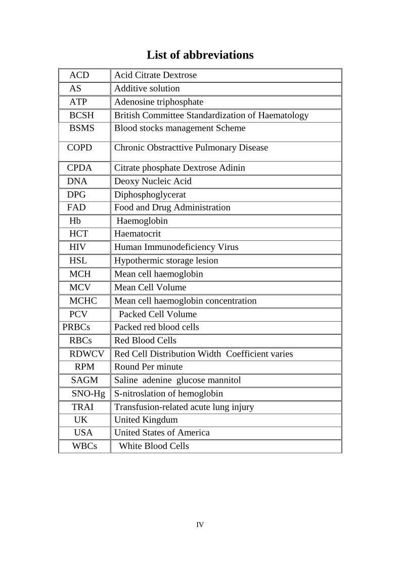
List of abbreviations
ACD Acid Citrate Dextrose
AS Additive solution
ATP Adenosine triphosphate
BCSH British Committee Standardization of Haematology
BSMS Blood stocks management Scheme
COPD Chronic Obstracttive Pulmonary Disease
CPDA Citrate phosphate Dextrose Adinin
DNA Deoxy Nucleic Acid
DPG Diphosphoglycerat
FAD Food and Drug Administration
Hb Haemoglobin
HCT Haematocrit
HIV Human Immunodeficiency Virus
HSL Hypothermic storage lesion
MCH Mean cell haemoglobin
MCV Mean Cell Volume
MCHC Mean cell haemoglobin concentration
PCV Packed Cell Volume
PRBCs Packed red blood cells
RBCs Red Blood Cells
RDWCV Red Cell Distribution Width Coefficient varies
RPM Round Per minute
SAGM Saline adenine glucose mannitol
SNO-Hg S-nitroslation of hemoglobin
TRAI Transfusion-related acute lung injury
UK United Kingdum
USA United States of America
WBCs White Blood Cells
V
ةــــالصـــاخل
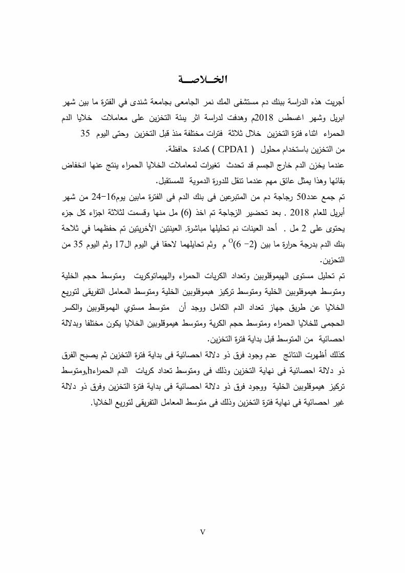
في الفترة ما بين شهر أجريت هذه الدراسة ببنك دم مستشفى المك نمر الجامعى بجامعة شندىعلى معامالت خاليا الدم اثر يىئة التخزينة وهدفت لدراس م8102ابريل وشهر اغسطس
53 وحتى اليوم قبل التخزين منذ الحمراء اثناء فترة التخزين خالل ثالثة فترات مختلفة .حافظة كمادة ( CPDA1) لتخزين باستخدام محلول ا من
ا انخفاض ينتج عنه لخاليا الحمراءمعامالت التحدث تغيرات قد عندما يخزن الدم خارج الجسم . للمستقبل للدورة الدموية وهذا يمثل عائق مهم عندما تنقل بقائها
من شهر 82-01رجاجة دم من المتبرعين فى بنك الدم فى الفترة مابين يوم 31تم جمع عددوقسمت لثالثة اجزاء كل جزء هامل من (1)تم اخذ بعد تحضير الزجاجة . 8102للعام بريلأ
خريتين تم حفظهما في ثالحة العينتين األ .حد العينات نم تحليلها مباشرةأ .مل 8يحتوى على 1 -8)بنك الدم بدرجة حرارة ما بين
Oمن 53وثم اليوم 01الفي اليوم الحقاهما ثم تحايلو م )
.التحزينالهيموقلوبين وتعداد الكريات الحمراء والهيماتوكريت ومتوسط حجم الخلية تحليل مستوى تم
هيموفلوبين الخلية ومتوسط تركيز هبموفلوبين الخلية ومتوسط المعامل التفريقى لتوريع ومتوسطوالكسر مستوي الهموقلوبينمتوسط ووجد أن الخاليا عن طريق جهاز تعداد الدم الكامل
يكون مختلفا وبداللة رية ومتوسط هيموقلوبين الخالياالحجمى للخاليا الحمراء ومتوسط حجم الك .من المتوسط قبل بداية فترة التخزين احصائية
ظهرت النتائج عدم وجود فرق ذو داللة احصائية فى بداية فترة التخزين ثم يصبح الفرق أكذلك ومتوسط h,ذو داللة احصائية فى نهاية التخزين وذلك فى ومتوسط تعداد كريات الدم الحمراء
فى بداية فترة التخزين وفرق ذو داللة ووجود فرق ذو داللة احصائية تركيز هيموقلوبين الخلية .غير احصائية فى نهاية فترة التخزين وذلك فى متوسط المعامل التفريقى لتوريع الخاليا
VI
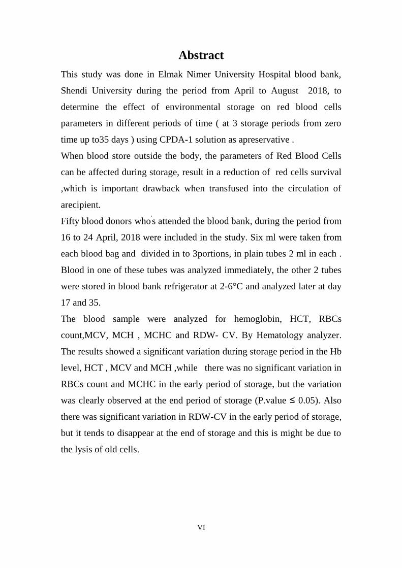
Abstract
This study was done in Elmak Nimer University Hospital blood bank,
Shendi University during the period from April to August 2018, to
determine the effect of environmental storage on red blood cells
parameters in different periods of time ( at 3 storage periods from zero
time up to35 days ) using CPDA-1 solution as apreservative .
When blood store outside the body, the parameters of Red Blood Cells
can be affected during storage, result in a reduction of red cells survival
,which is important drawback when transfused into the circulation of
arecipient.
Fifty blood donors who's attended the blood bank, during the period from
16 to 24 April, 2018 were included in the study. Six ml were taken from
each blood bag and divided in to 3portions, in plain tubes 2 ml in each .
Blood in one of these tubes was analyzed immediately, the other 2 tubes
were stored in blood bank refrigerator at 2-6°C and analyzed later at day
17 and 35.
The blood sample were analyzed for hemoglobin, HCT, RBCs
count,MCV, MCH , MCHC and RDW- CV. By Hematology analyzer.
The results showed a significant variation during storage period in the Hb
level, HCT , MCV and MCH ,while there was no significant variation in
RBCs count and MCHC in the early period of storage, but the variation
was clearly observed at the end period of storage (P.value ≤ 0.05). Also
there was significant variation in RDW-CV in the early period of storage,
but it tends to disappear at the end of storage and this is might be due to
the lysis of old cells.
VII
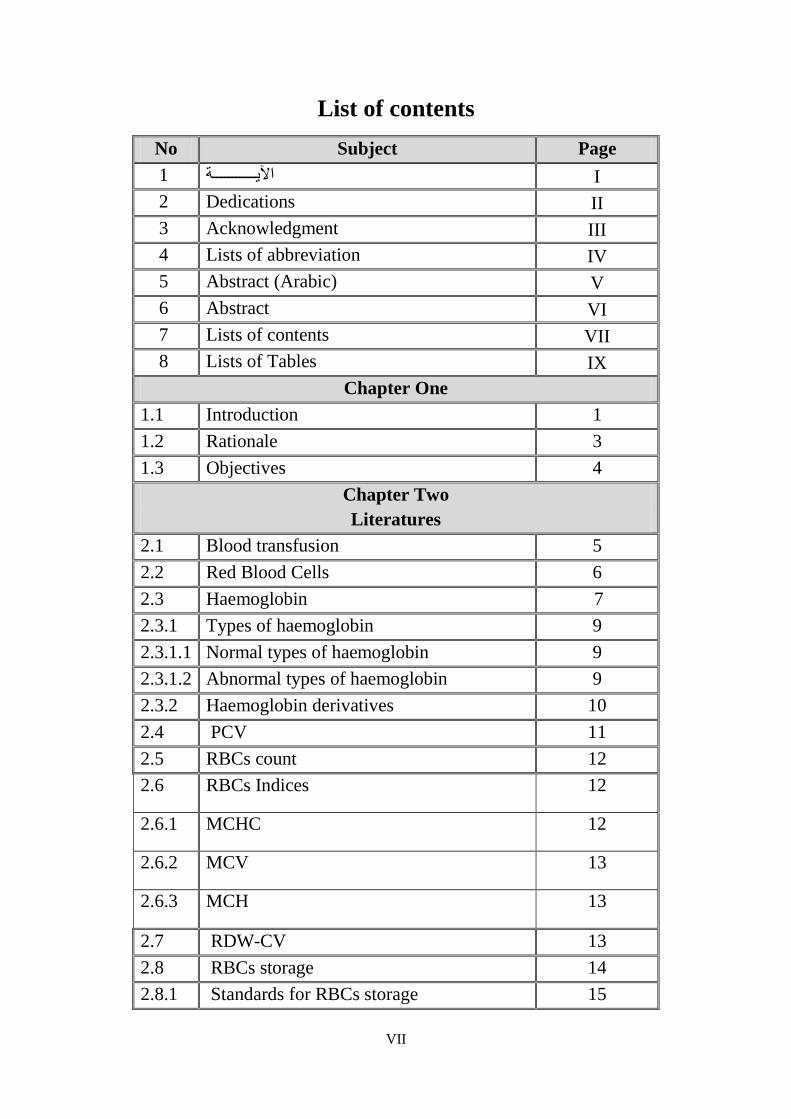
List of contents
No Subject Page
I اآليـــــــــــة 1
2 Dedications II
3 Acknowledgment III
4 Lists of abbreviation IV
5 Abstract (Arabic) V
6 Abstract VI
7 Lists of contents VII
8 Lists of Tables IX
Chapter One
1.1 Introduction 1
1.2 Rationale 3
1.3 Objectives 4
Chapter Two
Literatures
2.1 Blood transfusion 5
2.2 Red Blood Cells 6
2.3 Haemoglobin 7
2.3.1 Types of haemoglobin 9
2.3.1.1 Normal types of haemoglobin 9
2.3.1.2 Abnormal types of haemoglobin 9
2.3.2 Haemoglobin derivatives 10
2.4 PCV 11
2.5 RBCs count 12
2.6 RBCs Indices 12
2.6.1 MCHC 12
2.6.2 MCV 13
2.6.3 MCH 13
2.7 RDW-CV 13
2.8 RBCs storage 14
2.8.1 Standards for RBCs storage 15
VIII
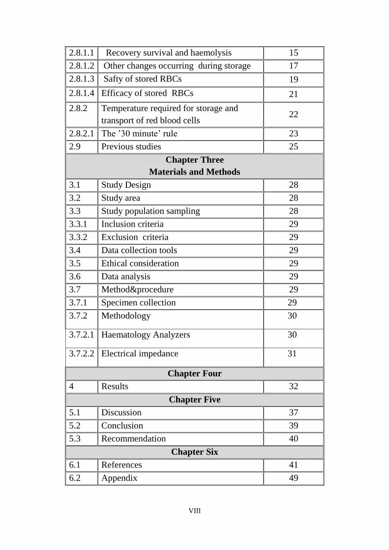
2.8.1.1 Recovery survival and haemolysis 15
2.8.1.2 Other changes occurring during storage 17
2.8.1.3 Safty of stored RBCs 19
2.8.1.4 Efficacy of stored RBCs 21
2.8.2 Temperature required for storage and
transport of red blood cells 22
2.8.2.1 The ’30 minute’ rule 23
2.9 Previous studies 25
Chapter Three
Materials and Methods
3.1 Study Design 28
3.2 Study area 28
3.3 Study population sampling 28
3.3.1 Inclusion criteria 29
3.3.2 Exclusion criteria 29
3.4 Data collection tools 29
3.5 Ethical consideration 29
3.6 Data analysis 29
3.7 Method&procedure 29
3.7.1 Specimen collection 29
3.7.2 Methodology 30
3.7.2.1 Haematology Analyzers 30
3.7.2.2 Electrical impedance 31
Chapter Four
4 Results 32
Chapter Five
5.1 Discussion 37
5.2 Conclusion 39
5.3 Recommendation 40
Chapter Six
6.1 References 41
6.2 Appendix 49
IX
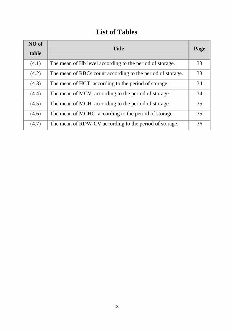
List of Tables
NO of
table Title Page
(4.1) The mean of Hb level according to the period of storage. 33
(4.2) The mean of RBCs count according to the period of storage. 33
(4.3) The mean of HCT according to the period of storage. 34
(4.4) The mean of MCV according to the period of storage. 34
(4.5) The mean of MCH according to the period of storage. 35
(4.6) The mean of MCHC according to the period of storage. 35
(4.7) The mean of RDW-CV according to the period of storage. 36

Introduction Justification
Objectives
1
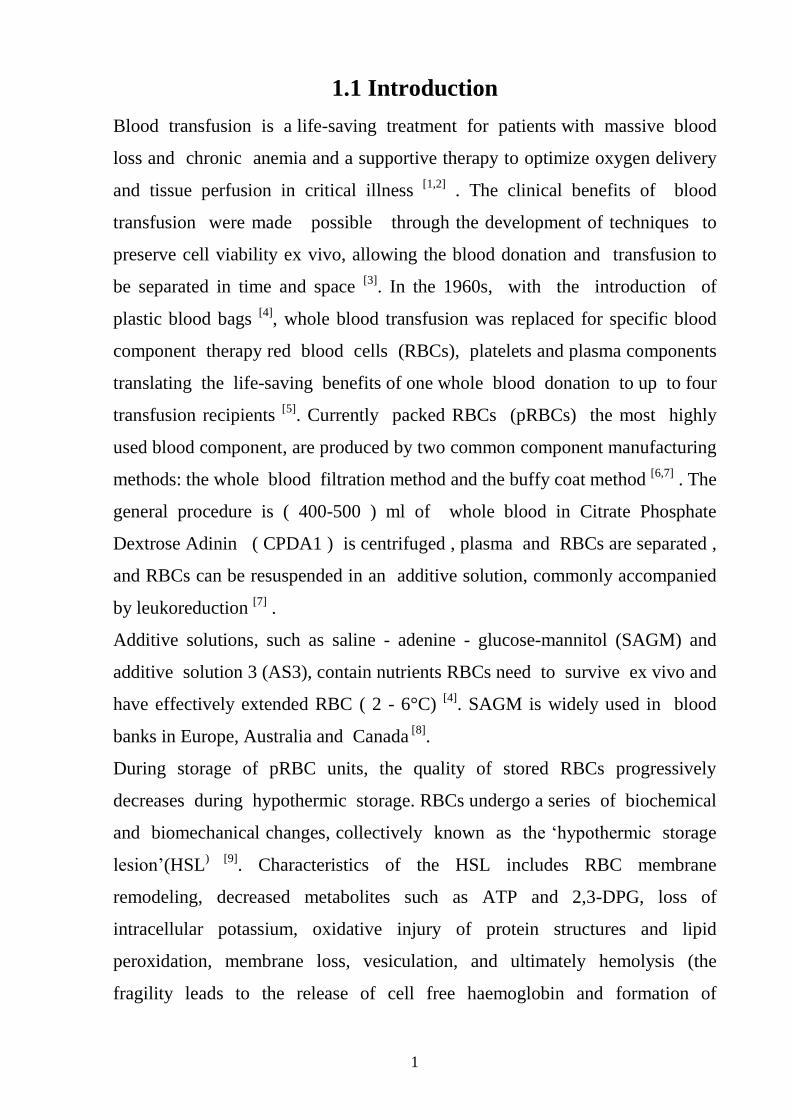
Introduction 1.1
Blood transfusion is a life-saving treatment for patients with massive blood
loss and chronic anemia and a supportive therapy to optimize oxygen delivery
and tissue perfusion in critical illness [1,2]
. The clinical benefits of blood
transfusion were made possible through the development of techniques to
preserve cell viability ex vivo, allowing the blood donation and transfusion to
be separated in time and space [3]
. In the 1960s, with the introduction of
plastic blood bags [4]
, whole blood transfusion was replaced for specific blood
component therapy red blood cells (RBCs), platelets and plasma components
translating the life-saving benefits of one whole blood donation to up to four
transfusion recipients [5]
. Currently packed RBCs (pRBCs) the most highly
used blood component, are produced by two common component manufacturing
methods: the whole blood filtration method and the buffy coat method [6,7]
. The
general procedure is ( 400-500 ) ml of whole blood in Citrate Phosphate
Dextrose Adinin ( CPDA1 ) is centrifuged , plasma and RBCs are separated ,
and RBCs can be resuspended in an additive solution, commonly accompanied
by leukoreduction [7]
.
Additive solutions, such as saline - adenine - glucose-mannitol (SAGM) and
additive solution 3 (AS3), contain nutrients RBCs need to survive ex vivo and
have effectively extended RBC ( 2 - 6°C) [4]
. SAGM is widely used in blood
banks in Europe, Australia and Canada [8]
.
During storage of pRBC units, the quality of stored RBCs progressively
decreases during hypothermic storage. RBCs undergo a series of biochemical
and biomechanical changes, collectively known as the ‘hypothermic storage
lesion’(HSL) [9]
. Characteristics of the HSL includes RBC membrane
remodeling, decreased metabolites such as ATP and 2,3-DPG, loss of
intracellular potassium, oxidative injury of protein structures and lipid
peroxidation, membrane loss, vesiculation, and ultimately hemolysis (the
fragility leads to the release of cell free haemoglobin and formation of
2
microparticles submicron haemoglobin containing vesicles and additional
haemolysis [10,11,12]
.
There are increasing concerns regarding the effect of the HSL on hemorheology,
including RBC aggregability, deformability and membrane remodeling, effects
that could potentially lead to impairment of the oxygen delivery capacity of
transfused blood [13,14,15] .
3
Rationale 1.2
Red blood cells (RBCs) concentrates are the most transfused blood component
worldwide.
For having a sufficient and available blood supply blood can be stored in
asolution of Citrate-Phosphate-Dextrose-Adenine (CPDA1) as a combined
anticoagulant and energy source for up to 35 days at( 2-6 )°C.
This study focuses on an analysis of storage related changes in RBCs parameter
with duration of storage up to 35 days that may lead to impairment of the
oxygen delivery capacity ( decrease active desirable substances such as
hemoglobin and viable red blood cells) of transfused blood.
The importance of this study is, its result valuable for the quality control in
hospital blood bank.
4
Objectives 1.3
General objective:
To evaluate the effect of environmental storage on Red Blood Cells parameters
in Elmak Nimer University Hospital Blood Bank.
Specific objectives:
- To measure haemoglobin level during red blood cells storage.
-To determine Red Blood Cells count during red blood cells storage.
-To Calculate hematocrit (Hct) during red blood cells storage.
-To estimate mean corpuscular volume (MCV) during red blood cells storage.
-To measure mean corpuscular Hgb (MCH) during red blood cells storage.
-To estimate mean corpuscular Hgb concentration (MCHC) during red blood
cells storage.
-To estimate Red blood cell distribution width-Coefficient varies (RDW -CV)
during red blood cells storage.

Literature Review

5
2 Literature Review
2.1 Blood transfusion:
Blood is always considered essential for life, is a mixture of cells and watery
liquid, called plasma that the cells float in. It also contains other things like
nutrients (such as sugar, hormones, clotting agents and waste products to be
flushed out of the body). There are three kinds of cells in the blood; red blood
cells, white blood cells and platelets [16, 17]
.
A place where blood is collected from donors separated into different types,
stored, and / or prepared for transfusion to the recipient, a blood bank may be a
separate free – standing facility or part of a larger laboratory in a hospital [18]
.
The blood transfusion was first attempted in (1422) great strides have been
achieved in the field of blood donation, the discovery and recognition of the
standard blood groups in (1901), the edition of dextrose to the storage medium
in (1914), the importance of refrigeration of stored blood in (1937), and the
discovery of the Rh factor in (1940) [19,20]
.
Blood is collected into a plastic bag for blood collection consist of 450 ml of
blood mixed with anticoagulants, these include citrate – phosphate dextrose
(CPD), acid – citrate dextrose (ACD), with adenine to prolong red cell storage
[18,21] .
The indications of fresh blood transfusion in case of anemia, leukemia,
thrombocytopenia, sever liver diseases, burns, hemodialysis, hemolytic disease
of new born and treatment of coagulation disorders, usually the specimen for
collected is tested for hepatitis B and C, Human Immune Virus (HIV), malaria
and other infectious diseases, the only blood that tests negative for these are
given to patients [17, 22 – 24]
.
Each unit of whole blood normally is separated into several components, red
blood cells may be stored under refrigeration for a maximum of 42 days, or they
may be frozen for up to 10 years. Red blood cells are used to treat anemia [25-
27],while the platelets are important in the control of bleeding and are generally
used in patients with leukemia and other forms of cancer, the platelets are stored

6
at room temperature and may be kept for a maximum of five days, while the
fresh frozen plasma used to control bleeding due to low levels of some clotting
factors is kept in a frozen state for usually up to one year [17,28-30]
.
While the granulocytes are some times used to fight infections, although their
efficacy is not well – established, they must be transfused with 24 hours of
donation [31, 32]
.
Whole blood may be preserved for up to 21 days, without losing its usefulness
in blood transfusions an anticoagulant is added to prevent clotting blood plasma,
the fluid portion of the blood, may be frozen and / or dried and stored
indefinitely [21]
.
2.2 Red Blood Cells:
RBCs manufactured in the bone marrow , RBCs are enucleated biconcave
discs that are continuously being produced [33]
.
Once circulating, theses RBCs serve a great purpose of delivering oxygen to
tissues; however, overtime these RBCs brake down, lose their efficiency and
ultimately are eliminated. The biconcave disc shape is crucial to the function of
RBCs, presenting a maximal surface area for the capture of oxygen in the lungs
and its subsequent release to the tissue beds [33]
.
The cells are flexible and able to change their shape in order to traverse
the tiny tubules of the capillary beds [33]
.
Since the cells are enucleated and lack mitochondria, they are unable to
carry out cellular repair of damage or enzyme inactivation and therefore must
rely on anaerobic glycolysis for energy [33]
.
Structurally, RBCs depend on an intact membrane and an internal
cytoskeleton to function normally. This cytoskeleton, the structural support that
maintains the RBC’s biconcave shape, is made of protein, microfilament,
intermediate filaments, and microtubules [33]
.
Functioning RBCs have very high levels of 2,3 diphosphoglycerate ( 2.3
DPG ), in which 2,3, DPG binds the beta chain of de oxyhemoglobin in apH
7
dependent environment. Adequate levels of 2,3 DPG are necessary to lower the
oxygen affinity for hemoglobin thereby increasing oxygen tissue delivery [33]
.
Therefore, RBC function centers on the ability of an RBC to bind oxygen , if
the RBC has a normal biochemical environment and sound structure or
morphology, it functions normally by releasing the carried oxygen to the tissues.
Furthermore, as part of this process each RBC must have an energy supply to
survive and maintain its integrity and function [33]
.
Adenosine triphosphate (ATP) is such an energy molecule that the cell depends
upon to maintain its integrity and function [33]
2.3 Haemoglobin:
Haemoglobin is iron-containing protein attached red blood cell that transport
oxygen from the lungs to the rest of the body. Haemoglobin bonds with oxygen
in the lung exchanges it for carbon dioxide at cellular level, and then transport
the carbon dioxide back to the lung to be exhaled [34]
.
Each red blood cell contains approximately 640 million haemoglobin molecules.
Each molecules of normal adult haemoglobin consist of four poly peptide chains
two alpha (a2) and two beta (β2) each with it is own haem group. The molecular
weight of haemoglobin 68.000 [34]
.
Haem synthesis occurs largely in the mitochondria by a series biochemical
reactions commencing with the condensation of glycin and succinyle co-enzyme
A under the action of the key rate-limiting enzyme δ-amino laevulinic acid
synthase. Byridoxal phosphate (vitamin B6) is a co-enzyme for this reaction
which is stimulated by erythropoietin which gives δ-amino laevulinic acid inside
mitochondria which gives prophobilinogen outside the mitochondria to give
uroporphyrinogen which give coproporphyrinogen to protoporphyrin which
combine with iron to form the heam part [35]
.
Gloin synthesis occurs largely in ribosome which contain from poly peptide
chain [35]
.
Globin synthesis from amino acids such as glycin, lysine, leucin, glutamic acid,
arginin, asparatic and … etc [35]
.
8
Each molecule of haem combines with globin chain made on poly ribosomes. A
tetramer of four globin chains each with it is own haem group in a ' pocket ' then
formed to make up haemoglobin molecule [35].
Whether haemoglobin binds with oxygen or carbon dioxide depend on the
relative concentration of each around the red blood cell. When it reaches the
oxygen-rich lung, it releases the less abundant carbon-dioxide to bind with
oxygen, when it goes back out into the body where cells areproducing carbon
dioxide, it release the oxygen and bind with carbon dioxide this is called the
Boher effect [34]
.
When carbon monoxide is present, it competes with oxygen at the haem binding
sites. And since haemoglobin is 200 times more likely bind with carbon
monoxide, forming very bright red form of haemoglobin as low as 0.02% in the
air can cause nausea and headache, 0.1% causes un consciousness and death
(compare that with normal 20% oxygen saturation of the air) persons, who
expose themselves regularly to carbon monoxide, may have as many as 20% of
their hemoglobin's oxygen sites blocked by carbon monoxide [34]
.
Haemoglobin abnormalities result in very serious hereditary disease, such as
sickle cell anemia and thalassemia [34]
.
Haemoglobin is made up of four subunits with a haem (iron-containing) group
in each for oxygen binding. There are slightly different haemoglobin in adult
when compared to children fetuses [34]
.
High 2–3 diphosphoglycerate levels are found in people who live in high
altitudes, this chemical allows larger amount of oxygen to be delivered to the
tissue, preventing altitudes sicknes [34]
.
Like all proteins the ' blue print' for haemoglobin exists in DNA (the material
that makes up genes). Normally, one individed has four genes that two genes
code for the alpha chain. Two other genes code for the beta chain (two
additional genes code for gamma chain in fetus) [36]
.
9
The alpha chain and the beta chain are made in precisely equal amount, despite
the differing number of genes. The protein chain join in developing red blood
cells, and remain together for the life of the red blood cell [35]
.
There are hundreds of haemoglobin variants that involve genes both from the
alpha and beta gene clusters [35]
.
2.3.1 Types of haemoglobin:
2.3.1.1Normal types:
Embryo haemoglobin which found in the first weeks of gestation
Gower1 which contain of two epsilon(ε2) and two zeta (ȥ) chain
Gower2 which contain of two epsilon (ε2) and two alpha (α2) chain
Portland which contain of two Zeta (ȥ2) and two gamma (γ2) chain
Haemoglobin F (Fetal haemoglobin)
This type is major respiratory pigment in intrauterine life which found in fetuses
and new born babies, it compose from two alpha (α2) chain and two gamma
chain ,and its replaced by haemoglobin A,shortly after burth ,only small amount
of haemoglobin F are made after birth [35]
.
Some disease, such as sickle cell anemia, a plastic and leukemia have abnormal
types of haemoglobin and higher amount of haemoglobin F [35]
.
Haemoglobin A" adult haemoglobin"
This is the most common types of haemoglobin found normally in adult. Some
disease, such as sever from thalassemia, may cause haemoglobin F level to be
high [37]
.
Haemoglobin A sub2
This is normal type of haemoglobin contain of two alpha and two Delta(δ )chain
[36].
Haemoglobin A2 is found in small amount in adult about 2% [36]
.
2.3.1.2 Abnormal types of haemoglobins:
Haemoglobin S
Is the most common of abnormal haemoglobin and the basic of sickle cell trait
and sickle cell disease, differ from normal adult haemoglobin only by single
10
amino acid substitution, valine replacing glutamic acid 6th position of the beta
chain globin [35]
.
Haemoglobin C
Is abnormal haemoglobin with substitution of lysine for glutamic acid at 6th
position of the beta globin [38]
.
Haemoglobin E
Is abnormal haemoglobin, It formed when glutamic acid is replaced by lysine at
26th position of beta chain of globin
[38]
Punjab haemoglobin D
Is abnormal haemoglobin with substitution of glutamine residue for glutamic
acid at 121th position of the Beta globin chain [38]
Haemoglobin Arab
Is abnormal haemoglobin with substitution of lysine residue for glutamic acid at
121th position of the Beta chain [38]
Haemoglobin H
Is abnormal haemoglobin containing from four beta (β4) chains and this
haemoglobin is unsuitable for life [38]
.
Haemoglobin Bart
Is abnormal haemoglobin containing from four alpha chains and this
haemoglobin is unsuitable for life [38]
2.3.2 Haemoglobin derivatives:
Oxygenhaemoglobin
Is a normal forms of haemoglobin that a attaches to oxygen by ferrous iron (Fe+2
– o-2
) [39]
Carbo-amino haemoglobin
Is a normal form of haemoglobin that attaches to the carbon dioxide(40)
.
Carboxyhaemoglobin
Is a normal forms of haemoglobin that a attacks to the carbon monoxide instead
of oxygen or carbon dioxide
. High amount of this type of abnormal
haemoglobin prevent the normal movement of oxygen by blood [39]
.
11
Sulfohaemoglobin
Is an abnormal form of haemoglobin that cannot carry oxygen. It may result
from certain medicines such as phenaccetin or sulfonamides [39]
.
Met haemoglobin
When the iron that is part of haemoglobin is changed to ferric state so that
doesn't carry oxygen [39]
.
2.4 : Packed cell volume:
The haematocrit or packed cell volume are on the measures of the proportion of
blood volume that is occupied by red blood cells. It's normally 45+7 (38-52%)
for males and 42+5 (37-47%) for females [40]
.
Elevated PCV:
In case danger fever, where the full blood counts done, Daily, high haematocrit
is danger of an increased risk of dengue shock syndrome [39]
Polycythaemia Vera is associated with elevated haematocrit [39]
.
Smoking, COPD, and other pulmonary condition associated with hypoxia may
elicit an increased production of red blood cells, this increase is mediated by the
increased level of erythropoietin by the kidney in response to hypoxia[39]
.
There have been cases where the blood for testing was in advertently drawn
from the same arm with the intravenous running in a transfusion of packed red
cells. In this sample the haemoglobin measurement will be high because it is
measuring the fluted being transfused (that is mostly red blood cells) rather than
the diluted serum, in this case, the haematocrit measurement will be artificially
very high [40]
.
Lowered PCV:
Lowered haematocrit can imply significant haemorrhage. MCV, RDW can be
quiet help full in evaluating lower than normal haematocrit , because can help
the clinician determine whether blood loss is chronic or acute. The MCV is the
size of red blood cell and RDW is relative measure of the variation in size of the
red cell population. A low haematocrit with a low MCV with high RDW suggest
a chronic iron deficient erythropoiesis, but normal RDW suggest blood loss that
12
is more acute such as haemorrhage [11]
. Conversely, if blood for haematology
testing a drawn from proximal to that of an intravenous infusing line fluid into
patient, the blood sample will be diluted by those fluid and the haematocrit will
be or artificially low [40]
.
Estimation of PCV:
The packed cell volume can be determined by centrifuging heparinized blood in
a capillary tube (also known as a micro haematocrit tube). Is typically
centrifuged at10.000 RPM for five minute,This Separates the blood into layers
(packed cell, Buffy coat -WBCs + platelet, plasma). And the tube read by
haematocrit reader [40]
.
And estimated haematocrit as a percentage may be derived by multiplying
haemoglobin concentration in g/dl three times and dropping the units [39]
.
2.5 Red blood cells count:
Total red blood cells is the number of red cells is given as an absolute number
per litre [38]
.
Haemoglobin - The amount of hemoglobin in the blood, expressed in grams per
decilitre. (Low hemoglobin is called anaemia.)
Hematocrit or packed cell volume (PCV) - This is the fraction of whole blood
volume that consists of red blood cells [38]
.
2.6 Red Cell Indices:
2.6.1 MCHC:
The MCHC gives the concentration of haemoglobinin g/l in 1 litre of packed red
cells. It is calculated from the haemoglobin (Hb) and PCV.
A guideline reference range for MCHC in health is 315–360 g/l (31.5–36.0 g/dl.
* Low MCHC values are found in iron deficiency anaemia and other conditions
in which the red cells are microcytic and hypochromic (MCHC may be normal
in thalassaemia trait).
*An increased MCHC can occur in marked spherocytosis but this is a rare
condition. Arise MCHC is more often due to a calculation error or an incorrect
haemoglobin or PCV [38]
.
13
2.6.2 MCV:
The mean red cell volume (MCV) provides information on red cell size. It is
measured in femtolitres (fl) and is determined from the PCV and electronically
obtained RBC count ,A guideline reference range is 80–98 fl.[38]
● Low MCV values: are found in microcytic anaemias particularly iron
deficiency anaemia, anaemia of chronic disease and thalassaemia. The
MCV is low in infancy (about 70 fl at 1 year of age) .
● Raised MCV values: are found in macrocytic anaemias, marked
reticulocytosis [38]
.
2.6.3 MCH:
The MCH gives the amount of haemoglobin in picograms (pg) in an average red
cell. It is calculated from the haemoglobin and electronically obtained RBC
count [38]
.
A guideline reference range for MCH in health is 27–32 pg [38]
.
Low MCH values: are found in microcytic hypochromic anaemias and also
when red cells are microcytic and normochromic, In thalassaemia minor the
MCH is low even when anaemia is mild (MCHC is often normal) [38]
.
Raised MCH values: are found in macrocytic normochromic anaemias. MCH is
also raised in neoborns [38]
.
2.7 Red blood cell distribution width (RDW):
It is an index that is calculated by the analyzers by two methods, based on the
values of the MCV and the RBCs. The first is referred to as the RDW- CV,
which is the ratio of the width of the RBCs distribution curve at 1 SD divided by
the MCV. The normal value for adults is11-14.5% . Microcytosis tends to
increase its value, while macrocytosis minimizes the changes in the RDW-CV.
The second method refers to RDW- SD, that is a direct measurement of the
RBCs distribution width taken at the 20% frequency level normally( RDW-SD =
42 ± 5 fL). It is more sensitive to the appearance of minor populations of
macrocytes or microcytes. This index reflects a state of anisocytosis
(heterogeneous population of RBC) [38] .
14
2.8 Red Blodd Cells Storage:
Peyton Rous was the first person to store red blood cells. He had learned from
Roger Lee that citrate was an anticoagulant [41]
.
He kept rabbit red blood cells in a mixture of citrate and glucose for 4 weeks in
a refrigerator and observed that they did not haemolyse [42]
. When these stored
red blood cells were infused back into the donor rabbits, they raised the
haematocrit and did not cause haemoglobinuria or bilirubinuria [43]
.
Two years later, in military hospitals adjacent to World War I battle fields,
Rous’s post-doctoral fellow, Oswald Robertson used this solution to store
human red blood cells for up to 26 days and used this ‘banked’ blood to
resuscitate soldiers in shock [44,45]
. However, Robertson’s US Army colleagues
became concerned about the possibility of bacterial contamination of the stored
blood, and the commission that approved stored blood transfusion for general
use approved it only for storage in citrate without glucose and only for days of
storage [46]
. Robertson, in his private writings, noted that this restriction both
limited the utility of blood banking and reduced the quality of stored blood,
because some units ran out of glucose in less than 5 days [46]
.
The controversy between those who seek longer red blood ran out of glucose in
less than 5 days cell storage for logistical reasons and those who have concerns
about the safety and efficacy of stored blood continues. Storing red blood cells
for longer times does have advantages. It allows the accumulation of inventory,
takes advantage of economies of scale in collection, processing and testing, and
allows the development of quality controls. Longer cold storage reduces
potential transmission of syphilis and reduces transfusion-associated graft-
versus-host disease. However stored red blood cells lose functional capacity
during storage:
they lose membrane, and they eventually become nonviable [4]
.
Stored red blood cells can be frankly dangerous with high potassium
concentrations, bacterial overgrowth, and the lytic elaboration of toxic lipids [4]
.
15
2.8.1 Standards for red blood cell storage:
2.8.1.1 recovery survival and haemolysis:
The first standards for red blood cell storage were that the cells did not
haemolyse in the bottle and that they appeared to circulate when reinjected into
the donor or were transfused into a recipient [47]
. In a sense, these remain the
only standard [47]
.
They are now formalized in the US licensure requirements that at the end of the
approved storage period, an average of at least 75% of the cells remain in the
circulation 24 h after infusion and that haemolysis be less than 1% For 50 years,
labelling red blood cells with chromium-51 has been the accepted way to
measure their recovery and survival [47]
. The recovery is the fraction of the
injected cells that circulate after infusion and their survival is the length of time
that either the average cell or the longest surviving cell circulates. With the
recognition of the high frequency of post-transfusion hepatitis, autologous
recovery and survival
measures, where a volunteer donor’s own red blood cells are evaluating blood
storage systems. Such studies have the added advantage that as the infused red
blood cells are the donor’s own, antibody-mediated clearance of the cells does
not typically interfere with the observations, and documented reductions in
recovery or survival can be presumed to be the result of damage to the cells
inflicted by the storage system or the passage of time[48]
. The standard measure
is now the 24-h post-infusion in vivo recovery, with the survival measured as the
half-life of the radioactive label. One laboratory has developed and used a
system for measuring recovery of allogenic red blood cells by measuring the
fraction carrying alloantigens by flow cytometry [49]
.
The establishment of 75% as the recovery USA came out of historical
experience [50]
. Whole blood stored for 3 weeks in acid–citrate–dextrose solution
had an approximately 75% autologous in vivo recovery. With 3-week storage of
whole blood in CPD solution, this improved to 79% [51]
.
16
When adenine was added to CPD solution to make CPDA-1, the licensure study
showed 81% autologous in vivo recovery after 5-week storage as whole blood,
but only a 72% autologous in vivo recovery after 5-week storage as packed red
blood cells [51]
. The solution was licenced recovery, but because of the sense that
performance had actually gotten worse, the Food and Drug Administration
(FAD) raised the standard to 75% in 1985. All of the red blood cell additive
solution storage systems licenced subsequently in the USA, AS-1, AS-3, and
AS-5, have met this higher standard [52]
.
Furthermore, in a large review of licensure trials by Dumont USA, AS-1, AS-3,
and AS-5, and AuBuchon, all appeared to be equivalent, with approximately
82% 24-h in vivo recovery when stored as red blood cells in additive solution
for 6 weeks [4]
. When the red blood cells are leucoreduced at the time of initial
processing, the red blood cell recovery is about 2% higher [53]
. The survival of
red blood cells that circulate for 24 h has been normal with a half-life of about
60 days in all systems where it has been measured [53]
.
The problem with 24-h in vivo recovery as a red blood cell storage quality
standard is that its measurement is time consuming, expensive to conduct,
requires exposing volunteers to modest amounts of radiation, and gives quite
different results from one volunteer to another [54]
. Examination of the results of
a large number of such studies shows that the population distribution has a large
standard deviation and negative skew with a long lower tail [53]
. Examinations
of cross-over studies where individual volunteers are measured several times
show that some volunteers’ red blood cells consistently store better than others
[54].
Measures of haemolysis are easier to perform, and several very large series are
available from national blood service quality-assessment programmes.
Typically, red blood cells in additive solution have 0·2–0·4% haemolysis after
5–6 weeks of storage, and 1–4% of such cells typically exceed standards
Leucoreduction tends to reduce storage haemolysis by about 50 % [55]
.
17
2.8.1.2 Other changes occurring during storage:
The red blood cell storage lesion.
There are many other changes that occur during red blood cell storage that have
not served as conditions of storage system licensure in the past [4]
. These
changes include shape change, slowed metabolism with decreased
concentrations of adenosine 5′-triphosphate (ATP), acidosis with resulting
decreased concentrations of DPG, loss of cation pumping with loss of
intracellular potassium, oxidative injury with changes in band 3 structure and
lipid peroxidation, and apoptotic changes with membrane phospholipid
racemization and membrane loss [55]
.
Storing living red blood cells in a closed plastic bag means that the products of
ongoing glycolytic metabolism, lactic acid and protons accumulate over time (55)
.
Other metabolic processes, such as the breakdown of adenosine by adenosine
deaminase, mean that other breakdown products accumulate as well, but the
generally small amounts of ammonia and inosine formed do not seem to be
clinically important for themselves. The protons, however, decrease the pH in
the blood bag and alter glycolysis, first leading to a rapid drop in DPG
concentrations with a concomitant burst in ATP production, followed by an
increased slowing of glycolysis and falling ATP production as acid accumulates.
DPG is typically gone by the 10th day of red blood cell storage, whereas ATP
concentrations initially increase or are stable during the first 2 to 4 weeks of
storage with generally declining concentrations thereafter. New experimental
solutions may be able to extend high concentrations of ATP longer [56]
.
Acidification and decreasing ATP concentrations both affect red blood cell
shape [55]
. Acidosis causes the initial manifestations of red blood cell shape
change during storage, the development of bumps that grow to become the
typical surface protrusions of echinocytes. Most of the early aspects of
echinocytic shape change appear to be reversible with red blood cell warming
and certainly disappear when stored red blood cells are incubated in a neutral pH
solution of nutrients, a process called rejuvenation ,however, as red blood cell
18
ATP concentrations fall, irreversible changes associated with increased red
blood cell calcium concentrations develop [13,15]
.
These include the loss of phospholipid asymmetry, the development of
negatively charged phospholipid rafts on the cell opment of negatively charged
phospholipid rafts on the cell surface, and their shedding as microvesicles [13,15]
.
Membrane loss during red blood cell storage would appear to be permanent. As
storage progresses, red blood cells become more rigid and more adherent to
endothelium [13,15]
. Red blood cell concentrates are not a pure product, being
derived from whole blood by simple centrifugation techniques. Many red blood
cell concentrates are still made this way with the white blood cells left behind
as a buffycoat when the platelet-rich plasma is removed.
When these white blood cells are exposed to the acidic conditions of storage and
refrigerated, they respond with activation and cytokine production before they
die [57]
. After they die, the white blood cells break down and release constituents
including enzymes such as phospholipase-A2 [57]
. Phospholipase-A2 in turn
attacks and breaks down phospholipids released by red blood cells, creating
lysophospholipids such as the dialkylglycerol platelet-activating factor. The
longer the red blood cells are stored, the more of these biologically active lipids
are produced. Leucoreduction of red blood cell concentrate shortly after
collection markedly reduces the concentrations of lysophospholipids.
Leucoreduction also decreases the changes that cause stored red blood cells to
stick to endothelial cells in culture and probably to post-capillary venules in the
circulation [58]
.
Oxidative damage also occurs to red blood cells during storage [59]
. The
haemoglobin in venous blood is partially saturated with oxygen, so the oxygen
is constantly leaving one haemoglobin molecule and binding to another. This
reaction is not perfectly reversible, and occasionally, the leaving oxygen
takes an electron with it, forming ferric methemoglobin and superoxide.
Normally, methemoglobin is reduced and superoxide is desmuted without
consequences, but occasionally superoxide interacts with iron and water in the
19
Fenton reaction to form hydroxyl radical which can attack and damage proteins
and lipids. Damage to spectrin and glycoprotein band 3 can occur, and
interaction with triacylglycerols can lead to deacylation and the formation of
lysophospholipids. While damage to glycoprotein band 3 appears to have
consequences as a determinant in the natural 120- day lifespan of red blood cells
in the circulation its much slower rate during cold storage probably reduces its
importance as part of the storage lesion. On the other hand, the slow
accumulation of lysophospholipids in the blood bag during storage, without an
opportunity for their continuous removal and detoxification, remains as a safety
concern.
2.8.1.3 Safety of stored red blood cells:
There are several circumstances in which transfusion or even reinfusion of
stored red blood cells are associated with bad outcomes. Deaths have been
associated with the overgrowth of red blood cell units by cold-growing bacteria,
with the rapid central infusion of older units with high concen various causes,
and from transfusion-related acute lung injury( TRALI) from oxidation-induced
lysophospholipids . There may also be hypercoagulation associated with the
infusion of microvesicles exposing negatively charged phospholipids. One in
2000 units of blood is contaminated from skin or blood at the time that it is
drawn [60,61]
. Despite leucoreduction and cold storage, about 1 in 30 000 stored
red blood cell units can be demonstrated at some point to be bacterially
contaminated. Infections related to bacterial contamination could be
demonstrated in 1 in 5 million red blood cell units , and in a typical year, about
one of the five annual deaths from bacterially contaminated blood products is
reported to be associated with a unit of red blood cells [62]
. Most bacterial
organisms do not survive in the cold, but a few such as serratia marcesans,
Yersinia enterocolitica, and Aeromonas species can grow at refrigerator
temperatures [63]
. They tend to grow slowly in cold blood, dividing about once a
day and so to take approximately 27 days for a single organism to grow to
organisms and present with an overwhelming infection or endotoxic shock.
20
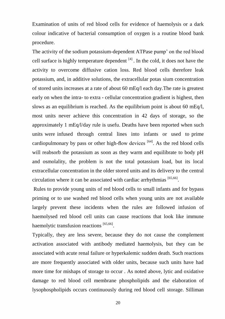
Examination of units of red blood cells for evidence of haemolysis or a dark
colour indicative of bacterial consumption of oxygen is a routine blood bank
procedure.
The activity of the sodium potassium-dependent ATPase pump’ on the red blood
cell surface is highly temperature dependent [4]
. In the cold, it does not have the
activity to overcome diffusive cation loss. Red blood cells therefore leak
potassium, and, in additive solutions, the extracellular potas sium concentration
of stored units increases at a rate of about 60 mEq/l each day.The rate is greatest
early on when the intra- to extra - cellular concentration gradient is highest, then
slows as an equilibrium is reached. As the equilibrium point is about 60 mEq/l,
most units never achieve this concentration in 42 days of storage, so the
approximately 1 mEq/l/day rule is usefu. Deaths have been reported when such
units were infused through central lines into infants or used to prime
cardiopulmonary by pass or other high-flow devices [64]
. As the red blood cells
will reabsorb the potassium as soon as they warm and equilibrate to body pH
and osmolality, the problem is not the total potassium load, but its local
extracellular concentration in the older stored units and its delivery to the central
circulation where it can be associated with cardiac arrhythmias [65,66]
.
Rules to provide young units of red blood cells to small infants and for bypass
priming or to use washed red blood cells when young units are not available
largely prevent these incidents when the rules are followed infusion of
haemolysed red blood cell units can cause reactions that look like immune
haemolytic transfusion reactions [65,66]
.
Typically, they are less severe, because they do not cause the complement
activation associated with antibody mediated haemolysis, but they can be
associated with acute renal failure or hyperkalemic sudden death. Such reactions
are more frequently associated with older units, because such units have had
more time for mishaps of storage to occur . As noted above, lytic and oxidative
damage to red blood cell membrane phospholipids and the elaboration of
lysophospholipids occurs continuously during red blood cell storage. Silliman
21
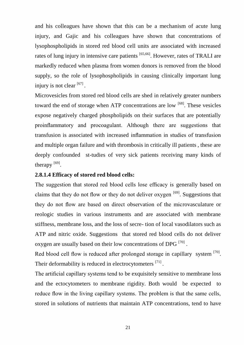
and his colleagues have shown that this can be a mechanism of acute lung
injury, and Gajic and his colleagues have shown that concentrations of
lysophospholipids in stored red blood cell units are associated with increased
rates of lung injury in intensive care patients [65,66]
. However, rates of TRALI are
markedly reduced when plasma from women donors is removed from the blood
supply, so the role of lysophospholipids in causing clinically important lung
injury is not clear [67]
.
Microvesicles from stored red blood cells are shed in relatively greater numbers
toward the end of storage when ATP concentrations are low [68]
. These vesicles
expose negatively charged phospholipids on their surfaces that are potentially
proinflammatory and procoagulant. Although there are suggestions that
transfusion is associated with increased inflammation in studies of transfusion
and multiple organ failure and with thrombosis in critically ill patients , these are
deeply confounded st-tudies of very sick patients receiving many kinds of
therapy [69]
.
2.8.1.4 Efficacy of stored red blood cells:
The suggestion that stored red blood cells lose efficacy is generally based on
claims that they do not flow or they do not deliver oxygen [69]
. Suggestions that
they do not flow are based on direct observation of the microvasculature or
reologic studies in various instruments and are associated with membrane
stiffness, membrane loss, and the loss of secre- tion of local vasodilators such as
ATP and nitric oxide. Suggestions that stored red blood cells do not deliver
oxygen are usually based on their low concentrations of DPG [70]
.
Red blood cell flow is reduced after prolonged storage in capillary system [70]
.
Their deformability is reduced in electrocytometers [71]
.
The artificial capillary systems tend to be exquisitely sensitive to membrane loss
and the ectocytometers to membrane rigidity. Both would be expected to
reduce flow in the living capillary systems. The problem is that the same cells,
stored in solutions of nutrients that maintain ATP concentrations, tend to have
22
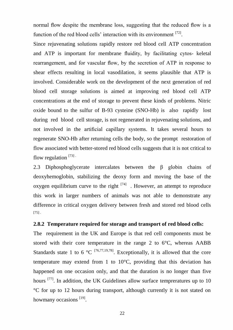
normal flow despite the membrane loss, suggesting that the reduced flow is a
function of the red blood cells’ interaction with its environment [72]
.
Since rejuvenating solutions rapidly restore red blood cell ATP concentration
and ATP is important for membrane fluidity, by facilitating cytos- keletal
rearrangement, and for vascular flow, by the secretion of ATP in response to
shear effects resulting in local vasodilation, it seems plausible that ATP is
involved. Considerable work on the development of the next generation of red
blood cell storage solutions is aimed at improving red blood cell ATP
concentrations at the end of storage to prevent these kinds of problems. Nitric
oxide bound to the sulfur of B-93 cysteine (SNO-Hb) is also rapidly lost
during red blood cell storage, is not regenerated in rejuvenating solutions, and
not involved in the artificial capillary systems. It takes several hours to
regenerate SNO-Hb after returning cells the body, so the prompt restoration of
flow associated with better-stored red blood cells suggests that it is not critical to
flow regulation [73] .
2.3 Diphosphoglycerate intercalates between the β globin chains of
deoxyhemoglobin, stabilizing the deoxy form and moving the base of the
oxygen equilibrium curve to the right [74]
. However, an attempt to reproduce
this work in larger numbers of animals was not able to demonstrate any
difference in critical oxygen delivery between fresh and stored red blood cells
[75] .
2.8.2 Temperature required for storage and transport of red blood cells:
The requirement in the UK and Europe is that red cell components must be
stored with their core temperature in the range 2 to 6°C, whereas AABB
Standards state 1 to 6 °C [76,77,19,78]
. Exceptionally, it is allowed that the core
temperature may extend from 1 to 10°C, providing that this deviation has
happened on one occasion only, and that the duration is no longer than five
hours [77]
. In addition, the UK Guidelines allow surface tempreratures up to 10
°C for up to 12 hours during transport, although currently it is not stated on
howmany occasions [19]
.
23
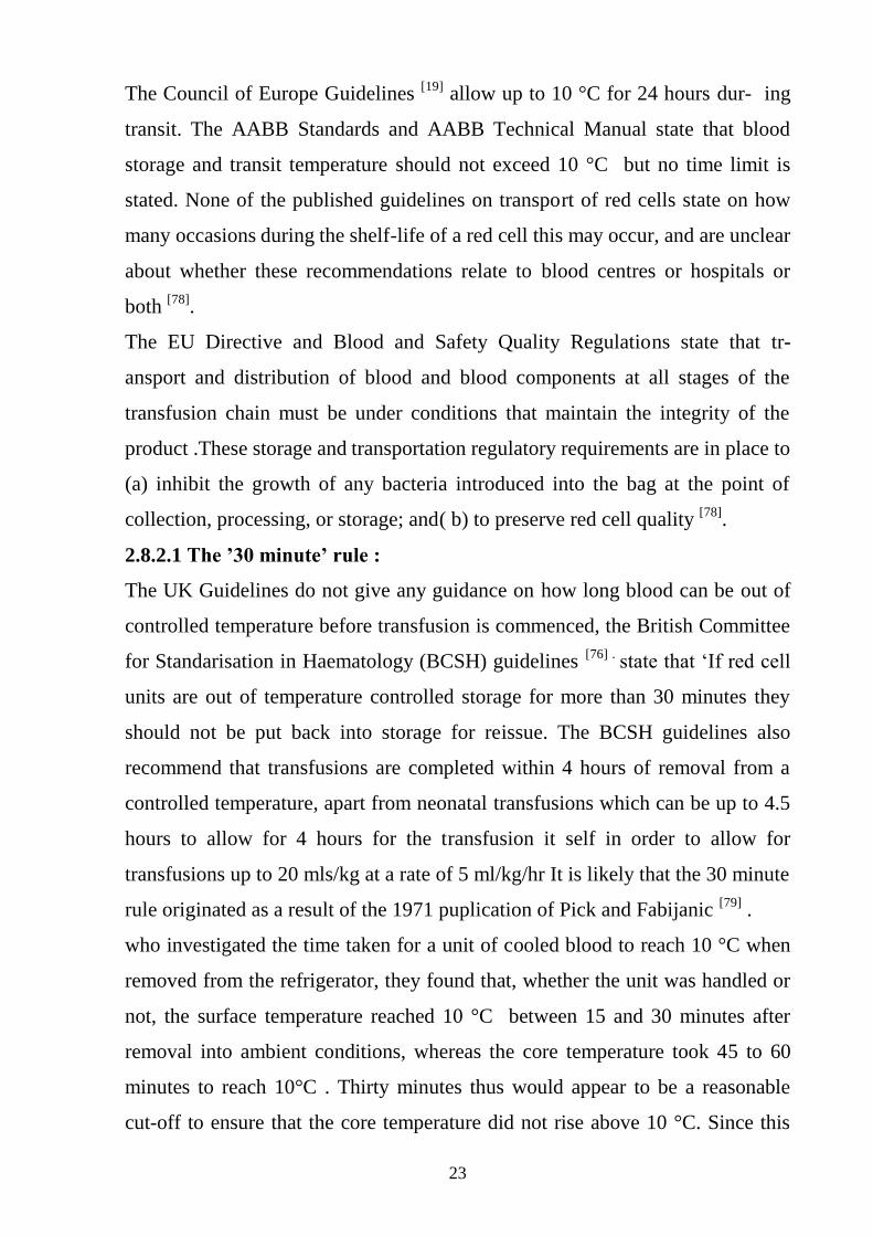
The Council of Europe Guidelines [19]
allow up to 10 °C for 24 hours dur- ing
transit. The AABB Standards and AABB Technical Manual state that blood
storage and transit temperature should not exceed 10 °C but no time limit is
stated. None of the published guidelines on transport of red cells state on how
many occasions during the shelf-life of a red cell this may occur, and are unclear
about whether these recommendations relate to blood centres or hospitals or
both [78]
.
The EU Directive and Blood and Safety Quality Regulations state that tr-
ansport and distribution of blood and blood components at all stages of the
transfusion chain must be under conditions that maintain the integrity of the
product .These storage and transportation regulatory requirements are in place to
(a) inhibit the growth of any bacteria introduced into the bag at the point of
collection, processing, or storage; and( b) to preserve red cell quality [78]
.
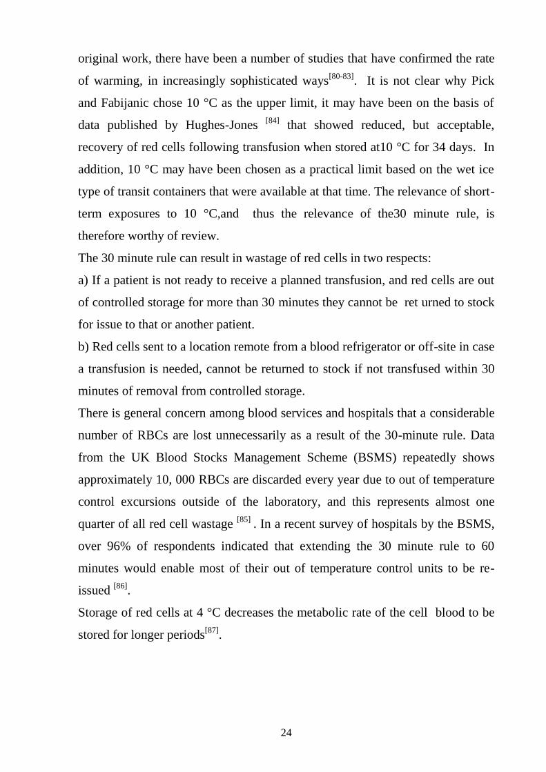
2.8.2.1 The ’30 minute’ rule :
The UK Guidelines do not give any guidance on how long blood can be out of
controlled temperature before transfusion is commenced, the British Committee
for Standarisation in Haematology (BCSH) guidelines [76] .
state that ‘If red cell
units are out of temperature controlled storage for more than 30 minutes they
should not be put back into storage for reissue. The BCSH guidelines also
recommend that transfusions are completed within 4 hours of removal from a
controlled temperature, apart from neonatal transfusions which can be up to 4.5
hours to allow for 4 hours for the transfusion it self in order to allow for
transfusions up to 20 mls/kg at a rate of 5 ml/kg/hr It is likely that the 30 minute
rule originated as a result of the 1971 puplication of Pick and Fabijanic [79]
.
who investigated the time taken for a unit of cooled blood to reach 10 °C when
removed from the refrigerator, they found that, whether the unit was handled or
not, the surface temperature reached 10 °C between 15 and 30 minutes after
removal into ambient conditions, whereas the core temperature took 45 to 60
minutes to reach 10°C . Thirty minutes thus would appear to be a reasonable
cut-off to ensure that the core temperature did not rise above 10 °C. Since this
24
original work, there have been a number of studies that have confirmed the rate
of warming, in increasingly sophisticated ways[80-83]
. It is not clear why Pick
and Fabijanic chose 10 °C as the upper limit, it may have been on the basis of
data published by Hughes-Jones [84]
that showed reduced, but acceptable,
recovery of red cells following transfusion when stored at10 °C for 34 days. In
addition, 10 °C may have been chosen as a practical limit based on the wet ice
type of transit containers that were available at that time. The relevance of short-
term exposures to 10 °C,and thus the relevance of the30 minute rule, is
therefore worthy of review.
The 30 minute rule can result in wastage of red cells in two respects:
a) If a patient is not ready to receive a planned transfusion, and red cells are out
of controlled storage for more than 30 minutes they cannot be ret urned to stock
for issue to that or another patient.
b) Red cells sent to a location remote from a blood refrigerator or off-site in case
a transfusion is needed, cannot be returned to stock if not transfused within 30
minutes of removal from controlled storage.
There is general concern among blood services and hospitals that a considerable
number of RBCs are lost unnecessarily as a result of the 30-minute rule. Data
from the UK Blood Stocks Management Scheme (BSMS) repeatedly shows
approximately 10, 000 RBCs are discarded every year due to out of temperature
control excursions outside of the laboratory, and this represents almost one
quarter of all red cell wastage [85]
. In a recent survey of hospitals by the BSMS,
over 96% of respondents indicated that extending the 30 minute rule to 60
minutes would enable most of their out of temperature control units to be re-
issued [86]
.
Storage of red cells at 4 °C decreases the metabolic rate of the cell blood to be
stored for longer periods[87]
.
25
2.9 Previous studies:
Study conducted by Dr. Sonia Chhabra, Dr. Saurav Chaudhary , Dr. P.K.
Sehgal , Dr. Sunita Singh , Dr. Monika Gupta, Dr. Rajeev Sen.
This study done in India in July 2017 aims to study the efficacy of stored whole
blood for a period of 28 days and to delineate the changes that occur in RBCs
indices in CPDA stored whole blood. Samples were collected and tested for
various hematological parameters:
(haemoglobin, RBC count, haematocrit, mean corpuscular volume, mean
corpuscular haemoglobin, mean corpuscular haemoglobin concentration, red cell
distribution-coefficient of variance, ) at days 1 and 28 respectively on Mindray
BC 5800 (5 part analyser). Statistically significant changes(increased) were
observed in mean corpuscular volume (p <0.05) while statistically non
significant changes were observed in other parameters (p >0.05) [88]
.
Study conducted by Ahmed Y. Dallal Bashi, Bashar M. Saleh.
In Dept. of Medical Biochemistry, College of Medicine, Mosul University
Dept. of Clinical Biochemistry- Mosul Central Blood Bank, Nineveh Health
General Office, Mosul.
This study was done to determine certain hematochemical effects on blood when
stored during different periods of time (at 7 storage periods (from zero time up
to 35 days) in both sexes using CPDA1 solution as preservative. Fifty blood
donors (25 males and 25 females) who were attending the Central Blood Bank,
Al-Zahrawi Hospital, Mosul (IRAQ) during the period from 1st October 2002 to
31st March 2003. A blood sample consisted of 50 ml was taken from each
blood bag and this was divided into 7 portions, each contained about 7 ml of
blood added into plain tubes. Blood in one of these tubes was analyzed
immediately. The other six tubes were analyzed later on at intervals of 3 days, 1,
2, 3, 4, and 5 weeks. The blood samples were analyzed for Hb, PCV %. The
results of this study showed that there was a significant decrease (P<0.05) in Hb,
packed cell volume [89]
.
26
Study Conducted by Behrooz Ghezelbash, Azita Azarkeivan, Ali Akbar
Pourfathollah, Mohammadreza Deyhim, Esmerdis Hajati, Alireza Goodarzi in
Laboratory Hematology and Blood Bank, Blood Transfusion Research Center,
High Institute for Research and Education inTransfusi- on Medicine, Tehran,
Iran.In 2007-2008.
This study was planned to observe the biochemical and hematological changes
in pre-storage leukoreduced RBCs compared with unfiltered RBC during in
vitro storage.
Ten unit RBCs were collected, processed and stored . Every unit was split into
two equal parts, unfiltered RBC and filtered. Samples were collected and tested
on weeks of storage. hematology analyzer was used to monitor the change of
RBC indices such as (MCV), (MCH) and (MCHC). The RBC indices remained
within the expected levels in both groups [90]
.
Study conducted by : Karama M. I. Al – Nuaimy B.D.S., M. Sc. Department of
Basic Sciences / College of Dentistry University of Mosul 2008. This study was
conducted to determine the effect of storage for varying periods on some
haematological parameters.in this study hemoglobin and (P.C.V.) were
significantly affected (decreased) by storage period of blood but (P.C.V.) is
more affected than hemoglobin [91]
.
Study which was conducted by Akbar Hashemi Tayer PhD1, Naser Amirizadeh
PhD1, Mahtab Mghsodlu MD1, Mahin Nikogoftar PhD1, Mohammad Reza
Deyhim PhD2, Minoo Ahmadinejad MD1.In:
1. Blood Transfusion Research Center, High Institute for Research and
Education in Transfusion Medicine, Tehran, Iran Biochemistry lab, 2.Iranian
Blood Transfusion Organization, Tehran, Iran.in 2017 The aim of the study was
to evaluate various storage quality measures in RBC concentrates during storage
under blood bank condition. In this descriptive study, twenty leuko-depleted
packed RBCs bags from healthy donors were prepared and stored at 4°C for up
to 42 days. Samples were withdrawn at seven different times and evaluated for
27
various hematological measures. The assessment of RBCs during cold storage
showed significant : increase in hematocrit (Hct) [92]
.
Study which conducted by Teddy C Adias and his college in college of health
,health technology, Nigeria 2012. Sample were collected and tested for
haematological red blood cells parameters and no significant changes were
observed during the storage period [93]
.
Materials and Method
28
3. Materials and methods
3.1 Study design:
This is prospective analytical cross sectional study conducted in Elmak Nimer
University Hospital blood bank, and aimed to study the effect of storage on
haematological parameters of Red Blood Cells.
3.2 Study area:
Shendi is city located in northern Sudan, situated on the east bank of the Nile
River 150 Km northeast of Khartoum city. Shendi is also about 45 Km
southwest of the ancient city of Meroe. Located in the River Nile Wilayah,
Shendi is the center of the Jaaliin tribe and an important historic trading center.
Amajor traditional rout across the Bayuda Desert connects AL-Matamma to
Marawi and Napata, 250 Km to the north west.
The University exist within the city from 1990 with several faculties. There are
4 hospital within the city:
1- Elmek Nimer university hospital.
2-Shendi teaching hospital.
3- Molecular medicine center.
4- The military hospital.
There are also two blood bank within the city :
1-Elmik Nimer university hospital blood bank .
2- Shendi teaching hospital blood bank.
3.3 Study population sampling:
450 ± 50 ml whole blood was collected into blood bags containing CPD A1
anticoagulant solution (63 ml) from healthy donors in Elmik Nimer university
hospital blood bank .
Anon probability blood samples were tacken from afresh collected blood bags,
which were already collected from donors for the purpose of clinical routein
work after physicion approval as fit person,suitable for donation .
29
3.3.1-Inclusion criteria: Healthy donors
3.3.2.Exclusion: Ages out of range between 18-45 years , weight less than 45.4
Kg,the subject donate blood in at last 56 dayes and with presence of chornic
disease such as hypo/ hypertension, metabolic syndorm, upper respiratory tract
infection , to have Jaundice ,hyperlipidemia, anemia, vitaminB12 or vitamin D
deficiency, leukocytosis, leucopenia or any hematological or serological
abnormalities.
3.4 Data collection tools:
1.Structure Questionnaire and observation of lab experiment results.
3.5 Ethical consideration:
The permission for collection of sample taken from Shendi University, Faculty
of Graduate Studies & scientific Research, Elmak Nimer University Hospital,
Blood Bank and verbally from the donors, also identification number is used.
3.6 Data analysis:
SPSS statistics is a software package used for statistical analysis it is now
officially named (IBM SPSS statistic).
Results of day (0) used as normal control.
The data was subjected to paired t - test for calculating degree of variation. p
values were obtained and <0.05 were considered significant.
3.7 Method & procedure:
3. 7.1. Specimen collection :
Blood collection bag was labeled with donor identification number before
withdrawal of blood of about 450 ± 50 ml, into blood bags containing CPD A1
anticoagulant solution (63 ml) from healthy donors.
Blood bags after being given aunique number, and after preparation of packed
red blodd cells, samples from them sent as possible early to hematology
laboratory to study haematological parameter at day 0.
Then sample of RBCs concentrates are stored in the refrigerator at 2-6 °C. 2
ml of stored sample was sent to hematology laboratory for analysis at (days
30
17, and 35). In 3 part analyze with Mindray BC 3000 automated
haematology analyzer.
3.7.2. Methodology :
In this analytical study, 50 healthy donors who attended to Elmik Nimer
university hospital blood bank in period from 16 to 24 April 2018.Ages: were
ranged between 18-45 years , the weight: at least 45.4 Kg,the subject should not
donate blood in at last 56 dayes , Blood pressure : (Systolic BP between 100-130
mm of Hg and diastolic between 60-90 mmHg,Tempreature : normal,Pulse :
Pulse rate between 50-100/min and regular, and without having any known
disorder.They were selected and serologically examined for syphilis , hepatitis
B, C virus and HIV.
450 ± 50 ml whole blood were collected into blood bags containing CPD A1
anticoagulant solution (63 ml), which contain sodium citrate(2.63gm) ,citric
acid(anhydrous 0.3gm ) ,sodium biphosphate (monohydrate 0.22gm ), dextrose
(monohydrate 3.19 gm ), adenine(0.027 gm ),and water (63 ml) . The units were
centrifuged at 1750 cycle per minute for 11 minutes at temperature of 25°C to
extract plasma from the original blood donation bag . Ablood smple consistef of
6 ml from each blood bag was taken . Each sample was divided in to 3 portion ,
each portion consisted of 2 ml of blood was added into plain test tube.
In these conditions, RBC concentrates can be stored up to 35 dayes at special
blood bank refrigators at 2-6°C.
At each testing point (days 0, 17 , and 35), samples were thoroughly mixed and
was analyzed hematologically for: hemoglobin , HCT, RBCs count, MCV,
MCH , MCHCand RDW-CV. By Hematology analyzer.
3.7.2.1 Hematology Analyzers:
The there main physical technologies used in hematology analyzers are:
electrical impedance, flow cytometry, and fluorescent flow cytometry. These are
used in combination with chemical reagents that lyse or alter blood cells to
extend the measurable parameters. For example, electrical impedance can
differentiate red blood cells(RBCS),WBCS, and Platelete by volume. Adding a
31
nucleating agent that shrinks lymphocytes more than other WBCS makes it
possible to differentiate lymphocytes by volume.
3.7.2.2 Electrical impedance
The traditional method for counting cells is electrical impedance, also known as
the coulter principle. It is used in almost every hematology analyzer.
Whole blood is passed between two electrodes through an aperture so narrow
that only one cell can pass through at a time. The impedance changes as a cell
passes through. The change in impedance is proportional to cell volume,
resulting in a cell count and measure of volume.
Impedance analysis returns CBCs and three-part WBC differentials
(granulocytes, lymphocyte, and monocyte) but can not distinguish between the
similarly sized granular leukocytes: eosinophils, basophils and neutrophils.
Counting rates of up to 10,000 cells per second can be achieved and a typical
impedance analysis can be carried out in less than a minute [94]
.

Results
32
4. Results
This descriptive prospective analytical cross sectional study which aimed to
determine the effect of storage on Red Blood Cells parameters.
According to the table (4-1) the mean of haemoglobin level in day zero was
(24.1 g/dl), while in day 17 was (22.6 g/dl ) then decreased to (23.5 g/dl) in day
35.
Also according to the table (4-2) the mean of red blood cells count in day zero
was (7.4×10¹²
/L), while in day 17 was (7.4×10¹²
/L ) then increased to (7.5×10¹²
/L) in day 35.
The mean of haematocrit in day zero was (72.8%), while in day 17 was (69.4%)
then decreased to (68.1 %) in day 35 as demonstrated in table (4-3).
While the mean of mean cell volume in day zero was (91.1fL), while in day 17
was (93.8 fL ) then incresed to (99.6 fL) in day 35 as noted in table (4-4).
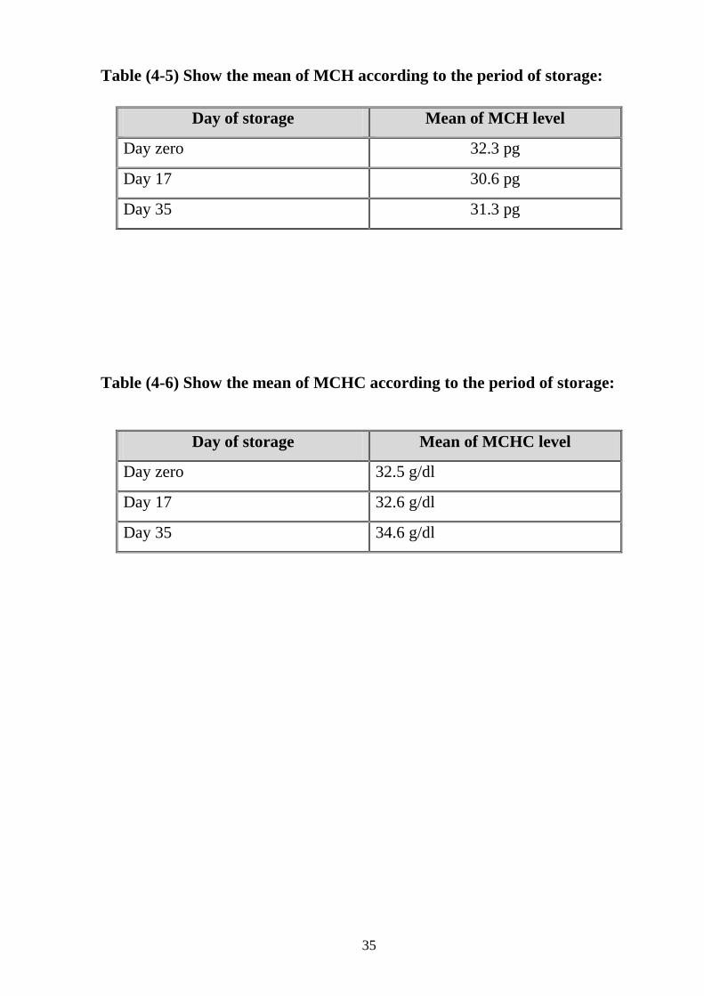
According to the table (4-5) the mean of mean cell haemoglobin level in day
zero was (32.3 pg), while in day 17 was (30.6 pg ) then decreased to (31.3 pg)
in day 35.
Also according to the table (4-6) the mean of mean cell haemoglobin
concentration in day zero was (32.5 g/dl), while in day 17 was (32.6 g/dl ) then
incresed to (34.6 g/dl) in day 35.
The mean of of red distrbuation width in day zero was (15.5 %), while in day 17
was (14.1 % ) then decreased to (14.3%) in day 35 as referred in table (4-7).
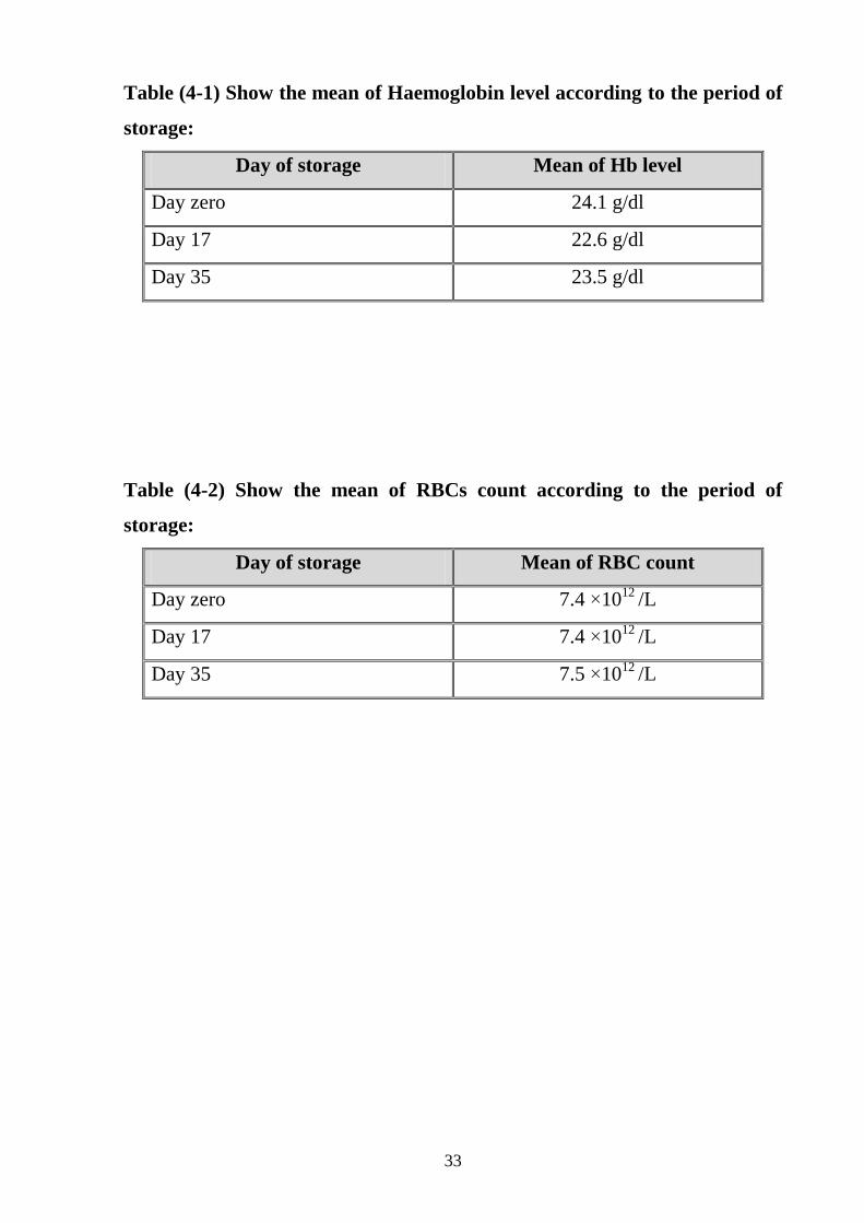
33
Table (4-1) Show the mean of Haemoglobin level according to the period of
storage:
Day of storage Mean of Hb level
Day zero 24.1 g/dl
Day 17 22.6 g/dl
Day 35 23.5 g/dl
Table (4-2) Show the mean of RBCs count according to the period of
storage:
Day of storage Mean of RBC count
Day zero 7.4 ×10¹²
/L
Day 17 7.4 ×10¹²
/L
Day 35 7.5 ×10¹²
/L
34
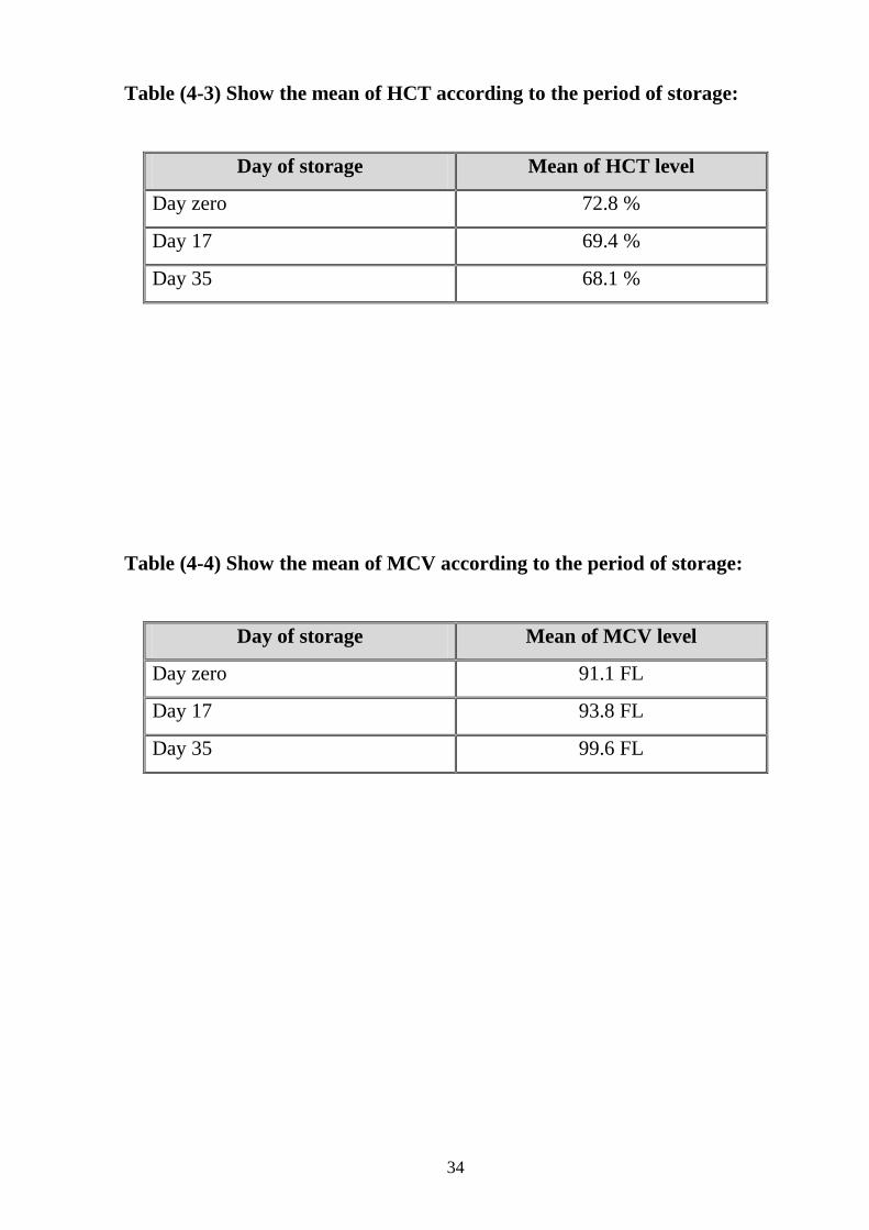
Table (4-3) Show the mean of HCT according to the period of storage:
Day of storage Mean of HCT level
Day zero 72.8 %
Day 17 69.4 %
Day 35 68.1 %
Table (4-4) Show the mean of MCV according to the period of storage:
Day of storage Mean of MCV level
Day zero 91.1 FL
Day 17 93.8 FL
Day 35 99.6 FL
35
Table (4-5) Show the mean of MCH according to the period of storage:
Day of storage Mean of MCH level
Day zero 32.3 pg
Day 17 30.6 pg
Day 35 31.3 pg
Table (4-6) Show the mean of MCHC according to the period of storage:
Day of storage Mean of MCHC level
Day zero 32.5 g/dl
Day 17 32.6 g/dl
Day 35 34.6 g/dl

36
Table (4-7) Show the mean of RDW-CV according to the period of storage:
Day of storage Mean of RDW-CV level
Day zero 15.5 %
Day 17 14.1 %
Day 35 14.3 %

Discussion Conclusion
Recommendations
37
5.1. Discussion
This descriptive prospective cross sectional analytical study was conducted in
Elmak Nimer university hospital blood bank during the period of April to
August 2018 and aimed to determine the effect of storage on Red Blood Cells
Parameters. A total of voluntary donors satisfying the inclusion criteria were
taken.
The results of this study showed that the mean of heamoglolobin level in day
zero was (24.1 g/dl), while in day 17 was ( 22.6 g/dl) and (23.5 g/dl) in day 35.
Statistical analysis showed that there was significant variation with P.value of
(0.000 ) in day 17 and (0.000) in day 35.This result was similar to result of
study done by Karama MI et al in Mosul 2008.,in which though significant fall
in haemoglobin from 10 days onwards of storage (p < 0.05) . This decrease can
be attributed to hemolysis which occurs during storage. Significant fall from 7th
day onward was also observed by Ahmed Y et al in Mosul 2003, ( p<0.05).
The mean of RBCs count in day zero was (7.4 ×10¹²
/L), while in day 17 was
(7.4 ×10¹²
/L) and (7.5 ×10¹²
/L) in day 35.Statistical analysis showed that there
was no significant variation with P.value of (0.491) in day 17. This result was
similar to result of study done by Sonia Chhabra in Indi 2017, ( p <0.05), and
other study done by Adias TC et al (p = 0.376) in Nigeria 2012 . The statistically
significant rise in the mean of RBCs count on day 35 with p.value of ( 0.008)
and this might be due to the delaying in sample processing or improper mixing
of blood.
The mean of HCT was (72.8 % )in day 0 ,while in day 17 was (69.4%) and
(68.1%) in day 35. Statistical analysis showed that there was significant
variation with P.value of (0.017 ) in day 17 and (0.017) in day 35.This result
was similar to result of study done by Karama MI et al in Mosul 2008 and
other study done by Ahmed Y et al (p = 0.008) in Mosul 2003 .
The mean of MCV in day zero was (91.1 fL), while in day 17 was (93.8 fL)
and (99.6 fL) in day 35. Statistical analysis showed that there was significant
variation with P.value of (0.002) in day 17 and (0.000) in day 35.This result was
38
similar to result of study done by Sonia Chhabra in Indi , 2017 (P>0.05) ,
However statistically non significant increased were observed by Adias TC et al
in Nigeria 2012 (p=0.677) The rise in MCV is attributed to the swelling of
RBCs during the storage period.
The mean of MCH in day zero was (32.3 pg ), while in day 17 was ( 30.6 pg)
and (31.3pg) in day 35. Statistical analysis showed that there was significant
variation with P.value of (0.000) in day 17 and (0.004) in day 35 , However
statistically non significant changes were observed by Sonia Chhabra in Indi
2017 and by Adias TC et al in Nigeria 2012 (p =0.805) .
The mean value of MCHC was( 32.5) g/dl in day zero then ( 32.6 g/dl)in day 17
and ( 34.6) g/dl in day 35. Statistical analysis showed that there was in
significant variation with P.value of (0.513) in day 17 , This result was similar
to result of study done by Sonia Chhabra in Indi and by Adias TC et al (p
=0.470) in Nigeria 2012. Also Statistical analysis showed that there was
significant variation with P.value of (0.000) in day 35, this may be attributed to
gradual fall in haematocrit during storage.
The mean of RDW-CV in day zero was (15.5 %), while in day 17 was (14.1 %)
and (14.3%) in day 35. Statistical analysis showed that there was significant
variation with P.value of (0.000) in day 17. The statistically in significant
variation with P.value (0.234) in day 35 was similar to result were observed
by Adias TC et al in Nigeria (2012), with p. value of (0.316) during the 28 day
storage period ,and by Sonia Chhabra et al in India (2017) .
39
5-2.Conclusion
By the end of this study we conclude that:
There was significant variation in Hb ,HCT,MCV,MCH and significant
,insignificant variation during storage in RBCs count ,MCHC and RDW-
CV
The mean of heamoglolobin level in day zero was (24.1 g/dl), while in day
17 was ( 22.6 g/dl) and (23.5 g/dl) in day 35.
The mean of RBCs count in day zero was (7.4 ×10¹²
/L), while in day 17
was (7.4 ×10¹²
/L) and (7.5 ×10¹²
/L) in day 35.
The mean of HCT was (72.8 % )in day 0 ,while in day 17 was (69.4% )
and ( 68.1% ) in day 35.
The mean of MCV in day zero was (91.1 fL), while in day 17 was (93.8
fL) and (99.6 fL) in day 35.
The mean of MCH in day zero was (32.3 pg), while in day 17 was ( 30.6
pg) and (31.3pg) in day 35.
The mean of MCHC in day zero was( 32.5 g/dl) , while in day 17 was (
32.6 g/dl) and ( 34.6 g/d) in day 35.
The mean of RDW-CV in day zero was (15.5 %), while in day 17 was
(14.1 %) and (14.3 %) in day 35
40
5-3.Recommendations
1. Further study about this topic should be done with increase sample size to
obtain accurate result. With increased quality control in hematology lab.
2. Periodic follow up and monitoring of haematological parameters in stored
red blood cells to assess the environmental storage .

References
Appendices
41
6.1. References
1. Kaur P, Basu S, Kaur G, Kaur R. Transfusion protocol in trauma. J Emerg
Trauma Shock. 2011;4:103–108.
2. Lelubre C, Vincent JL. Red blood cell transfusion in the critically ill
patient. Ann Intensive Care. 2011;1:43 .
3. Hess JR. Red cell storage. J Proteomics.2010;73:368–373.
4. Hess JR. An update on solutions for red cell storage. Vox Sang.2006; 91:13–
19.
5. Grindon AJ. Brief history of blood transfusion. In: Hillyer CD, Shaz BH,
Zimring JC, Abshire TC, editors. Transfusion Medicine and Hemostasis:
Clinical and Laboratory Aspects. Burlington: Elsevier; 2009. pp. 9–11.
6. Hess JR. Red cell changes during storage.Transfus Apheresi
Sci. 2010s;43:51–59.
7. Hardwick J. Blood processing. ISBT Sci Ser. 2008;3:148–176.
8. Greening DW, Glenister KM, Sparrow RL, Simpson RJ. International blood
collection and storage: clinical use of blood products. J Proteomics.
2010;73:386–395.
9. Van De Watering L. Red cell storage and prognosis. Vox Sang . 2011; 100: 36
– 45.
10. Rinalducci S, D'Amici GM, Blasi B, Vaglio S, Grazzini G, Zolla L.
Peroxiredoxin-2 as a candidate biomarker to test oxidative stress levels of stored
red blood cells under blood bank condition .Transfusion 2011; 51:1439–1449.
11. Blasi B, D'Alessandro A, Ramundo N, Zolla L. Red blood cell storage and
cell morphology. Transfus Med. 2012;22:90–96.
12. Gevi F, D'Alessandro A, Rinalducci S, Zolla L. Alterations of red blood cell
metabolome during cold liquid storage of erythrocyte concentrates in CPD-
SAGM. J Proteomics. 2012;76:168–180.
13. Yedgar S, Koshkaryev A, Barshtein G. The red blood cell in vascular
occlusion.Pathophysiol Haemost Thromb.2002;32:263–268.
42
14. Berezina TL, Zaets SB, Morgan C, Spillert CR, Kamiyama M, Spolarics Z,
Deitch EA, Machiedo GW. Influence of storage on red blood cell rheological
properties. J Surg Res.2002;102:6–12.
15. Relevy H, Koshkaryev A, Manny N, Yedgar S, Barshtein G. Blood banking-
induced alteration of red blood cell flow properties. Transfusion, 2008; 48:136–
146.
16. Dern R.J. and Brower G.J. Blood J. Lab. Clin. Med., 101: 23 – 35 (1999).
17. McCrosson L. and Masterson G. Br. J. Anaesth., 8: 6 – 9 (2002).
18. Lave. EM., Jones H., Williamson L.M., et al. "Serious hazards of
transfusion". Annual Report. ISDM 09532789 3X. (2000).
19. Swan H. and Schechter D.C. Surgery, 2: 545 – 557 (1962).
20. Rassmusen M.G., Lesses M.F., Anstall H.B. New Eng. J. Med., 264: 1088 –
1095 (1961).
21. Hebert P.C. and Chin – Yee I. "Clinical consequences of prolonged blood
storage; should old red cell be transfused in critically ill patients ?" In: Vincent
J–L, ed. "Year Book of Intensive Care and Emergency Medicine". Berlin,
Heidelberg: Springer – Verlag, 494 –506. (2000).
22. Cohle S.D., Abdus S. Effect of the storage of blood on the stability of
haematological parameters. Am. J. Clin. Pathol., 76: 67 – 69 (1981).
23. Wood B.L., Andrews J., Millers S., Sabath D.E. Am. J. Clin. Pathol., 112:
687 – 695 (1999).
24. Tvedten H., Weiss D.j. "Classification and Laboratory Evaluation of
Anemia". In: Feldman B.F., Zink J.G., Jain N.C., eds. "Schalm's Veterinary
Hematology". 5th ed. Philadelphia, Pa: Lippincott, Williams and Wilkins. Pp.
143 – 150 (2000).
25. Zelmanovic D., Hetherington E.J. Vet. Clin. Pathol., 27: 2 – 9(1998)
26. Walsh T.S., McArdle F., Maclver C., Maginnis M., McClelland D.B. Care
Med., 27: S247 (2001).
27. Vamvakas E.C. and Carven J.H. Transfusion, 40: 101 – 109 (2000).
43
28. The Stationary Office. "Guide Lines for the Blood Transfusion Services in
the United Kingdom". 5th ed., (2000).
29. Kuter D.J., Cebon J., Harker L.A., et al. Transfusion, 39: 321 – 332 (1999).
30. Vedten H.W. Vet. Clin. Pathol., 28: 80 – 82 (1999).
31. Heaton W.A., Holme S., Smith K., et al. Br.J. Haematol., 88: 368-402
(1999).
32. Tvedten H. and Korcal D. Vet. Clin. Pathol., 25: 14 – 22 (1996).
33.Zimrin AB, Hess JR Current issues relating to the transfusion of stored red
blood cells.VoxSanguinis(2009) 96:93-103. 1
34. Jhonson RA ,Lavesa M.Askari B,et al. Aheme oxygenase
product,presumably carbon monoxide,mediates avasodepressor function in rats
(February 1995) .retrived on November 14th
2012.
35.Hoffprand A.V, Pettit J.E., Moss P.A.H Essential Haematology. (2001), 4th
Edition. Pages 1, 14,17,71,83.
36. American Society of Aerospace Medicine Specialists . Clinical practice
guideline for Sickle Cell Disease Trait . Retrived 3May 2013.
37. Sasabil MA,Islam M,Jahan D,KhamMA. Inherited Haemoglobin Disorders
Among Apparently Healthy Individuals : An Analysis of 105 cases. JAFMC
Bangladesh,2014;10(2):90-94
38. Blood Test Result-Normal Ranges: Blood book.Com. Retrived on Jan 7,2009
39.Monica, Cheesbrough, District laboratory practice in tropical countries. Part
2 (2007) 2nd
Edition. Pages 299-313.
40. John P.Greer. Lippin Cott Williams&Wilkins. Wintrobe,s Clinical
Hematology,Volume 1. Front Cover. 2009.
41. Lee RI, Vincent B: The coagulation of normal human blood, an
experimental study. Arch Intern Med1914; 13:395–425
42. Rous P, Turner JR: The preservation of living red blood cells in vitro
Methods of preservation. J Exp Med1916; 23:219–237
43. Rous P, Turner JR: The transfusion of kept cells. J Exp Med1916; 23: 239–
247.
44
44. Robertson OH: A method of citrated blood transfusion. Br Med J1918;
1:477–47.
45. Robertson OH: Transfusion with preserved red blood cells. Br Med J1918;
1:691–695
46. Crile GW, Boggs TR, Cannon WB, Moss WL, Vincent B, Hussey RG, Lee
BJ: Report Upon Transfusion of Blood for the Recently Injured in the US Army.
Paris, American Red Cross, 1918.
47. Dumont LJ, Aubuchon JP, Biomedical Excellence for Safer transfusion
(BEST) Collaborative: Evaluation of proposed FDA criteria for the evaluation of
radiolabeled red cell recovery trials Transfusion. 2008; 48:1053–1060
48. Moroff G, Sohmer PR, Button LN: Proposed standardization of methods for
determining the 24-hour survival of stored red cells. transfus ion1984; 24:109–
114.
49. Luten M, Roerdinkholder-Stoelwinder B, Bost HJ, Bosman GJ: Survival of
the fittest? – Survival of stored red blood cells after transfusion. Cell Mol Biol
(Noisy-le-grand) 2004; 50:197–203
50. FDA Workshop on Red Cells Stored in Additive Solution Systems; 1985 Apr
25; Bethesda, MD.
51. Orlina AR, Josephson AM: Comparative viability of blood stored in ACD
and CPD. Transfusion1969; 9:62–69.
52. Zuck TF, Bensinger TA, Peck CC, Chillar RK, Beutler E, Button LN,
McCurdy PR, Josephson AM, Greenwalt TJ: The in vivo survival of red
blood cells stored in modified CPD with adenine: report of a multi-institutional
cooperative effort. Transfusion 1977; 17:374–382.
53. Heaton WAL, Holme S, Smith K, Brecker ME, Pineda A, AuBuchon JP,
Nelson E: Effects of 3–5 log10 pre-storage leukocyte depletion on red cell
storage and metabolism. Br J Haematol 1994; 87:363–368.
54. Reid TJ, Babcock JG, Derse-Anthony CP, Hill HR, Lippert LE, Hess JR:
The viability of autologous human red blood cells stored in additive solution-5
and exposed to 250C for 24 hours. transfusion 1999; 39:991–997.
45
55. Hess JR, Greenwalt TJ: Storage of red blood cells: New approaches.
Transfus Med Rev 2002; 16:283–295.
56. de Korte D, Kleine M, Korsten HG, Verhoeven AJ: Prolonged maint enance
of 2,3-diphosphoglycerate acid and adenosine triphosphate in red blood cells
during storage. Transfusion 2008; 48.
57. Weisbach V, Wanke C, Zingsem J, Zimmermann R, Eckstein R: Cytokine
generati- ion in whole blood, leukocyte-depleted and temporarily warmed red
blood cell concentrates. Vox Sang 1999; 76:100–106.
58. Anniss AM, Sparrow RL: Storage duration and white blood cell content of
red blood cell (RBC) products increases adhesion of stored RBCs to
endothelium under flow conditions. Transfusion, 2006; 46:1561–1567.
59. Sharifi S, Dzik WH, Sadrzadeh SM: Human plasma and tirilazad mesylate
protect stored human erythrocytes against the oxidative damage of irradiation.
Transfus Med 2000; 10:125–130.
60. Yomtovian R, Lazarus HM, Goodnough LT, Hirschler NV, Morrissey
AM, Jacobs MR: A prospective microbiologic surveillance program to detect
and prevent the transfusion of bacterially contaminated platelets. Transfusion
1993; 33:902–909.
61. de Korte D, Marcelis JH, Soeterboek AM: Determination of the degree of
bacterial contamination of whole-blood collections using an automated microbe-
detection system. Transfusion2001; 41:815–818.
62. Kuehnert MJ, Roth VR, Haley NR, Gregory KR, Elder KV, Schreiber GB,
Arduino MJ, Holt SC, Carson LA, Banerjee SN, Jarvis WR: Transfusion-
transmitted bacterial infection in the United States, 1998 through 2000.
Transfusion 2001; 41:1493–1499.
63. Brecher ME, Hay SN: Bacterial contamination of blood components. Clin
Microbiol Rev 2005; 18:195–204.
64. Baz EM, Kanazi GE, Mahfouz RA, Obeid MY: An unusual case of
hyperkalaemia-induced cardiac arrest in a paediatric patient during transfusion
of a ‘fresh’ 6-day-old blood unit. Transfus Med 2002; 12:383–386.

46
65. Silliman CC: The two-event model of transfusion-related acute lung injury.
Crit Care Med 2006; 34:S124–S131.
66. Gajic O, Rana R, Winters JL, Yilmaz M, Mendez JL, Rickman OB, O’Byrne
MM, Evenson LK, Malinchoc M, DeGoey SR, Afessa B, Hubmayr RD, Moore
SB: Transfusion-related acute lung injury in the critically ill: prospective nested
case-control study. Am J Respir Crit Care Med 2007; 176:886–891
67. Eder AF, Herron R, Strupp A, Dy B, Notari EP, Chambers LA, Dodd RY,
Benjamin RJ: Transfusion-related acute lung injury (2003–2005) and the
potential impact of the selective use of plasma from male donors in the
American Red Cross. Transfusion 2007; 47:599–607.
68.Greenwalt TJ: The how and why of exocytic vesicles. Transfusion 2006;
46:143–152.
69. Tinmouth A, Chin-Yee I: The clinical consequences of the red cell storage
lesion. Transfus Med Rev 2001; 15:91–107.
70. Tsai AG, Cabrales P, Intaglietta M: Microvascular perfusion upon exchange
transfusion with stored red blood cells in normovolemic anemic conditions.
Transfusion 2004; 44:1626–1634.
71. Hardeman MR, Ince C: Clinical potential of in vitro measured red cell
deformability, a myth? Clin Hemorheol Microcirc 1999; 21:277–284.
72. Raat NJ, Verhoeven AJ, Mik EG, Gouwerok CW, Verhaar R, Goedhart PT,
de Korte D, Ince C: The effect of storage time of human red cells on intestinal
microcirculatory oxygenation in a rat isovolemic exchange model. Crit Care
Med 2005; 33:39–45.
73. Isbell TS, Sun CW, Wu LC, Teng X, Vitturi DA, Branch BG, Kevil CG,
Peng N, Wyss JM, Ambalavanan N, Schwiebert L, Ren J, Pawlik KM, Renfrow
MB, Patel RP, Townes TM: SNO-hemoglobin is not essential for red blood cell-
dependent hypoxic vasodilation. Nat Med 2008; 14:773–777.
74. d’Almeida MS, Gray D, Martin C, Ellis CG, Chin-Yee IH: Effect of
prophylactic transfusion of stored RBCs on oxygen reserve in response to acute
isovolemic hemorrhage in a rodent model. Transfusion 2001; 41:950–956.
47
75. Torres Filho IP, Spiess BD, Pittman RN, Barbee RW, Ward KR:
Experimental analysis of critical oxygen delivery. Am J Physiol Heart Circ
Physiol 2005; 288:H1071–H1079.
76. British Committee for Standards in Haematology: Guideline on the
administration of blood.; in: 2009.
77. Guidelines for the Blood Transfusion Services in the United Kingdom: ed 8th
The Stationary Office, London, 2013.
78. AABB Standards Program Committee: Standards for blood banks and
transfusion services, ed 28. Bethesda, aaBB, 2012.
79. Pick P, Fabijanic J: Temperature changes in donor blood under different
storage conditions. Transfusion 1971; 11(4):213-215.
80. Johnson V, Langeberg A, Taye-Makuria A, Sandler SG: Temperature-
sensitive labels for containers of RBCs. Am J Clin Pathol 2006; 126(3):406-410.
81. McLaughlin N, Sadek I, Anderson D: Bacterial growth in RBC units
exposed to room temperature for greater than 30 minutes. Transfusion 2010; 50
(Suppl2):76a.
82. Perry HE, Prasad P, Kirwan S, Huang YQ: Core temperature changes in
resuspended red blood cells (RBCs) and pediatric RBCs removed from
refrigerated storage. Transfusion 2010; 50(1):174-177.
83. Reiter U, Reiter G, Wagner T, Kozma N, Roland J, Schollnast H, Ebner F,
Lanzer G: Four-dimensional temperature distributions in red blood cells
withdrawn from storage and exposed to ambient temperature: a magnetic
resonance thermometry study . Transfusion 2013; 53(1):167-173.
84. Hughes-Jones NC: Storage of red cells at temperatures between + 10 degrees
C and -20 degrees C. Br J Haematol 1958; 4(3):249-255.
85. Blood Stocks Management Scheme: Annual report 2012-2014; in: 2015.
86. Brunskill S, Thomas S, Whitmore E, McDonald CP, Doree C, Hopewell S,
Staves J, Cardigan R, Murphy MF: What Is the Maximum Time That a Unit of
Red Blood Cells Can Be Safely Left Out of Controlled Temperature Storage?
Transfus Med Rev 2011.
48
87. Hogman CF, Meryman HT: Storage parameters affecting red blood cell
survival and function after transfusion. Transfus Med Rev 1999; 13(4):275-296
88. Sonia Chhabra, Saurav Chaudhary, P.K.Sehgal, Sunita Singh, Monika
Gupta, Rajeev Sen. International J. of Healthcare and Biomedical Research,
Volume: 05, Issue: 04, July 2017, 69-75.
89. Ahmed Y, Dallal B, Bashar M. Effect of blood storage on certain
hematological parameters. Tikrit Med Jour. 2009;15(1):171-80.
90. Behrooz Ghezelbash, Azita Azarkeivan, Ali Akbar Pourfathollah,
Mohammadreza Deyhim, Esmerdis Hajati, Alireza Goodarzi. International
Journal of Hematology –Oncology and Stem Cell Research 2018.12(1):35-42.
91. Karma MI, Nuamy AL. Hematological changes in stored blood. Jour
Edu2008;21(4):53-4.
92. Akbar Hashemi Tayer , Naser Amirizadeh , Mahtab Mghsodlu , Mahin
Nikogoftar , Mohammad Reza. Iran J Ped Hematol Oncol.2017,Vol 7.No
3,171-179.
93. Adias TC, MooreIgwe B, Jeremiah ZA. Storage related Hematological and
Biochemical changes of CPDA-1 stored blood in a limited Resource Setting. J
Blood Disorders. 2012;3(3):1-4.
94. Society for Medical Laboratory Sciences,Srilanka . Hematology Analyzer-
From Complete Blood Count to Cell Morphology. August 11,2017
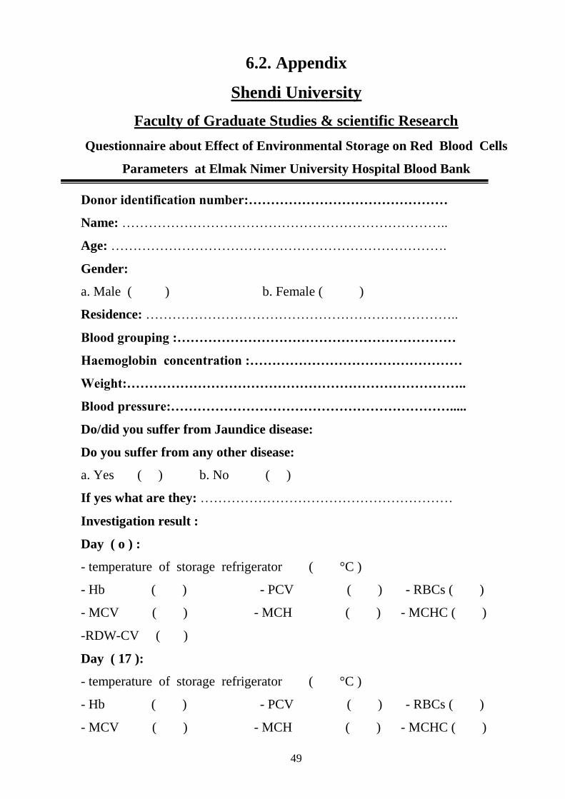
49
6.2. Appendix
Shendi University
Faculty of Graduate Studies & scientific Research
Questionnaire about Effect of Environmental Storage on Red Blood Cells
Parameters at Elmak Nimer University Hospital Blood Bank
Donor identification number:………………………………………
Name: ………………………………………………………………..
Age: ………………………………………………………………….
Gender:
a. Male ( ) b. Female ( )
Residence: ……………………………………………………………..
Blood grouping :………………………………………………………
Haemoglobin concentration :…………………………………………
Weight:…………………………………………………………………..
Blood pressure:……………………………………………………….....
Do/did you suffer from Jaundice disease:
Do you suffer from any other disease:
a. Yes ( ) b. No ( )
If yes what are they: …………………………………………………
Investigation result :
Day ( o ) :
- temperature of storage refrigerator ( °C )
- Hb ( ) - PCV ( ) - RBCs ( )
- MCV ( ) - MCH ( ) - MCHC ( )
-RDW-CV ( )
Day ( 17 ):
- temperature of storage refrigerator ( °C )
- Hb ( ) - PCV ( ) - RBCs ( )
- MCV ( ) - MCH ( ) - MCHC ( )
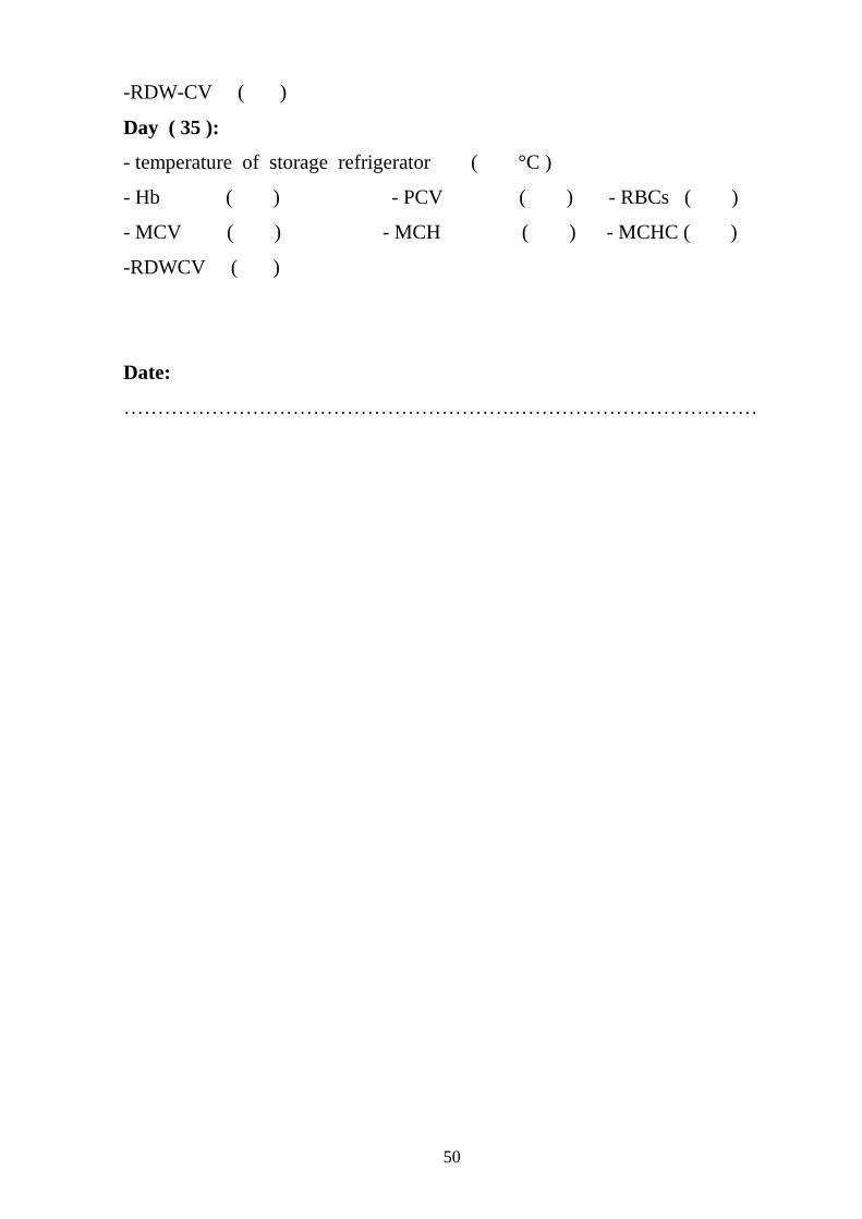
50
-RDW-CV ( )
Day ( 35 ):
- temperature of storage refrigerator ( °C )
- Hb ( ) - PCV ( ) - RBCs ( )
- MCV ( ) - MCH ( ) - MCHC ( )
-RDWCV ( )
Date:
………………………………………………….………………………………