University of Louisville Journal of Respiratory Infections
8
*Johnson Britto 1 Introduction Blastomycosis is caused by the dimorphic fungus Blastomyces dermatitidis. Blastomycosis is an endemic mycosis that occurs in defined geographic areas and usually in regions that are warm with soil rich in organic debris, such as decaying vegetation and decomposed wood [1, 2]. In North America, blastomycosis usually occurs in the southeastern and south-central states that border the Mississippi and Ohio Rivers, the midwestern states and Canadian provinces that border the Great Lakes and Saint Lawrence Riverway [1-5]. Blastomycosis is a systemic pyogranulomatous infection. The clinical spectrum of blastomycosis is varied, including asymptomatic infection, acute or chronic pneumonia, extrapulmonary and disseminated disease. The lungs are the most common site of infection, followed by the skin, bones, and genitourinary system. A subset of individuals with acute pulmonary blastomycosis can progress to fulminant multi-lobar pneumonia and acute respiratory distress syndrome [10]. Diagnostic delays are not uncommon and often result in increased morbidity and mortality. Here, we describe a case of progressive pulmonary blastomycosis in a young healthy adult. Case Report A 31 year-old African American female with no significant past medical history presented to an outside hospital complaining of 1-week history of subjective fevers and chills, fatigue, nonproductive cough, dyspnea and left sided pleuritic chest pain. Initial vital signs were temperature 100.5ºF, heart rate 95 beats per minute, respiratory rate 18 breaths per minute, blood pressure 148/88 mm Hg, and oxygen saturation 99% on room air. On physical exam, she had no respiratory distress, no lymphadenopathy, examination of the respiratory system revealed bilateral vesicular breath sounds and crackles at the left lower lobe. Examination of the cardiovascular system, abdomen, central nervous system, and skin were unremarkable. Initial diagnostic laboratory work up showed leucocyte count of 14,500 cells/mm 3 (76% segmented neutrophils, 13% lymphocytes and 11% monocytes), hemoglobin 12.9 g/dl, hematocrit 38.9% and platelet count 316,000/mm 3 . Serum electrolytes, renal function, and liver function tests were normal. Rapid Influenza test was positive for Influenza A. Urine streptococcal and legionella antigens were negative. Chest X-ray (CXR) showed an ovoid superior segment left lower lobe focus of consolidation (Figure 1). A diagnosis of Influenza A and possible community acquired bacterial pneumonia was made and patient was admitted to the hospital. She was started on oral oseltamivir and intravenous ceftriaxone and azithromycin. University of Louisville Journal of Respiratory Infections Non-resolving Community Acquired Pneumonia (CAP) due to Blastomyces dermatitidis (Pulmonary Blastomycosis): Case Report and Review of Literature Abstract In this case report, we describe a case of progressive acute pulmonary blastomycosis in a healthy adult living in Kentucky, initially presenting with flu like illness with a left sided consolidation, who did not respond to antibiotic therapy. Patient’s clinical condition deteriorated with development of necrotizing bronchopneumonia, mediastinal lymphadenopathy, tree-in-bud reticulonodularity and pleural effusion. A diagnosis of progressive pulmonary blastomycosis was established by radiological findings as well as transbronchial needle aspiration cytology and bronchoalveolar lavage culture demonstrating Blastomyces dermatitidis. Patient showed significant clinical improvement with resolution of pulmonary lesions on antifungal treatment. Since symptoms of blastomycosis are often similar to the symptoms of flu or other lung infections, our case highlights the importance of maintaining a high index of suspicion and appropriate microbiologic and histologic evaluation especially in patients who live in or have traveled to areas endemic for blastomycosis and are not responding to antibiotic therapy. Early diagnosis coupled with prompt initiation of antifungal treatment may lead to favorable outcomes. DOI: 10.18297/jri/vol2/iss1/9 Received Date: February 12, 2018 Accepted Date: March 26, 2018 Website: https://ir.library.louisville.edu/jri Affiliations: 1 St. Elizabeth Physicians, Infectious Disease, Crestview Hills, KY, USA ©2018, The Author(s). 39 ULJRI Vol 2, (1) 2018 REVIEW ARTICLE *Correspondence To: Johnson Britto, MD, MPH Work Address: St. Elizabeth Physicians, Infectious Disease, Crestview Hills, KY, USA Work Email: [email protected]
-
Upload
khangminh22 -
Category
Documents
-
view
1 -
download
0
Transcript of University of Louisville Journal of Respiratory Infections

*Johnson Britto1
Introduction
Blastomycosis is caused by the dimorphic fungus Blastomyces dermatitidis. Blastomycosis is an endemic mycosis that occurs in defined geographic areas and usually in regions that are warm with soil rich in organic debris, such as decaying vegetation and decomposed wood [1, 2]. In North America, blastomycosis usually occurs in the southeastern and south-central states that border the Mississippi and Ohio Rivers, the midwestern states and Canadian provinces that border the Great Lakes and Saint Lawrence Riverway [1-5]. Blastomycosis is a systemic pyogranulomatous infection. The clinical spectrum of blastomycosis is varied, including asymptomatic infection, acute or chronic pneumonia, extrapulmonary and disseminated disease. The lungs are the most common site of infection, followed by the skin, bones, and genitourinary system. A subset of individuals with acute pulmonary blastomycosis can progress to fulminant multi-lobar pneumonia and acute respiratory distress syndrome [10]. Diagnostic delays are not uncommon and often result in increased morbidity and mortality. Here, we describe a case of progressive pulmonary blastomycosis in a young healthy adult.
Case Report
A 31 year-old African American female with no significant past medical history presented to an outside hospital complaining
of 1-week history of subjective fevers and chills, fatigue, nonproductive cough, dyspnea and left sided pleuritic chest pain.
Initial vital signs were temperature 100.5ºF, heart rate 95 beats per minute, respiratory rate 18 breaths per minute, blood pressure 148/88 mm Hg, and oxygen saturation 99% on room air. On physical exam, she had no respiratory distress, no lymphadenopathy, examination of the respiratory system revealed bilateral vesicular breath sounds and crackles at the left lower lobe. Examination of the cardiovascular system, abdomen, central nervous system, and skin were unremarkable.
Initial diagnostic laboratory work up showed leucocyte count of 14,500 cells/mm3 (76% segmented neutrophils, 13% lymphocytes and 11% monocytes), hemoglobin 12.9 g/dl, hematocrit 38.9% and platelet count 316,000/mm3. Serum electrolytes, renal function, and liver function tests were normal. Rapid Influenza test was positive for Influenza A. Urine streptococcal and legionella antigens were negative. Chest X-ray (CXR) showed an ovoid superior segment left lower lobe focus of consolidation (Figure 1).
A diagnosis of Influenza A and possible community acquired bacterial pneumonia was made and patient was admitted to the hospital. She was started on oral oseltamivir and intravenous ceftriaxone and azithromycin.
University of LouisvilleJournal of Respiratory Infections
Non-resolving Community Acquired Pneumonia (CAP) due to Blastomyces dermatitidis (Pulmonary Blastomycosis): Case Report and Review of Literature
Abstract
In this case report, we describe a case of progressive acute pulmonary blastomycosis in a healthy adult living in Kentucky, initially presenting with flu like illness with a left sided consolidation, who did not respond to antibiotic therapy. Patient’s clinical condition deteriorated with development of necrotizing bronchopneumonia, mediastinal lymphadenopathy, tree-in-bud reticulonodularity and pleural effusion. A diagnosis of progressive pulmonary blastomycosis was established by radiological findings as well as transbronchial needle aspiration cytology and bronchoalveolar lavage culture demonstrating Blastomyces dermatitidis. Patient showed significant clinical improvement with resolution of pulmonary lesions on antifungal treatment. Since symptoms of blastomycosis are often similar to the symptoms of flu or other lung infections, our case highlights the importance of maintaining a high index of suspicion and appropriate microbiologic and histologic evaluation especially in patients who live in or have traveled to areas endemic for blastomycosis and are not responding to antibiotic therapy. Early diagnosis coupled with prompt initiation of antifungal treatment may lead to favorable outcomes.
DOI: 10.18297/jri/vol2/iss1/9Received Date: February 12, 2018Accepted Date: March 26, 2018Website: https://ir.library.louisville.edu/jriAffiliations:1St. Elizabeth Physicians, Infectious Disease, Crestview Hills, KY, USA
©2018, The Author(s).
39ULJRI Vol 2, (1) 2018
REVIEW ARTICLE
*Correspondence To: Johnson Britto, MD, MPH Work Address: St. Elizabeth Physicians, Infectious Disease, Crestview Hills, KY, USAWork Email: [email protected]
Due to a lack of clinical improvement after three days of antimicrobial treatment, chest computed tomographic (CT) scan was obtained that showed a dense consolidation involving the majority of the left lower lobe, and a trace left pleural effusion (Figure 2). Bronchoscopy was performed. Bronchoscopy did not show any endobronchial lesions, Gram’s stain of bronchoalveolar lavage (BAL) of left lower lobe showed many white blood cells and rare Gram-positive bacilli, culture grew 10,000-100,000 CFU/mL of normal respiratory flora, KOH Prep was negative for fungal elements, acid-fast bacilli was negative, as was legionella PCR. BAL sample was not sent for cytological analysis. She was hospitalized for five days during which there was no significant change in her clinical status and she remained without clinical improvement or deterioration. She completed a five day course of oral oseltamivir. Intravenous antimicrobials were switched to oral levofloxacin to complete a course of seven days and she was discharged home with outpatient follow up.
Figure 1. Initial Chest X-ray (CXR) showing ovoid superior segment left lower lobe consolidation.
Figure 2. Chest computed tomographic (CT) scan showing dense consolidation involving the majority of the left lower lobe and trace left pleural effusion.
After discharge from hospital, she reported worsening of her symptoms and complained of fevers and chills, productive cough with hemoptysis, dyspnea, pleuritic left sided chest pain and worsening fatigue. She presented to our hospital for evaluation of these symptoms about four days after discharge from an outside hospital.
Vital signs at the time of initial evaluation in our emergency department were as follows: temperature 102.0ºF, heart rate 115 beats per minute, respiratory rate 20 per minute, blood pressure 126/66 mm Hg, and oxygen saturation 95% on room air. On physical exam, she had no respiratory distress, no lymphadenopathy, examination of the respiratory system revealed diminished breath sounds, whisper pectoriloquy, egophony and crackles at the left lower lobe, examination of the cardiovascular system, abdomen, central nervous system and skin were unremarkable.
Pertinent initial diagnostic laboratory work up showed leucocyte count of 33.3 x 10³ cells/mm3 (83% segmented neutrophils, 12% lymphocytes and 4% monocytes). Hemoglobin 11.5 g/dl, hematocrit 34% and platelet count 539,000/mm3. Serum electrolytes revealed serum sodium 139 mmol/L, potassium of 3.1 mmol/L, renal function revealed blood urea nitrogen and serum creatinine of 6.0 mg/dL and 0.46 mg/dL respectively, liver function tests revealed alkaline phosphatase 255 U/L, AST 145 U/L, ALT 110 U/L, albumin 3.1 g/dL, and total bilirubin 1.0 mg/dL.
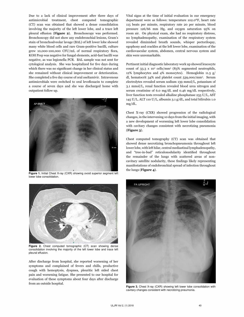
Chest X-ray (CXR) showed progression of the radiological changes, in the intervening 10 days from the initial imaging, with a new development of worsening left lower lobe consolidation with cavitary changes consistent with necrotizing pneumonia (Figure 3).
Chest computed tomography (CT) scan was obtained that showed dense necrotizing bronchopneumonia throughout left lower lobe, with left hilar, central mediastinal lymphadenopathy, and “tree-in-bud” reticulonodularity identified throughout the remainder of the lungs with scattered areas of non-cavitary satellite nodularity, these findings likely representing manifestations of endobronchial spread of infection throughout the lungs (Figure 4).
Figure 3. Chest X-ray (CXR) showing left lower lobe consolidation with cavitary changes consistent with necrotizing pneumonia.
40ULJRI Vol 2, (1) 2018

The patient was admitted to the hospital with a diagnosis of non-resolving pneumonia. She was started on empiric intravenous vancomycin and piperacillin/tazobactam. Sputum Gram’s stain showed Gram-positive cocci in pairs and sputum culture grew 2+ indigenous oral flora. Sputum for acid-fast bacilli was negative, as was MRSA nasal PCR, viral respiratory panel, serum interferon gamma release assay, human immunodeficiency virus serology, urine streptococcal and legionella antigens, and blood cultures. Procalcitonin level was 0.3 ng/mL.
Bronchoscopy was performed, it did not show any endobronchial lesions. A large amount of purulent secretions were seen at the superior segment of left lower lobe which were suctioned out without difficulty. Bronchoalveolar lavage (BAL) and transbronchial lung biopsies were obtained from left lower lobe segments.
Despite being on intravenous antibiotics for four days, she developed worsening of her clinical status. Repeat chest X-ray (CXR) and chest computed tomographic (CT) scan were obtained that showed worsening pneumonia with complete consolidation of the left lower lobe and new patchy consolidation and nodules in the right lung as shown in Figure 5 and Figure 6 respectively. Additional laboratory work up including fungal
41ULJRI Vol 2, (1) 2018
Figure 4. Chest computed tomographic (CT) scan showing dense necrotizing bronchopneumonia seen throughout left lower lobe with left hilar, central mediastinal lymphadenopathy, and “tree-in-bud” reticulonodularity identified throughout the remainder of lungs with scattered areas of non-cavitary satellite nodularity.
Figure 5. Chest X-ray (CXR) showing persistent diffuse bilateral pneumonia, left greater than right, with a moderate left-sided pleural effusion.
Figure 6. Chest computed tomographic (CT) scan showing worsening diffuse pneumonia with complete consolidation of the left lower lobe, new patchy consolidation in the left upper lobe/lingula and right lower lobe and new/enlarging nodules in the right lung. New small left pleural effusion with fluid in the fissure and layering over the apex.

blood cultures, urine Histoplasma antigen, serum Aspergillus Galactomannan antigen by EIA, serum (1-3)-Beta-D-Glucan, serum Coccidioides immitis antibodies were ordered. Fungal blood cultures, Aspergillus Galactomannan antigen, serum Coccidioides immitis antibodies were negative. Urine Histoplasma antigen was detected and (1-3)-Beta-D-Glucan was strongly positive at >500 pg/mL. (Less than 60 pg/mL are interpreted as negative. 60 to 79 pg/mL are interpreted as indeterminate).
Patient was empirically started on IV amphotericin B. The transbronchial lung biopsy results from the left lower lobe returned and showed numerous lymphocytes, histiocytes, many multinucleated giant cells, forming ill-defined granulomas. The biopsy also revealed the presence of thick-walled single yeast forms of approximately 8–15 µm in size as shown in Figure 7a. The organisms stained positively with Gomori methenamine silver (GMS) stain as shown in Figure 7b. Some of these yeast forms showed eccentric broad-based budding, overall morphology was consistent with blastomycosis. Bronchial biopsy and BAL cultures using Sabouraud Dextrose Agar (SDA) showed growth of fungus as shown in Figures 8a, 8b and 8c.
The patient showed significant clinical improvement on antifungal treatment. She was treated with amphotericin for 2 weeks followed by oral itraconazole. She tolerated the treatment well without any side effects. Follow-up imaging of the lungs showed resolution of the pulmonary lesions.
Figure 7a. Hematoxylin-and-eosin-stained section of left lower lobe lung biopsy, low-power (Bottom) and high-power (Top) magnification, shows numerous lymphocytes, histiocytes, many multinucleated giant cells, forming ill-defined granulomas and presence of thick-walled single yeast forms of approximately 8–15 µm in size with eccentric broad-based budding overall morphology consistent with blastomycosis.
Figure 7b. Gomori methenamine silver (GMS) (1000X magnification) stain shows a budding yeast of Blastomyces dermatitidis with the characteristic broad-based bud.
Figure 8a. Phase-contrast microscopy: (1000X magnification): Shows the typical appearance of Blastomyces dermatitidis. Round to oval shape, thick, doubly refractile cell wall, and single broad-based bud.
Discussion
In this case report, we have described a case of progressive acute pulmonary blastomycosis in a healthy adult living in Kentucky, initially presenting with flu like illness with a left sided consolidation on chest x-ray. This review focuses on the epidemiological, clinical, diagnostic, and therapeutic aspects of blastomycosis.
42ULJRI Vol 2, (1) 2018

Figure 8b. Lacto-Phenol-Cotton-Blue Stain (LPCB) (1000X magnification): Shows the mold form of Blastomyces dermatitidis characterized by delicate, septate hyphae, unbranched conidiophores of rather short, yet varying length extending from the hyphae. Conidia are produced singly at the apex of the conidiophore or develop directly on the hyphae. Conidia are unicellular, round to pyriform (tear drop) in shape (~2 to 10 µm dia.). Conidia at the terminal end of the conidiophore resemble a “lollipop” in structure.
Figure 8c. Bronchial and BAL culture using Sabouraud Dextrose Agar (SDA): Blastomyces dermatitidis takes 2 to 3 weeks to grow at 25 to 30 degrees C. Blastomyces dermatitidis appears as white to beige cottony, mold (mycelium) on Sabouraud dextrose agar.
Epidemiology
Most cases of blastomycosis have been reported in North America. Endemic areas include the southeastern and south-central states bordering the Mississippi and Ohio River basins, the midwestern states and Canadian provinces bordering the Great Lakes and Saint Lawrence Riverway [1-5]. Outside of North America, blastomycosis has been reported in Africa with occasional cases reported from Mexico, Central and South America, India, and the Middle East.
Mycology
Blastomyces dermatitidis is a thermally dimorphic fungus. It grows as the mycelial phase at room temperature and as the yeast phase at 37ºC. This dimorphism appears to result from a hybrid histidine kinase that senses host signals and triggers the transition of Blastomyces dermatitidis from mold to yeast form [6].
Pathogenesis
Blastomycosis pulmonary infection occurs by inhalation of the conidia of Blastomyces dermatitidis. The conidia convert to yeast phase in the lung. The yeast forms are more resistant to phagocytosis and killing [7].
This is an important step in terms of pathogenesis because this conversion results in a survival advantage and probably contributes to infection by Blastomyces dermatitidis.
Blastomycosis is a systemic pyogranulomatous infection. The typical inflammatory response consists of clusters of neutrophils and noncaseating granulomas with epithelioid and giant cells. This response is similar to that seen with other dimorphic fungi including coccidioidomycosis, histoplasmosis, and sporotrichosis.
Clinical Manifestations
The clinical spectrum of blastomycosis is varied, including asymptomatic infection, acute or chronic pneumonia, extrapulmonary and disseminated disease [8]. The lungs are the most common site of infection, followed by the skin, bones, and genitourinary system in decreasing order of frequency [9]. A subset of individuals with acute pulmonary blastomycosis can progress to fulminant multi-lobar pneumonia and acute respiratory distress syndrome [10]. Diagnostic delays are not uncommon and often result in increased morbidity and mortality.
Acute infection is frequently unrecognized unless related to a cluster of cases or group exposure. The signs and symptoms of acute pulmonary blastomycosis usually begin abruptly and may be indistinguishable from viral or bacterial pneumonia [8]. Acute blastomycosis may mimic other diseases [11]. This is due to the variability of the presentation of blastomycosis and its relative rarity. Our case is an example of how clinical presentation of acute pulmonary blastomycosis may be initially considered as community-acquired bacterial or viral pneumonia.
Acute pulmonary blastomycosis can also present with varied radiographic findings. Alveolar infiltrates with or without cavitation, mass lesions, and fibronodular infiltrates are most common [8, 9]. The radiographic findings in our patient included lobar and segmental alveolar infiltrates progressing to cavitation, fibronodular infiltrates, tree in bud opacities, hilar and mediastinal adenopathy, pleural effusion and diffuse bilateral pulmonary involvement from presumed endobronchial spread.
43ULJRI Vol 2, (1) 2018

Spontaneous resolution of acute pulmonary blastomycosis has been reported, but its frequency is unknown [1, 12-14]. Although it has been reported that pulmonary blastomycosis in some patients may resolve without therapy, our case reminds us that previously healthy patients can have progression of acute pulmonary blastomycosis. Hence, our case highlights the importance of prompt diagnosis and institution of treatment in all patients suspected to have acute pulmonary blastomycosis. Also, cases have been reported where treatment of acute pulmonary blastomycosis was withheld and patients later presented with extra-pulmonary disease [15].
Though our patient did not develop severe respiratory failure despite bilateral pulmonary involvement and endobronchial spread of the lungs, blastomycosis is known to cause acute respiratory distress syndrome with a high morbidity and mortality [10] and therefore an early diagnosis coupled with prompt treatment may lead to favorable outcomes.
Diagnosis
Due to the variability of the presentation of blastomycosis and its relative rarity, a high index of suspicion and appropriate microbiologic and histologic evaluation should be performed when the diagnosis is uncertain and patients live in or have traveled to areas endemic for blastomycosis.
No clinical syndrome or radiological features are absolutely diagnostic of blastomycosis. The spectrum of disease that Blastomyces dermatitidis causes overlaps with those of other fungal pathogens and malignancy. Hence definitive diagnosis requires the growth of the organism from clinical specimens [8]. Unlike some fungi such as candida species, colonization or contamination with Blastomyces dermatitidis does not occur, which means that visualizing the organism on histology or obtaining a positive culture confirms the diagnosis of blastomycosis.
A presumptive diagnosis may be made by visualization of the characteristic yeast form in clinical specimens including histopathology or by culture of the organism in body fluids or tissue. In the appropriate clinical setting, visualization of the fungus can prompt the initiation of antimicrobial therapy.
Diagnosis can be established by examination of wet preparations of clinical specimens (10% KOH and/or calcofluor white) but they have a low diagnostic yield (36-46%) [16]. Despite these low diagnostic results, the simplicity and the low cost of the procedure, as well as the potential for rapid diagnosis, justify a wet preparation on all clinical specimens.
Cytology has been shown to be particularly useful in diagnosis, permitting identification of the etiologic agent in 56% of all cases and 72% of pulmonary cases of blastomycosis in a retrospective study [17]. When visualized, the yeast cells are easily differentiated from others on the basis of their size (8–15 µm in diameter), multinucleate with thick refractile cell wall, and single, broad-based buds. Bronchoscopy is useful for diagnosis, especially in patients who are not producing sputum or in whom the radiograph indicates the possibility of malignancy [16]. Bronchial washings, lavage fluid, and post-bronchoscopy sputum samples should be sent for cytology
as well as smear and culture because the organism is often visualized on Papanicolaou preparations [18]. Papanicolaou staining of respiratory tract specimens has a high diagnostic yield (93%); the organism appears to be refractile and pale blue-green with this stain.
A pyogranulomatous tissue response is frequently seen in blastomycosis and should signal the possibility of this fungus. The characteristic host response to infection with Blastomyces dermatitidis is a mixed inflammatory reaction with clusters of polymorphonuclear leukocytes and granulomas, usually noncaseating, with epitheloid histiocytes and giant cells, primarily the foreign body type.
Identification of Blastomyces dermatitidis in hematoxylin-eosin (H & E) stained histopathologic specimens is often difficult, and special stains, such as Gomori methenamine-silver (GMS) or periodic acid-Schiff (PAS) are useful in examining the tissue for the presence of fungal elements. Serology is not useful for the diagnosis of blastomycosis due to high degree of cross-reactivity shared by other endemic mycoses especially Histoplasma capsulatum [19, 20]. The urine of patients with blastomycosis may contain cross-reactive or shared antigens with H. capsulatum var. capsulatum; in one study, 12 of 19 patients with blastomycosis tested positive with the H. capsulatum urine antigen assay [20]. This was noted in our case as well.
Although not an antigen test, detection of (1-3)-Beta-D-Glucan in serum has been shown to be a useful adjunct for diagnosis of invasive fungal infections, especially candidiasis, aspergillosis, and histoplasmosis [21-23]. In contrast, preliminary data suggest that the test is not of value with patients with blastomycosis. In a very small study of four patients with microbiologically proven Blastomyces dermatitidis infection, (1-3)-Beta-D-Glucan was detected in only one patient, who had chronic disseminated disease with multiple skin lesions [24]. Our patient had a strongly positive (1-3)-Beta-D-Glucan.
Culture of Blastomyces dermatitidis from environmental sources remains difficult, in patients with pulmonary blastomycosis, sputum culture has a high positive yield (e.g., 75% per single sample, 86% per patient) [16]. Specimens obtained for culture by bronchoscopy yielded a positive diagnosis in 92% of patients. Any material obtained should be placed on Sabouraud agar or, preferably, more enriched agar (e.g., Sabhi, brain-heart infusion, Gorman medium). Because initial growth is more dependable at 30°C, this temperature is recommended. Specimens contaminated with bacteria should also be cultured on medium containing an antibacterial antibiotic such as chloramphenicol. The mycelial form of Blastomyces dermatitidis is not diagnostic, and conversion to the yeast form is required for confirmation. Early identification of mycelial cultures is possible with commercially available chemiluminescent DNA probes that recognize unique RNA sequences of Blastomyces dermatitidis [25].
Differential Diagnosis
Endospores of Coccidioides immitis may resemble single yeast cells, but the presence of budding can be used to distinguish Blastomyces dermatitidis. While yeast forms of Blastomyces dermatitidis as described in our case report, show broad based
44ULJRI Vol 2, (1) 2018

budding, Paracoccidioides brasiliensis, rarely seen in the United States, is distinguished by the presence of multiple, narrow-based buds. Blastomyces dermatitidis may sometimes be as small as Cryptococcus neoformans, although the capsule and narrow-based bud of the latter, aid in differentiation.
Treatment
The greatest challenge in making the diagnosis of blastomycosis is considering it in the first place. Although the disease can be fatal, most patients are cured with antifungal therapy.
Before the availability of chemotherapy for treatment of blastomycosis, the disease as reported had a progressive course with eventual extrapulmonary disease and a mortality rate exceeding 60%. Even though isolated cutaneous disease had a better prognosis, skin lesions were progressive, and spontaneous recovery was uncommon. Thus, after the introduction of effective antifungal therapy, it was accepted that all patients with blastomycosis should be treated.
Amphotericin B (AmB) was previously considered the treatment of choice for all clinical forms of blastomycosis [26, 27]. However, ketoconazole, itraconazole, and fluconazole are effective alternatives for immunocompetent patients with mild to moderate disease.
The Infectious Diseases Society of America (IDSA) has published clinical practice guidelines that are based upon open-label studies, case reports, and expert opinion.
AmB remains the drug of choice for blastomycosis with life threatening disease or with central nervous system involvement, for patients who are immunocompromised or for whom azole treatment has failed [27].
For moderate to severe pulmonary disease, current guidelines recommend treatment with a lipid formulation of AmB at a dosage of 3–5mg/kg per day intravenous or AmB deoxycholate at a dosage of 0.7–1mg/kg per day intravenous for 1-2 weeks or until improvement is noted, followed by oral itraconazole [27]. However, the cost and toxicity are substantial, especially of AmB deoxycholate. Among the azole antifungals, ketoconazole was the first one used with success in treatment of blastomycosis. However, in the current IDSA guidelines for Blastomycosis, itraconazole has replaced ketoconazole as the azole of choice for its better antimycotic effect, better tolerability, and higher (up to 95%) rate of cure [27]. Treatment duration is 6–12 months or till resolution of radiological abnormalities.
Current guidelines recommend step-down therapy to an azole after an initial therapeutic response to AmB. Patients who are immunocompetent and do not have central nervous system disease may be switched to step-down therapy of oral itraconazole, 200 mg orally three times daily for three days, followed by 200 mg twice daily for 6 to 12 months [27]. Relapse rates of most patients treated with AmB are <5%. Relapse is more common in immunocompromised patients, especially those with acquired immune deficiency syndrome [27]. Some authorities recommend lifelong suppressive therapy with itraconazole [27]. These patients still require close follow-up.
Prevention
Vaccine to prevent blastomycosis is not available. There is no data to suggest that modifying individual behavior would change the incidence of the blastomycosis [11]. Blastomyces dermatitidis is not transmitted from person to person, and therefore, blastomycosis is generally not contagious. Pets, particularly dogs, can get blastomycosis, but it is not contagious between animals and people [28]. Blastomycosis is uncommon, even in areas in which the disease is endemic.
Funding Source: No financial support.Conflict of Interest: All authors declared no conflict of interest in relation to the main objective of this work.
References
1. Klein BS, Vergeront JM, Weeks RJ, et al. Isolation of Blastomyces dermatitidis in soil associated with a large outbreak of blastomycosis in Wisconsin. N Engl J Med. 1986;314:529-34. https://doi.org/10.1056/NEJM198602273140901 PMID:3945290
2. Klein BS, Vergeront JM, DiSalvo AF, et al. Two outbreaks of blastomycosis along rivers in Wisconsin: Isolation of Blastomyces dermatitidis from riverbank soil and evidence of its transmission along waterways. Am Rev Respir Dis. 1987;136: 1333-8. https://doi.org/10.1164/ajrccm/136.6.1333PMID:3688635
3. Dworkin MS, Duckro AN, Proia L, Semel JD, Huhn G. The epidemiology of blastomycosis in Illinois and factors associated with death. Clin Infect Dis 2005 Dec;41(12):e107–11. https://doi.org/10.1086/498152 PMID:16288388
4. Crampton TL, Light RB, Berg GM, et al. Epidemiology and clinical spectrum of blastomycosis diagnosed at Manitoba hospitals. Clin Infect Dis 2002 May;34:1310–6. https://doi.org/10.1086/340049PMID:11981725
5. Cano MV, Ponce-de-Leon GF, Tippen S, Lindsley MD, Warwick M,Hajjeh RA. Blastomycosis in Missouri: epidemiology and risk factors for endemic disease. Epidemiol Infect. 2003 Oct;131(2):907–14. https://doi.org/10.1017/S0950268803008987 PMID:14596532
6. Nemecek JC, Wüthrich M, Klein BS. Global control of dimorphism and virulence in fungi. Science. 2006 Apr;312:583-8. https://doi.org/10.1126/science.1124105PMID:16645097
7. Klein BS. Immunology of blastomycosis. In: Al-Doory Y, DiSalvo AF, editors. Blastomycosis. New York (NY): Plenum Publishing Corporation; 1992. pp. 133–63. https://doi.org/10.1007/978-1-4615-3306-1_8.
8. Saccente M, Woods GL. Clinical and laboratory update on blastomycosis. Clin Microbiol Rev. 2010 Apr;23(2):367-81. https://doi.org/10.1128/CMR.00056-09 PMID:20375357
9. Chapman SW, Lin AC, Hendricks KA, et al. Endemic blastomycosis in Mississippi: epidemiological and clinical studies. Semin Respir Infect 1997 Sep;12(3):219–28. PMID:9313293
10. Lemos LB, Baliga M, Guo M. Acute respiratory distress syndrome and blastomycosis: presentation of nine cases and review of the literature. Ann Diagn Pathol. 2001 Feb;5(1):1–9. https://doi.org/10.1053/adpa.2001.21473 PMID:11172200
45ULJRI Vol 2, (1) 2018

11. Bradsher RW, Chapman SW, Pappas PG. Blastomycosis. Infect Dis Clin North Am 2003 Mar;17(1):21-40
12. Sarosi GA, Davies SF, Phillips JR. Self-limited blastomycosis: a report of 39 cases. Semin Respir Infect. 1986 Mar;1(1):40–4. PMID:3685662
13. Sarosi GA, Hammerman KJ, Tosh FE, Kronenberg RS. Clinical features of acute pulmonary blastomycosis. N Engl J Med. 1974 Mar;290(10):540–3. https://doi.org/10.1056/NEJM197403072901004 PMID:4811099
14. Recht LD, Philips JR, Eckman MR, Sarosi GA. Self-limited blastomycosis: a report of thirteen cases. Am Rev Respir Dis. 1979 Nov;120(5):1109–12. PMID:507527
15. Lagging LM, Breland CM, Kennedy DJ, Milligan TW, Sokol-Anderson ML, Westblom TU. Delayed treatment of pulmonary blastomycosis causing vertebral osteomyelitis, paraspinal abscess, and spinal cord compression. Scand J Infect Dis. 1994;26(1):111–5. https://doi.org/10.3109/00365549409008601 PMID:8191232
16. Martynowicz MA, Prakash UB. Pulmonary blastomycosis: an appraisal of diagnostic techniques. Chest. 2002 Mar;121(3):768–73. https://doi.org/10.1378/chest.121.3.768 PMID:11888958
17. Lemos LB, Guo M, Baliga M. Blastomycosis: organ involvement and etiologic diagnosis. A review of 123 patients from Mississippi. Ann Diagn Pathol. 2000 Dec;4(6):391–406. https://doi.org/10.1053/adpa.2000.20755 PMID:11149972
18. Lemos LB, Baliga M, Taylor BD, Cason ZJ, Lucia HL. Bronchoalveolar lavage for diagnosis of fungal disease. Five years’ experience in a southern United States rural area with many blastomycosis cases. Acta Cytol. 1995 Nov-Dec;39(6):1101–11. PMID:7483983
19. Wheat LJ. Antigen detection, serology, and molecular diagnosis of invasive mycoses in the immunocompromised host. Transpl Infect Dis. 2006 Sep;8(3):128–39. https://doi.org/10.1111/j.1399-3062.2006.00165.x PMID:16913971
20. Wheat J, Wheat H, Connolly P, Kleiman M, Supparatpinyo K, Nelson K et al. Cross-reactivity in Histoplasma capsulatum variety capsulatum antigen assays of urine samples from patients with endemic mycoses.
Clin Infect Dis. 1997 Jun;24(6):1169–71. https://doi.org/10.1086/513647 PMID:9195077
21. Odabasi Z, Mattiuzzi G, Estey E, Kantarjian H, Saeki F, Ridge RJ et al. Beta-D-glucan as a diagnostic adjunct for invasive fungal infections: validation, cutoff development, and performance in patients with acute myelogenous leukemia and myelodysplastic syndrome. Clin Infect Dis. 2004 Jul;39(2):199–205. https://doi.org/10.1086/421944 PMID:15307029
22. Ostrosky-Zeichner L, Alexander BD, Kett DH, Vazquez J, Pappas PG, Saeki F et al. Multicenter clinical evaluation of the (1—>3) beta-D-glucan assay as an aid to diagnosis of fungal infections in humans. Clin Infect Dis. 2005 Sep;41(5):654–9. https://doi.org/10.1086/432470 PMID:16080087
23. Pickering JW, Sant HW, Bowles CA, Roberts WL, Woods GL. Evaluation of a (1->3)-beta-D-glucan assay for diagnosis of invasive fungal infections. J Clin Microbiol. 2005 Dec;43(12):5957–62. https://doi.org/10.1128/JCM.43.12.5957-5962.2005PMID:16333082
24. Girouard G, Lachance C, Pelletier R. Observations on (1-3)-beta-D-glucan detection as a diagnostic tool in endemic mycosis caused by Histoplasma or Blastomyces. J Med Microbiol. 2007 Jul;56(Pt 7):1001–2. https://doi.org/10.1099/jmm.0.47162-0PMID:17577071
25. Stockman L, Clark KA, Hunt JM, Roberts GD. Evaluation of commercially available acridinium ester-labeled chemiluminescent DNA probes for culture identification of Blastomyces dermatitidis, Coccidioides immitis, Cryptococcus neoformans, and Histoplasma capsulatum. J Clin Microbiol. 1993 Apr;31(4):845–50. PMID:8463395
26. Sarosi GA. Management of fungal diseases. Am Rev Respir Dis. 1983 Feb;127(2):250–3. PMID:6299148
27. Chapman SW, Dismukes WE, Proia LA, et al. Clinical practice guidelines for the management of blastomycosis: 2008 update by the Infectious Diseases Society of America. Clin Infect Dis 2008; 46:1801
28. Brömel C, Sykes JE. Epidemiology, diagnosis, and treatment of blastomycosis in dogs and cats. Clin Tech Small Anim Pract. 2005 Nov;20(4):233–9. https://doi.org/10.1053/j.ctsap.2005.07.004PMID:16317913
46ULJRI Vol 2, (1) 2018