Universities Research Journal, Vol.4, No.1, 2011. Myanmar
355
The Government of The Republic of the Union of Myanmar Ministry of Education Department of Higher Education (Lower Myanmar) and Department of Higher Education (Upper Myanmar) Universities Research Journal Vol. 4, No. 1 December, 2011
-
Upload
independent -
Category
Documents
-
view
1 -
download
0
Transcript of Universities Research Journal, Vol.4, No.1, 2011. Myanmar
The Government of The Republic of the Union of Myanmar
Ministry of Education
Department of Higher Education (Lower Myanmar)
and Department of Higher Education (Upper Myanmar)
Universities Research Journal
Vol. 4, No. 1 December, 2011
The Government of The Republic of the Union of Myanmar
Ministry of Education
Department of Higher Education (Lower Myanmar)
and Department of Higher Education (Upper Myanmar)
Universities
Research Journal
Vol. 4, No. 1 to 7 December, 2011
The Government of The Republic of the Union of Myanmar
Ministry of Education
Department of Higher Education (Lower Myanmar)
and Department of Higher Education (Upper Myanmar)
Universities
Research Journal
Vol. 4, No. 1 to 7 December, 2011
Universities Research Journal 2011, Vol. 4, No. 1
Contents
PagePharmacognostic Study on the Leaf of Piper betel L. Swe Mar Tin
1
Terrestrial Orchids from Southern Kachin State Khin Win Naing and Soe Myint Aye
21
Effectiveness of Oil Palm Wastes as Organic Fertilizers Khin Lat Lat Mon
35
Effects of Different Organic Fertilizers on Zea mays (L.) var. rugosa Bonaf. Cultivation Sandar Thein
51
Natural Colorant from Gardenia jasminoides Ellis (Cape jasmine) Khin Thantsin
65
Morphology of Commercial House Plants from Pyin Oo Lwin Ngu Wah Win
75
Morphology and Nutritional Values of Green Alga Cladophora from Kachin State Moat War Dine Naw and Soe Soe Win
99
Isolated Soil Fungi and their Biological Properties Yin Yin Mya
113
Phytochemical Investigation and Antimicrobial Activities of the Leaves of Crinum asiaticum L. (Amaryllidaceae) Zan Zan Win
123
Estrogenic Activities of Pueraria Candollei Grah. Tuberous Roots Wai Wai Thein
139
Some Medicinal Plants Grown in Phaya-thone-zu Village, Bago Township Ohnmar Aung, Khin Sein Kyi and Nu Yi
151
Extraction, Isolation and Identification of Chemical Constituents from the leaves of Clerodendrum Inerme Gaertn. San San Maw
161
Taxonomic Study on Five Vanda Species from Goktwin Area, Northern Shan State Ah Nge Htwe
175
Pollen Morphology of Genera Coffea and Rubia Swe Swe Linn
187
Universities Research Journal 2011, Vol. 4, No. 1
Page Taxonomic Study on Five Species of the Family Poaceae Khin Moe Moe Khine
199
Pollen Morphology of Four Species in Family Euphorbiaceae Thi Thi Htun
211
Morphological and Microscopical characters of Eleusine indica (L.) Gaertn. Khin Ohnmar Saw
225
Shoot Propagation of Tectona grandis L. f. by Tissue Culture Yin Yin Waing
245
Some Plants of Syzygium Found in Magway Township Pa Pa Win and May Than Su
261
Isolation and Identification of Mangrove Fungus from Bruguiera sexangula (Lour.) Poir. Khin Min Min Phyo
273
Morphological and Microscopical characters of Aglaia odorata Lour. Tin Tin Pyone
279
Phytochemical Analysis and Antimicrobial Activities of Carissa carandas L. Aye Aye Naing
293
Hydrocarbon-Oxidizing Activity of Isolated Microorganisms from No.1 Refinery, Thanlyin Aye Cho Mar
305
Quantitative Analysis of Forest Structure in the Middle Part of the Goktwin Area, Northern Shan State Nilar Win
321
Chemical Investigation and Antimicrobial Activities of Sesbania grandiflora L. Aye Aye Aung
337
Effect of Spirulina Biofertilizer Suspension on Growth and Yield of Vigna radiata (L.) Wilczek. Khin Lay Nandar Aung
351
Sn50 Sn50
The Government of The Republic of the Union of Myanmar
Ministry of Education
Department of Higher Education (Lower Myanmar)
and Department of Higher Education (Upper Myanmar)
Universities Research Journal
Vol. 4, No. 1 December, 2011
Universities Research Journal 2011, Vol. 4, No. 1
Professor, Department of Botany, Lashio University
Pharmacognostic Study on the Leaf of Piper betle L. Swe Mar Tin
Abstract Specimens were identified according to Hooker (1879), Kirtikar and Basu (1933), Backer (1963), Hutchinson (1967), Brandis (1971) and Dassanayake (1987). Fresh and powder leaves of Piper betle L. were studied with the methods of Wallis (1967), Trease and Evans (1978) and Evans (2002). Elemental analysis was conducted by using Energy Dispersive X-Ray Fluorescence (EDXRF) and Atomic Absorption Spectrophotometery (AAS) methods. Piper betle L. contained the highest Mg concentration having 8.412 ± 0.007 ppm. Various solvents extracts of leaves showed the antimicrobial activity against Bacillus subtilis, Staphylococcus aureus, Pseudomonas aeruginosa, Bacillus pumalis, Candida albicans and Escherichia coli. Water extract of Piper betle L. showed activity against Bacillus subtilis respectively.
Key words: Piper betle, morphology, anatomy, fresh and powdered leaves, EDXRF, AAS, antimicrobial activity
Introduction
The study of traditional medicinal plants and their therapeutic properties play a very important role in the health care system of the country. In Myanmar, most of the populations have been using traditional medicine for centuries. Myanmar traditional practitioners use a variety of effective medicines mostly based on plant materials available. Such medicines may consist of a single potent plant or in combination with others in divergent ratios by mass or by volume.
In this paper, not only the morphological characters of Piper betle, but also its histological characters of fresh and powdered leaves, antimicrobial activities on 6 pathogenic microorganisms and preliminary phytochemical, physicochemical tests with elemental analysis are presented.
Leaves of Piper betle L. are stimulant, antiseptic and sialogogue. The chief constituent of the leaves is volatile oil. The oil is an active local stimulant used in the treatment of respiratory catarrhs as a local application or gargle, also as an inhalant in diphtheria (Grieve 1975). The leaves are used as a counter-irritant to suppress the secretion of milk in mammary abscesses. The fresh leaves and the fresh juice and the oil of betel vine have
Universities Research Journal 2011, Vol. 4, No. 1 2
aromatic, carminative and astringent properties. The warm leaves form a valuable application to the chest in cases of bronchial difficulty, and are applied to the mammae to check the secretion of milk (Dey 1978). By means of its properties, the plant Piper betle was undertaken and studied.
The aims of the study is to explore the potent and qualitative medicine to promote the health of people and to facilitate easy identification of the herbs before their use, where there is no easy contact to drugstores and hospitals. Furthermore, to use the outcome results in upgrading the future traditional medicine to level up with the modern medicines.
The objectives are to identify and standardize the characters of medicinal plants used in traditional medicine, to determine the leaves of Piper betle L., to determine the antimicrobial activities.
Materials and Methods The specimens utilized in this research were collected from Lashio,
growing as cultivated plants. The collected specimens were studied and identified in the Department of Botany, University of Lashio with the help of literatures (Hooker 1879, Kirtikar and Basu 1933, Backer 1963, Hutchinson 1967, Brandis 1971, and Dassanayake 1987). The morphology and taxonomical studies were made from the fresh specimens of both the vegetative and reproductive parts.
The histological studies of the leaves as lamina, midrib and petiole of Piper betle L. were undertaken on fresh specimens. Free hand sections were made by using razor blades for microscopic study and chloral hydrate solution was used as clearing agents. The sections were stained with standard saffranin and studied. The stained selected sections and component cells were mounted in glycerin, enclosed with a cover slip and studied under the microscope. The observation of the powdered leaves was made by using the powder of the dried leaves.
Phytochemical investigations were carried out to determine the presence or absence of chemical constituents such as alkaloids, glycosides, flavonoids, terpene, steroids, saponins, reducing sugar, tannin, polyphenol, lipophelic and phenolic compounds. The powdered leaf samples were tested qualitatively by the methods of Central council for Research in Unani Medicine, 1987; Trease and Evans 1980; Santra 1999.
Universities Research Journal 2011, Vol. 4, No. 1 3
For elemental analysis, the energy dispersive X-ray fluorescence spectrometer (EDX 700, Shimadzu) and atomic absorption spectrophotometer (AAS instrument in Perkin Elma Analyst 800 spectrophotometer) were used to analyze the sample.
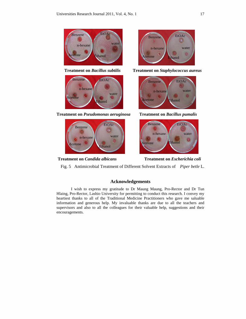
For antimicrobial activities, the powdered leaves of Piper betle L. extracted by using n-hexane, benzene, acetone, ethyl acetate, ethanol and water. The solvent extracts were tested against 6 pathogenic microorganisms; Bacillus subtilis (Jap-0221215), Staphylococcus aureus (ATCC-12277), Pseudomonas aeruginosa (IFO-3080), Bacillus pumalis (IFO-12102), Candida albicans (IFO-1060) and Escherichia coli (ATCC-25922) by using agar-well diffusion method.
Results Scientific name – Piper betle L.
Myanmar name – Kun
English name – Betel vine
Family – Piperaceae
Flowering period – November to January
Part Used – Leaves
This species is growing as cultivated plant.
Outstanding Characters Evergreen, root climbing herb, node jointed and rooted. Leaves simple, alternate, petiolate, caudate at the base, entire along the margin, acuminate at the apex. Inflorescence spike. Flowers minute, hypogynous, perianth absent. Filaments distinct; anthers dithecous. Ovary superior, 1-loculed with a solitary ovule; style 1. Fruit a small drupe. Seeds small.
Folk uses – carminative, stimulant, tonic, antiseptic,
appetizing, antispasmodic, laxative, sedative.
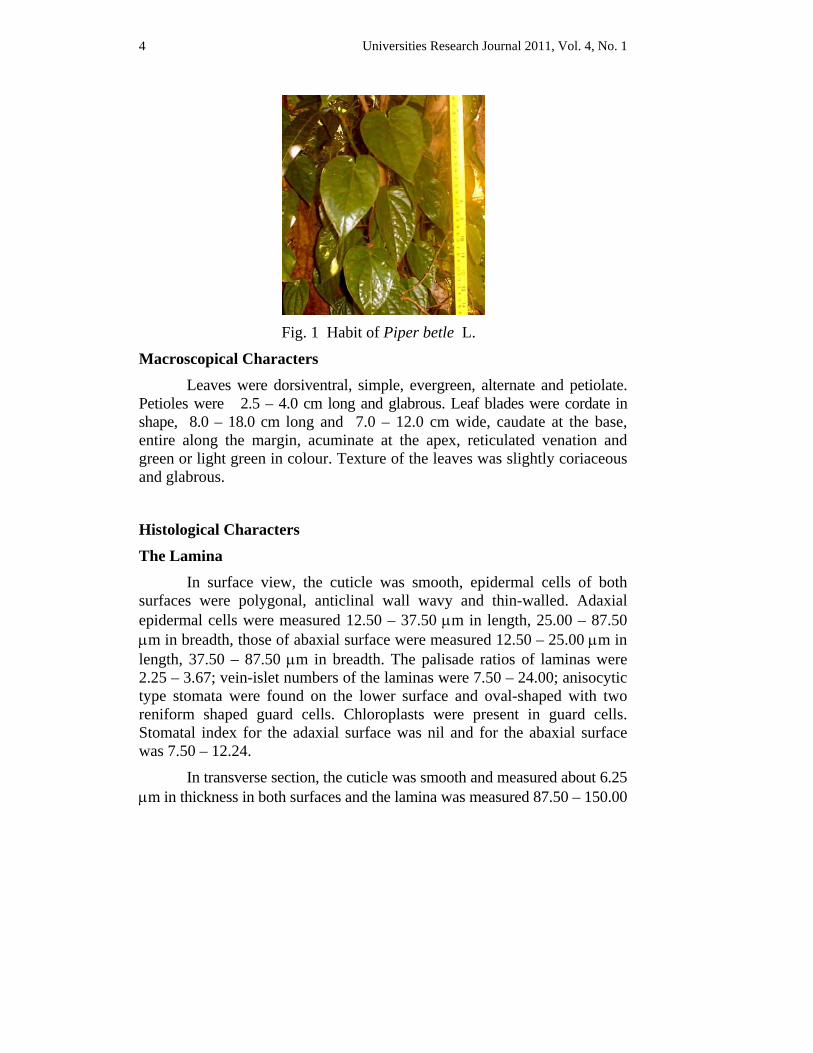
Specimen examined – Thein-ni Road, No. 12 quarter, Lashio (Fig. 1).
Universities Research Journal 2011, Vol. 4, No. 1 4
Fig. 1 Habit of Piper betle L.
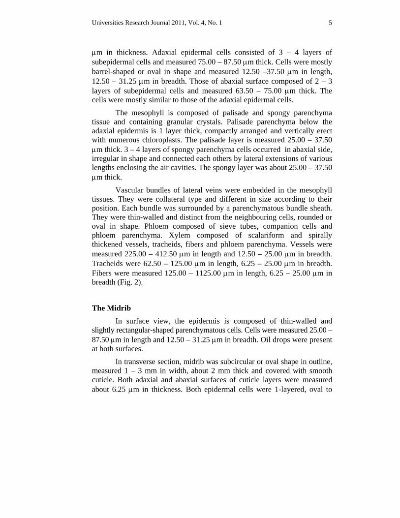
Macroscopical Characters Leaves were dorsiventral, simple, evergreen, alternate and petiolate. Petioles were 2.5 – 4.0 cm long and glabrous. Leaf blades were cordate in shape, 8.0 – 18.0 cm long and 7.0 – 12.0 cm wide, caudate at the base, entire along the margin, acuminate at the apex, reticulated venation and green or light green in colour. Texture of the leaves was slightly coriaceous and glabrous.
Histological Characters
The Lamina In surface view, the cuticle was smooth, epidermal cells of both surfaces were polygonal, anticlinal wall wavy and thin-walled. Adaxial epidermal cells were measured 12.50 – 37.50 μm in length, 25.00 – 87.50 μm in breadth, those of abaxial surface were measured 12.50 – 25.00 μm in length, 37.50 – 87.50 μm in breadth. The palisade ratios of laminas were 2.25 – 3.67; vein-islet numbers of the laminas were 7.50 – 24.00; anisocytic type stomata were found on the lower surface and oval-shaped with two reniform shaped guard cells. Chloroplasts were present in guard cells. Stomatal index for the adaxial surface was nil and for the abaxial surface was 7.50 – 12.24.
In transverse section, the cuticle was smooth and measured about 6.25 μm in thickness in both surfaces and the lamina was measured 87.50 – 150.00
Universities Research Journal 2011, Vol. 4, No. 1 5
μm in thickness. Adaxial epidermal cells consisted of 3 – 4 layers of subepidermal cells and measured 75.00 – 87.50 μm thick. Cells were mostly barrel-shaped or oval in shape and measured 12.50 –37.50 μm in length, 12.50 – 31.25 μm in breadth. Those of abaxial surface composed of 2 – 3 layers of subepidermal cells and measured 63.50 – 75.00 μm thick. The cells were mostly similar to those of the adaxial epidermal cells.
The mesophyll is composed of palisade and spongy parenchyma tissue and containing granular crystals. Palisade parenchyma below the adaxial epidermis is 1 layer thick, compactly arranged and vertically erect with numerous chloroplasts. The palisade layer is measured 25.00 – 37.50 μm thick. 3 – 4 layers of spongy parenchyma cells occurred in abaxial side, irregular in shape and connected each others by lateral extensions of various lengths enclosing the air cavities. The spongy layer was about 25.00 – 37.50 μm thick.
Vascular bundles of lateral veins were embedded in the mesophyll tissues. They were collateral type and different in size according to their position. Each bundle was surrounded by a parenchymatous bundle sheath. They were thin-walled and distinct from the neighbouring cells, rounded or oval in shape. Phloem composed of sieve tubes, companion cells and phloem parenchyma. Xylem composed of scalariform and spirally thickened vessels, tracheids, fibers and phloem parenchyma. Vessels were measured 225.00 – 412.50 μm in length and 12.50 – 25.00 μm in breadth. Tracheids were 62.50 – 125.00 μm in length, 6.25 – 25.00 μm in breadth. Fibers were measured 125.00 – 1125.00 μm in length, 6.25 – 25.00 μm in breadth (Fig. 2).
The Midrib In surface view, the epidermis is composed of thin-walled and slightly rectangular-shaped parenchymatous cells. Cells were measured 25.00 – 87.50 μm in length and 12.50 – 31.25 μm in breadth. Oil drops were present at both surfaces.
In transverse section, midrib was subcircular or oval shape in outline, measured 1 – 3 mm in width, about 2 mm thick and covered with smooth cuticle. Both adaxial and abaxial surfaces of cuticle layers were measured about 6.25 μm in thickness. Both epidermal cells were 1-layered, oval to
Universities Research Journal 2011, Vol. 4, No. 1 6
barrel in shape, compactly arranged and measured 6.25 – 12.50 μm in length, 12.50 – 25.00 μm in breadth. The cortex was made up of collenchyma and thin–walled parenchyma. The outer collenchyma cells were 3 – 4 layered and measured 25.00 – 87.50 μm thick. Cells were measured 6.50 – 25.00 μm in diameter. Parenchyma cells above the vascular bundle was 4 – 10 layered and measured 212.50 – 337.50 μm thick, cells were measured 6.25 – 75.00 μm in diameter. Parenchyma cells below the vascular bundle was 5 – 10 layered and measured 375.00 – 450.00 μm thick, cells were measured 18.75 – 75.00 μm in diameter.
Three vascular bundles were present with accessory bundles and oval in shape. They were measured about 150.00 – 162.50 μm in width, 137.50 – 150.00 μm in thickness. Phloem tissues were 62.50 – 150.00 μm thick and composed of sieve tube elements and companion cells. Xylem layer was 150.00 – 287.50 μm thick and consists of scalariform and spirally thickened vessels, tracheids, fiber-tracheids and xylem parenchyma cells. Vessels were measured 137.50 – 450.00 μm in length and 25.00 – 62.50 μm in breadth. Tracheids were measured 50.00 – 112.50 μm in length and 12.50 – 25.00 μm in breadth. Fibers were measured 312.5 – 750.00 μm in length and 6.25 –25.00 μm in breadth.
The Petiole In surface view, the cuticle was smooth and the epidermis is composed of thin-walled and slightly rectangular-shaped parenchymatous cells. Cells were measured 12.50 – 25.00 μm in length and 12.50 – 50.00 μm in breadth. Oil drops were present at both surfaces.
In transverse section, petioles were semicircular in outline, forming a concave at the middle region of the adaxial side and measured 2.0 mm – 3.5 mm in width, 1 mm – 2.5 mm in thickness. The cuticle was smooth and 6.25 – 12.50 μm in thickness. Epidermal cells were 1-layered and barrel or rectangular in shaped. Cells were measured 25.00 – 50.00 μm in breadth, 12.50 – 25.00 μm in thickness. Cortex was made up of two types of tissues, collenchymatous tissues towards the peripheral region and thin–walled parenchymatous tissues the vascular bundles. Collenchymatous tissues were consisting of 5 – 7 layers and measured 37.50 – 75.00 μm thick. Cells were measured 6.25 – 37.50 μm in diameter. Parenchymatous tissues consisted of
Universities Research Journal 2011, Vol. 4, No. 1 7
4 – 7 layers and measured 162.50 – 312.50 μm thick. Cells were rounded or oval in shape and measured. 25.00 – 87.50 μm in diameter. Pith consisted of parenchymatatous cells and measured 437.50 – 1000.00 μm thick. Cells were rounded and measured 12.50 – 100.00 μm in diameter.
Vascular bundles were arranged in a ring of two rows. Each bundle was oval-shaped in outline.The inner large bundles, measured 187.50 – 275.00 μm in width and 187.50 – 375.00 μm in thickness were alternating with the outer small bundles, measured 75.00 – 200.00 μm in width and 87.50 – 112.50 µm in thickness. Phloem tissues were measured 31.25 – 75.00 μm thick and composed of sieve tube elements and companion cells. Xylem tissues were measured 37.50 – 137.50 μm thick and consisted of scalariform and spirally thickened vessels, tracheids, fibers and xylem parenchyma cells. Vessels were measured 125.0 – 412.5 μm in length and 12.5 – 25.0 μm in breadth. Tracheids were 125.0 – 250.0 μm in length, 6.25 – 12.5 μm in breadth. Fibers were measured 250.00 – 300.00 μm in length and 12.5 – 25.00 μm in breadth.
Sensory Characters of Powdered Leaf The leaf was light green in colour and aromatic. The taste was
slightly bitter and pungent.
Diagnostic Characters of the Powdered Leaf
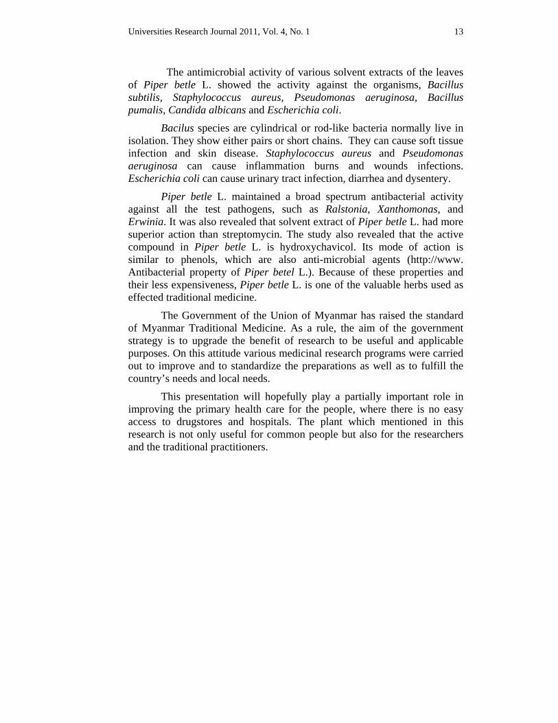
The fragments of epidermal cells were measured 62.50 – 350.00 μm in length and 12.50 – 237.50 μm in breadth with anisocytic type of stomata. The fragments of palisade cells were measured 75.00 – 187.50 μm in length and 125.00 – 287.50 μm in breadth. The fragments of vessels were found not very abundant and associated with the mesophyll cells. They were measured 87.50 – 187.50 μm in length and 12.50 – 25.00 μm in breadth. Crystals were present as sandy like and measured 12.50 – 25.00 μm in the powdered leaves (Fig. 3).
Universities Research Journal 2011, Vol. 4, No. 1 8
Traditional medicinal uses of preparation methods The liquid of boiling betel leaf and decoction of ginger with a little amount of rock salt are given for remedial using in hacking cough, whooping cough and asthma. * 1, 2,3,4,5,6,7,8,9,10
Salt packed with betel leaf is baked and made into powder. It is taken for coughing. * 1,2,3,4,5,6,7, 8,9,10
Slightly heated betel leaf smeared with coconut oil is applied on the fontanelle in an infant for coryza and also applied in layers over chest, especially of a child for the treatment of cough, pulmonary affections and bronchitis. * 1, 2,3,4,5, 6, 7, 8,
The decoction of the betel leaves is used as eye drops in ophthalmic and other painful eye diseases and night blindness. * 1,2,3,4,5,6,7
The fresh leaves applied externally around the eyes are also useful in eye diseases. * 1, 2,3,4,5, 6, 7, 8, 9, 10
Betel petiole dipped in castor oil is used as a suppository for constipating infants. * 1, 2, 3,4,5,6, 7, 8, 9, 10
In fusion of betel leaf juice and honey are given to children for therapeutic uses in fever, flatulence and digestive disorders. * 1.5,6
The leaves are chewed to reduce bad breath, to remove foul odour from mouth and to improve the voice. * 1, 2,3,4,5, 6, 8, 10
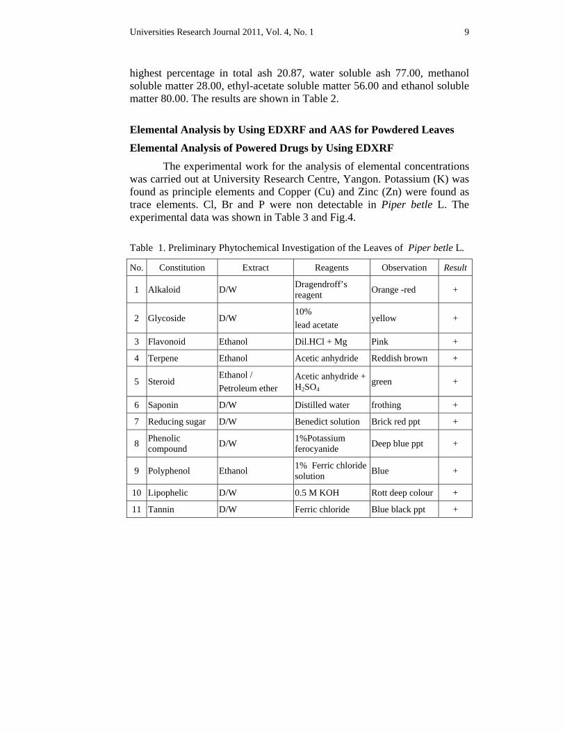
Preliminary Phytochemical Investigations The preliminary investigations were analysed for the determination of
chemical constituents from the leaves of Piper betle L. Alkaloid, glycoside, flavonoid, reducing sugar, phenolic compound, polyphenol, lipophelic, steroid, saponin, terpene and tannin were present. The results are shown in Table 1.
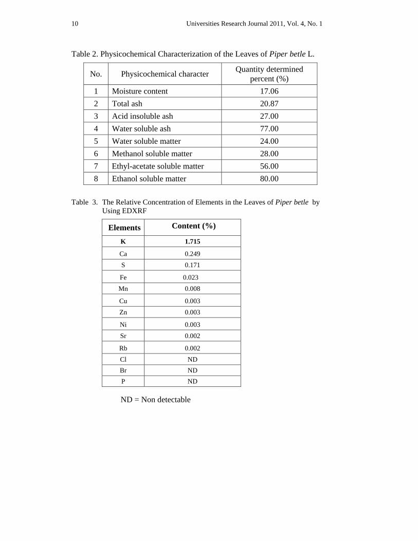
Physicochemical Characterization In physicochemical investigation, the percentage of moisture content, total ash, acid insoluble ash, water soluble ash, water soluble matter, ethanol soluble matter, ethyl-acetate soluble matter and methanol soluble matter were analysed. It was found that Piper betle L. possesses the
Universities Research Journal 2011, Vol. 4, No. 1 9
highest percentage in total ash 20.87, water soluble ash 77.00, methanol soluble matter 28.00, ethyl-acetate soluble matter 56.00 and ethanol soluble matter 80.00. The results are shown in Table 2.
Elemental Analysis by Using EDXRF and AAS for Powdered Leaves
Elemental Analysis of Powered Drugs by Using EDXRF The experimental work for the analysis of elemental concentrations was carried out at University Research Centre, Yangon. Potassium (K) was found as principle elements and Copper (Cu) and Zinc (Zn) were found as trace elements. Cl, Br and P were non detectable in Piper betle L. The experimental data was shown in Table 3 and Fig.4.
Table 1. Preliminary Phytochemical Investigation of the Leaves of Piper betle L.
No. Constitution Extract Reagents Observation Result
1 Alkaloid D/W Dragendroff’s reagent Orange -red +
2 Glycoside D/W 10% lead acetate
yellow +
3 Flavonoid Ethanol Dil.HCl + Mg Pink +
4 Terpene Ethanol Acetic anhydride Reddish brown +
5 Steroid Ethanol / Petroleum ether
Acetic anhydride + H2SO4
green +
6 Saponin D/W Distilled water frothing +
7 Reducing sugar D/W Benedict solution Brick red ppt +
8 Phenolic compound D/W 1%Potassium
ferocyanide Deep blue ppt +
9 Polyphenol Ethanol 1% Ferric chloride solution Blue +
10 Lipophelic D/W 0.5 M KOH Rott deep colour +
11 Tannin D/W Ferric chloride Blue black ppt +
Universities Research Journal 2011, Vol. 4, No. 1 10
Table 2. Physicochemical Characterization of the Leaves of Piper betle L.
No. Physicochemical character Quantity determined percent (%)
1 Moisture content 17.06 2 Total ash 20.87 3 Acid insoluble ash 27.00 4 Water soluble ash 77.00 5 Water soluble matter 24.00 6 Methanol soluble matter 28.00 7 Ethyl-acetate soluble matter 56.00 8 Ethanol soluble matter 80.00
Table 3. The Relative Concentration of Elements in the Leaves of Piper betle by Using EDXRF
ND = Non detectable
Elements Content (%)
K 1.715
Ca 0.249 S 0.171
Fe 0.023 Mn 0.008
Cu 0.003
Zn 0.003
Ni 0.003
Sr 0.002
Rb 0.002 Cl ND Br ND P ND
Universities Research Journal 2011, Vol. 4, No. 1 11
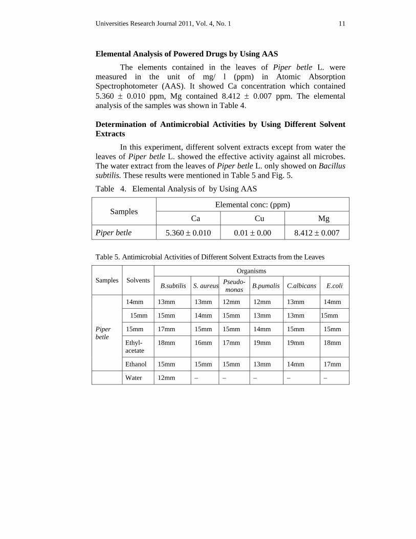
Elemental Analysis of Powered Drugs by Using AAS The elements contained in the leaves of Piper betle L. were measured in the unit of mg/ l (ppm) in Atomic Absorption Spectrophotometer (AAS). It showed Ca concentration which contained 5.360 ± 0.010 ppm, Mg contained 8.412 ± 0.007 ppm. The elemental analysis of the samples was shown in Table 4. Determination of Antimicrobial Activities by Using Different Solvent Extracts In this experiment, different solvent extracts except from water the leaves of Piper betle L. showed the effective activity against all microbes. The water extract from the leaves of Piper betle L. only showed on Bacillus subtilis. These results were mentioned in Table 5 and Fig. 5.
Table 4. Elemental Analysis of by Using AAS
Samples Elemental conc: (ppm)
Ca Cu Mg
Piper betle 5.360 ± 0.010 0.01 ± 0.00 8.412 ± 0.007
Table 5. Antimicrobial Activities of Different Solvent Extracts from the Leaves
Samples Solvents Organisms
B.subtilis S. aureus Pseudo-monas B.pumalis C.albicans E.coli
Piper betle
14mm 13mm 13mm 12mm 12mm 13mm 14mm
15mm 15mm 14mm 15mm 13mm 13mm 15mm
15mm 17mm 15mm 15mm 14mm 15mm 15mm
Ethyl-acetate
18mm 16mm 17mm 19mm 19mm 18mm
Ethanol 15mm 15mm 15mm 13mm 14mm 17mm
Water 12mm – – – – –
Universities Research Journal 2011, Vol. 4, No. 1 12
Discussion and Conclusion Piper betle L. (Kun) is an aromatic, climbing herb, belonging to family Piperaceae. Myanmar traditional practitioners described that betel leaves are not only used as expectorant, but also taken the boiled betel leaves with tumeric and a little amount of salt for fever. The juice of fresh leaf is used as eye drops for ophthalmic and fever in Myanmar folk medicine. Practitioners of Asian medicine have used P. betle L. for asthma and rheumatic arthritis for a long time.
In histological studies, the leaves of Piper betle L. presented in this study are dorsiventral type and reticulate venations. The studies of the epidermal cells of leaves are wavy in surface view and multi-layered sub epidermal cells are found. The mucilage cannal was observed in the petiole and midrib. Stomata nearly always confined to the lower surface of leaves. The vascular bundles are found in the petiole as closed circle of separate bundles and scattered like those of the monocotyledons. These characters are in agreement with those mentioned by Metcalfe and Chalk (1972).
The results of phytochemical investigations showed that the leaves of Piper betle L. contain phenolic compound. Phenol is no longer used as an antiseptic and seldom as disinfectant because it irritates the skin and has disagreeable odour. It is used as a standard for measuring the effectiveness of other disinfectants. Phenolic is derivatives of phenol. They altered to reduce its irritating qualities or increase its antibacterial activity. As a group, phenolics exert anti-microbial activity by injuring plasma membranes, inactivating enzymes and denaturing proteins. They are frequently used as disinfectants because they remain active in the presence of organic compounds. They are stable and they persist for long periods of time after application. For these reasons phenolics are suitable agents for disinfecting pus, saliva and feces.
According to elemental analysis of powdered drugs by using EDXRF method, K and Ca are found as principle elements in the powdered leaves. They also contain S, Fe, Mn, Cu, Zn, Rb, Ni and Sr. By the results of AAS, they showed no toxic metal Pb, Hg, Cd and As. Devaraj (2001) mentioned that the leaves of Piper betle L. contain betel-phenol, chavibetol and chavicol and cadinene. 100g of betel leaves consist of vitamin A 9339 I.U., vitamin B1 68 mcg, vitamin B2 31 mcg, vitamin C 3.5 mg, carbohydrate 4.8 g, fat 0.7 mg, protein 3.8 g and phosphorus 10 g. These may be believed useful for medicinal function.
Universities Research Journal 2011, Vol. 4, No. 1 13
The antimicrobial activity of various solvent extracts of the leaves of Piper betle L. showed the activity against the organisms, Bacillus subtilis, Staphylococcus aureus, Pseudomonas aeruginosa, Bacillus pumalis, Candida albicans and Escherichia coli.
Bacilus species are cylindrical or rod-like bacteria normally live in isolation. They show either pairs or short chains. They can cause soft tissue infection and skin disease. Staphylococcus aureus and Pseudomonas aeruginosa can cause inflammation burns and wounds infections. Escherichia coli can cause urinary tract infection, diarrhea and dysentery.
Piper betle L. maintained a broad spectrum antibacterial activity against all the test pathogens, such as Ralstonia, Xanthomonas, and Erwinia. It was also revealed that solvent extract of Piper betle L. had more superior action than streptomycin. The study also revealed that the active compound in Piper betle L. is hydroxychavicol. Its mode of action is similar to phenols, which are also anti-microbial agents (http://www. Antibacterial property of Piper betel L.). Because of these properties and their less expensiveness, Piper betle L. is one of the valuable herbs used as effected traditional medicine.
The Government of the Union of Myanmar has raised the standard of Myanmar Traditional Medicine. As a rule, the aim of the government strategy is to upgrade the benefit of research to be useful and applicable purposes. On this attitude various medicinal research programs were carried out to improve and to standardize the preparations as well as to fulfill the country’s needs and local needs.
This presentation will hopefully play a partially important role in improving the primary health care for the people, where there is no easy access to drugstores and hospitals. The plant which mentioned in this research is not only useful for common people but also for the researchers and the traditional practitioners.
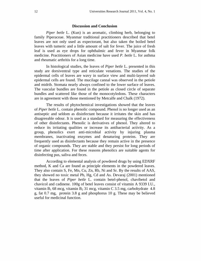
Universities Research Journal 2011, Vol. 4, No. 1 14
Adaxial surface of lamina Abaxial surface of lamina
Transverse section of lamina
Surface view of midrib Transverse section of midrib
Surface view of petiole Transverse section of petiole
pl = palisade parenchyma cell cr = cortex epi = epidermal cell sp = spongy parenchyma cell vb = vascular bundle st = stomata ad epi= adaxial epidermal cell tri = trichome mc= mucilagenous canal ab epi =abaxial epidermal cell sub epi= subepidermal cell
Fig. 2 Histological Characters of Leaf
st ad epi
ab epi
mc epi pith
ad epi
sub epi pl sp
ab epi
vb
vb
mc
cr
cr
epi
Universities Research Journal 2011, Vol. 4, No. 1 15
Fragment of mesophyll cells
ms= mesophyll cell
crys= crystals
Various sizes of crystals
epi = epidermal cell, vs = vessel
st = stomata, fib = fiber
Fig. 3 Powdered Leaf of Piper betle L.

Universities Research Journal 2011, Vol. 4, No. 1 16
Fig. 4 Elemental Analysis of the Leaf of Piper betle L.
Universities Research Journal 2011, Vol. 4, No. 1 17
Treatment on Bacillus subtilis Treatment on Staphylococcus aureus
Treatment on Pseudomonas aeruginosa Treatment on Bacillus pumalis
Treatment on Candida albicans Treatment on Escherichia coli
Fig. 5 Antimicrobial Treatment of Different Solvent Extracts of Piper betle L.
Acknowledgements I wish to express my gratitude to Dr Maung Maung, Pro-Rector and Dr Tun
Hlaing, Pro-Rector, Lashio University for permitting to conduct this research. I convey my heartiest thanks to all of the Traditional Medicine Practitioners who gave me saluable information and generous help. My invaluable thanks are due to all the teachers and supervisors and also to all the colleagues for their valuable help, suggestions and their encouragements.
Ethanol
EtOAc
water
Benzene
Acetone
n-hexane
Benzene
Acetone
n-hexane
EtOAc
Ethanol
water
Benzene
Acetone
n-hexane
EtOAc
water
Ethanol
Benzene
Acetone
n-hexane
EtOAc
water
Ethanol
Benzene
Acetone
n-hexane
EtOAc
water
Ethanol
Benzene
Acetone
n-hexane
EtOAc
water
Ethanol
Universities Research Journal 2011, Vol. 4, No. 1 18
List of the Traditional Medicinal Practitioners for Information U Nay Thu Yaine Kyaw, Traditional Medicine Practitioner Thein-ni Road, No. 12 Quarter,
Lashio.
U Sai Aung Kyi (Ta sa 02891), Traditional Medicine Practitioner 2/534, Sao San Htun Road, Nam Ton Quarter, Kyaukme.
U Sai Nyunt Maung (Ta sa 02890), Traditional Medicine Practitioner Shwe-Myin-Pyan Clinic, Haw Kone Quarter, Kyaukme.
U Sein Win (Ta sa 00465), Traditional Medicine Practitioner Traditional Medicinal Clinic, No. 1 Quarter, Namtu.
U Saw Htwe Moe Aung (Ta sa 2986), Traditional Medicine Practitioner Yaung-Chi Traditional Medicinal Clinic, Yan-kin Road, No. 7 Quarter, Lashio.
U Thein Hlaing (Ta sa 03054), Traditional Medicine Practitioner Aye-yeik-chan-thar Traditional Medicinal Clinic Bu-tar Road, No. 9 Quarter, Lashio.
Daw Tin Tin Ywe (Ta sa 05194), Traditional Medicine Practitioner Aye-yeik-chan-thar Traditional Medicinal Clinic Bu-tar Road, No. 9 Quarter, Lashio.
U Saw Dar Le Oo, Traditional Medicine Practitioner Tain-phyu-thit-sar Traditional Medicinal Clinic, No. 8 Quarter, Lashio.
U Myo Nyunt Oo ( Ta sa 02490 ), Traditional Medicine Practitioner Pan-Haike No. 1 Quarter, Namtu.
U Sai Pon Khum, Traditional Medicine Producer Sin-lin-aung Traditional Medicine Pharmacy No. 5 Quarter, Tangyan.
References Anonymous (2003). Collection of Commonly Used Herbal Plants, (in Myanmar Version)
Department of Traditional Medicine, Ministry of Health, Bo-ta-htaung Township, Yangon.
Chopra, R.N. (1958). Chopra’s Indigenous Drugs of India, U.N. Dhur & Sons Private Limited. Calcutta.
Dassanayake, M.D. (1987). A Revised Handbook to the Flora of Ceylon, Vol. VI . University of Peraseniya, Department of Agriculture, Published for the Smithsonian Institution, and the National Science Foundation, Washington DC: Amerind Publishing Co. Pvt. Ltd.
Dastur, J.F., F.N.I. (1962). Medicinal plants of India and Pakistan, D.B. Taraporevala Sons & Co. Private LTD. Bombay.
Devaraj, T.L. (2001).Speaking of Ayurvedic Herbal Cures, Sterling Publishers Pvt. Ltd. , New Delhi.
Universities Research Journal 2011, Vol. 4, No. 1 19
Evans, W.C. (2002). Pharmacognosy, 15th Edition, W.B. Saunders, Harcourt Publishers Limited.
Hooker, J.D. (1879). The Flora of British India, Reeve & Co, Ltd. 5. Herietta Street, Convent Garden, London.
Kapoor, L.D. (2001). Hand Book of Ayruvedic Medicinal Plants. CRC Press. Boca Raton. London New York Washington DC.
Metcalfe, C.R. & Chalk (1972). Anatomy of The Dicotyledons, Vol. I & II, The Clerendon Press, Oxford.
Ashin, Na-ga-thein, (1973). Illustrated Dictionary of Medicinal Plants (in Myanmar Version), Mingalar Press, Yangon.
Wallis, T.E. (1967). Textbook of Pharmacognosy. J & A. Churchill LTD., Gloucester Place, London.
http://www. Antibacterial activity of Piper betel.
http://www. Antioxidant activity of Piper betel leaf extract and its constituents.
Universities Research Journal 2011, Vol. 4, No. 1
1. Associate Professor, Department of Botany, Myitkyina University 2. Associate Professor, Department of Botany, Myitkyina University
Terrestrial Orchids from Southern Kachin State Khin Win Naing1 and Soe Myint Aye2
Abstract The species of terrestrial orchids were collected in Southern Kachin State, between 23° 27'- 28° 25' N. latitude and 96° 44' - 98° 45' E. longitude, during the years 2006 to 2010. The research work consists of 14 species belonging to 9 genera. Taxonomic information and diagnostic characters of individual species are given. The studied area is represented by many area of natural vegetation. The mostly distributed terrestrial orchids were Geodorum recurvum, Eulophia andamanensis, Arundina graminifolia, Spathoglottis plicata and Phaius tankervilleae. Although Spathoglottis affinis and Malaxis latifolia were frequently found, Phaius wallichii becomes greatly influenced at the higher elevation.
Key words: terrestrial orchids, natural vegetation, higher elevation
Introduction
Myanmar due to its unique geographical position endowed with a rich diversity of flora and fauna. Among these flora, orchids are one of the most striking, beautiful and glamorous flowers to be found in nature. They are widely distributed throughout the country. Since 50% of the rain forest covering the world had been destroyed by human activities, the orchid population is at risk of extinction due to their habitat destruction.
Hooker (1894) included 123 genera 1104 species and varieties of orchids in his “The Flora of British India”. In 1895, Grant reported that Myanmar orchids included a total of 86 genera and 581 species. Hundley (1987) listed 128 genera and 739 species in Myanmar.
Kachin State lies between 23° 45' and 28° 31' North latitude and 95° 45' and 98° 45' East longitude. It shares an international boundary with India on the west and with China on the north and east. It is bounded on the west by Sagaing Region and on the south by Shan State. The study area of Kachin State composed of six Townships − Myitkyina Township, Waingmaw Township, Ingyanyan Township, Mogaung Township, Mohnyin Township and Simbo Township.
Lasi Bauk Naw (1999) also noted that Cymbidium lowianum, Phaius tankervilleae and Arundina graminifolia were found in Hkakaborazi. He
Universities Research Journal 2011, Vol. 4, No. 1 22
also stated that Eulophia spp., Goodyera spp., Habenaria spp. and Phaius tankervilleae were found in Nong Mung area in 2007. Saw Lwin (2002) stated that Cymbidium lowianum and Cymbidium ensifolium were found in Hpon Kan Razi area. Thanegi and Saw Lwin (2003) noted that Cymbidium lowianum and Arundina graminifolia were found in Kachin State.
The aim and objectives of this research are to conduct and record the native terrestrial orchids of southern part of Kachin State that can partially fulfill on the information on orchids of Kachin State in Myanmar.
Materials and Methods A method of specimens’ collection, preparation and preservation was
followed to Pandey (2007). The habitats and locations of specimens were determined by using Global Position System (GPS) Device. The identification was done by using keys given by Flora of British India (Hooker 1954), Flora of Ceylon (Dassanaayake 1981), Flora of Java (Backer 1963), The Orchids of Indochina (Seidenfaden 1992), Flora of Malaya (Holttum 1964) and Orchids of Peru (Schweinurth 1960). The nomenclatural data referred to the International Plant Name Index. The herbarium specimens were deposited at the herbarium of Mandalay University (ASM) for references and other scientific studies.
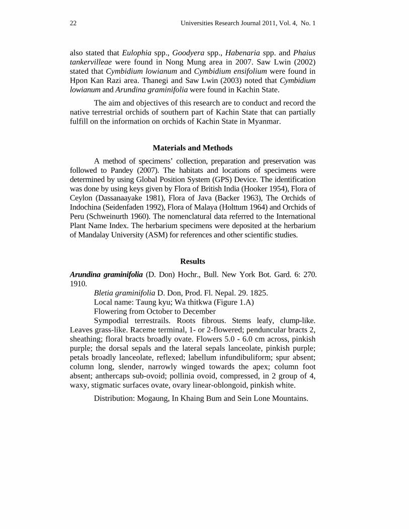
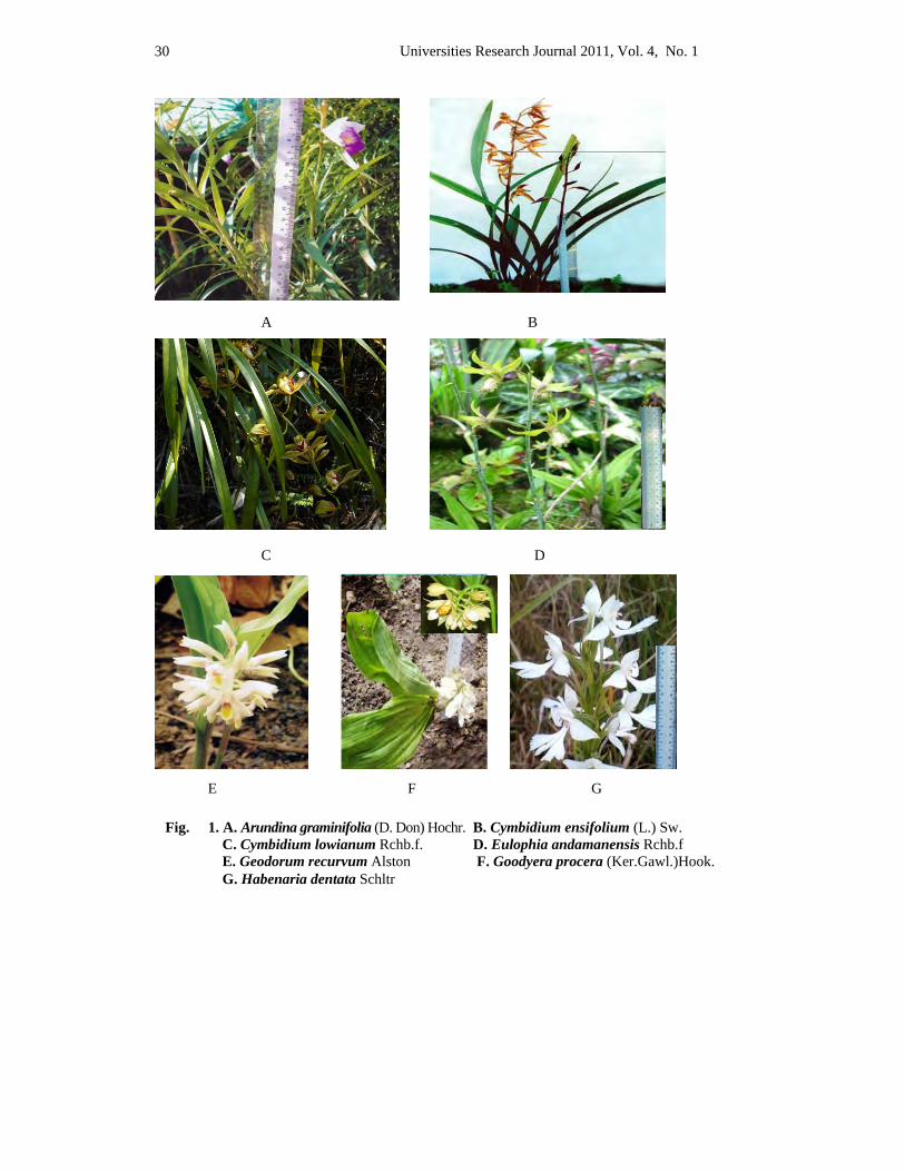
Results Arundina graminifolia (D. Don) Hochr., Bull. New York Bot. Gard. 6: 270. 1910. Bletia graminifolia D. Don, Prod. Fl. Nepal. 29. 1825. Local name: Taung kyu; Wa thitkwa (Figure 1.A) Flowering from October to December Sympodial terrestrails. Roots fibrous. Stems leafy, clump-like. Leaves grass-like. Raceme terminal, 1- or 2-flowered; penduncular bracts 2, sheathing; floral bracts broadly ovate. Flowers 5.0 - 6.0 cm across, pinkish purple; the dorsal sepals and the lateral sepals lanceolate, pinkish purple; petals broadly lanceolate, reflexed; labellum infundibuliform; spur absent; column long, slender, narrowly winged towards the apex; column foot absent; anthercaps sub-ovoid; pollinia ovoid, compressed, in 2 group of 4, waxy, stigmatic surfaces ovate, ovary linear-oblongoid, pinkish white.
Distribution: Mogaung, In Khaing Bum and Sein Lone Mountains.
Universities Research Journal 2011, Vol. 4, No. 1 23
Specimens examined: Myitkyina Township, along the road side near Myitson village, N 25°30' 06.2", E 97°20' 49.0", 750 m; 10th December, 2007; Khin Win Naing 2.
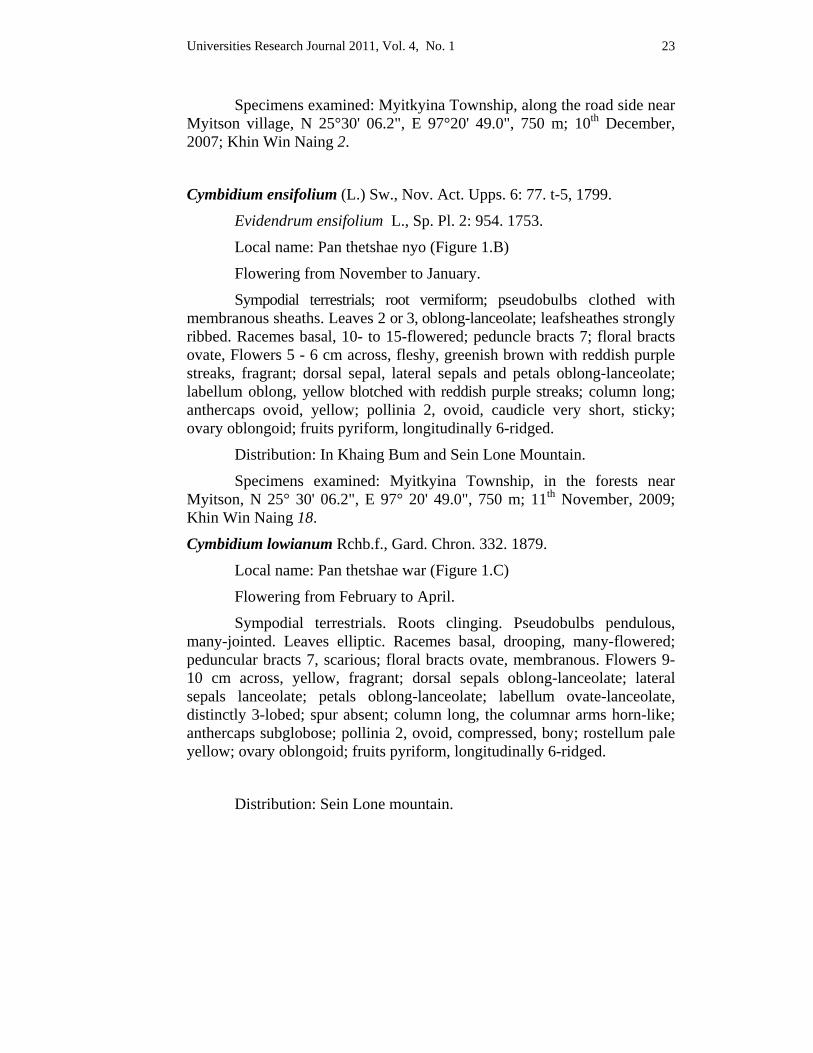
Cymbidium ensifolium (L.) Sw., Nov. Act. Upps. 6: 77. t-5, 1799.
Evidendrum ensifolium L., Sp. Pl. 2: 954. 1753.
Local name: Pan thetshae nyo (Figure 1.B)
Flowering from November to January.
Sympodial terrestrials; root vermiform; pseudobulbs clothed with membranous sheaths. Leaves 2 or 3, oblong-lanceolate; leafsheathes strongly ribbed. Racemes basal, 10- to 15-flowered; peduncle bracts 7; floral bracts ovate, Flowers 5 - 6 cm across, fleshy, greenish brown with reddish purple streaks, fragrant; dorsal sepal, lateral sepals and petals oblong-lanceolate; labellum oblong, yellow blotched with reddish purple streaks; column long; anthercaps ovoid, yellow; pollinia 2, ovoid, caudicle very short, sticky; ovary oblongoid; fruits pyriform, longitudinally 6-ridged.
Distribution: In Khaing Bum and Sein Lone Mountain.
Specimens examined: Myitkyina Township, in the forests near Myitson, N 25° 30' 06.2", E 97° 20' 49.0", 750 m; 11th November, 2009; Khin Win Naing 18.
Cymbidium lowianum Rchb.f., Gard. Chron. 332. 1879.
Local name: Pan thetshae war (Figure 1.C)
Flowering from February to April.
Sympodial terrestrials. Roots clinging. Pseudobulbs pendulous, many-jointed. Leaves elliptic. Racemes basal, drooping, many-flowered; peduncular bracts 7, scarious; floral bracts ovate, membranous. Flowers 9- 10 cm across, yellow, fragrant; dorsal sepals oblong-lanceolate; lateral sepals lanceolate; petals oblong-lanceolate; labellum ovate-lanceolate, distinctly 3-lobed; spur absent; column long, the columnar arms horn-like; anthercaps subglobose; pollinia 2, ovoid, compressed, bony; rostellum pale yellow; ovary oblongoid; fruits pyriform, longitudinally 6-ridged.
Distribution: Sein Lone mountain.
Universities Research Journal 2011, Vol. 4, No. 1 24
Specimens examined: Bamaw Township, Sein Lone mountain, near Pum mu village, N 29° 14' 16.5", E 97° 30' 23.9", 2170 m; 21st February, 2009; Khin Win Naing 7.
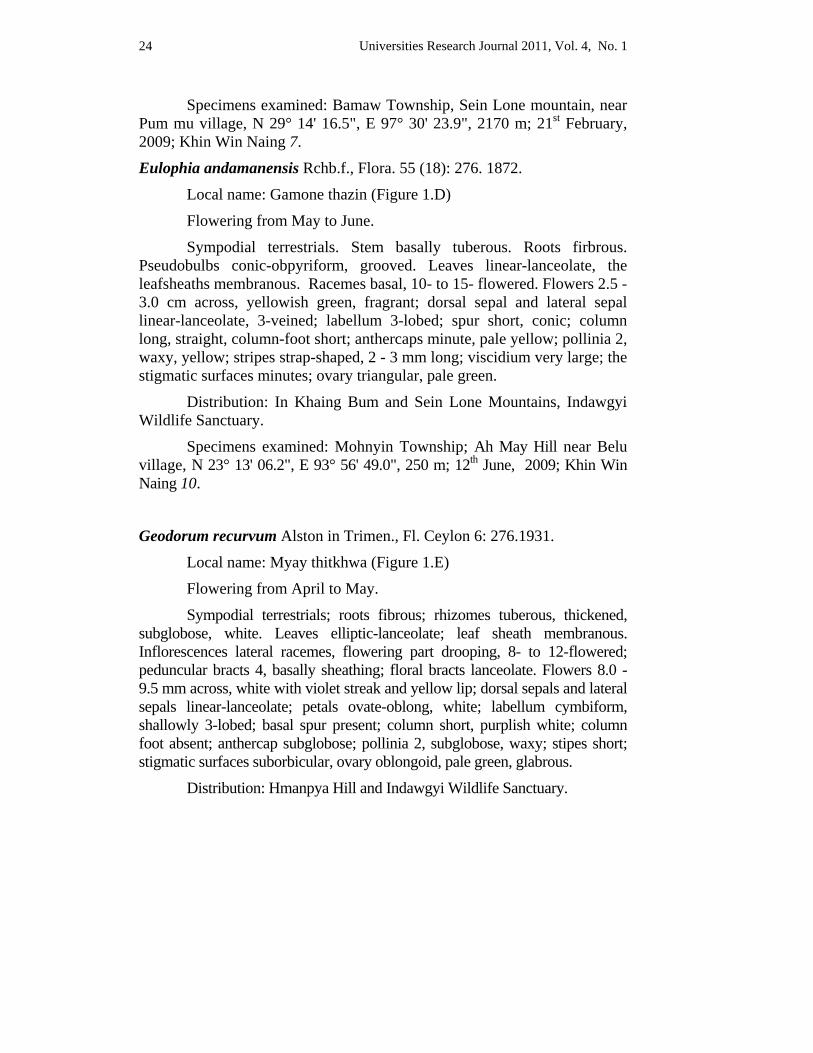
Eulophia andamanensis Rchb.f., Flora. 55 (18): 276. 1872.
Local name: Gamone thazin (Figure 1.D)
Flowering from May to June.
Sympodial terrestrials. Stem basally tuberous. Roots firbrous. Pseudobulbs conic-obpyriform, grooved. Leaves linear-lanceolate, the leafsheaths membranous. Racemes basal, 10- to 15- flowered. Flowers 2.5 - 3.0 cm across, yellowish green, fragrant; dorsal sepal and lateral sepal linear-lanceolate, 3-veined; labellum 3-lobed; spur short, conic; column long, straight, column-foot short; anthercaps minute, pale yellow; pollinia 2, waxy, yellow; stripes strap-shaped, 2 - 3 mm long; viscidium very large; the stigmatic surfaces minutes; ovary triangular, pale green.
Distribution: In Khaing Bum and Sein Lone Mountains, Indawgyi Wildlife Sanctuary.
Specimens examined: Mohnyin Township; Ah May Hill near Belu village, N 23° 13' 06.2", E 93° 56' 49.0", 250 m; 12th June, 2009; Khin Win Naing 10.
Geodorum recurvum Alston in Trimen., Fl. Ceylon 6: 276.1931.
Local name: Myay thitkhwa (Figure 1.E)
Flowering from April to May.
Sympodial terrestrials; roots fibrous; rhizomes tuberous, thickened, subglobose, white. Leaves elliptic-lanceolate; leaf sheath membranous. Inflorescences lateral racemes, flowering part drooping, 8- to 12-flowered; peduncular bracts 4, basally sheathing; floral bracts lanceolate. Flowers 8.0 - 9.5 mm across, white with violet streak and yellow lip; dorsal sepals and lateral sepals linear-lanceolate; petals ovate-oblong, white; labellum cymbiform, shallowly 3-lobed; basal spur present; column short, purplish white; column foot absent; anthercap subglobose; pollinia 2, subglobose, waxy; stipes short; stigmatic surfaces suborbicular, ovary oblongoid, pale green, glabrous.
Distribution: Hmanpya Hill and Indawgyi Wildlife Sanctuary.
Universities Research Journal 2011, Vol. 4, No. 1 25
Specimens examined: Myitkyina Township; along the road side of Hmanpya Hill, N 23° 13' 06.2", E 93° 56' 49.0", 450 m; 25th May, 2008; Khin Win Naing 3.
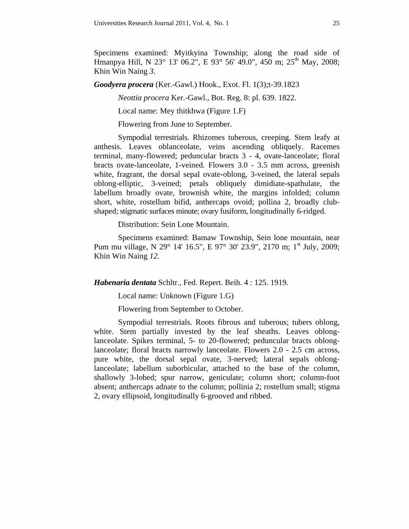
Goodyera procera (Ker.-Gawl.) Hook., Exot. Fl. 1(3);t-39.1823
Neottia procera Ker.-Gawl., Bot. Reg. 8: pl. 639. 1822.
Local name: Mey thitkhwa (Figure 1.F)
Flowering from June to September.
Sympodial terrestrials. Rhizomes tuberous, creeping. Stem leafy at anthesis. Leaves oblanceolate, veins ascending obliquely. Racemes terminal, many-flowered; peduncular bracts 3 - 4, ovate-lanceolate; floral bracts ovate-lanceolate, 1-veined. Flowers 3.0 - 3.5 mm across, greenish white, fragrant, the dorsal sepal ovate-oblong, 3-veined, the lateral sepals oblong-elliptic, 3-veined; petals obliquely dimidiate-spathulate, the labellum broadly ovate, brownish white, the margins infolded; column short, white, rostellum bifid, anthercaps ovoid; pollina 2, broadly club-shaped; stigmatic surfaces minute; ovary fusiform, longitudinally 6-ridged.
Distribution: Sein Lone Mountain.
Specimens examined: Bamaw Township, Sein lone mountain, near Pum mu village, N 29° 14' 16.5", E 97° 30' 23.9", 2170 m; 1st July, 2009; Khin Win Naing 12.
Habenaria dentata Schltr., Fed. Repert. Beih. 4 : 125. 1919.
Local name: Unknown (Figure 1.G)
Flowering from September to October.
Sympodial terrestrials. Roots fibrous and tuberous; tubers oblong, white. Stem partially invested by the leaf sheaths. Leaves oblong-lanceolate. Spikes terminal, 5- to 20-flowered; peduncular bracts oblong-lanceolate; floral bracts narrowly lanceolate. Flowers 2.0 - 2.5 cm across, pure white, the dorsal sepal ovate, 3-nerved; lateral sepals oblong-lanceolate; labellum suborbicular, attached to the base of the column, shallowly 3-lobed; spur narrow, geniculate; column short; column-foot absent; anthercaps adnate to the column; pollinia 2; rostellum small; stigma 2, ovary ellipsoid, longitudinally 6-grooved and ribbed.
Universities Research Journal 2011, Vol. 4, No. 1 26
Distribution: In Khaing Bum and Sein Lone Mountains.
Specimens examined: Bamaw Township, Sein lone mountain, near Pum mu village, N 29° 14' 16.5", E 97° 30' 23.9", 2170 m; 25th October, 2009; Khin Win Naing 17.
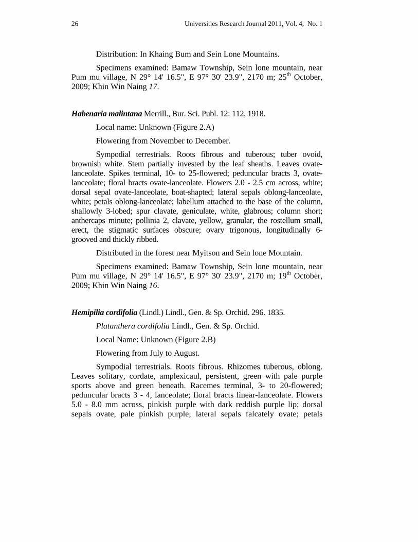
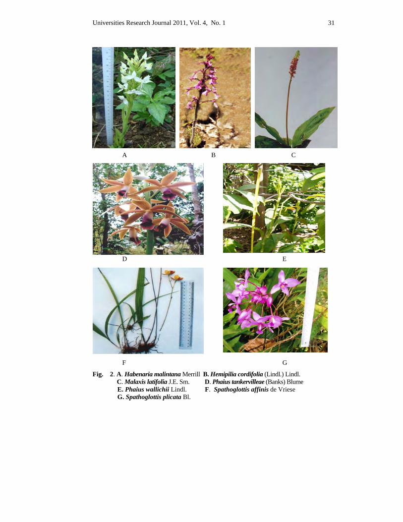
Habenaria malintana Merrill., Bur. Sci. Publ. 12: 112, 1918.
Local name: Unknown (Figure 2.A)
Flowering from November to December.
Sympodial terrestrials. Roots fibrous and tuberous; tuber ovoid, brownish white. Stem partially invested by the leaf sheaths. Leaves ovate-lanceolate. Spikes terminal, 10- to 25-flowered; peduncular bracts 3, ovate-lanceolate; floral bracts ovate-lanceolate. Flowers 2.0 - 2.5 cm across, white; dorsal sepal ovate-lanceolate, boat-shapted; lateral sepals oblong-lanceolate, white; petals oblong-lanceolate; labellum attached to the base of the column, shallowly 3-lobed; spur clavate, geniculate, white, glabrous; column short; anthercaps minute; pollinia 2, clavate, yellow, granular, the rostellum small, erect, the stigmatic surfaces obscure; ovary trigonous, longitudinally 6- grooved and thickly ribbed.
Distributed in the forest near Myitson and Sein lone Mountain.
Specimens examined: Bamaw Township, Sein lone mountain, near Pum mu village, N 29° 14' 16.5", E 97° 30' 23.9", 2170 m; 19th October, 2009; Khin Win Naing 16.
Hemipilia cordifolia (Lindl.) Lindl., Gen. & Sp. Orchid. 296. 1835.
Platanthera cordifolia Lindl., Gen. & Sp. Orchid.
Local Name: Unknown (Figure 2.B)
Flowering from July to August.
Sympodial terrestrials. Roots fibrous. Rhizomes tuberous, oblong. Leaves solitary, cordate, amplexicaul, persistent, green with pale purple sports above and green beneath. Racemes terminal, 3- to 20-flowered; peduncular bracts 3 - 4, lanceolate; floral bracts linear-lanceolate. Flowers 5.0 - 8.0 mm across, pinkish purple with dark reddish purple lip; dorsal sepals ovate, pale pinkish purple; lateral sepals falcately ovate; petals
Universities Research Journal 2011, Vol. 4, No. 1 27
broadly ovate; labellum obscurely 3-lobed; spur trumpet-shaped, pinkish; column very short; anthercaps oblong; pollinia 2, clavate; rostellum small; stigmatic surfaces obscure; ovary oblongoid, longitudinally 6-ridged.
Distribution: In Khaing Bum and Sein Lone Mountains.
Specimens examined: Bamaw Township, Sein lone mountain, near Pum mu village, N 29° 14' 16.5", E 97° 30' 23.9", 2170 m; 19th August, 2009; Khin Win Naing 14.
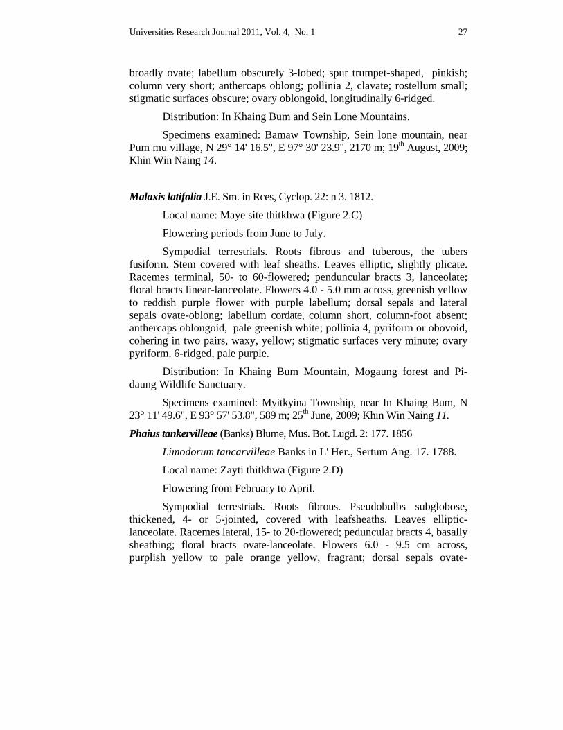
Malaxis latifolia J.E. Sm. in Rces, Cyclop. 22: n 3. 1812.
Local name: Maye site thitkhwa (Figure 2.C)
Flowering periods from June to July.
Sympodial terrestrials. Roots fibrous and tuberous, the tubers fusiform. Stem covered with leaf sheaths. Leaves elliptic, slightly plicate. Racemes terminal, 50- to 60-flowered; penduncular bracts 3, lanceolate; floral bracts linear-lanceolate. Flowers 4.0 - 5.0 mm across, greenish yellow to reddish purple flower with purple labellum; dorsal sepals and lateral sepals ovate-oblong; labellum cordate, column short, column-foot absent; anthercaps oblongoid, pale greenish white; pollinia 4, pyriform or obovoid, cohering in two pairs, waxy, yellow; stigmatic surfaces very minute; ovary pyriform, 6-ridged, pale purple.
Distribution: In Khaing Bum Mountain, Mogaung forest and Pi-daung Wildlife Sanctuary.
Specimens examined: Myitkyina Township, near In Khaing Bum, N 23° 11' 49.6", E 93° 57' 53.8", 589 m; 25th June, 2009; Khin Win Naing 11.
Phaius tankervilleae (Banks) Blume, Mus. Bot. Lugd. 2: 177. 1856
Limodorum tancarvilleae Banks in L' Her., Sertum Ang. 17. 1788.
Local name: Zayti thitkhwa (Figure 2.D)
Flowering from February to April.
Sympodial terrestrials. Roots fibrous. Pseudobulbs subglobose, thickened, 4- or 5-jointed, covered with leafsheaths. Leaves elliptic-lanceolate. Racemes lateral, 15- to 20-flowered; peduncular bracts 4, basally sheathing; floral bracts ovate-lanceolate. Flowers 6.0 - 9.5 cm across, purplish yellow to pale orange yellow, fragrant; dorsal sepals ovate-
Universities Research Journal 2011, Vol. 4, No. 1 28
lanceolate, 7-veined; lateral sepals falcately ovate-lanceolate, 7-veined; petals oblong-lanceolate to elliptic-lanceolate, 7-veined; labellum distinctly 3-lobed; spur funnelform, the column long; the anthercaps oblong, 2-lobed; pollinia oblong, in a group of 8, yellow, waxy; stigmatic surfaces oblong; ovary trigonous.
Distribution: throughout the studied area.
Specimens examined: Myitkyina Township, near In Khaing Bum, N 23° 11' 49.6", E 93° 57' 53.8", 589 m; 10th March, 2007; Khin Win Naing 1.
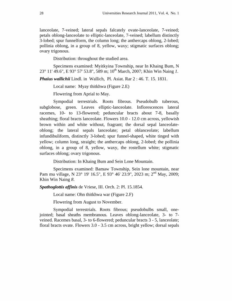
Phaius wallichii Lindl. in Wallich, Pl. Asiat. Rar 2 : 46. T. 15. 1831.
Local name: Myay thitkhwa (Figure 2.E)
Flowering from Aprial to May.
Sympodial terrestrials. Roots fibrous. Pseudobulb tuberous, subglobose, green. Leaves elliptic-lanceolate. Inflorescences lateral racemes, 10- to 13-flowered; peduncular bracts about 7-8, basally sheathing; floral bracts lanceolate. Flowers 10.0 - 12.0 cm across, yellowish brown within and white without, fragrant; the dorsal sepal lanceolate-oblong; the lateral sepals lanceolate; petal oblanceolate; labellum infundibuliform, distinctly 3-lobed; spur funnel-shaped, white tinged with yellow; column long, straight; the anthercaps oblong, 2-lobed; the pollinia oblong, in a group of 8, yellow, waxy, the rostellum white; stigmatic surfaces oblong; ovary trigonous.
Distribution: In Khaing Bum and Sein Lone Mountain.
Specimens examined: Bamaw Township, Sein lone mountain, near Pam mu village, N 23° 19' 16.5", E 93° 46' 23.9", 2023 m; 2nd May, 2009; Khin Win Naing 8.
Spathoglottis affinis de Vriese, III. Orch. 2: Pl. 15.1854.
Local name: Ohn thitkhwa war (Figure 2.F)
Flowering from August to November.
Sympodial terrestrials. Roots fibrous; pseudobulbs small, one-jointed; basal sheaths membranous. Leaves oblong-lanceolate, 3- to 7- veined. Racemes basal, 3- to 6-flowered; peduncular bracts 3 - 5, lanceolate; floral bracts ovate. Flowers 3.0 - 3.5 cm across, bright yellow; dorsal sepals
Universities Research Journal 2011, Vol. 4, No. 1 29
oblong, bright yellow; lateral sepals broadly oblong; petals obovate-oblong, yellow; labellum attached to the base of the column, distinctly 3-lobed; column subclavate, 2-winged, yellow; column foot absent; anthercaps obovate, pale yellow; pollinia clavate, in 2 group of 4, waxy, yellow, viscidium minute; stigmatic surfaces discoid, yellow; ovary broadly oblongoid, pale green, glabrous.
Distribution: Mogaung Area
Specimens examined: Mogaung Township, along the road side near Mayan village, N 22° 20' 37.6", E 96° 49' 14.1", 680 m; 24th August, 2008; Khin Win Naing 4.
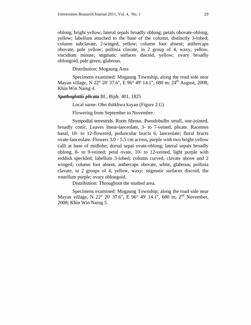
Spathoglottis plicata Bl., Bijdr. 401, 1825
Local name: Ohn thitkhwa kayan (Figure 2.G)
Flowering from September to November.
Sympodial terrestrids. Roots fibrous. Pseudobulbs small, one-jointed, broadly conic. Leaves linear-lanceolate, 3- to 7-veined, plicate. Racemes basal, 10- to 12-flowered, peduncular bracts 6, lanceolate; floral bracts ovate-lanceolate. Flowers 3.0 - 5.5 cm across, purple with two bright yellow calli at base of midlobe; dorsal sepal ovate-oblong; lateral sepals broadly oblong, 8- to 9-veined; petal ovate, 10- to 12-veined, light purple with reddish speckled; labellum 3-lobed; column curved, clavate above and 2 winged; column foot absent, anthercaps obovate, white, glabrous; pollinia clavate, in 2 groups of 4, yellow, waxy; stigmatic surfaces discoid, the rostellum purple; ovary oblongoid.
Distribution: Throughout the studied area.
Specimens examined: Mogaung Township; along the road side near Mayan village, N 22° 20' 37.6", E 96° 49' 14.1", 680 m; 2nd November, 2008; Khin Win Naing 5.
Universities Research Journal 2011, Vol. 4, No. 1 30
A B
C D
E F G
Fig. 1. A. Arundina graminifolia (D. Don) Hochr. B. Cymbidium ensifolium (L.) Sw. C. Cymbidium lowianum Rchb.f. D. Eulophia andamanensis Rchb.f E. Geodorum recurvum Alston F. Goodyera procera (Ker.Gawl.)Hook. G. Habenaria dentata Schltr
Universities Research Journal 2011, Vol. 4, No. 1 31
A B C
D E
F G
Fig. 2. A. Habenaria malintana Merrill B. Hemipilia cordifolia (Lindl.) Lindl. C. Malaxis latifolia J.E. Sm. D. Phaius tankervilleae (Banks) Blume E. Phaius wallichii Lindl. F. Spathoglottis affinis de Vriese G. Spathoglottis plicata Bl.
Universities Research Journal 2011, Vol. 4, No. 1 32
Discussion and Conclusion Southern part of Kachin State area had been studied by dividing into
4 portions. The eastern portion included Hmanpya hill and Washang village. The southern portion was Sein Lone mountain and the riverbank of Sinbo, Tar law gyi villages. The south-west portion was in Pidaung Wildlife Sanctuary and Indawgyi Wildlife Sanctuary. The northern and north eastern portions of In Khaing bum hill were also studied.
In Hmanpya hill area, the species of Geodorum recurvum and Malaxis latifolia occur abundantly. In the northern part of Myitson area Phaius tankervilleae, Phaius wallichi and Cymbidium ensifolium can be found. The most widely distributed species in Sinbo and Tar law gyi area is Malaxis latifolia. Cymbidium lowianum, Cymbidium ensifolium, Habenaria malintana, Habenaria dentata, Hemipilla cordifolia and Goodyera procera can be found in Sein lone mountain area. In Indawgyi Wildlife Sanctuary, Spathoglottis affinis, Spathoglottis plicata, Phaius tankervilleae and Eulophia andamanensis species can be found. In Pidaung Wildlife Sanctuary, Malaxis latifolia, Arundina graminifolia, Spathoglottis affinis and Spathoglottis plicata were distributed. The species of Geodorum recurvum and Cymbidium ensifolium were found in Wa Shaung area.
Goodyera procera was the only species widely distributed in Sein Lone mountain. Habenaria dentata was a species of orchid native to the Himalaya, China, India, Indochina, Thailand and Myanmar. Habenaria malintana were also found in the study area.
Spathoglottis affinis possesses a beautiful golden flower and dwarf species can be found in Mogaung. Spathoglothis plicata is the most wide spread species in the genus and the lip of the flowers is a dark purple.
Many species of genus Cymbidium are native to Myanmar. Cymbidium or boat orchids bloom during the winter, and each plant develops up to fifteen or more flowers. The fantastic range of colours for this genus can be seen according to various patterns. The flowers last about ten weeks. Therefore the members of the genera are valuable ornamental plants.
Orchids are resourced plants of Non timber Forest Products (NTFP) for exploitation of products. They are not only significant world wide in the horticultural industry but also valued locally for their medicinal, nutritional and ornamental qualities in many countries. Therefore, it is inevitably our
Universities Research Journal 2011, Vol. 4, No. 1 33
sole duty at least to get the resourced species of orchids and to report the corresponding government responsible officials to prevent the loss of Myanmar treasure plant, the orchid.
The terrestrial orchids are still distributed as a wild in Southern Kachin State and it is sincerely hoped that this study will partially fulfill the requirement of orchid information of Kachin State in Myanmar.
Acknowledgements Our heartfelt gratitude and thank go to Dr Ko Ko Myint, Rector, Myitkyina
University, for his encouragement. We are also very thankful to Dr Khin Phyu Phyu Aye, Professor and Head of the Department of Botany, Myitkyina University, for her kind suggestion.
References Backer, C.A. and R.C. Backuizen Van Den Brink (1963-1968). Flora of Java, Vol III.
Noordhoff. Ltd. Groningen.
Dassanaayake, M.D. (1981). A Revised Handbook to the flora of Ceylon. Vol II, University of Peradeniya, Department of Agriculture, Peradeniya, Sir Lanka and the Smithsonian Institution, Washington, D.C., U.S.A.
Grant, B. (1966-1986). The Orchid of Burma. (Including the Andaman Islands), Central Press, Rangoon.
Holttum, R.E. (1964). Flora of Malaya, Volume I - Orchids, 3rd ed. Government Printing Office, Singapore.
Hooker, J.D. (1894). Flora of British India. Part V & VI Recve Co. Itd. Kent, London.
Hooker, H.D. and B.D. Jackson (1895). Index Kewensis. Vol.I, Vol.II, A-Z and Supplements. Clarendon Press, Oxford Univ. London.
Hundley, H.G. and Chit Ko Ko (1961). List of Trees, Shrubs, Herbs and Climbers, etc. Record from Burma, 4th ed. Government Printing Office. Rangoon.
Lasi Bawk Naw (2007). Traditions, Beliefs and Parctices. Link with Nature Conservation in Kachin State, Today Press, Yangon.
Lasi Bawk Naw (1999). Biodiversity, Culture, Indigenous Knowledge Nature and Wildlife Conservation Programmes in Kachin State, Myanmar. Sonpan Press, Yangon.
Pandey (2007). Text book of Botany Vol. 11, Viks Publishing House PVL. Ltd.
Saw Lwin (2002). Report on Orchid and its ecology observed during Biological expedition in Hpon Kan Razi Area.

Universities Research Journal 2011, Vol. 4, No. 1 34
Schweinurt, C. 1960. Orchids of Peru. Vol. 30. Natural History Museum Press, Chicago. U.S.A.
Seidenfaden, G. (1990). Orchid Genera in Thailand V. Orchidoideae. Odense, Printed in Denmark.
Seidenfaden, G. (1992). The Orchids of Indochina. Opera Bol. 114: 1-502. Copenhagen. Printed in Denmark.
Thanegi (2003). Myanmar, A Guide to Tourism Destinations and Beyond Vol.2, No.4, July-September 2003. Published by the Ministry of Hotels & Tourism in collaboration with Swiftwinds Services.
http://www.ipni.org.
Universities Research Journal 2011, Vol. 4, No. 1
Associate Professor, Department of Botany, Taungoo University
Effectiveness of Oil Palm Wastes as Organic Fertilizers
Khin Lat Lat Mon
Abstract Oil palm wastes were recycled to be used as organic fertilizers for community uses. Their effectiveness and response were studied on the growth and yield of okra. The experiment was carried out in the open field of Thingangyun Township, Waizayandar Road, Yangon Division from May to November, 2010. Four treatments (Organic wastes, Fuller Earth or Bleaching Earth, Cow dung, and Soil) were applied in this experiment. The physical and chemical properties of soil and applied organic waste fertilizers, and cow dung were analyzed before growing okra. The experiment was laid out in Completely Randomized Design (CRD) using four treatments with three replicates. The results of analyzed soil using in cultivation of okra showed that the soil is sandy loam including 97.75% of sand, silt, and clay; very low organic carbon content (0.862%); humus (1.486%); low nitrogen content (0.18%); high phosphorous content (126ppm); and high potassium content (21.64 mg/100g). The pH of soil is 6.8 hence it is nearly neutral. The moisture content of soil is 2.88%. The analyzed results of fertilizer showed that the organic waste fertilizer from crude palm oil mill had higher CN ratio (13.13:1) than bleaching earth from refine mill (10.35:1) and cow dung (9.61:1). But CN ratio of both fertilizers were higher than that of control. The results demonstrated that the organic waste fertilizer has vegetative plant growth (48.2 cm in height, 43.53cm of leaf length, 23.45 cm leaf width,14.61cm petiole length, and 379.53 cm2 leaf area. The results of reproductive growth showed that the first and 50% flowering days of 26.2 and 27.4; first and 50% fruit setting days of 30.2 and 31.0), yield of 0.87617 kg treatment-1 and 1.16 t ha-1 respectively. The results of pod (fruit) characters showed that organic waste fertilizer had maximum pod length, width, and pod weight 20.38cm,10.25cm and 35.77g. The organic waste fertilizer had maximum fresh weight 265.23 g but minimum dry weight 22.47g. However, the waste from refined palm oil mill had lower yield (0.62 kg treatment-1 and 0.82 t ha-1) than cow dung but it had influence effects than control. In the treatment of fuller earth or bleaching earth which is waste from the Refined palm oil mill, contained residues of oil, the results were poorer than those of organic fertilizer.
Key words: Oil palm wastes, community, CRD
Universities Research Journal 2011, Vol. 4, No. 1 36
Introduction
Myanmar is blessed with abundant natural recourses and bears a favourable climate for cultivation of commercial crops including oil palm. Oil palm is widely cultivated in Tanintharyi Region. Among these cultivation areas, Yuzana oil palm plantation and crude palm oil mill are located at 38 miles, Khamaukgyi Township, Kawthaung District, Tanintharyi Region. Vast amount of fresh fruit bunches are annually produced from this plantation. Wastes from the palm oil mill process include Palm Oil Mill Effluent (POME) generated mainly from oil extraction, washing and cleaning up processes. Discharging untreated effluent into water streams may cause considerable environmental problems. However, the solid wastes generated are mainly decanter cake, empty fruit bunches, seed shells, and fibre from the mesocarp. POME as well as solid wastes may rapidly deteriorate the surrounding environment. Hence there is an urgent need for a sustainable waste management system to tackle these wastes.
As these wastes are organic in origin, they are rich in plant nutrients. Composting of wastes generated from palm oil mill is a good practice as it is helpful in recycling useful plant nutrients. This experiment deals with various aspects of wastes management practices in palm oil mill and possibility of composting the wastes. Organic agriculture includes all agricultural systems that promote environmentally, socially and economically sound production of foods. Organic farming dramatically reduces external inputs by avoiding the use of chemosynthetic fertilizer and pesticides (Safwat, 2007).
Recycling organic materials much of which is harvested from the palm are the organic wastes. Most of tropical soils are low in organic content; hence recycling of wastes is a current concern (Wood, 1986). Empty oil palm bunches were used as a source of organic fertilizer which contains nutrients needed by the soil and plants. The empty bunches of oil palm reach 23% of the total utilization of oil palm waste as an alternative organic fertilizer will also provide other benefits from the economic view.
Most vegetable farmers in tropical countries such as India, Malaysia, Indonesia, Philippines, South Pacific and Tropical Africa are small holders who cannot afford cost of inorganic fertilizers, although soil fertility limits yields of vegetables especially in urban and periurban centres. Hence farmers depend largely on locally available organic fertilizers. Organic
Universities Research Journal 2011, Vol. 4, No. 1 37
fertilizers are being developed from farm and city wastes. Different organic wastes influence nutritional quality of crops. Evaluating of different brands depends on composition of organic and organo-mineral fertilizers.
Organic and organo-mineral fertilizers were found to increase the yield of maize and vegetables such as pepper, tomato, okra, melon and amaranthus significantly (Ipinmoroti, 2003; Fagbola and Dare, 2003; Olowokere, 2004; Adeoye et al., 2008, Ojeniyi and Adejobi , 2002; Ojeniyi et al., 2009; Akanni and Ojeniyi, 2008; Makinde, 2007). Hence this research has been carried out to record the value of oil palm waste on okra. This study was aimed to use the recycled oil palm wastes as organic fertilizer, to analyze the effectiveness of recycled oil palm wastes in cultivation of okra, to approve the recycled oil palm wastes as useful materials for agricultural aspect.
Materials and Methods This experiment was carried out in the open field of Thingangyun Township, Waizayandar Road, Yangon Region from May to November 2010. The oil palm wastes fertilizer was collected from Yuzana Crude Palm Oil Mill, 38 miles, Kha-mauk-gyi Township, Kawthaung District, Tanintharyi Region. Bleaching earth were collected from Yuzana Refined Palm Oil Mill, Tharkayta Township, Yangon Region.
Experimental layout and growing of okra The experimental lay out was Completely Ramdomized Design (CRD). Four treatments with 9 replicates were assigned in the block. The size of each plot was 150cm × 60cm. The spacing between plants and row were 30cm each and between plots were 30 x 60cm. The total cultivation area was 630 x 210 cm.
Before preparation of seed beds, the soil sample from the cultivation area was taken according to the method mentioned in the (Dierolf et al., 2001). The soil sample was put into the plastic bag and its physical and chemical properties were analyzed in the soil laboratory, Land Use Department, Myanmar Agriculture Service (MAS). Similarly, the physical and chemical properties of applied fertilizers were analyzed in this lab. Then 6.7 kg each of oil palm wastes fertilizer, Fuller Earth, and cow dung were mixed in a respective soil bed. Before growing okra, the assigned fertilizer and soil were thoroughly mixed. Then the soil mix was saturated
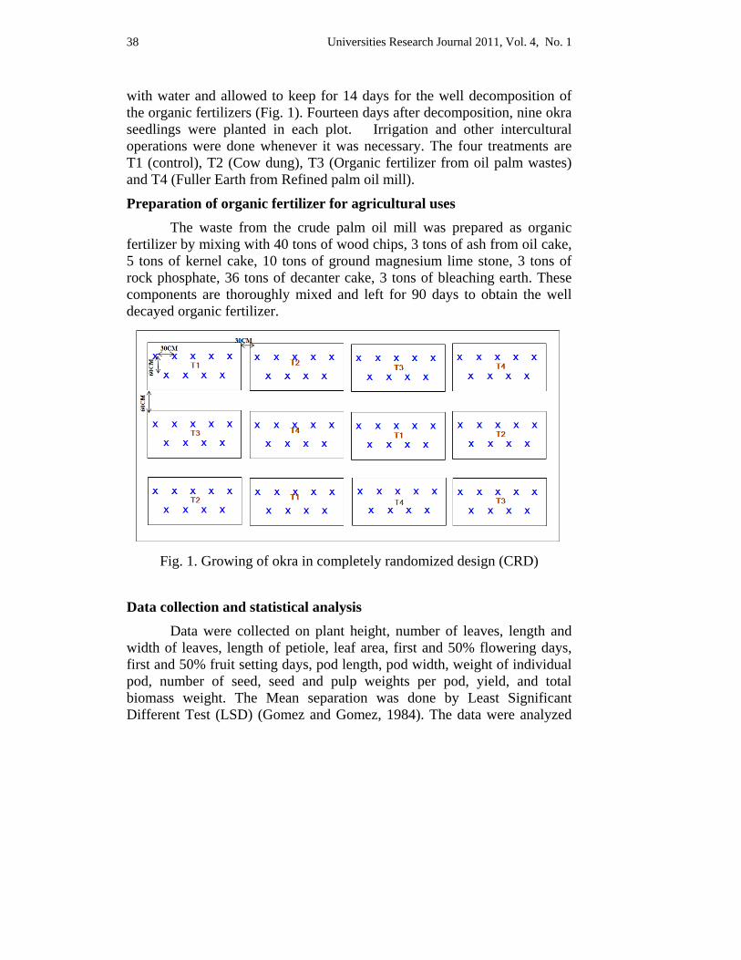
Universities Research Journal 2011, Vol. 4, No. 1 38
with water and allowed to keep for 14 days for the well decomposition of the organic fertilizers (Fig. 1). Fourteen days after decomposition, nine okra seedlings were planted in each plot. Irrigation and other intercultural operations were done whenever it was necessary. The four treatments are T1 (control), T2 (Cow dung), T3 (Organic fertilizer from oil palm wastes) and T4 (Fuller Earth from Refined palm oil mill).
Preparation of organic fertilizer for agricultural uses The waste from the crude palm oil mill was prepared as organic fertilizer by mixing with 40 tons of wood chips, 3 tons of ash from oil cake, 5 tons of kernel cake, 10 tons of ground magnesium lime stone, 3 tons of rock phosphate, 36 tons of decanter cake, 3 tons of bleaching earth. These components are thoroughly mixed and left for 90 days to obtain the well decayed organic fertilizer.
Fig. 1. Growing of okra in completely randomized design (CRD)
Data collection and statistical analysis Data were collected on plant height, number of leaves, length and width of leaves, length of petiole, leaf area, first and 50% flowering days, first and 50% fruit setting days, pod length, pod width, weight of individual pod, number of seed, seed and pulp weights per pod, yield, and total biomass weight. The Mean separation was done by Least Significant Different Test (LSD) (Gomez and Gomez, 1984). The data were analyzed
Universities Research Journal 2011, Vol. 4, No. 1 39
using the IRRISTAT software package, version 4, developed by International Rice Research Institute (IRRI), Los Baños, the Philippines.
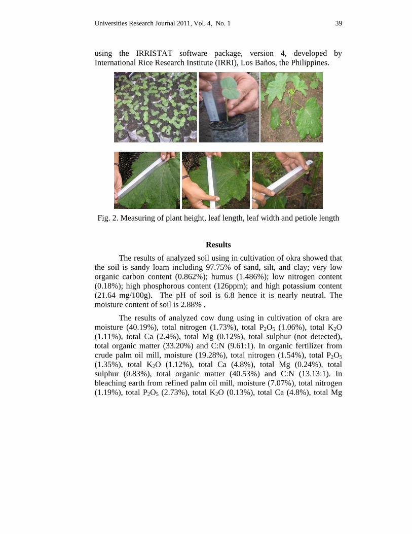
Fig. 2. Measuring of plant height, leaf length, leaf width and petiole length
Results The results of analyzed soil using in cultivation of okra showed that
the soil is sandy loam including 97.75% of sand, silt, and clay; very low organic carbon content (0.862%); humus (1.486%); low nitrogen content (0.18%); high phosphorous content (126ppm); and high potassium content (21.64 mg/100g). The pH of soil is 6.8 hence it is nearly neutral. The moisture content of soil is 2.88% .
The results of analyzed cow dung using in cultivation of okra are moisture (40.19%), total nitrogen (1.73%), total P2O5 (1.06%), total K2O (1.11%), total Ca (2.4%), total Mg (0.12%), total sulphur (not detected), total organic matter (33.20%) and C:N (9.61:1). In organic fertilizer from crude palm oil mill, moisture (19.28%), total nitrogen (1.54%), total P2O5 (1.35%), total K2O (1.12%), total Ca (4.8%), total Mg (0.24%), total sulphur (0.83%), total organic matter (40.53%) and C:N (13.13:1). In bleaching earth from refined palm oil mill, moisture (7.07%), total nitrogen (1.19%), total P2O5 (2.73%), total K2O (0.13%), total Ca (4.8%), total Mg
Universities Research Journal 2011, Vol. 4, No. 1 40
(0.24%), total sulphur (0.99%), total organic matter (24.54%) and C:N (10.35:1).
Effects of different organic fertilizers from oil palm wastes on yield and yield contributing characters of okra have been given below:
Vegetative growth
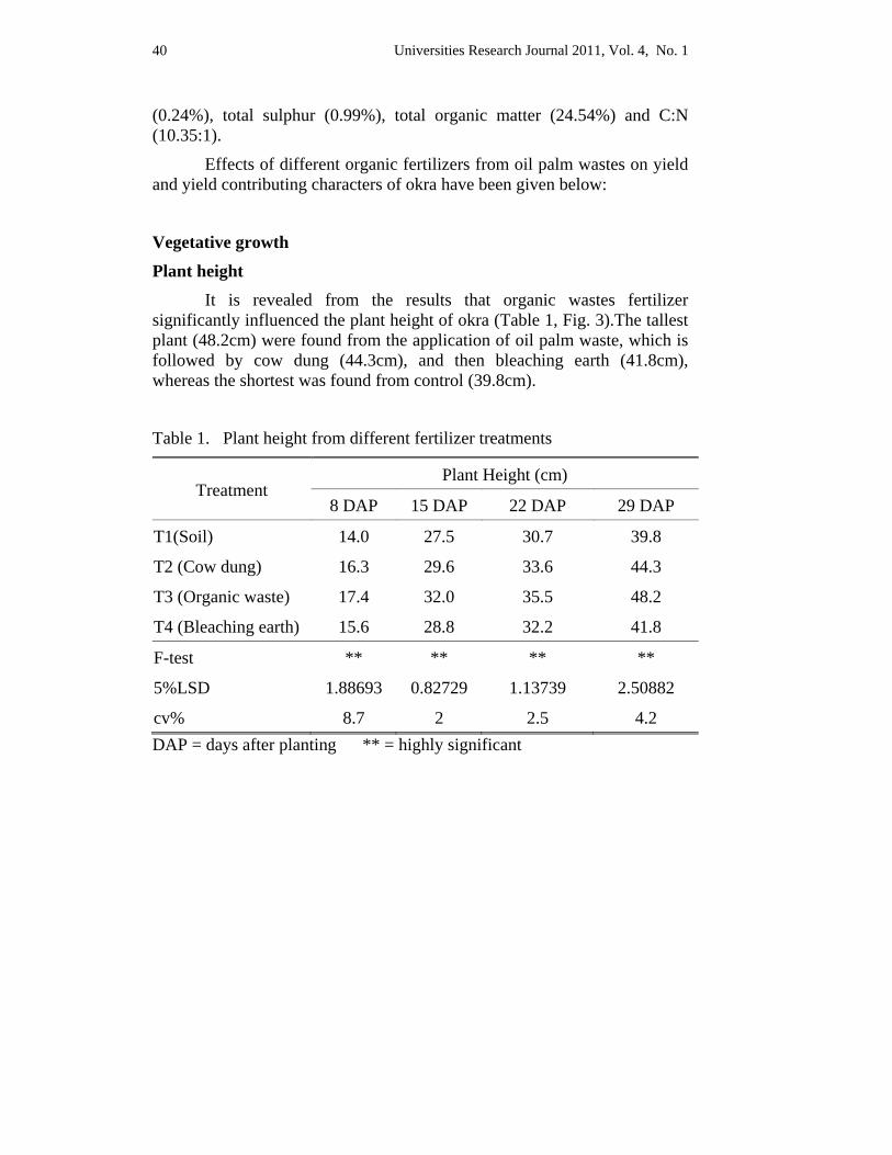
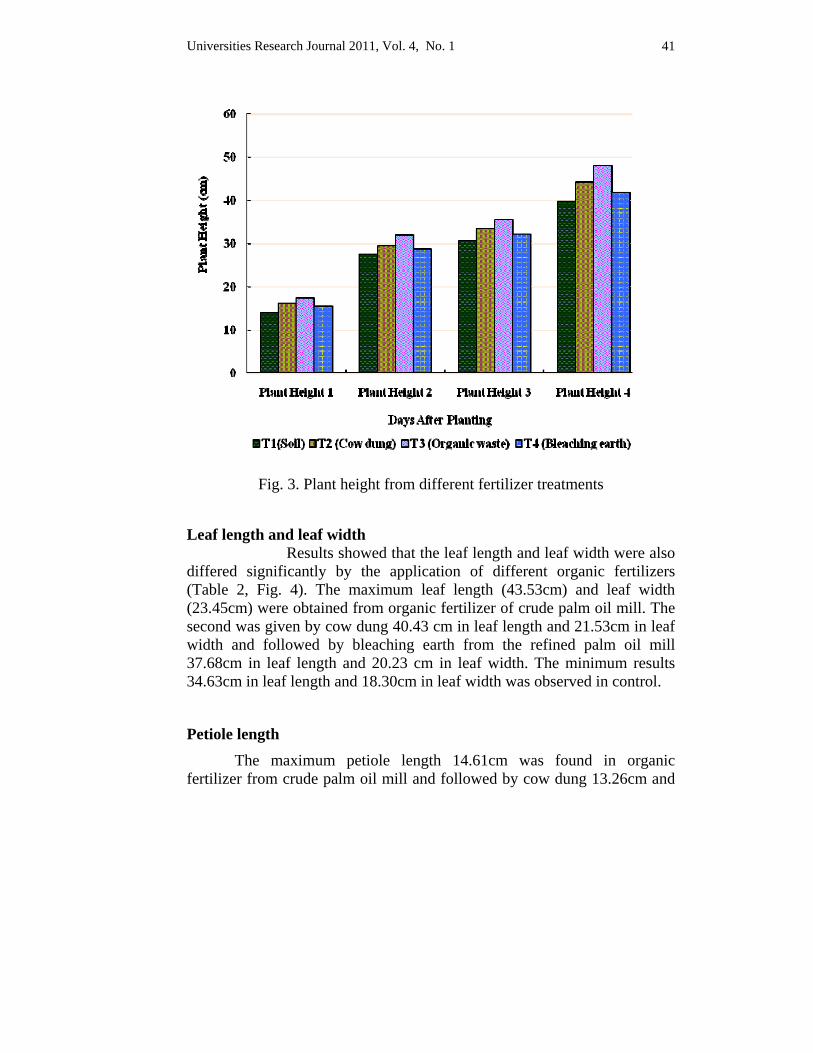
Plant height It is revealed from the results that organic wastes fertilizer
significantly influenced the plant height of okra (Table 1, Fig. 3).The tallest plant (48.2cm) were found from the application of oil palm waste, which is followed by cow dung (44.3cm), and then bleaching earth (41.8cm), whereas the shortest was found from control (39.8cm).
Table 1. Plant height from different fertilizer treatments
Treatment Plant Height (cm)
8 DAP 15 DAP 22 DAP 29 DAP
T1(Soil) 14.0 27.5 30.7 39.8
T2 (Cow dung) 16.3 29.6 33.6 44.3
T3 (Organic waste) 17.4 32.0 35.5 48.2
T4 (Bleaching earth) 15.6 28.8 32.2 41.8
F-test ** ** ** **
5%LSD 1.88693 0.82729 1.13739 2.50882
cv% 8.7 2 2.5 4.2 DAP = days after planting ** = highly significant
Universities Research Journal 2011, Vol. 4, No. 1 41
Fig. 3. Plant height from different fertilizer treatments
Leaf length and leaf width Results showed that the leaf length and leaf width were also differed significantly by the application of different organic fertilizers (Table 2, Fig. 4). The maximum leaf length (43.53cm) and leaf width (23.45cm) were obtained from organic fertilizer of crude palm oil mill. The second was given by cow dung 40.43 cm in leaf length and 21.53cm in leaf width and followed by bleaching earth from the refined palm oil mill 37.68cm in leaf length and 20.23 cm in leaf width. The minimum results 34.63cm in leaf length and 18.30cm in leaf width was observed in control.
Petiole length The maximum petiole length 14.61cm was found in organic
fertilizer from crude palm oil mill and followed by cow dung 13.26cm and
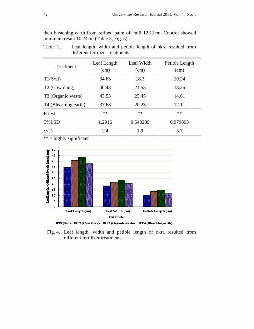
Universities Research Journal 2011, Vol. 4, No. 1 42
then bleaching earth from refined palm oil mill 12.11cm. Control showed minimum result 10.24cm (Table 3, Fig. 5).
Table 2. Leaf length, width and petiole length of okra resulted from different fertilizer treatments
Treatment Leaf Length
(cm) Leaf Width
(cm) Petiole Length
(cm)
T1(Soil) 34.63 18.3 10.24
T2 (Cow dung) 40.43 21.53 13.26
T3 (Organic waste) 43.53 23.45 14.61
T4 (Bleaching earth) 37.68 20.23 12.11
F-test ** ** **
5%LSD 1.2916 0.543289 0.979893
cv% 2.4 1.9 5.7 ** = highly significant
Fig. 4. Leaf length, width and petiole length of okra resulted from
different fertilizer treatments
Universities Research Journal 2011, Vol. 4, No. 1 43
Leaf area The organic fertilizer from crude palm oil mill gave the largest leaf
area 360.60cm².The second largest area 307.59cm² was attained by cow dung and then followed by bleaching earth 269.09cm² and control 224.18cm².
The results of reproductive growth such as the first and 50% flowering days showed that 26.2 days and 27.4 days in organic fertilizer from crude palm oil mill, 31.4 days and 32.6 days in cow dung, 30.00 days and 31.80 days in bleaching earth and 39.80 days and 45.20 days in control. The first and 50%fruit setting days showed that 30.20 days and 31.00 days in organic fertilizer from crude palm oil mill, 36.2 days and 38.2 days in cow dung, 42.2 days and 45.6 days in bleaching earth and control as 51.6 days and 57.00 days.
Marketable yield of okra resulted from different treatments showed that maximum yield 0.87617kg per treatment and 1.16 tons per hectare from organic fertilizer from crude palm oil mill and followed by 0.75405kg per treatment and 1.00 ton per hectare in cow dung, 0.62409kg per treatment and 0.82 ton per hectare from bleaching earth and the lowest yield 0.34815kg per treatment and 0.46 ton per hectare was found in control.
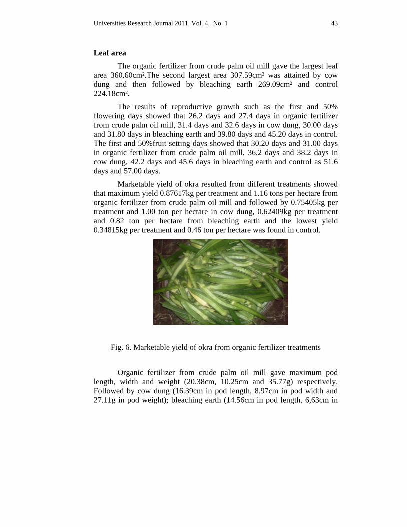
Fig. 6. Marketable yield of okra from organic fertilizer treatments
Organic fertilizer from crude palm oil mill gave maximum pod length, width and weight (20.38cm, 10.25cm and 35.77g) respectively. Followed by cow dung (16.39cm in pod length, 8.97cm in pod width and 27.11g in pod weight); bleaching earth (14.56cm in pod length, 6,63cm in
Universities Research Journal 2011, Vol. 4, No. 1 44
pod width and 24.45g in pod weight). The minimum value was achieved by control (11.54cm in pod length, 5.85cm in pod width and 20.19g in pod weight.
The maximum results of seed number, seed weight and seed pulp per pod of okra were found in organic fertilizer from crude palm oil mill had 57.40 in seed number, 4.66g in seed weight and 31.10g in pulp weight. Cow dung could attain by 52.33 in seed number, 3.19g per pod in seed weight and 23.91g in pulp weight and then followed by bleaching earth 47.73 seed number per pod, 2.88g seed weight per pod and 21.56g pulp weight per pod. Control gave the minimum results 39.67 seed number per pod, 2.26g seed weight per pod and 17.83g pulp weight per pod.
The organic fertilizer from crude palm oil mill gave the maximum fresh weight 265.22g followed by cow dung 247.45g, then bleaching earth 241.00g and control gave minimum fresh weight 208.33g. Among vegetative parts, the stem from organic waste fertilizer gave the highest fresh weight, 527.67 g.
In the dry weight, the maximum dry weight (265.22 g) was obtained from organic waste fertilizer whereas the minimum dry weight (203.33 g) was resulted from control.
Discussion and Conclusion Agricultural wastes such as organic wastes from crude palm oil mill and bleaching earth from refined mill are effective sources of nutrients because of their addition to the soil which enhanced the leaf and plant height of okra. The results of analyzed soil using in cultivation of okra showed that the soil is sandy loam including 97.75 % of sand, silt, and clay; very low organic carbon content (0.862 %); humus (1.486 %); low nitrogen content (0.18 %); high phosphorous content (126 ppm); and high potassium content (21.64 mg/100 g). The pH of soil is 6.8 hence it is nearly neutral. The moisture content of soil is 2.88 %.
The application of organic materials increased soil pH. This confirms the findings of Akande et al. (2003) who reported that the application of organic materials could improve slightly acidic tropical soil for increase crop production. The waste of crude palm oil mill (organic waste fertilizer) and bleaching earth from refined palm oil mill is prepared as an organic fertilizer and these fertilizers were used in cultivation of okra.

Universities Research Journal 2011, Vol. 4, No. 1 45
Approving the effect of organic wastes in agricultural sector, application of organic wastes together with cow dung is established in this experiment.
The results showed that waste from the crude palm oil mill had maximum effects on vegetative plant growth (48.2 cm in height, 43.53 cm of leaf length, 23.45 cm leaf width, 14.61 cm petiole length, and 360.60 cm2 leaf areas). The results of reproductive growth showed that the first and 50% flowering days of 26.2 and 27.4; first and 50% fruit setting days of 30.2 and 31.0; yield of 0.87617 kg treatment-1 and 1.16 t ha-1 respectively. The results of pod (fruit) characters showed that organic waste fertilizer gave maximum pod length, pod width, and pod weight 20.38 cm, 10.25 cm and 35.77 g. The organic waste fertilizer gave the maximum fresh weight 265.22 g but minimum dry weight 22.45 g.
Provision of a sustainable environment in the soil by amending with good quality organic additives that enhances water holding capacity and nutrient supplying capacity of soil and also the development of resistance in plants to pest and diseases and increases the yield. However, the waste from refined palm oil mill had lower yield (0.62 kg treatment-1 and 0.82 t ha-1) than cow dung but it had effects than control. Regarding to this, the organic fertilizers was utilized to sustain soil fertility for vegetables production. The people of Myanmar should practice the useful organic fertilizers enhance the balance between the soil nutrition and soil health. Bayu et al (2006) mentioned that the application of organic waste economically reduces the farmer ´s expenditure spending on crop fertilization. Besides, the inclusion of organic fertilizer could reduce environmental pollution and improve the environment as well as reduce the cost of fertilizing crops.
Using organic fertilizers also reduces the harmful impact on the environment, which is teetering on the brink of a major ecological catastrophe. These types of fertilizers also strengthen the plants toward off many pests and diseases, and even in the long run they do not lose their effectiveness. However, they do have a few drawbacks but these are of minimal consequence (Safwat, 2007). He also reported that the organic matters in such fertilizers are essential for microorganisms, which build up the soil rich in humus. Besides, the organic fertilizers release the nutrients in a slow and consistent rate that the plants can utilize it. Organic fertilizer provided balanced nutrition to the plants due to the presence of a broad range of trace elements and it is safe for all types of plants and no danger of burning due to salt concentration. Organic matter binds to the soil where the
Universities Research Journal 2011, Vol. 4, No. 1 46
roots can access it. So, it is long lasting as the organic fertilizers do not leach out. Organic fertilizers also make the plants stronger to resist space disease and pest attacks. Plants that fed on organic fertilizers are also able to resist the advance of weeds and other parasitic plants. The results of analyzed soil expressed that the soil of the cultivation area was sandy loam, low organic matter content, low nitrogen but high phosphorous and potassium. The pH of this soil is 6.8.
The researchers mentioned that Okra prefers to grow in soil that is neutral or slightly sour, meaning a soil that has a pH lower than 6.0. Preferred pH levels for okra are 5.8 to 6.5 and thus the proper fertilization can adjust these levels. However, high concentration of phosphorous causes maximum growth of Okra. The inclusion of organic materials improves the texture of the soil. The organic materials helps the soil to handle water and hold nutrients better. Besides, recent studies have shown that people who constantly eat organic, natural food products are less likely to suffer from skin disorders, heart attacks or stroke. This positive correlation is linked to the natural nutrients that are absorbed when fertilizing with organic fertilizers. On the other hand, this event was opposed to heavy, toxic nutrients absorbed by the plants when using the chemical fertilizers. The application of organic materials increased the soil available phosphorous, showing the potentials of organic materials as a source of P to the soil (http://www.haworthpress.com/store/products.asp?). The observation was agreed with Omoti et al.(1990) and Obatoolu (1999) who reported that oil palm bunch ash, and cow dung applied at 15t ha-1 increased the plant height of Cocoa seedlings. These findings were agreed with that of Adv-Dapph et al. (1994) and Folorunso (1999) who reported that Cocoa pod husk and oil palm bunch ash were good sources of N, P, K, Ca, and Mg to the soils for uptake of coffee and okra crops. Moyin Jesu (2002) reported that one important mechanism to improve nutrient recycling is through the use of applied organic inputs and retention of crop residues. Yet in many tropical cropping systems, little or no agricultural residues are returned to the soils leading to decline in soil organic matter (OM). The type of fertilizer greatly governed the quality of product. Farmers all over the world are using chemical fertilizers; however, many are now shifting to organic fertilizers due to the apparent benefits of the latter. Most of the organic fertilizers can be prepared locally or in the farm itself. Hence the cost of these fertilizers is much less than the cost of chemical fertilizers. Organic fertilizers ensure that the farms remain fertile for hundreds of years. Land in ancient
Universities Research Journal 2011, Vol. 4, No. 1 47
civilizations such as India and China are still fertile though agriculture has been practised since thousands of years. The fertility is still maintained as organic fertilizers were used in the past. However, now with the increased usage of chemical fertilizers, land is rapidly becoming infertile forcing many farmers to further increase the chemical fertilizer inputs or leave farming. Organic fertilizers are easily biodegradable and hence do not cause environmental pollution. On the other hand, chemical fertilizers contaminate land and water which is the cause of diseases for human beings and extinction of a number of plant, animal and insects. The carbon nitrogen ratio of organic materials is the most important aspect of composting. The process of conversion of organic materials into manure is chiefly microbiological and is, therefore influenced by the proportion of carbonaceous and nitrogenous materials that are present in organic wastes to start with. Microorganisms need carbon for growth and nitrogen for protein synthesis. If the organic materials are poor in N or in other words, carbon: nitrogen ratio is wide, biological activities diminish and several successions/cycles of organisms may be required degrade carbonaceous materials. High C:N are generally caused by organic materials poor in nitrogen (FAO, 1980).
The analyzed results of applied fertilizer showed that moisture and total nitrogen content of cow dung was the highest (40.19% and 1.73%). The phosphorous content of bleaching earth was the highest (2.73%) (Table 4.1 and 4.2). However, organic matter and K contents of organic waste fertilizer were highest (40.53% and 1.12%). When compared the C:N of applied three fertilizers, organic waste fertilizer was the highest (13.13). The C:N of organic materials is the most important aspect of composting. The C:N of 30 could be the most desirable for efficient composting. The C:N between 26-55 as reported by many workers, provide for rapid and efficient composting (FAO, 1980). Moyin- jesu (2007) reported that wood ash and cocoa husk were the most effective in improving okra pod weight, pod nutrients, ash content, root length and soil fertility. This was because of the wood ash and cocoa husk had lower C:N and higher nutrient composition and thus, the former enhanced an increase in pod nutrients, composition for better human dietary intake, increased the root length, pod weight of okra and improved soil fertility and plant nutrition crop. In addition, the increase in plant nutrition and soil fertility would help to reduce the high cost of buying synthetic inorganic fertilizers and maintain the long term productivity of soils for sustainable cultivation of okra.
Universities Research Journal 2011, Vol. 4, No. 1 48
Acknowledgements I am greatly indebted to Dr. Thet Thet May, Professor and Head, and Dr. Aye Pe, Professor, Department of Botany, University of Yangon for allowing me to undertake this research and for their constant encouragement during study period. I owe special thanks to Dr. Daw Aye Kyi, Professor and Head, Retired, Department of Botany, University of Yangon and Dr. Thanda Aye, Lecturer, Department of Botany, Dawei University, for their guidance, helpful suggestions, encouragement and valuable advice throughout my research work.
References Adeoye G.O., O.O.Ade Oluwa, M. Oyekunle, M.K.C. Shridhar, E.A. Makinde &
OLowoake, A.A., (2008). Comparative evaluation of organomineral fertilizer (OMF) and mineral fertilizer (NPK) on yield and quantity of maize, Nigerian Journal of Soil Science 18,132-137.
Adv-Dappah, H.K; J. Cobbina and E.O. Asare. (1994). Effect of Cocoa pod ash on the growth of maize. Journal of Agric. Science. Cambridge. 132: 31-33.
Akande, M.O.; F.l. Oluwatoyyyinbo; J.A. Adediran; K.W. Buari and I.O. Yusuf., (2003). Soil amendments affect the release of P from rock phosphate and the development and yield of okra. J of Veg. Crop Production. 9 (2) 3-9. Bayu,W.; N.F.G. Rethma; P.S.
Akanni, D.I. and Ojeniyi S.O. (2008). Residual effect of goat and poultry manures on soil properties and yield of Amaranthus in Southwest Nigeria Research Journal of Agronomy 2, 44-47.
Bayu, W., N.F.G, Rethma, P.S, Hammers and G.Alemu, (2006). Effects of farmyard manure and inorganic fertilizers on Sorghum growth, yield and Nitrogen use in a semi arid area of Ethiopia, Plant Nutrition, 29; 391-407.
FAO. (1980). Improving soil fertility through organic recycling. FAO/ UNDPRegional Project RAS / 75/004. New Delhi, India.
Folorunso, O.O. (1999). Use of plant residues for improving soil fertility and the yield of okra Abelmuschus esculentum L. and Amaranthus viridis L. Ph. D Thesis, Federal University of Technology, Akure.
Gomez, K. A and A. A. Gomez. (1984). Statistical Procedures for Agricultural Research (2nd edition). Wiley, New York. 680 pp.
Hammers and G. Alemu., (2006). Effects of farmyard manure and inorganic fertilizers on Sorghum growth, yield and nitrogen use in a semi arid area of Ethiopia. J. Plant Nutrition. 29: 391-407.
Universities Research Journal 2011, Vol. 4, No. 1 49
Ipinmoroti, R.R., Adeoye G.O. and Daniel M.A. (2003). The comparison of locally blended organic fertilizers on Amaranthus cruentus L. production at Ibadan SouthwesternNigeria. Proceeding Horticultural Society of Nigeria 21st conference, Ikorodu. P 58-65.
Makinde, E.A. (2007). Evaluation of organomineral fertilizer on growth and yield and quality of Amaranthus cruentus on two soil types in Logos State, Nigeria. Ph.D. Thesis Department of Agronomy, University of Ibadan. Pp 154.
Moyin-jesu, E. I. (2007). Use of plant residues for improving soil fertility, pod nutrients, root growth and pod weight of okra (Abelmoschus esculentum L.). Bioresource Technology, Vol. 98. Issue 11.
Obatoolu, L.R.(1999). The growth of Cocoa seedlings in the nursery using Chromeline odoration and cow dung, in Proc. of 26th Annual Conference of Soil Science Society of Nigeria, Nov 17-22 , Bening, Nigeria.
Ojeniyi, S.O., Owolabi O., Akinola O.M. and Odedina S.A. (2009). Field study of effect of organomineral fertilizer on maize growth yield soil and plant nutrient composion in Ilesa, Southwest Nigeria. Nigeria Journal of Soil Science 19 11.
Olowokere, F.A. (2004). Response of pepper and tomato intercrop to different rates and methods of application of poultry based organomineral fertilizer. Proceeding 29th Annual Comference of Soil Science Society of Nigeria, UNAAB. P 186-191.
Omoti, U. L.P. Obatolu and J.A. Fagbenra. (1990). Complimentary use of organic and inorganic fertilizers for tree and forest crop production in Nigeria. In Proc. of 3rd Annual Organic Fertilizer Conference. Durbar Hotel, Kaduna, Nigeria. p. 163-175.
Safwat, M.S.A. (2007). Organic farming of Date palm and Recycling of their wastes, African Crop Science Conference Proceedings Vol.8. pp. 2109-2111, printed in El-Mina, Egypt. ISSN 1023-070x/2007.
Wood, B.J. (1986). A Brief Guide to Oil Palm Science, published by the Incorporated Society of planters, 29, 31 & 33 Jalan Taman U Thant, 55000 Kuala Lumpur, Malaysia.
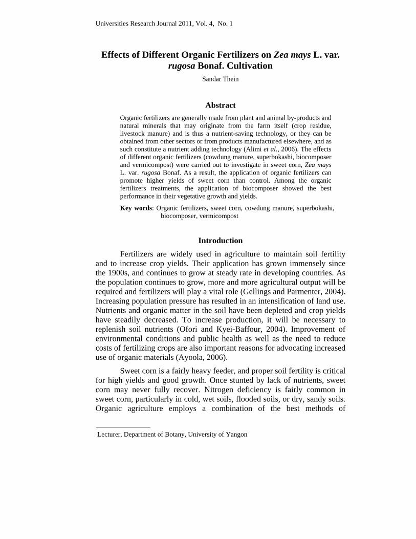
Universities Research Journal 2011, Vol. 4, No. 1
Lecturer, Department of Botany, University of Yangon
Effects of Different Organic Fertilizers on Zea mays L. var. rugosa Bonaf. Cultivation
Sandar Thein
Abstract Organic fertilizers are generally made from plant and animal by-products and natural minerals that may originate from the farm itself (crop residue, livestock manure) and is thus a nutrient-saving technology, or they can be obtained from other sectors or from products manufactured elsewhere, and as such constitute a nutrient adding technology (Alimi et al., 2006). The effects of different organic fertilizers (cowdung manure, superbokashi, biocomposer and vermicompost) were carried out to investigate in sweet corn, Zea mays L. var. rugosa Bonaf. As a result, the application of organic fertilizers can promote higher yields of sweet corn than control. Among the organic fertilizers treatments, the application of biocomposer showed the best performance in their vegetative growth and yields.
Key words: Organic fertilizers, sweet corn, cowdung manure, superbokashi, biocomposer, vermicompost
Introduction
Fertilizers are widely used in agriculture to maintain soil fertility and to increase crop yields. Their application has grown immensely since the 1900s, and continues to grow at steady rate in developing countries. As the population continues to grow, more and more agricultural output will be required and fertilizers will play a vital role (Gellings and Parmenter, 2004). Increasing population pressure has resulted in an intensification of land use. Nutrients and organic matter in the soil have been depleted and crop yields have steadily decreased. To increase production, it will be necessary to replenish soil nutrients (Ofori and Kyei-Baffour, 2004). Improvement of environmental conditions and public health as well as the need to reduce costs of fertilizing crops are also important reasons for advocating increased use of organic materials (Ayoola, 2006).
Sweet corn is a fairly heavy feeder, and proper soil fertility is critical for high yields and good growth. Once stunted by lack of nutrients, sweet corn may never fully recover. Nitrogen deficiency is fairly common in sweet corn, particularly in cold, wet soils, flooded soils, or dry, sandy soils. Organic agriculture employs a combination of the best methods of
Universities Research Journal 2011, Vol. 4, No. 1 52
traditional agriculture and modern technology. At present, organic growers use tried and true practices such as crop rotation, growing a diversity of crops, planting cover crops, and adding organic matter to the soil. Adding organic matter such as feedlot manure and compost to the soil increases the level of nutrients, improves soil microbial activity and increase water holding and nutrient-holding capacity. Organic matter also improves the physical condition of the soil for cultivation and improves soil structure. Thus, the surface of the soil does not crust. Any soil can be improved through the addition of organic matter (Devis, 2004). In this study, applications of organic fertilizer are very important in only promoting the organic farming system but also the production of high yield.
Materials and Methods
Experimental Site A pot experiment was conducted at the Vegetable and Fruit Research Development Centre (VFRDC), Vegetable Science Laboratory in Yemon, Hlegu Township, Yangon Region. Preparation of Materials Soil for sweet corn was prepared with the different organic fertilizers (biocomposer, cowdung manure, superbokashi and vermicompost) for the pot experiment of the study. Soil samples from 10 places at 15 cm depth of soil were randomly taken from experimental field. The samples of all kinds of organic fertilizers used in the study and collected soil samples from the field were analyzed for their physical and chemical properties at Soil Analysis Laboratory of Myanmar Agriculture Service (Land use), Insein Township, Yangon Region before planting sweet corn.
Firstly, all organic fertilizers for this study were exactly weighed to get 200 g of each, respectively. Only half (100 g) of total amount of respective organic fertilizer was taken and then mixed thoroughly with the cultivated soils. The prepared soils were enough to fill in the black polyethylene bags (PEB). The size of PEB was (25 cm x 37.5 cm). Finally, the prepared soils with the different organic fertilizers were watered with enough amount of water to wet the soil two weeks before sowing seeds.
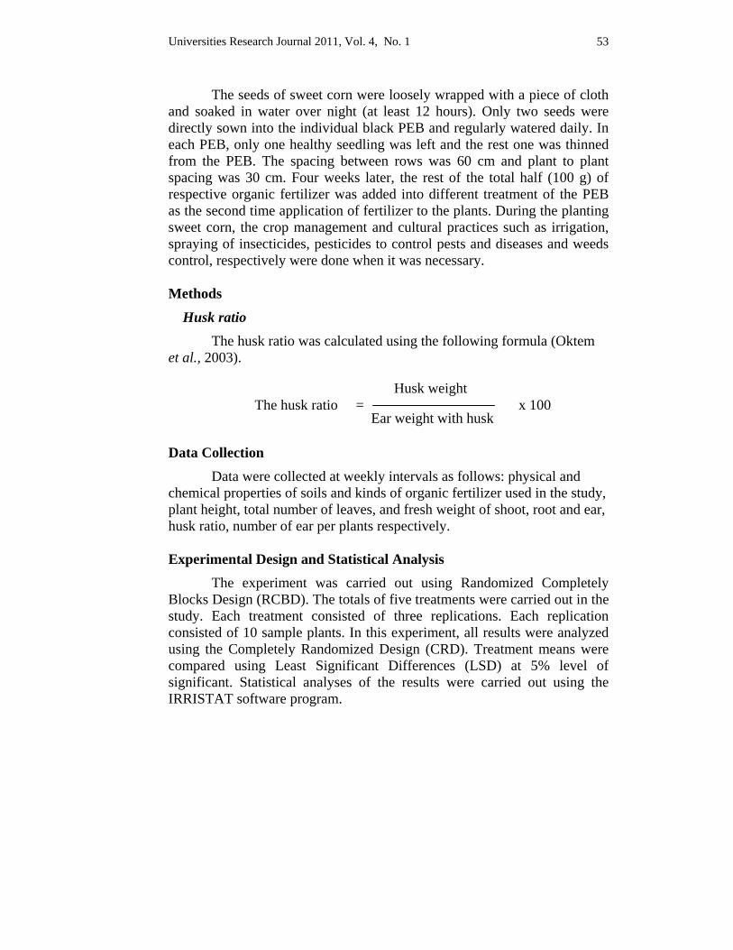
Universities Research Journal 2011, Vol. 4, No. 1 53
The seeds of sweet corn were loosely wrapped with a piece of cloth and soaked in water over night (at least 12 hours). Only two seeds were directly sown into the individual black PEB and regularly watered daily. In each PEB, only one healthy seedling was left and the rest one was thinned from the PEB. The spacing between rows was 60 cm and plant to plant spacing was 30 cm. Four weeks later, the rest of the total half (100 g) of respective organic fertilizer was added into different treatment of the PEB as the second time application of fertilizer to the plants. During the planting sweet corn, the crop management and cultural practices such as irrigation, spraying of insecticides, pesticides to control pests and diseases and weeds control, respectively were done when it was necessary. Methods
Husk ratio The husk ratio was calculated using the following formula (Oktem
et al., 2003).
The husk ratio = x 100
Data Collection Data were collected at weekly intervals as follows: physical and
chemical properties of soils and kinds of organic fertilizer used in the study, plant height, total number of leaves, and fresh weight of shoot, root and ear, husk ratio, number of ear per plants respectively. Experimental Design and Statistical Analysis
The experiment was carried out using Randomized Completely Blocks Design (RCBD). The totals of five treatments were carried out in the study. Each treatment consisted of three replications. Each replication consisted of 10 sample plants. In this experiment, all results were analyzed using the Completely Randomized Design (CRD). Treatment means were compared using Least Significant Differences (LSD) at 5% level of significant. Statistical analyses of the results were carried out using the IRRISTAT software program.
Husk weight
Ear weight with husk
Universities Research Journal 2011, Vol. 4, No. 1 54
Figure 1. An experimental layout in CRD design for the pot experiment of
sweet corn cultivation
Results
Analysis on Physical and Chemical Properties of Soil and Fertilizers According to the soil tests, the cultivated soil from VFRDC field
was acidic soil (soil pH 4.9) and silty loam in soil type. The available nutrients (P and K2O), exchangeable Cation (K+), total nitrogen contents from soil tests were shown medium in soil samples. Moisture content of soil was 3.16%, respectively. Among organic fertilizers, the content of N, P, K and Ca++ attained the maximum percentages in biocomposer. The percentage of calcium contents was maximum biocomposer (5%) followed by vermicompost (4.4%). The results of organic matter contents showed that superbokashi had 41.44% and cowdung manure had 32.15% respectively. The moisture contents of cowdung manure had 42% and 25.32% in superbokashi.
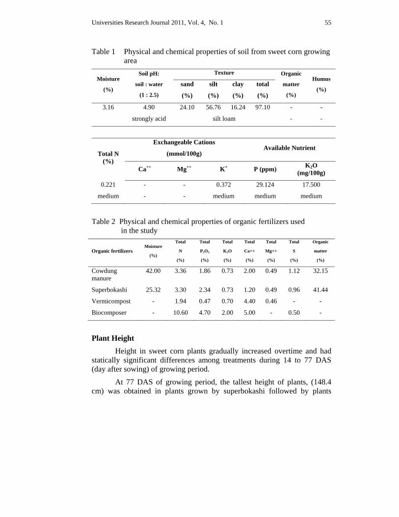
Universities Research Journal 2011, Vol. 4, No. 1 55
Table 1 Physical and chemical properties of soil from sweet corn growing area
Moisture
(%)
Soil pH:
soil : water
(1 : 2.5)
Texture Organic
matter
(%)
Humus
(%) sand
(%)
silt
(%)
clay
(%)
total
(%)
3.16 4.90 24.10 56.76 16.24 97.10 - -
strongly acid silt loam - -
Total N (%)
Exchangeable Cations
(mmol/100g) Available Nutrient
Ca++ Mg++ K+ P (ppm) K2O (mg/100g)
0.221 - - 0.372 29.124 17.500
medium - - medium medium medium
Table 2 Physical and chemical properties of organic fertilizers used
in the study
Organic fertilizers Moisture
(%)
Total
N
(%)
Total
P2O5
(%)
Total
K2O
(%)
Total
Ca++
(%)
Total
Mg++
(%)
Total
S
(%)
Organic
matter
(%)
Cowdung manure
42.00 3.36 1.86 0.73 2.00 0.49 1.12 32.15
Superbokashi 25.32 3.30 2.34 0.73 1.20 0.49 0.96 41.44
Vermicompost - 1.94 0.47 0.70 4.40 0.46 - -
Biocomposer - 10.60 4.70 2.00 5.00 - 0.50 -
Plant Height Height in sweet corn plants gradually increased overtime and had statically significant differences among treatments during 14 to 77 DAS (day after sowing) of growing period.
At 77 DAS of growing period, the tallest height of plants, (148.4 cm) was obtained in plants grown by superbokashi followed by plants
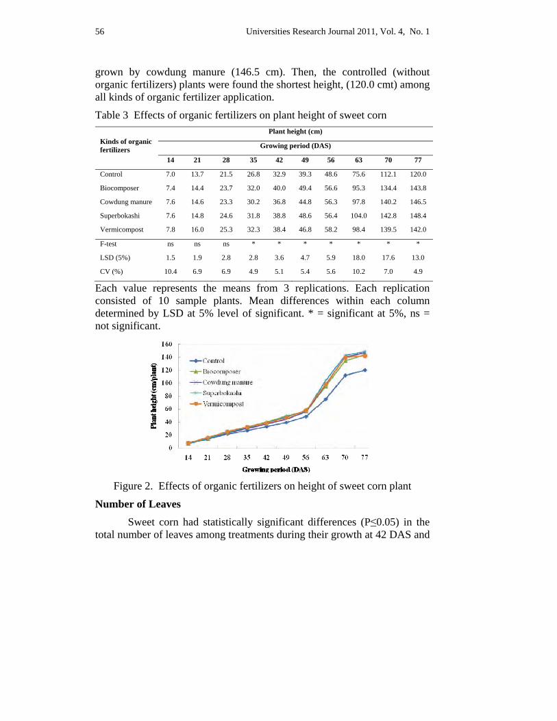
Universities Research Journal 2011, Vol. 4, No. 1 56
grown by cowdung manure (146.5 cm). Then, the controlled (without organic fertilizers) plants were found the shortest height, (120.0 cmt) among all kinds of organic fertilizer application.
Table 3 Effects of organic fertilizers on plant height of sweet corn
Kinds of organic fertilizers
Plant height (cm)
Growing period (DAS)
14 21 28 35 42 49 56 63 70 77
Control 7.0 13.7 21.5 26.8 32.9 39.3 48.6 75.6 112.1 120.0
Biocomposer 7.4 14.4 23.7 32.0 40.0 49.4 56.6 95.3 134.4 143.8
Cowdung manure 7.6 14.6 23.3 30.2 36.8 44.8 56.3 97.8 140.2 146.5
Superbokashi 7.6 14.8 24.6 31.8 38.8 48.6 56.4 104.0 142.8 148.4
Vermicompost 7.8 16.0 25.3 32.3 38.4 46.8 58.2 98.4 139.5 142.0
F-test ns ns ns * * * * * * *
LSD (5%) 1.5 1.9 2.8 2.8 3.6 4.7 5.9 18.0 17.6 13.0
CV (%) 10.4 6.9 6.9 4.9 5.1 5.4 5.6 10.2 7.0 4.9
Each value represents the means from 3 replications. Each replication consisted of 10 sample plants. Mean differences within each column determined by LSD at 5% level of significant. * = significant at 5%, ns = not significant.
Figure 2. Effects of organic fertilizers on height of sweet corn plant
Number of Leaves Sweet corn had statistically significant differences (P≤0.05) in the total number of leaves among treatments during their growth at 42 DAS and
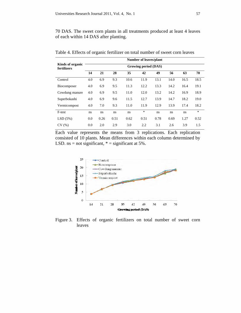
Universities Research Journal 2011, Vol. 4, No. 1 57
70 DAS. The sweet corn plants in all treatments produced at least 4 leaves of each within 14 DAS after planting.
Table 4. Effects of organic fertilizer on total number of sweet corn leaves
Kinds of organic fertilizers
Number of leaves/plant
Growing period (DAS)
14 21 28 35 42 49 56 63 70
Control 4.0 6.9 9.3 10.6 11.9 13.1 14.0 16.5 18.5
Biocomposer 4.0 6.9 9.5 11.3 12.2 13.3 14.2 16.4 19.1
Cowdung manure 4.0 6.9 9.5 11.0 12.0 13.2 14.2 16.9 18.9
Superbokashi 4.0 6.9 9.6 11.5 12.7 13.9 14.7 18.2 19.0
Vermicompost 4.0 7.0 9.3 11.0 11.9 12.9 13.9 17.4 18.2
F-test ns ns ns ns * ns ns ns *
LSD (5%) 0.0 0.26 0.51 0.62 0.51 0.78 0.69 1.27 0.52
CV (%) 0.0 2.0 2.9 3.0 2.2 3.1 2.6 3.9 1.5
Each value represents the means from 3 replications. Each replication consisted of 10 plants. Mean differences within each column determined by LSD. ns = not significant, * = significant at 5%.
Figure 3. Effects of organic fertilizers on total number of sweet corn
leaves
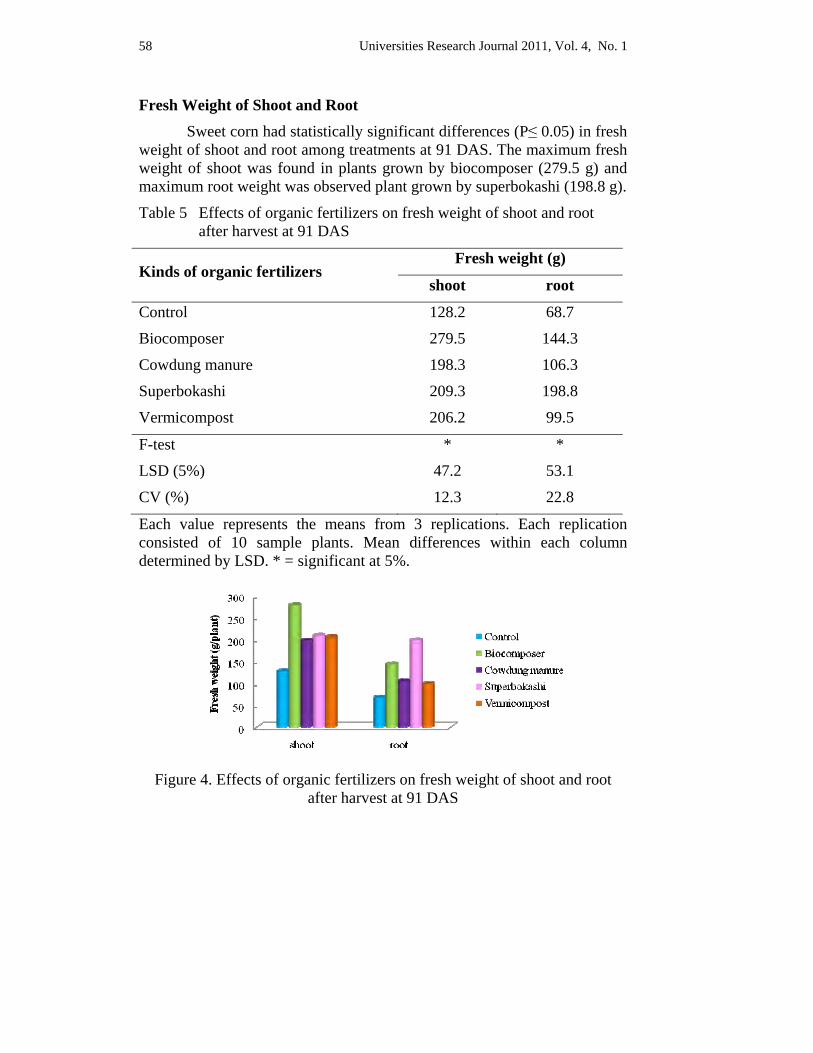
Universities Research Journal 2011, Vol. 4, No. 1 58
Fresh Weight of Shoot and Root Sweet corn had statistically significant differences (P≤ 0.05) in fresh
weight of shoot and root among treatments at 91 DAS. The maximum fresh weight of shoot was found in plants grown by biocomposer (279.5 g) and maximum root weight was observed plant grown by superbokashi (198.8 g).
Table 5 Effects of organic fertilizers on fresh weight of shoot and root after harvest at 91 DAS
Kinds of organic fertilizers Fresh weight (g)
shoot root
Control 128.2 68.7
Biocomposer 279.5 144.3
Cowdung manure 198.3 106.3
Superbokashi 209.3 198.8
Vermicompost 206.2 99.5
F-test * *
LSD (5%) 47.2 53.1
CV (%) 12.3 22.8
Each value represents the means from 3 replications. Each replication consisted of 10 sample plants. Mean differences within each column determined by LSD. * = significant at 5%.
Figure 4. Effects of organic fertilizers on fresh weight of shoot and root
after harvest at 91 DAS
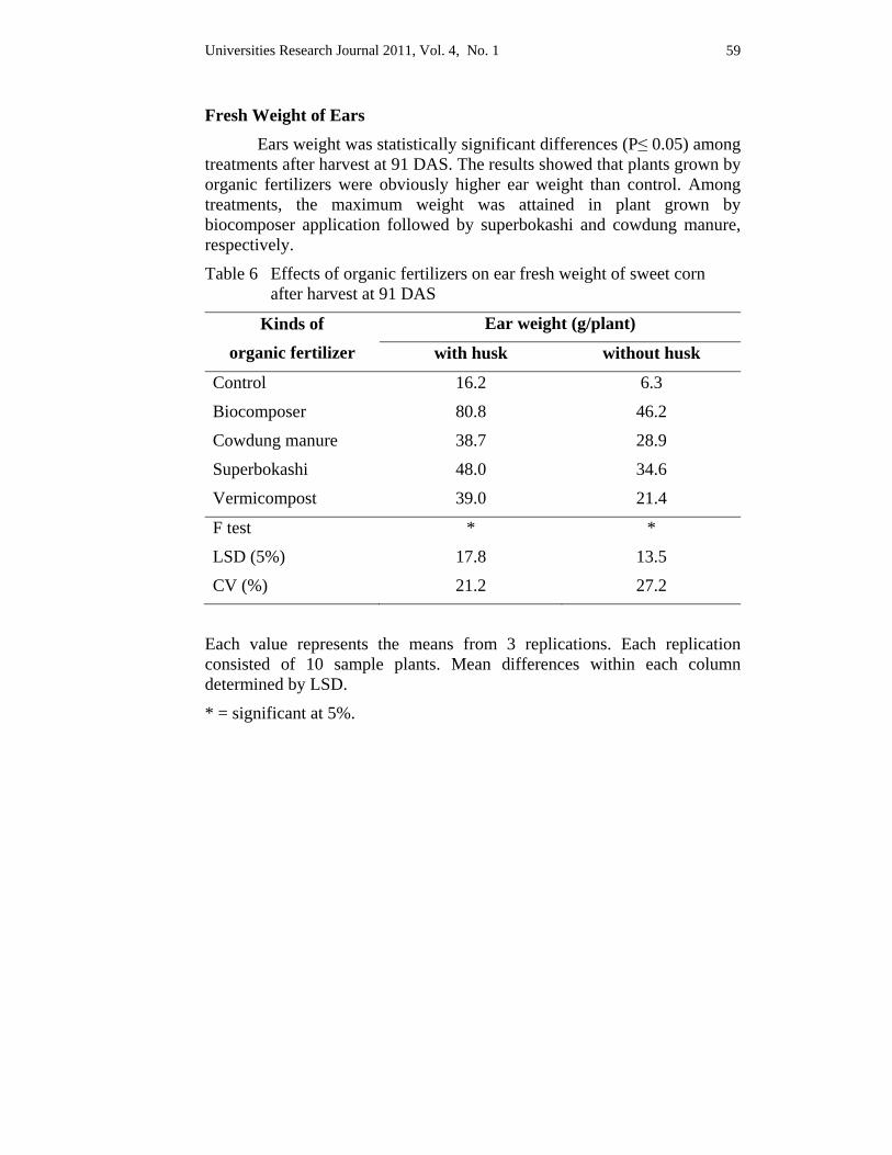
Universities Research Journal 2011, Vol. 4, No. 1 59
Fresh Weight of Ears Ears weight was statistically significant differences (P≤ 0.05) among treatments after harvest at 91 DAS. The results showed that plants grown by organic fertilizers were obviously higher ear weight than control. Among treatments, the maximum weight was attained in plant grown by biocomposer application followed by superbokashi and cowdung manure, respectively.
Table 6 Effects of organic fertilizers on ear fresh weight of sweet corn after harvest at 91 DAS
Kinds of
organic fertilizer
Ear weight (g/plant)
with husk without husk
Control 16.2 6.3
Biocomposer 80.8 46.2
Cowdung manure 38.7 28.9
Superbokashi 48.0 34.6
Vermicompost 39.0 21.4
F test * *
LSD (5%) 17.8 13.5
CV (%) 21.2 27.2
Each value represents the means from 3 replications. Each replication consisted of 10 sample plants. Mean differences within each column determined by LSD.
* = significant at 5%.

Universities Research Journal 2011, Vol. 4, No. 1 60
Figure 5. Effects of organic fertilizers on ear fresh weight of sweet corn
after harvest at 91 DAS
Husk Ratio The results of the study showed that plants grown by cowdung manure had lowest percentage of husk ratio (25.5) among treatment. In contrast, the highest percentage of husk ratio was found in control (61.3) followed by vermicompost (45.1) and biocomposer application (42.8).
Table 7 Effects of organic fertilizers on percentage of husk ratio of sweet corn after harvest at 91 DAS
Kinds of organic fertilizers Husk ratio
Control 61.3
Biocomposer 42.8
Cowdung manure 25.5
Superbokashi 27.9
Vermicompost 45.1
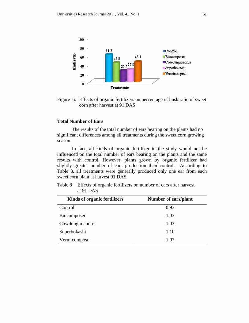
Universities Research Journal 2011, Vol. 4, No. 1 61
Figure 6. Effects of organic fertilizers on percentage of husk ratio of sweet
corn after harvest at 91 DAS
Total Number of Ears The results of the total number of ears bearing on the plants had no significant differences among all treatments during the sweet corn growing season.
In fact, all kinds of organic fertilizer in the study would not be influenced on the total number of ears bearing on the plants and the same results with control. However, plants grown by organic fertilizer had slightly greater number of ears production than control. According to Table 8, all treatments were generally produced only one ear from each sweet corn plant at harvest 91 DAS.
Table 8 Effects of organic fertilizers on number of ears after harvest at 91 DAS
Kinds of organic fertilizers Number of ears/plant
Control 0.93
Biocomposer 1.03
Cowdung manure 1.03
Superbokashi 1.10
Vermicompost 1.07
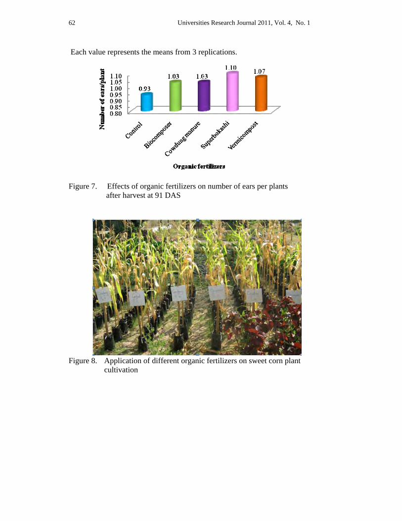
Universities Research Journal 2011, Vol. 4, No. 1 62
Each value represents the means from 3 replications.
Figure 7. Effects of organic fertilizers on number of ears per plants after harvest at 91 DAS
Figure 8. Application of different organic fertilizers on sweet corn plant
cultivation
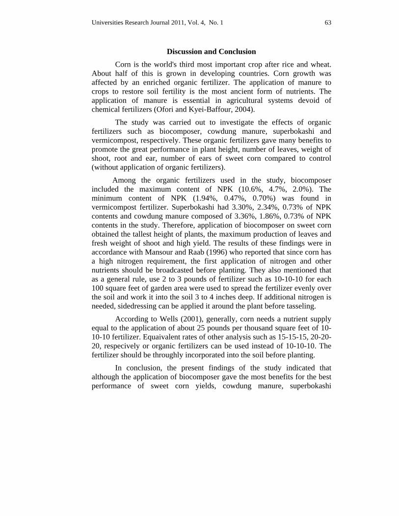
Universities Research Journal 2011, Vol. 4, No. 1 63
Discussion and Conclusion Corn is the world's third most important crop after rice and wheat. About half of this is grown in developing countries. Corn growth was affected by an enriched organic fertilizer. The application of manure to crops to restore soil fertility is the most ancient form of nutrients. The application of manure is essential in agricultural systems devoid of chemical fertilizers (Ofori and Kyei-Baffour, 2004).
The study was carried out to investigate the effects of organic fertilizers such as biocomposer, cowdung manure, superbokashi and vermicompost, respectively. These organic fertilizers gave many benefits to promote the great performance in plant height, number of leaves, weight of shoot, root and ear, number of ears of sweet corn compared to control (without application of organic fertilizers).
Among the organic fertilizers used in the study, biocomposer included the maximum content of NPK (10.6%, 4.7%, 2.0%). The minimum content of NPK (1.94%, 0.47%, 0.70%) was found in vermicompost fertilizer. Superbokashi had 3.30%, 2.34%, 0.73% of NPK contents and cowdung manure composed of 3.36%, 1.86%, 0.73% of NPK contents in the study. Therefore, application of biocomposer on sweet corn obtained the tallest height of plants, the maximum production of leaves and fresh weight of shoot and high yield. The results of these findings were in accordance with Mansour and Raab (1996) who reported that since corn has a high nitrogen requirement, the first application of nitrogen and other nutrients should be broadcasted before planting. They also mentioned that as a general rule, use 2 to 3 pounds of fertilizer such as 10-10-10 for each 100 square feet of garden area were used to spread the fertilizer evenly over the soil and work it into the soil 3 to 4 inches deep. If additional nitrogen is needed, sidedressing can be applied it around the plant before tasseling.
According to Wells (2001), generally, corn needs a nutrient supply equal to the application of about 25 pounds per thousand square feet of 10-10-10 fertilizer. Equaivalent rates of other analysis such as 15-15-15, 20-20-20, respecively or organic fertilizers can be used instead of 10-10-10. The fertilizer should be throughly incorporated into the soil before planting.
In conclusion, the present findings of the study indicated that although the application of biocomposer gave the most benefits for the best performance of sweet corn yields, cowdung manure, superbokashi
Universities Research Journal 2011, Vol. 4, No. 1 64
vermicompost also gave a lot of benefits to promote higher yield compared with control (without application organic fertilizers). On the other hand, using organic fertilizer has been traditional means used to improve soil organic matter and physical properties of the soil. The application of organic fertilizer is essential in agricultural systems devoid of chemical fertilizers and sustains cropping systems through better nutrient recycling. Therefore the application of organic fertilizer gave many benefits on sweet corn cultivation. However, the components of soil and fertilizer can influence on sweet corn production.
Acknowledgements
The author is greatly indebted to Dr. Thet Thet May, Professor and Head, Department of Botany, University of Yangon, for her kind permisssion to conduct and use the reseasrch facilities in Botany Department. I am thankful to Dr. Aye Pe, Professor, Department of Botany, University of Yangon, for his kind permission. I am indebted to my supervisor Dr. Soe Soe Aung Department of Botany, University of Yangon, for her suggestions, invaluable advice, overall supervision and encouragement.
References Alimi, T., O.C. Ajewole, and E.O. Idowu (2006). Economic rationale of commercial
organic fertilizer technology in vegetable production on Osun State of Nigeria. Journal of applied horticulture, 8 (2) : p. 159-164.
Gellings, C.W., and K.E. Parmenter (2004). Energy efficiency in fertilizer production and use, in efficient use and conservation of energy. UNESCO, Oxford, UK.
Ofori, E., and N. Kyei-Baffour (2004). Agrometeorlog and maize production.
Ayoola, O.T. (2006). “Effects of fertilizer treatments on soil chemical properties and crop yields in a cassava-based cropping system”. Institute of Agricultural Research and Training, Ibadan, Nigeria.
Davis, J.M. (2004). Organic sweet corn production. Extension Horticultural Science. North Carolina.
Mansour, N.S., and C. Raab. (1996). Grow your own sweet corn. Oregon State University Extension Service, U.S. Department of Agriculture.
Oktem, A., A.G. Okterm, and Y. Coskun. (2003). Determination of sowing dates of sweet corn under sanliurfa conditions. Turkey, J. Agric. for 28: 83-31.
Wells, O.S. 2001. Growing sweet corn. Family, Home and Garden Education Center, University of New Hampshire, cooperative extension.
http://www.agrometeorology.org/fileadmin/insam/repoaitory/et.
Universities Research Journal 2011, Vol. 4, No. 1
Lecturer, Department of Botany, University of Yangon
Natural Colorant from Gardenia jasminoides Ellis (Cape jasmine)
Khin Thantsin
Abstract The recent research related to an efficient method for the extraction of high stability, superior quality, value added, ready to use pigments from fruits of Gardenia jasminoides Ellis. The process comprises steps of mixing fruits powder of Gardenia jasminoides Ellis. with one food-grade solvent or a combination of more than one food-graded-solvent, macerating and agitating the mixture with continuance protection from light condition, filtering the macerated mixture to remove undesirable fibrous plant material, cooling the filtrate immediately, and concentrating the cooled filtrate under reduced pressure to obtain crude material, with yellow and reddish brown color pigments and with recovery of about 95%. These natural pigments concentrate are used of range between 0.05 to 3% in food, pharmaceutical, and allied industries for flavor and color.
Key words: Gardenia jasminoides Ellis., natural pigments
Introduction
The future anticipates the increase in the total amount of organic food and beverage product of Nowadays, a strong economic incentive places natural colorant crops under organic system of production. Most naturals colorants are derived from international fruits and vegetable crops grown in developing countries. But some fruits and vegetables crops are typically consumed where they are grown. Hence, there is a limited supply of the requisite fruits and vegetables crops needed for the creation of natural colorant.
As the natural source, Gardenia jasminoides Ellis is from the plant family Rubiaceae and its native to China. It is an evergreen shrub. That grows 4-8 feet tall and wide. It is a fragrant flowering evergreen tropical plant which is commonly grown in Vietnam, Southern China, Taiwan and Japan. The fruits are oval in shape about one-half inch to one inch long and orange in color. The major component of the fruit of Gardenia jasminoides Ellis is the iridoid glycoside geniposide. In recent years, usage of the extract has developed in the processed food industries in Western Europe as a less
Universities Research Journal 2011, Vol. 4, No. 1
66
expensive colorant substitute for saffron in applications where the flavor is not required.
Furthermore, the fruit of gardenia (Gardenia jasminoides Ellis) has been used in traditional Chinese medicine. Its folkloric use has been for the treatment of inflammation, jaundice, headache, edema, fever, hepatic disorders and hypertension for years (Tseng et al., 1995), and the extracts of gardenia fruit could give yellow, red and blue colors (Toshiro and Shigeru, 2000; Sang Won et al., 2003), and has been used as food colorants in oriental countries in products such as noodles and confectioneries (Yamada et al., 1996). In Japan some 320 tons of gardenia yellow is consumed (Nakamura, 1995), and demand has been increasing. What is more, this plant has potential horticultural value because of its larger, attractive fragrant flowers. There has been more than 2000 years history of planting G. jasminoides since Han Dynasty, but due to increased demand since the 1960s the species has been endangered.
The current trend in food colorants has increasingly centered on naturally occurring pigments because of consumer preferences. Crocin, the digentiobioside of crocetin, and other crocetin glycosides namely crocetin gentiobioside glucoside crocetin diglucoside and others are probably the only true water soluble natural carotenoid pigments. Total crocetin content per mass of dried whole gardenias fruit was quantified as 3.4μ mole/ g of crocetin aglycone (Yeon et al. 2001). The Gardenia nuts also contain carbohydrates, minerals, vitamins, fat, picrocrocin and volatile oils. It’s utility as a ready to serve coloring material, the product and process is likely to be well received by food and pharmaceutical industry (Agrwal et al. 2006). In addition, crocins obtained may also be used for anti-carcinogenic activity (Ames, 1983, Ikken et al. 1999) and other pharmacological studies. Ikken et al., 1999, also reported for antimutagenic effect of Gardenia nuts.
The market for natural food colours has been steadily growing over recent years. However, the expectation that a large proportion of synthetic colours would gradually be replaced by natural colours has proved too optimistic. Even though many consumers state a preference for naturally coloured food, the synthetically coloured products appeal to many. Natural colours are usually less bright. While consumers look for foodstuffs with a fresh and appealing colour, a decreasing number of people are willing to eat or drink foodstuffs, which are coloured synthetically. Because of this there is a clear tendency towards more genuine natural food colours.
Universities Research Journal 2011, Vol. 4, No. 1
67
Manufacturers of natural food colours only use edible fruits, vegetables and plants as basic raw materials and they try to utilize manufacturing processes that are as close to nature as technically possible.
This research was done for the development of and efficient methods for the extraction of high stability, superior quality, value added, standardized, ready to use pigments concentrate without solvent residue, and also due to growing concern over the safety of synthetic dyes and to produce organic certified natural colorant from Myanmar source.
Materials and Methods The fruits of Gardenia jasminoides Ellis. were collected from Site
Khung village, Sesai Township, Southern Shan State. They were dried at room temperature about one month,and then in temperature controlled oven at 40°C until to get constant weight. Then, they were ground to get fine powder and stored in an air tight container to prevent moisture changes and contamination. As the chemical materials, Ethyl alcohol was used in recent investigation.
The recent invention relates to an efficient method for the extraction of high stability, superior quality, value added, standardized, ready to use pigments said process comprising steps of mixing Gardenia nuts powder with one food graded-solvent or a combination of more than one food-grade-solvents, macerating and agitating the mixture with continuance protection from light condition, filtering the macerated mixture to remove undesirable fibrous plant material, cooling the filtrate immediately, After 24 hrs filtering the yellow precipitate (clouded yellow color when dissolve in water), the remnant was concentrated at 60°C to get crude product. The obtaining is reddish brown colored residue when it dissolves in water clear honey color. Steps of production were shown in Figure 1.
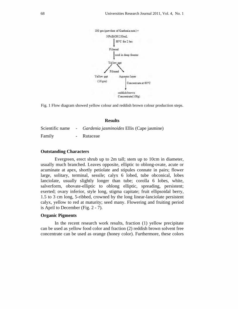
Universities Research Journal 2011, Vol. 4, No. 1
68
Fig. 1 Flow diagram showed yellow colour and reddish brown colour production steps.
Results Scientific name - Gardenia jasminoides Ellis (Cape jasmine)
Family - Rutaceae
Outstanding Characters Evergreen, erect shrub up to 2m tall; stem up to 10cm in diameter, usually much branched. Leaves opposite, elliptic to oblong-ovate, acute or acuminate at apex, shortly petiolate and stipules connate in pairs; flower large, solitary, terminal, sessile; calyx 6 lobed, tube obconical, lobes lanciolate, usually slightly longer than tube; corolla 6 lobes, white, salverform, obovate-elliptic to oblong elliptic, spreading, persistent; exerted; ovary inferior, style long, stigma capitate; fruit ellipsoidal berry, 1.5 to 3 cm long, 5-ribbed, crowned by the long linear-lanciolate persistent calyx, yellow to red at maturity; seed many. Flowering and fruiting period is April to December (Fig. 2 - 7).
Organic Pigments In the recent research work results, fraction (1) yellow precipitate can be used as yellow food color and fraction (2) reddish brown solvent free concentrate can be used as orange (honey color). Furthermore, these colors
Universities Research Journal 2011, Vol. 4, No. 1
69
are naturally carotenoids so they are powerful precious antioxidants. Hence, the result of two colorant are useful either food and beverage or pharmaceutical industries (Fig. 8-13).
Discussion and Conclusion Considerable attention has been focused on anticarcinogenic agents that occur naturally or are added to food and beverages for human consumption (Ames, 1983). Many antioxidants are being identified as anticarcinogens (Ames, 1983; Ferguson, 1994); antimutagens have also been suggested as cancer chemopreventive agents (Stavric, 1994; Ferguson, 1994). Antioxidants are often added to foods to prevent the radical chain reactions of oxidation, and they act by inhibiting the initiation and propagation step leading to the termination of the reaction and delay the oxidation process (Shahidi et al., 1992). At this point, the fruits of Gardenia jasminoides Ellis. are not only natural food colorant but also strongly effected antioxidant carotenoids.
The present invention provides a process, which uses so to say natural solvents with total recovery of desirable materials in quantitative yield without solvent residues along with the separation of value added fraction of high utility from the starting materials.
Since saffron has been used for a long time as food additive, its harmlessness for human consumption has been more than proved, hence of the product thereof. Its great anti-oxidative (Verma and Bordia, 1998; Pham et al. 2000; Kanakis et al. 2007) and anti-tumor activities have been demonstrated. Furthermore, saffron is an antioxidant, is positively effective on sperm morphology and motility in infertile men, while it does not increase sperms count (Heidary et al. 2008). Hence, saffron production from Gardenias nut is the very useful and an efficient for National development. This research was done for slow developing area of Shan State where Gardenia jasminoides Ellis. are well grown and cultivated. At that location even though they produced several tons of Gardenias nut, haven’t technology to produce commercial product saffron (crocetin). Recent research might be an easy way to produce commercial product saffron.
The invention particularly relates to a novel and standardized product for the production of the original colour of saffron. More
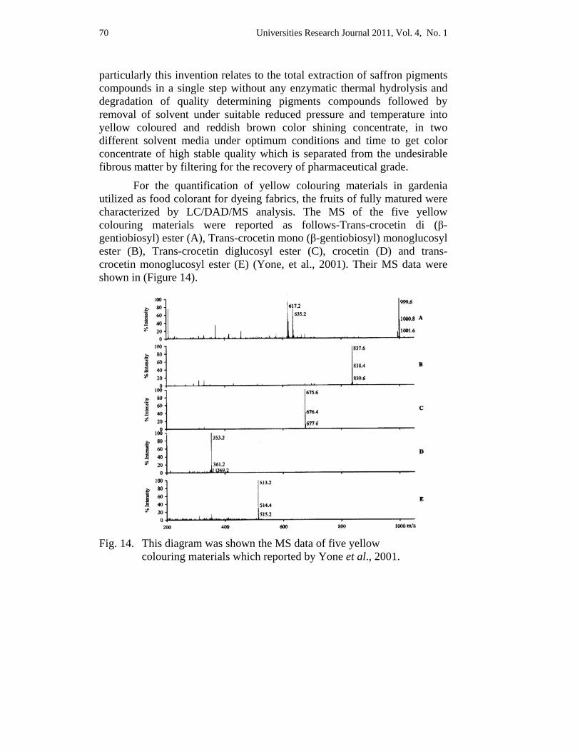
Universities Research Journal 2011, Vol. 4, No. 1
70
particularly this invention relates to the total extraction of saffron pigments compounds in a single step without any enzymatic thermal hydrolysis and degradation of quality determining pigments compounds followed by removal of solvent under suitable reduced pressure and temperature into yellow coloured and reddish brown color shining concentrate, in two different solvent media under optimum conditions and time to get color concentrate of high stable quality which is separated from the undesirable fibrous matter by filtering for the recovery of pharmaceutical grade.
For the quantification of yellow colouring materials in gardenia utilized as food colorant for dyeing fabrics, the fruits of fully matured were characterized by LC/DAD/MS analysis. The MS of the five yellow colouring materials were reported as follows-Trans-crocetin di (β-gentiobiosyl) ester (A), Trans-crocetin mono (β-gentiobiosyl) monoglucosyl ester (B), Trans-crocetin diglucosyl ester (C), crocetin (D) and trans-crocetin monoglucosyl ester (E) (Yone, et al., 2001). Their MS data were shown in (Figure 14).
Fig. 14. This diagram was shown the MS data of five yellow colouring materials which reported by Yone et al., 2001.
Universities Research Journal 2011, Vol. 4, No. 1
71
Furthermore, colours are sold in different forms all over the world: such as powder form, blended powder form, solution form, emulsion form and granular form. Among those five different types of dyes in global market, solution and granular are the best. Recent invention is solution type. Even though it might be an obscurity to deliver from one place to another; solution form is one of the best types of dyes in global market.
Successful and significant penetration of the international market will be dependent upon the introduction of quality products either as such in the form of saffron or through value added newer products. This research related to an efficient method for the extraction of high stability, superior quality, and value added, standardized, and ready to use colour pigments concentrate from fruits of Gardenia jasminoides Ellis. Therefore, recent result can believe helpfulness for National development not only health but also financial.
Acknowledgements The authors acknowledge the Ruby Dragon Companies for the supplement of the plant sample and matured fruits samples for recent research. I would like to express my thanks to Professor and Head of Department of Botany, Yangon University, Dr. Daw Thet Thet May for her permission of carrying out this research.
References Agrwal, S.G., Thappa, R.K., Agnihotri, V.K., Suri, O.P., Quazi, G.N., (2006). Method for
the extraction of saffron pigments and flavor concentrate. United State Patent, Patent No. US. 7070823B2.
Ames, B.N., (1983). Dietary carcinogens and anticarcinogens: Oxygen Radicals and Degenerative Diseases. Science 221, 1256–1263.
Ferguson, L.R., (1994). Antimutagens as cancer chemopreventive agents in the diet. Mutation Research 307, 395–410.
Flora of Hong Kong. 2009. Hong Kong Agriculture, Fisheries and Conservation Department & South China. Volume 3.
Heidary, M., Jahanbakhsh Reza Nejadi, Bahram Delfan, Mehdi Birjandi, Hossein Kaviani, Soudabeh Givrad, (2008). Effect of Saffron on Semen Parameters of Infertile Men. Urology Journal 5 (4): 255-25
Ikken, Y., Morales, P., Martinez, A., Marin, M.L., Haza, A.I., Cambero, M.I., (1999). Antimutagenic effect of fruit and vegetable ethanolic extracts against N-nitrosamines evaluated by the Ames test. Journal of Agricultural Food Chemistry 47, 3257–3264.
Universities Research Journal 2011, Vol. 4, No. 1
72
Kanakis C.D., Tarantilis P,A,, Tajmir-Riahi H,A., Polissiou M,G., (2007). Crocetin, dimethylcrocetin, and safranal bind human serum albumin: stability and antioxidative properties. Journal of Agricultural Food Chemistry. 55:970-7.
Lemmens. R.H.M.J and N. Wulijarni-Soetjipto. (1992). Plant Resources of South-East Asia. No.3. Dye and tannin-producing plants.
Nakamura, M., (1995). A look-out of food additives application in food-colorants. Food Sanit. Res. 45, 35e57.
Pham, T.Q., Cormier, F., Farnworth, E., Tong, V.H., Van Calsteren, M.R., (2000). Antioxidant properties of crocin from Gardenia jasminoides Ellis and study of the reactions of crocin with linoleic acid and crocin with oxygen. Journal of Agriculturl Food Chemistry 48:1455-61.
Sang Won, L., Jong Min, L., Seong Hee, B., (2003). Colorimetric determination of amino acids using genipin from Gardenia jasminoides. Annual. Chim. Acta 480, 267e274.
Shahidi, F., Janitha, P.K., Wanasundara, P.D., (1992). Phenolic antioxidants. Critical Reviews in Food Science and Nutrition 32, 67–103.
Stavric, B., (1994). Antimutagens and anticarcinogens in foods. Food and Chemical Toxicology 32, 79–90.
Toshiro, W., Shigeru, T., (2000). Analysis of natural food pigments by capillary electrophoresis. Journal of Chromatography. A 880, 311e322.
Tseng, T.H., Chu, C.Y., Huang, J.M., Shiow, S.J., Wang, C.J., (1995). Crocetin protects against damage in rat primary hepatocytes. Cancer Letters 97, 61e67.
Verma S.K., Bordia A., (1998). Antioxidant property of Saffron in man. Indian Journal of Medical Science 52:205-7.
Yamada, S., Oshima, H., Saito, I., Hayakawa, J., (1996). Adoption of crocetin as an indicator compound for detection of gardenia yellow in food products (Analysis of natural coloring matters in food V). Journal of Food Hyg. Soc. Jpn. 37, 372e377.
Yeon, S.p., Heung-Jin, C.,Sek, H.Y., Sung-Hwan, K., Tae, K.K., Nam, S.Y., Yong, J.L., (2001). Quantitative Analysis of Crocetin Colorants in Gardenias (Gardenia jasminoides Ellis) by LC/DAD/MS. Journal of Industrial Energy Chemistry 7(6) 375-379.

Universities Research Journal 2011, Vol. 4, No. 1
73
Fig. 2. Habit of Gardenia
jasminoides Ellis Fig. 3. Flower of Gardenia
jasminoides Ellis
Fig. 4. Fruits of Gardenia
jasminoides Ellis Fig. 5. Dried fruits of Gardenia
jasminoides Ellis with fruit walls
Fig. 6. Dried fruits of Gardenia
jasminoides Ellis without fruit walls
Fig. 7. Dried fruits Powder of Gardenia jasminoides Ellis
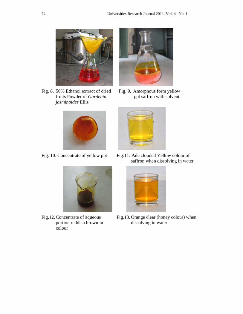
Universities Research Journal 2011, Vol. 4, No. 1
74
Fig. 8. 50% Ethanol extract of dried
fruits Powder of Gardenia jasminoides Ellis
Fig. 9. Amorphous form yellow ppt saffron with solvent
Fig. 10. Concentrate of yellow ppt Fig.11. Pale clouded Yellow colour of
saffron when dissolving in water
Fig.12. Concentrate of aqueous
portion reddish brown in colour
Fig.13. Orange clear (honey colour) when dissolving in water
Universities Research Journal 2011, Vol. 4, No. 1
Lecturer, Department of Botany, University of Mandalay
Morphology of Commercial House Plants from Pyin Oo Lwin Ngu Wah Win
Abstract Twenty two cultivars of commercially cultivated potted flowering plants have been collected from Pyin Oo Lwin Township. These cultivars belonged to 19 genera of 16 families. In these cultivars, Anthurium ‘Rubrum’ and ‘Rubustum’, Euphorbia and splendens Boj., are cultivated for house decoration in pots. Antirrhinum majus L., Calendula officinalis Lour., Kickxia ramosissima (Wall.) Janch., Petunia hybrida, Phlox drummonidii Hk., Mesembryanthmum crinoflorum 'Sparkle Mixed', Tropaeolum majus L., and Viola tricolor L. var. hortensis are cultivated for landscaping plants. Morphological and agronomical studies have been done and described. Cultivar name, family name, local name, English name, taxonomic characters were studied in morphological description. Preparation of soil, cultivation, fertilizer requirement and containers used were presented in agronomical description.
Key words: potted flowering plants, landscaping plants, agronomical description
Introduction
Pyin Oo Lwin Township is formerly known as “Flower City”. Although flowers are beautiful everywhere, there are no well known places as Pyin Oo Lwin for the cultivation of flowers. The flowers are commercially cultivated in most of the area of Pyin Oo Lwin Township and transported commercially to other area of upper and lower Myanmar, especially to Mandalay and Yangon. The study intended to know the commercially cultivated flowers of plant resources, to give detail taxonomic information on commercial flowers, and to give the knowledge of cultivated procedure of traditional ways from study area to other interested peoples and researchers. This township is one of the most interesting places for researchers botanists and agriculturalists, it possesses natural vegetation and commercially cultivated ornamental plants.
Pyin Oo Lwin Township, situated in Central Myanmar, lies on the eastern part of Mandalay Region. Its average elevation is 3539 feet above sea level. The area of Pyin Oo Lwin Township is 488,794 acres or 763.74 square miles. It is situated between the latitudes of 21° 24´ and 22° 46´
Universities Research Journal 2011, Vol. 4, No. 1 76
North and the longitudes of 95° 54´ and 96° 46´ East. The yearly mean temperature of Pyin Oo Lwin Township is 19.3 °C. In humidity condition, the month of highest average humidity in the morning in September is 86.1% and the lowest is March 53 %, according to the data from 2001 to 2005. The highest average humidity month, September has 10.39 inches of rainfall and the lowest month, March is 0.43 inches of rainfall. As Pyin Oo Lwin Township is a mountainous upland region, ‘Mountainous Red Earth’ is also found commonly in the Township. The natural vegetation of Pyin Oo Lwin Township depends upon the temperature, rainfall, altitudes and soil.
The present research had accumulated the 22 cultivars of house plants from Pyin Oo Lwin Township.
House plants are grown usually for the purpose of interior decoration, for their fascinating foliage and flowers or bizarre life forms. In recent year there has been a manifold increase in the demand of house plants as people are getting conscious about their significance in making environment healthy and beautiful.
Ornamental plants and flowers have been playing a vital role in folk festivals, religious worships, public celebration, ritual activities and all kinds of other social occasions that have led to the development of a blooming trade around the world.
Pyin Oo Lwin Township is economically growing plants nearly all the year round. Beautiful flowers are cultivated not only in the fields but also in pots.
Despite their commercial value, there was no systematic survey on taxonomy and agronomy of commercial flowers grown in Pyin Oo Lwin Township. Therefore, present study emphasizes on the 22 cultivars of commercial house plants by describing its morphology, preparation of soil, the methods of propagation.
Materials and Methods
The map of Pyin Oo Lwin Township is detected from Land Record Department, Pyin Oo Lwin. For the morphological investigation, the specimen collection was done by potted commercial flowering plants.
Universities Research Journal 2011, Vol. 4, No. 1 77
In potted commercial flowering plants, before collection, the list of plants was first accumulated from professional gardeners in this township. Then, the plants were collected and the accurate cultivars names were identified as the above method.
These cultivars are recorded with photographs and checked the specimens by using Flora of British India (1897), Flora of Java (1963-68) and Internet information. The species and cultivars are arranged in alphabetical order of their botanical names. The Myanmar names of commercial cultivars are stated by the reference of Myanmar-English Dictionary.
Agronomic data were recorded by interviewing to commercially cultivated farmers and professional gardeners of potted plants. Ornamental value of commercial house plants available was detected with the help of plant growers by interviewing them.
Results
A Morphological Description of Potted Flowering plants 1. Scientific name - Anthurium andraeanum Andre
Family - Araceae
Myanmar name - Peinpan
English name - Unknown
Anthurium andraeanum ‘Rubrum’ Perennial herbs with tuberous rhizomes. Leaves simple, crowded in
a rosette; petioles tapering upward, very long, sheathing at the base; blades reflexed, ovate-sagittate, deeply cordate at the base, entire along the margin, apiculate at the apex, glabrous on both surfaces. Inflorescences axillary spadix, within the spathe, terminated by a conspicuous sterile appendage. Spathe ovate or broadly ovate or orbicular, deeply cordate at the base, apiculate at the apex, deep red, waxy. Spadix white, yellow-tipped. Flowers small, unisexual. Tepals 4. Stamen 4; filaments strap-shaped; anthers dehiscent by slit. Ovary globose, 2-loculed, with one ovule on the sub-apical placenta; style short; stigma globose (Fig. 1.A).
Universities Research Journal 2011, Vol. 4, No. 1 78
Ornamental value: Cultivated for its beautiful spadix inflorescences with waxy deep red color.
Durable period of inflorescences : 30-45 days
Anthurium andraeanum ‘Rubustum’ Perennial herbs with tuberous rhizomes. Leaves simple, large;
petioles very long, sheathing at the base; blades broadly ovate-cordate, cordate at the base, entire along the margin, apiculate at the apex, glabrous on both surfaces. Inflorescences axillary spadix, within the spathe, terminated by a conspicuous sterile appendage. Spathe cordate, green, waxy; spathe-vein brownish-purple. Spadix brownish-purple. Flowers small, unisexual. Tepals 4. Stamen 4; filaments strap-shaped; anthers dehiscent by slit. Ovary globose, 2-loculed, with one ovule on the sub-apical placenta; style short; stigma globose (Fig. 1.B).
Ornamental value: Cultivated for its beautiful spadix inflorescences with waxy green color.
2. Scientific name - Antirrhinum majus L.
Family - Scrophulariaceae
Myanmar name - Kya pazat
English name - Snapdragon
Unarmed, erect-ascending, annual herbs. Stems and branches terete, often glandular-hairy toward apex. Leaves simple, with lower leaves oppostite or spirally arranged or phyllotaxis mixed, green; blades lanceolate, narrowed towards both ends, aequilateral at the base, entire along the margin, acute at the apex; shortly petiolate. Inflorescences terminal, racemes; pedicels with viscid hairs. Flowers bisexual, zygomorphic, white, red, orange, deep pink or mauve, personate, 2.0-3.2 cm across at anthesis; bract ovate. Calyx 5, green, with glandular-hairy, lobe ovate-elliptic, obtuse, unequal. Corolla 5, bilabiate, white, red, orange, deep pink or mauve; tube saccate; upper lip 2-lobed, in bud outside, with recurved margin; lower lip 3-lobed, apex of palate often yellow, opposite the anthers, mid-lobe of lower lip smallest. Stamens 4, included, didynamous; filaments free; anthers dithecous, divergent. Ovary bilocular,
Universities Research Journal 2011, Vol. 4, No. 1 79
many ovules in each locule, axil placentation; style filiform, red at the tip; stigma small, green (Fig. 1.C).
Ornamental value: Cultivated for its beautiful terminal raceme with red, orange or deep pink personate flowers.
Durable period of inflorescence: 15-20 days
3. Scientific name - Arctotis stoechadifolia Berg.
Family - Asteraceae (Compositae)
Myanmar name - Kya myet lone
English name - African Daisy
Perennial herbs. Stems ribbed, densely grey-lanate. Leaves simple, alternate; sessile or narrowed into the petiole; blades oblong- spatulate, attenuate or cuneate at the base, lacerate along the margin, acute at the apex. Inflorescences terminal axillary head, involucre bracts 3 - 4 seriate, semi-globose; outer bracts with ovate lower part and lanceolate grey-lanate upper part; inner bracts elliptic-oblong, rounded at the top, membranous, glabrous. Receptacles flat, long pilose. Pappus scales oblong, rounded at the top. Ray florets with 3-toothed, numerous, ligulate, unisexual, various in color. Disk florets numerous, tubuliform, 5-lobed, bisexual, yellowish above and paler base with violet. Stamens 5, syngenesious; anther obtuse or shortly sagittate at the base and acute at the tip. Ovary oblong, unilocular with one ovule on basal placenta; style arms truncate (Fig. 1.D).
Ornamental value: Cultivated for its beautiful inflorescences head with various colour.
Durable period of inflorescence : 25-30 days
4. Scientific name - Begonia semperflorens ‘Lotto Mixed’
Family - Begoniaceae
Myanmar name - Begonia
English name - Unknown
Erect, fleshy, inermous perennial herbs. Stems and branches terete, red or green, glabrous. Leaves simple, alternate, the stipules semipersistent; blades cordate, green or reddish-green, oblique at the base, dentate,
Universities Research Journal 2011, Vol. 4, No. 1 80
aculeolate, red along the margin, acute at the apex. Inflorescences axillary, dichasial cyme. Flowers unisexual, zygomorphic, deeply pink, pale pink, white or red, 1.5-2.0 cm across at anthesis; pedicellate; bracteolate. Perianths 4, the 2 outer tepals rounded shape, 1.2-1.5 cm by 1.0-1.3 cm, the 2 inner ones ligulate, 0.3-0.5 cm by 1.0-1.3 cm, deeply pink, pale pink, white or red in staminate flowers. Perianths 5, free, unequal, the 2 outer tepals rounded shape, the 2 inner ones rounded and another ligulate in pistillate flowers. Stamens numerous, exserted, yellow; filaments slightly connate; anthers dithecous, basifixed, longitudinal dehiscence. Ovary inferior, 3-winged, unilocular with many ovules in each locule, pink; style 3, very short; stigma 3, obvious, often twisted (Fig. 1.E).
Ornamental value: Cultivated as pot plants for its fleshy green habit, various colored
inflorescences and covering with flowers on whole plants.
Durable period of inflorescence: 5-7 days
5. Scientific name - Brunsfelsia pauciflora Benth.
Family - Solanaceae
Myanmar name - Phyu pya mu yar
English name - Unknown
Erect, unarmed, glabrous shrubs to small trees. Stems and branches terete. Leaves simple, spirally arranged, sessile; petiolate; blades oval or obovate, cuneate at the base, undulate along the margin, acute or slightly obtuse at the apex. Inflorescences terminal, solitary, sessile. Flowers unisexual, actinomorphic, white and blue, 3.0-3.5 cm across at anthesis, fragrant; pedicel short; bracteolate. Calyx 5-lobed, sympetalous, green, campanulate; lobes ovate-triangular. Corolla 5, white and blue, hypocrateriform; tube narrow, curved near apex, widen curvature above; limb patent; lobes broad, rounded. Stamens 4, inserted within widened apex of corolla-tube; filaments didynamous, short curved at apex, running downwards as finely pubescent ridges; anthers dithecous, basifixed, short, longitudinal dehiscence. Ovary sessile, 2-loculed with many ovules in each locule; style filiform, curved at thickened at apex; stigma 2-lobed (Fig. 1.F).
Universities Research Journal 2011, Vol. 4, No. 1 81
Ornamental value: Cultivated for its fragrant and showy flowers with two colours.
Durable period of inflorescence : 8-10 days
6. Scientific name - Calendula officinalis ‘Citrus Cocktail’
Family - Asteraceae ( Compositae )
Myanmar name - Shwe ta sok
English name - Calendula; Pot marigold
Aromatic, glandular-hairy, annual herbs. Stems angular, solid, much branched from the base. Leaves simple, alternate or lower one rosulate, sessile; blades lanceolate or spatulate, pale-green, semi-amplexicaul at the base, entire or undulate along the margin, obtuse or rounded at the apex, glandular hairy on both surface. Inflorescences terminal, solitary or corymbose heads, large, radiate; involcure bracts 2 to 3 seriate, campanculate; bracts gladiate with gland-hairs, red. Receptacles flat. Ray florets numerous, 3-seriate, oblong-spatulate, unisexual, 3-toothed at the apex, the tube short with gland-hairs, the limb 1.8-2.0 cm by 0.4-0.7 cm, orange. Disk florets numerous, tubuliform or funnel-shaped, bisexual, 5-lobed at the apex, 0.7-0.9 cm, orange. Stamens 5, syngenesious, attached to the corolla-tube; anther sagittate at the base. Ovary linear, unilocular with one ovule on basal placenta; style arms long, subulate in ray florets, entire or shortly 2-lobed in disk florets, orange (Fig. 2.A).
Ornamental value: Cultivated for its long persistant orange head.
Durable period of inflorescence: 30-40 days
7. Scientific name - Cyclamen persicum 'Lazer Mixed' Family - Primulaceae
Myanmar name - Nil
English name - Cyclamen
Perennial herbs with tuberous subterraneous rhizome. Stems without a superterraneous. Leaves simple, spirally arranged exstipulate; blades cordate-ovate, fleshy, glabrous, rounded lobes at the base, irregularly, obtusely denticulate-crenulate along the margin, acute at the apex, pale green beneath, dark green above, pale-blotched; petiole 7.0-12.0 cm long,
Universities Research Journal 2011, Vol. 4, No. 1 82
pale greenish-white or red. Inflorescences axillary, solitary. Flowers bisexual, actinomorphic, pink, rotate, 4.0-5.5 cm across at anthesis; pedicels erect or obliquely erect, usually longer than the petioles, red. Calyx 5-segments, overlapping to the right, ovate, pale green with reddish hairs. Corolla 5 or 6, sympetalous; tube short, cup-shaped, 0.5-0.7 cm long; limb much exceeding the tube, ovate, reflexed, often twisted, 2-3 cm long, pink. Stamens 5 or 6, inserted at the base of the corolla-tube; filaments very short; anthers dithecous, acuminate, basifixed. Ovary globose, unilocular with many ovules in each locule; style subulate; stigma simple (Fig. 2.B).
Ornamental value: Cultivated for its beautiful pink flowers
Durable period of inflorescence: 10-15 days
8. Scientific name - Euphorbia splendens Boj.
Family - Euphorbiaceae
Myanmar name - Sha zaung tinganeck
English name - Kiss me quick
Perennial, monoecious, aculeate, laticiferous shrubs. Stems and branches obtusangular-ribbed, brown-purple, covered with thin hoary layer, with 5 or 6 verticle row of tubercles, bearing many prickles on the tubercles; prickles 2-4 together at each tubercles. Leaves simple, alternate; blades obovate-oblong, cuneate at the base, entire along the margin, retuse at the apex, glabrous on both surfaces. Inflorescences axillary or terminal, cyathia, combined into corymbose dichasia; bracts 2, crescent-shaped, red, white, pink, green or yellowish-green. Involucre 5-lobed, companulate, apically bearing 5-nectary glands; lobes ovate, red; glands orange or red. Flowers unisexual, actinomorphic, apetalous, various in coloured, 4-6.5 cm across at anthesis. Staminate flowers numerous, small, with articulate pedicels, surround a single pistillate flower. Stamen 1, greenish-yellow; filaments pinkish-orange; anthers dithecous, basifixed. Pistillate flowers solitary, central, sessile. Ovary globose, trilocular, with one ovule in each locule on the pendulous placenta; style 1; stigma 3, each bifid (Fig. 2.C).
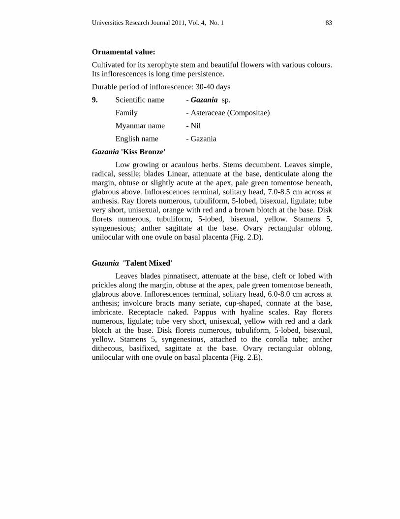
Universities Research Journal 2011, Vol. 4, No. 1 83
Ornamental value: Cultivated for its xerophyte stem and beautiful flowers with various colours. Its inflorescences is long time persistence.
Durable period of inflorescence: 30-40 days
9. Scientific name - Gazania sp.
Family - Asteraceae (Compositae)
Myanmar name - Nil
English name - Gazania
Gazania 'Kiss Bronze' Low growing or acaulous herbs. Stems decumbent. Leaves simple, radical, sessile; blades Linear, attenuate at the base, denticulate along the margin, obtuse or slightly acute at the apex, pale green tomentose beneath, glabrous above. Inflorescences terminal, solitary head, 7.0-8.5 cm across at anthesis. Ray florets numerous, tubuliform, 5-lobed, bisexual, ligulate; tube very short, unisexual, orange with red and a brown blotch at the base. Disk florets numerous, tubuliform, 5-lobed, bisexual, yellow. Stamens 5, syngenesious; anther sagittate at the base. Ovary rectangular oblong, unilocular with one ovule on basal placenta (Fig. 2.D).
Gazania 'Talent Mixed' Leaves blades pinnatisect, attenuate at the base, cleft or lobed with prickles along the margin, obtuse at the apex, pale green tomentose beneath, glabrous above. Inflorescences terminal, solitary head, 6.0-8.0 cm across at anthesis; involcure bracts many seriate, cup-shaped, connate at the base, imbricate. Receptacle naked. Pappus with hyaline scales. Ray florets numerous, ligulate; tube very short, unisexual, yellow with red and a dark blotch at the base. Disk florets numerous, tubuliform, 5-lobed, bisexual, yellow. Stamens 5, syngenesious, attached to the corolla tube; anther dithecous, basifixed, sagittate at the base. Ovary rectangular oblong, unilocular with one ovule on basal placenta (Fig. 2.E).
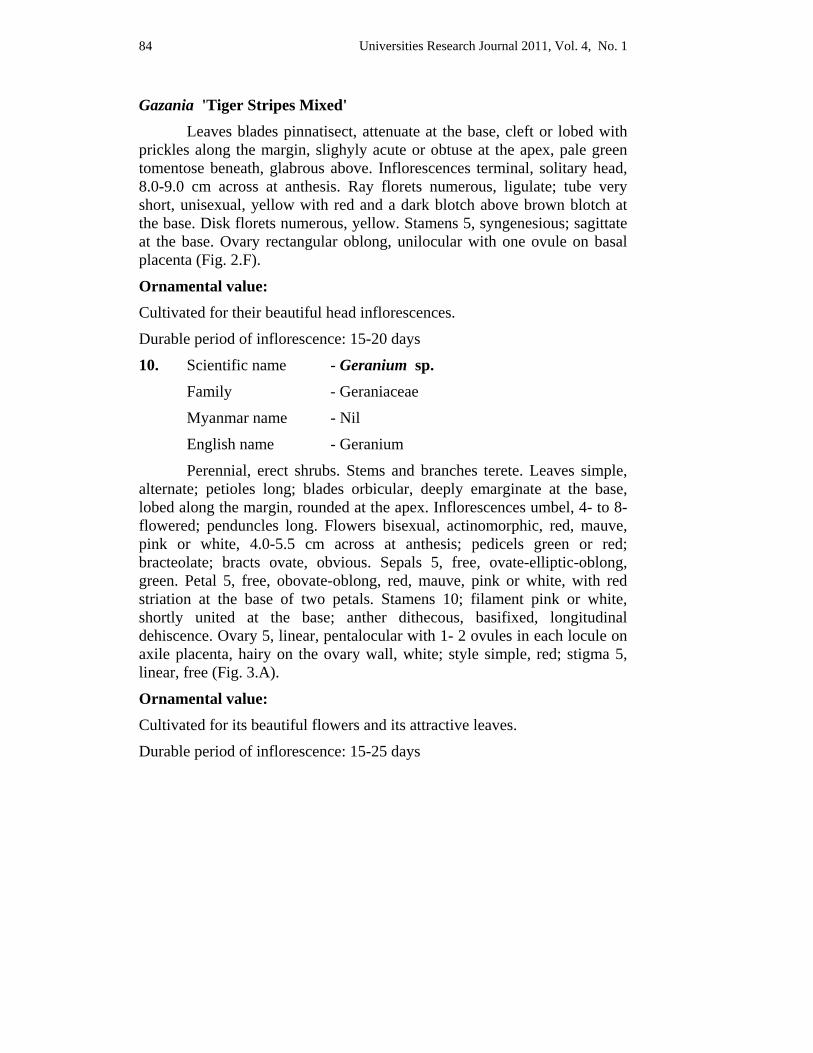
Universities Research Journal 2011, Vol. 4, No. 1 84
Gazania 'Tiger Stripes Mixed' Leaves blades pinnatisect, attenuate at the base, cleft or lobed with prickles along the margin, slighyly acute or obtuse at the apex, pale green tomentose beneath, glabrous above. Inflorescences terminal, solitary head, 8.0-9.0 cm across at anthesis. Ray florets numerous, ligulate; tube very short, unisexual, yellow with red and a dark blotch above brown blotch at the base. Disk florets numerous, yellow. Stamens 5, syngenesious; sagittate at the base. Ovary rectangular oblong, unilocular with one ovule on basal placenta (Fig. 2.F).
Ornamental value: Cultivated for their beautiful head inflorescences.
Durable period of inflorescence: 15-20 days
10. Scientific name - Geranium sp.
Family - Geraniaceae
Myanmar name - Nil
English name - Geranium
Perennial, erect shrubs. Stems and branches terete. Leaves simple, alternate; petioles long; blades orbicular, deeply emarginate at the base, lobed along the margin, rounded at the apex. Inflorescences umbel, 4- to 8-flowered; penduncles long. Flowers bisexual, actinomorphic, red, mauve, pink or white, 4.0-5.5 cm across at anthesis; pedicels green or red; bracteolate; bracts ovate, obvious. Sepals 5, free, ovate-elliptic-oblong, green. Petal 5, free, obovate-oblong, red, mauve, pink or white, with red striation at the base of two petals. Stamens 10; filament pink or white, shortly united at the base; anther dithecous, basifixed, longitudinal dehiscence. Ovary 5, linear, pentalocular with 1- 2 ovules in each locule on axile placenta, hairy on the ovary wall, white; style simple, red; stigma 5, linear, free (Fig. 3.A).
Ornamental value:
Cultivated for its beautiful flowers and its attractive leaves.
Durable period of inflorescence: 15-25 days
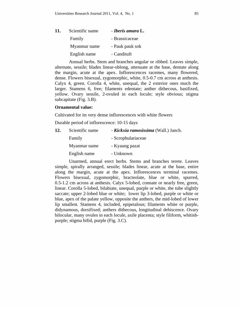
Universities Research Journal 2011, Vol. 4, No. 1 85
11. Scientific name - Iberis amara L.
Family - Brassicaceae
Myanmar name - Pauk pauk sok
English name - Candituft
Annual herbs. Stem and branches angular or ribbed. Leaves simple, alternate, sessile; blades linear-oblong, attenuate at the base, dentate along the margin, acute at the apex. Inflorescences racemes, many flowered, dense. Flowers bisexual, zygomorphic, white, 0.5-0.7 cm across at anthesis. Calyx 4, green. Corolla 4, white, unequal, the 2 exterior ones much the larger. Stamens 6, free; filaments edentate; anther dithecous, basifixed, yellow. Ovary sessile, 2-ovuled in each locule; style obvious; stigma subcapitate (Fig. 3.B).
Ornamental value: Cultivated for its very dense inflorescences with white flowers
Durable period of inflorescence: 10-15 days
12. Scientific name - Kickxia ramosissima (Wall.) Janch.
Family - Scrophulariaceae
Myanmar name - Kyaung pazat
English name - Unknown
Unarmed, annual erect herbs. Stems and branches terete. Leaves simple, spirally arranged, sessile; blades linear, acute at the base, entire along the margin, acute at the apex. Inflorescences terminal racemes. Flowers bisexual, zygomorphic, bracteolate, blue or white, spurred, 0.5-1.2 cm across at anthesis. Calyx 5-lobed, connate or nearly free, green, linear. Corolla 5-lobed, bilabiate, unequal, purple or white, the tube slightly saccate; upper 2-lobed blue or white; lower lip 3-lobed, purple or white or blue, apex of the palate yellow, opposite the anthers, the mid-lobed of lower lip smallest. Stamens 4, included, epipetalous; filaments white or purple, didynamous, dorsifixed; anthers dithecous, longitudinal dehiscence. Ovary bilocular, many ovules in each locule, axile placenta; style filiform, whitish-purple; stigma bifid, purple (Fig. 3.C).

Universities Research Journal 2011, Vol. 4, No. 1 86
Ornamental value: Cultivated for its inflorescence with spurrate flowers in various color.
Durable period of inflorescence : 7-10 days
13. Scientific name - Mesembryanthmum crinoflorum
'Sparkle Mixed' Family - Ficoidaceae ( Aizoaceae )
Myanmar name - Hti pan
English name - Livingstone Daisy
Prostrate or ascending fleshy herbs. Stems and branches terete, green or red, fleshy. Leaves simple, opposite, sessile; blades oblong-lanceolate-linear, very thickly fleshy, green or reddish-green, spatulate at the base, entire along the margin, slightly acute or rounded at the apex. Inflorescences axillary, solitary. Flowers bisexual, actinomorphic, pedicellate, purple or mauve, campanulate, 3.0-5.0 cm across at anthesis. Perianths numerous, free, purple or mauve, linear, attached at the perigone, showy. Perigone 5 or 6-partite; tube obconical; lobes imbricate, unequal, the 2-lobed ligulate, the 3 or 4-lobed small, oblong. Stamens numerous, free, inserted on tube of perigone; filaments filiform, red; anthers dithecous, dorsifixed, longitudinal dehiscence. Carpels 5, syncarpouos, inferior, pentalocular, many ovules in each locule on axile placenta, red; styles 5, free, red; stigma inconspicuous (Fig. 3.D).
Ornamental value: Cultivated for its attracting flowers in various colours.
Durable period of inflorescence : 20-25 days
14. Scientific name - Petunia hybrida 'Pearls Azure Blue'
Family - Solanaceae
Myanmar name - Hne pan
English name - Garden petunia
Universities Research Journal 2011, Vol. 4, No. 1 87
Annual erect, unarmed, viscid, slightly fetid, hairy herbs. Stems terete, hollow, glandular-hairy. Leaves simple, alternate, sessile; blades ovate-oval-lanceolate, cuneate decurrent at the base, undulate along the margin, acute to obtuse at the apex. Inflorescences axillary, solitary. Flowers bisexual, actinomorphic, purplish-white, funnel-shaped, 5.0-7.2 cm across at anthesis; pedicels erecto-patent, soft-hairy. Calyx 5, symsepalous, green, glandular-hairy, persistent. Corolla 5, sympetalous, funnel-shaped, purplish-white; tube widened upwards, with red striations, patently hairy; lobe obtuse or broadly rounded, shortly acuminate or divided into short segments. Stamens 5, inserted below the mid of corolla-tube; filaments shorter than the corolla, curved, unequal; anthers dithecous, oval-globose, longitudinal dehiscence. Ovary 2, many ovules in each locule; style filiform; stigma capitate (Fig. 3.E).
Ornamental value: Cultivated for its funnel-shaped purplish-white flowers with viscid hairs.
Durable period of inflorescence: 17-25 days
15. Scientific name - Phlox drummonidii 'Baby Buttons'
Family - Polemoniaceae
Myanmar name - Thayet hte pan
English name - Unknown
Annual herbs. Stem and branches densely patently glandular-hairy, green. Leaves simple, lower leaves opposite, higher ones spirally arranged, sessile; blades oblanceolate, subcordate-rounded at the base, entire along the margin, acute at the apex, pubescent. Inflorescences terminal, corymb cymes. Flowers bisexual, actinomorphic, violet, explanate, 2.3-2.5 cm across at anthesis. Calyx 5, symsepalous, green, recurved, tipped by a short awn. Corolla 5, sympetalous, deep pink, rotate, red or white in the center; tube 1.2-1.5 cm long, hairy; limb 1.0-1.3 cm by 1.2-1.4 cm. Stamens 5, on the corolla-tube, alternating with the segments; filament free, very short, included; anthers dithecous, dorsifixed, longitudinal dehiscence. Ovary trilocular, with 1-2 ovules in each locule, inferior; style short; stigma trifid (Fig. 3.F).
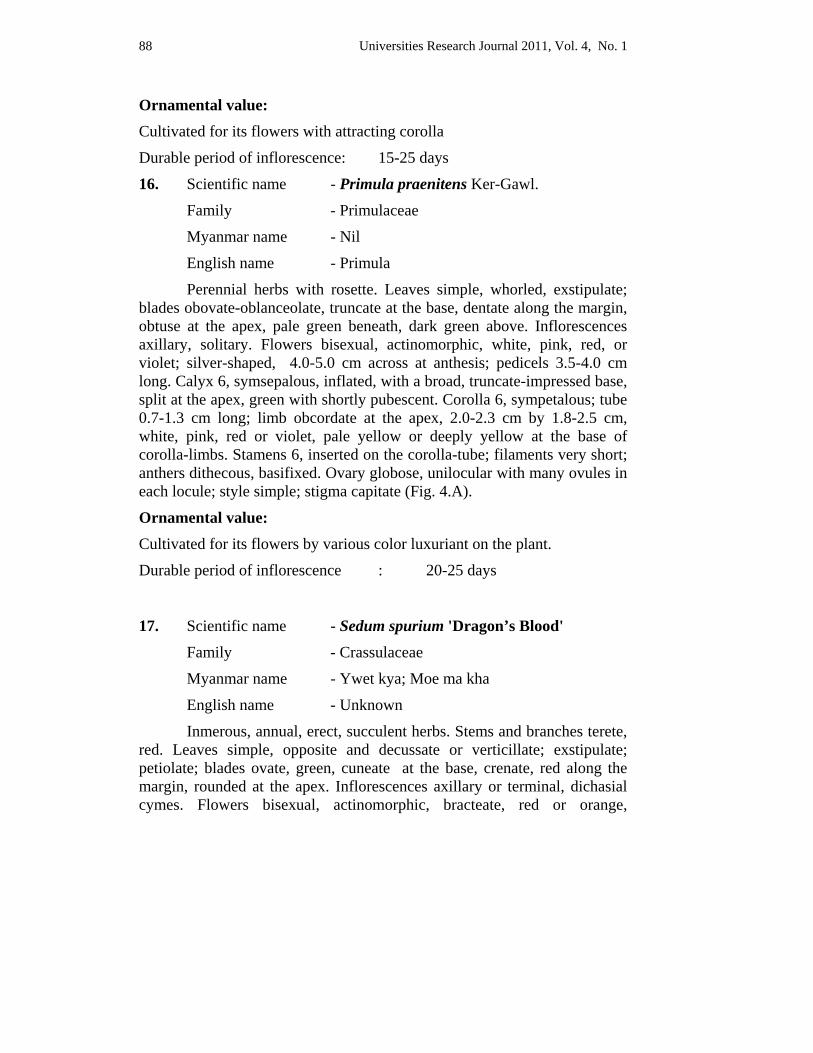
Universities Research Journal 2011, Vol. 4, No. 1 88
Ornamental value: Cultivated for its flowers with attracting corolla
Durable period of inflorescence: 15-25 days
16. Scientific name - Primula praenitens Ker-Gawl.
Family - Primulaceae
Myanmar name - Nil
English name - Primula
Perennial herbs with rosette. Leaves simple, whorled, exstipulate; blades obovate-oblanceolate, truncate at the base, dentate along the margin, obtuse at the apex, pale green beneath, dark green above. Inflorescences axillary, solitary. Flowers bisexual, actinomorphic, white, pink, red, or violet; silver-shaped, 4.0-5.0 cm across at anthesis; pedicels 3.5-4.0 cm long. Calyx 6, symsepalous, inflated, with a broad, truncate-impressed base, split at the apex, green with shortly pubescent. Corolla 6, sympetalous; tube 0.7-1.3 cm long; limb obcordate at the apex, 2.0-2.3 cm by 1.8-2.5 cm, white, pink, red or violet, pale yellow or deeply yellow at the base of corolla-limbs. Stamens 6, inserted on the corolla-tube; filaments very short; anthers dithecous, basifixed. Ovary globose, unilocular with many ovules in each locule; style simple; stigma capitate (Fig. 4.A).
Ornamental value: Cultivated for its flowers by various color luxuriant on the plant.
Durable period of inflorescence : 20-25 days
17. Scientific name - Sedum spurium 'Dragon’s Blood'
Family - Crassulaceae
Myanmar name - Ywet kya; Moe ma kha
English name - Unknown
Inmerous, annual, erect, succulent herbs. Stems and branches terete, red. Leaves simple, opposite and decussate or verticillate; exstipulate; petiolate; blades ovate, green, cuneate at the base, crenate, red along the margin, rounded at the apex. Inflorescences axillary or terminal, dichasial cymes. Flowers bisexual, actinomorphic, bracteate, red or orange,
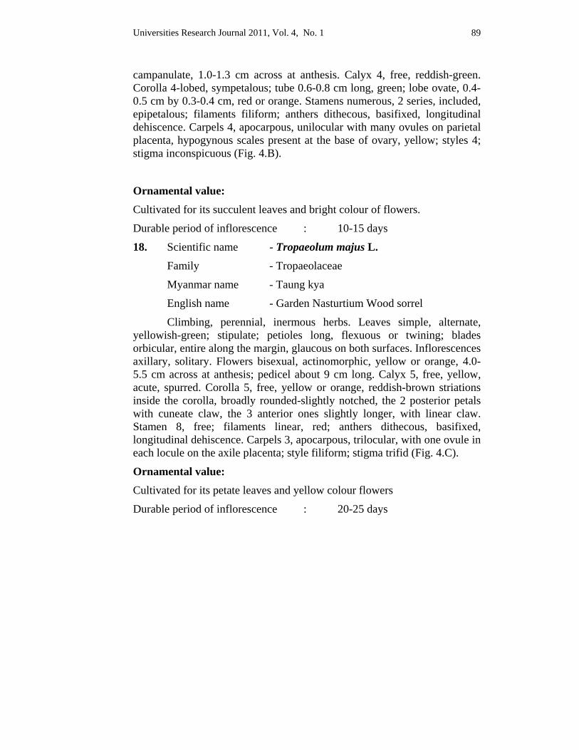
Universities Research Journal 2011, Vol. 4, No. 1 89
campanulate, 1.0-1.3 cm across at anthesis. Calyx 4, free, reddish-green. Corolla 4-lobed, sympetalous; tube 0.6-0.8 cm long, green; lobe ovate, 0.4-0.5 cm by 0.3-0.4 cm, red or orange. Stamens numerous, 2 series, included, epipetalous; filaments filiform; anthers dithecous, basifixed, longitudinal dehiscence. Carpels 4, apocarpous, unilocular with many ovules on parietal placenta, hypogynous scales present at the base of ovary, yellow; styles 4; stigma inconspicuous (Fig. 4.B).
Ornamental value: Cultivated for its succulent leaves and bright colour of flowers.
Durable period of inflorescence : 10-15 days
18. Scientific name - Tropaeolum majus L.
Family - Tropaeolaceae
Myanmar name - Taung kya
English name - Garden Nasturtium Wood sorrel
Climbing, perennial, inermous herbs. Leaves simple, alternate, yellowish-green; stipulate; petioles long, flexuous or twining; blades orbicular, entire along the margin, glaucous on both surfaces. Inflorescences axillary, solitary. Flowers bisexual, actinomorphic, yellow or orange, 4.0-5.5 cm across at anthesis; pedicel about 9 cm long. Calyx 5, free, yellow, acute, spurred. Corolla 5, free, yellow or orange, reddish-brown striations inside the corolla, broadly rounded-slightly notched, the 2 posterior petals with cuneate claw, the 3 anterior ones slightly longer, with linear claw. Stamen 8, free; filaments linear, red; anthers dithecous, basifixed, longitudinal dehiscence. Carpels 3, apocarpous, trilocular, with one ovule in each locule on the axile placenta; style filiform; stigma trifid (Fig. 4.C).
Ornamental value: Cultivated for its petate leaves and yellow colour flowers
Durable period of inflorescence : 20-25 days
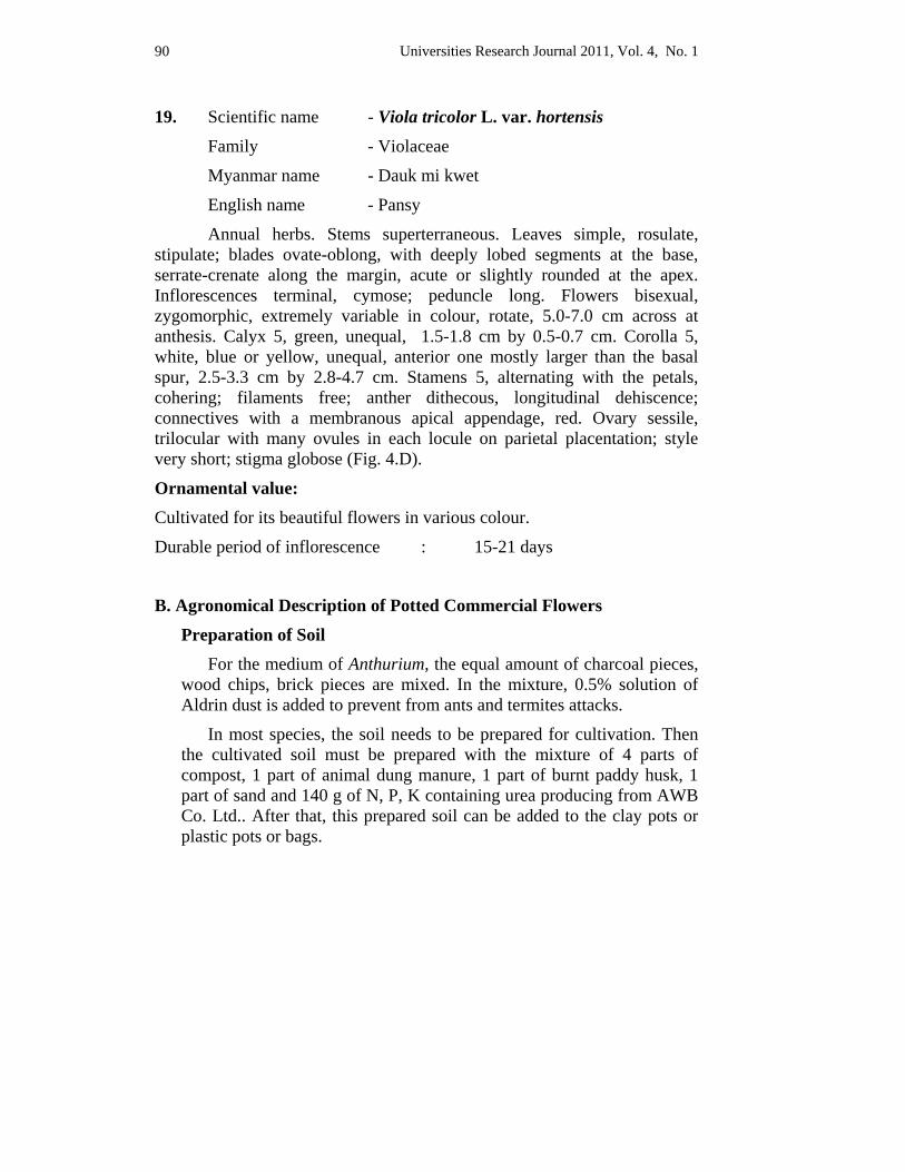
Universities Research Journal 2011, Vol. 4, No. 1 90
19. Scientific name - Viola tricolor L. var. hortensis
Family - Violaceae
Myanmar name - Dauk mi kwet
English name - Pansy
Annual herbs. Stems superterraneous. Leaves simple, rosulate, stipulate; blades ovate-oblong, with deeply lobed segments at the base, serrate-crenate along the margin, acute or slightly rounded at the apex. Inflorescences terminal, cymose; peduncle long. Flowers bisexual, zygomorphic, extremely variable in colour, rotate, 5.0-7.0 cm across at anthesis. Calyx 5, green, unequal, 1.5-1.8 cm by 0.5-0.7 cm. Corolla 5, white, blue or yellow, unequal, anterior one mostly larger than the basal spur, 2.5-3.3 cm by 2.8-4.7 cm. Stamens 5, alternating with the petals, cohering; filaments free; anther dithecous, longitudinal dehiscence; connectives with a membranous apical appendage, red. Ovary sessile, trilocular with many ovules in each locule on parietal placentation; style very short; stigma globose (Fig. 4.D).
Ornamental value: Cultivated for its beautiful flowers in various colour.
Durable period of inflorescence : 15-21 days
B. Agronomical Description of Potted Commercial Flowers
Preparation of Soil For the medium of Anthurium, the equal amount of charcoal pieces,
wood chips, brick pieces are mixed. In the mixture, 0.5% solution of Aldrin dust is added to prevent from ants and termites attacks.
In most species, the soil needs to be prepared for cultivation. Then the cultivated soil must be prepared with the mixture of 4 parts of compost, 1 part of animal dung manure, 1 part of burnt paddy husk, 1 part of sand and 140 g of N, P, K containing urea producing from AWB Co. Ltd.. After that, this prepared soil can be added to the clay pots or plastic pots or bags.
Universities Research Journal 2011, Vol. 4, No. 1 91
Cultivation Anthurium must be sown by the young plants. If the young plant
appears near the bottom of the mother plant, this plant must be pulled out. Potted flowering plants of some species are grown by seeds or cutting or budding methods.
Fertilizer requirement For Anthurium sp., the natural fertilizers from charcoal pieces,
coconut fibres, and wood chips are used. The chemical fertilizers are Aldrin dust and Burplex.
For other species, the compost soil, animal drug manure and burnt paddy husk are natural fertilizers. The chemical fertilizers are using N, P, K that is contained in urea.
Containers using The transparent plastic bags or black plastic bags can be used to
cultivate the local potted plants for sale. The showing plants are usually cultivated in porcelain pots, clay pots and plastic pots.
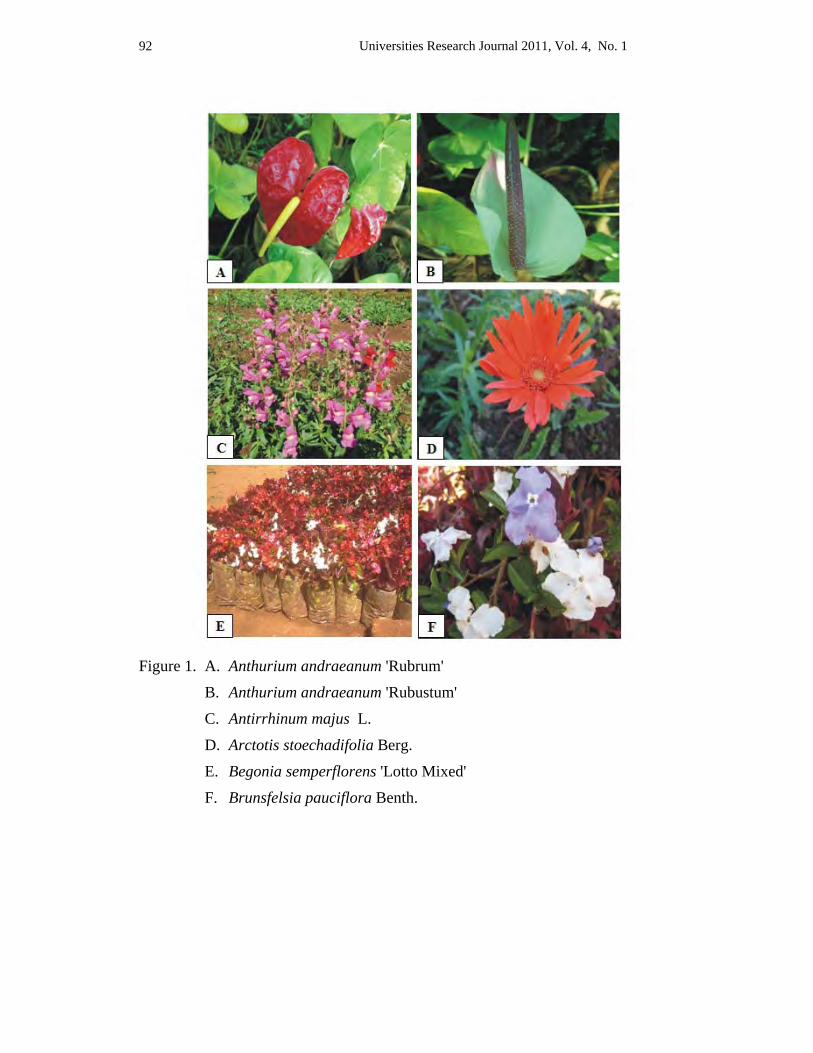
Universities Research Journal 2011, Vol. 4, No. 1 92
Figure 1. A. Anthurium andraeanum 'Rubrum'
B. Anthurium andraeanum 'Rubustum'
C. Antirrhinum majus L.
D. Arctotis stoechadifolia Berg.
E. Begonia semperflorens 'Lotto Mixed'
F. Brunsfelsia pauciflora Benth.
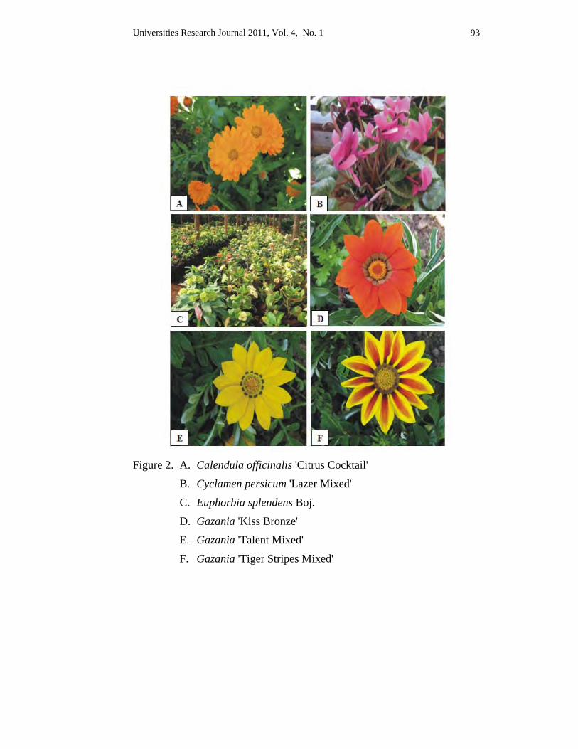
Universities Research Journal 2011, Vol. 4, No. 1 93
Figure 2. A. Calendula officinalis 'Citrus Cocktail'
B. Cyclamen persicum 'Lazer Mixed'
C. Euphorbia splendens Boj.
D. Gazania 'Kiss Bronze'
E. Gazania 'Talent Mixed'
F. Gazania 'Tiger Stripes Mixed'
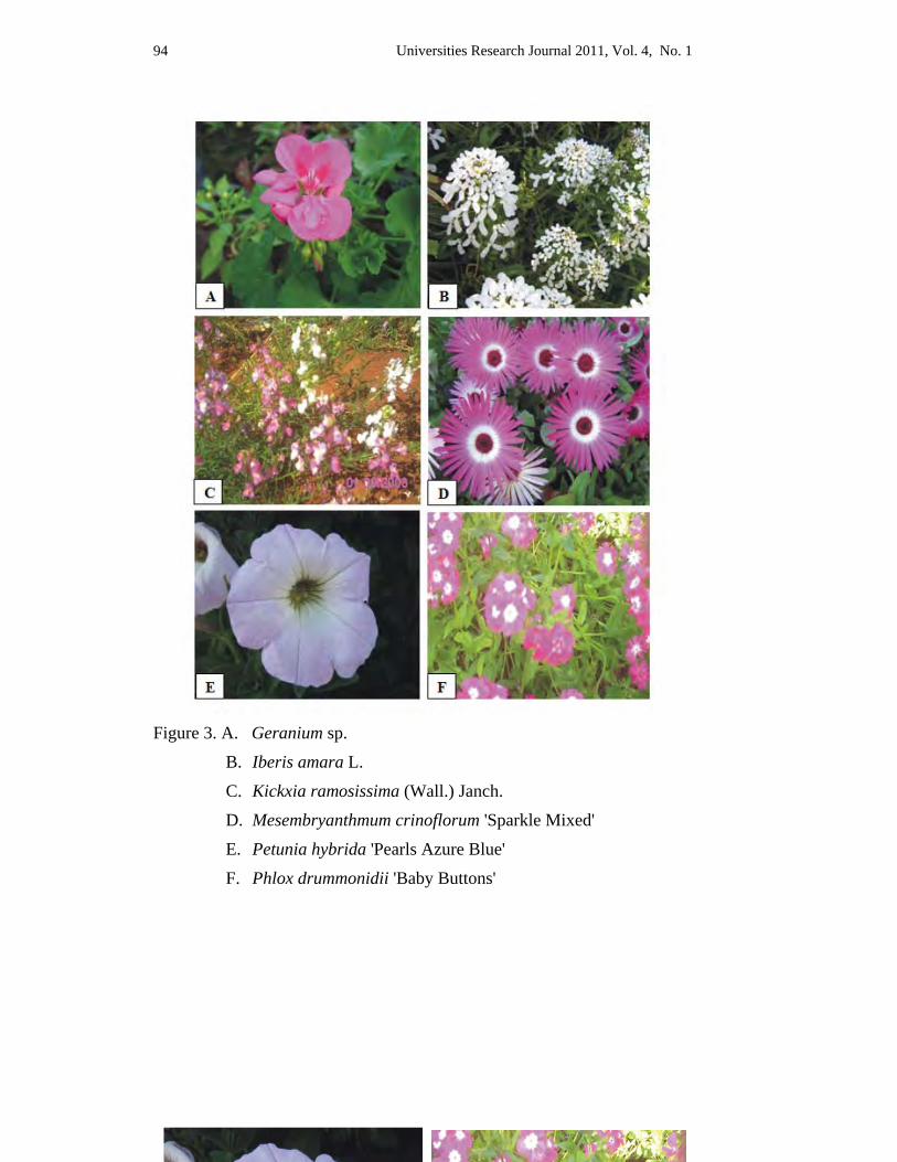
Universities Research Journal 2011, Vol. 4, No. 1 94
Figure 3. A. Geranium sp.
B. Iberis amara L.
C. Kickxia ramosissima (Wall.) Janch.
D. Mesembryanthmum crinoflorum 'Sparkle Mixed'
E. Petunia hybrida 'Pearls Azure Blue'
F. Phlox drummonidii 'Baby Buttons'
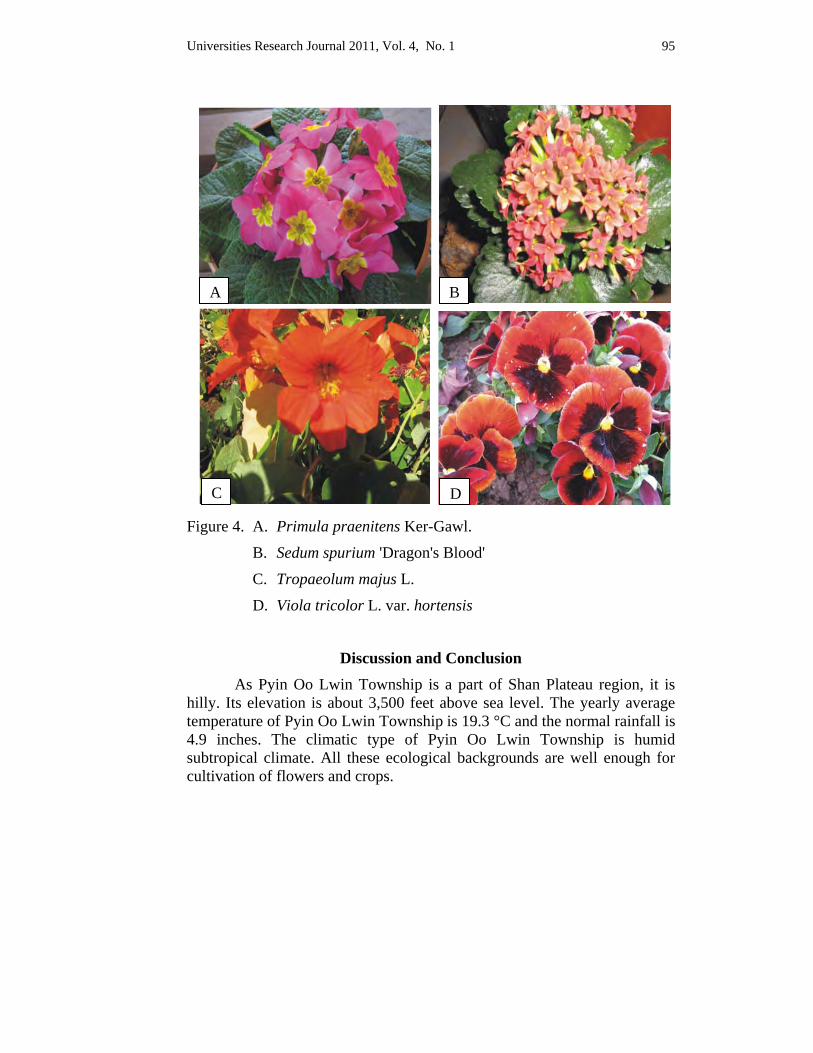
Universities Research Journal 2011, Vol. 4, No. 1 95
Figure 4. A. Primula praenitens Ker-Gawl.
B. Sedum spurium 'Dragon's Blood'
C. Tropaeolum majus L.
D. Viola tricolor L. var. hortensis
Discussion and Conclusion
As Pyin Oo Lwin Township is a part of Shan Plateau region, it is hilly. Its elevation is about 3,500 feet above sea level. The yearly average temperature of Pyin Oo Lwin Township is 19.3 °C and the normal rainfall is 4.9 inches. The climatic type of Pyin Oo Lwin Township is humid subtropical climate. All these ecological backgrounds are well enough for cultivation of flowers and crops.
A B
C D
Universities Research Journal 2011, Vol. 4, No. 1 96
New cultivars of flowers are tested to grow continuously there. The flowers can be economically grown by fields and by potted flowering plants.
The favourable physical conditions, the soil texture and road and communication must be fine. these facts partially supported to yield the best flowers. All these facts that the flowers are blooming continuously throughout the year and Pyin Oo Lwin, “Flower City”.
In the present research, 22 cultivars house plants have been collected from Pyin Oo Lwin Township. Most of these cultivars are herbs. Euphorbia splendens Boj. is shrub. Spadix inflorescences as in Anthurium ‘Rubrum’ and ‘Rubustum’; pernate flowers in Antirrhinum majus L., Kickxia ramosissima (Wall.) Janch.; two kinds of florets (Ray and Disk florets) in Calendula officinalis Lour.; staminate and pistillate flowers separating in one plant in Begonia semperflorens Link & Otto; xerophytic stem in Euphorbia splendens Boj.; spurrate calyx in Tropaeolum majus L.; flowers with viscid hairs in Petunia hybrida and plants with fleshy leaves in Mesembryanthmum crinoflorum ‘Sparkle Mixed’ must be collected by various distinct characters in potted flowering plants at Pyin Oo Lwin Township. In these species, Mesembryanthmum crinoflorum ‘Sparkle Mixed’ and Gazania plants habit to bloom from 10:00 AM to 4:00 PM. These plants with distinctive characters as house plants in this township have practically cultivated for commercially.
In these cultivars, Anthurium ‘Rubrum’ and ‘Rubustum’, and Euphorbia splendens Boj., are cultivate for decoration in house by pots.
Antirrhinum majus L., Calendula officinalis Lour, Kickxia ramosissima (Wall.) Janch., Petunia hybrida, Phlox drummonidii Hk., Mesembryanthmum crinoflorum 'Sparkle Mixed', Tropaeolum majus L., Viola tricolor L. var. hortensis should be cultivated for landscaping plants choosing suitable climate everywhere. Mesembryanthmum crinoflorum 'Sparkle Mixed' is especially suitable for cool weather.
Iberis amara L., Petunia x hybrida, Phlox drummonidii Hk., Viola tricolor L. var. hortensis should be cultivated for decoration in garden, park and home garden. Begonia semperflorens Link & Otto, Cyclamen persicum Mill. 'Lazer Mixed', Erigeron kerwinskianum DC., Gazania 'Kiss Bronze', Gazania 'Talent Mixed' and Gazania 'Tiger Stripes Mixed', Eschscholtzia douglasii Walp., Primula praenitens Ker-Gawl. and Mesembryanthmum
Universities Research Journal 2011, Vol. 4, No. 1 97
crinoflorum 'Sparkle Mixed' should be grown as table flowering plants in parlour and office room.
The cultivation of potted flowering plants is easy. In Pyin Oo Lwin Township, these plants are economically cultivated like goods produced from factory.
These potted flowering plants can mostly be grown from seeds. Some plants can be cultivated by budding and by cutting as in Begonia semperflorens ‘Lotto Mixed’and Brunsfelsia pauciflora Benth.. Potted flowering plants mainly need natural fertilizers, such as compost soil, animal dung manures, burnt paddy husk, coconut fibres, wood chips, barks and chemical fertilizers use a little. Potted plants from Pyin Oo Lwin Township are cultivated everywhere in present. Besides, the seeds of house plants cultivars were economically sold and prepared soil bags and pots, as well.
Some cultivars must be grown to sell in clay pots. These should be grown in house for ornamental. These pots should be grown in office table as table plants for decoration.
In the present study, selling in plastic potsis the mostprofitable but it must be dull. The natural lovers mostly buy the plants in cultivating plastic pots for decorating in house.
In Pyin Oo Lwin Township, the house plants are mostly sold in plastic bags because it can be easily carried, the cost is cheap, and it is economical. The organizations buy these plants with growing plastic bags for landscaping and decorating in office.
Therefore, some plant growers from Pyin Oo Lwin Township earn their living by cultivating of house plants.
According to the study of commercial flowering plants in Pyin Oo Lwin Township, there were 9133 cultivators. Among them 8203 persons were engaged in cultivation of the Garden lands. Some cultivars were grown as vegetables and most were economically grown flowering plants.
Finally, it is hoped that the present study will give some information to botanist and horticulturists, landscape architects, interior decorators and designers, the nursery men and professional gardeners, nature lovers and those who desire to grow house plants in the homes. Then, this is also
Universities Research Journal 2011, Vol. 4, No. 1 98
attractive to the business man and gives some information for the future study of researchers and the knowledge of cultivation for the students.
Acknowledgements I would like to express my deep gratitude to the Ministry of Education, Department of Higher Education (Upper Myanmar), Rector, Pro-rector, Dr Nu Nu Yee, Head and Professor, Dr Thida Oo, Professor, Department of Botany, University of Mandalay for kind, permission to do this research work. I am very thankful to my supervisor, Dr Soe Myint Aye, Associate Professor, Department of Botany, Myitkyina University, for his guidance and invaluable suggestions. Finally, I am also thankful to all my teachers for teaching me to carry out research works.
References Backer, C. A.and R. C. Bakkuizen Van Den Brick Jr. (1963-68). Flora of Java, Vol. I-
III. Rijksher barium, leyden, N.V.P. Noordhoff.
Hooker J.D. (1897). The Flora of British India, Vol. VII. L. Reeve & Co-5, Henrietta Street, Covent Garten, London.
Kapoor, S. L. & A. N. Sharga. (1993). House Plants. Published Pradashan. by Vatika
Khin Myo Myo Chun. (1995). Market Gardening of Pyin Oo Lwin Township. M.A. (Thesis), Geography Department, Mandalay University, Mandalay, Upper Myanmar.
Lawrences G.H.M. (1969). Taxonomy of Vascular Plants. The Macmillan Co. New York.
Universities Research Journal 2011, Vol. 4, No. 1
1. Lecturer, Department of Botany, University of Mandalay 2. Lecturer, Department of Botany, Myitkyina University
Morphology and Nutritional Values of Green Alga Cladophora from Kachin State
Moat War Dine Naw1 and Soe Soe Win2
Abstract The green alga collected from six streams in southern Kachin State was studied. In this paper, the morphology of seven species and two varieties of Cladophora were described. It was found that Cladophora species contain not only protein, chlorophyll-a, carotenoids and lipids, but also minerals such as Calcium, Iron, Potassium, Magnesium, Manganese, Sodium and Zinc.
Key words: Cladophora, protein, lipids
Introduction
People from many cultures, ancient and modern, have used algae for a variety of purposes. For millennia, people throughout the world have collected algae for food, fodder, fertilizer and production of gelling agents such as alginates, caregeenan, agar, agarose and pharmaceuticals, antiviral and antifungal compounds (Lembi et al. 1988). According to Graham (2000) it is estimated that humans have used some 500 species of algae for food or chemicals. In China, written records confirm that human have harvested seaweeds there for more than 2000 years, and today the Chinese collect 74 species of red, green, brown and blue-green algae. Johnston (1970) also mentioned edible algae of fresh and brackish waters in his paper. Both marine and freshwater algae are considered to be an important nutrient source for diets and food additives, because of their high content of essential free amino acids. Among them, edible species have high nutrition of carbohydrates, proteins, fats and minerals. Therefore, they are widely used as food source in many countries, such as Japan, Korea, China, USA, UK, French and Asian countries (Lembi et al.1988).
The Japan marine species, Cladophora rupestris (L.) Kuetzing has polysaccharides, arabinose, galactose, rhamnose, xylose, protein, carotenoids and sulfuric acid. The seaweed can be used as food addictives in Japan (Heiba et al. 1997).
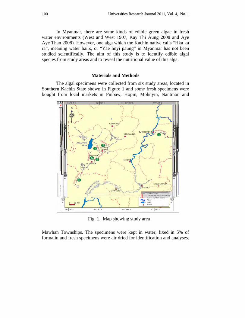
Universities Research Journal 2011, Vol. 4, No. 1 100
In Myanmar, there are some kinds of edible green algae in fresh water environments (West and West 1907, Kay Thi Aung 2008 and Aye Aye Than 2008). However, one alga which the Kachin native calls “Hka ka ra”, meaning water hairs, or “Yae hnyi paung” in Myanmar has not been studied scientifically. The aim of this study is to identify edible algal species from study areas and to reveal the nutritional value of this alga.
Materials and Methods The algal specimens were collected from six study areas, located in Southern Kachin State shown in Figure 1 and some fresh specimens were bought from local markets in Pinbaw, Hopin, Mohnyin, Nantmon and
Mawhan Townships. The specimens were kept in water, fixed in 5% of formalin and fresh specimens were air dried for identification and analyses.
Fig. 1. Map showing study area
Universities Research Journal 2011, Vol. 4, No. 1 101
The identifications were made from fresh specimens and taxonomic description and nomenclature were employed by West & West (1907), Skuja (1949), Prescott (1962), van den Hoek (1963, 1995), Graham (2000), and Leliaert (2004).
The fresh specimens of Cladophora were washed by water and air dried for the determination of protein, chlorophyll-a, carotenoids, moisture, ash, lipids and minerals. All the determinations of nutritional value and mineral contents of Cladophora were carried out according to laboratory procedures at Myanmar Pharmaceutical Factory, Sagaing.
Results The filamentous green alga attached to the stones and rocks (Fig.
2.A), and some were floating in the streams (Fig.2.B). It occurred commonly from October to April. This alga was found growing abundantly in the streams of the study areas and local people harvest and sell it in the market as food items (Fig. 2. C).
The green alga was identified based on their morphology. This alga was Genus Cladophora. It belongs to family Cladophoraceae, order Cladophorales, class Cladophorophyceae of Division Chlorophyta. At present study, seven species and two varieties of Cladophora have been found from six streams of Southern Kachin State. These species are Cladophora rivularis (L.) Hoek., Cladophora globulina (Kutz.) Kutz., Cladophora glomerata (L.) Kuetzing, Cladophora glomerata f. kuetzingiana (Grunow) Heering, Cladophora fracta (Dillw.) Kuetzing, Cladophora fracta var. laustris (Kuetz.) Brand, Cladophora oligoclona (Kuetzing) Kuetzing, Cladophora insignis (C.Agardh) Kuetzing and Cladophora crispata (Roth) Kuetzing. The morphology of genus Cladophora and its species are mentioned.
Cladophora kuetzing Plant branched, either regularly or irregularly, although in a few species branches are rare or wanting. A repeatedly-branched filamentous thallus with basal-distal differentiation, attached when young but in some species becoming free-floating; forming feathery tufts on substrates; especially in flowing water; branching alternate, opposite, or sometimes
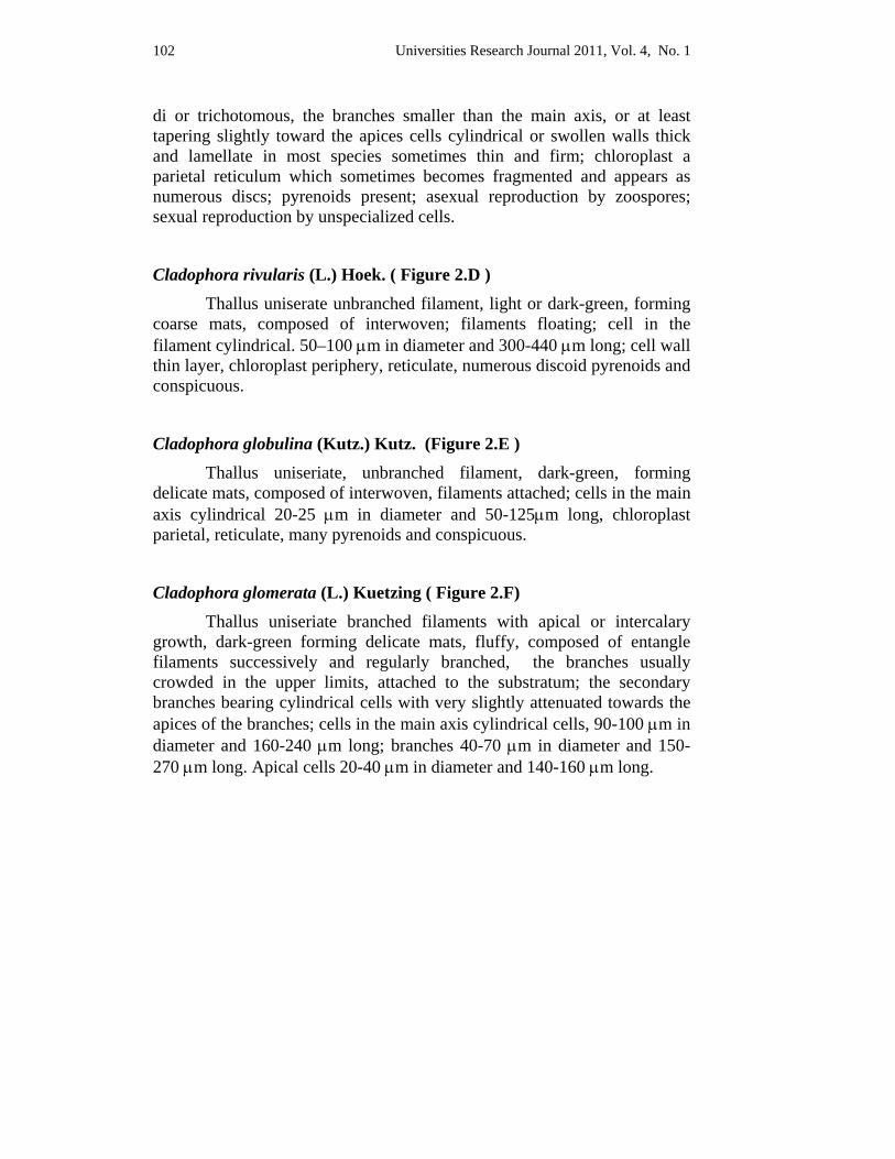
Universities Research Journal 2011, Vol. 4, No. 1 102
di or trichotomous, the branches smaller than the main axis, or at least tapering slightly toward the apices cells cylindrical or swollen walls thick and lamellate in most species sometimes thin and firm; chloroplast a parietal reticulum which sometimes becomes fragmented and appears as numerous discs; pyrenoids present; asexual reproduction by zoospores; sexual reproduction by unspecialized cells.
Cladophora rivularis (L.) Hoek. ( Figure 2.D ) Thallus uniserate unbranched filament, light or dark-green, forming
coarse mats, composed of interwoven; filaments floating; cell in the filament cylindrical. 50–100 μm in diameter and 300-440 μm long; cell wall thin layer, chloroplast periphery, reticulate, numerous discoid pyrenoids and conspicuous.
Cladophora globulina (Kutz.) Kutz. (Figure 2.E ) Thallus uniseriate, unbranched filament, dark-green, forming
delicate mats, composed of interwoven, filaments attached; cells in the main axis cylindrical 20-25 μm in diameter and 50-125μm long, chloroplast parietal, reticulate, many pyrenoids and conspicuous.
Cladophora glomerata (L.) Kuetzing ( Figure 2.F) Thallus uniseriate branched filaments with apical or intercalary
growth, dark-green forming delicate mats, fluffy, composed of entangle filaments successively and regularly branched, the branches usually crowded in the upper limits, attached to the substratum; the secondary branches bearing cylindrical cells with very slightly attenuated towards the apices of the branches; cells in the main axis cylindrical cells, 90-100 μm in diameter and 160-240 μm long; branches 40-70 μm in diameter and 150-270 μm long. Apical cells 20-40 μm in diameter and 140-160 μm long.
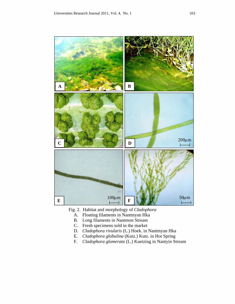
Universities Research Journal 2011, Vol. 4, No. 1 103
Fig. 2. Habitat and morphology of Cladophora A. Floating filaments in Nantmyan Hka B. Long filaments in Nantmon Stream C. Fresh specimens sold in the market D. Cladophora rivularis (L.) Hoek. in Nantmyan Hka E. Cladophora globulina (Kutz.) Kutz. in Hot Spring F. Cladophora glomerata (L.) Kuetzing in Nantyin Stream
A B
C D 200μm
E 100μm
F 50μm
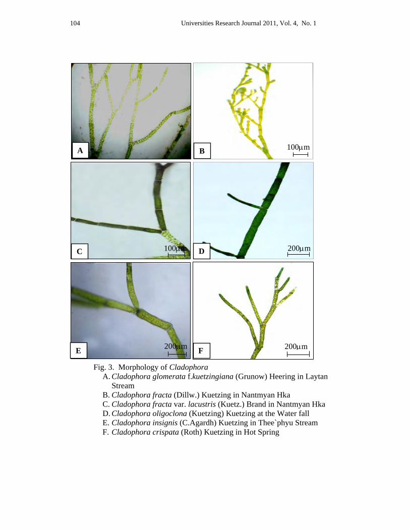
Universities Research Journal 2011, Vol. 4, No. 1 104
Fig. 3. Morphology of Cladophora A. Cladophora glomerata f.kuetzingiana (Grunow) Heering in Laytan
Stream B. Cladophora fracta (Dillw.) Kuetzing in Nantmyan Hka C. Cladophora fracta var. lacustris (Kuetz.) Brand in Nantmyan Hka D. Cladophora oligoclona (Kuetzing) Kuetzing at the Water fall E. Cladophora insignis (C.Agardh) Kuetzing in Thee`phyu Stream F. Cladophora crispata (Roth) Kuetzing in Hot Spring
100μm A B
C 100μm D 200μm
E 200μm F 200μm
Universities Research Journal 2011, Vol. 4, No. 1 105
Cladophora glomerata f. kuetzingiana (Grunow) Heering ( Figure 3.A) Thallus uniseriate branched filament with apical or intercalary growth, light green, forming delicate mats, composed of interwoven loosely branched, attached to the rocks; cells in the main axis cylindrical, 50-80 μm in diameter and 200-250 μm long, the branches dichotomous, 35-50 μm in diameter and 150-200 μm long. Apical cells rounded, 20-25 μm in diameter and 110-130 μm long. Cladophora fracta (Dillw.) Kuetzing ( Figure 3.B) Thallus uniseriate branched filament with intercalary or apical growth, light-green, forming coarse mats, composed of interwoven; irregularly branched filaments, the branches often curving, floating; the secondary branches bearing irregularly swollen or clavate or cylindrical cells; cells in the main axis cylindrical cells or swollen, 60-120 μm in diameter and 120-360 μm long, branches 50-60 μm in diameter and 130-250 μm long. Apical cells cylindrical or clavate, 30-40 μm in diameter and 110-170 μm long. Cladopohora fracta var. lacustris (Kuetz.) Brand ( Figure 3.C) Thallus uniseriate branched filament with intercalary growth, light-green or dark-green forming composed of coarse, rigid form with very little branching, floating, the branches arising irregularly, cells in the main axis long and cylindrical or swollen at the base, 60 – 70 μm in diameter and 200 – 400 μm long; branches 80 – 70 μm in diameter and 200 – 350 μm long; branches bearing many cells or 1-celled. Cladophora oligoclona (kuetzing) Kuetzing ( Figure 3.D) Thallus uniseriate branched filaments with intercalary growth, yellowish-green, forming delicate mats, composed of interwoven; little branched filaments, opposite or dichotomous, loosely attached to the substratum; the secondary branches alternate or unilaterally disposed, clavate or thorn-like l-celled or many-celled branches; cells in the main axis cylindrical, 50-60 μm in diameter and 150-360 μm long; branches 20- 40 μm in diameter and 300-500 μm long. Apical cells cylindrical with rounded tip, 30-50 μm in diameter and 300-400 μm long.
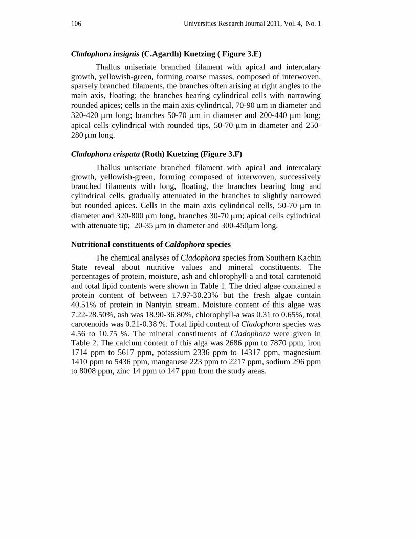
Universities Research Journal 2011, Vol. 4, No. 1 106
Cladophora insignis (C.Agardh) Kuetzing ( Figure 3.E) Thallus uniseriate branched filament with apical and intercalary growth, yellowish-green, forming coarse masses, composed of interwoven, sparsely branched filaments, the branches often arising at right angles to the main axis, floating; the branches bearing cylindrical cells with narrowing rounded apices; cells in the main axis cylindrical, 70-90 μm in diameter and 320-420 μm long; branches 50-70 μm in diameter and 200-440 μm long; apical cells cylindrical with rounded tips, 50-70 μm in diameter and 250-280 μm long. Cladophora crispata (Roth) Kuetzing (Figure 3.F) Thallus uniseriate branched filament with apical and intercalary growth, yellowish-green, forming composed of interwoven, successively branched filaments with long, floating, the branches bearing long and cylindrical cells, gradually attenuated in the branches to slightly narrowed but rounded apices. Cells in the main axis cylindrical cells, 50-70 μm in diameter and 320-800 μm long, branches 30-70 μm; apical cells cylindrical with attenuate tip; 20-35 μm in diameter and 300-450μm long. Nutritional constituents of Caldophora species
The chemical analyses of Cladophora species from Southern Kachin State reveal about nutritive values and mineral constituents. The percentages of protein, moisture, ash and chlorophyll-a and total carotenoid and total lipid contents were shown in Table 1. The dried algae contained a protein content of between 17.97-30.23% but the fresh algae contain 40.51% of protein in Nantyin stream. Moisture content of this algae was 7.22-28.50%, ash was 18.90-36.80%, chlorophyll-a was 0.31 to 0.65%, total carotenoids was 0.21-0.38 %. Total lipid content of Cladophora species was 4.56 to 10.75 %. The mineral constituents of Cladophora were given in Table 2. The calcium content of this alga was 2686 ppm to 7870 ppm, iron 1714 ppm to 5617 ppm, potassium 2336 ppm to 14317 ppm, magnesium 1410 ppm to 5436 ppm, manganese 223 ppm to 2217 ppm, sodium 296 ppm to 8008 ppm, zinc 14 ppm to 147 ppm from the study areas.
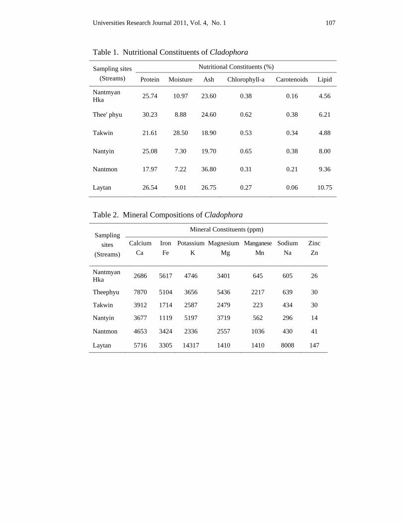
Universities Research Journal 2011, Vol. 4, No. 1 107
Table 1. Nutritional Constituents of Cladophora
Sampling sites (Streams)
Nutritional Constituents (%)
Protein Moisture Ash Chlorophyll-a Carotenoids Lipid
Nantmyan Hka 25.74 10.97 23.60 0.38 0.16 4.56
Thee' phyu 30.23 8.88 24.60 0.62 0.38 6.21
Takwin 21.61 28.50 18.90 0.53 0.34 4.88
Nantyin 25.08 7.30 19.70 0.65 0.38 8.00
Nantmon 17.97 7.22 36.80 0.31 0.21 9.36
Laytan 26.54 9.01 26.75 0.27 0.06 10.75
Table 2. Mineral Compositions of Cladophora
Sampling sites
(Streams)
Mineral Constituents (ppm)
Calcium Ca
Iron Fe
PotassiumK
MagnesiumMg
ManganeseMn
Sodium Na
Zinc Zn
Nantmyan Hka 2686 5617 4746 3401 645 605 26
Theephyu 7870 5104 3656 5436 2217 639 30
Takwin 3912 1714 2587 2479 223 434 30
Nantyin 3677 1119 5197 3719 562 296 14
Nantmon 4653 3424 2336 2557 1036 430 41
Laytan 5716 3305 14317 1410 1410 8008 147
Universities Research Journal 2011, Vol. 4, No. 1 108
Discussion and Conclusion More than 600 species and subspecific taxa have been attributed to
the genus Cladophora over the past 100 years, though the present circumscription is estimated at about 120 species. Van den Hoek (1995) revealed that the exact number of species in the Cladophorophyceae is uncertain and estimated that they range from 200 to 420. The highest number of taxa has been described in the genus Cladophora over 1000 names. Cladophora has been well studied in the Northern Atlantic, Southern Australia and Japan, but the tropical and subtropical representatives remain poorly known (van den Hoek 1963).
According to previous studies, Zeller (1873) described eight species Cladophora from some areas of Myanmar. Only one species (Cladophora crispate (Roth) Kuetzing) has been recorded by West and West (1907) from Myanmar. Three species of Cladophora in Myanmar have been recorded by Skuja (1949). At present study, seven species and two varieties of Cladophora have been found from six streams of Southern Kachin State. They were edible species and collected by native people as food in the study area.
Fresh water Cladophora occurs in a variety of morphological forms that vary in cell dimensions and branching pattern (Graham 2000). According to van den Hoek (1964) thallus organization of the genus Cladophora Kuetzing ranges from unbranched or branched, uniseriate filaments to more complex architectural types. The genus Cladophora is one of the macroscopic green algae, and among the most difficult to classify (Sakai 1958). This is due mostly to the great variation of differences in their appearance, which is greatly affected by habitat, age and environmental conditions.
Yana and Peerapornpisal (2009) reported that some macroalgae such as Cladophora species, Microspora species and Spirogyra species were used as food by local people in the northern and north-eastern part of Thailand. The local people use the algae for Yam or salad e.g. Yam Kai, Yam Tao, Yam Lon, Kai Yae and Hor Nueng Kai in this research, Kai and Tao were processed to 24 varities of food as cake, cookie, crispy, Thai sweet, snack and algal noodle. The survey of edible macroalgae in the tributaries of the Mekong River was also known. Fresh water green algae Cladophora species has been reported from Laos to be obtained from Mekong River (Hasni 2007).
Universities Research Journal 2011, Vol. 4, No. 1 109
Human had noticed nutritional value of algae and had been using it for centuries. As a matter of fact, the Japanese eats about 4.3 grams of dried algae daily. Edible Cladophora and laminaria were collected by the Ainu in northern Japan as early as the eighth century (Lembi et al. 1988). Asian countries use Porphyra dates back to at least A.D, 533-544 and it continues to be highly valued in soup and as flavoring in many dishes. Because of its content of essential amino acids, vitamins and minerals, Porphyra species, known as Amanori in Japanese is largely used as traditional diet in Japan. Arasaki and Arasaki (1983) gave a delightful and detailed account of the historical use of seaweeds in Japan. The algae are consumed in large quantities mainly in Japan. The green algae Ulva, Enteromorpha, Monostroma and Cladophora contain very high dimethyl- sulfide content. So these algae are added to dried nori sheets to increase flavor. Hydrolysis of Cladophora biomass yields arabinose, glatose, xylose, rhamnose and glucose. B12 and glycine betaine are also found in Cladophora. Some species is known to contain acrylic acid which shows antibacterial activity and antifungal properties. Other species of Cladophora have been used in Korea since the 6th century to make and strengthen paper. The algae are washed and dried until clean before they are pressed in paper processing, this paper is used for calligraphy (Arasaki and Arasaki 1983).
Hasni (2007) reported that protein content of Cladophora glomerata (L.) Kuetzing from Kalari Lake in Hub valley side of Karachi was 7.81 to 14.57 %. The protein contents of fresh water edible algae Nostoc, Prasiola and Spirogyra from Myanmar are 16.00%, 38.03% and 28.05% respectively (May Kyawk Khaing 2004, Kay Thi Aung 2008 and Aye Aye Than 2008). Results from the present study indicated that protein content of dried Cladophora was approximately 17.97 to 30.23%. The protein content of fresh specimen was found to be 40.51% (data not mentioned). The nutrient content of fresh Cladophora is greater than that of dried one. Potassium content was higher and Zinc content was lower than other elements. The highest potassium content was 14317 ppm. The highest protein contents of this alga were 40.51% (fresh), 30.23% (dried) and lipids 10.75 ppm. This protein percent is greater than the most popular algal sheet Porphyra (39%), nori in Japanese and other edible algae from fresh waters.
Algae have been eaten only in limited areas by relatively few people. Someone had the mistaken idea that only backward races with low cultural levels eat algae. At present, however, when much research has been done on them, algae are better understood and their great values are
Universities Research Journal 2011, Vol. 4, No. 1 110
beginning to be appreciated. Young filaments are soft and suitable for preparing traditional salad. The collecting time for young filaments is from December to February. During this time, local people collect and sell fresh or steamed algae (Yae hnyi paung) at the local market. They also consumed algae as soup, fried and salad seasonally. Because of the high nutritional value it is a special dish for the native. Making steamed algae is simple, the algae is mixed with pounded glutinous rice, fold with banana leaf and steamed. According to local people of the study area, consuming this filamentous alga is good for intestinal tract of human, because it serve as roughage in the human diets, preventing constipation. The result of this study is useful for local people who are collecting and selling the dried algae in the local markets. Moreover, this alga can be used as ingredient of food items such as cake, cookies, snack and noodle. Cladophora species from Kachin State are considered to be an important source for diet and food additives because it has high content of protein and minerals.
Acknowledgements We would like to thank Dr Nu Nu Yee, Professor and Head and Dr Thida Oo, Professor, Department of Botany, University of Mandalay for their encouragement throughout this work and providing facilities in carrying out this research.
References Arasaki, S. and Arasaki, T. (1983). Vegetables from the Sea. Tokyo, Japan. Inc. 196 pp.
Aye Aye Than, (2008). Studies on edible green alga: Spirogyra (Chlorophyta). Ph D Thesis. Department of Botany. University of Mandalay.
Graham, L.E, Wilcox, L.W. (2000). Algae. Wisconsin University Prentice-Hall, Inc. Upper Saddle River, NJ07458.
Hasni, S. (2007). Phycochemical Studies on Some Fresh Water Green Algae of Karachi. University of Karachi, India.
Heiba, H. and Al. Easa, Hs. and Rizk, A.F. (1997). Fatty acid composition of twelve algae from the coastal zone of Qutar, Egypt.
Hoek C. van den. (1963). Revision of the European species of Cladophora. E.j. Bill. Leiden, pp.1-32.
Hoek C. van den. (1964). Criteria and Procedures in Present-Day Algal Taxonomy. Plenum Press, New York.
Hoek C. van den. (1995). Algae-An Introduction to Phycology. Cambridge University Press.
Universities Research Journal 2011, Vol. 4, No. 1 111
Johnston, H. W. (1970). The Biological and Economic Importance of Algae, Part 3. Edible algae of fresh and brackish water. Journal of Biology society 18:19-34.
Kay Thi Aung, (2008). Edible Prasiola (Chlorophyta) from Kachin State. PhD Thesis, Department of Botany. University of Mandalay.
Leliaert, F. (2004). Taxonomic and phylogenetic studies in the Cladophorophyceae (Chlorophyta). University of Wisconsin, Milwaukee Press.
Lembi, A. C. and Waalant J. R. (1988). Algae and Human Affairs. Cambrige University Press.
May Kyawt Khaing, (2004). A study on the edible Cyanobacteria (blue-green algae): Nostoc species in upper Myanmar.
Prescott, G.W. (1962). Algae of the Western Great Lakes Area W.M. C Brown Campany Publisher, Dubugue, lowa USA.
Sakai, Y. (1958). The science of Cladophora from Japan and its vicinity. Hokkaido University. Sapporao, Japan.
Skuja, H. (1949). Zur Susswasseralgen on Flora Burmas Uppsala., ser. 14: 1-188.
West, W. and West, G.S. (1907). Vol VI, Part II, Fresh Water Algae from Burma, including a few from Bengal and Madras.
Yana, E. and Peerapornpisal, Y. (2009). Diversity of Benthic Algae and water quality in Tributaries of the Mekong River, Passing Thailand and some Part of Laos PDR. Chaing Mai University, Chiang Mai, 50200, Thailand.
Zeller, G. H. (1873). Algae collected by Mrs. S. Kurz in Arakan British Burma
Universities Research Journal 2011, Vol. 4, No. 1
Lecturer, Department of Botany, Pathein University
Isolated Soil Fungi and their Biological Properties Yin Yin Mya
Abstract In this study of the screening program, 5 different soil samples were collected at Pathein Township in Ayeyarwady region. In this isolation, 18 different soil fungi and 8 bacteria were isolated from 5 different soil samples. Eight different soil fungi were utilized for testing with antimicrobial activities. Among them, strain PT-18 showed high antibacterial activity against Bacillus subtilis (24 mm of inhibitory zone). Therefore PT-18 was selected for further investigations according to their results of antibacterial activity. In the investigation of optimizing the fermentation medium 1, 2 and 3 were utilized for producing antibacterial metabolite of PT-18. Medium 2 showed high antibacterial activity. Therefore 5%, 10% 15%, 20% and 25% of seed culture were inoculated into 95%, 90%, 85%, 80% and 75% of fermentation medium 2 and incubated for 4 to 10 days at 27ºC. Three days seed culture, 20% of inoculum size and fermentation medium 2 showed high antibacterial activities according to their results (24 mm of inhibitory zone).
Key words: antibacterial activities, microorganisms, soil fungi
Introduction
Microorganisms have significant functions in ecosystems and one found in all kinds of habitats (Kurtzman, 1992). Antibacterial and Antifungal antibiotics produced by microorganisms still do not have the required qualities in some cases to be used as safe and effective antibacterial agents (Phay, 1997) Since the frequencies and types of life-threatening fungal and bacterial infections are increasing recently, the need of new and effective metabolites are desired.
It is very hard to find substrata not isolated any microbes in nature. Therefore, any substrata collected in nature are useful materials for isolating microorganisms. A typical materials are soil, living and fallen leaves, leaf litters, dung, insect, fresh water, marine water, marine sources and so on (Ando, 2004).Soil sample is the most effective and popular materials for especially isolating a number of microorganisms such as fungi (Harayama,2002).
Universities Research Journal 2011, Vol. 4, No. 1 114
Seed culture must be made in order to have enough inoculums for a large fermenter (Gaden, 1959). Media used in the cultivation of microorganisms must contain all elements in a form suitable for the synthesis of cell substances and for the production of metabolic products (Dale, 1984). During this study, the antibacterial activities of microorganisms were isolated from soil fungi.
Materials and Methods Five different soil samples were utilized for the isolation of
microorganisms. These soil samples were collected at Pathein Township in Ayeyarwady Region. Soil samples were suspended in definite amount of sterile water. These soil suspensions were vigorously agitated. The supernatant were diluted by the physical dilution method. Samples from the dilution series were cultured on soil fungi culture media and incubated at 27ºC for 7 days. Pure colonies from the plates were cultured and purified by re-streaking. The pure strains were cultured as agar media in the test tubes. The soil isolated microorganisms were inoculated into seed medium and incubated for 3 days at 27ºC.
Seed cultures (5%, 10%, 15%, 20% and 25%) were transferred to the fermentation medium (95%, 90%, 85%, 80%, 75%). The fermentation was carried out for 4 to 10 days and tested with test organisms. After the end of fermentation, the fermented broth was used to check the antimicrobial activity against test organisms by paper disc diffusion assay. The fermented broth (10 μl per disc) were dissolved and allowed to dry.
One percent of test organism was added to assay medium, then poured into plates. After solidification, paper discs impregnated with samples (fermented broth) were applied on the agar plates and the plates were incubated for 24-36 hours at 28º to 30ºC. Clear zones (inhibitory zones) surrounding the paper discs indicate the presence of bioactive metabolites which inhibit the growth of test organisms.
The test organisms used in paper disc diffusion assay were Agrobacterum tumefaciens, Escherichia coli, Bacillus subtilis, Micrococcus luteus, Staphylococcus aureus and Saccharomyceae serevisiae. In the investigation of optimizing the fermentation, medium 1, 2 and 3 were utilized for producing antibacterial metabolite of PT-18.
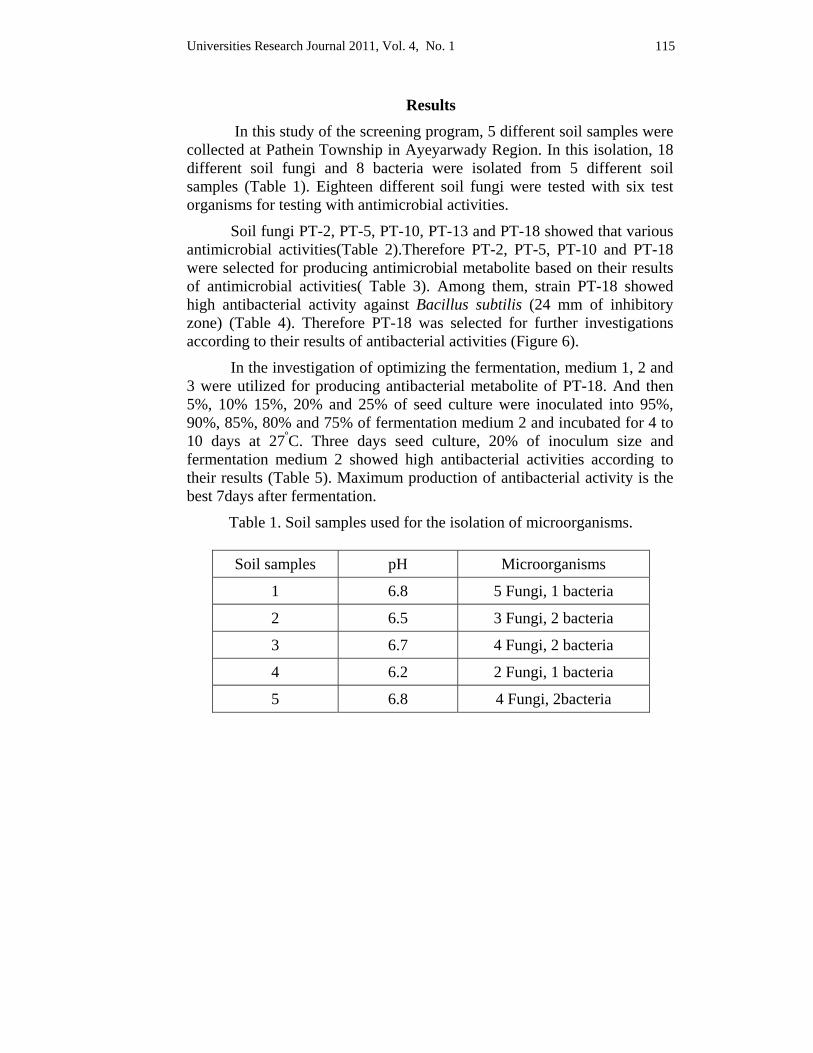
Universities Research Journal 2011, Vol. 4, No. 1 115
Results In this study of the screening program, 5 different soil samples were collected at Pathein Township in Ayeyarwady Region. In this isolation, 18 different soil fungi and 8 bacteria were isolated from 5 different soil samples (Table 1). Eighteen different soil fungi were tested with six test organisms for testing with antimicrobial activities.
Soil fungi PT-2, PT-5, PT-10, PT-13 and PT-18 showed that various antimicrobial activities(Table 2).Therefore PT-2, PT-5, PT-10 and PT-18 were selected for producing antimicrobial metabolite based on their results of antimicrobial activities( Table 3). Among them, strain PT-18 showed high antibacterial activity against Bacillus subtilis (24 mm of inhibitory zone) (Table 4). Therefore PT-18 was selected for further investigations according to their results of antibacterial activities (Figure 6).
In the investigation of optimizing the fermentation, medium 1, 2 and 3 were utilized for producing antibacterial metabolite of PT-18. And then 5%, 10% 15%, 20% and 25% of seed culture were inoculated into 95%, 90%, 85%, 80% and 75% of fermentation medium 2 and incubated for 4 to 10 days at 27ºC. Three days seed culture, 20% of inoculum size and fermentation medium 2 showed high antibacterial activities according to their results (Table 5). Maximum production of antibacterial activity is the best 7days after fermentation.
Table 1. Soil samples used for the isolation of microorganisms.
Soil samples pH Microorganisms
1 6.8 5 Fungi, 1 bacteria
2 6.5 3 Fungi, 2 bacteria
3 6.7 4 Fungi, 2 bacteria
4 6.2 2 Fungi, 1 bacteria
5 6.8 4 Fungi, 2bacteria
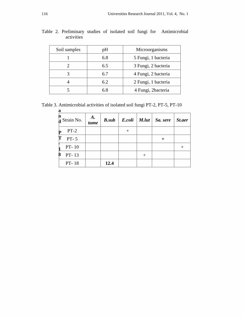
Universities Research Journal 2011, Vol. 4, No. 1 116
Table 2. Preliminary studies of isolated soil fungi for Antimicrobial activities
Table 3. Antimicrobial activities of isolated soil fungi PT-2, PT-5, PT-10
and PT-18
Soil samples pH Microorganisms
1 6.8 5 Fungi, 1 bacteria
2 6.5 3 Fungi, 2 bacteria
3 6.7 4 Fungi, 2 bacteria
4 6.2 2 Fungi, 1 bacteria
5 6.8 4 Fungi, 2bacteria
Strain No. A. tume B.sub E.coli M.lut Sa. sere St.aer
PT-2 +
PT- 5 +
PT- 10 +
PT- 13 +
PT- 18 12.4
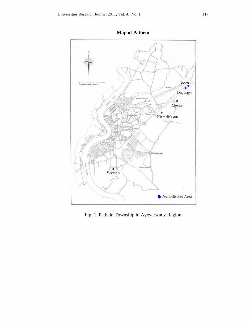
Universities Research Journal 2011, Vol. 4, No. 1 117
Map of Pathein
Fig. 1. Pathein Township in Ayeyarwady Region
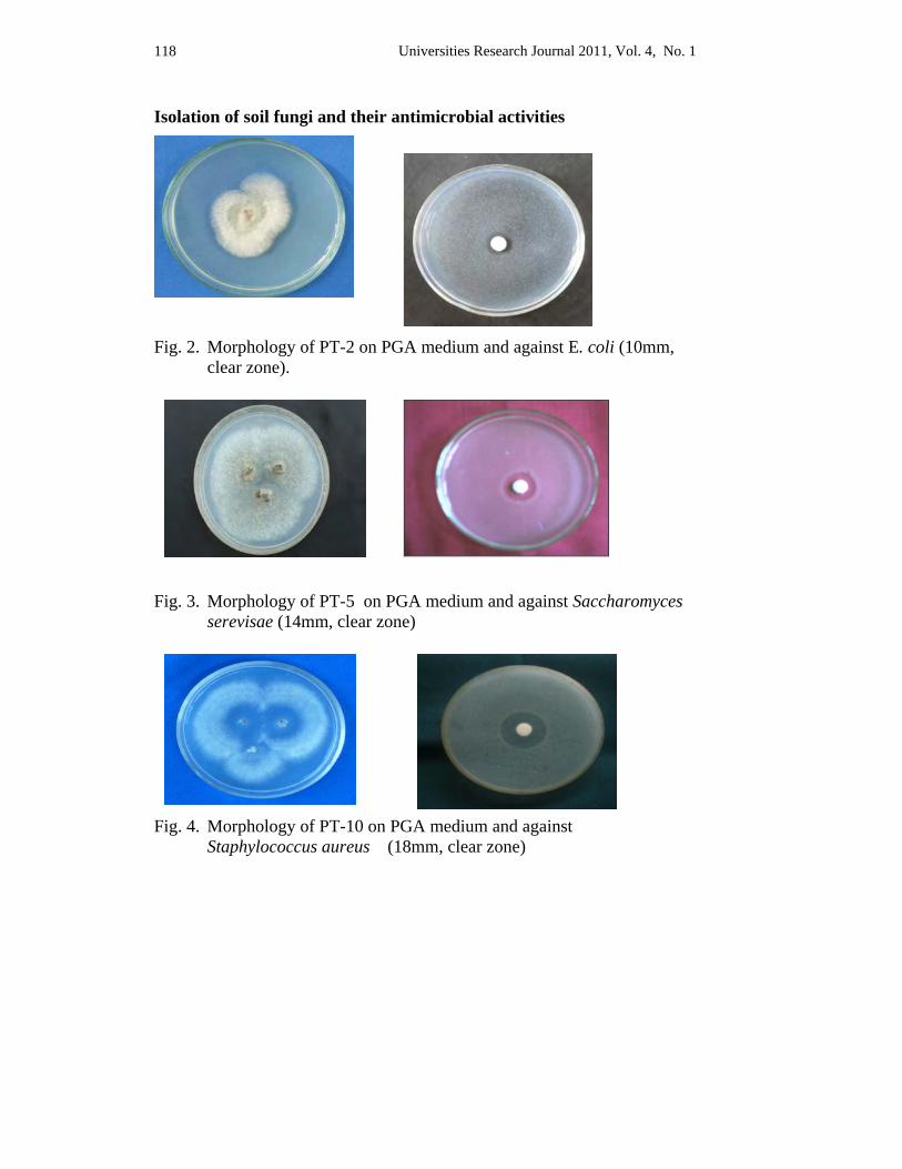
Universities Research Journal 2011, Vol. 4, No. 1 118
Isolation of soil fungi and their antimicrobial activities
Fig. 2. Morphology of PT-2 on PGA medium and against E. coli (10mm,
clear zone).
Fig. 3. Morphology of PT-5 on PGA medium and against Saccharomyces
serevisae (14mm, clear zone)
Fig. 4. Morphology of PT-10 on PGA medium and against Staphylococcus aureus (18mm, clear zone)
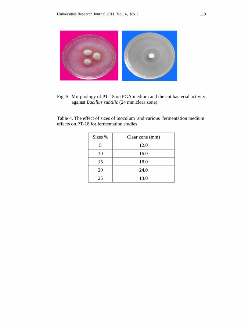
Universities Research Journal 2011, Vol. 4, No. 1 119
Fig. 5. Morphology of PT-18 on PGA medium and the antibacterial activity
against Bacillus subtilic (24 mm,clear zone)
Table 4. The effect of sizes of inoculum and various fermentation medium effects on PT-18 for fermentation studies
Sizes % Clear zone (mm)
5 12.0
10 16.0
15 18.0
20 24.0
25 13.0
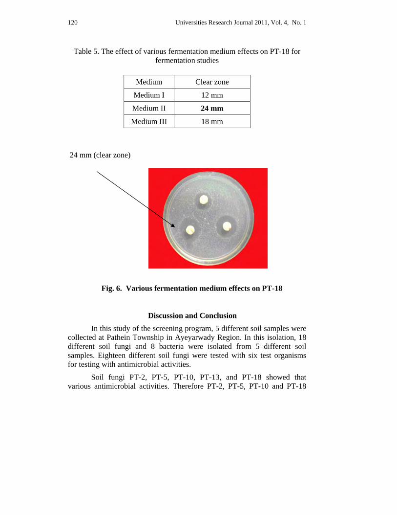
Universities Research Journal 2011, Vol. 4, No. 1 120
Table 5. The effect of various fermentation medium effects on PT-18 for fermentation studies
24 mm (clear zone)
Fig. 6. Various fermentation medium effects on PT-18
Discussion and Conclusion
In this study of the screening program, 5 different soil samples were collected at Pathein Township in Ayeyarwady Region. In this isolation, 18 different soil fungi and 8 bacteria were isolated from 5 different soil samples. Eighteen different soil fungi were tested with six test organisms for testing with antimicrobial activities.
Soil fungi PT-2, PT-5, PT-10, PT-13, and PT-18 showed that various antimicrobial activities. Therefore PT-2, PT-5, PT-10 and PT-18
Medium Clear zone
Medium I 12 mm
Medium II 24 mm
Medium III 18 mm
Universities Research Journal 2011, Vol. 4, No. 1 121
were selected for producing antimicrobial metabolite based on their results of antimicrobial activities. Among them, strain PT-18 showed high antibacterial activity against Bacillus subtilis (24 mm of inhibitory zone).Therefore PT-18 was selected for further investigations according to their results of antibacterial activities.
In the investigation of optimizing the fermentation, medium 1, 2 and 3 were utilized for producing antibacterial metabolite of PT-18. And then 5%, 10% 15%, 20% and 25% of seed culture were inoculated into 95%, 90%, 85%, 80% and 75% of fermentation medium 2 and incubated for 4 to 10 days at 27ºC. Three days seed culture, 20% of inoculum size and fermentation medium 2 showed high antibacterial activities according to their results. Maximum productions of antibacterial activity are the best after 7 days of fermentation.
Acknowledgements I wish to tender my thanks to Professor Dr. Nyunt Phay, Rector and Dr Than Soe,
Pro-Recter, Pathein University for their permission to do this research. I am indebted to Dr Kay Thi Mya, Professor and Head of Botany Department, Pathein University for her valuable advice and permission. My thanks are due to Daw Kin Kin Si and Dr War War Lwin, Associate Professor, Department of Botany, Pathein University for their advice and suggestions.
References Ando, K. and S. Inaba. (2004 May). Sampling and isolation methods of fungi, Workshop
at Pathein University.
Dale, B. E and J. C. Linden (1984). Fermentation substates and economics, Fermentation Technology, 23,135
Gaden,E.L(1959). Fermentation process kinetics. J. Biochem. Microbio. Techn. Eng 1,413-429
Harayama, T. and K. Isono (2002), Sources of Microorganisms, Microbiology, 48:46 50
Kurtzman, C. P (1992). Impact of mycology on the needs of the 21st century, In Proceeding of the Asian Mycological Symposium, Seoul, Korea, 1-6
Phay, N: Doctoral Thesis (1997). Studies on highly selective antibiotics, Faculty of Agriculture, Hokkaido University, Japan
Universities Research Journal 2011, Vol. 4, No. 1
Lecturer, Department of Botany, Yadanarbon University
Phytochemical Investigation and Antimicrobial Activities of the levels of Crinum asiaticum L.
Zan Zan Win
Abstract Crinum asiaticum L. is a member of the family Amaryllidaceae and well known for its ornamental value and medicinal importance. The present work investigated the anatomy, Phytochemistry and antimicrobial activity of the leaves of this species. Anatomical study recorded the presence of parasitic type stomata and closed collateral type vascular bundles. The phytochemical tests detected the presence of alkaloids, flavonoid, terpene, polyphenol, glycoside, sugar, saponin, phenolic compound and liphophilic compound. Antimicrobial tests showed that water and petroleum ether extracts revealed no antimicrobial activity on any of the microbial strains investigated. Contrary to the above described results, ethyl acetate extract of the leaves of C. asiaticum displayed significant inhibitory effect on the growth of E. coli (ATCC – 25922). The result obtained in this study suggests that above noted extract has the potential to be used for the diseases caused by above mentioned pathogenic strain.
Key words: Crinum asiaticum, alkaloids, flavonoid, terpene, polyphenol, glycoside, sugar, saponin, phenolic compound, liphophilic compound, E. coli.
Introduction
Crinum plants are widely distributed throughout the world and also grown widely in Myanmar. They are well known because of their economic and medicinal importance and ornamental value of some members.
Up to now, most studies reported on the genus Crinum are mainly focused on the Phytochemistry and pharmacology of some species of this genus because some members of this genus produce the compounds having medicinal value, and they are potent to treat some ailments and diseases. In Myanmar, a little study has been carried out on the genus Crinum, and it still remains a potent one to work on as yet. The present study detects the anatomical characters and the phytochemical ingredients of the leaves of C. asiaticum and examines their inhibition on the growth of some pathogenic microbial strains.
In recent years, secondary metabolites (phytochemical), previously with unknown pharmacological activities, have been extensively
Universities Research Journal 2011, Vol. 4, No. 1 124
investigated as a source of medicinal agents (Krishnaraju et al, 2005). Phytochemical analysis has recently yielded a vast array of compounds, including more than 150 different alkaloids in the genus Crinum (Fennell and Staden 2001). The compounds possessing medicinal importance identified in this genus make some authors to pay much more attentions to continue to investigate the new and valuable phytochemical from it and to examine their usage as phytomedicine. Crinum species are widely used in indigenous medicine for many diseases; mainly leaf and bulb extracts are of medicinal importance.
The leaf juice of C. asiaticum is used with a little common salt for ear complaints. In Southeast Asia countries, C. asiaticum has a considerable medicinal reputation as a potent folk medicine in the treatment of injury and inflamed joints (Burkill 1966). In the Philippines, for instance, the leaves are prepared as emollient for external use to treat inflamed joints and sprains (Valenzuela et al. 1930). Besides Southeast Asia, some other countries such as China, India, Polynesia and Egypt also used the C. asiaticum plant for various medicinal purposes. Tubers of C. asiaticum possessed both antibacterial and antifungal activity (Swarnkar & Katewa, 2009).
To date, most studies have been carried out on C. asiaticum is to treat a variety of ailments. However, no reports on the use of crude extracts of the leaves of above noted species for antimicrobial purposes have been described. Antimicrobial tests was, therefore, conducted to expand the current knowledge of the genus Crinum and also intended to describe the antimicrobial activity of leaf crude extracts on some pathogenic microbial strains because the use of plant extracts and phytochemical, with known antimicrobial properties, is a promising way to cure the diseases caused by multi drug resistant microorganisms.
The main objectives of this study are to describe anatomical characteristics of leaves, to detect the phytochemical constituents of the leaves and to investigate their antimicrobial efficacy on some pathogenic strains.
Materials and Methods
Plant Material Crinum asiaticum L. was used as experimental material in this
study. This specimen was collected from the roadside of Ma Ha Aung Myae
Universities Research Journal 2011, Vol. 4, No. 1 125
Township, Mandalay District during the period from July to December, 2009. After the collection, leaves were washed throughly with water, followed by distilled water and then cut into small pieces, air dried for three weeks and powdered. The powder samples were extracted by using acetone, benzene, ethyl acetate, and ethanol, distilled water, methanol and petroleum ether for about one week and then filtered. The filtrates were used for further studies.
Microbial Strains Bacillus subtilis (Jap-0221215), B. pumalis (IFO-12102),
Staphylococcus aureus (ATCC-12277), Pseudomonas aeruginosa (IFO- 3080), Candida albican (IFO-1060) and Escherichia coli (ATCC-25922) used in antimicrobial assay were obtained from Development Centre for Pharmaceutical Technology, Yangon, Myanmar. Of these, C. albicans is a yeast-like fungus and others are bacteria.
Anatomical Observations Free-hand sections were prepared according to Johnson’s (1940) with a few exceptions. Briefly, thin sections were cleared in chloral hydrate solution on a glass slide and stained with safranin solution, and temporarily mounted in dilute glycerin solution and observed by a light microscope (Olympus). Maceration of the specimens was made by boiling the materials in equal volume of 50% of acetic acid solution and 50% of hydrogen peroxide solution. Photo micrographic plates of the free-hand sections and macerated materials were presented.
Phytochemical Test The phytochemical studies had been done by using the methods of Harbone (1973).
Antimicrobial Tests
Various solvent extracts and the agar well diffusion method were used according to Gould and Bowie, 1952 (cited by Cruickshank 1970).
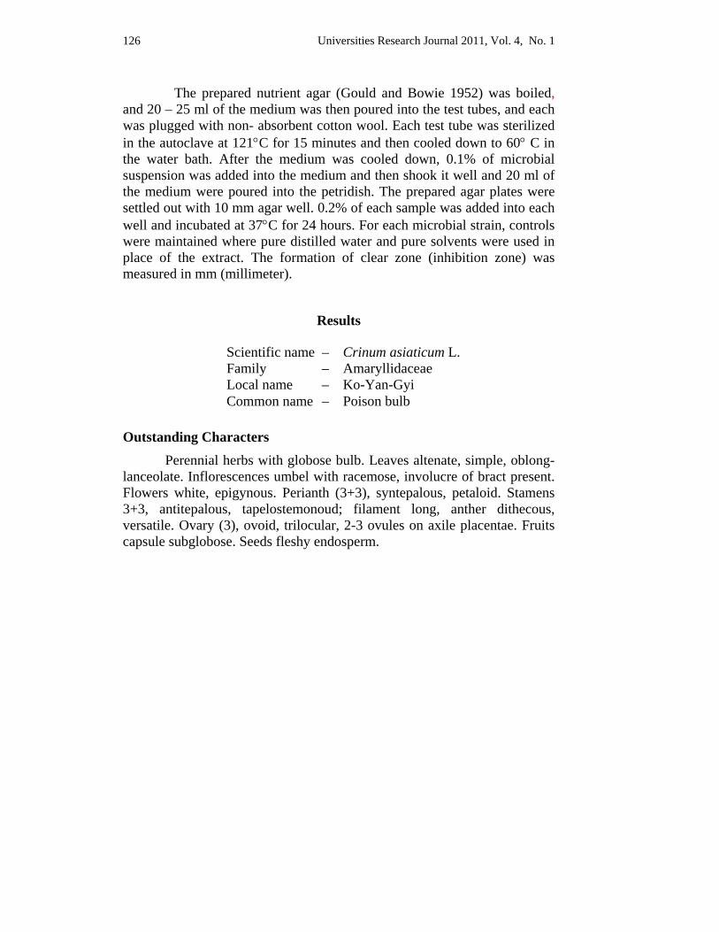
Universities Research Journal 2011, Vol. 4, No. 1 126
The prepared nutrient agar (Gould and Bowie 1952) was boiled, and 20 – 25 ml of the medium was then poured into the test tubes, and each was plugged with non- absorbent cotton wool. Each test tube was sterilized in the autoclave at 121°C for 15 minutes and then cooled down to 60° C in the water bath. After the medium was cooled down, 0.1% of microbial suspension was added into the medium and then shook it well and 20 ml of the medium were poured into the petridish. The prepared agar plates were settled out with 10 mm agar well. 0.2% of each sample was added into each well and incubated at 37°C for 24 hours. For each microbial strain, controls were maintained where pure distilled water and pure solvents were used in place of the extract. The formation of clear zone (inhibition zone) was measured in mm (millimeter).
Results
Outstanding Characters Perennial herbs with globose bulb. Leaves altenate, simple, oblong-lanceolate. Inflorescences umbel with racemose, involucre of bract present. Flowers white, epigynous. Perianth (3+3), syntepalous, petaloid. Stamens 3+3, antitepalous, tapelostemonoud; filament long, anther dithecous, versatile. Ovary (3), ovoid, trilocular, 2-3 ovules on axile placentae. Fruits capsule subglobose. Seeds fleshy endosperm.
Scientific name – Crinum asiaticum L. Family – Amaryllidaceae Local name – Ko-Yan-Gyi Common name – Poison bulb
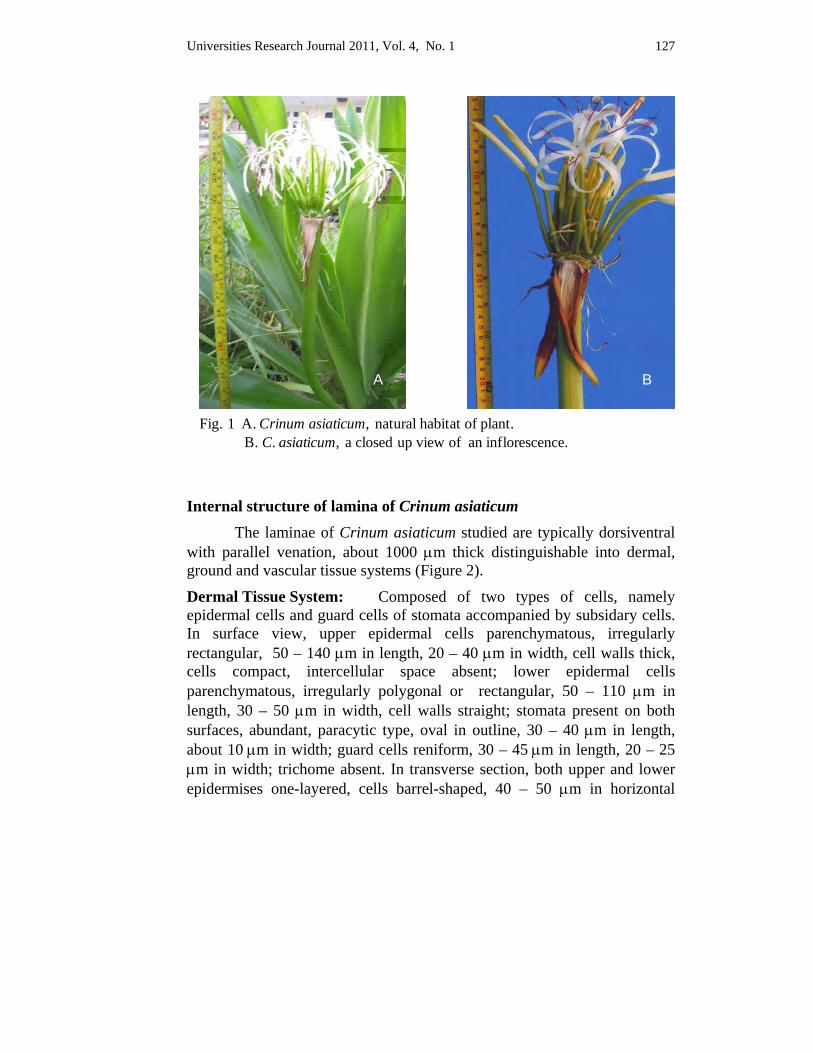
Universities Research Journal 2011, Vol. 4, No. 1 127
Fig. 1 A. Crinum asiaticum, natural habitat of plant.B. C. asiaticum, a closed up view of an inflorescence.
A B
Internal structure of lamina of Crinum asiaticum The laminae of Crinum asiaticum studied are typically dorsiventral with parallel venation, about 1000 μm thick distinguishable into dermal, ground and vascular tissue systems (Figure 2).
Dermal Tissue System: Composed of two types of cells, namely epidermal cells and guard cells of stomata accompanied by subsidary cells. In surface view, upper epidermal cells parenchymatous, irregularly rectangular, 50 – 140 μm in length, 20 – 40 μm in width, cell walls thick, cells compact, intercellular space absent; lower epidermal cells parenchymatous, irregularly polygonal or rectangular, 50 – 110 μm in length, 30 – 50 μm in width, cell walls straight; stomata present on both surfaces, abundant, paracytic type, oval in outline, 30 – 40 μm in length, about 10 μm in width; guard cells reniform, 30 – 45 μm in length, 20 – 25 μm in width; trichome absent. In transverse section, both upper and lower epidermises one-layered, cells barrel-shaped, 40 – 50 μm in horizontal
Universities Research Journal 2011, Vol. 4, No. 1 128
diameter, 20 – 30 μm in vertical diameter, anticlinal walls straight, outer and inner walls covex; cuticle thin on both surfaces, about 10 µm thick.
Ground tissue system: Mesophyll differentiated into palisade and spongy parenchyma. Palisade parenchyma 1-layered, cells elongated, 50 – 70 μm in horizontal diameter, 20 – 40 μm in vertical diameter, cells compact; spongy parenchyma 9 to 13 layered, the layer 450 – 500 μm thick, cells rounded or irregular, 10 – 30 μm in diameter, cells loose, chloroplast abundant, intercellular spaces present.
Vascular Tissue System: Vascular bundle embedded in the ground tissue; closed collateral, oval, 180 – 250 μm in horizontal diameter, 250 – 350 μm in vertical diameter; bundle sheath single, parenchymatous, chloroplast abundant, cells oval or rounded, 30 – 40 μm in length, 20 – 35 μm in width, cell walls thin, xylem on the adaxial sides and phloem on the abaxial sides; phloem 10- to 15-layered, the layers 150 – 200 μm thick, cells polygonal 10 – 20 μm in tangential diameter, 10 – 20 μm in radial diameter, phloem composed of sieve tube and companion cells; xylem 5- to 7-layered, the layers 90 – 100 μm thick, cells polygonal, 10 – 30 μm in tangential diameter, 10 – 20 μm in radial diameter, xylem composed of vessel elements, tracheids, fibers and xylem parenchyma. Vessel elements thick-walled, lateral walls straight, end walls oblique or transverse, thickening, spiral, annular, perforation plate simple, cells 1100 – 2200 μm in length, 80 – 150 μm in width; tracheids elongate, lateral walls straight, end walls tapering, thickening spiral, cells 1400 – 2000 μm in length, 30 – 60 μm in width; fibers long, lumen narrow, lateral walls straight, end walls acute, cells 900 – 1000 μm in length, 20 – 40 μm in width.
Phytochemical Tests The phytochemical investigations were done for the determination
of chemical constituents of the leaves of C. asiaticum. These results were shown in Table 1.
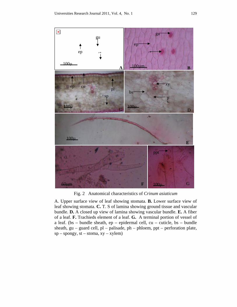
Universities Research Journal 2011, Vol. 4, No. 1 129
Fig. 2 Anatomical characteristics of Crinum asiaticum
A. Upper surface view of leaf showing stomata. B. Lower surface view of leaf showing stomata. C. T. S of lamina showing ground tissue and vascular bundle. D. A closed up view of lamina showing vascular bundle. E. A fiber of a leaf. F. Trachieds element of a leaf. G. A terminal portion of vessel of a leaf. (bs – bundle sheath, ep – epidermal cell, cu – cuticle, bs – bundle sheath, gu – guard cell, pl – palisade, ph – phloem, ppt – perforation plate, sp – spongy, st – stoma, xy – xylem)
50µm
100µA
ep
gu
st
100µm B
ep
st
gu
110µC
plcu ep
sp
100µE
60µm F 100µ
ppt
xy
ph
bs
100µD
G
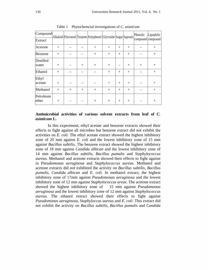
Universities Research Journal 2011, Vol. 4, No. 1 130
Table 1 Phytochemcial investigations of C. asiaticum
Compound Alkaloid Flavonoid Terpene Polyphenol Glycoside Sugar Saponin Phenolic
compound Lipophilic compound Extract
Acetone + – – + + + + – +
Benzene + – – + + + + – +
Distilled water + – + + + – + + +
Ethanol + – – – + + + – +
Ethyl acetate + – – – + + + – +
Methanol + + + + + + + – +
Petroleum ether + – – + + + + – +
Antimicrobial activities of various solvent extracts from leaf of C. asiaticum L. In this experiment, ethyl acetate and benzene extracts showed their effects to fight against all microbes but benzene extract did not exhibit the activities on E. coli. The ethyl acetate extract showed the highest inhibitory zone of 20 mm against E. coli and the lowest inhibitory zone of 15 mm against Bacillus subtilis. The benzene extract showed the highest inhibitory zone of 18 mm against Candida albican and the lowest inhibitory zone of 14 mm against Bacillus subtilis, Bacillus pumalis and Staphylococcus aureus. Methanol and acetone extracts showed their effects to fight against in Pseudomonas aeruginosa and Staphylococcus aureas. Methanol and acetone extracts did not exhibited the activity on Bacillus subtilis, Bacillus pumalis, Candida albican and E. coli. In methanol extract, the highest inhibitory zone of 17mm against Pseudomonas aeruginosa and the lowest inhibitory zone of 12 mm against Staphylococcus areas. The acetone extract showed the highest inhibitory zone of 15 mm against Pseudomonas aeruginosa and the lowest inhibitory zone of 12 mm against Staphylococcus aureus. The ethanol extract showed their effects to fight against Pseudomonas aeruginosa, Staphyloccus aureus and E. coli. This extract did not exhibit the activity on Bacillus subtilis, Bacillus pumalis and Candida
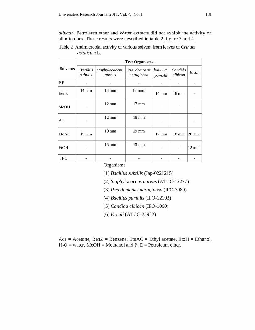
Universities Research Journal 2011, Vol. 4, No. 1 131
albican. Petroleum ether and Water extracts did not exhibit the activity on all microbes. These results were described in table 2, figure 3 and 4.
Table 2 Antimicrobial activity of various solvent from leaves of Crinum asiaticum L.
Solvents Test Organisms
Bacillus subtilis
Staphylococcus aureus
Pseudomonas aeruginosa
Bacillus pumalis
Candida albican E.coli
P.E - - - - - -
BenZ 14 mm
14 mm
17 mm.
14 mm 18 mm -
MeOH - 12 mm
17 mm
- - -
Ace - 12 mm
15 mm
- - -
EtoAC 15 mm 19 mm
19 mm
17 mm 18 mm 20 mm
EtOH - 13 mm
15 mm
- - 12 mm
H2O - - - - - -
Organisms
(1) Bacillus subtilis (Jap-0221215)
(2) Staphylococcus aureus (ATCC-12277)
(3) Pseudomonas aeruginosa (IFO-3080)
(4) Bacillus pumalis (IFO-12102)
(5) Candida albican (IFO-1060)
(6) E. coli (ATCC-25922)
Ace = Acetone, BenZ = Benzene, EtoAC = Ethyl acetate, EtoH = Ethanol, H2O = water, MeOH = Methanol and P. E = Petroleum ether.
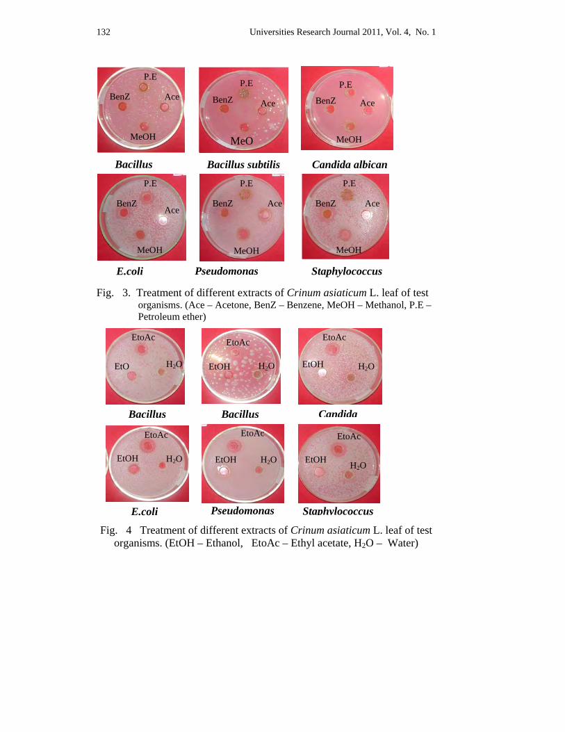
Universities Research Journal 2011, Vol. 4, No. 1 132
E.coli Pseudomonas
Bacillus subtilis Bacillus Candida albican
P.E
Ace
MeO
BenZ
P.E
Ace
MeOH
BenZ P.E
Ace
MeOH
BenZ
P.E
Ace
MeOH
BenZ
P.E
MeOH
BenZ Ace
P.E
Ace
MeOH
BenZ
Staphylococcus
Fig. 3. Treatment of different extracts of Crinum asiaticum L. leaf of test organisms. (Ace – Acetone, BenZ – Benzene, MeOH – Methanol, P.E – Petroleum ether)
Fig. 4 Treatment of different extracts of Crinum asiaticum L. leaf of test organisms. (EtOH – Ethanol, EtoAc – Ethyl acetate, H2O – Water)
E.coli
Bacillus CandidaBacillus
EtoAc
EtOH
EtoAc
H2O EtO
EtoAc
H2O EtOH
EtoAc
EtOH
EtoAc
H2O EtOH
EtoAc
H2O EtOH
Pseudomonas Staphylococcus
H2O
H2O
Universities Research Journal 2011, Vol. 4, No. 1 133
Discussion and Conclusion The genus Crinum is a true representative of the family
Amaryllidaceae (Elgorashi et al. 2002), and the members of this genus have commercial, economical and medicinal importance (Nguyen T. M. et al., 2002). C. asiaticum investigated in this work also is well known for its ornamental and medicinal value. However, a few reports are available on the area of anatomy, and it was also carried out in the present study. The leaves of C. asiaticum observed in this study are dorsiventral. In ground tissue, the mesophyll cells are composed of palisade and spongy cells. In surface view, the stomata type is paracytic. The vascular bundles of lamina are oval-shaped and closed-collateral which are agreed with those described by Esau (1965).
Crinum species have attracted considerable attention due to their alkaloidal contents (Ghosal et al. 1985). Phytochemical analyses have recently yielded more than 170 different compounds, most of which are alkaloids (Fennell and Staden 2001) and the others are flavonoids, chromones, terpenoid (Yamasaki K. et al., 1985), and phenolic (Quian Sun et al. 2007), saponin and so forth.
Phytochmeical tests of leaf extracts of C. asiaticum in the present study detected the presence of alkaloid, flavonoid, terpene, polyphenol, glycoside, sugar, saponin, phenolic compound and lipophilic compound. Of these, alkaloid, saponin and lipophilic compound were found in each phytochemical test. It is hoped that phytochemcial results obtained from this work corroborate the previous authors’ reports.
Crinum has a wide range of application, in traditional as well as modern medicine. The reason why the Crinums are used for medicinal purpose is the compounds yielded by Crinum species having interesting biological activity. Crinum plants attract considerable attention due to various medicinal properties as antitumor, immunostimulating, analgesis, antiviral, antibacterial and antifungal (Nguyen T. M. et al. 2002).
Literature survey revealed that little information is available on the antimicrobial assay of Crinum species, and antimicrobial test of leaf crude extracts of C. asiaticum is still lacking. In this study the aqueous and different solvent extracts of the leaves of above noted species were used to evaluate their antimicrobial activity on six pathogenic microbial strains, viz., Bacillus subtilis (Jap – 0221215), Bacillus pumalis (IFO – 12102),
Universities Research Journal 2011, Vol. 4, No. 1 134
Candida albican (IFO – 1060), Escherichia coli (ATCC – 25922), Pseudomonas aeruginosa (IFO – 3080) and Staphylococcus aureus (ATCC – 12277) by using agar well diffusion method. This work records the lack of antimicrobial activity in water and petroleum ether extracts of the leaves of C. asiaticum on any of the microbial strains investigated.
But it is difficult to state that why the water and petroleum ether extracts of C. asiaticum were not shown their antimicrobial activity on any of the studied strains although most compounds were found in each of these extracts. On the other hand, it is possible that active chemical constituents of the leaves of C. asiaticum were not soluble in both water and petroleum ether because the present study cannot be identify the specific molecule. As stated by Swarnker and Katewa (2009), water extracts of the bulbs of C. asiaticum revealed strong antimicrobial activity on the fungus C. albicans whilst weak activity on such bacteria as P. aeruginosa, S. aureus and Klebsiella pneumoniae. This result suggests that water extract of the bulbs of C. asiaticum is potential for antimicrobial purpose, particularly fungus C. albicans.
Apart from the early mentioned extracts, all extracts of the studied plant have antimicrobial activity ranging from weak (10 mm – 14mm), moderate (15 mm – 19 mm) to significant (20 mm and above) but only ethyl acetate extracts showed their activity on all studied microbes. This investigation states that C. asiaticum have both antibacterial and antifungal activities. The activity similar with C. asiaticum was also reported by Swarnker and Katewa (2009). Other Crinum species viz., C. jagus, C. macowanii and C. zeylanicum respectively showed antibacterial activity (Adesanya 2005), weak antifungal activity in vitro against Candida albican (Gundidza 1986) and efficient antibacterial and antifungal activities (Theingi Kyaw and Zan Zan Win 2010). The above mentioned data, therefore, states that C. asiaticum has both antibacterial and antifungal activity for antimicrobial purpose. The present study also observed that ethyl acetate extract of C. asiaticum exhibited significant inhibitory effect on the growth of E. coli (ATCC – 25922). It is generally accepted that microbial inhibition zone having 20 mm and above is good to use for antimicrobial purpose, and above described extracts have this standard. Other extracts which have antimicrobial activity lesser than 20 mm inhibition zone has no strong efficacy to control the pathogenic strains examined.
Universities Research Journal 2011, Vol. 4, No. 1 135
It is hoped that ethyl acetate extracts of C. asiaticum has promising antimicrobial activity on the E. coli (ATCC – 25922). The potential for developing antimicrobials from higher plants appears rewarding as it will lead to the development of a phytomedicine to act against microorganisms (Parekh and Chanda 2006). Plant-based antimicrobials have enormous therapeutic potential as they can serve the purpose with lesser side effects that are often associated with synthetic antimicrobials (Iwu et al. 1999). The present investigation is hence encouraging in recognizing a plant of interesting antimicrobial activity.
The anatomical characteristics of C. asiaticum in this work are proper uses of the plants of the regions. The compounds detected in the phytochemistry of present work were alkaloid flavonoid, terpene, polyphenol, glycoside, sugar, saponin, phenolic compound and lipophilic compound. Though the present phytochemical investigation cannot be described the pure molecules, this work is the first report for the phytochemistry of the C. asiaticum grown in Myanmar, and also it is hoped that it will come in handy for further studies. Antimicrobial assay by using agar well diffusion method also recorded that ethyl acetate extract of C. asiaticum showed significant antimicrobial activity against the E.coli (ATCC – 25922) with zone of inhibition of 20 mm. The results obtained from this work elucidated that the leaves of studied species contained bioactive agents which are connected with antimicrobial properties. The present investigation is reported for the antimicrobial efficacy of the leaf crude extracts of C. asiaticum on some pathogenic microorganisms. The fining could also be of interest to the research institutes in the production of new drugs.
Acknowledgements First of all, I would like to express my heartfelt thanks to U Nay Win, Professor and Head, Department of Botany, Yadanabon University, for allowing me to undertake this research and for his valuable suggestion on it. Many thanks are due to Dr Khin Htay Win, Assistant Lecturer, Department of Chemistry, Yadanabon University and Daw Cherry Aung, Research Assistant, Development Center for Pharmaceutical Technology, Yangon for their immeasurabe assistance on phytochemical analysis and antimicrobial assay, respectively.
Universities Research Journal 2011, Vol. 4, No. 1 136
References Adesanya, S.A., Olughade, T.A., Odebiyi, O.O. and Aladesanmi, J.A., (1992).
Antibacterial alkaloids in Crinum. jagus. International Journal of Pharmacognosy 30,303-307.
Cruickshank, R., J. P. Duguid., B. P. Marinion and R.H.A Swain., (1970). Medicinal Microbiology. Churchill Livingstone Ltd.
Esau, K., (1965). Plant Anatomy. John Wiley and Sons, Inc. New York, London.
Elgorashi, E.E., Drewes, S.E., Morris, C. and Van Standen, (2002). Variation among three Crinum species in alkaloid content. Biochemical systematics and Ecology 31 (2003) 601 – 615.
Fennell, C.W., Staden, J.van., (2001). Crinum species in traditional and modern medicine. Elsevier Science Ireland Ltd.
Ghosal, S., Saini., K.S. and Razdon, S., (1985). Crinum alkaloids: their chemistry and biology. Phytochemistry 24, 2141 – 2156.
Gundidza, M., (1986). Screening of extracts from Zimbabwean higher plants II: Antifungal properties. Fitoterapia 57, 111 – 114.
Harbone, J.B., (1973). Phytochemical Methods. Chapman and Hill, London.
Iwu, M.W., Duncan. A.R., Okunj. Co. (1999). New antimicrobials of plant origin.In: Janick J.ed. Perspectives on New Crops and New Uses. Alexandria, VA: ASHS Press.
Jayaweera, D.M.A., (1981). Medicinal Plants used in Ceylon, Part I. National Science Council of Sri Lanka, Colombo.
Krishnaraju AV. And Sundararaju, D. (2005). Assessment of bioactivity of Indian Medicinal plants using Brine Shrimp (Artemia salina ) lethality assay. International Journal Appl Sci Eng 2: 125 – 134.
Le Maout, E., Decaisne, J., (1873). A General System of Botany : Descriptive and Analytical, in Two Parts. Longmans Green, London.
Nguyen T.M., Titorenkova T.V., Bankovia V.St., Handjieva N.V. and Popa S.S., (2002). Review Crinum L. (Amaryllidaceae). Fitoterapia 73 (2002)163 – 208.
Parekh, J. and Chanda, S., (2006). In vitro Antimicrobial Activity and Phytochemical Analysis of some Indian Medicinal Plants. Turk Journal Biol 31 (2007) 53-58.
Qian Sun, Wei Dong Zhang, Yun Heng Shen, Chuan Zhang, Hui liang li, (2007). A new phenolic compound from Crinum asiaticum L.

Universities Research Journal 2011, Vol. 4, No. 1 137
Swarnkar, S., Katewa, S.S., (2009). Antimicrobial activities of some Tuberous medicinal plants from Aravallihills of Rajasthan. Journal of Herbal Medicine and Taxicology 3 (1) 53 – 58.
Theingi Kyaw, (2010). Study on Morphological and Anatomical Characteristics of Crinum asiaticum (L.) (Ko-Yan-Gyi) and C. zeylanicum (L.) L. (Khatta) and Phytochemical and Antimicrobial Tests of their leaves.
Yamasaki. K. (2007). Phytochemicstry 54 (2000) 891.
Universities Research Journal 2011, Vol. 4, No. 1
Lecturer, Department of Botany, Maubin University
Estrogenic Activities of Pueraria candollei Grah. Tuberous Roots
Wai Wai Thein
Abstract
Comparative study of estrogenic activity of 70% ethanolic extract and dried powder of Pueraria candollei Grah was conducted with premarin cream in albino mice. Mice were divided into twelve groups (n=5) receiving different treatments, consisting of 0.1 ml/kg distilled water, dried powder and 70% ethanolic extract of Tuberous roots of Pueraria candollei Grah at five different doses (viz., 50, 100, 200, 400 and 500 mg/kg body weight) and standard drug premarin cream at a dose of 50 mg/kg body weight. The dried powder and 70% ethanolic extract were administered orally and 50 mg/kg BW (Premarin cream) was applied intravaginally for five consecutive days. Estrogenic activity was assessed by counting the percentages of vaginal epithelial cells maturation. The significant differences at p<0.01 level were found in 200 mg/kg and 400 mg/kg of 70% ethanolic extract compared to the control.
Key words: estrogenic activity, Pueraria candollei Grah
Introduction
The plant Pueraria candollei Grah. belongs to the family Fabaceae. The English name of Pueraria candollei Grah.is Burmese Kudzu. In Thailand, the plant is known as “Kua ta lan” and “Ma-U-Nwe” in Myanmar. They are widely distributed in Myanmar as wild plants, especially in Yangon, Bago Region and Mon State.
The habit of Pueraria candollei Grah. is twining herbs or shrubs with tuberous roots. The leaves of Pueraria candollei Grah. are alternate and pinnately tri-foliolate. Leaflets are subcoriaceous, entire, rarely faintly lobed and roundish with an acute point. Lateral leaflets are oblique and smaller than terminal leaflet.
The tuberous roots are up to 60 cm long and up to 38 cm in diameter. Tuberous roots are in different sizes depending on the soil condition or the environment and the time of cropping. The tuberous roots are sweet, watery and cool in taste. In Thailand, the tubers are used as a tonic and aphrodisiac, to treat mammary gland expansion and for their oestrogenic effect (Praptiwi,
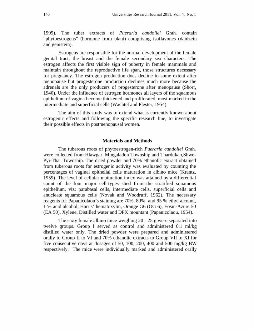
Universities Research Journal 2011, Vol. 4, No. 1 140
1999). The tuber extracts of Pueraria candollei Grah. contain “phytoestrogens” (hormone from plant) comprising isoflavones (daidzein and genistein).
Estrogens are responsible for the normal development of the female genital tract, the breast and the female secondary sex characters. The estrogen affects the first visible sign of puberty in female mammals and maintain throughout the reproductive life span, those structures necessary for pregnancy. The estrogen production does decline to some extent after menopause but progesterone production declines much more because the adrenals are the only producers of progesterone after menopause (Shorr, 1940). Under the influence of estrogen hormones all layers of the squamous epithelium of vagina become thickened and proliferated, most marked in the intermediate and superficial cells (Wachtel and Plester, 1954).
The aim of this study was to extend what is currently known about estrogenic effects and following the specific research line, to investigate their possible effects in postmenopausal women.
Materials and Methods The tuberous roots of phytoestrogen-rich Pueraria candollei Grah. were collected from Hlawgar, Mingaladon Township and Thardukan,Shwe-Pyi-Thar Township. The dried powder and 70% ethanolic extract obtained from tuberous roots for estrogenic activity was evaluated by counting the percentages of vaginal epithelial cells maturation in albino mice (Krantz, 1959). The level of cellular maturation index was attained by a differential count of the four major cell-types shed from the stratified squamous epithelium, viz: parabasal cells, intermediate cells, superficial cells and anucleate squamous cells (Novak and Woodruff, 1962). The necessary reagents for Papanicolaou’s staining are 70%, 80% and 95 % ethyl alcohol, 1 % acid alcohol, Harris’ hematoxylin, Orange G6 (OG 6), Eosin-Azure 50 (EA 50), Xylene, Distilled water and DPX mountant (Papanicolaou, 1954).
The sixty female albino mice weighing 20 - 25 g were separated into twelve groups. Group I served as control and administered 0.1 ml/kg distilled water only. The dried powder were prepared and administered orally to Group II to VI and 70% ethanolic extracts to Group VII to XI for five consecutive days at dosages of 50, 100, 200, 400 and 500 mg/kg BW respectively. The mice were individually marked and administered orally
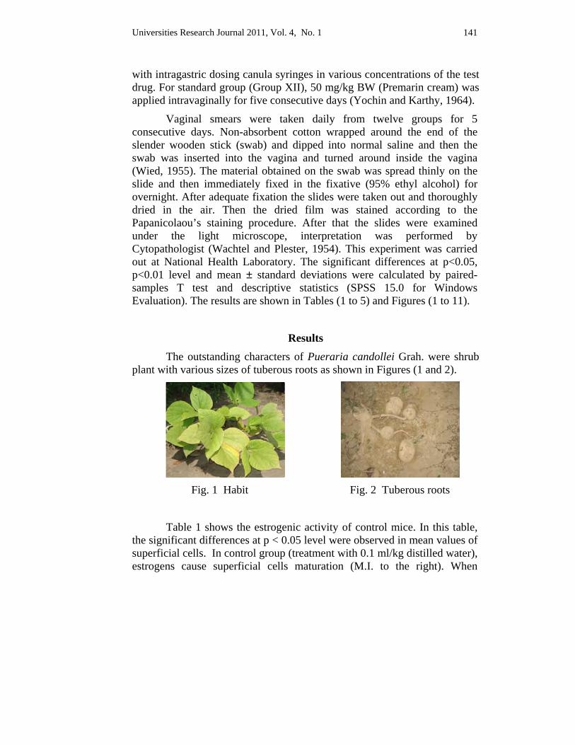
Universities Research Journal 2011, Vol. 4, No. 1 141
with intragastric dosing canula syringes in various concentrations of the test drug. For standard group (Group XII), 50 mg/kg BW (Premarin cream) was applied intravaginally for five consecutive days (Yochin and Karthy, 1964).
Vaginal smears were taken daily from twelve groups for 5 consecutive days. Non-absorbent cotton wrapped around the end of the slender wooden stick (swab) and dipped into normal saline and then the swab was inserted into the vagina and turned around inside the vagina (Wied, 1955). The material obtained on the swab was spread thinly on the slide and then immediately fixed in the fixative (95% ethyl alcohol) for overnight. After adequate fixation the slides were taken out and thoroughly dried in the air. Then the dried film was stained according to the Papanicolaou’s staining procedure. After that the slides were examined under the light microscope, interpretation was performed by Cytopathologist (Wachtel and Plester, 1954). This experiment was carried out at National Health Laboratory. The significant differences at p<0.05, p<0.01 level and mean ± standard deviations were calculated by paired-samples T test and descriptive statistics (SPSS 15.0 for Windows Evaluation). The results are shown in Tables (1 to 5) and Figures (1 to 11).
Results The outstanding characters of Pueraria candollei Grah. were shrub plant with various sizes of tuberous roots as shown in Figures (1 and 2).
Fig. 1 Habit Fig. 2 Tuberous roots
Table 1 shows the estrogenic activity of control mice. In this table, the significant differences at p < 0.05 level were observed in mean values of superficial cells. In control group (treatment with 0.1 ml/kg distilled water), estrogens cause superficial cells maturation (M.I. to the right). When
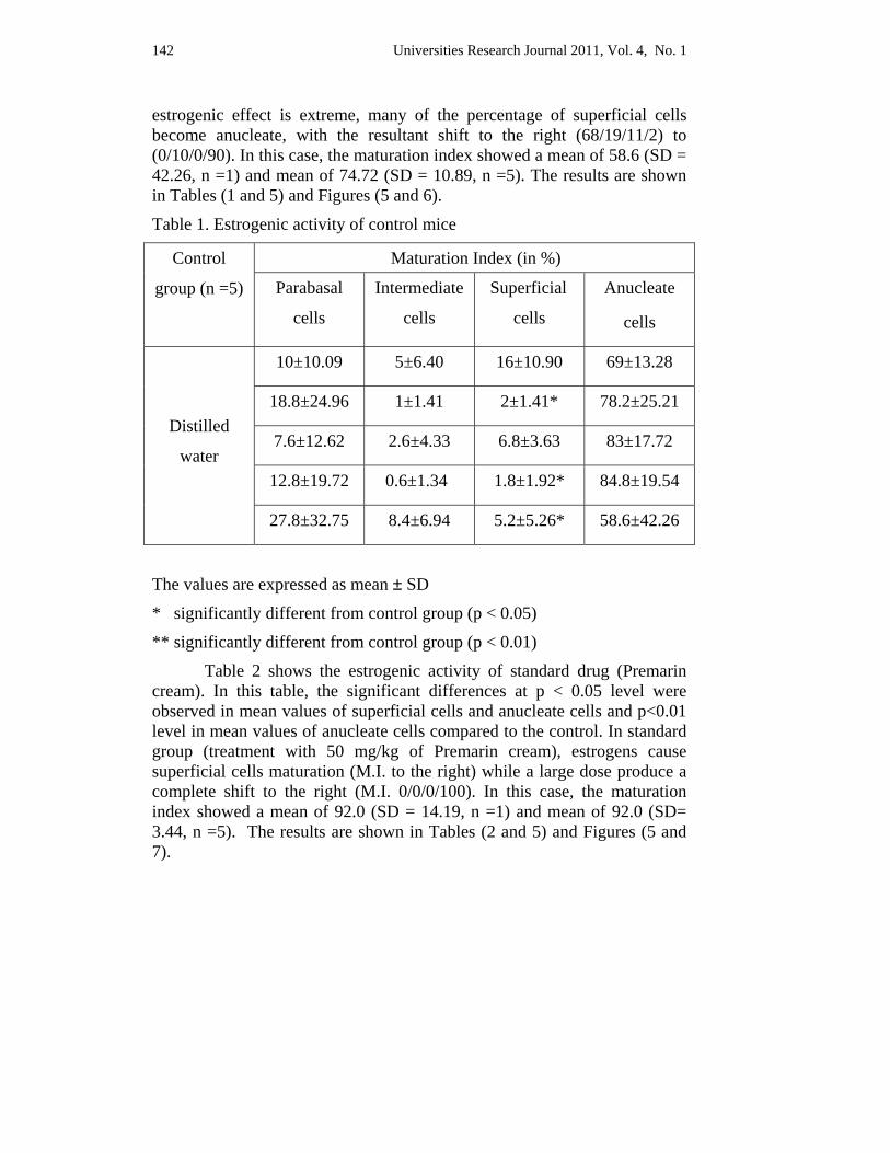
Universities Research Journal 2011, Vol. 4, No. 1 142
estrogenic effect is extreme, many of the percentage of superficial cells become anucleate, with the resultant shift to the right (68/19/11/2) to (0/10/0/90). In this case, the maturation index showed a mean of 58.6 (SD = 42.26, n =1) and mean of 74.72 (SD = 10.89, n =5). The results are shown in Tables (1 and 5) and Figures (5 and 6).
Table 1. Estrogenic activity of control mice
Control
group (n =5)
Maturation Index (in %)
Parabasal
cells
Intermediate
cells
Superficial
cells
Anucleate
cells
Distilled
water
10±10.09 5±6.40 16±10.90 69±13.28
18.8±24.96 1±1.41 2±1.41* 78.2±25.21
7.6±12.62 2.6±4.33 6.8±3.63 83±17.72
12.8±19.72 0.6±1.34 1.8±1.92* 84.8±19.54
27.8±32.75 8.4±6.94 5.2±5.26* 58.6±42.26
The values are expressed as mean ± SD
* significantly different from control group (p < 0.05)
** significantly different from control group (p < 0.01)
Table 2 shows the estrogenic activity of standard drug (Premarin cream). In this table, the significant differences at p < 0.05 level were observed in mean values of superficial cells and anucleate cells and p<0.01 level in mean values of anucleate cells compared to the control. In standard group (treatment with 50 mg/kg of Premarin cream), estrogens cause superficial cells maturation (M.I. to the right) while a large dose produce a complete shift to the right (M.I. 0/0/0/100). In this case, the maturation index showed a mean of 92.0 (SD = 14.19, n =1) and mean of 92.0 (SD= 3.44, n =5). The results are shown in Tables (2 and 5) and Figures (5 and 7).
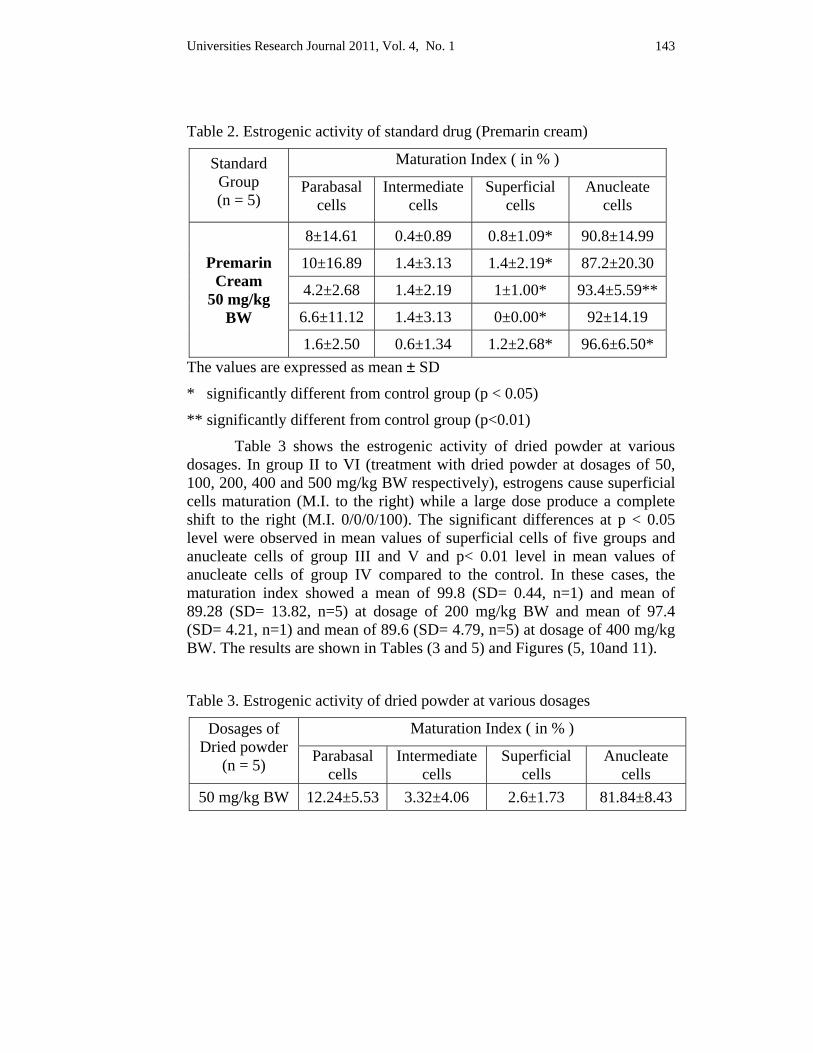
Universities Research Journal 2011, Vol. 4, No. 1 143
Table 2. Estrogenic activity of standard drug (Premarin cream)
Standard Group (n = 5)
Maturation Index ( in % )
Parabasal cells
Intermediate cells
Superficial cells
Anucleate cells
Premarin Cream
50 mg/kg BW
8±14.61 0.4±0.89 0.8±1.09* 90.8±14.99
10±16.89 1.4±3.13 1.4±2.19* 87.2±20.30
4.2±2.68 1.4±2.19 1±1.00* 93.4±5.59**
6.6±11.12 1.4±3.13 0±0.00* 92±14.19
1.6±2.50 0.6±1.34 1.2±2.68* 96.6±6.50* The values are expressed as mean ± SD
* significantly different from control group (p < 0.05)
** significantly different from control group (p<0.01) Table 3 shows the estrogenic activity of dried powder at various
dosages. In group II to VI (treatment with dried powder at dosages of 50, 100, 200, 400 and 500 mg/kg BW respectively), estrogens cause superficial cells maturation (M.I. to the right) while a large dose produce a complete shift to the right (M.I. 0/0/0/100). The significant differences at p < 0.05 level were observed in mean values of superficial cells of five groups and anucleate cells of group III and V and p< 0.01 level in mean values of anucleate cells of group IV compared to the control. In these cases, the maturation index showed a mean of 99.8 (SD= 0.44, n=1) and mean of 89.28 (SD= 13.82, n=5) at dosage of 200 mg/kg BW and mean of 97.4 (SD= 4.21, n=1) and mean of 89.6 (SD= 4.79, n=5) at dosage of 400 mg/kg BW. The results are shown in Tables (3 and 5) and Figures (5, 10and 11).
Table 3. Estrogenic activity of dried powder at various dosages
Dosages of Dried powder
(n = 5)
Maturation Index ( in % )
Parabasal cells
Intermediate cells
Superficial cells
Anucleate cells
50 mg/kg BW 12.24±5.53 3.32±4.06 2.6±1.73 81.84±8.43

Universities Research Journal 2011, Vol. 4, No. 1 144
Dosages of Dried powder
(n = 5)
Maturation Index ( in % )
Parabasal cells
Intermediate cells
Superficial cells
Anucleate cells
100 mg/kg BW 12.8±10.08 1.92±0.91 1.44±0.99 83.84±11.59
200 mg/kg BW 8.68±11.34 1.04±2.1 1±0.58 89.28±13.82
400 mg/kg BW 4.96±2.62 2.84±1.6 2.6±2 89.6±4.79
500 mg/kg BW 9.88±1.93 0.6±0.54 1.32±0.87 88.2±2.36 The values are expressed as mean ± SD
0
20
40
60
80
100
50 100 200 400 500Dosages of dried powder (mg/kg BW)
Mat
urat
ion
inde
x (%
) Parabasal cells
Intermediate cells
Superficial cells
Anucleate cells
Fig. 3. Estrogenic activity of dried powder at various dosages
Table 4 shows the estrogenic activity of ethanolic extract at various dosages. In this table, the significant differences at p < 0.05 level were observed in mean values of intermediate cells (0 to 1.8) at dosage of 200 mg/kg BW and parabasal cells (1 to 10.2) at dosage of 400 mg/kg BW and p< 0.01 level in mean values of parabasal cells (0.8 to 3.6) and anucleate cells (92.6 to 97) at dosage of 200 mg/kg BW and anucleate cells (87.6 to 97) at dosage of 400 mg/kg BW compared to the control. In group VII to XI (treatment with ethanolic extract at dosages of 50, 100, 200, 400 and 500 mg/kg BW respectively), estrogens cause superficial cells maturation (M.I. to the right) while a large dose produce a complete shift to the right (M.I. 0/0/0/100). The significant differences at p < 0.05 were observed in mean values of superficial cells and anucleate cells and p<0.01 level in mean values of anucleate cells compared to the control. In this case, the
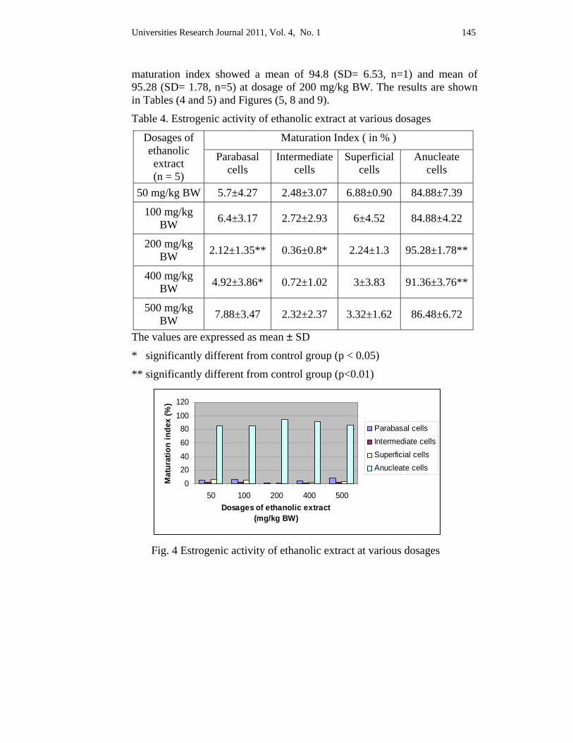
Universities Research Journal 2011, Vol. 4, No. 1 145
maturation index showed a mean of 94.8 (SD= 6.53, n=1) and mean of 95.28 (SD= 1.78, n=5) at dosage of 200 mg/kg BW. The results are shown in Tables (4 and 5) and Figures (5, 8 and 9).
Table 4. Estrogenic activity of ethanolic extract at various dosages
Dosages of ethanolic extract (n = 5)
Maturation Index ( in % )
Parabasal cells
Intermediatecells
Superficial cells
Anucleate cells
50 mg/kg BW 5.7±4.27 2.48±3.07 6.88±0.90 84.88±7.39
100 mg/kg BW 6.4±3.17 2.72±2.93 6±4.52 84.88±4.22
200 mg/kg BW 2.12±1.35** 0.36±0.8* 2.24±1.3 95.28±1.78**
400 mg/kg BW 4.92±3.86* 0.72±1.02 3±3.83 91.36±3.76**
500 mg/kg BW 7.88±3.47 2.32±2.37 3.32±1.62 86.48±6.72
The values are expressed as mean ± SD
* significantly different from control group (p < 0.05)
** significantly different from control group (p<0.01)
020406080
100120
50 100 200 400 500Dosages of ethanolic extract
(mg/kg BW)
Mat
urat
ion
inde
x (%
)
Parabasal cellsIntermediate cellsSuperficial cellsAnucleate cells
Fig. 4 Estrogenic activity of ethanolic extract at various dosages
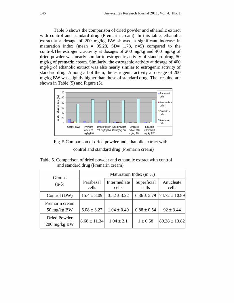
Universities Research Journal 2011, Vol. 4, No. 1 146
Table 5 shows the comparison of dried powder and ethanolic extract with control and standard drug (Premarin cream). In this table, ethanolic extract at a dosage of 200 mg/kg BW showed a significant increase in maturation index (mean = 95.28, SD= 1.78, n=5) compared to the control.The estrogenic activity at dosages of 200 mg/kg and 400 mg/kg of dried powder was nearly similar to estrogenic activity of standard drug, 50 mg/kg of premarin cream. Similarly, the estrogenic activity at dosage of 400 mg/kg of ethanolic extract was also nearly similar to estrogenic activity of standard drug. Among all of them, the estrogenic activity at dosage of 200 mg/kg BW was slightly higher than those of standard drug. The results are shown in Table (5) and Figure (5).
0
20
40
60
80
100
120
Control (DW) Premarincream 50mg/kg BW
Dried Powder200 mg/kg BW
Dried Powder400 mg/kg BW
Ethanolicextract 200mg/kg BW
Ethanolicextract 400mg/kg BW
mat
urat
ion
inde
x (%
) Parabasalcells
Intermediatecells
Superficialcells
Anucleatecells
Fig. 5 Comparison of dried powder and ethanolic extract with
control and standard drug (Premarin cream)
Table 5. Comparison of dried powder and ethanolic extract with control and standard drug (Premarin cream)
Groups (n-5)
Maturation Index (in %)
Parabasal cells
Intermediate cells
Superficial cells
Anucleate cells
Control (DW) 15.4 ± 8.09 3.52 ± 3.22 6.36 ± 5.79 74.72 ± 10.89
Premarin cream 50 mg/kg BW
6.08 ± 3.27
1.04 ± 0.49
0.88 ± 0.54
92 ± 3.44
Dried Powder 200 mg/kg BW
8.68 ± 11.34 1.04 ± 2.1 1 ± 0.58 89.28 ± 13.82
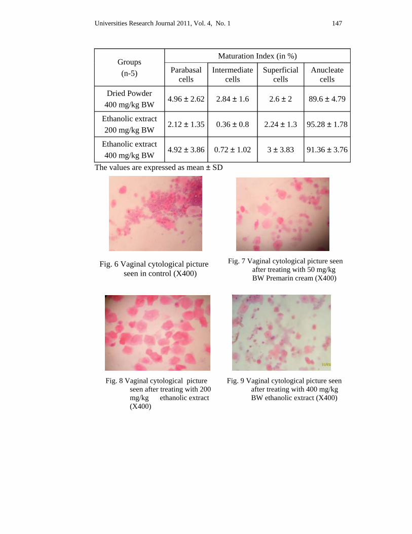
Universities Research Journal 2011, Vol. 4, No. 1 147
Groups (n-5)
Maturation Index (in %)
Parabasal cells
Intermediate cells
Superficial cells
Anucleate cells
Dried Powder 400 mg/kg BW
4.96 ± 2.62 2.84 ± 1.6 2.6 ± 2 89.6 ± 4.79
Ethanolic extract 200 mg/kg BW
2.12 ± 1.35 0.36 ± 0.8 2.24 ± 1.3 95.28 ± 1.78
Ethanolic extract 400 mg/kg BW
4.92 ± 3.86 0.72 ± 1.02 3 ± 3.83 91.36 ± 3.76
The values are expressed as mean ± SD
Fig. 6 Vaginal cytological picture seen in control (X400)
Fig. 7 Vaginal cytological picture seen after treating with 50 mg/kg BW Premarin cream (X400)
Fig. 8 Vaginal cytological picture seen after treating with 200 mg/kg ethanolic extract (X400)
Fig. 9 Vaginal cytological picture seen after treating with 400 mg/kg BW ethanolic extract (X400)
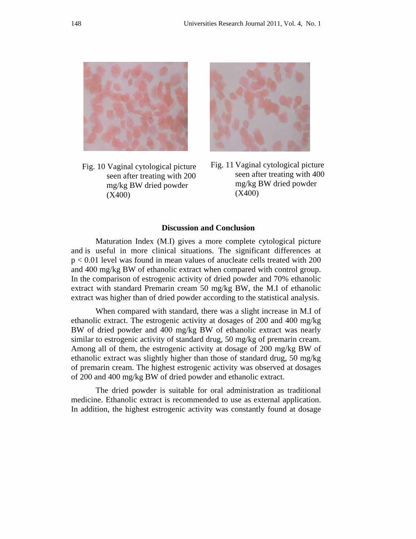
Universities Research Journal 2011, Vol. 4, No. 1 148
Discussion and Conclusion Maturation Index (M.I) gives a more complete cytological picture
and is useful in more clinical situations. The significant differences at p < 0.01 level was found in mean values of anucleate cells treated with 200 and 400 mg/kg BW of ethanolic extract when compared with control group. In the comparison of estrogenic activity of dried powder and 70% ethanolic extract with standard Premarin cream 50 mg/kg BW, the M.I of ethanolic extract was higher than of dried powder according to the statistical analysis.
When compared with standard, there was a slight increase in M.I of ethanolic extract. The estrogenic activity at dosages of 200 and 400 mg/kg BW of dried powder and 400 mg/kg BW of ethanolic extract was nearly similar to estrogenic activity of standard drug, 50 mg/kg of premarin cream. Among all of them, the estrogenic activity at dosage of 200 mg/kg BW of ethanolic extract was slightly higher than those of standard drug, 50 mg/kg of premarin cream. The highest estrogenic activity was observed at dosages of 200 and 400 mg/kg BW of dried powder and ethanolic extract.
The dried powder is suitable for oral administration as traditional medicine. Ethanolic extract is recommended to use as external application. In addition, the highest estrogenic activity was constantly found at dosage
Fig. 10 Vaginal cytological picture seen after treating with 200 mg/kg BW dried powder (X400)
Fig. 11 Vaginal cytological picture seen after treating with 400 mg/kg BW dried powder (X400)
Universities Research Journal 2011, Vol. 4, No. 1 149
of 200 mg/kg BW of both dried powder and ethanolic extract. Therefore, 200 mg/kg dosage of both dried powder and ethanolic extract from tuberous roots of Pueraria candollei Grah. can be safely applied in therapeutic purposes especially for menopausal women.
Thus, tuberous roots of Pueraria candollei Grah. are as qualified as standard drug, premarin cream. They can be scientifically and systematically used in hormone replacement therapy especially for post-menopausal women. It can also be used locally as a rejuvenator and anti-aging agent such as breast-firming cream, skin-nourishing cream, night cream and so on.
Acknowledgements
First and foremost I would like to express my sincere gratitude to Rector Dr. Htay Linn Maung, Maubin University and Professor Dr. Yee Yee Myint, Head of Botany Department, Maubin University for their permission to do the research paper. I would like to express special thanks to Dr. Daw Aye Kyi, Professor and Head (Retired), Department of Botany, Yangon University and Daw Naw Wah Wah Paw, Professor and Head (Retired), Department of Botany, Dagon University for their instructions and advice. My warm thanks are also extended to Dr. Than Aung Soe, Veterinarian, Administrative Officer of National Health Laboratory, and Professor Dr. Thein Thein Aye (Lt. Col., Retired), Consultant Pathologist, for their supervision and research facilities.
References Krantz, K. E. (1959). The gross and microscopic anatomy of the vagina. Ann. N. Y.
Acad. Sci., The Vagina, 83:89 – 104.
Novak, A. B. and B. S. Woodruff (1962). Gynecologic and obstetric pathology with clinical and endocrine relations. (5 th Ed.). W. Saunder Company. Philadelphia-London.
Papanicolaou, G. N. and E. Shorr (1936). The action of ovarian follicular hormone in the menopause as indicated by vaginal smears. Amer. Obst. Gynec., 31:806.
Papanicolaou, G. N. (1954). Atlas of exfoliative cytology. Harrard Univ. Press. Cambridge, Mass. Also supplements.
Praptiwi (1999). Medicinal and poisonous plants. Plant resources of South-East Asia. No. 12 (1). Prosea Foundation, Bogor, Indonesia. Pp. 417-420.
Shorr, E. (1940). The menopause. Bull. N.Y. Acad. Med., 16:453.

Universities Research Journal 2011, Vol. 4, No. 1 150
Wachtel, E. and J. A. Plester (1954). Hormonal assessment by vaginal cytology. J.Obstet. Gynaec. Brit. Emp., 61: 155.
Wied, G. L. (1955). Importance of the site from which vaginal cytologic smears taken. Amer. J. Clin. Path., 25: 742.
Yochin, M. X. and I. L. Mc. Karthy (1964). Experimental endocrinology. Academic Press. NY & London.
Universities Research Journal 2011, Vol. 4, No. 1
1. Lecturer, Department of Botany, Bago University 2. Associate Professor, Department of Botany, Bago University
Some Medicinal Plants Grown in Phaya-thone-zu Village, Bago Township
Ohnmar Aung1, Khin Sein Kyi1 and Nu Yi2
Abstract The medicinal plants of Phaya-thone-zu village, Bago Township were studied from June, 2008 to July, 2010. A total of 9 species belonging to (9) genera of (8) families were collected from Phaya-thone-zu village. About (2) species are antidiabetic properties, (2) species are for blood hypertensive diseases, and (2) wild plants are used in wound healthy activities and other diseases. In this paper, morphological characteristics and medicinal uses of each species are mentioned and parts used have been recorded with photographs.
Key words : medicinal plants, antidiabetic properties, blood hypertensive diseases
Introduction
Today, nearly 88% of the global populations switch to plant derived medicines as their first line of defense for maintaining health and combating diseases (Kintzios et al., 2006). In future, the discovery of novel therapeutic agents will be only dependent on plant origin (Perumalsamy et al., 1999).
In Myanmar, thousands of species are known to have medicinal value and the use of different parts of several medicinal plants to cure specific ailments has been in vague since ancient times. Medicinal plants are a source of great economic value in Myanmar. Nature has best owned on us a very rich botanical wealth and a large number of diverse types of plants grown in different parts of the country. Traditional medicine in Myanmar is widely practised by the majority of population, partly as a supplement and partly as an alternative to modern medicine. (Thein Swe & SeinWin, 2005)
All plants containing active compounds are important. The beneficial medicinal effects of plant materials typically result from the combinations of secondary products present in the plant. In plants, these compounds are mostly secondary metabolites such as alkaloids, steroids, tannins, and phenol compounds, which are synthesized and deposited in specific parts or in all parts of the plant. The extract of exudates of
Universities Research Journal 2011, Vol. 4, No. 1 152
medicinal plants are used to treat the diseases such as diarrhoea, dysentery, malaria, boils, sores and also used to cure to promote wound healing. A large majority of plant species present are indigenous and some of them are endemic only to Myanmar.
Phaya-thone-zu village is situated in the south of Bago Township where medicinal plants are growing abundantly and naturally. Only a few are cultivated species. It is essential to survey how these resource plants are valuable and effective to human beings. It is for this reason, an attempt has been made to undertake this research work.
Materials and Methods A collection of some medicinal plants grown in Phaya-thone-zu village was carried out during the flowering and fruiting periods. The specimens were carefully pressed, thoroughly dried, poisoned with spirit and finally mounted on the herbarium sheets. The specimens have been studied systematically and identified by matching with the existing herbarium sheets from Botany Department, University of Yangon.
Results In the present study, 9 species of plants which are possessed of 9
genus belonging to 8 families have been selected from Phaya-thone-zu village. These include 2 shrubs, 5 herbs and 2 climbers. 8 families are dicots and only one family is monocot, and these families are angiosperms.
Alysicarpus vaginalis ( L.) DC Family : Fabaceae
English name : Alyce clover
Local name : Than-manaing-kyauk-manaing
This plant is an annual robust herbs with suffruticose hispid branches, woody at the base, the stems erect. Leaves alternate, stipulate. Inflorescences terminal and axillary raceme. Flowers reddish yellow or pale purple. Fruits oblong, lomentoid pods, cylindrical, deep maroon, rugulose and the small seeds dark red (Figure 1).
Flowering and fruiting periods from April to May.
Universities Research Journal 2011, Vol. 4, No. 1 153
Parts used : The whole plant
Roots : (decoction) - Cough
Leaf extract : Anticancer
Renal stone - Taking 4 gm of the whole plant powder
with lime juice three times a day. Boil 80 gm of the
whole plant in 320 ml of water until ¼ of original
volume is obtained. 20 ml of this preparation with little
sugar is orally taken three to five times a day
Renal oedema - 4 gm of the whole plant powder with
sugar is used three times a day
Anisomeles indica (L.) O. K. Family : Lamiaceae
English name : Malabar catmint
Local name : Taw-hnan
Parts used : The whole plant
This plant is an annual aromatic herb with quadrangular stem, slightly grooved. Leaves opposite and decussate, alternate in the lower portion, a pair of glands present at the base of petiole, the laminae oblong, the bases aequilateral. Inflorescences axillary and solitary cymes. Flowers bluish-purple, deep purple spots and strips present in bilabiate petals. Fruits capsule, ellipsoid, shinning black and seeds small and black (Figure 2).
Flowering and fruiting periods from August to December.
Parts used : The whole plant
Uses : Carminative, astringent and tonic properties
Dried or fresh : Eczema, pruritis, snakebites
Leaf : Chewed for toothaches, rheumatism, cold, fever,
abdominal pain, intermittent fever, dyspepsia
Whole plant : Burn for mosquito-repellent
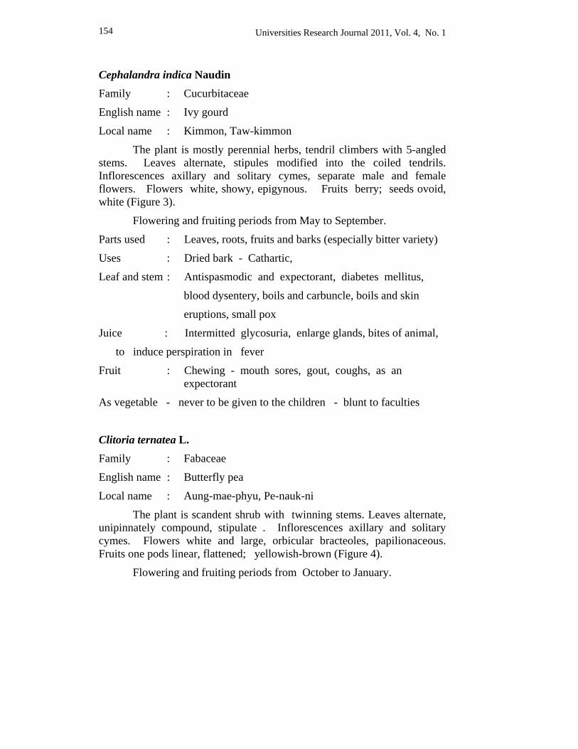
Universities Research Journal 2011, Vol. 4, No. 1 154
Cephalandra indica Naudin Family : Cucurbitaceae
English name : Ivy gourd
Local name : Kimmon, Taw-kimmon
The plant is mostly perennial herbs, tendril climbers with 5-angled stems. Leaves alternate, stipules modified into the coiled tendrils. Inflorescences axillary and solitary cymes, separate male and female flowers. Flowers white, showy, epigynous. Fruits berry; seeds ovoid, white (Figure 3).
Flowering and fruiting periods from May to September.
Parts used : Leaves, roots, fruits and barks (especially bitter variety)
Uses : Dried bark - Cathartic,
Leaf and stem : Antispasmodic and expectorant, diabetes mellitus,
blood dysentery, boils and carbuncle, boils and skin
eruptions, small pox
Juice : Intermitted glycosuria, enlarge glands, bites of animal,
to induce perspiration in fever
Fruit : Chewing - mouth sores, gout, coughs, as an expectorant
As vegetable - never to be given to the children - blunt to faculties
Clitoria ternatea L. Family : Fabaceae
English name : Butterfly pea
Local name : Aung-mae-phyu, Pe-nauk-ni
The plant is scandent shrub with twinning stems. Leaves alternate, unipinnately compound, stipulate . Inflorescences axillary and solitary cymes. Flowers white and large, orbicular bracteoles, papilionaceous. Fruits one pods linear, flattened; yellowish-brown (Figure 4).
Flowering and fruiting periods from October to January.
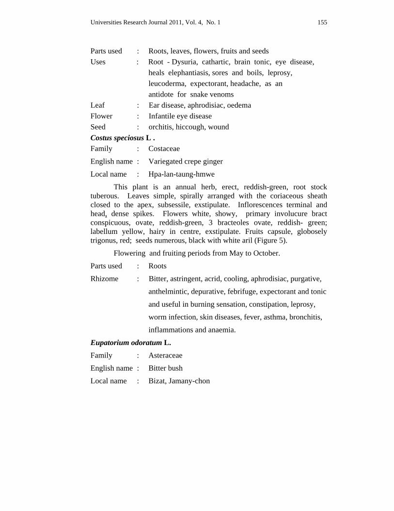
Universities Research Journal 2011, Vol. 4, No. 1 155
Parts used : Roots, leaves, flowers, fruits and seeds Uses : Root - Dysuria, cathartic, brain tonic, eye disease, heals elephantiasis, sores and boils, leprosy, leucoderma, expectorant, headache, as an antidote for snake venoms Leaf : Ear disease, aphrodisiac, oedema Flower : Infantile eye disease Seed : orchitis, hiccough, wound Costus speciosus L . Family : Costaceae
English name : Variegated crepe ginger
Local name : Hpa-lan-taung-hmwe
This plant is an annual herb, erect, reddish-green, root stock tuberous. Leaves simple, spirally arranged with the coriaceous sheath closed to the apex, subsessile, exstipulate. Inflorescences terminal and head, dense spikes. Flowers white, showy, primary involucure bract conspicuous, ovate, reddish-green, 3 bracteoles ovate, reddish- green; labellum yellow, hairy in centre, exstipulate. Fruits capsule, globosely trigonus, red; seeds numerous, black with white aril (Figure 5).
Flowering and fruiting periods from May to October.
Parts used : Roots
Rhizome : Bitter, astringent, acrid, cooling, aphrodisiac, purgative,
anthelmintic, depurative, febrifuge, expectorant and tonic
and useful in burning sensation, constipation, leprosy,
worm infection, skin diseases, fever, asthma, bronchitis,
inflammations and anaemia.
Eupatorium odoratum L. Family : Asteraceae
English name : Bitter bush
Local name : Bizat, Jamany-chon
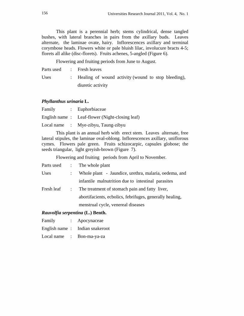
Universities Research Journal 2011, Vol. 4, No. 1 156
This plant is a perennial herb; stems cylindrical, dense tangled bushes, with lateral branches in pairs from the axillary buds. Leaves alternate, the laminae ovate, hairy. Inflorescences axillary and terminal corymbose heads. Flowers white or pale bluish lilac, involucure bracts 4-5; florets all alike (disc-florets). Fruits achenes, 5-angled (Figure 6).
Flowering and fruiting periods from June to August.
Parts used : Fresh leaves
Uses : Healing of wound activity (wound to stop bleeding),
diuretic activity
Phyllanthus urinaria L. Family : Euphorbiaceae
English name : Leaf-flower (Night-closing leaf)
Local name : Mye-zibyu, Taung-zibyu
This plant is an annual herb with erect stem. Leaves alternate, free lateral stipules, the laminae oval-oblong. Inflorescences axillary, uniflorous cymes. Flowers pale green. Fruits schizocarpic, capsules globose; the seeds triangular, light greyish-brown (Figure 7).
Flowering and fruiting periods from April to November.
Parts used : The whole plant
Uses : Whole plant - Jaundice, urethra, malaria, oedema, and
infantile malnutrition due to intestinal parasites
Fresh leaf : The treatment of stomach pain and fatty liver,
abortifacients, ecbolics, febrifuges, generally healing,
menstrual cycle, venereal diseases
Rauvolfia serpentina (L.) Benth.
Family : Apocynaceae
English name : Indian snakeroot
Local name : Bon-ma-ya-za
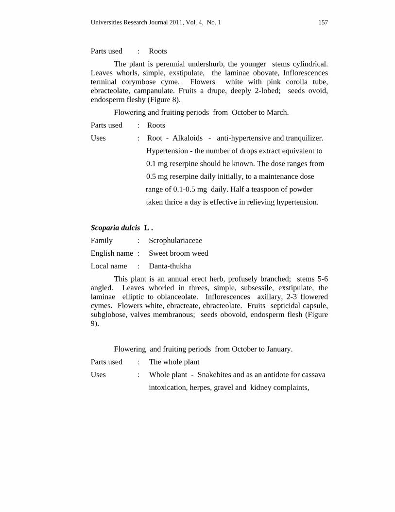
Universities Research Journal 2011, Vol. 4, No. 1 157
Parts used : Roots
The plant is perennial undershurb, the younger stems cylindrical. Leaves whorls, simple, exstipulate, the laminae obovate, Inflorescences terminal corymbose cyme. Flowers white with pink corolla tube, ebracteolate, campanulate. Fruits a drupe, deeply 2-lobed; seeds ovoid, endosperm fleshy (Figure 8).
Flowering and fruiting periods from October to March.
Parts used : Roots
Uses : Root - Alkaloids - anti-hypertensive and tranquilizer.
Hypertension - the number of drops extract equivalent to
0.1 mg reserpine should be known. The dose ranges from
0.5 mg reserpine daily initially, to a maintenance dose
range of 0.1-0.5 mg daily. Half a teaspoon of powder
taken thrice a day is effective in relieving hypertension.
Scoparia dulcis L . Family : Scrophulariaceae
English name : Sweet broom weed
Local name : Danta-thukha
This plant is an annual erect herb, profusely branched; stems 5-6 angled. Leaves whorled in threes, simple, subsessile, exstipulate, the laminae elliptic to oblanceolate. Inflorescences axillary, 2-3 flowered cymes. Flowers white, ebracteate, ebracteolate. Fruits septicidal capsule, subglobose, valves membranous; seeds obovoid, endosperm flesh (Figure 9).
Flowering and fruiting periods from October to January.
Parts used : The whole plant
Uses : Whole plant - Snakebites and as an antidote for cassava
intoxication, herpes, gravel and kidney complaints,
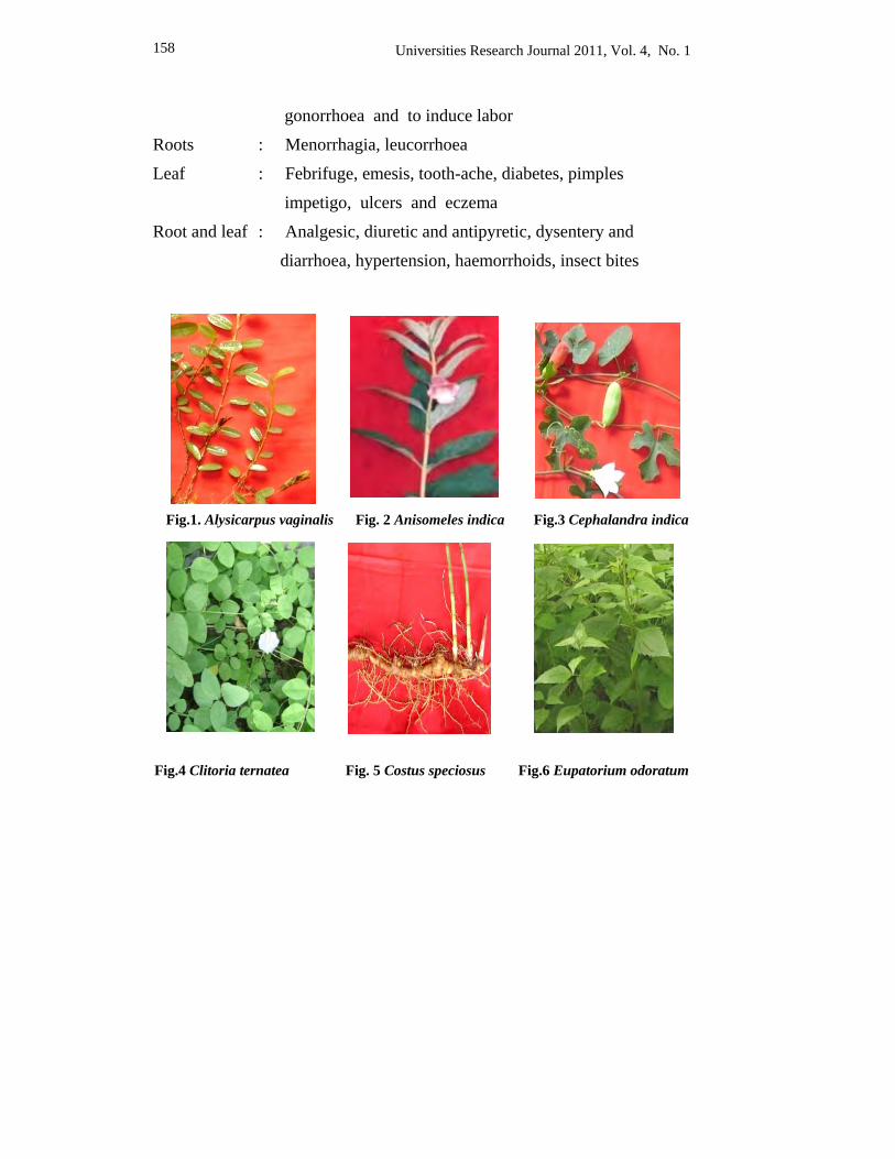
Universities Research Journal 2011, Vol. 4, No. 1 158
gonorrhoea and to induce labor
Roots : Menorrhagia, leucorrhoea
Leaf : Febrifuge, emesis, tooth-ache, diabetes, pimples
impetigo, ulcers and eczema
Root and leaf : Analgesic, diuretic and antipyretic, dysentery and
diarrhoea, hypertension, haemorrhoids, insect bites
Fig.1. Alysicarpus vaginalis Fig. 2 Anisomeles indica Fig.3 Cephalandra indica
Fig.4 Clitoria ternatea Fig. 5 Costus speciosus Fig.6 Eupatorium odoratum
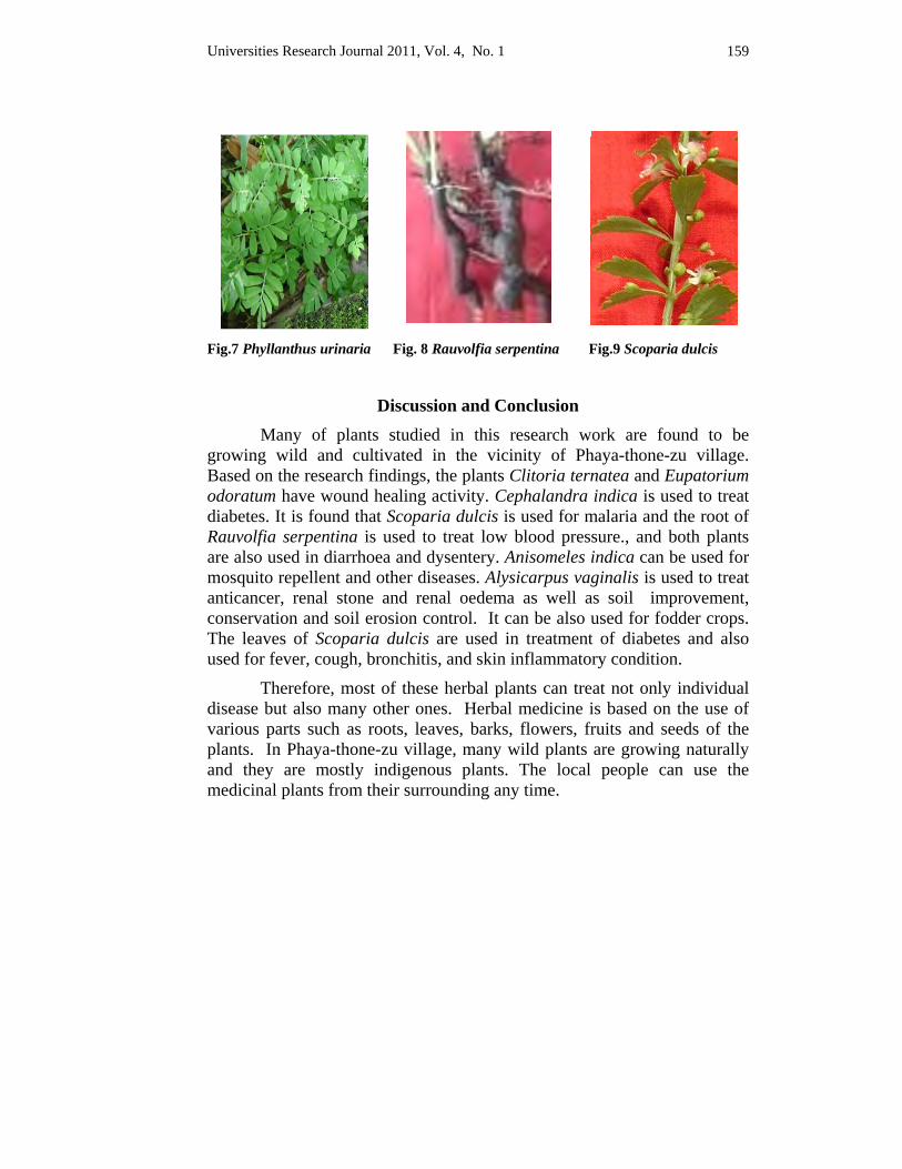
Universities Research Journal 2011, Vol. 4, No. 1 159
Fig.7 Phyllanthus urinaria Fig. 8 Rauvolfia serpentina Fig.9 Scoparia dulcis
Discussion and Conclusion Many of plants studied in this research work are found to be
growing wild and cultivated in the vicinity of Phaya-thone-zu village. Based on the research findings, the plants Clitoria ternatea and Eupatorium odoratum have wound healing activity. Cephalandra indica is used to treat diabetes. It is found that Scoparia dulcis is used for malaria and the root of Rauvolfia serpentina is used to treat low blood pressure., and both plants are also used in diarrhoea and dysentery. Anisomeles indica can be used for mosquito repellent and other diseases. Alysicarpus vaginalis is used to treat anticancer, renal stone and renal oedema as well as soil improvement, conservation and soil erosion control. It can be also used for fodder crops. The leaves of Scoparia dulcis are used in treatment of diabetes and also used for fever, cough, bronchitis, and skin inflammatory condition. Therefore, most of these herbal plants can treat not only individual disease but also many other ones. Herbal medicine is based on the use of various parts such as roots, leaves, barks, flowers, fruits and seeds of the plants. In Phaya-thone-zu village, many wild plants are growing naturally and they are mostly indigenous plants. The local people can use the medicinal plants from their surrounding any time.
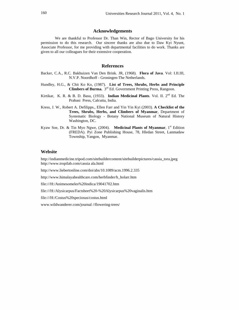
Universities Research Journal 2011, Vol. 4, No. 1 160
Acknowledgements We are thankful to Professor Dr. Than Win, Rector of Bago University for his
permission to do this research. Our sincere thanks are also due to Daw Kyi Nyunt, Associate Professor, for me providing with departmental facilities to do work. Thanks are given to all our colleagues for their extensive cooperation.
References Backer, C.A., R.C. Bakhuizen Van Den Brink. JR, (1968). Flora of Java. Vol: I.II.III,
N.V.P. Noordhoff - Groningen-The Netherlands.
Hundley, H.G., & Chit Ko Ko, (1987). List of Trees, Shrubs, Herbs and Principle Climbers of Burma. 3rd Ed. Government Printing Press, Rangoon.
Kirtikar, K. R. & B. D. Basu, (1933). Indian Medicinal Plants. Vol. II. 2nd Ed. The Prabasi Press, Calcutta, India.
Kress, J. W., Robert A. Defilipps., Ellen Farr and Yin Yin Kyi (2003). A Checklist of the Trees, Shrubs, Herbs, and Climbers of Myanmar, Department of Systematic Biology - Botany National Museum of Natural History Washington, DC.
Kyaw Soe, Dr. & Tin Myo Ngwe, (2004). Medicinal Plants of Myanmar, 1st Edition (FREDA). Pyi Zone Publishing House, 78, Hledan Street, Lanmadaw Township, Yangon, Myanmar.
Website http://indianmedicine.tripod.com/sitebuildercontent/sitebuilderpictures/cassia_tora.jpeg http://www.tropilab.com/cassia ala.html
http://www.liebertonline.com/doi/abs/10.1089/acm.1996.2.335
http://www.himalayahealthcare.com/herbfinder/h_holarr.htm
file:///H:/Animesomeles%20indica/19041702.htm
file:///H:/Alysicarpus/Factsheet%20-%20Alysicarpus%20vaginalis.htm
file:///H:/Costus%20speciosus/costus.html
www.wildwanderer.com/journal //flowering-trees/
Universities Research Journal 2011, Vol. 4, No. 1
Assistant Lecturer, Department of Botany, University of Yangon
Extraction, Isolation and Identification of Chemical Constituents from the Leaves of Clerodendrum inerme
Gaertn. San San Maw
Abstract The plant Clerodendrum inerme Gaertn is an evergreen straggling shrub (Pinle-kyauk-pan) which belongs to the family Verbenaceae. The plants were collected from Ka-mar-aung creek in Dalla Township, Yangon Division. Ethanol was used as a solvent for extraction. Extraction, isolation and identification of chemical constituents of compounds A, B and C were collected from ethanolic extract of leaves of C. inerme Gaertn by selective solvent solubility method. The three isolated compounds were identified by thin layer chromatography, melting point, ultraviolet (UV) and Infra-red (FTIR) spectroscopic methods.
Keywords: Clerodendrum inerme Gaertn, leaves extract, chemical constituents
Introduction The plant Clerodendrum inerme Gaertn is locally known as Pinle-
kyauk-pan, Kywe-yan-nge and Garden quinine. It found growing on seashore, at the edge of mangrove swamps and tidal river banks. The plant has a great reputation as a febrifuge, the juice of the leaves is used as substitute of quinine and its dose was half an ounce. (Kirtikar and Basu, 1933). The leaves and roots of the plant are used in the treatment of skin diseases, asthma, topical burns, urinary infection, wound infection and eye infection. The active constituents of the leaves revealed the presence of glycosides, flavonoids, sterols, flavones, triterpenes, diterpenes and quinones (Monoharam 2006). β sitosterol, apigenin and scutellarein were isolated from the leaves of C. inerme Gaertn. (Sankara, 1973). β-sitosterol is used for the inhibition of induced carcinogenesis and the treatment of diabetes, breast cancer, anti-imflamatory and antipyretic activity. (Stecher, 1996). Apigenin is one of the flavonoids and it has been shown to possess chemopreventive activities including those of lung cancer, skin cancer, cervica cancer, prostate cancer and leukemia. Scutellarein is used for the treatment of diuretic, anti-cancer, antioxidant and anti-imflammatory activities. Thus, the aim of this study is to examine the medicinal plants
162 Universities Research Journal 2011, Vol. 4, No. 1
scientifically to know the medicinal values. The main objectives are to isolate the active constituents from the leaves of Clerodendrum inerme Gaertn. These compounds are identified by melting point, TLC, UV and FTIR methods.
Materials and Methods
The plant Clerodendrum inerme Gaertn. were collected from Ka- mar-aung creek in Dalla Township. The sample leaves were washed, dried at room temperature and grounded to get powder. Extraction and isolation of compounds A, B and C were carried out by using selective solvents solubility method and partition chromatography and identified by using column, TLC, melting point, UV and FTIR spectroscopic method.
Extraction and isolation of compounds A, B and C from the leaves of C. inerme Gaertn. Powdered leaves 250 g were introduced into a round-bottomed flask and followed by the addition of 1 litre of 80 % ethanol. The extraction was carried out at refluxing for about 6 hours and filtered. The marc was re-extracted twice with fresh solvent. The ethanol extracts were combined and concentrated under vaccum. The ethanolic concentrate was repeatedly shaken with benzene and ether in succession.
Isolation of compound A from benzene extract of the powdered sample For compound A, benzene extract was fractionated by using silica gel column chromatography. Elution was performed successively with petroleum ether and benzene (3:1) v/v and 5 ml of each fraction was collected and checked by TLC. The fractions 21, 22, 23 with similar Rf values were combined and evaporated to small volume and kept in the refrigerator. When crude crystals were separated out, filtered and crystal-lized with ethanol. Colourless plate shape crystals were obtained. It was designated as compound A. The flow diagram was shown in Figure (1). Isolation of compounds B and C Compounds B and C were isolated by silica gel preparative thin layer chromatography (PTLC) method.Ether residue 2.6 g was dissolved in ethanol and applied on the TLC plate as a band by using capillary tube. The
Universities Research Journal 2011, Vol. 4, No .1 163
sample spot of the ether residue was also spotted along the starting line, at the corner of the TLC plate. The chromatogram was run by using n-butanol, acetic acid and water 4:1:5 v/v mixtures as solvent system.
PTLC plate was spraying with ferric chloride solution while the sample spot was covered with glass plate, the fraction portion was observed that the sample spot contained two flavonoid compounds with Rf value of 0.83 and 0.16 respectively. The zones which have the Rf value 0.83 and 0.16 were quantitatively scrapped off from the P TLC plate into the round bottomed flasks.
The fractions were extracted with 30 ml of ethyl acetate and filtered. Each of ethyl acetate extracts were evaporated to small volume and kept in a freezer overnight. Precipitate B and C were separated out from each fraction. Crystallization with pyridine gave pure compound B (yellow needle). Precipitate C was crystallized with methanol gave pure compound C (yellow leaflet). Identification of isolated compounds A, B and C
Observation of colour and shape of crystals The colour of the crystals were visualized with naked eye. The shape of the crystals were observed under microscope.
Determination by thin layer chromatography for compound A Isolated compound A and standard β-sitosterol solution were applied
on precoated silica gel plate by capillary tubes. The chromato-graphic plate was developed with pet-ether : benzene (3:1 v/v) as solvent system. Iodine vapour was used for detection of sterol. It was observed that isolated compound A and standard β-sitosterol gave only one spot in the same Rf of 0.53. It was shown in Figure (2).
Determination by TLC for compounds B and C The solution of compounds B and C were spotted on the precoated
silica gel plate. The solvent system was n-butanol, acetic acid and distilled water (4 : 1 : 5 v/v). Ferric chloride solution was used as spraying reagent,
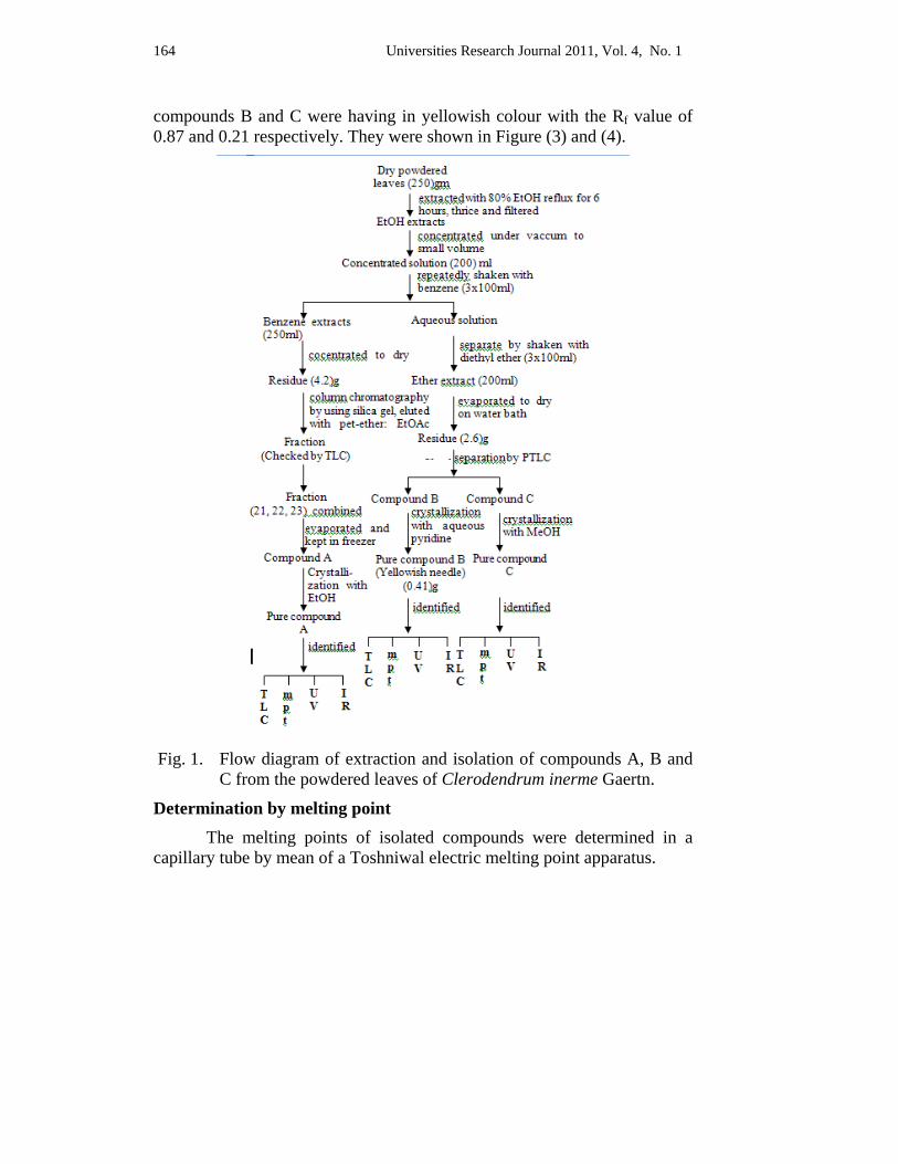
164 Universities Research Journal 2011, Vol. 4, No. 1
compounds B and C were having in yellowish colour with the Rf value of 0.87 and 0.21 respectively. They were shown in Figure (3) and (4).
Fig. 1. Flow diagram of extraction and isolation of compounds A, B and
C from the powdered leaves of Clerodendrum inerme Gaertn.
Determination by melting point The melting points of isolated compounds were determined in a capillary tube by mean of a Toshniwal electric melting point apparatus.
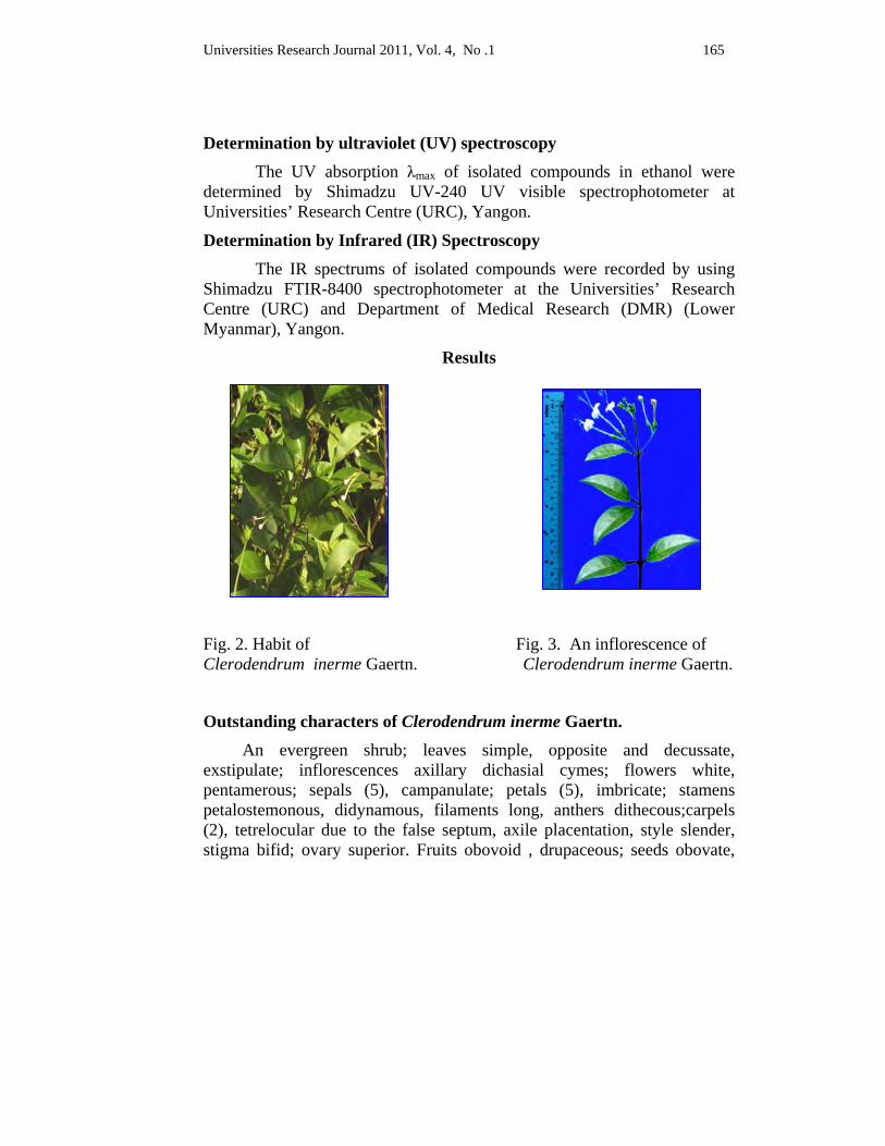
Universities Research Journal 2011, Vol. 4, No .1 165
Determination by ultraviolet (UV) spectroscopy The UV absorption λmax of isolated compounds in ethanol were determined by Shimadzu UV-240 UV visible spectrophotometer at Universities’ Research Centre (URC), Yangon.
Determination by Infrared (IR) Spectroscopy The IR spectrums of isolated compounds were recorded by using Shimadzu FTIR-8400 spectrophotometer at the Universities’ Research Centre (URC) and Department of Medical Research (DMR) (Lower Myanmar), Yangon.
Results
Fig. 2. Habit of Fig. 3. An inflorescence of Clerodendrum inerme Gaertn. Clerodendrum inerme Gaertn.
Outstanding characters of Clerodendrum inerme Gaertn. An evergreen shrub; leaves simple, opposite and decussate, exstipulate; inflorescences axillary dichasial cymes; flowers white, pentamerous; sepals (5), campanulate; petals (5), imbricate; stamens petalostemonous, didynamous, filaments long, anthers dithecous;carpels (2), tetrelocular due to the false septum, axile placentation, style slender, stigma bifid; ovary superior. Fruits obovoid , drupaceous; seeds obovate,

166 Universities Research Journal 2011, Vol. 4, No. 1
exalbuminous. Flowering and fruiting period from May to November (Figure 2 and 3).
Extraction and isolation of compounds A, B and C from the powdered leaves of Clerodendrum inerme Gaertn.
The colourless crystals of isolated compound A were plate shape. It was isolated from benzene extract and yield was 0.36 % respectively. The yellowish crystals of isolated compounds B, C were needle shape and leaflet shape. These compounds were isolated from ether extract and the yield were 0.41 % and 0.28 % respectively.
The compound A melted at 139 °C and compounds B and C were melted at 347°C and 328°C respectively. The comparison of the melting point of isolated and standard compounds were tabulated in Table.(1). On thin layer Chromatogram of compound A and standard β-sitosterol gave any one spot on the same Rf value of 0.53 shown in Figure (4).
The isolated compounds B and C showed single spot on thin layer chromatogram and these compounds were not contaminated with other compounds. The Rf value was 0.87 and 0.2 respectively was shown in Figure (5) and (6).
The wavelength of absorption band of isolated compound A was found at 205.5 nm and the UV spectral data was shown in Table (2) and Figure (14). The absorption maximum of compounds B and C were observed at 268, 342 nm and 285, 340 nm. The UV spectrum of these compounds were shown in Table. (2) and Figure (8) and (9).
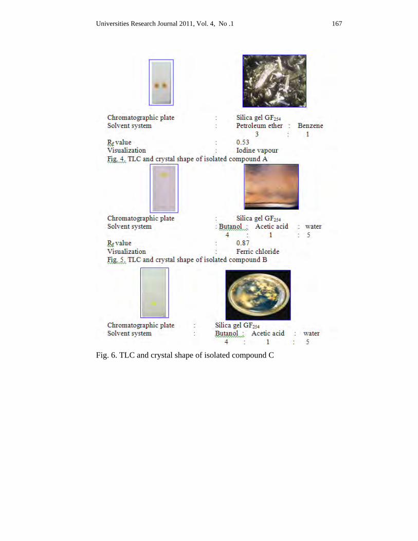
Universities Research Journal 2011, Vol. 4, No .1 167
Fig. 6. TLC and crystal shape of isolated compound C
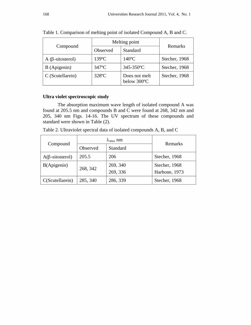
168 Universities Research Journal 2011, Vol. 4, No. 1
Table 1. Comparison of melting point of isolated Compound A, B and C.
Compound Melting point
Remarks Observed Standard
A (β-sitosterol) 139ºC 140ºC Stecher, 1968
B (Apigenin) 347ºC 345-350ºC Stecher, 1968
C (Scutellarein) 328ºC Does not melt below 300ºC
Stecher, 1968
Ultra violet spectroscopic study The absorption maximum wave length of isolated compound A was
found at 205.5 nm and compounds B and C were found at 268, 342 nm and 205, 340 nm Figs. 14-16. The UV spectrum of these compounds and standard were shown in Table (2).
Table 2. Ultraviolet spectral data of isolated compounds A, B, and C
Compound λmax nm
Remarks Observed Standard
A(β-sitosterol) 205.5 206 Stecher, 1968
B(Apigenin) 268, 342
269, 340 269, 336
Stecher, 1968 Harbone, 1973
C(Scutellarein) 285, 340 286, 339 Stecher, 1968
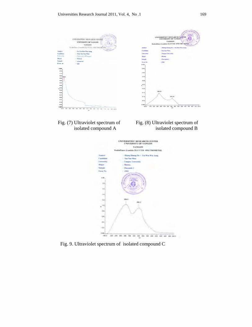
Universities Research Journal 2011, Vol. 4, No .1 169
Fig. (7) Ultraviolet spectrum of isolated compound A
Fig. (8) Ultraviolet spectrum of isolated compound B
Fig. 9. Ultraviolet spectrum of isolated compound C
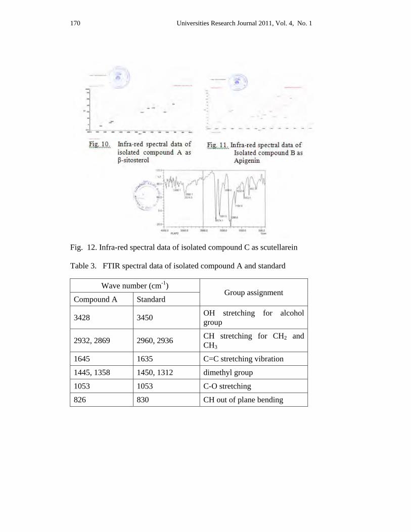
170 Universities Research Journal 2011, Vol. 4, No. 1
Fig. 12. Infra-red spectral data of isolated compound C as scutellarein Table 3. FTIR spectral data of isolated compound A and standard
Wave number (cm-1) Group assignment
Compound A Standard
3428 3450 OH stretching for alcohol group
2932, 2869 2960, 2936 CH stretching for CH2 and CH3
1645 1635 C=C stretching vibration
1445, 1358 1450, 1312 dimethyl group
1053 1053 C-O stretching
826 830 CH out of plane bending
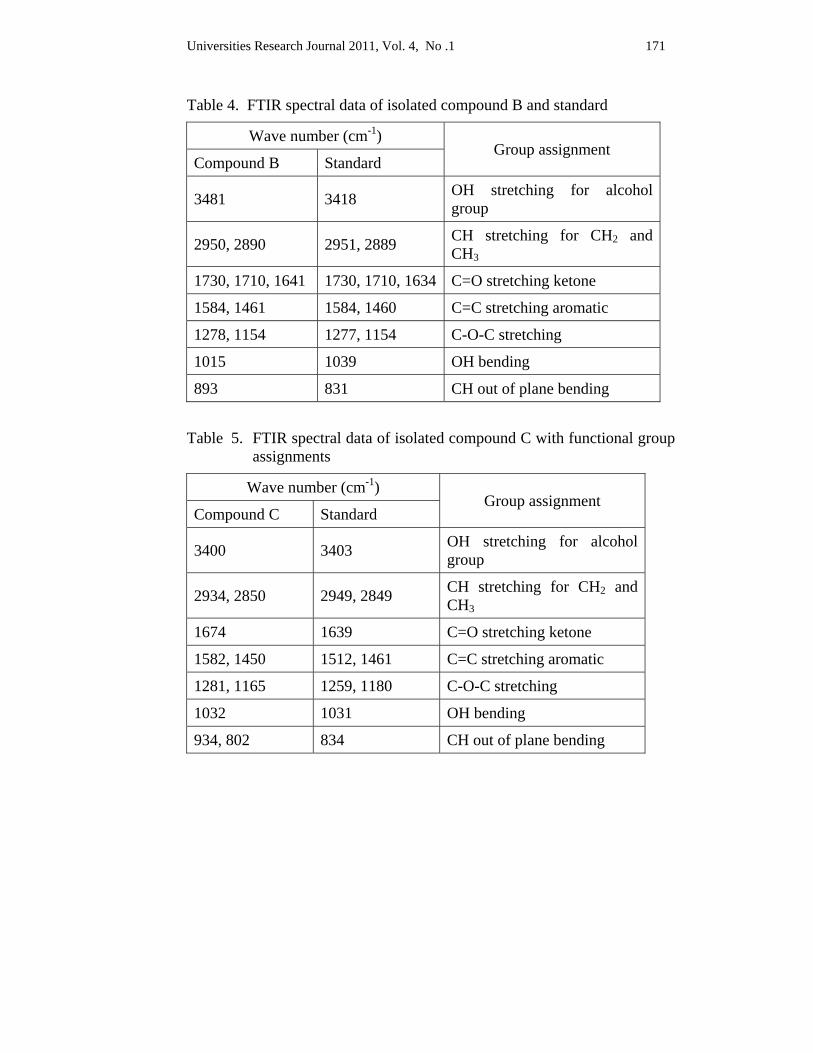
Universities Research Journal 2011, Vol. 4, No .1 171
Table 4. FTIR spectral data of isolated compound B and standard
Wave number (cm-1) Group assignment
Compound B Standard
3481 3418 OH stretching for alcohol group
2950, 2890 2951, 2889 CH stretching for CH2 and CH3
1730, 1710, 1641 1730, 1710, 1634 C=O stretching ketone
1584, 1461 1584, 1460 C=C stretching aromatic
1278, 1154 1277, 1154 C-O-C stretching
1015 1039 OH bending
893 831 CH out of plane bending
Table 5. FTIR spectral data of isolated compound C with functional group assignments
Wave number (cm-1) Group assignment
Compound C Standard
3400 3403 OH stretching for alcohol group
2934, 2850 2949, 2849 CH stretching for CH2 and CH3
1674 1639 C=O stretching ketone
1582, 1450 1512, 1461 C=C stretching aromatic
1281, 1165 1259, 1180 C-O-C stretching
1032 1031 OH bending
934, 802 834 CH out of plane bending
172 Universities Research Journal 2011, Vol. 4, No. 1
Infra-red spectroscopic study The IR spectrum of isolated compound A was shown in Figure (8) and spectral data was shown in Table (3). The wave numbers of compound A show the OH stretching for alcohol group at 3428 cm-1, C=C stretching for CH2 and CH3 appear at 2932, 2869 cm-1, C=C stretching vibration at 1645 cm-1, the presence of dimethyl group indicate at 1445, 1358 cm-1, C-O stretching appears at 1053 cm-1 and CH out of plane bending appears at 826 cm-1.
The IR spectrum of isolated compound B was shown in Figure (9) and spectral data was shown in Table (4). The wave numbers of compound B show OH stretching at 3481 cm-1 , CH stretching for CH2 and CH3 at 2950, 2890 cm-1, C=O stretching for ketone band appears at 1730, 1710,1641 cm-1 C=C stretching for aromatic ring at 1584, 1461 cm-1, C-O-C stretching shows at 1278, 1154 cm-1 , OH bending appears at 1015 cm-1 and CH out of plane bending at 893 cm-1.
The IR spectrum of isolated compound C was shown in Figure (10) and its data was shown in Table (5). The wave numbers of compound C show the OH stretching for alcohol group appears at 3400 cm-1, CH stretching for CH2 and CH3 observe at 2934, 2850 cm-1, C=O stretching for ketone band at 1674 cm-1, C=C stretching of aromatic ring at 1582, 1450 cm-1, C-O-stretching indicate at 1281, 1165 cm-1, OH bending appears at 1032 cm-1 and CH out of plane bending at 934, 802cm-1.
Discussion and Conclusion The three active compounds were isolated from the leaves of C.
inerme Gaertn. The extraction and isolation of compound A was carried out by column chromatography method. Compounds B and C were carried out by using selective solvents solubility and preparative thin layer chromatography method. Identification of isolated compounds was carried out by TLC, melting point, UV and FTIR spectroscopic methods.
Compound A which was isolated from column chromatography was showed a single spot on TLC when using solvent system of petroleum ether: benzene (3:1) and Rf value was 0.53 and the melting point was 139°C, the results were agreed with Stecher (1968) and Sankara (1973) who mentioned as the melting point of this compound was 140°C. According to Stecher (1968), the maximum UV absorption wavelength for β-sitosterol
Universities Research Journal 2011, Vol. 4, No .1 173
showed at 206 nm which was more or less exactly the same result with compound A in this study. The wave number of IR absorption peaks of compound A were generally the same results with all the functional group of β-sitosterol as described in Atlas (1975). In fact, compound A should be considered as β-sitosterol.
According to thin layer chromatography of compound B and C, each compound gave a single spot with Rf values of 0.87 and 0.21 respectively. It indicated that compounds were pure. Melting point of compound B was 347°C with yellowish needle-shaped crystal and compound C was 328°C with yellowish leaflet-shaped crystals. These were in accordance with apigenin and scutellarein crystals shape and melting point mentioned in Stecher (1968). The UV absorption λmax of compound B and C were 268 - 342 nm and 285 - 340 nm indicating the presence phenolic group and aromatic carbonyl group. These characters were the agreement with those given by Stecher (1968).
The wave number of IR spectral bands of compound B were indicated the spectral characteristics of apigenin was described by Bieman (1989). In addition, IR spectral data of compound C were identical to the spectral characteristics of scutellarein as mentioned in the literature Myanmar effective medicinal plants (2002).
Due to the above data, compounds B and C may be considered as apigenin and scutellarein.
Acknowledgements I would like to express my gratitude to Dr. Thet Thet May, Professor and Head,
and Dr. Aye Pe, Professor, Department of Botany, University of Yangon, for their kind permission to do the research work. I am grateful to U Htin Aung Kyaw, Deputy Director (Retired), Yangon Technological University for his supervision and I am thankful to Dr. Nu Nu Swe, Professor, Department of Botany, Meiktila University for her encouragement and valuable advice.
References Atlas (1975). Spectral data and physical contents for organic compound: (2nd ed.).
CRC Press Inc vol. 3.
Bieman, K, (1980). Table of spectral data for structure determination of organic compounds. (2nd ed.).
174 Universities Research Journal 2011, Vol. 4, No. 1
Hundley, H. G. & Chit Ko Ko (1961). List of tress, shrubs, herbs and principle climbers of Burma. (3rd ed.) Yangon: Government Printing Press.
KirtiKar, K. R and Basu, B. D. (l935). Indian medicinal plants. (2nd ed.). New York: The Macmillan Co.
Kress, W. J., DeFilipps, R. A., Farr, E. & Yin Yin Kyi. (2003). A checklist of the Trees, Shrubs, Herbs, and Climbers of Myanmar. Department of Systematic Biology Botany, National Museum of Natural History, Washington DC: U.S.A.
Manoharam, S., Kavichi, K., Senthel, N., Renju, G. L., (2006). Singapore medicine journal vol. 12, Pg 1043.
Rastogi, R. P. (1993). Compendium of Indian medicinal plants, vol. 2-3, Central Drug Research Institute, Lucknow and National Institute of Science Communication New Delhi.
Sankara, S. (1973). The Indian journal of pharmacy. Bombay.
Stecher, G. P. (1968). The merck index, An encyclopedia of chemical and drugs.. U.S.A: Published by Merck and Co., Inc.
Myanmar Effective Medicinal Plants, (2002). vol.1-2, Myanmar, Ministry of Science and Technology.
Universities Research Journal 2011, Vol. 4, No. 1
Assistant Lecturer, Department of Botany, University of Mandalay
Taxonomic Study on Five Vanda Species from Goktwin Area, Northern Shan State
Ah Nge Htwe
Abstract Shan State is mostly covered with tropical evergreen forests and distribution of many wild orchids. The members of Orchidaceae from Goktwin tracts between Naungcho Township and Kyaukme Township of Northern Shan State in Myanmar, between 96° 48' E and 96° 57' E Longitude and 22° 16' N and 22° 22' N Latitude had been collected and studied. The research work deals with 5 species of Vanda. All collected species are fully described with necessary photographs. Moreover, an artificial key to the species of the plants is also described. The system of Seidenfaden (1992) was adopted as the classification system in the present research.
Key words : wild orchids, classification, present research
Introduction Tropical Asia which includes Myanmar still possesses forests rich in
biodiversity. Myanmar is one of the member countries of the ASEAN and it is geographically located between 9° 58' and 28° 31' north Latitude and between 92° 9' and 101° 1' east Longitude in Southeast Asia. In Myanmar, the mountain ranges stretch from north to south. All the mountain ranges are covered with forests where many Myanmar native orchids with great diversity exist as a gift from mother nature due to varied climatic conditions, diverse habitats and wide altitudinal variations.
Orchids are one of the most outstanding and significant flowering plants of the Myanmar flora due to their glamorous, striking and elegant flowers. The present research deals with some members of plants growing in Goktwin area between Naungcho Township and Kyaukme Township, Northern Shan State in Myanmar. The area is famous for its rich biodiversity and its valuable natural resources. The study area of Goktwin is situated between 22° 16' – 22° 22' E Longitude and between 96° 50' – 90°57' N Latitude. The area covers about 104 square kilometer. It is about 90 kilometer far from Mandalay. The elevation of study area is 274 to 1025 meter above sea level.

176 Universities Research Journal 2011, Vol. 4, No. 1
In this study, all of the specimens collected had been arranged following the classification system of Seidenfaden (1992) represented that a total of 140 genera and about 800 species and varieties of orchids are included. In 1895, Grant reported that a total of 86 genera and 581 species and varieties of orchids were distributed in Myanmar. In 1961, Hundley and Chit Ko Ko listed 113 genera and 850 species and varieties of Myanmar orchids and 128 genera and 739 species in 1986 as Myanmar native orchids. Although several workers had worked out on selected genera in flora of Goktwin area, the orchids are widely distributed and its taxonomic information is still needed to be recorded in the area.
The studies on the species of Orchidaceae are based on their morphological characters. Taxonomic description of each species, photographic plates have been given together with the distinctive characters. The species were collected monthly during from 2007 to 2010, December. All the collected specimens have been deposited at the herbarium of the Department of Botany, Mandalay University.
The aim and objectives of this research are to conduct and record the Orchidaceae flora of Northern Shan State, to investigate their distribution and morphological characteristics, to support further studies for their valuable information natural orchid species from Goktwin area, and to give a partial fulfillment of the orchid information of Shan State in Myanmar.
Materials and Methods The plants were collected from Goktwin area during the year 2007-2010. The field studies had been done in every two methods intervals. Identification of collected specimens was carried out by referring to flora of British India (1894), Flora of Java (1968) Flora of Ceylon (1981), The orchids of Indochina (1992). The classification system for this study is based on that of Seidenfaden in 1992. The first step is solving the generic name. Again the species names of the collected plants are resolved. Specimens had been identified and described in morphological characteristics, then constructed their artificial key.

Universities Research Journal 2011, Vol. 4, No. 1 177
Results
Taxonomic description of Vanda
Family Orchidaceae Perennial, but sometimes short-lived, terrestrial, epiphytic, or
lithophytic, autotrophic or rarely saprophytic herbs (or rarely scrambling vines), with rhizomes, tubers or rootstocks with mycorrhizal fungi in roots. Stems either sympodial or less commonly monopodial, usually leafy, but leaves sometimes reduced to bract-like scales, one or more internodes at base often swollen to form a pseudobulb; epiphytic species with aerial, assimilating adventitious roots, often bearing one or more layer of dead cells (velamen). Leaves 1 to many, glabrous or very rarely hairy, entire in some cases except at apex, alternate or occassionally opposite, often distichous, frequently fleshy or leathery, sometimes articulated at base of lamina and sometimes with a false frequently sheathes stem, sometimes articulated at base of lamina and sometimes with a false petiole. Inflorescences erect to pendent, spicate, racemose, or paniculate, 1-to many flowered, basal, lateral, or terminal, flowers rarely secund, subumbellate, or distichously arranged. Flowers small to large, often quite showy, hermaphroditic (or rarely monoecious and polymorphic outside region), sessile or variously pedicellate, most often twisted through 180, occassionally not twisted or twisted through 360. Ovary inferior, 1-locular and placentation parietal (or rarely 3-locular and placentation axile). Sepals usually free but sometimes variously adnate, median (dorsal) one often dissimilar to lateral, laterals sometimes adnate to a column foot to form a saccate, conic, or spurlike mentum. Petals free or rarely partly adnate to sepals, similar to sepals or not, often showy; lip entire, variously lobe or 2-or 3-partite, ornamented or not with calli, ridges, hair cushions, or crests, with or without a basal spur or nectary, margins entire to laciniate. Column short to long, with or without a basal foot, occasionally winged or with lobes or arms at apex or ventrally; anther 1 (or rarely 2 or 3 outside region), terminal or ventral on column, caplike or opening by longitudinal slits; pollinia mealy, waxy or horny, sectile or not, 2, 4, 6, or 8, sessile or attached by stalks to 1 or 2 sticky viscidia; stigma 3-lobed, midlobe often modified to form a rostellum, other lobes either sunken on ventral surface of column behind anther or with 2 lobes porrect. Fruit a capsule, usually opening laterally by 3 or 6 slits. Seeds numerous, dustlike, lacking endosperm, rarely winged.
178 Universities Research Journal 2011, Vol. 4, No. 1
About 725 genera and 17,500 species (some estimates suggesting as many as 30,000 species): worldwide, except for Antarctica, most numerous in the humid tropics and subtropics; 193 genera (11 endemic) and 1350 species (505 endemic) in China (Seidenfaden, 1992).
Vanda R. Br., Bot. Reg. 6: 506. 1820.
Epiphytes; stems long with vermiform roots, non-pseudobulbous; leaves flat, distichous, recurved, thickly coriaceous, bilobed at apex or terete; inflorescence simple, more or less erect, of few large or medium-sized flowers, peduncle leaf-opposed; sepals and petals nearly equal, narrowed to the base, the edges more or less reflexed or crisped; lip smaller than sepals, attached immovably to a short column-foot, spur short, funnel-shaped, 3-lobed, often laterally flattened without calli; side lobes erect, close to the column; mid lobe erect or recurved, narrow or broad with longitudinal ridges and 2 small calli at the base; column short, thick; foot short or absent; anther 2-chambered with 2 large, waxy, cleft pollinia on a short strap and gland.
About 80 species distributed throughout India, Ceylon, Malaya, Myanmar, Thailand, Cambodia, Indo-China and China, extending southwards into Java, Sumatra, Borneo, Celebes, Australia and the Philippine Islands (Dassanaayake, 1981).
Artificial key to the species 1. Sepal and petal with tessellation ------------------------------------------ 2
1. Sepal and petal without tessellation -------------------------------------- 4
2. Floral bracts ovate-lanceolate ----------------------------------------- 3
2. Floral bracts triangular-ovate -------------------- 1. Vanda bensonii
3. Dorsal sepal ovate-oblong; sidelobes of lips semirotund ----- 2.Vanda brunnea
3. Dorsal sepal suborbicular; sidelobes of lips linear-oblong --- 3.Vanda coerulea
4. Floral bracts ovate-lanceolate; flower yellow -- 5.Vanda testacea
4. Floral bracts ovate; flower dark bluish ---- 4. Vanda coerulescens
Universities Research Journal 2011, Vol. 4, No. 1 179
Vanda bensonii Batem. in Bot.Mag. t.5611; Reichb. f.Xen.Orchid. ii.
138; Gard. Chron. 1867, 180; Flore des Serres, t. 2329.
Monopodial epiphyte; stem short, 15.0 - 20.0 cm long and 1.0 - 1.2 cm wide, leafy, green, glabrous, nonpseudobulbous. Leaves alternate and distichous, narrrow, strap-shaped, recurved, distichous, thick, coriaceous, 20.0 - 25.0 cm long and 1.5 - 2.0 cm wide, margins entire, tips muticous, green, glabrous. Inflorescence simple racemes, terminal and axillary, pendulous, 15.0 - 25.0 cm long and 0.5 - 1.0 cm wide, 4-to 10-flowered; peduncular bracts ovate, 0.2 - 0.3 cm long and 2.0 - 3.0 cm wide, green; peduncle long, inclined many-fid, 25.0 - 28.0 cm long and 0.5 - 0.6 cm wide, green; floral bracts triangular-ovate, clasping the rachis, 2.5 - 2.7 mm long and 6.0 - 6.2 mm wide, green. Flower large, 6.0 - 12.0 cm across at anthesis, light lavender sepals and petals with brown tessellation, lip rose purple, fragrant; pedicel slender, 2.0 - 2.5 cm long and 0.1 - 0.2 cm wide, white, glabrous; dorsal sepal obovate, 1.5 - 1.8 cm long and 1.0 - 1.2 cm wide, light lavender, 5-veined, coriaceous, glabrous, margins entire, tips faintly trifid; lateral sepal broadly obovate, 2.0 - 2.1 cm long and 1.2 - 1.3 cm wide, light lavender, 7-veined, margins undulate, tips obtuse, coriaceous, glabrous; lateral petals spathulately obovate, 1.5 - 1.7 cm long and 1.1 - 1.2 cm wide, dull yellow spotted with red-brown, 7-veined, coriaceous, margins andulate, tips obtuse, glabrous; lip nearly as long as the sepal, blue, side lobes triangular recurved terminal lobes, 1.9 - 2.0 cm long and 0.6 - 0.9 cm wide, rose purple, mid lobe of lip with 3 obtuse ridges; spur conical obtuse, attached to the base of the column, 0.2 - 0.3 cm long and 0.1 - 0.2 cm wide, blue; column short, 0.2 - 0.3 cm long and 0.1 - 0.2 cm wide, pale blue, stout; anthercaps minute, ovoid, anther 2-celled, white; pollinia 2, pairwise cohering, yellow, waxy, stipes round, white; ovary triangular, 0.2 - 0.3 cm long and 0.1 - 0.2 cm wide, glabrous, pale green. Fruits are not seen (Fig-1. A) . Vanda brunnea Rchb. f., Xen-Orchid. ii. 139; 1883.
Monopodial epiphytes. Roots long drooping and clinging, 4.0 - 5.0 mm in diameter, cylindrical, white, and glabrous. Stem leafy, erect, cylindrical, 25.0 - 30.0 cm long and 2.0 - 2.2 cm wide, internodes 1.5 - 1.6 cm long. Leaves alternate and distichous, oblong-lanceolate, long narrowly ligulate emarginate, 23.0 - 27.0 cm long and 1.7 - 2.0 cm wide, more or less
180 Universities Research Journal 2011, Vol. 4, No. 1
channeled, rigid, nearly straight, coriaceous, margins entire, tips bifid, leafy at anthesis, persistent, green, glabrous. Inflorescences axillary raceme, more or less erect, 1 - 2 on each stem, 20.0 - 25.0 cm long and 1.5 - 2.0 cm wide, 15 - 20 flowered; peduncular bracts 3 - 4, sheathing, ovate- lanceolate, 5.0 - 6.0 mm long and 2.0 - 3.0 mm wide, membranous, persistent, margins entire, tips acute, brownish green, glabrous; peduncle stout, cylindrical, long, 12.0 - 15.0 cm long and 3.0 - 4.0 mm wide, green, glabrous; floral bracts ovate- lanceolate, 3.0 - 4.0 mm long and 2.0 - 3.0 mm wide, margins entire, tips acute, brownish white, glabrous. Flowers 1.5 - 2.0 cm across at anthesis, dark brown waxy sepals and petals, lip and column purple striped with reddish brown tessellation; pedicels slender, 2.5 - 3.0 cm long and 2.0 - 3.0 mm wide, yellowish white, glabrous; dorsal sepals ovate-oblong, shortly claw and flat, 1.2 - 1.3 cm long and 7.0 - 8.0 mm wide, margins undulate, tips obtuse, dark brown waxy above and brownish white beneath, glabrous; lateral sepal obovate, 1.1 - 1.2 cm long and 7.0 - 8.0 mm wide, margins slightly undulate, tips obtuse, dark brown waxy above and brownish white beneath, glabrous; lateral petals broadly obovate, shortly claw, 1.2 - 1.3 cm long and 8.0 - 9.0 mm wide, reflexed, margins slightly undulate, tips obtuse, dark brown and brownish white beneath, glabrous; lip distinctly 3-lobe, sidelobe semirotund, 4.0 - 5.0 mm long and 3.0 - 3.5 cm wide, margins undulate, tip obtuse, coriaceous, reflexed, reddish brown, midlobe ligulate, 2-lobed, 4.0 - 5.0 mm long and 1.3 - 1.5 mm wide, margins entire, tips retuse, purple striped with brown, glabrous; spur acutely conical, 4.0 - 5.0 mm long and 2.0 - 2.5 mm wide, tip acute, brownish yellow, glabrous; column short, stout, 5.0 - 6.0 mm long and 3.0 - 4.0 mm wide, pale brown; anthercap broadly ovoid, 3.0 - 4.0 mm long and 2.0 - 2.5 mm wide, white, glabrous; pollinia 2, obovoid, each 1.5 - 2.0 mm long and 1.0 - 1.2 mm wide, yellow, waxy; caudicle short, flat, 1.3 - 1.5 mm long and 0.9 - 1.0 mm wide, white; viscidium membranous, 2.0 - 2.2 mm long and 1.8 - 2.0 mm wide, white; ovary narrowly oblongoid, 2.0 - 2.5 cm long and 2.0 - 3.0 mm wide, yellowish-white, glabrous. Fruit capsule, fusiform, 5.0 - 6.0 cm long and 1.0 - 1.3 cm wide, coriaceous, 6-ridges, green, glabrous (Fig-1. B).
Vanda coerulea Griff. ex. Lindl., Bot. Reg. Sub. t. 30. 1847.
Monopodial epiphytes. Roots long drooping and clinging, 5.0 - 6.0 mm in diameter, cylindrical, white, glabrous. Stems densely leafy, erect, cylindrical, stout, 10.0 - 21.0 cm long and 1.2 - 1.5 cm wide,
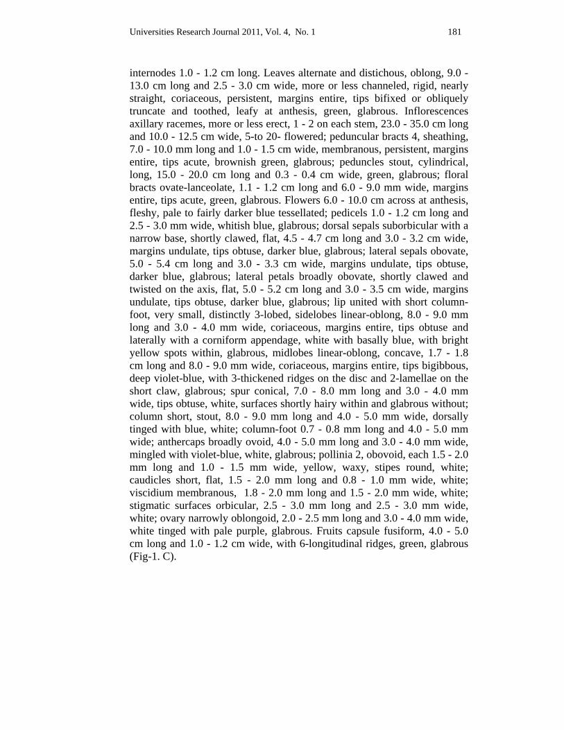
Universities Research Journal 2011, Vol. 4, No. 1 181
internodes 1.0 - 1.2 cm long. Leaves alternate and distichous, oblong, 9.0 - 13.0 cm long and 2.5 - 3.0 cm wide, more or less channeled, rigid, nearly straight, coriaceous, persistent, margins entire, tips bifixed or obliquely truncate and toothed, leafy at anthesis, green, glabrous. Inflorescences axillary racemes, more or less erect, 1 - 2 on each stem, 23.0 - 35.0 cm long and 10.0 - 12.5 cm wide, 5-to 20- flowered; peduncular bracts 4, sheathing, 7.0 - 10.0 mm long and 1.0 - 1.5 cm wide, membranous, persistent, margins entire, tips acute, brownish green, glabrous; peduncles stout, cylindrical, long, 15.0 - 20.0 cm long and 0.3 - 0.4 cm wide, green, glabrous; floral bracts ovate-lanceolate, 1.1 - 1.2 cm long and 6.0 - 9.0 mm wide, margins entire, tips acute, green, glabrous. Flowers 6.0 - 10.0 cm across at anthesis, fleshy, pale to fairly darker blue tessellated; pedicels 1.0 - 1.2 cm long and 2.5 - 3.0 mm wide, whitish blue, glabrous; dorsal sepals suborbicular with a narrow base, shortly clawed, flat, 4.5 - 4.7 cm long and 3.0 - 3.2 cm wide, margins undulate, tips obtuse, darker blue, glabrous; lateral sepals obovate, 5.0 - 5.4 cm long and 3.0 - 3.3 cm wide, margins undulate, tips obtuse, darker blue, glabrous; lateral petals broadly obovate, shortly clawed and twisted on the axis, flat, 5.0 - 5.2 cm long and 3.0 - 3.5 cm wide, margins undulate, tips obtuse, darker blue, glabrous; lip united with short column-foot, very small, distinctly 3-lobed, sidelobes linear-oblong, 8.0 - 9.0 mm long and 3.0 - 4.0 mm wide, coriaceous, margins entire, tips obtuse and laterally with a corniform appendage, white with basally blue, with bright yellow spots within, glabrous, midlobes linear-oblong, concave, 1.7 - 1.8 cm long and 8.0 - 9.0 mm wide, coriaceous, margins entire, tips bigibbous, deep violet-blue, with 3-thickened ridges on the disc and 2-lamellae on the short claw, glabrous; spur conical, 7.0 - 8.0 mm long and 3.0 - 4.0 mm wide, tips obtuse, white, surfaces shortly hairy within and glabrous without; column short, stout, 8.0 - 9.0 mm long and 4.0 - 5.0 mm wide, dorsally tinged with blue, white; column-foot 0.7 - 0.8 mm long and 4.0 - 5.0 mm wide; anthercaps broadly ovoid, 4.0 - 5.0 mm long and 3.0 - 4.0 mm wide, mingled with violet-blue, white, glabrous; pollinia 2, obovoid, each 1.5 - 2.0 mm long and 1.0 - 1.5 mm wide, yellow, waxy, stipes round, white; caudicles short, flat, 1.5 - 2.0 mm long and 0.8 - 1.0 mm wide, white; viscidium membranous, 1.8 - 2.0 mm long and 1.5 - 2.0 mm wide, white; stigmatic surfaces orbicular, 2.5 - 3.0 mm long and 2.5 - 3.0 mm wide, white; ovary narrowly oblongoid, 2.0 - 2.5 mm long and 3.0 - 4.0 mm wide, white tinged with pale purple, glabrous. Fruits capsule fusiform, 4.0 - 5.0 cm long and 1.0 - 1.2 cm wide, with 6-longitudinal ridges, green, glabrous (Fig-1. C).
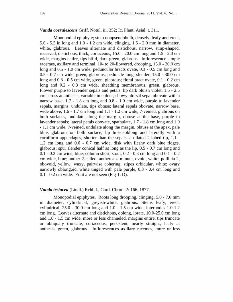
182 Universities Research Journal 2011, Vol. 4, No. 1
Vanda coerulescens Griff. Notul. iii. 352; Ic. Plant. Asial. t. 311.
Monopodial epiphyte; stem nonpseudobulb, densely, leafy and erect, 5.0 - 5.5 in long and 1.0 - 1.2 cm wide, clinging, 1.5 - 2.0 mm in diameter, white, glabrous. Leaves alternate and distichous, narrow, strap-shaped, recurved, distichous, thick, coriaceous, 15.0 - 20.0 cm long and 1.5 - 2.0 cm wide, margins entire, tips bifid, dark green, glabrous. Inflorescence simple racemes, axillary and terminal, 10- to 20-flowered, drooping, 15.0 - 20.0 cm long and 0.5 - 1.0 cm wide; peduncular bracts ovate, 0.3 - 0.5 cm long and 0.5 - 0.7 cm wide, green, glabrous; peduncle long, slender, 15.0 - 30.0 cm long and 0.3 - 0.5 cm wide, green, glabrous; floral bract ovate, 0.1 - 0.2 cm long and 0.2 - 0.3 cm wide, sheathing membranous, green, glabrous. Flower purple to lavender sepals and petals, lip dark bluish violet, 1.5 - 2.5 cm across at anthesis, variable in colour, showy; dorsal sepal obovate with a narrow base, 1.7 - 1.8 cm long and 0.8 - 1.0 cm wide, purple to lavender sepals, margins, undulate, tips obtuse; lateral sepals obovate, narrow base, wide above, 1.6 - 1.7 cm long and 1.1 - 1.2 cm wide, 7-veined, glabrous on both surfaces, undulate along the margin, obtuse at the base, purple to lavender sepals; lateral petals obovate, spathulate, 1.7 - 1.8 cm long and 1.0 - 1.1 cm wide, 7-veined, undulate along the margin, obtuse at the apex, pale blue, glabrous on both surface; lip linear-oblong and laterally with a corniform appendages, shorter than the sepals, a dilated 2-lobed tip, 1.1 - 1.2 cm long and 0.6 - 0.7 cm wide, disk with fleshy dark blue ridges, glabrous; spur slender conical half as long as the lip, 0.5 - 0.7 cm long and 0.1 - 0.2 cm wide, blue; column short, stout, 0.2 - 0.3 cm long and 0.1 - 0.2 cm wide, blue; anther 2-celled, anthercaps minute, ovoid, white; pollinia 2, obovoid, yellow, waxy, pairwise cohering, stipes orbicular, white; ovary narrowly oblongoid, white tinged with pale purple, 0.3 - 0.4 cm long and 0.1 - 0.2 cm wide. Fruit are not seen (Fig-1. D).
Vanda testacea (Lindl.) Rchb.f., Gard. Chron. 2: 166. 1877.
Monopodial epiphytes. Roots long drooping, clinging, 5.0 - 7.0 mm in diameter, cylindrical, greyish-white, glabrous. Stems leafy, erect, cylindrical, 25.0 - 30.0 cm long and 1.0 - 1.5 cm wide, internodes 1.0-1.2 cm long. Leaves alternate and distichous, oblong, lorate, 10.0-25.0 cm long and 1.0 - 1.5 cm wide, more or less channeled, margins entire, tips truncate or obliqualy truncate, coriaceous, persistent, nearly straight, leafy at anthesis, green, glabrous. Inflorescences axillary racemes, more or less

Universities Research Journal 2011, Vol. 4, No. 1 183
erect, 1 - 3 on each stem, 22.0 - 23.0 cm long and 2.5 - 3.5 cm wide, 15 to 30 flowered; peduncular bracts 4 - 5, sheathing, broadly ovate, 4.5 - 5.0 mm long and 5.0 - 5.5 mm wide, membranous, persistent, margins entire, tips acute, brownish green, glabrous; peduncles long, cylindrical, 15.0 - 17.0 cm long and 3.0 - 3.2 mm wide, green with purple spots, glabrous; floral bracts broadly ovate-lanceolate, 1.5 - 1.8 mm long and 2.2 - 2.3 mm wide, membranous, deciduous, margins entire, tips acute, brownish-white, glabrous. Flowers 1.5 - 2.0 cm across at anthesis, yellow flower with purple lip; pedicels angular, 1.8 - 2.0 cm long and 0.8 - 1.0 mm wide, twisted, yellow with pink tinged, glabrous; dorsal sepal obovate-spathulate, 8.0 - 6.5 mm long and 3.0 - 4.5 mm wide, margins entire, tips obtusely rounded and incurved, yellow, glabrous; lateral sepals obovate, 6.07.0 mm long and 3.0 - 4.0 mm wide, margins slightly undulate, tips obtuse and incurved, yellow, glabrous; lateral petals spathulately obovate, flat, shortly clawed and twisted on the axis, 8.0 - 9.0 mm long and 3.0 - 4.0 mm wide, concave, narrow and twisted at the base downwards, margins slightly undulate, tips obtuse, yellow, glabrous; lip united with short column-foot, 3-lobed, sidelobe oblong, 3.0 - 3.5 mm long and 1.5 - 1.8 mm wide, sub-coriaceous, margins entire, tips obtuse, pale-purple, glabrous, midlobes subquadrate oblong, recurved, 6.0 - 6.5 mm long and 3.0 - 3.2 mm wide, sub-coriaceous, margins crenulate, tips dilated or emarginated, 2-broad fleshy ridges on disc, pale-purple, glabrous; spur narrow, funnel-shaped, 2.5 - 2.8 mm long and 0.8 - 1.0 mm wide, tips subacute, pale-purple, glabrous, column short, stout, 2.2 - 2.5 mm long and 2.5 - 3.0 mm wide, pale purple; column-foot very short, not distinct; anthercaps sub globose, 2-loculed, 1.2 - 1.4 mm long and wide, white, glabrous; pollinia 2, oblongoid, bifid, 0.6 - 0.8 mm long and 0.3 - 0.4 mm wide, waxy, yellow, stipes round, white; caudicles short, flat, 0.8 - 1.0 mm long and wide; viscidium very minute, white; stigmatic surfaces very minute; ovary trigonous, with ridges, 2.5 - 2.8 mm long and 1.2 - 1.4 wide, pale yellow with purple striation, glabrous (Fig-1. E).
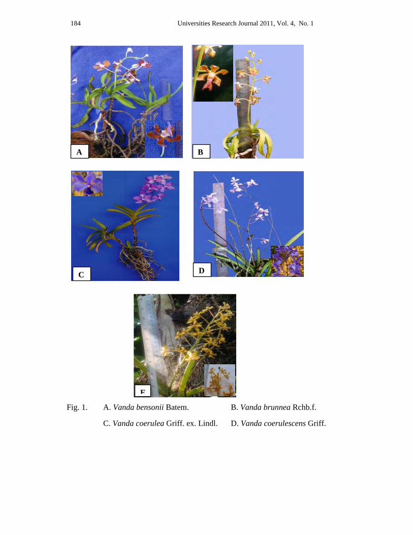
184 Universities Research Journal 2011, Vol. 4, No. 1
Fig. 1. A. Vanda bensonii Batem. B. Vanda brunnea Rchb.f.
C. Vanda coerulea Griff. ex. Lindl. D. Vanda coerulescens Griff.
C D
A
E
B

Universities Research Journal 2011, Vol. 4, No. 1 185
Discussion and Conclusion The selected area for this monographic study is Goktwin area, between Naungcho Township and Kyaukme Township, Northern Shan State in Myanmar. According to the distribution of vegetation types in Myanmar by Kress et al. (2003), the study area is covered by evergreen forests and semievergreen forests. Goktwin reserve forests and Goktwin extension reserve forest occured in the study area.
All members of the Vandeae tribe, constituting the subfamily Vandoideae the monopodial orchid are Vandeae tribes include subtribes the Cymbidieae and Sarantheae. The names of Vandoideae popular member genera are distinguished by their showy flowers. Vanda, Aerides, etc. In some instances the relationship between two genera appears to be closer than that between two species in the same genera; eg. strap-leaf Vanda is genetically closer to Ascocentrum than to terete Vanda. There are three distinct groups of Vanda which are distinguished by the appearance of their leaves.
About 80 species of Vanda distributed throughout India, Malaya, Ceylon, Myanmar, Thailand, Cambodia, Indo-China and China, extending southwards to Java, Sumatra, Borneo, Celebes, Australia and the Philippine Islands. In 1895, Grant reported that 13 species of Vanda are distributed in Myanmar. In the present studies 5 species of Vanda have been collected.
Vanda coerulea is one of the most handsome orchid species and is repsonsible for all the beautiful round, blue Vanda is in cultivation. It is found at 800-1,500 m in Goktwin extension forest, Goktwin forest in Goktwin villages. Vanda brunnea grows at high elevation. It is a beautiful Vanda with good form and a pleasant fragrance.
The flowers of Vanda coerulescens are smaller than Vanda coerulea. This flower is purple to lavender sepals and petals, lip dark bluish violet. Vanda bensonii is large flowers and a pleasant fragrance. Vanda testacea is yellow flower with purple lip.
Therefore it can be concluded that some of the valuable orchids are still widely thrived in the study area and it is needed to conserve the orchid’s resources from extinctions of rare species.
186 Universities Research Journal 2011, Vol. 4, No. 1
According to the present study, it is hoped that the orchids are still distributed as a wild in Goktwin area and the valuable information upon the orchid resources can be resulted by further studies. This study will partially fulfill the requirement of orchid’s information of the Northern Shan State in Myanmar.
Acknowledgements
I would like to express my thanks to Dr Nu Nu Yee, Professor and Head of Botany Department, University of Mandalay, for her invaluable guidance, advice and encouragement. Dr Thida Oo, Professor, Department of Botany, University of Mandalay for permission to conduct this work. I would like expecially thank to my supervisor, Dr Soe Myint Aye, Associate Professor, Department of Botany, Myitkyina University for his guidance and invaluable suggestion.
References Backer, C.A. and C. Bakhuizen Van Den Brink. JR. (1968). Flora of Java Vol. III.
Noordhoff, ltd. Groningen.
Botany Department, (2003) Myanmar Native Orchids, Vol 1, Department of Botany, University of Yangon.
Cronquist, A., (1981). A integrated Sysem of Classification of Flowerng Plants, Columbia University press, New York.
Dassanaayake, M.D., (1981). A Revised Handbook to the flora of Ceylon, Vol II, University of Peradeniya, Department of Agriculture, Peradeniya, Sir Lanka, and the Smithsonian Institution, Washington, D.C., U.S.A.
Grant, B. (1895). The Orchid of Burma. Central Press. Rangoon.
Hooker, J.D., (1894). The Flora of British India. Vol. V & VI. L. Reeve & Co, London.
Hundley, H.G. and Chit Ko Ko, (1961). List of Trees, Shrubs, Hrebs and Principle Climbers of Myanmar. Supdt., Govt. Printing and Stay.
Khin San Win. (2007). A Study on Orchids Resources of Southern Shan State. Ph.D. Thesis, Mandalay University.
Kress, Robert and Yin Yin Kyi, (2003). A Checklist of the Trees, Shrubs, Herbs and Climbers of Myanmar. Department of Systematic Biology-Botany. National Museum of Natural History, Washington DC. USA.
Seidenfaden, G., (1992). The Orchids of Indochina. Opera Bol. 114: 1-502. Copenhagen. ISBN 87. 88702-61-8. Printed in Denmark AIO Print Ltd.
Universities Research Journal 2011, Vol. 4, No. 1
Assistant Lecturer, Department of Botany, University of Mandalay
Pollen Morphology of Genera Coffea and Rubia Swe Swe Linn
Abstract The taxonomic descriptions and pollen morphology of 4 species belonging to 2 genera of the Rubiaceae have been collected, identified and classified. The specimens were collected from Pyin Oo Lwin Township and Aungban Township. Pollen morphology of all the studied species have been described with their aperture type, number, position, shape, size and sculpture pattern of exine. The aperture types of 4 species are colpate and colporate. The exine sculpture of these species are distinctly and obscurely reticulate. The size of the grains are small or medium. A pollen key to the species has also been constructed on the basis of palynological data.
Keywords : pollen morphology, palynological data
Introduction Rubiaceae is one of the large flowering plant families, comprising approximately 650 genera and more than 12,000 species distributed throughout the world, chiefly in tropical regions (Deprete 1999). Kress et al. (2003) described that 75 genera and 380 species in Myanmar.
This family, variously called the madder family, or coffee family, because of Rubia is a genus of the madder family, source of a red dye (Rubia-latin, red referring to the color of the dye extracted from the root). Rubia cordifolia L. was an economically important source of a red pigment in many regions of Asia, Europe and Africa. It was extensively cultivated from antiquity until the mid nineteenth century. The plant's roots contain an organic compound called Alizarin, that gives its red colour to a textile dye known as Rose madder. It was also used as a colourant, especially for paint, that is referred to as Madder lake (Zomlefer 1994).
Coffee, the most important product of this family, is obtained mainly from Coffea arabica L. Coffee is economically important in several part of the world. They are shrubs or small tree, native to subtropical Africa and Southern Asia. Seeds of several species are the source of the popular beverage coffee. After their outer hull is removed, the seeds are commonly called "beans". Coffee beans are widely cultivated in tropical and sub-
188 Universities Research Journal 2011, Vol. 4, No. 1
tropical countries on plantations, for both local consumption and export to probably every other country in the world.
Palynology was coined by Hyde and Williams in 1944, to denote pollen and spore science; they chose this term based on the Greek words paluno meaning "to sprinkle and pale meaning "dust"(Latin for pollen). The word pollen, originally meaning "fine flour", deals chiefly with the walls of pollen grains and spores, not with their live interior (Erdtman 1952).
Pollen is built up of three concentric layers consisting of the living cell or protoplast, disappears quickly if pollination does not occur. The middle layer is the intine, which is a cellulosis coat and easily destroyed. The third outermost layer is the exine, which is a typically complex structure of extraordinary durableness. It is the exine which survices relatively unchanged; ages of decay processes and long chemical treatment. It may be said that remarkable durability is the basis of palynology (Walker and Doyle 1975) .
Pollen has been used for a wide variety of basic as well as applied studies, eg., plant taxonomy and breeding, genetics, palynology, large scale production of haploid and homozygous diploids, control of fertilization, parthenocarpy, chemotropism, incompatibility reactions, the nutritive value for insects and human beings, allergic responses in man, cell wall formation, and role of nucleic acids in growth and development (Vasil 1987 as cited in Kodela 2006).
The main aim of the present study is to describe the taxonomic description and pollen morphology of 4 species and to evaluate the value of pollen features in identification of the taxa.
Materials and Methods
Plant Collection The plants were collected from some area of Pyin Oo Lwin Township and Aungban Township. The morphological characters of the specimens were identified and described, by using the literature of flora of British India Vol. III (Hooker 1885), Hundley & Chit Ko Ko (1987), flora of Ceylon Vol. XII (Dassanayake 1998), and Kress et al. (2003).
Universities Research Journal 2011, Vol. 4, No. 1 189
Pollen Collection Pollen samples were freshly collected from the anthers of flowers bloom. Pollen of each species was stored in glass vials with 1cc of glacial acetic acid and the specimen was labelled. Acetolysis of Pollen Grains (Erdtman 1952) The specimen in a glass vial was crushed with a glass rod and 1cc of acetic acid added. This mixture was transferred into a test tube and 5-9 drops (depending on the quantity of the specimen) of concentrated sulphuric acid was added. The test tube was put in a water-bath (in a beaker of water) for 15-30 minutes (depending on the size, sculpturing and structure of the pollen grain). On cooling, the material was diluted with distilled water and transferred to a centrifuge tube and centrifuged for 20-30 minutes. This process was repeated at least twice, decanting the distilled water each time. After centrifuging, the distilled water was removed and then the specimen was transferred to the storage bottles. Slide Preparation The sample bottle was stirred with a glass rod. A drop of sample was taken and placed on a glass slide, then covered with a cover slip. The mounted slide was examined under light microscope with (X 400) and photomicrographed by sony digital camera (DSC-W 300). The size of the pollen was measured (polar axis and equatorial diameter equatorial view), and 20 pollen from each species were measured and recorded. The terminology used in the identification of pollen are according to Erdtman 1952, 1964; Moore et al. 1991; Hoen 1999 and Paldet 2005.
Results
Pollen Key to the Species
The present research comprised 4 species from 2 genera of the Rubiaceae. The results of this data are presented in description and illustrated in figure 1-2.
Pollen Key to the Species
1. Pollen colporate ---------------------------------------- Coffea arabica (1)
1. Pollen colpate ------------------------------------------------------------ 2
190 Universities Research Journal 2011, Vol. 4, No. 1
2. Subprolate -----------------------------------Rubia edgeworthii (3)
2. Suboblate ------------------------------------------------------------- 3
3. Sexine thicker than nexine ------------------------------- R. cordifolia (2)
3. Sexine as thick as nexine --------------------------- R. sikkimensis (4)
Taxonomic descriptions and pollen morphology
1 Coffea arabica L., Sp. Pl. ed. 1:172. 1753. Coffea moka Hort. ex Heynh. Nom. 2 : 153.
Coffea vulgaris Moench, Meth. 504.
Myanmar Name : Kar phee
English Name : Coffee
Perennial evergreen shrub or small trees, up to 1.5-2.5 m in high; stem and branches terete, woody, stout, pale brown bark, glabrous. Leaf-blades elliptic-oblong or broadly oblong-lanceolate, 10-20 cm by 5-8 cm, the bases obtuse or acute, the margins entire or wavy, the apices acuminate, glabrous on both surfaces; petioles terete, 8-15 mm long, glabrescent; stipules broadly triangular, about 5 mm by 7 mm, the base broad, the apex cuspidate or acute, glabrescent. Inflorescences axillary and terminal, clusters of cymes, about 5 cm in diameter, usually 3 to 6 flowered; peduncles subsessile. Flowers showy, white, turning to yellow in age, about 3.5 cm in across at anthesis, scented; bracts triangular, about 3 mm by 1.5 mm, pubescent; pedicels terete, about 3 mm long, glabrous; bracteoles minute, about 2.5 mm by 1 mm, glabrous. Calyx ovate or turbinate, lobes 4-7, almost truncate, glabrous. Corolla, funnelform or salverform; tube slender, 7-10 mm long, white, glabrous within and without; lobes 5, oblong or elliptic lanceolate, 10-15 mm by 3-5 mm, longer than the tube, glabrous within and without. Stamens 5, exserted, adnate at the middle of the corolla-tube; filaments about 3 mm long, white, anthers linear, exserted, 6-8 mm long, yellow, dorsifixed, often twisted. Ovary ovoid, bilocular with one ovule in each locule; style filiform, about 1.5 cm long, white, persistent; stigma linear, about 3.5 mm long, cleft into two revolute arms, white . Fruits oblongoid berry, 12-18 mm by 8-15 mm, fleshy, smooth, initially green but turning red at maturity, glabrous. Seeds 2 oblong plano-convex, about 1 cm by 0.7 cm, brown (Figure 1.A).
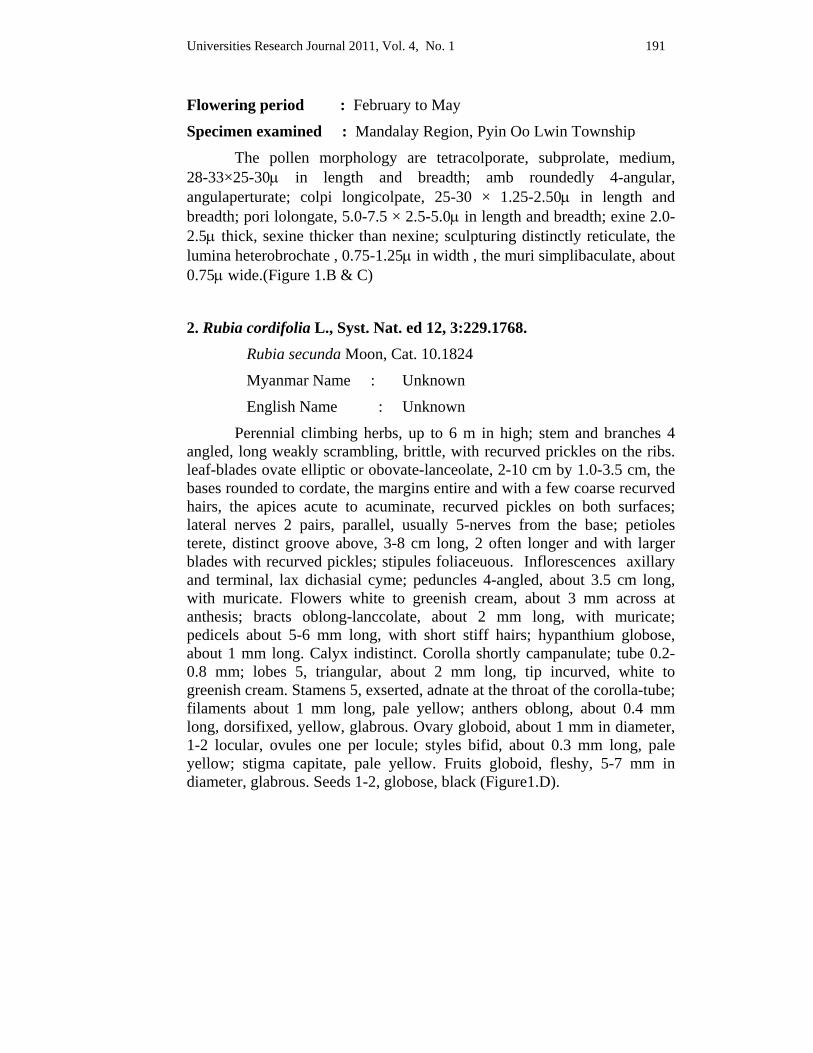
Universities Research Journal 2011, Vol. 4, No. 1 191
Flowering period : February to May
Specimen examined : Mandalay Region, Pyin Oo Lwin Township
The pollen morphology are tetracolporate, subprolate, medium, 28-33×25-30μ in length and breadth; amb roundedly 4-angular, angulaperturate; colpi longicolpate, 25-30 × 1.25-2.50μ in length and breadth; pori lolongate, 5.0-7.5 × 2.5-5.0μ in length and breadth; exine 2.0-2.5μ thick, sexine thicker than nexine; sculpturing distinctly reticulate, the lumina heterobrochate , 0.75-1.25μ in width , the muri simplibaculate, about 0.75μ wide.(Figure 1.B & C)
2. Rubia cordifolia L., Syst. Nat. ed 12, 3:229.1768. Rubia secunda Moon, Cat. 10.1824
Myanmar Name : Unknown
English Name : Unknown
Perennial climbing herbs, up to 6 m in high; stem and branches 4 angled, long weakly scrambling, brittle, with recurved prickles on the ribs. leaf-blades ovate elliptic or obovate-lanceolate, 2-10 cm by 1.0-3.5 cm, the bases rounded to cordate, the margins entire and with a few coarse recurved hairs, the apices acute to acuminate, recurved pickles on both surfaces; lateral nerves 2 pairs, parallel, usually 5-nerves from the base; petioles terete, distinct groove above, 3-8 cm long, 2 often longer and with larger blades with recurved pickles; stipules foliaceuous. Inflorescences axillary and terminal, lax dichasial cyme; peduncles 4-angled, about 3.5 cm long, with muricate. Flowers white to greenish cream, about 3 mm across at anthesis; bracts oblong-lanccolate, about 2 mm long, with muricate; pedicels about 5-6 mm long, with short stiff hairs; hypanthium globose, about 1 mm long. Calyx indistinct. Corolla shortly campanulate; tube 0.2-0.8 mm; lobes 5, triangular, about 2 mm long, tip incurved, white to greenish cream. Stamens 5, exserted, adnate at the throat of the corolla-tube; filaments about 1 mm long, pale yellow; anthers oblong, about 0.4 mm long, dorsifixed, yellow, glabrous. Ovary globoid, about 1 mm in diameter, 1-2 locular, ovules one per locule; styles bifid, about 0.3 mm long, pale yellow; stigma capitate, pale yellow. Fruits globoid, fleshy, 5-7 mm in diameter, glabrous. Seeds 1-2, globose, black (Figure1.D).
192 Universities Research Journal 2011, Vol. 4, No. 1
Flowering period : October to November
Specimen examined : Mandalay Region, Pyin Oo Lwin Township
The pollen morphology are penta-hexacolpate, suboblate, small, 21-25 × 24-29μ in length and breadth; amb circular; colpi longicolpate, 18-22× 3.75-5.00μ in length and breadth; exine 2.0-2.5μ thick, sexine thicker than nexine; sculpturing obscurely reticulate. (Figure1. E & F)
3. Rubia edgeworthii Hook. f., Fl. Brit. Ind. 3 :203. 1853. Myanmar Name : Unknown
English Name : Unknown
Perennial climbing herbs, up to 4 m in high; stem and branches subquandragular, scabrid hispid, yellowish green, pubescent. Leaf-blades lanceolate acuminate, 4.0-5.5 cm by 1.0-1.5 cm, the bases rounded to subcordate, the margins entire and hardly scabrid, the apices acute. Sparsely pubescent above, densely pubescent beneath and on the midrib; lateral nerves 3-4 pairs, parallel, usually 3 nerves from the base; petioles terete, 0.5-2.5 cm long, distinct groove above, pubescent. Inflorescences axillary and terminal, paniculate cyme; peduncles slender, 3-4 cm long, pubescent. Flowers pale yellowish green, small, 2-3 mm across at anthesis; bract linear, about 1 mm long, pubescent; pedicels very short, 1-2 mm long, pubescent. Calyx globose. Corolla shortly campanulate; tube minute; lobes 5, lanceolate acuminate, about 2 mm long, pale yellowish green, glabrous within, pubescent without. Stamens 5, exserted, adnate at the mouth of the corolla-tube; filaments short, up to 1 mm long, white, glabrous; anthers ellipsoid, about 0.4 mm long, yellowish brown, dorsifixed. Ovary globoid, about 2 mm in diameter, bilocular with one ovule in each locule; styles bifid, up to 1 mm long, pale green; stigma capitate. Fruits globoid, fleshy, 3-4 mm in diameter, becoming black at maturity, glabrous. Seeds 2, globose, black (Figure2.A).
Flowering period : July to September
Specimen examined : Southern Shan State, Aungban Township
The pollen morphology are penta-hexacolpate, subprolate, medium, 28-33 × 25-30μ in length and breadth; circular; colpi ¾ way upto the poles,
Universities Research Journal 2011, Vol. 4, No. 1 193
21-26 × 2.5-3.0μ in length and breadth; exine 2.5-3.0μ thick, sexine as thick as nexine; sculpturing obscurely reticulate. (Figure 2.B & C)
4. Rubia sikkimensis Kurz., Journ. As. Soc. Beng. 14. 2: 188. 1874. Myanmar Name : Unknown
English Name : Unknown
Perennial scandent herbs, up to 3 m in high; stem and branches 4-angled, stout retorsely scabrid, yellowish green. Leaf-blades elliptic or ovate-lanceolate acuminate, 3-5 cm by 1.5-2.5 cm, the bases cuneate, the margins entire and scabrid, the apices acuminate, scabrid above, scabrid and pubescent beneath and on the nerves; lateral nerves 3-5 pairs, parallel, usually 3-nerves from the base; petioles sessile. Inflorescences axillary and terminal, paniculate cyme; peduncles 4-angled, about 5 cm long, scabrid. Flowers pale yellow, small, 3-5 mm across at anthesis; bracts elliptic-lanceolate, 3-5 mm long, pubescent on both surfaces, the margins prickly; pedicels 3-5 mm long, scabrid. Calyx globose. Corolla shortly campanulate, tube very short, about 0.5 mm long, glabrous within and without; lobes 5, ovate-acute, 1.0-1.5 mm by 0.8-1.0 mm, pale yellow, glabrous within and without. Stamens 5, exserted, adnate at the mouth of the corolla-tube; filaments very short, about 0.5 mm long, white, glabrous; anthers globose, about 0.5 mm long, yellow, dorsifixed. Ovary ovoid, about 1.5 mm in diameter, bilocular with one ovule in each locule; styles bifid, up to 0.5 mm long, white; stigma capitate. Fruits globoid to ovoid, fleshy, 2-3 mm in diameter, smooth, Seeds 2, black (Figure 2.D).
Flowering period : July to September
Specimen examined : Southern Shan State, Aungban Township
The pollen morphology are penta-hexacolpate, suboblate, small, 13-20 × 17-23μ in length and breadth; amb circular; colpi longicolpate, 12-15× 2.5-3.0μ in length and breadth; exine 2.0-2.5μ thick, sexine as thick as nexine; sculpturing obscurely reticulate. (Figure 2.E & F).
2 A
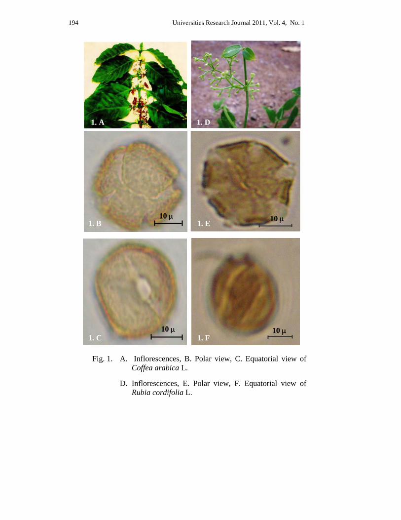
194 Universities Research Journal 2011, Vol. 4, No. 1
Fig. 1. A. Inflorescences, B. Polar view, C. Equatorial view of Coffea arabica L.
D. Inflorescences, E. Polar view, F. Equatorial view of Rubia cordifolia L.
3 A 1. A 1. D
1. B 10 μ
1. E 10 μ
1. C 10 μ
1. F 10 μ
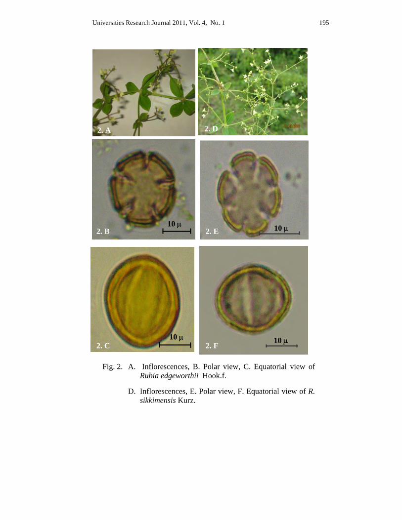
Universities Research Journal 2011, Vol. 4, No. 1 195
Fig. 2. A. Inflorescences, B. Polar view, C. Equatorial view of Rubia edgeworthii Hook.f.
D. Inflorescences, E. Polar view, F. Equatorial view of R. sikkimensis Kurz.
2. A
2. B 10 μ
2. C 10 μ
10 μ
10 μ
2. D
2. E
2. F

196 Universities Research Journal 2011, Vol. 4, No. 1
Discussion and Conclusion It this study, the taxonomic description and pollen morphology of 4
species of Rubiaceae have been described. This family is widely distributed in the tropic and subtropic with some species represented in temperate regions. Most tropical species are trees or shrubs while all temperate ones are herbaceous. The species Coffea arabica L. is shrub and genus Rubia is herbaceous.
Rubiaceae is eurypalynous family, different pollen types are observed. The eurypalynous character of the group is eye catching, monad, tetrad and polyads are observed (Persson 1993). In the studies of 4 economic species of grains were monad. Apertures are distinct regions in the pollen wall through which the pollen tube emerges. Apertures of Rubiaceae is colpate, porate and colporate grains (Erdtman 1952). In the present study the grains were found colporate type in Coffea arabica L. and colpate type in 3 species of Rubia.
The shape in equatorial view, pollen is described prolate, subprolate, oblate, suboblate, spheroidal perprolate and peroblate. It was observed that the shape of the grains are subprolate in 2 species (Coffea arabica L.,
Rubia edgeworthii Hook.f.) and subolate in 2 species (Rubia cordifolia L., R. sikkimensis Kurz.).
Erdtman (1952) recorded that the pollen grains size classes based on the length of the longest spore axis have been described. The grains size less than 10μ are very small, 10-25μ are small, 25-50μ are medium, 50-100μ are large, 100-200μ are very large and above 200μ are gigantic ones. In this study, the grains size varies with the species on measurement of length. They were found small and medium size.
In the present study, the ornamentation of exine sculpture were observed in 2 types, distinctly reticulate were found in Coffea arabica L. and obscurely reticulate in 3 species of Rubia.
Therefore, the aperture, shape, size, sculpture and exine stratification forms useful taxonomical character in systematic study of this family. Hence pollen characters may provide additional identification and classification of flowering plants. It was concluded that the present results will support useful information for the further studies on the pollen morphology of members in other family.
2 A
Universities Research Journal 2011, Vol. 4, No. 1 197
Acknowledgements I would like to express to my gratitude Dr Nu Nu Yee, Professor and Head, Department of Botany, University of Mandalay and Dr Thidar Oo Professor, Department of Botany, University of Mandalay, for providing me with all the necessary facilities. I would like to express my heartfelt sincere gratitude to my supervisor Dr Sai Aung Hsan, Pro-Rector, Panglong University, for suggesting this research topic. I am also very thankful to Dr Aye Aye Htun, Professor and Head, Department of Botany, University of Distance Education, Mandalay, for her suggestion and valuable knowledge and patient teaching regarding pollen morphology.
References Dassanayake, M.D. (1998). A Revised Handbook of Flora of Ceylon, Vol. 12.
Rubiaceae-P-141-343. Univ. of Peradeniya, Dept. of Agriculture, Peradeniya, Srilanka.
Deprete, P.G. (1999). Rondeletieae (Rubiaceae). Part 1– Fl. Neotrop. Monogr.77: 1 - 226. Erdtman, G. (1952). Pollen Morphology and Plant Taxonomy. Angiosperms. An
Introduction to Palynology. I. Almquist & Wiksell, Stockholm. The Chronica Botanica Co. Waltham. Mass., U.S.A.
Erdtman, G. (1964). Handbook of Palynology, Morphology, Taxonomy, Ecology. Hafnar Publishing Co., Inc. New York.
Hoen, P. (1999). Glossary of Pollen and Spore Terminology. Second and revised edition. Utrecht. The Netherlands.
Hooker, J.D. (1879). Flora of British India. Vol III. L. Reeve & Co, 5 Henrietta Street, Covent Garden, London.
Kodela, PG. (2006). Pollen morphology of some rainforest taxa occuring in the illawarra region of New South Wales, Australia.
Kress, J. et al., (2003). A Checklist of the Trees, Shrubs, Herbs and Climbers of Myanmar. Dept. of Systematic Biology. Botany National Museum of Natural History Washington, DC. U.S.A.
Moore, P.D., J.A Webb, & M.E. Collinson. (1991). Pollen Analysis. Second Edition. Oxford. Blackwell Scientific Publication London.
Paldat, (2005). Illustrated Handbook on Pollen Terminology. Univ. of Vienna, Rennweg 14. A 1030 Wien. Austria.
Persson, C. (1993). Pollen morphology of the Gardenieae. (Rubiaceae). Nordic Journal of Botany 13, 561-582.
Walker, J.W., & J.A. Doyle. (1975). The bases of Angiosperm phylogeny. Ann, Missovri Bot. Gard. 62.664 - 723.
Zomlefer, W. B., (1994). Flowering Plant of the World. The Univ. of North Carolina Press. Chapel Hill & London.
Universities Research Journal 2011, Vol. 4, No. 1
Assistant Lecturer, Department of Botany, University of Mandalay
Taxonomic Study on Five Species of the Family Poaceae Khin Moe Moe Khine
Abstract
Grasses are widely distributed plants among the Angiosperms. In the present study, 5 species belonging to 5 genera of the family Poaceae from Mandalay District have been undertaken. The morphological characters of collected species have been thoroughly studied and described with Scientific name, Myanmar name, Flowering period and their uses. An artificial key to the studied species is constructed.
Key words: Grasses, angiosperms, artificial key
Introduction The study area, Mandalay District is located between north latitudes
of 21°10'13" and 22°10'46" and east longitude of 95°59'20" and 96°17'01". It has an area of 351.31 square miles. This area consists of seven townships (Maung Maung 1999).
The grass family, Poaceae is very large and one of the gross morphological complexity (Lawrence 1964). This family contains about 9,000 species distributed among 651 genera (Neil 1994). Poaceae is abundantly found in the tropical and subtropical regions of both hemispheres. Grasses are characterized by the spikelets which are not found in any other family except in Cyperaceae. The spikelet is the unit of inflorescences. It consists of a pair of alternate and distichous bracts at the base, called empty bracts or glumes followed by a floret or florets. The florets are distichously arranged and alternate on a jointed or tough axis, the rhachilla. Each floret is subtended by the two bracts called flowering bracts or lemma (the outer) and palea (the inner). It consists of a perianth represented by two or three minute scales called lodicules, a single whorl of two or three stamens or two alternating whorls of six stamens and a central ovary bearing a pair of lateral or rarely solitary style and usually contain plumose stigmas.
Grain, sugar, spices, paper, perfume and thousands of other items of daily uses are the products of various grasses. The world's population depends on the grain of cultivated grasses (rice, wheat, corn, sorghum, etc.) for their staple food. Grasses are either as forage for grazing animals or as
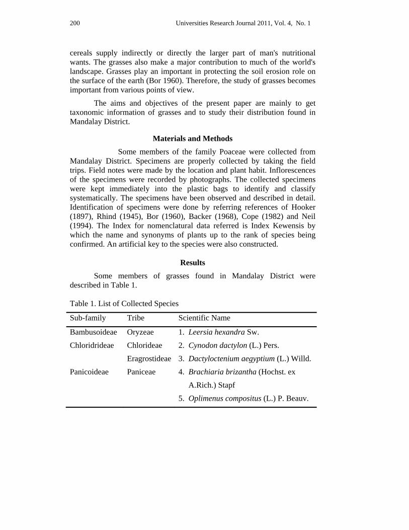
200 Universities Research Journal 2011, Vol. 4, No. 1
cereals supply indirectly or directly the larger part of man's nutritional wants. The grasses also make a major contribution to much of the world's landscape. Grasses play an important in protecting the soil erosion role on the surface of the earth (Bor 1960). Therefore, the study of grasses becomes important from various points of view.
The aims and objectives of the present paper are mainly to get taxonomic information of grasses and to study their distribution found in Mandalay District.
Materials and Methods Some members of the family Poaceae were collected from
Mandalay District. Specimens are properly collected by taking the field trips. Field notes were made by the location and plant habit. Inflorescences of the specimens were recorded by photographs. The collected specimens were kept immediately into the plastic bags to identify and classify systematically. The specimens have been observed and described in detail. Identification of specimens were done by referring references of Hooker (1897), Rhind (1945), Bor (1960), Backer (1968), Cope (1982) and Neil (1994). The Index for nomenclatural data referred is Index Kewensis by which the name and synonyms of plants up to the rank of species being confirmed. An artificial key to the species were also constructed.
Results Some members of grasses found in Mandalay District were
described in Table 1. Table 1. List of Collected Species
Sub-family Tribe Scientific Name
Bambusoideae Oryzeae 1. Leersia hexandra Sw.
Chloridrideae Chlorideae 2. Cynodon dactylon (L.) Pers.
Eragrostideae 3. Dactyloctenium aegyptium (L.) Willd.
Panicoideae Paniceae 4. Brachiaria brizantha (Hochst. ex
A.Rich.) Stapf
5. Oplimenus compositus (L.) P. Beauv.
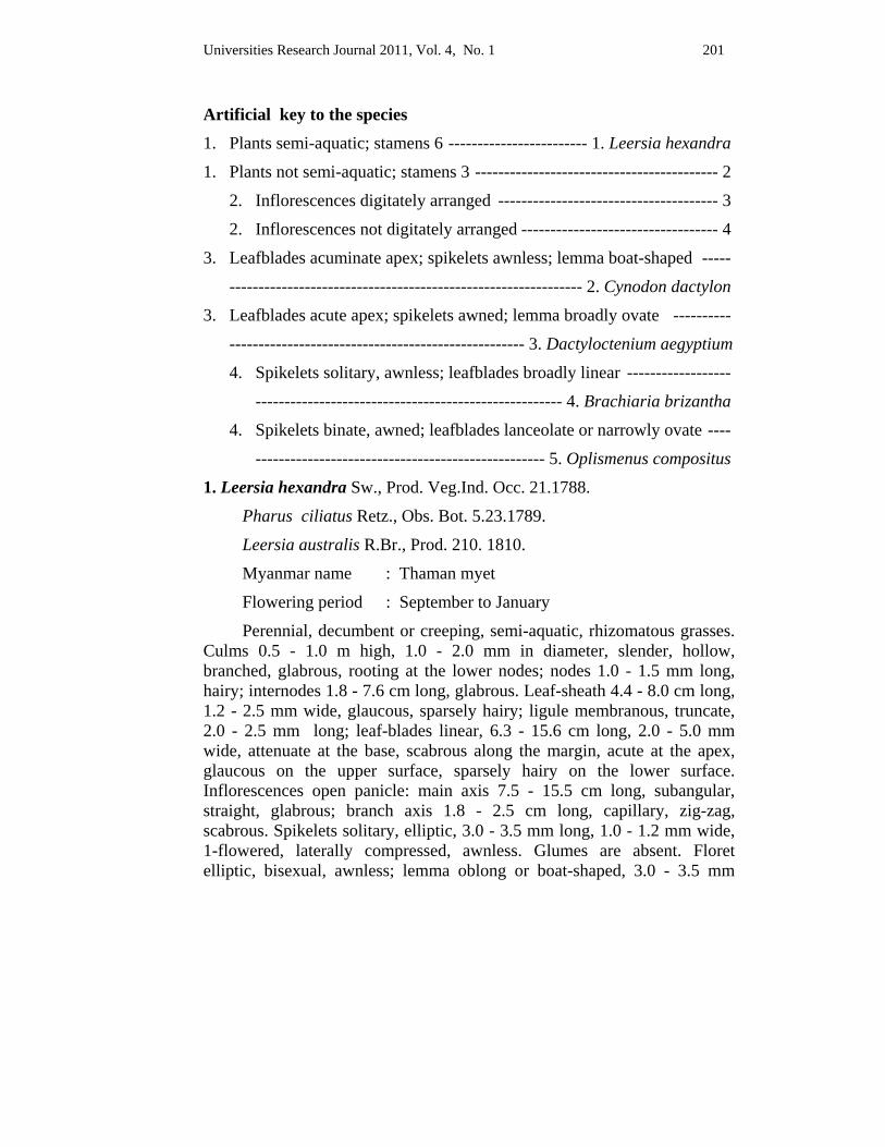
Universities Research Journal 2011, Vol. 4, No. 1 201
Artificial key to the species 1. Plants semi-aquatic; stamens 6 ------------------------ 1. Leersia hexandra
1. Plants not semi-aquatic; stamens 3 ------------------------------------------ 2
2. Inflorescences digitately arranged -------------------------------------- 3
2. Inflorescences not digitately arranged ---------------------------------- 4
3. Leafblades acuminate apex; spikelets awnless; lemma boat-shaped -----
------------------------------------------------------------- 2. Cynodon dactylon
3. Leafblades acute apex; spikelets awned; lemma broadly ovate ----------
--------------------------------------------------- 3. Dactyloctenium aegyptium
4. Spikelets solitary, awnless; leafblades broadly linear ------------------
----------------------------------------------------- 4. Brachiaria brizantha
4. Spikelets binate, awned; leafblades lanceolate or narrowly ovate ----
-------------------------------------------------- 5. Oplismenus compositus
1. Leersia hexandra Sw., Prod. Veg.Ind. Occ. 21.1788.
Pharus ciliatus Retz., Obs. Bot. 5.23.1789.
Leersia australis R.Br., Prod. 210. 1810.
Myanmar name : Thaman myet
Flowering period : September to January
Perennial, decumbent or creeping, semi-aquatic, rhizomatous grasses. Culms 0.5 - 1.0 m high, 1.0 - 2.0 mm in diameter, slender, hollow, branched, glabrous, rooting at the lower nodes; nodes 1.0 - 1.5 mm long, hairy; internodes 1.8 - 7.6 cm long, glabrous. Leaf-sheath 4.4 - 8.0 cm long, 1.2 - 2.5 mm wide, glaucous, sparsely hairy; ligule membranous, truncate, 2.0 - 2.5 mm long; leaf-blades linear, 6.3 - 15.6 cm long, 2.0 - 5.0 mm wide, attenuate at the base, scabrous along the margin, acute at the apex, glaucous on the upper surface, sparsely hairy on the lower surface. Inflorescences open panicle: main axis 7.5 - 15.5 cm long, subangular, straight, glabrous; branch axis 1.8 - 2.5 cm long, capillary, zig-zag, scabrous. Spikelets solitary, elliptic, 3.0 - 3.5 mm long, 1.0 - 1.2 mm wide, 1-flowered, laterally compressed, awnless. Glumes are absent. Floret elliptic, bisexual, awnless; lemma oblong or boat-shaped, 3.0 - 3.5 mm
202 Universities Research Journal 2011, Vol. 4, No. 1
long, 0.7 - 0.8 mm wide, 5-nerved, acute at the apex, hispid ciliate on the mid-nerved, curved upwards, chartaceous yellowish brown or pale pink; palea elliptic, 3.0 - 3.5 mm long, 1.0 - 1.2 mm wide, 3-nerved, hispid ciliate along the margin, acute at the apex, chartaceous, yellowish brown or pale pink. Lodicules 2, very minute, cuneate. Stamens 6; filaments filiform, 0.5 - 1.0 mm long, white; anther dithecous, 1.8 - 2.0 mm long, yellow; ovaries ovoid, 0.2 - 0.3 mm long, pale brown; styles 2, 0.7 - 0.8 mm long, white; stigmas 2, plumose, 1.0 - 1.2 mm long, yellow. Caryopsis ovoid, 0.5 - 1.0 mm long, brown (Figure 1. A).
Specimen examined : Mandalay District, Chanmyathasi Township, Aungpinle; Mahaaungmye Township, Mahamyaing; Patheingyi Township, Yan kin village, Khin Moe Moe Khine, No. 87, September 9, 2006 & December 4, 2007.
Uses : It can be cultivated for hay in wet rice-fields with a very high yield (Backer 1968).
2. Cynodon dactylon (L.) Pers., Syn. Pl. 1: 85. 1805.
Panicum dactylon L., Sp. ll. ed. 1.1: 58. 1753.
Myanmar name : Mye sa myet
Flowering period : Throughout the year
Perennial, erect, stoloniferous sward-forming grasses. Culms 30.0 - 40.0 cm high, 1.0 - 1.8 mm in diameter, slender, branched, glabrous, smooth; nodes 1.0 - 2.0 mm long, glabrous; internodes 1.0 - 4.8 cm long, glabrous. Leaf-sheath 0.9 - 2.2 cm long, 1.2 - 2.0 mm wide, terete, glabrous, bearded at the mouth; ligule a short membrane with ciliate margin, 0.2 - 0.3 mm long; leaf-blades linear-lanceolate, 1.5 - 6.0 cm long, 0.1 - 0.2 cm wide, not attenuate at the base, scabrescent along the margin, acuminate at the apex, sparsely hairy on both surfaces, glaucous. Inflorescences 4- to 6- digitately arranged spikes, all of them borne together; main axis straight, glabrous, terete; each spike 3.0 - 3.7 cm long, the rhachis of spikes continuous and ending in a spikelet. Spikelets narrowly ovate, 2.0 - 3.0 mm long, 1.2 - 1.5 mm wide, strongly laterally compressed, imbricate in 2 rows, subsessile, glabrous; flower bisexual, light green, awnless. Glumes 2, unequal, keeled; lower glume lanceolate, 1.2 - 1.5 mm long, 0.4 - 0.5 mm wide, 1-nerved, hyaline, acute to acuminate at the apex, scabrescent on the nerve, upper glume lanceolate, 1.5 - 2.0 mm long, 0.7 - 0.8 mm wide, 1-nerved, acute to acuminate at the apex, hyaline, scabrescent on the nerve.
Universities Research Journal 2011, Vol. 4, No. 1 203
Floret solitary, ovate or elliptic, 2.5 - 3.0 mm long, 1.0 - 1.5 mm wide, awnless, shortly-pedicellate, bisexual; lemma boat-shaped, 2.0 - 2.5 mm long, 1.0 - 1.5 mm wide, 3-nerved, awnless, membranous, glabrous, keeled, acute at the apex, hairy at the mid-nerve, the lateral nerves close to the margin; palea elliptic-oblong, 2.0 - 2.5 mm long, 0.5 - 0.7 mm wide, 2-nerved, entire along the margin, acute at the apex, membranous, glabrous. Lodicules 2, very minute, ovate-cuneate. Stamens 3, exserted at the anthesis; filaments slender, 1.0 - 1.2 mm long, white; anther dithecous, 0.6 - 1.2 mm long, pale yellowish green; ovaries ellipsoid, 0.5 - 0.7 mm long, pale brown; styles 2, 0.3 - 0.4 mm long, glabrous; stigmas 2, plumose, 0.3 - 0.5 mm long, pale yellowish green, laterally exserted. Caryopsis ellipsoid, 0.8 - 1.0 mm long, laterally compressed, brown (Figure 1. B).
Specimen examined : Throughout the Mandalay District area, Khin Moe Moe Khine, No. 59, April 12, 2005 & December 25, 2006.
Uses : It is grazed by ruminants, to control erosion and as a turf grass (Mannetje et al. 1992). Fresh juice is memulcent, astringent and diuretic. The plant is hemostatic and laxative (Kapoor 2001).
3. Dactyloctenium aegyptium (L.) Willd., Enum. Hort. Berol. 1029. 1809.
Cynosurus aegyptius L. Sp. Pl. ed. 1.1: 72. 1753.
Eleusine aegyptia (L.) Desf., Fl. At Lant. 1:85. 1789.
Myanmar name : Myet le gwa; Dedok chi
Flowering period : Throughout the year
Annual, erect, stoloniferous robust grasses. Culms 18.0 - 46.0 cm high, 1.0 - 2.5 mm in diameter, slender, solid, branched, glabrous; nodes 1.0 - 2.5 mm long, glabrous; internodes 2.5 - 8.0 cm long, glabrous. Leaf-sheath 2.0 - 5.0 cm long, 2.5 - 4.0 mm wide, slightly compressed, glabrous; ligule membranous ring, 1.0 - 2.0 mm long; leaf-blades linear, 2.5 - 8.0 cm long, 2.0 - 7.0 mm wide, subrounded at the base, ciliate along the margin, acute at the apex, pubescent on the lower surface, glabrous on the upper surface. Inflorescences 2- to 6- digitately arranged spikes, all of them borne together, main axis straight, slightly compressed; each branch axis 1.2 - 3.5 cm long, continuous, ending in a spikelet. Spikelets ovate, 3- to 4- flowered, 2.8 - 3.0 mm long, 1.0 - 2.0 mm wide, awned, laterally compressed, sessile, biseriate on the midrib of rhachis of a spike; rhachilla disarticulating above the glume and between the floret. Glumes 2, unequal,
204 Universities Research Journal 2011, Vol. 4, No. 1
deciduous, strongly keeled; lower glume ovate-oblong, 2.0 - 2.2 mm long, 1.0 - 1.2 mm wide, 1-nerved, inflexed along the margin, acute at the apex, membranous, scabrescent on the nerves; upper glume elliptic, 2.0 - 2.5 mm long, 1.0 - 1.2 mm wide, 1-nerved, inflexed along the margin, hyaline, scabrescent on the nerve, awned; awns 0.5 - 1.0 mm long, recurved. Florets solitary, sessile, bisexual, awnless; lemma broadly ovate, 2.8 - 3.0 mm long, 1.0 - 1.2 mm wide, 3-nerved, inflexed along the margin, mucronate at the apex, membranous, scabrescent on the nerve, with faint submarginal lateral nerves; palea elliptic, 2.9 - 3.2 mm long, 1.0 - 1.2 mm wide, 2-nerved, acute at the apex, hyaline, scabrescent on the nerve. Lodicules 2, very minute, obconical. Stamens 3; filaments slender, 0.2 - 0.4 mm long; white; anther dithecous, 0.2 - 0.3 mm long, yellow; ovaries ovate-oblong, 0.4 - 0.5 mm long; styles 2, 0.4 - 0.6 mm long, white; stigma 2, plumose, 1.0 - 1.2 mm long, white. Caryopsis ovate-oblong, about 1.0 mm long, coarsely transversely rugose, brownish yellow (Figure 1. C).
Specimen examined : Mandalay District, Khin Moe Moe Khine, No.8, May, 17, 2005 & November 25, 2006.
Uses : This grass is widely used as forage and is relished by all types of ruminants. Although a valuable forage, it can also be a troublesome weed of cultivation. It makes excellent hay. It times of scarcity it is used as a food grain (Mannetje et al. 1992).
4. Brachiaria brizantha (Hochst. ex A. Rich.) Stapf in Prain, Fl. Trop. Afr.
9:531.1919.
Panicum brizanthum Hochst. ex A.Rich. Tent. Fl. Abyas.
2: 363.1851.
Myanmar name : Unknown
Flowering period : October to January
Perennial, erect or prostrate, rhizomatous and stoloniferous tufted grasses. Culms 30.0 - 80.0 cm high, 1.0 - 3.5 mm in diameter, terete, solid, branched, glabrous, rooting at the lower nodes; nodes 0.5 - 1.5 mm long, glabrous; internodes 1.0 - 10.5 cm long, glabrous. Leaf-sheath 4.0 - 9.7 cm long, 1.5 - 4.0 mm wide, glabrous; ligule membranous, 1.0 - 1.5 mm long, leaf-blades broadly linear, 10.3 - 28.0 cm long, 0.5 - 1.3 cm wide, rounded at the base, scabrous along the margin, acuminate at the apex, sparsely hairy on both surfaces. Inflorescences 3- to 7- spike-like secund racemes; main
Universities Research Journal 2011, Vol. 4, No. 1 205
axis 6.7 - 10.5 cm long, subangular, scabrescent; branch axis 4.3 - 9.5 cm long, narrowly winged, scabrous. Spikelets solitary, elliptic, 3.0 - 4.5 mm long, 1.0 - 1.5 mm wide, 2-flowered, pubescent, awnless, subsessile. Glumes 2, unequal; lower glume broadly obovate, 1.5 - 2.0 mm long, 0.8 - 1.0 mm wide, 3-nerved, inflexed along the margin, obtuse at the apex, membranous, pale yellow; upper glume elliptic, 3.0 - 4.5 mm long, 1.0 - 1.5 mm wide, 5- nerved, inflexed along the margin, acute at the apex, membranous, pale yellowish brown, sparsely hairy. Lower floret elliptic-oblong, neuter, awnless; lower lemma elliptic, 3.0 - 4.5 mm long, 1.0 - 1.5 mm wide, 5-nerved, inflexed along the margin, acute at the apex, membranous, pale yellow, glabrous; lower palea oblong, 1.2 - 1.5 mm long, 0.8 - 1.0 mm wide, nerveless, entire along the margin, obtuse at the apex, membranous, creamy. Upper floret ovate-oblong, bisexual, awnless; upper lemma ovate-oblong, 2.5 - 3.0 mm long, 1.0 - 1.2 mm wide, indistinct nerves, coriaceous, pale yellow, granulose, glabrous; upper palea ovate-oblong, 1.5 - 2.0 mm long, 0.8 - 1.0 mm wide, nerveless, inflexed along the margin, obtuse at the apex, coriaceous, creamy, glabrous. Lodicules 2, very minute, cuneate. Stamens 3; filaments filiform, 0.5-1.0 mm long, white; anther dithecous, 0.7-1.2 mm long, yellow; ovaries ovoid, 0.3-0.5 mm long, pale yellow; styles 2, 0.5 - 0.7 mm long, white; stigmas 2, plumose, 0.5 - 1.0 mm long, yellowish brown. Caryopsis ovoid, 1.0 - 1.2 mm long, yellow (Figure 1. D).
Specimen examined : Mandalay District, Patheingyi Township, Kyi gone, Khin Moe Moe Khine, No. 127, November 25, 2006 & December 7, 2007.
Uses : The main use of this grass is as forage in permanent pastures for grazing or for cut and carry systems. It has also proved useful as grazed ground cover in tree plantation (Mannetje et al. 1992). It is excellent soil binder (Bor 1960).
5. Oplismenus compositus (L.) P.Beauv., Ess. Agrost. 54.168.169. 1812.
Panicum compositum L., Sp. Pl. 1:57.1753.
Myanmar name : Myet let the
Flowering period : September to December
206 Universities Research Journal 2011, Vol. 4, No. 1
Perennial, rambling and creeping, stoloniferous grasses. Culms 20.0 - 50.0 cm high, 1.5 - 2.5 mm in diameter, slender, slightly compressed, solid, glabrous, rooting at the lower nodes; nodes 0.5 - 1.5 mm long, glabrous; internodes 0.7 - 4.9 cm long, glabrous leaf-sheath 2.2 - 7.4 cm long, 1.0 - 2.5 mm wide, sparsely hairy; ligule membranous with fringed hairs, 0.5 - 1.0 mm long; leaf-blades lanceolate or narrowly ovate, 1.4 - 10.6 cm long, 0.3 - 0.9 cm wide, oblique at the base, scabrescent along the margin, acuminate at the apex, sparsely hairy on both surfaces. Inflorescences 3- to 7- spike-like racemes; main axis 5.8 - 18.5 cm long, subterete, scabrous; branch axis 1.8 - 5.6 cm long, angular, narrowly winged, hairy. Spikelets binate, lanceolate or ovate-oblong, secund, 2.5 - 3.0 mm long, 1.0 - 1.2 mm wide, 2-flowered, glabrous, awned, dorsally compressed, pedicellate; pedicels unequal, 0.3 - 0.5 mm long, hairy. Glumes 2, unequal; lower glume ovate, 2.0 - 2.5 mm long, 0.7 - 1.0 mm wide, 3-nerved, inflexed along the margin, awned at the apex, hairy, membranous, awns 0.5 - 0.6 cm long, smooth, pale green; upper glume ovate, 2.5 - 3.0 mm long, 0.8 - 1.0 mm wide, 3- to 5-nerved, inflexed along the margin, acute at the apex, sparsely hairy, membranous. Lower floret elliptic-lanceolate, neuter, awnless; lower lemma ovate, 2.0 - 2.5 mm long, 1.0 - 1.2 mm wide, 5- to 7- nerved, hairy along the margin, acute at the apex, sparsely hairy, membranous; lower palea narrowly lanceolate or linear-oblong, 2.0 - 2.3 mm long, 0.2 - 0.5 mm wide, nerveless, slightly inflexed along the margin, acute and hairy at the apex, membranous. Upper floret ovate-oblong, bisexual, awnless; upper lemma ovate-oblong, 2.3 - 2.5 mm long, 0.9 - 1.0 mm wide, nerveless, inflexed along the margin, acute at the apex, glabrous, coriaceous, pale green; upper palea ovate-oblong, 2.0 - 2.3 mm long, 0.8 - 1.0 mm wide, nerveless, inflexed along the margin, acute at the apex, glabrous, pale green. Lodicules 2, very minute, obconical. Stamens 3; filaments filiform, 0.5 - 0.7 mm long, white; anther dithecous, 1.5 - 1.7 mm long, yellowish-brown; ovaries ellipsoid, 0.5 - 0.7 mm long, pale green; styles 2, flattened at the base and pointed at the apex, 0.8-1.0 mm long, white; stigmas 2, plumose, 1.2 - 1.3 mm long, yellowish brown. Caryopsis ellipsoid, 1.0 - 1.5 mm long, brown (Figure 1. E).
Specimen examined : Mandalay District, Patheingyi Township, Mya kha nauk Hill & Ye da khun Hill, Khin Moe Moe Khine, No. 57. November, 26, 2005 & December 5, 2006.
Uses : It is used for epizoic dispersal and adhere to passing animals, man and vehicles (Backer 1968).

Universities Research Journal 2011, Vol. 4, No. 1 207
Discussion and Conclusion The present study deals with some species of the family Poaceae growing in Mandalay District. Totally 5 species belonging to 5 genera have been identified, classified and described. An artificial key to the species have been constructed based on characters of the studied species. One species in Oryzeae, one species in Chlorideae, one species in Eragrostideae and two species in Paniceae were recorded.
According to the resulting data, species of Cynodon dactylon (L.) Pers. and Dactyloctenium aegyptium (L.) Willd. are abundantly found in the study area. Leersia hexandra Sw., Brachiaria brizantha (Hochest. ex A. Rich.) Stapf and Oplimenus compositus (L.) P. Beauv. are slightly found in this area.
Most of the species are used as fodder but some species of Cynodon dactylon (L.) Pers. is used as a medicinal plant. Digitately arranged inflorescences can be seen in two species and the three species are not digitately arranged inflorescences. Awned spikelets are found in Dactyloctenium aegyptium (L.) Willd. and Oplismenus compositus (L.) P. Beauv. and other three species are awnless spikelets. Six stamens are found in one species and three stamens are found in four species. Hairy nodes can be seen in Leersia hexandra while the glabrous in Cynodon dactylon, Dactyloctenium aegyptium, Brachiaria brizantha and Oplismenus compositus.
It is sincerely hoped that the present paper of grass resources from Mandalay District can stand up valuable information for the further investigation of researchers who are facing with some difficulties to know about the species and anyone who are looking for the diversity of the members of the genus. This study will partially fulfill the requirement of grasses information of the Mandalay District.
Acknowledgements I am grateful to Dr Nu Nu Yee, Professor and Head, Department of Botany, University of Mandalay, for her permission to carry out this paper and for providing the necessary facilities. I am very thankful to Dr Thida Oo, Professor, Department of Botany, University of Mandalay, for her permission continued, encouragement and valuable advice. I am very grateful to my supervisor, Dr Soe Myint Aye, Associate Professor, Department of Botany, Myitkyina University, for his invaluable suggestions.
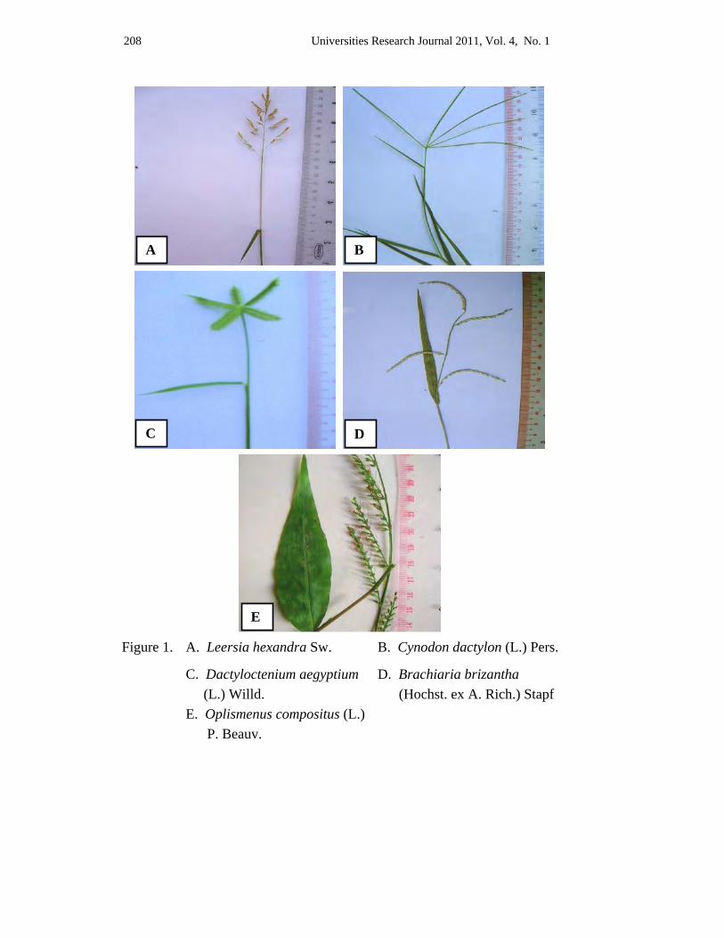
208 Universities Research Journal 2011, Vol. 4, No. 1
Figure 1. A. Leersia hexandra Sw. B. Cynodon dactylon (L.) Pers.
C. Dactyloctenium aegyptium D. Brachiaria brizantha (L.) Willd. (Hochst. ex A. Rich.) Stapf E. Oplismenus compositus (L.) P. Beauv.
A B
C D
E
Universities Research Journal 2011, Vol. 4, No. 1 209
References Backer, C.A. & R.C. Bakhuizen Van Den Brink, (1968). Flora of Java, Vol. III. Rijksher
barium, leyden, N.V.P. Noordhoff.
Bor, N.L. (1960). The Grasses of Burma, Ceylon, India and Pakistan. Pergamon, Oxford, London, New York & Paris.
Cope, Thomas. (1982). Flora of Pakistan No. 143. (Poaceae)
Hooker, J.D. (1897). The Flora of British India, Vol. VII. L. Reeve & Co-5, Henrietta Street, Covent Garten, London.
Jackson, B.D. et al. (1895). Index Kewensis Vol. 1 & 2 and Supply. 1-13. An enumaration of the genera and species of Flowering plants. Clarendon Press. London Oxford.
Kapoor, L.D. (2001). Handbook of Ayurvedic Medicinal Plants. India.
Kress, J.W. Robert A. DeFilipps, Ellen Farr and Daw Yin Yin Kyi (2003). A Checklist of the Trees, shrubs, Herbs & Climbers of Myanmar. Department of Systematic Biology-Botany. National Museum of Natural History, Washington DC. USA.
Lawrence, G.H.M. (1969). Taxonomy of Vascular Plants. The Macmillan Co. New York.
Mannetje, L.'t and Jones R.M. (1992). Plant Resources of South-East Asia. No.4. Forages. Bogor. Indonesia.
Maung Maung, (1999). Geomorphology of Mandalay District. M.Res. University of Mandalay.
Neil, A. Harriman in Dassanayake M.D; (1994). A Revised Handbook to the Flora of Ceylon Vol. VIII (Poaceae). Amerind Publishing Co. Pvt. Ltd., New Delhi.
Rhind, D.I.A.S. (1945). The Grasses of Burma, Published under the authority of the Government of Burma.
Universities Research Journal 2011, Vol. 4, No. 1
Assistant Lecturer, Department of Botany, University of Mandalay
Pollen Morphology of Four Species in Family Euphorbiaceae Thi Thi Htun
Abstract The taxonomic descriptions and pollen morphology of 4 species belonging to 4 genera of family Euphorbiaceae were described in the present paper. The studied materials were collected throughout Mandalay Region. The plants were identified and their pollen has been examined under the microscope. In the present study, 4 species have been described together with the taxonomic descriptions and colour photograph of inflorescence. Pollen morphology and pollen keys to the species were also constructed. In Ricinus communis, the outline of a pollen grain seen in polar view (amb) is rounded triangular and in equatorial view it was observed as suboblate. The remaining 3 species are spheroidal. The exine sculptures of 4 species were Pilate, distinctly reticulate and croton pattern. The numbers of apertures possessed by the pollen grains of 4 species were tricolporate, polyporate and inaperturate. The studied pollen grains are observed in 2 sizes, medium (25-50µ) and large (above 50µ).
Key words: Ricinus communis, Euphorbiaceae,
Introduction
The Euphorbiaceae is a large family of flowering plants, including some 300 genera and over 5,000 species of dicotyledonous herbs, shrubs and trees (Heywood 1978). The family is taxonomically characterized by the presence of watery juice, latex or milky sap, the unisexual flowers, the superior ovary and trilocular with axial placentaion, the collateral and pendulous ovules. According to the Heywood (2007), the Euphorbiaceae is a large, diverse family with unisexual flowers, superior syncarpous ovaries, and 1 ovule per locule.
The family contains many species of both economic and decorative importance species. Manihot esculenta having thick tuberous roots rich in starch, and which yield cassava or tapioca is an important food plant of the tropics. Castor oil is obtained from the oily endospermic seeds of Ricinus communis. In Myanmar, Biodiesel is produced from seeds of Jatropha curcas and it is popular nowadays. It is also used as powerful purgative. The bark, roots and leaves of Croton persimilis are used in reducing chronic enlargement of liver and they are applied externally to sprains, bruises, and rheumatic swellings.
212 Universities Research Journal 2011, Vol. 4, No. 1
Pollen is one plant material best resistant to various kinds of treatment. It remains unchanged in structure and sculpture for millions of year, when fossilized in the soil. Pollen mother cells (microsporocytes) are differentiated from sporogenous cells in the young male organ. These cells are poorly attached to each other by plasma connections.
During growth, the pollen mother cells become spherical and then separate from each other; these cells undergo meiosis to produce tetrads, which in the haploid phase are called microspores. The members of the tetrad become separated from one another by a callose wall which is continuous with that such surrounding the entire tetrad. The surface features of the mature grain are clearly related to the original orientation of the microspore within the tetrad. Within each microspore, peripheral elements of the endoplasmic reticulum are fixed which polymerize sporopollenin to form the sexine matrix on its surface – when this happens, the callose disappears. Later, the sexine thickens without changing its structure. Each microspore in turn differentiates into a pollen grain (Iwanami et al. 1988).
Pollen grains represent the gametophytic generation. A large number of genes, some unique to the pollen and others common to the sporophytic generation are expressed during its development as well as during the post germination phase. Studies on the expression of these genes are important for the application of pollen selection, for recombinant DNA technology, and for the induction of pollen embryos. In addition to these areas of pollen biology, which have direct relevance to its function, other areas that are equally important include pollen in relation to taxonomy and phylogeny (Erdtman 1966), fossil palynology (Faegri & Iversen 1989), aeropalynology and pollen allergy (Stanley & Linskens 1974, Knox 1979, Singh et al. 1991, Mohapatra & Knox 1996), and the use of pollen to analyze the effects of ecotoxin chemicals (Kappler & Kristen 1987, Wolters & Martens 1987, Strube et al. 1991 & Pfahler 1992).
These all information and knowledge push to study the taxonomy and pollen morphology of family Euphorbiaceae. This research work is going to be carried out to identify, describe and record the taxonomic characters and pollen morphology of 4 economic importance species of family Euphorbiaceae.
Universities Research Journal 2011, Vol. 4, No. 1 213
Materials and Methods The plants are collected from Mandalay Region. The collected plants are photographed, identified and pressed for further studies. Plant identification was followed to Hooker (1885), Backer (1965), Dassanayake (1997).For pollen study the collected flowers were immediately fixed in glass vial containing glacial acetic acid. Each of the specimens was labeled and stored in a dry and cool place. Pollen samples were acetolysed according to Erdtman (1960). A prepared slide was examined to ascertain the shape, size, aperture and exine sculpture of the pollen. Photographs were taken with Sony digital camera directly from the eyepiece of microscope. Measurements were based on 20 grains per sample; values of polar axis length (P) and equatorial diameter (E) were measured and recorded. Pollen aperture was observed directly. Terminology used followed to that of Erdtman (1952 & 1969), Hoen (1999) and Paldat (2005).
Results The taxonomic descriptions and pollen morphology of 4 species
belonging to 4 genera of the Euphorbiaeae have been described.
Pollen key to the species 1. Inaperturate………………………………….. 2
1. Aperturate……………………………………. 3
2. Pilate………………………………… Jatropha curcas (2) 2. Croton pattern……………………… Croton persimilis (1)
3. Distinctly reticulate,grains less than 50μ in
diameter……………………………………… Ricinus communis (4)
3. Croton pattern, grains more than100μ in
diameter……………………………………… Manihot esculenta (3)
214 Universities Research Journal 2011, Vol. 4, No. 1
Taxonomic description and pollen morphology 1. Croton persimilis Muell. Arg. emend. philcox (hic); Muell. Arg. Linnaea
34: 116. 1865.(Figure 1.A,B,C)
Myanmar name : Thet Yin Gyi
English name : Unknown
Flowering period : December to March
Perennial, deciduous and monoecious tree; up to 9 m high; young branches terete, densely lepidote, with distinct leaf-scars. Leaves simple, alternate, stipulates, petiolate; stipules caducous; petioles 3 - 7 cm long, terete, lepidote; blades elliptic-oblong, ovate-lanceolate or elliptic, 7.5 - 26.5 cm by 6.0 - 11.5 cm, lepidote on both surfaces when young, glabrous in age, with two yellowish glands on either side of the leaf base, penninerved with 9 to 16 pairs of lateral veins, obtuse at the base, serrate along the margin, acute at the apex. Inflorescences axillary or terminal racemes, elongate, up to 25.0 cm long, androgynous, the staminate flowers at the upper portion and the pistillate flowers at the lower portion. Staminate flowers 1 cm across at anthesis; bracts deciduous; pedicels about 5 mm long, lepidote. Sepals 5, ovate, greenish yellow, glabrous on both surfaces, valvate in bud. Petals 5, obovate, greenish yellow, glabrous without, pubescent within, valvate in bud. Disk glands 5, orange, alternate with the petals; stamens 10 to 12, exerted on the hairy receptacles; filaments 4 mm long, filiform, yellowish green, glabrous; anthers 1.5 mm long and wide, dithecous, introrse, yellow, basifixed, longitudinally dehiscent. Pistillate flowers 5.5 - 7.0 mm across at anthesis; bracts ovate, caducous; pedicels about 1.5 mm long, lepidote, persistent. Sepals 5, ovate, pale green, lepidote without, pubescent within, valvate in bud. Petals 5, greenish yellow, valvate in bud. Disk glands 5, depressed, yellow. Ovary superior, globoid, about 2 mm long and wide, lepidote, trilocular with one ovule in each locule on the axile placenta; styles 3, basally connate; stigmas 3, each bifid, glabrous. Capsules globoid, distinctly 3-lobed, lepidote, with persistent sepals, dehiscent into three 2-valved cocci, each coccus 1-seeded. Seeds oblongoid, whitish brown, smooth with small caruncle.
Pollen morphology of this species is inaperturate, spheroidal, large, 55-67μ in diameter; exine 5-6μ thick; sexine 3-4μ thick, sexine thicker than nexine; sculpturing croton pattern, the lower part of the muroid ridges separated by rounded foveoloid areas, the upper part of well defined regular
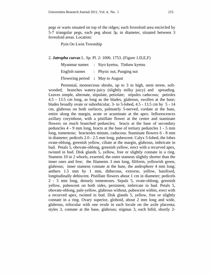
Universities Research Journal 2011, Vol. 4, No. 1 215
pegs or warts situated on top of the ridges; each foveoloid area encircled by 5-7 triangular pegs, each peg about 3μ in diameter, situated between 3 foveoloid areas. Location:
Pyin Oo Lwin Township
2. Jatropha curcas L. Sp. Pl. 2: 1006. 1753. (Figure 1.D,E,F)
Myanmar names : Siyo kyetsu, Tinbaw kyetsu
English names : Physic nut, Punging nut
Flowering period : May to August
Perennial, monoecious shrubs, up to 3 m high, stem terete, soft-wooded; branches watery-juicy (slightly milky juicy) and spreading. Leaves simple, alternate, stipulate, petiolate; stipules caducous; petioles 4.5 - 13.5 cm long, as long as the blades, glabrous, swollen at the base; blades broadly ovate or suborbicular, 3- to 5-lobed, 4.5 - 13.5 cm by 5 - 14 cm, glabrous on both surfaces, palmately 5-nerved, cordate at the base, entire along the margin, acute or acuminate at the apex. Inflorescences axillary corymbose, with a pistillate flower at the centre and staminate flowers on much branched peduncles; bracts at the base of secondary peduncles 4 - 9 mm long, bracts at the base of tertiary peduncles 1 - 5 mm long, tomentose; bracteoles minute, caducous. Staminate flowers 6 - 8 mm in diameter; pedicels 2.0 - 2.5 mm long, pubescent. Calyx 5-lobed, the lobes ovate-oblong, greenish yellow, ciliate at the margin, glabrous, imbricate in bud. Petals 5, obovate-oblong, greenish yellow, erect with a recurved apex, twisted in bud. Disk glands 5, yellow, free or slightly connate in a ring. Stamens 10 in 2 whorls, exserted, the outer stamens slightly shorter than the inner ones and free; the filaments 3 mm long, filiform, yellowish green, glabrous; inner stamens connate at the base, the androphore 4 mm long; anthers 1.5 mm by 1 mm, dithecous, extrorse, yellow, basifixed, longitudinally dehiscent. Pistillate flowers about 1 cm in diameter; pedicels 2 - 5 mm long, densely tomentoses. Sepals 5, ovate-oblong, greenish yellow, pubescent on both sides, persistent, imbricate in bud. Petals 5, obovate-oblong, pale-yellow, glabrous without, pubescent within, erect with a recurved apex, twisted in bud. Disk glands 5, yellow, free or slightly connate in a ring. Ovary superior, globoid, about 2 mm long and wide, glabrous, trilocular with one ovule in each locule on the axile placenta; styles 3, connate at the base, glabrous; stigmas 3, each bifid, shortly 2-
216 Universities Research Journal 2011, Vol. 4, No. 1
lobed, sagittate. Capsules ovoid or subgloboid, glabrous, slightly 3-lobed, with persistent calyx, dehsicent into three 2-valved cocci, each coccus 1-seeded. Seeds ovoid-oblongoid, black, glabrous, carunculate.
Pollen morphology of this species is inapertuate, spheroidal, large, 53-67μ in diameter; exine 5-6μ thick; sexine 3-4μ thick, sexine thicker than nexine; sculpturing pilate; the pila 3.0-4.5μ in diameter, about 4.5μ in length, pila head 3.0-4.5μ in diameter.
Location: Mahar aung myae Township
3. Manihot esculenta Cantz. Inst 1: 167. 1766. .(Figure 2.A,B,C)
Myanmar name : Palaw pinan
English names : Cassava, Tapioca
Flowering period : October to March
Perennial, monoecious shrubs, up to 5 m high, with latex; branches erect or spreading, glabrous, with leaves towards the upper portions and with prominent leaf-scars at the lower portions. Leaves palmately compound, alternate, stipulate, petiolate; stipules 2- to 3-lobed, deciduous; petioles 4.0 - 11.5 cm long, usually longer than the blades, glabrous; blades palmately divided; lobes 3 to 5, obovate-lanceolate, 4.0 - 10.5 cm by 1.0 - 2.5 cm, glabrous on both surfaces, palminerved with 3 to 5 costae, rounded at the base, entire along the margin, acuminate at the apex. Inflorescences axillary racemes, 1 to 5 female flowers at the basal portions with long pedicelled and male flowers at the upper portions with short pedicelled. Staminate flowers 1.2 - 1.6 cm in diameter; bracts 2 mm long, deciduous; pedicels 0.5 - 1.0 cm long, glabrous. Calyx 5-partite, campanulate, the lobes triangular, yellowish red, imbricate in bud. Disk glands with 5 double lobes, orange, nectariferous. Stamens 10 in 2 whorls of 5 each, alternately short and long, inserted; filaments 4 - 7 mm long, filiform, white, free, glabrous; anthers 2 mm by 1 mm, dithecous, oblongoid, yellow, basifixed, longitudinally dehiscent. Pistillate flowers about 3 cm in diameter;bracts 3 mm long, deciduous; pedicels 1 - 2 cm long, glabrous, longer in fruit. Calyx deeply 5-partite, the lobes lanceolate, yellowish red, imbricate in bud. Disk glands 10-lobed, orange. Ovary superior, subgloboid, 3 mm in diameter, glabrous, with 6 red ridges, trilocular with one ovule in each locule on the axile placenta; styles 3,
Universities Research Journal 2011, Vol. 4, No. 1 217
connate at the base, glabrous, surmounted by 3-lobed stigmas. Capsules globoid, glabrous with 6 narrow longitudinal wings, dehiscent into three 2-valved cocci, each coccus 1-seeded. Seeds ellipsoid, grey with dark blotches,
Pollen morphology of this species is polyporate, pantoporate, spheroidal, large, about 130-160μ in diameter; pori circular, 50-20μ in diameter; exine about 8μ thick; sexine about 5μ thick, sexine thicker than nexine; sculpturing croton pattern, 5-7 triangular pegs, each peg about 7μ in diameter.
Location: Patheingyi Township
4. Ricinus communis L. Sp. Pl. 1007. 1753. (Figure 2.D,E,F)
Myanmar names : Kyet su
English name : Castor oil plant
Flowering period : Almost throughout the year
Perennial, monoecious, tree like shrubs, up to 4.5 m high; stems and branchlets erect, hollow, with prominent leaf-scars. Leaves simple, alternate, stipulate, petiolate; stipules caducous; petioles 9.5 – 25.0 cm long, hollow, glabrous; blades peltate, palmately lobed or partite, 8 - 19 cm by 5 - 14 cm, glabrous on both surfaces, palminerved with 5 to 8 costae; lobes 5 to 8, lanceolate, glandular-serrate along the margin, acuminate at the apex. Inflorescences terminal or leaf-opposed racemes, pistillate flowers at the terminal portions and staminate flowers at the lower portions. Staminate flowers 1.3 - 1.7 cm in diameter, apetalous; bracts 3 mm by 2 mm, ovate, glabrous, caducous; pedicels 5 - 10 mm long, glabrous. Calyx deeply 5-partite, the lobes 6 - 10 mm by 3 - 5 mm, ovate-lanceolate, greenish yellow, glabrous, valvate in bud. Stamens numerous, exserted, monadelphous, in a dense fascicle; filaments filiform, 0.5 - 1.0 mm long, unequal in length, white, glabrous; anthers 0.5 mm long and wide, dithecous, yellow, basifixed, longitudinally dehiscent. Pistillate flowers 1.0 - 1.5 cm in diameter at anthesis, apetalous; bracts minute, caducous; pedicels 1.5 - 2.0 mm long, glabrous. Calyx deeply 5-partite, the lobes lenceolate,
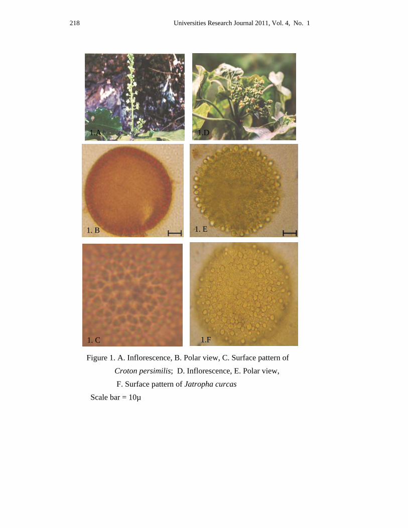
218 Universities Research Journal 2011, Vol. 4, No. 1
Figure 1. A. Inflorescence, B. Polar view, C. Surface pattern of
Croton persimilis; D. Inflorescence, E. Polar view,
F. Surface pattern of Jatropha curcas
Scale bar = 10µ
1. C 1.F
1. B 1. E
1.A 1.D
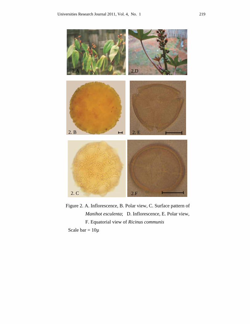
Universities Research Journal 2011, Vol. 4, No. 1 219
2.D2.A
2. E2. B
Figure 2. A. Inflorescence, B. Polar view, C. Surface pattern of
Manihot esculenta; D. Inflorescence, E. Polar view,
F. Equatorial view of Ricinus communis
Scale bar = 10µ
2. C 2.F
220 Universities Research Journal 2011, Vol. 4, No. 1
greenish yellow, glabrous, valvate in bud, caducous. Ovary superior, globoid, 2.0 - 3.5 mm in diameter, covered with fleshy soft green spines, trilocular with one ovule in each locule on the axile placenta; styles 3, connate at the base; stigmas 3, each bifid, densely papillose inside. Capsules globoid or subgloboid, 3-lobed, usually spiny, dehiscent into three 2-valved cocci, each coccus 1-seeded. Seeds ovoid, pale brown and mottled, with a large caruncle.
Pollen morphology of this species are tricolporate, suboblate, medium, 21-25 x 25-31μ in length and breadth; amb rounded triangular; colpi longicolpate, about 20 x 2μ in length and breadth; pori lalongate, about 4 x 13μ in length and breadth; exine about 1.5μ thick; sexine about 0.7μ thick, sexine as thick as nexine; sculpturing obscurely reticulate.
Location: Pyin Oo Lwin Township
Discussion and Conclusion In the present study, taxonomy and pollen morphology of 4 species belonging to 4 genera of the Euphorbiaceae were examined.
In taxonomic study, the flowers of all members are very small. All species are monoecious and perennial. Croton persimlis is characterized by the presence of two yellowish glands on either side of the leafbase, and axillary or terminal raceme with the staminate flowers at the upper portions and the pistillate flowers at the lower portions.The distinctive characters of Jatropha curcas are swollen bases of petioles, corymbose cymes with a solitary pistillate flower at the centre and staminate flowers on much- branched peduncles. Milky latex are present in all parts of Manihot esculenta. The outstanding characters of this species are prominent leaf-scars on the older branches, 6-ridged ovary, three styles surmounted by 3-lobed stigmas, and 6 narrow longitudinal wings of capsules. The outstanding characters of Ricinus communis are hollow stems and petioles, terminal or leaf-opposed racemes, variously connate fascicles of stamens and presence of fleshy soft green spines on the capsules.
It is very difficult to set up an identification due to the presence of smaller flowers, which consisting of incomplete floral parts. Therefore more easily identification system is required and others only character such as pollen morphology must be essential to identify.
Universities Research Journal 2011, Vol. 4, No. 1 221
The classification of pollen has been based on the basis of the type, numbers and position of the aperture, shape and sculpturing the grains. In this paper the inaperturate is found in 2 species (Croton persimilis, Jatropha curcas) and the remaining are aperturate type. Manihot esculenta is polyporate and Ricinus communis is tricolporate. Depending on the position of apertures, Manihot esculenta is pantoporate and the latter is zonoporate. In the present research, it was observed that the shape of the pollen are spheroidal (Croton persimilis, Jatropha curcas, Manihot esculenta) and suboblate (Ricinus communis). Sizes ranging from medium to large. Longicolpate found in Ricinus communis and the shape of pori was lalongate. The ornamentation of exine were observed in 3 types. Croton pattern were found in 2 species (Croton persimilis, Manihot esculenta), pilate found in Jatropha curcas and distinctly reticulate in Ricinus communis. Exine thickness also ranged from 1.5- 8.0µ, thickness of sexine as thick as or thicker than nexine.
In this 4 genera, pollen characters are different from each other but within family characters are closely similar and pollen character alone is not sufficient in identification and classification of taxa.
All of these data leads to the conclusion that pollen morphology is one of the keys for identification of plants and will give a great help for the study of taxa and both morphological characters and pollen characters are required for identification and classification of taxa.
Acknowledgements I wish to express my heartfelt sincere gratitude to Prof. Dr. Sai Aung Hsan,
Rector, Panglong University, for providing me all the knowledge concerned with pollen characters. I wish to extend my sincere thanks to Dr. Nu Nu Yee, Professor and Head, and Dr. Thida Oo, Professor, Department of Botany, University of Mandalay, for their encouragement throughout this work. I am also very thankful to Dr Aye Aye Htun, Professor and Head, Department of Botany, University of Distance Education, Mandalay for her invaluable knowledge concerning pollen morphology. I wish to extend my sincere thanks to Dr. Soe Myint Aye, Associate professor, University of Myitkyina and Dr. New’ New’ Yi, Lecturer, Yaenanchaung Degree College, for suggestions in taxonomical view.
222 Universities Research Journal 2011, Vol. 4, No. 1
References Backer, C. A. (1965). Flora of Java, Vol.1, Netherland. 441-505.
Brummitt, R. K. (1992). Vascular Plants Families and Genera. Royal Botanical Garden, Kew. London.
Cronquist, A. (1981). An integrated system of classification of flowering plants, Columbia UniversityPress,NewYork.736-740.
Dyassanayake. M. D. (1997). A revised handbook to the Flora of Ceylon. Vol.11 and Vol.13. University of Peradeniya, Department of Agriculture, Peradeniya, Sri Lanka. 80-283 and 80-107.
Erdtman, G. (1952). Pollen morphology and Plant Taxonomy Angiosperms (An Introduction to PalynologyI). Almquist & Wiksell, Stockholm.
Erdtman, G. (1960). The acetolysis method. Svensk. Bot. Tidskr. 54. 561-564.
Erdtman, G. (1966). Pollen morphology and Plant Taxonomy, Angiosperms. New York. Hefner.
Erdtman, G. (1969). Handbook of Palynology-An introduction to the Study of Pollen Grains and Spores. Munksgaard, Copenhagen.
Faegri, K. and Iversen, J. (1989). Textbook of Pollen Analysis, IV ed., eds. K. Faegri, P.E. Kaland, and K. Krzywinski. John Wiley and Sons. New York.
Heywood, V. H. (1978). Flowering plants of the World, Oxford University Press, London. 185-187.
Heywood, V. H., Brummitt, R.K., Culham, A., and Seberg, O., (2007). Flowering plants of the World, Oxford Firefly Books: Ontario, Canada.
Hoen, P., Punt, W., Blackmore, S., Nilsson, S. and Le Thomas, A., (1999). Glossary of pollen and spore terminology, 2nd edn, http:// www. bio. uu. nl/~ palaeo/ glossary/ glos-int.htm.
Hooker, J. D. (1885). The Flora of British India, Vol.5, L . Reeve & Co, Ltd, England. 239-477. Hundley, H. G. and Chit Ko Ko. 1961. List of Trees, Shrubs, Herbs and Principle Climber of Burma, Yangon.
Iwanami, Y., Sasakuma, T., Yamada, Y. (1988). Pollen: Illustrations and Scanning Electronmicrographs, Kodansha.Tokyo.
Kappler, R. and Kristen, U. (1987). Photometric quantification of in vitro pollen tube growth: a new method suited to determine the cytotoxicity of various environmental substances. Environ. Exp. Bot. 27: 305-309.
Knox, R. B. (1979). Pollen Allergy. Studies in Biology, Vol. 107. London: Arnold.
Mohapatra, S. S. and Knox, R. B., eds. (1996). Pollen Biotechnology : Gene expression and Allergen Characterization. New York.
Universities Research Journal 2011, Vol. 4, No. 1 223
Paldat, (2005). Illustrated Handbook on Pollen Terminology. University of Vienna, Rennweg. 14.A 1030 Wien Austria.
Pfahler, P. L. (1992). Analysis of ecotoxic agents using pollen tests. In Modern Methods of Plant Analysis Vol. 13. eds. H. F. Linskens and J. F. Jackson, pp. 317- 331. Berlin: Springer- Verlag.
Singh, M. B., Hough. T., Theerakulpist, P., Avjioglu, A., Davis, S., Smith. P.M., Taylor, P., Sampson, RJ., Ward, L.D., Mc Clusky, J., Puy, R. and Knox, R. B. (1991). Isolation of cDNA encoding a newly identified major allergenic protein of rye grass pollen; intracellular targeting to the amyloplast. Proc. Natt. Acad. Sci. USA. 88: 1384- 1388.
Stanley, R. G and Linskens, H. F. (1974). Pollen biology, Biochemistry, Management, New York. Springer-Verlag.
Strube, K., Janke, D., Kappler, R. and Kristen, U. (1991). Toxicity of some herbicides to in vitro growing tobacco pollen tubes (the pollen test). Environ. Exp. Bot. 31: 217-222.
Wolters, J. H. B. and Martens, J. M. (1987). Effects of airpollutants on pollen. Bot. Rev. 53: 372-414.
Universities Research Journal 2011, Vol. 4, No. 1
Assistant Lecturer, Department of Botany, Yangon University of Distance Education
Morphological and Microscopical characters of Eleusine indica (L.) Gaertn.
Khin Ohnmar Saw
Abstract Eleusine indica (L.) Gaertn. locally known as sin-ngo-myet was collected from North Dagon Myothit Township, Yangon Division and the whole plant was studied in this research. According to the morphological characters present in the vegetative and reproductive parts of the plants, it has been identified by referring available literature. Eleusine indica (L.) Gaertn. belongs to the family Poaceae, naturally grown in fields and open grounds and found abundantly during the rainy season. The microscopical characters of fresh leaves, culms, roots and the dried powder of the whole plant were examined to asceratin its identification. The epidermal cells are arranged in parallel row. The bulliform or motor cells and two types of vascular bundles are observed in transverse section of lamina. The vascular bundles are scattered, except in the center of the culm in transverse section.
Key words : sin-ngo-myet , culm, lamina, leaf-sheath
Introduction One of the traditional medicinal plants namely Eleusine indica (L.)
Gaertn. which belongs to the family Poaceae was selected in this study, The plant is a common herbage with long, narrow leaves and tubular culm, including cereals, bamboo, sugarcane, fodder grass, goose grass, wire grass etc. (Hundley, 1987). Family Poaceae is the largest of the world flora and contain a very wide range of chemical constituents. However, a large proportion of chemical work has been devoted, foodstuffs, starch, sugars and volatile oil (Trease and Evans, 2002). Eleusine indica (L.) Gaertn. is a worldwide weed of the tropics and one of the serious weedy grasses of the world. This plant is locally known as sin-ngo-myet. Kress et al., (2003) mentioned that this plant as myet-thakwa, se-gwa, sin-myet. Owing to the rapidity with which it occupies distributed ground especially useful though it is annual as soil binder activities (Bor, 1960). The plants are used for diaphoretic, febrifuge (antipyretic) and asthma (Nagathein, 1977). The whole plant, but more especially the root is considered diaphoretic, used for liver complaints and convulsion as antipyretic (Kirtikar and Basu, 1935;
226 Universities Research Journal 2011, Vol. 4, No. 1
Chopra, 1956). The juice of the leaves is given to a woman after child-birth to bring about the discharge of after birth (Burkill, 1935). Eleusine indica (L.) Gaertn. is an annual, erect, tufted, branched and glabrous grass. The culms are slender, terete and have nodes and internodes. The lamina arises from the nodes above leaf-sheath that overlaps and surrounds the culms. Spikes are terminal, the spikelets are numerous, two seriate. The florets are bisexual, the stamens 3, the ovaries ellipsoid, and the lodicules 2. In the present research, the morphological characters and microscopical characters of leaves, culms, roots and the dried powder of whole plant of Eleusine indica (L.) Gaertn. were studied to ascertain the identification of the specimen.
Materials and Methods In this research, the specimens were collected from North Dagon
Myothit Township during flowering and fruiting period. After the collection, the vegetative and floral parts of fresh specimens were used to identify the specimen. Taxonomic status of this plant was verified with the help of literatures (Hooker, 1894; Hitchock, 1950; Bor, 1960; Backer, 1963; Hafliger et al., 198l). All necessities were documented by photographs and drawings. Herbarium specimens were also prepared and kept in the herbarium, Department of Botany, Yangon University. The collected plants were washed, cut into small pieces and dried under shade, after which were powdered with grinding machine. The powder was kept in the airtight container until required for further experiments.
For the microscopical studies, free hand sections of the fresh lamina, leaf-sheath, midrib, culm and root of Eleusine indica (L.) Gaertn. were prepared by using a razor blade. Microscopical characters were studied as determined by (Metcalfe and Chalk, 1950; Pandey, 1993). The stomatal index in leaves was determined by the method of (Wallis, 1955; Trease and Evans, 1978). The diagnostic characters of the powder were made by using the powder of the whole plant. The powder was cleared by chloral hydrate solution on a glass slide and observed under the microscope. N/50 iodine solution was used for the examination of starch. All lignified tissues were tested with 1% Phloroglucinol solution (B.P) followed by concentrated hydrochloric acid.
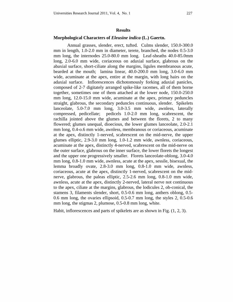
Universities Research Journal 2011, Vol. 4, No. 1 227
Results
Morphological Characters of Eleusine indica (L.) Gaertn. Annual grasses, slender, erect, tufted. Culms slender, 150.0-300.0
mm in length, 1.0-2.0 mm in diameter, terete, branched, the nodes 0.5-3.0 mm long, the internodes 25.0-80.0 mm long. Leaf-sheaths 40.0-85.0mm long, 2.0-6.0 mm wide, coriaceous on adaxial surface, glabrous on the abaxial surface, short-ciliate along the margins, ligules membranous acute, bearded at the mouth; lamina linear, 40.0-200.0 mm long, 3.0-6.0 mm wide, acuminate at the apex, entire at the margin, with long hairs on the adaxial surface. Inflorescences dichotomously forking adaxial panicles, composed of 2-7 digitately arranged spike-like racemes, all of them borne together, sometimes one of them attached at the lower node, 150.0-250.0 mm long, 12.0-15.0 mm wide, acuminate at the apex, primary peduncles straight, glabrous, the secondary peduncles continuous, slender. Spikelets lanceolate, 5.0-7.0 mm long, 3.0-3.5 mm wide, awnless, laterally compressed, pedicellate; pedicels 1.0-2.0 mm long, scabrescent, the rachilla jointed above the glumes and between the florets, 2 to many flowered; glumes unequal, dioecious, the lower glumes lanceolate, 2.0-2.1 mm long, 0.4-o.6 mm wide, awnless, membranous or coriaceous, acuminate at the apex, distinctly 1-nerved, scabrescent on the mid-nerve, the upper glumes elliptic, 2.9-3.0 mm long, 1.0-1.2 mm wide, awnless, coriaceous, acuminate at the apex, distinctly 4-nerved, scabrescent on the mid-nerve on the outer surface, glabrous on the inner surface, the lower florets the longest and the upper one progressively smaller. Florets lanceolate-oblong, 3.0-4.0 mm long, 0.8-1.0 mm wide, awnless, acute at the apex, sessile, bisexual, the lemma broadly ovate, 2.8-3.0 mm long, 0.8-1.0 mm wide, awnless, coriaceous, acute at the apex, distinctly 1-nerved, scabrescent on the mid-nerve, glabrous, the paleas elliptic, 2.5-2.6 mm long, 0.8-1.0 mm wide, awnless, acute at the apex, distinctly 2-nerved, lateral nerve not continuous to the apex, ciliate at the margins, glabrous, the lodicules 2, ob-conical, the stamens 3, filaments slender, short, 0.5-0.6 mm long, anthers oblong, 0.5-0.6 mm long, the ovaries ellipsoid, 0.5-0.7 mm long, the styles 2, 0.5-0.6 mm long, the stigmas 2, plumose, 0.5-0.8 mm long, white.
Habit, inflorescences and parts of spikelets are as shown in Fig. (1, 2, 3).
228 Universities Research Journal 2011, Vol. 4, No. 1
Microscopical characters of Eleusine indica (L.) Gaertn.
Lamina In surface view, the epidermal cells of both surfaces with sinuous
anticlinal walls are arranged in parallel rows. The cells are of two types; long and short cells. The short cells mostly solitary but occasionally in pairs, both over and between the veins, abundant, the intercostals short cells, filled with silica bodies. Long cells, between the veins with thin to moderately thick sinuous walls, vertically elongated. Stomata present on both surfaces. The stomata are graminaceous type. Stomata with markedly triangular subsidiary cells.
In transverse section, the cuticle thin layered is 0.55 μ thick. The epidermis is one layered thick. The upper epidermal cells are rectangular in shape. The lower epidermal cells are similar to upper epidermal cells. One large vascular bundle alternate with three small vascular bundles. The ribs arched over vascular bundles. The layers of sclerenchymatous cells 2-3 cells thick, 8-19 cells wide over and under large vascular bundles and 2-3 cells thick, 7-9 cells wide over and under small vascular bundles. The vascular bundles are circular in outline structure. The bundle sheath of small bundle is single layered and parenchymatous but that of larger bundles are double layered with the outer parenchymatous and the inner sclerenchymatous layers. The bulliform cells are present at the furrows of the upper epidermis, oval or circular in shape and distinct. Xylem found towards upper surface and phloem towards lower surface. Xylem composed of vessel elements, fibers and xylem parenchyma. Phloem composed of sieve tube elements, companion cells and phloem parenchyma. The vessels present are 2-4 in each bundle while the phloem consists of 25-40 cells. These characters are the same in all the apical, middle and basal regions but different in sizes. The microscopical characters of lamina are as shown in (Fig. 4-6). he measurement of cells in surface view and transverse section of lamina are given in Table (1-2).
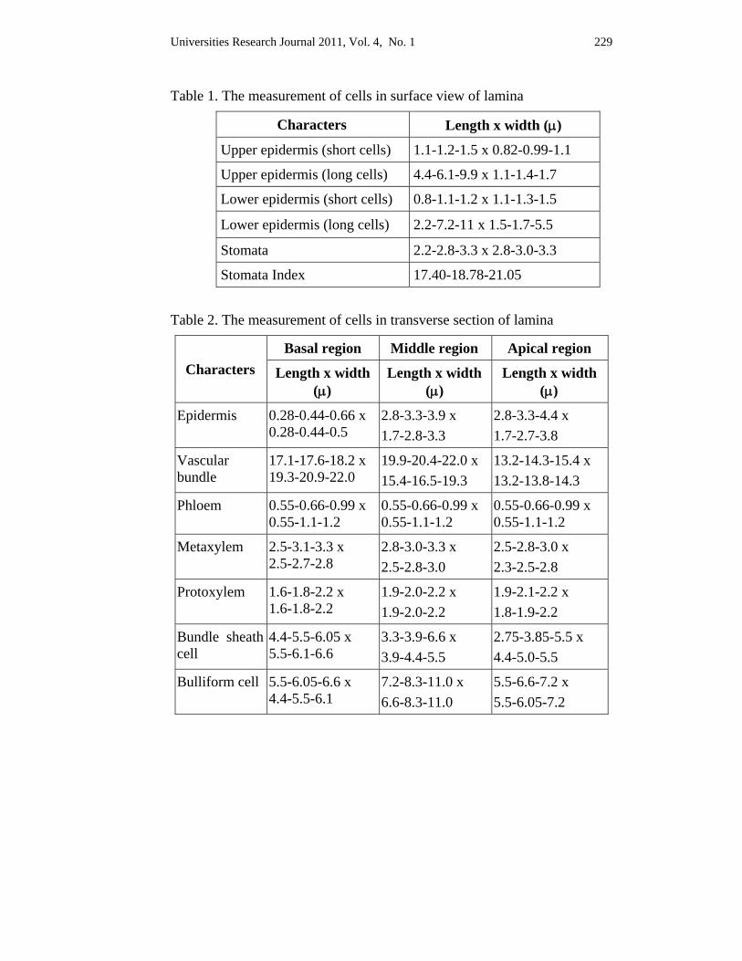
Universities Research Journal 2011, Vol. 4, No. 1 229
Table 1. The measurement of cells in surface view of lamina
Characters Length x width (μ)
Upper epidermis (short cells) 1.1-1.2-1.5 x 0.82-0.99-1.1
Upper epidermis (long cells) 4.4-6.1-9.9 x 1.1-1.4-1.7
Lower epidermis (short cells) 0.8-1.1-1.2 x 1.1-1.3-1.5
Lower epidermis (long cells) 2.2-7.2-11 x 1.5-1.7-5.5
Stomata 2.2-2.8-3.3 x 2.8-3.0-3.3
Stomata Index 17.40-18.78-21.05 Table 2. The measurement of cells in transverse section of lamina
Characters Basal region Middle region Apical region
Length x width (μ)
Length x width (μ)
Length x width (μ)
Epidermis 0.28-0.44-0.66 x 0.28-0.44-0.5
2.8-3.3-3.9 x 1.7-2.8-3.3
2.8-3.3-4.4 x 1.7-2.7-3.8
Vascular bundle
17.1-17.6-18.2 x 19.3-20.9-22.0
19.9-20.4-22.0 x 15.4-16.5-19.3
13.2-14.3-15.4 x 13.2-13.8-14.3
Phloem 0.55-0.66-0.99 x 0.55-1.1-1.2
0.55-0.66-0.99 x 0.55-1.1-1.2
0.55-0.66-0.99 x 0.55-1.1-1.2
Metaxylem 2.5-3.1-3.3 x 2.5-2.7-2.8
2.8-3.0-3.3 x 2.5-2.8-3.0
2.5-2.8-3.0 x 2.3-2.5-2.8
Protoxylem 1.6-1.8-2.2 x 1.6-1.8-2.2
1.9-2.0-2.2 x 1.9-2.0-2.2
1.9-2.1-2.2 x 1.8-1.9-2.2
Bundle sheath cell
4.4-5.5-6.05 x 5.5-6.1-6.6
3.3-3.9-6.6 x 3.9-4.4-5.5
2.75-3.85-5.5 x 4.4-5.0-5.5
Bulliform cell 5.5-6.05-6.6 x 4.4-5.5-6.1
7.2-8.3-11.0 x 6.6-8.3-11.0
5.5-6.6-7.2 x 5.5-6.05-7.2
230 Universities Research Journal 2011, Vol. 4, No. 1
Midrib In surface view, the epidermal cells are rectangular in shape,
compact and parallel. The cells are two types; long and short. The anticlinal walls are sinuous. Graminaceous types of stomata are present.
In transverse section, the cuticle is about 0.28 μ thick present on outer surface only. The epidermal cells are rectangular in shape, compactly arranged. The parenchymatous cells are hexagonal to polygonal in shape, about 6-8 layers at the basal region, 3-5 layers at the middle region and 2-3 layers at the apical region. The vascular bundles are closed collateral type, each circular, have outer and inner bundle sheaths. Outer sheath composed of parenchymatous cells, 12-16 cells at the basal region, 14-16 cells at the middle region and 10-12 cells at the apical region, complete, inner sheath sclerenchymatous cells, 30-36 cells at the basal region, 30-34 cells at the middle region and 30-32 cells at the apical region, complete. The bundle with layers of sclerenchyma, about 4-6 cells high and 30-38 cells wide at the basal region, 3-5 cells high and 30-36 cells wide at the middle region and 2-3 cells high and 23-28 cells wide at apical region. Phloem lies towards outer surface. Number of vessels 2-5 in each bundle, xylem composed of vessel elements, fiber and xylem parenchyma. Number of phloem cells 45- 70 in each bundle, phloem composed of sieve tube elements, companion cells and phloem parenchyma. The microscopical characters of midribs are as shown in Fig. (7-12). The measurements of cells in transverse section of midrib are given in Table (3).

Universities Research Journal 2011, Vol. 4, No. 1 231
Table 3. The measurement of cells in transverse section of midrib
Characters Basal region Middle region Apical region
Length x width (μ)
Length x width (μ)
Length x width (μ)
Epidermis 2.2-2.47-2.75 x 0.66-0.83-0.99
2.2-2.4-2.6 x 0.55-0.83-0.99
2.1-2.3-2.5 x 0.55-0.66-0.83
Parenchyma 4.4-5.5-7.7 x 3.85-4.95-8.25
5.5-7.2-7.7 x 6.6-7.2-8.3
4.4-6.6-7.7 x 4.4-7.2-7.7
Vascular bundle
15.40-16.0-16.5 x 15.9-16.5-17.6
16.5-18.2-19.3 x 20.4-20.9-22.0
14.9-15.4-16.5 x 16.5-17.6-19.3
Phloem 0.55-0.85-1.1 x 0.55-0.85-1.1
0.55-0.88-1.1 x 0.55-0.83-0.99
0.55-0.66-0.85 x 0.44-0.55-0.85
Metaxylem 2.7-2.9-3.02 x 2.7-3.02-3.3
2.8-2.9-3.1 x 2.8-2.9-3.1
2.4-2.6-2.8 x 2.5-2.6-3.0
Protoxylem 1.1-1.38-1.40 x 1.37-1.40-1.65
1.3-1.5-1.7 x 1.2-1.3-1.5
1.2-1.3-1.5 x 1.2-1.4-1.5
Leaf-sheath In surface view, the epidermal cells of both surfaces are dissimilar
in shape and size. The cells are in parallel rows, rectangular and compactly arranged. Stomata are present on the outer surface and rare on the inner surface when present. The stomata are graminaceous type.
In transverse section, the cuticle is present on both surfaces. Cuticle layer is thin on the outer surface about 0.28 μ thick and thicker on the inner surface about 1.1μ. Both inner and outer epidermal cells one layered, compact, anticlinal walls straight, periclinal walls convex but the shape of the inner epidermal cells are barrel-shaped and that of the outer epidermal cells are rectangular. The ground parenchymatous cells 4-12 layers, compact, thin walled, hexagonal in shape, variable in size. The vascular bundles are scattered, closed collateral type, oval to circular, bundle sheath complete, and parenchymatous cells 12-18 cells. The number of vessels 4-7 in each bundle, xylem composed of vessel elements, fibers and xylem
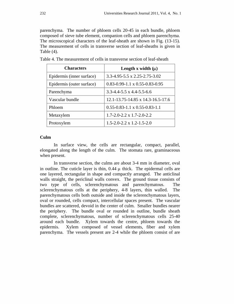
232 Universities Research Journal 2011, Vol. 4, No. 1
parenchyma. The number of phloem cells 20-45 in each bundle, phloem composed of sieve tube element, companion cells and phloem parenchyma. The microscopical characters of the leaf-sheath are shown in Fig. (13-15). The measurement of cells in transverse section of leaf-sheaths is given in Table (4).
Table 4. The measurement of cells in transverse section of leaf-sheath
Characters Length x width (μ)
Epidermis (inner surface) 3.3-4.95-5.5 x 2.25-2.75-3.02
Epidermis (outer surface) 0.83-0.99-1.1 x 0.55-0.83-0.95
Parenchyma 3.3-4.4-5.5 x 4.4-5.5-6.6
Vascular bundle 12.1-13.75-14.85 x 14.3-16.5-17.6
Phloem 0.55-0.83-1.1 x 0.55-0.83-1.1
Metaxylem 1.7-2.0-2.2 x 1.7-2.0-2.2
Protoxylem 1.5-2.0-2.2 x 1.2-1.5-2.0
Culm In surface view, the cells are rectangular, compact, parallel,
elongated along the length of the culm. The stomata rare, graminaceous when present.
In transverse section, the culms are about 3-4 mm in diameter, oval in outline. The cuticle layer is thin, 0.44 μ thick. The epidermal cells are one layered, rectangular in shape and compactly arranged. The anticlinal walls straight, the periclinal walls convex. The ground tissue consists of two type of cells, sclerenchymatous and parenchymatous. The sclerenchymatous cells at the periphery, 4-8 layers, thin walled. The parenchymatous cells both outside and inside the sclerenchymatous layers, oval or rounded, cells compact, intercellular spaces present. The vascular bundles are scattered, devoid in the center of culm. Smaller bundles nearer the periphery. The bundle oval or rounded in outline, bundle sheath complete, sclerenchymatous, number of sclerenchymatous cells 25-40 around each bundle. Xylem towards the centre, phloem towards the epidermis. Xylem composed of vessel elements, fiber and xylem parenchyma. The vessels present are 2-4 while the phloem consist of are
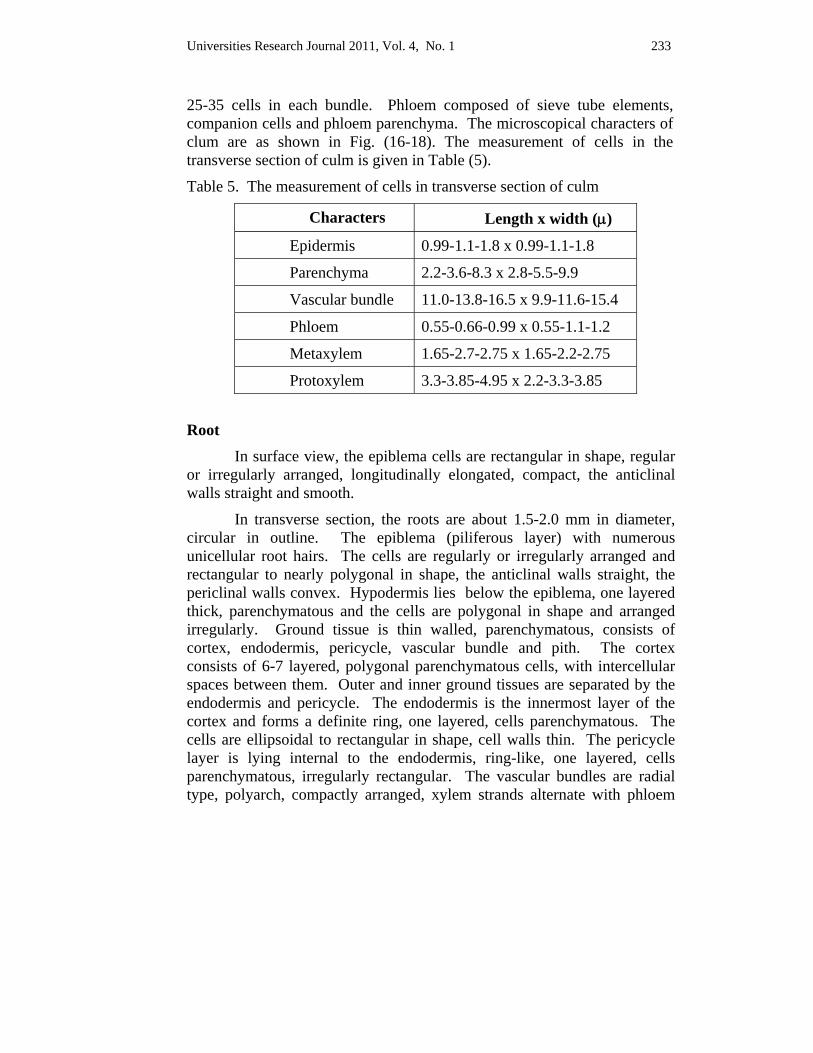
Universities Research Journal 2011, Vol. 4, No. 1 233
25-35 cells in each bundle. Phloem composed of sieve tube elements, companion cells and phloem parenchyma. The microscopical characters of clum are as shown in Fig. (16-18). The measurement of cells in the transverse section of culm is given in Table (5).
Table 5. The measurement of cells in transverse section of culm
Characters Length x width (μ)
Epidermis 0.99-1.1-1.8 x 0.99-1.1-1.8
Parenchyma 2.2-3.6-8.3 x 2.8-5.5-9.9
Vascular bundle 11.0-13.8-16.5 x 9.9-11.6-15.4
Phloem 0.55-0.66-0.99 x 0.55-1.1-1.2
Metaxylem 1.65-2.7-2.75 x 1.65-2.2-2.75
Protoxylem 3.3-3.85-4.95 x 2.2-3.3-3.85
Root In surface view, the epiblema cells are rectangular in shape, regular
or irregularly arranged, longitudinally elongated, compact, the anticlinal walls straight and smooth.
In transverse section, the roots are about 1.5-2.0 mm in diameter, circular in outline. The epiblema (piliferous layer) with numerous unicellular root hairs. The cells are regularly or irregularly arranged and rectangular to nearly polygonal in shape, the anticlinal walls straight, the periclinal walls convex. Hypodermis lies below the epiblema, one layered thick, parenchymatous and the cells are polygonal in shape and arranged irregularly. Ground tissue is thin walled, parenchymatous, consists of cortex, endodermis, pericycle, vascular bundle and pith. The cortex consists of 6-7 layered, polygonal parenchymatous cells, with intercellular spaces between them. Outer and inner ground tissues are separated by the endodermis and pericycle. The endodermis is the innermost layer of the cortex and forms a definite ring, one layered, cells parenchymatous. The cells are ellipsoidal to rectangular in shape, cell walls thin. The pericycle layer is lying internal to the endodermis, ring-like, one layered, cells parenchymatous, irregularly rectangular. The vascular bundles are radial type, polyarch, compactly arranged, xylem strands alternate with phloem
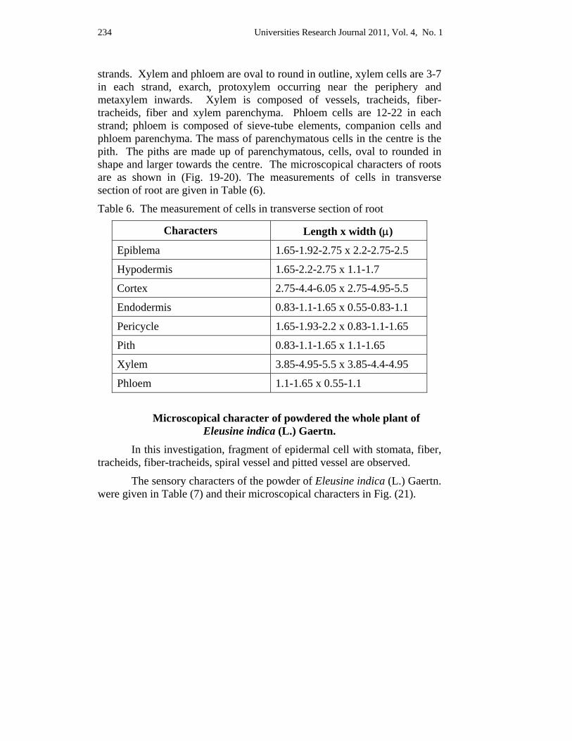
234 Universities Research Journal 2011, Vol. 4, No. 1
strands. Xylem and phloem are oval to round in outline, xylem cells are 3-7 in each strand, exarch, protoxylem occurring near the periphery and metaxylem inwards. Xylem is composed of vessels, tracheids, fiber-tracheids, fiber and xylem parenchyma. Phloem cells are 12-22 in each strand; phloem is composed of sieve-tube elements, companion cells and phloem parenchyma. The mass of parenchymatous cells in the centre is the pith. The piths are made up of parenchymatous, cells, oval to rounded in shape and larger towards the centre. The microscopical characters of roots are as shown in (Fig. 19-20). The measurements of cells in transverse section of root are given in Table (6).
Table 6. The measurement of cells in transverse section of root
Characters Length x width (μ)
Epiblema 1.65-1.92-2.75 x 2.2-2.75-2.5
Hypodermis 1.65-2.2-2.75 x 1.1-1.7
Cortex 2.75-4.4-6.05 x 2.75-4.95-5.5
Endodermis 0.83-1.1-1.65 x 0.55-0.83-1.1
Pericycle 1.65-1.93-2.2 x 0.83-1.1-1.65
Pith 0.83-1.1-1.65 x 1.1-1.65
Xylem 3.85-4.95-5.5 x 3.85-4.4-4.95
Phloem 1.1-1.65 x 0.55-1.1
Microscopical character of powdered the whole plant of Eleusine indica (L.) Gaertn.
In this investigation, fragment of epidermal cell with stomata, fiber, tracheids, fiber-tracheids, spiral vessel and pitted vessel are observed.
The sensory characters of the powder of Eleusine indica (L.) Gaertn. were given in Table (7) and their microscopical characters in Fig. (21).

Universities Research Journal 2011, Vol. 4, No. 1 235
Table 7. Sensory characters of powder of whole plant
Sensory Characters Whole Plants
Colour Greenish
Odour Slightly aromatic
Taste Bitter
Texture Granular
Fig. 1 Habit Fig. 2 Inflorescence
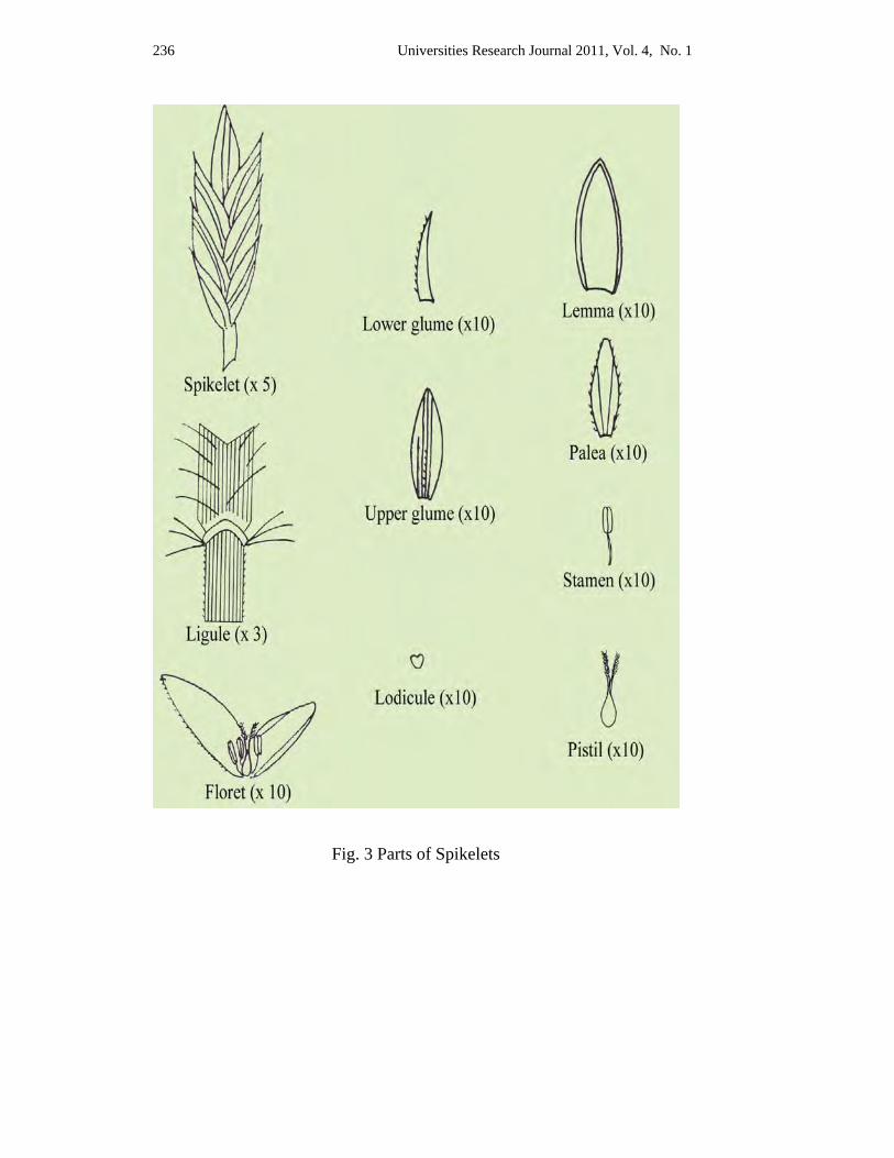
236 Universities Research Journal 2011, Vol. 4, No. 1
Fig. 3 Parts of Spikelets
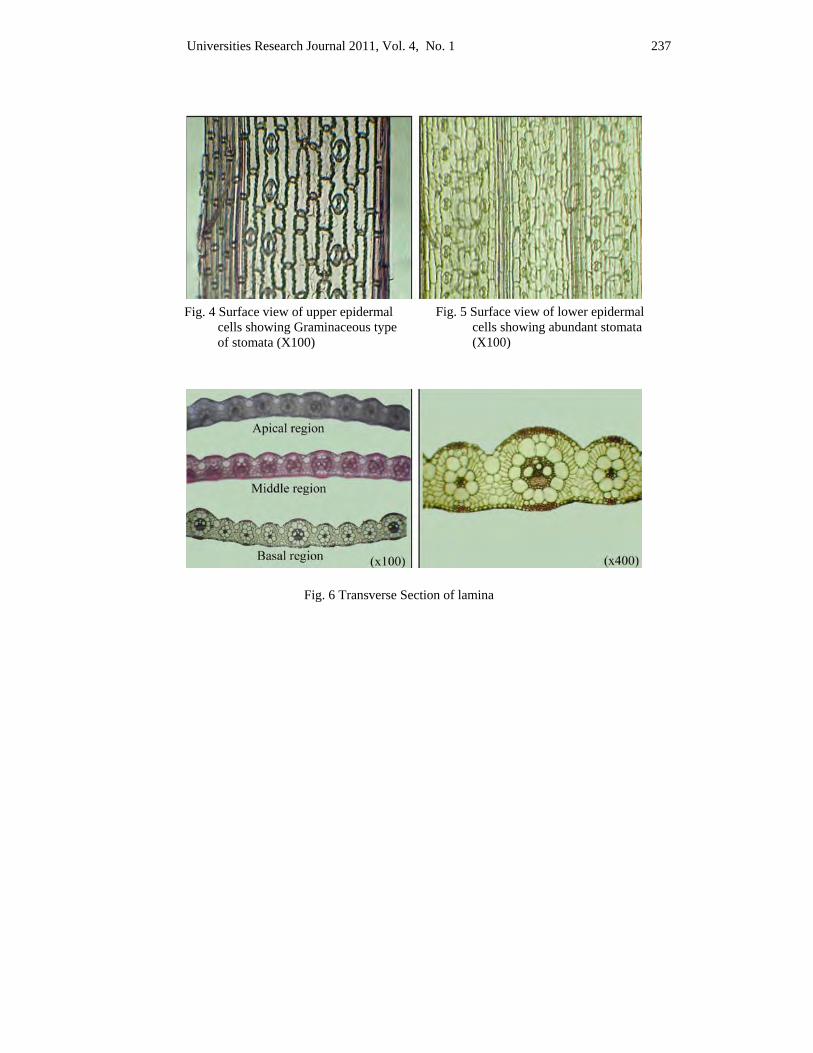
Universities Research Journal 2011, Vol. 4, No. 1 237
Fig. 4 Surface view of upper epidermal cells showing Graminaceous type of stomata (X100)
Fig. 5 Surface view of lower epidermal cells showing abundant stomata (X100)
Fig. 6 Transverse Section of lamina
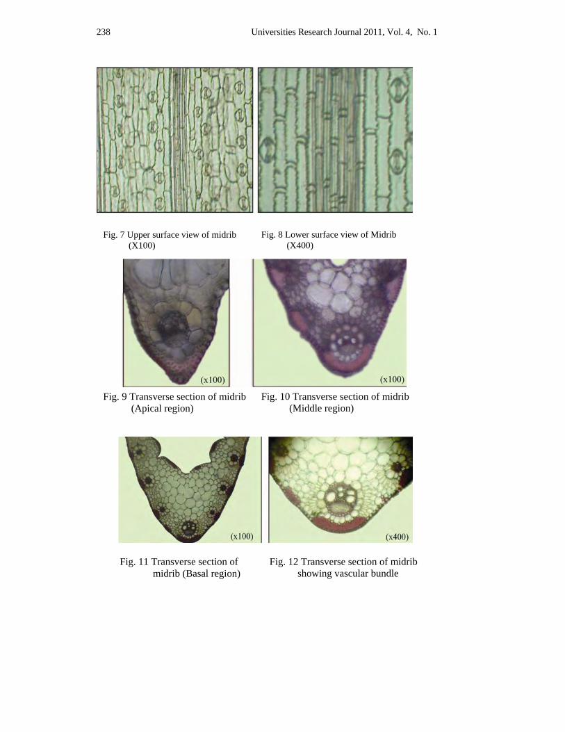
238 Universities Research Journal 2011, Vol. 4, No. 1
Fig. 9 Transverse section of midrib (Apical region)
Fig. 10 Transverse section of midrib (Middle region)
Fig. 11 Transverse section of midrib (Basal region)
Fig. 12 Transverse section of midrib showing vascular bundle
Fig. 7 Upper surface view of midrib (X100)
Fig. 8 Lower surface view of Midrib (X400)
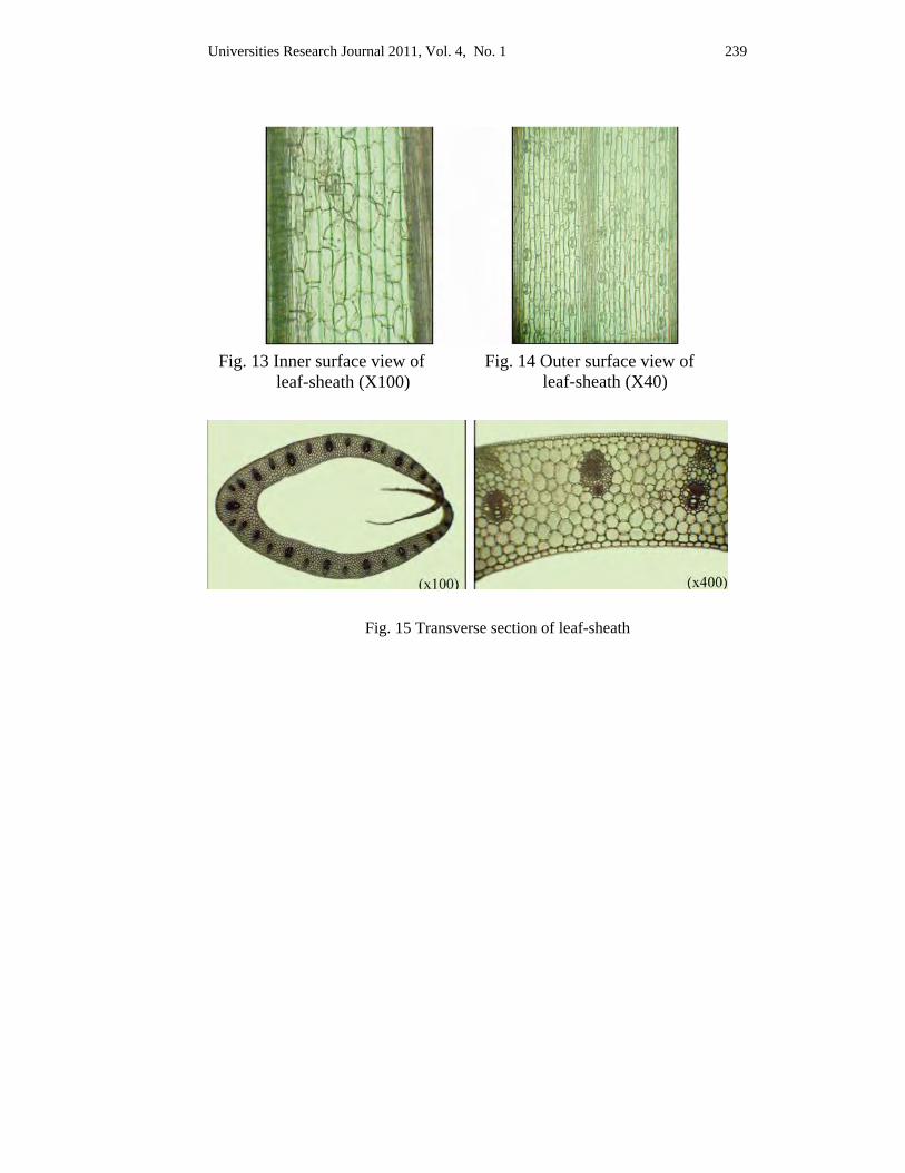
Universities Research Journal 2011, Vol. 4, No. 1 239
Fig. 13 Inner surface view of leaf-sheath (X100)
Fig. 14 Outer surface view of leaf-sheath (X40)
Fig. 15 Transverse section of leaf-sheath
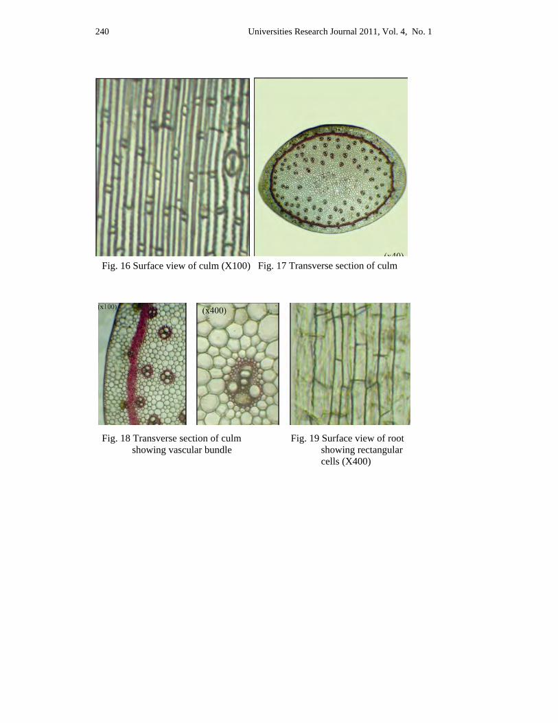
240 Universities Research Journal 2011, Vol. 4, No. 1
Fig. 16 Surface view of culm (X100) Fig. 17 Transverse section of culm
Fig. 18 Transverse section of culm showing vascular bundle
Fig. 19 Surface view of root showing rectangular cells (X400)
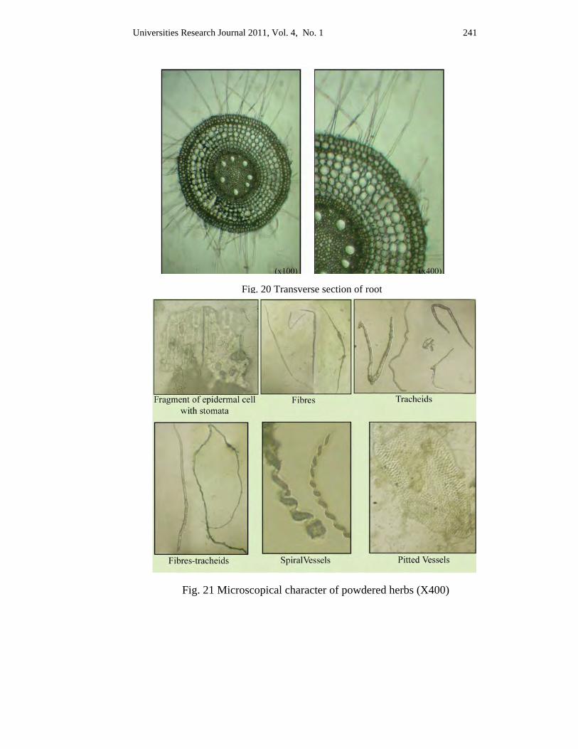
Universities Research Journal 2011, Vol. 4, No. 1 241
Fig. 20 Transverse section of root
Fig. 21 Microscopical character of powdered herbs (X400)

242 Universities Research Journal 2011, Vol. 4, No. 1
Discussion and Conclusion The plant Eleusine indica (L.) Gaertn. is usually found growing wild
throughout the country during the rainy season. The morphological studies on both vegetative and reproductive parts as well as the microscopical examination of leaves, culms, and roots verified the plant to be Eleusine indica (L.) Gaertn. It is annual herb, erect, slender and tufted. The leaves are linear, parallel venation, distinguished into leaf-blades and leaf-sheaths. Ligules are present. The inflorescences are terminal spike. The spikelets are two to many flowered, awnless, and laterally compressed. The florets are awnless, bisexual, the lowest florets longest and upper one progressively smaller. The lodicules 2, ob-conical, the stamens 3, anther oblong, filament short, the ovary ellipsoid, styles 2, stigmas 2, plumose. All morphological characters of this plant are in agreement with those stated by Hooker (1894); Hitchock (1950); Bor (1960); Backer (1963); Hafliger et al., (1981).
In microscopical studies, the epidermal cells of the surface view of lamina, midrib and leaf-sheaths are parallel rowed. The cells are of two types, long and short, walls sinuous. Bulliform cells are found in furrows of upper epidermis of lamina in transverse section. Epidermis is one layered thick. The cuticle is present on transverse section of midrib and leaf-shealth. The epidermal cells are one layered, compact, rectangular and that of inner surface of leaf-shealth barrel-shaped. Close collateral type of vascular bundles are found in all parts of leaf. In surface view of roots, the epiblema cells are rectangular. In transverse section of roots, the epiblema with numerous unicellular root hairs, cells are rectangular to nearly polygonal in shape. The ground tissue is thin-walled, parenchymatous, composed of cortex, endodermis, pericycle and pith. Vascular bundles are polyarch, radial type. In surface view of culm, the epidermal cells are elongated along the length of culm. In transverse section, the cuticle thin, epidermal cells are compact and rectangular in shape. Vascular bundles are scattered, but devoid in the center of culm. Fragment of epidermal cells with stomata, fibers, tracheids, fiber-tracheids, spiral vessels and pitted vessels are observed in the microscopical characters of powdered samples. These characters are agreement with Metcalfe & Chalk, (1950); Wallis, (1955); Esau, (1965); Pandey, (1993) and Sundara rajan, (2000). The sensory characters were found to be greenish, slightly aromatic, bitter, granular and these characters can be used for the identification and standardization of drug.
Universities Research Journal 2011, Vol. 4, No. 1 243
Acknowledgements I am greatly indebted to Dr. Yee Yee Wynn, Professor and Head, Department of Botany, Yangon University of Distance Education for allowing me to undertake this research.
I am also greatly indebted to Dr. Thet Thet May, Professor and Head, Dr. Aye Pe, Professor Department of Botany, Yangon University for their kind help, invaluable advice for this research. I am also greatly indebted to Professor Naw Wah Wah Paw, Retired, Professor and Head, Department of Botany, Dagon University, Dr. Moe Moe Lwin, Lecturer, Department of Botany, Pathein University close guidance and numerous suggestions and comment offered during the research and Dr. Khin Myo Thwet, Lecturer, Department of Botany, Yangon University of Distance Education, for her kind help.
References Backer, C.A., (1963). Flora of Java. Vol. 3. Noordhoof Griningen Company, The
Netherland
Bor, N. L., (1960). The Grasses of Burma, Ceylon, India and Pakistan. Pergamon Press, Oxford, London, New York and Paris
Burkill, M. A., (1935). A Dictionary of the economic products of Malay Penisula. Vol. 1. London
Chopra, R. N., (1956). Glossary of Indian medicinal Plants. Council of Scientific and Industrial Research, New Delhi
Esau, K., (1965). Plant Anatomy. Second Edition, Printed in Japan, By Toppan Printing Company, Ltd
Hafliger, E. and H. Schlog., (1981). Monocot Weeds 3 and Grass Weed 1, 2. CIBA. Geigy Ltd. Basl, Switzerland
Hitchock, A. S., 1950. Mannual of The Grasses of The United States. Second Edition. Government Printing Office. United States
Hooker, J. D., (1894). Flora of British India Vol.7. Reeve and Co., Ltd. London
Hundley, H. G and Chit Ko Ko, (1961). List of Trees, Shrubs, Herbs and Principal Climbers etc. recorded from Burma. P. 294-324. Supdt. Govt. Printing and Stationary, Burma
Kirtikar, K. R. and B. D. Basu, (1933). Indian Medicinal Plants. 2nd ed., 49, Leader Road, Allalhabad, India
Kress, J. and et al., (2003). A Checklist of Trees, Shrubs, Herbs and Climbers of Myanmar. Washington D.C
Metcalfe, C. R. and L. Chalk, (1950). Anatomy of Monocotyledon Vol. 1. The Clarendon Press, Oxford
244 Universities Research Journal 2011, Vol. 4, No. 1
Nagathein, Ashin, (1977). Pon-pya-say-A-bidon. Vol. 1. Mingala Press. Yangon
Pandey, B. P., (1993). Plant Anatomy. S. Chand & Company Ltd. Pamlvagar, New Delhi
Sundara rajan, S. (2000). Plant Anatomy and Embryology. Anmol Publications. Ltd., Newdelhi
Trease, G. E., and W. C. Evans, (1978). A Text Book of Pharmacognosy. 11th Edition. Bailliere Tindall, London
Trease G. E. and W. C. Evans (2002). Pharmacognosy. 15th ed., Baillere Tindoll, London
Wallis (1955). Text Book of Pharmacognosy. 3th Edition., J & A Churchill Ltd., London
Universities Research Journal 2011, Vol. 4, No. 1
Assistant Lecturer, Department of Botany, Dagon University
Shoot Propagation of Tectona grandis L. f. by Tissue Culture Yin Yin Waing
Abstract An efficient study of teak propagation by tissue culture technique was done in Plant Tissue Culture Laboratory, Department of Botany, Dagon University. Teak (kyun) botanically known as Tectona grandis L. f. belonging to the family Verbenaceae is one of the most valuable and extremely durable timber yielding plant in Myanmar. In this study, the shoot tips were used as experimental explants. The shoot tips containing apical meristem were taken from the plants which were grown under shaded places. The explants were cultured on White basal medium (1954) and sub-cultured on Woody Plant Medium (WPM) which were supplemented with the combination of BAP (0.01, 1.0 mgL-1) and NAA (0.005, 0.5 mgL-1) for shoot multiplication respectively. Among these treatments, WPM medium supplemented with the combination of BAP (1.0 mgL-1) and NAA (0.005 mgL-1), the T4 treatment showed the best shoot proliferation.
Key words: tissue culture, propagation, multiplication
Introduction Tectona grandis L. f. (Teak) is one of the most important timber yielding trees of Asian countries particularly India, Indonesia, Myanmar and Thailand. It is also the most popular, valuable and extremely durable wood of indigenous timber among all over the world. "Tectona and teak" are both derived from the Portuguese name "teca and tekton". In Greek it is known as "a carpenter". Whereas "grandis" is a Latin word means "large"(Schubert, 1959).
Teak grows widely as natural vegetation in Cambodia, India, north-west Laos, Myanmar, North Thailand and Vietnam. But it has been widely cultivated throughout its natural range since the fourteenth century. In Myanmar and India, it has been cultivated for timber production since at least 1840 (Schubert, 1959).
In Myanmar, teak plays a dormant role in socioeconomic life of the people of the nation and is environmentally conserved. The vegetation of teak has been officially protected by government. The teak's plantations are
246 Universities Research Journal 2011, Vol. 4, No. 1
being managed only by the government. But nowadays, it can be done by public under government restriction.
It is usually grown within tropical semi-evergreen forests, mixed deciduous forest and deciduous Dipterocarp forests. Under natural stand, teak is a large deciduous tree which is up to at least 20 m in height. It is one of the dominant trees in Upper Mixed Deciduous Forest (UMDF). It requires a high light intensity for its growth and development. It grows best in a warm, moist tropical climate (Schubert, 1959).
The optimum pH range of the soil is from 6.5 to 7.5. The teak plant, a "calcicolous" species requires a relatively large amount of calcium in the soil for growth and development (White, 1991). Teak is found to grow where the precipitation ranges from 800 mm to 2500 mm (Kaosa-ard, 1986).
Teak usually germinates by epigeal germination and is difficult to germinate evenly and adequately. Nearly all of the teak's seeds possess some degree of dormancy and need to pretreat to break its dormancy. Although the real cause of the seed dormancy is still unknown; however, there are three main factors which influence seed dormancy: seed structure, seed maturity and seed biochemistry (Kaosa-ard, 1986). The main cause of the delay of germination is due to its thick hard woody pericarp (Kadambi, 1972).
The wood of the plants are used in constructions, building, furniture, crafts and instruments. The bark of roots and young leaves produce yellowish-brown or reddish substance for fabric dye, thus can be used to dye paper, clothes and matting. Teaks' sawdust is also useful in making incense (Soerianegara and Lemmens, 1994).
The wood is very heavy, strong and durable, resists white-ants and contains oil with strong characteristic scent which preserves the timber. Some parts of the plant have been used in numerous medicines. The wood can also be used to alleviate headaches, dyspepsia and stomach complaints. A concoction from the bark and flowers can be prepared to relieve bronchitis.
The teak plants can be propagated by vegetative propagation such as cutting, budding, grafting and air layering. However, the quantitative production was limited due to the poor rooting in cuttings collected from mature trees. The availability of stocks is also limited due to the intensive
Universities Research Journal 2011, Vol. 4, No. 1 247
maintenance of stock plants. Moreover, some of the other major limiting factors in vegetative propagation depend on season and slow multiplication rate. Although, propagation could be achieved through seeds germination, there are certain disadvantages like low fruit production, abortive seeds and low germination rate. Besides, it can be infected with latent diseases. Although propagation by vegetative cutting is a predominant technique in teak but it does not ensure healthy and disease-free plants.
Therefore, scientists started an alternative method of plant propagation, the tissue culture method; called micropropagation method which could overcome the disadvantages of the vegetative propagation. It involves the culture of whole organism from cells or tissues or plant parts via in vitro on a defined medium under aseptic conditions. With such technique, the enormous multiplication capacity could be available to generate propagules around the year. High uniformity may help to ease the management of the teak plantations. The tissue cultured teak plant can be pre-selected to propagate from the mother plants which have straight with no branching (Ahloowalia, 1996 and Yasodha et al., 2004).
The aim of this present paper was to investigate the proper concentration and combination of selected plant growth regulators (BAP and NAA), to increase the number of teak plantations and thereby to establish the forests cover which are under rapid depletion caused by over exploitations. Therefore, in vitro clonal propagation technique may hope to offer an alternative technique for superior trees.
Materials and Methods The study of Tectona grandis L. f. was conducted at Plant Tissue Culture Laboratory in Department of Botany, Dagon University during 2009 - 2010. It involved two stages: initial stage and multiplication stage for the production of shoot-lets from the shoot tips of teak plant under aseptic condition. A. Initial Stage for shoot tips culture of teak (In vitro)
(1) Collection of stock plants The teak seeds (Figure 1) were collected from Mawbe Township, Yangon Region. The seeds were alternatively soaked in water and dried
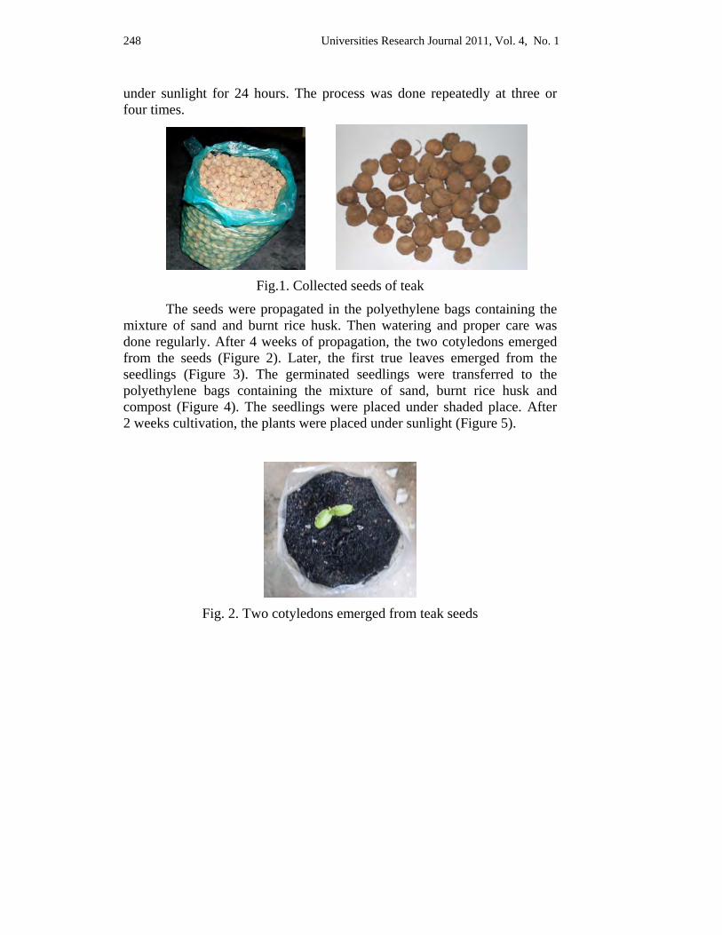
248 Universities Research Journal 2011, Vol. 4, No. 1
under sunlight for 24 hours. The process was done repeatedly at three or four times.
Fig.1. Collected seeds of teak
The seeds were propagated in the polyethylene bags containing the mixture of sand and burnt rice husk. Then watering and proper care was done regularly. After 4 weeks of propagation, the two cotyledons emerged from the seeds (Figure 2). Later, the first true leaves emerged from the seedlings (Figure 3). The germinated seedlings were transferred to the polyethylene bags containing the mixture of sand, burnt rice husk and compost (Figure 4). The seedlings were placed under shaded place. After 2 weeks cultivation, the plants were placed under sunlight (Figure 5).
Fig. 2. Two cotyledons emerged from teak seeds
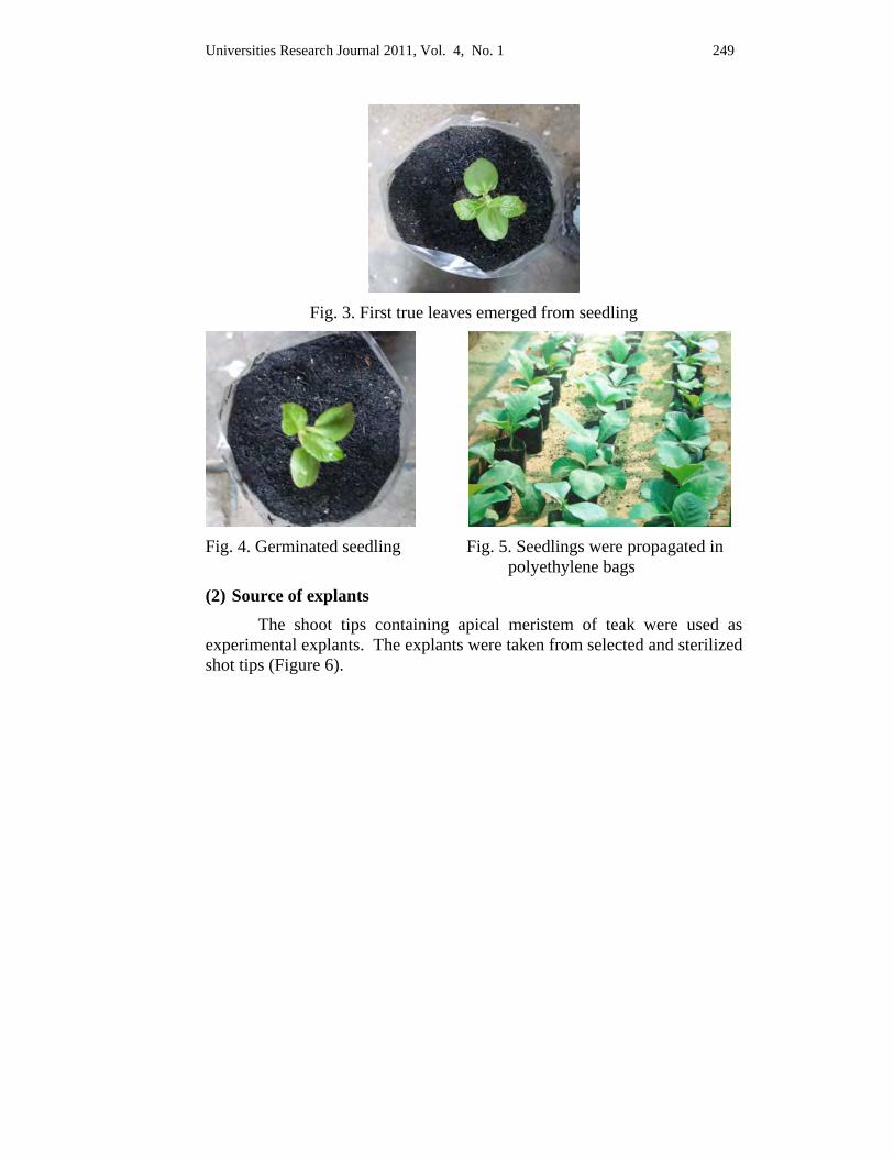
Universities Research Journal 2011, Vol. 4, No. 1 249
Fig. 3. First true leaves emerged from seedling
Fig. 4. Germinated seedling Fig. 5. Seedlings were propagated in
polyethylene bags
(2) Source of explants The shoot tips containing apical meristem of teak were used as experimental explants. The explants were taken from selected and sterilized shot tips (Figure 6).
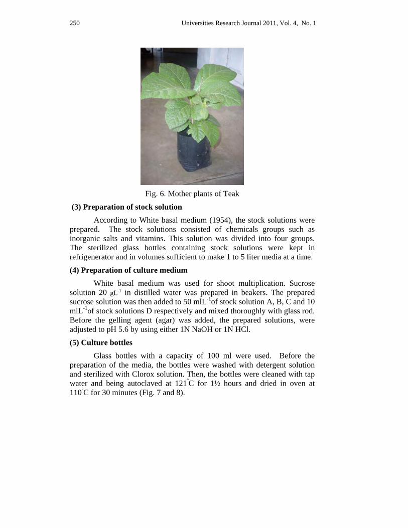
250 Universities Research Journal 2011, Vol. 4, No. 1
Fig. 6. Mother plants of Teak
(3) Preparation of stock solution According to White basal medium (1954), the stock solutions were prepared. The stock solutions consisted of chemicals groups such as inorganic salts and vitamins. This solution was divided into four groups. The sterilized glass bottles containing stock solutions were kept in refrigenerator and in volumes sufficient to make 1 to 5 liter media at a time.
(4) Preparation of culture medium White basal medium was used for shoot multiplication. Sucrose solution 20 gL-1 in distilled water was prepared in beakers. The prepared sucrose solution was then added to 50 mlL-1of stock solution A, B, C and 10 mlL-1of stock solutions D respectively and mixed thoroughly with glass rod. Before the gelling agent (agar) was added, the prepared solutions, were adjusted to pH 5.6 by using either 1N NaOH or 1N HCl.
(5) Culture bottles Glass bottles with a capacity of 100 ml were used. Before the preparation of the media, the bottles were washed with detergent solution and sterilized with Clorox solution. Then, the bottles were cleaned with tap water and being autoclaved at 121ºC for 1½ hours and dried in oven at 110ºC for 30 minutes (Fig. 7 and 8).
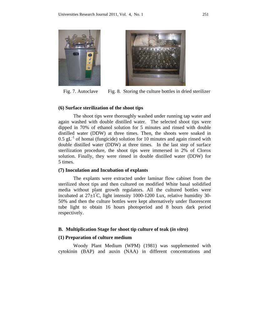
Universities Research Journal 2011, Vol. 4, No. 1 251
Fig. 7. Autoclave Fig. 8. Storing the culture bottles in dried sterilizer
(6) Surface sterilization of the shoot tips The shoot tips were thoroughly washed under running tap water and again washed with double distilled water. The selected shoot tips were dipped in 70% of ethanol solution for 5 minutes and rinsed with double distilled water (DDW) at three times. Then, the shoots were soaked in 0.5 gL-1 of homai (fungicide) solution for 10 minutes and again rinsed with double distilled water (DDW) at three times. In the last step of surface sterilization procedure, the shoot tips were immersed in 2% of Clorox solution. Finally, they were rinsed in double distilled water (DDW) for 5 times.
(7) Inoculation and Incubation of explants The explants were extracted under laminar flow cabinet from the sterilized shoot tips and then cultured on modified White basal solidified media without plant growth regulators. All the cultured bottles were incubated at 27±1ºC, light intensity 1000-1200 Lux, relative humidity 30-50% and then the culture bottles were kept alternatively under fluorescent tube light to obtain 16 hours photoperiod and 8 hours dark period respectively.
B. Multiplication Stage for shoot tip culture of teak (in vitro)
(1) Preparation of culture medium Woody Plant Medium (WPM) (1981) was supplemented with cytokinin (BAP) and auxin (NAA) in different concentrations and
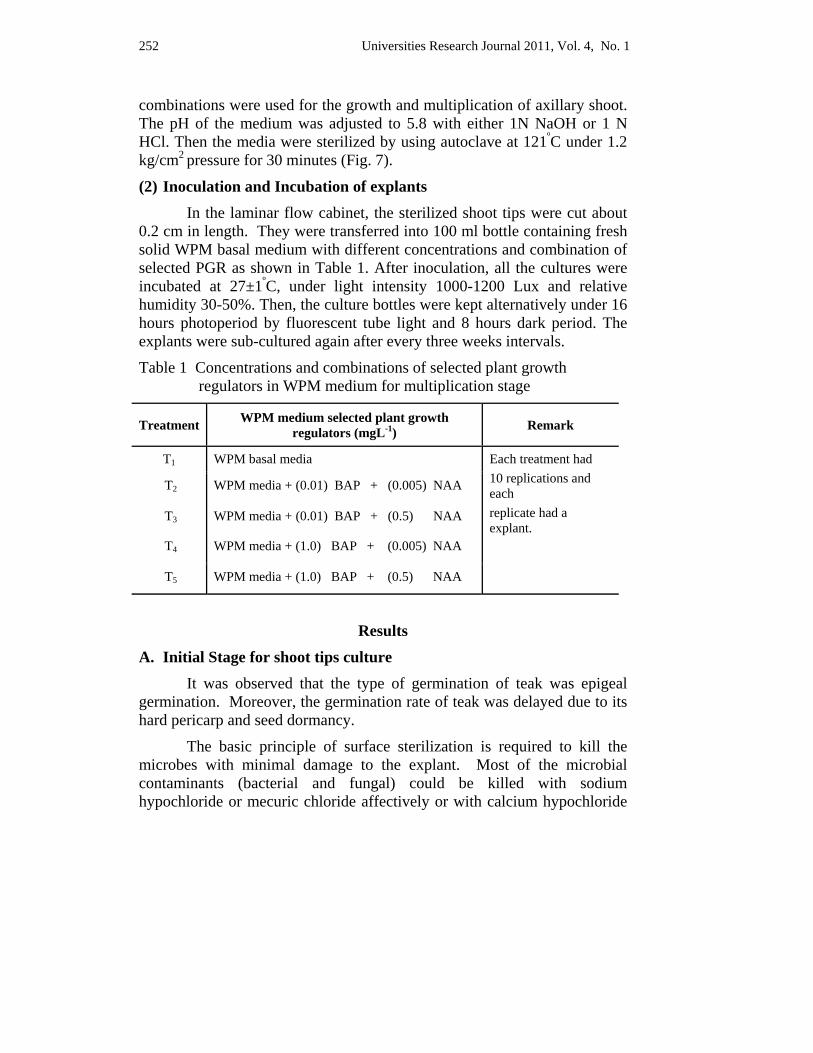
252 Universities Research Journal 2011, Vol. 4, No. 1
combinations were used for the growth and multiplication of axillary shoot. The pH of the medium was adjusted to 5.8 with either 1N NaOH or 1 N HCl. Then the media were sterilized by using autoclave at 121ºC under 1.2 kg/cm2
pressure for 30 minutes (Fig. 7).
(2) Inoculation and Incubation of explants In the laminar flow cabinet, the sterilized shoot tips were cut about 0.2 cm in length. They were transferred into 100 ml bottle containing fresh solid WPM basal medium with different concentrations and combination of selected PGR as shown in Table 1. After inoculation, all the cultures were incubated at 27±1ºC, under light intensity 1000-1200 Lux and relative humidity 30-50%. Then, the culture bottles were kept alternatively under 16 hours photoperiod by fluorescent tube light and 8 hours dark period. The explants were sub-cultured again after every three weeks intervals.
Table 1 Concentrations and combinations of selected plant growth regulators in WPM medium for multiplication stage
Treatment WPM medium selected plant growth regulators (mgL-1) Remark
T1 WPM basal media Each treatment had 10 replications and each replicate had a explant.
T2 WPM media + (0.01) BAP + (0.005) NAA
T3 WPM media + (0.01) BAP + (0.5) NAA
T4 WPM media + (1.0) BAP + (0.005) NAA
T5 WPM media + (1.0) BAP + (0.5) NAA
Results
A. Initial Stage for shoot tips culture
It was observed that the type of germination of teak was epigeal germination. Moreover, the germination rate of teak was delayed due to its hard pericarp and seed dormancy.
The basic principle of surface sterilization is required to kill the microbes with minimal damage to the explant. Most of the microbial contaminants (bacterial and fungal) could be killed with sodium hypochloride or mecuric chloride affectively or with calcium hypochloride
Universities Research Journal 2011, Vol. 4, No. 1 253
or benzyalkonium chloride which was being effected for highly contaminated tissues.
In this study, Clorox solution (sodium hypochloride) was used for surface sterilization of explants. After one week, some of the explants were found to survive in all replications except the contamination ones. The result was consistent with George (1993). The presented outline of sterilization procedures were employed throughout the experiments. Sodium hypochloride was used, for it was easily available and found to be less expensive.
The survived flesh explants were found to be observed in White basal medium. The survived shoot tips with initiated callus at the base of the cut surface of two weeks old were then cultured for multiplication stage (Figure 11).
B. Multiplication Stage for shoot tips culture The survived shoot tips were transferred to the WPM medium supplemented with different concentrations and combinations of BAP (0.01 mgL-1, 1.0 mgL-1) and NAA (0.005 mgL-1, 0.5 mgL-1) for shoot multiplication. BAP and NAA were used to calculate the proper balance between cytokinin and auxin for proliferation of shoots.
After six weeks, the shoot initiation was found to be occurred in T2 (WPM medium supplemented with the combination of 0.01 mgL-1 BAP + 0.005 mgL-1NAA), T4 (1.0 mgL-1 BAP + 0.005 mgL-1NAA) and T5 (1.0 mgL-1 BAP + 0.5 mgL-1NAA) except in T1 (WPM basal medium) and T3 (0.01 mgL-1 BAP + 0.5 mgL-1NAA). However slightly large callus formation were observed in T3 (0.01 mgL-1 BAP + 0.5 mgL-1 NAA). The survived shoot-lets and callus in all treatments were transferred to the respective fresh medium for further development.
After two weeks, the transferred ones were found to grow axillary shoots with well developed in T2 (0.01 mgL-1 BAP + 0.005 mgL-1NAA), T4 (1.0mgL-1 BAP + 0.005mgL-1NAA) and T5 (1.0mgL-1BAP + 0.5mgL-1
NAA). Initiation of shoots from the callus found to arose in T3 (0.01 mgL-1 BAP + 0.5 mgL-1 NAA). These were shown in Fig. 12, 13, 14, 15 and 16. But in T1 (WPM basal medium) was found to develop a more large callus formation.
254 Universities Research Journal 2011, Vol. 4, No. 1
After four weeks, the cultures showed more proliferation of shoots in T4 (1.0 mgL-1 BAP + 0.005 mgL-1 IBA) and followed by T5 and T2. The poor proliferation of shoots was found in T1 and T3.
After six weeks, the transferred cultures about 3 – 4 cm in length of the survived shoot-lets in all treatments were cut and again transferred to the respective fresh medium for the proliferation of axillary shoots.
After two weeks sub-cultured, T4 (WPM medium supplemented with the combination of 1.0 mgL-1 BAP + 0.005 mgL-1NAA) showed the best proliferation of shoots than the other treatments (T1, T2, T3 and T5) and was shown in Fig. 17. In these treatments, T2 and T5 showed more shoots proliferation than T1 and T3. When compared the treatments T2 and T5, the more shoots proliferation were observed in T5 (WPM medium supplemented with the combination of 1.0 mgL-1 BAP and 0.5 mgL-1 NAA).
Then, the well developed shoots were sub-cultured on the best medium, T4 (WPM medium supplemented with the combination of 1.0 mgL-1 BAP and 0.005 mgL-1 NAA) and was shown in Fig. 18. It was chosen as the best medium to obtain well developed and vigorous shoots to proceed further sub-culture. The shoots were sub-cultured after every four weeks of intervals.
The correlation of shoots length, number of shoots and used medium supplemented with plant growth regular (PGR) such as BAP and NAA were calculated in this experiment. It was observed that they were correlated with each other. The R2 values of the shoot length and this treatment was 0.6723 and that of number of shoots was 0.6606 (Figure 9 and 10).
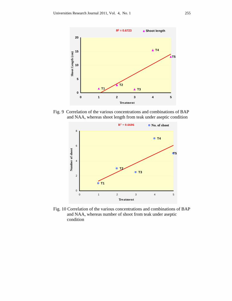
Universities Research Journal 2011, Vol. 4, No. 1 255
T1T2
T3
T4
T5
R2 = 0.6723
0
5
10
15
20
0 1 2 3 4 5
Treatment
Shoo
t Len
gth
(cm
)
Shoot length
Fig. 9 Correlation of the various concentrations and combinations of BAP
and NAA, whereas shoot length from teak under aseptic condition
T1
T2T3
T4
T5
R2 = 0.6606
0
2
4
6
8
0 1 2 3 4 5
Treatment
Num
ber
of sh
oot
No. of shoot
Fig. 10 Correlation of the various concentrations and combinations of BAP
and NAA, whereas number of shoot from teak under aseptic condition
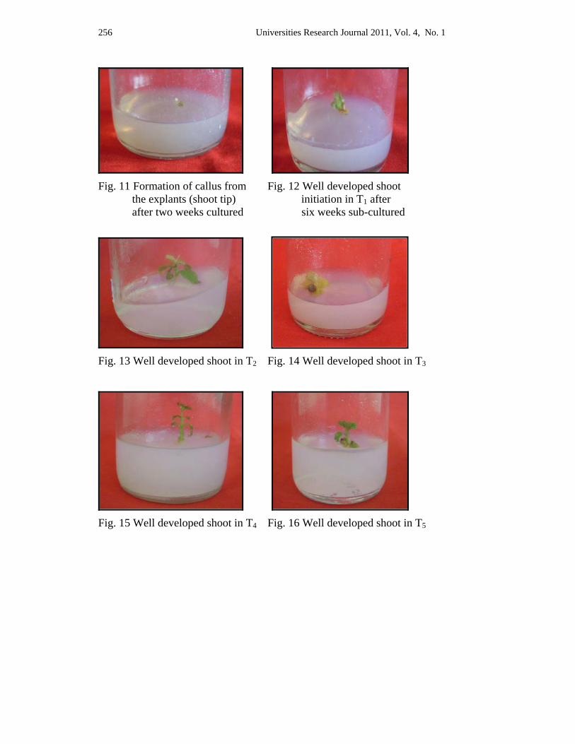
256 Universities Research Journal 2011, Vol. 4, No. 1
Fig. 11 Formation of callus from Fig. 12 Well developed shoot the explants (shoot tip) initiation in T1 after after two weeks cultured six weeks sub-cultured
Fig. 13 Well developed shoot in T2 Fig. 14 Well developed shoot in T3
Fig. 15 Well developed shoot in T4 Fig. 16 Well developed shoot in T5
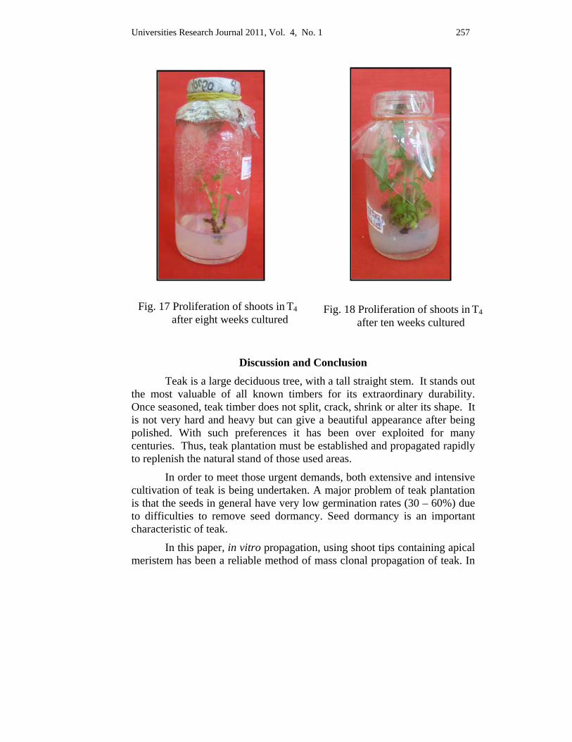
Universities Research Journal 2011, Vol. 4, No. 1 257
Discussion and Conclusion Teak is a large deciduous tree, with a tall straight stem. It stands out the most valuable of all known timbers for its extraordinary durability. Once seasoned, teak timber does not split, crack, shrink or alter its shape. It is not very hard and heavy but can give a beautiful appearance after being polished. With such preferences it has been over exploited for many centuries. Thus, teak plantation must be established and propagated rapidly to replenish the natural stand of those used areas.
In order to meet those urgent demands, both extensive and intensive cultivation of teak is being undertaken. A major problem of teak plantation is that the seeds in general have very low germination rates (30 – 60%) due to difficulties to remove seed dormancy. Seed dormancy is an important characteristic of teak.
In this paper, in vitro propagation, using shoot tips containing apical meristem has been a reliable method of mass clonal propagation of teak. In
Fig. 17 Proliferation of shoots in T4 after eight weeks cultured
Fig. 18 Proliferation of shoots in T4 after ten weeks cultured
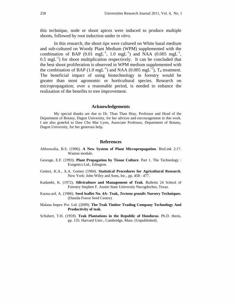
258 Universities Research Journal 2011, Vol. 4, No. 1
this technique, node or shoot apices were induced to produce multiple shoots, followed by root induction under in vitro.
In this research, the shoot tips were cultured on White basal medium and sub-cultured on Woody Plant Medium (WPM) supplemented with the combination of BAP (0.01 mgL-1, 1.0 mgL-1) and NAA (0.005 mgL-1, 0.5 mgL-1) for shoot multiplication respectively. It can be concluded that the best shoot proliferation is observed in WPM medium supplemented with the combination of BAP (1.0 mgL-1) and NAA (0.005 mgL-1), T4 treatment. The beneficial impact of using biotechnology in forestry would be greater than most agronomic or horticultural species. Research on micropropagation; over a reasonable period, is needed to enhance the realization of the benefits to tree improvement.
Acknowledgements My special thanks are due to Dr. Than Than Htay, Professor and Head of the
Department of Botany, Dagon University, for her advices and encouragement in this work. I am also grateful to Daw Cho Mar Lynn, Associate Professor, Department of Botany, Dagon University, for her generous help.
References Ahloowalia, B.S. (1996). A New System of Plant Micropropagation. BioLink 2:17.
Waston module.
Georage, E.F. (1993). Plant Propagation by Tissue Culture. Part 1. The Technology : Exegetics Ltd., Edington.
Gomez, K.A., A.A. Gomez (1984). Statistical Procedures for Agricultural Research. New York: John Wiley and Sons, Inc., pp. 458 - 477.
Kadambi, K. (1972). Siliviculture and Management of Teak. Bulletin 24 School of Forestry Stephen F. Austin State University Nacogdoches, Texas.
Kaosa-ard, A. (1986). Seed leaflet No. 4A- Teak, Tectona grandis Nursery Techniques. (Danida Forest Seed Centre).
Malana Impex Pvt. Ltd. (2009). The Teak Timber Trading Company Technology And Productivity of teak.
Schubert, T.H. (1959). Teak Plantations in the Republic of Honduras. Ph.D. thesis, pp. 135. Harvard Univ., Cambridge, Mass. (Unpublished).

Universities Research Journal 2011, Vol. 4, No. 1 259
Soerianegra, I. and R.H.M.J. Lemmens (1994). Plant Resources of South-East Asia 5(1). Timber trees: Major commercial timbers. Wageningen: Pudoc Scientific Publishers. pp. 610.
White, K.J. (1991). Teak. Some aspects of Research and Development. FAO Regional Office for Asia and the Pacific (RAPA) Bangkok.
Yasodha, R., R. Sumathi and K. Gurumurthi (2004). Micropropagation for Quality Propagule Production in Plantation Forestry. Indian Journal of Biotechnology, vol. 3, pp. 159-170.
Universities Research Journal 2011, Vol. 4, No. 1
1. Assistant Lecturer, Department of Botany, Magway University 2. Professor, Department of Botany, Magway University
Some Plants of Syzygium Found in Magway Township Pa Pa Win1 and May Than Su2
Abstract Seven species of Syzygium found in Magway Township were studied to identify these species and varieties of plants from January to March, 2010. Seven species are Syzygium grande, Syzygium jambos, Syzygium uniflora, Syzygium aromaticum, Syzygium spp. Var. Sein-thabye, Syzygium spp. var. Aung-thabye and Syzygium spp. var. Patamyar-thabye. In this research paper, outstanding characters, artificial key, distribution and identification of Syzygium were described. The habits, foliage, flowers and fruits of Syzygium sp. were recorded by photo.
Key words: Syzygium, identification, species, varieties, distribution
Introduction Magway Township is situated in the Dry Zone of Central Myanmar.
It is located between North latitude 20°5′30″ and 20°10′10″ and East 94°55′ and 94 °57′. It has an area of 436623 acres. It is in the western sector of the Bago Yoma or the Central Mountain Range and on the eastern bank of the Ayeyarwady river. April is the hottest month and December is the coldest month in this region.
Myrtaceae is abundant in the tropical and subtropical regions of both hemispheres. Syzygium is a genus of flowering plants that belongs to the myrtle family, Myrtaceae. These genus comprises about 1100species, and has a native range that extends from Africa and Madagascar through southern Asia East through the Pacific. Its highest levels of diversity occur from Malaysia to northeastern Australia, where many species are very poorly known and many more have not been described taxonomically. Some of the edible species of Syzygium are planted throughout the tropics worldwide. At times Syzygium was confused taxonomically with the genus Eugenia (1000 species), but the latter genus has its highest specific diversity in the tropics. Syzygium and Eugenia are among the most poorly known of the large ( >500 species) genera of vascular plants.
The natural vegetation of this area consists mostly of shrubs and trees of the genus Syzygium. Shrubs plants like Syzygium spp. Var. Aung-
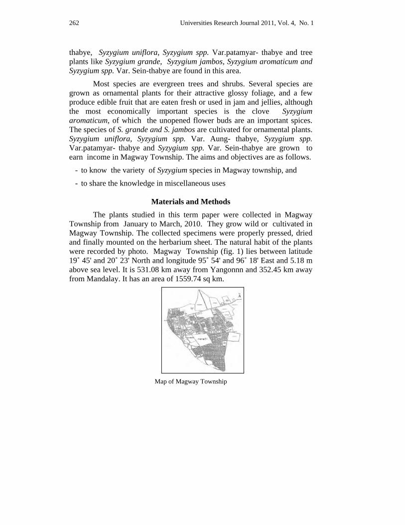
262 Universities Research Journal 2011, Vol. 4, No. 1
thabye, Syzygium uniflora, Syzygium spp. Var.patamyar- thabye and tree plants like Syzygium grande, Syzygium jambos, Syzygium aromaticum and Syzygium spp. Var. Sein-thabye are found in this area.
Most species are evergreen trees and shrubs. Several species are grown as ornamental plants for their attractive glossy foliage, and a few produce edible fruit that are eaten fresh or used in jam and jellies, although the most economically important species is the clove Syzygium aromaticum, of which the unopened flower buds are an important spices. The species of S. grande and S. jambos are cultivated for ornamental plants. Syzygium uniflora, Syzygium spp. Var. Aung- thabye, Syzygium spp. Var.patamyar- thabye and Syzygium spp. Var. Sein-thabye are grown to earn income in Magway Township. The aims and objectives are as follows.
- to know the variety of Syzygium species in Magway township, and
- to share the knowledge in miscellaneous uses
Materials and Methods The plants studied in this term paper were collected in Magway
Township from January to March, 2010. They grow wild or cultivated in Magway Township. The collected specimens were properly pressed, dried and finally mounted on the herbarium sheet. The natural habit of the plants were recorded by photo. Magway Township (fig. 1) lies between latitude 19˚ 45' and 20˚ 23' North and longitude 95˚ 54' and 96˚ 18' East and 5.18 m above sea level. It is 531.08 km away from Yangonnn and 352.45 km away from Mandalay. It has an area of 1559.74 sq km.
Map of Magway Township
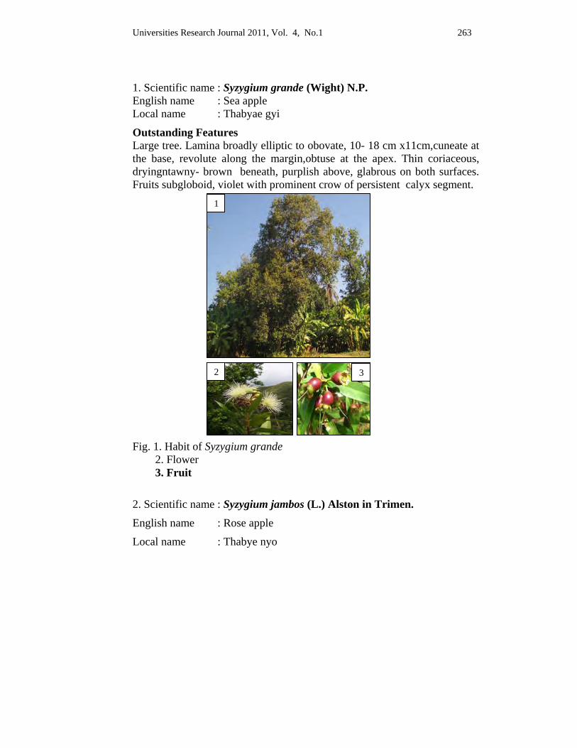
Universities Research Journal 2011, Vol. 4, No.1 263
1. Scientific name : Syzygium grande (Wight) N.P. English name : Sea apple Local name : Thabyae gyi
Outstanding Features Large tree. Lamina broadly elliptic to obovate, 10- 18 cm x11cm,cuneate at the base, revolute along the margin,obtuse at the apex. Thin coriaceous, dryingntawny- brown beneath, purplish above, glabrous on both surfaces. Fruits subgloboid, violet with prominent crow of persistent calyx segment.
Fig. 1. Habit of Syzygium grande 2. Flower 3. Fruit
2. Scientific name : Syzygium jambos (L.) Alston in Trimen.
English name : Rose apple
Local name : Thabye nyo
1
2 3
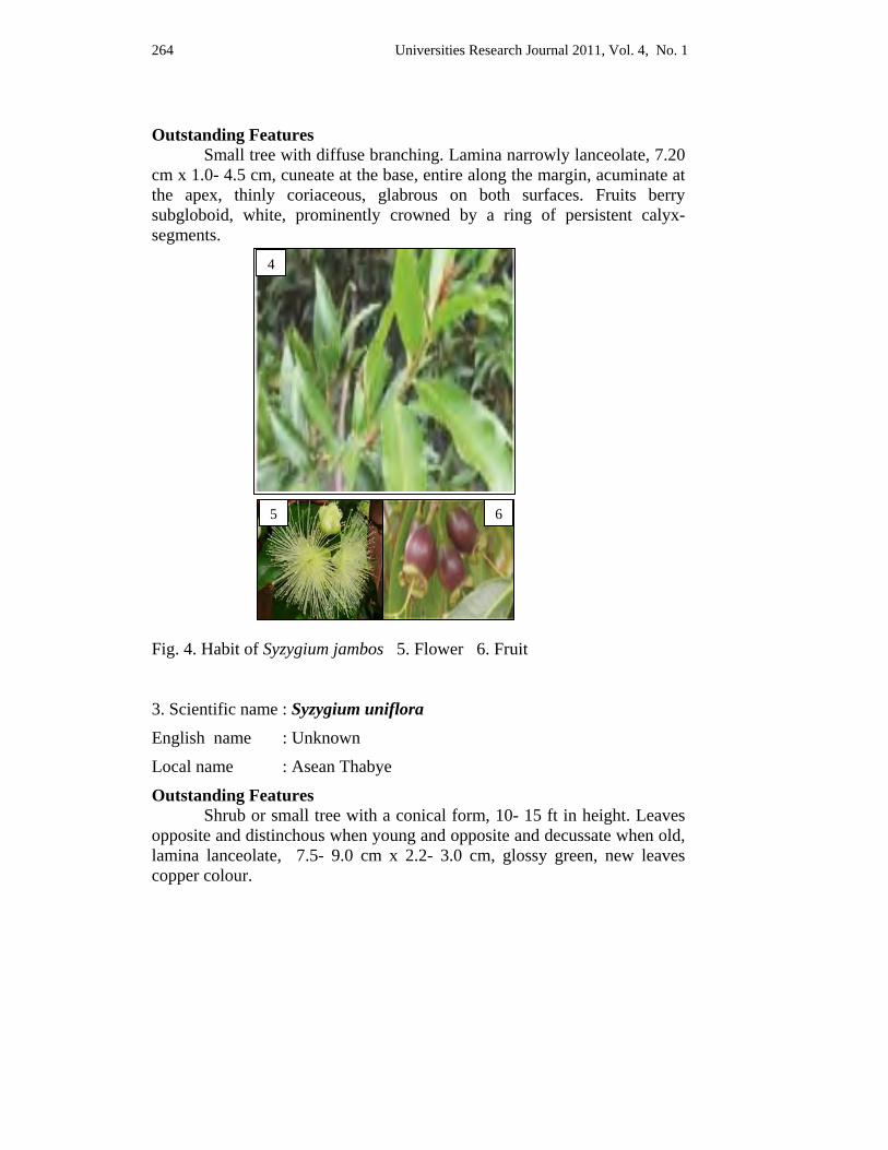
264 Universities Research Journal 2011, Vol. 4, No. 1
Outstanding Features
Small tree with diffuse branching. Lamina narrowly lanceolate, 7.20 cm x 1.0- 4.5 cm, cuneate at the base, entire along the margin, acuminate at the apex, thinly coriaceous, glabrous on both surfaces. Fruits berry subgloboid, white, prominently crowned by a ring of persistent calyx- segments.
Fig. 4. Habit of Syzygium jambos 5. Flower 6. Fruit 3. Scientific name : Syzygium uniflora
English name : Unknown
Local name : Asean Thabye
Outstanding Features Shrub or small tree with a conical form, 10- 15 ft in height. Leaves opposite and distinchous when young and opposite and decussate when old, lamina lanceolate, 7.5- 9.0 cm x 2.2- 3.0 cm, glossy green, new leaves copper colour.
4
5 6
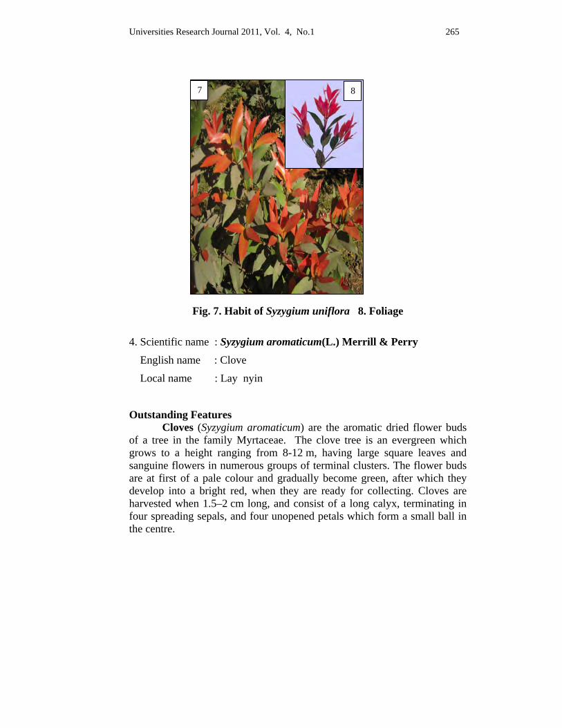
Universities Research Journal 2011, Vol. 4, No.1 265
Fig. 7. Habit of Syzygium uniflora 8. Foliage
4. Scientific name : Syzygium aromaticum(L.) Merrill & Perry
English name : Clove
Local name : Lay nyin
Outstanding Features Cloves (Syzygium aromaticum) are the aromatic dried flower buds
of a tree in the family Myrtaceae. The clove tree is an evergreen which grows to a height ranging from 8-12 m, having large square leaves and sanguine flowers in numerous groups of terminal clusters. The flower buds are at first of a pale colour and gradually become green, after which they develop into a bright red, when they are ready for collecting. Cloves are harvested when 1.5–2 cm long, and consist of a long calyx, terminating in four spreading sepals, and four unopened petals which form a small ball in the centre.
7 8
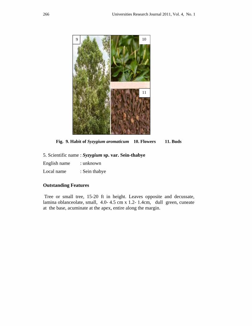
266 Universities Research Journal 2011, Vol. 4, No. 1
Fig. 9. Habit of Syzygium aromaticum 10. Flowers 11. Buds
5. Scientific name : Syzygium sp. var. Sein-thabye
English name : unknown
Local name : Sein thabye
Outstanding Features Tree or small tree, 15-20 ft in height. Leaves opposite and decussate, lamina oblanceolate, small, 4.0- 4.5 cm x 1.2- 1.4cm, dull green, cuneate at the base, acuminate at the apex, entire along the margin.
9 10
11
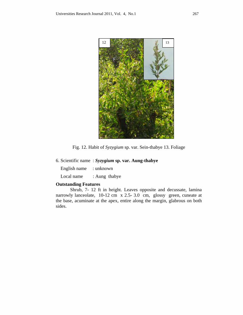
Universities Research Journal 2011, Vol. 4, No.1 267
Fig. 12. Habit of Syzygium sp. var. Sein-thabye 13. Foliage
6. Scientific name : Syzygium sp. var. Aung-thabye
English name : unknown
Local name : Aung thabye
Outstanding Features Shrub, 7- 12 ft in height. Leaves opposite and decussate, lamina narrowly lanceolate, 10-12 cm x 2.5- 3.0 cm, glossy green, cuneate at the base, acuminate at the apex, entire along the margin, glabrous on both sides.
12 13
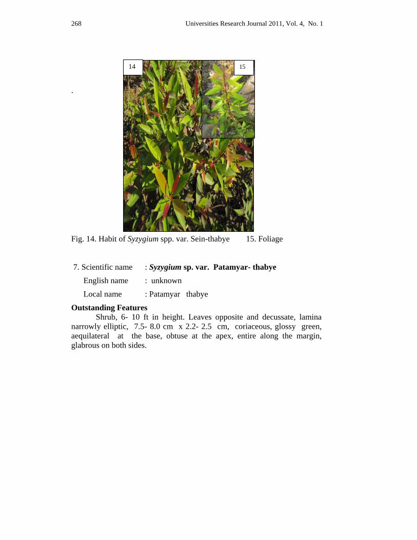
268 Universities Research Journal 2011, Vol. 4, No. 1
.
Fig. 14. Habit of Syzygium spp. var. Sein-thabye 15. Foliage 7. Scientific name : Syzygium sp. var. Patamyar- thabye
English name : unknown
Local name : Patamyar thabye
Outstanding Features Shrub, 6- 10 ft in height. Leaves opposite and decussate, lamina narrowly elliptic, 7.5- 8.0 cm x 2.2- 2.5 cm, coriaceous, glossy green, aequilateral at the base, obtuse at the apex, entire along the margin, glabrous on both sides.
14 15
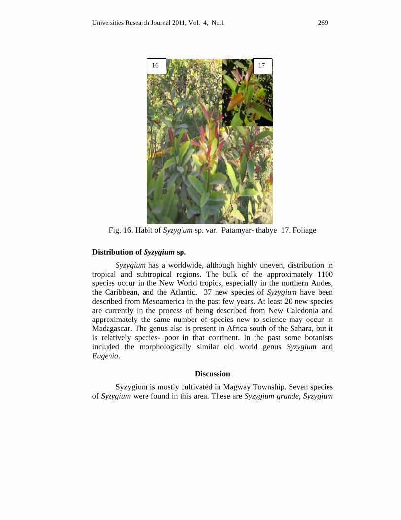
Universities Research Journal 2011, Vol. 4, No.1 269
Fig. 16. Habit of Syzygium sp. var. Patamyar- thabye 17. Foliage
Distribution of Syzygium sp. Syzygium has a worldwide, although highly uneven, distribution in tropical and subtropical regions. The bulk of the approximately 1100 species occur in the New World tropics, especially in the northern Andes, the Caribbean, and the Atlantic. 37 new species of Syzygium have been described from Mesoamerica in the past few years. At least 20 new species are currently in the process of being described from New Caledonia and approximately the same number of species new to science may occur in Madagascar. The genus also is present in Africa south of the Sahara, but it is relatively species- poor in that continent. In the past some botanists included the morphologically similar old world genus Syzygium and Eugenia.
Discussion Syzygium is mostly cultivated in Magway Township. Seven species
of Syzygium were found in this area. These are Syzygium grande, Syzygium
16 17
270 Universities Research Journal 2011, Vol. 4, No. 1
jambos, Syzygium uniflora, Syzygium aromaticum, Syzygium sp. var. Sein-thabye, Syzygium sp. var. Aung-thabye and Syzygium sp. var. Patamyar- thabye.
The present study deals with the members of Syzygium growing in Magway Township. A total of seven species of Syzygium has been identified and described. According to the resulting data, the common plants found in this area are Syzygium grande and S. jambos.
It can be pruned back hard, as it is a vigorous grower and the shortened new shoots appear with 6-8 pairs of leaves. The better shaping results are achieved with pruning. Leaf pruning can be done in summer on vigorous plants, but is not generally needed, as better leaf reduction results from timely pruning, and this plant has relatively small leaves in the first place.
Leaf growth for the renovation of canopy requires substantial amounts of nutrients and water. It is propagated by cutting in summer or seeds in fall or air- layering. Watering is needed more in summer and less in winter. It needs humidity, so moisting can be beneficial.
Conclusion According to the collected specimens, twenty one species belonging
to Syzygium could be identified and all the species were described with outstanding features, local names and scientific names. Syzygium is a large family of evergreen trees and shrubs. The Syzygium has many traits, attractive foliage, flowers and berries.
Among them S. grande and S. jambos were cultivated for ornamental, other species were economically important plant (Phayapan) in the studied area. People donated the young shoots as well as the flowers to the pagoda. Aung- thabye is very famous in Magway Township because people offer Syzygium to the pagoda and believe all the good fortune will come to them. Syzygium uniflora has the most beautiful colour copper colour. Patamyar- thabye is the most diuretic because the foliage is coriaceous and glossy green.
Cloves are native to Indonesia and used as a spice in cuisines all over the world. The English name derives from Latin clavus 'nail' (also the origin of French clou 'nail') as the buds vaguely resemble small irregular
Universities Research Journal 2011, Vol. 4, No.1 271
nails in shape. Cloves are now harvested primarily in Indonesia, Madagascar, Zanzibar, Pakistan, and Sri Lanka.
It is hoped that the present study will give the variety of Syzygium in Magway environs with colourful photographs. They are also Myanmar traditional ethical plants used for a long time.
Acknowledgements
We would like to express our gratitude to Dr. Aye Kyaw, Rector, Magway University for his encouragement to carry out this research work. Our thanks are also due to all the staff of Botany Department, Magway University for their kind help throughout the research.
References
Cronquist, 1981. An Integrated System of classification of flowering plants, Columbia University press New York.
Dassanayake, M. D., 1980-2001. A Revised Handbook to the Flora of Ceylon, Vol 1 to 14. University of Peradeniya, Department of Agriculture, Peradenya, Sri Lanka.
Khin Pale , 2008. Floristic study on angiospermae of yeywa area, Mandalay division, PhD Thesis, Department of Botany, University of Mandalay.
Website:
N. Sivasothi, a.k.a. Otterman, Raffles Museum of Biodiversity Research, Department of Biological Sciences, National University of Singapore. Since 1998 with origins from OneList.
File://F:\Sizygium\Syzygium.htm. Syzygium- wikipedia, the free encyclopedia
File://C:DOCUME-1\ADMINI~1\ COCALS~1\ Temp\5NT7GNE2.htm.
http://www.tradewindsfruit.com./java-plum.htm.
http://www.tradewindsfruit.com./water- cherry.htm.
http://www.tradewindsfruit.com./clove.htm.
File://F:\Sizygium\Syzygium.htm. Syzygium- wikipedia, the free encyclopedia
File://F:\Sizygium\Syzygium.htm. Syzygium- wikipedia, the free encyclopedia
http://www.tradewindsfruit.com./rose- apple .htm
http://www.tradewindsfruit.com./clove..htm.
Universities Research Journal 2011, Vol. 4, No. 1
Assistant Lecturer, Department of Botany, Pathein University
Isolation and Identification of Mangrove Fungus from Bruguiera sexangula (Lour.) Poir.
Khin Min Min Phyo
Abstract In the isolation of endophytic fungi, Bruguiera sexangula (Lour.) Poir. (Rhizophoraceae) was utilized and collected from Chaung Tha. In this study, 2 endophytic fungi were isolated. Firstly, only one fungus was investigated for identification. In the study of identification, the endophytic fungus can be keyed out as Chalaropsis sp. according to the macroscopical and microscopical characters. The other fungus will be identified for further investigation.
Key words: isolation, endophytic fungi
Introduction Mangrove represents a rich and diverse living resource. The word ‘mangroves’ stands for tropical plants and their communities which grow in the intertidal zone. The intertidal zone is the area under tidal influence along the coast line, such as seashores, estuaries, lagoons and river banks. Mangrove forests are now recognized as being a major protector of coastal environments and a valuable national economic resource. Nowadays, the awareness of coastal communities to the importance of mangrove forest conservation has been increasing. Moreover, the useful mangroves should be preserved as the other useful plants due to the effective metabolites producing microorganism which can also be extracted from the mangroves (Tomlinson, 1986; Bali and Lombok 1997).
Identification of organisms is an important step in understanding and analyzing biological processes. The ability to recognize and to name the organisms is the important key (Phyo, Phay, Suto and Ando, 1994).
This research aims to study the isolation of endophytic fungi, to investigate the identification of endophytic fungi according to their macroscopical and microscopical characters and to have the sense for preservation of mangrove plants.
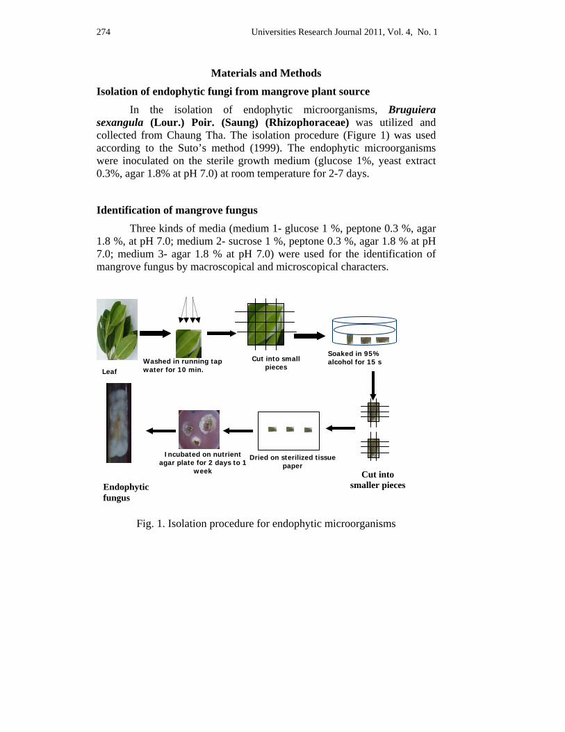
274 Universities Research Journal 2011, Vol. 4, No. 1
Materials and Methods
Isolation of endophytic fungi from mangrove plant source
In the isolation of endophytic microorganisms, Bruguiera sexangula (Lour.) Poir. (Saung) (Rhizophoraceae) was utilized and collected from Chaung Tha. The isolation procedure (Figure 1) was used according to the Suto’s method (1999). The endophytic microorganisms were inoculated on the sterile growth medium (glucose 1%, yeast extract 0.3%, agar 1.8% at pH 7.0) at room temperature for 2-7 days.
Identification of mangrove fungus
Three kinds of media (medium 1- glucose 1 %, peptone 0.3 %, agar 1.8 %, at pH 7.0; medium 2- sucrose 1 %, peptone 0.3 %, agar 1.8 % at pH 7.0; medium 3- agar 1.8 % at pH 7.0) were used for the identification of mangrove fungus by macroscopical and microscopical characters.
Fig. 1. Isolation procedure for endophytic microorganisms
Leaf Washed in running tap water for 10 min.
Cut into small pieces
Soaked in 95% alcohol for 15 s
Dried on sterilized tissue paper
Incubated on nutrient agar plate for 2 days to 1
week Cut into smaller pieces Endophytic
fungus
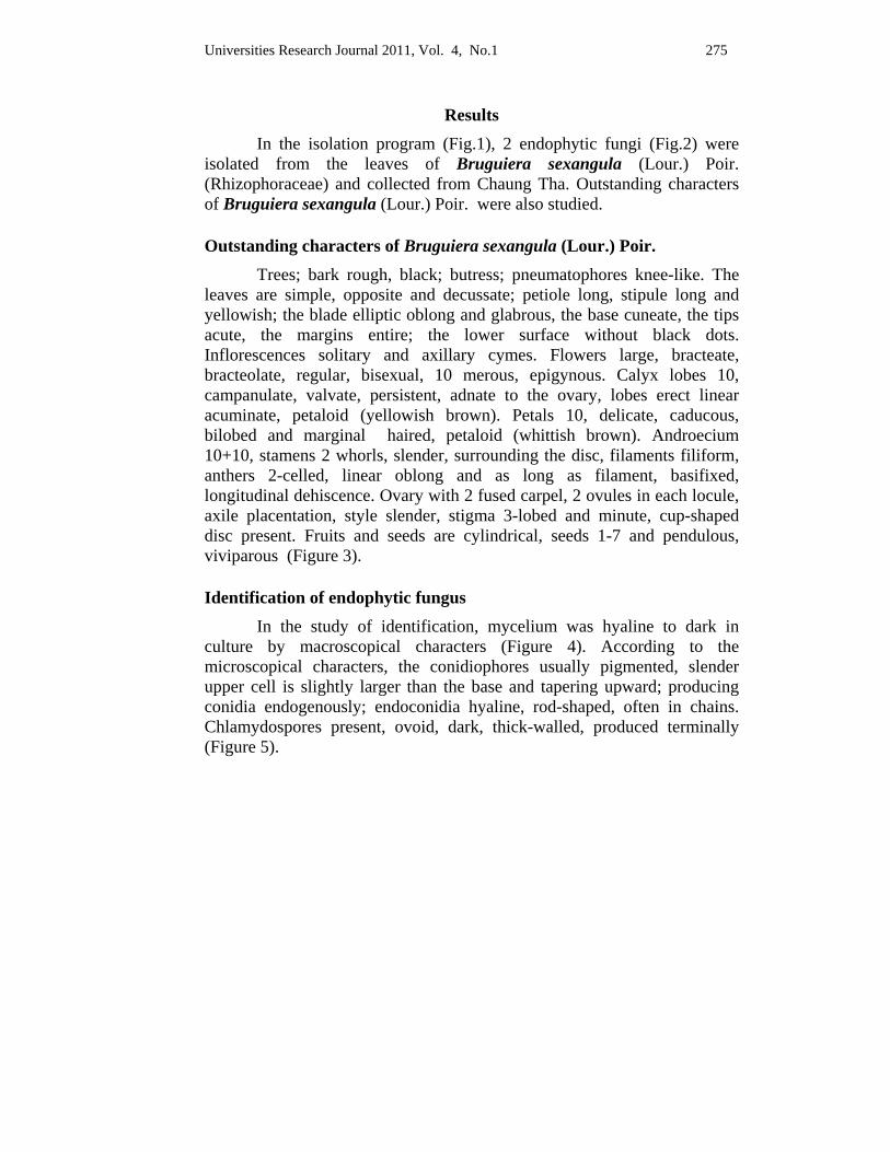
Universities Research Journal 2011, Vol. 4, No.1 275
Results In the isolation program (Fig.1), 2 endophytic fungi (Fig.2) were
isolated from the leaves of Bruguiera sexangula (Lour.) Poir. (Rhizophoraceae) and collected from Chaung Tha. Outstanding characters of Bruguiera sexangula (Lour.) Poir. were also studied. Outstanding characters of Bruguiera sexangula (Lour.) Poir.
Trees; bark rough, black; butress; pneumatophores knee-like. The leaves are simple, opposite and decussate; petiole long, stipule long and yellowish; the blade elliptic oblong and glabrous, the base cuneate, the tips acute, the margins entire; the lower surface without black dots. Inflorescences solitary and axillary cymes. Flowers large, bracteate, bracteolate, regular, bisexual, 10 merous, epigynous. Calyx lobes 10, campanulate, valvate, persistent, adnate to the ovary, lobes erect linear acuminate, petaloid (yellowish brown). Petals 10, delicate, caducous, bilobed and marginal haired, petaloid (whittish brown). Androecium 10+10, stamens 2 whorls, slender, surrounding the disc, filaments filiform, anthers 2-celled, linear oblong and as long as filament, basifixed, longitudinal dehiscence. Ovary with 2 fused carpel, 2 ovules in each locule, axile placentation, style slender, stigma 3-lobed and minute, cup-shaped disc present. Fruits and seeds are cylindrical, seeds 1-7 and pendulous, viviparous (Figure 3).
Identification of endophytic fungus
In the study of identification, mycelium was hyaline to dark in culture by macroscopical characters (Figure 4). According to the microscopical characters, the conidiophores usually pigmented, slender upper cell is slightly larger than the base and tapering upward; producing conidia endogenously; endoconidia hyaline, rod-shaped, often in chains. Chlamydospores present, ovoid, dark, thick-walled, produced terminally (Figure 5).
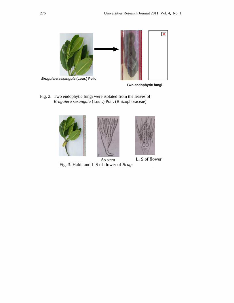
276 Universities Research Journal 2011, Vol. 4, No. 1
Fig. 2. Two endophytic fungi were isolated from the leaves of
Bruguiera sexangula (Lour.) Poir. (Rhizophoraceae)
Fig. 3. Habit and L S of flower of Bruguiera sexangula
Bruguiera sexangula (Lour.) Poir.
Two endophytic fungi
As seen L. S of flower
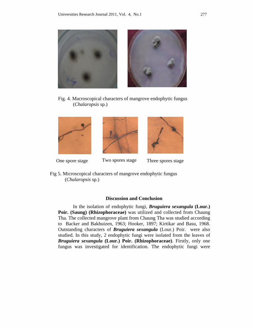
Universities Research Journal 2011, Vol. 4, No.1 277
Fig. 4. Macroscopical characters of mangrove endophytic fungus
(Chalaropsis sp.)
Discussion and Conclusion In the isolation of endophytic fungi, Bruguiera sexangula (Lour.)
Poir. (Saung) (Rhizophoraceae) was utilized and collected from Chaung Tha. The collected mangrove plant from Chaung Tha was studied according to Backer and Bakhuizen, 1963; Hooker, 1897; Kirtikar and Basu, 1968. Outstanding characters of Bruguiera sexangula (Lour.) Poir. were also studied. In this study, 2 endophytic fungi were isolated from the leaves of Bruguiera sexangula (Lour.) Poir. (Rhizophoraceae). Firstly, only one fungus was investigated for identification. The endophytic fungi were
One spore stage Two spores stage Three spores stage
Fig 5. Microscopical characters of mangrove endophytic fungus (Chalaropsis sp.)
278 Universities Research Journal 2011, Vol. 4, No. 1
placed onto the nutrient agar plates at three points, equidistant from the center and incubated for 7 days at room temperature. After 7 days, the developing fungal colony was examined by color of colony with reverse phase and spore formation. In the study of identification, the endophytic fungus can be keyed out as Chalaropsis sp. according to the macroscopical and microscopical characters. This endophytic fungus was identified according to the key steps of Barnett (1955). The other fungus will be identified for the further investigation.
Acknowledgements
I would like to express my sincere thanks to Professor Dr. Nyunt Phay, Rector, Pathein University, Head of Biological Resources and Biotechnology Development Centre, for his kind permission to use the required facilities to carry out the present study. My acknowledgements should also go to Dr. Kay Thi Mya, Professor and Head and Dr. War War Lwin, Associate Professor, Department of Botany, Pathein University, for their permissions and constant encouragements to do this research. I would also like to record my thanks to Dr. San Tha Tun, Associate Professor, Department of Marine Science, Pathein University, for his patient help to collect the mangrove specimens.
References Backer, C.A. and R.C. Bakhuizen (1963). Flora of Java, Vol. I, N.V.P., Netherlands.
Bali and Lombok (1997), Handbook of Mangrove in Indonesia, JICA, Japan.
Barnett, H. L. (1955). Imperfect fungi, Plant Pathology Dept., West Virginia.
Hooker, J.D. (1897). Flora of British India Vol. II, L. Reeve & Co.Ltd., England. Hyde, K.D. (1990).
Kirtikar, E.R. and B.D. Basu (1968). Indian medicinal plants, Vol.I. Jayyed Press, New Dehli.
Phyo, K.M.M., N. Phay, M. Suto, and K. Ando, (1994). 10th International Congress for Culture Collections, Tsukuba, Japan, 10-25: 601-602.
Suto, M. (1999). Isolation of endophytes from plants, in Molecular tools on isolation and screening of microbes for useful materials, Workshop in Malaysia.
Tomlinson, P.B. (1986). The Botany of Mangroves; Cambridge University Press, London.
Universities Research Journal 2011, Vol. 4, No.1
Assistant Lecturer, Department of Botany, Sittway University
Morphological and Microscopical Characters of Aglaia odorata Lour.
Tin Tin Pyone
Abstract Aglaia odorata Lour. belongs to the family Meliaceae which is the cultivated plant as ornamental. It was collected from University of Yangon. "Thanat-kha-pan" is the Myanmar name of this species. It is used in herbal medicine especially in the treatment of heart stimulant and febrifuge. In this research, the morphological and microscopical characters of leaves, stems and roots were studied to make their correct verification. Microscopical characters of leaves, stems and roots were also undertaken and examinations of powdered drug were carried out for standardization of drugs.
Key words: ornamental, herbal medicine.
Introduction All plants possess several characters of a morphological, histological, embryological, chemical and genetic nature which are potentially available for building up a classification of the plant kingdom. In the artificial schemes of botanical classification the characters employed were ones which, by experience, had shown that they could be used to produce suitable groups or taxa. These system arrange taxa (any groups used for classification such as orders, families, genera, etc.) indicate the possible relationship of one taxon to another.
The plant kingdom holds many species of plants containing substances of medicinal value which have yet to be discovered. Compared with morphological characters and chemical constituents are more precisely definable and may be of more fundamental significance for classification purpose.
All the information, both botanical and chemical, should be taken into account but as botanists themselves point out, they are almost successful in defining limits as the trained taxonomist (Trease and Evans, 1964). Morphological and microscopical studies are prerequisite for medicinal purpose. The odour of Aglaia odorata Lour. is pleasant and the flowers are beautiful in colour and grown for ornamental in home garden.
280 Universities Research Journal 2011, Vol. 4, No.1
So in this research, morphological and microscopical characters of fresh specimens and dried powder of plant Aglaia odorata Lour. were found out.
Materials and Methods Aglaia odorata Lour. was collected from University of Yangon. This plant was collected during flowering period from May to September.
The fresh leaves, stems and roots were used to study its morphological and microscopical characters of the plant. Free hand sections were made and studied under the microscope. Stomatal index, palisade ratio, vein islets and vein termination number in leaves. Stems and roots were determined by the method of Wallis (1967) and Trease and Evans (2002).
The collected samples were washed with water to remove impurities. After washing and cleaning, the sample was air dried and ground to get powder and stored in air-tight container.
1. Chloral hydrate solution B.P for clearing reagent.
2. Phloroglucinol solution B.P, followed by concentration HCl for testing lignin.
3. Acetic acid for testing calcium oxalate crystals.
4. Iodine solution for testing starch.
5. Glycerin was prepared for temporary mounts.
Results
Morphological characters of Aglaia odorata Lour. Small ever-green shrubby trees, about 8-10 m high. Stems woody,
cylindrical glabrous, the lenticels present. Leaves alternate, unipinnately compound, imparipinnate, the leaflets 3-5, opposite, obovate elliptic, (3.5-9.0) cm length and (1.8-3.5) cm broad, the tips acute to obtuse, the upper surface glabrous, the lower ones glabrescent, unicostate, petiolules about (0.2-0.4) cm long glabrous, petioles about (2.5-3.0) cm long, puberulent, racheae about (5.5-7.5) cm long, glabrescent, exstipulate. Inflorescence axillary, paniculate cymes, (9.0-15.0) cm long, length and (4.0-8.0) cm
Universities Research Journal 2011, Vol. 4, No.1 281
wide, peduncles angular, (1.8-2.5) cm long, glabrous, bracts minute, linear, less than 1 mm long, glabrous, pedicles about (1.5-2.0) mm long, glabrous, ebracteolate. Flowers yellow, globoid, about (3.0-4.0) mm length and (2.0-3.0) mm wide, complete, bisexual, regular, actinomorphic, 5-merous, hypogenous. Calyx 5, aposepalous, green, ovate, less than (0.4-0.5) mm length and less than 1 mm wide, quincuncial, glabrous within and without. Corolla 5, apopetalous, yellow, nearly orbicular, about (2.0-2.5) mm long and (1.0-1.1) mm wide, quincuncial glabrous without and within. Stamens 5, monadelphous, the filaments sessile, staminal tube yellow, globoid urceolate with nearly truncate apices, about (2.0-2.2) mm long (1.0-1.5) mm wide, the anther dithecous, dorsifixed, longitudinal dehiscence, introrse, ovary superior. Pistil 1, globose about (0.4-0.5) mm in diameter, bicarpellary, syncarpous, unilocular, one pendulous ovule in the locule, tomentose, style short, cylindrical, about (0.4-0.5) mm long, glabrous, stigma capitate, disc absent, superior.
Found growing in dry place. Flowering period from May to September (Figure 1, 2, 3, 4, 5, 6).
Microscopical characters of Leaves, Stems and Roots
Lamina In surface view, the epidermal cells of both surfaces were parenchymatous and thin-walled. The cell walls of the upper surfaces and lower surfaces were polygonal to barrel in shape.
Stomata were present on the lower surfaces and absent on the upper surface. It is anomocytic stomata. The guard cells were reniform in shape and contains abundant chloroplast. In transverse sections, the cuticle layer of the upper surface was thick. The lower cuticle layer was thicker than the upper surface. The upper epidermal cells were polygonal to barrel shape. The lower epidermal cells were similar in shape and size.
The mesophyll consisted of palisade and spongy parenchyma. The palisade mesophyll was made up of two layers of vertically elongated cylindrical cells, which were closely packed with one another. Secretory cavities of various size and shape occur in palisade layers. The spongy mesophyll consisted of 6-7 layers of cells, which were irregular to oval in shape and loosely arranged. They contain numerous chloroplasts.
282 Universities Research Journal 2011, Vol. 4, No.1
The vascular bundle of lateral vein consisted of phloem lying towards the upper epidermis and xylem lying towards the inner and collateral type. Each bundle is surrounded by a compact layer of thin-walled parenchymatous sheath, distinct from the neighboring cells.
Aglaia odorata Lour. xylem was composed of annular and spirally thickened vessels, tracheids, fibres and xylem parenchyma. Phloem is composed of sieve-tubes, companion cells and phloem parenchyma (Figure 9).
Midrib In surface view, the epidermal cells of both surfaces were parenchymatous and elongated along the length of the midrib. The lower epidermal cells were comparatively smaller than the upper epidermis.
In transverse section, the midrib was slightly curved inwards and cuticle thick. The epidermal cells of both surfaces are polygonal to rectangular in shape.
The cortex was made up of lamellar collenchyma and thin-walled parenchyma cells. The collenchyma cells were 2-3 layers in thickness towards the upper surface and 3-5 layers in thickness towards the lower surface. They were polygonal to round in shaped.
The parenchyma cells were 4-5 layers in thickness above the vascular bundle and 5-7 layers in thickness below the vascular bundle. They were thin-walled and rounded to oval in shape.
Pith from the central part of the midrib which remains enclosed by the vascular tissue. It consisted of loosely arranged parenchymatous cells with abundant intercellular spaces. The parenchymatous tissue of pith consisted of 5-6 layers, rounded to oval in shape.
The vascular bundle was more or less rounded in outline. Each vascular bundle was surrounded by a compact layer of sclerenchymatous cells known as the bundle sheath in layers. The cells were thick-walled, and lignified.
Vascular bundles of midrib were collateral type, the xylem cells are hexagonal, thick-walled, and lignified, composed of vessels, tracheids, fibers, and xylem parenchyma. The phloem cells were thin-walled and composed of sieve-tube, companion cells and phloem parenchyma (Figure 10).
Universities Research Journal 2011, Vol. 4, No.1 283
Petiole In surface view, the epidermal cells of both surfaces were parenchymatous, thin-walled and rectangular to polygonal in shape and elongated along the length of the petiole. The upper and lower epidermal cells were similar shape and size.
In transverse section, the petiole of leaves was basically arc-shaped. The cuticle layers were thin and smooth. The epidermal cells were polygonal to rectangular in shape.
The cortex was made up of two different types of tissue, the lamellar collenchymatous tissue towards the peripheral region and thin-walled parenchymatous tissues towards the inner region.
The collenchymatous tissues consisted of 3-4 layers in thickness. The parenchymatous tissues consisted of 5-7 layers in thickness. The parenchyma cells were rounded to oval in shape. Intercellular spaces were numerous among the tissue. Crystals and group of stone cells are present in the parenchymatous cells.
Pith from the central part of the petiole which remained enclosed by the vascular tissue. It consisted of loosely arranged parenchymatous cells. The parenchymatous tissue of pith consisted of 5-7 layers of cells in thickness. The parenchyma cells were rounded to oval in shape. Intercellular spaces were numerous among the tissue. Calcium-oxalate crystals were present in the parenchymatous cells.
The vascular bundle was arc-shaped, collateral type. Each vascular bundle was surrounded by a compact layer of the sclerenchymatous cells known as the bundle sheath. The cells are thick-walled, and lignified.
The xylems were present between the phloem and pith. Xylem is composed of vessels, tracheids, fibers and xylem parenchyma. Xylem cells were lignified, thick-walled, and hexagonal. Phloem is composed of sieve-tube, companion cells and phloem parenchyma (Figure 11, 12).
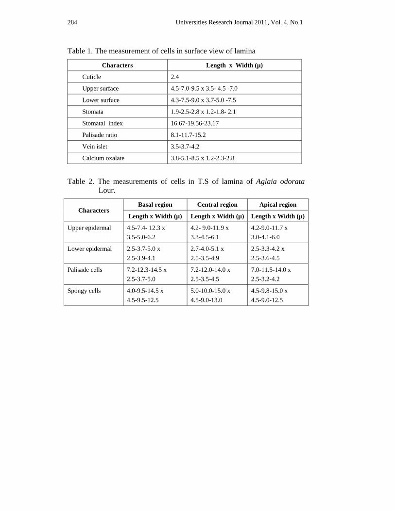
284 Universities Research Journal 2011, Vol. 4, No.1
Table 1. The measurement of cells in surface view of lamina
Characters Length x Width (µ)
Cuticle 2.4
Upper surface 4.5-7.0-9.5 x 3.5- 4.5 -7.0
Lower surface 4.3-7.5-9.0 x 3.7-5.0 -7.5
Stomata 1.9-2.5-2.8 x 1.2-1.8- 2.1
Stomatal index 16.67-19.56-23.17
Palisade ratio 8.1-11.7-15.2
Vein islet 3.5-3.7-4.2
Calcium oxalate 3.8-5.1-8.5 x 1.2-2.3-2.8
Table 2. The measurements of cells in T.S of lamina of Aglaia odorata
Lour.
Characters Basal region Central region Apical region
Length x Width (µ) Length x Width (µ) Length x Width (µ)
Upper epidermal 4.5-7.4- 12.3 x 3.5-5.0-6.2
4.2- 9.0-11.9 x 3.3-4.5-6.1
4.2-9.0-11.7 x 3.0-4.1-6.0
Lower epidermal 2.5-3.7-5.0 x 2.5-3.9-4.1
2.7-4.0-5.1 x 2.5-3.5-4.9
2.5-3.3-4.2 x 2.5-3.6-4.5
Palisade cells 7.2-12.3-14.5 x 2.5-3.7-5.0
7.2-12.0-14.0 x 2.5-3.5-4.5
7.0-11.5-14.0 x 2.5-3.2-4.2
Spongy cells 4.0-9.5-14.5 x 4.5-9.5-12.5
5.0-10.0-15.0 x 4.5-9.0-13.0
4.5-9.8-15.0 x 4.5-9.0-12.5
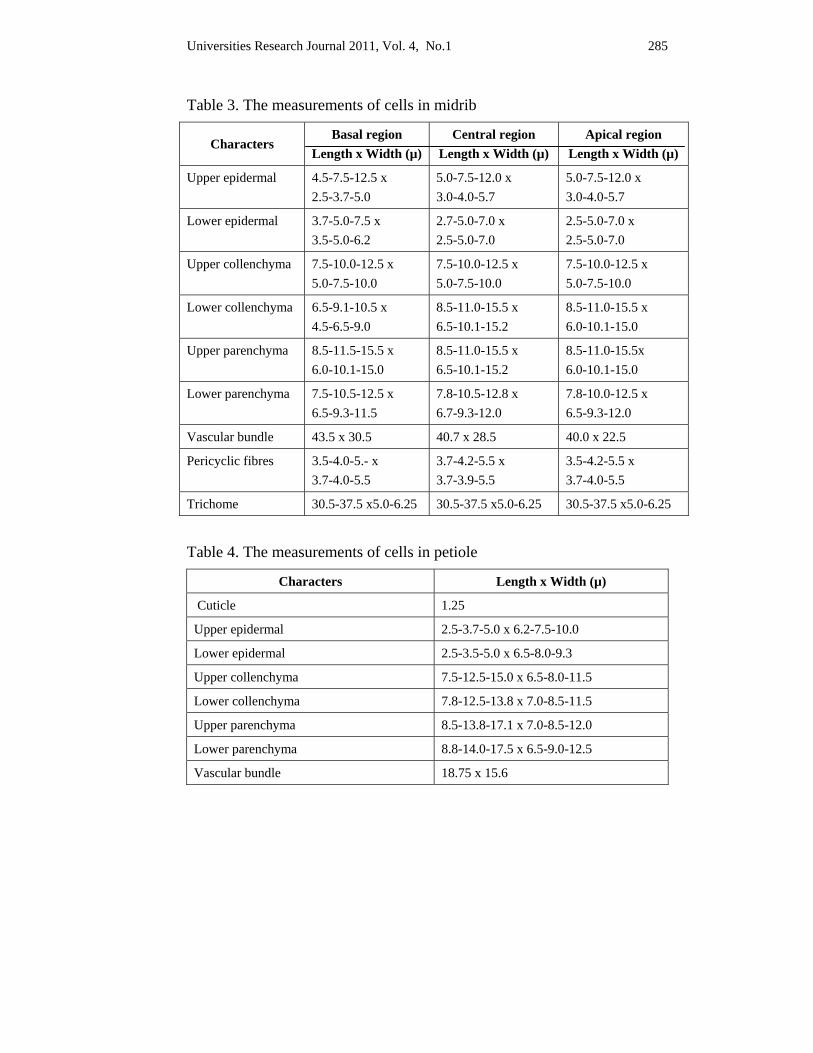
Universities Research Journal 2011, Vol. 4, No.1 285
Table 3. The measurements of cells in midrib
Characters Basal region
Length x Width (µ) Central region
Length x Width (µ) Apical region
Length x Width (µ)
Upper epidermal 4.5-7.5-12.5 x 2.5-3.7-5.0
5.0-7.5-12.0 x 3.0-4.0-5.7
5.0-7.5-12.0 x 3.0-4.0-5.7
Lower epidermal 3.7-5.0-7.5 x 3.5-5.0-6.2
2.7-5.0-7.0 x 2.5-5.0-7.0
2.5-5.0-7.0 x 2.5-5.0-7.0
Upper collenchyma 7.5-10.0-12.5 x 5.0-7.5-10.0
7.5-10.0-12.5 x 5.0-7.5-10.0
7.5-10.0-12.5 x 5.0-7.5-10.0
Lower collenchyma 6.5-9.1-10.5 x 4.5-6.5-9.0
8.5-11.0-15.5 x 6.5-10.1-15.2
8.5-11.0-15.5 x 6.0-10.1-15.0
Upper parenchyma 8.5-11.5-15.5 x 6.0-10.1-15.0
8.5-11.0-15.5 x 6.5-10.1-15.2
8.5-11.0-15.5x 6.0-10.1-15.0
Lower parenchyma 7.5-10.5-12.5 x 6.5-9.3-11.5
7.8-10.5-12.8 x 6.7-9.3-12.0
7.8-10.0-12.5 x 6.5-9.3-12.0
Vascular bundle 43.5 x 30.5 40.7 x 28.5 40.0 x 22.5
Pericyclic fibres 3.5-4.0-5.- x 3.7-4.0-5.5
3.7-4.2-5.5 x 3.7-3.9-5.5
3.5-4.2-5.5 x 3.7-4.0-5.5
Trichome 30.5-37.5 x5.0-6.25 30.5-37.5 x5.0-6.25 30.5-37.5 x5.0-6.25
Table 4. The measurements of cells in petiole
Characters Length x Width (µ)
Cuticle 1.25
Upper epidermal 2.5-3.7-5.0 x 6.2-7.5-10.0
Lower epidermal 2.5-3.5-5.0 x 6.5-8.0-9.3
Upper collenchyma 7.5-12.5-15.0 x 6.5-8.0-11.5
Lower collenchyma 7.8-12.5-13.8 x 7.0-8.5-11.5
Upper parenchyma 8.5-13.8-17.1 x 7.0-8.5-12.0
Lower parenchyma 8.8-14.0-17.5 x 6.5-9.0-12.5
Vascular bundle 18.75 x 15.6
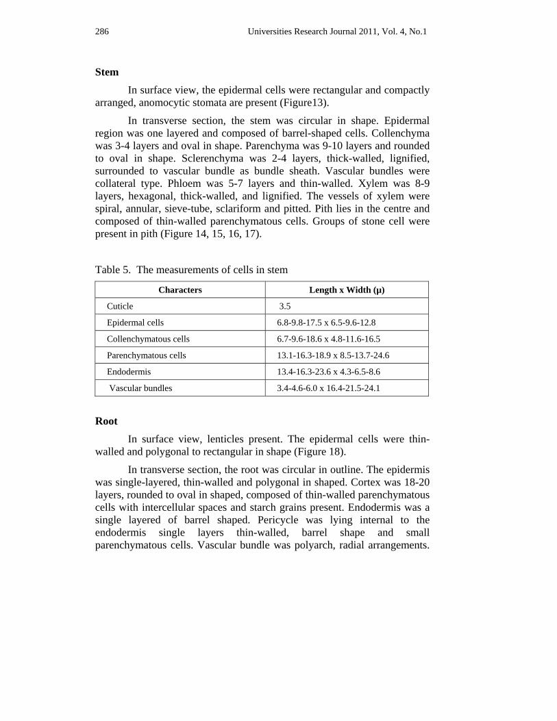
286 Universities Research Journal 2011, Vol. 4, No.1
Stem In surface view, the epidermal cells were rectangular and compactly arranged, anomocytic stomata are present (Figure13).
In transverse section, the stem was circular in shape. Epidermal region was one layered and composed of barrel-shaped cells. Collenchyma was 3-4 layers and oval in shape. Parenchyma was 9-10 layers and rounded to oval in shape. Sclerenchyma was 2-4 layers, thick-walled, lignified, surrounded to vascular bundle as bundle sheath. Vascular bundles were collateral type. Phloem was 5-7 layers and thin-walled. Xylem was 8-9 layers, hexagonal, thick-walled, and lignified. The vessels of xylem were spiral, annular, sieve-tube, sclariform and pitted. Pith lies in the centre and composed of thin-walled parenchymatous cells. Groups of stone cell were present in pith (Figure 14, 15, 16, 17).
Table 5. The measurements of cells in stem
Characters Length x Width (µ)
Cuticle 3.5
Epidermal cells 6.8-9.8-17.5 x 6.5-9.6-12.8
Collenchymatous cells 6.7-9.6-18.6 x 4.8-11.6-16.5
Parenchymatous cells 13.1-16.3-18.9 x 8.5-13.7-24.6
Endodermis 13.4-16.3-23.6 x 4.3-6.5-8.6
Vascular bundles 3.4-4.6-6.0 x 16.4-21.5-24.1
Root In surface view, lenticles present. The epidermal cells were thin-walled and polygonal to rectangular in shape (Figure 18).
In transverse section, the root was circular in outline. The epidermis was single-layered, thin-walled and polygonal in shaped. Cortex was 18-20 layers, rounded to oval in shaped, composed of thin-walled parenchymatous cells with intercellular spaces and starch grains present. Endodermis was a single layered of barrel shaped. Pericycle was lying internal to the endodermis single layers thin-walled, barrel shape and small parenchymatous cells. Vascular bundle was polyarch, radial arrangements.
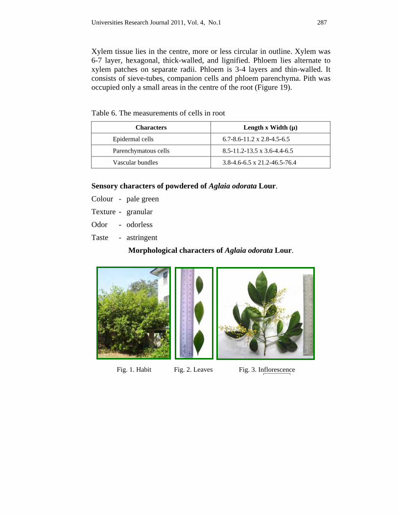
Universities Research Journal 2011, Vol. 4, No.1 287
Xylem tissue lies in the centre, more or less circular in outline. Xylem was 6-7 layer, hexagonal, thick-walled, and lignified. Phloem lies alternate to xylem patches on separate radii. Phloem is 3-4 layers and thin-walled. It consists of sieve-tubes, companion cells and phloem parenchyma. Pith was occupied only a small areas in the centre of the root (Figure 19).
Table 6. The measurements of cells in root
Characters Length x Width (µ)
Epidermal cells 6.7-8.6-11.2 x 2.8-4.5-6.5
Parenchymatous cells 8.5-11.2-13.5 x 3.6-4.4-6.5
Vascular bundles 3.8-4.6-6.5 x 21.2-46.5-76.4
Sensory characters of powdered of Aglaia odorata Lour.
Colour - pale green
Texture - granular
Odor - odorless
Taste - astringent
Morphological characters of Aglaia odorata Lour.
Fig. 3. Inflorescence Fig. 2. Leaves Fig. 1. Habit
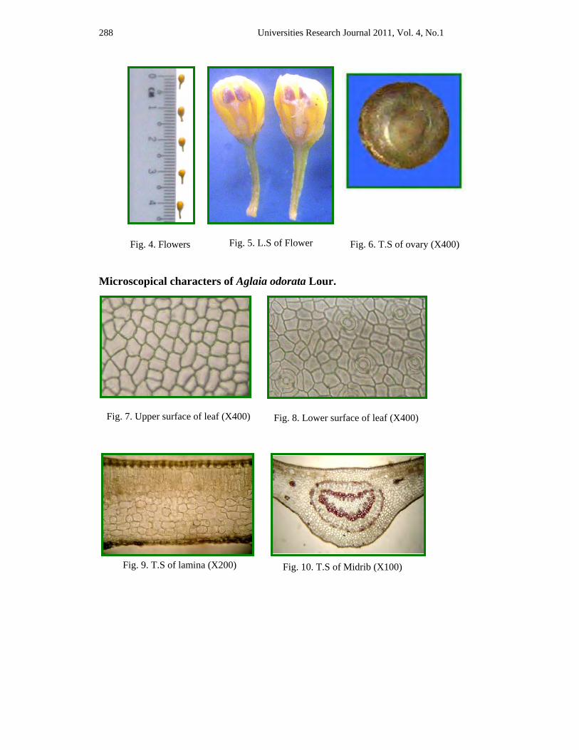
288 Universities Research Journal 2011, Vol. 4, No.1
Microscopical characters of Aglaia odorata Lour.
Fig. 4. Flowers Fig. 5. L.S of Flower Fig. 6. T.S of ovary (X400)
Fig. 7. Upper surface of leaf (X400) Fig. 8. Lower surface of leaf (X400)
Fig. 10. T.S of Midrib (X100) Fig. 9. T.S of lamina (X200)
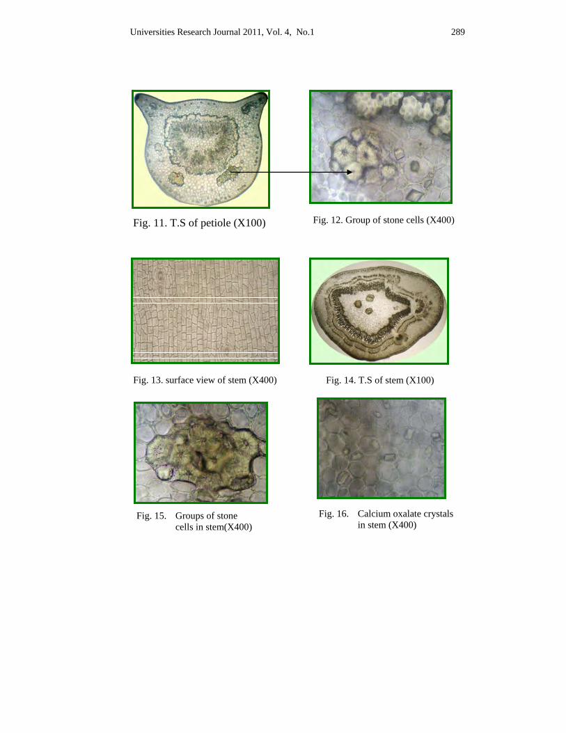
Universities Research Journal 2011, Vol. 4, No.1 289
Fig. 13. surface view of stem (X400) Fig. 14. T.S of stem (X100)
Fig. 15. Groups of stone cells in stem(X400)
Fig. 16. Calcium oxalate crystals in stem (X400)
Fig. 11. T.S of petiole (X100) Fig. 12. Group of stone cells (X400)
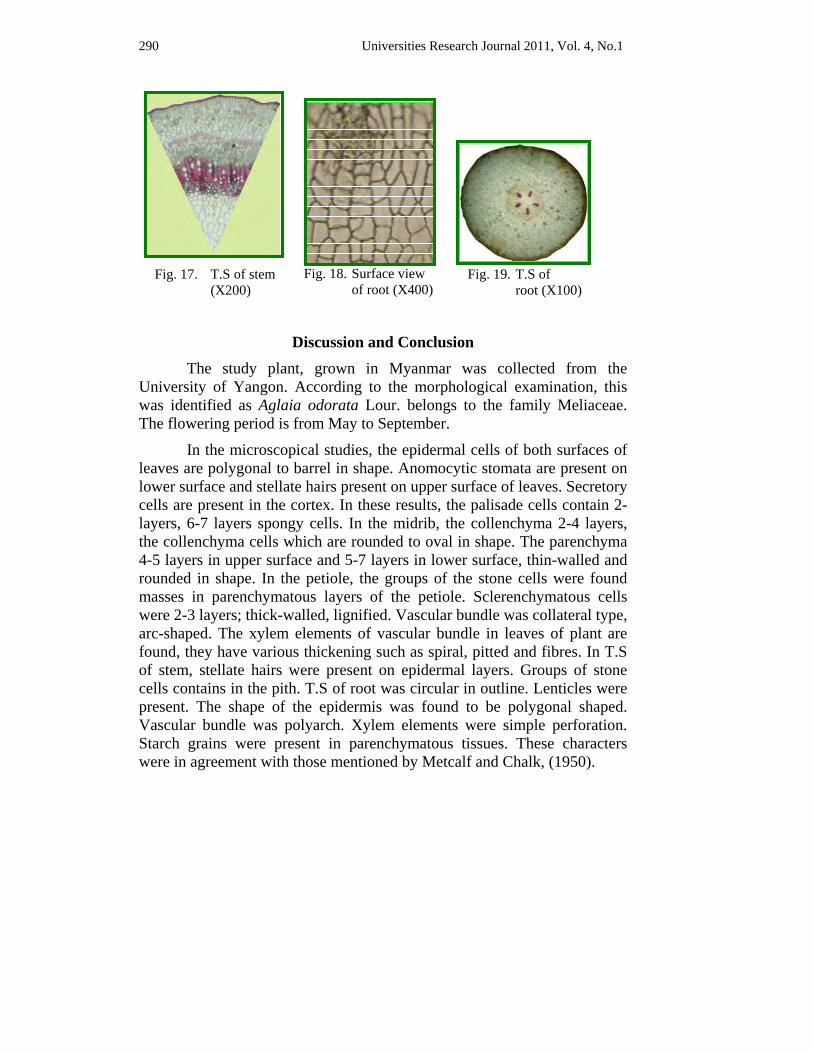
290 Universities Research Journal 2011, Vol. 4, No.1
Discussion and Conclusion The study plant, grown in Myanmar was collected from the University of Yangon. According to the morphological examination, this was identified as Aglaia odorata Lour. belongs to the family Meliaceae. The flowering period is from May to September.
In the microscopical studies, the epidermal cells of both surfaces of leaves are polygonal to barrel in shape. Anomocytic stomata are present on lower surface and stellate hairs present on upper surface of leaves. Secretory cells are present in the cortex. In these results, the palisade cells contain 2-layers, 6-7 layers spongy cells. In the midrib, the collenchyma 2-4 layers, the collenchyma cells which are rounded to oval in shape. The parenchyma 4-5 layers in upper surface and 5-7 layers in lower surface, thin-walled and rounded in shape. In the petiole, the groups of the stone cells were found masses in parenchymatous layers of the petiole. Sclerenchymatous cells were 2-3 layers; thick-walled, lignified. Vascular bundle was collateral type, arc-shaped. The xylem elements of vascular bundle in leaves of plant are found, they have various thickening such as spiral, pitted and fibres. In T.S of stem, stellate hairs were present on epidermal layers. Groups of stone cells contains in the pith. T.S of root was circular in outline. Lenticles were present. The shape of the epidermis was found to be polygonal shaped. Vascular bundle was polyarch. Xylem elements were simple perforation. Starch grains were present in parenchymatous tissues. These characters were in agreement with those mentioned by Metcalf and Chalk, (1950).
Fig. 17. T.S of stem (X200)
Fig. 19. T.S of root (X100)
Fig. 18. Surface view of root (X400)
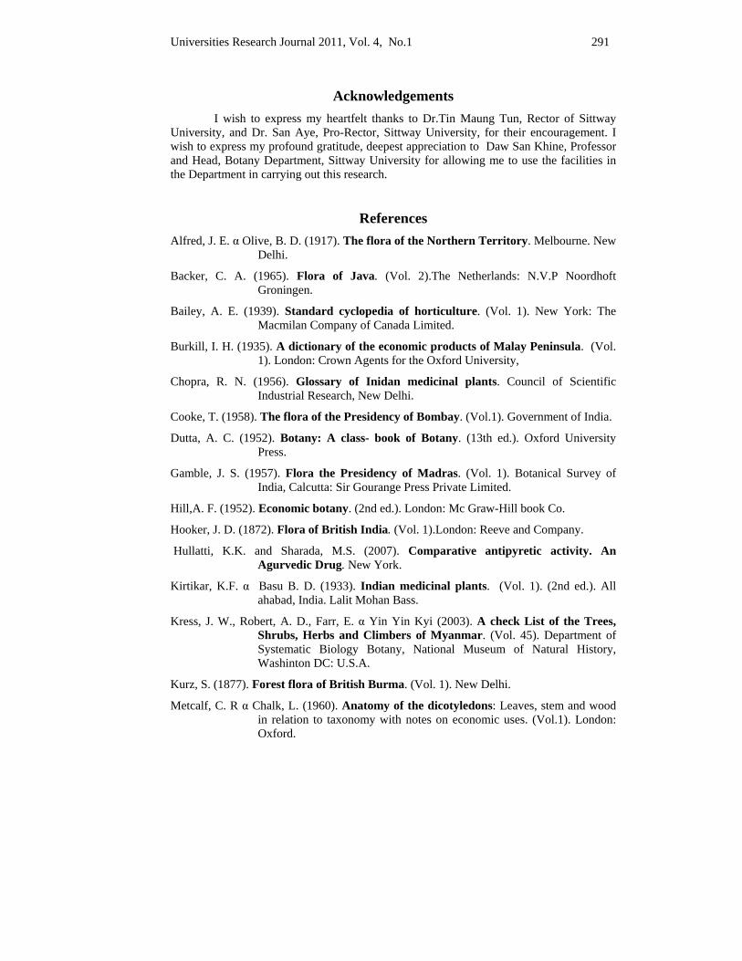
Universities Research Journal 2011, Vol. 4, No.1 291
Acknowledgements I wish to express my heartfelt thanks to Dr.Tin Maung Tun, Rector of Sittway University, and Dr. San Aye, Pro-Rector, Sittway University, for their encouragement. I wish to express my profound gratitude, deepest appreciation to Daw San Khine, Professor and Head, Botany Department, Sittway University for allowing me to use the facilities in the Department in carrying out this research.
References Alfred, J. E. α Olive, B. D. (1917). The flora of the Northern Territory. Melbourne. New
Delhi.
Backer, C. A. (1965). Flora of Java. (Vol. 2).The Netherlands: N.V.P Noordhoft Groningen.
Bailey, A. E. (1939). Standard cyclopedia of horticulture. (Vol. 1). New York: The Macmilan Company of Canada Limited.
Burkill, I. H. (1935). A dictionary of the economic products of Malay Peninsula. (Vol. 1). London: Crown Agents for the Oxford University,
Chopra, R. N. (1956). Glossary of Inidan medicinal plants. Council of Scientific Industrial Research, New Delhi.
Cooke, T. (1958). The flora of the Presidency of Bombay. (Vol.1). Government of India.
Dutta, A. C. (1952). Botany: A class- book of Botany. (13th ed.). Oxford University Press.
Gamble, J. S. (1957). Flora the Presidency of Madras. (Vol. 1). Botanical Survey of India, Calcutta: Sir Gourange Press Private Limited.
Hill,A. F. (1952). Economic botany. (2nd ed.). London: Mc Graw-Hill book Co.
Hooker, J. D. (1872). Flora of British India. (Vol. 1).London: Reeve and Company.
Hullatti, K.K. and Sharada, M.S. (2007). Comparative antipyretic activity. An Agurvedic Drug. New York.
Kirtikar, K.F. α Basu B. D. (1933). Indian medicinal plants. (Vol. 1). (2nd ed.). All ahabad, India. Lalit Mohan Bass.
Kress, J. W., Robert, A. D., Farr, E. α Yin Yin Kyi (2003). A check List of the Trees, Shrubs, Herbs and Climbers of Myanmar. (Vol. 45). Department of Systematic Biology Botany, National Museum of Natural History, Washinton DC: U.S.A.
Kurz, S. (1877). Forest flora of British Burma. (Vol. 1). New Delhi.
Metcalf, C. R α Chalk, L. (1960). Anatomy of the dicotyledons: Leaves, stem and wood in relation to taxonomy with notes on economic uses. (Vol.1). London: Oxford.
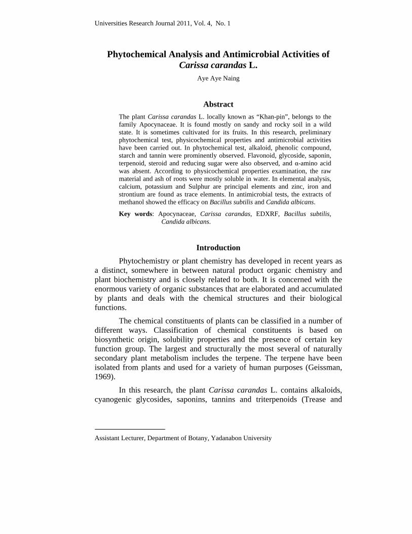
Universities Research Journal 2011, Vol. 4, No. 1
Assistant Lecturer, Department of Botany, Yadanabon University
Phytochemical Analysis and Antimicrobial Activities of Carissa carandas L.
Aye Aye Naing
Abstract The plant Carissa carandas L. locally known as “Khan-pin”, belongs to the family Apocynaceae. It is found mostly on sandy and rocky soil in a wild state. It is sometimes cultivated for its fruits. In this research, preliminary phytochemical test, physicochemical properties and antimicrobial activities have been carried out. In phytochemical test, alkaloid, phenolic compound, starch and tannin were prominently observed. Flavonoid, glycoside, saponin, terpenoid, steroid and reducing sugar were also observed, and α-amino acid was absent. According to physicochemical properties examination, the raw material and ash of roots were mostly soluble in water. In elemental analysis, calcium, potassium and Sulphur are principal elements and zinc, iron and strontium are found as trace elements. In antimicrobial tests, the extracts of methanol showed the efficacy on Bacillus subtilis and Candida albicans.
Key words: Apocynaceae, Carissa carandas, EDXRF, Bacillus subtilis, Candida albicans.
Introduction Phytochemistry or plant chemistry has developed in recent years as a distinct, somewhere in between natural product organic chemistry and plant biochemistry and is closely related to both. It is concerned with the enormous variety of organic substances that are elaborated and accumulated by plants and deals with the chemical structures and their biological functions.
The chemical constituents of plants can be classified in a number of different ways. Classification of chemical constituents is based on biosynthetic origin, solubility properties and the presence of certain key function group. The largest and structurally the most several of naturally secondary plant metabolism includes the terpene. The terpene have been isolated from plants and used for a variety of human purposes (Geissman, 1969).
In this research, the plant Carissa carandas L. contains alkaloids, cyanogenic glycosides, saponins, tannins and triterpenoids (Trease and
294 Universities Research Journal 2011, Vol. 4, No. 1
Evans, 2002). Roots of this plant also contain fixed oil, volatile oil, dark yellow resin and alkaloid (Nadkarni, 1954).
The preliminary phytochemical tests were investigated to determine the presence of various compounds and physicochemical tests were done to obtain important information concerning the moisture content and solubility. The antimicrobial activity was also tested to know whether it possess many medicinal values.
Materials and Methods
Preliminary phytochemical and physicochemical characterization on roots of Carissa carandas L. Preliminary phytochemical test In this study, only the qualitative determination of organic constituents in extract of the roots was investigated. It was carried out to know the presence or absence of alkaloids, carbohydrates, flavonoids, glycosides, phenolic Compound, saponins, starches, terpenoids, steroids, reducing sugar, tannins and α - amino acid. The experimental procedure was prepared by the methods of British Pharmacopeia, 1968; Harbone, 1973; Trease and Evans, 2002; Marini Bettolo et al. 1981 and Central Council for Research in Unani Medicine, 1987). Physicochemical characters of powdered roots from
Carissa carandas L. The physicochemical characters such as moisture contents, total ash, acid insoluble ash, water soluble ash, water, ethanol, methanol, acetone, ethyl acetate, chloroform and pet-ether soluble matter were analyzed according of the method of British Pharmacopoeia (1968), and WHO (1998). Elemental analysis from roots of Carissa carandas L. by using EDXRF
The relative concentration of elements was analyzed by using Energy Dispersive X-ray Fluorescence (EDXRF) spectrometer technique at the Universities’ Research Center, Yangon. The parameters of each part of the spectrometer are given below.
Detector type : Si (Li) detector

Universities Research Journal 2011, Vol. 4, No. 1 295
LN2 supply : Only during measurement
Dewar capacity : 3 Liters
LN2 consumption : Less than 1 liter per day
Detection area : 10 mm2
Resolution : Less than 155 ev (Mn Ka, 1500 H2)
The EDX-700 Shimadzu spectrometer can detect a wide range of the elements form sodium (Na) to uranium (U). The required data can be produced in a few minutes and it has a high degree of resolution for the spectrum evaluation. Due to its high sensitivity the spectrometer can detect the concentrations of elements in the parts per million (ppm) ranges. Antimicrobial activity of crude extracts from roots of
Carissa carandas L.
Antimicrobial activity of crude extracts For the determination of antimicrobial activity of crude extract agar-well diffusion method was used because of its simplicity, speed of performance, economy and reproducibility (Cruickshank, 1975). Extraction and test organisms
The dried powder sample was extracted with petroleum ether (60-80) ° C, chloroform, acetone, methanol, ethyl acetate, ethanol and water by percolation method. Crude extracts of various solvents of roots were tested on six microorganisms such as Bacillus subtilis, Staphylococcus aureus, Pseudomonas aeruginosa, Bacillus pumalis, Candida albicans, Escherichia coli. The test organisms used in this study were obtained from the Development Centre for Pharmaceutical Technology for determination of the antimicrobial activities. Preparation of sample for the test of antimicrobial activity Nutrient agar was prepared by method of Cruickshank (1975). Nutrient agar was used as basal cultural medium for the bacteria test organisms. The broth culture 20 ml was poured into the sterile petridish. Then, 0.2 ml of test organisms was added into the dishes.
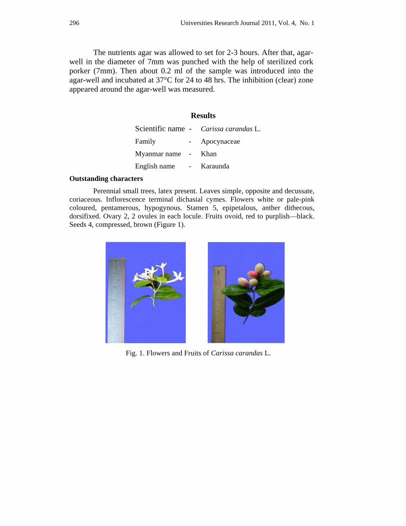
296 Universities Research Journal 2011, Vol. 4, No. 1
The nutrients agar was allowed to set for 2-3 hours. After that, agar-well in the diameter of 7mm was punched with the help of sterilized cork porker (7mm). Then about 0.2 ml of the sample was introduced into the agar-well and incubated at 37°C for 24 to 48 hrs. The inhibition (clear) zone appeared around the agar-well was measured.
Results Scientific name - Carissa carandas L.
Family - Apocynaceae
Myanmar name - Khan
English name - Karaunda
Outstanding characters
Perennial small trees, latex present. Leaves simple, opposite and decussate, coriaceous. Inflorescence terminal dichasial cymes. Flowers white or pale-pink coloured, pentamerous, hypogynous. Stamen 5, epipetalous, anther dithecous, dorsifixed. Ovary 2, 2 ovules in each locule. Fruits ovoid, red to purplish—black. Seeds 4, compressed, brown (Figure 1).
Fig. 1. Flowers and Fruits of Carissa carandas L.
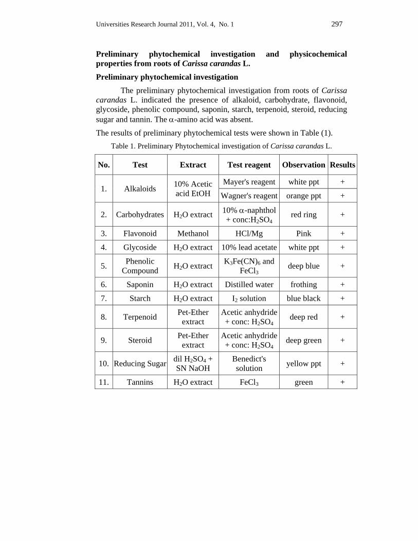
Universities Research Journal 2011, Vol. 4, No. 1 297
Preliminary phytochemical investigation and physicochemical properties from roots of Carissa carandas L.
Preliminary phytochemical investigation The preliminary phytochemical investigation from roots of Carissa carandas L. indicated the presence of alkaloid, carbohydrate, flavonoid, glycoside, phenolic compound, saponin, starch, terpenoid, steroid, reducing sugar and tannin. The α-amino acid was absent.
The results of preliminary phytochemical tests were shown in Table (1). Table 1. Preliminary Phytochemical investigation of Carissa carandas L.
No. Test Extract Test reagent Observation Results
1. Alkaloids 10% Acetic acid EtOH
Mayer's reagent white ppt +
Wagner's reagent orange ppt +
2. Carbohydrates H2O extract 10% α-naphthol + conc:H2SO4
red ring +
3. Flavonoid Methanol HCl/Mg Pink +
4. Glycoside H2O extract 10% lead acetate white ppt +
5. Phenolic Compound H2O extract K3Fe(CN)6 and
FeCl3 deep blue +
6. Saponin H2O extract Distilled water frothing +
7. Starch H2O extract I2 solution blue black +
8. Terpenoid Pet-Ether extract
Acetic anhydride + conc: H2SO4
deep red +
9. Steroid Pet-Ether extract
Acetic anhydride + conc: H2SO4
deep green +
10. Reducing Sugar dil H2SO4 + SN NaOH
Benedict's solution yellow ppt +
11. Tannins H2O extract FeCl3 green +
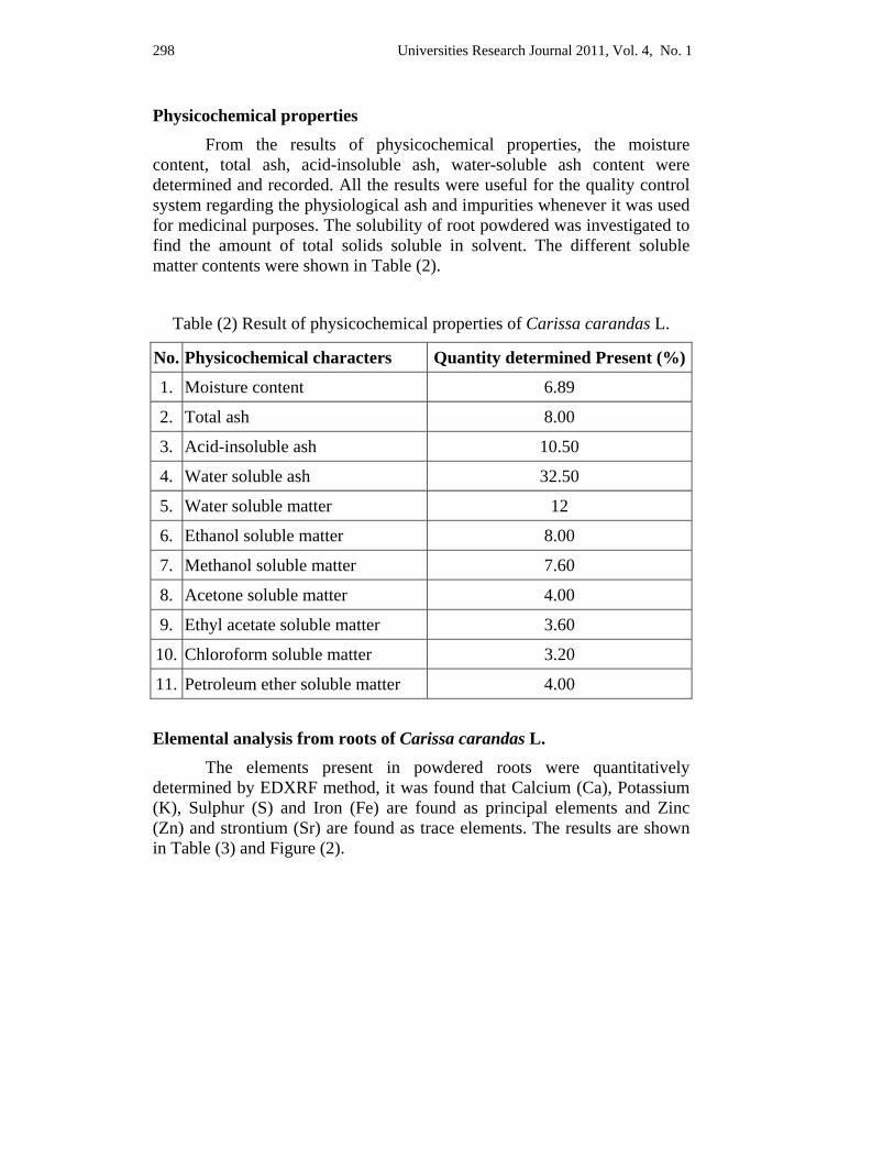
298 Universities Research Journal 2011, Vol. 4, No. 1
Physicochemical properties From the results of physicochemical properties, the moisture content, total ash, acid-insoluble ash, water-soluble ash content were determined and recorded. All the results were useful for the quality control system regarding the physiological ash and impurities whenever it was used for medicinal purposes. The solubility of root powdered was investigated to find the amount of total solids soluble in solvent. The different soluble matter contents were shown in Table (2).
Table (2) Result of physicochemical properties of Carissa carandas L.
No. Physicochemical characters Quantity determined Present (%)
1. Moisture content 6.89
2. Total ash 8.00
3. Acid-insoluble ash 10.50
4. Water soluble ash 32.50
5. Water soluble matter 12
6. Ethanol soluble matter 8.00
7. Methanol soluble matter 7.60
8. Acetone soluble matter 4.00
9. Ethyl acetate soluble matter 3.60
10. Chloroform soluble matter 3.20
11. Petroleum ether soluble matter 4.00
Elemental analysis from roots of Carissa carandas L. The elements present in powdered roots were quantitatively determined by EDXRF method, it was found that Calcium (Ca), Potassium (K), Sulphur (S) and Iron (Fe) are found as principal elements and Zinc (Zn) and strontium (Sr) are found as trace elements. The results are shown in Table (3) and Figure (2).
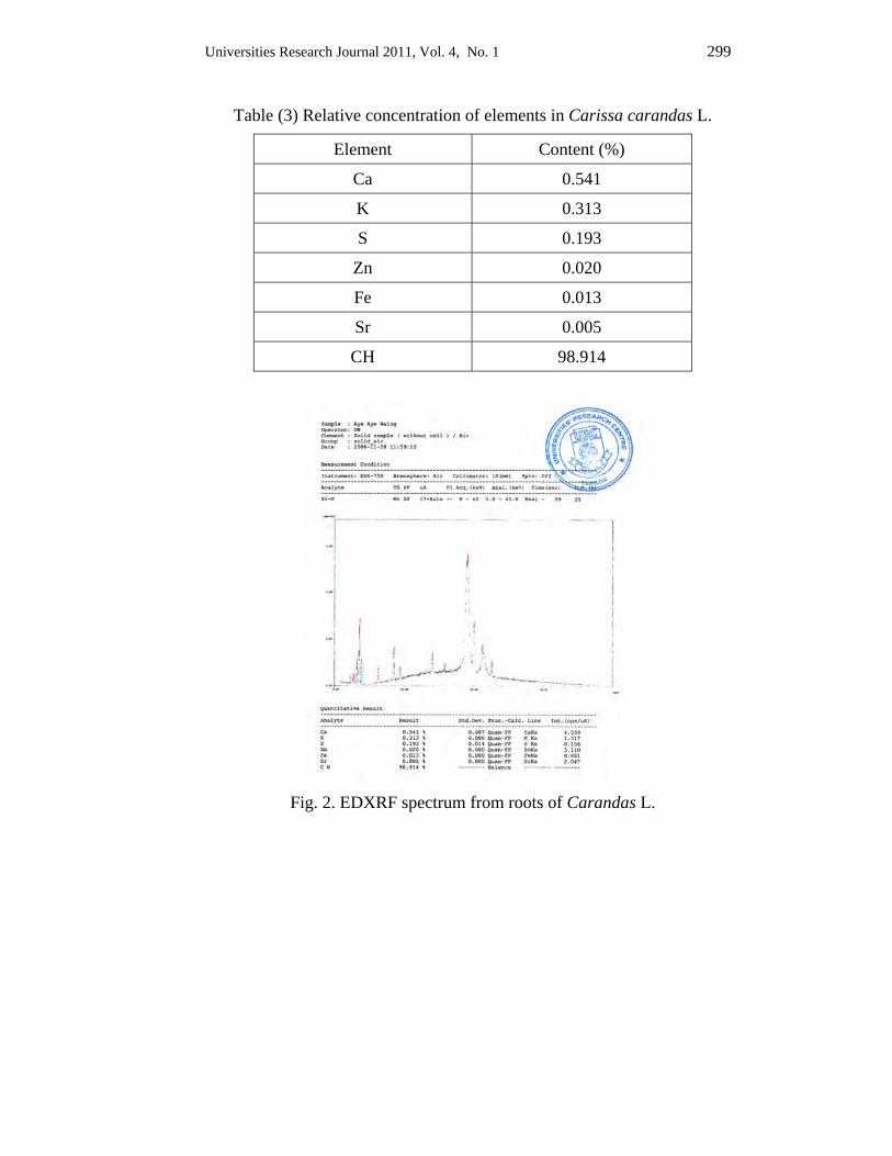
Universities Research Journal 2011, Vol. 4, No. 1 299
Table (3) Relative concentration of elements in Carissa carandas L.
Element Content (%)
Ca 0.541
K 0.313
S 0.193
Zn 0.020
Fe 0.013
Sr 0.005
CH 98.914
Fig. 2. EDXRF spectrum from roots of Carandas L.
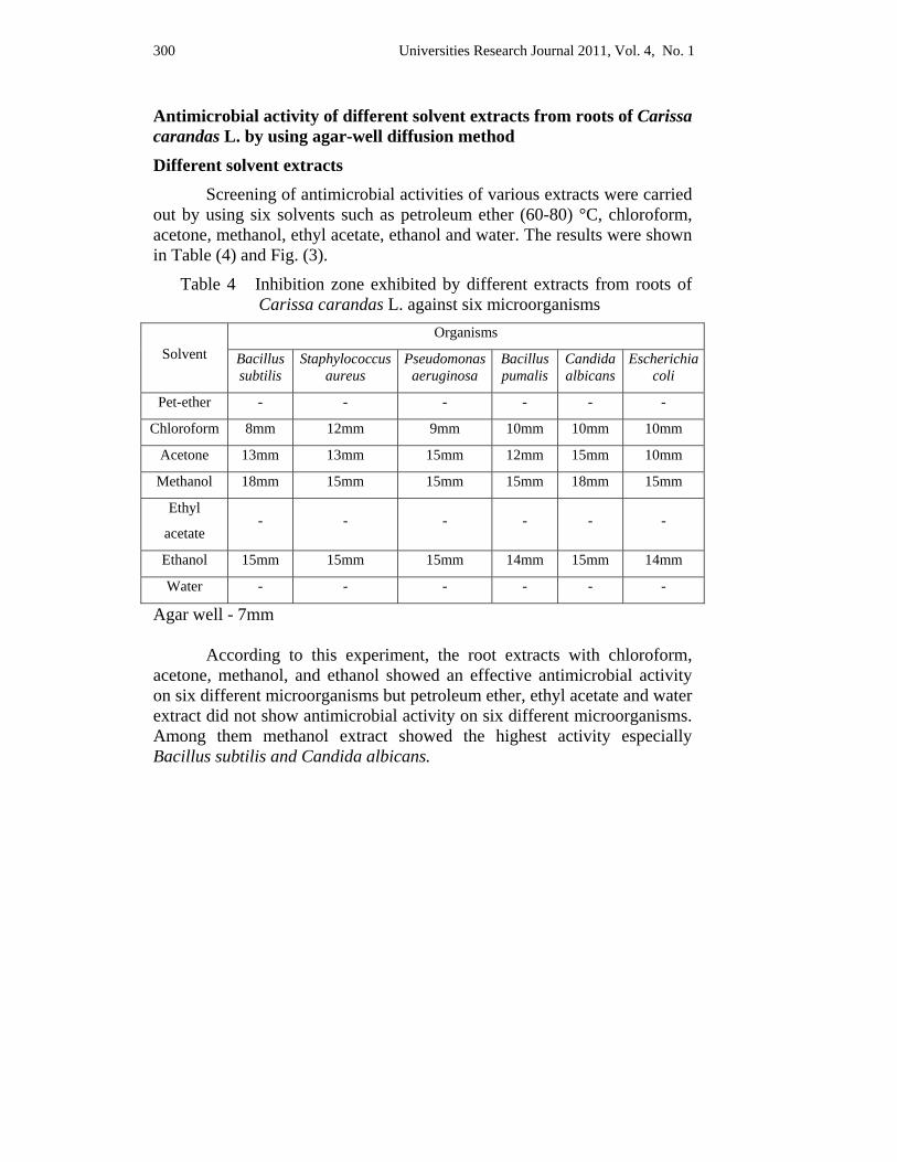
300 Universities Research Journal 2011, Vol. 4, No. 1
Antimicrobial activity of different solvent extracts from roots of Carissa carandas L. by using agar-well diffusion method
Different solvent extracts Screening of antimicrobial activities of various extracts were carried out by using six solvents such as petroleum ether (60-80) °C, chloroform, acetone, methanol, ethyl acetate, ethanol and water. The results were shown in Table (4) and Fig. (3).
Table 4 Inhibition zone exhibited by different extracts from roots of Carissa carandas L. against six microorganisms
Solvent Organisms
Bacillus subtilis
Staphylococcusaureus
Pseudomonasaeruginosa
Bacilluspumalis
Candidaalbicans
Escherichia coli
Pet-ether - - - - - -
Chloroform 8mm 12mm 9mm 10mm 10mm 10mm
Acetone 13mm 13mm 15mm 12mm 15mm 10mm
Methanol 18mm 15mm 15mm 15mm 18mm 15mm
Ethyl
acetate - - - - - -
Ethanol 15mm 15mm 15mm 14mm 15mm 14mm
Water - - - - - -
Agar well - 7mm According to this experiment, the root extracts with chloroform, acetone, methanol, and ethanol showed an effective antimicrobial activity on six different microorganisms but petroleum ether, ethyl acetate and water extract did not show antimicrobial activity on six different microorganisms. Among them methanol extract showed the highest activity especially Bacillus subtilis and Candida albicans.

Universities Research Journal 2011, Vol. 4, No. 1 301
302 Universities Research Journal 2011, Vol. 4, No. 1
Discussion and Conclusion In this research, the preliminary phytochemical detection, the
physico-chemical characters, elemental analysis of Carissa carandas L. root had been studied.
According to the phytochemical detection, alkaloid, carbohydrate, flavonoid, glycoside, phenol, saponin, starch, terpenoid, steroid, reducing sugar, and tannin are tested. Among these findings, the presence of alkaloid, terpenoid, saponin and tannin in this plant which are agreed with Trease and Evans (2002). In the physico-chemical properties, a powdered sample of roots from Carissa carandas L. was mostly soluble in water and ethanol. Elemental analysis (EDXRF) reveals Calcium (Ca), Potassium (K), Sulphur (S) and Iron (Fe) are principal elements and Zinc (Zn) and Strontium (Sr) are found as trace elements.
Antimicrobial activity of various crude extracts of petroleum ether, chloroform, acetone, methanol, ethyl acetate, ethanol and water and isolated compounds A and B were tested on six different microorganisms by using agar-well diffusion method.
In this result, various crude extracts showed effective antimicrobial activity on six different microorganisms except petroleum ether, ethyl acetate and water extracts. Among them, methanol extract showed the highest activity especially more sensitive against Bacillus subtilis and Candida albicans. From this finding, it can be inferred that Carissa carandas L. can be effective in the formulation of medicine for the treatment of diseases, such as pathogenesis, alimentary tract, gastrointestinal tract, skin infection, sores, Candida sis and intense itching.
Acknowledgements I would like to express my sincere thanks to Dr. Khin Maung Oo, Rector of
Yadanabon University for his permission to submit this article. I am also indebted to Professor and Head, U Nay Win and Professor Dr. Cho Cho, Department of Botany, Yadanabon University for their invaluable sugessions and guidance. I am grateful to Dr. Nu Nu Swe, Professor, Department of Botany, Meiktila University for providing all kindly necessary instruction.
Universities Research Journal 2011, Vol. 4, No. 1 303
References British Pharmacopoeia (1968). Published under the direction of the General Medical
Council. Medical Act 1956. London: William Cloves and Sons, Limited.
Central Council for Research in Unani Medicine. (1987). Phytochemical Standard of Unani Formulation. New Delhi
Cruickshank, R. J. P. (1975). Medicinal Microbiology. London: Livingstone Ltd.
Geissman, T. A. (1995). In modern methods of plant. Analysis.
Harborne, J.B. (1973). Phytochemical methods. London : Academic press Inc.
Marini-Bettalo, G. B. (1981). Plant screening by chemical and chromatographic procedure under field condition. Journal of Chromatography.
Nadkarni, K. M. (1954). Indian materia medica. (Vol. 1). (3th ed.). Bombay.
Trease, G. E & Evans, W. C. (2002). Pharmacognosy. (15th ed.). London : Harcourt Publishers Limited.
World Health Organization (1998). Quality control methods for medicinal plant materials. Geneva.
Universities Research Journal 2011, Vol. 4, No. 1
Assistant Lecturer, Department of Botany, West Yangon University
Hydrocarbon-Oxidizing Activity of Isolated Microorganisms from No.1 Refinery, Thanlyin
Aye Cho Mar
Abstract In the present work, 10 strains of soil microorganisms have been screened from hydrocarbon polluted areas located in No.1 Refinery, Thanlyin. All the isolated strains were subjected to the preliminary tests as well as to the confirmatory tests to know the presence of hydrocarbon degradation activity. The isolated bacteria were enslaved in the specific tests to examine whether they can change the physical or chemical nature of hydrocarbon (Aviation Turbine Fuel). The conditions of physical nature before and after the treatment were comparatively detected by standard methods of petroleum chemistry. Moreover, the chemical nature observed after several bacterial treatments was evaluated with the help of spectroscopic methods such as UV and FTIR in the Universities Research Centre, University of Yangon.
Key words: soil microorganisms, hydrocarbon degradation activity
Introduction Various microorganisms have now been isolated from nature that
consumed spilled oil, solvents and oil derivatives, either directly at the site of the polluted area or later on after the toxic materials have pervaded soils or entered the ground water. The great diversity of microbes on the earth contains vast genetic resources for solutions to cleaning up the environment and much research work in this field is not still taking place in Myanmar. Therefore, it is essential to take action without hesitation to clean up the damaged environment by various means. Although there is still no serious environmental issue in the Myanmar territory, it is the proper step to take preventive measure because living things depend greatly on their close environment for their survival and the environment provides important conditions for human beings to survive.
When microorganisms break down petroleum hydrocarbons; the first step usually is the addition of a hydroxyl group to the end of an alkane chain or onto an unsaturated ring of a polycyclic aromatic hydrocarbon, forming an alcohol, then progressively oxidize the alcohol to an aldehyde and subsequently a carboxylic acid, which leads to chain length reduction. These in turn are converted into carbon dioxide, water and simpler
306 Universities Research Journal 2011, Vol. 4, No. 1
compounds that do not affect the environment. To gain these resultant simpler compounds microbes grow an oil films and sticks and attach to insoluble oil droplets and begin the process of decomposition. The term bioremediation refers to the clean up of oil or other pollutants by microorganisms and in recent years the importance of bioremediation in oil spills has been amply established in several major crude oil spills in the marine environment.
Microorganisms just like to humans, eat and digest organic substances for nutrients and energy. Certain microbes can digest organic substances such as fuels or solvents that are hazardous to humans. Applied Microbial Bioremediation can use their naturally occurring microbes or genetically engineered microbes (Alexander, 1965). The presence of hydrocarbon oxidizing activity in the isolated strains was estimated and the positive strains were subjected in the study for evaluation of degenerative potential or oxidative decomposition activity using ATF (aviation turbine fuel) as hydrocarbon substrate. Objectives in the present work are to detect the presence of hydrocarbon degeneration potential in the isolates and to find out the proof in hydrocarbon degradation by standard petroleum chemistry method and spectroscopic analyses. By performing the above objectives, aim to initiate and participate not only in Bioremediation but also in environment conservation in ever green Myanmar.
Materials and Methods
Screening of microorganisms isolation of pure culture and identification
Sample collection has been done from four different sites of No.1 Refinery, Thanlyin and all the selected area have been contaminated with respective hydrocarbon compound. Isolation and maintenance of pure cultures have been performed.
Preliminary study of hydrocarbon oxidizing activity of isolated strains All the isolated strains were subjected for preliminary hydrocarbon
degrading activity by Red-spot Test using colourless 2,3,5-trimethyl tetrazolium chloride, according to the test method reported by Hill et al., (1967).
Universities Research Journal 2011, Vol. 4, No. 1 307
Assay of hydrocarbon degradation potential of isolated bacteria by the change of physical nature of selected hydrocarbon product
As for experimental design, the bacterial broth cultures obtained by bacterial mixture were treated by selected hydrocarbon compound (ATF) in the ratio of bacterial broth to ATF (1:1-1:5) and the reverse ratio of ATF to bacterial broth (1:1-1:5) respectively.
All the lids of the containers were perforated by pin-tip not only for aerobic bacterial respiration but also for prevention of volatile condition of hydrocarbon compound. Then all the containers with bacterial broth and ATF were agitated on the shaker for 7 hours each day for 10 and 30 days separately. As for control experiment, the treatment only with the ATF (without bacteria) was made under the same experimental conditions. After shaking, the hydrocarbons (ATF) in respective containers were separated by using separating funnel. During the agitation procedure, so much emulsion was seen within the ATF-water interfaces and this condition was shown in Figure (1). These emulsions were examined by microscopic observation and this examination can be seen in Figure (2) and Figure (3).
Pure ATF of before treatment with isolated bacteria (as standard) and after bacterial treated with isolated bacteria were applied to examine their physical nature by American Standard Test Methods.
Standard Methods of Tests;
(1) Hydrocarbon Types in Liquid Petroleum Products by Fluorescent Indicator Adsorption. ASTM D1319,
(2) Refractive Index and Refractive Dispersion of Hydrocarbon Liquids, ASTM D 1218,
(3) Flash Point by TAG Closed Tester ASTM D 56 (4) Distillation of Petroleum Products ASTM D.86 (5) Detection of Copper Corrosion from Petroleum Products by
The Copper Strip Tarnish test. ASTM D 130 (6) Density and Relative Density (Specific Gravity) of Liquids by
Bingham Pycnometer. ASTM D 1217 and (7) Smoke Point of Aviation Turbine Fuels ASTM D 1322.
In general, the most amenable of the hydrocarbons to microbial attack are the 10-18 carbons, normal straight chain alkanes, and growth on
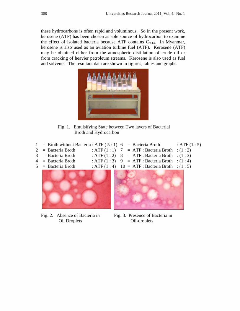
308 Universities Research Journal 2011, Vol. 4, No. 1
these hydrocarbons is often rapid and voluminous. So in the present work, kerosene (ATF) has been chosen as sole source of hydrocarbon to examine the effect of isolated bacteria because ATF contains C9-14. In Myanmar, kerosene is also used as an aviation turbine fuel (ATF). Kerosene (ATF) may be obtained either from the atmospheric distillation of crude oil or from cracking of heavier petroleum streams. Kerosene is also used as fuel and solvents. The resultant data are shown in figures, tables and graphs.
Fig. 2. Absence of Bacteria in Fig. 3. Presence of Bacteria in
Oil Droplets Oil-droplets
1 = Broth without Bacteria : ATF ( 5 : 1)2 = Bacteria Broth : ATF (1 : 1) 3 = Bacteria Broth : ATF (1 : 2) 4 = Bacteria Broth : ATF (1 : 3) 5 = Bacteria Broth : ATF (1 : 4)
Fig. 1. Emulsifying State between Two layers of Bacterial Broth and Hydrocarbon
6 = Bacteria Broth : ATF (1 : 5) 7 = ATF : Bacteria Broth : (1 : 2) 8 = ATF : Bacteria Broth : (1 : 3) 9 = ATF : Bacteria Broth : (1 : 4) 10 = ATF : Bacteria Broth : (1 : 5)
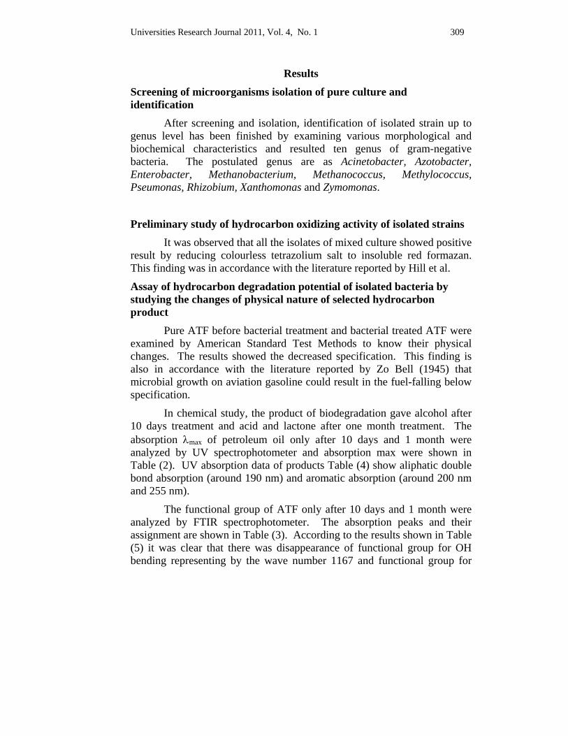
Universities Research Journal 2011, Vol. 4, No. 1 309
Results
Screening of microorganisms isolation of pure culture and identification
After screening and isolation, identification of isolated strain up to genus level has been finished by examining various morphological and biochemical characteristics and resulted ten genus of gram-negative bacteria. The postulated genus are as Acinetobacter, Azotobacter, Enterobacter, Methanobacterium, Methanococcus, Methylococcus, Pseumonas, Rhizobium, Xanthomonas and Zymomonas.
Preliminary study of hydrocarbon oxidizing activity of isolated strains It was observed that all the isolates of mixed culture showed positive
result by reducing colourless tetrazolium salt to insoluble red formazan. This finding was in accordance with the literature reported by Hill et al.
Assay of hydrocarbon degradation potential of isolated bacteria by studying the changes of physical nature of selected hydrocarbon product
Pure ATF before bacterial treatment and bacterial treated ATF were examined by American Standard Test Methods to know their physical changes. The results showed the decreased specification. This finding is also in accordance with the literature reported by Zo Bell (1945) that microbial growth on aviation gasoline could result in the fuel-falling below specification.
In chemical study, the product of biodegradation gave alcohol after 10 days treatment and acid and lactone after one month treatment. The absorption λmax of petroleum oil only after 10 days and 1 month were analyzed by UV spectrophotometer and absorption max were shown in Table (2). UV absorption data of products Table (4) show aliphatic double bond absorption (around 190 nm) and aromatic absorption (around 200 nm and 255 nm).
The functional group of ATF only after 10 days and 1 month were analyzed by FTIR spectrophotometer. The absorption peaks and their assignment are shown in Table (3). According to the results shown in Table (5) it was clear that there was disappearance of functional group for OH bending representing by the wave number 1167 and functional group for
310 Universities Research Journal 2011, Vol. 4, No. 1
C = C stretching for aromatic representing by wave number 1607 from standard ATF after 10 days and one month duration.
The appearance of functional group for OH stretching for alcohol representing by the wave number 3125 and 3135 in bacterial treated ATF after 10 days and one month could be seen. By comparing these changes in functional group, ATF indicated the oxidation and had proceeded to form an alcohol product. This finding was in accordance with the literature reported by Atlas and Bartha, (1998) who pointed out that the enzyme hydrolyzed the hydrocarbon molecules had involved first in the initial stage. The results shown in Table (5) also indicated the appearance of C = O stretching (lactone) functional group representing by the wave number 1715 and 1748. This finding was also in accordance with the literature reported by Atlas and Bartha, 1998 that the alcohol product was then oxidized into an aldehyde and finally, to a fatty acid.
In this investigation, it was observed that the hydrocarbon molecules yield alcohols products from petroleum oil biodegradation by bacteria after 10 days treatment and yield acid product after 1 month treatment.
Table 1. Showing the Physical Changes of ATF Before and After Bacteria
treated for ten days
Sr No
Sample No Density Specific
Gravity Refractive
Index
AromaticVol%
content
SmokePoint (mm)
FlashPoint( c)
Copper Strip
Corrosion
IBP FBP ( c)
1 I
Std 0.8045 0.8059 1.44294
(14.648) 21.00% 22 51 1(a) 162/241
2 II
H:Br 1:5
0.8042 0.8056 1.44249(14.6) 20.90% 21 52 1(a) 162/241
3 XII B:H 1:1
0.7999 0.8014 1.44405 (14.765)
20.50% 21 53 1(a) 165/241
4 XIII B:H 1:2
0.8001 0.8015 1.44437(14.800) 20.60% 21 53 1(a) 165/243
Universities Research Journal 2011, Vol. 4, No. 1 311
Sr No
Sample No Density Specific
GravityRefractive
Index
AromaticVol%
content
SmokePoint (mm)
Flash Point ( c)
Copper Strip
Corrosion
IBPFBP ( c)
5 XIII B:H 1:2
0.8002 0.8016 1.44437(14.800) 20.60% 21 53 1(a) 163/242
6 XV B:H 1:4
0.8011 0.8025 1.44437(14.800) 20.70% 21 52 1(a) 162/242
7 XVI B:H 1:5
0.8018 0.8032 1.44437(14.800) 20.70% 22 52 1(a) 162/241
8 XVII H:B 1:2
0.8000 0.8014 1.44390(14.750) 20.40% 21 54 1(a) 166/242
9 XVIII H:B 1:3
0.7996 0.8010 1.44343(14.700) 20.30% 21 54 1(a) 167/245
10 XIX H:B 1:4
0.7995 0.8009 1.44343(14.700) 20.00% 21 54 1(a) 168/264
11 XX H:B 1:5
0.7993 0.8007 1.44315(14.669) 20.00% 21 55 1(a) 169/243
312 Universities Research Journal 2011, Vol. 4, No. 1
0.796
0.797
0.798
0.799
0.8
0.801
0.802
0.803
0.804
0.805
I II XII XII XIV XV XVI XVII XVIII XIX XX
Isolated Bacterial Strains
Fig. 4. Effect of Isolated Bacteria on the Change of Density of ATF
0.798
0.799
0.8
0.801
0.802
0.803
0.804
0.805
0.806
0.807
I II XII XII XIV XV XVI XVII XVIII XIX XX
Isolated Bacterial Strains
Fig. 5. Effect of Isolated Bacteria on the Change of Specific Gravity of ATF
Universities Research Journal 2011, Vol. 4, No. 1 313
1.4415
1.442
1.4425
1.443
1.4435
1.444
1.4445
1.445
1.4455
I II XII XII XIV XV XVI XVII XVIII XIX XX
Isolated Bacterial Strains
Fig. 6. Effect of Isolated Bacteria on the Change of Refractive Index of ATF
19.4
19.6
19.8
20
20.2
20.4
20.6
20.8
21
21.2
I II XII XII XIV XV XVI XVII XVIII XIX XX
Isolated Bacterial Strains
Fig. 7. Effect of Isolated Bacteria on the Change of Aromatic Content of ATF

314 Universities Research Journal 2011, Vol. 4, No. 1
49
50
51
52
53
54
55
56
I II XII XII XIV XV XVI XVII XVIII XIX XX
Isolated Bacterial Strains
Fig. 8. Effect of Isolated Bacteria on the Change of Flash Point of ATF
Table 2. UV λmax of Petroleum Oil (ATF) Biodegradation by Isolated Bacteria after 10 days
No Sample Observed (nm)
Reference
1 ATF only 202, 220, 265
William Kemp. (1979)
2 ATF + Medium 201, 220, 272
3 ATF + Bacteria (5:1) 204, 219, 266
4 ATF + Bacteria (1:1) 203, 220, 272
5 ATF + Bacteria (1:5) 211, 220, 250, 271, 286
Reference C = C bond absorption bands = around 190 nm.
Benzene (aromatic) absorption bands = around 200 nm and 255 nm.
Extension of the conjugation moves λmax longer weave length.
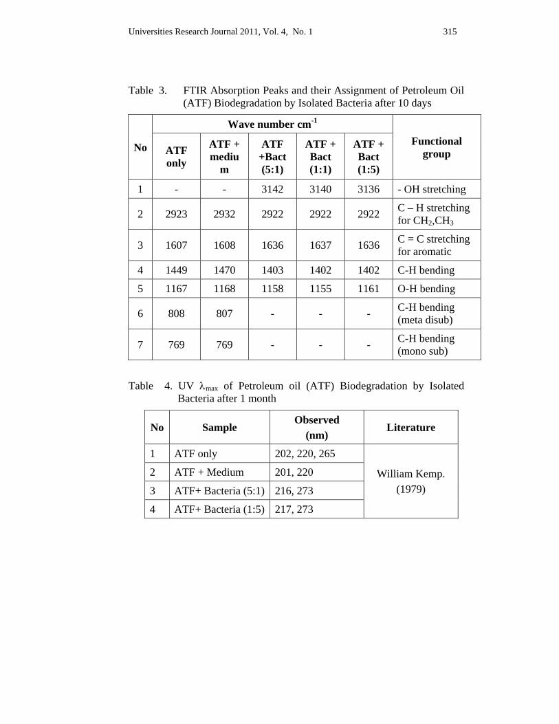
Universities Research Journal 2011, Vol. 4, No. 1 315
Table 3. FTIR Absorption Peaks and their Assignment of Petroleum Oil
(ATF) Biodegradation by Isolated Bacteria after 10 days
No
Wave number cm-1 Functional
group ATF only
ATF + mediu
m
ATF +Bact (5:1)
ATF + Bact (1:1)
ATF + Bact (1:5)
1 - - 3142 3140 3136 - OH stretching
2 2923 2932 2922 2922 2922 C – H stretching for CH2,CH3
3 1607 1608 1636 1637 1636 C = C stretching for aromatic
4 1449 1470 1403 1402 1402 C-H bending
5 1167 1168 1158 1155 1161 O-H bending
6 808 807 - - - C-H bending (meta disub)
7 769 769 - - - C-H bending (mono sub)
Table 4. UV λmax of Petroleum oil (ATF) Biodegradation by Isolated Bacteria after 1 month
No Sample Observed
(nm) Literature
1 ATF only 202, 220, 265
William Kemp. (1979)
2 ATF + Medium 201, 220
3 ATF+ Bacteria (5:1) 216, 273
4 ATF+ Bacteria (1:5) 217, 273
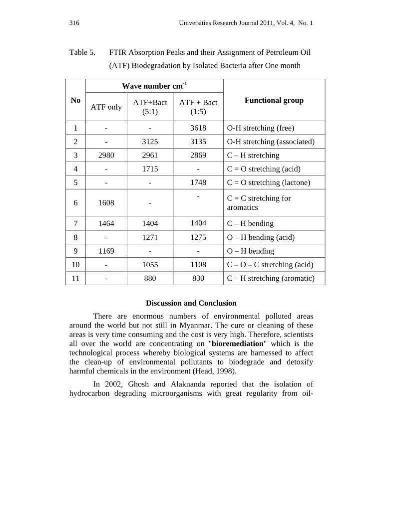
316 Universities Research Journal 2011, Vol. 4, No. 1
Table 5. FTIR Absorption Peaks and their Assignment of Petroleum Oil
(ATF) Biodegradation by Isolated Bacteria after One month
Discussion and Conclusion There are enormous numbers of environmental polluted areas around the world but not still in Myanmar. The cure or cleaning of these areas is very time consuming and the cost is very high. Therefore, scientists all over the world are concentrating on "bioremediation" which is the technological process whereby biological systems are harnessed to affect the clean-up of environmental pollutants to biodegrade and detoxify harmful chemicals in the environment (Head, 1998).
In 2002, Ghosh and Alaknanda reported that the isolation of hydrocarbon degrading microorganisms with great regularity from oil-
No
Wave number cm-1
Functional group ATF only ATF+Bact
(5:1) ATF + Bact
(1:5)
1 - - 3618 O-H stretching (free)
2 - 3125 3135 O-H stretching (associated)
3 2980 2961 2869 C – H stretching
4 - 1715 - C = O stretching (acid)
5 - - 1748 C = O stretching (lactone)
6 1608 - - C = C stretching for
aromatics
7 1464 1404 1404 C – H bending
8 - 1271 1275 O – H bending (acid)
9 1169 - - O – H bending
10 - 1055 1108 C – O – C stretching (acid)
11 - 880 830 C – H stretching (aromatic)
Universities Research Journal 2011, Vol. 4, No. 1 317
polluted ecosystems was strongly suggestive of their significance in the self-purification of these ecosystems. The elimination of oil-pollutants by microbial degradation process in nature resulted from the combined enzyme and community based activities of microbes whose regulation and kinetics were likely to be highly individualized and variable with space and time. Therefore, in the present study, the mixture of isolated bacteria is used to react in synergistic manner to ATF, not by individual isolate. Moreover, the duration of treatment is extended from 10 days to 30 days to provide complete degeneration by mixture of isolated strains.
Vecchoiol et al. (1990) revealed that an individual species was not capable of degrading all oil constituents. Some microbes could degrade a wide variety of compounds while others were seemingly only capable of degrading one compound. Many mixed culture of selected hydrocarbon degrading bacteria had been reported to increase the biodegradation rate. The similar results by Rosenberg et al. (1992) Lal and Khanna, (1996) also supported the finding of present works in which isolated strains were mixed and collectively treated to hydrocarbon ATF. In the present study, the FTIR results proved that ATF which was composed of C9 to C14 chain was broken down to small fragments it was also in accord with those reported by above workers.
Sokatch, (1969) and Anthony, University of Fort Hare, (2006) had reported the biodegradation alternative in the cleanup of petroleum hydrocarbon pollutants. The following diagram shows an example of breakdown of typical hydrocarbon molecules. Although it is necessary to confirm the structural proof by analyzing the actual nature of reaction products resulted from each biodegradation experiments, it is quite reasonable to explain that reaction patterns of present research are in accords with the pattern reported by Sokatch (1969). The results in the present study are prominently in agreement with those shown in the end products of first stage given by reactions of Sokatch (1969).

318 Universities Research Journal 2011, Vol. 4, No. 1
Fig. 9. The Postulated Reaction Sequence of Hydrocarbon Degradation by Microorganisms by Sokatch (1969) (www.dmctech.org)
There are still no effective research works concerning the bioremediation in Myanmar. The present research aims to help in the conservation of ever green and clean environs of Myanmar but also to share the microbiological knowledge of what can be done by microbiologists in Department of Botany to upgrade the nation.
Although the present data have strongly recommended the actual nature of microbes and presence of hydrocarbon degradation potential in these isolates, there are many points to be clarified the effects of bacteria on other types of hydrocarbons and the chemical natures of degraded products.
Universities Research Journal 2011, Vol. 4, No. 1 319
Acknowledgements I would like to acknowledge my heartfelt thanks to Daw Myat Thitar, Professor and Head Botany Department, West Yangon University, for her encouragement. I am thankful to Dr. Ye Ye Nwe, Assistant Lecturer, Department of Botany, Kyinetone University for her invaluable advice during this research. Thanks are especially due to Dr. U Win, Retired Rector, Hinthada University for providing necessary equipment, literature and departmental facilities and for his interest and help during my experimental work.
References Alaknanada, C.Patel and S.K.Ghosh. (2001). Spatial and Temporal diversity in
Petroleum degrading Bacterial Population. Asian.J.Microbial. Biotech.Env. Sc.(In Press)
Alexander, M. (1930). Introduction to Soil Microbiology. 2nd Edition, New York.
Alexander, M. (1965). Biodegradation Problems of Molecular Recalcitrant and Microbial Fallibility, Adv. Appl. Microbiol. 7:35-80.
American Society for Testing and Materials; Annual Book of ASTM Standards Vol. I & II Easton, U.S.A.
Head, I. M., (1998). Bioremediation: Towards a Credible Technology in Microbiology, 1998, 144; 599-608.
Odu, C.T.I. (1972). Microbiology of Soil Contaminated with Petroleum Hydrocarbon : I. Extent of Contamination and Some Soil and Microbial Properties after Contamination. J. Inst. Pet. 58; 201-208.
Rosenberg, E., Legmann, R., Kushmaro, A., Taube, R., Alder, E. and Ron, E. Z. (1992). Petroleum Bioremediation – A Multiphase Problem, Biodegradation 3:337-350.
Vogel, A.I (1956). A Text-book of Practical Organic Chemistry Including Qualitative Organic Analysis. English Language Book Society and Longman Group Limited.
Zobell, C.E. (1946). Action of Microorganisms on Hydrocarbons. Bacteriological Review.
Universities Research Journal 2011, Vol. 4, No. 1
Demonstrator, Department of Botany, University of Mandalay
Quantitative Analysis of Forest Structure in the Middle Part of the Goktwin Area, Northern Shan State
Nilar Win
Abstract The present study deals with the assessment of quantitative structure and floristic composition of forest in Goktwin area, Northern Shan State. A total of 72 plant species representing 51 genera and 28 families were analyzed in the study area. Importance Value Index (IVI), Shannon-Wiener (H) index, Simpson (D) index, Shannon-Wiener evenness (E) index were calculated. Shorea siamensis (Kurz.) Miq. (Ingyin) is found to be one of the ecologically dominant species contributing highest (IVI) (24.82%) with relative density (6.74%), relative frequency (3.25%), and relative dominance (14.83%). The diversity indices of Shannon-Wiener (H) and Simpson (D) value for tree species were 5.401, 0.965, shrubs were 3.062, 0.828, and herbs were 4.228, 0.931 respectively. Shannon-Wiener evenness (E) index for trees, shrubs and herbaceous plants were 0.875, 0.804 and 0.90. All the diversity indices pointed out that the study site is floristically high diverse.
Key words : floristic composition, Importance Value Index, ecologically dominant speciesm diversity indices
Introduction The quantitative study of vegetation gives description of the
vegetation, prediction and classification of its pattern and knowing useful and value species in a meaningful way. It indicates species diversity which determines the distribution of individuals among the species in a particular habitat (Horkar and Khatri 2003 as cited in Sahu et al. 2007).
The diversity of tree is fundamental to total forest biodiversity because trees provide resources and habitat structure for almost all other species. Information on the distribution and abundance of tree species is of primary importance in the planning and implementation of biodiversity conservation. Tree species diversity in the study area varies greatly from place to place mainly due to variation in biogeography, habitat and disturbance (Padalia et al. 2004).
On account of their economic exploitation, the forests are most threatened ecosystems in the Goktwin area, Northern Shan State. But so far
322 Universities Research Journal 2011, Vol. 4, No. 1
no efforts have been made to quantify the tree species parameters in this study area. The present study is an attempt to record structural composition and diversity of tree species in the Goktwin area, Northern Shan State.
The main objective of the present study are to present information on the vegetation and floristic composition of plant species, to assess the plant species diversity, the quantitative analysis of the forest structure.
Materials and Methods
Study Area The study area is located in Goktwin of Northern Shan State. It lies
between 96° 48' and 96° 57' E longitude, and between 22° 16' and 22° 22' N latitude. The total area is 103.62 square kilometers and stretches for 12.97 kilometers from East to West and 8.05 kilometers from North to South.
The elevation of Goktwin area varies from 270 to 1010 m above sea level. This area consist of Shan plateau, deep valleys and plains. The location map of the study area as shown in Figure (1).
The climate condition of the study area is warm and wet tropical. The monthly mean rainfall, temperature and relative humidity were recorded for 2008-2009. According to the record, the area received maximum rainfall during August (413 mm) in 2008 and during August (525 mm) in 2009 while minimum rainfall (1mm) occur in March 2008 and (2 mm) occur in November 2009. The highest mean temperature was 25.6°C in April 2008, 25.7°C in April 2009. The highest relative humidity was recorded in December (89%) during 2008 and in August (87%) during 2009.
Data Collection Plant cover data were collected during the months of July and October in 2009. A total of 10 sample plots (each sample plot was 30x30m) were laid down and studied. All the plots were systematically surveyed for all trees ≥ 10cm diameter at breast height (DBH). To account overall species diversity two subplots 5×5m (for shrubs), four subplots 1×1m (for herbs) were laid down in the plot ( Braun-Blanquet 1964).
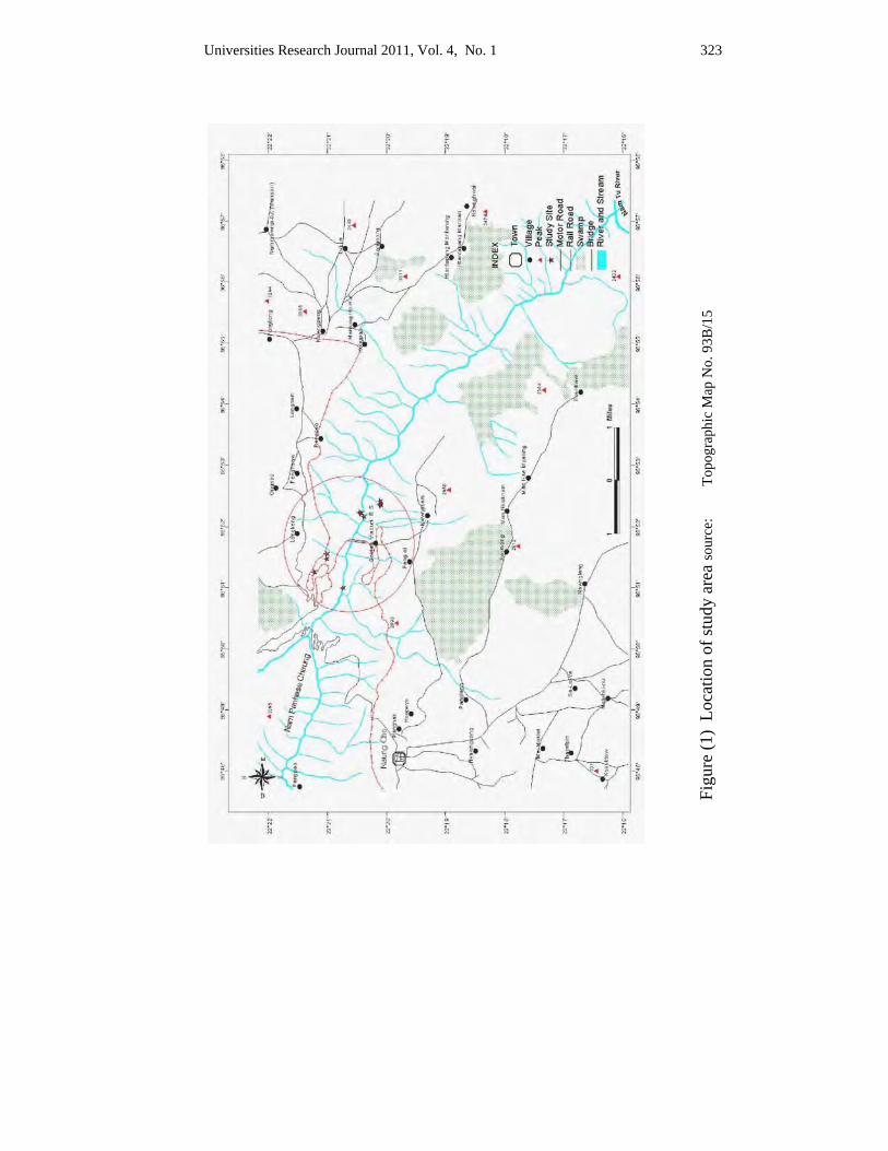
Universities Research Journal 2011, Vol. 4, No. 1 323
Figu
re (1
) Lo
catio
n of
stud
y ar
ea so
urce
: To
pogr
aphi
c M
ap N
o. 9
3B/1
5
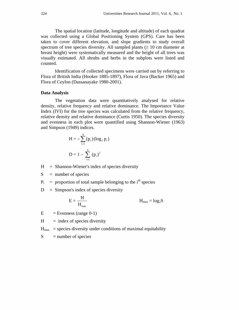
324 Universities Research Journal 2011, Vol. 4, No. 1
The spatial location (latitude, longitude and altitude) of each quadrat was collected using a Global Positioning System (GPS). Care has been taken to cover different elevation, and slope gradients to study overall spectrum of tree species diversity. All sampled plants (≥ 10 cm diameter at breast height) were systematically measured and the height of all trees was visually estimated. All shrubs and herbs in the subplots were listed and counted.
Identification of collected specimens were carried out by referring to Flora of British India (Hooker 1885-1897), Flora of Java (Backer 1965) and Flora of Ceylon (Dassanayake 1980-2001).
Data Analysis The vegetation data were quantitatively analysed for relative density, relative frequency and relative dominance. The Importance Value Index (IVI) for the tree species was calculated from the relative frequency, relative density and relative dominance (Curtis 1950). The species diversity and evenness in each plot were quantified using Shannon-Wiener (1963) and Simpson (1949) indices.
H = −S
i 2 ii 1
(p ) (log p )=∑
D = 1 − S
2i
i 1(p )
=∑
H = Shannon-Wiener's index of species diversity
S = number of species
Pi = proportion of total sample belonging to the ith species
D = Simpson's index of species diversity
E = max
HH
Hmax = log2S
E = Evenness (range 0-1)
H = index of species diversity
Hmax = species diversity under conditions of maximal equitability
S = number of species
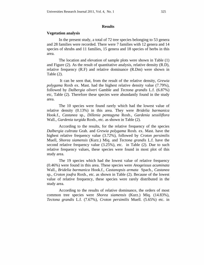
Universities Research Journal 2011, Vol. 4, No. 1 325
Results
Vegetation analysis In the present study, a total of 72 tree species belonging to 53 genera and 28 families were recorded. There were 7 families with 12 genera and 14 species of shrubs and 11 families, 15 genera and 18 species of herbs in this area.
The location and elevation of sample plots were shown in Table (1) and Figure (2). As the result of quantitative analysis, relative density (R.D), relative frequency (R.F) and relative dominance (R.Dm) were shown in Table (2).
It can be seen that, from the result of the relative density, Grewia polygama Roxb ex. Mast. had the highest relative density value (7.79%), followed by Dalbergia oliveri Gamble and Tectona grandis L.f. (6.87%) etc, Table (2). Therefore these species were abundantly found in the study area.
The 10 species were found rarely which had the lowest value of relative density (0.13%) in this area. They were Bridelia barmanica Hook.f., Castanea sp., Dillenia pentagyna Roxb., Gardenia sessiliflora Wall., Gardenia turgida Roxb., etc. as shown in Table (2).
According to the results, for the relative frequency of the species Dalbergia cultrata Grah. and Grewia polygama Roxb. ex. Mast. have the highest relative frequency value (3.72%), followed by Croton persimilis Muell, Shorea siamensis (Kurz.) Miq. and Tectona grandis L.f. have the second relative frequency value (3.25%), etc. in Table (2). Due to such relative frequency values, these species were found in most plot of this study area.
The 19 species which had the lowest value of relative frequency (0.46%) were found in this area. These species were Anogeissus acuminata Wall., Bridelia barmanica Hook.f., Castanopsis armata Spach., Castanea sp., Croton joufra Roxb., etc. as shown in Table (2). Because of the lowest value of relative frequency, these species were rarely distributed in the study area.
According to the results of relative dominance, the orders of most common tree species were Shorea siamensis (Kurz.) Miq. (14.83%), Tectona grandis L.f. (7.67%), Croton persimilis Muell. (5.65%) etc. in
326 Universities Research Journal 2011, Vol. 4, No. 1
Table (2). Thus Shorea siamensis (Kurz.) Miq. had larger basal diameter and occupied the area more than other tree species.
Gardenia sessiliflora Wall., Rhus semialata Murr., possessed the lowest relative dominance value (0.01%). Thus they had small size of stem and occupied the smaller area more than other tree species.
Ranking of ecological significance by IVI of 72 major tree species in the study area were given in Table (2). As a combination results of the relative values of density, frequency and dominance of all recorded tree species, the highest IVI of major dominant tree species was Shorea siamensis (Kurz.) Miq. (24.82 %). The second most dominant species is Tectona grandis L.f (17.78 %) and Croton persimilis Muell. (14.71 %) is third in Figure (3).
According to the results of quantitative analysis, the majority of the high IVI value tree species were ecologically and economically important.
Diversity indices are better measure of the species diversity of a forest and more informative than species counts alone. The calculated diversity parameter is shown in Table (3).
According to the results of the diversity indices of Shannon-Wiener (H) and Simpson (D) value for tree species were 5.401, 0.965, shrubs were 3.062, 0.828 and herbs were 4.228, 0.931 respectively.
As the result of Shannon-Wiener Evenness (E) index for trees, Shrubs and herbaceous plants were 0.875, 0.804 and 0.90. It was observed that as the species diversity was higher, the evenness was higher in the study area. Therefore, all the diversity indices indicated that the study site is floristically high diverse.
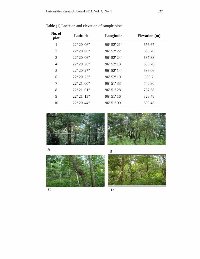
Universities Research Journal 2011, Vol. 4, No. 1 327
B A
C D
Table (1) Location and elevation of sample plots
No. of plot Latitude Longitude Elevation (m)
1 22º 20' 06" 96º 52' 21" 656.67
2 22º 20' 06" 96º 52' 22" 685.76
3 22º 20' 06" 96º 52' 24" 637.88
4 22º 20' 26" 96º 52' 13" 605.76
5 22º 20' 27" 96º 52' 14" 686.06
6 22º 20' 23" 96º 52' 10" 599.7
7 22º 21' 00" 96º 51' 33" 746.36
8 22º 21' 01" 96º 51' 28" 787.58
9 22º 21' 13" 96º 51' 16" 828.48
10 22º 20' 44" 96º 51' 00" 609.45
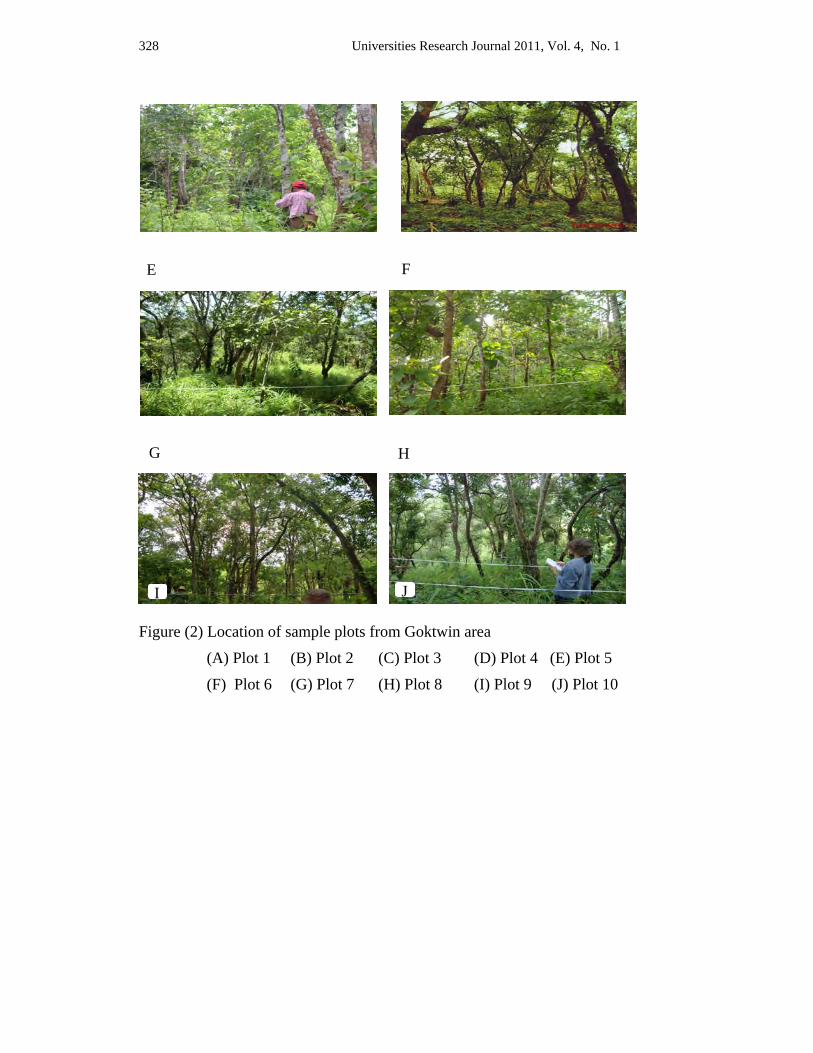
328 Universities Research Journal 2011, Vol. 4, No. 1
E
Figure (2) Location of sample plots from Goktwin area
(A) Plot 1 (B) Plot 2 (C) Plot 3 (D) Plot 4 (E) Plot 5
(F) Plot 6 (G) Plot 7 (H) Plot 8 (I) Plot 9 (J) Plot 10
F
G
I
H
J
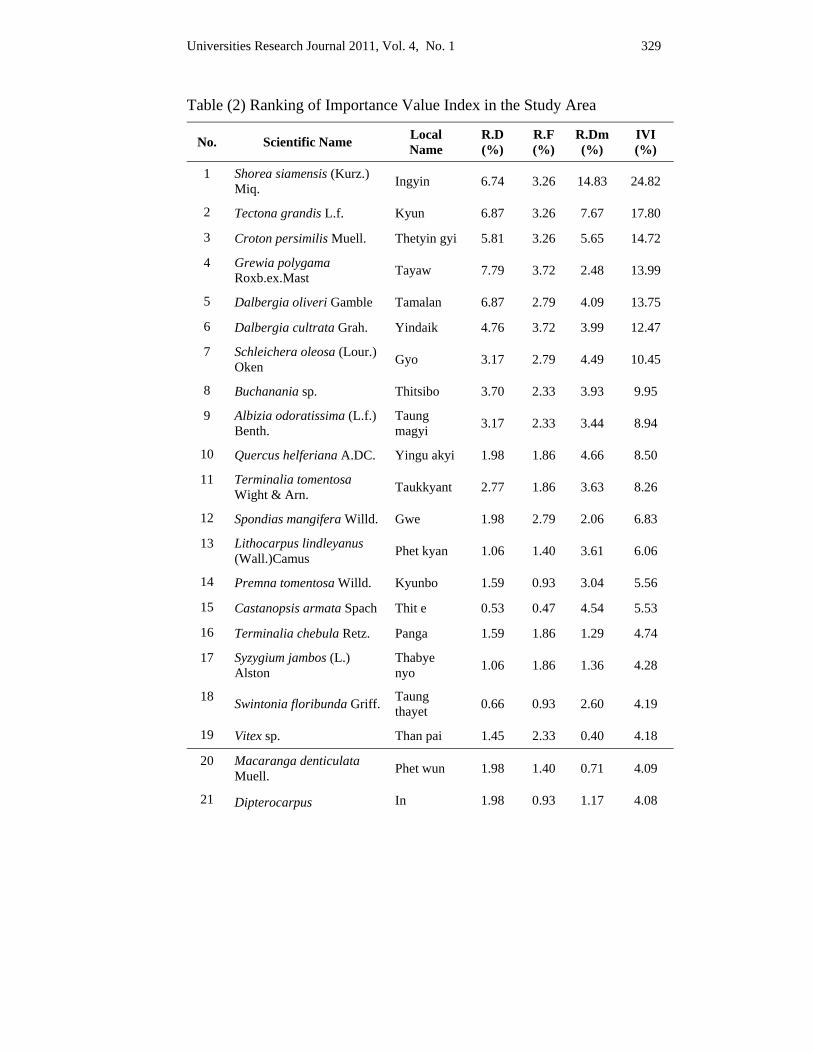
Universities Research Journal 2011, Vol. 4, No. 1 329
Table (2) Ranking of Importance Value Index in the Study Area
No. Scientific Name Local Name
R.D (%)
R.F (%)
R.Dm (%)
IVI (%)
1 Shorea siamensis (Kurz.) Miq. Ingyin 6.74 3.26 14.83 24.82
2 Tectona grandis L.f. Kyun 6.87 3.26 7.67 17.80
3 Croton persimilis Muell. Thetyin gyi 5.81 3.26 5.65 14.72
4 Grewia polygama Roxb.ex.Mast Tayaw 7.79 3.72 2.48 13.99
5 Dalbergia oliveri Gamble Tamalan 6.87 2.79 4.09 13.75
6 Dalbergia cultrata Grah. Yindaik 4.76 3.72 3.99 12.47
7 Schleichera oleosa (Lour.) Oken Gyo 3.17 2.79 4.49 10.45
8 Buchanania sp. Thitsibo 3.70 2.33 3.93 9.95
9 Albizia odoratissima (L.f.) Benth.
Taung magyi 3.17 2.33 3.44 8.94
10 Quercus helferiana A.DC. Yingu akyi 1.98 1.86 4.66 8.50
11 Terminalia tomentosa Wight & Arn. Taukkyant 2.77 1.86 3.63 8.26
12 Spondias mangifera Willd. Gwe 1.98 2.79 2.06 6.83
13 Lithocarpus lindleyanus (Wall.)Camus Phet kyan 1.06 1.40 3.61 6.06
14 Premna tomentosa Willd. Kyunbo 1.59 0.93 3.04 5.56
15 Castanopsis armata Spach Thit e 0.53 0.47 4.54 5.53
16 Terminalia chebula Retz. Panga 1.59 1.86 1.29 4.74
17 Syzygium jambos (L.) Alston
Thabye nyo 1.06 1.86 1.36 4.28
18 Swintonia floribunda Griff. Taung thayet 0.66 0.93 2.60 4.19
19 Vitex sp. Than pai 1.45 2.33 0.40 4.18
20 Macaranga denticulata Muell. Phet wun 1.98 1.40 0.71 4.09
21 Dipterocarpus In 1.98 0.93 1.17 4.08
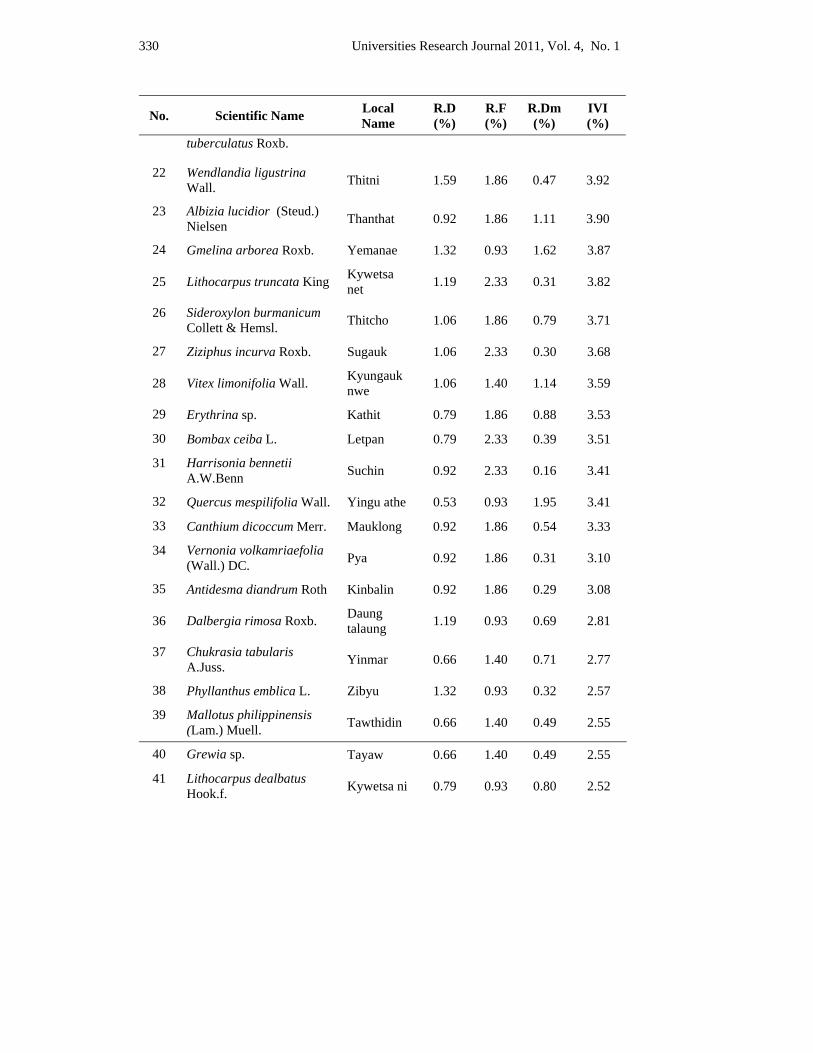
330 Universities Research Journal 2011, Vol. 4, No. 1
No. Scientific Name Local Name
R.D (%)
R.F (%)
R.Dm (%)
IVI (%)
tuberculatus Roxb.
22 Wendlandia ligustrina Wall. Thitni 1.59 1.86 0.47 3.92
23 Albizia lucidior (Steud.) Nielsen Thanthat 0.92 1.86 1.11 3.90
24 Gmelina arborea Roxb. Yemanae 1.32 0.93 1.62 3.87
25 Lithocarpus truncata King Kywetsa net 1.19 2.33 0.31 3.82
26 Sideroxylon burmanicum Collett & Hemsl. Thitcho 1.06 1.86 0.79 3.71
27 Ziziphus incurva Roxb. Sugauk 1.06 2.33 0.30 3.68
28 Vitex limonifolia Wall. Kyungauk nwe 1.06 1.40 1.14 3.59
29 Erythrina sp. Kathit 0.79 1.86 0.88 3.53
30 Bombax ceiba L. Letpan 0.79 2.33 0.39 3.51
31 Harrisonia bennetii A.W.Benn Suchin 0.92 2.33 0.16 3.41
32 Quercus mespilifolia Wall. Yingu athe 0.53 0.93 1.95 3.41
33 Canthium dicoccum Merr. Mauklong 0.92 1.86 0.54 3.33
34 Vernonia volkamriaefolia (Wall.) DC. Pya 0.92 1.86 0.31 3.10
35 Antidesma diandrum Roth Kinbalin 0.92 1.86 0.29 3.08
36 Dalbergia rimosa Roxb. Daung talaung 1.19 0.93 0.69 2.81
37 Chukrasia tabularis A.Juss. Yinmar 0.66 1.40 0.71 2.77
38 Phyllanthus emblica L. Zibyu 1.32 0.93 0.32 2.57
39 Mallotus philippinensis (Lam.) Muell. Tawthidin 0.66 1.40 0.49 2.55
40 Grewia sp. Tayaw 0.66 1.40 0.49 2.55
41 Lithocarpus dealbatus Hook.f. Kywetsa ni 0.79 0.93 0.80 2.52
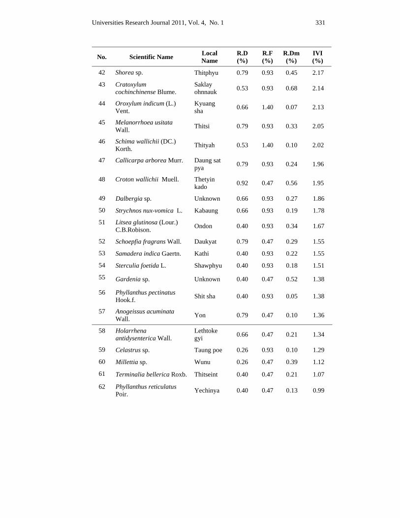
Universities Research Journal 2011, Vol. 4, No. 1 331
No. Scientific Name Local Name
R.D (%)
R.F (%)
R.Dm (%)
IVI (%)
42 Shorea sp. Thitphyu 0.79 0.93 0.45 2.17
43 Cratoxylum cochinchinense Blume.
Saklay ohnnauk 0.53 0.93 0.68 2.14
44 Oroxylum indicum (L.) Vent.
Kyuang sha 0.66 1.40 0.07 2.13
45 Melanorrhoea usitata Wall. Thitsi 0.79 0.93 0.33 2.05
46 Schima wallichii (DC.) Korth. Thityah 0.53 1.40 0.10 2.02
47 Callicarpa arborea Murr. Daung sat pya 0.79 0.93 0.24 1.96
48 Croton wallichii Muell. Thetyin kado 0.92 0.47 0.56 1.95
49 Dalbergia sp. Unknown 0.66 0.93 0.27 1.86
50 Strychnos nux-vomica L. Kabaung 0.66 0.93 0.19 1.78
51 Litsea glutinosa (Lour.) C.B.Robison. Ondon 0.40 0.93 0.34 1.67
52 Schoepfia fragrans Wall. Daukyat 0.79 0.47 0.29 1.55
53 Samadera indica Gaertn. Kathi 0.40 0.93 0.22 1.55
54 Sterculia foetida L. Shawphyu 0.40 0.93 0.18 1.51
55 Gardenia sp. Unknown 0.40 0.47 0.52 1.38
56 Phyllanthus pectinatus Hook.f. Shit sha 0.40 0.93 0.05 1.38
57 Anogeissus acuminata Wall. Yon 0.79 0.47 0.10 1.36
58 Holarrhena antidysenterica Wall.
Lethtoke gyi 0.66 0.47 0.21 1.34
59 Celastrus sp. Taung poe 0.26 0.93 0.10 1.29
60 Millettia sp. Wunu 0.26 0.47 0.39 1.12
61 Terminalia bellerica Roxb. Thitseint 0.40 0.47 0.21 1.07
62 Phyllanthus reticulatus Poir. Yechinya 0.40 0.47 0.13 0.99
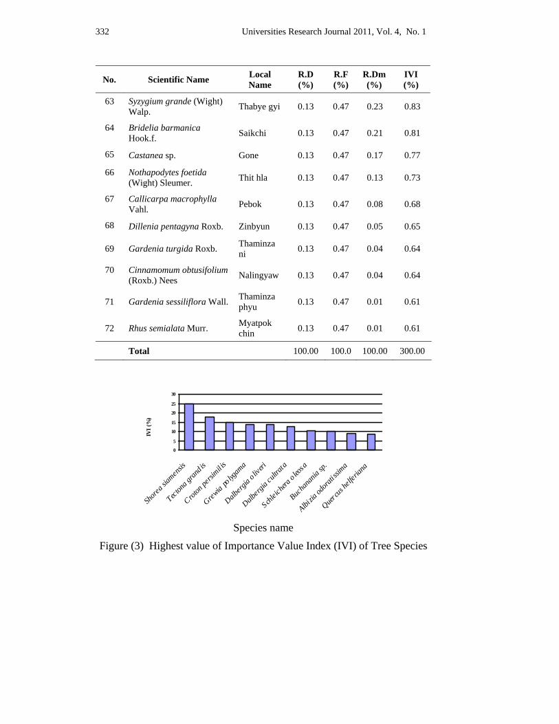
332 Universities Research Journal 2011, Vol. 4, No. 1
No. Scientific Name Local Name
R.D (%)
R.F (%)
R.Dm (%)
IVI (%)
63 Syzygium grande (Wight) Walp. Thabye gyi 0.13 0.47 0.23 0.83
64 Bridelia barmanica Hook.f. Saikchi 0.13 0.47 0.21 0.81
65 Castanea sp. Gone 0.13 0.47 0.17 0.77
66 Nothapodytes foetida (Wight) Sleumer. Thit hla 0.13 0.47 0.13 0.73
67 Callicarpa macrophylla Vahl. Pebok 0.13 0.47 0.08 0.68
68 Dillenia pentagyna Roxb. Zinbyun 0.13 0.47 0.05 0.65
69 Gardenia turgida Roxb. Thaminza ni 0.13 0.47 0.04 0.64
70 Cinnamomum obtusifolium (Roxb.) Nees Nalingyaw 0.13 0.47 0.04 0.64
71 Gardenia sessiliflora Wall. Thaminza phyu 0.13 0.47 0.01 0.61
72 Rhus semialata Murr. Myatpok chin 0.13 0.47 0.01 0.61
Total 100.00 100.0 100.00 300.00
0
5
10
15
20
25
30
Shore
a siam
ensis
Tecton
a gran
d is
Croton
persi
mil is
Grewia
polyg
ama
Dalberg
ia o liv
eri
Dalberg
ia cu
ltrata
Schleic
hera o
leosa
Bucha
nania
sp.
Albizia
odora
tissim
a
Quercus
helfe
riana
IVI
(%)
Species name
Figure (3) Highest value of Importance Value Index (IVI) of Tree Species
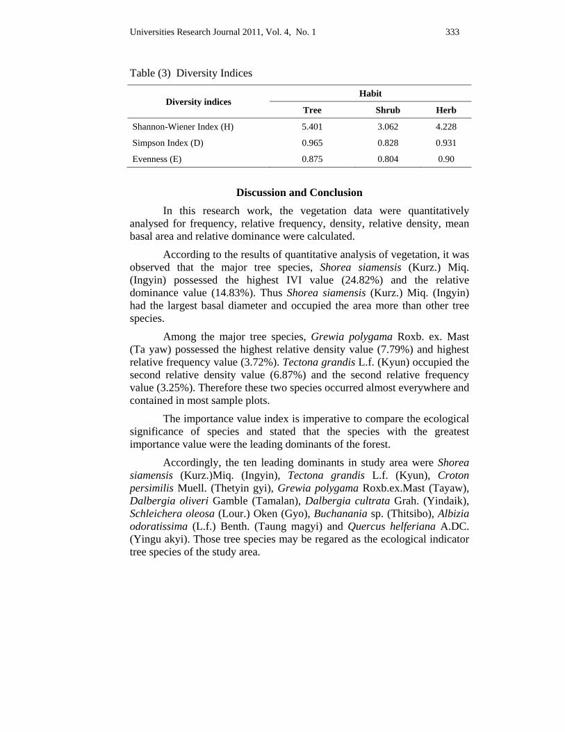
Universities Research Journal 2011, Vol. 4, No. 1 333
Table (3) Diversity Indices
Diversity indices Habit
Tree Shrub Herb
Shannon-Wiener Index (H) 5.401 3.062 4.228
Simpson Index (D) 0.965 0.828 0.931
Evenness (E) 0.875 0.804 0.90
Discussion and Conclusion In this research work, the vegetation data were quantitatively analysed for frequency, relative frequency, density, relative density, mean basal area and relative dominance were calculated.
According to the results of quantitative analysis of vegetation, it was observed that the major tree species, Shorea siamensis (Kurz.) Miq. (Ingyin) possessed the highest IVI value (24.82%) and the relative dominance value (14.83%). Thus Shorea siamensis (Kurz.) Miq. (Ingyin) had the largest basal diameter and occupied the area more than other tree species.
Among the major tree species, Grewia polygama Roxb. ex. Mast (Ta yaw) possessed the highest relative density value (7.79%) and highest relative frequency value (3.72%). Tectona grandis L.f. (Kyun) occupied the second relative density value (6.87%) and the second relative frequency value (3.25%). Therefore these two species occurred almost everywhere and contained in most sample plots.
The importance value index is imperative to compare the ecological significance of species and stated that the species with the greatest importance value were the leading dominants of the forest.
Accordingly, the ten leading dominants in study area were Shorea siamensis (Kurz.)Miq. (Ingyin), Tectona grandis L.f. (Kyun), Croton persimilis Muell. (Thetyin gyi), Grewia polygama Roxb.ex.Mast (Tayaw), Dalbergia oliveri Gamble (Tamalan), Dalbergia cultrata Grah. (Yindaik), Schleichera oleosa (Lour.) Oken (Gyo), Buchanania sp. (Thitsibo), Albizia odoratissima (L.f.) Benth. (Taung magyi) and Quercus helferiana A.DC. (Yingu akyi). Those tree species may be regared as the ecological indicator tree species of the study area.
334 Universities Research Journal 2011, Vol. 4, No. 1
The highly valued tree species like Shorea siamensis (kurz.) Miq. (Ingyin) economically significant species like Tectona grandis L.f. (Kyun), Dalbergia oliveri Gamble (Tamalan) were abundantly found in the study area. So this forest is not only important ecologically but also remarkably commercial.
Diversity indices are better measure of the species diversity of a forest and more informative than species counts alone. According to the Magurran (1988), species diversity is often expressed by two indices: Shannon-Wiener Index (H), Evenness (E) and Simpson Index (D).
The number of tree species growing in study area was 72, the individual number of trees in diameter of 10cm and above in 10 sample plots was 757. The total of 789 shrubs individuals, representing 14 species and the total of 275 herbs individuals, representing 18 species were recorded.
According to the results of Shannon-Wiener Index (H) it was observed that the index of this area was 5.401 for trees, 3.062 for shrubs and 4.228 for herbs. Similarly Simpson Index (D) of the area was 0.965 for trees, 0.828 for shrubs, 0.931for herbs. At the results of Shannon-Wiener evenness (E) 0.875 for trees, 0.804 for shrubs and 0.90 for herbs.
In this study, there was a correlation between species diversity and evenness. It was observed that as the species diversity was higher, the evenness was higher in the study area.
Human impact like cutting for fire wood, charcoal, illegal logging and agricultural encroachment occurred in the study area. It is one of the influencing factors for forest structure. Logging was the major destructive force in the forest. Shorea siamensis (Kurz.) Miq. (Ingyin), Tectona grandis L.f. (Kyun). Danbergia oliveri Gamble (Tamalan), Dipterocarpus tuberculatus Roxb. (In), Gmelina arborea Roxb. (Yemanae) were the preferred species.
According to the IUCN Red List of Globally Threatened and Endangered species (2008, 2009). Shorea siamensis (Kurz.) Miq. (Ingyin), Dipterocarpus tuberculatus Roxb. (In) and Dalbergia oliveri Gamble (Tamalan) in study area are included in globally threatened species Table (4). Therefore special priority should be given to conserve these species.
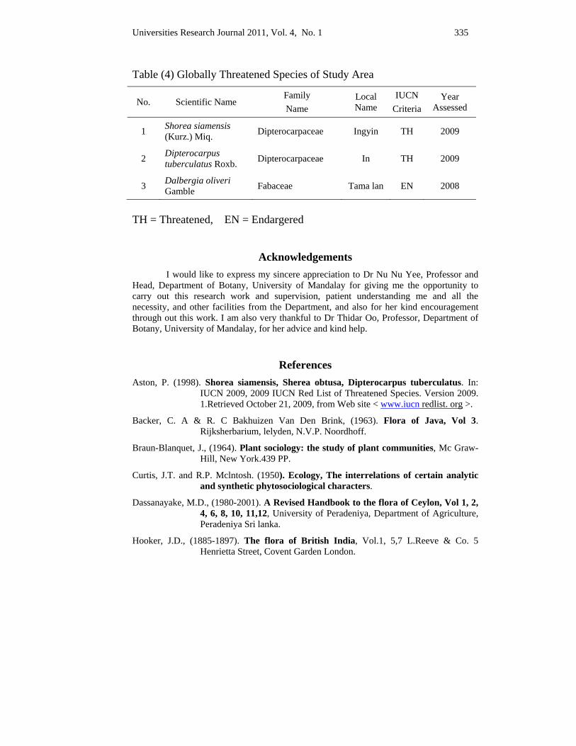
Universities Research Journal 2011, Vol. 4, No. 1 335
Table (4) Globally Threatened Species of Study Area
No. Scientific Name Family Name
Local Name
IUCN Criteria
Year Assessed
1 Shorea siamensis (Kurz.) Miq. Dipterocarpaceae Ingyin TH 2009
2 Dipterocarpus tuberculatus Roxb. Dipterocarpaceae In TH 2009
3 Dalbergia oliveri Gamble Fabaceae Tama lan EN 2008
TH = Threatened, EN = Endargered
Acknowledgements I would like to express my sincere appreciation to Dr Nu Nu Yee, Professor and Head, Department of Botany, University of Mandalay for giving me the opportunity to carry out this research work and supervision, patient understanding me and all the necessity, and other facilities from the Department, and also for her kind encouragement through out this work. I am also very thankful to Dr Thidar Oo, Professor, Department of Botany, University of Mandalay, for her advice and kind help.
References Aston, P. (1998). Shorea siamensis, Sherea obtusa, Dipterocarpus tuberculatus. In:
IUCN 2009, 2009 IUCN Red List of Threatened Species. Version 2009. 1.Retrieved October 21, 2009, from Web site < www.iucn redlist. org >.
Backer, C. A & R. C Bakhuizen Van Den Brink, (1963). Flora of Java, Vol 3. Rijksherbarium, lelyden, N.V.P. Noordhoff.
Braun-Blanquet, J., (1964). Plant sociology: the study of plant communities, Mc Graw-Hill, New York.439 PP.
Curtis, J.T. and R.P. Mclntosh. (1950). Ecology, The interrelations of certain analytic and synthetic phytosociological characters.
Dassanayake, M.D., (1980-2001). A Revised Handbook to the flora of Ceylon, Vol 1, 2, 4, 6, 8, 10, 11,12, University of Peradeniya, Department of Agriculture, Peradeniya Sri lanka.
Hooker, J.D., (1885-1897). The flora of British India, Vol.1, 5,7 L.Reeve & Co. 5 Henrietta Street, Covent Garden London.
336 Universities Research Journal 2011, Vol. 4, No. 1
Magurran, Anne, E., (1988). Ecological Diversity and its Measurement Princeton University Press. New Jersey.
Nghia, N.H. (1988). Dalbergia oliveri. In: IUCN 2008, 2008 IUCN Red List of Threatened Species. Retrieved March 20, 2009, from Web site: http://www.iucnredlist.org/.
Padalia, H., Nidhi Chauhan, M.C. Porwal and P.S. Roy. (2004). Phytosociological observations on tree species diversity of Andaman Islands, India, Current Science, Vol. 87, No. 6.
Sahu, S.C., N.K. Dhal, C. Sudhakar Reddy, Chiranjibi Pattanaik and M. Bralunam. (2007). Phytosociological Study of Tropical Dry Deciduous Forest of Boudh District, Orissa, India, Research Journal of Forestry 1 (2):66-72, Academic Journals Inc.
Shannon, C.E., and W. Wiener, (1963). The Mathematical Theory of Communication, University of Illinois Press, Urbana, USA.
Simpson, E.H., (1949). Measurement of diversity', Nature, 163, 688.
Universities Research Journal 2011, Vol. 4. No. 1
Demonstrator, Department of Botany, East Yangon University
Chemical Investigation and Antimicrobial Activities of Sesbania grandiflora L.
Aye Aye Aung
Abstract Sesbania grandiflora L. belongs to the family, Fabaceae, order Fabales. It is commonly known in Myanmar as Pauk-pan-phyu and Pauk-pan-ni. The plants were collected from Hpa-an Township, Kayin State and prepared for chemical studies in this work. According to the phytochemical investigation, alkaloid, α-amino acid, carbohydrate, starch, reducing sugar, glycoside, phenolic compound, saponin, tannin, terpenoid/steroid and flavonoid were found to be present and cyanogenic glycoside were absent in these plants. In physicochemical studies, the methanol and ethanol soluble matter content were found to be greater than soluble matter content of other organic solvents in leaves. These plants analysis are to determine the abundance amount of elements in Sesbania grandiflora L. (Pauk-pan-phyu and Pauk-pan-ni) which are a quantitative indication of the level of nutrients in plant and is obtained by using EDXRF (Energy Dispersive X-ray Fluorescence) spectrometer. In this experiments, chloroform extracts of leaves showed more significant activity against all test organisms especially on Pseudomonas aeruginosa.
Key words: Sesbania grandiflora L., Qualitative and Quantitative analysis, Antimicrobial Activities.
Introduction Sesbania grandiflora L. belonging to Fabaceae commonly known as
Sesbania which is often planted for its edible flowers and pods in tropical countries. It was known as Pauk-pan-phyu and Pauk-pan-ni in Myanmar. It is believed to have originated either in India or Southeast Asia and grown primarily in hot and humid areas of the world. From present generation, it is a green manure crop useful in paddy cultivation; it is one of the most valuable medicinal herbs. The tree is grown as an ornamental shade tree, and for reforestation. (Watt, 1972 and James, 1983). The crude methanolic extracts of the flowers, seeds, rachis and leaves of Sesbania grandiflora were strongly positive for saponin (by Fehling’s A and B test). The flowers, seeds and leaves were slightly positive for alkaloids. Sterols were markedly found in the rachis and leaves; strongly present in the flowers; and slightly present in the seeds. The rachis and leaves contained plenty of tannins
338 Universities Research Journal 2011, Vol. 4. No. 1
(Rastogi, 1969).This paper deals with chemical investigations including phytochemical tests, physicochemical characterization, elemental analysis, nutritional values.
Materials and Methods
Collection, classification and identification of Sesbania grandiflora L. Sesbania grandiflora L. plant parts were collected from Hpa-an
Township, Kayin State, during the months of July to December in 2009. The dried samples were made into fine powder using a mortar and pestle and stored in air tight container to prevent moisture and other contamination.
Qualitative and quantitative analysis of Sesbania grandiflora L.
Qualitative analysis Qualitative analysis concerned with the presence of alkaloids, amino
acid, carbohydrate, starch, reducing sugar, cyanogenic glycoside, glycoside, phenolic compound, saponin, tannin, steroid and flavonoid were investigated by the methods of British Pharmacopoeia (1968), Trease and Evans (2002). Preliminary phytochemical examination was carried out in the phytochemical laboratory of Chemistry Department, University of Yangon.
Quantitative analysis Quatitative analysis including moisture content, total ash, acid
soluble, water soluble ash, acetone soluble, chloroform soluble, petroleum ether soluble, ethyl acetate soluble, ethanol soluble, methanol soluble and water soluble contents of leaves were determined by the methods of in B P, (1968).
Elemental analysis and nutritional values of Sesbania grandiflora L.
Elemental analysis of leaves The relative concentration of elements in Sesbania grandiflora L.
powdered leaves were analyzed by using Energy Dispersive X-ray Fluorescence (EDXRF) spectrometer, Shimadzu Co. Ltd., Japan, at the Universities’ Research Center, University of Yangon.
Some nutrient elements such as Copper (Cu), Potassium (K), Calcium (Ca), Iron (Fe) and Phosphorus (P) in the leaves were also quantitatively analyzed by A A Analyst.800 Atomic Absorption
Universities Research Journal 2011, Vol. 4. No. 1 339
Spectrophotometer England (A A S) at the Universities’ Research Center, University of Yangon.
Nutritional values of leaves Carbohydrate, protein, fibre, fat, vitamin B1 and vitamin C contents
have been determined in National Nutritional Centre, Department of Health, according to the procedures given in the methods of A.O.A.C. (Horwitz, 1980).
Antimicrobial activity of different solvent extracts of leaves
Extraction and test organisms The powdered leaves were extracted with chloroform, acetone, petroleum-ether, methanol, ethyl acetate, ethanol and distilled water by percolation method. The filtrate solvents were evaporated by using water bath .
Crude extracts of various solvents were tested on six pathogenic microorganisms such as Bacillus pumalis, Bacillus subtilis, Escherichia coli, Pseudomonas aeruginosa, Staphylococcus aureus and Candida albicans. The test organisms used in this research were supplied from the Development Centre for Pharmaceutical Technology (DCPT) for determination of antimicrobial activities.
The study of antimicrobial activities was determined by agar-well diffusion method described by Cruickshank, et al., (1975). Nutrient agar was prepared and boiled and 20 - 25 ml of the medium was poured into a test tube and plugged with cotton wool and steriled at 121˚C for 15 minutes in an autoclave. Then the tubes were cooled down to 30 - 35˚C and poured into the sterile petridishes and 0.1 - 0.2 ml of the test organisms were also added into the dishes. They were allowed to set for 2 - 3 hours. Then 10 mm agar - well was made by the help of sterilized agar-well cutter. After then about 0.2 ml of sample was introduced into the agar-well and incubated at 37˚C for 24 hours. The inhibition zones which appeared around the agar - well indicated the presence of antimicrobial activities. The diameter of the inhibition zones were measured and recorded in mm, the controlled experiments using solvent only were prepared for the comparison with leaves plant extracts.
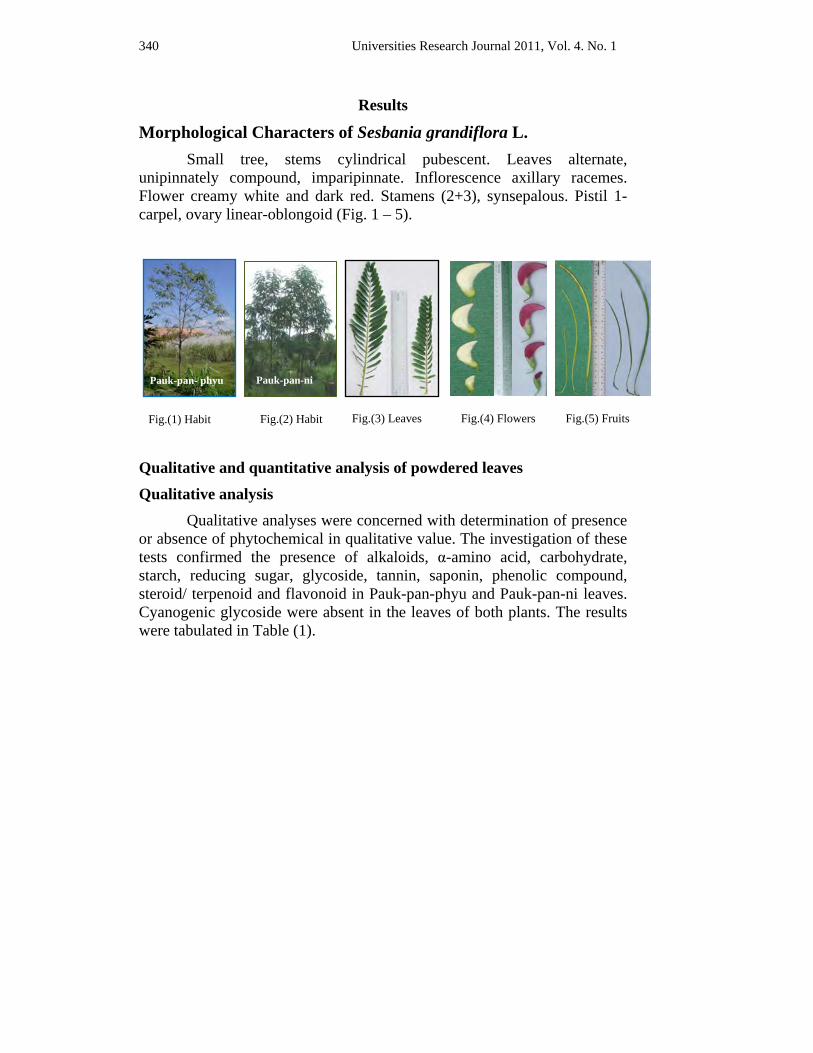
340 Universities Research Journal 2011, Vol. 4. No. 1
Results
Morphological Characters of Sesbania grandiflora L. Small tree, stems cylindrical pubescent. Leaves alternate,
unipinnately compound, imparipinnate. Inflorescence axillary racemes. Flower creamy white and dark red. Stamens (2+3), synsepalous. Pistil 1- carpel, ovary linear-oblongoid (Fig. 1 – 5).
Qualitative and quantitative analysis of powdered leaves
Qualitative analysis Qualitative analyses were concerned with determination of presence or absence of phytochemical in qualitative value. The investigation of these tests confirmed the presence of alkaloids, α-amino acid, carbohydrate, starch, reducing sugar, glycoside, tannin, saponin, phenolic compound, steroid/ terpenoid and flavonoid in Pauk-pan-phyu and Pauk-pan-ni leaves. Cyanogenic glycoside were absent in the leaves of both plants. The results were tabulated in Table (1).
Fig.(1) Habit Fig.(2) Habit Fig.(3) Leaves Fig.(4) Flowers Fig.(5) Fruits
Pauk-pan- phyu Pauk-pan-ni
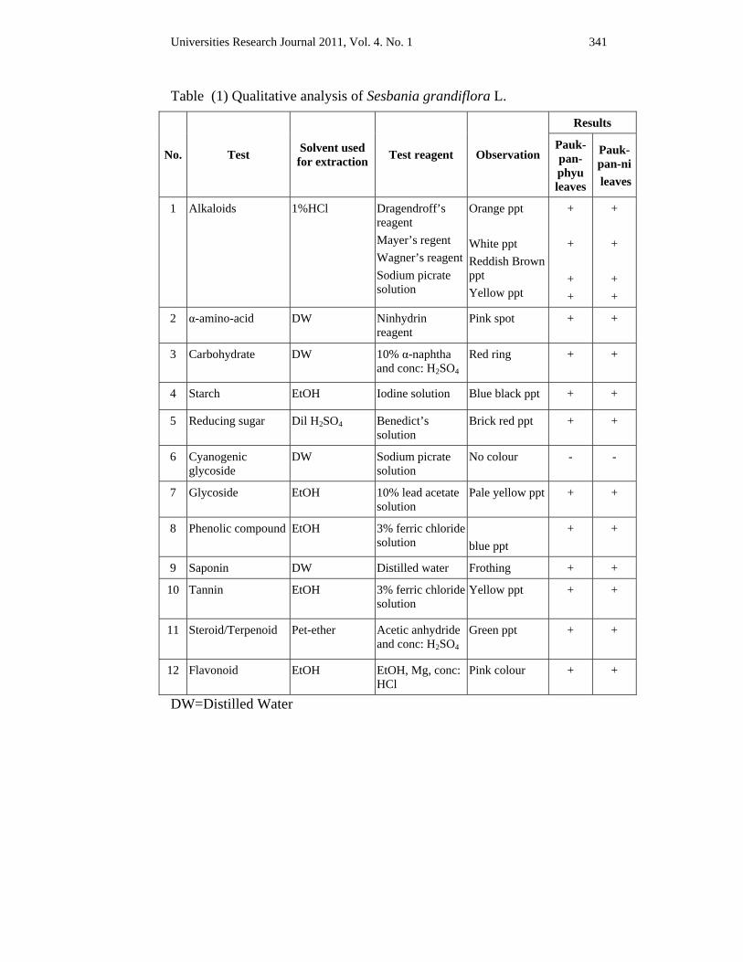
Universities Research Journal 2011, Vol. 4. No. 1 341
Table (1) Qualitative analysis of Sesbania grandiflora L.
No. Test Solvent used for extraction Test reagent Observation
Results
Pauk-pan-phyu leaves
Pauk-pan-ni leaves
1 Alkaloids 1%HCl Dragendroff’s reagent Mayer’s regent Wagner’s reagentSodium picrate solution
Orange ppt White ppt Reddish Brown ppt Yellow ppt
+
+
+ +
+
+
+ +
2 α-amino-acid DW Ninhydrin reagent
Pink spot + +
3 Carbohydrate DW 10% α-naphtha and conc: H2SO4
Red ring + +
4 Starch EtOH Iodine solution Blue black ppt + +
5 Reducing sugar Dil H2SO4 Benedict’s solution
Brick red ppt + +
6 Cyanogenic glycoside
DW Sodium picrate solution
No colour - -
7 Glycoside EtOH 10% lead acetate solution
Pale yellow ppt + +
8 Phenolic compound EtOH 3% ferric chloride solution
blue ppt
+ +
9 Saponin DW Distilled water Frothing + +
10 Tannin EtOH 3% ferric chloride solution
Yellow ppt + +
11 Steroid/Terpenoid Pet-ether Acetic anhydride and conc: H2SO4
Green ppt + +
12 Flavonoid EtOH EtOH, Mg, conc: HCl
Pink colour + +
DW=Distilled Water
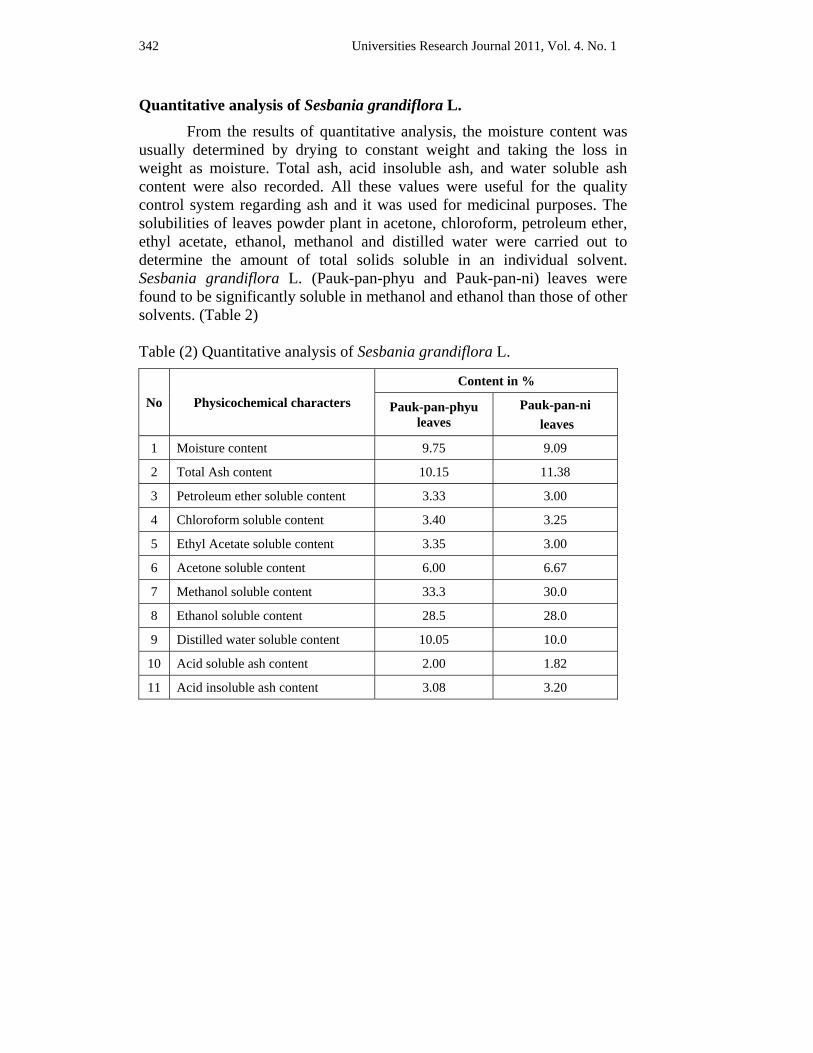
342 Universities Research Journal 2011, Vol. 4. No. 1
Quantitative analysis of Sesbania grandiflora L. From the results of quantitative analysis, the moisture content was
usually determined by drying to constant weight and taking the loss in weight as moisture. Total ash, acid insoluble ash, and water soluble ash content were also recorded. All these values were useful for the quality control system regarding ash and it was used for medicinal purposes. The solubilities of leaves powder plant in acetone, chloroform, petroleum ether, ethyl acetate, ethanol, methanol and distilled water were carried out to determine the amount of total solids soluble in an individual solvent. Sesbania grandiflora L. (Pauk-pan-phyu and Pauk-pan-ni) leaves were found to be significantly soluble in methanol and ethanol than those of other solvents. (Table 2)
Table (2) Quantitative analysis of Sesbania grandiflora L.
No Physicochemical characters Content in %
Pauk-pan-phyu leaves
Pauk-pan-ni leaves
1 Moisture content 9.75 9.09
2 Total Ash content 10.15 11.38
3 Petroleum ether soluble content 3.33 3.00
4 Chloroform soluble content 3.40 3.25
5 Ethyl Acetate soluble content 3.35 3.00
6 Acetone soluble content 6.00 6.67
7 Methanol soluble content 33.3 30.0
8 Ethanol soluble content 28.5 28.0
9 Distilled water soluble content 10.05 10.0
10 Acid soluble ash content 2.00 1.82
11 Acid insoluble ash content 3.08 3.20

Universities Research Journal 2011, Vol. 4. No. 1 343
Elemental analysis and nutritional values of Sesbania grandiflora L. Elemental analysis was to determine the amount of elements in
Sesbania grandiflora L. (Pauk-pan-phyu and Pauk-pan-ni) leaves. It was a quantitative indication of the level of nutrients in plant obtained by using EDXRF (Energy Dispersive X-ray Fluorescence) spectrometer. It was found that Calcium (Ca), Phosphorus (P), Potassium (K), Sulphur(S), Chlorine (Cl) and Iron (Fe) are macronutrient elements, where as Strontium (Sr), Brominr (Br), Manganese (Mn) and Zinc (Zn) are found to be as micronutrient elements. Above the elements were relatively abundant in the leaves of both samples heavy toxic metals Lead (Pb), Arsenic (As) and Mercury (Hg) were absent. (Table 3)
Table (3) Comparative studies on relative elements of Sesbania grandiflora L. by EDXRF
No. Elements Relative abundance in %
Pauk-pan-phyu leaves
Pauk-pan-ni leaves
1 Calcium (Ca)** 1.327 1.072
2 Phosphorus(P)** 0.997 0.700
3 Potassium(K)** 0.447 0.548
4 Sulphur(S)** 0.288 0.203
5 Chlorine(Cl)** 0.225 0.106
6 Iron(Fe)** 0.021 0.022
7 Strontium(Sr)* 0.013 0.008
8 Bromine(Br)* 0.011 0.006
9 Manganese(Mn)* 0.011 0.011
10 Zninc(Zn) 0.003 -
**= macronutrient, *= micronutrient Elemental analysis by AAS
Due to the results of AAS analysis, Cu content in powdered leaves of Pauk-pan-phyu and Pauk-pan-ni were 3.65 and 4.71 ppm respectively which was found to be higher than the other elements. (Table 4)
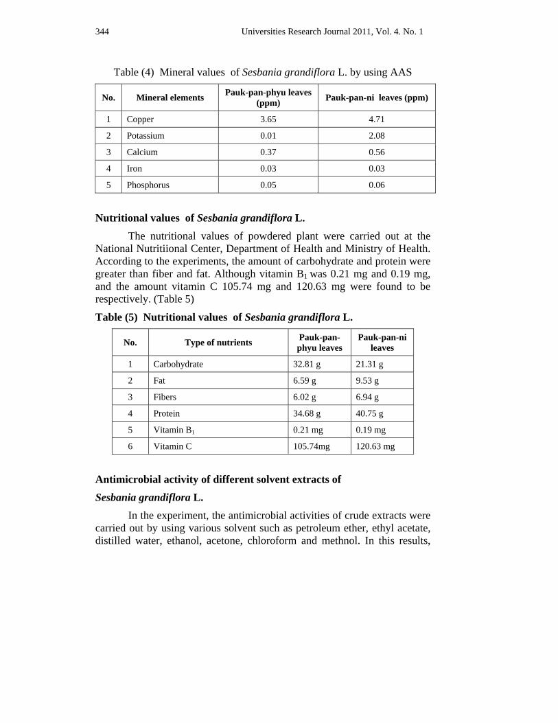
344 Universities Research Journal 2011, Vol. 4. No. 1
Table (4) Mineral values of Sesbania grandiflora L. by using AAS
No. Mineral elements Pauk-pan-phyu leaves (ppm) Pauk-pan-ni leaves (ppm)
1 Copper 3.65 4.71
2 Potassium 0.01 2.08
3 Calcium 0.37 0.56
4 Iron 0.03 0.03
5 Phosphorus 0.05 0.06
Nutritional values of Sesbania grandiflora L. The nutritional values of powdered plant were carried out at the
National Nutritiional Center, Department of Health and Ministry of Health. According to the experiments, the amount of carbohydrate and protein were greater than fiber and fat. Although vitamin B1 was 0.21 mg and 0.19 mg, and the amount vitamin C 105.74 mg and 120.63 mg were found to be respectively. (Table 5)
Table (5) Nutritional values of Sesbania grandiflora L.
No. Type of nutrients Pauk-pan-phyu leaves
Pauk-pan-ni leaves
1 Carbohydrate 32.81 g 21.31 g
2 Fat 6.59 g 9.53 g
3 Fibers 6.02 g 6.94 g
4 Protein 34.68 g 40.75 g
5 Vitamin B1 0.21 mg 0.19 mg
6 Vitamin C 105.74mg 120.63 mg
Antimicrobial activity of different solvent extracts of
Sesbania grandiflora L.
In the experiment, the antimicrobial activities of crude extracts were carried out by using various solvent such as petroleum ether, ethyl acetate, distilled water, ethanol, acetone, chloroform and methnol. In this results,
Universities Research Journal 2011, Vol. 4. No. 1 345
among various extracts of leaves Sesbania grandiflora L. (pauk-pan-phyu and pauk-pan-ni), chloroform extract showed the highest activity on all six different types of microorganisms especially on Pseudomonas aeruginosa. The results were shown in Figure (6 -11).
Discussion and Conclusion In this research, chemical studies such as preliminary phytochemical
investigation, physicochemical characterization, elemental analysis, examination of nutritional values and vitamins contents of Sesbania grandiflora L. (Pauk-pan-phyu and Pauk-pan-ni) leaves had been studied.
For preliminary phytochemical investigation, Sesbania grandiflora L. (Pauk-pan-phyu and Pauk-pan-ni) leaves plant samples were collected from Hpa-an and prepared, alkaloids, α-amino acid, carbohydrate, starch, reducing sugar, glycoside, tannin, saponin, phenolic compound, steroid/terpenoid and flavonoid were detected in leaves. Cyanogenic glycoside was absent in the leaves of both plants. Therefore, the plants Sesbania grandiflora L. (Pauk-pan-phyu and Pauk-pan-ni) have phytoconstituents. Physicochemical characteristics such as moisture contents, ash content and different solubility of organic constituents in organic solvents were determined. The solubility in methanol and ethanol were found to be the greatest in these solubility tests. The more polar phytoconstituents can also be found.
According to the EDXRF result of elemental analysis, it was found that the macronutrient elements in calcium, phosphorus, potassium, sulphur, chlorine and iron. The micronutrient elements in strontium, bromine, manganese and zinc were found in Pauk-pan-phyu and Pauk-pan-ni leaves.
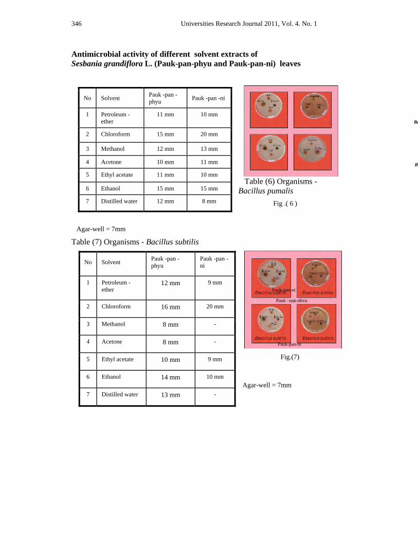
346 Universities Research Journal 2011, Vol. 4. No. 1
Antimicrobial activity of different solvent extracts of Sesbania grandiflora L. (Pauk-pan-phyu and Pauk-pan-ni) leaves
Table (6) Organisms - Bacillus pumalis Fig .( 6 )
Agar-well = 7mm
Table (7) Organisms - Bacillus subtilis
Fig.(7)
No Solvent Pauk -pan -phyu Pauk -pan -ni
1 Petroleum -ether
11 mm 10 mm
2 Chloroform 15 mm 20 mm
3 Methanol 12 mm 13 mm
4 Acetone 10 mm 11 mm
5 Ethyl acetate 11 mm 10 mm
6 Ethanol 15 mm 15 mm
7 Distilled water 12 mm 8 mm
No Solvent Pauk -pan -phyu
Pauk -pan -ni
1 Petroleum -ether
12 mm 9 mm
2 Chloroform 16 mm 20 mm
3 Methanol 8 mm -
4 Acetone 8 mm -
5 Ethyl acetate 10 mm 9 mm
6 Ethanol 14 mm 10 mm
7 Distilled water 13 mm -
Pauk-pan-ni
Ba
Ba
Agar-well = 7mm
Pauk –pan-phyu
Pauk-pan-ni
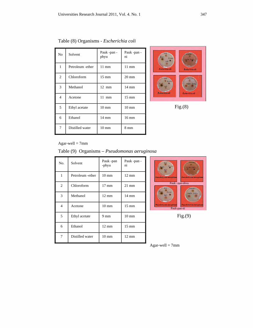
Universities Research Journal 2011, Vol. 4. No. 1 347
Table (8) Organisms - Escherichia coli
Fig.(8)
Agar-well = 7mm
Table (9) Organisms – Pseudomonas aeruginosa
Fig.(9)
Agar-well = 7mm
No Solvent Pauk -pan -phyu
Pauk -pan -ni
1 Petroleum -ether 11 mm 11 mm
2 Chloroform 15 mm 20 mm
3 Methanol 12 mm 14 mm
4 Acetone 11 mm 15 mm
5 Ethyl acetate 10 mm 10 mm
6 Ethanol 14 mm 16 mm
7 Distilled water 10 mm 8 mm
No. Solvent Pauk -pan -phyu
Pauk -pan -ni
1 Petroleum -ether 10 mm 12 mm
2 Chloroform 17 mm 21 mm
3 Methanol 12 mm 14 mm
4 Acetone 10 mm 15 mm
5 Ethyl acetate 9 mm 10 mm
6 Ethanol 12 mm 15 mm
7 Distilled water 10 mm 12 mm
Pauk –pan-phyu
Pauk-pan-ni
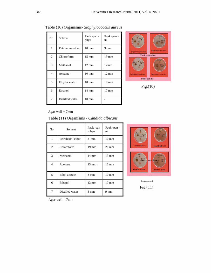
348 Universities Research Journal 2011, Vol. 4. No. 1
Table (10) Organisms- Staphylococcus aureus
Fig.(10)
Agar-well = 7mm
Table (11) Organisms - Candida albicans
Agar-well = 7mm
No. Solvent Pauk -pan -phyu
Pauk -pan -ni
1 Petroleum -ether 10 mm 9 mm
2 Chloroform 15 mm 19 mm
3 Methanol 12 mm 12mm
4 Acetone 10 mm 12 mm
5 Ethyl acetate 10 mm 10 mm
6 Ethanol 14 mm 17 mm
7 Distilled water 10 mm -
No. Solvent Pauk -pan -phyu
Pauk -pan -ni
1 Petroleum -ether 8 mm 10 mm
2 Chloroform 19 mm 20 mm
3 Methanol 14 mm 13 mm
4 Acetone 13 mm 13 mm
5 Ethyl acetate 8 mm 10 mm
6 Ethanol 13 mm 17 mm
7 Distilled water 8 mm 9 mm
Pauk –pan-phyu
Pauk-pan-ni
Pauk –pan-phyu
Pauk-pan-ni
Fig.(11)
Universities Research Journal 2011, Vol. 4. No. 1 349
There are six main classes of nutrients that the body needs: carbohydrates, proteins, fats, vitamins, minerals, and water. It is important to consume these six nutrients on a daily basis to build and maintain healthy bodily function. The important nutritional values found in this plant might also be major contributors to the medicinal value of Pauk-pan-phyu and Pauk-pan-ni.
The antimicrobial activities with six different microorganisms were also tested by using seven crude extracts such as chloroform, acetone, petroleum ether, methanol, ethanol, aqueous extract of Pauk-pan-phyu and Pauk-pan-ni leaves. In this experiment, chloroform extracts of Pauk-pan-phyu and Pauk-pan-ni leaves showed the highest activity on test organisms. Methanol, acetone and aqueous extracts were activity of Bacillus subtilis and aqueous extracts was no activity of Staphylococcus aureus Pauk-pan-ni leaves.
From the overall assessment of chemical studies of Pauk-pan-phyu and Pauk-pan-ni plants which can be used as nutritious food due to the results of the vitamins and nutrient elements. Furthermore, the presence of phytoconstituents, steroids, and flavones in these plants indicate the medicinal properties.
Acknowledgements
I would like to express my deepest gratitude and special thanks to Dr. Thet Thet May, Professor and Head and Professor Dr. Aye Pe, from the Department of Botany, University of Yangon for their invaluable advice and kind suggestions. I would like to express my deepest gratitude to my supervisor Dr. Moe Moe Lwin, Lecturer, Department of Botany, Pathein University for her close supervision, valuable guidance, constant encouragement for the success of my research work..
References A O A C, (1965). Methods of Analysis, 3rd ed., Washinton, Bejamin Franlin Station
Cruickshank R., et al., (1975). Medical Microbiology, Twelve edition, vol.II, printed in
Great Britain, Distributed in the USA by Longman Inc, New York
Harborne, J. B. (1984). Phytochemical Methods, A guide to modern Technique of plants, 2nd ed., Chapman and Hall. London.
Hooker, J. D., (1879). The Flora of British India, Vol. IV, Published under L Reeve and Co.,Ltd., Coventganden London
350 Universities Research Journal 2011, Vol. 4. No. 1
James, A., Duke. (1983). Medicinal plants of the world. Computer index with more than 85,000 entries Vol.III.
Marini Bettolo, G.B.,Nicolettic,M. and Patamia,M., (1981). Plant Screening by ChemicalChromatographic Procedure Under Field Conditions
Rastogi,R.P. and B.N. Mehrotra, (1979). Compendium of Indian medicinal Plants, vol.II, Second print, printted at the printing unit of the National Institute of Science Communication, Council of Scientific and Industrial Research,New Delhi
Trease G.E. and W.C.Evans, (2002). Pharmacognosy, 15th edi, Baillere Tindoll,London British Pharmacopeia, 1968. The Pharmaceutical Press, London and Bradfod
Central Council for Research in Unani Medicine, (1989). Phytochemical Standards of Unani Formulations New Delhi
Watt, G. (1972). A Dictionary of the Economic Products of India. Vol, IV. 2nd ed. Paper, 2. Printed at Jayyed Press, Ballimaran, Delhi.
Wealth of India, (1972). A Dictionary of India Raw Materials and Industrial Products, Vol. IX Council of Scientific and Industrial Research India, Published by the Council of Scientific and Industrial research New Delhi and printed at Ses Saraswaty press H.D., Calcutt.
Universities Research Journal 2011, Vol. 4, No. 1
Demonstrator, Department of Botany, East Yangon University
Effect of Spirulina Biofertilizer Suspension on Growth and Yield of Vigna radiata (L.) Wilczek
Khin Lay Nandar Aung
Abstract Various concentrations of Spirulina suspension were applied as biofertilizer on growth and yield of green gram in plot experiment during the year 2008-2009. A randomized complete block design (RCB) with four replications was used. Among all treatments, the highest leaf area index, crop growth rate, relative growth rate, leaf area ratio, maximum total dry matter, harvest index and yield were obtained by 7 g l-l of Spirulina suspension treatment and produced the highest yield, 237.60 gm-2 (2376 kg ha-l). The second highest yield 228.9 gm-2 (2289 kg ha-l) was obtained from 9 g l-l of Spirulina suspension treatment, compared with other treatments and control 172.2 (1722 kg ha-l).
Key words : Spirulina, biofertilizer
Introduction Myanmar is an agricultural-based country and increase in
production of edible and exportable crops is one of the objectives in nation. Green gram, black gram, pigeon pea, soy bean, cow pea and kidney bean are the major export pulses (MOAI, 2009). Green gram is one of the important traditional crops of the world. Cultivation time is short and serves as an excellent source of protein as seed or sprout (Ahmad et al., 2005). It is cultivated in both upper and lower Myanmar.
According to market demand and preferable advantages from production of pulses for the farmers, growing areas were extended than in the past. The current yield level of green gram was largely dependent upon the utilization of chemical fertilizer. Long term use of chemicals in agricultural farming may effect the soil environment and grain quality. Therefore, application of biofertilizer as an alternative to chemical fertilizer in green gram cultivation has the excellent opportunity which can become established for organic farming or sustainable agriculture.
Microalgae and macroalgae were used as biofertilizer in many countries during the last century. Biofertilizers is a living fertilizer compound of microbial inoculants or groups of microorganisms (Nguvu, 2009).
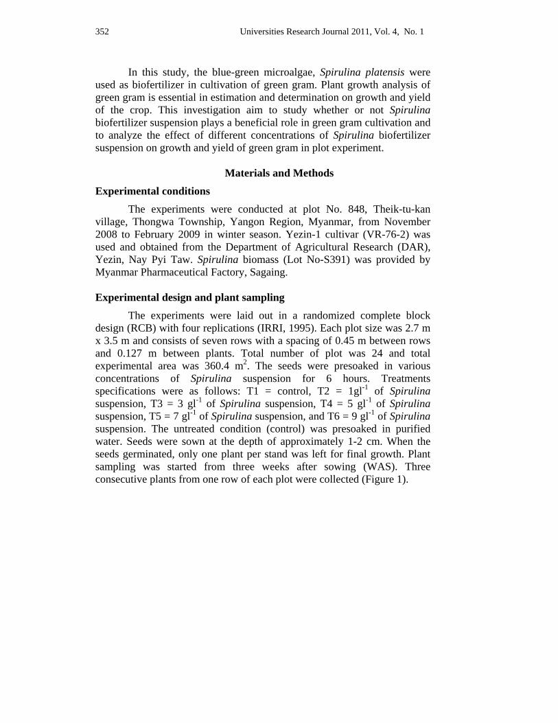
352 Universities Research Journal 2011, Vol. 4, No. 1
In this study, the blue-green microalgae, Spirulina platensis were used as biofertilizer in cultivation of green gram. Plant growth analysis of green gram is essential in estimation and determination on growth and yield of the crop. This investigation aim to study whether or not Spirulina biofertilizer suspension plays a beneficial role in green gram cultivation and to analyze the effect of different concentrations of Spirulina biofertilizer suspension on growth and yield of green gram in plot experiment.
Materials and Methods
Experimental conditions The experiments were conducted at plot No. 848, Theik-tu-kan
village, Thongwa Township, Yangon Region, Myanmar, from November 2008 to February 2009 in winter season. Yezin-1 cultivar (VR-76-2) was used and obtained from the Department of Agricultural Research (DAR), Yezin, Nay Pyi Taw. Spirulina biomass (Lot No-S391) was provided by Myanmar Pharmaceutical Factory, Sagaing.
Experimental design and plant sampling
The experiments were laid out in a randomized complete block design (RCB) with four replications (IRRI, 1995). Each plot size was 2.7 m x 3.5 m and consists of seven rows with a spacing of 0.45 m between rows and 0.127 m between plants. Total number of plot was 24 and total experimental area was 360.4 m2. The seeds were presoaked in various concentrations of Spirulina suspension for 6 hours. Treatments specifications were as follows: T1 = control, T2 = 1gl-1 of Spirulina suspension, T3 = 3 gl-1 of Spirulina suspension, T4 = 5 gl-1 of Spirulina suspension, T5 = 7 gl-1 of Spirulina suspension, and T6 = 9 gl-1 of Spirulina suspension. The untreated condition (control) was presoaked in purified water. Seeds were sown at the depth of approximately 1-2 cm. When the seeds germinated, only one plant per stand was left for final growth. Plant sampling was started from three weeks after sowing (WAS). Three consecutive plants from one row of each plot were collected (Figure 1).
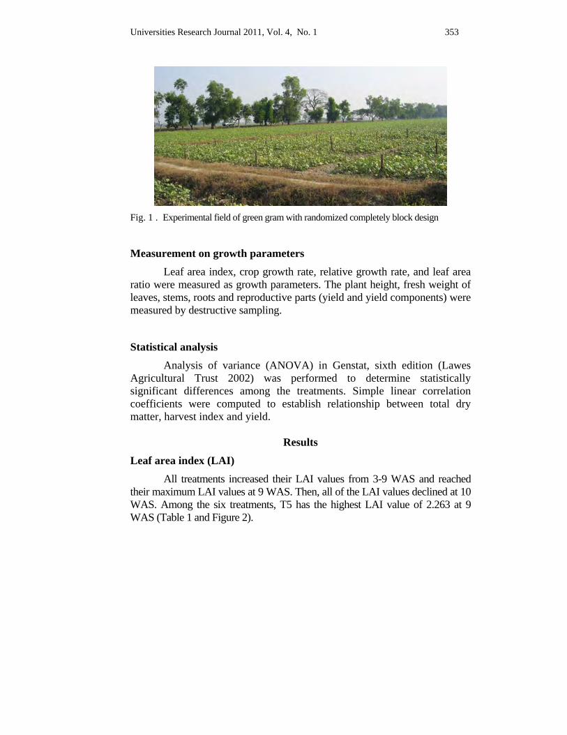
Universities Research Journal 2011, Vol. 4, No. 1 353
Fig. 1 . Experimental field of green gram with randomized completely block design
Measurement on growth parameters Leaf area index, crop growth rate, relative growth rate, and leaf area
ratio were measured as growth parameters. The plant height, fresh weight of leaves, stems, roots and reproductive parts (yield and yield components) were measured by destructive sampling.
Statistical analysis Analysis of variance (ANOVA) in Genstat, sixth edition (Lawes Agricultural Trust 2002) was performed to determine statistically significant differences among the treatments. Simple linear correlation coefficients were computed to establish relationship between total dry matter, harvest index and yield.
Results
Leaf area index (LAI) All treatments increased their LAI values from 3-9 WAS and reached their maximum LAI values at 9 WAS. Then, all of the LAI values declined at 10 WAS. Among the six treatments, T5 has the highest LAI value of 2.263 at 9 WAS (Table 1 and Figure 2).
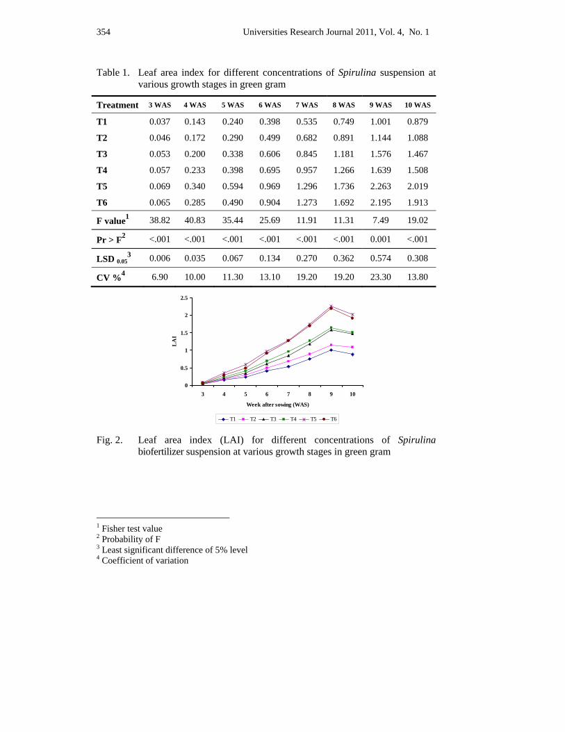
354 Universities Research Journal 2011, Vol. 4, No. 1
Table 1. Leaf area index for different concentrations of Spirulina suspension at various growth stages in green gram
Treatment 3 WAS 4 WAS 5 WAS 6 WAS 7 WAS 8 WAS 9 WAS 10 WAS
T1 0.037 0.143 0.240 0.398 0.535 0.749 1.001 0.879
T2 0.046 0.172 0.290 0.499 0.682 0.891 1.144 1.088
T3 0.053 0.200 0.338 0.606 0.845 1.181 1.576 1.467
T4 0.057 0.233 0.398 0.695 0.957 1.266 1.639 1.508
T5 0.069 0.340 0.594 0.969 1.296 1.736 2.263 2.019
T6 0.065 0.285 0.490 0.904 1.273 1.692 2.195 1.913
F value1 38.82 40.83 35.44 25.69 11.91 11.31 7.49 19.02
Pr > F2 <.001 <.001 <.001 <.001 <.001 <.001 0.001 <.001
LSD 0.053 0.006 0.035 0.067 0.134 0.270 0.362 0.574 0.308
CV %4 6.90 10.00 11.30 13.10 19.20 19.20 23.30 13.80
Fig. 2. Leaf area index (LAI) for different concentrations of Spirulina
biofertilizer suspension at various growth stages in green gram
1 Fisher test value 2 Probability of F 3 Least significant difference of 5% level 4 Coefficient of variation
0
0.5
1
1.5
2
2.5
3 4 5 6 7 8 9 10
Week after sowing (WAS)
LA
I
T1 T2 T3 T4 T5 T6
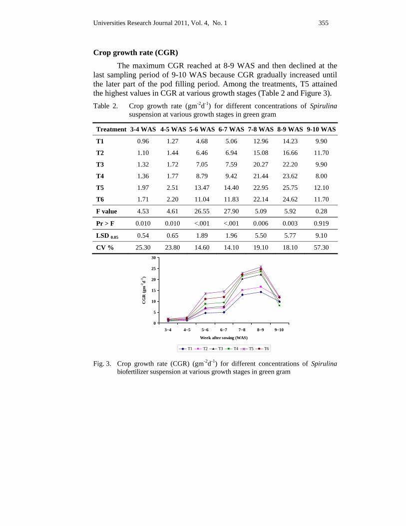
Universities Research Journal 2011, Vol. 4, No. 1 355
Crop growth rate (CGR) The maximum CGR reached at 8-9 WAS and then declined at the
last sampling period of 9-10 WAS because CGR gradually increased until the later part of the pod filling period. Among the treatments, T5 attained the highest values in CGR at various growth stages (Table 2 and Figure 3). Table 2. Crop growth rate (gm-2d-1) for different concentrations of Spirulina
suspension at various growth stages in green gram
Treatment 3-4 WAS 4-5 WAS 5-6 WAS 6-7 WAS 7-8 WAS 8-9 WAS 9-10 WAS
T1 0.96 1.27 4.68 5.06 12.96 14.23 9.90
T2 1.10 1.44 6.46 6.94 15.08 16.66 11.70
T3 1.32 1.72 7.05 7.59 20.27 22.20 9.90
T4 1.36 1.77 8.79 9.42 21.44 23.62 8.00
T5 1.97 2.51 13.47 14.40 22.95 25.75 12.10
T6 1.71 2.20 11.04 11.83 22.14 24.62 11.70
F value 4.53 4.61 26.55 27.90 5.09 5.92 0.28
Pr > F 0.010 0.010 <.001 <.001 0.006 0.003 0.919
LSD 0.05 0.54 0.65 1.89 1.96 5.50 5.77 9.10
CV % 25.30 23.80 14.60 14.10 19.10 18.10 57.30
Fig. 3. Crop growth rate (CGR) (gm-2d-1) for different concentrations of Spirulina
biofertilizer suspension at various growth stages in green gram
0
5
10
15
20
25
30
3−4 4−5 5−6 6−7 7−8 8−9 9−10
Week after sowing (WAS)
CG
R (g
m-2
d-1)
T1 T2 T3 T4 T5 T6

356 Universities Research Journal 2011, Vol. 4, No. 1
Relative growth rate (RGR) All treatments showed no significant differences in relative growth rate except 4-5 weeks. The maximum RGR values were observed in T5 at 3-4 WAS and 5-6 WAS, respectively. Other treatments, T4 and T6 reached the maximum at 5-6 WAS (Table 3 and Figure 4). Table 3. Relative growth rate (gg-1d-1) for different concentrations of Spirulina
biofertilizer suspension at various growth stages in green gram
Treatment 3-4 WAS 4-5 WAS 5-6 WAS 6-7 WAS 7-8 WAS 8-9 WAS 9-10 WAS
T1 0.14 0.08 0.14 0.07 0.10 0.06 0.03
T2 0.15 0.09 0.16 0.08 0.10 0.06 0.03
T3 0.16 0.09 0.15 0.08 0.11 0.07 0.02
T4 0.16 0.09 0.17 0.08 0.10 0.06 0.02
T5 0.19 0.10 0.19 0.08 0.07 0.06 0.02
T6 0.17 0.09 0.17 0.08 0.09 0.06 0.02
F value 2.02 2.93 1.36 1.64 2.14 0.88 0.54
Pr > F 0.134 0.048 0.292 0.210 0.117 0.516 0.74
LSD 0.05 0.036 0.008 0.040 0.007 0.026 0.011 0.022
CV % 15.10 5.60 16.10 5.90 18.60 12.50 62.80
Fig. 4. Relative growth rate (RGR) (gg-1d-1) for different concentrations of Spirulina
biofertilizer suspension at various growth stages in green gram
00.020.040.060.080.1
0.120.140.160.180.2
3−4 4−5 5−6 6−7 7−8 8−9 9−10
Week after sowing (WAS)
RG
R (g
g-1d-1
)
T1 T2 T3 T4 T5 T6
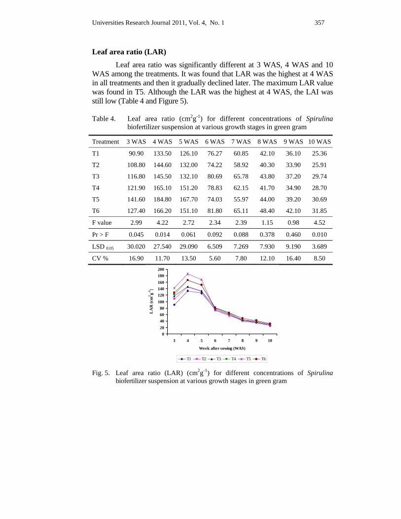
Universities Research Journal 2011, Vol. 4, No. 1 357
Leaf area ratio (LAR) Leaf area ratio was significantly different at 3 WAS, 4 WAS and 10
WAS among the treatments. It was found that LAR was the highest at 4 WAS in all treatments and then it gradually declined later. The maximum LAR value was found in T5. Although the LAR was the highest at 4 WAS, the LAI was still low (Table 4 and Figure 5).
Table 4. Leaf area ratio (cm2g-1) for different concentrations of Spirulina
biofertilizer suspension at various growth stages in green gram
Treatment 3 WAS 4 WAS 5 WAS 6 WAS 7 WAS 8 WAS 9 WAS 10 WAS
T1 90.90 133.50 126.10 76.27 60.85 42.10 36.10 25.36
T2 108.80 144.60 132.00 74.22 58.92 40.30 33.90 25.91
T3 116.80 145.50 132.10 80.69 65.78 43.80 37.20 29.74
T4 121.90 165.10 151.20 78.83 62.15 41.70 34.90 28.70
T5 141.60 184.80 167.70 74.03 55.97 44.00 39.20 30.69
T6 127.40 166.20 151.10 81.80 65.11 48.40 42.10 31.85
F value 2.99 4.22 2.72 2.34 2.39 1.15 0.98 4.52
Pr > F 0.045 0.014 0.061 0.092 0.088 0.378 0.460 0.010
LSD 0.05 30.020 27.540 29.090 6.509 7.269 7.930 9.190 3.689
CV % 16.90 11.70 13.50 5.60 7.80 12.10 16.40 8.50
Fig. 5. Leaf area ratio (LAR) (cm2g-1) for different concentrations of Spirulina
biofertilizer suspension at various growth stages in green gram
020406080
100120140160180200
3 4 5 6 7 8 9 10
Week after sowing (WAS)
LA
R (c
m2 g-1
)
T1 T2 T3 T4 T5 T6
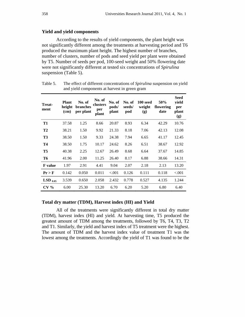
358 Universities Research Journal 2011, Vol. 4, No. 1
Yield and yield components According to the results of yield components, the plant height was
not significantly different among the treatments at harvesting period and T6 produced the maximum plant height. The highest number of branches, number of clusters, number of pods and seed yield per plant were obtained by T5. Number of seeds per pod, 100-seed weight and 50% flowering date were not significantly different at tested six concentrations of Spirulina suspension (Table 5).
Table 5. The effect of different concentrations of Spirulina suspension on yield
and yield components at harvest in green gram
Treat-ment
Plant height (cm)
No. of branches per plant
No. of clusters
per plant
No. of pods/ plant
No. of seeds/ pod
100 seed weight
(g)
50% flowering
date
Seed yield per
plant (g)
T1 37.58 1.25 8.66 20.87 8.93 6.34 42.29 10.76
T2 38.21 1.50 9.92 21.33 8.18 7.06 42.13 12.08
T3 38.50 1.50 9.33 24.38 7.94 6.65 41.17 12.45
T4 38.50 1.75 10.17 24.62 8.26 6.51 38.67 12.92
T5 40.38 2.25 12.67 26.49 8.68 6.64 37.67 14.85
T6 41.96 2.00 11.25 26.40 8.17 6.88 38.66 14.31
F value 1.97 2.91 4.41 9.04 2.07 2.18 2.13 13.20
Pr > F 0.142 0.050 0.011 <.001 0.126 0.111 0.118 <.001
LSD 0.05 3.539 0.650 2.058 2.432 0.778 0.527 4.135 1.244
CV % 6.00 25.30 13.20 6.70 6.20 5.20 6.80 6.40
Total dry matter (TDM), Harvest index (HI) and Yield All of the treatments were significantly different in total dry matter
(TDM), harvest index (HI) and yield. At harvesting time, T5 produced the greatest amount of TDM among the treatments, followed by T6, T4, T3, T2 and T1. Similarly, the yield and harvest index of T5 treatment were the highest. The amount of TDM and the harvest index value of treatment T1 was the lowest among the treatments. Accordingly the yield of T1 was found to be the
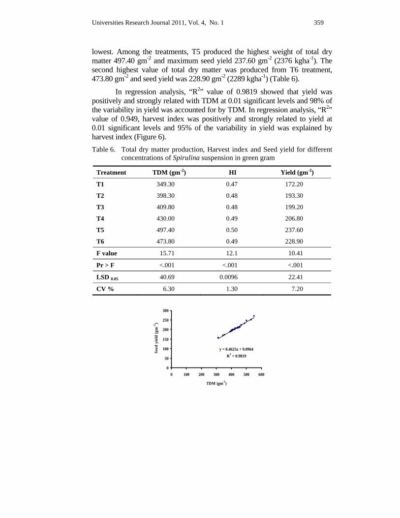
Universities Research Journal 2011, Vol. 4, No. 1 359
lowest. Among the treatments, T5 produced the highest weight of total dry matter 497.40 gm-2 and maximum seed yield 237.60 gm-2 (2376 kgha-1). The second highest value of total dry matter was produced from T6 treatment, 473.80 gm-2 and seed yield was 228.90 gm-2 (2289 kgha-1) (Table 6).
In regression analysis, “R2” value of 0.9819 showed that yield was positively and strongly related with TDM at 0.01 significant levels and 98% of the variability in yield was accounted for by TDM. In regression analysis, “R2” value of 0.949, harvest index was positively and strongly related to yield at 0.01 significant levels and 95% of the variability in yield was explained by harvest index (Figure 6). Table 6. Total dry matter production, Harvest index and Seed yield for different
concentrations of Spirulina suspension in green gram
Treatment TDM (gm-2) HI Yield (gm-2)
T1 349.30 0.47 172.20
T2 398.30 0.48 193.30
T3 409.80 0.48 199.20
T4 430.00 0.49 206.80
T5 497.40 0.50 237.60
T6 473.80 0.49 228.90
F value 15.71 12.1 10.41
Pr > F <.001 <.001 <.001
LSD 0.05 40.69 0.0096 22.41
CV % 6.30 1.30 7.20
y = 0.4625x + 9.0964R2 = 0.9819
0
50
100
150
200
250
300
0 100 200 300 400 500 600
TDM (gm-2)
Seed
yie
ld (g
m-2
)
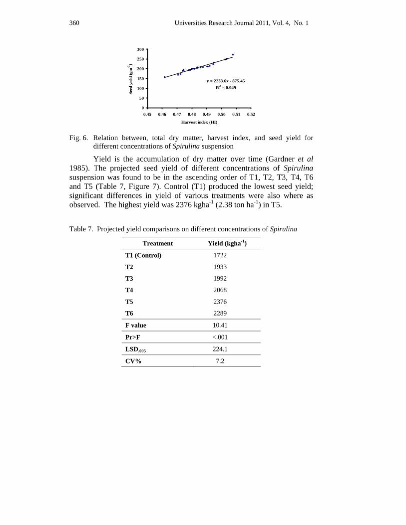
360 Universities Research Journal 2011, Vol. 4, No. 1
y = 2233.6x - 875.45R2 = 0.949
0
50
100
150
200
250
300
0.45 0.46 0.47 0.48 0.49 0.50 0.51 0.52
Harvest index (HI)
Seed
yie
ld (g
m-2
)
Fig. 6. Relation between, total dry matter, harvest index, and seed yield for
different concentrations of Spirulina suspension
Yield is the accumulation of dry matter over time (Gardner et al 1985). The projected seed yield of different concentrations of Spirulina suspension was found to be in the ascending order of T1, T2, T3, T4, T6 and T5 (Table 7, Figure 7). Control (T1) produced the lowest seed yield; significant differences in yield of various treatments were also where as observed. The highest yield was 2376 kgha-1 (2.38 ton ha-1) in T5.
Table 7. Projected yield comparisons on different concentrations of Spirulina
Treatment Yield (kgha-1)
T1 (Control) 1722
T2 1933
T3 1992
T4 2068
T5 2376
T6 2289
F value 10.41
Pr>F <.001
LSD.005 224.1
CV% 7.2
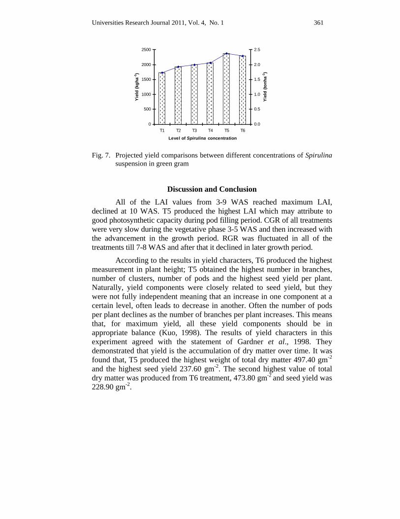
Universities Research Journal 2011, Vol. 4, No. 1 361
0
500
1000
1500
2000
2500
T1 T2 T3 T4 T5 T6Level of Spirulina concentration
Yiel
d (k
gha-1
)
0.0
0.5
1.0
1.5
2.0
2.5
Yiel
d (to
nha-1
)
Fig. 7. Projected yield comparisons between different concentrations of Spirulina
suspension in green gram
Discussion and Conclusion All of the LAI values from 3-9 WAS reached maximum LAI,
declined at 10 WAS. T5 produced the highest LAI which may attribute to good photosynthetic capacity during pod filling period. CGR of all treatments were very slow during the vegetative phase 3-5 WAS and then increased with the advancement in the growth period. RGR was fluctuated in all of the treatments till 7-8 WAS and after that it declined in later growth period.
According to the results in yield characters, T6 produced the highest measurement in plant height; T5 obtained the highest number in branches, number of clusters, number of pods and the highest seed yield per plant. Naturally, yield components were closely related to seed yield, but they were not fully independent meaning that an increase in one component at a certain level, often leads to decrease in another. Often the number of pods per plant declines as the number of branches per plant increases. This means that, for maximum yield, all these yield components should be in appropriate balance (Kuo, 1998). The results of yield characters in this experiment agreed with the statement of Gardner et al., 1998. They demonstrated that yield is the accumulation of dry matter over time. It was found that, T5 produced the highest weight of total dry matter 497.40 gm-2 and the highest seed yield 237.60 gm-2. The second highest value of total dry matter was produced from T6 treatment, 473.80 gm-2 and seed yield was 228.90 gm-2.
362 Universities Research Journal 2011, Vol. 4, No. 1
As a result of plot experiment, Spriulina biofertilizer suspension was seen to have an effective potential on growth and yield of green gram. The control would give the normal conventional growth and yield. However, the highest stimulation of growth and yield were observed in T5 (7 g l-1). Research on yield improvement of green gram with application of Spirulina biofertilizer will be a better way to provide good production of green gram. It is hoped that, the present experiment would be able to provide some information to the agricultural sector of the country especially for the cultivation of green gram with safety organic farming practices.
Acknowledgements I am very grateful to Dr Nu Nu Yee, Professor and Head, Department of Botany,
University of Mandalay, Dr Daw San Aye, Professor and Head, and Daw Hla Myint, Professor, for their intellectual guidance and permission to carry out this experiment. I would like to express thanks to my supervisor, Dr Khin Thidar, Pro-Rector, Banmaw University, for her guidance and supervision throughout my experiment. I would like to mention my deepest appreciation to my co-supervisor, Dr Min Thein, Managing Director (Retd.), Advisor, Myanmar Pharmaceutical Factory, Ministry of Industry No.1, Yekharr, Sagaing, for his inspirable supervision and providing required equipment and materials to accomplish my experiment. My special thanks are due to Dr Thanda Kyi, Deputy-Director, International Relation Section, Department of Agricultural Planning, Ministry of Agriculture and Irrigation, for her advice and generous help to complete this experiment.
References Ahmad, S., A. Wahid, E. Rasul and A. Wahid, (2005). Salt Tolerance of Green gram Genotypes at
Various Growth Stages. Botanical Bulletin of Academia Sinica, Vol 46: 135-142
Cherkezov, N.V. and C.D. Christov, (1996). Influence of Green and Blue-green algae on the Germination of Vegetable Crops. Proceedings Conference on Progress in Plant Science from Plant Breeding to Growth Regulation. 17-19 June, Hungary.
Costa, De W.A.J.M., K.N. Shanmugathasan, and K.D.S.M. Joseph, (1999). Physiology of Yield Determination of Mung bean (Vigna radiata (L.) Wilczek.) under Various Irrigation Regimes in the Dry and Intermediate Zones of Sri Lanka. Elsevier Science. Field Crop Research 61: 1-12
Gardner F.P., R.B. Pearce and R.L. Mitchell (1985) Physiology of Crop Plants. The Iowa State University Press: p. 66.
IRRI (1995) Experimental Design and Data Analysis. International Rice Research Institute, Los Banos, Philippines: p 63-107.
Universities Research Journal 2011, Vol. 4, No. 1 363
Kuo, G.C. (1998). Growth, Development, and Physiological Aspects of Mung bean Yield. http://libnts.avrdc.org.tw/fulltest_pdf/eam0114.pdf
Lawes Agricultural Trust (2002) GenStat, 6th Edition. BSN International, Wilkisnson House, Jordan Hill Road, Oxford, U.K.
MOAI, (2009). Myanmar Agriculture in Brief, Settlement and Land Records Department, Ministry of Agriculture and Irrigation.
Piccardi, R., N. Biondi, A. Frosini, L. Rodolfi, M.C. Margheri and M.R. Tredici, (2001). Cyanobacteria of the Genus Nostoc as a Source of Novel Agroactive Compounds. Book of Abstracts Symposium on Microalgae and Sea Weeds Products in Plants/Soil-System, June 20-22, Hungary.
http://www.Biofert.htm