UNIVERSIDAD DE LAS PALMAS DE GRAN CANARIA TESIS ...
191
UNIVERSIDAD DE LAS PALMAS DE GRAN CANARIA DEPARTAMENTO DE BIOLOGÍA TESIS DOCTORAL JUAN LUIS GÓMEZ PINCHETTI Las Palmas de Gran Canaria, mayo de 1993 CARACTERIZACIÓN ENZIMÁTICA DE EXTRACTOS DIGESTIVOS DE FICÓFAGOS Y SU APLICACIÓN FICOTECNOLÓGICA EN EL AISLAMIENTO DE PROTOPLASTOS DE MACROALGAS MARINAS © Universidad de Las Palmas de Gran Canaria. Biblioteca Digital, 2003
-
Upload
khangminh22 -
Category
Documents
-
view
3 -
download
0
Transcript of UNIVERSIDAD DE LAS PALMAS DE GRAN CANARIA TESIS ...

UNIVERSIDAD DE LAS PALMAS DE GRAN CANARIA
DEPARTAMENTO DE BIOLOGÍA
TESIS DOCTORAL
JUAN LUIS GÓMEZ PINCHETTI
Las Palmas de Gran Canaria, mayo de 1993
CARACTERIZACIÓN ENZIMÁTICA DE EXTRACTOS DIGESTIVOS DE FICÓFAGOS Y SU APLICACIÓN
FICOTECNOLÓGICA EN EL AISLAMIENTO DE PROTOPLASTOS DE MACROALGAS MARINAS
© U
nive
rsid
ad d
e La
s P
alm
as d
e G
ran
Can
aria
. Bib
liote
ca D
igita
l, 20
03

34-1992193 UNIVERSIDAD DE LAS PALMAS DE GRAN CANARIA
UNIDAD DE TERCER CICLO Y POSTGRADO Reunido d Uia de la fecha,d TtiVUnu! nombrado por el
Excrno.Sr.Rector Magfco. de esta Universidad, el aspirante expuso esta TESIS DOCTORAL.
Terminada la lectura y contestadas por el Doctorando las objeciones formuladas por los señores 'ueces del Tribunal,&te calificó dicho trabajo con la nota de 4 PT~> c u h ui u4 E ( "4 ')
Las Palmas de G. C., a 9 de Julio de 1993. El Presidente: Dr. . Angel Luque Escalona, f
A C -
e' - El Secretario: Dr. D. Santiago Hernandez León,
La Vocal: Dr" Da Marian
V
El Vocal: Dr, D. Francisco VaIdés Gondlez,
La Vocal: Dra Da
nchetti,

DOCTORADO EN CIENCIAS DEL MAR
DEPARTAMENTO DE BIOLOGIA
PROGRAMA DE BIOLOGIA VEGETAL
TITULO DE LA TESIS
CARACTEREACION ENZIMATICA DE EXTRACTOS DIGESTIVOS
DE FICOFAGOS Y SU APLICACION F'ICOTECNOLOGICA EN EL
AISLAMIENTO DE PROTOPLASTOS DE MACROALGAS MARINAS.
Tesis Doctoral presentada por D. Juan Luis Gómez Pinchetti
Dirigida por el Dr. D. GuiUermo García Reina
El Director El Doctorando
Las Palmas de Gran Canaria, a 28 de Mayo de 1993

CARACTERIZACION ENZIMATICA DE EXTRACTOS DIGESTIVOS
DE FICOFAGOS Y SU APLICACION F'íCOTECNOLOGICA EN EL
AISLAMIENTO DE PROTOPLASTOS DE MACROALGAS MARINAS.
Memoria que presenta, el Lcdo. en Ciencias del
Mar, Juan Luis Gómez Piuchetti para aspirar al
Las Palmas de Gran Canaria, Mayo de 1993.

GUILLERMO GARCIA REINA, DOCTOR EN BIOLOGIA Y
PROFESOR TITULAR DEL DEPARTAMENTO DE BIOLOGIA
DE LA UNIVERSIDAD DE LAS PALMAS DE GRAN CANARIA
Hace constar:
Que el Lcdo. en Ciencias del Mar D. Juan Luis Gómez Pinchetti ha
realizado el presente trabajo como Memoria de Tesis Doctoral, en el
Instituto de Algología Aplicada del Dpto. de Biología de la
Universidad de Las Palmas de Gran Canaria, bajo su dirección, y se
presenta con su VOBO.
Las Palmas de Gran Canaria, Mayo de 1993.

Agradecimientos
Desde estas líneas quiero dejar constancia de mi más sincero agradecimiento,
* Al Dr. Guillermo García Reina por la dirección, el apoyo y el
entusiasmo que he recibido de 61 para la realización de este trabajo.
* A la Universidad de Las Palmas de Gran Canaria por la beca de
Tercer Ciclo que ha permitido financiar este período de formación y por
ofrecerme los medios con los que llevar a cabo este trabajo.
* A la Fundación Universitaria de Las Palmas por la concesión de la
primera beca que me ayudó a comenzar los estudios de doctorado. * d i o e c q z , 1 1 A D A K : ~ . I P ~ uatircr n u v r h u n a i l i r S,&, ivue; uw
Jiménez del Rio, Antera Martel Quintana y Yolanda Freile Pelegrín por todos
los buenos ratos que hemos pasado juntos, tanto en el Laboratorio como fuera
de él.
* A D. Rafael Armisén Abós, Director de Investigación de Hispanagar
S.A. por compartir conmigo gran parte de sus conocimientos (que no son
pocos), ideas, interés, ágares, agarosas y carragenatos que se ajustaban a mis
necesidades y por supuesto, su increíble entusiasmo.
* To Dr. Marianne Pedersén, Dr. Mats Bjork, Dr. Shukun Yu, Dr. Kurt
Haglund, Dr. Peter Lindblad, Jonas Collen, Katrin ~sterlund, Erica Young
and the other members of the *al group, Dpt. of Physiological Botany,
Uppsala University (Sweden) to share with me their knowledge on seaweed
cultivation and physiology and make me feel at home during my very cold and
white stages in Üppsaía.
* Al Dr. Zakir Ramazanov por las largas, provechosas y entretenidas
horas trabajando a su lado.
* A los Dres. Angel Luque Escalona, Rafael Robaina Romero, Pascual
Caballero Ortega, Ma Ascensión Viera Rodríguez, a María del Mar Berna1
Suárez y a todos los miembros del Dpto. de Biología (por cuestión de espacio
no los puedo nombrar a todos) por su apoyo en todo momento.

* A Hipólito Femández-Palacios, Lidia Robaina, María Salhi, Juan
Sncnrro y todos Ios miembros dd g n p de cultivn de peces de! lCCM p r
estar siempre disponibles en el momento de pedirles un favor.
* A la Svensk-Spanska Stiftelsen, Fundación Margit y Folke Pehrzon,
Gobierno de Canarias, Plan de Formación del Profesorado y Plan de
Formación del PAS de la Universidad de Las Palmas de Gran Canaria por
financiar mis estancias en el extranjero y mis asitencias a congresos y cursos,
tanto dentro como fuera de España.
* A la compañía Air Europa S .A, por las facilidades ofrecidas en los
viajes al extranjero.
* Y por último pero no por ello menos importantes, a Cristina e Isabel
por su apoyo durante todo este tiempo.

A mis Padres.
PREFACIO
Esta tesis compila los resultados de los artículos que se citan a continuación
así como otros resultados no publicados. En el texto, los artículos se indican
por sus números romanos:
1.
Eí*
m.
IV.
v.
VI*
Gómez Pinchetti JL, García Reina G (1993) Enzymes from marine
phycophages that degrade cell walls of seaweeds. Marine Biology (en
prensa).
G6mez Pii?chetti E,, G-xeia Echa U (1993) AcA Umxjryih~n~~!ezse
activity in crude extracts from marine phycophages used for seaweed
protoplast isolation. Scientia Marina (enviado).
Bjork M, Gómez Pinchetti JL, García Reina G, Pedersén M (1992)
Protoplast isolation from Ulva rigida(Ch1orophyta). British Phycological
J o u W 27: 401-407.
Gómez Pinchetti JL, Bjork M, Pedersén M, García Reina G (1993)
Factors affecting protoplast yield of the carrageenophyte Solieria
fil~formis (Gigartinales, Rhodoph y ta) . Plant Cell Reports (en prensa).
Gómez Pinchetti JL, Ramazanov Z, García Reina (1992) Effect of
inhibitors of carbonic anhycirase activity on pholosynih&s iñ &e red
alga Solien'a Jiliformis (Gigartinales, Rhodoph yta) . Marine Biology
114: 335-339.
García Reina G, Gómez Pinchetti JL, Robledo DR, Sosa P (1991)
Actual, potential and speculative applications of seaweed cellular
biotechnolog y: some specific comments on Gelidim. Hydrobiologia
221: 181-194.

Este mbqjO a ueYa&j a MbO p Jü&i Luis GeIlieZ Ph3&&ii
Instituto de Algología Aplicada del Departamento de Biología de la
Universidad de Las Palmas de Gran Canaria. Parte del trabajo de los arh'culos
IiI y IV fue realizado en cooperación con el Dr. Mats Bj6rk del Dpt.
Physiological Botany de la Universidad de Uppsala, en base al convenio de
cooperación científica entre la Universidad de Las Palmas de G.C. y la
Universidad de Uppsala.

INDICE GENERAL
................................................ Prefacio i
......................................... Indice general iii
......................................... Indice de Tablas vii
........................................ Indice de Figuras viii
........................................... Abreviaciones ix
................................... Introducción general i
........................................ 1.. Introducción 5
1 . 1 ..Estructura y composición de la pared celular de las
............................... macroalgas marinas 5
................... 1 . 1 . l‘..Componentes del esqueleto 5
.................... 1.1.2..Componentes de la matriz 6
............................ Agar y agarasas 7
.................. Carragenato y carragenasas 13
.................... Alginato y alginato liasas 15
. . . . . . . . . . . l.S..Efecto de la dieta sobre la actividad enzimática 17
1.3. . Actividad deoxiribonucleasa @Nasa) en extractos de
origen marino .................................... 18
. . . . . . . . 1.4..Aislamiento de protoplastos de macroalgas marinas 19
. . . . . . . . . . . . . . . . . . . . . . . . . . . . . . . 1 . . l . f i 20
................................. 1.4.2. -Feofitas 24
. . . . . . . . . . . . . . . . . . . . . . . . . . . . . . . 1.4.3..Rodofitas 24
. . . . . . . . 1.5..Factores que afectan el rendimiento de protoplastos 26
. . . . . . . . . . . .. 1 .5 . 1 Enzimas digestoras de pared celular 26
.................. 1.5.2..Material vegetal de partida 26
................... 1.5.3. -Condiciones de incubación 28
. . . . . . . . . . . . . . . . . . . . . . . . . ~empektura y pH 28

'10 catiuiiw . . . . . . . . . . . . . . . . . . . . . . . . . . . . . . . . -40
Osmolalidad . . . . . . . . . . . . . . . . . . . . . . . . . . . . . 29
1.6.. Objetivos . . . . . . . . . . . . . . . . . . . . . . . . . . . . . . . . . . . . . . . . 30
2.. Material y métodos . . . . . . . . . . . . . . . . . . . . . . . . . . . . . . . . . 33
2.1. . Descripción de las especies herbívoras . . . . . . . . . . . . . . . . . . 33
2.1.1. -Aplysia dactylomela Rang . . . . . . . . . . . . . . . . . . . . 33
2.1.2..Littorina striata King et Broderip . . . . . . . . . . . . . . 33
2.1.3. -Haliotis coccinea cananensis Nordsieck . . . . . . . . . . 34
2.1.4. -Diadema antillanun Phillipi . . . . . . . . . . . . . . . . . . 34
2.2.. Extncci6n y ~ ~ ~ c t e ~ z u c i A n de !es extravt~s eilzimAticus . . . . 34
2.3.. Cultivos bacterianos . . . . . . . . . . . . . . . . . . . . . . . . . . . . . . . . 35
2.4.-Cultivo de Aplysia dactylomela y Haliotis coccinea
. . . . . . . . . . . . . canariensis . Ensayo de dietas monoespecíficas 35
2.5.-mirificación parcial de la agarasa de los extractos
crudos de Aplysia daciylomela . . . . . . . . . . . . . . . . . . . . . . . . . 36
2.6..Descripción de las especies vegetales . . . . . . . . . . . . . . . . . . . 36
2.6. 1 . -Solieria filiformis (Kützing) Gabrielson . . . . . . . . . . 36
2.6.2..Hypnea muscifomtis (Wulfen) Lamouroux . . . . . . . 37
. . . . . . . . 2.6.3. -Grateloupia doryphora (Montagne) Howe 37
. . . . . . 2.6.4..Gracilaria tenuistipitata var liui Zhang et Xia 37
. 2.6.5. . Ulva rigida C Agardh . . . . . . . . . . . . . . . . . . . . . . 38
2.7.. Cultivo de las especies vegetales . . . . . . . . . . . . . . . . . . . . . . 38
2.8. . Aisiamiento de protopiastos . . . . . . . . . . . . . . . . . . . . . . . . . . 38
2.9. . Fotosíntesis, alcalinización del medio y actividad anhidrasa
. . . . . . . . . . . . . . . . . . . . . . . . . carbónica en Solieriafiliformis 39
. . . . . . . . . . . . . . . . . . . . . . . . . . . . . . . . . . . . . . . . . 3.. Resultados 41
. . . . . . . . . . . . . . . . . . . . . . . . . . . . . . 3.1 .. Método de extracción 41
. . . . . . . . . . . . . 3.1.1. -Precipitación con sulfato amónico 41
3.1.2..Diálisis y filtración en gel ................... 4 1
........................... 3.2.. Actividades enzimáticas m 42
3.2.1 ..Actividad celulasa ......................... 44
3.2.2..Actividad agarasa .......................... 44
3.2.3..Actividad alginato liasa ..................... 44
3.3..E1 pH de los sistemas digestivos [IIJ .................... 45
3.4.. Cultivos bacterianos ................................ 45
3.5..Actividad DNasa m ............................... 45
..................... 3.6.. Ensayo de dietas monoespecíficas 46
................................ 3.7..Purificaci6n parcial 50 '2 Q D r ~ A . r n n . X n rln i.rntnmlantnn C 1 .......................... J . U . . X L W U U U L V l l UU Y L V L y L a J L V J J I
3.8.1. -Gracilaria tenuistipitata . . . . . . . . . . . . . . . . . . . . . . 51
3.8.2. -Grateloupia doryphora ...................... 53
3.8.3..Hypnea muscifomis ........................ 53
........................... 3.8.4.. Ulva rigida [a 54
3.8.5..SolieriaJilifomis [N] ....................... 59
3.9.. Captación de carbono inorgánico en Solieria Jil~ormis p] .... 63
. . . . . . . . . . . . . . . . . . . . . . . . . . . . . . . . . . . . . . . . . . 4.. Discusión 65
......................... 4.1 ..Metodología de la extracción 65
. . . . . . . . . . . . . . . . . . . . . . . . . . . 4.2. . ~ ~ t i ~ i d ~ d ~ mzimgti~a m 65
......................... 4.3..pH del sistema digestivo [Il[I 67
............................... 4.4.. Actividad bacteriana 68
............................... 4.5 ..Actividad DNasa [m 68
................... 4.6. . Influencia de la dieta monoespecífica 69
. . . . . . . . . . . . . . . . . . . . . . . . . . . . . . . . 4.7.. Purificación parcial 70
. . . . . . . . . . . . . . . . . . . . . . . . . . 4.8. . Aislamiento de protoplastos 70 .
. . . . . . . . . . . . . . . . . . . . . . 4.8.1 ..Soluciones enzimáticas 71
. . . . . . . . . . . . . . . . . . . . . . . . . . 4.8.2..Material vegetal 72
. . . . . . . . . . . . . . . . 4.8.3..Efectos, de los pretratamientos 74
. . . . . . . . . . . . . . 4.9.. Capacidad fotosintética de los protoplastos 75

r r* ---- 1 - - 2 qc 3.- L U ~ C I ~ S I U I I ~ S . . . . . . . . . . . . . . . . . . . . . . . . . . . . . . . . . . . . . . . 1 w
6.- Bibliografía . . . . . . . . . . . . . . . . . . . . . . . . . . . . . . . . . . . . . . . . 79
7.- Artículos . . . . . . . . . . . . . . . . . . . . . . . . . . . . . . . . . . . . . . . . . . . 95

vii
Indice de tablas
Tabla 1. Principales polisacáridos estructurales en las paredes
celulares de algunas macroalgas marinas de las
Divisiones Clorofita, Feofita y Rodofita ................. 8
Tabla 2. Condiciones físicas del aislamiento, rendimiento y
viabilidad de protoplastos de macroalgas ............... 21
Tabla 3. Porcentaje de actividad enzimática (medida
como actividad agarasa) en los extractos
de Aplysia dactylomela ............................. 41
Tabla 4. Actividades enzimáticas de los diferentes extractos
expresadas como pg AR h-' en diferentes condiciones
.............................. de temperatura y pH 43
Tabla 5. Actividades enzimáticas medidas como pg AR h" de
los extractos crudos de Aplysia dactylomela cultivada
con dietas monoespecíficas durante 1 mes .............. 47
Tabla 6. Acción de diferentes combinaciones de enzimas
comerciales y extractos crudos de herbívoros
marinos en la producción de protoplastos de
las especies de algas rojas, Gracilaria tenuistipitata,
52 Grím&xpi~ &r@cm e ,Yyy",n,~u mmcijf~,mis ............. - - Tabla 7. Actividad enzimitica, en U m~- ' , de los extractos
crudos utilizados en las soluciones
enzimiticas para el aislamiento de protoplastos .......... 54

... V l l l
Figura 1. Estructura química de los principales polisacáridos
del esqueleto y la matriz de las macroalgas marinas . ' . . . .... 9
Figura 2. Secuencia de la hidrólisis enzimática completa de la
molécula de agar según van der Meulen (1975) . . . . . . . . . . 11
Figura 3. Evolución de la actividad DNasa en función del
tiempo de L. striata, A. dactylomela, AAP
y DNasa estandar . . . . . . . . . . . . . . . . . . . . . . . . . . . . . . . . . 46
Figura 4. Incremento en peso de Aplysia dactylomela alimentada A O con 5 r h rigkh, VrmililRa spp. y Sdie;.l:clJ5~foI~Ci~ . . . . . . . . . 90
. Figura 5. Evolución del peso fresco de las diferentes especies
de macroalgas con las que fue alimentado
Haliotis coccinea canuriensis por un periodo de 4 dias . . . . . . 49
F i r a 6. Perfil cromatográfico de la agarasa obtenida del
extracto crudo de Aplysia dacíylomela . . . . . . . . . . . . . . . . . . 50
Figura 7. (A) Corte transversal de un talo de Ulva rigida.
(B) Protoplastos fotografiados en contraste de fases . . . . . . . 57
F i r a 8. Producción de protoplastos de Solieria Jilifomzis . . . . . . . . . . 6 1

Abreviaciones
AAP
AC
AC,
ACint
AR
Bis-Tris
CMC
DBS
DBAZ
DCMU
DNasa
DTT
EDTA
EF
EGTA
EZ
IDA
HEPES
f
Hw
MoF
NPA
PA
PF
PS
Tris
U
Abalone Acetone Powder, extracto en acetona de Haliotis sp.
Anhidrasa Carbónica
AC extracelular
AC intracelular
Azúcares reductores
Bis[2-Hidroxietillimino-trisFidroximetil]metano
Carboximetil celulosa
Sulfonamida unida al dextrano (Dextran-bound sulfonamide)
Acetazolamida unida al dextrano (Dextran-bound acetazolarnide) c,
3-(3,4-diclorofeni1)-1, 1 -dimetilurea - m
O
Deoxiribonucleasa - o
m
Ditiotreitol E
O
Acido etilenodiaminotetraacético g a
E
Extracto frío de Gracilaria spp. a
a a
Acido etilenoglicol-bis(í3-aminoetil eter) NNNIN'-tetra-acético a
5 6-etoxizolamida O
Diacetato de fluoresceína
N-[2-Hidroxietillpiperazina-N'-[2-acido etanosulfónico]
mEoiis &icilbG& ev;; dieta mGfimSp&f;,~a
Haliotis sin alimentación específica (salvaje)
Acido 3-(N-morfo1ino)propanosulfÓnico
Agarasa de Pseudomonas atlantica no purificada
Agarasa de Pseudomonas atlantica purificada
Peso fresco
Peso seco
Tris(hidroximeti1)aminometano
Unidades de actividad enzimAtica


Introducción genera 1 1
Introducción general
La palabra Biotecmlogía se ha convertido Últimamente en un término muy
familiar que define, en su sentido más amplio, "cualquier técnica que utiliza
organismos vivos (o parte de estos organismos) para preparar o modificar
productos, mejorar las características de plantas y animales o desarrollar
microorganismos para usos específicos" (Office of Technology Assessment,
Anónimo 1984). Para completar esta definición habría que incluir "el marcado
interés económicow asociado al desarrollo de estas técnicas (Nonomura 1988).
La reciente interacción de la Fitología con la biología molecular y celular, la
ingeniería química, la maricultura, el cultivo de células y protoplastos y otras
disciplinas relacionadas para usos específicamente comerciales, han dado
contenido a lo que Lewin en 1983 denominó Ficotecmlogía.
De las biotecnologías aplicadas a vegetales, el cultivo de protoplastos
se viene consolidando como una de las preferidas debido a la cantidad y
exclusividad de aplicaciones potenciales que permite en campos como la
fisiología, la bioquímica y, más recientemente, en biología molecular e
ingeniería genética. Parafraseando a Coury et al. (1991): "cuando observamos
un protoplasto, estamos observando una de las unidades intactas más frágiles
de la Naturaleza, y un poderoso sistema experimental para la realización de ,=siu&js f i ~ ~ : @ ~ ~ ~ +~ytc bQsicos c=m= @ r ~ & ~ "
La palabra protoplasto define a la célula vegetal de la cual la pared
celular ha sido separada por métodos físicos o enzimáticos (Cocking 1972).
Las técnicas para el aislamiento rutinario de gran número de protoplastos
viables en plantas terrestres, utilizando enzimas degradadoras de la pared
celular, fueron desarrolladas en los años 60, desde que Cocking (1960) utilizó
el extracto crudo de un hongo, que contenía celulasa, para preparar protoplas-
tos de Lycopersicon spp. A partir de ese momento, el aislamiento y cultivo de
protoplastos se ha convertido en una poderosa herramienta con la que se han
obtenido resultados como la regeneración de pared celular, la división celular
y, m& rcximtemefite, !a demos'uiíASn de tsUpkfieia (íegeiiei~~i3ii de phitíi
completa) (Evans y Bravo 1984).
Las algas son un grupo heterogéneo que exhibe una gran variedad y
complejidad en la organización de las paredes celulares, lo que contrasta con
la relativa homogeneidad de las plantas terrestres (Butler et al. 1990). En
principio, no hay razón para que las técnicas tantas veces aplicadas en plantas
terrestres no puedan ser aplicadas con éxito en macroaigas. Sin embargo, la
gran complejidad y diversidad de los componentes estructurales de sus
paredes celulares (sobre todo en las especies de mayor interés industrial), y
la escasa efectividad de las polisacaridasas comerciales potencialmente
utilizables para su digestión, han dado como resultado el que sólo reciente-
mente se hayan obtenido ciertos éxitos en el aislamiento y cultivo de
protoplastos de macroalgas marinas. La digestión ineficaz de las paredes
celulares más diferenciadas de macroalgas (principalmente de las Divisiones
Feofita y Rodofita) es debida no s61o a cambios en la constitución celular (y
consecuentemente en la producción de protoplastos) con relación a su edad,
ciclo de vida, estado fisiológico y condiciones de cultivo (Kloareg y Quatrano
1988, Bjork et al. 1990), al igual que en plantas terrestres (Evans y Bravo
1984, Eriksson 1985), sino también a la ausencia de eficaces enzimas
degradadoras de sus complejas paredes celulares. La implantación del cultivo
de protoplastos de macroalgas marinas como t b i c a rutinaria al nivel de
plantas terrestres, precisa aún del conocimiento de técnicas para la modifica-
ción de los enlaces entre los polímeros que conforman la pared celular, así
como de métodos de extracción, purificación y caracterización de enzimas
efectivas que actúen sobre los polímeros específicos (Butler et al. 1990).
Paralelamente, las características fisiológicas de las macroalgas están
directamente relacionadas con el desarrollo estructural de su pared celular.
El estudio de la relación entre las características del metabolismo del carbono
y del nitrógeno de las algas con las que se trabaja y de los protoplastos que
se obtienen de ellas, hacen que los estudios fisiológicos sean un complemento
(cuando no una finalidad) fundamental para llevar a cabo el desarrollo de

Introducción general 3
estas técnicas con éxito.
Gran cantidad de bacterias y otros microorganismos capaces de degra-
dar los polisacáridos estructurales de macroalgas marinas (incluyendo el agar,
canagenato y alginato), son comunes en el medio marino tanto de forma libre
(Yaphe 1957, Sarwar et al. 1983) como asociados a las algas que contienen
estos polisacáridos (Chesters et al. 1956, Quatrano y Caldwell 1978, Torzilli
y Andrykovitch 1980, Wainwright 1980, Bellion et al. 1982, Greer y Yaphe
1984, Hodgkiss y leung 1986, Aoki et al. 1990, Schaumann y Weide 1990,
Kitamikado et al. 1992, Lavilla-Pitogo 1992), e incluso, en los sistemas
digestivos de algunos herbívoros marinos (Lasker y Giese 1954, Galli y Giese 1 ílQ? 1959, Prim y Lawrence í975, Minamitaice eí d. 1986, Seideiei ei d. 170 1 ,
Vitalis et al. 1988). La utilización de las enzimas producidas por microorganis-
mos ha sido, en general, la principal herramienta para el análisis estructural
de estas, macromoléculas (Yaphe 1957, Duckworth y Yaphe 1971, Young et
al. 1978, Greer y Yaphe 1984, Morrice et al. 1984, Brown y Preston 1991). Sin
embargo, el desarrollo de estudios estructurales más exhaustivos y, sobre todo,
el desarrollo de nuevas aplicaciones (como las técnicas de cultivo de células
y protoplastos) han generado la demanda y el estudio de nuevas y más poten-
tes fuentes de enzimas.
La existencia de gran cantidad de invertebrados marinos con dietas
herbiversls w r e n c e 1975; Hawkins y Hartnoll 1983, Carefoot 1987,
Tutschulte y Connell 1988), capaces de digerir macroalgas, y la descripción del
gran equipamiento enzimiitico (particularmente en enzimas digestivas) tanto
de moluscos como equinodermos (van Weel 1961, Lawrence íS.75 j, han propi-
ciado el estudio de sus sistemas digestivos como fuente alternativa para la
obtención de enzimas específicas. No obstante, la descripción de enzimas
capaces de degradar los polisacáridos matriciales complejos de la pared
celular de macroalgas marinas ha sido escasa, debido principalmente a la
complejidad del proceso de degradación (Lewis 1964, Horiuchi y Lane 1965,
Usov y Miroshnikova 1975, Benítez y Macaranas 1979). Aunque poco
estudiados, resulta evidente que estos herbívoros han de poseer mecanismos
(taqts mw&qicvs wme e+simicms) ~ p ~ ~ ~ s de digefii efimmefiie p&
celular de las macroalgas marinas.
Entre las diferentes metodologías descritas para la preparación y
caracterización de este tipo de extractos enzimáticos (Crabtree et al. 1979,
Scopes 1982, Deutscher 1990), el empleo de la precipitación con sulfato
amónico y la desalación son las técnicas más frecuentemente utilizadas, junto
con la extracción en acetona. Este proceso de purificación parcial evita los
problemas inherentes al uso de extractos crudos que, con gran probabilidad,
contienen otras sustancias que dificultan la actividad de los enzimas y su
aplicación cuando son utilizados en el aislamiento de protoplastos (Cocking
1972, Berliner 1981, Butler et al. 1990).
En este estudio se han desarrollado técnicas para la preparación y
caracterización de extractos digestivos de herbívoros marinos con la finalidad
de obtener fuentes enzimáticas eficaces para la digestión de la pared celular
y la obtención de protoplastos de macroalgas.

Introducción 5
1 .- INTRODUCCION
1.1.- Estructura y composición de la pared celular de las
macroalgas marinas
La pared celular de las macroalgas marinas, como la de las plantas terrestres,
está compuesta por una fase cristalina (el esqueleto) encajada en una fase más
amorfa (la matriz). Sin embargo, la pared celular de las algas se diferencia de
la de las plantas terrestres tanto por la abundancia y variedad de los compo-
nentes de la matriz con respecto a los del esqueleto como por un mayor
contenido en polisacáridos polianiónicos con respecto a los neutros. Estas
diferencias han sido asociadas a funciones específicas relacionadas con la
regulación mecánica, osrnótica o iónica de las algas en el medio marino
(Kloareg y Quatrano 1988).
1.1.1 .- Componentes del esqueleto
En la mayoría de las macroalgas marinas los polímeros del esqueleto son
polisacáridos lineales neutrales donde, como en las plantas terrestres, el
polisacárido más común es la celulosa (Kreger 1962), un B(l-4) glucano de
conformación helicoidal plana (Tabla 1). Este polímero es muy abundante en
Ulvaceas (Clorofitas) constituyendo aproximadamente el 70 % de la pared
c d u ! ~ ,tes?m 1974), pero relativamente escaso (menos del 10% del peso
seco) en Feofitas y Rodofitas, e incluso inexistente en algunas especies (Butler
et al. 1990). Xilanos y mananos también forman parte del esqueleto de
macroaigas marinas, en proporciones que varían dependiendo de la especie
y de la fase del ciclo de vida.
La celulasa (EC 3.2.1.4),enzima que hidroliza los enlaces B(l-4) de la
celulosa, está ampliamente distribuída en la naturaleza y ha sido detectada en

sume=- miGnes mwLyG (Yakm y Yas~maso !9@, Ej&ava
et al. 1968, Horiuchi y h e 1965, Koningsor et al. 1972, Brock et al. 1986,
Boyen et al. 1990).
1.1.2 .- Componentes de la matriz
Los polisacándos de la matriz han sido el objeto de intensas investigaciones
en los últimos años. El interés en estos polímeros radica principalmente en su
capacidad para formar soluciones acuosas altamente viscosas o geles compac-
tos estables. Estos ficocoloides se emplean en las industrias alimentaria,
cosméticaj texti', f m - c é u t ; ~ y hi&wnol6gin_ (I\m-i&n y G&ta 1987,
McHugh 1987, Stanley 1987) generando un mercado que mueve más de 3x10~
Tm en peso fresco por año (McHugh 1991).
Sus propiedades físico-químicas están basadas en la estereoquímica de
sus cadenas que, a su vez, dependen de la composición química y sus
estructuras secundaria, terciaria o cuatemaria. Contrariamente a los
componentes del esqueleto, los componentes de la matriz pueden ser
extraídos en agua en presencia de aditivos como sales, ácidos, bases o
quelantes. Típicamente están formados por polisacáridos ácidos, sulfatados y10
carboxilados. De todos los componentes encontrados en la matriz de las
distintas Divisiones, son los de las Rodofitas (algas rojas) y Feofitas (algas
pardas) los de mayor interés por sus características físico.químicas particula-
res. T I \ - nnmnn.~a~.tan mn.inAtnAnm A a l o mnt4.r a+.\ l nc P l n r n G t n o fnlrrnn -3 ~ V I I I ~ I I G I I L C ~ J u 1 a y v u u u l u a UG la u I a u A & b 1 1 la3 LLULVLLULJ (a5a3
verdes) son polisacáridos ramificados, sulfatados y solubles en agua. En
diferentes especies de Ulva y Enteromorpha los polímeros están formados
principalmente por ramanosa, xilosa y ácido glucurónico.
En las Rodofitas se encuentran principalmente galactanos lineales 6 sulfatados, con pesos moleculares frecuentemente superiores a 10 . Básica-
mente consisten en la repetición regular de un disacárido (AB) compuesto por
dos unidades de galactosa enlazadas alternativamente por enlaces 8(1-4) y

Introducción 7
a(1-3) (Tabla 1). Esta estructura regular suele completarse con sustituciones
de grupos sulfatos, metilos o piruvatos, pero siempre conteniendo D-galactosa
enlazada (1,3) como unidad B. Los elementos que ocupan la unidad A, enla-
zados (1,4), pueden ser tanto configuraciones D-, en los carragenatos, como
L-, en el agar o el porfirán. Las otras diferencias básicas radican en el número
y localización de los sustituyentes en la molécula.
En las Feofitas el componente más abundante de la matriz es el ácido
algínico, complementado con polisacáridos sulfatados, principalmente he-
teromoléculas conteniendo fucosa, y diferentes proporciones de xilosa, ácido
glucurónico, galactosa y manosa (Percival 1978).
Er! g e ~ e d , hs proprci~nec de 10s diferentes pl j~cArid~s matriciales
dependen tanto de la especie como del hábitat y la estación del año. La
composición de la matriz también puede ser característica de las distintas
generaciones del ciclo de vida, como ocurre en el caso de los polisacáridos del
esqueleto. Incluso en algas con generaciones isomórficas, como en algunas de
las especies pertenecientes al orden Gigartinales, los garnetofitos sintetizan
únicamente kappa-carragenato mientras los esporofitos sintetizan sólo lambda-
canagenato (McCandless et al. 1973, McCandless et al. 1982).
Agar y agarasas
El agar es el principal polisacárido matricial de los géneros Gelidium,
Gracilaria y Pterocludia. Su molécula está constituída por dos fracciones:
agarosa y agaropectina (Araki 1956).
La agarosa es una molécula larga, neutra, formada por D-galactosa,
conectada por enlaces a(1-3) con moléculas de 3,6-anhidro-L-galactosa que
a su vez están unidas por enlaces 8(1-4) a la siguiente molécula de D-
galactosa. Los enlaces entre los diferentes monómeros tienen diferente
resistencia a las hidrólisis química y enzimática. Los enlaces a(1-3) son
fácilmente hidrolizados por ácidos suaves, dando como resultado una serie de
oligosacáridos con la 3,6-anhidro-L-galactosa como extremo reductor. La
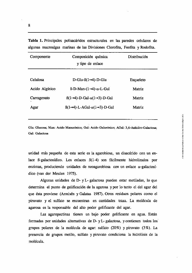
Componente Composición química Distribución
y tipo de enlace
Celulosa D-Glu-B(14)-D-Glu Esqueleto
Acido Algínico B-D-Man-(14)-a-L-Gul Matriz
Carragenato D(14)-D-Gal-a(1+3)-D-Gal Matriz
Agar B(14)-L-AGal-a(1+3)-D-Gal Matriz
Glu: Glucosa; Man: Acido Manurónico; Gul: Acido Gulurónico; AGal: 3,6-Anhidro-Galactosa;
Gal: Galactosa
unidad más pequeña de esta serie es la agarobiosa, un disacárido con un en-
lace B-galactosídico. Los enlaces B(1-4) son fácilmente hidrolizados por
enzimas, produciendo unidades de neoagarobiosa con un enlace a-galactosí-
dico (van der Meulen 1975).
Alniincac i i n ; & d ~ c AD n- 11 L- mcalcartncca ?wiadan ac tor rna t i ldcac ln n i i o L S 1 6 U l l U L 1 UllLUUUVIl UV Y J 6LYUVLW- YUVUVll YU- IIIYCLIUUUU) I W YUU
determina el punto de gelificación de la agarosa y por lo tanto el del agar del
que ésta proviene (Armisén y Galatas 1987). Otros residuos polares como el
piruvato y el sulfato se encuentran en cantidades traza. La molécula de
agarosa es la responsable del alto poder gelificante del agar.
Las agaropectinas tienen un bajo poder gelificante en agua. Están
formadas por unidades alternativas de D- y L- galactosa, y contienen todos los '
grupos polares de la molécula de agar: sulfato (20%) y piruvato (3%). La
presencia de grupos metilo, sulfato y piruvato condiciona la hidrólisis de la
molécula.

Introducción 9
Aunque se conocen bacterias agarolíticas desde comienzos de siglo, el
aislamiento, purificación, caracterización y aplicación de las agarasas no se
inició hasta fmales de la década de los 50. La agarasa obtenida de Pseudomo-
nas kyotoensis permitió la obtenci6n de oligosacáridos de la agarosa, dando
lugar al establecimiento del primer modelo de la estructura de dicho polímero
(Araki 1956). Las agarasas de Pseudomonus atlantica y Qtophaga NCMB 1327
Figura 1. Estructura química de los principales polisacáridos del esqueleto y
la matriz de las macroalgas marinas.
celulosa
4- neoagarobiosa -------)
4-. agarobiosa -)
agarosa

4-' neocarrabiosa -+
4- carrabiosa S-b
Kappa- (R=H) lota- (R=SOz)
RO oso;
Lambda- @=SO 3 o H)
carragenatos
COOH COOH
t ac. polimanurónico S-b
4- ac. poligulurónico .-b
ácido algínico

Introducción 11
fueron utilizadas para identificar los polisacáridos matriciales de agarofitas y
obtener información de la estructura de los diferentes galactanos del tipo agar
(Yaphe 1957, Duckworth y Turvey 1969, Duckworth y Yaphe 1971). No obs-
tante, los estudios sobre purificación y especificidad de las agarasas se han
llevado a cabo recientemente (van der Meulen 1975, Morrice et al. 1983a,
1983b, 1984, Aoki et al. 1990, Yamaura et al. 1991).
La hidrólisis enzimática de la molécula de agar hasta obtener sus
monómeros es un proceso complejo que implica la acción de diferentes
enzimas (Fig. 2) (van der Meulen 1975). En general, las agarasas muestran
actividad sobre oligosacáridos de mayor .tamaño que el hexámero de la mo-
iécuia cie agamsa. m enwma, uña 'E'rmm-i&sa, iqri'e p%-iL hi-
drolizar el tetrámero y una disacaridasa para hidrolizar el dímero resultante.
Figura 2. Secuencia de la hidrólisis enzimática completa de la molécula de
agar según van der Meulen (1975).
AGAROSA
nn Ragarasa -agarasa
/- / \ \ 1 NEOAGAROTETRAOSA \ AGAROTETRAOSA
NEOAGAROBIOSA AGAROBIOSA
D-GALACTOSA + 3,6-ANHIDRO-L-GALACTO.SA
I B-disacaridasa
D-GALACTOSA + 3,6-ANHIDRO-L-GALACTOSA

hs tepaaa-ih y &iaiM;ih &teT& de fS2&,"Wr"a lQS
únicas oligosacaridasas que han sido estudiadas (Young et al. 1971). Estos
mismos autores clasificaron las agarasas en dos grupos: a-agarasas que
hidrolizan los enlaces a-, y las B-agarasas (agarosa 3-glucanohidrolasa, E.C.
3.2.1.8 1) que hidrolizan los enlaces B- en la molécula de agarosa.
Enzimas agarolíticas extracelulares con actividad sobre los enlaces 13-
del agar (productores de oligosacáridos del tipo neoagarobiosa) han sido
obtenidos de diferentes especies bacterianas: Pseudonwnas kyotoensis (Araki
y Arai 1957), Qtophaga sp. (Turvey y Christison 1967), y Pseudomonas
atlantica (Morrice et al. 1983a).
Morrice et al. (1983a, 1983b, 1984) aislaron y caracterizaron dos 11-
agarasas de Pseudomonas atlantica: 1) B-agarasa 1, enzima predominante, con
actividad endo-, actuando sobre el agar tanto en solución como gelificado y
2) B-agarasa 11, con una actividad similar, aunque también con actividad sobre
el tetrasacárido de la agarosa. Una tercera enzima, neoagarobiosa hidrolasa,
que degrada la molécula de neoagarobiosa en sus monómeros había sido
purificada previamente por Day y Yaphe (1974) a partir de la misma especie.
Aunque las B-agarasas han sido descritas con frecuencia, las agarasas
que hidrolizan los enlaces a- de la molécula del agar únicamente lo han sido
en raras ocasiones (Young et al. 1971, 1978).
La metodología empleada normalmente para cuantificar la actividad
agarolítica se ha basado tanto en el aumento del poder reductor como en
la disminución de la viscosidad de soluciones de agar o agarosa.
La temperatura óptima descrita para la mayoría de las agarasas bacte-
rimas es de 40°C. Sin embargo, este óptimo puede estar influido por la
metodología, que obliga a realizar las mediciones a elevadas temperaturas
para prevenir la gelificación de las soluciones. S610 el uso de sustratos
formados por neoagaro-oligosacáridos solubles podría confirmar la influencia
del proceso de gelificación en la actividad de la enzima y clarificar por tanto
los valores reales de temperatura óptimos (van der Meulen 1975).
Los valores óptimos de pH para la mayoría de las agarasas bacterianas

Introducción 13
se han descrito entre 5.0 y 7.2 y sus pesos moleculares entre 18 y 34 kDa
(Morrice et al. 1983a, Aoki et al. 1990, Yamaura et al. 1991). El efecto inhibi-
torio de los iones metálicos zn2+, cu2+, co2+, lJe2+ y ~ 1 ~ ' ha sido des-cri-
to recientemente (Yamaura et al. 1991).
Los antecedentes sobre la actividad agarolítica en herbívoros marinos
son escasos y en ningún caso ha sido cuantificada. La degradación de una
solución de agar, entre otros polisacáridos complejos, por extractos crudos de
Diadema ann'llanun fue descrita por Lewis en 1964. Kristensen (1972)
estudiando diferentes carbohidrasas de algunos invertebrados marinos detectó
un aumento mínimo en la concentración de azúcares reductores al incubar
diferentes extractos en una soiución de agar.
La purificación parcial por cromatografía de afinidad de una agarasa
del hepatopancreas de Littorina mandrhurica ha sido descrita por Usov y
Miroshnikova (1975) indicando valores de pH óptimos del extracto crudo y de
la agarasa purificada de 5.6 y 6.0 respectivamente.
Al igual que sucede con las agarasas bacterianas, las condiciones de
temperatura, dependiendo del tipo de sustrato, influencian en gran medida los
ensayos de cuantificación de la actividad enzimática de estos extractos, con lo
que la información obtenida hasta el momento no ha sido todo lo completa
que cabría esperar.
Carragenatos y carragenasas
El carragenato constituye el principal polisacárido matricial de la pared
celular de los géneros Eucheuma, Chondm, Hypnea y Gigarn'na. Químicarnen-
te son galactanos altamente sulfatados (20-38 %) y fuertemente aniónicos,
característica que los diferencia del agar y el alginato.
En cuanto a su composición química los diferentes carragenatos tienen
una estructura básica común, siendo polisacándos lineales formados por D-
galactosa unida alternativamente por enlaces a(1-3) y B(1-4). La diferencia
entre los distintos tipos de carragenatos (kappa-, lambda-, iota-, entre otros)

. nn- m U i ~ eii d ii-díriero y la pskiúíi Ge iüs grupos suifato (Sicaniey LYUI).
Las carragenasas se extraen principalmente de Pseudomonas carragee-
novora, Klebsiella pneumoniae (Knutsen 1991) y de diferentes cepas de
Cytophuga (Sarwar et al. 1983, Potin et al. 1991). Diferentes carragenasas
capaces de degradar kappa-, iota- y ld&-carragenato han sido aisladas,
purificadas y caracterizadas, y enzimas específicas capaces de hidrolizar los
enlaces l3- de los carragenatos kappa- y iota- han sido descritas (Bellion et al.
1982). Tanto la kappa- como la iota-carragenasa son endoenzimas que produ-
cen oligosacáridos del tipo neocarrabiosa. Enzimas digestoras de estos oligosa-
cáridos han sido aislados de Pseudomm carrageenovora (McLean y William-
son 1979, 198 1). Sin embargo: no se ha descrito aiín la existencia de enzimas
que degraden el disacárido en monómeros.
La cuantificación de actividad carragenasa se basa en la misma meto-
dología utilizada para la actividad agarolítica. Las condiciones óptimas de
temperatura y pH vienen determinadas en función del tipo de carragenato y
de la fuente de enzimas. La kappa-carragenasa de Pseudomna-s carragenovora
(Pm = 35 W>a) muestra actividad óptima a 40°C y pH 8.0 (McLean y William-
son 1979). La actividad óptima de la carragenasa obtenida de un cultivo de
Cytophuga sp. fue detectada en un rango de temperatura que varió entre 25
y 50" C y a pH 7 .O (Sarwar et al. 1983) siendo inhibida en presencia de HgC12,
AgN03 y NaC1 (en concentraciones > 1. S % ) . Una iota-carragenasa (de una
especie no identificada) con Pm aparente de 57 kDa degradó la molécula de
iota-carragenato hasta producir neocarratetraosa y neocarrahexaosa en unas
condiciones óptimas de 40°C, pH 8.0 y en presencia de NZ+ (0.1 1M) (Greer
y Yaphe 1984). Una kappa-carragenasa de una cepa de Cytophaga, con un Pm
estimado de 40 kDa y pH óptimo de 7.0 y 7.2 (dependiendo del tampón) ha
sido purificada recientemente por Potin et al. (199 1).
Las únicas referencias de canagenasas en herbívoros marinos las
constituyen los trabajos de Horiuchi y h e (1965) y Benítez y Macaranas
(1979). Los primeros cuantificaron la degradación de diferentes polisacáridos,
entre ellos el carragenato, por extractos crudos obtenidos de Strombus gigas.

Introducción 15
Los segundos purificaron parcialmente una kappa-carragenasa del erizo de
mar Diadema setosurn.
Alginatos y alginato liasas
El ácido algínico es un polímero lineal basado en dos unidades monoméricas,
el ácido B-1,4-D-manurónico y su epímero, el ácido a-1,4-L-gulurónico. Es so-
luble en soluciones alcalinas y constituye de un 10 a un 45 % del peso seco del
talo de algas pardas. Se forma por la unión de los monómeros en las posi-
ciones C1 y C4 por medio de un puente eter-oxígeno. La cadena del polímero
está constituída por tres clases de regiones o bloques. Los bloques G que
contienen sólo unidades derivadas del ácido L-gulurónico, los bloques M que
sólo contienen unidades de ácido D-manurónico y los bloques MG que están
constituídos por unidades alternativas de ácidos D-manurónico y L-gulurónico.
La proporción de estos bloques varía con el alga, y las propiedades físicas de
los alginatos dependen de la proporción relativa de estos tres tipos de bloques.
La formación de geles por adición de ca2+ está relacionada con la propor-
ción de bloques G de la molécula por lo que, a mayor cantidad de bloques G,
mayor fuerza de gel. A su vez, la solubilidad de los alginatos en ácido
depende de la proporción de bloques MG (McHugh 1987). A l a -e-. -,-.,-'a A, a1,am -,-A,, - - - t ;anan n l n ; n n t ~ 1-0 nAnnrnn liuiiquc la g r a i iiiayuiia UG a i p a yawa kuiiuui~ii argriiaw, iuii e;uii-iuii
Laminan's, Macrocystis y Ascophyllurn constituyen las principales fuentes para
la industria. Las propiedades del alginato no sólo varían de una especie a otra,
sino también con el tipo y la edad del tejido, la estación del año y las
condiciones de crecimiento (McHugh 1987).
La presencia de enzimas capaces de degradar alginato ha sido demos-
trada en diferentes especies de bacterias (Doubet y Quatrano 1984, Preston
et al. 1985, Boyen et al. 1990, Brown y Preston 1991, Kitamikado et al. 1992,
Aasen et al. 1992) y en gran variedad de invertebrados marinos. Está general-
mente aceptado que el proceso de la degradación del alginato transcurre
como una reacción por B- eliminación en la cual el poliurónido es degradado

, . a d @ í l i ~ ~ ~ ~ , eoii üii ACZG ürmieo 4,5 ~ S Z ~ ~ ~ E K I G ei d extreze cc ~ ~ U C ~ C X
del residuo. Las alginato liasas obtenidas de bacterias varían en su especifici-
dad sobre el sustrato (dependiendo del tipo de bloque, ya sean bloques poli-
manuronatos, poliguluronatos o combinaciones de estos) y su modo de acción.
La mayoría parecen poseer actividad endo-. La descripción de enzirnas con
actividad exo- ha sido menos frecuente, aunque se ha argumentado que este
tipo de actividad debería ser común entre bacterias que utilizan el alginato
como su única fuente de carbono (Gacesa 1988).
La metodología para la cuantificación de la actividad alginato liasa se
basa en la reducción en viscosidad de las soluciones de alginato, la producción
de azúcares reductores y otros métodos espectrofotométricos. Las alginato lia-
sas bacterianas han sido utilizadas para analizar la estructura del alginato y
la organización macromolecular de la pared celular de las algas pardas
(Kloareg y Quatrano 1988,Bstgaard 1992).
Las alginato liasas, contrariamente a lo que ocurre con las agarasas y
canagenasas, han sido ampliamente estudiadas y caracterizadas en herbívoros
marinos (Eppley y Lasker 1959, Franssen y Jeuniaux 1965, Nakada y Sweeny
1967, Nisizawa et al. 1968, Kristensen 1972, Elyakova y Favorov 1974, Mura-
matsu et al. 1977, Favorov et al. 1979, Muramatsu y Egawa 1980, Seiderer et
al. 1982, Zhu et al. 1987a, 198%) y, recientemente, en el aislamiento de
protoplastos de algas pardas (Boyen et al. 1990). Las alginato liasas parecen
ser enzimas constitutivas del equipamiento enzimático de los moluscos, ya que
no sólo están presentes en especies ficófagas marinas sino también en especies
cardvnm~ y heI'hiv~r~s ?erest_res n de agua dulce, No obstante parece existir
una alta correlación entre los hábitos alimenticios y la actividad alginato liasa
de los órganos digestivos de estos organismos (Franssen y Jeuniaux 1965).
Se han descrito diferentes tipos de alginato liasas en función de su
afinidad por los diferentes bloques que componen la molécula de alginato.
Una alginato liasa específica de los bloques constituídos por el ácido D-
manurónico (alginasa I, con actividad endo-) y otra específica de los bloques
constituídos por el ácido L-gulurónico (alginasa 11, con actividad exo-) fueron
Introducción 17
purificadas a partir del hepatopancreas de Haliotis ncfscens y H. cormgata
(Nakada y Sweeny 1967). Un extracto de Littorina littorea degradó tanto una
solución de ácido algínico como su sal sódica a pH 7.6 (Kristensen 1972).
Estudios más detallados sobre la purificación de alginato liasas de los
extractos del hepatopancreas de Littorina sp. dieron como resultado la
caracterización de una enzima con peso molecular 40 kDa, pH óptimo 5.6, y
actividad endo-, específico de los enlaces glicosídicos entre dos residuos de
ácido manurónico (Elyakova y Favorov 1974, Favorov et al. 1979). Zhu et al.
(1987a, 198%) purificaron y caracterizaron tres alginato liasas diferentes del
tracto digestivo de Lunella coronuta coreensis con pH Óptimos de 7.6,6.6 y 5.6.
Las tres enzimas fueron activaáas por Ki y N&i, iiih'ituií'w p i XiiLZZ, y
actuaron con mayor actividad sobre las cadenas que contenían residuos de
ácido manurónico. Resultados similares (en cuanto a la activación y especifici-
dad de las alginato liasas) han sido recientemente descritos por Boyen et al.
(1990) en extractos de Aplysia depilans y Haliotis tuberculata. En general, el
pH óptimo para la mayoría de las alginato liasas parece estar entre 7.2 y 7.8.
Sin embargo, en algunos casos las actividades parecen aumentar al hacerlo los
valores de pH como en el caso de A. depilans (8.0) y H. tuberculata (9.6)
(Boyen et al. 1990). Actividades alginato liasas óptimas se han descrito en un
amplio rango de temperaturas que va desde 20 a 40°C.
1.2.- Efecto de la dieta sobre la actividad enzimática
Aunque los estudios sobre las preferencias alimenticias de ficófagos marinos
son relativamente numerosos (Carefoot 1980,1982, Harada y Kawasaki 1982,
Sakata et al. 1984, 1986, Shunula y Ndibalema 1986, Granado y Caballero
1991), su capacidad de adaptación enzimática en base a la relación directa
entre digestión y dieta ha sido muy poco considerada. En los contados trabajos
que han estudiado las características de la digestión (incluyendo la actividad
enzimática) al alimentar ciertos herbívoros con dietas específicas, tanto
naturales como artificiales, los resultados han sido variables (van Weel 196 1,
N ~ u s ~ u ! 1484, Wjefi et d. 199Q.
Prosser y van Weel (1958) y van Weel (1959) detectaron un aumento
de las carbohidrasas en el molusco Achatina filica alimentado con dietas ricas
en almidón. Estos resultados indicaban una evidente adaptación enzimática
con la dieta, aunque la inexistencia de alteraciones en la actividad proteasa
cuando el molusco fue alimentado con una dieta rica en proteínas no pudo ser
interpretada por los autores.
Los ensayos con tres especies de Haliotis (H. cracherodii, H. corrugata
y H. ncfescens) realizados por Neushul (1984), indicaron que la dieta algal con
la que fueron alimentados (durante un mes) no afectaba significativamente la
actividad enzimática, tomando como método de cuantificación la capacidad
para disgregar Sargassum sp., especie con la que habían sido alimentados.
Resultados similares se obtuvieron con el erizo de mar, Strongylocentrotus
purpuratus, alimentado monoespecíficamente con Sargassum muticum y
Gelidiwn robustum. No obstante, la alimentación con una dieta monoespecífi-
ca durante períodos cortos (inferiores a un mes) se considera insuficiente para
alterar de manera significativa la producción de enzimas en moluscos (van
Weel 1961). Asimismo, la evidente subjetividad del método de cuantificación
de la actividad enzimática empleado por Neushul no permite obtener conclu-
siones definitivas del efecto que produce la dieta sobre la composición
enzimática de estos herbívoros.
1.3.- Actividad. deoxiribonucleasa @Nasa) en extractos de
origen marino
Diversos autores han sugerido un posible efecto tóxico sobre las células y
protoplastos (tanto de plantas terrestres como de micro y macroalgas) aislados
enzimáticamente, debido a la presencia de proteasas, lipasas, peroxidasas y
ribonucleasas tanto en las enzimas comerciales (celulasas, pectinasas, etc.)
como en los extractos crudos de herbívoros (Tribe 1955, Schenk y Hildebrandt

Introducción 19
1969, Berliner 1981, Cocking 1972, Fitzsimons y Weyers 1985, Butler et al.
1990). Sin embargo, no se le ha prestado atención a la cuantificación y
caracterización de tales enzimas potencialmente tóxicas.
La purificación de las enzimas específicas, como una solución a este
problema, puede resultar en una digestión de la pared celular menos efectiva,
debido al hecho de que enzimas adicionales que se encuentran en los ex-
tractos crudos pueden ser necesarias para completar la digestión (Schenk y
Hildebrandt 1969).
Los extractos crudos de Aplysia depilans y Haliotis tuberculata contienen
bajos niveles de proteasas (Boyen et al. 1990). Otras proteasas y lipasas han
sido medidas en diferentes especies de moiuscos marinos y peces (Ciifforá ei
al. 1982, Feral 1989, Teo y Sabapathy 1990). Sin embargo, las ribonucleasas
(DNasas y RNasas) han sido estudiadas únicamente en algunos peces e
invertebrados (Ashe et al. 1965, Rasskazov et al. 1975, Domingo et al. 1986,
Chou y Liao 1990, Stríetkvem et al. 1990) incluyendo unos pocos moluscos
(Georgatsos y Antonoglou 1963), ninguno de los cuales ha sido empleado para
la digestión de la pared celular y el aislamiento de protoplastos de algas
marinas. Estos extractos de origen marino han revelado la existencia de, al
menos, dos grupos de enzimas depolimerizadoras de DNA con diferentes
propiedades, denominadas DNasas ácida y alcalina (Georgatsos y Antonoglou
1963, Rassk;17.ov et al. 1975)-
1.4.- Aislamiento de protoplastos de macroalgas marinas
La primera referencia sobre la obtención de protoplastos de macroalgas
marinas (Enteromorpha intestinalis) data de 1979 (Millner et al.). Desde
entonces, nuevos géneros de algas verdes y varios géneros de algas pardas y
rojas han sido añadidos a la lista (Tabla 2), y la descripción de procesos de
regeneración completa y de fusión de protoplastos son cada vez más fre-
cuentes. Con el avance en el desarrollo de estas técnicas en macroalgas,
recientemente han sido publicados trabajos más específicos en los que se
A,,, iL,, 1, ,,,,c,L,,,:X, #a, u c ~ ~ r r v c i i la m a u a I I x l u u i I UG ~ ~ U L G I I I Q J 0 ki d~t~ríriifi~~i6ii de 18 eoficmtía-
ción de oxígeno intracelular en protoplastos de diferentes especies (Amano
y Noda 1992, Matsue et al. 1992).
1.4.1 .- Clorofitas
El empleo de enzimas comerciales [Tabla 3, VIj ha sido el recurso comun-
mente empleado para el aislamiento de protoplastos de algas verdes (Tabla
2), aunque el empleo de extractos crudos de herbívoros marinos comienza a
ser utilizado con mayor frecuencia (Reddy et al. 1989, Reddy et al. 1990,
R&dy y F~ji& 1991). L_-! olat;,va f~nciuo ~ft_nl~tfipJ & dgar ( c y
pared celular se asemeja a la de las plantas terrestres) hace el aislamiento de
protoplastos un proceso mucho más reproducible, comparándolo con el de
algas más complejas. De ahí que el empleo únicamente de Celulasa R10 haya
producido rendimientos de hasta 4 . 5 ~ lo6 protoplastos g-l PF de Monostroma
angicava (Saga 1984, Saga y Kudo 1989). La adición de otras enzimas @.e.,
Macerozima) no mejora significativamente los rendimientos en Enteromorpha,
Monostroma y Ulva (Saga 1984). Sin embargo, el empleo exclusivo de celulasa
en la digestión de diferentes especies de Ulva no fue suficiente para alcanzar 6 buenos rendimientos, que sí se obtuvieron (6.0 x 10 protoplastos g-l PF) al
combinar un extracto crudo de Haliotis sp. y celulasa (Reddy et al. 1989).
El éxito y la estandarización de los procesos de aislamiento y regenera-
ción de los protoplastos de las especies citadas anteriormente (Fujita y Migita
1985, Fujimura ei d. 1989a, Rddy et d. i989, Saga y Kubü 1989) ha1
posibilitado el desarrollo de técnicas como la immobilización (Fujimura et al.
1989b) y la preparación de hi'bridos somáticos por electrofusión (Reddy et al.
1992) o por utilización del polietilen glicol (Reddy y Fujita 1989).

Tabla 2. Condiciones físicas del aislamiento, rendimiento y viabilidad de los protoplastos de macroalgas marinas. EC: = Extracto Crudo.
Pre- = Pre-tratamiento (esterilización, pre-plasmolisis, etc.); T = 'f emperatura de incubación; t = Tiempo de incubación; Viab = Viabilidad
(+ = Regeneración o División celular). LAP= Extracto en acetona de Partella sp. AL= Alginato liasas.
Especie I Chlorophyta I
I Enteromorpha intestinalis
Ulva linza Monostroma angicava
Enteromorpha linza Monostroma zostericola Ulva p c w a
Enteromorpha linza Monostroma nitiduna Ulva p c w a
I Enteromorpha intestinalis
I Enteromorpha intestinalis Ulva angustata
Ulva p e w a Ulva fasciata Ulva conglobata
l Ulva pertuia
- Pre-
-
+ +
+
-
4% Driselasa + 0.4% Pectinasa
4% Celulasa R10 + 2 % Pectoliasa
2% Celulasa R10
10% Celulasa R10
5 % Celulasa R10 10% Celulasa R10
2% Celulasa R10
3 % Celulasa RS + 1 % Macerozirna
HaZioris (Eí!) + 5 % Celulasa R10
2% Celulasa R10 + 2% Macenxima + 2% Driselasa
Sorbitol
Glucosa
Manitol
Manitol
Sorbitol
?
Manitoi
Manitol
Rendimiento (potSg-l PF)
- Viab
(m
85-90
73 +
> 80
> 45
> 90 >41
?
?
6499
90
Referencia
Niiilner et al. 1979
Zhang 1983
Saga 1984
Fujita y Migita 1985
Saga et al. 1986
Pdne-Fuller y Gibor 1987
R.eddy et al. 1989
Fhjimura et al. 1989a

l Especie
Monosrroma angicava I Enteromorpha linza Enreromorpha compressa Enreromorpha prolifera
I iuminaria japonica
I Macrocystis pyrifera Sargassum muticum
Dyctioia dichoroma Dyctiopteh prolgera Dyctiopteris undulara
Fucus distichus (zigotos)
Sargassum muticum
Sphacelana spp.
Undaria pinnamda
l Macrocystis pynyera
Laminaria saccharina iuminaria digitafa
Pre-
+
+ + +
+ + +
+
5 % Celulasa R10
2% Turbo (EC) + 3 % Celulasa RS
Strongylocentrotus (EC)
Haliotis (EC) + 1 % Macerozima + 1 % Pectinasa
Haliotis (EC), Batillus @C), Crassosrrea (EC) + 2% Celu- lasa R10 + 2% Macerozima + 2% Driselasa + 2% Hemi- celulasa + 1 % Pectoliasa
1.2-2% Celulasa + 6-10U AL Haliotis y Aplysia (EC)
2% Celulasa RS + 10% LAP (EC)
2% Celulisin + 0.5 % Pecto- liasa + 0.2% AL (EC)
Aplysia (EC) o Haliotis sp (Ec)
2% Celulasa + 30U AL (EC)
2% Celulasa + AL (Haliotis 0.5-5U y Psudomonas 1-2U) 0
2% Celulasa R10 + 1 % Macerozima + 2% AL de Aplysia (EC)
Manitol
Manitol
Manitol
Sorbitol
Manitol
Sucrosa
Sorbitol
Manitoi
Manitol
Sohito1
NaCl
Manitol
tOi)
1-10
3
Var
1 o
3-4
8-14 6
24
12
1-1.5
2-3
3-8
18
Rendimiento (potsg-l PF)
4.0-5.0 x 10⁶
>95% cel >95% cel
?
Viab (96)
>80+
85-90+
< 10
?
70-80
? ?
?
?
?
2 1-76
> 80
0-57
Referencia
Saga y Kudo 1989
Reddy y Fujita 1991
Saga y Sakai 1984
Saga et al. 1986
Kajiwara et al. 1988
Kloareg y Quatrano 1987
Fisher y Gibor 1987
Ducreaux y Kloareg 1988
Tokuda y Kawashima 1988
Kloareg et al. 1989
Butier et al. 1989
Mejjad et al. 1992

Especie Pre-
Rhodophyta
Porphyra suborbiculata
Porphyra yezoensis
Porphyra perforara
Gracilana tikvahiae Gracilana lemaneifomis
Porphyra leucosticta
Chondrus cnspus
Gigarrina corymbifera Gigamrnna exasperatu Gigammna harveyana
Gracilana lemmeifonnis Gracilana sordida Gracilaria verrucosa Gracilana tenuistipitata
Porphyra nereocystis
Chondrus ocellanrs
Palmaria palmara
Turbo (EC) + 2% Celulasa
Srrongyiocenrrorus (EC)
Haliotis spp (EC)
2% Celulasa R10 + 3 % Macerozima + 1 % Agarasa + 0.5 % Pectoliasa
Liitorina (EC) + 1 .S % Celu- lasa R10 + 0.5 % Pectinasa
Pseudomonas (EC) + 1 % Ce Masa
5U carcagenasa Pseudomonas + 2% Celulasa + 2% Mace- rozima + 0.2% Pectoliasa
2% Celulisin + 0.01% Aga- rasa
10% Papaína y 2% Haliotis SP (EC)
8 % Haliotis sp + 3 % Celula- sa
0.1-0.2% Haliotis + 3% Celulasa R10
Glucosa
Manitoi
Sorbitol
Manitol
Sorbitol
NaCl
Manitol
Manitoi
Manitol
Sorbitol
Manitol
?
Var
5-10
2-2.5
1
12-18
2-4
1-4
2
?
1-1.5
Rendimiento (prot.gl PF)
Viab (%)
?
< 10
80
31
?
> 90
> 95
?
? +
? +
70 15-20 +
Referencia
-
Tgng 1982
Saga y Sakai 1984
Polne Fuller y Gibor 1984
Cbeney et al. 1986
Cben 1987
i&aii et al. 1990
Gimss 1990
B@rk et al. 1990
VVaaland et al. 1990
Zhang 1991
Liu et al. 1992
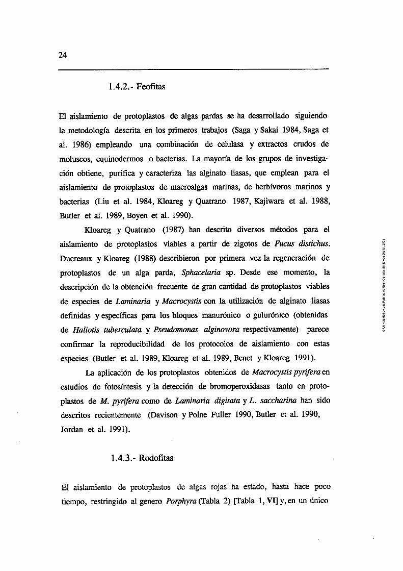
El aislamiento de protoplastos de algas pardas se ha desarrollado siguiendo
la metodología descrita en los primeros trabajos (Saga y Sakai 1984, Saga et
al. 1986) empleando una combinación de celulasa y extractos crudos de
moluscos, equinodermos o bacterias. La mayoría de los grupos de investiga-
ción obtiene, purifica y caracteriza las alginato liasas, que emplean para el
aislamiento de protoplastos de macroalgas marinas, de herbívoros marinos y
bacterias (Liu et al. 1984, Kloareg y Quatrano 1987, Kajiwara et al. 1988,
Butler et al. 1989, Boyen et al. 1990).
Hoareg y Q?iatmn (1987) descrito diversos métodos para el
aislamiento de protoplastos viables a partir de zigotos de Fucus distichus.
Ducreaux y Kloareg (1988) describieron por primera vez la regeneración de
protoplastos de un alga parda, Sphucelaria sp. Desde ese momento, la
descripción de la obtención frecuente de gran cantidad de protoplastos viables
de especies de Laminaria y Macrocystis con la utilización de alginato liasas
definidas y específicas para los bloques manurónico o gulurónico (obtenidas
de Haliotis tuberculata y Pseudom0na.s alginovora respectivamente) parece
confirmar la reproducibilidad de los protocolos de aislamiento con estas
especies (Butler et al. 1989, Kloareg et al. 1989, Benet y Kloareg 1991).
La aplicación de los protoplastos obtenidos de Macrocystis pynfera en
estudios de fotosíntesis y la detección de bromoperoxidasas tanto en proto-
plastos de M. pynifera como de Laminaria digitata y L. saccharina han sido
&.sc$Los reien~emen~e y 301i?e fi!!cí Ijj0, &uei & d. !99e,
Jordan et al. 199 1).
El aislamiento de protoplastos de algas rojas ha estado, hasta hace poco
tiempo, restringido al genero Porphyra (Tabla 2) [Tabla 1, .VI] y, en un único
Introducción 25
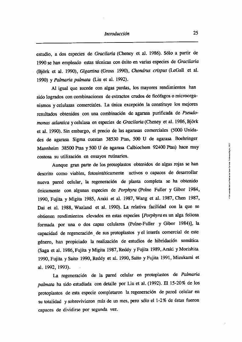
estudio, a dos especies de Gracilaria (Cheney et al. 1986). S610 a partir de
1990 se han empleado estas técnicas con éxito en varias especies de Gracilaria
(Bjork et al. 1990), Gigartina (Gross 1990), Chortdlzcs crispus (LeGall et al.
1990) y Palmaria p a l m a (Liu et al. 1992).
Al igual que sucede con algas pardas, los mayores rendimientos han
sido logrados con combinaciones de extractos crudos de ficófagos o microorga-
nismo~ y celulasas comerciales. La única excepción la constituye los mejores
resultados obtenidos con una combinación de agarasa purificada de Pseudo-
monas atlantica y celulasa en especies de Gracilaria (Cheney et al. 1986, Bj6rk
et al. 1990). Sin embargo, el precio de las agarasas comerciales (5000 Unida-
des de agarasa Sigma cuestan 38530 Ytas, 5W V de agar-asa B&iihiger
Mannheim 38500 Ptas y 500 U de agarasa Calbiochem 92400 Ptas) hace muy
costosa su utilización en ensayos rutinarios.
Aunque gran parte de los protoplastos obtenidos de algas rojas se han
descrito como viables, fotosintéticamente activos o capaces de desarrollar
nueva pared celular, la regeneración de planta completa se ha obtenido
únicamente con algunas especies de Porphyra (Polne Fuller y Gibor 1984,
1990, Fujita y Migita 1985, Araki et al. 1987, Wang et al. 1987, Chen 1987,
Dai et al. 1988, Waaland et al. 1990). La relativa facilidad con la que se
obtienen rendimientos elevados en estas especies [Pophyra es un alga foliosa
f~mmdz p r cna o dos capas celulares (Polne-Fuller y Gibor 1984)], la
capacidad de regeneración de sus protoplastos y el interés comercial de este
género, han propiciado la realización de estudios de hibridación somática
(Saga et al. 1986, Fujita y Migita 1987, Reddy y Fujita 1989, Araki y Morishita
1990, Fujita y Saito 1990, Reddy et al. 1990, Saito y Fujita 1991, Mizukarni et
al. 1992, 1993).
La regeneración de la pared celular en protoplastos de Palmaria
palmata ha sido estudiada con detalle por Liu et al. (1992). El 15-20% de los
protoplastos de esta especie completaron la regeneración de pared celular en
su totalidad y sobrevivieron más de un mes, pero sólo el 1-2% de éstas fueron
capaces de dividirse por segunda vez.
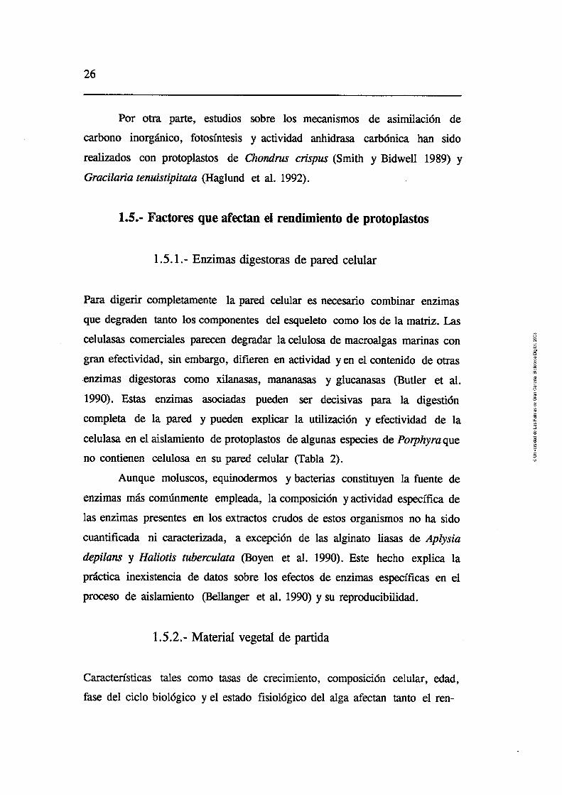
?er cm parte, estudbs wb:e !m maxiiiismos de aJimi!aciSn de
carbono inorgánico, fotosíntesis y actividad anhidrasa carbónica han sido
realizados con protoplastos de Chondm crispus (Smith y Bidwell 1989) y
Gracilaria tenuistipitata (Haglund et al. 1992).
1.5.- Factores que afectan el rendimiento de protoplastos
1 S. 1 .- Enzimas digestoras de pared celular
Para digerir completamente la pared celular es necesario combinar enzimas
que degraden tanto los cmnpnentes d̂ l eywleti rmm Ins de !z matriz. h s
celulasas comerciales parecen degradar la celulosa de macroalgas marinas con
gran efectividad, sin embargo, difieren en actividad y en el contenido de otras
-enzimas digestoras como xilanasas, mananasas y glucanasas (Butler et al.
1990). Estas enzimas asociadas pueden ser decisivas para la digestión
completa de la pared y pueden explicar la utilización y efectividad de la
celulasa en el aislamiento de protoplastos de algunas especies de Porphyra que
no contienen celulosa en su pared celular (Tabla 2).
Aunque moluscos, equinodemos y bacterias constituyen la fuente de
enzimas más comúnmente empleada, la composición y actividad específica de
las enzimas presentes en los extractos crudos de estos organismos no ha sido
cuantificada ni caracterizada, a excepción de las alginato liasas de Aplysia
depilans y Haliotis ncberculata (Boyen et al. 1990). Este hecho explica la -rXI.Ann ;nev;ote..n;n Aa nnhr,. 1~~ A, ,,,.,.... -----?C--- Y*CLCIU- IIlbAlJLGllb14 UC. WLUJ JVLilFi IUJ CICCILVJ UG C X ~ ~ l l l ¿ l S C J ~ I I I M S en d
proceso de aislamiento (Bellanger et al. 1990) y su reproducibilidad.
1.5.2. - Material vegetal de partida
Caractensticas tales como tasas de crecimiento, composición celular, edad,
fase del ciclo biológico y el estado fisiológico del alga afectan tanto el ren-
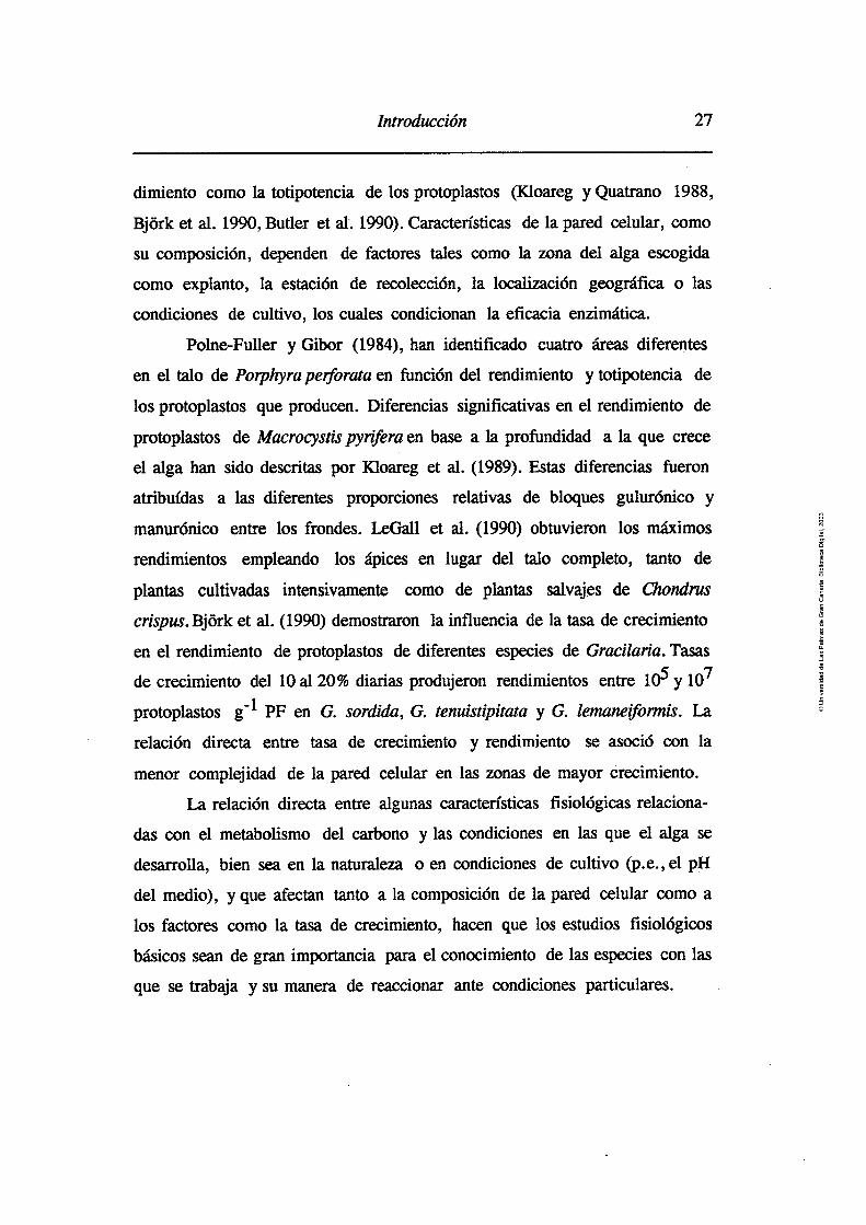
Introducción 27
dimiento como la totipotencia de los protoplastos (Kloareg y Quatrano 1988,
Bjork et al. 1990, Butler et al. 1990). Características de la pared celular, como
su composición, dependen de factores tales como la zona del alga escogida
como explanto, la estación de recolección, la localización geográfica o las
condiciones de cultivo, los cuales condicionan la eficacia enzimática.
Polne-Fuller y Gibor (1984), han identificado cuatro áreas diferentes
en el talo de Porphyraper$orata en función del rendimiento y totipotencia de
los protoplastos que producen. Diferencias significativas en el rendimiento de
protoplastos de Macrocystis pyrifra en base a la profundidad a la que crece
el alga han sido descritas por Kloareg et al. (1989). Estas diferencias fueron
aUi'Puícías a ias áiferentes proporciones relativas cie bioques güiürónico y
manurónico entre los fronda. LeGall et al. (1990) obtuvieron los máximos
rendimientos empleando los ápices en lugar del talo completo, tanto de
plantas cultivadas intensivamente como de plantas salvajes de Chondrus
crispus. Bjork et al. (1990) demostraron la influencia de la tasa de crecimiento
en el rendimiento de protoplastos de diferentes especies de Gracilaria. Tasas
de crecimiento del 10 al 20% diarias produjeron rendimientos entre lo5 y lo7
protoplastos g-l PF en G. sordida, G. tenuistipitata y G. Iemaneifomis. La
relación directa entre tasa de crecimiento y rendimiento se asoció con la
menor complejidad de la pared celular en las zonas de mayor crecimiento.
Li- rdxirin directa entre algunas, ca_ctedstica_s fisiológicas relaciona-
das con el metabolismo del carbono y las condiciones en las que el alga se
desarrolla, bien sea en la naturaleza o en condiciones de cultivo @.e., el pH
del medio), y que afectan tanto a la composición de la pared celular como a
los factores como la tasa de crecimiento, hacen que los estudios fisiológicos
básicos sean de gran importancia para el conocimiento de las especies con las
que se trabaja y su manera de reaccionar ante condiciones particulares.

Temperatura y pH
La actividad de las enzimas digestoras de la pared celular depende tanto de
la temperatura como del pH. Aunque las actividades máximas de las polisa-
caridasas se obtienen a temperaturas superiores a 30°C, estas temperaturas
afectan negativamente la viabilidad celular. Los rangos utilizados para la
digestión enzimática de macroalgas varían de 10 a 25°C (Tabla 2) aunque
temperaturas entre 20 y 25 "C parecen producir los mejores resultados (Saga
y Kudo 1989). T - l - . _ 3 T L . l f p _ 3 _ _ - _ - _ _* _?_*_. . . f - ~ . L _
LOS varores ae pn uuuzaaos para el aisiamento en macroaigas vari'an
entre 6.0 y 7.0, superiores a los empleados en plantas terrestres (que varían
de 5.4 a 6.2) (Evans y Bravo 1984). Las actividades enzimáticas óptimas de
diversas enzimas utilizadas en la digestión suelen ser bastante diferentes @.e.,
celulasas, pH 6.0, y alginato liasas, pH 8.0) (Butler et al. 1990). En este caso,
Kloareg y Quatrano (1987) han utilizado con éxito la incubación del tejido en
dos fases.
Cationes
La disociación parcial de los componentes de la matriz celular e intercelular n. .nAn m a r 11nxmA- 0 0-hn nn- mnAn A- n n i n l ~ n t n n n r i n rn~nn;nnon nnn nqt;nnnc puwb *a ~ r b v a u u a MUV IIIWLV ub \IU~LR.LILCIJ yuu LMUUIUIIULI w u 1 1 W U U L I ~ J
como el ca2+ en alginofitas o el K+ en carragenofitas, disminuyendo la
compactación entre los polisacáridos matriciales y aumentando la accesibilidad
de la celulosa, el alginato o el carragenato a las enzimas digestivas (Butler et
al. 1989, 1990, LeGall et al. 1990).
El tratamiento previo a la digestión enzimática del tejido con un que-
lante de calcio, el EGTA, en macroalgas como Macrocystis o Laminaria pro-
dujo un incremento en el rendimiento entre 4 y 5 veces superior al obtenido
con plantas sin tratamiento (Butler et al. 1989, Kloareg et al. 1989). Un
aumento del 50% fue obtenido por LeGall et al. (1990) en Chondw crispus

Introducción 29
cuando emplearon el Kryptofix 222, un quelante específico de potasio. Sin
embargo, en este último estudio cuando los experimentos se llevaron a cabo
utilizando EDTA, EGTA o variando las concentraciones de ClK del medio
de incubación, no se produjeron cambios significativos en los rendimientos.
Osmolalidad
El proceso de aislamiento de protoplastos debe ser llevado a cabo en solu-
ciones hipertónicas para asegurar la estabilidad celuIar durante la digestión
de la pared (Butler et al. 1990). Las células incubadas directamente en la
solución enzimática pueden no alcanzar el equilibrio con el agente osmótico
antes de que la digestión de la pared tenga lugar provocando la explosión de
los protoplastos. Con la pre-plasmólisis del tejido se asegura que la mayoría
de las células estén plasmolizadas antes de que la digestión ocurra. Sin
embargo, son escasos los trabajos en los que se han estudiado los efectos de
la pre-plasmolisis en los procesos de aislamiento de protoplastos en macroal-
gas marinas (Butler et al. 1989, Bjork 1992).
Por otro lado, durante el proceso de plasmolisis los protoplastos
absorben sustancias del medio (Cocking 1972) por lo que la plasmolisis
directa en la solución enzimática puede provocar la acumulación de enzimas
y agentes contaminantes en la célula. El efectuar la pre-plasmolisis puede
Los factores que influyen en el proceso de plasmolisis y en el
aislamiento de protoplastos incluyen el potencial osmótico del medio y el
agente osmótico utilizado. El ajuste entre 1000 y 1700 mOsrn kg-' de las
soluciones enzimáticas para el aislamiento de protoplastos de macroalgas
(Butler et al. 1989, Bjork 1992) es superior al utilizado con plantas terrestres
(de 300 a 1000 mOsm kgml). Los agentes osmóticos mas frecuentemente
utilizados se muestran en la Tabla 2.

-
+ #- n L 2 - A . Z - - - - 1.0.- UIDJeLlVUS
Los objetivos de la Tesis se han centrado en los sigientes puntos:
Desarrollar un método de extracción, purificación parcial y caracteriza-
ción enzimática para cuantificar las actividades agarasa, celulasa,
carragenasa y alginato liasa de herbívoros marinos, compararlas con las
fuentes comerciales de enzima tradicionalmente empleadas, y determi-
nar la influencia de la temperaturas y pH sobre su actividad in vitro.
DP-~~U= un niip r \ ~ n n i t i ~ r a !y c i ~ q t ; , f i ~ ~ i ~ n YP, a~t;,~i&Y Y-- r-*s=---*-
agarasa a temperaturas adecuadas para el aislamiento de protoplastos
viables.
Estimar la capacidad de adaptación enzimática de herbívoros marinos
para la digestión de polisacáridos matriciales, con la finalidad de
obtener extractos enzimáticos específicos.
Cuantificar la actividad deoxiribonucleasa de los extractos enzimáticos
de herbívoros y su posible toxicidad en el aislamiento de protoplastos.
Estimar la relación entre la actividad in vitro de las polisacaridasas de
los extractos digestivos de herbívoros marinos y los comerciales, y su
&&vidad eii la &iges&dbíi de la pz-& e&:ar & iiizciG&as.
Determinar la influencia del material vegetal (tasa de crecimiento,
edad, pH del medio de cultivo, características fisiológicas) y de
pretratamientos del explanto (efecto de ultrasonidos, pre-plamolisis,
quelantes, pH) sobre el rendimiento de protoplastos.

. Introducción 3 1
En los artículos 1 y 11 se describen la preparación y caracterización de los
extractos crudos de herbívoros marinos, en los artículo III y IV el aislamiento
de protoplastos y en el III y V se desarrollan los estudios de fotosíntesis y
asimilación de carbono inorgánico tanto en protoplastos como en macroalgas.
El artículo VI constituye una revisión del "estado del arte" de la aplicación de
estas técnicas a macroalgas marinas.


Material y métatOs 33
2.- MATERIAL Y METODOS
Las metodologías utilizadas para llevar a cabo los ensayos se describen
detenidamente en los capítulos de material y métodos de los anexos
respectivos. En este capítulo se describe la metodología de resultados no
publicados.
2.1 .- Descripción de las especies herbívoras
2.1.1 .- Aplysia dacylomela Rang
Molusco gasterópodo perteneciente a la Familia Aplysüdae, Orden Aplysiacea.
Conocido como "liebre de mar", es un animal con una pequeña concha
interna. Puede alcanzar hasta 40 cm de longitud y 1400 g de peso. Su ciclo de
vida varía entre los 10 y los 1 1 meses.
Se localiza en la zona intermareal y fondos próximos sobre sustratos
rocosos donde se alimenta de gran variedad de macroalgas, tanto diferentes
especies de Rhodophytas @.e., Corallina, LaurenciQ, Gracilaria) como de
Chlorophytas @.e., Ulva, Cladophora) (Carefoot 1987). Su apetito es voraz y
sus tasas de crecimiento elevadas.
Los animales analizados en este trabajo fueron recolectados en la
plataforma rocosa de la zona intermareal de la playa de Arinaga, donde existe
gran variedad y cantidad de es_~ecies de macroalgas. -
2.1.2 .- Littorina striata King et Broderip
Molusco gasterópodo perteneciente a la Familia Littorinidae, Orden Mesogas-
tropoda. El régimen alimenticio de las diferentes especies de Littorina es
omnívoro aunque prefieren los organismos vegetales, tanto microflora (dia-
tomeas y aigas verde-miesj como macroaigas wawkins y Eanrioii 1963 j.
Los individuos utilizados en el presente estudio fueron recolectados en
la Punta de Taliarte (Telde).
2.1.3.- Haliotis coccinea canariensis Nordsieck
Molusco gasterópodo perteneciente a la Familia Haliotidae, Orden Archaeo-
gastropoda. Se localiza en la zona infralitoral, bajo piedras o rocas a poca
profundidad. Es un molusco de hábitos nocturnos y su alimentación se basa
preferentemente en diferentes especies de Rodofítas, Feofitas y Clorofitas.
hnJ LT&)Fi&GGs m-&OS ec este uab8jo fUerGn mulw,os e;: d
muelle de Taliarte.
2.1.4.- Diadema antillanun Phillipi
De los ficófagos estudiados, es el único que pertenece al Phylum Equinoder-
mata (Familia Diadematoidae, Orden Diadematoida) . Al igual que otras
especies de erizos son organismos ramoneadores generalistas nocturnos,
dependiendo de la disponibilidad se alimentan de una gran variedad de algas
(su dieta predominante), organismos incrustantes y sésiles, detrito o el mismo
siisLit ̂ (TIwcnre 1975, U z w b ~ s y Haemll 19831,
Los individuos analizados en este estudio fueron recolectados en
Arinaga y Taliarte.
2.2.- Extracción y caracterización de los extractos
enzimáticos
La metodología de extracción y cuantificación de actividad enzimática de las
diferentes polisacaridasas, descripción de sustratos, otras fuentes de enzimas
y la determinación de proteínas y azúcares reductores se describen en el ar-

Material y métodos 35
tículo 1. La cuantificación de la actividad DNasa y la medida del pH del
sistema digestivo de los diferentes herbívoros marinos se describen en el
artículo 11, y la cuantificación de actividad carragenasa se describe en el
artículo IV.
2.3.- Cultivos bacterianos
Para determinar la existencia de bacterias agarolíticas en los diferentes siste-
mas digestivos de los herbívoros estudiados, se realizaron siembras en placa
de muestras tomadas de los restos del tracto digestivo obtenidos tras el
proceso de homogeneizado. El medio de cultivo enriquecido empleado fue el
descrito por Morrice et al. (1983a). Una vez el pH del medio era ajustado a
7.5, era esterilizado en el autoclave durante 20 min y dispensado en placas
Petri de 9 cm de 0.
2.4.- Cultivo de Aplysul ductylomela y Haliotis coccinea
canariensis. Ensayo de dietas monoespecíficas
Los ejemplares de Aplysia dactyomela (de peso medio 449.37 f 57.66 g
(n= 10)) fueron cultivados en tanques de fibra (50 x 70 cm), a una densidad
de 3-5 individuos por tanque, con flujo continuo de agua de mar durante 35-
40 días. La altura del agua en los tanques se mantuvo a 20 cm y la aireación
constante. Los individuos fueron alimentados con tres dietas monoespecíficas
de Ulva rigida, Solieriafilifonnis y Gracilaria spp., consumiendo aproximada-
mente unos 100 g PF de alga día-' antes de proceder a la extracción de los
siternas digestivos.
Los extractos de los diferentes individuos fueron preparados del
esófago (VI= 19.37 f 3.50 mL) y la glándula digestiva (VZ= 21.00 + 5.93
mL) .

. . h s ejeiiiplales de fííihíis eoeeiriú: eammemu ltiei~ii ahe~ tados , e::
las mismas condiciones, durante una semana con mezclas de Solieriafiliformis,
Hypnea muscifomis, Grateloupia doryphora y Gracilaria tenuistipitata,
mostrando una clara preferencia por Solieria filiformis por lo que fueron
cultivados durante dos meses con esta especie antes de proceder a la
extracción de los sistemas digestivos.
2.5.- Purificación parcial de la agarasa de los extractos
crudos de Aplysia dactylomela
La separacih de las fracciones de íos extractos ciei esófago y de ia giánauia
digestiva de Aplysia dactylomela se efectuó por cromatografía estándar con
fdtración en gel. La columna (2.6.x 40 cm) se empaquetó con dos geles,
Sephacryl S-300 y Sephadex G-100, previamente equilibrados con agua
destilada siguiendo el procedimiento descrito por la compañía Pharmacia
(1991). Una vez empaquetada, y antes de cargar la muestra, la columna fue
equilibrada con tampón fosfato (0.1 M, pH 6.0). Un volumen de 5 .O mL de
extracto fue pasado a través de la columna, eluído con tampón fosfato (O. 1 M,
pH 6.0) a un flujo de 1.0 mL min-' y las fracciones recogidas en un colector
en volúmenes de 5.0 mL. La concentración de proteínas fue determinada
midipnd~ 1- Asghrn de las b-cciones recolectadas. La actividad agarasa se
determinó siguiendo la metodología descrita en el artículo P.
2.6.- Descripción de las especies vegetales
2.6.1 .- Solieria fllijiormis (Kützing) Gabrielson
Especie perteneciente a la Familia Solieriaceae del Orden Gigartinales
(Rhodophyta). Previamente denominada Agardhiella tenera (J. Agardh)

Material y memetodos 37
Schmitz o Solieria tenera (J. Agardh) Wynne et Taylor, presenta crecimiento
multiaxial, crece erecta desde un sistema basa1 fibroso y puede alcanzar una
altura de 15-20 cm. Productora de iota-carragenato, las algas utilizadas en este
estudio fueron recolectadas en el interior del muelle de Taliarte donde crecen
libremente (no fijadas al sustrato) conformando una morfología esférica laxa.
2.6.2. - Hypnea musc~omis (Wul fen) Lamouroux
Especie perteneciente a la Familia Hypnaceae del Orden Gigartinales (Rho-
dophyta). El género Hypnea es uno de los géneros de algas rojas de mayor
polimorfismo. Se encuentra ampliamente distribuído en las costas tropicales,
. donde es muy abundante, y sub-tropicales. Las especies pertenecientes a este
género han sido descritas principalmente como productoras de kappa-
carragenato. Especie intermareal, en las islas se puede encontrar en charcos,
asociada a un sustrato o a diferentes especies de algas pardas como Sargassum
y Cystoseira. La recolección de esta especie se llevó a cabo en la plataforma
intermareal de la playa de Arinaga.
2.6.3. - Grateloupia doryphora (Montagne) Howe
Perteneciente a la Familia Halymeniaceae del Orden Cryptonemiales (Rhodo-
phyta). Es abundante en ambientes protegidos y semiexpuestos al oleaje,
viviendo sobre rocas en niveles bajos de marea mostrando gran plasticidad
morfológica. Productora de lambda-carragenato, las plantas utilizadas en este
estudio fueron recolectadas en la punta de Taliarte.
2-6.4.- Gracilaria tenuistipitata var. liui Zhang et Xia
Perteneciente a la Familia Gracilareaceae del Orden Gigartinales (Rhodo-
phyta). Normalmente crece en forma libre (no fijada al sutrato). Esta especie

agarofih, hi eresal ps PrOIii&&s fls~G~@m iG; eia
a las variaciones de salinidad y temperatura y su resistencia a los pH elevados
y a las infecciones por epífitos.
2.6.5.- Ulva rigida C, Agardh
Perteneciente a la Familia Ulvaceae del Orden Ulvales (Chlorophyta). Si bien
muestra un ligero carácter estacional, esta especie fue recolectada en el
interior del muelle de Taliarte donde se la puede encontrar principalmente
creciendo en cuerdas muy cerca de la superficie, en immersión constante.
2.7.- Cultivo de las especies vegetales
El cultivo de las diferentes especies se realizó en un invernadero, donde la
irradiación no supera los 1300 pmoles m -2 S-', en tanques de 300 L con
bombeo continuo de aire. El agua de mar (20 + 2°C) era enriquecida con
nutrientes (Provasoli 1968) y el medio renovado semanalmente.
Otras condiciones de cultivo utilizadas con Ulva rigida y Solieria
fllifomis se describen en los artículos m, IV y V.
La metodología del aislamiento de protoplastos se describe en los artículos IiI
y IV. Aunque en estos artículos no se describe el aislamiento de protoplastos
de Hypnea rnuscifonnis, Graieloupia doryphora y Gracilaria tenuistipitaia el
proceso empleado en los experimentos fue exactamente igual para todas las
especies.

Material y métodos 39
2.9.- Fotosíntesis, alcalinización del medio y actividad
anhidrasa carbónica en Solieria filifomis
La descripción de la metodología utilizada para llevar a cabo estos experimen-
tos es descrita detenidamente en el artículo V. En este estudio no se realizó
otro tipo de experimentación más que la que es descrita en él.


Resultados 41
3.- RESULTADOS
3.1.- Método de extracción
3.1.1 .- Precipitación con sulfato amónico
En la Tabla 3 se muestran los porcentajes de actividad obtenidos con las
distintas concentraciones de sulfato amónico añadidos a las diferentes
soluciones enzimáticas. Concentraciones de sulfato amónico superiores al 80%
no aumentaron las actividades enzimáticas en ningún extracto.
Tabla 3. Porcentaje de actividad enzimática (medida como actividad agarasa)
de extractos de ApEysia dactylomela.
3.1.2.- Diálisis y filtración en gel
Aunque la eliminación de solutos de bajo peso molecular ((NH d2S04, NaC1,
etc.) por medio del dializado fue efectiva, se detectaron ciertas desventajas al
comparar este método con la filtración del extracto a través de columnas pre-
empaquetadas con ~ephadex ' G-25 (Pharmacia) . El problema principal del dia-

iizauo h e ia aegraáación de las boisas de ciiállsis (compuesAt. de &iio@
debido probablemente a la acción de las celulasas contenidas en los extractos,
dando como resultado la pérdida de los mismos.
3.2.- Actividades enzimáticas m
Todos los sistemas enzirnáticos degradaron los sustratos experimentados
aunque se observaron diferencias significativas de actividad entre i) especies,
ii) tipo de sustrato y iii) condiciones de temperatura y pH.
Debido a la essasez de individuos de Haliotis coccinea canariemis (aun-
qoe s i i d d r i ~ , m es üíia especie ahfidmte), !m extractcx prepmxks de e"
especie se utilizaron en experimentos muy concretos de cuantifícación de
actividad carragenasa y aislamiento de protoplastos cuyos resultados se
describen en los apartados 3.6 y 3.8, y en el artículo W .
En general, la concentración total de proteínas fue de 3 a 5 veces
superior en los extractos de Aplysia dactylomela y Haliotis coccinea canariensis
que en los de AAP, Littorina striata y Diadema antillanun [Tabla 1, I]. Estas
diferencias en la concentración de proteínas afectaron los resultados al
expresarlos como actividad enzimática específica. Los extractos de A.
dactylomela fueron los que mayor actividad celulasa y agarasa mostraron,
mientras que el AAP mostró la mayor actividad alginato liasa (Tabla 4). En
la Fig. 2 [I] se muestra la evolución de la producción de azúcares reductores
de los extractos en diferentes condiciones de temperatura, pH y sobre los
diferentes tipos de sustratos. -i.s extractos de A. dactyiorneia mosúaron ia
mayor tasa de producción de anícares reductores en todos los sustratos,
exceptuando el alginato de sodio, sobre el que el AAP mostró poseer la
mayor actividad. En la Fig. 2A m se muestran las diferencias de actividad
agarasa entre los sutratos agar y extracto frío (EF). Después de 4 horas de
incubación, no se detectaron aumentos en la producción de azúcares
reductores.

1 Tabla 4. Actividades enzimáticas de los diferentes extractos expresadas como pg AR h- en diferentes condiciones de temperatura y
pH. EF: Extracto frío de Gracilaria spp. CMC: Carboximetil ce:lulosa. Alg: Alginato de sodio. AAP: Extracto en acetona de Haliotis
sp. (Sigma).
Extracto PH Actividad enzimática (pg AR h-l)
EF AiW CMC - Alg -
25°C 40°C 25 "C 40°C 25 "C 40°C 25 " C 40°C
A. dactylomela
AAP
H. coccinea canariensis
L. striata
D. antillarum

Todos los extractos mostraron óptimos de actividad celulasa a 40°C. Sin
embargo, los óptimos de pH variaron en función de la especie. En los extrac-
tos de las diferentes especies de Haliotis (Haliotis coccinea canariensis y el
AAP) y Diadema antillanun las mayores actividades específicas se obtuvieron
a pH 6.0, mientras que en Aplysia dactylomela y Littorina striata fueron
observadas a pH 7.2 [Tabla 1, I]. La mayor producción de azúcares reducto-
res, en pg AR h-' (medidos como glucosa), se obtuvo con los extractos de A.
dmtylomela.
3.2.2 .- Actividad agarasa
La actividad agarasa de los extractos estuvo fuertemente condicionada por el
tipo de sustrato utilizado. El EF de Gracilaria spp. fue digerido con mayor
facilidad que las soluciones de agar natural y agarosa [Tablas 1 ,2 y Fig. 2A,
I]. Las condiciones óptimas de temperatura y pH fueron 40°C y 6.0 en la
mayor parte de los extractos ensayados. En Linopina striata la mayor actividad
específica tanto para el extracto frío como para el agar se obtuvo a 25°C.
La actividad agarasa de los extractos de Aplysia dactylomela, expresada
como pg AR h-l? fue superior a las determinadas de los demás extractos, y a
las agarasas bacterianas, tanto purificada (PA) como no purificada (NPA)
[Tablas 1 y 2, Fig. 2A y 2B, I].
3.2.3 .- Actividad alginato liasa
Las mayores actividades específicas en la degradación de las soluciones de al-
ginato de sodio se obtuvieron a 40°C y pH 7.2, exceptuando las obtenidas con
los extractos de Diadema antillanun que fueron a 25°C y pH 6.0 y a su vez,
las de menor actividad [Tabla 1, I]. Las mayores actividades se obtuvieron con

Resultados 45
el AAP.
3.3.- El pH de los sistemas digestivos m
En la Tabla 1 [a se muestra el pH de los diferentes órganos digestivos en el
momento de la disección de cada uno de los herbívoros estudiados. El pH de
los diferentes órganos varió entre 5 .O y 6.0 en el esófago y la glándula digestiva
de Aplysia dactylomela, entre 5.5 y 6.0 en el hornogeneizado de Littorina striata
y entre 6.5 y 7.0 en el hepatopancreas de Haliotis coccinea canariensis y el
tracto digestivo de Diadema antillanun.
3.4.- Cultivos bacterianos
Las bacterias, existentes en los diferentes sistemas digestivos, que crecieron
en el medio agarizado no mostraron actividad agarasa (ni por licuefacción, ni
por formación de depresiones en el gel) en ninguna de las siembras, ni en los
recultivos posteriores.
3.5.- Actividad DNasa (n_l
La actividad DNasa de los distintos extractos se muestra en la Tabla 1 [a. Los extractos crudos degradaron DNA en presencia de a pH 5.0, ex-
cepto los extractos de la glándula digestiva de A. ductytomela. A pH 6.0,inde-
pendientemente del tipo de tampón (20 mM Bis-tris en agua de mar 6 0.1 M
fosfato), no se detectó actividad DNasa en ninguno de los extractos.
En la Fig. 3 se muestran los incrementos en As6(),, a lo largo del
tiempo de algunos de los extractos estudiados.

0.40
0.35
0.30
0.25
0.20
0.15
0.10
0.05
0.00 O 200 400 600 800 1,000
Tiempo (S)
Figura 3. Evolución de la actividad DNasa en función del tiempo de Littorina
striata, Aplysia dactylomela, AAP y DNasa estandar.
3.6. - Ensayo de dietas monoespecíficas
En la Tabla 5 se muestran las actividades enzimáticas del esófago y la glán-
dula digestiva de Aplysia dactylomela tras someter a los individuos a dietas ---------:4Z-m- A,. 711.,n -*,&Afl C7n7;fl-'n & 7 X n - ; 0 ., f?wnfi;7nm-n A,,--+- 24- I I I V I I V C J ~ l l I M J U& U C Y U 1 L & L U U , i J V L L C I L U J b C L J V I I I C L 3 V I U L C C L C I CCC JYp. U U L Q I I L b JJ-
40 días. Tanto los extractos obtenidos del esófago como de la glándula diges-
tiva degradaron los sutratos en las condiciones empleadas.
La actividad celulasa de los animales alimentados con Ulva rigida
(Tabla 5) fue similar a la detectada en los extractos obtenidos de animales
salvajes (control) (Tabla 4), y fue superior en los alimentados con Gracilaria
spp. y Solieria jilifomis.
La actividad agarasa fue superior en los animales alimentados con

Tabla 5. Actividades enzimáticas medidas como pg AR Y' de los extractos digestivos (del esófago y de la glándula dligestiva) de Aplysia
dactylomela cultivada con dietas monoespecíficas (Ulva rigida, ifracilaria spp. y Solieriacfiliformis) durante más de 30 días. EF: extracto
frío de Gracilaria spp. ; Alg: alginato de sodio; CMC: carboximetil celulosa. T= 25 "C; pH= 6.0.
Sustrato Ulva rígida 0;racilaria spp. Solieriacfiliformis -
Esof Hepat Esof Hepat Esof Hiepat
EF
Alg
CMC

&milede spp. e~ rdpIPU,q&fi 2 !es &mei,ta&s si, ~ 7 ~ ~ ) ~ ~ g j & y &ljg,jg
jilifomis (especies no agarófitas) (Tabla 5). Sin embargo, la actividad agarasa
no fue superior a la detectada en el control (Tabla 4).
La actividad alginato liasa fue similar a la de los extractos de los
individuos sin alimentación específica, aunque se detectó un ligero aumento
en la actividad de los extractos obtenidos de individuos alimentados con
Gracilaria spp. (Tabla 5).
En la Fig. 4 se muestran los incrementos de peso de los animales di-
mentados con dietas monoespecíficas. La tasa de crecimiento diario de los
individuos alimentados con SolieriaJilifonnis casi duplicó a la de los individuos
alimentados con Gracilaria spp. y Ulva rigida, que mostraron tasas de creci-
miento diario similares.
O 2 4 6 8 1 O 12
Incremento peso (gr d -1)
Figura 4. Incremento en peso (media + sd) de Aplysia dactylomela alimentada
con Ulva rigida, Gracilaria spp. y Solieriacfilifomzis. Los pesos fueron tomados
semanalmente durante el periodo de un mes. N= 3 por dieta.

Resultados 49
Haliotis coccinea canariemis mostró una clara predilección por Solieria
filifonnis, aunque en ausencia de ésta consumió sin problemas Hypnea musci-
fonnis y Gracilaria tenuistipitata (Fig. 5). Sin embargo, mostró un claro rechazo
a la ingesta de Grateloupia doryphora (Fig. 5) . Los extractos obtenidos de los
individuos alimentados durante dos meses con S. Jiliformis mostraron un
incremento en su actividad carragenasa (la actividad fue de 30 U mL-')
comparada con la obtenida de los extractos de animales salvajes (10 U m~- ' )
[m.
Figura 5. Evolución del peso fiesco de las especies de macroalgas suministra-
das como alimento a Haliotis coccinea canariensis durante un periodo de 4
días. Hypnea muscifonnis ( +) ; Solieria Jiliformis (O) ; Grateloupia doryphora
( O ) y Gracilaria tenuistipitata (v).

El perfil de elución de la agarasa del esófago de Aplysia dactylomela en una
columna de Sephacryl S-300 HR se muestra en la Fig. 6. Aunque no se obtuvo
una buena separación se distinguen claramente dos picos de actividad
(fracciones 16-3 1 y 37-46). El perfil de elución del extracto en la columna de
Sephadex G-100 fue muy similar. Sin embargo, la elución del extracto de la
glándula digestiva en ambas columnas no mostró un perfil con suficiente
resolución de picos.
O 20 40 60 80 1 O0
Fraccion no
Figura 6. Cromatografía en una columna de Sephacryl S-300 (2.6 x 40 cm) de
la agarasa del extracto crudo obtenido del esófago de Aplysia dactylomela. La
columna fue equilibrada y luego eluída con tampón fosfato 0.1 M @H 6.0).

Resultados 5 1
3.8.- Producción de protoplastos
3 7 Rendimientos de protoplastos que variaron entre 10 y 10 protoplastos g-l
PF fueron obtenidos en todas las especies utilizadas. Sin embargo, los
rendimientos y la viabilidad de los mismos variaron dependiendo de la especie
y de las enzimas empleadas (Tabla 6). Experimentos reproducibles norrnal-
mente en condiciones estandares fueron obtenidos únicamente con Ulva rigida
y SolieriaJilifonnis. La producción de protoplastos de Gracilaria tenuistlpitaia,
Graieloupia doryphora e Hypnea muscifonnis varió considerablemente durante
el tiempo que duraron los experimentos, de tal manera que, en iguales
condiciones y con las mismas combinaciones enzimfiticas los resultados fueron
completamente diferentes de un día para otro en muchos de los casos.
En la Tabla 7 se muestran las actividades enzimáticas de algunos de los
extractos crudos utilizados en las soluciones enzimáticas para el aislamiento
de protoplastos de las especies descritas.
3.8.1 .- Gracilaria tenuistipitata
Se obtuvieron rendimientos entre 0.8 y 5.2 x lo5 protoplastos g-l PF con las
combinaciones de agarasa purificada (Sigma) y celulasa (Cellulysin, Calbio-
chem). La utilización de extractos crudos de Aplysia dactylomela o HaIiotis
coccinea canariensis en combinación con celulasa no produjo efecto alguno, o
en el mejor de los casos una ligera degradación del tejido. Los mejores
resultados se obtuvieron con algas cultivadas a tasas de crecimiento superiores
al 10 % . La pre-plasmolisis del material aumentó ligeramente los rendimientos
aunque no de manera significativa. La adición de quelantes como EDTA o
EGTA no afectó el rendimiento. El tiempo de incubación medio para la
obtención de rendimientos máximos varió entre tres y cuatro horas. El tamaño
de los protoplastos varió de 15 a 20 pm, mostraron forma esférica y explotaron
al ser transferidos a agua destilada.
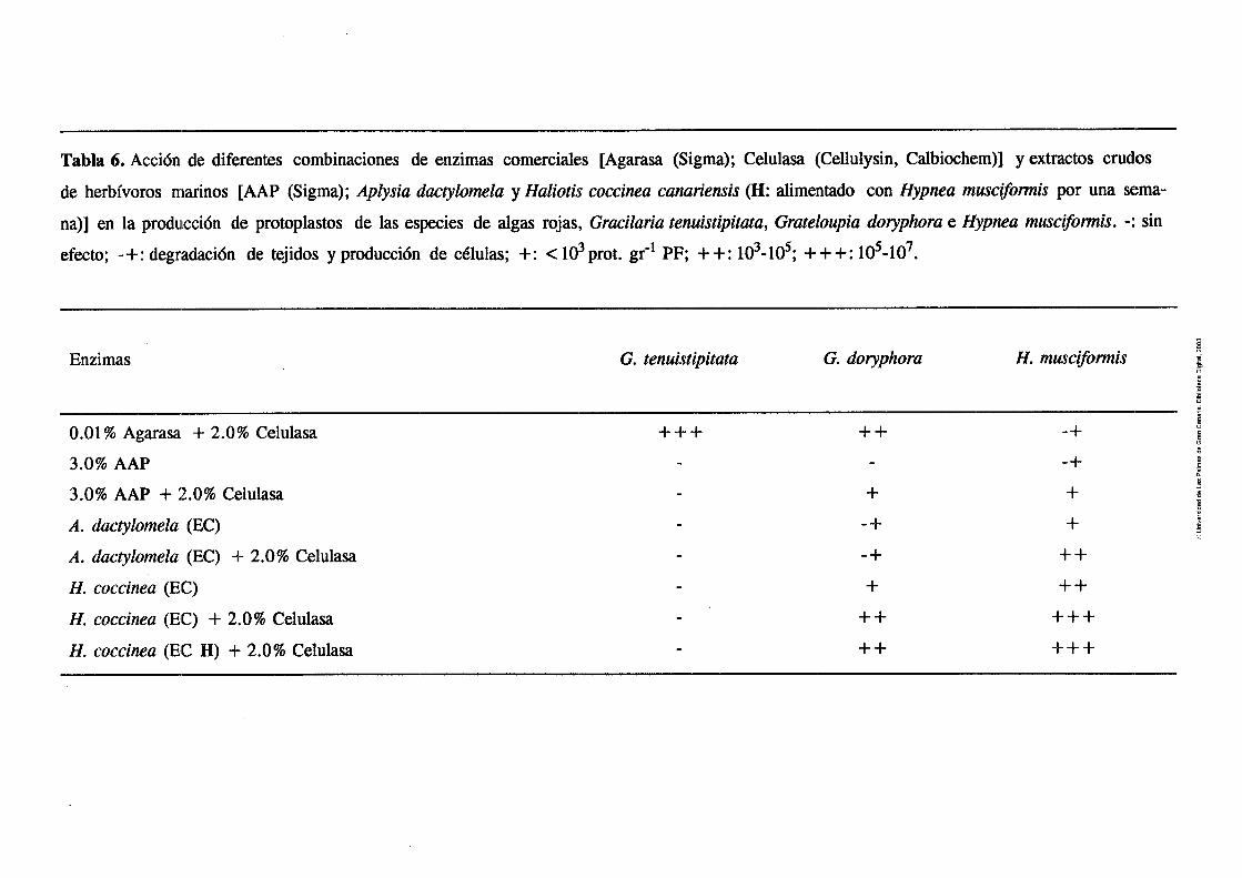
Tabla 6. Acción de diferentes combinaciones de enzimas comercia le:^ [Agarasa (Sigma); Celulasa (Cellulysin, Calbioche:m)] y extractos crudos
de herbívoros marinos [AAP (Sigma); Aplysia dacíylomela y Haliotirr coccinea canariensis (H: alimentado con Hypnea rnuscifomtis por una sema-
na)] en la producción de protoplastos de las especies de algas rojas, Gracilaritz tenuistipitata, Grateloupia doryphora e Hypnea muscijiomtis. -: sin
efecto; -+: degradación de tejidos y producción de c6lulas; +: < 103prot. gr-l PF; + +: lo3-16; ++ +: 10~-10~.
Enzimas G. te,nuistipitata G. doryphora H. muscifomtis
0.01 % Agarasa + 2.0% Celulasa
3.0% AAP
3.0% AAP + 2.0% Celulasa
A. dactylomela (EC)
A. dactylomela (EC) 9 2.0% Celulasa
H. coccinea (EC)
H. coccinea (EC) + 2.0 $6 Celulasa
H. coccinea (EC H ) + 2.0% Celulasa
Resultados 53
3.8.2. - Grateloupia doryphora
Esta especie mostró grandes variaciones en el rendimiento dependiendo en
gran medida de la combinación de enzimas utilizados. Se obtuvieron
rendimientos entre 1.0 y 3.0 x 1 6 tanto con los extractos crudos de Haliotis
coccinea canariensis como con combinaciones de agarasa y celulasa (Tabla 6)
a pesar de ser Grateloupia doryphora una especie carragenofíta. Los rendi-
mientos no variaron entre plantas salvajes y las cultivadas en tanque (cuya
tasa de crecimiento no superó valores del 1% d-l). Se obtuvieron mayores
rendimientos de explantos tomados de knas juveniles (1 03- 1 6 protoplastos
g-i PF) que de zonas adultas ( < ld protoplastos g' PF). A diferencia de
Gracilaria tenuistipitata, los mayores rendimientos se obtuvieron con tiempos
de incubación entre 7 y 10 h. El diámetro de los protoplastos osciló entre 20
y 25 pm.
Todas las enzimas y combinaciones enzimáticas (Tabla 6) degradaron en
mayor o menor medida los tejidos de Hypnea rnusciformis. Los mejores
rendimientos se obtuvieron con la combinación de celulasa y extractos crudos
de Haliotis coccinea canariemis (tanto de los inchiduos saivajes como ae los
alimentados una semana con H. rnuscifomzis). Los extractos de Aplysia
dactylornela también produjeron protoplastos aunque en menor cantidad. En
esta especie, al igual que en Gracilaria tenuistipitata, la pre-plasmolisis y la
utilización de explantos de material cultivado a tasas de crecimiento entre el
5 y el 10 % diario, aumentaron el rendimiento entre un 2.0 y un 5.0%. Sin
embargo, el pre-tratamiento con agentes quelantes como EDTA o EGTA no
afectó de manera significativa los rendimientos. El diámetro de los protoplas-
tos osciló entre 12 y 15 pm, manteniéndose viables durante más de 24 h. Al
igual que en Grateloupia doryphora, no se lograron rendimientos repetibles,

Tabla 7. Actividad enzimática, en U mL-l, de los extractos crudos utilizados
en las soluciones enzimáticas para el aislamiento de protoplastos de Gracilaria
tenuistipitata, Grateloupia doryphora, Hypnea muscijormis, Ulva rigida y Solieria
JiIifomis. Haliotis coccinea canariemis fue alimentado con Hypnea muscifomis
durante 1 semana y con Solieria filijormis durante 2 meses (especies entre
paréntesis). 1 U de actividad es definida como la cantidad de enzima que
produce un aumento de la A=.,--- min-l de O. l en el ensayo de los azúcares - -3 1UIiIíi * * *
reductores. Medido como actividad celulasa; Medido como actividad ***
agarasa; Medido como actividad iota-carragenasa.
AAP 23 U m~- '*
Aplysia dactylomela 15 U m ~ - l * *
10 U mL- 1***
Haliotis coccinea canariensis
H. coccinea canariensis (H. musciformis) 12 U m ~ - '
H. coccinea canariensis (S. jZijormis) 30 U mL-'
Pseudomonas carrageenovora 3 u mL-l
3.8.4.- Ulva rigida m
En la Tabla 1 m se muestra la variación en rendimiento de los explantos de
algas salvajes en función de los tratamientos enzimáticos. La combinación de
1.5 % del extracto comercial de Haliotis sp. (Sigma) y 1.5 % Celulasa (Ce-
llulysin, Calbiochem) produjo los mayores rendimientos, cercanos a 10 7
protoplastos g-l PF. Concentraciones superiores no aumentaron los rendi-
mientos. Ninguna de las dos fuentes de enzimas produjo por sí sóla rendimien-
Resultados 55
tos significativos.
La emisión de protoplastos comenzó a partir de la segunda hora de
incubación y continuó hasta la digestión completa de los tejidos, alrededor de
24 h. Sin embargo, muchos protoplastos comenzaron a explotar a partir de las
8 h en la solución enzimática. El diámetro de los protoplastos osciló entre 10
y 30 pm [Fig. 1, m. En la Figura 7A se muestra un corte transversal del talo
de Ulva rigida y en la 7B los protoplastos de esta especie.
El tratamiento de pre-plasmolisis como mínimo duplicó el rendimiento
[Fig. 2, HIJ, aunque no se detectaron diferencias en cuanto a su viabilidad.
Los mejores rendimientos se obtuvieron con explantos juveniles y, al
igual que con tiraciiana tenuistipitata, ñ ' y p ~ a musciJormis y Soiieriajiiijonnis,
estuvieron condicionados por la tasa de crecimiento. Tasas de crecimiento del
20 % d" produjeron rendimientos máximos pig. 2, iiIJ que disminuyeron con-
siderablemente con plantas cultivadas sin adición de nu trien tes.
La tinción con FDA mostró una viabilidad entre el 70 y el 90%. Tras
24 h en cultivo, los protoplastos regeneraron pared celular, y después de 5 días
aún se mantenían viables.
En la Figura 3 [IIl[l se muestra la evolución de oxígeno fotosintético
tanto de los protoplastos como de explantos de Ulva rigida. La evolución de
oxígeno fotosintético de los protoplastos fue dependiente del pH [Fig. 3 b,
a, siefidn clmto xxes mayor a pH 6.5 que a 7.5 ú 8.5.A pH 6.5?tanto los
protoplastos como los explantos mostraron saturación fotosintética a una
concentración de HCO 3- entre 400 y 500 pM [Fig. 3 a, IIIJ. La concentración
de oxígeno en el medio disminuyó notablemente en la oscuridad.


F@m 7. (A) Corte transversal de un talo de Ulva rigida.Escala= 20 )cm. (B)
Protapíastos fotografiados en contraste de fases. 20 pm.


Resultados 59
3.8.5 .- Solieria jtlifomis ln7]
En la Fig. 1 [N] se observan las diferencias morfológicas entre algas salvajes
(Fig. 1A) y las cultivadas durante más de dos meses en tanques de 300 L (Fig.
1B).
La combinación de celulasa y extractos crudos de Haliotis coccinea
canariensis, tanto de individuos salvajes como mantenidos en el laboratorio
durante dos meses alimentados con Solieriaflliformis, produjo rendimientos
entre lo4 y lo7 protoplastos g-l PF, aunque fue con los extractos de Hf con
los que se obtuvo los mejores resultados nabla 1, m. No se observó aumento
en el rendimiento por adición de carragenasas bacterianas (obtenidas de
Pseudomonas carrageenovora), o pectoliasa. Sin embargo, se observó un ligero
aumento del rendimiento cuando estos enzimas se añadieron a la solución que
contenía el H, pabla 1, N]. La celulasa tuvo influencia directa en la
digestión de la pared celular. El aumento de su concentración del 1 .O al 3.0%
aumentó el rendimiento de protoplastos (de ninguno al 1 .O%, a lo4 al 3.0%
tras 2 h de incubación) sin afectar la viabilidad de las células. El AAP sólo o
en combinación con celulasas únicamente produjo la degradación del tejido
[Tabla 1, IV].
La producción de protoplastos comenzó a partir de la segunda hora de
incubación, aunque los mayores ietiuimient~s (de 1-02 8.5 x 1c6 yLwwy- nrntnni actnc "--- gel PP) fueron observados después de las 12 h. Se distinguieron dos tipos de
protoplastos: protoplastos de 20-25 pm de 0 provenientes de la región sub-
cortical y medula [Fig . 1 C, IV] , y de 10- 15 pm de 0 , en mayor cantidad, pro-
ducidos de la zona cortical pig. ID, IV].
El 70-80% de los protoplastos mostraron ser viables al teñirlos con
FDA. Al cabo de 48 h, el 10-15% de los protoplastos regeneraron pared
celular y se observó la formación de agrupaciones de 5 a 10 células. No se
observó división celular.
Los ápices de algas "salvajes" (recolectadas del medio natural) y
"cilindro" (extraídos de algas creciendo en fotobioreactores cilíndricos [VI)

m,." m..-.. -1 -m\
lllu&au~. los mayores ref idimi~ni~~ (6.0-8.5 r proiupiasios g - rr,. En
los pocos casos en los que se obtuvieron protoplastos de talos maduros, los 4 rendimientos no superaron los 10 protoplastos g-l PF. Los rendimientos de
los "ápices esféricos" (extraídos de algas cultivadas en tanques que generaron
morfologías esf6ricas apicales) fueron aún menores nabla 2, IV]. Las
diferencias en los rendimientos tienen una relacidn directa con las tasas de
crecimiento. Las algas cultivadas durante períodos cortos en los cilindros
mostraron tasas de crecimiento entre el 10 y el 14% diario mientras que el de
las cultivadas durante períodos largos (mayores de 2 meses) en tanques no fue
superior al 1 %. Las algas utilizadas en el experimento de los diferentes pH
del medio de cultivo mostraron tasas de crecimiento intermedias [entre el 0.5
y el 3 % diario) y la producción de protoplastos fue igualmente intermedia.
El pH del medio de cultivo afectó el rendimiento de protoplastos de
Solieriafilijiomis siendo máximo a pH 6.0 pig -2, N]. La correlación entre la
producción de protoplastos y la tasa de crecimiento (coeficiente de correlación
de Pearson, a= 0.86) indicó una clara influencia del pH del medio de cultivo
y ambos parámetros.
La pre-plasmolisis del tejido aumentó entre un 1.0 y un 2.0% los
rendimientos y la viabilidad. La viabilidad de los protoplastos obtenidos de
explantos no pre-plasmolizados fue menor del 70% y no mostraban el aspecto
uniforme que mostraron los obtenidos de explantos pre-plasmolizados, cuya
viabilidad fue del 90 % . La incubacidn del explanto en un baño de ultrasonidos (previa a la pre-
plasmolisis) durante p&&s de t;_emp fin ISQ=~BQOS a 2 mh ~ u u ~ P , I ? ~ ~ el
rendimiento en un 20%. Períodos más largos produjeron ablandamiento del
explanto.
La adición de los agentes quelantes EDTA y EGTA a la solución de
pre-plasmolisis no tuvo efectos significativos sobre los rendimientos de
protoplastos en Solieriafilifomis.


. Resultados 63
3.9.- Captación de carbono inorgánico en Solieriafiliformis
M
En la Fig. 2 [VI se muestra la evolución de oxígeno fotosintético en los fenoti-
pos "salvaje" y "esférico" de Solieria Jilifomis. La evolución de oxígeno fue
dependiente, en ambos casos, de la irradiación y del pH del medio. El "fenoti-
po salvaje' mostró saturación fotosintéüca a 100 y 200 pmol m-2 S-' tanto a
pH 8.2 como a pH 6.5. En el "fenotipo esf6ricoU, ésta fue observada a 150 y
200 pmol S-' en ambos pH. Sin embargo, la evolución de oxígeno del
"fenotipo esférico" fue un 25 % superior a ambos pH. Esta diferencia fue más -2 -1
significativa a mayores irradiaciones (400 pmol m S ).
En la Tabla 1 [VI se muestran las concentraciones de clorofila y
ficoeritrina, y sus proporciones en ambos fenotipos. La concentración de
clorofila y la proporción Clorofi1a:Ficoeritrina fue mayor en el "fenotipo
salvaje" que en el "fenotipo esférico".
Se observó actividad anhidrasa carbónica (AC) tanto en los sobrena-
dantes como en los precipitados de los extractos crudos de Solieriafiliformis.
La actividad AC del so-brenadante fue de 7.0 U mg-' de proteínas (135 pg
mL-l) y en el precipitado de 1 17 U mg-' de proteínas (34 pg mLml).
En la Fig. 3 [VI se muestra el efecto de EZ (50 pM) y DBS (40 pM),
en la evolución de oxígeno fotosúittctico deí "fenotipo saivaje" de Soiieria
~lijionnis. La evolución de oxígeno (sin adición de los inhibidores de AC) fue
aproximadamente un 25 % superior a pH 6.5 que a pH 8.2. A pH 8.2 tanto EZ
como DBS inhibieron la fotosíntesis un 90 y un 50%, respectivamente. El
aumento de la concentración de DBS, en las mismas condiciones, no aumentó
la inhibición. A pH 6.5 no se observó inhibición. Los mismos resultados fueron
observados con el "fenotipo esférico".
En la Fig. 4 v] se muestra la tasa de alcalinización del medio de
incubación por los dos fenotipos estudiados. Las tasas de alcalinización fueron
dependientes de la irradiación. El "fenotipo esférico" mostró mayor tasa de al-

S-', la tasa de alcalinización del "fenotipo esférico" aumentó aproximadamente
dos veces. En el "fenotipo salvaje", la alcalinización fue saturada a una
intensidad de luz menor (100 pmol m-2 S-'). .
Aunque no de manera significativa, las tasas de alcalinización fueron
superiores con la utilización del filtro m l .
En la Fig. 5 [VI se muestran los efectos de la adición de E%, DBS,
DCMU y de la oscuridad sobre la tasa de alcalinización del medio del
"fenotipo salvaje" de SolieriQfiliformis. La tasa de alcalinización fue inhibida
al añadir los inhibidores y en la oscuridad. Sin embargo, como se observó en
los experimentos de fotosíntesis, el EZ inhibió la tasa de aldnización en
mayor medida. Los mismos resultados fueron observados con el "fenotipo
esférico".

Discusión 65
4.- DISCUSION
4.1.- Metodología de la extracción
En base a las actividades enzimáticas detectadas, la metodología para la
extracción y preparación de los extractos crudos, basada en la precipitación
con sulfato amónico y la posterior desalación (tanto por diálisis como por
cromatografía por filtración en gel), mostró ser efectiva. No obstante, la
filtración en Sephadex G-25 aceleró el proceso de preparación de los extractos
crudos y evitó las posibles pérdidas de actividad, e incluso de extractos, que
ocasiona la diálisis, debido a la degradación enzimática de las bolsas.
La actividad enzimática del AAP, preparado comercialmente por
extracción en acetona, mostró valores similares a los descritos por otros
autores (Nisizawa et al. 1968, Wojtowicz 1972, Polne-Fuller 1987).
E
4.2.- Actividades enzimáticas m a
n n
n
3
O
Las elevadas actividades enzimáticas de los extractos de Aplysia dactylomela
sobre todos los sustratos estudiados indican la alta capacidad digestiva de un
amplio rango de componentes estructurales de la pared celular, lo que es de
suponer, le confiere una gran capacidad para alimentarse eficientemente de
macroalgas verdes, pardas y rojas. En este sentido, las diferencias de actividad
de los extractos de Haliotis coccinea canariensis, Littorina striata y Diadema
antillanun, pueden ser debidas a las preferencias alimenticias y a los tipos de
especies disponibles en su entorno.
Los resultados de actividad celulasa de todos los herbívoros estudiados,
fueron similares a los descritos en otras especies de invertebrados marinos
(Yokoe y Yasumasu 1964, Kristensen 1972, Brock et al. 1986, Suzuki et al.
1986, Boyen et al. 19901, cuyos óptimos de temperatura (entre 24 y 35°C) y
pH (6.0 - 7.0) también concuerdan wn nuestros valores óptimos de actividad
(IIvriuchi y L m e 1965, Ermk et d. 1986).
Boyen et al. (1990) encontraron actividades similares entre una celulasa
comercial y extractos crudos y purificados de Aplysia depilans y Haliotis
tuberculata usando celulosa microcrístalina como sustrato. Sin embargo,
cuando estos extractos se utilizaron para el aislamiento de protoplastos, la
eficacia fue practicamente nula, en comparación con la celulasa comercial.
Asimismo, Lewis (1964) no detectó actividad celulasa en extractos de Diadema
antillanun empleando papel de filtro como sustrato. En nuestro caso, los
extractos crudos de todos los herbívoros degradaron eficazmente carboximetil
celulosa. Estas diferencias son obviamente debidas al tipo de sustrato
empleado y ponen de manifiesto la importancia de su correcta elección para
determinar la existencia de actividad.
No obstante, aunque existe actividad celulasa en los extractos de todos
los herbívoros estudiados, su utilización exclusiva no permitió la obtención de
gran número de protoplastos (Tabla 6), que sin embargo se obtuvieron al
suplementar los extractos con celulasas comerciales. La incapacidad de los
extractos de digerir la celulosa de la pared celular indica que la existencia de
actividad sobre la CMC no prueba su efectividad sobre la pared celular. Estos
resultados demuestran experimentalmente la hipótesis de Friesen (1980)
cuestionando la capacidad real de digestión de celulosa en base a la actividad
celulasa detectada con la CMC en extractos de invertebrados marinos.
La temperatura (40 "C) y pH (6.0) óptimos de actividad agarasa de los
extractos, fueron similares a los de las agarasas bacterianas (40°C y pH entre
5.0 y 7.2; Yaphe 1966; Duckworth y Turvey 1969, van d̂ r Meden 1975,
Momce et al. 1984).
El extracto frío de Gracilana spp. fue el mejor sustrato para la
determinación de la actividad agarasa, tanto porque no forma geles en
solución (y por tanto permite la cuantificación de actividad a temperaturas por
debajo del punto de gelifrcación del agar), como por la mayor sensibilidad del
sustrato pabla 1 y 2, Fig. 2A, I]. La dificultad para la determinación de
actividad agarasa utilizando sustratos convencionales (agar y agarosa), debido
Discusión 67
a la imposibilidad de obtener soluciones homogéneas, descrita por van der
Meulen (1975) puede evitarse con la utilización del El?.
Las agarasas bacterianas (tanto NPA como PA) no mostraron las
actividades enzimáticas indicadas por las empresas suministradoras. Esta
pérdida de actividad pudo haberla ocasionado el transporte defectuoso de los
enzimas.
Los resultados de actividad alginato liasa son similares a los descritos
en diferentes especies de Aplysia, Haliotis y Littorina (Franssen y Jeuniaux
1965, Nakada y Sweeny 1967, Elyakova y Favorov 1974, Boyen et al. 1990),
aunque esta actividad se describe por primera vez en el género Diadema.
4.3.- pH del sistema digestivo
La correlación entre el pH del sistema digestivo uabla 1, IIJ y los valores de
pH en los que se obtuvieron las mayores actividades (Tabla 4), indica que la
digestión se realiza por hidrólisis enzimática, confirmando los resultados
obtenidos en otros invertebrados (Sphaeroma walken, Vallabhan 1989; P e m
vindis, Tw y Sabapathy 1990). Asimismo pone de manifiesto la conveniencia
de determinar el pH del tracto digestivo del herbívoro previamente a su
elección para la obtención de enzimas para la digestión de la pared celular de * --- --- -- ---:m+-- A-6r.n nm Gn;nntcrn l n a rra lnrpc A p 1\U QictPma rnacroaigas. muiyue iiu GIUJLGII u a w a adiLbLbliwo ivo rULVLVo -.-A Y-l------
digestivo en peces herbívoros (3.5 + 0.5 en Lagodon rhomboides, Montgomery
y Targett 1992) parece indicar que en éstos la digestión se efectúa por
hidrólisis ácida.
Los valores de pH del tracto digestivo de Diadema antillanun [Tabla
1, IIl coincidieron con los valores de pH descritos en esta especie por Lewis
(1964). Asimismo, los valores de pH de los órganos digestivos de los
herbívoros estudiados entran en el rango de pH óptimos de las actividades
polisacaridasas de los extractos de invertebrados marinos (Onishi et al. 1985,
Barlocher et al. 1989).

La ausencia de bacterias agarolíticas en el sistema digestivo de los herbívoros
estudiados contrasta con su detección en el equinodermo Strongylocentrotus
pulpurratus (Lasker y Giese 1954), el gasterópodo Tegula funebralis (Galli y
Giese 1959) y Haliotis sp. (Minamitake et al. 1986). Además de agarasas,
dichas bacterias también disponían de carragenasas y alginato liasas.
Sin embargo, nuestros resultados concuerdan con la ausencia de
bacterias agarolíticas en Aplysia juliana descrita por Vitalis et al. (1988),
aunque estos autores mencionan su capacidad para degradar glucosa, maltosa,
dmi,U& v rarmoenatn y g p i b l e qje ~ ~ f i @ ~ & i ~ t ~ ~ ~ j J -'-bWA---'
debidos al mayor o menor contenido en bacterias agarolíticas asociadas a las
algas consumidas por los herbívoros.
4.5.- Actividad DNasa [HII
La detección de una deoxiribonucleasa ácida @H 5 .O) en los extractos pabla
1, Iil concuerda con los resultados obtenidos en otros invertebrados marinos
(Georgatsos y Antonoglou 1963, Rasskazov et al. 1975) en los que también se
detectan DNasas alcalinas (a valores de pH superiores a 7.0).
La mayor actividad DNasa específica de los extractos de Litmrinn
striata y Diadema antillam [Tabla 1,111 se debe posiblemente a la contami-
nación por otros órganos (los extractos de L. striata se prepararon utilizando
todo el animal y durante el aislamiento del sistema digestivo de D. antillanun
fue muy difícil evitar el contacto con otros fluídos y órganos).
Sin embargo, la ausencia de actividad DNasa a pH 6.0 [Tabla 1, 111 indica la ausencia de efecto tóxico en el rango de pH óptimo para el'
aislamiento de protoplastos de macroalgas marinas. En este sentido, la
inexistencia de actividad a pH 6.0 indica que la adición de inhibidores de
actividad DNasa a la solución enzimática que algunos autores utilizan para el

Discusión 69
aislamiento de protoplastos (Fujimura et al. 1989a, Waaland et al. 1990,
Reddy y Fujita 199 1) es una complejidad innecesaria.
4.6.- Influencia de la dieta monoespecífica
El aumento de actividad carragenasa de los extractos de Haliotis coccinea ea-
nariensis alimentados durante dos meses con Solieriafilifomis (de 10 U m ~ - '
en los individuos salvajes a 30 U m ~ - ' en los cultivados) indica la existencia
de adaptación enzimática a la dieta. La inexistencia de actividad carragenasa
en el AAP y,por el contrario su gran actividad alginato liasa (40 U m~- ' ) (en
contraste con la escasa actividad en Hw y Hf) corroboran la posibilidad de
estimular la producción de determinadas polisacaridasas en función de la dieta
(el AAP se extrae de Haliotis sp. alimentado con Macrocystis pynyera, una
especie alginofita) . Los extractos de Hf permitieron obtener rendimientos
superiores en la digestión de S. filiformis que los Hw (aptdo. 4.8).
Aunque en Aplysia dactylomela los cambios de actividades enzimáticas
no fueron tan espectaculares, se observaron diferencias entre los individuos
alimentados con las diferentes especies (Tabla 5, aptdo 3.6) y, en la mayoría
de los casos, con respecto a los controles.
Estos resultados concuerdan con el aumento de actividad de carbohi- en &)zU12Füj f :u lh &iiiieiibG&-j3 esfi nMS &j~-qt& m&
de ocho semanas (van Weel 1961). Sin embargo, Boyen et al. (1990) no
describen cambios sustanciales en la actividad alginato liasa de Aplysia
vaccaria, A. depilans y Haliotis ncfscem alimentados durante una semana con
Macrocystis pyrifera. No obstante, los individuos de H. mfescem empleados
fueron obtenidos de una granja en la que habían sido alimentados con M.
pynfera durante más de 6 años. Sus extractos contenían 50 veces más cantidad
de alginato liasa que las dos especies de Aplysia, con una actividad 28 a 38
veces superior en su actividad específica. A la vista de nuestros resultados, es
probable que de haber alimentado estos herbívoros durante un período más
prolongado, hubiesen detectado alteraciones enzimáticas más significativas.

Aunque la resolución obtenida no fue la suficiente (Fig . 6), es muy probable
la existencia de, al menos, dos agarasas en los extractos de Aplysia daczylome-
la. La gran complejidad del proceso de purificación de agarasa en Littorina
mandshurica ha sido exhaustivamente descrita por Usov y Miroshnikova
(1975), lo cual contrasta con la facilidad de la purificación de las agarasas de
origen bacteriano (Duckworth y Turvey 1969, van der Meulen 1975, Groleau
y Yaphe 1977, Morrice et al. 1983a, Aoki et al. 1990, Yamaura et al. 1991).
La disminución de la actividad enzimática de las fracciones en
compamcibn con !a de 10s P . Y ~ C ? ~ S cnidns se debe proh~h!eme~te 2 12
dilución del enzima al pasar por la columna.
La gran complejidad enzimática de los extractos de Aplysia dactylomela
indica la necesidad de utilizar columnas de mayor longitud y cromatografía de
afinidad para poder purificar las enzimas. No obstante las ventajas de la
purificación enzimática para su aplicación al aislamiento de protoplastos no
están claras. Aunque tanto para el aislamiento de protoplastos de plantas
terrestres (Schenk y Hildebrandt 1969) como para el de macroalgas marinas
(Butler et al. 1990) se ha sugerido la necesidad de purificar los complejos
enzimáticos, la inexistencia de actividad DNasa en los extractos (a pH 6.0),la
diversidad de polisacáridos de la pared celular de macroalgas y la pérdida de
enzimas durante el proceso de purificación, nos hacen pensar que la estrategia
debe ser precisamente la opuesta.
4.8.- Aislamiento de protoplastos
Aunque se obtuvieron elevados rendimientos de protoplastos viables de Ulva.
rigida y Solieria ~Ziformis de manera reproducible, los rendimientos con
Gracilaria tenuistipitata, Grateloupia doryphora e Hypnea muscifomis, aunque
elevados en algunos ensayos (Tabla 6), no fueron tan reproducibles, indicando

Discusión 71
la existencia de factores no controlados.
4.8.1 .- Soluciones enzimáticas
Los rendimientos obtenidos de Gracilaria tenuistipituta con la combinación de
agarasa purificada de Pseudomonas atlantica y celulasa, concuerdan con los
obtenidos por Bj6rk et al. (1990) con Gracilaria spp., incluída G. tenuistipitata
(id-lo7 protoplastos g-l PF). La utilización de extractos crudos (Tabla 6),
incluído el AAP, no produjo protoplastos, lo que contrasta con los resultados
obtenidos por Chou y Lu (1989) que deschbieron rendimientos similares a los
nuestros utilizando extractos de Haliotis diversicolor (alimentados con
Gracilaria) combinado con otras enzimas comerciales, aunque a concentracio-
nes muy superiores y sin aportar datos de viabilidad.
Los rendimientos obtenidos con Grateloupia doryphora utilizando tanto
la combinación de enzimas comerciales o combinaciones de extractos crudos
de Haliotis coccinea canariensis y celulasa (Tabla 6) son mayores que los
obtenidos por Chou y Lu (1989) con Grateloupia sp. y G. filicinn (< 2.0 x lo3
protoplastos g-l PF) utilizando mayores concentraciones de una combinación
de extractos de Haliotis diversicolor, pectoliasa, macerasa y celulasa. Los
elevados rendimientos obtenidos con la combinación de agarasa y celulasa
carragenofita.
La obtención de protoplastos del género Hypnea no había sido descrita
previamente. En el caso de Hypnea muscifomtis, la combinación de las
diferentes enzimas, tanto extractos crudos como enzimas comerciales,
mostraron diferentes efectos sobre el tejido y, en mayor o menor medida
produjeron células y protoplastos, obteniéndose los mayores rendimientos con
combinaciones de extractos de Aplysia dactylomela y Haliotis coccinea
canariensis (tanto salvaje como alimentado con H. muscifomis) (Tabla 6).
Estos resultados sugieren que dichos herbívoros tienen una gran preferencia
por especies carragenofitas.
b s rendimiegtcs obtenidos con U ~ G n g i h ( > 1& pi6t~pl-s g-l
PF) fueron similares a los descritos en otras especies de Ulva (Zhang 1983,
Fujita y Migita 1985, Reddy et al. 1989), aunque en estos casos las concentra-
ciones enzimáticas fueron superiores, lo que indica la posibilidad de aumentar
la viabilidad de los protoplastos (reduciendo el efecto tóxico de los enzima)
a igualdad de rendimientos.
En Solieriajiliformis, la combinación de los extractos crudos de Haliotis
coccinea canariensis con celulasa fue la más efectiva, con rendimientos
significativamente superiores a los obtenidos con el AAP pabla 1, m. Los
mayores rendimientos con Hf (respecto a H,) corroboran las mayores activi-
dades enzimáticas detectadas in vitro y la posibilidad de inducir mayores
actividades mediante dietas monoespecíficas, y demuestran la correlación
entre actividades enzimáticas in vitro y las actividades reales sobre la pared
.celular. No obstante, la imposibilidad de obtener protoplastos, tanto de S.
jiliformis como de Ulva rigida, únicamente con extractos digestivos (aunque
poseen celulasas, aptdo. 4.2) indica que dicha correlación es válida para
polisacáridos matriciales, pero no para los del esqueleto (principalmente
celulosa).
4.8.2 .- Material vegetal
Los bajos rendimientos de explantos viejos en comparación con los juveniles
en todas las especies, similares a los resultados descritos en Grwilaria spp. m ,,
& s. ;390j, G&G,3.1cu (GiOSS iggjj, L"r26iw'i-i crsspw (RLRCrair et
al. 1990) y Macrocystis pynfera (Kloareg et al. 1989), demuestran la menor
complejidad de las paredes celulares de los tejidos juveniles.
Esta característica está directamente relacionada con la tasa de creci-
miento del alga (y por lo tanto con las condiciones de cultivo previas a la
extracción del explanto), ya que los mejores rendimientos en todas las especies
se obtuvieron con algas cultivadas a altas tasas de crecimiento (> 10% d-' en
la mayoría de los casos), exceptuando Grateloupia doryphora (cuya tasa de

crecimiento no superó el 1% d-'). La influencia de la tasa de crecimiento en
la composición del agar de Gracilaria sordida, G. vemosa y G. tenuistipitata
ha sido demostrada por Lignell y Pedersén (1989) y Ekman y Pedersén (1990).
En este sentido, los mayores rendimientos de los ápices "cilindro" en
comparación con los ápices "esf~ricos" de Solieriacfilijiormis (cuyo proceso de
formación y sus causas han sido descritos por Robledo y García-Reina 1993),
se explican por las mayores tasas de crecimiento de los primeros pabla 2, IV].
Las algas que crecieron a tasas de crecimiento intermedias mostraron rendi-
mientos intermedios. Los rendimientos similares entre los explantos de ápices
"salvajes" y "cilindro" podrían explicarse por el rápido crecimiento de las algas
en ei meúio natural en d momento de la ~ s d ~ i ó f i (S. $lfv^,mis muestra ur!
marcado crecimiento estaciona1 y durante los experimentos se encontraba en
una fase de rápido crecimiento).
La relación entre el rendimiento de protoplastos y el pH del medio de
cultivo Pig. 2, IV] indica un claro aumento del rendimiento a pH ácidos. No
obstante, el efecto del pH más que un efecto directo (excluyendo su hipotético
efecto en la compactación de la pared celular) se debe a su influencia sobre
la tasa de crecimiento, mediatizada por el mecanismo de captación de
carbono inorgánico del medio.
A pH 6.5, Solieria filiformis muestra una tasa de fotosíntesis 25 %
superior a la tasa observada a pH 8.2 pig. 2, VI. Estas diferencias se deben
a las distintas proporciones de carbono inorgánico fotosintéticamente
asimilables a ambos pH, lo que, a pesar de la existencia de ACext en S.
cfilijiomis permite mayores tasas de crecimiento en meÚios w n &vada
concentración de C02 @H 6.5).
Ademds de las diferencias en cuanto a la tasa de crecimiento, se
observaron diferencias fisiológicas entre ápices "salvajes" y "esféricos" en
cuanto al contenido en clorofila (superior en los ápices "salvajes") y ficoeritri-
na Pabla 1, VI. A pesar de estas diferencias, y de las diferencias en compleji-
dad de la pared celular (evidenciada por las diferencias en rendimiento), am-
bos tipos de explanto poseen ACext [VI. La existencia de ACext en ambos

ex@~tev h d i a qw, hdependientrnefity de su t is i de c~irr,i:tc, mxfd~=
gía y rendimiento, la viabilidad de los protoplastos estará condicionada por la
concentración de C 0 2 del medio de incubaci6n y de cultivo posterior. Por lo
tanto, el pH del medio de incubación no sólo afecta la actividad de las
polisacaridasas (y por tanto el rendimiento) y la viabilidad únicamente por la
desactivación de las DNasas, sino también por la capacidad fisiológica de
recuperación del protoplasto (después del estrés causado por la digestión) y
activar adecuadamente el metabolismo del carbono @.e. ,comenzar la síntesis
de nueva pared celular). Debido a la inhibición de las DNasas y a las
relativamente elevadas concentraciones de C02 a pH 6.0,el ajuste a un valor
próximo a pH 6.0 es el más adecuado para llevar a cabo los procesos de
aislamiento y cultivo de protoplastos.
4.8.3 .- Efecto de los pretratamientos
El aumento de los rendimientos en Solieriafilifomzis tras la incubación de los
explantos en un baño de ultrasonidos, durante períodos inferiores a dos
minutos, concuerda con los resultados descritos por Neushul (1984) con
Sargasswn spp. Este tratamiento produce la relajación de la estructura
compacta de las paredes celulares, facilitando por tanto la acción de las
enzimas.
El aumento del rendimiento y de la viabilidad (en base a la capacidad
fotosintética de los protoplastos de Ulva [Fig. 3, m, y por tinción con FDA
en !os de $elkrk) dernuectm !es efectm be~efici~ws. Ud tatr4ii;;& de prc-
plasmolisis de los explantos. Este efecto beneficioso, según Butler et al. (1989)
y Bjork (1992) se debe al efecto estabilizante y preventivo de captación de
sustancias tóxicas durante la digestión de la pared celular.
La ausencia de efectos apreciables en el rendimiento por adición de los
agentes quelantes (EDTA y EGTA) en Solieriacfilifomis, Hypnea musciformis
y Gracilan'a tenuistipitata, concuerda con los resultados observados por LeGall
et al. (1990) en Chondm crispus. La efectividad del EGTA en el aislamiento

Discusión 75
de protoplastos de Laminaria (Butler et al. 1989) se debe probablemente a la
especificidad de dicho quelante sobre el ca2+, el principal ion responsable
de la compactación de los poli~cándos matriciales en alginofitas.
4.9.- Capacidad fotosintética de los protoplastos
La capacidad de fotosintetizar de los protoplastos se ha empleado reciente-
mente como un indicador de la viabilidad de éstos. Unicamente en protoplas-
tos de Enteromorpha se han descrito tasas de fotosíntesis iguales, o ligeramen-
te superiores, a las de los explantos (Millner et al. 1979), ya que tanto en
Chondw crispus como en Macrocystispynjba (Smith y Bidwell 1989, Davison
y Polne-Fuller 1990) la capacidad fotosintética de sus protoplastos fue inferior
(30 y 40% respectivamente), en proporciones similares a los valores de los
protoplastos obtenidos de Ulva rigida [Fig . 3, m].
No obstante, las variaciones en la tasa de fotosíntesis, como indicador
de viabilidad de los protoplastos, sin considerar el pH, lleva a conclusiones
erróneas. Las diferencias de fotosíntesis entre protoplastos y explantos fueron
considerablemente menores a pH 6.5 que a pH 8.2 [Fig. 3,
Esta dependencia de la fotosíntesis de los protoplastos de Ulva rigida
del pH, confirma resultados similares descritos en Chondm crispus (Smith y ,.m-, Biciweii IYUY) y Graciiuriü ieriuisdpiiuiü @ag!üiib et d. 1992) y Yem~estm
tanto la incapacidad del HC03 - para atravesar la membrana plasmática como
la necesidad de una pared celular intacta para el funcionamiento de la ACext,
y la dependencia de los protoplastos de la concentración de C02 en el medio
(y por tanto del pH). Bjork et al. (1992) han demostrado la fuerte inhibición
producida por EZ (inhibidor de AC total) y nula por DBAZ (inhibidor de la
ACext) sobre la fotosíntesis de los protoplastos de U. rigida, lo que puede
significar que el mecanismo de asimilación de HC03- (catalizado por la
ACext) puede ser destruído en el momento de la digestión de la pared celular.
1.- Las elevadas actividades celulasa, agarasa, carragenasa y alginato liasa
cuantificadas en los extractos digestivos de los herbívoros estudiados
demuestran la capacidad de éstos para digerir los polisacáridos
estructurales de las paredes celulares de las macroalgas marinas. Estas
actividades mostraron poseer pH óptimos similares a los existentes en
los sistemas digestivos de los ficófagos, y ser más activos a temperatura
ambiente que las fuentes comerciales de enzima, lo que justifica su
alto interés para el aislamiento de protoplastos.
2.- La metodología de la cuantificación de actividad agarasa empleando
el "extracto frío" de Gracilarfa spp. ha permitido, por primera vez,
cuantificar la actividad agarasa a temperaturas inferiores a 40°C. Este
método ha puesto de manifiesto la menor actividad de algunas agarasas
bactaianas comerciales a las temperaturas de incubación óptimas para
el aislamiento de protoplastos de macroalgas marinas, en relación a la
actividad de los extractos de fidfagos.
La actividad agarasa del sistema digestivo de los herbívoros marinos
estudiados no es de origen bacteriano.
Las variaciones de las actividades polisacaridasas de los extractos de
Aplysia dactylomela y Haliotis coccinea canariensis tras su alimentación
c m Uietizs f ~ ? ~ n w s p e d f i ~ OUmte más de 30 dias, demesúii la
posibilidad de alterar la composición enzimática de su sistema digestivo
en función del tipo de polisacárido matricial existente en la dieta. La
demostración de esta capacidad posibilitaría la producción de extractos
enzimáticos hiperactivos sobre la pared celular de las especies que se
utilicen como alimento.

Conclusiones 77
4.- La inactividad de la deoxiribonucleasa ácida detectada en los extractos
a los valores de pH óptimos para el aislamiento de protoplastos,
demuestra la inexistencia de toxicidad de los extractos por actividad de
estos enzimas y la innecesariedad de añadir inhibidores de DNasas a
la solución enzimática.
5.- Los elevados rendimientos en SolieriaJilifomis y Ulva rigida con los
extractos digestivos demuestran la existencia de una relación entre las
actividades enzimáticas in vitro (tanto de herbívoros salvajes como de
los alimentados con dietas monoespecificas) y su efectividad real sobre
ia ciigestih cie ia pared ceiuiar. Sin embargo, ia imposibiiiáaá áe
obtener rendimientos elevados únicamente con extractos digestivos (sin
la adición de celulasas complementarias) demuestra que ésta relación
afecta únicamente a los polisacáridos matriciales y no a los del es-
queleto.
6.- La tasa de crecimiento influye directamente sobre el rendimiento de
protoplastos. El pH del medio de cultivo también afecta directamente
el rendimiento de protoplastos, pero este efecto se debe a la estimula-
ción de la tasa de crecimiento al aumentar la captación de carbono
inorgánico del medio.
El valor de pH mas adecuado para la digestión enzimática es 6.0, tanto
por la mayor actividad de las polisacaridasas y la inactividad de las
DNasas, como por favorecer la captación de C02 por los protoplastos,
al carecer estos de ACext.
Los pretratamientos con ultrasonidos (durante períodos inferiores a
dos minutos) y plasmolisis, aumentan el rendimiento y la viabilidad de
los protoplastos.

Aasen IM, Folkvord K, Levine DW (1992) Development of a process for large-scale chromatographic purification of an alginate lyase from Klebsiella pneumoniae. Appl. Microbiol. Biotechnol. 37: 55-60.
Amano H, Noda H (1992) Proteins of protoplasts from several seaweeds. Bull. Jap. Soc. Sci. Fish. 58: 291-299.
Anónimo (1984) Commercial Biotechnology: an international analysis. U.S. Congress, Office of Tchnology Assessment, OTA-BA-2 18. Government Printing Offiice, Washington, DC. 612 pp.
Aoki T, Araki T, Kitamikado M (1990) Purification and characterization of P-agarases from Vibrio sp. AP-2. Bull. Jap. Soc. Sci. Fish. 56: 825-830.
Araki C (1956) Some recent studies on the polysaccharides of agarophytes. Proc. 5th Int. Seaweed Symp. 5: 3-17.
Araki C, Arai K (1957) Studies on the chemical constitution of agar-agar. 20. Isolation of a tetrasaccharide by enzymatic hydrolysis of agar-agar. Bull. Soc. Chem. Jpn. 30: 287-293.
Araki T, Morishita T (1990) Fusion of protoplasts from wild type Porphyra yezoensis and green type P. tenera thalli (Rhodophyta). Bull. Jap. Soc. Sci. Fish. 56: 1161.
Araki T, Aoki T, Kitamikado M (1987) Preparation and regeneration of protoplasts from wild-type of Porphyra yezoensis and green variant of P. tenera. Bull. Jap. Soc. Sci. Fish. 53: 1623-1627.
Armisén R, Galatas F (1987) Production, properties and uses of agar. En: McHugh DJ (d.) Production and utilization of products from commercial seaweeds. FA0 Fish. Tech. Pap. 288: 1-57.
Ashe H, Searnan E, Van Vunakis H, Levine L (1965) Characterization of deoxyribonuclease of Mwtelw canis liver. Biochim. Biophys. Acta 99: 298-306.
Barlocher F, Arsuffi TI,, Newell SY (1989) Digestive enzymes of the saltmarsh periwinkle Littorina irrorata (Mollusca, Gastropoda). Oecologia 80: 39-43.
Bellanger F, Verdus MC, Henocq V, Christiaen D (1990) Determination of the composition of the fibrillar part of Gracilaria verrucosa (Gracilariales, Rhodophyta) cell wall in order to prepare protoplasts. Hydrobiologia

Bellion C, Hamer GK, Yaphe W (1982) The degradation of Eucheuma spimsum and E. cotonii carrageenans by i-carrageenases and k-carrageenases from marine bacteria. Can. J. Microbiol. 28: 874-880.
Benet H, Kloareg B (1991) Isolation of protoplasts from brown seaweeds. En: García-Reina G, Peúersén M (4s.) Seaweed cellular biotechnology, physiology and intensive cultivation. Cost 48 - Universidad de Las Palmas de Gran Canaria, Las Palmas, 287-290.
Benítez LV, Macaranas JM (1979) Partial purification of a carragerrnase from the tropical sea urchin Diadema setosum. Proc. 9th Inter. Seaweed Symp., Sci. Press, 9: 353-359.
Berliner M (1981) Protoplasts of eukaryotic algae. Intern. Rev. of Cytology 73: 1-19.
Bj6rk M (1992) Protoplast isolation from m a ~ e macroalgae and application to studies of inorganic carbon utilization. Ph.D. thesis, Dpt. Phy siological Botany, University of Uppsala, 47 pp.
Bj6rk M, Ekman P, Wallin A, Pedersen M (1990) Effects of growth rate and other factors on protoplast yield from four species of Gracilaria (Rhodophyta). Bot. Mar. 33: 433-439.
Bj6rk M, Haglund K, Rarnazanov Z, García-Reina G, Pedersén M (1992) Inorganic-carbon assimilation in the green seaweed Ulva rigida C. Ag. (Chlorophyta). Planta 187: 152- 156.
Boyen C, Kloareg B, Polne-Fuller M, Gibor A (1990) Preparation of alginate lyases from marine moiluscs for protoplast isolation in brown algae. Phycologia 29: 173- 18 1.
Brock V, Kennedy VS, Brock A (1986) Temperature dependency of carbo- hydrase activity in the hepatopancreas of thirteen estila-nne md cnastA bivalve species from the North American east coast. J. Exp. Mar. Biol. Ecol. 103: 87-101.
Brown BJ, Preston JFIII (1991) L-Guluronan-specific alginate lyase from a marine bacterium associated with Sargassm. Carbohyd. Res. 2 1 1 : 9 1- 102.-
Butler DM (1989) Isolation and culture of protoplasts and tissues of Laminaria spp. (Phaeophyta). Ph.D. thesis, Dpt. of Pure and Applieú Biology. The University of Leeds, 203 pp.

Butler DM, 0stgaard K, Boyen C, Evans LV, Jensen A, Kloareg B (1989) Isolation conditions for high yields of protoplasts from Laminaria saccharina and L. digitata (Phaeophyceae). J. Exp. Bot. 40: 1237-1246.
Carefoot TH (1980) Studies on the nutrition and feeding preferences of ApZysia: Development of an artificial diet. J. Exp. Mar. Biol. Ecol. 42: 24 1-252.
Carefoot TH (1982) Phagostimulatory properties of various chemical cornpounds to sea hares (Aplysia kurodai and A. daciylomela). Mar. Biol. 68: 207-2 15.
Carefoot TH (1987) Aplysia: its biology and ecology. Oceanogr. Mar. Biol. Annu. Rev. 25: 167-284.
Chen LC-M (1987) Protoplast morphogenesis of Pophyra leucosticta in culture. Bot. Mar. 30: 399-403.
Cheney DP, Mar E, Saga N, van der Meer J (1986) Protoplast isolation and cell division in the agar-producing seaweed Grda r i a (Rhodophyta). J. Phycol. 22: 238-243.
Chesters CGC, Turner M, Apinis A (1956) Decomposition of Laminarin by microorganisms. Proc. 2nd Inter. Seaweed Symp. 2: 14 1- 14.4.
Chou H-N, Lu H-K (1989) Protoplasts from seaweeds: isolation, culture and fusion. Current topics in Marine Biotechnology, 227-230.
Chou M-Y, Liao T-H (1990) Shrimp hepatopancreatic deoxyribonuclease - purification and characterization as well as comparison with bovine pancreatic deoxyribonuclease. Biochim. Biophys. Acta 1036: 95-100.
Clifford C, Walsh J, Reidy N, Johnson DB (1982) Digestive enzymes and subcellular localization of disaccharidases in some echinoderms. Comp. Biochem. Physiol. 71B: 105-1 10.
Cocking EC (1960) A method for the isolation of plant protoplasts and vacuoles. Nature 187: 962-963.
Cocking EC (1972) Plant cell protoplasts - isolation and development. Ann. Rev. Plant Physiol. 23: 29-50.

Coury DA, Hong Y, Polne-Fuller M (1991) Preparation of protoplasts from Porphyra, Macrocysts and Ulva. Phycol. Newslett. December-91: 9-1 1.
Crabtree B, Leech AR, Newsholme EA (1979) Measurement of enzyme activities in crude extracts of tissues. En: Techniques in metabolic research. Elsevier, North-Holland Scientific Publishers, Ltd. B211: 1-37.
Dai J, Bao Z, Tang Y, Liu W (1988) Studies on isolation of the thallodic cells of Polphyra with enzymes and cultivation of these d l s . Chinese J. Biotech. 4: 133-137.
Davison IR, Polne-Fuller M (1990) Photosynthesis in protoplasts of Macrocystis pynifera (Phaeophyta). J. Phycol. 26: 384-387.
Day DF, Yaphe W (1974) Enzymic hydrolysis of agarose to D-galactose and 3,6 anh ydro-L-galactose. Purification of an a-Neoagarobiose h ydrolase. Prm. Su*& syq. 8; 543-547.
Deutscher M (1990) Guide to protein purification. Methods in Enzymology, Vol. 182. 894 pp.
Domingo A, Cervera M, Martin E, Marco R (1986) Biochemical characteri- zation and developmental behavior of Artemia embryonic and nauplial deoxyribonucleases. Biochemistry 25: 4 125-413 1.
Doubet R, Quatrano RS (1984) Properties of alginate lyases from marine bacteria. Appl. envir. Microbiol. 47: 699-703.
Duckworth M, Turvey JR (1969) An extracellular agarase from a Cytophaga species. Biochem. J. 1 13: 139-142.
Duckworth M! Yaphe W (1971) The structure of azar. Part 11. The use of a bacteria1 agarase to elucidate structural features of the charged polysaccharides in agar. Carbohyd. Res. 16: 435-445.
Ducreaux G , _Klc>a_reg R (1988) Hant regpnemtbn frnm pr"tq!asts ef Sphacelaria (Pheophyceae). Planta 174: 25-29.
Ekman P, Pedersén M (1990) The influence of photon irradiance, day lenght, dark treatment, temperature, and growth rate on the agar composition of GrQcilaria sordida W. Nelson and G. vermcosa (Hudson) Papenfuss (Gigartinales, Rhodophyta). Bot. Mar. 33: 483-495.
Elyakova LA, Favorov VV (1974) Isolation and certain properties of alginate lyase VI from the mollusk Littorina sp. Biochim. Biophys. Acta 358: 34 1-354.

Bibliografa 83
Eppley RW, Lasker R (1959) Alginase in the sea urchin Strongylocentrotus pupuratus. Science 129: 214-215.
Enksson, TR (1985) Protoplast isolation and culture. En: Fowke LC, Constabel F (eds.) Plant protoplasts. CRC Press, Inc., Boca Raton, Florida, 1-20.
Evans DA, Bravo JE (1984) Protoplast isolation and culture. En: Evans DA, Sharp WR, Ammirato PV, Yamada Y (eds) Handbook of plant cell culture, Vol 4, Macmillan mibl. Co., New York, 97-132.
Favorov W, Vozhova EI, Denisenko VA, Elyakova LA (1979) A study of the reaction catalysed by alginate lyase VI from the sea mollusc Littorina sp. Biochim. ~ i o p h ~ s . Acta -569: 259-266.
Feral J-P (1989) Activity of the principal digestive enzymes in the detritivorous apodous holothuroid Leptosyruzpta galliennei and two other shailow water holothuoids. Mar. Biol. 101 : 367-379.
Fisher DD, Gibor A (1987) Production of protoplasts from the brown alga, Sargmswn muticwn (Yendo) Fensholt (Phaeophyta). Phycologia 26: 488-495.
Fitzsimons PJ, Weyers JDB (1985) Properties of some enzymes used for protoplast isolation. En: Pilet PE (ed.), The physiological properties of plant protoplasts. Springer-Verlag , Berlin-Heidelberg , 12-23.
Franssen J, Jeuniaux C (1965) Digestion de l'acide alginique chez les invertebres. Cah. Eioi. mar. ó: i-2i.
Friesen JA (1980) The structure of the gut of Mysis stenolepsis and the mechanism of cellulose digestion. M.Sc. thesis, Dalhousie University , Nova Scotia, 73 pp.
Fujimura T, Kawai T, Shiga M, Kajiwara T, Hatanaka A (1989a) Preparation of immobilized living cells from a marine green alga Ulva pertusa. Bull. Jap. Soc. Sci. Fish. 55: 2211.
Fujimura T, Kawai T, Shiga M, Kajiwara T, Hatanaka A (1989b) Regenera- tion of protoplasts into complete thalli in the marine green alga Ulva p e m a . Bull. Jap. Soc. Sci. Fish. 55: 1353-1359.
Fujita Y, Migita S (1985) Isolation and culture of protoplasts from some

seaweeds. Bull. Fac. Fish. Nagasaki Univ. 57: 39-45.
Fujita Y, Migita S (1987) Fusion of protoplasts from thalli of two different color types in Porphyrayezoensis Ueda and development of fusion products. Jap. J. Phycol. 35: 201-208.
Fujita Y, Saito M (1990) Protoplast isolation and fusion in Porphyra (Bangiales, Rhodophyta). Hydrobiologia 2041205: 16 1- 166.
Gacesa P (1988) Alginates. Carbohydr. Polym. 8: 161- 182.
Galli DR, Giese AC (1959) Carbohydrate digestion in a herbivorous snail, Tegula Jxnebralis. J. Exp. Zool. 140: 4 15-439.
Georgatsos JG, Antonoglou O (1963) Deoxyribonucleases in tissues of marine origin. Enzymologia 27: 14 1- 150.
Granado 1, Caballero P (1991) Feeding preferences by Littorina striata King and Broderip on different algal species. Sci. Mar. 55: 577-581.
Greer CW, Yaphe W (1984) Purification and properties of i-carrageenase from a marine bacterium. Can. J. Microbiol. 30: 1500-1506.
Groleau D, Yaphe W (1977) Enzymatic hydrolysis of agar: purification and characterization of B-neoagarotetraose hydrolase from Pseudomonas atlantica. Can. J. Microbiol. 23: 672-679.
Gross W (1990) Preparation of protoplasts from the carrageenophyte Gigartina corymbifera (Kiitz.) J. Ag. (Rhodophyta). J. Microbiol. Methods 12: 2 17-223.
Haglund K, Bjork M, Ramazanov 2, García-Reina G, Pedersén M (1992) Role of carbonic anhydrase in photosynthesis and inorganic-carbon assirnilation in the red alga Gracilaria tenuistipitata. Planta 1 87: 275-28 1.
Harada K, Kawasaki O (1982) The attractive effect of seaweeds based on the hchzvi~ml ospfis~.~ ~f ymng herhiv~mu~ abalnne Halintis discru~ Bull. Jap. Soc. Sci. Fish. 48: 617-621.
Hawkins SJ, Hartnoll RG (1983) Grazing of intertidal algae by marine invertebrates. Oceanogr. Mar. Biol. Annu. Rev. 21: 195-282.
Hodgkiss U, Leung HC (1986) Cellulase associated with mangrove leaf decomposition. Bot. Mar. 29: 467-469.
Horiuchi S, Lane CE (1965) Digestive enzymes of the crystalline style of Strombus gigaslinne. 1. ~ellulase and some other carbohydrases. Biol. Bull.

Jordan P, Kloareg B, Vilter H (1991) Detection of vanadate-dependent bromoperoxidases in protoplasts from the brown algae Laminaria digitata and L. sacchurina. J. Plant Physiol. 137: 520-524.
Kajiwara T, Hatanaka A, Fujimura T, Kawai T, Irie M (1988) Isolation of protoplasts from marine brown algae Dictyotaceae plants. Bull. Jap. Soc. Sci. Fish. 54: 1255.
Kitamikado M, Tseng C-H, Yarnaguchi K, Nakamura T (1992) Two types of bacteria1 alginate 1 yase. Appl. envir. Microbiol. 58: 2474-2478.
Kloareg B, Quatrano R (1987) Isolation of protoplasts from zygotes of Fucus distichus (L.) Powell (Phaeophyta). Plánt Sci. 50: 189- 194.
Kioareg B, Quatrano RS (1988) Structure of the cell walls of marine algae and ecophysiological functions of the matrix polysaccharides. Oceanogr. Mar. Biol. Annu. Rev. 26: 259-3 15.
Kloareg B, Polne-Fuller M, Gibor A (1989) Mass production of viable protoplasts from Macrocystispyrifera (L.) C. Ag. (Phaeophyta). Plan t Sci. 62: 105-1 12.
Knutsen S (1991) Large scale production of bacterial enzymes for depolimerization of matrix polysaccharides in seaweeds. En: García-Reina G, Pedersén M (eds.) Seaweed cellular biotechnology, physiology and intensive cultivation. Cost 48 - Universidad de Las Palmas de Gran Canaria, Las Palmas, 91-104.
Koningsor RL, McLean N, Hunsaker D (1972) Radiographic evidence for a dlgestive allulase in the sea nare, Apjsia vaccaria (GasuopWar Opisthobranchia). Comp. Biochem. Physiol. 43B: 237-240.
Kreger DR (1962) Cell walls. En: Lewin RD (d.), Physiology and biochemistry of algae. Academic Press, New York, 3 15-335.
Kristensen JH (1972) Carbohydrases of some marine invertebrates with notes on their food and on the natural occurrence of the carbohydrates studieú. Mar. Biol. 14: 130-142.
Lasker R, Giese AC (1954) Nutrition of the sea urchin, Strongylocentrotus purpuratus. Biol. Bull. 106: 328-340.
Lavilla-Pitogo CR (1992) Agar-digesting bacteria associated with 'rotten thallus syndrome' of Gracilaria sp. Aquaculture 102: 1-7.
Lawrence JM (1975) On the relationships between marine plants and sea urchins. Oceanogr. Mar. Biol. Annu. Rev. 13: 213-286.
LeGall Y, Braud JP, Kloareg B (1990) Protoplast production in Chondm crispus garnetophytes (Gigartinales, Rhodophyta). Pl. Cell Rep. 8: 582-585.
Lewin R (1983) Phycotechnology - how microbial geneticists rnight help. BioScience, 33: 177-179.
Lewis JB (1964) Feeding and digestion in the tropical sea urchin Diadema antillanun Philippi. Can. J . Zool. 42: 549-557.
Lignell A, Pedersén M (1989) Agar composition as a function of morphology and growth rate. Studies on some morphological strains of Gracilaria secundata and G. verrucosa (Rhodophyta). Bot. Mar. 32: 219-227.
Liu QY, Chen LC-M, Taylor ARA (1992) Ultrastructure of cell wall regeneration by isolated protoplasts of Palmaria palmata (Rhodophyta) . Bot. Mar. 35: 21-33.
Matsue T, Koike S, Abe T, Itabashi T, Uchida 1 (1992) An ultrarnicroe- lectrode for determination of intracellular oxygen. Light-irradiation-induced change in oxygen concentration in an alga1 protoplast. Biochim. Biophys. Acta 1101: 69-72.
McCandless JS, Craigie JS, Walter JA (1973) Carrageenans in the gameto- phytic and sporophytic stages of Chondw crispus. Planta 112: 201-212.
McCandless EL, West JA? Guiry MD (1982) Carrageenan patterns in the Phyllophoraceae. Biochem. Syst. Ecol. 10: 275-284.
McHugh DJ (1987) Production, properties and uses of alginates. En: McHugh DI (en) Prdnli.t;nn and utilizathn nf prducts from cnmmercial seaweeds+
FA0 Fish. Tech. Pap. 288: 58-115.
McHugh DJ (1991) Worlwide distribution of commercial resources of seaweeús including Gelidium. Hydrobiologia 22 1 : 19-29.
McLean MW, Williamson FB (1979) K carrageenase from Pseudomonas carrageenovora. Eur. J. Biochem. 93: 553-558.
McLean MW, Williamson FB (1981) Neucarratetraosa 4-O-monosulphate 13- hydrolase from Pseudomonas carragenovora. Eur. J. Biochem. 1 13: 447-456.

Mejjaci M, ~~~ise.aux-cie-S&f; S, Ducreüñ G (1992) Pi~t~i ; !a~t isdaticn, development, and regeneration in different strains of Pilayella littoralis (L.) Kjellm. (Phaeophyceae) . Protoplasma 169: 42-48.
Millner PA, Callow ME, Evans LV (1979) Preparation of protoplasts from the green alga Enteromorpha intestinalis (L.) Link. Planta 147: 174-177.
Minamitake S, Natori M, Nakajima M, Murooka H (1986) Production of agardecomposing enzyme by a marine bacterium and some properties of the enzyme. Bull. Coll. Agric. Vet. Med. 43: 84-91.
Mizukarni Y, Kito H, Okauchi M (1993) Factors affecting the electrofusion efficiency of PoPphyra protoplasts. J. Appl. Phycol. 5: 29-36.
Mizukami Y, Okauchi M, Kito H (1992) Effects of cell wall-lytic enzymes on t!x el-r,tt̂ fnsion efficiency of protoplasts from Pophyra yezoensis. Aquaculture 108: 193-205.
Montgomery JLM, Targett TE (1992) The nutritional role of seagrass in the diet of the omnivorous pinfish Lagodon rhomboides (L.). J . Exp. Mar. Biol. Ecol. 158: 37-57.
Morrice LM, McLean M, Williamson FB, Long WF (1983a) P-agarases 1 and II from Pseudomom atlantica. Purifications and some properties. Eur. J. Biochem. 135: 553-558.
Morrice LM, McLean M, Williamson TB, Long WF (1983b) P-Agarases 1 and 11 from Pseudomonas atlantica. Substrate specifities. Eur. J. Biochem. 137: 149-154.
Morrice LM, McLean M, Williamson FB, Long WF (1984) P-agarases from Pseudomnas atlantica. Hydrobiologia 1 í 6i 1 17: 576-579.
Muramatsu T, Egawa K (1980) Isozymes of alginate lyase in the mid-gut gland of Turbo cornutus. Agric. Biol. Chem. 44: 2587-2594.
Muramatsu T, Hirose S, Katayose M (1977) Isolation and properties of alginate lyase from the mid-gut gland of wreath shell Turbo cornutus. Agric. Biol. Chem. 41: 1939-1946.
Nakada HI, Sweeny PC (1967) Alginic acid degradation by eliminases from abaione hepatopancreas. J. Biol. Chem. 242: 86-85 1.
Neushul M (1984) Marine algal tissue culture. Topical technical report for Gas Research Institute 198 1- 1983, Gas Research Institute 8410076, Goleta, CA, 46 pp.

Nisizawa K, Fujibayashi - - S, Kashiwabara Y (1968) Alginate lyases in the hepatopancreas of a marine mollusc, Dolabella auricula Solander. J. Biochem. 64: 25-37.
Nonomura AM (1988) A future of phytotechnology. En: Lembi CA, Waaland JR (eds.) Algae and human affairs. Cambridge University Press, Cambridge, 529-552.
Onishi T, Suniki M, Kikuchi R (1985) The distribution of polysaccharide hydrolase activity in gastropods and bivalves. Bull. Jap. Soc. Sci. Fish. 51: 30 1-308.
0stgaard K (1992) Enzymatic microassay for the determination and characterization of alginates. Carbohyd. Polym. 19: 51-59.
Percival E (1978) Do the polysaccharides of brown and red seaweeds ignore taxonomy? En: írvine EEG, Rice jIi (ecis.j, itídeni apprüaches iü ihe taxonomy of red and brown algae. Academic Press, London, New York, 47-62.
Pharmacia LKB (199 1) Gel filtration: principies and methods. 5th d., 102 pp.
Polne-Fuller M (1987) Preparation of enzymes. En: Indergaard M, 0stgaard K, Guiry M (4s.) Seaweed protoplast and tissue culture. Cost 48, Trondheim, 29-30.
Polne-Fuller M, Gibor A (1984) Developmental studies in Porphyra. 1. Blade differentiation in Porphyraperjorata as expressed by morphology , enzymatic digestion, and protoplast regeneration. J. Phycol. 20: 609-616.
Polne-Fuller M, Gibor A (1987) Tissue culture of seaweeds. En: Bird KT, Renson PH (edso), Seaweed cultivation for renewable resources? Vol. 16. Elsevier, Amsterdam, 2 19-239.
Polne-Fuller M, Gibor A (1990) Developmental studies in Porphyra @mbpl,;vyue). ID. Effect of cdttirr cnnditbns m w-Y regenerztion md differentiation of protoplasts. J. Phycol. 26: 674-682.
Potin P, Sanseau A, Le Gall Y, Rochas C, Kloareg B (1991) Purification and characterization of a new k-carrageenase from a marine Cytophaga-like bacterium. Eur. J. Biochem. 201: 241-247.
Preston RD (1974) The physical biology of plant cell walls. Chapman & Hall, London. 49 1 pp.
Preston JF, Romeo T, Bromley JC, Robinson RW, Aldrich HC (1985)

A l-:--~- I..--A-~A.--~C:~.~ hnn+at;n n n n n A o t 4 4 t h &ha 01 I T ~ a e n ~ t ~ Cnrun~~llm f i l p I a u = IyaE -LGI.UI~ u a G b L r a c w i > u v i a ~ w ~ u i iv rus ~ W . A U
En: Underkofler LA (d.), Developments in industrial microbiology. Society for industrial microbiology, Arlington, VA, 727-740.
Prim P, Lawrence JM (1975) Utilization of marine plants and their constituents by bacteria isolated from the gut of Echinoids (Echinodermata). Mar. Biol. 33: 167- 173.
Prosser, CL, van Weel PB (1958) Effect of diet on digestive enzymes in midgut gland of African giant snail, Achatina JUlica Fér. Physiol. 2001. 3 1: 171-178.
Provasoli L (1968) Media and prospects for the cultivation of marine algae. En: Watanabe A, Hattori A (4s.) Culture and collections of algae. Jap. Soc. Plant Physiol. 63-75.
Quatrano RS, Caldwell BA (1978) Isolation of a unique marine bacterium capable of growth on a wide variety of polysaccharides from macroalgae. Appl. envir. Microbiol. 36: 979-98 1.
Rasskazov VA, Pirozhnikova VV, Galkin W (1975) Some properties and specificity of deoxyribonucleases from marine invertebrates and fishes. Comp. Biochem. Physiol. 5 1B: 343-347.
Reddy CRK, Fujita Y (1989) Protoplast isolation and fusion of Ulva pertusa and U. conglobata. En: Miyachi S, Karube 1, Ishida Y (eds.), Current topics in marine biotechnology. The Japanese Society for Marine Biotechnology, Tolqo, 235-238.
Reddy CRK, Fujita Y (1991) Regeneration of plantlets from Enteromorph (Ulvales, Chlorophyta) protoplasts in axenic culture. J. Appl. Phycol. 3: 265-275.
Reddy CRK, Iima M, Fujita Y (1992) Induction of fast-growing and morphologically different strains through intergeneric protoplast fusions of Ulva and Enteromorpha (Ulvales, Chíorophytaj. J. ~ p p i . Pnycoi. 4: 57-65.
Reddy CRK, Migita S, Fujita Y (1989) Protoplast isolation and regeneration of three species of Ulva in axenic culture. Bot. Mar. 32: 483-490.
Reddy CRK, Saito M, Migita S, Fujita Y (1990) Intrageneric fusions of isolated protoplasts from Ulva and Porphyra by electrofusion method. Bull. Fac. Fish. Nagasaki Univ. 68: 21-27.
Robledo, DR, García-Reina G (1993) Apical callus formation in Solienafili- fomzis (Gigartinales, Rhodophyta) cultured in tanks. Hydrobiologia (en

prensa)
Saga N (1984) Isolation of protoplasts from edible seaweeds. Bot. Mag. Tokyo 97: 423-427.
Saga N, Kudo T (1989) Isolation and culture of protoplasts from the marine green alga Momstroma angicava. J. Appl. Phycol. 1: 25-30.
Saga N, Sakai Y (1984) Isolation of protoplasts from h i n a r i a and Porphyra. Bull. Jap. Soc. Sci. Fish. 50: 1085.
Saga N, Polne-Fuller M, Gibor A (1986) Protoplasts from seaweed: production and fusion. En: Barclay WR, McIntosh RP (eds.), Algal Biomass Technologies: an interdisciplinary perspective, Vol. 83. J. Crarner, Berlin, 37-43.
S ~ i o F.ujcia y (irgij @j=irti&iOn of fusion conáirions in fie poiyethyiene glycol and the electric stimulation methods for the protoplasts of Porphyra. Bull. Jap. Soc. Sci. Fish. 57: 919-925.
Sakata K, Itoh T, Ina K (1984) A new bioassay method for phagostimulants for a young abalone, Haliotis discus Reeve. Agric. Biol. Chem. 48: 425-429.
Sakata K, Tsuge M, Ina K (1986) A simple bioassay for feeding-stimulants for the young seahare Aplysia juliana. Mar. Biol. 91: 509-5 11.
Sarwar G, Sakata T, Kakimoto D (1983) The production and characteristics of carrageenase from marine Qtophaga. Bull. Jap. Soc. Sci. Fish. 49: 1689- 1694.
Schaumann K, Weide G (1990) Enzymatic degradation of alginate by marine fungi. Hydrobiologia 2041205: 589-596.
Schenk RU, Hildebrandt AC (1969) Production of protoplasts from plant cells in liquid culture using purified commercial cellulases. Crop Sci. 9: 629-63 1.
Scopes R (1982) Protein purification. Principles and practice. Springer-Verlag, New York, 282 pp.
Seiderer LJ, Newell RC, Cook PA (1982) Quantitative significance of style enzymes from two marine mussels (Choromytilus meridionalis Krauss and Pema pema Linnaeus) in relation to diet. Mar. Biol. Lett. 3: 257-271.
Seiderer LJ, Newell RC, Schultes K, Robb FT, Turley CM (1987) Novel bacteriolytic activity associated with the style microflora of the mussel Mytilus edulis (L.). J. Exp. Mar. Biol. Ecol. 110: 213-224.

Smith RG, Bidwell RGS (1989) Inorganic carbon uptake by photosynthe- tically active protoplasts of the red macroalga Chondm crispus. Mar. Biol. 102: 1-4.
Stanley N (1987) Production, properties and uses of carrageenan. En: McHugh DJ ( e . ) Production and utilization of products from commercial seaweeds. FA0 Fish. Tech. Pap. 288: 116-146.
Stra!tkvern KO, Raae M, Walther BT (1990) Purification and physico- chemical properties of deoxyribonuclease from pyloric caeca of Atlantic cod (Gadus morhua L.). Fish Physiol. Biochem. 8: 529-539.
Suzuki M, Watanabe Y, Tanaka A, Ohnishi T (1986) The polysaccharide degradation activity in the apple snail. Bull. Tokai Reg. Fish. Res. Laboratory 120: 53-60.
Tang Y (1982) Isolation and cultivation of the vegetative cells and protoplasts of Porphyra suborbiculata Kjellm. J . Shandong Coll. Oceanol. 12: 38-50.
Teo LH, Sabapathy V (1990) Preliminary report on the digestive enzymes present in the digestive gland of Pema viridis. Mar. Biol. 106: 403-407.
Tokuda H, Kawashima Y (1988) Protoplast isolation and culture of a brown alga, Undaria pinnatz3da. En: Stadler T, Mollion J, Verdus M-C, Karamanos Y, Morvan H, Christiaen D (eds.), Algal Biotechnology. Elsevier applied science; London and New York, 151-157.
TorzZi AP, Andrykovitch G (í%j Ceii waii ciegraaing enzymes p r d u c d by the salt marsh fungus Buergenemla spammnae. Bot. Mar. 23: 645-650.
Tribe HT (1955) Studies on the physiology of parasitism. XIX. On the killing of plant cells by enzymes from Botrytis cinerea and Bacterium aroideae. Ann. Bot. 19: 351-371.
Turvey JR, Christison J (1967) The hydrolysis of algal galactans by enzymes from a Qtophaga species. Biochem. J . 105: 31 1-316.
Tutschulte TC, Connel JH (1988) Feeding behavior and algal foocl of three species of abalones (Haliotis) in Southern Califomia. Mar. Ecol. Prog. Ser. 49: 57-64.
Usov AJ, Miroshnikova LI (1975) Isolation of agarase from Littorina

mandshurica by affhity chromatography on Biogel A. Carbohyd. Res. 43: 204-207.
Vallabhan DL (1989) Studies on the physiology of digestion in the isopod Sphaeroma walkeri. 1. Digestive enzymes. Comp. Physiol. Ecol. 14: 139-144.
van der Meulen HJ (1975) The enzimatic hydrolysis of agar by Qtophaga flevensis sp. nov. Ph.D., Groningen University, 80 pp.
van Weel PB (1959) The effect of special diets on the digestion processes (enzyme production and resorption) in the african giant snail Achatina filica Bowdich. 2. vergl. Physiol. 42: 433-448.
van Weel PB (1961) The comparative physiology of digestion in molluscs. Am. Zool. 1: 245-252.
- .. . viraiis IZ, Spence Mj, Carefoof m (i988j Tne possi'iie roie of gut bacteria in nutrition and growth of the sea hare Aplysia. The Veliger 30: 333-341.
Waaland JR, Dickson LG, Watson BA (1990) Protoplast isolation and regeneration in the marine red alga Porphyra nereocystis. Planta 181: 522-528.
Wainwright M (1980) Alginate degradation by the marine fungus Dendry- phiella salina. Mar. Biol. Lett. 1: 351-354.
Wang S, Sun Y, Lu A, Wang G (1987) Early stage differentiation of thallus cells of Porphyra haitanensis (Rhodophyta). Chin. J . Oceanol. Limnol. 5: 2 17-224.
Wojtowicz MB (1972) Carbohydrases of the digestive gland and the crystalline *le of the atlmtic deep-sea sdlq (Plm~pecfen mngellnnicm Gmelin),
Comp. Biochem. Physiol. 43A: 131-141.
Yarnaura 1, Matsumoto T, Funatsu M, Shigeiri H, Shibata T (1991)
Yaphe W (1957) The use of agarase from Pseudomonas atlantica in the identification of agar in marine algae (Rhodophyceae). Can. J. Microbiol. 3: 987-993.
Yaphe W (1966) The purification and proprties of an agarase from a marine bacterium, Pseudomonas atlantica. Proc. 5th Int. Seaweed S ymp. 5: 333-335.
Yokoe Y, Yasumasu 1 (1964) The distribution of cellulase in invertebrates.
Young KS, Bhattacharjee SS, Yaphe W (1978) Enzymic cleavage of the a-linkages in agarose to yield agaro-oligosaccharides. Carbohyd. Res. 66: 207-2 12.
Young K, Hong KC, Duckworth M, Yaphe W (1971) Enzymic hydrolysis of agar and properties of bacterial agarases. Proc. 7th Inter. Seaweed Symp. 7: 469-472.
Zhang D (1983) Study on the protoplast preparation, culture and fusion of somatic cells from two species of green algae- Ulva lima and Momstroma angicava Kjellm. J . Shandong Coll. Oceanol. 13: 57-65.
Zhang Q (1991) Studies on the isolation, culture and regeneration of protoplasts from Ch0ndru.s ocellatus Holm. J . Ocean Univ. Qingdao 21: 52-62.
Zhu R, Shaoling C, Li H (1987a) A study of alginate lyases. 1. Isolation, purification and kinetics of lyases. Acta Oceanol. Sin. 6: 28 1-29 1.
Zhu R, Shi H, Liu R (1987b) A study of alginate lyases. 11. Their effects on substrate. Acta Oceanol. Sin. 6: 434-443.


7. Artículos


is:,usr:bedf;RAWDUMP,'PmirI-789.ut Tuc hla) 11 10.40.42 CDT 1W3 A u s g a k laser0. Matenalverbrauch 1 Seite(n) mzi 1 Kopidnl = 1 Blatt. Snte 1 agfi
Marine BioIogy (1 993) 1 12, 0-00
Marine -: -M- Biology
- - @ Springer-Verlag 1993
Enzymes from marine phycophages that degrade cell walls of seaweeds
J. L. Gómez-Pinchetti, G. Garúa-Reina
Instituto de Algología Aplicada, Universidad de Las Palmas de Gran Canana, Box 550, E-Las Palmas, Canary Islands, Spain
Received: 4 March 1993 / Accepted: 1 April 1993
Abstract. Agarase, ceiiulase and alginate lyase activities fiüm mi& eiriiílek íif A p h ~ i ~ &~c:~&zw!!u R4~g , E G ~ = tis coccinea canariensis Nordsieck, Littorina striata King et Brodenp and Diadema antillarum Phillipi were mea- sured in vitro to compare digestive efficiencies against severa1 components of complex seaweed cell walls. Com- mercial Abalone acetone powder (AAP, an extract from Haliotis sp.; Sigma, Ref. A-7514) and purified (Sigma, Ref. A-6306) and non-purified agarases from Pseu- domonas atlantica were used with the same objective. O p timum conditions for agarase and ceiiulase activities were 40 OC and pH 6.0. For alginate lyase, optimum tempera- ture and pH were species-dependent. Highest reducing sugar release was shown by crude extracts from A. dacty- lomela. These crude extracts displayed high agarase activ- ity compared with bacteria1 agarases at 40 "C, and were significantly higher at 25 "C. AAP and crude extracts from L. striata and D. antillanun exhibited high specific activities on al1 the substrates. Cold extract from Gracilaria spp. was the best substrate with which to mea- sure agarase activity.
íntroduciion
Protoplast and somatic hybridization have been suggest- ed as promising biotechnological tools for applied and basic studies in seaweed physiology, biochemistry and genetics (Cheney et al. 1986, Saga et al. 1986, Smith and Bidwell 1989, Butler et al. 1990, Davison and Polne- Fuller 1990, Jordan et al. 1991). but this potential is still constrained in agarophytes (and in other anatomically complex seaweeds) by the absence of highly efficient en- zymes which degrade their cell walls.
Cellulose is the most common skeletal polymer in most macroalgae (Kreger 1962). Agar [a linear sulphated galactan composed of two regularly repeated galactose units alternatively linked by B(1-4) and r(1-3) linkages]
and carrageenan [highly sulphated galactans composed - F - _ - - l - - t ~ ~ ~ nlta--nt;.,alri l;-k~ri h., R / 1 -A\ ~ n r l rnfl - 2 ) vi u galabrvab a i r i i i i a c i v i i y LALL b u vy IJ \A 7, uuu J, linkages] in Rhodophyta, and algin [a polysaccharide made up of two uronic acids, /?-1,4-D-mannuronic and ! a-1,4-L-guluronic acids] in Phaeophyta, are the most common matrix components of seaweeds (Kloareg and Quatrano 1988). -
0
m
Inefficient digestion of the cell walls of anatomically complex brown and red seaweeds is not only due to their inherent complexity, but also to changes in cell-wall con- stitution due to age, life history, physiological status and culture conditions (Kloareg and Quatrano 1988, Bjork $ et al. 1990). The complex and wide range of structural, matrix and storage carbohydrates found in seaweeds can be degraded by severa1 marine herbivores and microor- 5 ganisms (Galli and Giese 1959, Lewis 1964, Nisizawa " et al. 1968, Benítez and Macaranas 1979, Torzilli and Andrykovitch 1980, Liu et al. 1984, Morrice et al. 1984, Onishi et al. 1985, Preston et al. 1985, Sumki et al. 1986, Boyen et al. 1990). Broad-spectrum enzymatic systems freihly isnlated from microorganisms (Polne-Fuller and Gibor 1987a, Smith and Bidwell 1989, Le Gall et al. 1990) or from the digestive tract of phycophages (Zhu 1983, Polne-Fuller et al. 1984, Saga and Sakai 1984, Chen 1987, Tokuda and Kawashima 1988, Butler et al. 1989) have bee+, &sCrib& 8 s &fecti-:e fcr thp i r ~ ! a t i c ~ ^f ypz-
weed protoplasts, but the results have shown variable success.
There are very few data on enzymes from marine her- bivores that degrade cell walls of seaweeds, and no quan- titative studies of the agarolytic activity of extracts from marine molluscs have been reported. Knowledge of the specific polysaccharidase activity is a previous basic step to obtaining sustained high protoplast yields and plating efficiency (Boyen et al. 1990). The aim of the present study was the quantitative screening of enzymes from phycophages and bacteria that degrade seaweed cell walls, and the comparison of specific enzymatic activities at temperatures and pHs appropriate for the recovery of viable seaweed protoplasts.
/ Ms.No.4?(8 .89. . . . Author ~ . L . - . G P . ~ . z & . . . . . . . . . Spnnger-Verlag, Heidelberg/Tnltxh, Würzburg Provisional page numbers SGML Please corred in red ink
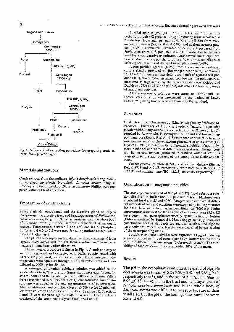

J. L. Gómez-Pinchetti and G. Garcia-Reina: Enzymes degrading seaweed cell walls 3
Table l . Ap-sia dactylotnelu. Haliotis coccinea canariensis, Lirtori- 40'C) and pH (6.0 and 7.2). RS: reducing sugars. CX: cold extract. na striara and Diadema antUarum. Specitic enzymatic activities of CMC: carboxymethyl cellulose. Alg: sodium alginate. -: not tested digestive enzymes at different conditions of temperature (25 and
PH Specific activity (pg RS mg protein-' h-') Protein conc. mean + SD
C X Agar CMC '41.5 (mg ml- ')
5 ' 40°C 25°C 40°C 25°C 40°C 25°C 40°C
A. dactylomela 6.0 63.4 76.2 7.2 38.6 58.1
Abalone acetone poirder 6.0 74.7 95.8 7.2 47.4 64.6
H. coccinea canariensis 6.0 8.9 - 7.2 6.9 -
L. striata 6.0 55.2 13.5 7.2 43.9 21.2
D. antiilarum 6.0 42.0 85.1 7.2 77.8 106.8
r ri 7 ~ n l i . r : n rlnrti>lnn*aln o n A Proridnmnnnr ntlnntirn ~ a ~ . e ,. ,,,,, ,,,,,,,..,., ,,., , ,.--,...,.., -..-....--. Agarase acriviry of crude extract from A. dactylornela. and purified (PA) and non-purified (NPA) agarases from P. atlantica. CX: cold extract; -: not tested
c,
Extract PH Agarase activity (pg RS h - ') D
- - m O
CX Agar Agarose - 0
25 "C m
4O0C 25°C 40 "C 25 "C 40°C E O
A. dacryiomela (crude) 6.0 393.1 472.4 120.3 291.4 118.0 261.8 7.2 239.3 360.2 122.7 286.4 - -
Agarase (PA)
Agarase (NPA) .6.0 119.5 138.5 37.0 90.0 33.3 77.0 7.2 111.9 54.9 76.0 109.0 67.5 51.1
7 O
Specific enzymatic activities and protein concentra- tion of crude extracts of Apljsia dactylomela, abalone acetone powder, Haliotis coccinea canariensis, Littorina striata and Diadema antiiiarum are $ven in iabie 1. Iorai protein concentration was 3- to 5-fold higher in A. dacty- lomela and H. coccinea canariensis than in the abalone acetone powder L. striata and D. antillarum extracts. These differences strongly affected the results for specific enzymatic activity on a protein concentration basis.
Al1 the enzymatic systems degraded the experimental substrates, although clear differences were observed in substrate-specificity and as a function of temperature and pH. Optimal temperature and pH for agarase and cellu- lase activity were 40°C and pH 6.0 for al1 the extracts, except for cellulase activity in Littorina striata (Table 1).
Highest alginate lyase activities were generally ob- tained at 40°C and pH 7.2, with some exceptions; AAP showed very high activities at 25 "C, although the highest was observed at 40 "C (pH 7.2). Diadema antillarum crude extracts displayed highest activity at 25"C, and Littorina striata at 40°C and pH 7.2 (Table 1).
Agarolytic activities from Apljsia dactylomela and both purified añd non-pürifid bacteria! soIiiCes, ex=
pressed as pg RS h-', are shown in Table 2. Optimum temperature and pH for bacterial agarases appeared to be the same as for crude extracts from phycophages (40°C and ó.0j. The agaroiyíic acíiviiy of A. c(ccciyi'üiííel~ cm& extracts was significantly higher against cold extract, agar and agarose, and varied as a function of temperature and pH (Table 2).
Fig. 2 shows the hydrolytic evolution of some extracts on different substrates. The extract from Ápiysia dacty- lomela had the highest RS-release rate on al1 the sub- strates except sodium alginate, and displayed significant differences between activities on cold extract and agar (Fig. 2A). No significant increases in RS were observed after 4 h of incubation.
Discussion
This is the first report which describes agarase activity from phycophages. Our results revealed similar optimum values for temperature and pH as those described for bacterial agarases, i.e., temperatures around 40 to 41 "C and i;H beween 5.G ünd 7.2 (Yüphe 1966, Duckiwrtk
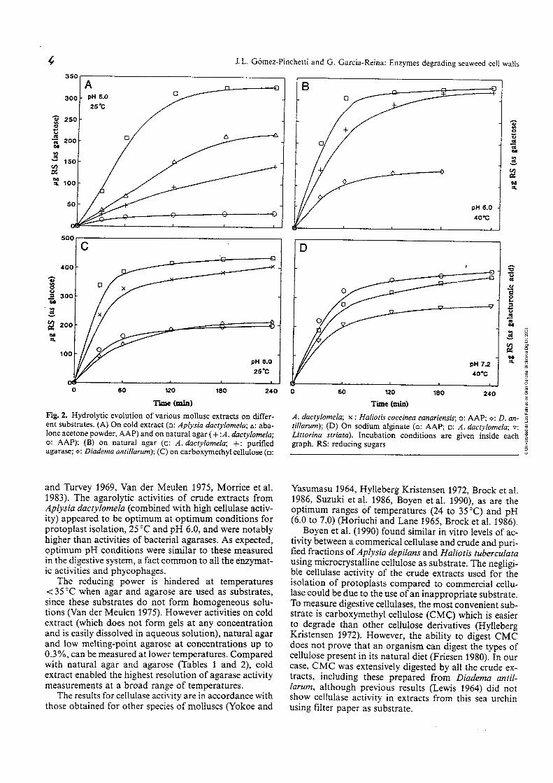
J. L. Gómez-Pinchetti and G. Garcia-Reina: Enzyrnes degradin~ seaweed cell walls
- n O - O a - - a u
2 - 2 M - =L
pH 6.0 - 4O0G
Fig. 2. Hydrolytic evolution of various mollusc extracts on differ- ent substrates. ( A ) On cold extract (o: Apíysia dactylomela; A: aba- lone acetone powder, AAP) and on natural agar ( + : A . dactylomela; o: AAP); ( B ) on natural agar (o: A. dactylomela; +: purified agarase; o: Diadema anriliarum); (C) on carboxymethyl cellulose (o:
and Turvey 1969, Van der Meulen 1975, Morrice et al. 1983). The agarolytic activities of crude extracts from Aplysia dactylomela (combined with high cellulase activ- ity) appeared to be optimum at optimum conditions for protoplast isolation, 25°C and pH 6.0, and were notably higher than activities of bacteria1 agarases. As expected, optimum pH conditions were similar to these measured in the digestive system, a fact cnmmnn tn a!! &e enzpat- ic activities and phycophages.
The reducing power is hindered at temperatures e 35 "C when agar and agarose are used as substrates, since these substrates do not form homogeneous solu- tions (Van der Meulen 1975). However activities on cold extract (which does not form gels at any concentration and is easily dissolved in aqueous solution), natural agar and low melting-point agarose at concentrations up to 0.39'0, can be measured at lower temperatures. Compared with natural agar and agarose (Sables 1 and 2), cold extract enabled the highest resolution of agarase activity measurements at a broad range of temperatures.
The results for cellulase activity are in accordance with those obtained for other species of molluscs (Yokoe and
a
A . dactylomela; x : Haliotis coccinea canariensis; o: AAP; o: D. un- ; tillarum); ( D ) On sodium aiginate (o: AAP; o: R. dactylomela; v: Littorina striata). Incubation conditions are given inside each graph. RS: reducing sugars 7
Yasumasu 1964, Hylleberg Kristensen 1972, Brock et al. 1986, Suzuki et al. 1986, Boyen et al. 1990), as are the optimum ranges of temperatures (24 to 35°C) 2nd pH (6.0 to 7.0) (Horiuchi and Lane 1965, Brock et al. 1986).
Boyen et al. (1990) found similar in vitro levels of ac- tivity between a commerical cellulase and crude and puri- fied fractions of Aplysia depilans and Haliotis tuberculata Iic;no rn;rrnrr~rct~ll;~~ pD I..l,ina ,r r.. -*--e- TL- - - - 1 : L --.s.& . . i i r i v r i j o u u u u b LLliz.uV>Zi a> J U ~ ~ L I ( I L C . 1 1 1 ~ L I C ~ ~ L ~ I -
ble cellulase activity of the crude extracts used for the isolation of protoplasts compared to commercial cellu- lase could be due to the use of an inappropriate substrate. To measure digestive cellulases, the most convenient sub- strate is carboxymethyl cellulose (CMC) which is easier to degrade than other cellulose denvatives (Hylleberg Kristensen 1972). However, the ability to digest CMC does not prove that an organism can digest the types of cellulose present in its natural diet (Friesen 1980). In our case, CMC was extensively digested by al1 the crude ex- tracts, including these prepared from Diadema antil- larum, although previous results (Lewis 1964) did not show cellulase activity in extracts from this sea urchin using filter paper as substrate.

J. L. Gómez-Pinchetti and C. Garcia-Reina: Enzymes degrading seaweed cell walls 5
Similar data on alginate lyase activities from Aplysia, Haliotis and Littorina species had been reported previ- ously (Franssen and Jeuniaux 1965, Nakada and Sweeny 1967, Elyakova and Favorov 1974. Boyen et al. 1 WO), but no[ for Uiaa'ema aniiíiarurn, aiíhough Cppley mi: Lasker ( 1 959) described degradation of sodium alginate solutions by other species of sea urchins.
Differences in agarase, cellulase and alginate lyase ac- tivities of crude extracts from Aplysia dactylomela, Halio- tis coccinea tanariensis, Littorina striata, and Diadema antillarum, are probably related to factors such as dietary preferences and availability of seaweeds. The highest al- ginate lyase activity was shown by the acetone powder from abalone (Haliotis sp.), and could be related to the preparation of extracts from individuals fed specific diets (Le., brown seaweeds such as Macrocystis pyrifera) for long periods.
The acetone powder method for the extraction of aba- lone entraiis was very effective, as reported previously by Polne-Fuller (1987). Substances which could be poten- tially harmful to plant tissues during the process of proto- plast isolation (Cocking 1972, Berliner 198 1) are removed from crude extracts by partial purification with ammoni- um su!phate mc! desa!ting. Nwerthe!essj new steps in the process of purification could be aimed at elucidating the behaviour and the biochemical properties of the different enzymes found in these phycophages.
Extracts from Aplysia dactylomela, commercial aba- lone acetone powder, Haliotis coccinea canariensis, Lit- torina striata and Diadema antillarum are highly effícient in digesting the complex carbohydrates in seaweeds cell walls, at a temperature (25 "C) and pH (6.0) optimal for the recovery of high protoplast yields and plating effi- cíencies of compIex seaweeds (PoIne-FuIler and Gibor 1987 b, Butler et al. 1990).
Acknowledgements. The authors are indebted to R. Armisén (His- panagar S.A.) for his valuable comments on this study. This work was supported by CICYT (Project MAR-1237191) and the Univer- sity of Las Palmas de Gran Canaria througb a grant to J. L. Gomez- Pinchetti. Support by Fundación Universitaria de Las Palmas and Air Europa are gratefully acknowiedged.
Literature cited
Benitez, L. V., Macaranas, J. M. (1979). Partial purification of a carrageenase from the tropical sea urchin Diadema setosurn. Proc. int. Seaweed Symp. 9: 353-359 [Jensen, A.; Stein, J. R. (eds.) Science Press, Princeton]
Berliner, M. D. (1981). Protoplasts of eukaryotic algae. Int. Rev. Cytol. 73: 1-19
Bjork, M., Ekman, P., Wallin, A., Pedersen, M. (1990). Effects of growth rate and other factors on protoplast yield from four species of Gracilaria (Rhodophyta). Botanica mar. 33: 433-439
Boven, C., Kloareg, B., Polne-Fuller. M., Gibor, A. (1990). Prepa- ration of alginate lyases from marine molluscs for protoplast isolation in brown algae. Phycologia 29: 173-181
Brock, V., Kennedy, V. S., Brock, A. (1986). Temperature depen- dency of carbohydrase activity in the hepatopancreas of thirteen estuarine and coastal bivalve species from the North American east coast. J. exp. mar. Biol. Ecol. 103: 87-101
Butler, D. M., Evans, L. V., Kloareg. B. (1990). Isolation of proto- plasts from manne macroalgae. In: Akatsuka, 1. (ed.) Introduc-
tion to applied phycology. SPB Academic Publishing. The Hague, p. 647-668
Butler, D. M., Dstgaard, K., Boyen, C.. Evans. L. V., Jensen, A.. Kloareg. B. (1989). Isolation conditions for high yields of proto- plasts from Latninaria saccharina and L. digitata (Phaeophyceae). j. exp. Bot. 49: i.237-1240
Chen. L. C.-M. (1987). Protoplast morphogenesis of Porphyra leu- costicta in culture. Botanica mar. 30: 399-403
Cheney. D. P., Mar, E., Saga, N., van der Meer, J. (1986). Proto- plast isolation and cell division in the agar-producing seaweed Gracilaria (Rhodophyta). J. Phycol. 22: 238-243
Cocking. E. (1972). Plant ceil protoplasts. Isolation and develop- ment. A. Rev. PI. Physiol. 23: 29-50
Davison, 1. R., Polne-Fuller, M. (1990). Photosynthesis in proto- plasts of Macrocystispyrifera (Phaeophyta). J. Phycol. 26: 384- 387
Duckworth, M., Turvey, J. R. (1969). An extracellular agarase from a Cytophaga species. Biochem. J. 113: 139-142
Elyakova, L. A., Favorov, V. V. (1974). Isolation and certain prop- erties of alginate lyase VI from the mollusk Littorina sp. Biochim. biophys. Acta 358: 341 -354
Eppley, R. W., Lasker, R. (1959). Alginase in the sea urchin Strong,vlocentrotus purpuratus. Science, N.Y. 129: 214-215
Franssen, J., Jeuniaux, C. (1965). Digestion de I'acide alginique chez les invertebres. Cah. Biol. mar. 6: 1-21
Friesen, J. A. (1980). The structure of the gut of Mysis stenolepis and the mechanism of cellulose digestion. M. Sc. thesis. Dalhousie University, Nova Scotia
Galli, D. R., Giese, A. C. (1959). Carbohydrate digestion in a herbivorous snail, Tegula funebralis. J. exp. 2001. 140: 415-439
Horiuchi, S., Lane, C. E. (1965). Digestive enzymes of the crys- talline style of Strornbusgigas Linne. 1. Cellulase and some other carbohydrases. Biol. Bull. mar. biol. Lab., Woods Hole 129: 273-281
Hylleberg Kristensen, J. (1972). Carbohydrases of some marine in- vertebrates with notes on their food and on the natural occur- rence of the carbohydrates studied. Mar. Biol. 14: 130-142
Jordan, P., Kloareg, B., Vilter, H. (1991). Detection of vanadate- dependent bromoperoxidases in protoplasts from the brown algae Laminaria digitata and L. saccharina. J. P1. Physiol. 137: 520-524
Kidby, D. K., Davidson, D. J. (1973). A convenient femcyanide estimation of reducing sugars in the nanomole range. Analyt. Biochem. 55: 321 -325
Kloareg, B., Quatrano, R. S. (1988). Structure of the cell walls of marine algae and ecophysiological functions of the matrix polysaccharides. Oceanogr. mar. Biol. A. Rev. 26: 259-315
Kreger, D. R. (1962). Cell walls. In: Lewin, R. D. (ed.) Physiology and biochemistry of algae. Academic Press, New York, p. 31.5- 335
Lanaya, M., Sochas, C., kápiie, W. (ii98ój. A new procedüie für determining the heterogeneity of agar polymers in the cell wall of Gracilaria spp. (Gracilariaceae, Rhodophyta). Can. J. Bot. 64: 579-585
Le Gall, Y., Braud, J. P., Kloareg, B. (1990). Protoplast production in Chondrus crispus gametophytes (Gigartinales, Rhodophyta). P1. Cell Rep. 8: 582-585
Lewis, J. B. (1964). Feeding and digestion in the tropical sea urchin Diadema antillarum Philippi. Can. J. Zool. 42: 549-557
Liu, W. S., Tang, Y. L., Liu, X. W., Fang, T. C. (1984). Studies on the preparation and on the properties of sea snail enzymes. Hydrobiologia 116/117: 319-320
Lowry, O. H., Rosebrough, N. J., Farr, A. L., Randall, R. J. (1951). Protein measurement with the Folin phenol reagent. J. biol. Chem. 193: 265-275
Morrice, L. M., McLean, M., Williamson, F. B., Long, W. F. (1983). /3-agarases 1 and 11 from Pseudomonas atlantica: purifications and some properties. Eur. J. Biochem. 135: 553-558
Morrice, L. M., McLean, M., Williamson, F. B., Long, W. F. (1984). /3-agarases from Pseudomonas atlantica. Hydrobiologia 1161 117: 576-579

6 J. L. Gómez-Pinchetti and G. Garcia-Reina: Enzymes degrading seaweed cell waIIs
Nakada. H. 1.. Sweeny. P. C. (1967). Alginic acid degradation by eliminases from abalone hepatopancreas. J. biol. Chem. 242: 845-851
Nelson. N. (1944). A photometric adaptation of the Somogyi meth- od for the deteriiiiiiatiün üf giüc-use. 1 bioi. Chem. 153: 375-380
Nisizawa. K.. Fujibayashi, S.. Kashiwabara, Y. (1968). Alginate lyases in the hepatopancreas of a marine mollusc, Dolabella auricula Solander. J. Biochem. 64: 25-37
Onishi. T.. Suzuki. M.. Kikuchi, R. (1985). The distnbution of polysaccharide hydrolase activity in gastropods and bivalves. Bull. Jap. Soc. scient. Fish. 51: 301 -308
Polne-Fuller, M. (1987). Preparation of enzymes. In: Indergaard, M., Dstgaard, K., Guiry, D. (eds.) Seaweed protoplast and tis- sue culture. COST 48. European Economic Community, Brus- sels. Proceedings of Workshop on Biotechnical Methods in sea- weed cultivation. E.E.C., Trondheim. p. 29-30
Polne-Fuller, M., Biniaminov, M., Gibor, A. (1984). Vegetative propagation of Porphyra perforara. Hydrobiologia 116/117: 308-313
Polne-Fuller, M., Gibor, A. (1987a). Microorganisms as digestors of seaweed cell walls. Hydrobiologa 151/152: 405-409
Polne-Fuller, M., Gibor. A. (1987 b). Tissue culture of seaweeds. In: Bird. K. T., Benson, P. H. (eds.) Seaweed cultivation for renew- able resources. Vol. 16. Elsevier, Amsterdarn, p. 219-239
Preston, J. F., Romeo, T.. Bromley, J. C.. Robinson, R. W, Aldnch, H. C. (1985). Alginate iyase-secreting bacteria Underkofler, L. A. (ed.) Developments in industrial microbiology. Society for Industrial Microbioiogy, Arlington. Virginia, p. 727-740
Saga, N., Polne-Fuller, M.. Gibor, A. (1986). Protoplasts from seaweeds: production and fusion. In: Barclay, W. R., McIntosh, R. P. (eds.) Algal biornass technologies: an interdisciplinary perspective. Vol. 83. J. Cramer, Berlin, p. 37-43
Saga, N., Sakai, Y (1984). Isolation of protoplasts frorn Laminaria and Porphyra. Bull. Jap. Soc. scient. Fish. 50: 1085
Smith. R. G.. Bidwell. R. G. S. (1989). Inorganic carbon uptake by photosynthetically active protoplasts of the red macroalga Chondrus crispcu. Mar. Biol. 102: 1-4
Somogyi. M. (1952). Notes on sugar determination. J. biol. Chem. 193; 19-23
Suzuki. M.. Watanabe, Y.. Tanaka. A.. Ohnishi. T. (1986). The polysaccharide degradation activity in the apple snail. Bull. Tokai reg. Fish. Res. Lab. 120: 53-60
Tokuda. H.. Kawashima. Y. (1988). Protoplast isolation and culture of a brown alga, Undariapinnatifida. In: Stadler, T., Mollion, J., Verdus, M.-C., Karamanos. Y.. Morran, H., Christiaen, D. (eds.) Algal biotechnology. Elsevier Applied Science, London, p. 152-157
Torzilli, A. P.. Andrykovitch. G. (1980). Cell wall degrading en- zymes produced by the salt marsh fungus Buergenerula sparti- nae. Botanica mar. 23: 645-650
Van der Meulen, H. J. (1975). The enzymatic hydrolysis of agar by Cytophagaflevensis sp. nov. Ph. D. thesis. University of Gronin- gen. The Netherlands
Yaphe, W. (1966). The purification and properties of an agarase from a marine bacterium, Pseudornonas atlantica. Proc. 5th int. Seaweed Syrnp. 5: 333-335 poung, E. G., McLachlan, J. L. (eds.) Pergamon Press, Oxford]
Yokoe, Y, Yasumasu, 1. (1964). The distribution of cellulase in invertebrates. Comp. Biochem. Physiol. 13: 323-338
Zhu, R. (1983). A comparative study of rnarine algae cell-wall de- composition by the digestive enzimes irolatd from three species of marine gastropods. Coll. ocean. Wks. Qingdao (China) 6: - 122- 126
Communicated by J. M. Pérks, Marseille


Acid deoxyribonuclease activity in crude extracts
from marine phycophages used for seaweed
protoplast isolation
JUAN LUIS GOMEZ-PINCHETTI and GUILLERMO GARCIA-REINA
Instituto de Algología Aplicada, Dpto. Biología, Universidad de Las Palmas
de Gran Canaria, Box 550, Las Palmas, Islas Canarias, Spain
Running title: DNase in crude extracts from marine phycophages
Key wordr: ph ycophages, seaweed, protoplasts, DNase.
SUMMARY
Deoxyribonuclease activity (DNase) was studied in crude extracts prepare.
from the digestive tracts of the marine molluscs Aplysia dactylomela Rang,
Haliotis coccinea canariensis Nordsieck, Littorina striata King et Broderip, the
eckhderrn DtuÚ!em~ ~.n t i lhm ?!Upi, md a cnmmcrcidilly av-ilahle abalone
acetone powder (AAP; Sigma, Ref. A-7514) to evaluate their toxic effects
when useú for seaweed protoplast isolation. DNase was detected at pH 5.0,
in presence of ~ g ~ + , in al1 the crude extracts except in those obtained from
the digestive gland of A. dactylomela. At pH 6.0, activity was not detected.
Maximal specific activities were measured in L. striata and D. antillarum crude
extracts. AAP showeú the lowest specific activity of al1 the assayeú extracts.
The pH in the different digestive organs of these phycophages ranged
from 5.0 to 7.0, measureú in vivo, providing new information about the
optirnum values at which digestion occurs and, together with results on DNase
activities, those which should be used to degrade seaweed cell walls.
RESUMEN
Actividad deoxiribonucleasa en extractos crudos de ficófagos
marinos empleados para el aislamiento de protoplastos de
macroaigas.~ctividad deoxiribonucleasa (DNasa) fue estudiada en extractos
crudos, preparados de los sistemas digestivos, de los moluscos Aplysia
dactylomela Rang, Haliotis coccinea canariensis Nordsieck, Littorina striata
King et Broderip, el equinodermo Diadema antillarum Phillipi, y un extracto
de Haliotis sp. en forma de polvos acetónicos, comercialmente disponible
(abalone acetone powder, AAP; Sigma, Ref. A-75 l4), para evaluar sus efectos
tóxicos al ser empleados en el aislamiento de protoplastos de macroalgas.
DNasa fue detectada a pH 5.0, en presencia de ~ g ~ + , en todos los extractos
crudos exceptuando aquellos obtenidos de la glándula digestiva de A..
dactylomela. A pH 6.0, actividad DNasa no fue detectada en ninguno de los
extractos. Las actividades específicas máximas fueron medidas en los extractos
crudos de L. striata y D. antillarum. El AAP mostró la actividad específica más
baja de todos los extractos ensayados.
El pH en los diferentes órganos digestivos de estos ficófagos, medido
en vivo, vari6 de 5.0 a 7.0, aportando nueva información sobre los valores
óptimos de pH en los que la digestión tiene lugar y, junto a los resultados
sobre la actividad DNasa, aquellos que deben ser usados para la digestión de la Yud =hla- rnacr "aas
Palabras clave: ficófagos, macroalgas, protoplastos, DNasa.

DNase in crude exnactsfrom marine phycophages
INTRODUCTION
The potential toxicity of proteases, lipases, peroxidases and deoxyribonucleases
of crude extracts from the digestive tracts of a number of phycophages on the
viability of plant protoplasts has been commented by several authors (see
TRIBE, 1955; SCHENK and HILDEBRANDT, 1969; BERLINER, 198 1 ;
COCKING, 1972; FITZSIMONS and WEYERS, 1985; BUTLER et al., 1990).
However, few attempts have been made on quantification and characterization
of such potentially toxic enzymes.
Most of the crude extracts used for seaweed protoplast isolation are
extracted from herbivorous marine molluscs and echinoderms such as Aplysia, m
Haliotis, Littorina or Diadema species (SAGA and S AKAI, 1984; CHEN, 1987; D
- - m O
BUTLER et al., 1989; BOYEN et al. 1990; GOMEZ-PINCHETTI and - - O
m
GARCIA-REINA, in press). Low protease activity has been detected in crude E
O
extracts of Aplysia depilans and Haliotis tuberculata (BOYEN et al., 1990). n
E
Lipases and proteases have been measured in other marine molluscs and - a
2
n
fishes (CLIFFORD et al., 1982; FERAL, 1989; TE0 and SABAPATHY, n n
1990). Deoxyribonucleases in the marine environment have been studied only 3
O
in some fishes and invertebrates (ASHE et al., 1965; RASSKAZOV et al.,
1975; DOMINGO et al., 1986; CHOU and LIAO, 1990; SMTKVERN et 4 \ 1 C ,... ,,ll..,,, f/-CADC A T C A C .-,..A A h m n h T I \ P - T d., 1YYU) 1IlGlUullig 2 I G W iiNJliUJLJ \UJ2Ur\Ufil DUO 411U Al1 I V L I V U L V V 1
1963), none of which are used for seaweed cell wall degradation and
protoplast isolation. These tissues of marine origin have revealed at least two
groups of DNA-depolymerizing enzymes with quite different properties,
namely acid and alkaline DNases (GEORGATSOS and ANTONOGLOU,
1963; RASSKAZOV et al., 1975).
In this study, we report on the acid DNase activity of crude extracts
from the most common phycophages used for the isolation of seaweed
protoplasts.

MATERIAL AND METHODS
Source of enzymes
Cmde extracts from Aplysia dactylomela, Haliotis coccinea canariensis, LittorinQ
striata and Diadema antillanun were prepared within 24 h of collection.
Abalone acetone powder (AAP; Sigma, St. Louis, USA, Ref. A-75 Id), a cmde
extract prepared from Haliotis sp. entrails was also used as a source of
enzymes.
Preparation of crude extracts
The oesophagus and the digestive gland (separately) from Aplysia dactylomela
the digestive tract and hepatopancreas from _Hnlioh cnrclnon cnn,nr'on~~is, the
gut from Diadema ant i l lam and the whole body of Littorina striata (after
shell removal), were used to prepare the crude extracts. Temperatures
between O and 4°C and 0.1 M phosphate buffer @H 6.0) were used to
perform al1 operations (except where indicated).
The pH of the different glands and organs were measured with a
microelectrode (PHR-146, Lazar Res. Lab., CA, USA) immediately after
dissection.
Cmde extracts were prepared, as previously described by GOMEZ-
PINCHETTI and GARCIA-REINA (in press) , with some modifications.
Glands and organs were homogenized and extracted with buffer, supplemen-
ted with EDTA Na, (2.0 mM), in a mortar under liquid nitrogen. The
extraction was carried out in 0.1 M phosphate buffer @H 6.0) supplemented
urith. YnTA Ni2 (2.9 mhl; 5 .O EL g Us~üe-'1. Hom~geiizies weie s q ü e z d
through a 170 pm nylon mesh and centrifuged at 27,000 x g for 30 min.
Ammonium sulphate was then added to the supernatant to 80% saturation.
After several hours equilibration, the suspension was centrifuged at 37,000 x
g for 20 min. Pellets were resuspended in buffer and passed through a pre-
packed Sephadex G-25 column (PD- 10, Pharmacia LKB, Uppsala, Sweden)
equilibrated with 20 mM Bis-tris buffer @H 6.0) dissolved in seawater, 1 .O M

sodium acetate buffer @H 5.0), or 0.1 M phosphate buffer @H 6.0) for
desalting and buffer exchange.
AAP was dissolved in buffer (2.0% wlv), equilibrated for several hours
between O and 4OC, centrifuged at 27,000 x g for 15 min and passed through
a Sephadex G-25 column.
Protein concentration was determined by the method of LOWRY et al.
(1951) using bovine serum albumin as the standard.
Quantpcation of DNase activity
A DNA (Ref. D-3664, Sigma) solution (0.004 % , final concentration) dissolved
in distilled water and supplemented with the addition of 1. O M sodium acetate
buffer (0.01 %, pH 5.0) and 0.1 M MgSO, (0.005 %) was used as substrate.
Activity was obtained by measuring the maximun linear rate at A , - , in a
spectrophotometer (UV-160A, Shimadzu, Kyoto, Japan) of a mixture contai-
ning 2.5 mL of substrate equilibrated at 25OC and 0.5 mL of the crude extract.
One unit (Kunitz unit) will produce a AA, of 0.001 per min per mL
at pH 5.0 at 25°C using DNA as the substrate (klg2+ concentration = 4.2
mM). Values were corrected to the standard activity of DNase 1 (EC 3.1.2 1.1 ; .
2,000 Uníts per vial, Ref. D-4263, Sigma).
The pH of the different digestive systems ranged between 5.0 and 7.0 (Table
l), coincident with the optimum values for in vitro activities of agarase,
cellulase, alginate lyase and other polysaccharidases from the digestive systems
of those wd other marine herbivores (ONISHI et al., 1985; GOMEZ-
PINCHETTI and GARCIA-REINA, in press) . DNase activities of the different crude extracts are shown in Table 1.
Al1 the crude extracts degraded DNA in the presence of Mg2+ at pH 5.0,
except those prepared from the digestive gland of Aplysia dactylomela. At pH

Crude extract PH Prot. Conc.
(mean f sd) mg ml-'
DNase activity
Units ml-'
--
Specific Activity
Units mgl prot
Aplysia dactylomela
Oesophagus 5.58 f 0.42 6.2 :t 1.6
Digestive gland 5.85 f 0.19 7.6 :i: 0.6
Haliotis coccinea
Hepatopancreas 6.56 f 0.15 6.7 :i: 1.2
Littorina striata
Homogenate
Diadema antillarum
Gut 6.92 f 0.18 1.9 f 0.5
AAP nm 1.9 :t 0.4
TABLE 1. - pH, after d.issection, of the different digestive systerns used as enzymatic sources, and DNase activity obf crude extracts at two
different pH [5.0(1.O M sodium acetate) and 6.0(20 mM Bis-Tris in seawater or O. 1 M phosphate)]. T= 25 "C. AAP: Abalone acetone powder;
-: no activity detected; rim: not measured.
DNase in crude extractsfrom marine phycophages
6.0 no DNase activity was detected in any of the different crude extracts
regardless buffer (20 mM Bis-tris in seawater or 0.1 M phosphate). Protein
concentration of the different extracts strongly affected the specific activities
(Table 1). The significantly higher DNase specific activities from Littorina
striata and Diadema antillarum might be explained by enzymatic contamination
from other organs. In L. striata DNase activity was measured from the whole
body of the animal. In D. antillanun, although the gut is easily distinguishable,
it was very difficult to excise the digestive system without contact from other
fluids and organs. Abalone acetone powder showed the lowest specific activity
of al1 the assayed crude extracts.
Adjustment of the pH of the enzymatic solution to vaíues between 6.0
and 7.0,depending on the phycophage and the seaweed to be digested, would
avoid the action of DNases and be closely related to the optimum conditions
in which natural digestion occurs in these phycophages and thus maximum
activity of the polysaccharidases. pH values higher than 8.0, optimal for the
action of alkaline DNases (GEORGATSOS and ANTONOGLOU, 1963;
RASSKAZOV et al., 1975), would be too high for the optimal action of cell
wall degrading enzymes. The addition of inhibitors (Le., potassium dextran
sulphate) to the enzymatic solutions to prevent the effects of ribonucleases,
employed by some authors (FUJIMURA et al., 1989; WAALAND et al., 1990;
PKEEDY =c! FUJI'TA, E$!), W O U ! ~ he cme.cescary if the pH ~f h e sol~tim
is adjusted close related to 6.0. New steps in specific enzyme partial
purification would be desirable made to avoid toxic effects (see SCHENK and
HILDEBRANDT, 1969; BUTLER et al., 1990), however, the risk of loosing
the degrading effects of other additional enzymes present in the crude extracts
would be considerable. These data give us new information to improve
seaweed protoplast production, cell viability and thallus regeneration.
ACKNOWLEDGEMENTS
We are grateful to R. Armisén (Hispanagar S.A.), Dr. P. Sosa and Dr. A.

Critchley for their valuable advice in the present study. This work was
a grant to J.L. Gómez-Pinchetti.
ASHE, H., E. SEAMAN, H. VAN VUNAKIS and L. LEVINE. - 1965. Characterization of deoxyribonuclease of Mustelus canis liver. Biochim. Biophys. Acta, 99: 298-306.
BERLINER, M. - 1981. Protoplasts of eukaryotic algae. Intern. Rev. of Cytology, 73: 1-19.
BOYEN, C., B. KLOAREG, M. POLNE-FULLER and A. GIBOR - 1990. Preparation of alginate lyases from marine molluscs for prt>tlo,nlast isnlation in brown algae. Phycologia, 29: 173-181. ,,
D
- =
BUTLER D.M., L.V. EVANS and B. KLOAREG. - 1990. Isolation of proto- m O
plasts from marine macroalgae. In: 1. AKATSUKA (ed.): Introduction to - e m
Applied Phycology, pp. 647-668. SPB Academic Publishing, The Hague. E
O
g BUTLER D.M. ,K. 0STGAARD, C. BOYEN, L.V. EVANS, A. JENSEN andl n
E
B. KLOAREG. - 1989. Isolation conditions for high yields of protoplasts a
from Laminaria saccharina and L. digitata (Phaeophyceae). J . Exp. Bot., 40: n n
1237- 1246. 3
O
CHEN L.C.-M. - 1987. Protoplast morphogenesis of Porphyra leucosticta in culture. Bot. Mar., 30: 399-403.
CHOU M.-Y. and T.-H. LIAO. - 1990. Shrimp hepatopancreatic deoxyribonu- ciease - purificauon and cnaracterization as well as comparison with bovine pancreatic deoxyribonuclease. Biochim. Biophys. Acta, 1036: 95-100.
CLIFFORD C., J. WALSH, N. REIDY and D.B. JOHNSON. - 1982. Diges- tive enzymes and subcellular localization of disaccharidases in some echinoderms. Comp. Biochem. Physiol., 71B: 105- 1 10.
COCKING E. C. - 1972. Plant cell protoplasts-isolation and development. Ann. Rev. Plant Physiol., 23: 29-50.
DOMINGO A-, M. CERVERA, E. MARTIN and R. MARCO. - 1986. Bio- chemical characterization and developmental behavior of Artemia embryonic and nauplial deoxyribonucleases. Biochemistry , 25: 4 125-4 13 1.

DNase in crude extrac&fiom manne phycophages
FERAL J.-P. - í989. Áctivity of the principal digesuve enzymes iñ the detritivorous apodous holothuroid Leptosynapta galliennei and two other shallow water holothuroids. Mar. Biol., 101 : 367-379.
FITZSIMONS P. J. and J.D.B. WEYERS. - 1985. Properties of some enzymes used for protoplast isolation. In: P.E. PILET (4.): The physiological properties of plant protoplasts, pp. 12-23. Springer-Verlag, Berlin - Heidelberg .
FUJIMURA T., T. KAWAI, M. SHIGA, T. KAJIWARA and A. HATANA KA. - 1989. Regeneration of protoplasts into complete thalli in the marine green alga Ulva pertusa. Bull. Jap. Soc. Sci. Fish., 55: 1353-1359.
GEORGATSOS J. G. and 0. ANTONOGLOU. - 1963. Deoxyribonucleases in tissues of marine origin. Enzymologia, 27: 141-150.
GOMEZ-PINCHE'ITI J.L. and G. GARCIA-REINA. - 1993. Marhe phyco- phages enzymes that degrade cell walls of seaweeds. Mar. Biol., (in press)
m
- m o
LOWRY, O.H., N.J. ROSEBROUGH, A.L. FARR and R.J. RANDALL. - - 0 m
195 1. Protein measurement with the Folin phenol reagent. J. Biol. Chem., E
193: 265-275. O
o n
E
ONISHI T., M. SUZUKI and R. KIKUCHI. - 1985. The distribution of a
polysaccharide hydrolase activity in gastropods and bivalves. Bull. Jap. Soc. n n
Sci. Fish. 51: 301-308. n
3
O
RASSKAZOV V. A., V.V. PIROZHNIKOVA and V. V. GALKIN. - 1975. Some properties and specificity of deoxyribonucleases from marine invertebrates and fishes. Comp. Biochem. Physiol., 5 1B: 343-347.
REDDY C.R.K. and Y. FUJITA. - 199 1. Regeneration of plantlets from Enteromorpha (Ulvales, Chlorophyta) protoplasts in axenic culture. J. Appl. Phycol., 3: 265-275.
SAGA N. and Y. SAKAI. - 1984. Isolation of protoplasts from Laminada and Porphyra. Bull. Jap. Soc. Sci. Fish., 50: 1085.
SCHENK R. U. and A. C. HILDEBRANDT. - 1969. Production of protoplasts from plant cells in liquid culture using purified commercial cellulases. Crop. Sci., 9: 629-63 1.
STRETKVERN K.O., A.J. RAAE and B.T. WALTHER. - 1990. Purification and physicochemical properties of deoxyribonuclease from pyloric caeca of Atlantic cod (Gadus morhua L.). Fish. Physiol. Biochem., 8: 529-539.
TE0 L.H. and V. SABAPATHY. - 1990. Preliminary report on the digestive enzymes present in the digestive gland of Pema viridis. Mar. Biol., 106: 4'3-497.
TRIBE H.T. - 1955. Studies on the physiology of parasitism. XIX. On the killing of plant cells by enzymes from Botrytis cinerea and Bacteriwn aroideae. Ann. Bot., 19: 351-371.
WAALAND J.R., L. G. DICKSON and B.A. WATSON. - 1990. Protoplast iso lation and regeneration in the marine red alga Porphyra nereocystis. Planta, 18 1 : 522-528.


Br. phycol. J. 27: 40 1-407 1 December 1992
Protoplast isolation from Uiva rigida (Cniorophyf a j
By MATS BJORK*, JUAN LUIS GÓMEZ-PINCHETTI?, GUILLERMO GARC~A-REINA? and MARIANNE PEDERSÉN*
*Department of Physiological Botany, Box 540 S-751 21 Uppsala, Sweden f Marine Plant Biotechnology Laboratory, University of Las Palmas, Box 550, E-35017,
Las Palmas, Spain
High numbers of protoplasts were isolated from wild and cultivated thalli of Ulva rigida. Optimal conditions for protoplast release were obtained with 1.5% Abalone Acetone Powder and 1.5% Cellulysin in 0.4 M mannitol. Treatment for 30 min with hypotonic (0-8 M) solution more than doubled the yield. Growth conditions prior to enzyme treatment also influenced the yield greatly. Protoplasts were photosynthetically active, and 70-90% of the protoplasts were estimated to be viable.
Protoplasts of higher plants are valuable for physiological, genetic, and biochemical studies (Eriksson, 1985). The photosynthesis of protoplasts has been studied for a number of species, like spinach (Nishimura & Akazawa, 1975) and wheat (Edwards et al., 1978). The photosynthetic activity of proto- plasts from the mesophyll compared to that of bundle sheath cells has been used to study the localization of photosynthetic enzymes in severa1 C, plants (Kanai & Edwards, 1973; Gutierrez et al., 1974). However, there are few reports of physiological studies using seaweed protoplasts. Protoplasts from the red macriz!ga Chnndrm rrispus have been used to study inorganic carbon uptake (Smith & Bidwell, 1989). It has also been suggested that protoplasts from Macrocystis pyijera can be =sed as a m&! fer pkysie- logical research (Davison & Polne-Fuller, 1990), and protoplasts from this species have recently been used for studies on bromo- peroxidases (Butler et al., 1990).
Ulva rigida is a common, widely distri- buted, green seaweed that has been used in many physiological studies (Zavodnik, 1987; MacFarlane & Smith, 1984; Fujita, Wheeler & Edwards, 1988), but to our knowledge no reports on protoplast isolation from this species have been published. However,
protoplasts have been isolated from Ulva Iinza (Zhang, 1983), Ulva pertusa (Saga, 1984), and Ulva Iactuca (Chou & Lu, 1989). Complete thalli have been obtained from protoplasts of Ulva fasciata, U. conglobata and U. pertusa (Reddy, Migita & Fujita, 1989; Fujimura et al., 1989a). Experiments on immobilization (Fujimura et al., 1989b), fusion (Reddy & Fujita, 1989) and produc- tion of bioflavour compounds (Fujimura & Kajiwara, 1990) of UIva protoplasts have been reported recently. However, no physio- logical experiments have been reported for these protoplasts. In the present study we describe a method to isolate large amounts of photosynthetically active protoplasts from Ulva rigida. Factors such as standardized precultivation conditions, and preplasmo- !y& that inflxnce ?he yk!d of protoplasts are considered.
MATERIALS AND METHODS
Ulva ngida C.Ag. was collected in May 1991 from the harbour of Taliarte, Gran Canaria, (Canary Islands, Spain). Plants were taken from populations growing on ropes near the surface under constant immersion. After collection, the plants were thoroughly cleaned in sterile sea- water and then maintained in 20 1 tanks with ninning sea-water or cultured in sea-water in 4 1 bottles with aeration. Fluorescent lamps (Thorn
O 1992 Bntish Phycological Society
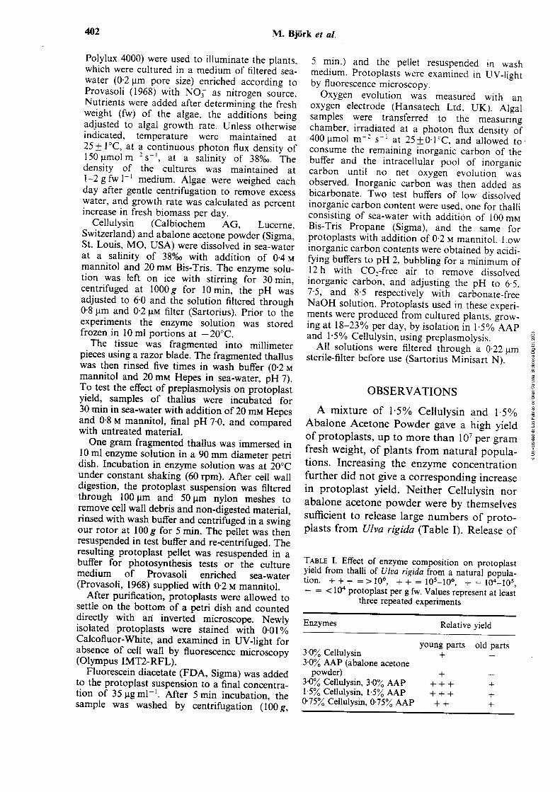
402 M. Bjork et al.
Polylux 4000) were used to illuminate the plants, which were cultured in a medium of filtered sea- water (0.2 prn pore size) enriched according to Provasoli (1968) with NO; as nitrogen source. Nutrients were added after determining the fresh ,,,P. ,-. (fw) vf i k algae, tire aciditions being adjusted to alga1 growth rate. Unless otherwise indicated, temperature were maintained at 25_+ 1°C, at a continuous photon flux density of 150 pmolm-' S-', at a salinity of 38%0. The density of the cultures was maintained at 1-2 g fw 1-' medium. Algae were weighed each day after gentle centrifugation to remove excess water, and growth rate was calculated as percent increase in fresh biomass per day,
Cellulysin (Calbiochem AG, Lucerne, Switzerland) and abalone acetone powder (Sigrna, St. Louis, MO, USA) were dissolved in sea-water at a salinity of 38'360 with addition of 0.4 M mannitol and 20 mM Bis-Tris. The enzyme solu- tion was left on ice with stirring for 30 min, centrifuged at lOOOg for 10 min, the pH was adjusted to 6.0 and the solution filtered through 0.8 pm and 0-2 p~ filter (Sartorius). Prior to the experiments the enzyme solution was stored frozen in 10 m1 portions at - 20°C.
The tissue was fragmented into millimeter pieces using a razor blade. The fragmented thallus was then rinsed five times in wash buffer (0.2 M
mannitol and 20 mM Hepes in sea-water, pH 7). To test the effect of preplasmolysis on protoplast yield, samples of thallus were incubated for 30 min in sea-water with addition of 20 mM Hepes and 0-8 M mannitol, final pH 7.0, and compared with untreated material.
One gram fragmented thallus was immersed in 10 m1 enzyme solution in a 90 mm diameter petri dish. Incubation in enzyme solution was at 20°C under constant shaking (60 rpm). After cell wall digestion, the protoplast suspension was filtered through 1 O0 pm and 50 pm nylon meshes to remove cell wall debis and non-digested material, iinsed wiih wash buffer and centrifugea in a swing our rotor at lOOg for 5 min. The pellet was then resuspended in test buffer and re-centrifuged. The resulting protoplast pellet was resuspended in a buffer for photosynthesis tests or the culture medium of Provasoli ennched sea-water (Provasoli, 1968) supplied with 0.2 M mannitol.
After purification, protoplasts were allowed to settle on the bottom of a petn dish and counted directly with an inverted microscope. Newly isolated protoplasts were stained with 0.01% Calcofluor-White, and examined in UV-light for absence of cell wall by fluorescence microscopy (Olympus IMTZRFL).
Fluorescein diacetate (FDA, Sigma) was added to the protoplast suspension to a final concentra- tion of 35 pg ml-'. After 5 rnin incubation, the sample was washed by centrifugation (IOOg,
5 rnin.) and the pellet resuspencled in wash medium. Protoplasts were examined in UV-light by fluorescence microscopy.
Oxygen evolution was measured with an oxygen electrode (Hansatech Ltd. UK). Alga1 sampies were transferred to the measuring chamber, irradiated at a photon flux density of 400 pmol m-' S- ' at 25&0.1"C, and allowed to consume the remaining inorganic carbon of the buffer and the intracellular pool of inorganic carbon until no net oxygen evolution was observed. Inorganic carbon was then added as bicarbonate. Two test buffers of low dissolved inorganic carbon content were used, one for thalli consisting of sea-water with addition of 100 mM Bis-Tris Propane (Sigma), and the same for protoplasts with addition of 0.2 M rnannitol. Low inorganic carbon contents were obtained by acidi- fying buffers to pH 2, bubbling for a minimum of 12 h with COI-free air to rernove dissolved inorganic carbon, and adjusting the pH to 6.5. 7.5, and 8.5 respectively with carbonate-free NaOH solution. Protoplasts used in these experi- ments w r e predured f r ~ m cü!türed plaiiis, giuw- ing at 18-23% per day, by isolation in 1.5% AAP and 1.5% Cellulysin, using preplasmolysis.
D
N -
Al1 solutions were filtered through a 0.22 pm sterile-filter before use (Sartorius Minisart N). - -
0 m
OBSERVATIONS O
A mixture of 1.5% Cellulysin and 1.5% Abalone Acetone Powder gave a high yield of protoplasts, up to more than 107 per gram 3 fresh weight, of plants from natural popula- 5 tions. Increasing the enzyme concentration further did not give a corresponding increase in protoplast yield. Neither Cellulysin nor abalone acetone powder were by themselves sufficient to release large numbers of protc- plasts from UIva rigida (Table 1). Release of
TABLE 1. Effect of enzyme composition on protoplast yield from ihaiii oí ú'iva rigiua irom a natural popula- tion. + + + = > lo6, + + = lo5-lo6, - = lo4-lo5, - = < lo4 protoplast per g fw. Values represent at least
three repeated experiments
Enzymes Relative yield
young parts old parts 3.0% Cellulysin + - 3.0% AAP (abalone acetone
powder) + - 3.0% Cellulysin, 3.0% AAP + + + + 1 5 % Cellulysin, 1.5% AAP + + + + 0.75% Cellulysin, 0.75% AAP + + +


404 M. Bjork et al.
O 5 10 15 20 2 5 3 0
Growth rate (% per 24 h)
FE. 2. Yield of protoplasts from Ulvu r-igirkr from plants cultured at diKerent growth rates. Each growth rate was tested once.
protoplasts started after about 1-2 h of incu- bation. The protoplasts were spherical and ranged between 10-30 pm in diameter (Fig. 1). The release of protoplasts continued for up to 24 h, but after 8-12 h large numbers of protoplasts started to lyse (visual observation). No cell wall material was found when protoplasts were observed with Calcofluor in UV light. Preplasmolysis prior to the enzyme incubation was effective in enhancing the yield by a factor of about 2-5 (Fig. 2), and there was no observed differ- ence in viability by this treatment. The growth rate of cultured algae was also important for the yield of protoplasts, with an optimum growth rate of about 20% biomass increase per day (Fig. 2). Only the young parts from natural populations plants
were used for protoplast isolation. The yield d e ~ r e ~ s e d rapidly when plants were stored in tanks, so that after 3 days of storage without growth, except on one occasion, no protop- lasts were released from this plant material.
The rate of photosynthetic oxygen evolu- tion in protoplasts from Ulva rigidrr was shown to be pH dependent (Fig. 3b). Photosynthetic oxygen evolution was higher at pH 6.5 than in 7.5 and 8.5. At pH 6.5, photosynthesis of protoplasts as well as thalli was close to saturation at a concentration of 400-500 V M HCOc [Fig. 3(a)]. Oxygen con- centration in the mediiim decreased in the dark. Incubation in FDA resulted in 70-90% fluorescing protoplasts. After 34 h, protoplasts in culture had regenerated cell walls and still appeared viable after five days. No attempts were made to further cultiire the protoplasts. ,, -
The toxicity to plant cells of crude enzynie extracts such as abalone acetone powder has been discussed extensively (e.g. Tribe, 1955; Cocking, 1972; Berliner, 198 1 ). Extracts of digestive enzymes from herbivorous organ- isms contain substances potentially harmful to protoplasts. Therefore low concentrations and short incubation times should be used. Addition of inhibitors of proteases during isolation has also been suggested to increase
b ) protoplast,
I
6.5 7.5 8.5
PH
FIG. 3. Photosynthetic oxygen evolution of protoplasts and cultured thalli of Ulva rigida. (a). pH 6.5 with different concentrations of added HCO,. (b). pH 6.5, 7-5 and 8.5, after addition of 50 ym HCO,. Irradiante was 400 pmol m-' S- ' , and temperature 25°C.


Protoplast isolation from UIva 405
protoplast viability. This rnay of course not always be compatible with an optimizing initial yield. In this study high yield was ebtui~rd using enzyme cmcentratims of 1.5% Cellulysin and 1.5% AAP. This is lower than the total enzyme concentrations previously used to obtain corresponding yields of protoplast from other species of Ulva (Reddy, Migita & Fujita, 1989; Fujita & Migita, 1985; Zhang, 1983). Improved protoplast yield was obtained in al1 material tested by the use of pretreatment with a plasmolytic solution. However, plasmolysis rnay be harmful to the cells. Premecz et al. (1978) showed a decrease in RNA and pro- tein synthesis in tobacco protoplasts exposed to osmotic stress, and inhibition of photo- synthesis under such conditions has been shown (Fleck et al., 1982). Nevertheless, pretreatment with a plasmolysing solution prior to enzyme treatment rnay increase both yield and viability of plant protoplasts. Tribe (1955) showed that plasmolysis greatly increased the viability of potato cells incu- bated in crude enzymes. Butler et al. (1989) reported a marked increase in yield and viability of protoplasts of Laminaria saccharina and L. digitata. Cocking (1972) suggested preplasmolysis of the tissue before enzyme treatment as a way to prevent uptake of crude enzymes solutions into the cyto- plasm. In this study protoplasts obtained with the use of preplasmolysis were healthy and demonstrated photosynthetic activity (Fig. 3). Thz statUs of the plan: materia! wccs important for isolation of protoplasts. Mature parts of collected thalli, and thalli in stagnant culture, were not suitable for proropiast isoiation, whereas aciiveiy grow- ing plants yielded high numbers. Cultured Gracilaria tenuistipitata and G. sordida showed the same characteristics (Bjork et al., 1990), and Kloareg, Polne-Fuller & Gibor (1989) demonstrated that higher yields of protoplasts of Macrocystis could be obtained from younger tissue than from old. Standardized culture conditions in this study produced reliable source tissue for protoplast isolation, yielding constant high numbers of protoplasts. However, variations in proto-
plast yield at the same growth rates indicate that factors other than the growth rate of the plant have to be considered during precul- ture. More work is needed to establish culture procedures for obtaining plants optimal for protoplast isolation. This rnay well prove as useful as the development of improved cell wall degrading enzymes.
The retention of photosynthetic and respiratory activity in protoplasts is addi- tional proof of viability. If the photo- synthetic rate of protoplasts in Fig. 3, is compared to that of intact thalli, it is shown that protoplasts retain about 65% of the photosynthetic capacity of source tissue. In the unicellular green alga Chorella, oxygen evolution rate of protoplasts was a third of that of intact cells (Webb, Berliner & Carlsson, 1980). Enteromorpha protoplasts had a slightly higher rate of oxygen evolu- tion than the intact thallus (Millner, Callow ,, - & Evans, 1979), whilst the photosynthetic
- - m O
capacity of Macrocystis protoplasts was - - 40% of the intact tissue (Davison &
0
m
E
Polne-Fuller, 1990). The higher rate of O
oxygen evolution of protoplasts at pH 6.5 n
than at 7.5 and 8.5 is to be considered in - a E
future experiments with protoplast culture. l n
This decrease at higher pH, rnay be caused n n
by the associated decrease in CO,, and/or a 3 O
decreased capacity by the protoplasts to utilize HCO;. Calculation of the spon- taneous dehydration of HCO; to CO, (Johnson, 1982) at the different pH condi- 0 th;0 ~t11J.l ~ P X I P Q ~ C t h ~ t t h ~ ' n h ~ t n - L I U 1 1 3 L 1 1 1 3 3 C W U J , I U V W L L I L I C I I U & C l l r yiivrv
synthetic rate of protoplasts did not exceed what can be expected by the formation of CO,. Different species of Ulva have been reporied i" ut;lize TAC0, í' ------
L~LISSUII ei d., 1990; Drechsler & Beer, 1991; Johnston, 1991), indicating that the HCO, utilization mechanism rnay be destroyed when the cell wali is removed, and that freshly isolated protoplasts are dependent solely on the CO, in the medium.
High yields and results from measure- ments of photosynthesis, as well as the result of viability tests, indicate that protoplasts isolated from actively growing Ulva rigida plants in good condition, preplasmolysed

406 M. Bjork et al.
prior to enzyme treatment with 1.5% Cellulysin and 1.5% Abalone Acetone Powder, may prove useful in studies of alga1 physic!=gy.
ACKNOWLEDGEMENTS
We thank Ms Erica Young for help with the manuscript and D r Sylvia Waara for advising us about staining procedures. This work has been partly supported by the Swedish Institute (SI), Swedish Council for Forest and Agricultura1 Research (SJFR), the Swedish Agency for Research Cooperation with Developing Countries (SAREC), and by a scholarship from the University of Las Palmas, G r a n Canaria to J.-L. Pinchetti. The pianning of the cooperation was facilitated by COST-48.
REFERENCES
BERLINER, M. D. (1981). Protoplasts of eukaryotic algae. Int. Rev. Cytol., 73: 1-19.
BJORK, M., EKMAN, P., WALLIN, A. & PEDERSÉN, M. (1990). Effects of growth rate and other factors on protoplast yield from four species of the red seaweed Gracilaria (Rhodophyta). Botanica mar., 33: 433-439.
BUTLER, A., SOEDJAK, H., POLNE-FULLER, M., GIBOR, A., BOYEN, C. & KLOAREG, B. (1990). Studies of vanadium-bromoxidase using surface and cortical protoplasts of Macrocystis pyrifera (Phaeophyta). J. Phycol., 26: 589-592.
BUTLER, D., OSTGAARD, K., BOYEN, C., EVANS, L., JENSEN, A., & KLOAREG, B. (1989). Isolation conditions for high yields of Protoplasts from Laminaria saccharina and L. digitata (Phaeophyceae). J . exp. Bot., 40: 1237-1246.
CHOU, H. N. & Lu, H. K. (1989). Protoplasts from seaweeds: Isolation, culture and fusion. In Current Topirs in M n r i n ~ _R!'n!echm!ogy. (Mirhi , S., Karube, 1. '& Ishida, Y., editors), 227-230. Proc. First Int. Mar. Biotech. Conf., Tokyo.
COCKING, E. (1972). Plant cell protoplasts-Isolation and development. A. Rev. P1. Physiol., 23: 29-50.
DAVISON, 1. & POLNE-FULLER. M. (1990). Photosynthesis in protoplasts of Macrocystis pyrifera (Phaeophyta). J. Phycol., 26: 384-387.
DRECHSLER, Z. & BEER, S. (1991). Utilization of inorganic carbon by Ulva lactuca. P1. Physiol., 97: 1439-1444.
EDWARDS, E. E., ROBINSON, S. P., TYLER, N. J. C. & WALKER, D. A. (1978). Photosynthesis by isolated protoplasts, protoplast extracts, and chloroplasts of wheat. PI. Physiol., 62: 213-319.
ERIKSSON, T. (1985). Protoplast isolation and culture. In Plant Protoplasts (Fowke, L. C. & Constabel, F., editors), 1-20. CRC Press Inc., Florida USA.
FLECK, J., DURR, A., FRITSCH, C., VERNET, T. & HIRT, L. (1982). Osmotic-shock "stress proteins" in protoplasts of Nicotiana sylvestris. Plant Sci. k i i . , 26: i59-iÓ5.
FGJIMCRA. T. & KAJIWARA. T. (1990). Production of bioflavor by regeneration from protoplasts of C'lva pertusa (Ulvales. Chlorophyta). H~drohiologia.. 204/205: 143- 149.
!Z . . . . .# .Tr , . , ,,uvL,,,, T., KAWA~. T., SH~GA. M.. KAJ~WAKA. T. & HATANAKA. A. ( L989a). Regeneration of protopiasts into complete thalli in the marine green alga Ulva pertitsa. Nippon Suisan Gakkaishi, 55: 1353-1359.
FGJIMURA. T.. KAWAI, T., SHIGA. M.. KAJIWARA, T. & HATANAKA, A. (1989b). Preparation of immobilized living cells from a marine green alga Ulva pertusa. Nippon Suisan Gakkaishhi. 55: 27 1 1 .
FUJITA, Y. & MIGITA, S. (1985). Isoiation and culture from some seaweeds. Bull. Fac. Fish. Nugasaki Univ., 57: 39-45.
FUJITA, R. M., WHEELER, P. A. & EDWARDS, R. L. (1988): Metabolic regulation of ammonium uptake by Ulva rigida (Chlorophyta): . A compartmental analysis of the rate-limiting step for uptake. J. Phycol. 24: 560-566.
GUTIERREZ, M., KANAI, R., HUBER. S. C., Kc, S. B. & EDWARDS, E. G. (1974). Photosynthesis in mesophyll protoplasts and bundle sheath cells of various types of C , plants 1: Carboxylases and C o z fixation studies. Z . Pfl. Physiol., 72: 305-3 19.
JOHNSON, K. S. (1982). Carbon dioxide hydraeion and dehydration kinetics in seawater. Limnol. Oceanogr., 27: 849-855.
JOHNSTON, A. M. (1991). The acquisition of inorganic carbon by marine macroalgae. Can. J. Bot.. 69, 1123-1 132.
KANAI, R. & EDWARDS, E. (1973). Separation of mesophyll protoplasts and bundle sheath cells from maize leaves for photosynthetic studies. PI. Physiol., 51: 1133-1 137.
KLOAREG, B., POLNE-FULLER, M. & GIBOR, A. (1989). Mass production of viable protopiasts from Macrocystis pyrifera (L.) C. Ag. (Phaeophyta). Plant Sci., 62: 105-1 12.
LARSSON, C., AXELSSON, L., CARLBERG, S., RYBERG, H. & UUSITALO, J. (1989). Inducible CO, concentrating mechanisms in green seaweeds 11. Ecology and field obsewations. In Current RWW& i P f i ~ i ~ ~ y i i i k ~ ~ i ~ ; C'üI. iV. (Baltscheffsky, M., editor), 529-532. Kluwer Academic Publishers, Dordrecht, The Netherlands.
MACFARLANE, J. J. & SMITH, F. A. (1984). Limitations of mernhrane trmspnrt in U!YG hy xnstirred layers. In Membrane Transport in PIants (Cram, W. J . , Janacek, K., Rybova, R. & Sigler, K., editors), 333-334. Proceedings of: "Symposium Membrane Transport in Plants" Prague (Czechoslovakia) 15-21 Aug 1983.
MILLNER, P. A., CALLOW, M. E. & EVANS, L. (1979). Preparation of protoplasts from the green alga Enteromorpha intestinalis (L.) Link. Planta, 147: 174-177.
NISHIMURA, M. & AKAZAWA, T. (1975). Photosynthetic activities of spinach leaf protoplasts. PI. Ph.vsio1.. 55: 712-716.
PREMECZ, G., RUZICSKA, P., OLAH, T. & FARKAS, G. (1978). Effect of "osmotic stress" on protein and nucleic acid synthesis in isolated tobacco protoplasts. Planta, 141: 33-36.

Protoplast isolation from Ulva 407
PROVASOLI. L. (1968). Media and prospects for the cultivation of rnarine algae. In Cultures and Co/lections of' Algae. (Watanabe, A. & Hattori, A., editors), 63-75. Proc. U.S.-Japan Conf. Hakone, jpn. Soc. Pi. Physioi.
REDDY, C. R. K. & FUJITA, Y. (1989). Protoplast Isolation and Fusion of Ulva pertusa and U. conglobata. In Current Topics in Marine Biutechnology. (Miachi, S., Karube, 1. & Ishida, Y.: editors), 235-238. Proc. First Int. Mar. Biotech. Conf., Tokyo.
REDDY, C. R. K., MICITA, S. & FUJITA, Y. (1989). Protoplast Isolation and Regeneration of Three species of Ulva in Axenic Culture. Botanica. mar., 32: 483-490.
SACA, N. (1984). Isolation of Protoplasts frorn Edible Seaweeds. Bot. Mag. Tokyo, 97: 423-427.
SMITH, R. G. & BIDWELL, R. G. S. (1989). Inorganic carbon uptake by photosynthetically active protoplasts of the red macroalga Chondrus crispus. Mar. Biol.. 102: 1-4.
TRIBE, H. T. (1955). Studies on the physiology of parasitism. XIX. On the killing of plant cells by enzymes from Botrytis cinerea and bacteriurn aroideae. Ann. Bot., 19: 351-371.
**. WEBB. T., BERLINER, M . & CARLSSVN, i . (i933j.
Photosynthesis in Chlorella rulgaris Cells and Protoplasts. Z . Pfl. Physiol.. 96: 325-329.
ZAVODNIK, N. (1987). Seasonal variations in the rate of photosynthetic activity and chemical composition of the littoral seaweeds C'lva rigidu and Porphyra leucos~icta frorn the North Adriatic. Botanica mar., 30: 71-82.
ZHANG, D. (1983). Study on the protoplast preparation, culture and fusion of sornatic cells frorn two species of green algae-Ulva 1in:a and Monostroma angicava Kjellrn. J. Shandong Coll. of Oceanology., 13: 57-65.
(Accepted 27 March 1992)


Factors affecting protoplast yield of the carrageenophyte Solieria
flifonnis (Gigartinales, Rhodophyta)
Juan Luis G6mez ~inchetti', Mats ~ j o r k ~ , Marianne pedersén2 and
GuilIermo García ~eina'
l Instituto de Algologia Aplicada, Dpto. Biología, Universidad de Las Palmas
de Gran Canaria, Box 550,35214 Las Palmas, Islas Canarias, Spain
Department of Physiological Botany , Uppsala University , Villavágen 6,
S-752 36 Uppsala, Sweden
Summary. The effect of age, pH of the culture medium, pre-treatment of d D
tissues, enzymes sources and enzymatic adaptability of phycophages fed with 5 O
a monospecific diet were analyzed on the protoplast yields of the red seaweed
Solieriafilifonis (Kützing) Gabrielson. New apices from fast growing plants
siiowd ihe higiíesi ~ 1 0 i ~ p l a ~ t j4!Us. "e probpkt yidd d z r a d ~ h e n the
pH of the culture medium increased from 6.0 to 9.0. Crude extracts from
abalone Haliotis coccinea canariensis Nordsieck, f e . with Solieria filifomis
thalli for two months in combination with cellulysin, released the highest
number of viable cells and protoplasts. Yields ranged from 1.0 to 8.5 x lo6
protoplasts per gram of fresh weight.
Key words: Protoplast - SolienajiliLfomis - Enzymes -Haliotis coccinea

. n e inefficient digestion of the cell walls of anatomically complex brown and
red seaweeds is usually due not only to changes in cell wall constitution (and
consequently protoplast yield) in relation to age, life story, physiological status
and culture conditions (Kloareg and Quatrano 1988; Bjork et al. 1990), as in
higher plants (Evans and Bravo 1984; Eriksson 1985), but also to the absence
of highly efficient, cell wall degrading enzymes.
Commercially available enzymes (mostly purified from fungi) have been
found- to be effective for the preparation and regeneration of higher plant
protovlasts (Eriksson 1985); but when usecl fnr rampkx = w d s , a high
number of failures have been reported (see review by Butler et al. 1990). k
great variety of marine herbivores and microorganisms have been found to
degrade the complex and wide range of structural and matrix carbohydrates
found in seaweeds (Benítez and Macaranas 1979; Zhu 1983; Liu et al. 1984;
Onishi et al. 1985; Polne-Fuller 1987; Aoki et al. 1990; Boyen et al. 1990;
Potin et al. 1991). However, results on protoplast yields and plating efficien-
cies from these enzymatic sources have shown variable success (Saga anal
Sakai 1984; Tokuda and Kawashima 1988; Smith and Bidwell 1989; Le Gall..
et al. 1990). Problems with brown algae, such as Laminaria and Macrocystis
species, appear to have been resolved since combinations of defined specific
alginate lyases from marine molluscs (Haliotis tuberculata) and bacteria
(Pseudomonas alginovora) have provided reproducible protocols (Butler et al.
1C)nC); FJ~arr;: et 1989; &fiet hqd Nmeg 1331 j.
Work done so far in the division Rhodophyta almost exclusively
concerns the genera Porphyra(Po1ne-Fuller and Gibor 1984,1990; Chen 1987;
Waaland et al. 1990) and Gracilaria (Cheney et al. 1986; Bjork et al. 1990).
However, recent reports have demonstrated success with new species such as
Chondm crispw (Smith and Bidwell 1989; Le Gall et al. 1990), Gigartina
corymbifera (Gross 1990) and most recently Palmaria palmata (Liu et al.

Factors Meeting protoplasr yield in S. filifonnis
1992). Except for protoplasts obtained from Gracilaria spp., cell wall degrada-
tions were successfully carried out with wmbinations of commercial cellulase
and crude extracts prepared from marine invertebrates or bacteria. Although
most of these protoplasts appeared to be viable, photosynthetically active, or
developed new cell walls, wmplete regeneration has been only described in
Porphyra species (Araki et al. 1987; Polne-Fuller and Gibor 1990; Waaland et
d. 1990).
Neushul (1984) pointed out the possibility that marine herbivores fed
with specific diets could alter specific enzymatic activities and, as a consequen-
ce, use of their digestive enzymes should improve protoplast yields. It
appeared that the alga1 diet which the anirnals were fed for a month did not
significantly affect their enzymatic activity, and new experiments were not
developed further.
In the present study we describe a reproducible method to isolate large
amounts of viable cells and protoplasts from the canageenophyte Solieria
Jiliformis using a commercial cellulase and various crude extracts prepared
from Haliotis coccinea canariemis, wild and fed on a diet of this alga. Factors
affecting growth, age and several pre-treatments of tissues were analyzed in
order to obtain the best wnditions for protoplast production.
Sources and cultivatianon of plant mdterial. Apices of Solieriafilifomis (no longer
than 1 cm in length) for enzymatic digestion were taken from healthy plants
from four different systems:
1) "Wild apices": from wild plants taken from an unattached population at
3-4 m depth (Fig. 1A) on the east coast of Gran Canaria, Canary Islands,
Spain. Plants were cleaned thoroughly and kept in 20 L tanks with UV-
filtered running seawater 1-3 h before the experiments. New material was
collected daily from the sea during the experimental period.

-\ lln-.l:- 3 - - .- L) Lylinuer apices": 'raicen from piants cuiuvated for í week in piexigias
cylinders, as described by Lignell et al. (1987).
3) "Ball apices": taken from young branches of balls ("Ball phenotype", Fig.
1B) developed after 2 months of culture in 300 L tanks.
4) Apices from plants cultured for 1 week in four different controlled pH
values (6.0,7.0,8.0 and 9.0), in 20 L tanks. pH was adjusted throughout the
day with diluted HCl and NaOH.
In the continuously aerated cultures @e., 2, 3 and 4), seawater (at a
salinity of 37%0) was enriched with minerals according to Provasoli (1968),
except vitarnins, and changed once a week. Maximum irradiante inside the
ereenhouse was 1300 pmol m-2 S-'= Growth rate war estimated d d y as - percent increase in fresh weight (FW) per day , after mild centrifugation (2,000
rpm, 10 S) to remove surface water.
Mature fragments from thalli and branches were assayed to compare
the effectiveness of degradation between tissues of different ages.
Sources of enzymes. Crude extracts were prepared from the endemic species
of abalone, Haliotis coccinea canariensis (Haliotidae) (average shell length 3.0
cm). Crude extracts from wild individuals (Hw) were prepared within 12 h of
collection and stored at - 20°C until used.
Crude extracts from diet-controlled specimens (Hf) were prepared from
abalone fed three times a week with whole plants of SolieriaJiliformis as theii
only source of food for 2 months. Animals were maintained in fibreglass tanks
in 1 q o n ~4th ~mrihg, CN=fUterd seavater a: I7 L.
Carrageenases were extracted from cultures of the marine bacterium
Pseudomonas carrageenovora grown on both kappa- (K - ) and iota- (L- )
carrageenans, following the methods described by Knutsen (1991). Carragee-
nases were prepared after bacteria were grown in 1 L flasks containing 250
mL medium (Knutsen 1991) and placed on a rotary shaker (100 rpm) at 22°C
for 48 h.
Factors aflecting protoplast yieM in S. filifonnis
Abalone acetone powder (AAP, Ref. A-7514, Sigma, St Louis, USA),
cellulysin (cellulase from Trichodem viride, Ref. 219466, Calbiochem, San
Diego, USA) cellulase (from Trichoderma viride, A-2274, Sigma), pectolyase
(from Aspergillus japonicus, P-3026, Sigma) and agarase (from Pseudomonas
atlantica, A-6306, Sigma) were also used singly or in combination (see Table
1).
Preparation of crude extracts. Crude extracts were prepared and characterized
as previously described (Gómez-Pinchetti and García-Reina, 1993), with some
modifications. The digestive tract and hepatopancreas from Haliotis coccinea
canariemis were ground under liquid nitrogen after removal from the animals.
The extraction was carried out in O. 1 M phosphate buffer @H 6.0) supplemen-
ted with EDTA NaZ (2.0 mM; 5.0 mL g tissue-l). Homogenates were
squeezed through a 170 pm nylon mesh and centrifuged at 27,000 g for 30
min. Ammonium sulphate was then added to the supernatant to 80%
saturation. After several hours equilibration, the suspension was centrifuged
at 37,000g for 20 min. Pellets were resuspended in buffer and passed through
a pre-packed Sephadex G-25 column (PD-10, Pharmacia LKB, Uppsala, Swe-
den) equilibrateú with 20 mM Bis-Tris buffer @H 6.0) dissolved in seawater
for desalting and buffer exchange. w a & s ~ ~ v & &ffei, qüIEbr<Lt& fGi gvelmJ hcUrs & 9-4 Oc,
centrifuged at 27,000g for 15 min and passed through a PD-10 column.
For bacteria1 carrageenases, cells were collecteú by centrifugation at
27,000 g for 30 min and the supematant used for enzyme preparation.
Ammonium sulphate was added to the solution to 90% saturation and the
following procedure carried out as described above. Al1 the extracts were
stored at - 20°C until use.
Qu~ntzjkation of eruymah'c activities. K- and L- carrageenases (EC 3.2.1.83 and
EC 3.2.1. -, respectively) and cellulase (EC 3.2.1.4) enzymatic activities of

erU& ext-dcis - w e ~ ai 25°C má p'fi 6.6 '7.2 on K- and ,,- carrageenans (kindly supplied by R. Armisén, Hispanagar S.A.) and car-
boxymethyl cellulose (CMC, Sigma, Ref. C-8758). The reactian mixture
consisted of 900 fiL of 0.3% (wlv) substrate dissolved in buffer and 100 pL
crude extract. Reducing sugars were measured spectrophotometrically by the
method of Nelson (1944) and Somogyi (1952). One unit (U) of enzyme activity
is defined as the amount of enzyme which produce an increase in AS miál
of 0.1 in the reducing-sugar assay.
Treannent with ultrasound. " Wild apices" in sterile seawater were treated in an
ultrasonic bath (50 Hz, Ultrasonic cleaner, Penta, Izasa S.A., Spain) for 1 to
3 min prior to pre-plasmolysis, (n=5).
Pre-plasmolysis. All alga1 material was chopped into millimeter pieces using a
razor blade and then rinsed five times in wash buffer (20 mM Hepes in
seawater, 0.2 M mannitol, pH 7.0) before incubation for 30 min in seawater
supplemented with 20 mM Hepes and 0.8 M mannitol, pH 7.0, (n =35).
Metal chelutors. EDTA (ethylenediarninetetraacetic acid) and EGTA (ethylene
glycol-bis(l2-aminoethyl ether) N N N' N'-tetra-acetic acid) were used
separately at concentrations from 20 to 40 mM in the pre-plasmolysis mediuna
to check the effect of metal chelators on the protoplast yield of "ball" and
"cylinder" apices, (n = 10).
np, e f f f ~ ~ f sf t,Se &ifferefit p=';-:me;;ts waj wr1p-d w?kI choppeb
apices directly incubated in the enzymatic solution (control).
Protoplast isolation. The enzymes mixtures used for the degradation procedure
are shown in Table 1. Enzymatic solutions, supplemented with 0.4 M
mannitol, were stirred for 30 min at 0-4"C, centrifuged at 27,000 g for 10 min,
pH adjusted to 6.0 and fdtered through 0.8and 0.2 pm Minisart filters (Sarto-
Factors afecting protoplast yieM in S. jilifonnis
rius, Gottingen, German y).
One grarn of chopped apices was incubated in 10 mL enzyme solution
.in a 90 mm diameter Petri dish at 20°C under constant shaking (80 rpm) in
the dark. After digestion, cell wall debris and non-digested material were
removed by filtration through a 45 pm nylon mesh and protoplast yields
estimated with a hemocytometer on an inverted microscope (IMT-2 Olympus,
Hamburg, Gerrnany). The suspension was rinsed with wash buffer and
centrifuged in a swing out rotor at 100 g for 10 min. The pellet was resuspen-
ded in buffer and protoplasts were washed twice by centrifugation at 100 g for
5 min. Protoplasts were maintained in 5 cm plastic Petri dishes with Provasoli
enriched seawater (Provasoli 1968) supplied with 0.2 M mannitol. Osmotic m
sensitivity of protoplasts was checked by immersion in distilled water. Cell O"
- m
viability was checked by the ability to accumulate fluorescein diacetate (FDA, O
- 0
Sigma, final foncentration of 35 pg m~-') (Larkin 1976). The absence and m
E
regeneration of cell walls were followed by staining with Calcofluor White n
E
(0.01 % W/V) (Gdbraith 1981). a
n n
n
Results and discussion 3
O
Isolation of protoplasts
High numbers of cells and protoplasts were observed after 2 h, although
maximum protoplasts yields (1 .O to 8.5 x lo6 protoplasts per g FW) were
obtained after 12 h of incubation in the enzymatic solution. Protoplasts from
the sub-cortex and medulla (20-25 pm) and mostly fiom cortical cells (10-15
pm in diameter)(Fig. 1 C) were released (Fig . ID). Viable protoplasts,
approximately 70-80%, appeared bright yellow-green when stained with FDA.
After 2 days, 10-15% of the protoplasts showed cell wall regeneration and
formation of cell clusters were observed.
The best protoplast yields were obtained with combinations of cellulysin

T&le 1. Effect of enzyme u>mpo*ofi piOp;& yield @rotqlas& g-'
FW) from "wild apices" of Soliena filiformis. -= no effect or slight tissue
degradation. Values represent at least three repeated experiments. Carr =
carrageenase from Pseudomnas carrageenovora; Pect = pectolyase from
Aspergillus japonicus.
Enzymes Yield
3 .O % Cellulysin (Calbiochem)
3.0 % Cellulase (Sigma)
6,0% C&dise (Sigm3
3.0% AAP
6.0% AAP
2.0% AAP + 2.0% Cellulase
0.0 1 % Agarase (Sigma) + 2.0 % Cellulysin
Carr (3 Ulml) + 1 .O% Cellulysin
H, (10 Ulml) + 1 .O% Cellulysin
H, (10 Ulml) + 0.25 % Pect + 1 .O% Cellulysin
H, (10 Ulml) + Carr (3 Ulml) + 1.0% Cellulysin
Hf (30 Ulml) + 1 .O% Cellulysin
Hf (30 Ulml) + 3.0% Cellulysin
Hf (30 Uiml) + Carr (3 Ulrnl) + 1 .O% Cellulysin
and crude extracts from Haliotis coccinea canariemis fed with Solieriafiliformis
(Hf) (Table, 1 ) . No yield enhancement was observeú when either bacterial
carrageenases from Pseudomnas carrageenovora, or pectolyase, were added
to the enzyme solution containing Hf crude extract, although an increase was
observed when addeú to the H, (Table 1). Cellulase played an important role
in cell wall digestion. Increases in cellulysin concentration in the enzyme

Factors gecting protoplasi yieid in S. JSlifonnis
solution from 1 .O to 3.0 % accelerated protoplast production (none at 1 % , to
lo4 at 3% after 2 h of incubation) without affecting cell viability. AAP, done
or in combination with cellulases, only degraded apices but no cells or
protoplasts were observed (Table 1).
The increased efficiencies of Hf (and H,), in wmparison to AAP
(Table l), could be directly related to the diet of these phycophages. AAP is
obtained from cultivated Haliotis sp. fed with brown seaweeds (mostiy Macro-
cystispynyera), which might explain its high alginate lyase activity (40 U ml-1
at pH 7.2 and 25 "C; Gómez-Pinchetti and García-Reina, 1993). Differences
in carrageenase and alginate lyase activities between the different crude
extracts (AAP, H, and Hf) were observed. While alginate lyase was higher
in AAP (40 U ml-l) but not detected in H, and Hf, carrageenase activity was
not detected in AAP, but 10 in H, and 30 U ml-l in Hf. These results are in
accordance to those described by van Weel (1961) who previously obtaineú
higher production of carbohydrases (arnylase and saccharase) when the
mollusc Achatinu filica was fed, during eight weeks, with a diet rich in starch.
Our data clearly shows the importante of the diet of phycuphages on the
effectiveness of their crude extracts for protoplast isolation.
Sources of plam material
Al1 the pieces used for the experiments showed the same anatomy and cell
distribution as observed in Fig. 1C. Although alterations were produced in the
morphology of plants cultureú for more than 2 months in tanks ("ball
phenotype", Fig. 1B) changes in the growth pattern of the "ball" apices were
not observed.
Large differences in the release and viability of protoplasts were found
depending on the donor tissue. "WildWand "cylinderWapices showed the highest
protoplast yields (6.0-8.5 x lo6 protoplast g-l FW). In the few cases where
protoplasts were obtauied from mature thalli, yields were no higher than lo4.


Factors aflecting protoplast yield in S. $.fonnis
Table 2. Protoplast yield of Solieriafiliformis in relation to growth rates (%
per day) and source of plant material.
Plant material Growth rate Yield
Ball apices
Cylinder apices
Wild apices
Similar results have been described by Gross (1990) and Le Gall et al. (1990)
in other carrageenophytes. Yields of protoplasts from "ball apices" (Fig. 1 B),
were even lower. The differences in protoplast yield between "cylinder" and
"ball" apices might be explained by differences in the growth rates of the
donor plants (Table 2). Bjork et al. (1990) described the production of higher
protoplast numbers from GraciZaria spp. deriveú from fast growing plants.
These results appear to be related to the fact that growth rate and culture
conditions have a very strong influence on the composition of the cell walls
(Lignell and Pedersén 1989; Ekman and Pedersén 1990) thereby requiring less
complex enzymatic degradation during protoplast production. Intermediate
growth rates obtained in the pH-experiment (Fig. S) gave intermediate
protoplast yields. An explanation of the similar yields observed with "wildWand
"cylinder" grown apices might be relateú to a fast growth rate (not quantified)
of wild plants during the period of the experiments. Solieria Jilifomzis shows
a marked seasonal growth and specimens were cdlected during the period of
most growth activity.

The pH of the culture medium directly affected the protoplast yield (Fig. 2).
Correlation between protoplast yield and growth rate (Pearson correlation
coeffícient, CY = 0.86) indicates a relationship between the pH of the culture
medium and both parameters. Previous results have shown an increase of 25 %
in photosynthetic oxygen evolution of Solieriafllifomtis at pH 6.5 compared to
measurements taken at pH 8.2 (Gómez-Pinchetti et al. 1992) which might
explain the influence of pH on the growth rate and, as a consequence, the
protoplast yield. However, the influence of the pH of the culture medium on
the resistance to enzymatic digestion of the cell wall might he S'SQ ~ t h e r fxtm
to be considered.
Pre-plasmolysis. Pre-plasmolysis slightly improved protoplast yield (1-2 %) andl
more so viability (90% of the cells appeared viable as detected by FDA,
staining) . Viability of protoplasts obtained from tissues no pre-plasmolyzed
was less than 70% and cells did not look as uniform in aspect as those
obtained from pre-plasmolyzed tissues. Similar beneficial effects of pre-
plasmolysis have been shown in other red (Gracilan'a, Bjork 1992), brown
(Laminaria, Butler et al. 1989) and green (Ulva, Bjork et al. 1992) seaweeds.
This has been generally described as a useful step to ensure protoplast
&&i!iv danLn.g cell wdl &g&~= (n,uuer fl &. l970) tc prwefii ibe
assimilation of toxic substances (Butler et al. 1989) present in enzymatic
preparations (Tribe 1955; Berliner 198 1 ; Fitzsimons and Weyers 1985). Pre-
plasmolysis could have reduced the possible assimilation of substances such
as proteases or ribonucleases, found in crude extracts, which might be
detrimental to protoplast survival at certain pH and temperature conditions.

Facwrs affecting protoplast yield in S. jFliformis
Fig. 2. Protoplast . yield and growth rates as a function of the pH of the culture
medium in SolieriafiliJiormis. O : Protoplast yield; 0 : Growth rate. (mean +_
SD; n=5).
Treatment with ultrasound plus pre-plasmolysis. Protoplast yields were :-------A L.. n - - r - v ; m n t n l . r 3n01̂ /sir;th rna-t ~$,i?e wneel) whefi Z @ P ~ S UIC.~MJCU uy appun11uaLblj AV IV W A W A W D ~ L
were incubated in an ultrasonic bath for no more than 2 min before pre-
plasmolysis. Longer periods produced softening of tissues but also damaged
cells, decreasing their viability. Neushul (1984) also reported the increase of
effectiveness of the enzymatic treatment on Sargassm spp. when sonication
was carried out prior to digestion. This kind of physical treatment of tissues
can help to desintegrate the structure of cell walls facilitating the action of
enzymes.

ReYI:míIiu:yS& cheiaiorr>. A d & ~ o n of med cheiators Such as
EDTA or EGTA to the pre-plasmolysis solution had no significant effect on
protoplast yield in Solieria. Although EGTA improved protoplast yield fronn
Laminaria, by removing calcium from the matrix poly-saccharides (Butler et
al. 1989), it seems unable to affect the matrix polysaccharides from carrage-
nophytes. Similar results have been reported in Chondncs crispus with eithe~
EDTA or EGTA, although a 50% enhancement was obtained when Kryptofix
222 (a potassium chelator) was used as a chelating agent in the pre-incubation
solution (Le Gall et al. 1990). Specificity of chelators in relation to the type
of polysaccharide to be degraded seerns to strongly affect the effectiveness of
this pre-treatment.
This report indicates the possibility to expand protoplast techniques to
difficult seaweeds species for digestion, inducing " tailor-made" , species-specific,
enzymes through specifíc dietary control of phycophages, and by an appropiate
manipulation of pre-culture factors.
Acknow ledgements . We are grateful to Dr Alan Critchley for his valuable help with the manus-
cript. This work was supported by CICYT Spain (project MAR-1237/91), the
ULPGC through a grant to J.L. Gómez-Pinchetti, the Swedish Institute (SI),
and the Swedish Council for Forest and Agricultura1 Research (SJFR). The
planning for the cooperation was facilitated by COST-48. Support by
Fundación Universitaria de Las Palmas is gratefully acknowledged.
References
Aoki T, Araki T, Kitamikado M (1990) Eur J Biochem 189:461-465
Araki T, Aoki T, Kitamikado M (1987) Bu11 Jap Soc Sci Fish 53:1623-1627
Benet H, Kloareg B (1991) In: García-Reina G, Pedersén M (eds) SeaweeEl
cellular biotechnology, physiology and intensive cultivation, COST-48-
Universidad de Las Palmas de GC, Las Palmas, pp 287-290

Factors affecting protaplast yield in S. Jifiliformis
Benítez LV, Macaranas JM (1979) Proc 9th Int Seaweed Symp 1979:353-359
Berliner M (198 1) Int Rev of Cytology 73: 1 - 19
Bjork M (1992) Ph D Thesis, Acta Universitatis Upsaliensis, 378
Bjork M, Ekman P, Wallin A, Pedersen M (1990) Bot Mar 33433-439
Bjork M, Gómez-Pinchetti JL, García-Reina G, Pedersén M (1992) Br Phycol
J, 27:401-407
Boyen C, Kloareg B, Polne-Fuller M, Gibor A (1990) Phycologia 29: 173- 18 1
Butler DM, 0stgaard K, Boyen C, Evans LV, Jensen A, Kloareg B (1989) J
Exp Bot 40: 1237-1246
Butler DM, Evans LV, Kloareg B (1990) In: Akatsuka 1 (ed) Introduction to
applied phycology, SPB Academic Publishing, The Hague, pp 647-668
Chen LC-M (1987) Bot Mar 30:399-403
Cheney DP, Mar E, Saga N, van der Meer J (1986) J Phycol 22:238-243
Ekrnan P, Pedersén M (1990) Bot Mar 33:483-495
Eriksson TR (1985) In: Fowke LC, Constabel F (eds) Plant Protoplasts, CRC
Press, Boca Raton, Florida, pp 1-20
Evans DA, Bravo JE (1984) In: Evans DA, Sharp WR, Ammirato PV,
Yamada Y (eds) Handbook of plant cell culture, Vol 4, Macmillan
Pub1 Co, New York, pp 97-132
Fitzsimons PJ, Weyers JDB (1985) In: Pilet PE (ed) The physiological
propeitGss euf pkiiit piGtGpkists, Spinger-Vedag, ~~~n=Eridr!!kerg, pp
12-23
Galbraith DW (1981) Physiol Plant 53: 11 1-1 16
Gómez-Pinchetti JL, García-Reina G (1993) Mar Biol, in press
Gómez-Pinchetti JL, Ramazanov Z, García-Reina G (1992) Mar Biol 114:
335-339
Gross W (1990) J Microbio1 Methods 12:217-223
Kloareg B, Quatrano RS (1988) Oceanogr Mar Biol Annu Rev 26:259-315
Kloareg B, Polne-Fuller M, Gibor A (1989) Plant Sci 62: 105-1 12

Knutsen SH (1991) In: García-Reina G, Peúersén M (eds) Seaweed cellular L!-L--*.~. - 1 - - viotamnoiogy, physioiogy aná intensive cuitivation, CVST-48 -
Universidad de Las Palmas de GC, Las Palmas, pp 277-281
Larkin PJ (1976) Planta 128:213-216
Le Gall Y, Braud JP, Kloareg B (1990) P1 Cell Rep 8:582-585
Lignell A, Pedersén M (1989) Bot Mar 32219-227
Lignell A, Ekman P, Peúersén M (1987) Bot Mar 30:417-424
Liu QY, Chen LC-M, Taylor ARA (1992) Bot Mar 35:21-33
Liu WS, Tang YL, Liu XW, Fang TC (1984) Hydrobiologia 1 l6/ 117:319-320
Nelson N (1944) J Biol Chem 153:375-380
Neushul M (1984) Topical technical report for Gas Research
Institute 198 1- 1983, Gas Research Institute 84IOW6, Goleta CA
Onishi T, Suzuki M, Kikuchi R (1985) Bu11 Jap Soc Sci Fish 51:301-308
Polne-Fuller M (1987) J Protozoo1 34:159-165
Polne-Fuller M, Gibor A (1 984) J Ph ycol 20: 609-6 16
Polne-Fuller M, Gibor A (1990) J Phycol 26:674-682
Potin P, Sanseau A, Le Gall Y, Rochas C, Kloareg B (1991) Eur J Biochem
20 1 : 24 1-247
Provasoli L (1968) In: Watanabe A, Hattori A (eds) Cultures and collections
of algae (Proc. US-Japan Conf., Hakone) Jpn Soc Plant Ph ysiol, pp 63-
75
Saga N, Sakai Y (1984) Bu11 Jap Soc Sci Fish 50:1085
Smith RG, Bidwell RGS (1989) Mar Biol 102:l-4
Somogyi M (1952) J Biol Chem 195: 19-23
Tnkiida H, Kzwrhima Y (1988) In: Skd!er T, M=!!i=:: 3, VeUUs h1-C, K a ~ í -
manos Y, Morvan H, Christiaen D (eds) Alga1 Biotechnology, Elsevier
applied science, London New York, pp 151-157
Tribe HJ (1955) Ann Bot 19:351-371
van Weel PB (1961) Am Zoologist 1:245-252
Waaland JR, Dickson LG, Watson BA (1990) Planta 181:522-528
Zhu R (1983) Collected Oceanic Works 6:122-126

Marine Biology 1 14, 335-339 (1992)
Marine -: .!.U- BiOIogy
Effect of inhibitors of carbonic anhydrase activity on photosynthesis in the red alga Soliera filiformis (Gigartinales: Rhodophyta)
J. L. Gómez-Pinchetti ', Z. Ramazanov '* and G. Garcia-Reina ' Laboratorio de Biotecnologia Vegetal Marina, Universidad de Las Palmas de Gran Canaria, Box 550,
Las Palmas, Islas Canarias, Spain Institute of Plant Physiology, Russian Acaderny of Sciences, Botanicheskaya 35, Moscow-276, Russia
Date of tina1 manuscript acceptance: May 27, 1992. Comrnunicated by J. M. Péris, Marseille
Abstract. The effect of light intensity, pH and carbonic anhydrase (CA) inhibitors on photosynthesis of the red marine macroalgae Solieriafiliformis (Kützing) Gabriel- son, collected from Taliarte (Gran Canaria, Canary Is- lands) in 1991, has been investigated. Plants taken from the sea ("wild phenotype") developed spherical morphol- ogy ("ball phenotype") after 2 mo culture in aerated tanks. The photosynthetic oxygen evolution in the wild phenotype was saturated at 100 pmol photons m-, S- ', while the "ball" phenotype displayed saturation at 200 pmol photons m-2s-1. The inhibitors of total CA activity (6-ethoxizolamide) and extracellular CA activity (dextran-bound sulfonamide) inhibited photosynthesis at pH 8.2, to 90 and 50% respectively, in both phenotypes. No inhibition of the photosynthetic oxygen evolution was detected at pH 6.5. CA activity was associated with both supernatant and pellet fractions of crude extracts of S. filiformis. The rate of alkalization of the medium by the algae was dependent on light intensity. We suggest that carbon dioxide is the general form of inorganic car- bon transported across the plasmamembrane in S. fili- formis. HCOJ transport into the cell takes place simulta- neously by an "indirect" mechanism (dehydration to CO, catalyzed by CAext) and by direct uptake. Extracel- lular (CAext) and intracellular (CAint) CAs are involved in the mechanisms of inorganic carbon assimilation by S. filiformis.
Introduction
The concentration of HCO; in natural seawater at pH 8.2 is -2.2mM, while dissolved COZ represents < 12 pM. Aquatic plants can use both CO, and HCO; as an inorganic carbon (Ci) source for photosynthesis (Sand-Jensen and Gordon 1984, Bidwell and McLachlan 1985, Smith and Bidwell 1987,1989). The mechanism of CO, and HCOJ uptake from the medium into the cell
and its transport over the chloroplast envelope are not fully understood, and severa1 transport mechanisms have been suggested. CO, can be transported from the medi- um into the cell and through the cell compartments by diffusion. In the unicellular green alga CJhiamydomnnm reinhardtii, an "active C0,-transporting system" (after adaptation to low CO, conditions) is considered an irr; portant component of the total Ci-concentrating mechag nism (Sültemeyer et al. 1989). However, a mechanism ca' "active" CO, transport in seaweeds has not yet beei demonstrated. E
In aquatic plants, HCO; can enter the cells by specific plasmamembrane ATPase-dependent HCO;! transporter, by a H+/HCO,- symport, or by an OH-i HCO; antiport mechanism (Lucas 1983, Raven and Lus cas 1985). However, a specific protein for such transpo~i of HCO; has not been identified (Marcus et al. 1988: Goyal and Tolbert 1989).
Recently, Smith and Bidwe11(1987,1989) described, ia the red seaweed Chondrus crispus, an "indirect" mecha- nism of HCO; assimilation; Le., after dehydration of HCO; to COZ, catalyzed by an extracelullar carbonic anhydrase CAext). However, Cook et al. (1986,1988) did not find CAext activity in several species of red seaweeds, and proposed an active mechanism for the direct trans- port of HCO; in marine plants. These contradictory re- sults may reflect species-specific capabilities related to growth conditions.
The present study presents evidente o^f the Invo!ve- ment of CAext and CAint (intracellular CA) in a dual mechanism of HCO; uptake in Solieriafiliformis by (a) an "indirect" mechanism after dehydration to CO, (cata- lyzed by CAext) and (b) by direct HCO; uptake from seawater.
Materials and methods
Plant material
* Present address: Departamento de Bioquirnica, Facultad de Cien- cias, Universidad de Córdoba, E-14071 Córdoba, Spain
SoIieriafiIiformis (Kützing) Gabrielson [formerly Agardhiella tenera (J. Agardh) Schmitz or S. tenera (J. Agardh) Wynne et Taylor
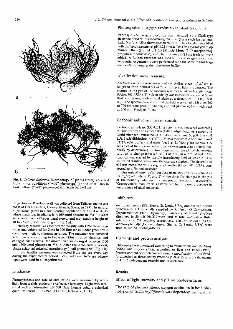
J. L. Gómez-Pinchetti et al.: Effect of CA inhibitors on photosynthesis in Solieria
Fig. 1 . Solieria filiforrnis. Morphology of plants freshly collected from in situ conditions ("wild" phenotype) (a) and after 2 mo in tank culture ("ball" phenotype) (b). Scale bars=2 cm
(Gigartinales: Rhodophyta)] was collected from Taliarte, on the east coast of Gran Canaria, Canary Islands, Spain, in 1991. In nature, S. jiliformis grows in a free-floating population at 3 to 4 m depth where maximum irradiance is c 100 pmo! p h ~ t ~ f i s S-'. P!mts grow erect from a fibrous basa1 system and may attain a height of 10 to 15 cm ("wild phenotype"; Fig. 1 a).
Healthy material was cleaned thoroughly with UV-filtered sea- water and cultivated for 2 mo in 300-litre tanks, under greenhouse conditions, with continuous aeration. The seawater was enriched with minerais according to Provasoii (i968j, but no vitamins, and changed once a week. Maximurn irradiance ranged between 1100 and 1300 pmol photons m-2 S-'. After the 2 m o culture period, plants exhibited spherical morphology ("ball phenotype"; Fig. 1 b).
Fresh healthy material was collected from the sea every day during the experimental period. Both, wild and ball-type pheno- types were used in al1 experiments.
Irradiance
Photosynthesis and rate of alkalization were measured by white light from a slide projector (Reflecta, Germany). Light was mea- sured with a radiometer LI-1000 Data Logger using a spherical quantum sensor. LI-193SA (LI-COR, Nebraska, USA).
Photosynthetic oxygen evolution in plant fragments
Photosynthetic oxygen evolution was measured by a Clark-type electrode fitted with a measuring chamber (Hansatech Instruments Ltd., Norfolk, UK) thermostated to 25°C. The chamber was filled with h ~ f i r e d seawter a! :,X 8.2 [5G rr?A4?% (Tri+jjdruxyme:hy!]- aminomethane)] or at pH 6.5 150 mM Mops (3-[N-morpholino]- propanesulphonic acid)] and plant fragrnents (25 mg fresh wt) were added. A flatbed recorder was used to follow oxvgen evolution. , - Sequential experiments were performed with the same thallus frag- ments after changing the incubation buffer.
Alkalization measurements
Alkalization rates were measured on thallus pieces of 2.0 cm in length in fresh natural seawater at different light irradiances. The change in the pH of the medium was measured with a pH-meter (Onon 701, USA). The eiectrode tip was immersed in a sealcd 50 m1 flask containing medium and algae at a density of up 1 .O g fresh wt/l. The spectral composition of the light was varied with blue (500 to 700 nm with peak at 640 nm) and red (400 to 600 nm with peak at 540 nm) Plexiglas lilters.
Carbonic anhydrase measurements
Carbonic anhydrase (EC 4.2.1.1 .) activity was measured according te ?.amazancv 2nd Semenenko (1900). Algal thaL ivcic grsüiid iii
liquid nitrogen, extracted in a buffer containing 50 pM Tris (pH 8.5), 5 pM dithiothreitol (DTT). 25 mM isoascorbic acid and 5 m M EDTA (CA buffer), and centrifuged at 13 000 x g for 60 min. CA activities of the supernatant and pellet were measured potentiomet- rically by determining the time required for the pH of the enzyme solution to change from 8.5 to 7.4 at 2"C, in a 2 ml sample. The reaction was started by rapidly introducing 2 m1 o í ice-cold C0,- saturated distilled water into the enzyme solution. The decrease in pH was measured with a digital pH-meter (Orion 701. USA), con- nected to a flatbed recorder.
One unit of activity (Wilbur Andersen. WA unit) was dcfined as 10 (T,/T) - 1, where T, and T = the times for changes in the pH of the nonenzymatic and the enzymatic reactions. respectively. Nonenzymatic reaction was established by the same procedure in the absence of algal material.
Inhibitors
6-ethoxizolamide (EZ; Sigma, St. Louis, USA) and dextran-bound sulfonamide (DBS; kindly supplied by Professor G. Samuelsson, Department of Plant Physiology, University of Umei, Sweden) dissoived in 50 mM NaOH were used as total and extracellular ínhibitors of CA activity, respectively. 100 pM DCMU [3-(3,4- dichloropheny1)-1,l-dimethylurea; Sigma, St Louis, USA] were used to inhibit photosynthesis.
Pigments and protein analysis
Chlorophyll was measured according to Wintermans and De Mots (1965), and phycoerythrin according to Beer and Eshel (1985). Protein content was determined using a modification of the Brad- ford method as described by Peterson (1983). Results are the means of 4 to 5 independent experiments in each case.
Results
Effect of light intensity and pH on photosynthesis
The rate of photosynthetic oxygen evolution in both phe- notypes of Solieriafiliformis was dependent on light in-

J.L. Gomez-Pinchetti et al.: Effect of CA inhibitors on photosynthesis in Solieria 337
Fig. 2. Solieria (m, o) and ball seawater at pH
irradiame hmol photons rnJ S")
filiformis. Photosynthetic oxygen evolution by wild (.,o) phenotypes (25 mg fresh wt/ml) in buffered 8.2 (50 mM Tris; o, o) and 6.5 (50 mM Mops; m, o)
Control EZ DBS Control EZ DBS
Fig. 3. Solieriafiliformis. Effect of 6-ethoxizolamide (EZ; 50 pM) and dextran-bound sulfonamide (DBS; 40 pM) on photosynthetic oxygen evolution by wild phenotype (25 mg fresh wt/ml) in buffered seawater a t pH 8.2 (50 mM Tris) and 6.5 (50 mM Mops). Data are maximum rates of linear portions of the O,-evolution kinetics; means of 5 measurements. Light intensity = 100 pmol photons
- 1 S
Tzh!e l. Selieris ,+i!~fer~is. Ch!orophy!! (ch!) 2nd phyrxrythrin (phy) concentrations and ratio of wild and ball phenotypes
Phenotype Chlorophyll Phycoerythrin ch1:phy ( P P / ~ U (Pg/ml)
Wild 24.23 $- 0.13 14.96 kü.65 i .62 Ball 15.26 + 0.66 16.84 + 0.39 0.91
tensity and the pH of the medium (Fig. 2). In the wild phenotype, photosynthetic saturation was observed at 100 and 200 pmol photons m-' S-' at pH 8.2 and 6.5, respectively. In the ball phenotype, photosynthetic satu- ration was observed at 150 and 200 pmol photons m-' S-' at pH 8.2 and 6.5, respectively. However, the rate of oxygen evolution was 25% higher than in the wild pheno- type at both pHs. The difference in oxygen evolution was
Ball Phenotype
White Red Blue
, Wild Phenotype
White Red Biue - = m
Fig. 4. Solieria filiformis. Alkalization (ApH/min) of medium iy wild and ball phenotypes (50 mg fresh wt/ml) at different lig; t intensities (100 and 230 pmol photons m-' S-'; fílled and hatchid bars, respectively) and spectral composition
O
o n
E
more significant at higher light intensities (400 pmol phi- tons m-' S-'; Fig. 2). n
Table 1 shows chlorophyll (chl) and phycoerythrin (phy) concentrations and their ratios for wild and b;Sil phenotypes. Chlorophyll concentration and the chkpl?y ratio were higher in the wild than in the ball phenotype.
Carbonic anhydrase activity
CA activity in Solieria filiforrnis plants was associated with both the supernatant and the pellet of the crude extracts. CA activity in the supernatant was 7 U/mg protein and in the pellet 117 U/mg protein. Low CA ac- + : . Y : + - . ' +ha -.m- --ato-+ n ~ . i l A h a - r m kapn ~ o i i a r 4 bxr the ~ i v i r y iii ~ i i b o u y e i i i a u a i i r rvuiu i i c r v v --AA ruuuru
relatively high protein concentration of this fraction (135 pg ml-') compared with that of the pellet (34 pg ml- l).
Effect of inhibitors of carbonic anhydrase activity on photosynthesis
The wild phenotype of Solieriafiliformis exhibited high photosynthetic rates in buffered seawater at pH 8.2 and 6.5 (Fig. 3). However, at pH 6.5 the rate of oxygen evolu- tion was -25% higher than at pH 8.2. At pH 8.2, EZ

338 J. L. Gomez-Pinchetti et al.: Effect of CA inhibitors on photosynthesis in Solieria
8.1 i
O 4 8 12 16 20
Tíme (min)
Fig. 5. Solieria filiformis. Effects of 100 pM of 6-ethoxizolamide (m), dextran-bound sulfonamide (o), 3-(3,4-dichloropheny1)-1,l- dimethylurea (DCMU) (o) and darkness ( x ) on alkalization rate of seawater by wild phenotype (50 mg fresh wt/ml); e: control. Light intensity 400 pmol photons m -' S - '
(50 pidj and DES (58 J L M ~ inhibited photosynthesis to 90 and 50%, respectively. Under the same conditions, in- creasing the DBS concentration to 100 and 200 pM did not change the percentage of inhibition. At pH 6.5, inhi- bition was not observed. The same results were exhibited by the ball phenotype (data not shown).
Effect of light intensity on alkalization rate of medium
Fig. 4 shows the rate of alkalization of the medium by wild and ball phenotypes. Alkalization rates were depen- dent on light intensity and spectral composition. The ball phenotype displayed higher alkalization rates than the wild phenotype. When light intensity was increased from 100 to 230 pmol photons m-, S- ' , the rate of alkaliza- tion by the ball phenotype increased by almost twice. In the wild phenotype, alkalization was saturated at low light intensity (100 pmol photons m-2 S-') regardless of spectral composition. Although there were no evident
in .&iha!izo;ioii iztes 4s a füricíiori of spcirai
composition, there was a slight increase in al1 measure- ments when blue light was used.
Effect of inhibitors of carhonic a ~ h y d r a . ~ ~ c t i x ~ i t ~ e:: alkalization rate of medium
Fig. 5 shows the rate of alkalization of the medium by the wild phenotype of Solieria jZiformis. Alkalization took place only in the light and was inhibited by DCMU (100 pM) and also by EZ and DBS. However, as in the case of photosynthesis, EZ inhibited the alkalization rate to a stronger degree. The same results were obtained with the ball phenotype (data not shown).
The results reveal important changes in the morphologi- cal and photosynthetic characteristics of Solieria fili- fOrmis cuitivated in aerated tanks. The morphological
changes (ball phenotype) would seem to be related to high water turbulence. The lower light-saturation point of í;hv:osq.iitheiic oxygeri evüluiio~i and the iower aika- lization rate of the wild phenotype are probably due to adaptation of the light-harvesting system to low light intensities characteristic of "shade-growing" plants (Falkowski and LaRoche 1991). The ball phenotype is protected from high light intensities by its morphology (self-shading) and the resulting decrease in chlorophyll concentration; this explains the absence of photoinhibi- tion at low light intensities in this phenotype (Fig. 2).
When bound- to dextran, sulfonamide cannot pene- trate the plasma membrane, and selectively inhibits CAext (Moroney et al. 1985, Palmqvist et al. 1990). It has been suggested that the role of CAext is to speed up the conversion of HCO; to CO, in the extracellular space (Smith and Bidwell 1989). CO, can then either diffuse through the plasma membrane (Simpssn et al. 1978, Smith and Bidwell 1989), or be actively transported to the cell (Sültemeyer et al. 1989). The inhibition of photosyn- thesis by DBS at pH 8.2 (Fig. 3) indicates the existence in &/ieriGJ$j&FGm& vf afi "ili&ieCi" iiieeiiariism of ES03 uptake catalyzed by CAext. However, the importance of the direct mechanism of uptake of inorganic carbon can- ? not be ignored, since the rate of photosynthetic oxygen evolution at pH 8.2, in natural seawater (2.2 mM HCO3) and in the presence of 50 pM DBS (Fig. 3), was higher than theoretical values based on a spontaneous supply of CO, from HC0; (Axelsson 1988). Thus, active HCO; transport in S.filiformis may constitute a significant con- tribution to the total uptake of inorganic carbon.
The stronger inhibition of photosynthetic oxygen evo- lution by EZ (Fig. 3) indicates the existence of intracellu- lar CA in Solieria filíformis.
The non-inhibitory effect of EZ and DBS at pH 6.5 (Fig. 3) evidences a high COZ-assimilation efficiency by Solieriaf;liformis, CO, being the main form of inorganic carbon entering through the cell wall and plasmalemma.
Alkalization of the medium by algae has been taken as evidence of the presence of active HCO; transport (Cook et al. 1988). Reont!y, it has been shcwi, :ha: alhzlizaiion by the green and red seaweeds Ulva rigida and Gracilaria tenuistipitata is performed by an extracellular mechanism requiring CAext (Bjork et al. 1992, Haglund et al. 1992). The efficiency of this mechanism is dependent on the cmcentro:ioí; of iiioigaiiic carbon in ihe rnedium (Beer and Israel 1990, Bj6rk et al. 1992, Haglund et al. 1992). Several authors have postulated that the process of ai- kalization of the growth medium by aquatic plants is effected by the release of 1 m01 OH- for each molecule of CO, transported (Simpson et al. 1978, Morgan et al. 1980, Lucas 1983). If OH- production and efflux to the medium is an exclusively intracellular mechanism, as maintained by Lucas, then DBS would not affect the rate of alkalization by Solieriafiliformis, as the present results show that it does (Fig. 5).
In conclusion, the different effects of EZ and DBS on the alkalization rate (and photosynthetic oxygen evolu- tion) of Solieria filiformis indicate the involvement of externa1 carbonic anhydrase in the uptake of inorganic carbon.

J. L. Gómez-Pinchetti et al.: Effect of CA inhibitors on photosynthesis in Solieria 339
Acknowledgemenfs. The authors wish to thank to D. Robledo Ramírez and M. J imhez del Rio for providing the cultured materi- al. This work was supported by a scholarship from the University of Las Palmas of Gran Canana to J. L. Gómez-Pinchetti and CICYT (project MAR-1237191 a n d a sabbatical to Z. Ramazanov, invited Professor of Ministerior de Educación y Ciencia, Spain). Support by Fundación Universitaria de Las Palmas and by Air Europa is gratefully acknowledged.
Literature cited
Axelsson, L. (1988). Changes in pH as a measure of photosynthesis by marine macroalgae. Mar. Biol. 97: 287-294
Beer, S., Eshel, A. (1985). Determining phycoerythrin and phyco- cyanin concentrations in aqueous crude extracts of red algae. Aust. J. mar. Freshwat. Res. 36: 785-792
Beer, S., Israel, A. (1990). Photsynthesis of Ulva fascista. 1V. pH, carbonic anhydrase and inorganic carbon conversions in the unstirred layer. Pl., Cell Envir. 13: 555-560
Bidwell, R. G. S . , McLachlan, J. (1985). Carbon nutrition of sea- weeds: photosynthesis, photorespiration and respiration. J. exp. mar. Biol. Ecol. 86: 15-46
Bjork, M., Haglund, K., Ramazanov, Z., Garcia-Reina, G., Pedersén, M. (1992). Inorganic carbon assimilation in the green seaweed Ulva rigida C. Ag. (Chlorophyta). Planta 187: 152- 156
Cook, C. M., Lanaras, T., Colman, B. (1986). Evidence for bicar- bonate transport in species of red and brown macrophytic ma- rine algae. J. exp. Bot. 37: 977-984
Cooh, C . FA., ki i i a ia~ , T., Rüibe!akis-Ange!akiu, K. A. (!9R?). Bicarbonate transport and alkalization of the medium by four species of Rhodophyta. J. exp. Bot. 39: 1185-1 198
Falkowski, P. G., LaRoche, J. (1991). Acclimation to spectral irra- diance in algae. J. Phycol. 27: 8-14
Goyal, A,, Tolbert, N. E. (1989). Uptake of inorganic carbon by isolated chloroplasts from air-adapted Dunaliella. P1. Physiol. 89: 1264-1269
Haglund, K., Bjork, M., Ramazanov, Z., Garcia-Reina, G., Pedersén, M. (1992). Role of carbonic anhydrase in photosyn- thesis and inorganic carbon assimilation in the red alga Gracilar- ia tenuistipitata. Planta 187: 275-281
Lucas, W. J. (1983). Photosynthetic assimilation of exogenous HCO; by aquatic plants. A. Rev. Pl. Physiol. 34: 71-104
Marcus, Y., Volokita, M., Kaplan, A. (1984). The location of the transporting system for inorganic carbon and the nature of the
form translocated in Chlamydomonas reinhardrii. J. exp. Bot. 35: 1136-1144
Morgan, K. C., Shacklock, P. F., Simpson, F. J. (1980). Some as- pects of the culture of Palamaria palmafa in greenhouse tanks. Botanica mar. 23: 765-770
Moroney, J. V., Husic, D. H., Tolbert, N. E. (1985). Effect of car- bonic anhydrase inhibitors on inoganie car'ion aüümü~atiüii
by Chlamydomonas reinhardtii. P1. Physiol. 77: 177-183 Palmqvist, K., Ramazanov, Z., Samuelsson, G. (1990). The role of
extracellular carbonic anhydrase for accumulation of inorganic carbon in the green alga Chlamydomonas reinhardtii. A compar- ison between wild-type and cell-walless mutant cells. Physiolo- gia P1. 80: 267-276
Peterson, G. L. (1983). Determination of total protein. Meth. En- zym. 91: 95-119
Provasoli, L. (1968). Media and prospects for the cultivation of marine algae. In: Watanabe, A., Hattori, A. (eds.) Cultures and collections of algae. Japanese Society of Plant Physiologists, Tokyo, p. 63-75 (Proc. U.S.-Japan Conf. Hakone)
Ramazanov, Z. M., Semenenko, V. E. (1988). Content of the COZ- dependent form of carbonic anhydrase as a function of light intensity and photosynthesis. Soviet P1. Physiol. 35: 340-344
Raven, J. A., Lucas, W. J. (1985). The energetics of carbon acquisi- tion. In: Lucas, W. J., Berry, J. A. (eds.) Inorganic carbon uptake by aquatic photosynthetic organisms. American Society of Plant Physiologists. Rockwell, Maryland, p. 305-324
Sand-Jensen, K., Gordon, D. M. (1984). Differential ability of ma- rine and freshwater macrophytes to utilize HCO; and CO,. Mar. Eio!. gG: 247-253
Simpson, F. J., Neish, A. C., Shacklock, P. F., Robson, D. R. (1978). The cultivation of Chondrus crispus. Effect of pH on growth a @ production of carrageenin. Botanica mar. 21: 229-235 2
Smith, R. G., Bidwell, R. G. S. (1987). Carbonic anhydrase-dep&- dent inorganic carbon uptake by the red macroalga, Chondi$ls crispus. P1. Physiol. 83: 735-738 =
n
Smith, R. G., Bidwell, R. G. S. (1989). Mechanism of photosynthg:- ic carbon dioxide uptake by the red macroalga, Chondrus o!;- pus. P1. Physiol. 89: 93-99 ~1
Sültemeyer, D. F., Miller, A. G., Espie, G. S., Fock, H., Canvin, ;h.
T. (1989). Active CO, transport by the green algae Chlan$f- domonas reinhardrii. P1. Physiol. 89: 1213-1219
Wintermans, J. F. G., De Mots, A. (1965). ~ ~ e c t r o ~ h o t o r n e t i c characteristics of chlorophylls a and b and their pheophytinsgn ethanol. Biochim. biophys. Acta 109: 448-453 9
5


Hydrobiologia 221: 18 1-194, 199 1. J. A. Juanes. B. Santelices & J . L. McLachlan (eds), International Worhhop on Gelidium. O 1991 Kluwer Academic Publishers. Printed in Belgium.
Actual, potential and speculative applications of seaweed cellular biotechnology: some specific comrnents on Gelidium
G. Garcia-Reina, J. L. Gómez-Pinchetti, D. R. Robledo & P. Sosa Marine. Plant Biotechnology Laboratory, University of Las Palmas, Box 550, Las Palmas, Canary Islands, Spain
Key words: callus, cell culture, domestication, protoplast, tissue culture
Abstract
Cellular biotechnology is a promising appiication in the propagation and selection of superior strains of seaweeds. Although axenic cultures, organogenetic tissue cultures, vegetative micro-propogation, callus induction and high yields of agar from calli have been described for several species of Gelidium, a number of basic problems remain to be solved. These include standardized methods for obtaining axenic cultures, identification of requirements for organic nutrients, PGR's, cellular disorganization and reorganization, 2 somaclonal variation and somatic incompatibilities. Future progress in seaweed biotechnology will depend on the resolution of many of these problems.
o z - = n
S Introduction Seaweed biotechnology 8
h
Cellular biotechnology was introduced into the phycological field at the beginning of the 1980's (Gibor, 1980), and is considered important in future development of seaweed cultivation. The four major techniques in seaweed cellular bio- technology, tissue-, callus-, cell- and the proto- plast-culture, offer means in understanding seaweeci domestication (propagation and seiec-
tion) germplasm storage and for production of biomass. We first discuss seaweed cellular biotechnology as highly effective techniques, and then consicier some stiii unresoived quesiioñs,
which limit their potential and actual applica- tions.
Vegetative micropropagation
5 The possibility of using small thallus fragments 2 (tissue culture) to establish cultures, instead of spores, exploits the organogenetic potential of explants. Chen and Taylor (1978) described the regeneration of whole plants from decorticated explaits of C l i ~ i í U ~ i ~ C ~ ~ S ~ Ü S Stackh. iVaa!mU (1982) and Sylvester and Waaland (1983) ob- tained plants following inoculation of finely- chopped fragments of the thallus of Gigartina into L - - : J - A -- oraiueu iupe. neK data indiezted thzt the best strategy for commerical purposes is to maximize the arnount of propagules per donor plant. During the early 1980's, similarly successful experiments were done with Gelidium nudgrons Gardner and G. robustum (Gardner) Hollenberg & Abbott (Polne-Fuller, 1988).
In China, Luqin et al. (1988), described a tech-

182
nique, using small explants of Gelidium pacificum Okamura, that permitted the firm attachment of small explants to ropes through the development of adventitious rhizoids and the regeneration of new plantlets. Garcia-Reina et al. (1988a) re- ported on organogenetic development from wounded explants of Gelidium versicolor (Gmel.) Lamouroux, and Robaina et al. (1 99Oa) discussed the effects of solid culture medium and its osmo- lality on organogenetic potential.
If the best commercial strategy for vegetative micropropagation is to maximize propagules per donor-plant ratios, the logical strategy would be to reduce the plant to single, somatic cells, using these as inocula. 'Somatic spores' implies toti- potentiality, as pointed out by Saga et al. (1978). Single somatic cells, obtained by enzymatic diges- tion of the thalli of Porphyra and Ulvaria, have been inoculated onto ropes and, from these, attached plants have developed (Polne-Fuller et al., 1984; Kapraun, 1987; Kapraun & Sherrnan, 1989). This technique has been tested in commercial Porphyra farms (Mumford, 1987).
Somatic-cell isolation in seaweeds is not a dif- ficult task as it has been achieved by partial enzymatic digestion of thalli (in Pophyra: Tang, 1982; Chen, 1986; Wang et al., 1986, 1987a, 1987b; Tait et al., 1990; Wang & Yan, 1990; Sphacelaria: Ducreux et al., 1988), by cell-wall regeneration from protoplasts (Pophyra: Chen, 1989) or spheroplasts (Undana: Kapraun & Sherman, 1989), by mechanical disruption of thalli (Prasiola: S c h 8 et al., 1972; Bingham & Schiff, 1973; Gniffithsia: DuiXeld et al., 1972; Porphyra, Tait et al., 1990), or of frozen thalli (Polphyra: Zhao & Zhang, 1984), by mechanical disruption of calli (Laminana: Saga et al., 1978 ; McLean & Connolly, 1989; Afana: Saga et al., 1978 ; Undaria: Zhang, 1982) or by combinations of mechanical and enzyrnatic treatments of both thallus and callus (Pterocladia: Liu & Gordon, 1987).
There are advantages to vegetative micropropa- gation over conventional propagation frorn spores. It is unnecessary to know or control the life-history of the species; 'seedlings' can be pro- duced in quantity, both when they are needed and
from a small number of donor plants, requiring cheap storage and handling.
These advantages are directly related to propa- gation, while the techniques are a~plicable to selection. Mono-phase crops (i.e. inonoculture of Gelidium sporophytes or of male or female gametophytes) can be produced. Mutant plants (natural or induced) can be propagated rapidly, even if sterile, and even if the selected phenotype has a non-genetic basis (epigenetic) but is stable.
Selec tion
Tissue culture of seaweeds is based on natural genetic variability of the somatic cells; for example, Gracilaria species (van der Meer, 1986) have been shown to have a high degree of somatic recombination. Genetic diversity among cells of the thallus, normally masked by more common non-variant cells, is exploited. as described for higher plant tissue culture (D'Amato, 1978). This 'island effect' (retention of pigmentation, gowth,! etc., by some cells or cell aggregates) is a common! phenomenon in seaweed tissue culture (Fries,; 1980; Polne-Fuller et al., 1986 ; Garcia-~einai et al., unpubl. data). O
Polne-Fuller and Gibor (1986) have described! the isolation and growth to callus on solid mediumi of a few living cells from Porphyra tissue culturesd contaminated by Pythium. The callus was 3 enzymatically digested to cells and small clumps$ of cells and co-cultured with Pythium, whereo healthy, normal callus developed. Although no plant regeneration was reported, this simple experiment shows how quickiy and effectively tissue-culture techniques can yield results by selection.
Selected strains of Sargassum and Entero- morpha (showing differences in temperature tolerance, orphology and vitamin auxotrophy) have been established through tissue culture (Polne-Fuiíer et al., 1986), apparently owing to intercellular variability among explants. Tissue culture (i.e. callus culture) is not, however, con- sidered the best technique for cultivar improve- ment. The application of callus culture for selection is based on a process referred to as

somaclonal variation by Larkin and Scowcroft (198 1). This term covers genomic, chromosomic, genetic and even epigenetic variations associated with or induced by the disorganized callus state, and is also expressed in the reorganized plants (Karp, 1989). Callus is associated with a high degree of genetic (or epigenetic) variability. Fang et al. (1983) have described genomic alteration in the callus cells of Laminaria, and Lee (1986), varying levels of polyploidy.
Callus culture has many advantages over con- ventional selection procedures. Within a small space (e.g. Petri dish incubated in growth cham- ber), large numbers of 'plants' (assuming cell = plant), having natural or induced genetic variabilities, can be screened. The stability of selected phenotypes can be determined in a short time, as the following generation is the next mitotic division, not the new adult sporophytic phase as in Laminana. Any selected vhenotype can be easily propagated to large amounts (using cell culture), even if it is an epigenetic but stable character.
In seaweeds, obtaining haploid cultures through cell culture is easy, whereas with higher plants the only way to obtain haploid cultures is by anther or poilen culture (Han & Hongyuan, 1986). For the latter group of plants, diploid cells can proliferate and few haploid cells remain via- ble, so that the eficiency is low. In seaweeds, haploid cultures depend only on the type of explant (gametophyte) chosen. Thus, exploitation of recessive information, haploid somatic recom- bination and gametoclonal variation (somaclonal variation in haploid cells, Evans et al., 1984), can be readily employed as can the production of fully-homozygous cells and plants by colchicine treatment or by spontaneous chromosome doubling. Al1 such characteristics (haploid cul- ture, gametoclonal variation, chromosome doubling) have been demonstrated and utilized for Laminaria-improvement programs (Fang, 1984; Wu & Lin, 1988). Reports of regeneration of variant plants (altered morphology and life- history : monospore producing) from isolated cells or protoplasts of Porphyra (Tang, 1982; Fujita & Migita, 1985; Chen, 1987) and Monostroma
gametophytes, with different developmental pat- terns and apogamy (Saga & Kudo, 1989), may be explained by differential regenerative potential or different variant cell strains pre-existing in the haploid thallus.
Selection through callus culture has some drawbacks, as the techniques allow selection only of cellular-based characteristics. It is impossible to screen for stipe-length or frond-thickness at the cellular level or for sulphate content in the cell walls of Gelidium. Monoclonal antibodies (Vreeland et al., 1987) can be useful markers. There are also possibilities of selecting variant cells with characteristics expressed only at the cellular level and not in fully-differentiated plants.
The regeneration of plants from calli can yield surprises, some of which could be useful, even in the absence of directionally seiective pressures. Several reports indicate inadvertent selection resulting from seaweed somaclonal variation. Yan (1984) described plants, reorganized from calli of Laminana and Undana, with more rapid growth and tolerance of high temperatures for a longer period than the normal sporophytes. Garcia-Reina et al. (1988b) selected two types of callus of Laurencia obtained from the sarne plant and similar in appearance (pigmentation, texture). These differed markedly in organogenetic poten- % tia1 (number of plantlets/callus) and growth-rate of the regenerated plants. n n
n
A major advantage of selection using cellular [ biotechnologies is in protoplast culture.' This " d o w s for somatic hybridization (through the breakdown of sexual barriers, through interspeci- fic or intergeneric fusion of somatic cells, and exclusively maternal extrachromosomal inheri- tance). There is also improved effíciency of genetic transfonnation by the removal of physical barriers, and host-vector recognition specificities through elimination of the cell wall.
Protoplast isolation and regeneration of whole plants have been successful (Table 1). The same techniques as applied to higher plant protoplasts induce somatic hybridization in seaweeds. These include electrofusion (Enteromorpha : Saga et al., 1986; Porphyra: Fujita & Saito, 1989) and PEG ( Ulva x Monostroma : Zhang, 1982 ; Enteromor-

pha, Saga et al., 1986; Porphyra: Fujita & Migita, 1988). Recently, Fujita & Migita (1987), with 1987; Fujita & Saito, 1989; Gracilaria: Cheney, wild x mutant Porphyra and Kapraun (1987) 1989). True somatic hybrids have been obtained Enteromorpha, in anatomically-simple species, in microalgae (Chlarn~vdomonas: Matagne et al., and Cheney (1989!, with m ~ t ~ m i c i l ! y mere rem- 1979; Dunaliella x Porphyridiurn: Lee & Tan, plex species (Gracilaria tikvahiae McLachlan x
Table l . Seaweeds from which protopiasts have been isolated, types of isolation (Iso; M = mechanical, F = enzymes obtained from phycophages, C = commercial enzymes, B = enzymes obtained from bacteria, A = enzymes obtained from amoeba) and types of further development (Dev; R = cell wall regeneration, R = without cell wall regeneration, R + = plant regeneration, Ca = callus formation, Ca + = calius and plants).
Iso. Dev.
Chlorophyta
Bryopsis plumosa Enteromorphalinza Enteromorphalinza E. intestinalis E. intestinalis Monostroma angicava Monostroma angicava Monostroma nitidum Monostroma zoostericola Ulva conglobuta Ulva fasciata Ulva linza Ulva pertusa Ulva pertusa Ulva pertusa Ulva pertusa Ulvaria oxysperma Uronema gigas
Phaeophyta
Fucus distichus Laminaria Laminaria digitata Laminaria digitata Laminaria japonica Ln.miznri~ snrrk,nrim
Laminaria saccharina Macrocystis pyrfera Macrocystis pyrifera Macrocystis pyrfera ~ ü r g ü ~ s u i i i e¿-hinocurpum Sargassum muticum Sargassum muticum Sargassum muticum Sargassum muticum Sargassum polyphyllum Sphacelaria Undaria pinnatifda Undaria pinnatifda
Tatewaki & Nagata 1970 Fujita & Migita 1985 Saga 1984 Millner et al. 1979 Saga et al. 1986 Zhang 1982 Szga & f i d c !989 Fujita & Migita 1985
m
Saga et al. 1986 Reddy et al. 1989 =
m O
Reddy et al. 1989 Zhang 1982 -
0 m
Fugita & Migita 1985 E
Saga 1984 O
Fujimura & Kajiwara 1989 Reddy et al. 1989 E
Kapraun & Sherman 1989 a
Gabriel 1970 n n
3
O
Kloareg et al. 1988 Saga & Sakai 1984 Butler & Evans 1988 Butler et al. 1989 Saga & Sakai 1984 Burier & Evans i988 Butler et al. 1989 Saga et al. 1986 Kloareg et al. 1989 Davison & Polne-Fuller 1990 Fisher & Gibor 1987 Neushul 1984 Saga et al. 1986 Fisher & Gibor 1987 Polne-Fuller & Gibor 1987c Fisher & Gibor 1987 Ducreux & Kloareg 1988 Fujita & Migita 1985 Tokuda & Kawashima 1988

Table 1. (Continued)
Iso. Dev.
Rhodophyta
Chondrus crispus Chondrus crispus Gracilaria lemaneiformis Gracilaria lemaneiformis Gracilaria secundata Gracilaria tenuistipitata Gracilaria tikvahiae Gracilaria verrucosa Palmaria palmata Porphyra Porphyra Porphyra haitanensis Porphyra leucosticta Porphyra linearis Porphyra maculosa Porph-vra nereocystis Porphyra perfora ta Pnmhi i r~ -. =. ., . nofirntn , -. - - - Porphyra perfora ta Porphyra pseudolanceolata Porphyra suborbiculata Porphyra yezoensis Porphyra yezoensis Porphyra yezoensis Porphyra yezoensis Porph-vra yezoensis Porphyra yezoensis Porphyra yezoensis
Ca, R + R + , Ca
LeGall et al. 1989 Smith & Bidwell 1989 Cheney et al. 1986 Bjork et al. 1990 Bjork et al. 1990 Bjork et al. 1990 Cheney et al. 1986 Bjork eral. 1990 Liu 1989 Liu etal. 1984 Fujita & Saito 1989 Wang et al. 1986 Chen 1987 Chen et al. 1988 Waaland & Dickson 1987 Waaland & Dickson 1987 Saga et al. 1986 Polne & Gihor 1984 Waaland & Dickson 1987 Waaland & Dickson 1987 m
D
Tang 1982 - - m
Araki et al. 1987 O
- Polne et al. 1984 - O
m
Polne & Gibor 1984 E
Saga & Sakai 1984 O
Fujita & Migita 1985 n
Saga et al. 1986 E -
Dai 1987 a 2
G. chilensis Bird, McLachlan & Oliveira), have described the recovery of somatic hybrids. These results suggest that the production of 'agarophytic corn' (Gelidium x Zea mays), of 'agarageenan' . . íGe/i&um j< Eucheuma j or oii"ier r;imag,naiive
hybrids, can be realized. There are a few somewhat speculative and
distantly related reports on genetic engineering in s.awee& Tiies iriclu& ~gro;aei~i~üin tüiiie-
faciens, used genetically to engineer algae, at least in Chlamydomonas (Ausich, 1983); the posible existence of Agrobacterium-like bacteria that pro- mote the induction and growth of seaweed tumours, galls and calli (Cantacuzene, 1930; Apt, 1988; Tsekos, 1982; Garcia-Reina et al., 1988a); DNA-transferring parasitic seaweeds, possibly
usabie as vectors (Goff & Coleman, 1984); chro- mosomal location and molecular biological regu- latory studies of alginate-related genes in Pseudomonas, with characteristics similar to those ili FUcüs dgliaes íi)eieiic ei 1987 j; develup- ment of cloning vectors in photosynthetic microorganisms (Chauvat et al., 1988).
Germplasm storage
Tissues and callus cultures can be highly efficient techniques for germplasm storage in selection programs or 'seed' storage for propagation as they avoid expensive whole-plant rnanagement. The longest, 5-year-old culture of seaweed calli
(Fang, 1983; Polne-Fuller & Gibor, 1987b), and culture of sornatic cells (Chen, 1989) parallels the irnmortality shown by higher plant-cell cultures (Gaiitheret, 1985). Cry~preservatlm wi!! reqcire even less handling, and van der Meer and Simpson (1984) have described techniques for Gracilaria tissue.
Production of biomass or metabolites
The cultivation of tissues, calli or cells in photo- bioreactors, bioreactors or immobilized bioreac- tors are alternative techniques for direct produc- tion of biomass or of economically-significant metabolites. The best example (Misawa, 1977) is described in Japanese patent No. 74-10 156 1 obtained by Nakamura in 1984 (Kureha Chem. Co. Ltd). This patent protects the right to produce q p r frem ca!h ef &)i&jrW u,w=csü L-=cr=gx
and G. subcostatum Okamura (among other agarophytes). Callus growth increased 1 1.3 times in 20 days and, from 100 g of dried callus, 75 g of agar was obtained.
In phycocolloid production in cell culture, Cheney et al. (1987) did not find a significant difference between iota-type carrageenan ex- tracted from tissue culture of Agardhiella subulata (C. Ag.) Kraft & Wynne compared with extractive from field-collected material. Tait et al. (1990) obtained comparative spectra for polysaccharides produced by Polphyra cell-cultures and the native plants, but, as the authors concluded, 'it should be demonstrated whether specific growth conditions can affect the structure and properties of the poiy- s ~ c c h ~ s & ~ prn&ced by ce!! ~il! t l~~e~, ' . by disorganized cells.
Bryhni (1978) found highly-significant quanti- tative and structural dserences between poly- s a ~ c h - d ~ ~ extrc~ted frem erg~qked str&s =f
Ulva mutabilis F q n and those from a strain of disorganized aggegates of undifferentiated cells. Liu et al. (1989) obtained 50 % agar yield from dried callus of Pterocladia capillacea (Gmel.) Born. & Thur., 25 % less than Nakamura's patent, but the agarose molecule contained less sulfate and fewer methyl groups. The callus of Laurencia
consists of small photoautotrophic, pseudo- meristematic cells, filled with starch-like grandes, and with cell walls at least twice the width of mrmr! ce!!s (Gurciu-Xeina et d., unpüb. dmt). These data indicate that phycocolloid production, with high quality and distinct characteristics, is possible through cell and callus culhre.
In addition to phycocolloid production, Chen (1989) has suggested the possibility of producing Porphyra for human consumption through cell culture. Fujimura and Kajiwara (1989) produced a bioflavor (released into the culture media) from Ulva pertusa Kjellman cell-suspension cultures.
Some critica1 considerations on applicability
Al1 the applications described above are possible. The question is whether application on a commer- cid se& be praciic-&J. Líiiíi&iiíu~a seleciion by
monoploid cell-culture has yielded industrial , D
results, and propagation by somatic cell-inocula- m
tion of morphologicaily simple species could be a achieved commercially in a few years. There remain, however, many unresolved basic questions, limiting types of applications and g species to which these processes can be applied. B
a
n n
Axenic cultures 3
O
Owing to seasonal variation of contaminants, differential interspecific sensitivities to biocides and the widespread phenomenon of endophytism in seaweeds and other algae, few generalizations cm be m& oii ~bt&iíig a e ~ i c cühres . Evaluation of axenic cultures do not include tests for viruses, mycoplasma, nckettsia and so forth. Even if judged axenic, contaminants can appear
thn n ~ ~ l t x ~ - ~ ~ nCt - ---+l. -- -v.---- ili t.ub bwt.wbs CLIC~I ~ U V ~ L ~ S VI y c + a a ( F i k ~ , 1963 ; Lee, 1986 ; Cheney, 1986 ; Tait et a l , 1 WO), and even with morphologically simple species, there are contradictory results of the eficacy of sterili- zation treatments (Bonneau, 1976).

Callus, callus control and PGR 'S
Ga!!, tumour, cancer? tumour-like, callus and cal- lus-like are al1 terms used to describe proliferative, disorganized growth in seaweeds (Apt, 1988). The term 'callus-like' has been widely-used to qualify, filamentous, crustose, hyperplasic and other abnormal outgrowth of the explant. If 'cal- lus-like' is not true callus, as some authors claim, the literature on seaweed callus culture can be dramatically reduced. A consensus of what a cal- lus in seaweed is has not been achieved.
Callus control implies callus'induction, growth (without the explant) and regeneration. Although auxins and cytokinins exist in seaweeds, their effects when applied in vitro are not clear. Based on the literature of cell and callus culture of agaro- .phytes (Gelidium and Pterocladia), three types of responses have heen described for the effects of these hormones: 'success', although not com- pared with hormone-free media (Nakamura, 1974; Gusev et al., 1987); 'no effect' (Polne-Fuller & Gibor, 1987a; Garcia-Reina et al., 1988a); 'not clear' (Liu & Gordon, 1987).
Ranking of calligenic potential (Table 2) can be done (Gusev et al., 1987; Garcia-Reina et al., 1988a; Polne Fuller & Gibor, 1987a), indicating genotypic differences in, disorganization (Fang et al., 1979). 'Wounding-response' seems to be the key factor in initiating callus growth in seaweeds.
True callus culture (excised from the explant) has been described for a few species (Gelidium and Gracilaria: Nakamura, 1974; Laurencia: Garcia-Reina et al.: l988b 3 Gratelo~~~ia: Robaina et al., 1990b; Phyllophora: Gusev et al., 1987; Sargassum, Cystoseira, Macrocystis, Ulva, Entero- rnorpha, ~uch&ma and Polphyra: Polne-Fuller & Gibor, 1987a, l987b; Ecklonia: Lawlor et al., 1989; Pterocladia: Liu & Gordon, 1987). Only the Nakamura (1974) patent describes (true) callus culture of Gelidium amansii and G. subcostatum. Calli from Gelidium vagum Okamura (Gusev et al., 1987), G. nudifrons, G. robustum (Polne- Fuller & Gibor, 1987a) and G. versicolor (Garcia- Reina et al., 1988a) have not been described, either excised from the explant or with .organo- genetic potential.
Table 2 . Ranking of 'calligenic potential' expressed as per- cent of tissue explants which developed callus (data from Gusev et al. 1987; Kawashima & Tokuda. 1989; Garcia- Reina, unpubl.; Polne-Fuller & Gibor, 1987a).
Chlorophyta
88.0 % Enteromorpha intestinalis 86.0% Ulva augusta
Phaeophyta
Ecklonia cava Macrocystis pynfea (gametophyte) Laminaria sinclairii (gametophyte) Sargassum muticum Pelvetia fastigiata Sargassum hystnx Cystoseira osmundacea Laminaria sinclairii (sporophyte) Sargassum jluitans Macrocystis pynyera (sporophyte)
Porphyra lanceolata Porphyra perforata Porphyra nereocystis Smithora naiadum Laurencia sp Phyllophora nervosa Furcellana fastigiota Gelidium vagum Gracüaria ferox Eucheuma alvarezii Gracilana verrucosa Ceramium kondoi Gigartina exasperata Eucheuma uncinatum Geliditlm robustum Gracilaria papenfusii Gelidium versicolor
Neither the physiologicd status of the explant (differential nutritional levels yielding differences in calligenic potential (Fries, 1980) seasonal dif- ferences in endogenous hormones (Mooney & van Staden, 1984; Featonby-Smith & van Staden, 1984) nor seasondy-different calligenic potentials, (Polne-Fuller & Gibor, 1987b) could be key factors for success. With so many un- known variables, it is difficult to project a produc- tive future in the short term for the application of c d u s culture to seaweeds.

Culture media
As organic requirements for axenic or aseptic seaweed c.filtUres ale largely uIikIiowii, ihe
common culture media are undefined (enriched seawater). The addition of organic complexes (co- conut milk, alga1 and yeast extracts, etc.) to increase low growth rates (indicating suboptimal conditions) of seaweed tissue (Fries, 1973; 1984) and callus cultures (Nakamura, 1974; Saga & Sakai, 1983) has resulted in both stimulatory effects and no effects (Robaina, 1988; Lawlor et al., 1988). Nutrient depletion could be the reason for callus and 'callus-like' inductions or reversions (Pedersén, 1968; Fries. 1980; Bradley & Cheney, 1986; Polne-Fuller & Gibor, 1986; Lawlor et al., 1989). Sugars have been reported to be unnecessary or even inhibitory to tissue (Fucus spiralis : Fries, l984), callus (Ecklonia radiata : iawior ei al., i 989 j and ceii cuiture (Porphyra umbilicalis: Tait et al., 1990) growth or organo- genesis. Our studies on the interrelationship between the osmolality of the culture medium and the effect of osmotically active sugar supplemen- tation has shown that, as in higher plants (van Rensburg & Vcelar, 1989), the effects of carbo- hydrates in seaweed tissue and callus culture seem osmotic or metabolic or both (Robaina et al., 1990a; 1990b). Solidity of culture media promotes disorganization (Robaina et al., 1990a; 1990b).
Heterotrophic growth is required for biomass production in bioreactors, but heterotrophic frowth of seaweed tissue remains to bedemon- strated (Fries, 1973; Robaina, 1988; Lawlor et u!., 1383; Rv"uiia ei al., IWOb j. Aiihoügh a callus of Gelidium has been obtained with the addition of sucrose (Nakamura, 1974), 20 g 1- ' mannitol (Gusev et al., 1987), or in the absence of ,.,,,,, :, *L, ,...IL~--- --A:- fn I- _ F..II. . a m.1 ~ u g a S 111 LIIG GUILLU~: i i ~ ~ u i i l [roirie-r uiier -mor, 1987a; Garcia-Reina et al., 1988a), callus pro- duction has not occurred in darkness. However, as seaweed tissues, (Fries, 1977; 198O), cell (Saga et al., 1978 ; Polne-Fuller et al., 1987 ; Tait et al., 1990) and callus cultures (Polne-Fuller et al., 1986 ; Polne-Fuller & Gibor, 1987b; Garcia- Reina et al., 1988a; Lawlor et al., 1989) are pho-
totrophic, including Gelidium species, photobio- reactors remain a possibility for biomass produc- tion.
True cell culture
Porphyra is the only genus for which continuous cell cultures have been established (Chen, 1989; Tait et al., 1990). Other reports on 'cell culture' refer to dynamic steps between protoplasts, pseudoprotoplasts or enzymatically/mechani- cally-isolated cells and their development to cal- lus or to plants. The application of cell culture of other species for selection and for the production of biomass or metabolites requires confirmation of true cell culture.
- P. vzabie protopiasts m D
Protoplast isolation is not a problem in sorne g seaweeds (Table l), whereas the isolation of large 1 numbers of highly-viable protoplasts is. Trial and
E error assays with cocktails of commercial (Table 3), phycophage-extracted (Table 4) or microorganism-extracted (Table 5) enzymes are -
common approaches to obtain protoplasts. Basic knowledge of composition and structure of cell walls of seaweeds and enzymatic activity of cell- wall digesting enzymes are scarce. Until more information becomes available, possible benefits from protoplast culture as applied to Gelidium, for exarnple, are likely limited.
Somatic incompatibility
Somatic incompatibility needs to be overcome to reaiize the potentiai for somatic hybrridization. In somatic incompatibility chromosomes, chloro- plasts and rnitochondria from one of the fused protoplasts degenerate, as noted in heterokaryons of Zygnema x Porphyndium, Zygnema x Mou- geotia (Ohiwa, 1978; 1980; 1981) and Daucus x Chlamydomonas (Fowke et al., 1979). Other undesirable processes could result from the

Table 3. Commercial enzymes commonly used for the diges- tion of seaweed cell walls.
Abalone acetone powder Agarase Cellulase Cellulisin Lamina rinase Limpet I Limpet . II Limpet I I I Hemvcelulase Papain Pectinase Pectolyase Protease XXI V Lysozyme Cellulase onozuka RS Cellulase onozuka R-1 O Macerozyme R-10 Cellulase onozuka R- 1 O Maceroyme R-200 Cellu&sin AInrnrnro 11s UCC, ,.'*S
Laminarinase Driselase Pronase E
Sigrna A75 14 e:--- A L 1 1 9 aigiiia n u i UL
Sigma C2415 Sigma C2274 Sigma L7758 Sigma L1251 Sigma LO630 Sigma L8755 Sigma H2125 Sigma P3 125 Sigma P240 1 Sigma P3026 Sigma P8163 Sigma L6876 Yakult Yakult Yakult Serva S e n a Calbiochem 2 19466 C l b i ~ c h e m 44!20! BDH 39120 2G Kyowa hakko Merck 7433
formation of different types of chimaera in plants regenerated from the heterokaryon. This occurred in the first seaweed somatic hybrid; irregularly- variegated chimeral thalli from the fusion of wild (red) and mutant (green) Povphyra protoplasts occurred (Fujita & Migita, 1987). Cheney (1989) claimed to verify true hybridization in green and red plants regenerated from the fused green mutant (Gracilaria tikvahiae) and red (G. chilen- sis) protoplasts by isoenzyme analysis. Although suggestive of true hybridization, this technique cannot distinguish chimaeric plants from true somatic-hybr'id plants.
As with somatic hybridization in microalgae (Matagne et al., 1979), the first recovery of a sta- h!e s e ~ ~ e e d heternkzym (Kapraunj 1987) has 6een achieved using haploid cells, although these were zoospores, not somatic protoplasts. If the reduction of somatic incompatibility from haploid state of the fusants can be confmed, potential applications of sekveed somatic hybridization can be reexamined with some likelihood of suc- cess.
Tablel. Phycophages from which have been isolated seaweed cell wall digestive enzymes.
Aplysia dactylomela A -l..-:- +l-..:1--- q u y ~ ~ u ucyriurra
Aplysia punctata Aplysia vaccaria Aplysia kurodai Crassostrea gigas Diadema antillarum
Diadema setosum Dolabella auricula Haliotis cracherodii Haliotis rufescens Haliotis tuberculata Haliotis corrugata Katherina tunicata Littorina sp Littorina striata Littorina Iittorea Littorina .brevicula Lunella cornata Molnarnnhin nothinnr A. Z'...',. "y'.."
Monodonta labio Mytilus edulis Nordotis discus Patella vulgata Purpura cfavigera Stron~ocenlrotus
intemedius Strongylocentrotus
purpuratus Tegula funebralis Turbo coronatus Turbo sp Turbo cornutus
Gomez-Pinchetti et al. 1989 D,.....- ""JCLI e : d . !i?V Kloareg & Quatrano 1987a, 1987b Kloareg et al. 1989 Tokuda & Kawashima 1988 Onishi et al. 1985 Gómez-Pinchetti et al. 1989,
Lewis 1964 Benitez & Macaranas 1979 Nisizawa et al. 1968 Dai 1987 Dai 1987 Ducreux & Kloareg 1988 Nakada & Sweeny 1967 Kloareg & Quatrano 1987a Elyakova & Favorov 1974 Gomez-Pinchetti et al. 1989 Chen 1986 Onishi et al. 1985 Zhu 1982 Li?? .P. G~rrl_gfi 1987 Zhu 1982 Onishi et al. 191985 ,
m D
Onishi et al. 1985 - - m
Ducreux et al. 1988 O
- Zhu 1982 - O
m
Saga & Sakai 1984 E
O
Neushul 1984 - E
Galli & Giese 1959 a
Tang 1982 n n
Liu et al. 1984 n
Muramatsu et al. 1977 3
O
Genetic engineering
Few data are available on seaweed gene-identifi- cation, location and cloning, vectors, effective- ness of DNA injection techniques or possible expression of foreign genes. The genetic engineer- ing of seaweeds is thus largely speculative at this time. When, for instance, the genes codifying agar synthesis are transferred, the beginning of phyco- colloid-producing bacterial biotechnology will be a real possibility.

TabIe 5 . Microorganisms from which has been isolated seaweed cell wall digestive enzymes.
Achrornobacter A e r ~ m o n m sp. F-25 A lcaligenes Alreromonas Amoeba (Am 1-7) Arthrobacter cytophaia fievensis Fla vobacterium Pseudomonas atlantica
Pseudomonas sp. strain P-1 Pseudomonas carrageenovora Vibrio sp. AP-2 Vibrio sp. AX-4
Quatrano & Caldwell 1978 Aiaki e: ÜL 1907 Quatrano & Caldwell 1978 Quatrano & Caldwell 1978 Polne-Fuller & Gibor 1987c Quatrano & Caldwell 1978 van der Meulen 1975 Quatrano & Caldwell 1978 Yaphe 1957, Morrice
et al. 1983 Fujita & Migita 1987 Smith & Bidwell 1989 Araki et al. 1987 Araki et al. 1987
References
Apt? K. E.: 1988. Etiology and devel~pment of hyperpl~sia induced by Streblonema sp. (Phaeophyta) on members of the Laminariales (Phaeophyta). J. Phycol. 24: 28-34.
Araki, T., T. Aoki & M. Kitamikado, 1987. Preparation and regeneration of protoplasts from wild-type of Porphyra yezoensis and green variant of P. ternera. Bull. Jpn. Soc. Sci. Fish. 53: 1623-1627.
Ausich, R. L., 1983. Method for introducing foreign genes into green algae utilizing T-DNA of agrobacterium. European patent application no. 83306603.8. Publication no. 0108580. Applied for by Standard Oil Company, 200 East Randoiph Drive, Chicago, IL 60601.
Benítez, L. V. & J. M. Macaranas, 1979. Partial purification of a carrageenase from the tropical sea urchin Diadema setosum. Proc. Int. Seaweed Symp. 9: 353-359.
Bingham, S. E. & J. A. Shiff, 1973. Conditions for attach- ment of single cells released from mechanically disrupted thalli of Prasiota stipitata. Biol. Bull. 154: 425.
Bjork, M., P. Ekman, A. Wailin & M. Pedersén, 1990. Effects of growth rate and other factors on protoplast yield from four species of the red seaweed Gracilaria (Rhodophyta). Bot. mar. 33: 433-439.
Bonneau, E. R., 1976. Variation in growth and morphology of Ulva in axenic culture. Thesis, Connecticut. University, Gorton CT; 155 p.
Boyen, C., B. Kloareg, M. Polne-Fuller & A. Gibor, 1990. Preparation of alginate lyases from marine molluscs for protoplast isolation in brown algae. Phycologia 29: 173-181.
Bradley, P. M. & D. P. Cheney, 1986. Morphogenetic var- iation in tissue cultures of a red seaweed. Plant Physiol. Suppl. 80: 129.
Bryhni, E., 1978. Quantitative differences between poly-
saccharide compositions in normal differentiated Ulia mutabilis and the undifferentiated mutant lurnpy. Phy- cologia 17: 119-124.
Butler, D. M. & L. V. Evans, 1988. Isolation of protoplasts f i ~ i i i Lü~i i i iük . Br. Phycoi. j. 23: 2%.
Butler, D. M., K. Ostgaard, C. Boyen, L. V. Evans, A. Jensen & B. Kloareg, 1989. Isolation conditions for high yields of protoplasts from Laminaria saccharina and L. digitara (Phaeophyceae). J . exp. Bot. 40: 1237-1246.
Cantacuzene, A., 1930. Contribution a l'étude des tumeurs bactériennes chez les Algues marines. Thése présentée a la Faculté des Sciences de I'Universite de Paris, Paris, 97 p.
Chauvat, F., J. Labarre, F. Ferino, P. Thuriaux & P. Fromageot, 1988. Gene transfer to the cyanobacterium S.vnechocystis PCC 6803. in Stadler T., J . Mollion, M.-C. Verdus, Y. Karamanos & M. H. Christiaen (eds), Alga1 Biotechnology. Elsevier. pp. 89-99.
Chen, L. C.-M. & A. R. A. Taylor, 1978. Medullary tissue culture of the red alga Chondrus crispus. Can. J . Bot. 56: 883-886.
Chen, L. C.-M., 1986. Cell development of Porphyra leucosticta in culture. Bot. mar. 30: 399-403.
Chen, L. C.-M., 1987. Protoplast morphogenesis of Porphyra !cuccs:i:u in c ~ h i e . Bo;. iiiai. 30: 399-43.
Chen, L. C.-M., M. F. Hong & J. S. Craigie, 1988. Proto- m
plasts development from Porphyra linearis - an edible mar- ine red alga. In Puite K. J., J. J. M. Dons. H. J. Huizing, A. J. Kool, M. Koornneef & F. A. Krens (eds), Progress in
- Plant Protoplast Research. Kluwer Academic Publishers. B 123-124. E
Chen, L. C.-M., 1989. Cell suspension culture from Porphyra linearis (Rhodophyta) a multicellular manne red alga. J. appl. Phycol. 1: 153-159. - E
a
Cheney, D. P., 1986. Genetic engineering in seaweeds: appli- : cations and current status. Nova Hedwigia 8 1: 22-29.
Cheney, D. P., E. Mar, N. Saga & J. van der Meer, 1986. 1 Protoplast isolation and cell division in the agar-producing 2 seaweed Gracilaria (Rhodophyta). J. Phycol. 22: 238-243.
Cheney, D. P., A. H. Luistro & P. M. Bradley, 1987. Carrageenan analysis of tissue cultures and whole plants of Agardhiella subulata. Hydrobiologia 15 1/ 152: 16 1-1 66.
Cheney, D. P., 1989. Interspecific 'protoplast fusion and somatic hyhridizrition in ?he . qgrcph,yte Grüci!aria. Abstracts XIII Int. Seaweed Symp. Vancouver, p. 17-69.
Dai, J., 1987. The effects of digestive enzymes of five species of marine shellfish on the isolation of Porphyra yezoensis cells. Trans. Oceanol. Limnol. 1: 84-88.
D'AríiZiO, F., 1 "'O "' --- - -
17 1 O. ~ ~ ~ i u t r i u s o m e number variation in cui- tured cells and regenerated plants. In Thorpe T. A. (ed.), Frontiers of Plant Tissue Culture Calgary, 288-296.
Davison, 1. R. & M. Polne-Fuller, 1990. Photosynthesis in protoplasts of Macrocys&pynfera (Phaeophyta). J. Phycol. 26: 384-387.
Deretic, V., J. F. Gil1 & A. M. Chakrabarty, 1987. Alginate biosynthesis: a model system for gene regulation and function in Pseudomonas. Biotechnology 5: 469-477.


seaweeds. In Bird K. & P. H. Benson (eds), Seaweed culti- vation for renewable resources. Elsevier. 219-239.
Polne-Fuller, M. & A. Gibor, 1987b. Calluses and callus-like griwth in eawceds. Induction and culture. Hydrobiologia 1511152: 131-137.
Polne-Fuller, M. & A. Gibor, 1987c. Microrganisms as digestors of seaweed cell walls. Hydrobiologia 151/152: 405-409.
Polne-Fuller, M., 1988. The past, present, and future, of tissue culture and biotechnology of seaweeds. In Stadler T., J. Mollion, M.-C. Verdus, Y. Kararnanos & M. H. Christiaen (eds), Algal Biotechnoiogy. Elsevier. 17-3 1.
Quatrano, R. S. & B. A. Caldwell, 1978. Isolation of a unique marine bacterium capable of growth on a wide variety of polysaccharides from macroalgae. Appl. envir. Microbiol. 36: 979-981.
Reddy, C. R. K., S. Migita & Y. Fujita, 1989. Protoplast isolation and regeneration of three species of Ulva in axenic culture. Bot. mar. 32: 783-490.
Robaina, R., 1988. Biotecnología del cultivo in vitro de algas rojas de interks industrial. Thesis. Universidad de Las Palmas de Gran Canaria. 236 pp.
Robaina, R., G. García-Reina & A. Luque, 1990a. The effects of the physical characteristic of the culture medium on the development of red seaweeds in tissue culture. Hydro- biologia (In press).
Robaina, R., P. García, G. García-Reina & A. Luque, 1990b. Morphogenetic effect of glycerol on tissue cultures of the red seaweed Grateloupia doryphora. J. appl. Phycol. 2: 137-143.
Saga, N., T. Uchida & Y. Sakai, 1978. Clone Laminaria from single isolated cell. Bull. Japan. Soc. Sci. Fish. 44: 87.
Saga, N. & Y. Sakai, 1983. Axenic tissue culture and callus formation of the marine brown alga Laminaria angusrata. Bull. Jpn. Soc. Sci. Fish. 49: 1561-1563.
Saga, N., 1984. Isolation of protoplasts from edible seaweeds. Bot. Mag. Tokyo 97: 423-427.
Saga, N. & Y. Sakai, 1984. Isolation of protoplasts from Laminaria and Porphyra. Bull. Japan. Soc. Sci. Fish. 50: 1085.
Saga, N,, M. Polne-Fuller & A. Gibor, 1986. Protoplasts from seaweeds: production and fusion. Nova Hedwigia 83: 37-43.
Saga, N. & T. Kudo, 1989. Isolation and culture of proto- plasts from the marine green alga Monostroma angicava. J. appl. Phycol. 1: 25-30.
Schiff, J. A., R. S. Quatrano, G. C. Harris, M. Legg & R. Stanley, 1972. Development of single cells released from mechanically disrupted thalli of Prasiola sripitara. Biol. Bull. 143: 476.
Smith, R. G. & R. G. S. Bidwell, 1989. Inorganic carbon uptake by photosynthetically active protoplasts of the red macroalga Chondm chpus. Mar. Biol. 102: 1-4.
Sylvester, A. W. & J. R. Waaland, 1983. Cloning the red alga Gigartina exasperata for culture on artificial substrates. Aquaculture 3 1 : 305-3 18.
Tait, M. I., A. M. Milne, D. Grant, J. A. Sorners. J. Staples. W. F. Long, F. B. Williamson & S. B. Wilson, 1990. Por- phya cell cultures: isolation and polysaccharide produc- tion. J. appl. Phycol. 2: 63-70.
Tang, Y., 1982. Isolation and cultivation of vegetative cells and protoplasts of Porphyra suborbiculara Kjellrn. J . Shandong Coll. Oceanol. 12: 49-50 (English abstract).
Tatewaki, M. & K. Nagata, 1970. Surviving protoplasts in vitro and their development in Bryopsis. J. Phycol. 6 : 40 1-403.
~ o k u d a , H. & Y. Kawashima, 1988. Protoplast isolation and culture of brown alga Undaria pinnatiJda. In Stadler T., J. Mollion, M.-C. Verdus, Y. Kararnanos & M. H. Christiaen (eds), Algal Biotechnology. Elsevier. 15 1-1 57.
Tsekos, I., 1982. Tumour-like growths induced by bacteria in the thallus of a red alga, Gigartina teedii (Roth) Larnour. Ann. Bot. 49: 123-126.
van der Meer, J. P. & F. J. Simpson, 1984. Cryopreseryation of Gracilaria tikvakiae (Rhodophyta) and other macro- phytic marine algae. Phycologia. 23: 195-202.
van der Meer, J. P., 1986. Genetic contributions to research on seaweeds. Prog. in Phycol. Res. 4: 1-38.
van der Meulen, H. J., 1975. The enzymatic hydrolysis of agar by CytophagafIevensLF sp. nov. TThesis, üniversity of Groningen, Netherlands, 80 pp. m
van Rensburg, J. G. J. & B. M. L'celar, 1989. The effect of the sucrose concentration on the initiation and growth of adventitious buds from leaf tissue of Lachenalia. S. Afr. J.
- Bot. 55: 117-121. 0
m
Vreeland, V., E. Zablackis, B. Doboszewski & W. M. Laetsch, 1987. Molecular markers for marine alga1 poly- saccharides. Hydrobiologia 1511152: 155-159. n
Waaland, J. R., 1982. Cloning marine algae for mariculture. J. World. Maricul. Soc. 14: 404-414.
a
2
Waaland, J. R. & L. G. Dickson, 1987. Preparation of Porphyra protoplasts. J. Phycol. 23: 13.
n
Wang, S., Z. Xu, G. Wang & Z. Xia, 1986. Ultrastructural 2 study on protoplasts of Porphyra haitanensLF (Bangio- phyceae, Rhodophyta). Mar. Sci. 10: 21-24 (Chinese with English abstract).
Wang, S., X. Zhang, Z. Zhidong & Y. Sun, 1986. A study on the cultivation of the vegetative cells and protoplasts of P.--:--- - L-:- ----- :- n--......i T :...-A e;-;,,,, 17. 917-221. urpnyru r i~ i iu r ;c r i~~ . VbG411U1. ~ r ~ ~ ~ ~ ~ ~ ~ ~ . UllllrLI , . ,
Wang, S., Y. Sun, A. Lu & G. Wang, 1987a. Early stage differentiation of thallus cells of Porphyra kaitanensis. Chin. J . Oceanol. Limnol. 5: 217-224.
Wang, S., G. Wang, Y. Sun & A. Lu, 1987b. Isolation and cultivation of the vegetative ceiis of Porphyra ha izan en si^^. Chin. J . Oceanol. Limnol. 5: 333-339.
Wang, S. & X. Yan, 1990. Observations on the monospore- like cells in the somatic cell culture of Porphyra huitanensis. Oceanol. Limniol. Sinica 21 : 166-169.
Wu, C. Y. & G. Lin, 1988. Progress in the genetics and breeding of economic seaweed in China. Hydrobiologia 151/152: 57-61.

Yan, Z. M., 1984. Studies on tissue culture of Laminaria japonica and Undaria pinnatifida. Hydrobiologia 116: 314-316.
Yaphe, W., 1957. The use of agarase from Pseudomonas atlanrica in the identification of agar in mzrine a!gre (Rhodophyceae). Can. J. Microbiol. 3: 987-993.
Zhang, D., 1982. Study on protoplasts preparation, culture and fusion of somatic cells from two species of green algae
Ulva linza and Monostroma angicar.~ Kjellm. J . Shandong College of Oceanol. 13: 57-65.
Zhao, H. & X. Zhang, 1984. On the cultivation of the isolated vegetative cells of Porph-vra yezoensis Ueda. J . Fish. China 8: !37-202.
Zhu, R., 1982. A comparative study of marine algae cell wall decomposition by the digestive enzymes isolated from three species of marine gastropods. Biochem. Biophys. 2: 43-45.
Powered by TCPDF (www.tcpdf.org)Powered by TCPDF (www.tcpdf.org)Powered by TCPDF (www.tcpdf.org)Powered by TCPDF (www.tcpdf.org)Powered by TCPDF (www.tcpdf.org)