
unit – i - invertebrate and chordata – sbc1201 - Sathyabama ...
218
SCHOOL OF BIO & CHEMICAL ENGINEERING DEPARTMENT OF BIOTECHNOLOGY SBC1201: ZOOLOGY UNIT – I - INVERTEBRATE AND CHORDATA – SBC1201
-
Upload
khangminh22 -
Category
Documents
-
view
0 -
download
0
Transcript of unit – i - invertebrate and chordata – sbc1201 - Sathyabama ...
SCHOOL OF BIO & CHEMICAL ENGINEERING
DEPARTMENT OF BIOTECHNOLOGY
SBC1201: ZOOLOGY
UNIT – I - INVERTEBRATE AND CHORDATA – SBC1201
1. INVERTEBRATE
Invertebrate, any animal that lacks a vertebral column, or backbone, in contrast to the cartilaginous
or bony vertebrates. More than 90 percent of all living animal species are invertebrates. Worldwide
in distribution, they include animals as diverse as sea stars, sea
urchins, earthworms, sponges, jellyfish, lobsters, crabs, insects, spiders, snails, clams, and squid.
Invertebrates are especially important as agricultural pests, parasites, or agents for the transmission
of parasitic infections to humans and other vertebrates.
Invertebrates serve as food for humans; are key elements in food chains that support birds, fish,
and many other vertebrate species; and play important roles in plant pollination. Despite providing
important environmental services, invertebrates are often ancillary in wildlife research and
conservation, with priority given instead to studies that focus on large vertebrates. In addition,
several invertebrate groups (including many types of insects and worms) are viewed solely as
pests, and by the early 21st century the heavy use of pesticides worldwide had caused
substantial population declines among bees, wasps, and other terrestrial insects.
Apart from the absence of a vertebral column, invertebrates have little in common. Indeed, they
are distributed into more than 30 phyla. In contrast, all vertebrates are contained within a single
phylum, the Chordata. (Phylum Chordata also includes the sea squirts and some other invertebrate
groups.) Invertebrates are generally soft-bodied animals that lack a rigid internal skeleton for the
attachment of muscles but often possess a hard outer skeleton (as in most mollusks, crustaceans,
and insects) that serves, as well, for body protection.
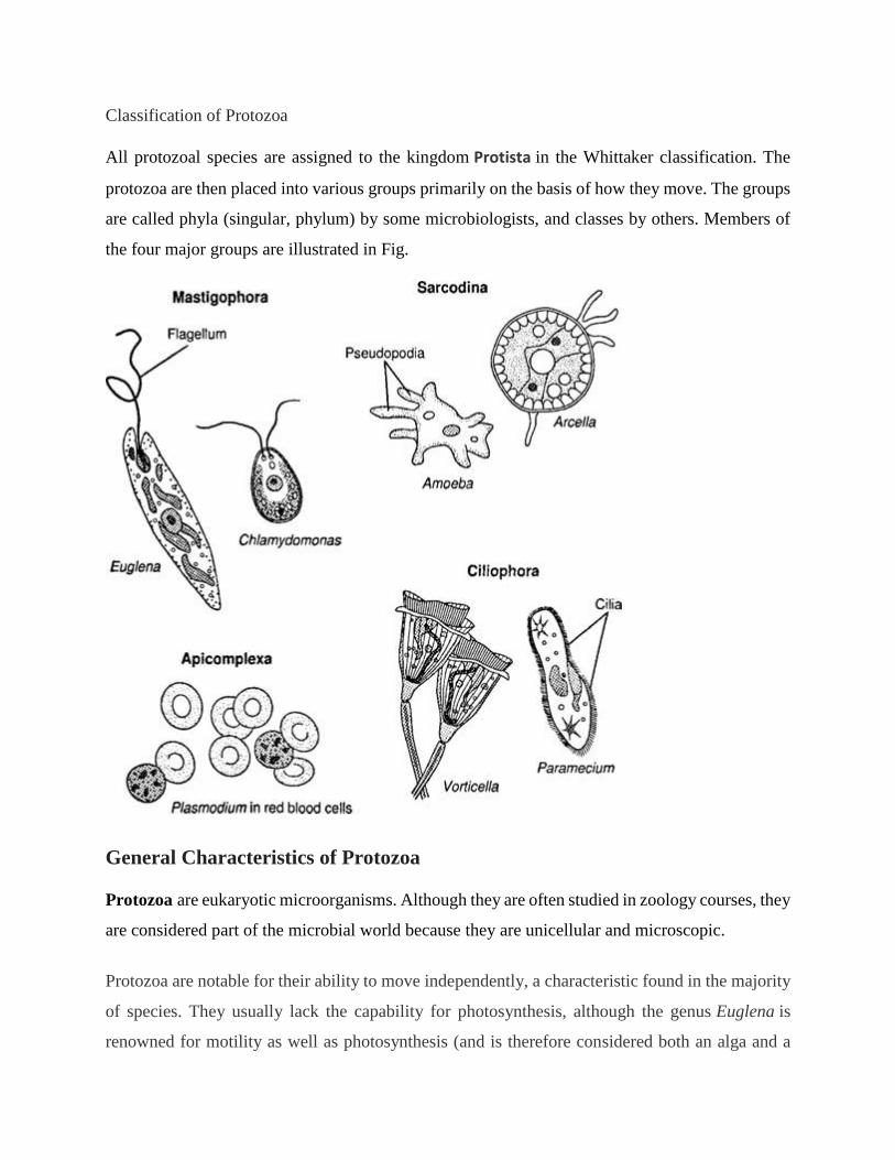
Classification of Protozoa
All protozoal species are assigned to the kingdom Protista in the Whittaker classification. The
protozoa are then placed into various groups primarily on the basis of how they move. The groups
are called phyla (singular, phylum) by some microbiologists, and classes by others. Members of
the four major groups are illustrated in Fig.
General Characteristics of Protozoa
Protozoa are eukaryotic microorganisms. Although they are often studied in zoology courses, they
are considered part of the microbial world because they are unicellular and microscopic.
Protozoa are notable for their ability to move independently, a characteristic found in the majority
of species. They usually lack the capability for photosynthesis, although the genus Euglena is
renowned for motility as well as photosynthesis (and is therefore considered both an alga and a

protozoan). Although most protozoa reproduce by asexual methods, sexual reproduction has been
observed in several species. Most protozoal species are aerobic, but some anaerobic species have
been found in the human intestine and animal rumen.
Protozoa are located in most moist habitats. Free-living species inhabit freshwater and marine
environments, and terrestrial species inhabit decaying organic matter. Some species are parasites
of plants and animals.
Protozoa play an important role as zooplankton, the free-floating aquatic organisms of the oceans.
Here, they are found at the bases of many food chains, and they participate in many food webs.
Size and shape. Protozoa vary substantially in size and shape. Smaller species may be the size of
fungal cells; larger species may be visible to the unaided eye. Protozoal cells have no cell walls
and therefore can assume an infinite variety of shapes. Some genera have cells surrounded by hard
shells, while the cells of other genera are enclosed only in a cell membrane.
Many protozoa alternate between a free-living vegetative form known as atrophozoite and a
resting form called a cyst. The protozoal cyst is somewhat analogous to the bacterial spore, since
it resists harsh conditions in the environment. Many protozoal parasites are taken into the body in
the cyst form.
Most protozoa have a single nucleus, but some have both a macronucleus and one or more
micronuclei. Contractile vacuoles may be present in protozoa to remove excess water, and food
vacuoles are often observed.
Nutrition and locomotion. Protozoa are heterotrophic microorganisms, and most species obtain
large food particles by phagocytosis. The food particle is ingested into a food vacuole. Lysosomal
enzymes then digest the nutrients in the particle, and the products of digestion are distributed
throughout the cell. Some species have specialized structures called cytostomes, through which
particles pass in phagocytosis.
Many protozoal species move independently by one of three types of locomotor organelles:
flagella, cilia, and pseudopodia. Flagella and cilia are structurally similar, having a “9-plus-2”
system of microtubules, the same type of structure found in the tail of animal sperm cells and
certain cells of unicellular algae. How a protozoan moves is an important consideration in
assigning it to a group.

2. Phylum Porifera- Characteristics, classification, examples
Porifera Definition
The Porifera may be defined as an asymmetrical or radially symmetrical multicellular organism
with a cellular grade of an organization without well- definite tissues and organs; exclusively
aquatic; mostly marine, sedentary, solitary or conical animals with body perforated by pores,
canals, and cambers through which water flows; with one or more internal cavities lined with
choanocytes; and with a characteristic skeleton made of calcareous spicules, siliceous spicules or
horny fibers of spongin.
Phylum Porifera Characteristics
1. Porifera are all aquatic, mostly marine except one family Spongillidae which lives in
freshwater.
2. They are sessile and sedentary and grow like plants.
3. The body shape is vase or cylinder-like, asymmetrical, or radially symmetrical.
4. The body surface is perforated by numerous pores, the Ostia through which water enters the
body and one or more large openings, the oscula by which the water exists.
5. The multicellular organism with the cellular level of body organization. No distinct tissues
or organs.
6. They consist of outer ectoderm and inner endoderm with an intermediate layer of
mesenchyme, therefore, diploblastic
7. The interior space of the body is either hollow or permeated by numerous canals lined with
choanocytes. The interior space of the sponge body is called spongocoel.
8. Characteristic skeleton consisting of either fine flexible spongin fibers, siliceous spicules, or
calcareous spicules.
9. Mouth absent, digestion intracellular.
10. Excretory and respiratory organs are absent.
11. Contractile vacuoles are present in some freshwater forms.
12. The nervous and sensory cells are probably not differentiated.
13. The primitive nervous system of neurons arranged in a definite network of bipolar or
multipolar cells in some, but is of doubtful status.
14. The sponges are monoecious.
15. Reproduction occurs by both sexual and asexual methods.
16. Asexual reproduction occurs by buds and gemmules.
17. The sponge possesses a high power of regeneration.
18. Sexual reproduction occurs via ova and sperms.
19. All sponges are hermaphrodite.
20. Fertilization is internal but cross-fertilization can occur.
21. Cleavage holoblastic.
22. Development is indirect through a free-swimming ciliated larva called amphiblastula or
parenchymula.
23. The organization of sponges are grouped into three types which are ascon type, sycon type,
and leuconoid type, due to simple and complex forms.
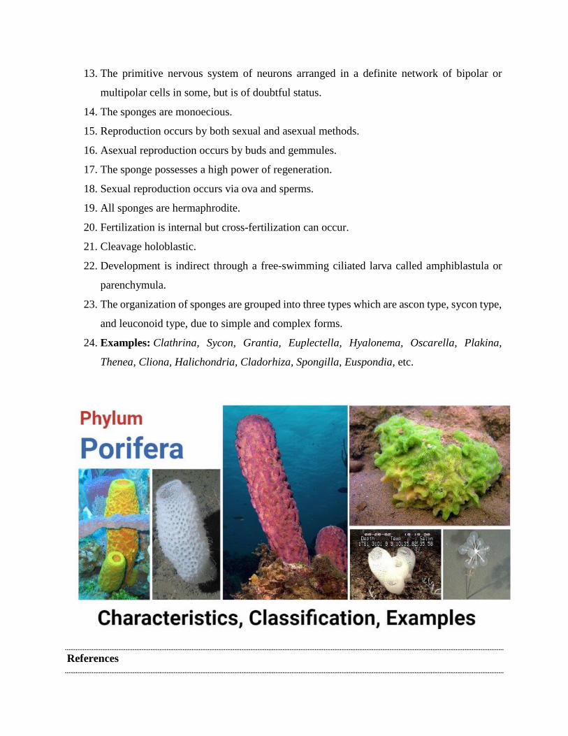
24. Examples: Clathrina, Sycon, Grantia, Euplectella, Hyalonema, Oscarella, Plakina,
Thenea, Cliona, Halichondria, Cladorhiza, Spongilla, Euspondia, etc.
References
1. Kotpal RL. 2017. Modern Text Book of Zoology- Invertebrates. 11th Edition. Rastogi
Publications.
2. Jordan EL and Verma PS. 2018. Invertebrate Zoology. 14th Edition. S Chand Publishing.
3. Phylum Coelenterata (Cnidaria)- Characteristics, classification, examples
Phylum Coelenterata (Cnidaria) Characteristics
1. They are aquatic, mostly marine except few freshwater forms like the hydra.
2. They are multicellular with tissue grade of organization.
3. They are solitary or conical. Sedentary or free-swimming.
4. Individuals are radially or biradially symmetrical about a longitudinal oral-aboral axis.
5. Body organization of cell-tissue grade. Cells mostly scattered and specialized for different
functions. Some cells form tissues like nerve nets or nervous tissues.
6. Exoskeleton chitinous (perisarc) or calcareous(corals).
7. They are diploblastic animals with 2 cellular layers-outer an epidermis and an inner
gastrodermis- with a gelatinous acellular mesoglea in between.
8. Acoelomate animals because they do not pose a second body cavity, the coelom.
9. Short and slender tentacles encircle the mouth in one or two whorls.
10. The tentacles are provided with nematocysts; tentacles serve for food capture, its ingestion,
serve for adhesion, and for defense.
11. Two types of individuals occur, attached sessile and asexual zooid (polyps) and free
swimming and sexual zooid (medusae). Some species are notable for polymorphism or
variety of forms.
12. They are usually carnivorous; digestion is extracellular as well as intracellular.
13. No anus.
14. Coelom and respiratory, circulatory, and excretory system wanting.

15. Nervous system primitive, consisting of a diffuse nerve net. Central nervous system absent.
16. The muscular system includes longitudinal and circular fibers formed by epithelia-muscle
and endothelial-muscle cells.
17. A single cavity, lined with gastrodermis, called gastrovascular cavity or coelenteron, into
which mouth opens.
18. Sensory organs form ocelli and statocysts.
19. Reproduction is both by asexual and sexual methods.
20. Asexual reproduction occurs by budding and sexual reproduction by the formation of
gametes.
21. The development includes a free-swimming ciliated planula larva.
22. Life history exhibits the phenomena of alternation of generation or metagenesis in which the
asexual polypoid, sessile generation alternates with sexual medusoid, free-swimming
generation.
4. Phylum Platyhelminthes- characteristics, classification, examples
Platyhelminthes (flatworms) definition
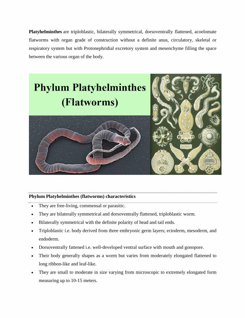
Platyhelminthes are triploblastic, bilaterally symmetrical, dorsoventrally flattened, acoelomate
flatworms with organ grade of construction without a definite anus, circulatory, skeletal or
respiratory system but with Protonephridial excretory system and mesenchyme filling the space
between the various organ of the body.
Phylum Platyhelminthes (flatworms) characteristics
They are free-living, commensal or parasitic.
They are bilaterally symmetrical and dorsoventrally flattened, triploblastic worm.
Bilaterally symmetrical with the definite polarity of head and tail ends.
Triploblastic i.e. body derived from three embryonic germ layers; ectoderm, mesoderm, and
endoderm.
Dorsoventrally fattened i.e. well-developed ventral surface with mouth and gonopore.
Their body generally shapes as a worm but varies from moderately elongated flattened to
long ribbon-like and leaf-like.
They are small to moderate in size varying from microscopic to extremely elongated form
measuring up to 10-15 meters.
Their body is unsegmented except in class Cestoda.
The majority of them are white, colorless and some derive color from ingested food while
free-living form are grey, brown-black or brilliantly colored.
Their anterior end of the body is differentiated into the head.
Mouth and genital pores on the ventral surface are well marked in turbellarians but less
marked in cestodes and trematodes.
Their parasitic form has adhesive structures like hooks, spines and suckers, and adhesive
secretions.
The body is covered with cellular or syncytial, frequently ciliated epidermis; while
trematodes cestodes, lacks epidermis and their body covered with cuticle.
Exo- and endoskeleton are completely absent, hence the body is generally soft. The hard part
consists of cuticle, spines, thorns, hooks, teeth.
They are acoelomate i.e. without any body cavity.
Space between various organs filled with special mesodermal tissues, the mesenchyme, and
parenchyma.
Their digestive system is branched and incomplete without an anus and totally absent in
acoela and cestode.
They lack skeletal, respiratory and circulatory systems.
The excretory system includes a lateral canal and a single or pair of protonephridia with flame
cells or bulbs. Absent in some primitive form.
Their nervous system is primitive, ladder-like. The main nervous system consists of a pair of
ganglia or brain and one or three pairs of longitudinal nerve cords connected by transverse
nerves.
Their sense organs are simple. A common occurrence in tubellaria but greatly reduced in
parasitic form. Chemo- and tangoreceptors commonly in the form of ciliated pits and grooves.
They are mostly monoecious (hermaphrodite).
Their reproductive system is highly evolved or complex in most of the forms.
Asexual reproduction occurs by fission in many freshwater turbellaria.
In the majority of form, eggs are devoid of yolk. They are produced separately in the yolk or
vitelline glands.
Fertilization is internal but cross-fertilization in trematodes and self-fertilization in cestodes.
Their life cycle is complicated involves one or more hosts.
Parthenogenesis and polyembryony commonly occur trematodes and tapeworms.
Some tapeworm propagates by endogenous or exogenous budding.
The flatworm is either free-living or ecto-or endocommensals or parasitic.
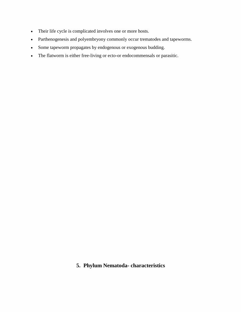
5. Phylum Nematoda- characteristics
Nematoda Definition
Nematodes (Gr., nema thread+ eidos, form) are commonly referred to as non-segmented
roundworm, threadworm or pinworm, as distinct from flatworm and higher segmented annelids.
Phylum Nematoda Characteristics
They are widely distributed, aquatic or terrestrial, parasitic or free-living.
Their body is elongated, cylindrical, unsegmented, worm-like, bilaterally symmetrical and
tapering at both ends.
They are triploblastic animals with perivisceral cavity more extensive than that
of platyhelminths.
The body is of organ -system grade organization.
The body is generally covered with thick, flexible multi-layered collagenous cuticle and often
bears cuticle setae (hairs), spines or annulations.
Cuticle moulted periodically.
They have cellular or syncytial epidermis I.e. the nuclei are not separated from each other by
cell membranes.
They consist of only longitudinal muscle fibers with four bands.
They lack true coelom. The body cavity is pseudocoel or blastocoel not lined by mesoderm
and filled with parenchyma in most cases.
They lack cilia.
circulatory and respiratory systems are absent. i.e. respiration occurs through general body
surface and aerobic in free-living form and anaerobic in parasitic form.
Internal cephalization is present but externally there is little differentiation between the
anterior and posterior region. i.e. distinct head is lacking. However, the mouth is present in
the anterior region.
The digestive system is complete with a distinct mouth and anus. Muscular pharynx and the
inner surface of the gut usually not lined by cilia.
Extracellular digestion occurs in them.
The mouth is surrounded by six lips.
Excretory without flame cell and nephridia. In the class Adenophorea glandular renette cells
with the duct.
The nervous system is not much developed. i.e. consists of circucumpharyngeal ring and
longitudinal nerve cord.
Sense organ are poorly developed in the form of papillae, which are well defined as amphid
(in mouth) and plasmid (anus).
Sexes are separate (gonochoristic). the male is smaller than females.
Tubular gonad is present in them. Male genital duct leads into the cloaca. Female genital
ducts with a separate opening.
Amoeboid sperm cells.
No asexual reproduction.
Fertilization is internal or maybe cross or self.
Development may be direct, with or without an intermediate host or indirect.
Various lateral lines and pores are present on the surface of the body.
6. Phylum Annelida- characteristics
Annelida definition
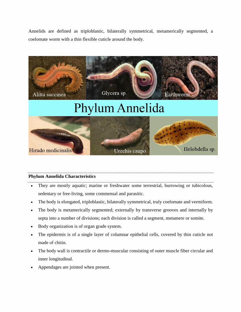
Annelids are defined as triploblastic, bilaterally symmetrical, metamerically segmented, a
coelomate worm with a thin flexible cuticle around the body.
Phylum Annelida Characteristics
They are mostly aquatic; marine or freshwater some terrestrial, burrowing or tubicolous,
sedentary or free-living, some commensal and parasitic.
The body is elongated, triploblastic, bilaterally symmetrical, truly coelomate and vermiform.
The body is metamerically segmented; externally by transverse grooves and internally by
septa into a number of divisions; each division is called a segment, metamere or somite.
Body organization is of organ grade system.
The epidermis is of a single layer of columnar epithelial cells, covered by thin cuticle not
made of chitin.
The body wall is contractile or dermo-muscular consisting of outer muscle fiber circular and
inner longitudinal.
Appendages are jointed when present.
Locomotory organs are segmentally repeated chitinous bristles called setae or chaetae,
embedded in the skin. It may be bored by lateral fleshy appendages or parapodia.
The presence of true schizocoelous coelom usually divided into compartments by transverse
septa. Mostly well-developed in leeches. Coelomic fluid with cells or corpuscles.
The alimentary canal is straight tube-like, complete, extending from mouth to anus. Digestion
is entirely extracellular.
Respiration occurs through moist skin or gills of parapodia and head.
The blood vascular system is a closed type. Blood is red due to the presence of hemoglobin
or erythromycin dissolved in plasma.
Excretion is by metamerically disposed coiled tubes; nephridia which communicate the
coelom to the exterior.
The nervous system consists of a pair of cerebral ganglia; brain and double ventral nerve cord
having segmentally arranged ganglia and lateral nerves in each segment.
Receptor organs include tactile organs, taste buds, statocysts, photoreceptor cells and
sometimes eyes with lenses in some.
They are monoecious i.e. hermaphroditic or sexes separate cleavage spiral and determinate;
dioecious or unisexual form also present.
Their development is direct in monoecious form but indirect in dioecious form.
Larva, when present is a trochophore is characteristics in case of indirect development, while
in others this stage is passed through development.
Regeneration is common.
Asexual reproduction occurs in some.
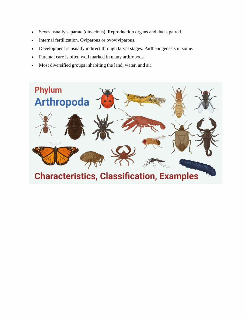
7. Phylum Arthropoda- Characteristics
Arthropoda (Arthropods) Definition
Arthropods are bilaterally symmetrical, triploblastic, metamerically segmented animals with
coelom which is reduced and modified. Their body is covered externally in a chitinous exoskeleton
which molts periodically and their appendages are joined.
Phylum Arthropoda Characteristics
They are bilaterally symmetrical, triploblastic, metamerically segmented animals.
Body organization is of an organ-system level.
The body is covered with a thick chitinous cuticle forming an exoskeleton.
Body segments usually bear lateral and jointed appendages with varied functions as jaws,
gills, legs, etc.
Body divisible into head, thorax, and abdomen. Head and thorax often fused to form a
cephalothorax.
The musculature is not continuous but comprises separate striped muscles capable of rapid
contraction.
The body cavity is hemocoel. The true coelom is reduced to the spaces of the genital and
excretory organs.
The complete digestive system with mouth and anus. Mouthparts adapted for various modes
of feeding.
Open circulatory system with dorsal heart and arteries but without capillaries.
Respiration by general body surface, gills in aquatic forms, trachea, or book-lungs in
terrestrial forms.
No true nephridia. Excretion organs are green glands or Malpighian tubules or coxal glands.
The nervous system is typically annelidan, with a dorsal brain connected with a nerve ring to
a double ventral nerve cord.
Cilia are entirely absent from all parts of the body.
Sensory organs comprise eyes (simple and compound), chemo- and tactile receptors,
balancing and auditory organs.
Sexes usually separate (dioecious). Reproduction organs and ducts paired.
Internal fertilization. Oviparous or ovoviviparous.
Development is usually indirect through larval stages. Parthenogenesis in some.
Parental care is often well marked in many arthropods.
Most diversified groups inhabiting the land, water, and air.
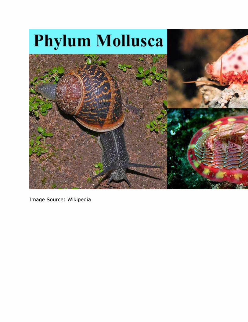
8. Phylum Mollusca- characteristics
Mollusca (Mollusks) Definition
Molluscs (also know as mollusks) are soft-bodied, bilaterally symmetrical, segmented, coelomate
animals; usually shelled having a mantle, ventral foot, anterior head, and a dorsal visceral mass.
Phylum Mollusca (Mollusks) Characteristics
They are essentially aquatic mostly marine, few freshwater and some terrestrial form.
They may be found as hidden parasites in the interior of other animals.
They vary in size from giant squids and clams to little snails, a millimeter long.
They have at least two characters radula and mantle not found elsewhere.
The body is soft, unsegmented (except in Monoplacophora), bilaterally symmetrical,
coelomate, triploblastic.
They have tissue-system grade of body organization
The body consists of head, foot, mantle, and the visceral mass.
The body is clothed with one-layered often ciliated epidermis.
The body is commonly protected by an exoskeleton calcareous shell of one or more piece
secreted by the mantle.
Head is distinct, bearing mouth, eyes, tentacles and other sense organs except in pelecypoda
and scaphodoa.
The ventral body is modified into a muscular plough-like surface, the foot which is variously
modified for creeping, burrowing and swimming.
Mantle or pallium is a fold of a body wall that leaves between itself the main body, mantle
cavity.
The visceral mass contains the vital organs of the body in the compact form taking the form
of dorsal humps or dome.
The body cavity is hemocoel. The coelom is reduced and represented mainly by the
pericardial cavity, gonadial cavity, and nephridia.
The digestive tract is simple with anterior mouth and posterior anus but in gastropods,
scaphodos, and cephalopods the intestine becomes U-shaped bringing anus to anterior part.
Rasping organs, radula usually present, except in pelecypoda.
The circulatory system is open type except in cephalopods.
Respiratory organs contain numerous gills or ctenidia usually provided with osphradiuma at
the base. The lung is developed in terrestrial forms.
Respiration is direct or by gills or lungs or both.
Haemocyanin is their respiratory pigments.
Excretion is by paired metanephridia (kidney).
The nervous system consists of paired cerebral, pleural, pedal and visceral ganglia joined by
longitudinal and transverse connections and nerves. Ganglia usually form a circumenteric
ring.
Sense organs consist of eyes, statocysts, and receptors for touch, smell, and taste.
Sexes are usually separate (dioecious) but some are monoecious (hermaphroditic).
Fertilization is external or internal.
Development is direct or with metamorphosis through the trochophore stage called veliger
larva.
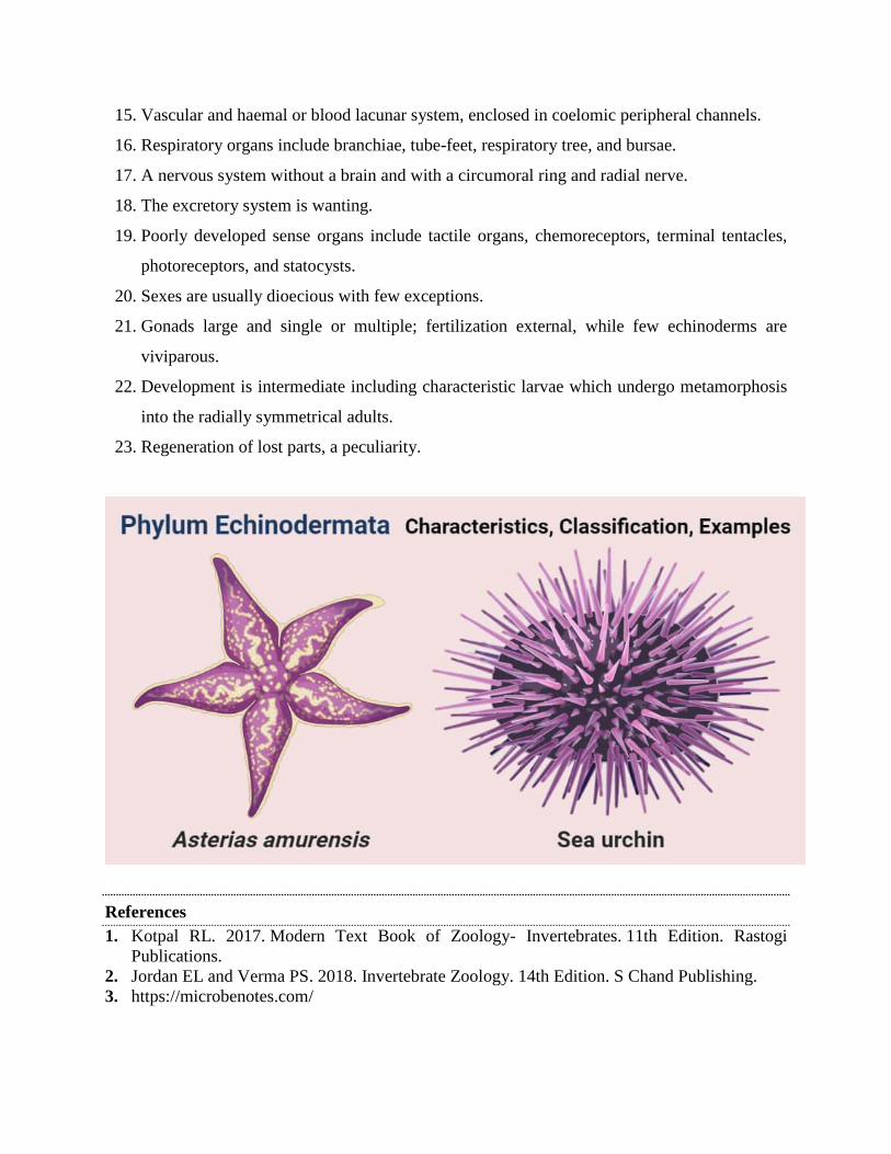
9. Phylum Echinodermata- Characteristics
Echinodermata (Echinoderms) Definition
Echinoderms are enterocoelous coelomates with pentamerous radial symmetry, without distinct
head or brain having a calcareous endoskeleton of separate plates or pieces and a peculiar water
vascular system of coelomic origin with podia or tube-feet projecting out of the body.
Phylum Echinodermata Characteristics
1. They are exclusively marine and are among the most common and widely distributed marine
animals.
2. They occur in all seas from the intertidal zones to great depths.
3. They have an organ grade system of body organization.
4. Symmetry usually radial, nearly always pentamerous.
5. The body is triploblastic, coelomate with distinct oral and aboral surfaces, and without
definite head and segmentation.
6. They are moderate to considerable size but none are microscopic.
7. Body shape globular, star-like, spherical, discoidal, or elongated.
8. The surface of the body is rarely smooth, typically covered by 5 symmetrically radiating
grooves called ambulacra with 5 alternating inter-radii or inter-ambulacra.
9. The body wall consists of an outer epidermis, a middle dermis, and an inner lining of the
peritoneum.
10. Endoskeleton consists of closely fitted, plates forming a shell usually called theca or test or
may be composed of separate small ossicles.
11. The coelom is spacious lined by peritoneum, occupied mainly by the digestive and
reproductive system, and develops from embryonic archenteron i.e. enterocoel.
12. Coelom of enterocoelous type constitutes the perivascular cavity of water vascular system;
coelom fluid with coelomocytes.
13. Water -vascular system of coelomic origin, including podia or tube feet for locomotion and
usually with a madreporite.
14. The alimentary canal is usually a coiled tube extending from the mouth located on the oral
surface to the anus on the aboral or oral surface.
15. Vascular and haemal or blood lacunar system, enclosed in coelomic peripheral channels.
16. Respiratory organs include branchiae, tube-feet, respiratory tree, and bursae.
17. A nervous system without a brain and with a circumoral ring and radial nerve.
18. The excretory system is wanting.
19. Poorly developed sense organs include tactile organs, chemoreceptors, terminal tentacles,
photoreceptors, and statocysts.
20. Sexes are usually dioecious with few exceptions.
21. Gonads large and single or multiple; fertilization external, while few echinoderms are
viviparous.
22. Development is intermediate including characteristic larvae which undergo metamorphosis
into the radially symmetrical adults.
23. Regeneration of lost parts, a peculiarity.
References
1. Kotpal RL. 2017. Modern Text Book of Zoology- Invertebrates. 11th Edition. Rastogi
Publications.
2. Jordan EL and Verma PS. 2018. Invertebrate Zoology. 14th Edition. S Chand Publishing.
3. https://microbenotes.com/
Phylum Chordata
The phylum Chordata contains all animals that have a dorsal notochord at some stage of
development; in most cases, this is the backbone.
Key Points
The phylum chordata is named for the notochord, a longitudinal, flexible rod between the
digestive tube and the nerve cord; in vertebrates, this is the spinal column.
The chordates are also characterized by a dorsal nerve cord, which splits into the brain and
spinal cord.
Chordata contains two clades of invertebrates: Urochordata (tunicates) and
Cephalochordata (lancelets), both of which are suspension feeders.
The phylum chordata includes all animals that share four characteristics, although they
might each possess some of them at different stages of their development: a notochord, a
dorsal nerve cord, pharyngeal slits, and a postanal tail.
Chordata contains five classes of animals: fish, amphibians, reptiles, birds, and mammals;
these classes are separated by whether or not they can regulate their body temperature, the
manner by which they consume oxygen, and their method of reproduction.
Key Terms
dorsal nerve cord: a hollow cord dorsal to the notochord, formed from a part of the
ectoderm that rolls, forming a hollow tube.
notochord: a flexible rodlike structure that forms the main support of the body in the
lowest chordates; a primitive spine
pharyngeal slit: filter-feeding organs found in non-vertebrate chordates (lancelets and
tunicates) and hemichordates living in aquatic environments

Phylum Chordata
Animals in the phylum Chordata share four key features that appear at some stage of their
development:
A notochord, or a longitudinal, flexible rod between the digestive tube and the nerve cord.
In most vertebrates, it is replaced developmentally by the vertebral column. This is the
structure for which the phylum is named.
A dorsal nerve cord which develops from a plate of ectoderm that rolls into a tube located
dorsal to the notochord. Other animal phyla have solid nerve cords ventrally located. A
chordate nerve cord splits into the central nervous system: the brain and spinal cord.
Pharyngeal slits, which allow water that enters through the mouth to exit without
continuing through the entire digestive tract. In many of the invertebrate chordates, these
function as suspension feeding devices; in vertebrates, they have been modified for gas
exchange, jaw support, hearing, and other functions.
A muscular, postanal tail which extends posterior to the anus. The digestive tract of most
nonchordates extends the length of the body. In chordates, the tail has skeletal elements
and musculature, and can provide most of the propulsion in aquatic species.
In some groups, some of these traits are present only during embryonic development. In addition
to containing vertebrate classes, the phylum Chordata contains two clades of invertebrates:
Urochordata (tunicates) and Cephalochordata (lancelets). However, even though they are
invertebrates, they share characteristics with other chordates that places them in this phylum. For
example, tunicate larvae have both a notochord and a nerve cord which are lost in adulthood. Most
tunicates live on the ocean floor and are suspension feeders. Cephalochordates, or lancelets, have
a notochord and a nerve cord (but no brain or specialist sensory organs) and a very simple
circulatory system. Lancelets are suspension feeders that feed on phytoplankton and other
microorganisms.
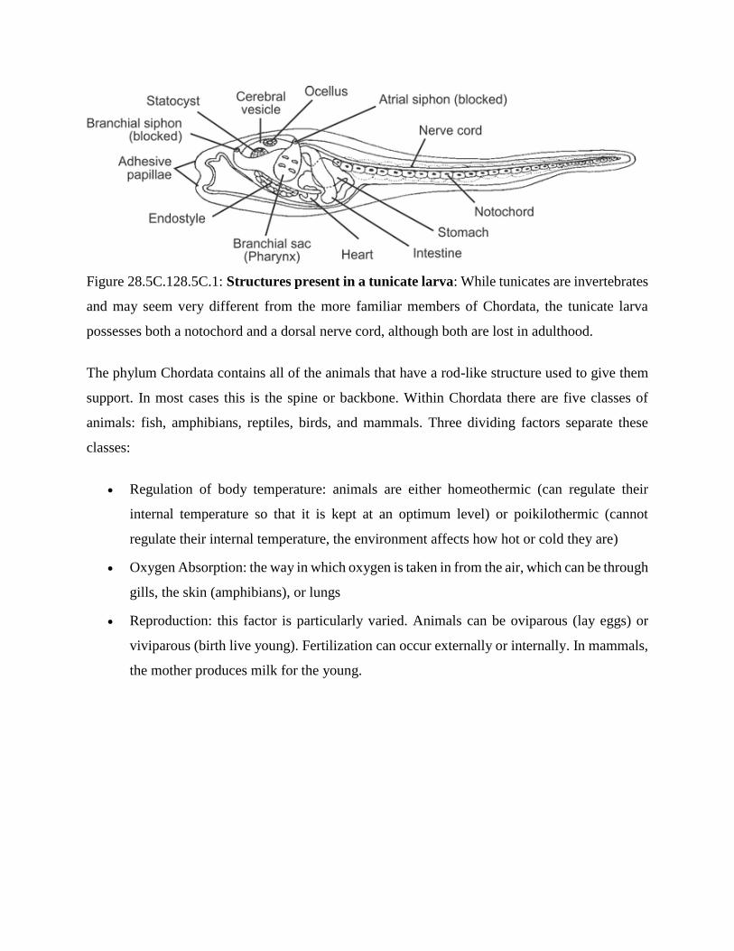
Figure 28.5C.128.5C.1: Structures present in a tunicate larva: While tunicates are invertebrates
and may seem very different from the more familiar members of Chordata, the tunicate larva
possesses both a notochord and a dorsal nerve cord, although both are lost in adulthood.
The phylum Chordata contains all of the animals that have a rod-like structure used to give them
support. In most cases this is the spine or backbone. Within Chordata there are five classes of
animals: fish, amphibians, reptiles, birds, and mammals. Three dividing factors separate these
classes:
Regulation of body temperature: animals are either homeothermic (can regulate their
internal temperature so that it is kept at an optimum level) or poikilothermic (cannot
regulate their internal temperature, the environment affects how hot or cold they are)
Oxygen Absorption: the way in which oxygen is taken in from the air, which can be through
gills, the skin (amphibians), or lungs
Reproduction: this factor is particularly varied. Animals can be oviparous (lay eggs) or
viviparous (birth live young). Fertilization can occur externally or internally. In mammals,
the mother produces milk for the young.
Protochordate
Protochordate, any member of either of two invertebrate subphyla of the phylum Chordata:
the Tunicata (sea squirts, salps, etc.) and the Cephalochordata (amphioxus). Like the remaining
subphylum of the chordates, the Vertebrata, the protochordates have a hollow dorsal nerve cord,
gill slits, and a stiff supporting rod, the notochord, the forerunner of the backbone. The
protochordates differ chiefly from the vertebrates in not having a backbone. Recent protochordates
are thought to have evolved from the same ancestral stock as that which gave rise to the vertebrates.
Two main theories have gained general acceptance as to how the vertebrates may have evolved.
One theory proposes that the ancestral form was sessile (attached), perhaps like a pterobranch but
with an unspecialized larva. This larva adapted to an independent pelagic life and became sexually
mature. Subsequently, the sessile stage was lost, and the vertebrates evolved from this free-
swimming animal. The other, more recent theory postulates that the chordates evolved from a
small fossil group called the mitrates.
Group ACRANIA (=PROTOCHORDATA)
(Primitive chordates without head and vertebral column)
Subphylum HEMICHORDATA, Balanoglossus,Cephalodiscus, Rhabdopleura, primitive and
doubtful chordates, now classified under non-chordates after echinoderms.
Subphylum UROCHORDATA, Herdmania, Salpa, Doliolum, Pyrosoma, Oikopleura sedentary
or planktonic tunicates in which chordate characters manifest in the larval stage.
Subphylum CEPHALOCHORDATA, Amphioxus, Asymmetron, typical chordates having
chordate characters in the larval as well as adult stage.
Group CRANIATA (=EUCHORDATA)
(Chordates with skull, with 54,000 species of true chordates)
Subphylum VERTEBRATA, chordates with head, brain and vertebral column.
Superclass AGNATHA, 90 species of paraphyletic group of jawless fishes, which were also the
first vertebrates. Living forms are elongated, scaleless, slimy parasites and scavengers that include
lampreys and hagfishes. They have no paired fins.
Class OSTRACODERMI, extinct shelled jawless fishes of Ordovician period. Cephalaspsis.
Class CYCLOSTOMATA, jawless fishes of today, without scales and paired fins.
Order Myxinoidea: the hagfishes, 40 species. Myxine, Bdellostoma, Eptatretus.
Order Petromyzontia: lampreys, 41 species, parasitic on other fishes. Petromyzon.
Superclass GNATHOSTOMATA, vertebrates with jaws that are modified gill arches and paired
appendages. They include cartilaginous fishes, bony fishes and tetrapods.
Characteristics of Protochordata
1. They are generally found in marine water.
2. Their body is bilaterally symmetrical, triploblastic, and coelomated.
3. At a certain stage of their lives, their body develops a long, rod-like structure for support
called the notochord.
4. They exhibit organ system level of organization.
E.g., Herdmania, Amphioxus.
Classifications of Protochordata
Hemichordata
They are found in marine water.
Some live solitarily, and some stay in colonies.
The body is cylindrical, unsegmented, and stout.
The body is divided into proboscis, collar, and trunk.
The collar bears arms and tentacles.
They have a complete digestive system.
They respire through gills or general body surface.
The circulatory system comprises a heart with two longitudinal vessels.
The blood has no colour and corpuscles.

The proboscis gland or glomerulus make up the excretory system.
Sexes may be separate or united and fertilization is either internal or external.
E.g., Cephalodiscus, Rhabdopeura.
Explore more: Excretory system.
Urochordata or Tunicata
They are found in the marine environment.
They are sessile and filter-feeders.
They are also known as tunicates because their body is surrounded by a leathery sheath
composed of tunicin (cellulose).
The notochord appears in the larval stage in the tail of the larva and disappears in the adult.
This is known as retrogressive metamorphosis.
The neural tube in the larva is replaced by a dorsal ganglion in the adults.
Respiration occurs through gills.
They have an open circulatory system.
The excretory organs are absent.
They reproduce asexually by budding.
E.g., Herdmania, Selpa
Cephalochordata
They are marine and filter-feeders.
The notochords remain throughout life and extend up to the head region.
The nerve cord and the tail also remain throughout life.
Solenocyts are the excretory organs.
They respire through gills which open in the atrium.
The body wall comprises myotomes.
E.g., Amphioxus
FISHES
The Superclass Pisces (L. Piscis = fish) are the truly jawed vertebrates. They have organs of
respiration and locomotion related to a permanently aquatic life. The respiratory organs are the
gills and the organs of locomotion are paired and impaired fins. All are poikilothermous.
General Characters:
1. Aquatic, either freshwater or marine, herbivorous or carnivorous, cold blooded, oviparous or
ovoviviparous vertebrates.
2. Body usually streamlined, spindle-shaped, some are elongated snake-like and a few are
dorsoventrally compressed, and differentiated into head, trunk and tail.
3. Locomotion by paired pectoral and pelvic fins along with median dorsal and caudal fins,
supported by true dermal fin-rays. Muscular tail used in propulsion.
4. Exoskeleton of dermal scales, denticles or bony plates (in Placodermi) covering body surface.
Placoid in Chondrichthyes and ganoid, cycloid or ctenoid in Osteichthyes.
5. Endoskeleton is cartilaginous or bony. The notochord in usually replaced by vertebrae, either
bone or cartilage. Presence of well-developed skull and a system of visceral arches, of which the
first pair forms the upper and lower jaws, the latter movably articulated with the skull.
6. Muscles arranged into segments called myotomes, with separate dorsal and ventral parts.
7. Alimentary canal with definite stomach and pancreas and terminates into cloaca or anus.
8. Organs of respiration are gills. Gill-slits 5 to 7 pairs, naked or covered by an operculum.
9. Heart is venous and two chambered, i.e., one auricle and one ventricle. Sinus venosus and renal
and portal systems present. Erythrocytes nucleated. Poikilothermous.
10. Kidneys mesonephros. Excretions ureotelic.
11. Brain with usual five parts. Cranial nerves ten pairs.
12. Nostrils are paired but do not open into pharynx except Dipnoi. Nasal capsules are partly
separate in Chondrichthyes and completely separate in Osteichthyes.
13. Tympanic cavity and ear ossicles are absent.
14. Internal ear with three semicircular canals.
15. Lateral line system is well developed.
16. Sexes separate. Gonads typically paired. Gonoducts open into cloaca or independently.
17. Fertilisation internal or external. Females of Chondrichthyes are oviparous or ovoviviparous
and of Osteichthyes are mostly oviparous and rarely ovoviviparous or viviparous. Eggs with large
amount of yolk. Cleavage meroblastic.
18. Extra-embryonic membranes are absent.
19. Development usually direct without or with little metamorphosis.
Classification:
About 40,000 species of fishes are known. Various workers have proposed different schemes of
classification of fishes. However, no classification has been universally accepted because of
confusion due to large number of fishes and great diversity in their shape, size, habits and habitat.
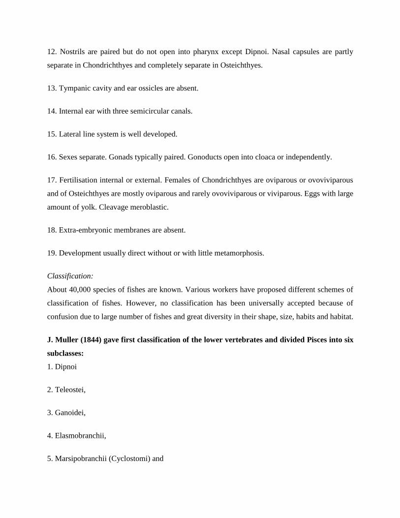
J. Muller (1844) gave first classification of the lower vertebrates and divided Pisces into six
subclasses:
1. Dipnoi
2. Teleostei,
3. Ganoidei,
4. Elasmobranchii,
5. Marsipobranchii (Cyclostomi) and
6. Leptocardii (Amphioxini).
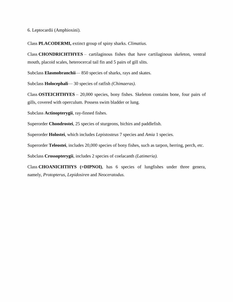
Class PLACODERMI, extinct group of spiny sharks. Climatius.
Class CHONDRICHTHYES – cartilaginous fishes that have cartilaginous skeleton, ventral
mouth, placoid scales, heterocercal tail fin and 5 pairs of gill slits.
Subclass Elasmobranchii— 850 species of sharks, rays and skates.
Subclass Holocephali— 30 species of ratfish (Chimaeras).
Class OSTEICHTHYES – 20,000 species, bony fishes. Skeleton contains bone, four pairs of
gills, covered with operculum. Possess swim bladder or lung.
Subclass Actinopterygii, ray-finned fishes.
Superorder Chondrostei, 25 species of sturgeons, bichirs and paddlefish.
Superorder Holostei, which includes Lepistosteus 7 species and Amia 1 species.
Superorder Teleostei, includes 20,000 species of bony fishes, such as tarpon, herring, perch, etc.
Subclass Crossopterygii, includes 2 species of coelacanth (Latimeria).
Class CHOANICHTHYS (=DIPNOI), has 6 species of lungfishes under three genera,
namely, Protopterus, Lepidosiren and Neoceratodus.

TETRAPODS
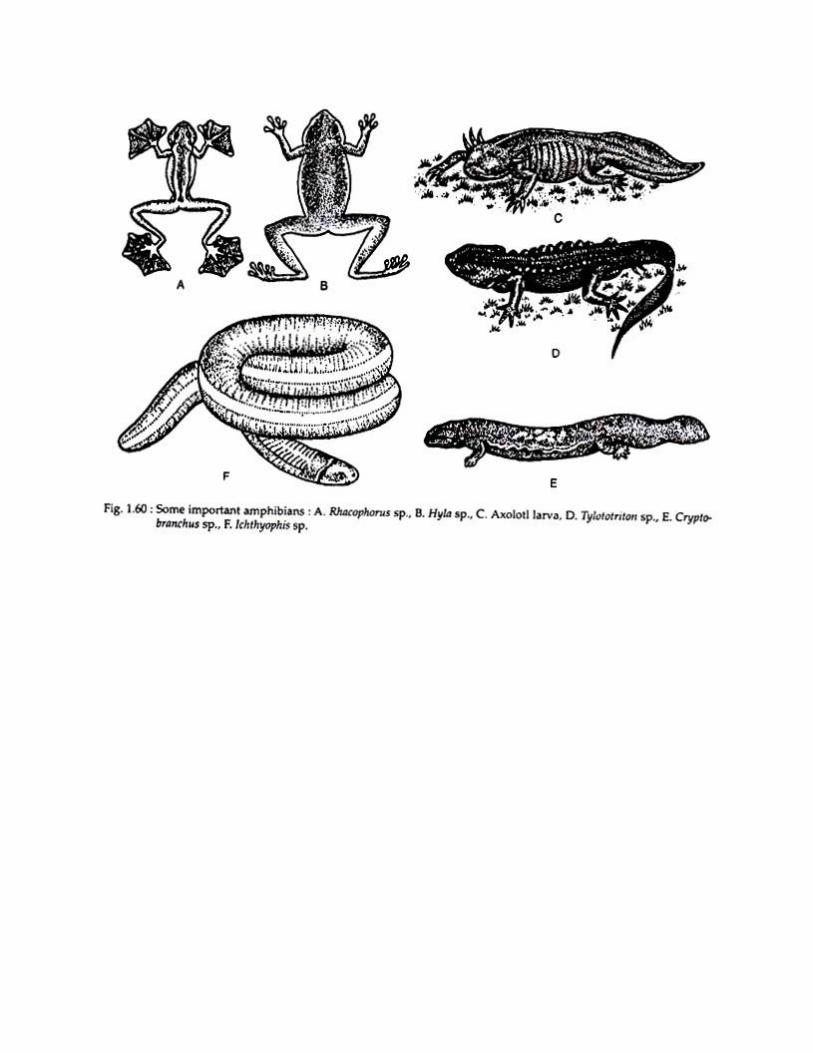
Class AMPHIBIA
Habit and Habitat of Amphibians:
Amphibians are cold blooded vertebrates having a smooth or rough, naked skin, rich in glands,
which keep it moist, if scales are present, it is hidden in the skin. They are quite numerous and
successful in the ecological niches that they occupy and make an important element in many food-
chains. There are nearly 2,000 species identified so far, and placed in 250 genera.
Although amphibians are well adapted for certain situations, it is remarkable that they do not
succeed in maintaining themselves in many different types of habitat. There are desert toads, e.g.,
Chiroleptes of Australia, but these survive by burrowing and by special abilities, such as the power
to hold large amounts of water, associated with loss of the glomeruli of the kidneys.
ADVERTISEMENTS:
The fossils of amphibian ancestors and early amphibians have been discovered in sediments of the
middle and late Devonian period in Greenland and Australia. Therefore, it is assumed that the first
amphibians originated during Devonian period. The classifications of amphibians are made by
various authors in different ways.
Origin of Amphibians:
During the later part of the Devonian period a population of osteolepid fishes started crawling from
pool to pool and spending more time on the land. They gave rise to a terrestrial population that we
distinguish as Amphibia. This is the common idea of amphibian origin. But some herpetologists
argued in different ways — these are as follows.
1. Polyphyletic view of amphibian origin:
Carroll and Currie (1975), Jarvik (1980) hypothesized that the three living orders, e.g., Anura,
Urodela and Apoda have evolved separately. Jarvik also pointed out that the amphibians originated
independently from more than one group of rhipidistian fishes. But this view is readily rejected by
all scientists.
ADVERTISEMENTS:
2. Di-phyletic view of amphibian origin:
Romer (1945), Romer and Watson (1962) opined that both salamanders and caecilians share a
common ancestor and anurans were developed separately. By emphasizing on the vertebral column
similarities in the different groups, it is considered that anurans have evolved from labyrinthodonts
and urodeles and apodans from lepospondyls.
3. Monophyletic view of amphibian origin:
According to this view all living amphibians have evolved from the earliest amphibians the
Ichthyostega and this group is also derived from Osteolepid fish. The proponents of this view are
Noble (1931), Bolt (1979), McFarland (1985), Duellman and Trueb (1986).
General Characters of class Amphibians:
ADVERTISEMENTS:
1. The body of amphibians comprises of a distinct head with elongated trunk. Neck and tail may
be present or absent.
2. Highly glandular, moist skin is naked. In some apodans, dermal scales are present.
3. The body of amphibians is provided with two pairs of pentadactylous limbs.
4. Forelimbs are with four and hind limbs are with five clawless digits.
5. The body is ectothermic (poikilothermous or cold blooded or adjusters).
6. Eyes are often with eyelids. A tympanum is present.
7. Gut ends into a cloaca.
8. Three chambered heart. R.B.C. nucleated.
9. In adults respiration is performed by lungs, skin and buccopharyngeal cavity.
ADVERTISEMENTS:
10. In adult salamanders, kidney is mesonephric type, while in caecillans it is opisthonephric type.
11. Central nervous system possess ten pairs of cranial nerves.
12. Procoelous vertebrae lacks ribs.
13. The skull possesses two occipital condyles. Post temporal fossa and ectopterygoid are absent.
14. Eggs are large, yolky and mesolecithal type.
15. Generally an aquatic tadpole larval stage is present in the life history.
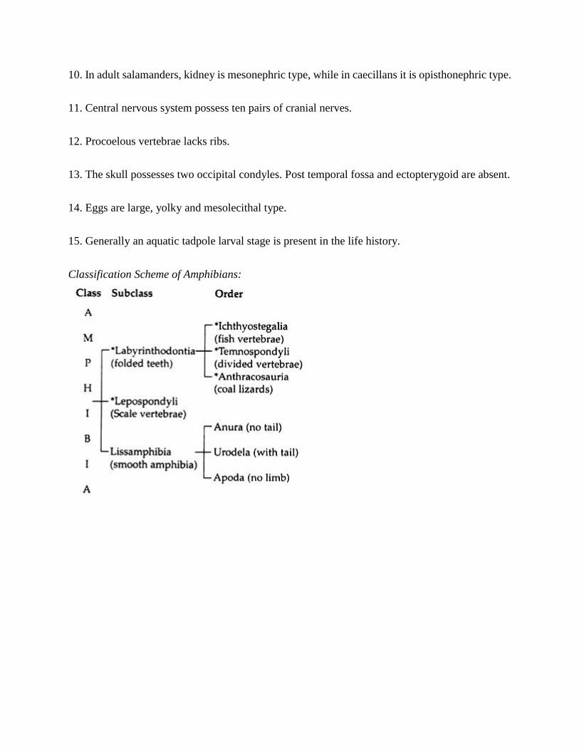
Classification Scheme of Amphibians:
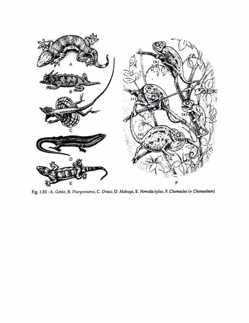
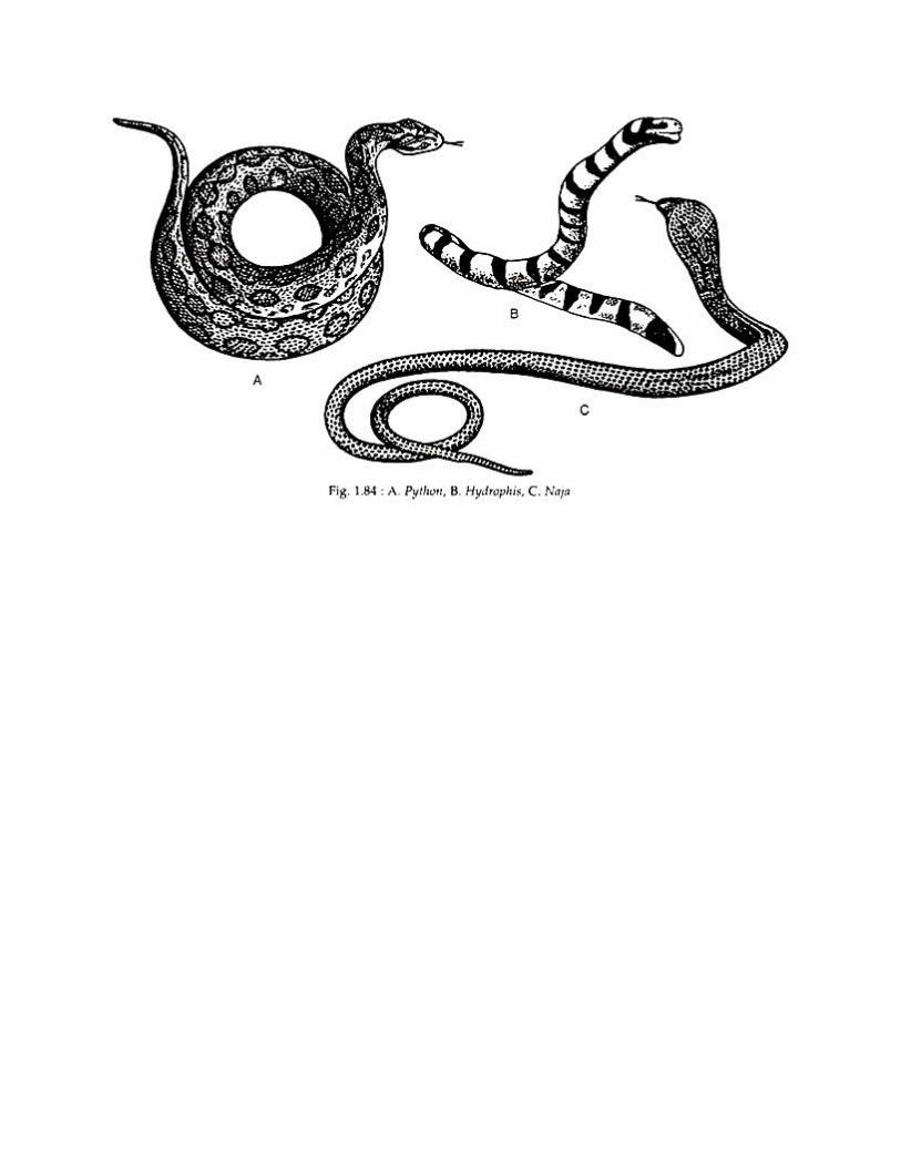
Class REPTILIA,
7800 species, turtles, crocodiles, lizards, snakes, etc.
They have internal fertilization and produce large cleioid eggs with leathery shells and are
ectotherms. Body covered with epidermal scales, vertebrae procoelous.
Reptiles are cold-blooded vertebrates, breath by lungs and having the body covered by scales or
scutes. A basioccipital bone is present in the skull which articulates with the vertebral column by
a single condyle. In 1895, herpetologists separated reptiles from Amphibia as a different class.
They classified reptiles especially on the basis of skeletal characters. The major characteristic
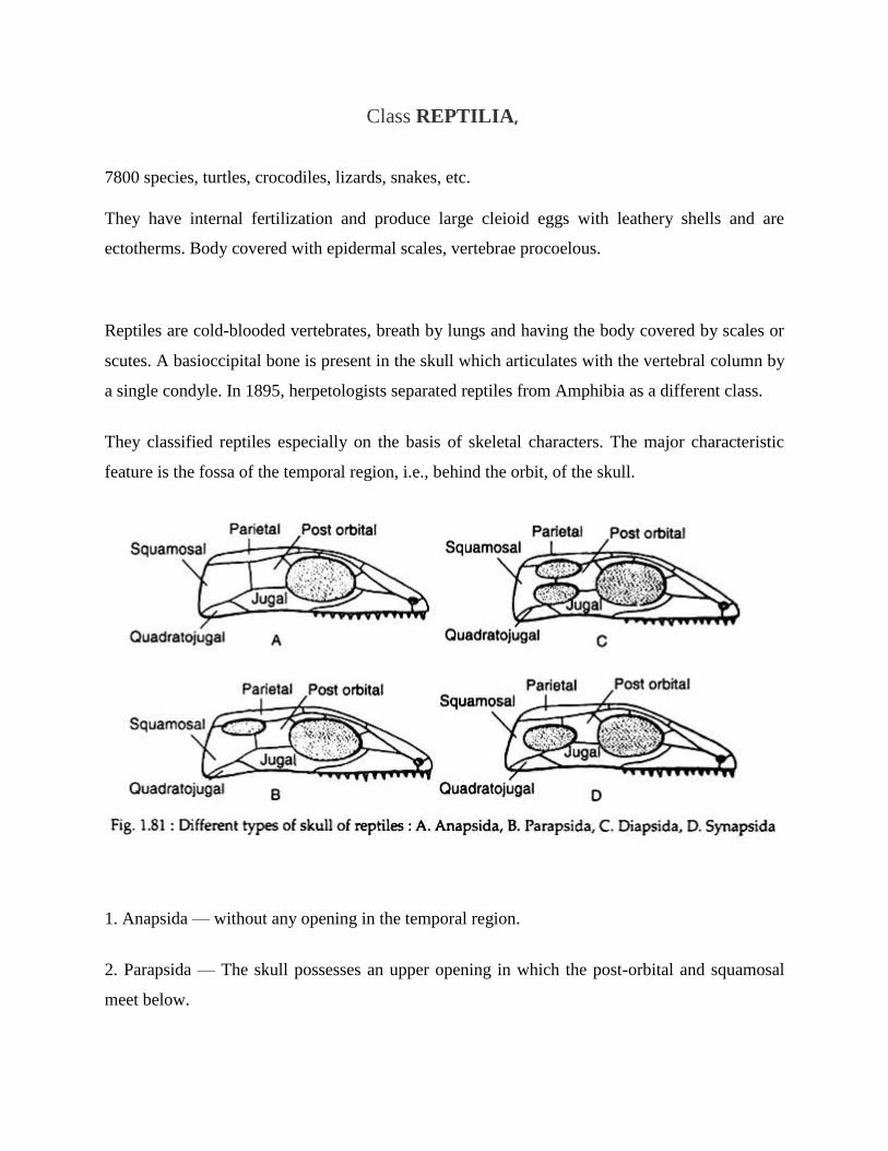
feature is the fossa of the temporal region, i.e., behind the orbit, of the skull.
1. Anapsida — without any opening in the temporal region.
2. Parapsida — The skull possesses an upper opening in which the post-orbital and squamosal
meet below.
ADVERTISEMENTS:
3. Diapsida — In this case there are two openings on each side, separated by pos- torbital and
squamosal bones.
4. Synapsida — In this group a single opening is present with postorbital and squamosal meeting
above.
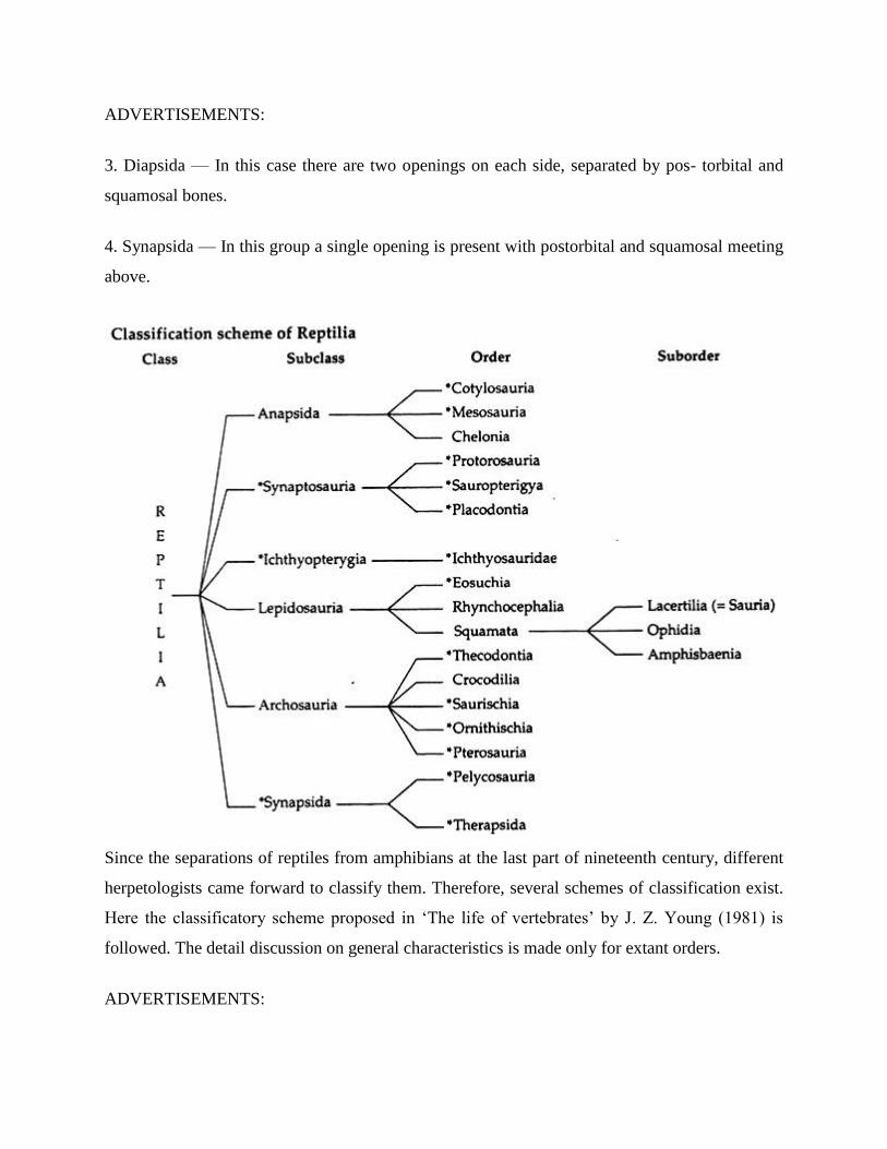
Since the separations of reptiles from amphibians at the last part of nineteenth century, different
herpetologists came forward to classify them. Therefore, several schemes of classification exist.
Here the classificatory scheme proposed in ‘The life of vertebrates’ by J. Z. Young (1981) is
followed. The detail discussion on general characteristics is made only for extant orders.
ADVERTISEMENTS:
Class — Reptilia:
General characters:
1. They are inhabitants of terrestrial and aquatic (both marine and freshwaters) environments.
2. Their skin is dry, cornified and usually covered by epidermal scales or scutes. There are a few
integumentary scent glands secreting pheromones during breeding seasons.
3. Single external nasal opening is present on the snout. Ear drums are slightly depressed.
4. Two pairs of pentadactyle limbs are present. The limbs end in clawed digits.
5. The cloacal opening is either transverse or longitudinal.
6. A post-anal tail is present.
7. The heart is composed of two auricles and a partially divided ventricle. There are right and left
systemic arches.
ADVERTISEMENTS:
8. The kidney is metanephric type.
9. Mullerian duct persists as oviduct in female and Wolffian duct is retained as vas deference in
male. Males possess copulatory organs.
10. Twelve pairs of cranial nerves are present.
11. Vomero-nasal organ (Organ of Jacobson) is well-developed.
12. Single occipital condyle in the skull is present for the attachment with atlas.
13. Mandible consists usually six pieces of bones.
14. Vertebrae are procoelous. Sternum is greatly developed with ribs.
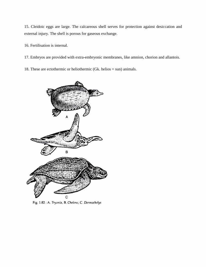
15. Cleidoic eggs are large. The calcareous shell serves for protection against desiccation and
external injury. The shell is porous for gaseous exchange.
16. Fertilisation is internal.
17. Embryos are provided with extra-embryonic membranes, like amnion, chorion and allantois.
18. These are ectothermic or heliothermic (Gk. helios = sun) animals.

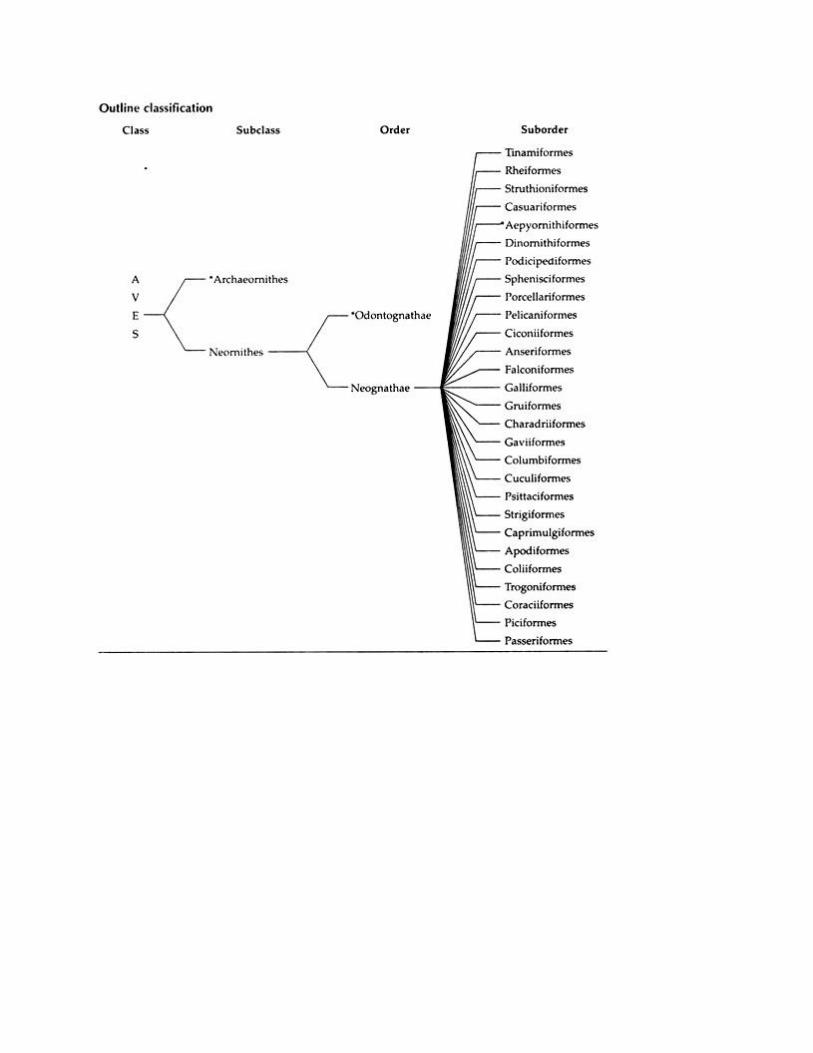
Class AVES, 9100 species.
Birds being feathered bipeds have internal fertilization and lay hard-shelled eggs and are
endotherms. Nearly every anatomical feature is related to ability to fly. They are the only animals
with feathers that are modified from reptilian scales.
General Characters of Birds:
1. Birds have spindle-shaped body is highly aero dynamically suitable and covered by feathers.
Birds are homoieothermal animals.
2. Small head is placed on a fairly long movable neck.
3. Mouth is provided with a specialised exoskeletal derivative called beak. Teeth are absent in
Birds.
4. Fore limbs are modified as wings, which is powered by strong flight muscles.
5. Hind limbs of birds possess four clawed digits.
6. Eyes of birds possess pecten.
ADVERTISEMENTS:
7. Bones become pneumatic to reduce body weight.
8. Alimentary canal contains gizzard for crushing the food due to absence of teeth.
9. Specialised respiratory system performs double respiration. Air sacs are present in association
with lungs.
10. Syrinx is the sound producing organ.
ADVERTISEMENTS:
11. Heart of birds is four-chambered. Only right aortic arch is present.
12. Kidney is metanephric type. Urine is semisolid. Urinary bladder is absent.
13. Only left ovary is present, right ovary absent in birds.
14. These are oviparous animals having telolecithal eggs. Cleavage is meroblastic.
15. During embryonic development four types of extra-embryonic membranes appear. These are
chorion, amnion, allantois and yolk sac.
16. Cloaca is divided into three chambers — coprodeum, urodeum and proctodeum.
17. Highly developed nervous system includes brain and sense organs.
Classification of Birds:
As in other chordates, Class Aves is also classified in various ways by various authors. The scheme
of classification adopted here is based on Young (1981) edn.
Class MAMMALIA, 4,500 species.
Mammals evolved in the late Triassic, the time dinosaurs first appeared and diversified greatly
following the extinction of dinosaurs during the Coenozoic. Characteristics include hairs for
protection and from heat loss; mammary glands; heterodont teeth; endothermy; 4 chambered heart
etc.
Definition of Mammals:
Among vertebrates, mammals became most fully suited for life on land. There are many species
of mammals in which the process of life are carried on under conditions far remote from those in
which life first arose.
The information in their DNA provides them with numerous special adaptive devices. The success
of the mammals in maintaining life in strange environments is largely due to the remarkable
powers they possess of keeping their own composition constant.
ADVERTISEMENTS:
Besides the regulation of temperature, there is also regulation of nearly all components of the
blood, which are kept constant within narrow limits. Therefore, the most characteristic features of
the modern mammals are seen to be largely in their behaviour and soft structures.
Mammals can be defined as ‘highly percipient and mobile animals, with large brains, spiral
cochlea, warm blood, left aortic arch, and water-proof, usually hairy skin, whose young are born
alive, and are nourished by milk.
General Characters of Mammals:
1. Body of mammals is covered by epidermal hair.
2. Integumentary glands are — sweat (sudoriferous), sebaceous (oil), scent (odoriferous) glands.
ADVERTISEMENTS:
3. Mammary glands are present to supply milk for the nourishment of suckling young.
4. External fleshy pinna is present in mammals.
5. Eyes with upper and lower eyelids and often with eyelashes.
6. Nictitating membrane is translucent and hairless; it is vestigial in higher mammals.
ADVERTISEMENTS:
7. A muscular diaphragm is present in between the thoracic and abdominal cavities.
8. Endo-thermal homoeotherm animals.
9. RBCs are non-nucleated, biconcave and usually circular in form.
10. The four-chambered heart is highly powerful.
11. Only left aortic arch is present in the arterial system.
12. Cerebral hemispheres are very large and highly convoluted.
13. Cerebellum is large, complex and solid in mammals.
14. There is a single urinary bladder in mammals.
15. Testes remain in scrotal sacs.
ADVERTISEMENTS:
16. Small eggs are devoid of yolk. Fertilisation is internal.
17. Mammals are viviparous animals.
18. The skull has double occipital condyles. Quadrate absent.
19. A bony palate is formed by the union of premaxillae, maxillae and palatines that separates the
nasal passage from the buccal cavity.

20. The lower jaw is composed of a pair of bones — the dentaries.
21. Vertebrae are acoelous type.
22. Ribs are double-headed — capitulum and tuberculum.
23. The teeth are heterodont, thecodont and diphyodont type.
24. Molars are tribosphenic (three-cusped).
25. Paired forelimbs and hind limbs are present in mammals.
26. The digits of the limbs are provided with either claw or nail or hoof.
27. Cranial nerves twelve pairs.
28. Kidneys are metanephric type.
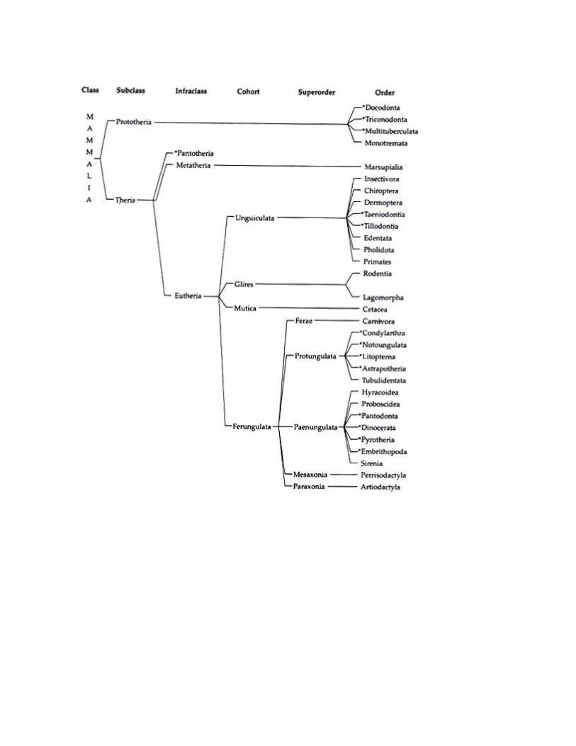
Scheme of Classification of Mammals:
Like other chordates, the classification of mammals is a very controversial and complex matter.
There are several schemes of classification that exists in different literatures. But none of the
existing classifications is beyond criticism.
However, in the present text, classificatory scheme of mammals as proposed by J. Z. Young (1981)
is followed. In the scheme all the groups up to order are mentioned. But, for description, only
living groups are considered. The extinct groups are marked with asterisks (*):
References:
https://www.notesonzoology.com/
UNIT – I
PART-A
Q.NO QUESTIONS CO (LEVEL)
1 Discuss the characteristic features of Protozoa. 1 (1)
2 Briefly explain about the general phylum Annelida 1 (2)
3 DefineArthropoda. 1 (1)
4 DefineMollusca 1 (1)
5 Classify Echinodermata with examples. 1 (2)
6 Explainthe characteristic features of Sharks and Rays 1 (2)
7 List the general characters of Fishes 1 (2)
8 Classify Amphibians 1 (2)
9 Summarize the general features of Avifauna 1 (2)
10 Highlight the importance of Mammalia 1 (1)
PART-B
Q.NO QUESTIONS CO (LEVEL)
1 Classify the phylum Protozoa with suitable diagrams. 1 (2)
2 Discuss in detail the classification of Porifera and Coelenterata 1 (1)
3 Summarize the general characters of phylum Platyhelminthes and
Nematoda 1 (2)
4 Illustrate the phylumAnnelida 1 (2)
5 Discuss the characteristic features of Insects with suitable examples 1 (1)
6 Explain the general characters of Mollusca with suitable figures 1 (2)
7 Classify the fishes with suitable examples 1 (2)
8 Explain the characteristic features of Amphibians with suitable figures 1 (2)
9 Explain the general characters of the phylum Reptiles 1 (2)
10 Describe the following characters of the following phyla
a) Aves b) Mammals
1 (1)

SCHOOL OF BIO & CHEMICAL ENGINEERING
DEPARTMENT OF BIOTECHNOLOGY
SBC1201: ZOOLOGY
UNIT – II - ANATOMY AND PHYSIOLOGY– SBC1201
1. Human Organs and Organ Systems
Human Organs
An organ is a collection of tissues joined in a structural unit to serve a common function. Organs
exist in most multicellular organisms, including humans, other animals, and plants. In single-celled
organisms (such as bacteria), the functional equivalent of an organ is an organelle.
Tissues in Organs
Although organs consist of multiple tissue types, many organs are composed of a main tissue that
is associated with the organ’s major function, along with other tissues that play supporting roles.
The main tissue may be unique to that specific organ. For example, the main tissue of the heart is
cardiac muscle, which performs the heart’s major function of pumping blood and is found only in
the heart. The heart also includes nervous and connective tissues that are required for it to perform
its major function. For example, nervous tissues control the beating of the heart, and connective
tissues make up heart valves that keep blood flowing in just one direction through the heart.
Vital Organs
The human body contains five organs that are considered vital for survival: the heart,
brain, kidneys, liver, and lungs. The locations of these five organs — and several other internal
organs — are shown in the figure below. If any of the five vital organs stops functioning and
medical intervention is not readily available, the organism's death will be imminent.
1. The heart is located in the center of the chest, and its function is to keep blood flowing through
the body. Blood carries substances to the cells they need. It also carries wastes away from cells.
2. The brain is located in the head and functions as the body’s control center. It is the seat of all
thoughts, memories, perceptions, and feelings.
3. The two kidneys are located in the back of the abdomen on either side of the body. Their
function is to filter blood and form urine, which is excreted from the body.

4. The liver is located on the right side of the abdomen. Its functions include filtering blood,
secreting bile that is needed for digestion, and producing proteins necessary for blood clotting.
5. The two lungs are located on either side of the upper chest. Their main function is exchanging
oxygen and carbon dioxide with the blood.
[Figure 2]
Use this shadow diagram of human anatomy to locate the five organs described above: heart,
brain, kidneys, liver, and lungs. Do you know the functions of any of the other organs in the
diagram?
Human Organ Systems
Functionally related organs often cooperate to form whole organ systems. The 12 diagrams in the
figures below show 11 human organ systems, including separate diagrams for the male and female
reproductive systems. Some of the organs and functions of the organ systems are identified in the
figure. Each system is also described in more detail in the text that follows. Most of these human
organ systems are also the subject of separate chapters in this book.
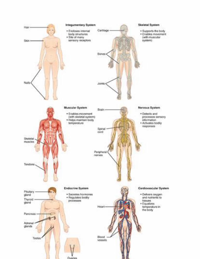

Integumentary System
Organs of the integumentary system include the skin, hair, and nails. The skin is the largest organ
in the body. It encloses and protects the body and is the site of many sensory receptors. The skin
is the body’s first defense against pathogens, and it also helps regulate body temperature and
eliminate wastes in sweat.
Skeletal System
The skeletal system consists of bones, joints, teeth. The bones of the skeletal system are connected
by tendons, ligaments, and cartilage. Functions of the skeletal system include supporting the body
and giving it shape. Along with the muscular system, the skeletal system enables the body to move.
The bones of the skeletal system also protect internal organs, store calcium, and produce red and
white blood cells.
Muscular System
The muscular system consists of three different types of muscles, including skeletal muscles,
which are attached to bones by tendons and allow for voluntary movements of the body. Smooth
muscle tissues control the involuntary movements of internal organs, such as the organs of
the digestive system, allowing food to move through the system. Smooth muscles in blood
vessels allow vasoconstriction and vasodilation, thereby helping to regulate body temperature.
Cardiac muscle tissues control the involuntary beating of the heart, allowing it to pump blood
through the blood vessels of the cardiovascular system.
Nervous System
The nervous system includes the brain and spinal cord — which make up the central nervous
system — and nerves that run throughout the rest of the body, making up the peripheral nervous
system. The nervous system controls both voluntary and involuntary responses of the human
organism, and also detects and processes sensory information.
Endocrine System

The endocrine system is made up of glands that secrete hormones into the blood, which then
carries hormones throughout the body. Endocrine hormones are chemical messengers that control
many body functions, including metabolism, growth, and sexual development. The master gland
of the endocrine system is the pituitary gland, which produces hormones that control
other endocrine glands. Some of the other endocrine glands include the pancreas, thyroid gland,
and adrenal glands.
Cardiovascular System
The cardiovascular system (also called circulatory system) includes the heart, blood, and three
types of blood vessels: arteries, veins, and capillaries. The heart pumps blood, which travels
through the blood vessels. The main function of the cardiovascular system is transport. Oxygen
from the lungs and nutrients from the digestive system are transported to cells throughout the
body. Carbon dioxide and other waste materials are picked up from the cells and transported to
organs (such as the lungs and kidneys) for elimination from the body. The cardiovascular system
also equalizes body temperature and transports endocrine hormones to cells in the body where they
are needed.
Lymphatic System
The lymphatic system is sometimes considered part of the immune system. It consists of a network
of lymph vessels and ducts that collect excess fluid (called lymph) from extracellular spaces in
tissues and transport the fluid to the bloodstream. The lymphatic system also includes many small
collections of tissue, (called lymph nodes) and an organ called the spleen, both of which remove
pathogens and cellular debris from the lymph or blood. In addition, the thymus gland in the
lymphatic system produces some types of white blood cells (lymphocytes) that fight infections.
Respiratory System
Organs and other structures of the respiratory system include the nasal passages, lungs, and a long
tube called the trachea, which carries air between the nasal passages and lungs. The main function
of the respiratory system is to deliver oxygen to the blood and remove carbon dioxide from the
body. Gases are exchanged between the lungs and blood across the walls of capillaries lining tiny
air sacs (alveoli) in the lungs.
Digestive System
The digestive system consists of several main organs — including the mouth, esophagus, stomach,
and small and large intestines — that form a long tube called the gastrointestinal (GI) tract. Food
moves through this tract, where it is digested. Its nutrients are then absorbed, and its waste products
are excreted. The digestive system also includes accessory organs (such as the pancreas and liver)
that produce enzymes and other substances needed for digestion, but through which food does not
actually pass.
Urinary System
The urinary system is part of the excretory system, which removes wastes from the body.
The urinary system includes the pair of kidneys, which filter excess water and a waste product
(called urea) from the blood and form urine. Two tubes called ureters carry the urine from the
kidneys to the urinary bladder, which stores the urine until it is excreted from the body through
another tube called the urethra. The kidneys also produce an enzyme called renin and a variety of
hormones. These substances help regulate blood pressure, the production of red blood cells, and
the balance of calcium and phosphorus in the body.
Male and Female Reproductive Systems
The reproductive system is the only body system that differs substantially between males and
females. Both male and female reproductive systems produce sex-specific sex hormones
(testosterone in males, estrogen in females) and gametes (sperm in males, eggs in females).
However, the organs involved in these processes are different. The male reproductive
system includes the epididymis, testes, and penis. The female reproductive system includes the
uterus, ovaries, and mammary glands. The male and female systems also have different additional
roles. For example, the male system has the role of delivering gametes to the female reproductive
tract, whereas the female system has the roles of supporting an embryo and fetus until birth and
also producing milk for the infant after birth.
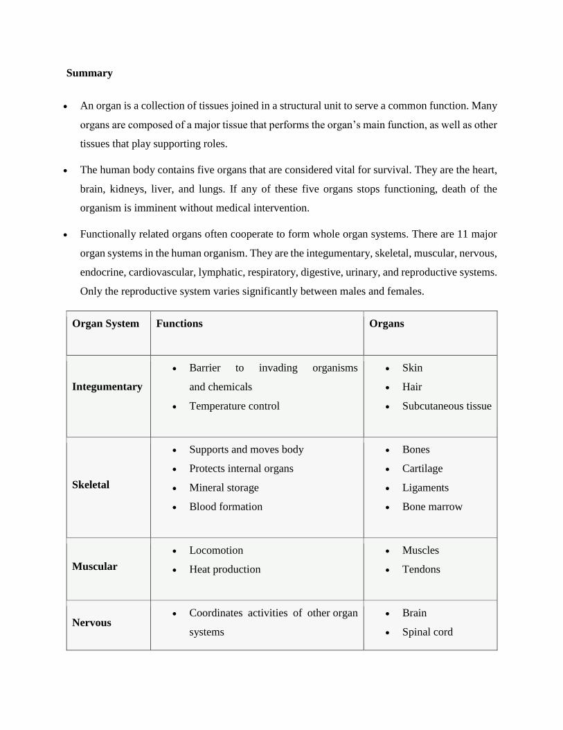
Summary
An organ is a collection of tissues joined in a structural unit to serve a common function. Many
organs are composed of a major tissue that performs the organ’s main function, as well as other
tissues that play supporting roles.
The human body contains five organs that are considered vital for survival. They are the heart,
brain, kidneys, liver, and lungs. If any of these five organs stops functioning, death of the
organism is imminent without medical intervention.
Functionally related organs often cooperate to form whole organ systems. There are 11 major
organ systems in the human organism. They are the integumentary, skeletal, muscular, nervous,
endocrine, cardiovascular, lymphatic, respiratory, digestive, urinary, and reproductive systems.
Only the reproductive system varies significantly between males and females.
Organ System Functions Organs
Integumentary
Barrier to invading organisms
and chemicals
Temperature control
Skin
Hair
Subcutaneous tissue
Skeletal
Supports and moves body
Protects internal organs
Mineral storage
Blood formation
Bones
Cartilage
Ligaments
Bone marrow
Muscular
Locomotion
Heat production
Muscles
Tendons
Nervous Coordinates activities of other organ
systems
Brain
Spinal cord
Responds to sensations Nerves
Eyes
Ears
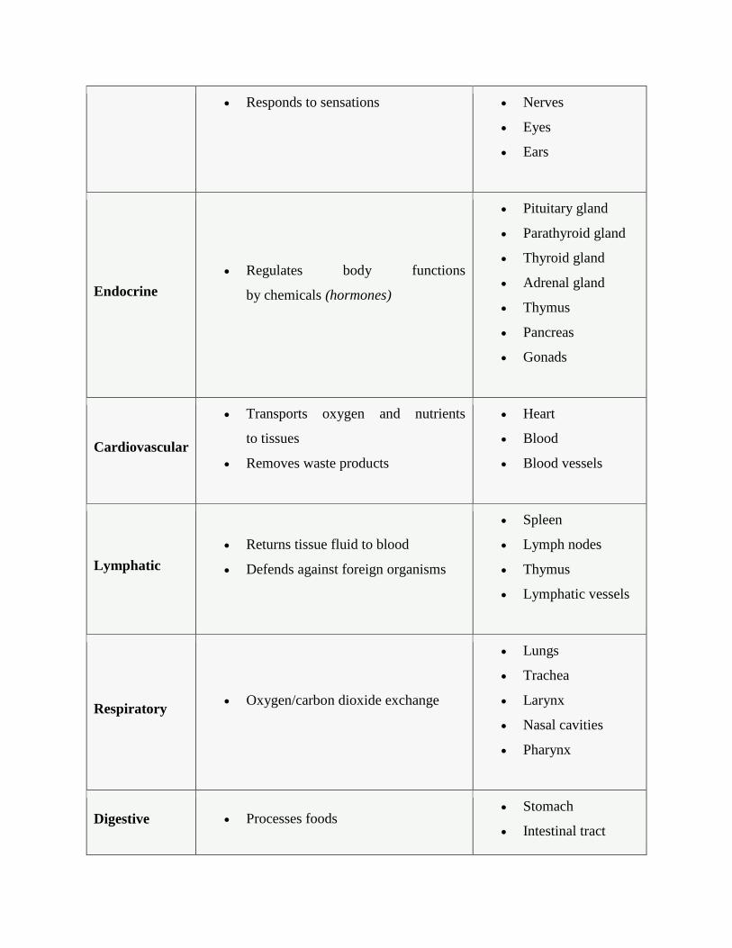
Endocrine
Regulates body functions
by chemicals (hormones)
Pituitary gland
Parathyroid gland
Thyroid gland
Adrenal gland
Thymus
Pancreas
Gonads
Cardiovascular
Transports oxygen and nutrients
to tissues
Removes waste products
Heart
Blood
Blood vessels
Lymphatic
Returns tissue fluid to blood
Defends against foreign organisms
Spleen
Lymph nodes
Thymus
Lymphatic vessels
Respiratory Oxygen/carbon dioxide exchange
Lungs
Trachea
Larynx
Nasal cavities
Pharynx
Digestive Processes foods Stomach
Intestinal tract
Absorption of nutrients into body Liver
Pancreas
Esophagus
Salivary glands
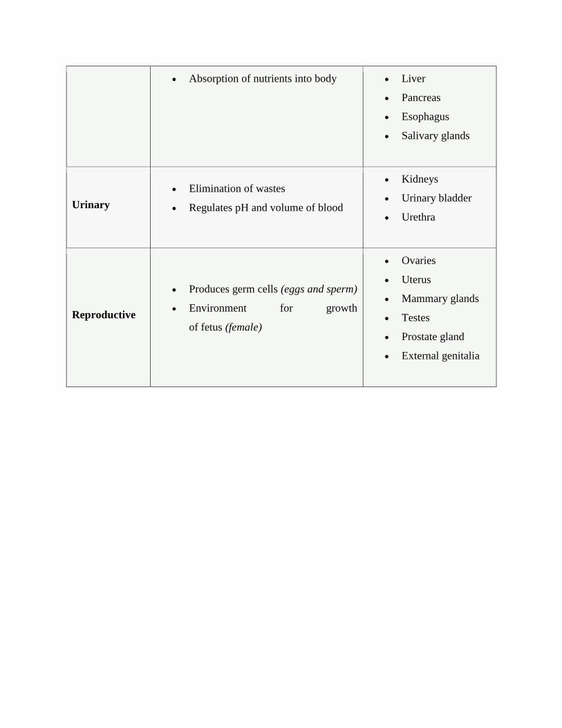
Urinary
Elimination of wastes
Regulates pH and volume of blood
Kidneys
Urinary bladder
Urethra
Reproductive
Produces germ cells (eggs and sperm)
Environment for growth
of fetus (female)
Ovaries
Uterus
Mammary glands
Testes
Prostate gland
External genitalia
UNIT – II
PART-A
Q.NO QUESTIONS CO (LEVEL)
1 Summarize the digestion system of carnivore animals 2 (2)
2 Define circulation system and relate with vertebrates 2 (1)
3 Relate the respiratory system of aquatic species 2 (1)
4 Explainthe skeleton system with an illustration. 2 (2)
5 Explain the nervous system 2 (2)
6 List out the sensory organs and its functions 2 (1)
7 Define animal behaviour and compare with plants 2 (1)
8 Discuss the animal behaviour based on their feeding habit 2 (1)
9 Define animal reproduction in general 2 (1)
10 Summarize how invertebrate reproduce 2 (2)
PART-B
Q.NO QUESTIONS CO (LEVEL)
1 Explain the general pattern of digestive system of vertebrates 2 (2)
2 Prepare an elaborate account on circulatory system 2 (3)
3 Investigate how the respiratory system works in aquatic and terrestrial
animals.
2 (5)
4 Assess the skeletal system of vertebrates 2 (5)
5 Review the functions of neurons and brain 2 (5)
6 Discuss in detail the sensory organs and their functions 2 (1)
7 Describe in detail aboutanimal behaviour 2 (1)
8 Investigate the different reproductive system of animal kingdom 2 (5)
9 Explain how the vertebrates reproduce 2 (2)
10 Illustrate the different mode of reproduction by invertebrates 2 (1)
SCHOOL OF BIO & CHEMICAL ENGINEERING
DEPARTMENT OF BIOTECHNOLOGY
SBC1201: ZOOLOGY
UNIT – III - DEVELOPMENTAL BIOLOGY– SBC1201

1. Developmental Biology
Developmental biology is the science that investigates how a variety of interacting processes
generate an organism’s heterogeneous shapes, size, and structural features that arise on the
trajectory from embryo to adult, or more generally throughout a life cycle. It represents an
exemplary area of contemporary experimental biology that focuses on phenomena that have
puzzled natural philosophers and scientists for more than two millennia. Philosophers of biology
have shown interest in developmental biology due to the potential relevance of development for
understanding evolution, the theme of reductionism in genetic explanations, and via increased
attention to the details of particular research programs, such as stem cell biology. Developmental
biology displays a rich array of material and conceptual practices that can be analyzed to better
understand the scientific reasoning exhibited in experimental life science. This entry briefly
reviews some central phenomena of ontogeny and then explores four domains that represent some
of the import and promise of conceptual reflection on the epistemology of developmental biology.
Animals and all other organized substances have no beginning … their apparent generation is only
a development, a kind of augmentation … a transformation like any other, for instance like that of
a caterpillar into a butterfly. (Smith 2011: 186–187)
A major theme that crystallized in this history of investigation is the distinction between epigenesis
and preformation (see the entry on epigenesis and preformationism). Proponents of epigenesis
claimed that heterogeneous, complex features of form emerge from homogeneous, less complex
embryonic structures through interactive processes. Thus, an explanation of the ontogeny of these
form features requires accounting for how the interactions occur. Proponents of preformation
claimed that complex form preexists in the embryo and “unfolds” via ordinary growth processes.
An adequate explanation involves detailing how growth occurs. Although preformation has a
lighter explanatory burden in accounting for how form emerges during ontogeny (on the
assumption that growth is easier to explain than process interactions), it also must address how the
starting point of the next generation is formed with the requisite heterogeneous complex features.
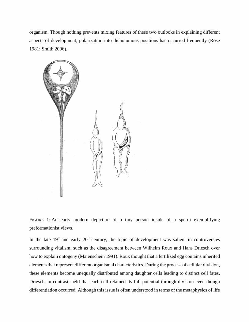
This was sometimes accomplished by embedding smaller and smaller miniatures ad
infinitum inside the organism (Figure 1). Epigenetic perspectives were often dependent on forms
of teleological reasoning (see the entry on teleological notions in biology) to account for why
interactions among homogeneous components eventually result in a complex, integrated whole
organism. Though nothing prevents mixing features of these two outlooks in explaining different
aspects of development, polarization into dichotomous positions has occurred frequently (Rose
1981; Smith 2006).
FIGURE 1: An early modern depiction of a tiny person inside of a sperm exemplifying
preformationist views.
In the late 19th and early 20th century, the topic of development was salient in controversies
surrounding vitalism, such as the disagreement between Wilhelm Roux and Hans Driesch over
how to explain ontogeny (Maienschein 1991). Roux thought that a fertilized egg contains inherited
elements that represent different organismal characteristics. During the process of cellular division,
these elements become unequally distributed among daughter cells leading to distinct cell fates.
Driesch, in contrast, held that each cell retained its full potential through division even though
differentiation occurred. Although this issue is often understood in terms of the metaphysics of life
(vitalism versus materialism), Driesch’s interpretation of development and the autonomy of an
organism had epistemological dimensions (Maienschein 2000). The explanatory disagreement
involved different experimental approaches and divergent views on the nature of differentiation in
early ontogeny (e.g., to what degree cells are pre-specified). A familiar philosophical theme
running through these discussions, both epistemological and metaphysical, is the status
of reductionism in biology. Through the middle of the 20th century, embryology—the scientific
discipline studying development—slowly transformed into developmental biology with a variety
of reworked and recalcitrant elements (Berrill 1961). In conjunction with the issue of reductionism,
a key aspect of this history is the molecularization of experimental (as opposed to comparative)
embryology (Fraser and Harland 2000), with a concomitant emphasis on the explanatory power of
genes (see the entry on gene and Section 3.1). This complex and fascinating history, including
interrelations with medicine and reproductive technology, has been detailed elsewhere (see, e.g.,
Oppenheimer 1967; Horder et al. 1986; Hamburger 1988; Hopwood 2019; Maienschein 2014;
Maienschein et al. 2005; Gilbert 1991; Embryo Project in Other Internet Resources).
Developmental biology has increasingly become an area of exploration for philosophy of
biology due to the potential relevance of development for understanding evolution (Love
2015; Section 5), the theme of reductionism in biology and explanations from molecular genetics
(Robert 2004; Rosenberg 2006; Section 3), and via increased attention to the details of particular
research programs, such as stem cell biology (Fagan 2013; Laplane 2016). However, it should not
be forgotten that ontogeny was on the radar of philosophical scholars in the 20th century, as seen
in Ernest Nagel’s treatment of hierarchical organization and reduction in the development of living
systems (Nagel 1961: 432ff). For contemporary philosophy of science, developmental biology
displays a rich array of material and conceptual practices that can be analyzed to better understand
the scientific reasoning exhibited in experimental life science (see the entry on experiment in
biology). After a brief review of some central phenomena of ontogeny, this entry explores four
domains that represent some of the import and promise of conceptual reflection on the
epistemology of developmental biology.
Developmental Phenomena
Developmental biology is the science that seeks to explain how the structure of organisms changes
with time. Structure, which may also be called morphology or anatomy, encompasses the
arrangement of parts, the number of parts, and the different types of parts. (Slack 2006: 6)
Most of the properties that developmental biologists attempt to explain are structural rather than
functional. For example, a developmental biologist concentrates more on how tissue layers fold or
how shape is generated than on what the folded tissue layers do or how the shape functions. The
ontogeny of function, at all levels of organization, is an element of developmental biology, but it
is often bracketed because of the predominance (both past and present) of questions surrounding
the ontogeny of form or structure (Love 2008).
Textbooks (e.g., Gilbert 2010; Slack 2013; Wolpert et al. 2010) typically describe a canonical set
of events surrounding the changing structures displayed during animal development.[1] The first of
these is fertilization (in sexually reproducing species), where an already semi-organized egg
merges with a sperm cell, followed by the fusion of their nuclei to achieve the appropriate
complement of genetic material. Second, the fertilized egg undergoes several rounds of cleavage,
which are mitotic divisions without cell growth that subdivide the zygote into many distinct cells
(Figure 2). After many rounds of cleavage, this spherical conglomerate of cells (now called
a blastula) begins to exhibit some specification of germ layers (endoderm, mesoderm, and
ectoderm) and then proceeds to invaginate at one end, a complex process referred to
as gastrulation that eventually yields a through-gut. All three germ layers, from which specific
types of cells are derived (e.g., neural cells from ectoderm), become established during gastrulation
or shortly after it completes.[2] Organogenesis refers to the production of tissues and organs
through the interaction and rearrangement of cell groups. Events confined to distinct taxonomic
groups include neurulation in chordates, whereas other events correlate with mode of development
(metamorphosis from a larval to adult stage) or individual trauma (regeneration of a limb).
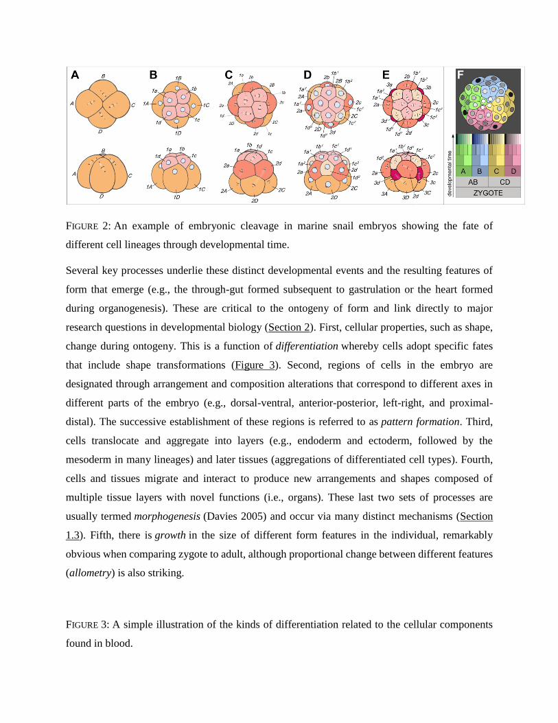
FIGURE 2: An example of embryonic cleavage in marine snail embryos showing the fate of
different cell lineages through developmental time.
Several key processes underlie these distinct developmental events and the resulting features of
form that emerge (e.g., the through-gut formed subsequent to gastrulation or the heart formed
during organogenesis). These are critical to the ontogeny of form and link directly to major
research questions in developmental biology (Section 2). First, cellular properties, such as shape,
change during ontogeny. This is a function of differentiation whereby cells adopt specific fates
that include shape transformations (Figure 3). Second, regions of cells in the embryo are
designated through arrangement and composition alterations that correspond to different axes in
different parts of the embryo (e.g., dorsal-ventral, anterior-posterior, left-right, and proximal-
distal). The successive establishment of these regions is referred to as pattern formation. Third,
cells translocate and aggregate into layers (e.g., endoderm and ectoderm, followed by the
mesoderm in many lineages) and later tissues (aggregations of differentiated cell types). Fourth,
cells and tissues migrate and interact to produce new arrangements and shapes composed of
multiple tissue layers with novel functions (i.e., organs). These last two sets of processes are
usually termed morphogenesis (Davies 2005) and occur via many distinct mechanisms (Section
1.3). Fifth, there is growth in the size of different form features in the individual, remarkably
obvious when comparing zygote to adult, although proportional change between different features
(allometry) is also striking.
FIGURE 3: A simple illustration of the kinds of differentiation related to the cellular components
found in blood.

None of these processes occur in isolation and explanations of particular form features usually
draw on several of them simultaneously, presuming other features that originated earlier in
ontogeny by different instantiations and combinations of the processes. This sets a broad agenda
for investigation: how do various iterations and combinations of these processes generate form
features during ontogeny? Consider the concrete example of vertebrate cardiogenesis. How does
the vertebrate heart, with its internal and external shape and structure originate during ontogeny
(Harvey 2002)? How does the heart come to exhibit left/right asymmetry in the body cavity? What
causes cells to adopt a muscle cell fate or certain tissues to interact in the prospective region of the
heart? How do muscle cells migrate to, aggregate in, and differentiate at the correct location? How
does the interior of the heart adopt a particular tubular structure with various chambers (that differs
among vertebrate species)? How does the heart grow at a particular rate and achieve a specific
size? Solutions relevant to explaining the ontogeny of form characterize causal factors that account
for how different processes occur and yield various outcomes (Section 3).
Reference:
https://plato.stanford.edu/entries/biology-developmental/
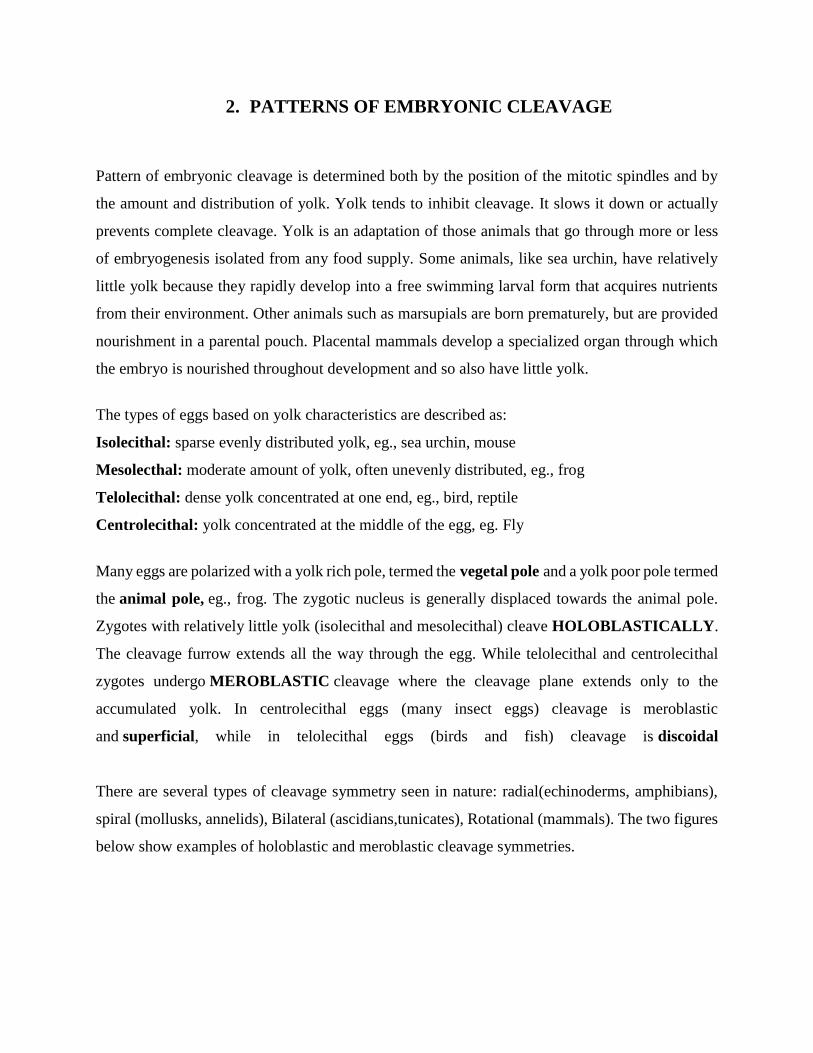
2. PATTERNS OF EMBRYONIC CLEAVAGE
Pattern of embryonic cleavage is determined both by the position of the mitotic spindles and by
the amount and distribution of yolk. Yolk tends to inhibit cleavage. It slows it down or actually
prevents complete cleavage. Yolk is an adaptation of those animals that go through more or less
of embryogenesis isolated from any food supply. Some animals, like sea urchin, have relatively
little yolk because they rapidly develop into a free swimming larval form that acquires nutrients
from their environment. Other animals such as marsupials are born prematurely, but are provided
nourishment in a parental pouch. Placental mammals develop a specialized organ through which
the embryo is nourished throughout development and so also have little yolk.
The types of eggs based on yolk characteristics are described as:
Isolecithal: sparse evenly distributed yolk, eg., sea urchin, mouse
Mesolecthal: moderate amount of yolk, often unevenly distributed, eg., frog
Telolecithal: dense yolk concentrated at one end, eg., bird, reptile
Centrolecithal: yolk concentrated at the middle of the egg, eg. Fly
Many eggs are polarized with a yolk rich pole, termed the vegetal pole and a yolk poor pole termed
the animal pole, eg., frog. The zygotic nucleus is generally displaced towards the animal pole.
Zygotes with relatively little yolk (isolecithal and mesolecithal) cleave HOLOBLASTICALLY.
The cleavage furrow extends all the way through the egg. While telolecithal and centrolecithal
zygotes undergo MEROBLASTIC cleavage where the cleavage plane extends only to the
accumulated yolk. In centrolecithal eggs (many insect eggs) cleavage is meroblastic
and superficial, while in telolecithal eggs (birds and fish) cleavage is discoidal
There are several types of cleavage symmetry seen in nature: radial(echinoderms, amphibians),
spiral (mollusks, annelids), Bilateral (ascidians,tunicates), Rotational (mammals). The two figures
below show examples of holoblastic and meroblastic cleavage symmetries.
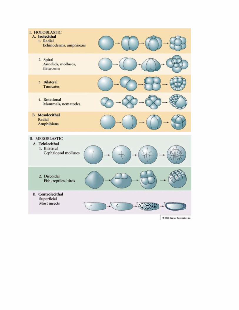
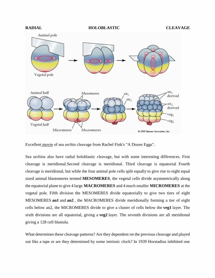
RADIAL HOLOBLASTIC CLEAVAGE
Excellent movie of sea urchin cleavage from Rachel Fink's "A Dozen Eggs".
Sea urchins also have radial holoblastic cleavage, but with some interesting differences. First
cleavage is meridional.Second cleavage is meridional. Third cleavage is equatorial Fourth
cleavage is meridional, but while the four animal pole cells split equally to give rise to eight equal
sized animal blastomeres termed MESOMERES, the vegetal cells divide asymmetrically along
the equatorial plane to give 4 large MACROMERES and 4 much smaller MICROMERES at the
vegetal pole. Fifth division the MESOMERES divide equatorially to give two tiers of eight
MESOMERES an1 and an2 , the MACROMERES divide meridionally forming a tier of eight
cells below an2, the MICROMERES divide to give a cluster of cells below the veg1 layer. The
sixth divisions are all equatorial, giving a veg2 layer. The seventh divisions are all meridional
giving a 128 cell blastula.
What determines these cleavage patterns? Are they dependent on the previous cleavage and played
out like a tape or are they determined by some intrinsic clock? In 1939 Horstadius inhibited one
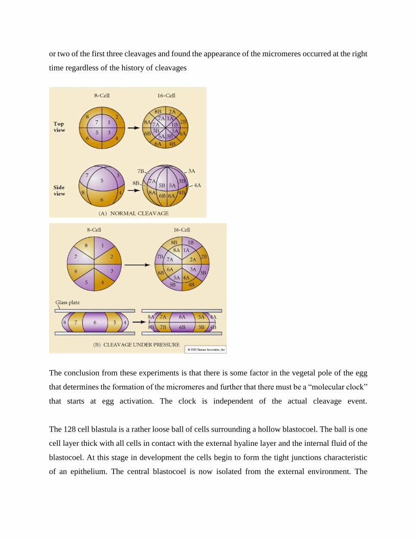
or two of the first three cleavages and found the appearance of the micromeres occurred at the right
time regardless of the history of cleavages
The conclusion from these experiments is that there is some factor in the vegetal pole of the egg
that determines the formation of the micromeres and further that there must be a “molecular clock”
that starts at egg activation. The clock is independent of the actual cleavage event.
The 128 cell blastula is a rather loose ball of cells surrounding a hollow blastocoel. The ball is one
cell layer thick with all cells in contact with the external hyaline layer and the internal fluid of the
blastocoel. At this stage in development the cells begin to form the tight junctions characteristic
of an epithelium. The central blastocoel is now isolated from the external environment. The
blastomeres continue to divide with their axis parallel to the hyaline layer, remaining a epithelium
one cell thick. The blastocoel continues to enlarge.
Two theories attempt to account for the pattern of enlargement of the blastocyst
1. The osmotic theory suggests that ions and proteins are secreted into the blastocoel by the
blastomeres and this results in a pressure buildup due to the osmotic flow of water. This pressure
would then be responsible for aligning the axis mitosis of the blastomeres and the enlargement of
the blastocoel.
2. The alternate theory by Wolpert and his colleagues suggests that it is really the adhesive
interactions among the blastomeres and between the blastomeres and the hyaline layer that aligns
the mitotic axis's. That is the adhesion to the hyaline is greatest, the adhesion to other blastomeres
is next, and finally the interaction with the blastocoel wall is least. The dominant adhesion with
the hyaline layer forces the expansion of the blastocyst and blastocoel.
The cells of the blastula grow cilia on their outer surface, secrete a “hatching enzyme” (hyalinase)
and become free swimming.
AMPHIBIAN CLEAVAGE
Cleavage in many amphibians is holoblastic with radial symmetry, however the large volume of
yolk (its mesolecithal) interferes with cleavage. At the animal pole first cleavage proceeds at about
1mm/min, while through the vegetal pole is proceeds 50-100 times slower (.02mm/min). While
the first cleavage is still incomplete in the yolky vegetal region of the egg the second meridional
cleavage begins to take place.
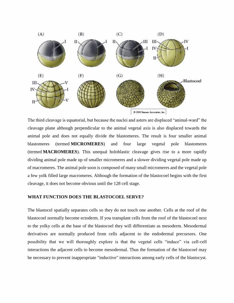
The third cleavage is equatorial, but because the nuclei and asters are displaced “animal-ward” the
cleavage plane although perpendicular to the animal vegetal axis is also displaced towards the
animal pole and does not equally divide the blastomeres. The result is four smaller animal
blastomeres (termed MICROMERES) and four large vegetal pole blastomeres
(termed MACROMERES). This unequal holoblastic cleavage gives rise to a more rapidly
dividing animal pole made up of smaller micromeres and a slower dividing vegetal pole made up
of macromeres. The animal pole soon is composed of many small micromeres and the vegetal pole
a few yolk filled large macromeres. Although the formation of the blastocoel begins with the first
cleavage, it does not become obvious until the 128 cell stage.
WHAT FUNCTION DOES THE BLASTOCOEL SERVE?
The blastocel spatially separates cells so they do not touch one another. Cells at the roof of the
blastocoel normally become ectoderm. If you transplant cells from the roof of the blastocoel next
to the yolky cells at the base of the blastocoel they will differentiate as mesoderm. Mesodermal
derivatives are normally produced from cells adjacent to the endodermal precursors. One
possibility that we will thoroughly explore is that the vegetal cells “induce” via cell-cell
interactions the adjacent cells to become mesodermal. Thus the formation of the blastocoel may
be necessary to prevent inappropriate "inductive" interactions among early cells of the blastocyst.
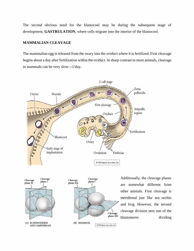
The second obvious need for the blastocoel may be during the subsequent stage of
development, GASTRULATION, where cells migrate into the interior of the blastocoel.
MAMMALIAN CLEAVAGE
The mammalian egg is released from the ovary into the oviduct where it is fertilized. First cleavage
begins about a day after fertilization within the oviduct. In sharp contrast to most animals, cleavage
in mammals can be very slow---1/day.
Additionally, the cleavage planes
are somewhat different from
other animals. First cleavage is
meridional just like sea urchin
and frog. However, the second
cleavage division sees one of the
blastomeres dividing
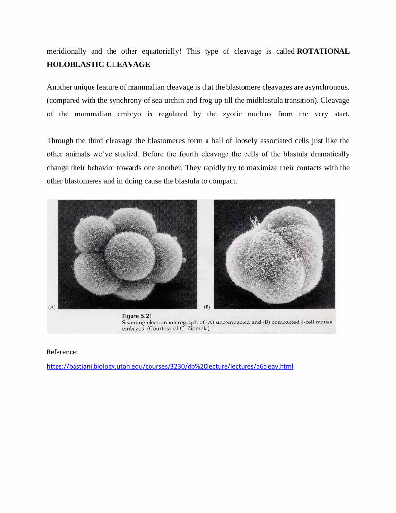
meridionally and the other equatorially! This type of cleavage is called ROTATIONAL
HOLOBLASTIC CLEAVAGE.
Another unique feature of mammalian cleavage is that the blastomere cleavages are asynchronous.
(compared with the synchrony of sea urchin and frog up till the midblastula transition). Cleavage
of the mammalian embryo is regulated by the zyotic nucleus from the very start.
Through the third cleavage the blastomeres form a ball of loosely associated cells just like the
other animals we’ve studied. Before the fourth cleavage the cells of the blastula dramatically
change their behavior towards one another. They rapidly try to maximize their contacts with the
other blastomeres and in doing cause the blastula to compact.
Reference:
https://bastiani.biology.utah.edu/courses/3230/db%20lecture/lectures/a6cleav.html
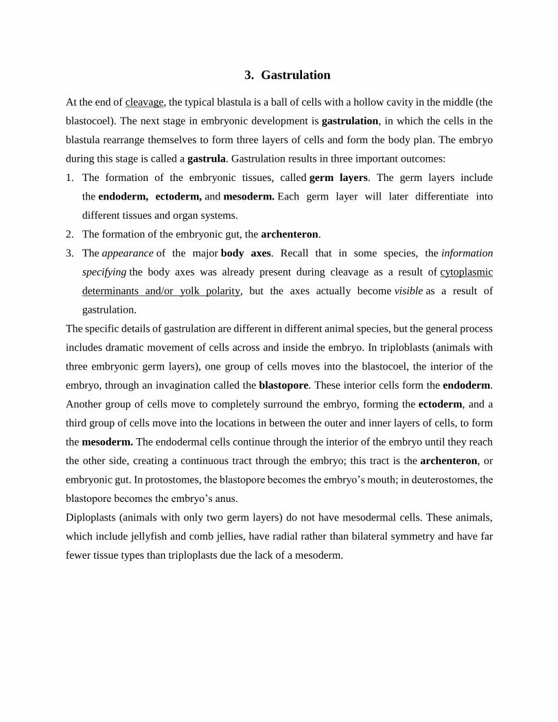
3. Gastrulation
At the end of cleavage, the typical blastula is a ball of cells with a hollow cavity in the middle (the
blastocoel). The next stage in embryonic development is gastrulation, in which the cells in the
blastula rearrange themselves to form three layers of cells and form the body plan. The embryo
during this stage is called a gastrula. Gastrulation results in three important outcomes:
1. The formation of the embryonic tissues, called germ layers. The germ layers include
the endoderm, ectoderm, and mesoderm. Each germ layer will later differentiate into
different tissues and organ systems.
2. The formation of the embryonic gut, the archenteron.
3. The appearance of the major body axes. Recall that in some species, the information
specifying the body axes was already present during cleavage as a result of cytoplasmic
determinants and/or yolk polarity, but the axes actually become visible as a result of
gastrulation.
The specific details of gastrulation are different in different animal species, but the general process
includes dramatic movement of cells across and inside the embryo. In triploblasts (animals with
three embryonic germ layers), one group of cells moves into the blastocoel, the interior of the
embryo, through an invagination called the blastopore. These interior cells form the endoderm.
Another group of cells move to completely surround the embryo, forming the ectoderm, and a
third group of cells move into the locations in between the outer and inner layers of cells, to form
the mesoderm. The endodermal cells continue through the interior of the embryo until they reach
the other side, creating a continuous tract through the embryo; this tract is the archenteron, or
embryonic gut. In protostomes, the blastopore becomes the embryo’s mouth; in deuterostomes, the
blastopore becomes the embryo’s anus.
Diploplasts (animals with only two germ layers) do not have mesodermal cells. These animals,
which include jellyfish and comb jellies, have radial rather than bilateral symmetry and have far
fewer tissue types than triploplasts due the lack of a mesoderm.
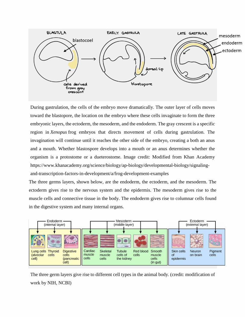
During gastrulation, the cells of the embryo move dramatically. The outer layer of cells moves
toward the blastopore, the location on the embryo where these cells invaginate to form the three
embryonic layers, the ectoderm, the mesoderm, and the endoderm. The gray crescent is a specific
region in Xenopus frog embryos that directs movement of cells during gastrulation. The
invagination will continue until it reaches the other side of the embryo, creating a both an anus
and a mouth. Whether blastospore develops into a mouth or an anus determines whether the
organism is a protostome or a dueterostome. Image credit: Modified from Khan Academy
https://www.khanacademy.org/science/biology/ap-biology/developmental-biology/signaling-
and-transcription-factors-in-development/a/frog-development-examples
The three germs layers, shown below, are the endoderm, the ectoderm, and the mesoderm. The
ectoderm gives rise to the nervous system and the epidermis. The mesoderm gives rise to the
muscle cells and connective tissue in the body. The endoderm gives rise to columnar cells found
in the digestive system and many internal organs.
The three germ layers give rise to different cell types in the animal body. (credit: modification of
work by NIH, NCBI)
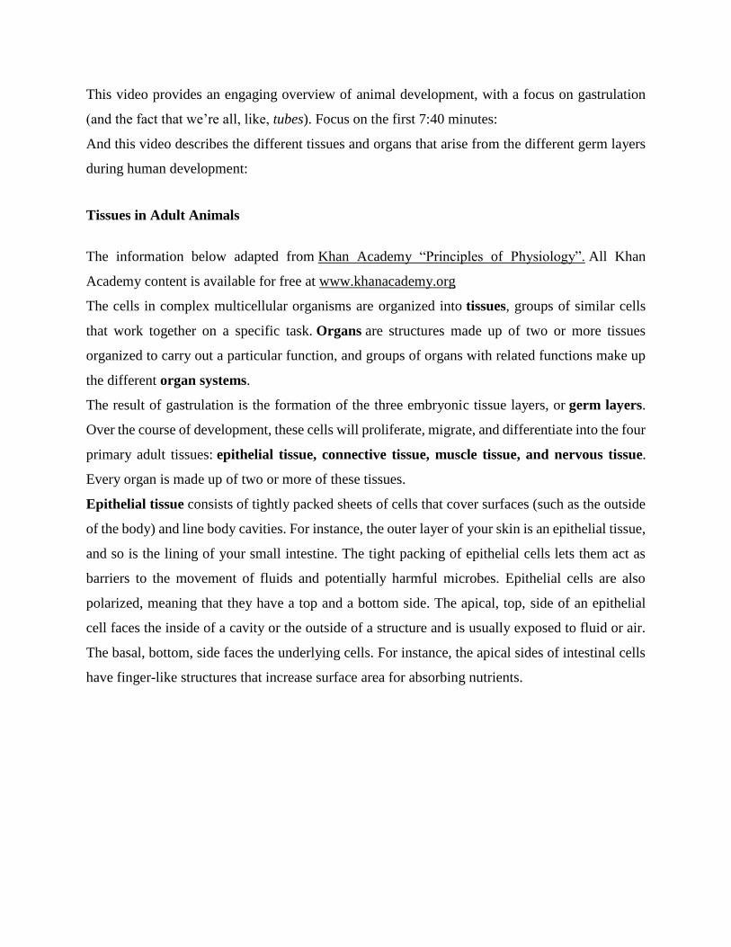
This video provides an engaging overview of animal development, with a focus on gastrulation
(and the fact that we’re all, like, tubes). Focus on the first 7:40 minutes:
And this video describes the different tissues and organs that arise from the different germ layers
during human development:
Tissues in Adult Animals
The information below adapted from Khan Academy “Principles of Physiology”. All Khan
Academy content is available for free at www.khanacademy.org
The cells in complex multicellular organisms are organized into tissues, groups of similar cells
that work together on a specific task. Organs are structures made up of two or more tissues
organized to carry out a particular function, and groups of organs with related functions make up
the different organ systems.
The result of gastrulation is the formation of the three embryonic tissue layers, or germ layers.
Over the course of development, these cells will proliferate, migrate, and differentiate into the four
primary adult tissues: epithelial tissue, connective tissue, muscle tissue, and nervous tissue.
Every organ is made up of two or more of these tissues.
Epithelial tissue consists of tightly packed sheets of cells that cover surfaces (such as the outside
of the body) and line body cavities. For instance, the outer layer of your skin is an epithelial tissue,
and so is the lining of your small intestine. The tight packing of epithelial cells lets them act as
barriers to the movement of fluids and potentially harmful microbes. Epithelial cells are also
polarized, meaning that they have a top and a bottom side. The apical, top, side of an epithelial
cell faces the inside of a cavity or the outside of a structure and is usually exposed to fluid or air.
The basal, bottom, side faces the underlying cells. For instance, the apical sides of intestinal cells
have finger-like structures that increase surface area for absorbing nutrients.
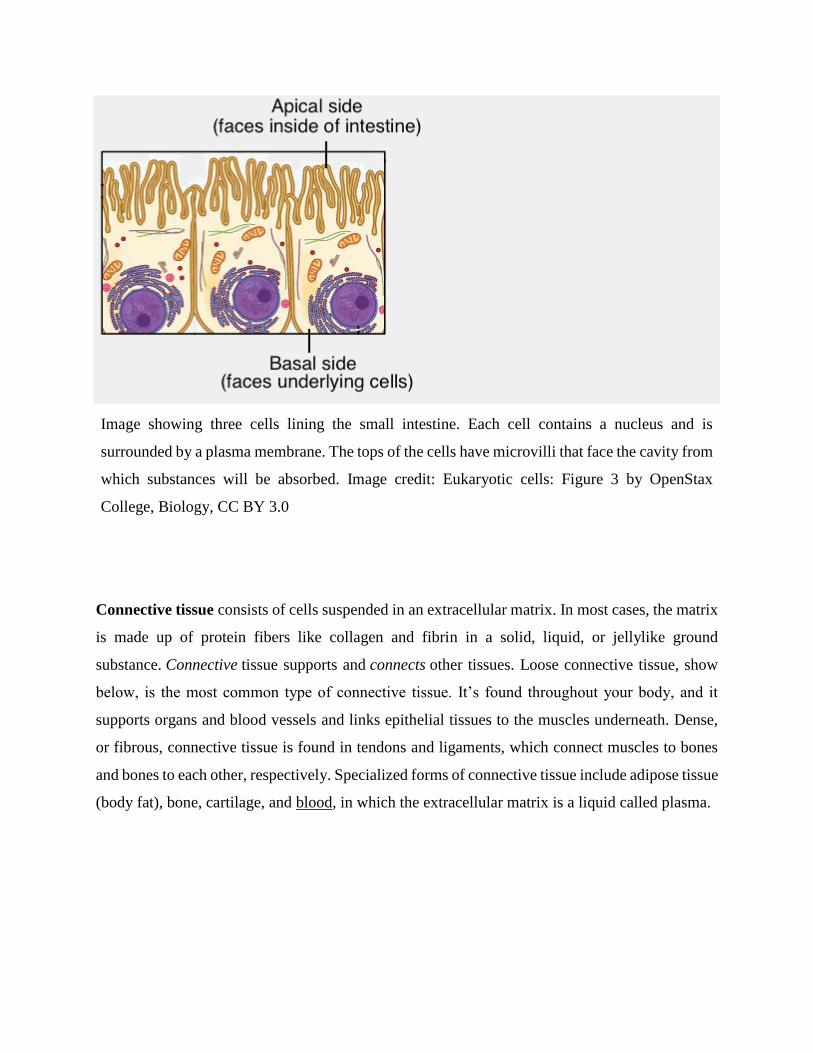
Image showing three cells lining the small intestine. Each cell contains a nucleus and is
surrounded by a plasma membrane. The tops of the cells have microvilli that face the cavity from
which substances will be absorbed. Image credit: Eukaryotic cells: Figure 3 by OpenStax
College, Biology, CC BY 3.0
Connective tissue consists of cells suspended in an extracellular matrix. In most cases, the matrix
is made up of protein fibers like collagen and fibrin in a solid, liquid, or jellylike ground
substance. Connective tissue supports and connects other tissues. Loose connective tissue, show
below, is the most common type of connective tissue. It’s found throughout your body, and it
supports organs and blood vessels and links epithelial tissues to the muscles underneath. Dense,
or fibrous, connective tissue is found in tendons and ligaments, which connect muscles to bones
and bones to each other, respectively. Specialized forms of connective tissue include adipose tissue
(body fat), bone, cartilage, and blood, in which the extracellular matrix is a liquid called plasma.
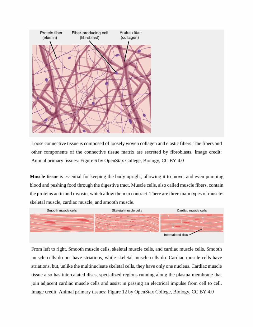
Loose connective tissue is composed of loosely woven collagen and elastic fibers. The fibers and
other components of the connective tissue matrix are secreted by fibroblasts. Image credit:
Animal primary tissues: Figure 6 by OpenStax College, Biology, CC BY 4.0
Muscle tissue is essential for keeping the body upright, allowing it to move, and even pumping
blood and pushing food through the digestive tract. Muscle cells, also called muscle fibers, contain
the proteins actin and myosin, which allow them to contract. There are three main types of muscle:
skeletal muscle, cardiac muscle, and smooth muscle.
From left to right. Smooth muscle cells, skeletal muscle cells, and cardiac muscle cells. Smooth
muscle cells do not have striations, while skeletal muscle cells do. Cardiac muscle cells have
striations, but, unlike the multinucleate skeletal cells, they have only one nucleus. Cardiac muscle
tissue also has intercalated discs, specialized regions running along the plasma membrane that
join adjacent cardiac muscle cells and assist in passing an electrical impulse from cell to cell.
Image credit: Animal primary tissues: Figure 12 by OpenStax College, Biology, CC BY 4.0
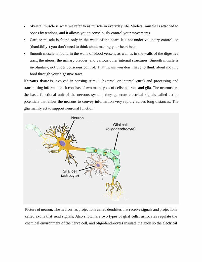
Skeletal muscle is what we refer to as muscle in everyday life. Skeletal muscle is attached to
bones by tendons, and it allows you to consciously control your movements.
Cardiac muscle is found only in the walls of the heart. It’s not under voluntary control, so
(thankfully!) you don’t need to think about making your heart beat.
Smooth muscle is found in the walls of blood vessels, as well as in the walls of the digestive
tract, the uterus, the urinary bladder, and various other internal structures. Smooth muscle is
involuntary, not under conscious control. That means you don’t have to think about moving
food through your digestive tract.
Nervous tissue is involved in sensing stimuli (external or internal cues) and processing and
transmitting information. It consists of two main types of cells: neurons and glia. The neurons are
the basic functional unit of the nervous system: they generate electrical signals called action
potentials that allow the neurons to convey information very rapidly across long distances. The
glia mainly act to support neuronal function.
Picture of neuron. The neuron has projections called dendrites that receive signals and projections
called axons that send signals. Also shown are two types of glial cells: astrocytes regulate the
chemical environment of the nerve cell, and oligodendrocytes insulate the axon so the electrical
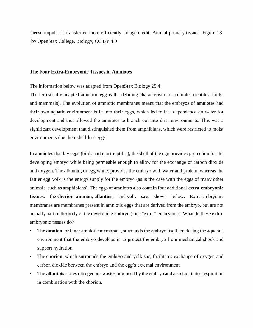
nerve impulse is transferred more efficiently. Image credit: Animal primary tissues: Figure 13
by OpenStax College, Biology, CC BY 4.0
The Four Extra-Embryonic Tissues in Amniotes
The information below was adapted from OpenStax Biology 29.4
The terrestrially-adapted amniotic egg is the defining characteristic of amniotes (reptiles, birds,
and mammals). The evolution of amniotic membranes meant that the embryos of amniotes had
their own aquatic environment built into their eggs, which led to less dependence on water for
development and thus allowed the amniotes to branch out into drier environments. This was a
significant development that distinguished them from amphibians, which were restricted to moist
environments due their shell-less eggs.
In amniotes that lay eggs (birds and most reptiles), the shell of the egg provides protection for the
developing embryo while being permeable enough to allow for the exchange of carbon dioxide
and oxygen. The albumin, or egg white, provides the embryo with water and protein, whereas the
fattier egg yolk is the energy supply for the embryo (as is the case with the eggs of many other
animals, such as amphibians). The eggs of amniotes also contain four additional extra-embryonic
tissues: the chorion, amnion, allantois, and yolk sac, shown below. Extra-embryonic
membranes are membranes present in amniotic eggs that are derived from the embryo, but are not
actually part of the body of the developing embryo (thus “extra”-embryonic). What do these extra-
embryonic tissues do?
The amnion, or inner amniotic membrane, surrounds the embryo itself, enclosing the aqueous
environment that the embryo develops in to protect the embryo from mechanical shock and
support hydration
The chorion. which surrounds the embryo and yolk sac, facilitates exchange of oxygen and
carbon dioxide between the embryo and the egg’s external environment.
The allantois stores nitrogenous wastes produced by the embryo and also facilitates respiration
in combination with the chorion.
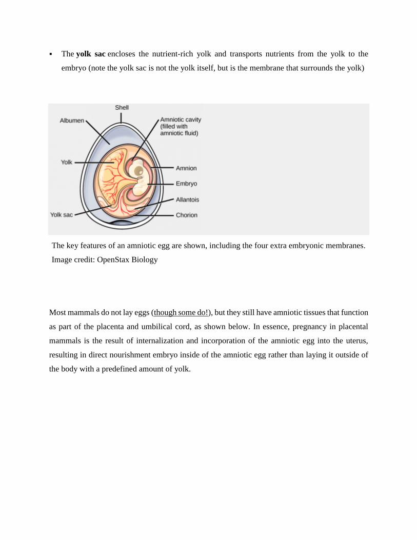
The yolk sac encloses the nutrient-rich yolk and transports nutrients from the yolk to the
embryo (note the yolk sac is not the yolk itself, but is the membrane that surrounds the yolk)
The key features of an amniotic egg are shown, including the four extra embryonic membranes.
Image credit: OpenStax Biology
Most mammals do not lay eggs (though some do!), but they still have amniotic tissues that function
as part of the placenta and umbilical cord, as shown below. In essence, pregnancy in placental
mammals is the result of internalization and incorporation of the amniotic egg into the uterus,
resulting in direct nourishment embryo inside of the amniotic egg rather than laying it outside of
the body with a predefined amount of yolk.
In the placenta, maternal and fetal blood components are conducted through the surface of the
chorionic villi, but maternal and fetal bloodstreams never mix directly. Image credit: OpenStax
Anatomy & Physiology
As you can see above, the chorion separates the fetal and maternal sides of the placenta, and the
aminon surrounds the developing fetus. Just as in the amniotic egg:
the chorion regulates gas exchange
the amnion encloses the fluid-filled cavity to provide an aqueous environment for the
developing fetus
together, the yolk sac, consisting of blood vessels that transport nutrients to the embryo, and
the allantois, which functions in waste disposal, both function as part of the mammalian
umbilical cord (not labeled above)
Reference:
https://organismalbio.biosci.gatech.edu/growth-and-reproduction/animal-development-ii/
4. Organogenesis
Organs form from the germ layers through the process of differentiation. During differentiation,
the embryonic stem cells express specific sets of genes which will determine their ultimate cell
type. For example, some cells in the ectoderm will express the genes specific to skin cells. As a
result, these cells will differentiate into epidermal cells. The process of differentiation is regulated
by cellular signaling cascades.
Scientists study organogenesis extensively in the lab in fruit flies (Drosophila) and the
nematode Caenorhabditis elegans. Drosophila have segments along their bodies, and the
patterning associated with the segment formation has allowed scientists to study which genes play
important roles in organogenesis along the length of the embryo at different time points. The
nematode C.elegans has roughly 1000 somatic cells and scientists have studied the fate of each of
these cells during their development in the nematode life cycle. There is little variation in patterns
of cell lineage between individuals, unlike in mammals where cell development from the embryo
is dependent on cellular cues.
In vertebrates, one of the primary steps during organogenesis is the formation of the neural system.
The ectoderm forms epithelial cells and tissues, and neuronal tissues. During the formation of the
neural system, special signaling molecules called growth factors signal some cells at the edge of
the ectoderm to become epidermis cells. The remaining cells in the center form the neural plate. If
the signaling by growth factors were disrupted, then the entire ectoderm would differentiate into
neural tissue.
The neural plate undergoes a series of cell movements where it rolls up and forms a tube called
the neural tube, as illustrated in Figure 43.28. In further development, the neural tube will give
rise to the brain and the spinal cord.
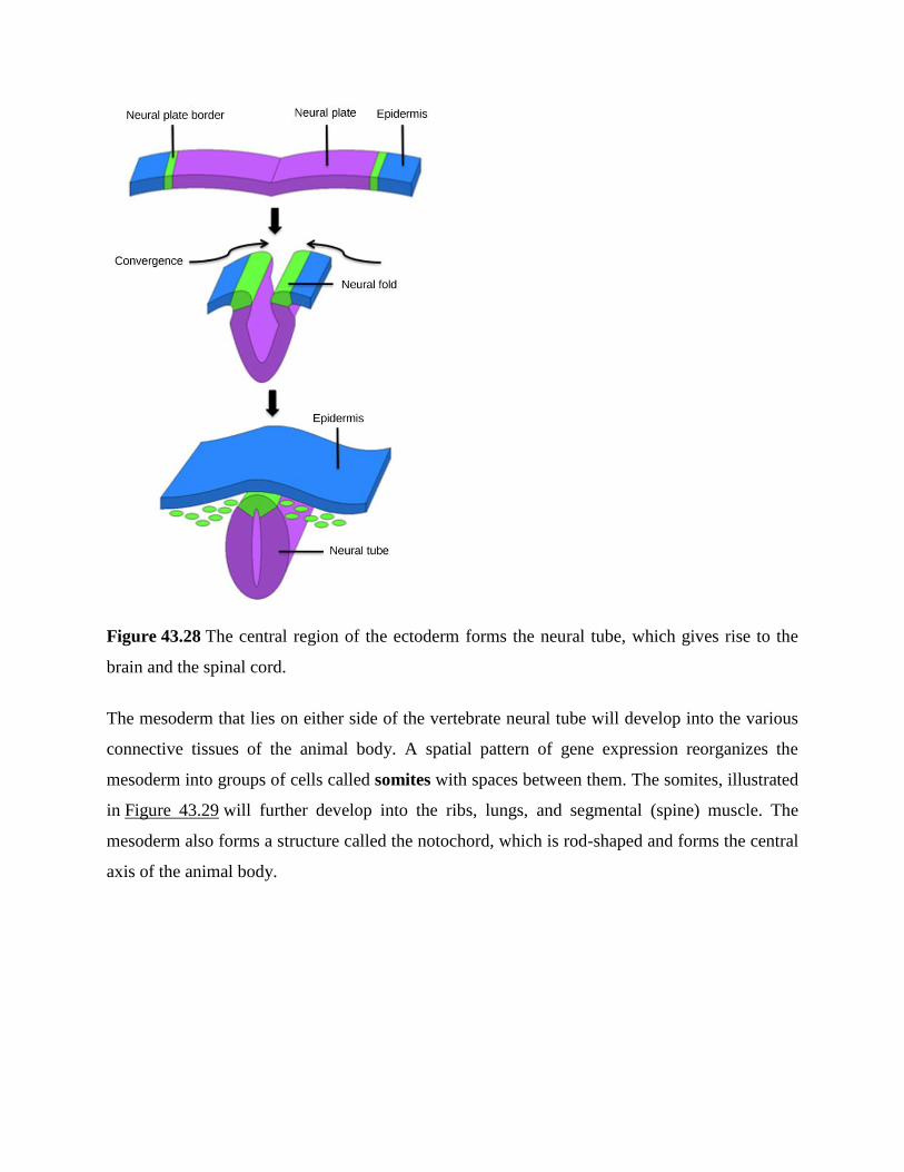
Figure 43.28 The central region of the ectoderm forms the neural tube, which gives rise to the
brain and the spinal cord.
The mesoderm that lies on either side of the vertebrate neural tube will develop into the various
connective tissues of the animal body. A spatial pattern of gene expression reorganizes the
mesoderm into groups of cells called somites with spaces between them. The somites, illustrated
in Figure 43.29 will further develop into the ribs, lungs, and segmental (spine) muscle. The
mesoderm also forms a structure called the notochord, which is rod-shaped and forms the central
axis of the animal body.
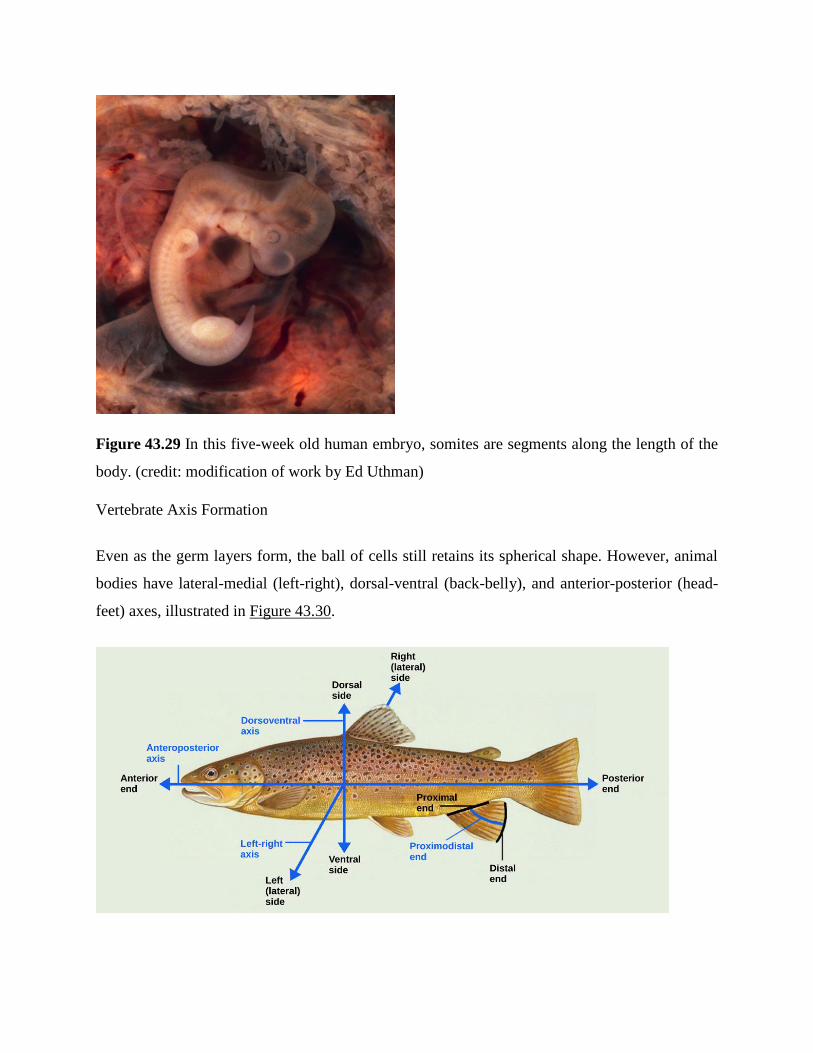
Figure 43.29 In this five-week old human embryo, somites are segments along the length of the
body. (credit: modification of work by Ed Uthman)
Vertebrate Axis Formation
Even as the germ layers form, the ball of cells still retains its spherical shape. However, animal
bodies have lateral-medial (left-right), dorsal-ventral (back-belly), and anterior-posterior (head-
feet) axes, illustrated in Figure 43.30.
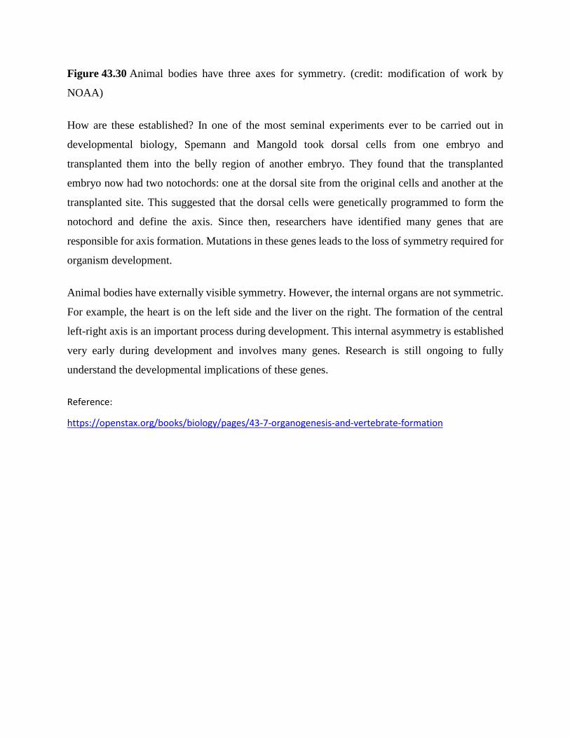
Figure 43.30 Animal bodies have three axes for symmetry. (credit: modification of work by
NOAA)
How are these established? In one of the most seminal experiments ever to be carried out in
developmental biology, Spemann and Mangold took dorsal cells from one embryo and
transplanted them into the belly region of another embryo. They found that the transplanted
embryo now had two notochords: one at the dorsal site from the original cells and another at the
transplanted site. This suggested that the dorsal cells were genetically programmed to form the
notochord and define the axis. Since then, researchers have identified many genes that are
responsible for axis formation. Mutations in these genes leads to the loss of symmetry required for
organism development.
Animal bodies have externally visible symmetry. However, the internal organs are not symmetric.
For example, the heart is on the left side and the liver on the right. The formation of the central
left-right axis is an important process during development. This internal asymmetry is established
very early during development and involves many genes. Research is still ongoing to fully
understand the developmental implications of these genes.
Reference:
https://openstax.org/books/biology/pages/43-7-organogenesis-and-vertebrate-formation
5. Placental Structure and Classification
The placentas of all eutherian (placental) mammals provide common structural and
functional features, but there are striking differences among species in gross and microscopic
structure of the placenta. Two characteristics are particularly divergent and form bases for
classification of placental types:
1. The gross shape of the placenta and the distribution of contact sites between fetal
membranes and endometrium.
2. The number of layers of tissue between maternal and fetal vascular systems.
Differences in these two properties allow classification of placentas into several fundamental
types.
Classification Based on Placental Shape and Contact Points
Examination of placentae from different species reveals striking differences in their shape
and the area of contact between fetal and maternal tissue:
Diffuse: Almost the entire surface of the allantochorion is involved in formation of
the placenta. Seen in horses and pigs.
Cotyledonary: Multiple, discrete areas of attachment called cotyledons are formed
by interaction of patches of allantochorion with endometrium. The fetal portions of
this type of placenta are called cotyledons, the maternal contact sites (caruncles), and
the cotyledon-caruncle complex a placentome. This type of placentation is observed
in ruminants.
Zonary: The placenta takes the form of a complete or incomplete band of tissue
surrounding the fetus. Seen in carnivores like dogs and cats, seals, bears, and
elephants.
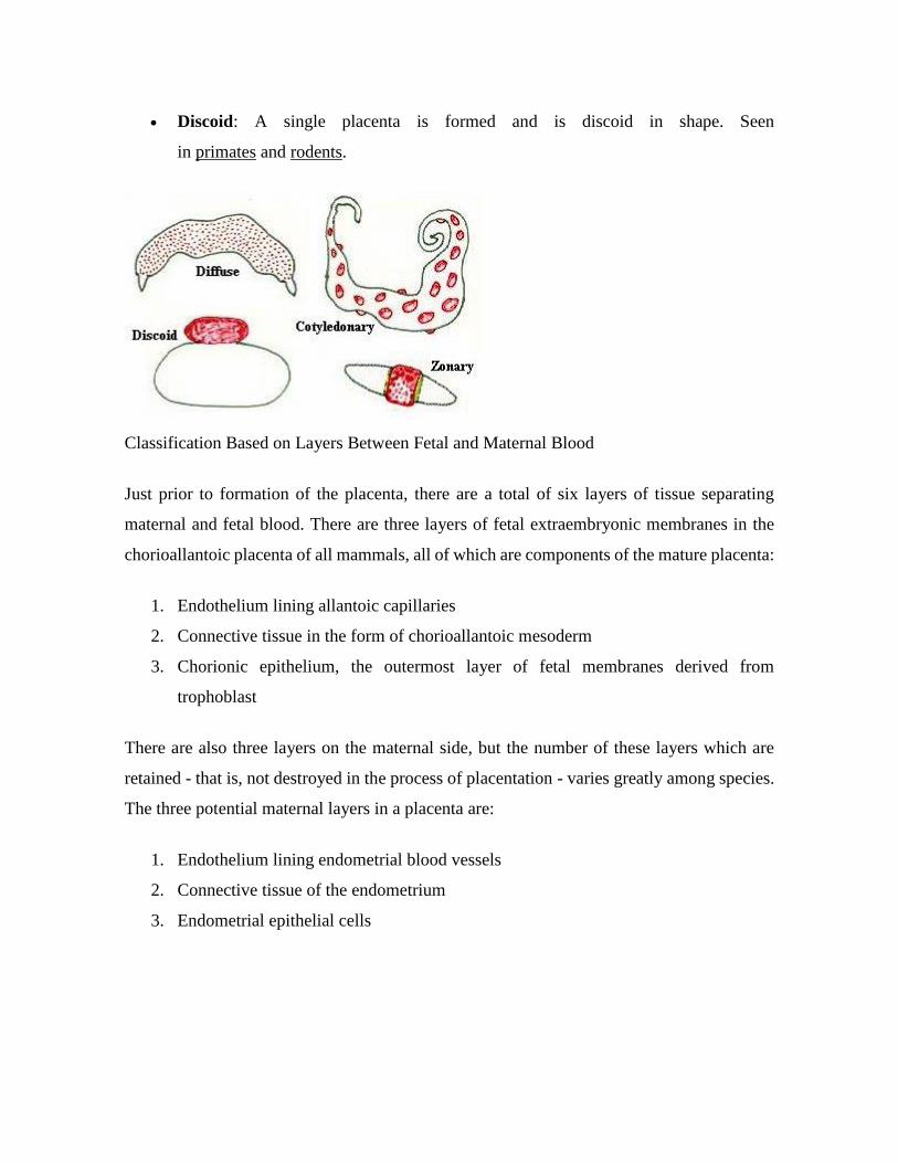
Discoid: A single placenta is formed and is discoid in shape. Seen
in primates and rodents.
Classification Based on Layers Between Fetal and Maternal Blood
Just prior to formation of the placenta, there are a total of six layers of tissue separating
maternal and fetal blood. There are three layers of fetal extraembryonic membranes in the
chorioallantoic placenta of all mammals, all of which are components of the mature placenta:
1. Endothelium lining allantoic capillaries
2. Connective tissue in the form of chorioallantoic mesoderm
3. Chorionic epithelium, the outermost layer of fetal membranes derived from
trophoblast
There are also three layers on the maternal side, but the number of these layers which are
retained - that is, not destroyed in the process of placentation - varies greatly among species.
The three potential maternal layers in a placenta are:
1. Endothelium lining endometrial blood vessels
2. Connective tissue of the endometrium
3. Endometrial epithelial cells
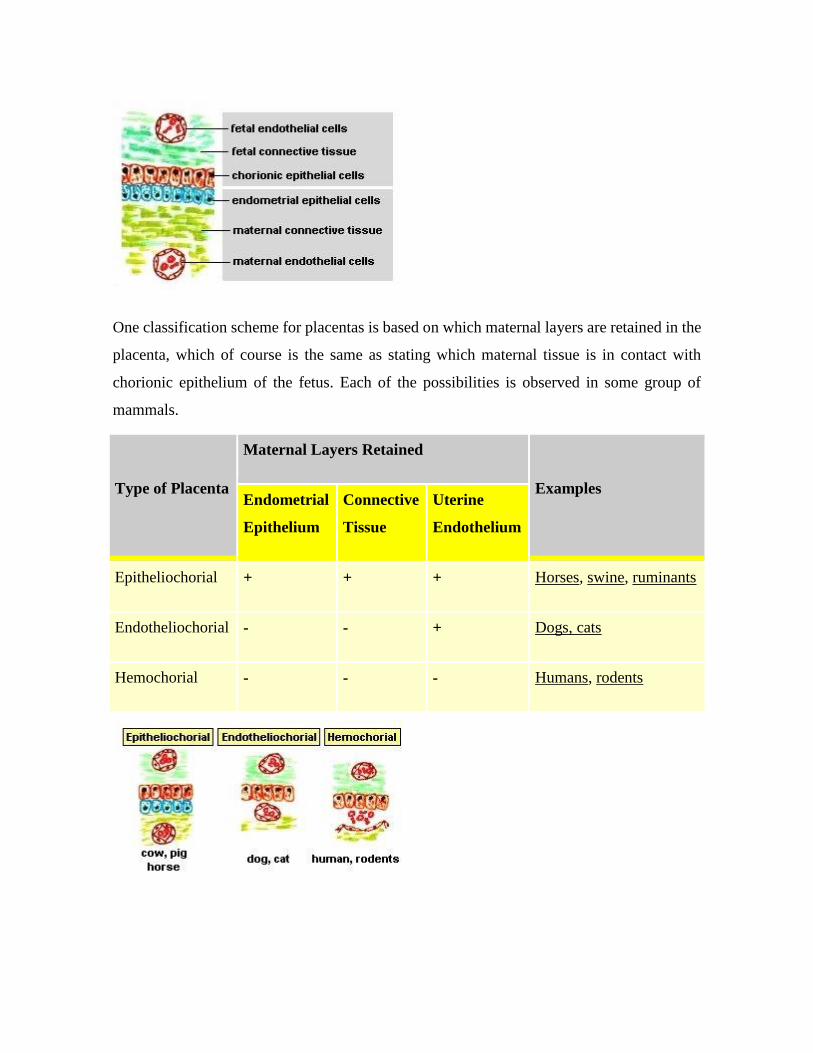
One classification scheme for placentas is based on which maternal layers are retained in the
placenta, which of course is the same as stating which maternal tissue is in contact with
chorionic epithelium of the fetus. Each of the possibilities is observed in some group of
mammals.
Type of Placenta
Maternal Layers Retained
Examples Endometrial
Epithelium
Connective
Tissue
Uterine
Endothelium
Epitheliochorial + + + Horses, swine, ruminants
Endotheliochorial - - + Dogs, cats
Hemochorial - - - Humans, rodents

In humans, fetal chorionic epithelium is bathed in maternal blood because chorionic villi
have eroded through maternal endothelium. In contrast, the chorionic epithelium of horse
and pig fetuses remains separated from maternal blood by 3 layers of tissue. One might thus
be tempted to consider that exchange across the equine placenta is much less efficient that
across the human placenta. In a sense this is true, but other features of placental structure
make up for the extra layers in the diffusion barrier; it has been well stated that "The newborn
foal provides a strong testimonial to the efficiency of the epitheliochorial placenta."
Summary of Species Differences in Placental Architecture
The placental mammals have evolved a variety of placental types which can be broadly
classified using the nomenclature described above. Not all combinations of those
classification schemes are seen or are likely to ever be seen - for instance, no mammal is
known to have a diffuse, endotheliochorial, or a hemoendothelial placenta. Placental types
for "familiar" mammals are summarized below, with supplemental information provided for
a variety of "non-familiar" species.
Type of Placenta Common Examples
Diffuse, epitheliochorial Horses and pigs
Cotyledonary, epitheliochorial Ruminants (cattle, sheep, goats, deer)
Zonary, endotheliochorial Carnivores (dog, cat, ferret)
Discoid, hemochorial Humans, apes, monkeys and rodents
Resources http://www.vivo.colostate.edu/hbooks/pathphys/reprod/placenta/structure.html
Comparative Placentation is an excellent and comprehensive site for obtaining information on placental structure for a large number of domestic and wild animals
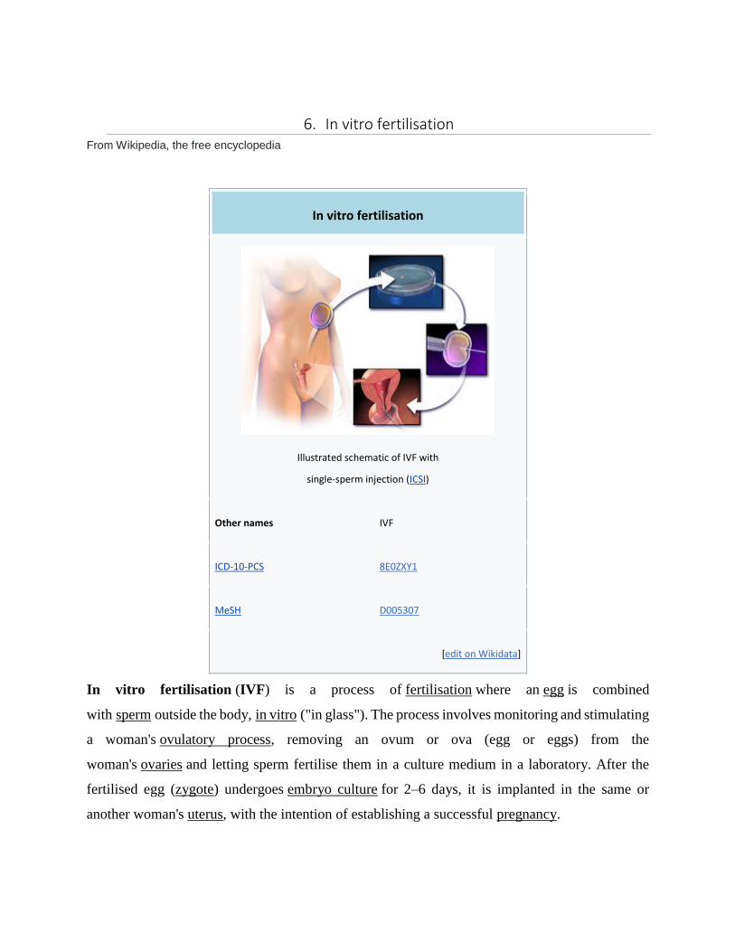
6. In vitro fertilisation From Wikipedia, the free encyclopedia
In vitro fertilisation
Illustrated schematic of IVF with
single-sperm injection (ICSI)
Other names IVF
ICD-10-PCS 8E0ZXY1
MeSH D005307
[edit on Wikidata]
In vitro fertilisation (IVF) is a process of fertilisation where an egg is combined
with sperm outside the body, in vitro ("in glass"). The process involves monitoring and stimulating
a woman's ovulatory process, removing an ovum or ova (egg or eggs) from the
woman's ovaries and letting sperm fertilise them in a culture medium in a laboratory. After the
fertilised egg (zygote) undergoes embryo culture for 2–6 days, it is implanted in the same or
another woman's uterus, with the intention of establishing a successful pregnancy.

IVF is a type of assisted reproductive technology used for infertility treatment and gestational
surrogacy. A fertilised egg may be implanted into a surrogate's uterus, and the resulting child is
genetically unrelated to the surrogate. Some countries have banned or otherwise regulate the
availability of IVF treatment, giving rise to fertility tourism. Restrictions on the availability of IVF
include costs and age, in order for a woman to carry a healthy pregnancy to term. IVF is generally
not used until less invasive or expensive options have failed or been determined unlikely to work.
In July 1978, Louise Brown was the first child successfully born after her mother received IVF
treatment. Brown was born as a result of natural-cycle IVF, where no stimulation was made. The
procedure took place at Dr Kershaw's Cottage Hospital (now Dr Kershaw's Hospice) in Royton,
Oldham, England. Robert G. Edwards was awarded the Nobel Prize in Physiology or Medicine in
2010. The physiologist co-developed the treatment together with Patrick Steptoe and
embryologist Jean Purdy but the latter two were not eligible for consideration as they had died and
the Nobel Prize is not awarded posthumously.[1][2]
With egg donation and IVF, women who are past their reproductive years, have infertile male
partners, have idiopathic female-fertility issues, or have reached menopause can still become
pregnant. After the IVF treatment, some couples get pregnant without any fertility treatments.[3] In
2018, it was estimated that eight million children had been born worldwide using IVF and other
assisted reproduction techniques.[4] However, a recent study that explores 10 adjuncts with IVF
(screening hysteroscopy, DHEA, testosterone, GH, aspirin, heparin, antioxidants in males and
females, seminal plasma, and PRP) suggests that until more evidence is done to show that these
adjuncts are safe and effective, they should be avoided.[5]
Reference:
https://en.wikipedia.org/wiki/In_vitro_fertilisation
7. Test-Tube Baby
By: Stephen C. Ruffenach
Published: 2009-01-13
Keywords: Fertilization, Reproduction
A test-tube baby is the product of a successful human reproduction that results from
methods beyond sexual intercourse between a man and a woman and instead utilizes
medical intervention that manipulates both the egg and sperm cells for
successful fertilization. The term was originally used to refer to the babies born from the
earliest applications of artificial insemination and has now been expanded to refer to
children born through the use of in vitro fertilization, the practice of fertilizing
an egg outside of a woman’s body. The use of the term in both media and scientific
publications in the twentieth century has been accompanied by discussion as well as
controversy regarding the ethics of reproduction technologies such as artificial
insemination and in vitro fertilization. The evolution of these terms over time mirrors the
perception of our ability to manipulate the human embryo, as seen by the general public
as well as the scientific community.
The term “test-tube baby,” prior to the development of in vitro fertilization technologies in
the twentieth century, was used to refer to babies born as a result of artificial
insemination. William Pancoast, a physician from Philadelphia, performed the first artificial
insemination that led to a successful birth in 1884, marking the birth of the first test-tube
baby. Despite the fact that this was the earliest instance of any sort of physician -assisted
reproduction, the grandeur of the event was not recognized by the public or media in any
notable way.
As reproduction technology continued to develop and in vitro fertilization research
advanced in the mid twentieth century, the media began to pay more attention to the idea
of test-tube babies and the impact their existence would have on the world. Publications
began to publish articles in the early twentieth century that discussed the ethics behind the
creation of children through means other than human sexual intercourse. Such publications
as The New York Times, Scientific American, and Newsweek, among others, published
articles discussing test-tube babies and the technologies used to create them, focusing on
what their existence meant for the development of the public’s understanding of
reproduction as well as the ethics involved with such an advanced understanding. The
articles refrained from being overtly outraged in response to the experiments but
recognized the controversy involved with these new scientific developments and included
this in their coverage.
By the middle of the twentieth century, in vitro fertilization had been thoroughly
researched and reported on, though no successful human reproduction had occurred from
its practice. However, the successful fertilization and reproduction of various species of
animal besides humans through in vitro fertilization was common practice by 1960. It was
also around this time that the discussion of a child born from in vitro fertilization began to
show up in numerous publications. Look magazine published an article in 1971 entitled
“The Test Tube Baby is Coming,” which reported on the work of Landrum Brewer Shettles,
an American biologist who would eventually attempt the first human in vitro fertilization in
America, which was not successful.
It was not until 25 July 1978 with the work of two British medical researchers, Patrick
Steptoe and Robert Edwards, that the revised definition of test-tube baby became a reality.
With their work, the first child born from a zygote fertilized outside of a human body was
created. The first baby born via in vitro fertilization was Louise Brown. It was also this
breakthrough that really caught the attention of the world, made clear in the media’s
reaction to Louise Brown’s birth. Newspapers reported the story as a significant scientific
and medical breakthrough while tabloids and other similar publications treated the birth
more like the arrival of a new celebrity into the world. The mixed media response to the
birth of Louise Brown was also very foretelling of the diverse public reactions that would
follow. Shortly after the birth, discussion of both legal and ethical implications regarding
the existence of test-tube babies began in the international media as well as in scientific
journals. These debates continue to this day as the ongoing research of embryos and
reproduction leads to the development of new medical practices.
After the birth of Louise Brown, scientists in other countries around the world began
pursuing the birth of their first child from in vitro fertilization. This goal was also
accompanied by the development of numerous fertility clinics in various countries. The
first fertility clinic in the United States opened at the Eastern Virginia Medical School in
Norfolk, Virginia on 1 March 1980. It was also here that Jordan Elizabeth Carr, the first
child who was a product of in vitro fertilization in America, was born on 28 December
1981. By this time, the practice of in vitro fertilization was much more publicly accepted,
as it was observed that children born under such circumstances developed normally. This
was not to say that there were no opponents to the practice at this time, but rather that
Carr’s birth was received with excitement from the general public and the media captured
this response when covering the event. More fertility clinics were established all over the
world and the number of successful test-tube baby births continued to grow as in
vitro fertilization became both a more refined and more accepted practice.
Through the work of various scientists, the idea of traditional fertilization and human
reproduction through sexual intercourse was no longer seen as the only means of fertilizing
an egg in order to create a human life. This concept was represented by the existence of
test-tube babies, the physical manifestation of a more advanced control over human
reproduction. Through the work of such scientists as Pancoast, Steptoe, and Edwards, the
definition of test-tube baby has changed over time but continues to refer to any child born
from an embryo created by means of medical intervention that directly manipulates
the sperm and egg cells.
Sources
Henig, Robin Marantz. Pandora’s Baby. New York: Houghton Mifflin Company, 2004.
American Experience: Test Tube Babies. “Timeline: Human Reproduction Research and In Vitro Fertilization.” Public Broadcasting System. http://www.pbs.org/wgbh/amex/babies/timeline/index.html (Accessed April 10, 2008).
Russel, Willis. “Among the New Words.” American Speech 23 (1948): 285–95.
“Steptoe, Patrick ; and Edwards, Robert.” Encyclopedia Britannica. 2008. Encyclopedia Britannica Online. 14 Apr. 2008 http://search.eb.com/eb/article-9001302.
How to cite
Ruffenach, Stephen C., "Test-Tube Baby". Embryo Project Encyclopedia (2009-01-13). ISSN: 1940-5030 http://embryo.asu.edu/handle/10776/1668.

8. Metamorphosis By:BD Editors
Reviewed by: BD Editors
Last Updated: October 4, 2019
Metamorphosis Definition
Metamorphosis is a process by which animals undergo extreme, rapid physical changes some time
after birth. The result of metamorphosis may be change to the organism’s entire body plan, such
as a change in the animal’s number of legs, its means of eating, or its means of breathing.
In species that use metamorphosis, metamorphosis is also typically required for sexual maturity.
Pre-metamorphic members of these species are typically unable to mate or reproduce.
Commonly known examples of metamorphosis include the process undergone by most insects,
and the transformation of tadpoles into frogs. The diagram below shows the stages of this change,
wherein the small fish-like tadpoles transform into what seems a completely different animal:
Animals that you may not know undergo metamorphosis include fish, mollusks, and many other
types of sea creatures which are related to insects, mollusks, or fish. Lobsters, for example, which
are closely related to insects, do undergo metamorphosis as part of their life cycle.
Metamorphosis is a remarkable process. The speed and extent of cell growth and differentiation is
astonishing. In most species, such rapid growth and such sweeping changes to cell type only

happen during embryonic development. Indeed, some scientists believe that the process of
metamorphosis involves a sort of re-activating of genes that allow animal cells to change from one
cell type to another.
The changes leading to metamorphosis are triggered by hormones, which the animal’s body
releases as the right conditions for metamorphosis approach. In some animals a hormone cascade
follows, with the trigger hormone causing the release of several other hormones that act on
different parts of the animal’s body.
The hormones cause drastic changes to the functioning of cells, and even behavioral changes such
as the caterpillar spinning its cocoon.
The effects of hormones on metamorphosis can be studied by artificially administering these
hormones to pre-metamorphic animals. Tadpoles, for example, can be triggered to begin losing
their tails and growing limbs early by the addition of thyroid hormones to their water supply.
Unfortunately this has a detrimental effect on the animal’s health.
Function of Metamorphosis
Scientists remain uncertain why metamorphosis evolved. For the animals of today, its purpose is
obvious: if metamorphosis did not occur, tadpoles could not become frogs and larvae could not
become full-grown adults capable of reproduction. Without reproductively mature members, these
species would quickly die off.
But why would these species evolve to need this extra step in the first place? Why not just hatch
full-grown butterflies or frogs from eggs?
At least some metamorphosing species did not start out that way: the earliest insects basically did
hatch as full-grown adults. But a few hundred million years ago, some species stumbled upon the
trick of metamorphosis. It was apparently wildly successful; it is thought that almost two-thirds of
species alive today use metamorphosis to accomplish large changes between their adult and
juvenile forms.

The benefit of metamorphosis may lie in its ability to reduce competition. Pre-metamorphic
animals typically consume completely different resources from their adult forms. Tadpoles live in
water, eating algae and plants. Frogs live on land, breathing air and eating insects. Caterpillars eat
leaves; butterflies live off of nectar. Etc..
This effectively prevents older members of the species from competing with younger members.
This may lead more members of the species to successfully reach sexual maturity, without the risk
of being out-competed by older members of their species.
Types of Metamorphosis
Complete Metamorphosis
In complete metamorphosis, a larva completely changes its body plan to become an adult. The
most famous example is that of the butterfly, which starts out as a worm-like, leaf-eating caterpillar
and transforms into a flying, nectar-drinking creature with an exoskeleton.
Organisms that undergo complete metamorphosis are called “holometabolous,” from the Greek
words “holo” for “complete” or “whole,” “meta” for “change,” and the noun “bole” for “to throw.”
“Holometabolous,” then, means “completely changing,” or “wholly changing.”
This transformation is so swift and complete that the caterpillar must spin a cocoon and lie dormant
for weeks while its body undergoes these radical changes.
Other animals which transform from a worm-like larval stage into an animal that looks completely
different include beetles, flies, moths, ants, and bees.
Some scientists believe that the larval stage of complete metamorphosis may have evolved from
insects which hatched from their eggs without developing properly. Some of these embryos may
have survived long enough to find food in the outside world; and this may have ended up giving
them an advantage, as they would be able to feed longer and gain more strength than their peers
before metamorphosing into the adult stage.
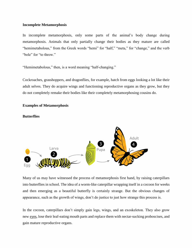
Incomplete Metamorphosis
In incomplete metamorphosis, only some parts of the animal’s body change during
metamorphosis. Animals that only partially change their bodies as they mature are called
“hemimetabolous,” from the Greek words “hemi” for “half,” “meta,” for “change,” and the verb
“bole” for “to throw.”
“Hemimetabolous,” then, is a word meaning “half-changing.”
Cockroaches, grasshoppers, and dragonflies, for example, hatch from eggs looking a lot like their
adult selves. They do acquire wings and functioning reproductive organs as they grow, but they
do not completely remake their bodies like their completely metamorphosing cousins do.
Examples of Metamorphosis
Butterflies
Many of us may have witnessed the process of metamorphosis first hand, by raising caterpillars
into butterflies in school. The idea of a worm-like caterpillar wrapping itself in a cocoon for weeks
and then emerging as a beautiful butterfly is certainly strange. But the obvious changes of
appearance, such as the growth of wings, don’t do justice to just how strange this process is.
In the cocoon, caterpillars don’t simply gain legs, wings, and an exoskeleton. They also grow
new eyes, lose their leaf-eating mouth parts and replace them with nectar-sucking proboscises, and
gain mature reproductive organs.
To accomplish this drastic change, a metamorphosing caterpillar basically digests itself.
A great deal of energy and raw materials are required to turn a caterpillar into a butterfly. So to
make it possible, caterpillars release enzymes that dissolve most of their bodies! Indeed, the hard
shell of the cocoon is required not just to protect the metamorphosing insect from attack: it is
required to keep its liquefying body bound together, lest it ooze away!
Not all of the caterpillar’s cells are dissolved by these enzymes. Special tissues called imaginal
discs survive – and they use the soup that used to be the rest of the caterpillar’s body for nutrition.
By consuming the proteins, vitamins, and minerals – everything you need to build a butterfly –
these imaginal discs are able to grow incredibly quickly, developing into the butterfly’s mature
body parts.
The new body has almost nothing in common with the old body. It has new legs, new sensory
organs, a new exoskeleton, a new reproductive system. Even its digestive system does not work
the same way, since it must now digest nectar instead of leaves. That’s all in addition to the
beautiful wings.
This radical change allows butterflies to complete their life cycle very efficiently, with no
competition between adult butterflies and caterpillars for food.
Many other insects pass through a similar process. They hatch as worm-like larva, eventually
encase themselves in hard pupas, and emerge as adults with legs, exoskeletons, and other features
that have little in common with the larva they once were. Bees, beetles, ants, and flies all use this
strategy.
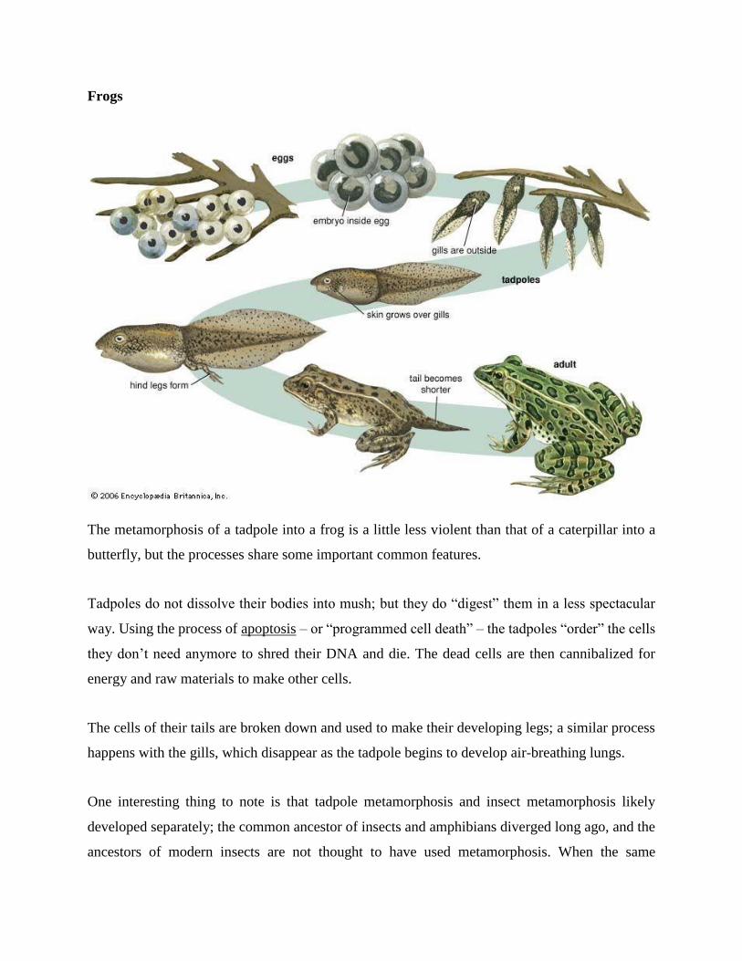
Frogs
The metamorphosis of a tadpole into a frog is a little less violent than that of a caterpillar into a
butterfly, but the processes share some important common features.
Tadpoles do not dissolve their bodies into mush; but they do “digest” them in a less spectacular
way. Using the process of apoptosis – or “programmed cell death” – the tadpoles “order” the cells
they don’t need anymore to shred their DNA and die. The dead cells are then cannibalized for
energy and raw materials to make other cells.
The cells of their tails are broken down and used to make their developing legs; a similar process
happens with the gills, which disappear as the tadpole begins to develop air-breathing lungs.
One interesting thing to note is that tadpole metamorphosis and insect metamorphosis likely
developed separately; the common ancestor of insects and amphibians diverged long ago, and the
ancestors of modern insects are not thought to have used metamorphosis. When the same

phenomenon evolves twice in radically different organisms, that’s a sure sign that it is a
useful adaptation!
Fish
Some species of fish undergo metamorphoses similar to those of the tadpole. Though those
changes are not so dramatic, they can result in changes in the fish’s food source, its body plan, and
where it’s able to live. Just like the more drastic forms of evolution, this may function to prevent
adults from competing with juveniles for food.
The salmon, for example, is a freshwater fish in its juvenile form. After undergoing a partial
metamorphosis, it becomes a saltwater fish.
When thinking about this process it is important to keep in mind that all organisms must regulate
their salt/water balance. This is why humans can’t drink seawater without dying: the salt would
overwhelm our cellular chemistry, and our cells would not function properly. In just the same way,
freshwater fish typically cannot live in saltwater. To become saltwater fish, then, salmon must
develop new organs and cellular mechanisms to cope with the salt water.
That’s why salmon must perform their annual migration upstream; adult salmon live in the ocean,
but their eggs must hatch in fresh water in order for the juveniles to survive. That means that adult
salmon must leave their homes in the ocean for freshwater rivers, and swim as far upstream as
possible before laying their eggs!
Flounders, bizarrely, undergo a metamorphosis in which one of their eyes and nostrils move from
one side of the head to the other. As juveniles, flounder look much like most fish: they swim
vertical relative to the current, with one eye and one nostril on each side of their bladelike body.
This body type allows them to swim fast like most other species of fish.
But in adulthood, flounder are flat fish which camouflage themselves by swimming on their
bellies, pressed against the sea bed. To accomplish this lifestyle change, juvenile flounder
essentially flip over on their sides and make one side of their body into their belly. Through cellular
changes, the eye and nostril from the belly side actually migrate to join the other eye and nostril
on what is now the “top” side of the fish.
Evolution sure has some creative ways of doing things!
Reference:
https://biologydictionary.net/metamorphosis/
9. Animals Reproduction
Some animals produce offspring through asexual reproduction while other animals produce
offspring through sexual reproduction. Both methods have advantages and disadvantages. Asexual
reproduction produces offspring that are genetically identical to the parent because the offspring
are all clones of the original parent. A single individual can produce offspring asexually and large
numbers of offspring can be produced quickly; these are two advantages that asexually
reproducing organisms have over sexually reproducing organisms. In a stable or predictable
environment, asexual reproduction is an effective means of reproduction because all the offspring
will be adapted to that environment. In an unstable or unpredictable environment, species that
reproduce asexually may be at a disadvantage because all the offspring are genetically identical
and may not be adapted to different conditions.
During sexual reproduction, the genetic material of two individuals is combined to produce
genetically diverse offspring that differ from their parents. The genetic diversity of sexually
produced offspring is thought to give sexually reproducing individuals greater fitness because
more of their offspring may survive and reproduce in an unpredictable or changing environment.
Species that reproduce sexually (and have separate sexes) must maintain two different types of
individuals, males and females. Only half the population (females) can produce the offspring, so
fewer offspring will be produced when compared to asexual reproduction. This is a disadvantage
of sexual reproduction compared to asexual reproduction.
Asexual Reproduction
Asexual reproduction occurs in prokaryotic microorganisms (bacteria and archaea) and in many
eukaryotic, single-celled and multi-celled organisms. There are several ways that animals
reproduce asexually, the details of which vary among individual species.
Fission
Fission, also called binary fission, occurs in some invertebrate, multi-celled organisms. It is in
some ways analogous to the process of binary fission of single-celled prokaryotic organisms. The
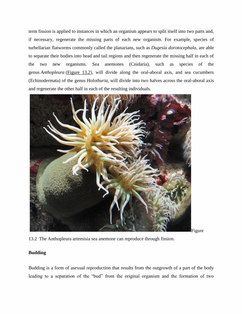
term fission is applied to instances in which an organism appears to split itself into two parts and,
if necessary, regenerate the missing parts of each new organism. For example, species of
turbellarian flatworms commonly called the planarians, such as Dugesia dorotocephala, are able
to separate their bodies into head and tail regions and then regenerate the missing half in each of
the two new organisms. Sea anemones (Cnidaria), such as species of the
genus Anthopleura (Figure 13.2), will divide along the oral-aboral axis, and sea cucumbers
(Echinodermata) of the genus Holothuria, will divide into two halves across the oral-aboral axis
and regenerate the other half in each of the resulting individuals.
Figure
13.2 The Anthopleura artemisia sea anemone can reproduce through fission.
Budding
Budding is a form of asexual reproduction that results from the outgrowth of a part of the body
leading to a separation of the “bud” from the original organism and the formation of two
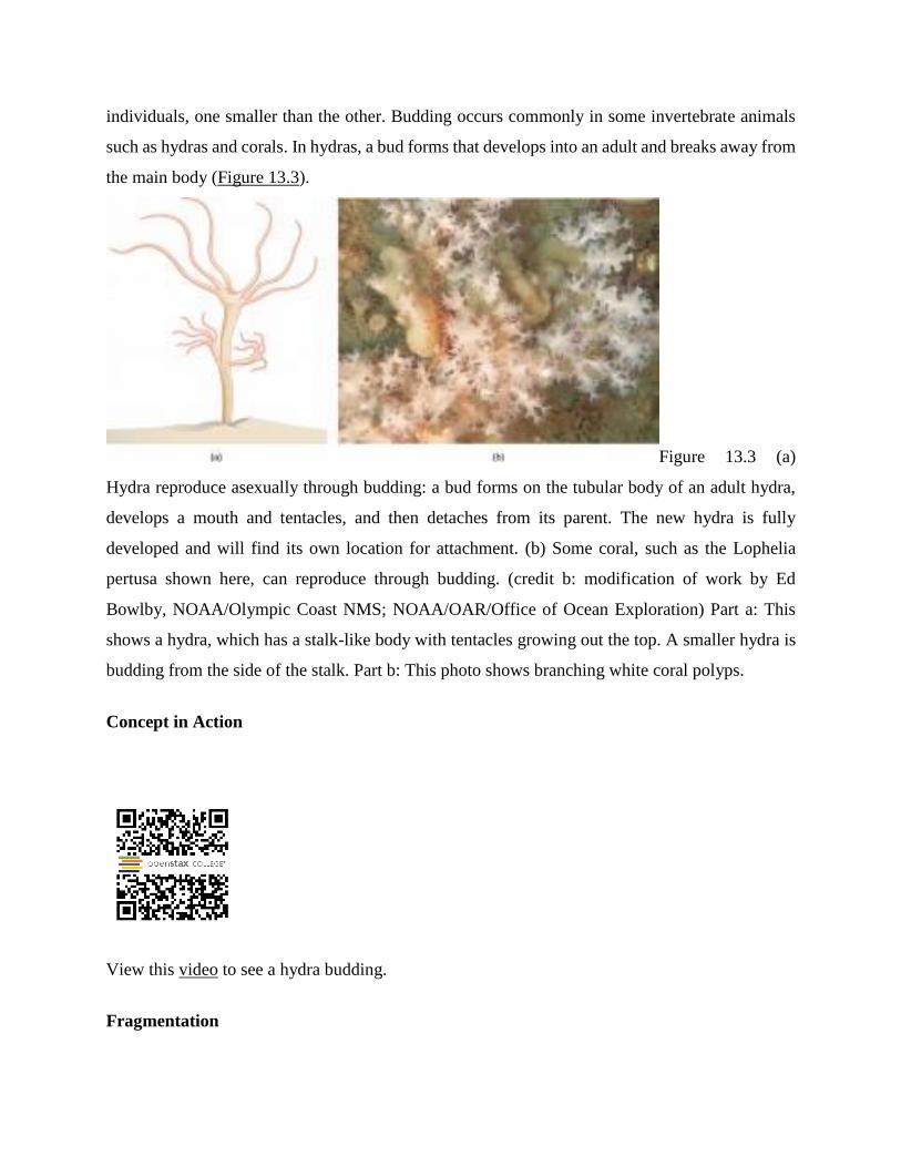
individuals, one smaller than the other. Budding occurs commonly in some invertebrate animals
such as hydras and corals. In hydras, a bud forms that develops into an adult and breaks away from
the main body (Figure 13.3).
Figure 13.3 (a)
Hydra reproduce asexually through budding: a bud forms on the tubular body of an adult hydra,
develops a mouth and tentacles, and then detaches from its parent. The new hydra is fully
developed and will find its own location for attachment. (b) Some coral, such as the Lophelia
pertusa shown here, can reproduce through budding. (credit b: modification of work by Ed
Bowlby, NOAA/Olympic Coast NMS; NOAA/OAR/Office of Ocean Exploration) Part a: This
shows a hydra, which has a stalk-like body with tentacles growing out the top. A smaller hydra is
budding from the side of the stalk. Part b: This photo shows branching white coral polyps.
Concept in Action
View this video to see a hydra budding.
Fragmentation
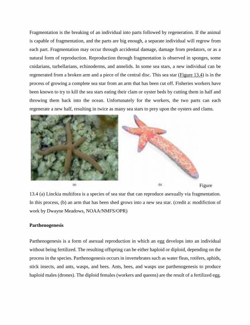
Fragmentation is the breaking of an individual into parts followed by regeneration. If the animal
is capable of fragmentation, and the parts are big enough, a separate individual will regrow from
each part. Fragmentation may occur through accidental damage, damage from predators, or as a
natural form of reproduction. Reproduction through fragmentation is observed in sponges, some
cnidarians, turbellarians, echinoderms, and annelids. In some sea stars, a new individual can be
regenerated from a broken arm and a piece of the central disc. This sea star (Figure 13.4) is in the
process of growing a complete sea star from an arm that has been cut off. Fisheries workers have
been known to try to kill the sea stars eating their clam or oyster beds by cutting them in half and
throwing them back into the ocean. Unfortunately for the workers, the two parts can each
regenerate a new half, resulting in twice as many sea stars to prey upon the oysters and clams.
Figure
13.4 (a) Linckia multifora is a species of sea star that can reproduce asexually via fragmentation.
In this process, (b) an arm that has been shed grows into a new sea star. (credit a: modifiction of
work by Dwayne Meadows, NOAA/NMFS/OPR)
Parthenogenesis
Parthenogenesis is a form of asexual reproduction in which an egg develops into an individual
without being fertilized. The resulting offspring can be either haploid or diploid, depending on the
process in the species. Parthenogenesis occurs in invertebrates such as water fleas, rotifers, aphids,
stick insects, and ants, wasps, and bees. Ants, bees, and wasps use parthenogenesis to produce
haploid males (drones). The diploid females (workers and queens) are the result of a fertilized egg.
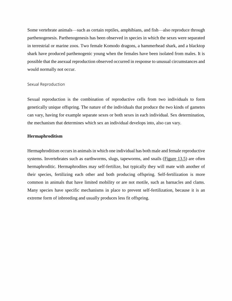
Some vertebrate animals—such as certain reptiles, amphibians, and fish—also reproduce through
parthenogenesis. Parthenogenesis has been observed in species in which the sexes were separated
in terrestrial or marine zoos. Two female Komodo dragons, a hammerhead shark, and a blacktop
shark have produced parthenogenic young when the females have been isolated from males. It is
possible that the asexual reproduction observed occurred in response to unusual circumstances and
would normally not occur.
Sexual Reproduction
Sexual reproduction is the combination of reproductive cells from two individuals to form
genetically unique offspring. The nature of the individuals that produce the two kinds of gametes
can vary, having for example separate sexes or both sexes in each individual. Sex determination,
the mechanism that determines which sex an individual develops into, also can vary.
Hermaphroditism
Hermaphroditism occurs in animals in which one individual has both male and female reproductive
systems. Invertebrates such as earthworms, slugs, tapeworms, and snails (Figure 13.5) are often
hermaphroditic. Hermaphrodites may self-fertilize, but typically they will mate with another of
their species, fertilizing each other and both producing offspring. Self-fertilization is more
common in animals that have limited mobility or are not motile, such as barnacles and clams.
Many species have specific mechanisms in place to prevent self-fertilization, because it is an
extreme form of inbreeding and usually produces less fit offspring.
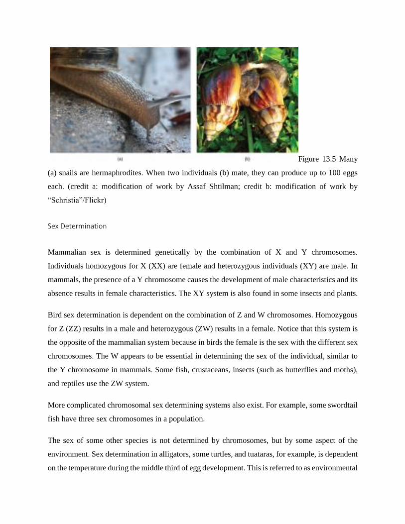
Figure 13.5 Many
(a) snails are hermaphrodites. When two individuals (b) mate, they can produce up to 100 eggs
each. (credit a: modification of work by Assaf Shtilman; credit b: modification of work by
“Schristia”/Flickr)
Sex Determination
Mammalian sex is determined genetically by the combination of X and Y chromosomes.
Individuals homozygous for X (XX) are female and heterozygous individuals (XY) are male. In
mammals, the presence of a Y chromosome causes the development of male characteristics and its
absence results in female characteristics. The XY system is also found in some insects and plants.
Bird sex determination is dependent on the combination of Z and W chromosomes. Homozygous
for Z (ZZ) results in a male and heterozygous (ZW) results in a female. Notice that this system is
the opposite of the mammalian system because in birds the female is the sex with the different sex
chromosomes. The W appears to be essential in determining the sex of the individual, similar to
the Y chromosome in mammals. Some fish, crustaceans, insects (such as butterflies and moths),
and reptiles use the ZW system.
More complicated chromosomal sex determining systems also exist. For example, some swordtail
fish have three sex chromosomes in a population.
The sex of some other species is not determined by chromosomes, but by some aspect of the
environment. Sex determination in alligators, some turtles, and tuataras, for example, is dependent
on the temperature during the middle third of egg development. This is referred to as environmental
sex determination, or more specifically, as temperature-dependent sex determination. In many
turtles, cooler temperatures during egg incubation produce males and warm temperatures produce
females, while in many other species of turtles, the reverse is true. In some crocodiles and some
turtles, moderate temperatures produce males and both warm and cool temperatures produce
females.
Individuals of some species change their sex during their lives, switching from one to the other. If
the individual is female first, it is termed protogyny or “first female,” if it is male first, it is termed
protandry or “first male.” Oysters are born male, grow in size, and become female and lay eggs.
The wrasses, a family of reef fishes, are all sequential hermaphrodites. Some of these species live
in closely coordinated schools with a dominant male and a large number of smaller females. If the
male dies, a female increases in size, changes sex, and becomes the new dominant male.
Fertilization
The fusion of a sperm and an egg is a process called fertilization. This can occur either inside
(internal fertilization) or outside (external fertilization) the body of the female. Humans provide
an example of the former, whereas frog reproduction is an example of the latter.
External Fertilization
External fertilization usually occurs in aquatic environments where both eggs and sperm are
released into the water. After the sperm reaches the egg, fertilization takes place. Most external
fertilization happens during the process of spawning where one or several females release their
eggs and the male(s) release sperm in the same area, at the same time. The spawning may be
triggered by environmental signals, such as water temperature or the length of daylight. Nearly all
fish spawn, as do crustaceans (such as crabs and shrimp), mollusks (such as oysters), squid, and
echinoderms (such as sea urchins and sea cucumbers). Frogs, corals, mayflies, and mosquitoes
also spawn (Figure 13.6).

Figure 13.6
During sexual reproduction in toads, the male grasps the female from behind and externally
fertilizes the eggs as they are deposited. (credit: Bernie Kohl)
Internal Fertilization
Internal fertilization occurs most often in terrestrial animals, although some aquatic animals also
use this method. Internal fertilization may occur by the male directly depositing sperm in the
female during mating. It may also occur by the male depositing sperm in the environment, usually
in a protective structure, which a female picks up to deposit the sperm in her reproductive tract.
There are three ways that offspring are produced following internal fertilization. In oviparity,
fertilized eggs are laid outside the female’s body and develop there, receiving nourishment from
the yolk that is a part of the egg (Figure 13.7 a). This occurs in some bony fish, some reptiles, a
few cartilaginous fish, some amphibians, a few mammals, and all birds. Most non-avian reptiles
and insects produce leathery eggs, while birds and some turtles produce eggs with high
concentrations of calcium carbonate in the shell, making them hard. Chicken eggs are an example
of a hard shell. The eggs of the egg-laying mammals such as the platypus and echidna are leathery.
In ovoviparity, fertilized eggs are retained in the female, and the embryo obtains its nourishment
from the egg’s yolk. The eggs are retained in the female’s body until they hatch inside of her, or
she lays the eggs right before they hatch. This process helps protect the eggs until hatching. This
occurs in some bony fish (like the platyfish Xiphophorus maculatus, Figure 13.7 b), some sharks,

lizards, some snakes (garter snake Thamnophis sirtalis), some vipers, and some invertebrate
animals (Madagascar hissing cockroach Gromphadorhina portentosa).
In viviparity the young are born alive. They obtain their nourishment from the female and are born
in varying states of maturity. This occurs in most mammals (Figure 13.7 c), some cartilaginous
fish, and a few reptiles.
Figure 13.7
In (a) oviparity, young develop in eggs outside the female body, as with these Harmonia axydridis
beetles hatching. Some aquatic animals, like this (b) pregnant Xiphophorus maculatus are
ovoviparous, with the egg developing inside the female and nutrition supplied primarily from the
yolk. In mammals, nutrition is supported by the placenta, as was the case with this (c) newborn
squirrel. (credit b: modification of work by Gourami Watcher; credit c: modification of work by
“audreyjm529″/Flickr)
Reference:
https://opentextbc.ca/biology/chapter/13-1-how-animals-
reproduce/#:~:text=Reproduction%20may%20be%20asexual%20when,budding%2C%20fragmentation%
2C%20and%20parthenogenesis.
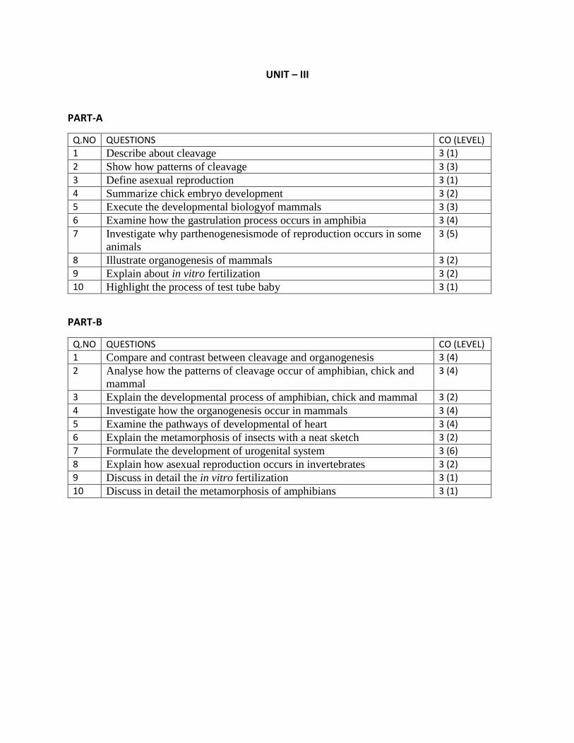
UNIT – III
PART-A
Q.NO QUESTIONS CO (LEVEL)
1 Describe about cleavage 3 (1)
2 Show how patterns of cleavage 3 (3)
3 Define asexual reproduction 3 (1)
4 Summarize chick embryo development 3 (2)
5 Execute the developmental biologyof mammals 3 (3)
6 Examine how the gastrulation process occurs in amphibia 3 (4)
7 Investigate why parthenogenesismode of reproduction occurs in some
animals
3 (5)
8 Illustrate organogenesis of mammals 3 (2)
9 Explain about in vitro fertilization 3 (2)
10 Highlight the process of test tube baby 3 (1)
PART-B
Q.NO QUESTIONS CO (LEVEL)
1 Compare and contrast between cleavage and organogenesis 3 (4)
2 Analyse how the patterns of cleavage occur of amphibian, chick and
mammal
3 (4)
3 Explain the developmental process of amphibian, chick and mammal 3 (2)
4 Investigate how the organogenesis occur in mammals 3 (4)
5 Examine the pathways of developmental of heart 3 (4)
6 Explain the metamorphosis of insects with a neat sketch 3 (2)
7 Formulate the development of urogenital system 3 (6)
8 Explain how asexual reproduction occurs in invertebrates 3 (2)
9 Discuss in detail the in vitro fertilization 3 (1)
10 Discuss in detail the metamorphosis of amphibians 3 (1)
SCHOOL OF BIO & CHEMICAL ENGINEERING
DEPARTMENT OF BIOTECHNOLOGY
SBC1201: ZOOLOGY
UNIT – IV - EVOLUTION, ADAPTIVE RADIATION AND FOSSILS– SBC1201
1. EVOLUTION
Evolution by natural selection describes a mechanism for how species change over time. That
species change had been suggested and debated well before Darwin began to explore this idea.
The view that species were static and unchanging was grounded in the writings of Plato, yet there
were also ancient Greeks who expressed evolutionary ideas. In the eighteenth century, ideas about
the evolution of animals were reintroduced by the naturalist Georges-Louis Leclerc Comte de
Buffon who observed that various geographic regions have different plant and animal populations,
even when the environments are similar. It was also accepted that there were extinct species.
During this time, James Hutton, a Scottish naturalist, proposed that geological change occurred
gradually by the accumulation of small changes from processes operating like they are today over
long periods of time. This contrasted with the predominant view that the geology of the planet was
a consequence of catastrophic events occurring during a relatively brief past. Hutton’s view was
popularized in the nineteenth century by the geologist Charles Lyell who became a friend to
Darwin. Lyell’s ideas were influential on Darwin’s thinking: Lyell’s notion of the greater age of
Earth gave more time for gradual change in species, and the process of change provided an analogy
for gradual change in species. In the early nineteenth century, Jean-Baptiste Lamarck published a
book that detailed a mechanism for evolutionary change. This mechanism is now referred to as an
inheritance of acquired characteristics by which modifications in an individual are caused by its
environment, or the use or disuse of a structure during its lifetime, could be inherited by its
offspring and thus bring about change in a species. While this mechanism for evolutionary change
was discredited, Lamarck’s ideas were an important influence on evolutionary thought.
Charles Darwin and Natural Selection
In the mid-nineteenth century, the actual mechanism for evolution was independently conceived
of and described by two naturalists: Charles Darwin and Alfred Russel Wallace. Importantly, each
naturalist spent time exploring the natural world on expeditions to the tropics. From 1831 to 1836,
Darwin traveled around the world on H.M.S. Beagle, including stops in South America, Australia,
and the southern tip of Africa. Wallace traveled to Brazil to collect insects in the Amazon rainforest
from 1848 to 1852 and to the Malay Archipelago from 1854 to 1862. Darwin’s journey, like
Wallace’s later journeys to the Malay Archipelago, included stops at several island chains, the last
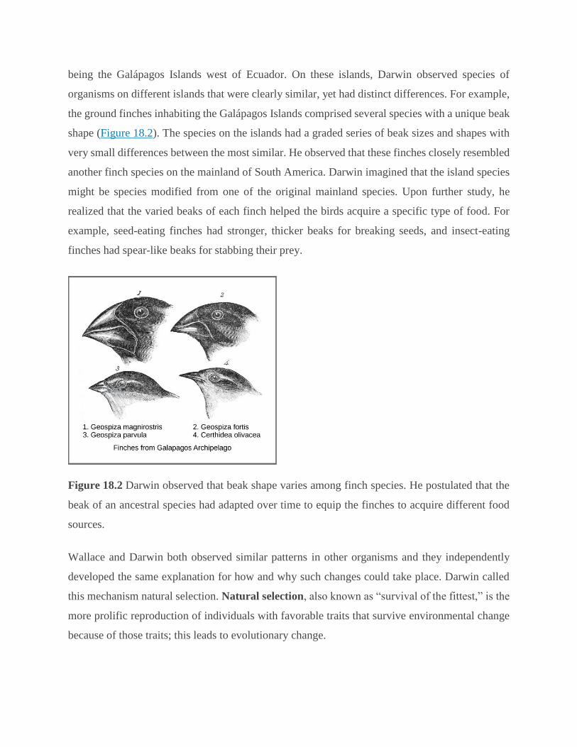
being the Galápagos Islands west of Ecuador. On these islands, Darwin observed species of
organisms on different islands that were clearly similar, yet had distinct differences. For example,
the ground finches inhabiting the Galápagos Islands comprised several species with a unique beak
shape (Figure 18.2). The species on the islands had a graded series of beak sizes and shapes with
very small differences between the most similar. He observed that these finches closely resembled
another finch species on the mainland of South America. Darwin imagined that the island species
might be species modified from one of the original mainland species. Upon further study, he
realized that the varied beaks of each finch helped the birds acquire a specific type of food. For
example, seed-eating finches had stronger, thicker beaks for breaking seeds, and insect-eating
finches had spear-like beaks for stabbing their prey.
Figure 18.2 Darwin observed that beak shape varies among finch species. He postulated that the
beak of an ancestral species had adapted over time to equip the finches to acquire different food
sources.
Wallace and Darwin both observed similar patterns in other organisms and they independently
developed the same explanation for how and why such changes could take place. Darwin called
this mechanism natural selection. Natural selection, also known as “survival of the fittest,” is the
more prolific reproduction of individuals with favorable traits that survive environmental change
because of those traits; this leads to evolutionary change.
For example, a population of giant tortoises found in the Galapagos Archipelago was observed by
Darwin to have longer necks than those that lived on other islands with dry lowlands. These
tortoises were “selected” because they could reach more leaves and access more food than those
with short necks. In times of drought when fewer leaves would be available, those that could reach
more leaves had a better chance to eat and survive than those that couldn’t reach the food source.
Consequently, long-necked tortoises would be more likely to be reproductively successful and pass
the long-necked trait to their offspring. Over time, only long-necked tortoises would be present in
the population.
Natural selection, Darwin argued, was an inevitable outcome of three principles that operated in
nature. First, most characteristics of organisms are inherited, or passed from parent to offspring.
Although no one, including Darwin and Wallace, knew how this happened at the time, it was a
common understanding. Second, more offspring are produced than are able to survive, so resources
for survival and reproduction are limited. The capacity for reproduction in all organisms outstrips
the availability of resources to support their numbers. Thus, there is competition for those
resources in each generation. Both Darwin and Wallace’s understanding of this principle came
from reading an essay by the economist Thomas Malthus who discussed this principle in relation
to human populations. Third, offspring vary among each other in regard to their characteristics and
those variations are inherited. Darwin and Wallace reasoned that offspring with inherited
characteristics which allow them to best compete for limited resources will survive and have more
offspring than those individuals with variations that are less able to compete. Because
characteristics are inherited, these traits will be better represented in the next generation. This will
lead to change in populations over generations in a process that Darwin called descent with
modification. Ultimately, natural selection leads to greater adaptation of the population to its local
environment; it is the only mechanism known for adaptive evolution.
Papers by Darwin and Wallace (Figure 18.3) presenting the idea of natural selection were read
together in 1858 before the Linnean Society in London. The following year Darwin’s book, On
the Origin of Species, was published. His book outlined in considerable detail his arguments for
evolution by natural selection.

Figure 18.3 Both (a) Charles Darwin and (b) Alfred Wallace wrote scientific papers on natural
selection that were presented together before the Linnean Society in 1858.
Demonstrations of evolution by natural selection are time consuming and difficult to obtain. One
of the best examples has been demonstrated in the very birds that helped to inspire Darwin’s
theory: the Galápagos finches. Peter and Rosemary Grant and their colleagues have studied
Galápagos finch populations every year since 1976 and have provided important demonstrations
of natural selection. The Grants found changes from one generation to the next in the distribution
of beak shapes with the medium ground finch on the Galápagos island of Daphne Major. The birds
have inherited variation in the bill shape with some birds having wide deep bills and others having
thinner bills. During a period in which rainfall was higher than normal because of an El Niño, the
large hard seeds that large-billed birds ate were reduced in number; however, there was an
abundance of the small soft seeds which the small-billed birds ate. Therefore, survival and
reproduction were much better in the following years for the small-billed birds. In the years
following this El Niño, the Grants measured beak sizes in the population and found that the average
bill size was smaller. Since bill size is an inherited trait, parents with smaller bills had more
offspring and the size of bills had evolved to be smaller. As conditions improved in 1987 and larger
seeds became more available, the trend toward smaller average bill size ceased.

CAREER CONNECTION
Field Biologist
Many people hike, explore caves, scuba dive, or climb mountains for recreation. People often
participate in these activities hoping to see wildlife. Experiencing the outdoors can be incredibly
enjoyable and invigorating. What if your job was to be outside in the wilderness? Field biologists
by definition work outdoors in the “field.” The term field in this case refers to any location
outdoors, even under water. A field biologist typically focuses research on a certain species, group
of organisms, or a single habitat (Figure 18.4).
Figure 18.4 A field biologist tranquilizes a polar bear for study. (credit: Karen Rhode)
One objective of many field biologists includes discovering new species that have never been
recorded. Not only do such findings expand our understanding of the natural world, but they also
lead to important innovations in fields such as medicine and agriculture. Plant and microbial
species, in particular, can reveal new medicinal and nutritive knowledge. Other organisms can play
key roles in ecosystems or be considered rare and in need of protection. When discovered, these
important species can be used as evidence for environmental regulations and laws.

Processes and Patterns of Evolution
Natural selection can only take place if there is variation, or differences, among individuals in a
population. Importantly, these differences must have some genetic basis; otherwise, the selection
will not lead to change in the next generation. This is critical because variation among individuals
can be caused by non-genetic reasons such as an individual being taller because of better nutrition
rather than different genes.
Genetic diversity in a population comes from two main mechanisms: mutation and sexual
reproduction. Mutation, a change in DNA, is the ultimate source of new alleles, or new genetic
variation in any population. The genetic changes caused by mutation can have one of three
outcomes on the phenotype. A mutation affects the phenotype of the organism in a way that gives
it reduced fitness—lower likelihood of survival or fewer offspring. A mutation may produce a
phenotype with a beneficial effect on fitness. And, many mutations will also have no effect on the
fitness of the phenotype; these are called neutral mutations. Mutations may also have a whole
range of effect sizes on the fitness of the organism that expresses them in their phenotype, from a
small effect to a great effect. Sexual reproduction also leads to genetic diversity: when two parents
reproduce, unique combinations of alleles assemble to produce the unique genotypes and thus
phenotypes in each of the offspring.
A heritable trait that helps the survival and reproduction of an organism in its present environment
is called an adaptation. Scientists describe groups of organisms becoming adapted to their
environment when a change in the range of genetic variation occurs over time that increases or
maintains the “fit” of the population to its environment. The webbed feet of platypuses are an
adaptation for swimming. The snow leopards’ thick fur is an adaptation for living in the cold. The
cheetahs’ fast speed is an adaptation for catching prey.
Whether or not a trait is favorable depends on the environmental conditions at the time. The same
traits are not always selected because environmental conditions can change. For example, consider
a species of plant that grew in a moist climate and did not need to conserve water. Large leaves
were selected because they allowed the plant to obtain more energy from the sun. Large leaves
require more water to maintain than small leaves, and the moist environment provided favorable
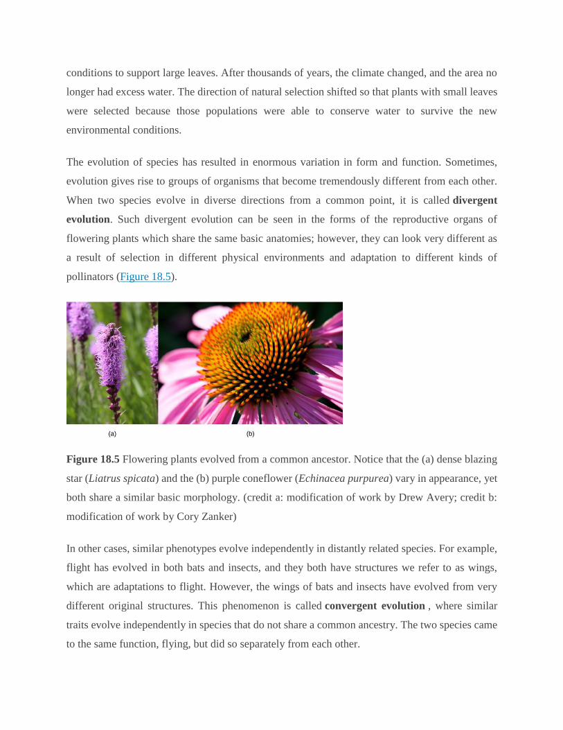
conditions to support large leaves. After thousands of years, the climate changed, and the area no
longer had excess water. The direction of natural selection shifted so that plants with small leaves
were selected because those populations were able to conserve water to survive the new
environmental conditions.
The evolution of species has resulted in enormous variation in form and function. Sometimes,
evolution gives rise to groups of organisms that become tremendously different from each other.
When two species evolve in diverse directions from a common point, it is called divergent
evolution. Such divergent evolution can be seen in the forms of the reproductive organs of
flowering plants which share the same basic anatomies; however, they can look very different as
a result of selection in different physical environments and adaptation to different kinds of
pollinators (Figure 18.5).
Figure 18.5 Flowering plants evolved from a common ancestor. Notice that the (a) dense blazing
star (Liatrus spicata) and the (b) purple coneflower (Echinacea purpurea) vary in appearance, yet
both share a similar basic morphology. (credit a: modification of work by Drew Avery; credit b:
modification of work by Cory Zanker)
In other cases, similar phenotypes evolve independently in distantly related species. For example,
flight has evolved in both bats and insects, and they both have structures we refer to as wings,
which are adaptations to flight. However, the wings of bats and insects have evolved from very
different original structures. This phenomenon is called convergent evolution , where similar
traits evolve independently in species that do not share a common ancestry. The two species came
to the same function, flying, but did so separately from each other.
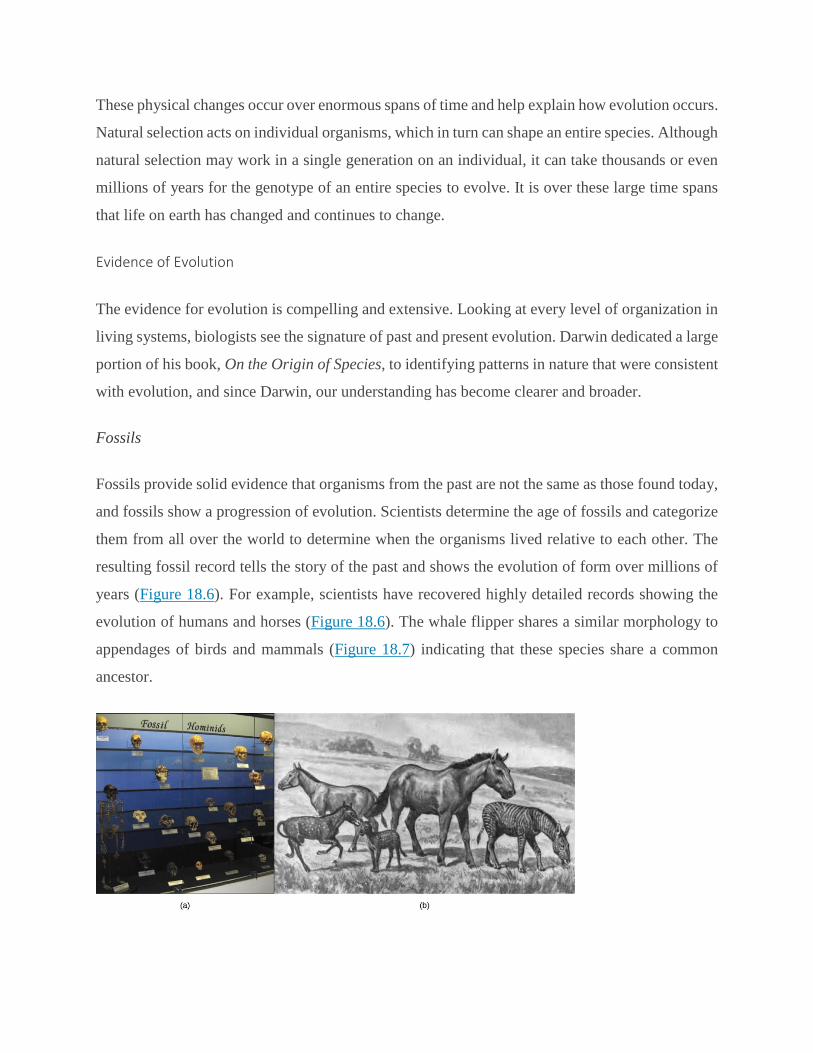
These physical changes occur over enormous spans of time and help explain how evolution occurs.
Natural selection acts on individual organisms, which in turn can shape an entire species. Although
natural selection may work in a single generation on an individual, it can take thousands or even
millions of years for the genotype of an entire species to evolve. It is over these large time spans
that life on earth has changed and continues to change.
Evidence of Evolution
The evidence for evolution is compelling and extensive. Looking at every level of organization in
living systems, biologists see the signature of past and present evolution. Darwin dedicated a large
portion of his book, On the Origin of Species, to identifying patterns in nature that were consistent
with evolution, and since Darwin, our understanding has become clearer and broader.
Fossils
Fossils provide solid evidence that organisms from the past are not the same as those found today,
and fossils show a progression of evolution. Scientists determine the age of fossils and categorize
them from all over the world to determine when the organisms lived relative to each other. The
resulting fossil record tells the story of the past and shows the evolution of form over millions of
years (Figure 18.6). For example, scientists have recovered highly detailed records showing the
evolution of humans and horses (Figure 18.6). The whale flipper shares a similar morphology to
appendages of birds and mammals (Figure 18.7) indicating that these species share a common
ancestor.

Figure 18.6 In this (a) display, fossil hominids are arranged from oldest (bottom) to newest (top).
As hominids evolved, the shape of the skull changed. An artist’s rendition of (b) extinct species of
the genus Equus reveals that these ancient species resembled the modern horse (Equus ferus) but
varied in size.
Anatomy and Embryology
Another type of evidence for evolution is the presence of structures in organisms that share the
same basic form. For example, the bones in the appendages of a human, dog, bird, and whale all
share the same overall construction (Figure 18.7) resulting from their origin in the appendages of
a common ancestor. Over time, evolution led to changes in the shapes and sizes of these bones in
different species, but they have maintained the same overall layout. Scientists call these
synonymous parts homologous structures.
Figure 18.7 The similar construction of these appendages indicates that these organisms share a
common ancestor.
Some structures exist in organisms that have no apparent function at all, and appear to be residual
parts from a past common ancestor. These unused structures without function are called vestigial
structures. Other examples of vestigial structures are wings on flightless birds, leaves on some
cacti, and hind leg bones in whales.
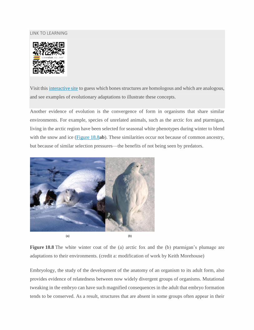
LINK TO LEARNING
Visit this interactive site to guess which bones structures are homologous and which are analogous,
and see examples of evolutionary adaptations to illustrate these concepts.
Another evidence of evolution is the convergence of form in organisms that share similar
environments. For example, species of unrelated animals, such as the arctic fox and ptarmigan,
living in the arctic region have been selected for seasonal white phenotypes during winter to blend
with the snow and ice (Figure 18.8ab). These similarities occur not because of common ancestry,
but because of similar selection pressures—the benefits of not being seen by predators.
Figure 18.8 The white winter coat of the (a) arctic fox and the (b) ptarmigan’s plumage are
adaptations to their environments. (credit a: modification of work by Keith Morehouse)
Embryology, the study of the development of the anatomy of an organism to its adult form, also
provides evidence of relatedness between now widely divergent groups of organisms. Mutational
tweaking in the embryo can have such magnified consequences in the adult that embryo formation
tends to be conserved. As a result, structures that are absent in some groups often appear in their
embryonic forms and disappear by the time the adult or juvenile form is reached. For example, all
vertebrate embryos, including humans, exhibit gill slits and tails at some point in their early
development. These disappear in the adults of terrestrial groups but are maintained in adult forms
of aquatic groups such as fish and some amphibians. Great ape embryos, including humans, have
a tail structure during their development that is lost by the time of birth.
Biogeography
The geographic distribution of organisms on the planet follows patterns that are best explained by
evolution in conjunction with the movement of tectonic plates over geological time. Broad groups
that evolved before the breakup of the supercontinent Pangaea (about 200 million years ago) are
distributed worldwide. Groups that evolved since the breakup appear uniquely in regions of the
planet, such as the unique flora and fauna of northern continents that formed from the
supercontinent Laurasia and of the southern continents that formed from the supercontinent
Gondwana. The presence of members of the plant family Proteaceae in Australia, southern Africa,
and South America is best by their presence prior to the southern supercontinent Gondwana
breaking up.
The great diversification of marsupials in Australia and the absence of other mammals reflect
Australia’s long isolation. Australia has an abundance of endemic species—species found nowhere
else—which is typical of islands whose isolation by expanses of water prevents species to migrate.
Over time, these species diverge evolutionarily into new species that look very different from their
ancestors that may exist on the mainland. The marsupials of Australia, the finches on the
Galápagos, and many species on the Hawaiian Islands are all unique to their one point of origin,
yet they display distant relationships to ancestral species on mainlands.
Molecular Biology
Like anatomical structures, the structures of the molecules of life reflect descent with modification.
Evidence of a common ancestor for all of life is reflected in the universality of DNA as the genetic
material and in the near universality of the genetic code and the machinery of DNA replication
and expression. Fundamental divisions in life between the three domains are reflected in major
structural differences in otherwise conservative structures such as the components of ribosomes
and the structures of membranes. In general, the relatedness of groups of organisms is reflected in
the similarity of their DNA sequences—exactly the pattern that would be expected from descent
and diversification from a common ancestor.
DNA sequences have also shed light on some of the mechanisms of evolution. For example, it is
clear that the evolution of new functions for proteins commonly occurs after gene duplication
events that allow the free modification of one copy by mutation, selection, or drift (changes in a
population’s gene pool resulting from chance), while the second copy continues to produce a
functional protein.
Misconceptions of Evolution
Although the theory of evolution generated some controversy when it was first proposed, it was
almost universally accepted by biologists, particularly younger biologists, within 20 years after
publication of On the Origin of Species. Nevertheless, the theory of evolution is a difficult concept
and misconceptions about how it works abound.
LINK TO LEARNING
This site addresses some of the main misconceptions associated with the theory of evolution.
Evolution Is Just a Theory
Critics of the theory of evolution dismiss its importance by purposefully confounding the everyday
usage of the word “theory” with the way scientists use the word. In science, a “theory” is
understood to be a body of thoroughly tested and verified explanations for a set of observations of
the natural world. Scientists have a theory of the atom, a theory of gravity, and the theory of
relativity, each of which describes understood facts about the world. In the same way, the theory
of evolution describes facts about the living world. As such, a theory in science has survived
significant efforts to discredit it by scientists. In contrast, a “theory” in common vernacular is a
word meaning a guess or suggested explanation; this meaning is more akin to the scientific concept
of “hypothesis.” When critics of evolution say evolution is “just a theory,” they are implying that
there is little evidence supporting it and that it is still in the process of being rigorously tested. This
is a mischaracterization.
Individuals Evolve
Evolution is the change in genetic composition of a population over time, specifically over
generations, resulting from differential reproduction of individuals with certain alleles. Individuals
do change over their lifetime, obviously, but this is called development and involves changes
programmed by the set of genes the individual acquired at birth in coordination with the
individual’s environment. When thinking about the evolution of a characteristic, it is probably best
to think about the change of the average value of the characteristic in the population over time. For
example, when natural selection leads to bill-size change in medium-ground finches in the
Galápagos, this does not mean that individual bills on the finches are changing. If one measures
the average bill size among all individuals in the population at one time and then measures the
average bill size in the population several years later, this average value will be different as a result
of evolution. Although some individuals may survive from the first time to the second, they will
still have the same bill size; however, there will be many new individuals that contribute to the
shift in average bill size.
Evolution Explains the Origin of Life
It is a common misunderstanding that evolution includes an explanation of life’s origins.
Conversely, some of the theory’s critics believe that it cannot explain the origin of life. The theory
does not try to explain the origin of life. The theory of evolution explains how populations change
over time and how life diversifies the origin of species. It does not shed light on the beginnings of
life including the origins of the first cells, which is how life is defined. The mechanisms of the
origin of life on Earth are a particularly difficult problem because it occurred a very long time ago,
and presumably it just occurred once. Importantly, biologists believe that the presence of life on
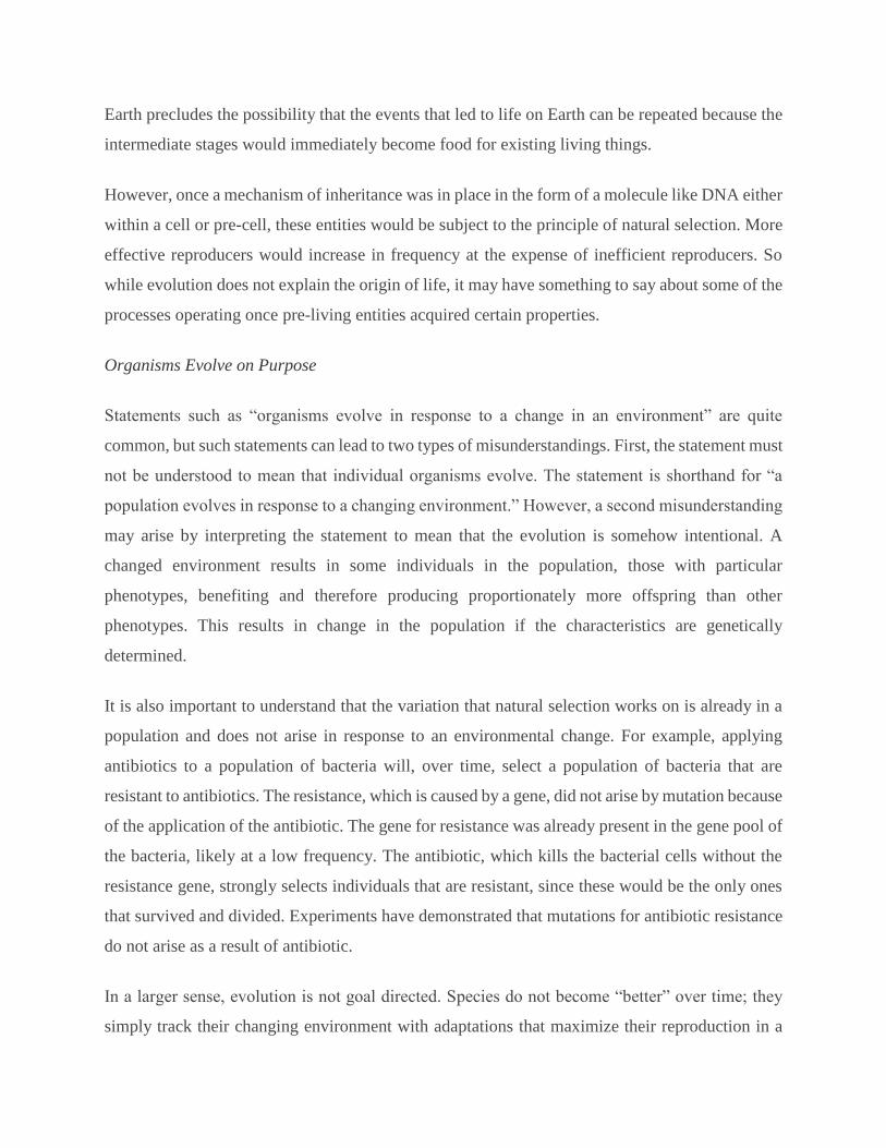
Earth precludes the possibility that the events that led to life on Earth can be repeated because the
intermediate stages would immediately become food for existing living things.
However, once a mechanism of inheritance was in place in the form of a molecule like DNA either
within a cell or pre-cell, these entities would be subject to the principle of natural selection. More
effective reproducers would increase in frequency at the expense of inefficient reproducers. So
while evolution does not explain the origin of life, it may have something to say about some of the
processes operating once pre-living entities acquired certain properties.
Organisms Evolve on Purpose
Statements such as “organisms evolve in response to a change in an environment” are quite
common, but such statements can lead to two types of misunderstandings. First, the statement must
not be understood to mean that individual organisms evolve. The statement is shorthand for “a
population evolves in response to a changing environment.” However, a second misunderstanding
may arise by interpreting the statement to mean that the evolution is somehow intentional. A
changed environment results in some individuals in the population, those with particular
phenotypes, benefiting and therefore producing proportionately more offspring than other
phenotypes. This results in change in the population if the characteristics are genetically
determined.
It is also important to understand that the variation that natural selection works on is already in a
population and does not arise in response to an environmental change. For example, applying
antibiotics to a population of bacteria will, over time, select a population of bacteria that are
resistant to antibiotics. The resistance, which is caused by a gene, did not arise by mutation because
of the application of the antibiotic. The gene for resistance was already present in the gene pool of
the bacteria, likely at a low frequency. The antibiotic, which kills the bacterial cells without the
resistance gene, strongly selects individuals that are resistant, since these would be the only ones
that survived and divided. Experiments have demonstrated that mutations for antibiotic resistance
do not arise as a result of antibiotic.
In a larger sense, evolution is not goal directed. Species do not become “better” over time; they
simply track their changing environment with adaptations that maximize their reproduction in a
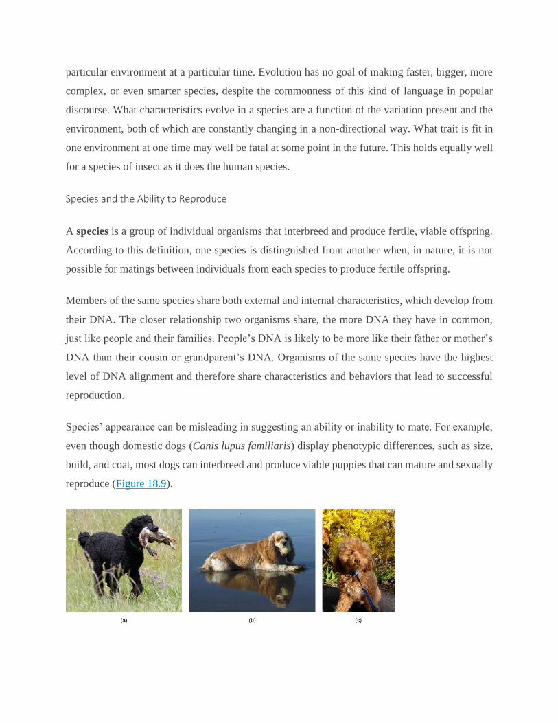
particular environment at a particular time. Evolution has no goal of making faster, bigger, more
complex, or even smarter species, despite the commonness of this kind of language in popular
discourse. What characteristics evolve in a species are a function of the variation present and the
environment, both of which are constantly changing in a non-directional way. What trait is fit in
one environment at one time may well be fatal at some point in the future. This holds equally well
for a species of insect as it does the human species.
Species and the Ability to Reproduce
A species is a group of individual organisms that interbreed and produce fertile, viable offspring.
According to this definition, one species is distinguished from another when, in nature, it is not
possible for matings between individuals from each species to produce fertile offspring.
Members of the same species share both external and internal characteristics, which develop from
their DNA. The closer relationship two organisms share, the more DNA they have in common,
just like people and their families. People’s DNA is likely to be more like their father or mother’s
DNA than their cousin or grandparent’s DNA. Organisms of the same species have the highest
level of DNA alignment and therefore share characteristics and behaviors that lead to successful
reproduction.
Species’ appearance can be misleading in suggesting an ability or inability to mate. For example,
even though domestic dogs (Canis lupus familiaris) display phenotypic differences, such as size,
build, and coat, most dogs can interbreed and produce viable puppies that can mature and sexually
reproduce (Figure 18.9).
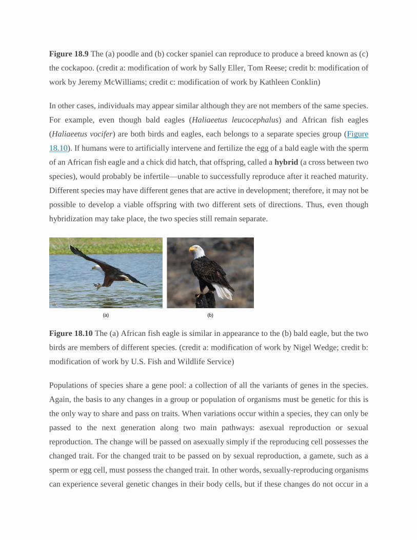
Figure 18.9 The (a) poodle and (b) cocker spaniel can reproduce to produce a breed known as (c)
the cockapoo. (credit a: modification of work by Sally Eller, Tom Reese; credit b: modification of
work by Jeremy McWilliams; credit c: modification of work by Kathleen Conklin)
In other cases, individuals may appear similar although they are not members of the same species.
For example, even though bald eagles (Haliaeetus leucocephalus) and African fish eagles
(Haliaeetus vocifer) are both birds and eagles, each belongs to a separate species group (Figure
18.10). If humans were to artificially intervene and fertilize the egg of a bald eagle with the sperm
of an African fish eagle and a chick did hatch, that offspring, called a hybrid (a cross between two
species), would probably be infertile—unable to successfully reproduce after it reached maturity.
Different species may have different genes that are active in development; therefore, it may not be
possible to develop a viable offspring with two different sets of directions. Thus, even though
hybridization may take place, the two species still remain separate.
Figure 18.10 The (a) African fish eagle is similar in appearance to the (b) bald eagle, but the two
birds are members of different species. (credit a: modification of work by Nigel Wedge; credit b:
modification of work by U.S. Fish and Wildlife Service)
Populations of species share a gene pool: a collection of all the variants of genes in the species.
Again, the basis to any changes in a group or population of organisms must be genetic for this is
the only way to share and pass on traits. When variations occur within a species, they can only be
passed to the next generation along two main pathways: asexual reproduction or sexual
reproduction. The change will be passed on asexually simply if the reproducing cell possesses the
changed trait. For the changed trait to be passed on by sexual reproduction, a gamete, such as a
sperm or egg cell, must possess the changed trait. In other words, sexually-reproducing organisms
can experience several genetic changes in their body cells, but if these changes do not occur in a
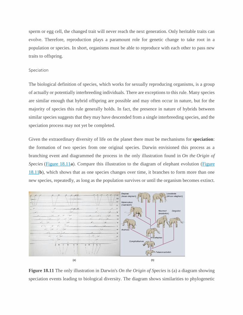
sperm or egg cell, the changed trait will never reach the next generation. Only heritable traits can
evolve. Therefore, reproduction plays a paramount role for genetic change to take root in a
population or species. In short, organisms must be able to reproduce with each other to pass new
traits to offspring.
Speciation
The biological definition of species, which works for sexually reproducing organisms, is a group
of actually or potentially interbreeding individuals. There are exceptions to this rule. Many species
are similar enough that hybrid offspring are possible and may often occur in nature, but for the
majority of species this rule generally holds. In fact, the presence in nature of hybrids between
similar species suggests that they may have descended from a single interbreeding species, and the
speciation process may not yet be completed.
Given the extraordinary diversity of life on the planet there must be mechanisms for speciation:
the formation of two species from one original species. Darwin envisioned this process as a
branching event and diagrammed the process in the only illustration found in On the Origin of
Species (Figure 18.11a). Compare this illustration to the diagram of elephant evolution (Figure
18.11b), which shows that as one species changes over time, it branches to form more than one
new species, repeatedly, as long as the population survives or until the organism becomes extinct.
Figure 18.11 The only illustration in Darwin's On the Origin of Species is (a) a diagram showing
speciation events leading to biological diversity. The diagram shows similarities to phylogenetic

charts that are drawn today to illustrate the relationships of species. (b) Modern elephants evolved
from the Palaeomastodon, a species that lived in Egypt 35–50 million years ago.
For speciation to occur, two new populations must be formed from one original population and
they must evolve in such a way that it becomes impossible for individuals from the two new
populations to interbreed. Biologists have proposed mechanisms by which this could occur that
fall into two broad categories. Allopatric speciation (allo- = "other"; -patric = "homeland")
involves geographic separation of populations from a parent species and subsequent
evolution. Sympatric speciation (sym- = "same"; -patric = "homeland") involves speciation
occurring within a parent species remaining in one location.
Biologists think of speciation events as the splitting of one ancestral species into two descendant
species. There is no reason why there might not be more than two species formed at one time
except that it is less likely and multiple events can be conceptualized as single splits occurring
close in time.
Allopatric Speciation
A geographically continuous population has a gene pool that is relatively homogeneous. Gene
flow, the movement of alleles across the range of the species, is relatively free because individuals
can move and then mate with individuals in their new location. Thus, the frequency of an allele at
one end of a distribution will be similar to the frequency of the allele at the other end. When
populations become geographically discontinuous, that free-flow of alleles is prevented. When
that separation lasts for a period of time, the two populations are able to evolve along different
trajectories. Thus, their allele frequencies at numerous genetic loci gradually become more and
more different as new alleles independently arise by mutation in each population. Typically,
environmental conditions, such as climate, resources, predators, and competitors for the two
populations will differ causing natural selection to favor divergent adaptations in each group.
Isolation of populations leading to allopatric speciation can occur in a variety of ways: a river
forming a new branch, erosion forming a new valley, a group of organisms traveling to a new
location without the ability to return, or seeds floating over the ocean to an island. The nature of
the geographic separation necessary to isolate populations depends entirely on the biology of the
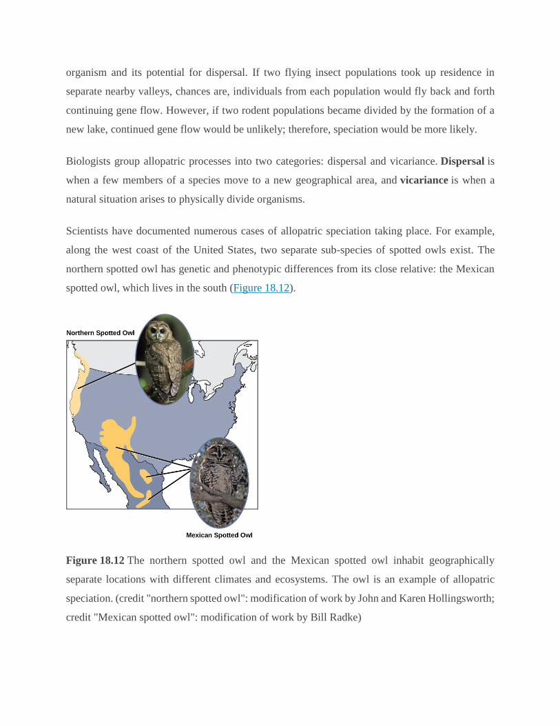
organism and its potential for dispersal. If two flying insect populations took up residence in
separate nearby valleys, chances are, individuals from each population would fly back and forth
continuing gene flow. However, if two rodent populations became divided by the formation of a
new lake, continued gene flow would be unlikely; therefore, speciation would be more likely.
Biologists group allopatric processes into two categories: dispersal and vicariance. Dispersal is
when a few members of a species move to a new geographical area, and vicariance is when a
natural situation arises to physically divide organisms.
Scientists have documented numerous cases of allopatric speciation taking place. For example,
along the west coast of the United States, two separate sub-species of spotted owls exist. The
northern spotted owl has genetic and phenotypic differences from its close relative: the Mexican
spotted owl, which lives in the south (Figure 18.12).
Figure 18.12 The northern spotted owl and the Mexican spotted owl inhabit geographically
separate locations with different climates and ecosystems. The owl is an example of allopatric
speciation. (credit "northern spotted owl": modification of work by John and Karen Hollingsworth;
credit "Mexican spotted owl": modification of work by Bill Radke)
Additionally, scientists have found that the further the distance between two groups that once were
the same species, the more likely it is that speciation will occur. This seems logical because as the
distance increases, the various environmental factors would likely have less in common than
locations in close proximity. Consider the two owls: in the north, the climate is cooler than in the
south; the types of organisms in each ecosystem differ, as do their behaviors and habits; also, the
hunting habits and prey choices of the southern owls vary from the northern owls. These variances
can lead to evolved differences in the owls, and speciation likely will occur.
Adaptive Radiation
In some cases, a population of one species disperses throughout an area, and each finds a distinct
niche or isolated habitat. Over time, the varied demands of their new lifestyles lead to multiple
speciation events originating from a single species. This is called adaptive radiation because
many adaptations evolve from a single point of origin; thus, causing the species to radiate into
several new ones. Island archipelagos like the Hawaiian Islands provide an ideal context for
adaptive radiation events because water surrounds each island which leads to geographical
isolation for many organisms. The Hawaiian honeycreeper illustrates one example of adaptive
radiation. From a single species, called the founder species, numerous species have evolved,
including the six shown in Figure 18.13.
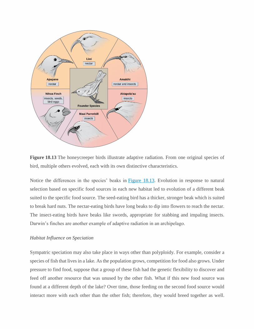
Figure 18.13 The honeycreeper birds illustrate adaptive radiation. From one original species of
bird, multiple others evolved, each with its own distinctive characteristics.
Notice the differences in the species’ beaks in Figure 18.13. Evolution in response to natural
selection based on specific food sources in each new habitat led to evolution of a different beak
suited to the specific food source. The seed-eating bird has a thicker, stronger beak which is suited
to break hard nuts. The nectar-eating birds have long beaks to dip into flowers to reach the nectar.
The insect-eating birds have beaks like swords, appropriate for stabbing and impaling insects.
Darwin’s finches are another example of adaptive radiation in an archipelago.
Habitat Influence on Speciation
Sympatric speciation may also take place in ways other than polyploidy. For example, consider a
species of fish that lives in a lake. As the population grows, competition for food also grows. Under
pressure to find food, suppose that a group of these fish had the genetic flexibility to discover and
feed off another resource that was unused by the other fish. What if this new food source was
found at a different depth of the lake? Over time, those feeding on the second food source would
interact more with each other than the other fish; therefore, they would breed together as well.
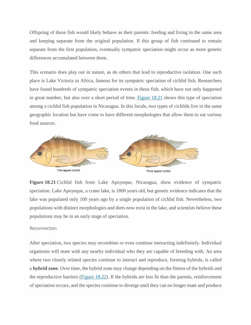
Offspring of these fish would likely behave as their parents: feeding and living in the same area
and keeping separate from the original population. If this group of fish continued to remain
separate from the first population, eventually sympatric speciation might occur as more genetic
differences accumulated between them.
This scenario does play out in nature, as do others that lead to reproductive isolation. One such
place is Lake Victoria in Africa, famous for its sympatric speciation of cichlid fish. Researchers
have found hundreds of sympatric speciation events in these fish, which have not only happened
in great number, but also over a short period of time. Figure 18.21 shows this type of speciation
among a cichlid fish population in Nicaragua. In this locale, two types of cichlids live in the same
geographic location but have come to have different morphologies that allow them to eat various
food sources.
Figure 18.21 Cichlid fish from Lake Apoyeque, Nicaragua, show evidence of sympatric
speciation. Lake Apoyeque, a crater lake, is 1800 years old, but genetic evidence indicates that the
lake was populated only 100 years ago by a single population of cichlid fish. Nevertheless, two
populations with distinct morphologies and diets now exist in the lake, and scientists believe these
populations may be in an early stage of speciation.
Reconnection
After speciation, two species may recombine or even continue interacting indefinitely. Individual
organisms will mate with any nearby individual who they are capable of breeding with. An area
where two closely related species continue to interact and reproduce, forming hybrids, is called
a hybrid zone. Over time, the hybrid zone may change depending on the fitness of the hybrids and
the reproductive barriers (Figure 18.22). If the hybrids are less fit than the parents, reinforcement
of speciation occurs, and the species continue to diverge until they can no longer mate and produce
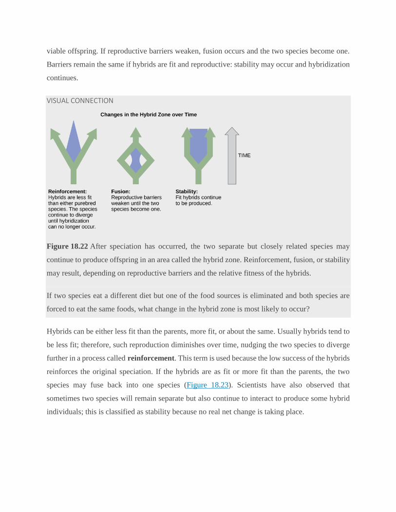
viable offspring. If reproductive barriers weaken, fusion occurs and the two species become one.
Barriers remain the same if hybrids are fit and reproductive: stability may occur and hybridization
continues.
VISUAL CONNECTION
Figure 18.22 After speciation has occurred, the two separate but closely related species may
continue to produce offspring in an area called the hybrid zone. Reinforcement, fusion, or stability
may result, depending on reproductive barriers and the relative fitness of the hybrids.
If two species eat a different diet but one of the food sources is eliminated and both species are
forced to eat the same foods, what change in the hybrid zone is most likely to occur?
Hybrids can be either less fit than the parents, more fit, or about the same. Usually hybrids tend to
be less fit; therefore, such reproduction diminishes over time, nudging the two species to diverge
further in a process called reinforcement. This term is used because the low success of the hybrids
reinforces the original speciation. If the hybrids are as fit or more fit than the parents, the two
species may fuse back into one species (Figure 18.23). Scientists have also observed that
sometimes two species will remain separate but also continue to interact to produce some hybrid
individuals; this is classified as stability because no real net change is taking place.
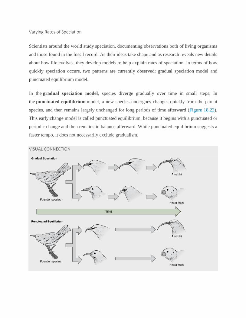
Varying Rates of Speciation
Scientists around the world study speciation, documenting observations both of living organisms
and those found in the fossil record. As their ideas take shape and as research reveals new details
about how life evolves, they develop models to help explain rates of speciation. In terms of how
quickly speciation occurs, two patterns are currently observed: gradual speciation model and
punctuated equilibrium model.
In the gradual speciation model, species diverge gradually over time in small steps. In
the punctuated equilibrium model, a new species undergoes changes quickly from the parent
species, and then remains largely unchanged for long periods of time afterward (Figure 18.23).
This early change model is called punctuated equilibrium, because it begins with a punctuated or
periodic change and then remains in balance afterward. While punctuated equilibrium suggests a
faster tempo, it does not necessarily exclude gradualism.
VISUAL CONNECTION
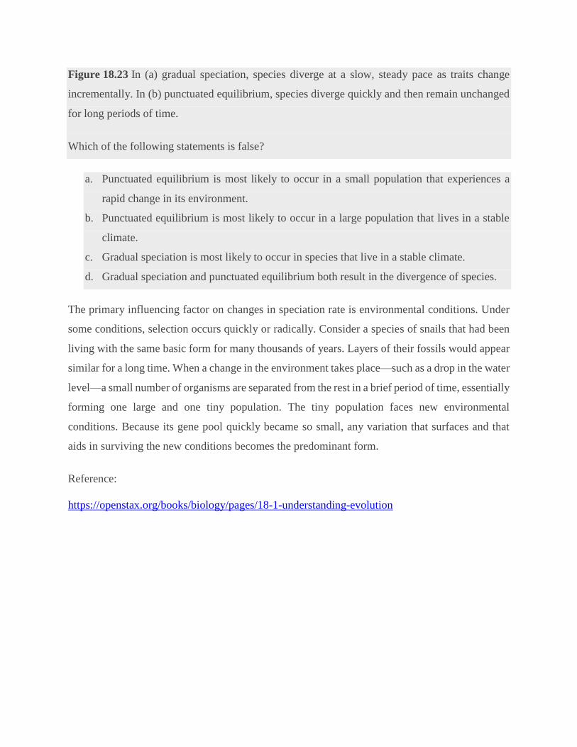
Figure 18.23 In (a) gradual speciation, species diverge at a slow, steady pace as traits change
incrementally. In (b) punctuated equilibrium, species diverge quickly and then remain unchanged
for long periods of time.
Which of the following statements is false?
a. Punctuated equilibrium is most likely to occur in a small population that experiences a
rapid change in its environment.
b. Punctuated equilibrium is most likely to occur in a large population that lives in a stable
climate.
c. Gradual speciation is most likely to occur in species that live in a stable climate.
d. Gradual speciation and punctuated equilibrium both result in the divergence of species.
The primary influencing factor on changes in speciation rate is environmental conditions. Under
some conditions, selection occurs quickly or radically. Consider a species of snails that had been
living with the same basic form for many thousands of years. Layers of their fossils would appear
similar for a long time. When a change in the environment takes place—such as a drop in the water
level—a small number of organisms are separated from the rest in a brief period of time, essentially
forming one large and one tiny population. The tiny population faces new environmental
conditions. Because its gene pool quickly became so small, any variation that surfaces and that
aids in surviving the new conditions becomes the predominant form.
Reference:
https://openstax.org/books/biology/pages/18-1-understanding-evolution

2. The Hardy-Weinberg Principle By: Christine A. Andrews (Biological Sciences Collegiate Division, University of Chicago) © 2010 Nature Education
Citation: Andrews, C. (2010) The Hardy-Weinberg Principle. Nature Education Knowledge 3(10):65
The Hardy-Weinberg theorem characterizes the distributions of genotype frequencies in
populations that are not evolving, and is thus the fundamental null model for population genetics.
Basic Mendelian Genetics
Under the now-discredited theory of blending inheritance, the hereditary material was conceived
as a fluid that combines the traits from two individuals into phenotypically intermediate offspring.
Given observed patterns of resemblance between parents and offspring, blending inheritance may
seem intuitively reasonable, as it did to many of Charles Darwin’s contemporaries. This mode of
inheritance, however, posed problems for Darwin’s theory of natural selection (1859), which
depends on the existence of heritable trait variation in populations of organisms. Blending
inheritance would quickly erode such variation, since all traits would be combined from one
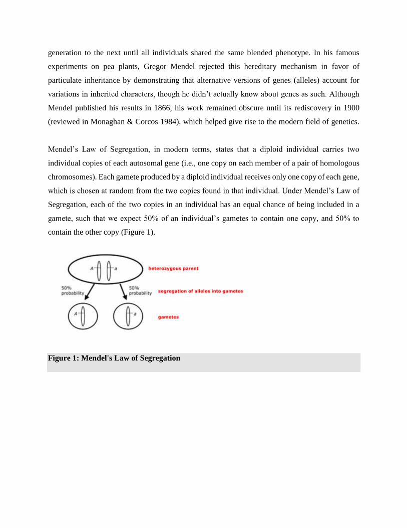
generation to the next until all individuals shared the same blended phenotype. In his famous
experiments on pea plants, Gregor Mendel rejected this hereditary mechanism in favor of
particulate inheritance by demonstrating that alternative versions of genes (alleles) account for
variations in inherited characters, though he didn’t actually know about genes as such. Although
Mendel published his results in 1866, his work remained obscure until its rediscovery in 1900
(reviewed in Monaghan & Corcos 1984), which helped give rise to the modern field of genetics.
Mendel’s Law of Segregation, in modern terms, states that a diploid individual carries two
individual copies of each autosomal gene (i.e., one copy on each member of a pair of homologous
chromosomes). Each gamete produced by a diploid individual receives only one copy of each gene,
which is chosen at random from the two copies found in that individual. Under Mendel’s Law of
Segregation, each of the two copies in an individual has an equal chance of being included in a
gamete, such that we expect 50% of an individual’s gametes to contain one copy, and 50% to
contain the other copy (Figure 1).
Figure 1: Mendel's Law of Segregation
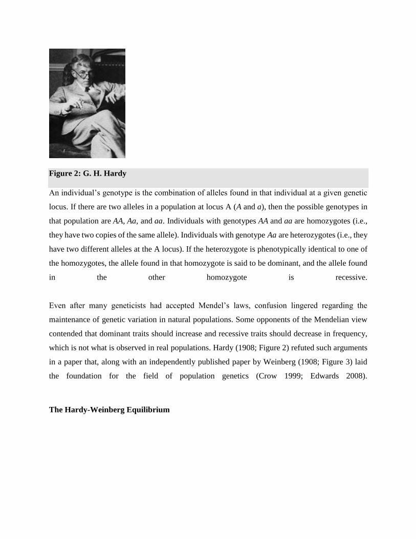
Figure 2: G. H. Hardy
An individual’s genotype is the combination of alleles found in that individual at a given genetic
locus. If there are two alleles in a population at locus A (A and a), then the possible genotypes in
that population are AA, Aa, and aa. Individuals with genotypes AA and aa are homozygotes (i.e.,
they have two copies of the same allele). Individuals with genotype Aa are heterozygotes (i.e., they
have two different alleles at the A locus). If the heterozygote is phenotypically identical to one of
the homozygotes, the allele found in that homozygote is said to be dominant, and the allele found
in the other homozygote is recessive.
Even after many geneticists had accepted Mendel’s laws, confusion lingered regarding the
maintenance of genetic variation in natural populations. Some opponents of the Mendelian view
contended that dominant traits should increase and recessive traits should decrease in frequency,
which is not what is observed in real populations. Hardy (1908; Figure 2) refuted such arguments
in a paper that, along with an independently published paper by Weinberg (1908; Figure 3) laid
the foundation for the field of population genetics (Crow 1999; Edwards 2008).
The Hardy-Weinberg Equilibrium
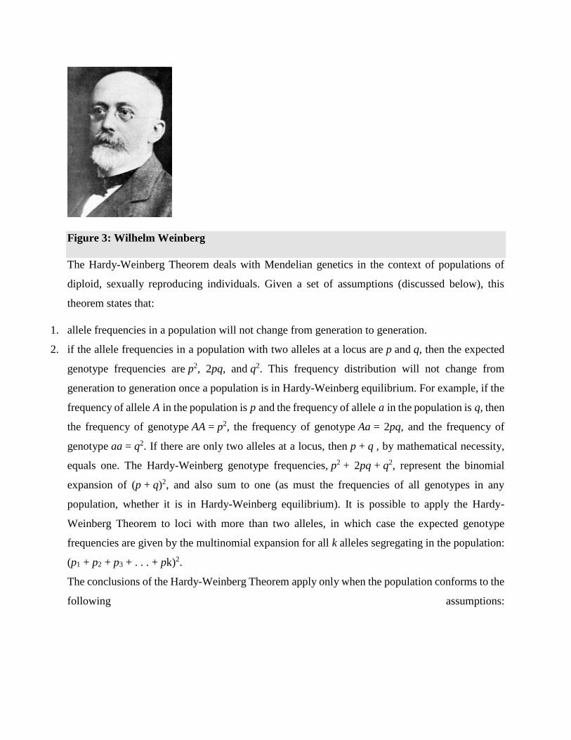
Figure 3: Wilhelm Weinberg
The Hardy-Weinberg Theorem deals with Mendelian genetics in the context of populations of
diploid, sexually reproducing individuals. Given a set of assumptions (discussed below), this
theorem states that:
1. allele frequencies in a population will not change from generation to generation.
2. if the allele frequencies in a population with two alleles at a locus are p and q, then the expected
genotype frequencies are p2, 2pq, and q2. This frequency distribution will not change from
generation to generation once a population is in Hardy-Weinberg equilibrium. For example, if the
frequency of allele A in the population is p and the frequency of allele a in the population is q, then
the frequency of genotype AA = p2, the frequency of genotype Aa = 2pq, and the frequency of
genotype aa = q2. If there are only two alleles at a locus, then p + q , by mathematical necessity,
equals one. The Hardy-Weinberg genotype frequencies, p2 + 2pq + q2, represent the binomial
expansion of (p + q)2, and also sum to one (as must the frequencies of all genotypes in any
population, whether it is in Hardy-Weinberg equilibrium). It is possible to apply the Hardy-
Weinberg Theorem to loci with more than two alleles, in which case the expected genotype
frequencies are given by the multinomial expansion for all k alleles segregating in the population:
(p1 + p2 + p3 + . . . + pk)2.
The conclusions of the Hardy-Weinberg Theorem apply only when the population conforms to the
following assumptions:
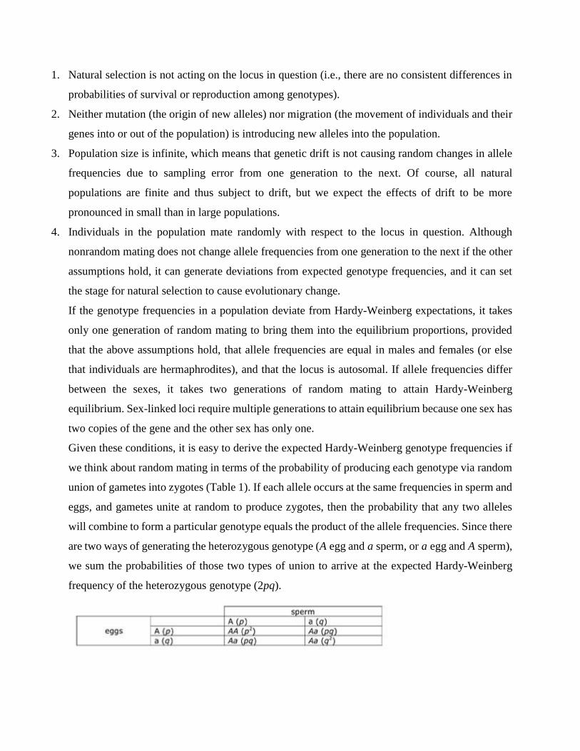
1. Natural selection is not acting on the locus in question (i.e., there are no consistent differences in
probabilities of survival or reproduction among genotypes).
2. Neither mutation (the origin of new alleles) nor migration (the movement of individuals and their
genes into or out of the population) is introducing new alleles into the population.
3. Population size is infinite, which means that genetic drift is not causing random changes in allele
frequencies due to sampling error from one generation to the next. Of course, all natural
populations are finite and thus subject to drift, but we expect the effects of drift to be more
pronounced in small than in large populations.
4. Individuals in the population mate randomly with respect to the locus in question. Although
nonrandom mating does not change allele frequencies from one generation to the next if the other
assumptions hold, it can generate deviations from expected genotype frequencies, and it can set
the stage for natural selection to cause evolutionary change.
If the genotype frequencies in a population deviate from Hardy-Weinberg expectations, it takes
only one generation of random mating to bring them into the equilibrium proportions, provided
that the above assumptions hold, that allele frequencies are equal in males and females (or else
that individuals are hermaphrodites), and that the locus is autosomal. If allele frequencies differ
between the sexes, it takes two generations of random mating to attain Hardy-Weinberg
equilibrium. Sex-linked loci require multiple generations to attain equilibrium because one sex has
two copies of the gene and the other sex has only one.
Given these conditions, it is easy to derive the expected Hardy-Weinberg genotype frequencies if
we think about random mating in terms of the probability of producing each genotype via random
union of gametes into zygotes (Table 1). If each allele occurs at the same frequencies in sperm and
eggs, and gametes unite at random to produce zygotes, then the probability that any two alleles
will combine to form a particular genotype equals the product of the allele frequencies. Since there
are two ways of generating the heterozygous genotype (A egg and a sperm, or a egg and A sperm),
we sum the probabilities of those two types of union to arrive at the expected Hardy-Weinberg
frequency of the heterozygous genotype (2pq).
Table 1: A Punnett square depicting the probabilities of generating all possible genotypes at
a diallelic Mendelian locus in a population that conforms to Hardy-Weinberg assumptions.
It is important to recognize that the Hardy-Weinberg equilibrium is a neutral equilibrium, which
means that a population perturbed from its Hardy-Weinberg genotype frequencies will indeed
reach equilibrium after a single generation of random mating (if it conforms to the other
assumptions of the theorem), but it will be a new equilibrium if allele frequencies have changed.
This property distinguishes a neutral equilibrium from a stable equilibrium, in which a perturbed
system returns to the same equilibrium state. It makes sense that the Hardy-Weinberg equilibrium
is not stable, since a change from the equilibrium genotype frequencies will generally be associated
with a change in allele frequencies (p and q), which will in turn lead to new values of p2,
2pq and q2. Thereafter, a population that meets Hardy-Weinberg assumptions will remain at the
new equilibrium until perturbed again.
Given a population in which we know the number of individuals with each genotype, we can test
for statistical deviation from Hardy-Weinberg equilibrium using a simple chi-square goodness-of-
fit test or a more powerful exact test. The latter class of methods has proved particularly useful for
large-scale genomic studies, in which scientists evaluate thousands of loci segregating for multiple
alleles (Wiggington et al. 2005). Observed genotype proportions in natural populations typically
conform to Hardy-Weinberg expectations, as we might expect given that a population perturbed
from equilibrium can achieve new equilibrium frequencies after only one generation of random
mating.
Although statistical deviation from Hardy-Weinberg expectations generally indicates violation of
the assumptions of the theorem, the converse is not necessarily true. Some forms of natural
selection (e.g., balancing selection, which maintains multiple alleles in a population) can generate
genotypic frequency distributions that conform to Hardy-Weinberg expectations. It may also be
true that migration or mutation is occurring, but at such low rates as to be undetectable using
available statistical methods. And, of course, all real populations are finite and thus susceptible to
at least some evolution via genetic drift.
Evolutionary Implications of the Hardy-Weinberg Theorem
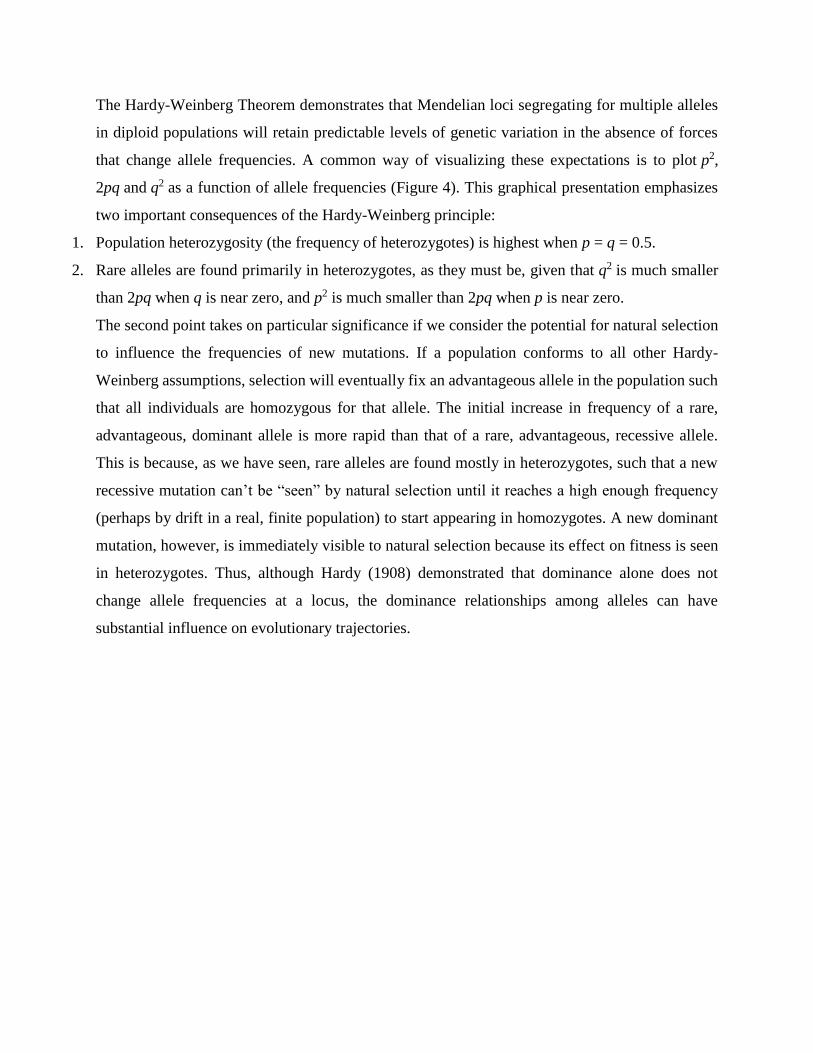
The Hardy-Weinberg Theorem demonstrates that Mendelian loci segregating for multiple alleles
in diploid populations will retain predictable levels of genetic variation in the absence of forces
that change allele frequencies. A common way of visualizing these expectations is to plot p2,
2pq and q2 as a function of allele frequencies (Figure 4). This graphical presentation emphasizes
two important consequences of the Hardy-Weinberg principle:
1. Population heterozygosity (the frequency of heterozygotes) is highest when p = q = 0.5.
2. Rare alleles are found primarily in heterozygotes, as they must be, given that q2 is much smaller
than 2pq when q is near zero, and p2 is much smaller than 2pq when p is near zero.
The second point takes on particular significance if we consider the potential for natural selection
to influence the frequencies of new mutations. If a population conforms to all other Hardy-
Weinberg assumptions, selection will eventually fix an advantageous allele in the population such
that all individuals are homozygous for that allele. The initial increase in frequency of a rare,
advantageous, dominant allele is more rapid than that of a rare, advantageous, recessive allele.
This is because, as we have seen, rare alleles are found mostly in heterozygotes, such that a new
recessive mutation can’t be “seen” by natural selection until it reaches a high enough frequency
(perhaps by drift in a real, finite population) to start appearing in homozygotes. A new dominant
mutation, however, is immediately visible to natural selection because its effect on fitness is seen
in heterozygotes. Thus, although Hardy (1908) demonstrated that dominance alone does not
change allele frequencies at a locus, the dominance relationships among alleles can have
substantial influence on evolutionary trajectories.
Figure 4: A plot of Hardy-Weinberg equilibrium genotype frequencies (p to the 2, 2pq, q to
the 2) as a function of allele frequencies (p and q).
Selection, mutation, migration, and genetic drift are the mechanisms that effect changes in allele
frequencies, and when one or more of these forces are acting, the population violates Hardy-
Weinberg assumptions, and evolution occurs. The Hardy-Weinberg Theorem thus constitutes a
null model for the discipline of population genetics, and is fundamental to the study of evolution.
----------------------------------------------------------------
Development and Evolution The relationships that obtain between development and evolution are complicated and under
ongoing investigation (for a review, see Love 2015). Two main axes dominate within a loose
conglomeration of research programs (Raff 2000; Müller 2007): (a) the evolution of
development, or inquiry into the pattern and processes of how ontogeny varies and changes
over time; and, (b) the developmental basis of evolution, or inquiry into the causal impact of
ontogenetic processes on evolutionary trajectories—both in terms of constraint and
facilitation. Two examples where the concepts and practices of developmental and
evolutionary biology intersect are treated here: the problematic appeal to functional
homology in developmental genetics that is meant to underwrite evolutionary generalizations
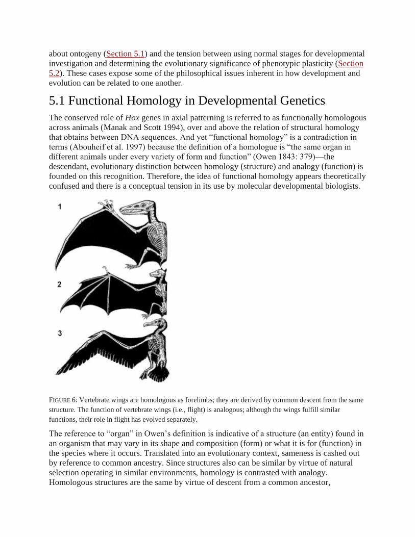
about ontogeny (Section 5.1) and the tension between using normal stages for developmental
investigation and determining the evolutionary significance of phenotypic plasticity (Section
5.2). These cases expose some of the philosophical issues inherent in how development and
evolution can be related to one another.
5.1 Functional Homology in Developmental Genetics
The conserved role of Hox genes in axial patterning is referred to as functionally homologous
across animals (Manak and Scott 1994), over and above the relation of structural homology
that obtains between DNA sequences. And yet “functional homology” is a contradiction in
terms (Abouheif et al. 1997) because the definition of a homologue is “the same organ in
different animals under every variety of form and function” (Owen 1843: 379)—the
descendant, evolutionary distinction between homology (structure) and analogy (function) is
founded on this recognition. Therefore, the idea of functional homology appears theoretically
confused and there is a conceptual tension in its use by molecular developmental biologists.
FIGURE 6: Vertebrate wings are homologous as forelimbs; they are derived by common descent from the same
structure. The function of vertebrate wings (i.e., flight) is analogous; although the wings fulfill similar
functions, their role in flight has evolved separately.
The reference to “organ” in Owen’s definition is indicative of a structure (an entity) found in
an organism that may vary in its shape and composition (form) or what it is for (function) in
the species where it occurs. Translated into an evolutionary context, sameness is cashed out
by reference to common ancestry. Since structures also can be similar by virtue of natural
selection operating in similar environments, homology is contrasted with analogy.
Homologous structures are the same by virtue of descent from a common ancestor,

regardless of what functions these structures are involved in, whereas analogous structures
are similar by virtue of selection processes favoring comparable functional outcomes,
regardless of common descent (Figure 6).
This is what makes similarity of function an especially problematic criterion of homology
(Abouheif et al. 1997). Because functional similarity is the appropriate relation for analogy,
it is not necessary for analogues to have the same function as a consequence of common
ancestry—similarity despite different origins suffices (Ghiselin 2005). Classic cases of
analogy involve taxa that do not share a recent common ancestor that exhibits the structure,
such as the external body morphology of dolphins and tuna (Pabst 2000). Thus, functional
homology seems to be a category error because what a structure does should not enter into an
evaluation of homologue correspondence and similarity of function is often the result of
adaptation via natural selection to common environmental demands, not common ancestry.
Although we might be inclined to simply prohibit the terminology of functional homology,
its widespread use in molecular and developmental biology should at least make us
pause.[18] While it is important to recognize this pervasive practice, some occurrences may be
illicit. Swapping structurally homologous genes between species to rescue mutant or null
phenotypes is not a genuine criterion of functional homology, especially when there is little
or no attention to establishing a phylogenetic context. This makes a number of claims of
functional homology suspect. To not run afoul of the conceptual tension, explicit attention
must be given to the meaning of “function.” Biological practice harbors at least four separate
meanings of function (Wouters 2003, 2005): activity (what something does), causal role
(contribution to a capacity), fitness advantage or viability (value of having something), and
selected effect or etiology (origination and maintenance via natural selection). Debate has
raged about which of them (if any) is most appropriate for different aspects of biological and
psychological reasoning or most general in scope (i.e., what makes them all function
concepts?) (see discussion in Garson 2016). Here the issue is whether we can identify a
legitimate concept of homology of function.
If we are to avoid mixing homology and analogy, then the appropriate notion of function
cannot be based on selection history, which is allied with the concept of analogy and
concerns a particular variety of function. Similarly, viability interpretations concentrate on
features where the variety of function is critical because of conferred survival advantages.
Any interpretation of function that relies on a particular variety of function (because it was
selected or because it confers viability) clashes with the demand that homology concern
something “under every variety of form and function.” A causal role interpretation
emphasizes a systemic capacity to which a function makes a contribution. It too focuses on a
particular variety of function, though in a way different from either selected effect or
viability interpretations. Only an activity interpretation (‘what something does’) accents the
function itself, apart from its specific contribution to a systemic capacity and position in a
larger context. Therefore, the most appropriate meaning to incorporate into homology of
function is “activity-function” because it is at least possible for activity-functions to remain
constant under every variety. An evaluation of sameness due to common ancestry is made
separately from the role the function plays (or its use), whether understood in terms of a
causal role, a fitness advantage, or a history of selection.[19] Activity-functions can be put to
different uses while being shared via common descent (i.e., homologous). More precisely,
homology of function can be defined as the same activity-function in different animals under
every variety of form and use-function (Love 2007). This unambiguously removes the
tension that plagued functional homology.
Careful discussions of regulatory gene function in development and evolution recognize
something akin to the distinction between activity- and use-function (i.e., between what a
gene does and what it is for in some process within the organism).
When studying the molecular evolution of regulatory genes, their biochemical and
developmental function must be considered separately. The biochemical function of PAX-
6 and eyeless are as general transcription factors (which bind and activate downstream
genes), but their developmental function is their specific involvement in eye morphogenesis
(Abouheif 1997: 407).
The biochemical function is the activity-function and the developmental function is the use-
function. This distinction helps to discriminate between divergent evolutionary trajectories.
Biochemical (activity-functions) of genes are often conserved (i.e., homologous), while
simultaneously being available for co-option to make causal role contributions (use-
functions) to distinct developmental processes. The same regulatory genes are evolutionarily
stable in terms of activity-function and evolutionarily labile in terms of use-function.[20] By
implication, claims about use-function homology for genes qua developmental function are
suspect compared to those concerning activity-function homology for genes qua biochemical
function because developmental functions are more likely to have changed as phylogenetic
distance increases.
The distinction between biochemical (activity) function and developmental (use) function is
reinforced by the hierarchical aspects of homology (Hall 1994). A capacity defining the use-
function of a regulatory gene at one level of organization, such as axial patterning, must be
considered as an activity-function itself at another level of organization, such as the
differentiation of serially repeated elements along a body axis. (Note that “level of
organization” need not be compositional and thus the language of “higher” and “lower”
levels may be inappropriate.) The developmental roles of Hox genes in axial patterning may
be conserved by virtue of their biochemical activity-function homologies but Hox genes are
not use-function homologues because of these developmental roles. Instead of focusing on
the activity of a gene component and its causal role in axial patterning, we shift to the
activity of axial patterning and its causal role elsewhere (or elsewhen) in embryonic
development.
Introducing a conceptually legitimate idea of homology of activity-function is not about
keeping the ideas of developmental biology tidy. It assists in the interpretation of evidence
and circumscribes the inferences drawn. For example, NK-2 genes are involved in mesoderm
specification, which underlies muscle morphogenesis. In Drosophila, the expression of a
particular NK-2 gene (tinman) is critical for both cardiac and visceral mesoderm
development. If tinman is knocked out and transgenically replaced with its vertebrate
orthologue, Nkx2-5, only visceral mesoderm specification is rescued; the regulation of
cardiac mesoderm is not (Ranganayakulu et al. 1998). A region of the vertebrate protein near
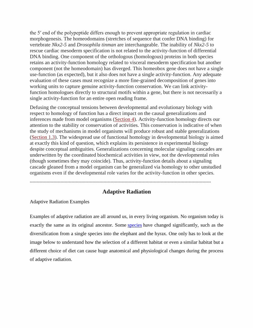
the 5′ end of the polypeptide differs enough to prevent appropriate regulation in cardiac
morphogenesis. The homeodomains (stretches of sequence that confer DNA binding) for
vertebrate Nkx2-5 and Drosophila tinman are interchangeable. The inability of Nkx2-5 to
rescue cardiac mesoderm specification is not related to the activity-function of differential
DNA binding. One component of the orthologous (homologous) proteins in both species
retains an activity-function homology related to visceral mesoderm specification but another
component (not the homeodomain) has diverged. This homeobox gene does not have a single
use-function (as expected), but it also does not have a single activity-function. Any adequate
evaluation of these cases must recognize a more fine-grained decomposition of genes into
working units to capture genuine activity-function conservation. We can link activity-
function homologues directly to structural motifs within a gene, but there is not necessarily a
single activity-function for an entire open reading frame.
Defusing the conceptual tensions between developmental and evolutionary biology with
respect to homology of function has a direct impact on the causal generalizations and
inferences made from model organisms (Section 4). Activity-function homology directs our
attention to the stability or conservation of activities. This conservation is indicative of when
the study of mechanisms in model organisms will produce robust and stable generalizations
(Section 1.3). The widespread use of functional homology in developmental biology is aimed
at exactly this kind of question, which explains its persistence in experimental biology
despite conceptual ambiguities. Generalizations concerning molecular signaling cascades are
underwritten by the coordinated biochemical activities in view, not the developmental roles
(though sometimes they may coincide). Thus, activity-function details about a signaling
cascade gleaned from a model organism can be generalized via homology to other unstudied
organisms even if the developmental role varies for the activity-function in other species.
----------------------------------------------------------------------------------------------------------------------------------
Adaptive Radiation
Adaptive Radiation Examples
Examples of adaptive radiation are all around us, in every living organism. No organism today is
exactly the same as its original ancestor. Some species have changed significantly, such as the
diversification from a single species into the elephant and the hyrax. One only has to look at the
image below to understand how the selection of a different habitat or even a similar habitat but a
different choice of diet can cause huge anatomical and physiological changes during the process
of adaptive radiation.
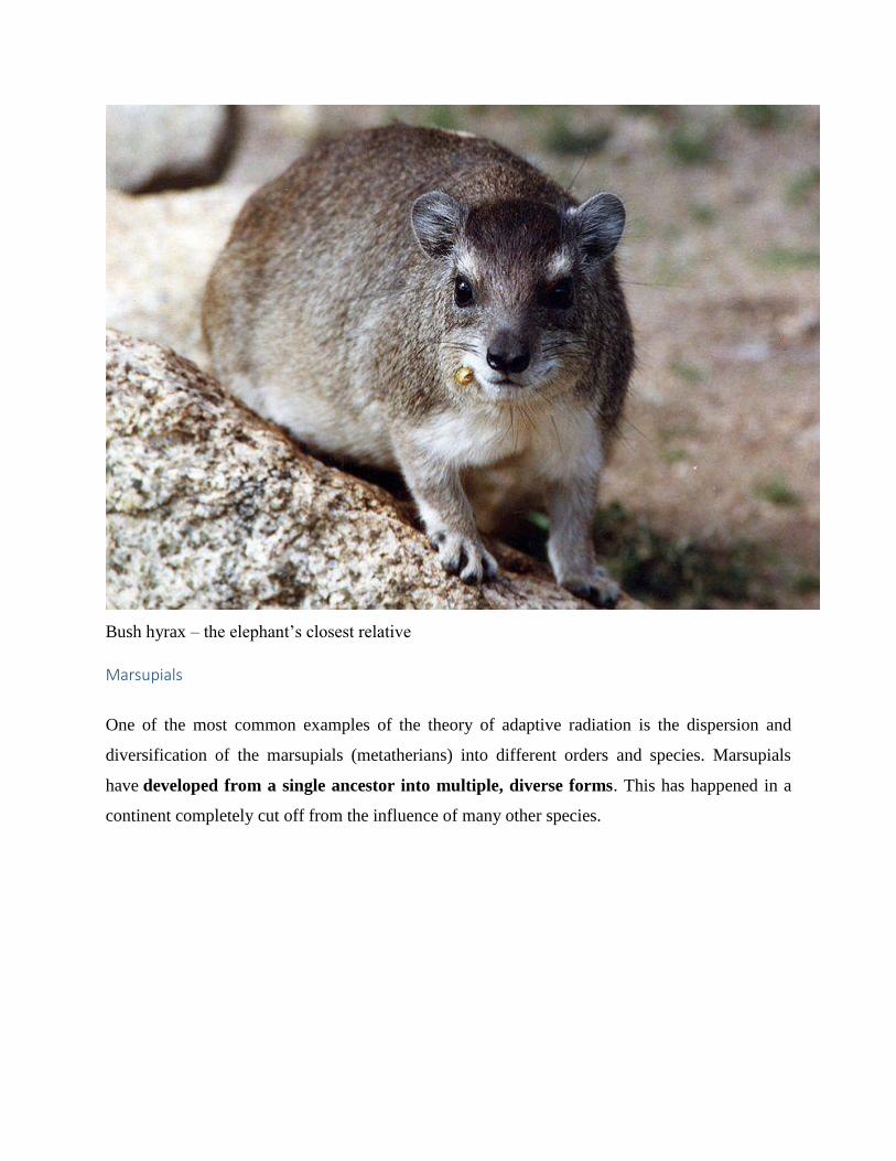
Bush hyrax – the elephant’s closest relative
Marsupials
One of the most common examples of the theory of adaptive radiation is the dispersion and
diversification of the marsupials (metatherians) into different orders and species. Marsupials
have developed from a single ancestor into multiple, diverse forms. This has happened in a
continent completely cut off from the influence of many other species.
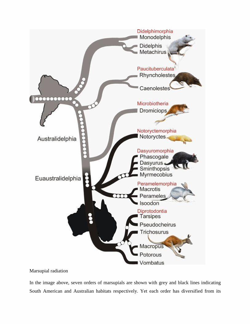
Marsupial radiation
In the image above, seven orders of marsupials are shown with grey and black lines indicating
South American and Australian habitats respectively. Yet each order has diversified from its
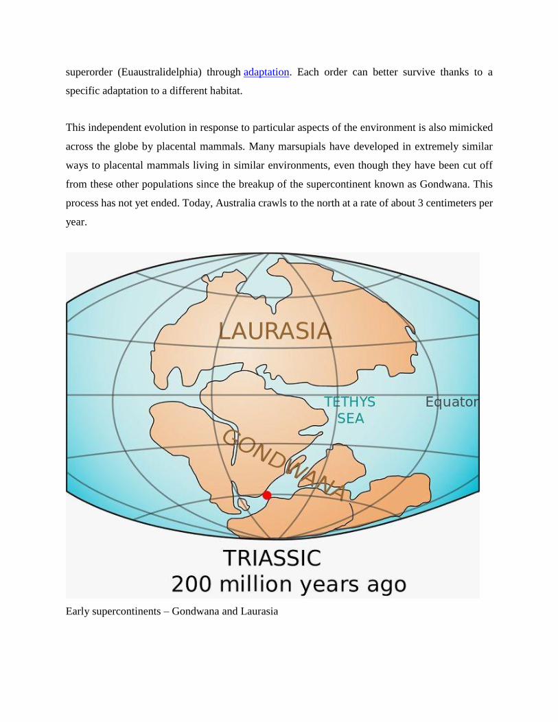
superorder (Euaustralidelphia) through adaptation. Each order can better survive thanks to a
specific adaptation to a different habitat.
This independent evolution in response to particular aspects of the environment is also mimicked
across the globe by placental mammals. Many marsupials have developed in extremely similar
ways to placental mammals living in similar environments, even though they have been cut off
from these other populations since the breakup of the supercontinent known as Gondwana. This
process has not yet ended. Today, Australia crawls to the north at a rate of about 3 centimeters per
year.
Early supercontinents – Gondwana and Laurasia
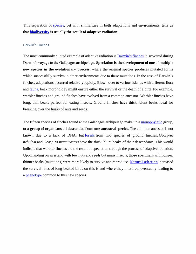
This separation of species, yet with similarities in both adaptations and environments, tells us
that biodiversity is usually the result of adaptive radiation.
Darwin’s Finches
The most commonly quoted example of adaptive radiation is Darwin’s finches, discovered during
Darwin’s voyage to the Galápagos archipelago. Speciation is the development of one of multiple
new species in the evolutionary process, where the original species produces mutated forms
which successfully survive in other environments due to these mutations. In the case of Darwin’s
finches, adaptations occurred relatively rapidly. Blown over to various islands with different flora
and fauna, beak morphology might ensure either the survival or the death of a bird. For example,
warbler finches and ground finches have evolved from a common ancestor. Warbler finches have
long, thin beaks perfect for eating insects. Ground finches have thick, blunt beaks ideal for
breaking over the husks of nuts and seeds.
The fifteen species of finches found at the Galápagos archipelago make up a monophyletic group,
or a group of organisms all descended from one ancestral species. The common ancestor is not
known due to a lack of DNA, but fossils from two species of ground finches, Geospiza
nebulosi and Geospiza magnirostris have the thick, blunt beaks of their descendants. This would
indicate that warbler finches are the result of speciation through the process of adaptive radiation.
Upon landing on an island with few nuts and seeds but many insects, those specimens with longer,
thinner beaks (mutations) were more likely to survive and reproduce. Natural selection increased
the survival rates of long-beaked birds on this island where they interbred, eventually leading to
a phenotype common to this new species.
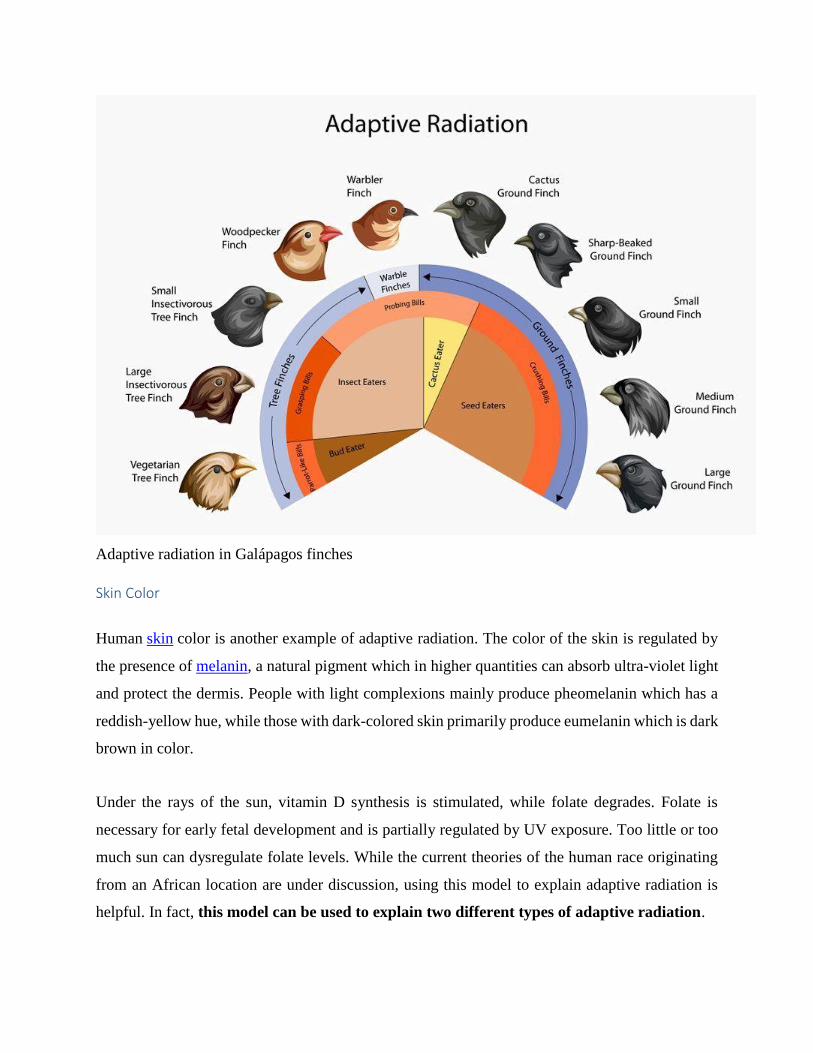
Adaptive radiation in Galápagos finches
Skin Color
Human skin color is another example of adaptive radiation. The color of the skin is regulated by
the presence of melanin, a natural pigment which in higher quantities can absorb ultra-violet light
and protect the dermis. People with light complexions mainly produce pheomelanin which has a
reddish-yellow hue, while those with dark-colored skin primarily produce eumelanin which is dark
brown in color.
Under the rays of the sun, vitamin D synthesis is stimulated, while folate degrades. Folate is
necessary for early fetal development and is partially regulated by UV exposure. Too little or too
much sun can dysregulate folate levels. While the current theories of the human race originating
from an African location are under discussion, using this model to explain adaptive radiation is
helpful. In fact, this model can be used to explain two different types of adaptive radiation.
The first concerns very early ancestors of man (the hominids) who were largely covered with hair
to keep them warm in largely forested areas. Hominid skin, protected by hair, was almost definitely
not as dark as his early descendants. We do not have the fossil evidence to prove this, but mammals
usually have much lighter skin when covered in thick layers of hair or fur, as opposed to mammals
with thin coats. Upon migrating to more open savannahs where the hominids could hunt more
successfully but directly under the rays of the equatorial sun, this hair became superfluous. To be
protected from the UV rays of the sun they developed darker skin. This darker skin reduced the
degradation of folic acid, meaning higher pregnancy and birth rates, while the constant availability
of the equatorial sun meant that vitamin D production was sufficient to ensure good health.
When these populations eventually moved away from the heat of the equator and into colder
regions high levels of melanin became more of a hindrance to the health and reproductive
capacity of this migrating population. Skin did not need as much melanin to protect it from the
meager sun; those with darker skin would block what little UV light there was and synthesize less
vitamin D, leading to lower levels of health and fitness (rickets) and dysregulated folate levels
(miscarriages).
Those who migrated to the far northern regions of the Arctic Circle became slightly lighter in
color, but darker than would usually be expected according to this theory. This has been explained
by their seafood diets which provide ample dietary vitamin D during the colder seasons, while a
darker skin color protected these populations from the UV radiation of the sun further reflected by
the snow-covered landscape during spring and summer months. Research today tells us that the
female Inuit population are more likely to experience folic acid deficiencies than lighter-skinned
females in colder, temperate regions unless they eat folate-fortified foods. This is perhaps the
reason why the color of their skin is not darker.
Reference:
https://biologydictionary.net/adaptive-radiation/
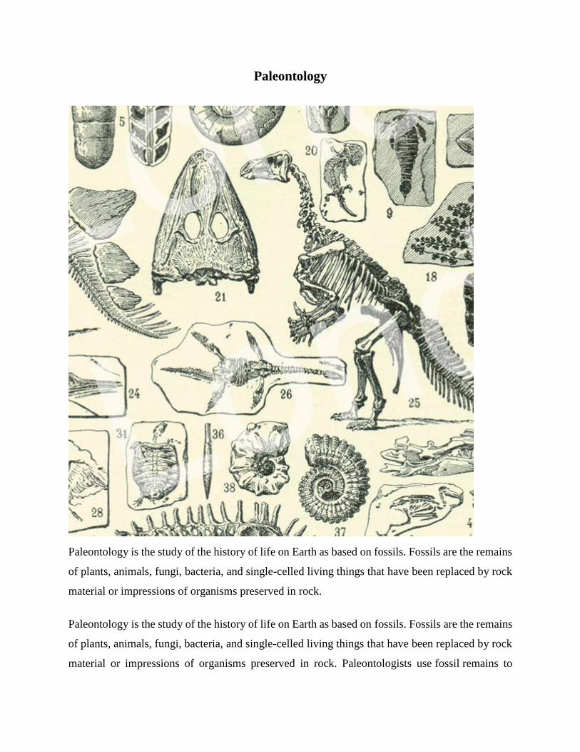
Paleontology
Paleontology is the study of the history of life on Earth as based on fossils. Fossils are the remains
of plants, animals, fungi, bacteria, and single-celled living things that have been replaced by rock
material or impressions of organisms preserved in rock.
Paleontology is the study of the history of life on Earth as based on fossils. Fossils are the remains
of plants, animals, fungi, bacteria, and single-celled living things that have been replaced by rock
material or impressions of organisms preserved in rock. Paleontologists use fossil remains to
understand different aspects of extinct and living organisms. Individual fossils may contain
information about an organism’s life and environment. Much like the rings of a tree, for example,
each ring on the surface of an oyster shell denotes one year of its life. Studying oyster fossils can
help paleontologists discover how long the oyster lived, and in what conditions. If the climate was
favorable for the oyster, the oyster probably grew more quickly and the rings would be thicker. If
the oyster struggled for survival, the rings would be thinner. Thinner rings would indicate an
environment not favorable to organisms like the oyster—too warm or too cold for the oyster, for
example, or lacking nutrients necessary for them to grow.
Some fossils show how an organism lived. Amber, for instance, is hardened, fossilized tree resin.
At times, the sticky resin has dripped down a tree trunk, trapping air bubbles, as well as small
insects and some organisms as large as frogs and lizards. Paleontologists study amber, called
“fossil resin,” to observe these complete specimens. Amber can preserve tissue as delicate as
dragonfly wings. Some ants were trapped in amber while eating leaves, allowing scientists to know
exactly what they ate, and how they ate it. Even the air bubbles trapped in amber are valuable to
paleontologists. By analyzing the chemistry of the air, scientists can tell if there was a volcanic
eruption or other atmospheric changes nearby.
The behavior of organisms can also be deduced from fossil evidence. Paleontologists suggest
that hadrosaurs, duck-billed dinosaurs, lived in large herds, for instance. They made
this hypothesis after observing evidence of social behavior,including a single site with
approximately 10,000 skeletons.
Fossils can also provide evidence of the evolutionary history of organisms. Paleontologists infer
that whales evolved from land-dwelling animals, for instance. Fossils of extinct animals closely
related to whales have front limbs like paddles, similar to front legs. They even have tiny back
limbs. Although the front limbs of these fossil animals are in some ways similar to legs, in other
ways they also show strong similarities to the fins of modern whales.
Subdisciplines of Paleontology
The field of paleontology has many subdisciplines. A subdiscipline is a specialized field of study
within a broader subject or discipline. In the case of paleontology, subdisciplines can focus on a
specific fossil type or a specific aspect of the globe, such as its climate.
Vertebrate Paleontology
One important subdiscipline is vertebrate paleontology, the study of fossils of animals with
backbones. Vertebrate paleontologists have discovered and reconstructed the skeletons of
dinosaurs, turtles, cats, and many other animals to show how they lived and their evolutionary
history.
Using fossil evidence, vertebrate paleontologists deduced that pterosaurs, a group of flying
reptiles, could fly by flapping their wings, as opposed to just gliding. Reconstructed skeletons of
pterosaurs have hollow and light bones like modern birds.
One type of pterosaur, Quetzalcoatlus, is considered one of the largest flying creatures in history.
It had a wingspan of 11 meters (36 feet). Paleontologists have competing theories about if and
how Quetzalcoatlus flew. Some paleontologists argue it was too heavy to fly at all. Others
maintain it could distribute its weight well enough to soar slowly. Still other scientists
say Quetzalcoatlus was muscular enough to fly quickly over short distances. These theories
demonstrate how vertebrate paleontologists can interpret fossil evidence differently.
Invertebrate Paleontology
Invertebrate paleontologists examine the fossils of animals without backbones—
mollusks, corals, arthropods like crabs and shrimp, echinoderms like sand dollars and sea
stars, sponges, and worms. Unlike vertebrates, invertebrates do not have bones—they do leave
behind evidence of their existence in the form of fossilized shells and exoskeletons, impressions
of their soft body parts, and tracks from their movement along the ground or ocean floor.
Invertebrate fossils are especially important to the study and reconstruction
of prehistoric aquatic environments. For example, large communities of 200-million-year-old
invertebrate marine fossils found in the deserts of Nevada, in the United States, tell us that certain
areas of the state were covered by water during that period of time.
Darwin’s On The Origin of Species observed somewhat similar sequencing in the living world.
Darwin suggested that new species evolve over time. New fossil discoveries supported Darwin’s
theory that creatures living in the distant past were different from, yet sometimes interconnected
with, those living today. This theory allowed paleontologists to study living organisms for clues
to understanding fossil evidence. The Archaeopteryx, for example, had wings like a bird, but had
other features (such as teeth) typical of a type of dinosaur called a theropod. Now regarded as a
very early bird, Archaeopteryx retains more similarities to theropods than does any modern bird.
Studying the physical features of Archaeopteryx is an example of how paleontologists and other
scientists establish a sequence, or ordering, of when one species evolved relative to another.
The dating of rock layers and fossils was revolutionized after the discovery of radioactivity in the
late 1800s. Using a process known as radiometric dating, scientists can determine the age of a rock
layer by examining how certain atoms in the rock have changed since the rock formed. As atoms
change, they emit different levels of radioactivity. Changes in radioactivity are standard and can
be accurately measured in units of time.
By measuring radioactive material in an ancient sample and comparing it to a current sample,
scientists can calculate how much time has passed. Radiometric dating allows ages to be assigned
to rock layers, which can then be used to determine the ages of fossils.
Paleontologists used radiometric dating to study the fossilized eggshells of Genyornis, an extinct
bird from Australia. They discovered that Genyornis became extinct between 40,000 and 50,000
years ago. Fossil evidence from plants and other organisms in the region shows that there was
abundant food for the large, flightless bird at the time of its extinction. Climate changes were too
slow to explain the relatively quick extinction.
By studying human fossils and ancient Australian cave paintings that were dated to the same time
period, paleontologists hypothesized that human beings—the earliest people to inhabit Australia—
may have contributed to the extinction of Genyornis.
Paleontology Today
Modern paleontologists have a variety of tools that help them discover, examine, and describe
fossils. Electron microscopes allow paleontologists to study the tiniest details of the smallest
fossils. X-ray machines and CT scanners reveal fossils' internal structures. Advanced computer
programs can analyze fossil data, reconstruct skeletons, and visualize the bodies and movements
of extinct organisms.
Paleontologists and biologists used a CT scan to study the preserved body of a
baby mammoth discovered in Siberia in 2007. A CT scanner allows scientists to construct 3-D
representations of the bones and tissue of the organism. Using this technology, scientists were able
to see that the baby mammoth had healthy teeth, bones, and muscle tissue. However,
the animal’s lungs and trunk were full of mud and debris. This suggested to scientists that the
animal was healthy, but most likely suffocated in a muddy river or lake.
Scientists can even extract genetic material from bones and tissues.
Paleontologists made a remarkable genetic discovery when the bones of a Tyrannosaurus rex were
broken during an excavation in the 1990s. Soft tissue was discovered inside the bones. Soft tissue
is the actual connective tissue of an organism, such as muscle, fat, and blood. Soft tissue is rarely
preserved during fossilization. Paleontologists usually must rely on fossilized remains—rocks.
Paleontologists now hope to use this rare discovery of 68-million-year-old tissue to study the
biology and possibly even the DNA of the T. rex.
Even with all these advancements, paleontologists still make important discoveries by using simple
tools and basic techniques in the field.
The National Geographic Society supports field work in paleontology throughout the
world. Emerging Explorer Zeresenay "Zeray" Alemseged conducts studies in northern Ethiopia.
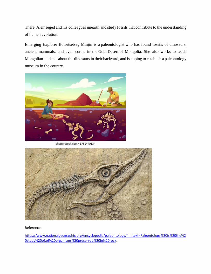
There, Alemseged and his colleagues unearth and study fossils that contribute to the understanding
of human evolution.
Emerging Explorer Bolortsetseg Minjin is a paleontologist who has found fossils of dinosaurs,
ancient mammals, and even corals in the Gobi Desert of Mongolia. She also works to teach
Mongolian students about the dinosaurs in their backyard, and is hoping to establish a paleontology
museum in the country.
Reference:
https://www.nationalgeographic.org/encyclopedia/paleontology/#:~:text=Paleontology%20is%20the%2
0study%20of,of%20organisms%20preserved%20in%20rock.
Evolution of modern humans
The origin of modern humans has probably been the most debated issue in evolutionary biology
over the last few decades.
Where did we come from?
“The exact origin of modern humans has long been a topic of debate.
Our evolutionary history is written into our genome?. The human genome looks the way it does
because of all the genetic changes that have affected our ancestors. The exact origin of modern
humans has long been a topic of debate.
KEY FACT
Modern humans originated in Africa within the past 200,000 years and evolved from their most
likely recent common ancestor, Homo erectus.
Modern humans (Homo sapiens), the species? that we are, means ‘wise man’ in Latin. Our species
is the only surviving species of the genus Homo but where we came from has been a topic of much
debate. Modern humans originated in Africa within the past 200,000 years and evolved from their
most likely recent common ancestor, Homo erectus, which means ‘upright man’ in Latin. Homo
erectus is an extinct species of human that lived between 1.9 million and 135,000 years ago.
Historically, two key models have been put forward to explain the evolution? of Homo sapiens.
These are the ‘out of Africa’ model and the ‘multi-regional’ model. The ‘out of Africa’ model is
currently the most widely accepted model. It proposes that Homo sapiens evolved in Africa before
migrating across the world.
On the other hand, the ‘multi-regional’ model proposes that the evolution of Homo sapiens took
place in a number of places over a long period of time. The intermingling of the various populations
eventually led to the single Homo sapiens species we see today.
“Current genomic evidence supports a single ‘out-of Africa’ migration of modern humans.
This is still very much an area of active research, however, current genomic evidence supports a
single ‘out-of Africa’ migration of modern humans rather than the ‘multi-regional’ model.
Although, studies of the genomes? of the extinct hominids Neanderthals and Denisovans suggest
that there was some mixing of genomes (1-3 per cent) with humans in Europe and Asia. This
interbreeding between two previously separated populations is called ‘admixture’ and results in a
mixing of genes? between the populations.
‘Out of Africa’: what’s the evidence?
‘Mitochondrial Eve’
KEY FACT
There is more genetic diversity in Africa compared with the rest of the world put together.
Genetic studies tend to support the ‘out of Africa’ model. The highest levels of genetic variation? in
humans are found in Africa. In fact there is more genetic diversity in Africa compared with the
rest of the world put together. In addition, the origin of modern DNA? in the mitochondria (the
‘powerhouses’ of our cells) has been tracked back to just one African woman who lived between
50,000 and 500,000 years ago – 'Mitochondrial Eve'.
Our genomes are a combination of DNA from both our mother and father. However, mitochondrial
DNA (mtDNA) comes solely from our mother. This is because the female egg contains large
amounts of mitochondrial DNA, whereas the male sperm contains just a tiny amount. The sperm
use their small amount of mitochondria to power their race to their egg before fertilisation. Once
a sperm merges with an egg, all the sperm mitochondria are destroyed.
KEY FACT
Your mitochondrial DNA is almost exactly the same as your mother’s and her mother’s.
As a result, mitochondrial DNA is described as being matrilineal (only the mother’s side survives
from generation to generation). So, your mitochondrial DNA is almost exactly the same as your
mother’s and her mother’s. Mitochondrial DNA has been extensively used by evolutionary
biologists, as it is easier to extract than DNA found in the nucleus? and there are many copies to
work with.
However, Mitochondrial Eve wasn’t the first or only woman on Earth at that time. She was simply
the point from which all modern generations of human appear to have grown. Evolutionary
biologists think the most likely reason for this is that an evolutionary ‘bottleneck’ occurred during
the time Eve was alive. This is when the majority of a species suddenly dies out, perhaps due to a
sudden catastrophe, bringing it to the brink of extinction. If Mitochondrial Eve was one of the few
women to survive then this could explain why her ‘matrilineal’ mitochondrial DNA ended up
being passed along so many generations.
Similarly, DNA from the Y chromosome? is only passed on from fathers to sons and a evolutionary
tree relating all present day male individuals also supports the ‘out of Africa’ model.
Mapping skulls
Further evidence for the ‘out of Africa’ model can be found in the size of human skulls. After
studying the genetics and skull measurements of 53 human populations from around the world,
scientists found that as you move further away from Africa, populations are less varied in their
genetic makeup. This may be because human populations became smaller as they spread out from
their original settlements in Africa and so genetic diversity within these populations was less. As
a result the scientists stated that modern humans could not have emerged in different places, but
instead had to have come from one region, Africa.
KEY FACT
The oldest known remains of anatomically modern humans are the Omo I and Omo II skulls.
The oldest known remains of anatomically modern humans are the Omo I and Omo II skulls. These
were found in 1967 in Omo National Park in south-western Ethiopia. The skulls have been dated
to 195,000 years ago, highlighting how humans have evolved relatively recently.
Moving out of Africa

Evidence shows that the first wave of humans to move out of Africa did not have too much success
on their travels. At times it appears they were on the brink of extinction, dwindling to as few as
10,000.
The eruption of a super volcano, Mount Toba, in Sumatra 70,000 years ago may have led to a
'nuclear winter', followed by a 1,000-year ice age. This sort of event would have put immense
pressure on humans. It may be that humans were only able to survive these extreme
conditions through cooperating with each other. This may have led to the formation of close family
groups or tribes and the development of some of the modern human behaviours we are familiar
with today, such as cooperation.
“Genetically, the six billion people of today’s world vary very little from the Homo sapiens that
ventured out of Africa.
Between 80,000 and 50,000 years ago another wave of humans migrated out of Africa. These
humans are likely to have been ‘modern’ in terms of their appearance and behaviour. Due to their
newly cooperative behaviour they were more successful at surviving and covered the whole world
in a relatively short period of time. As they migrated they would have encountered earlier,
primitive humans, eventually replacing them. Genetically, the six billion people of today’s world
vary very little from these earlier Homo sapiens that ventured out of Africa.
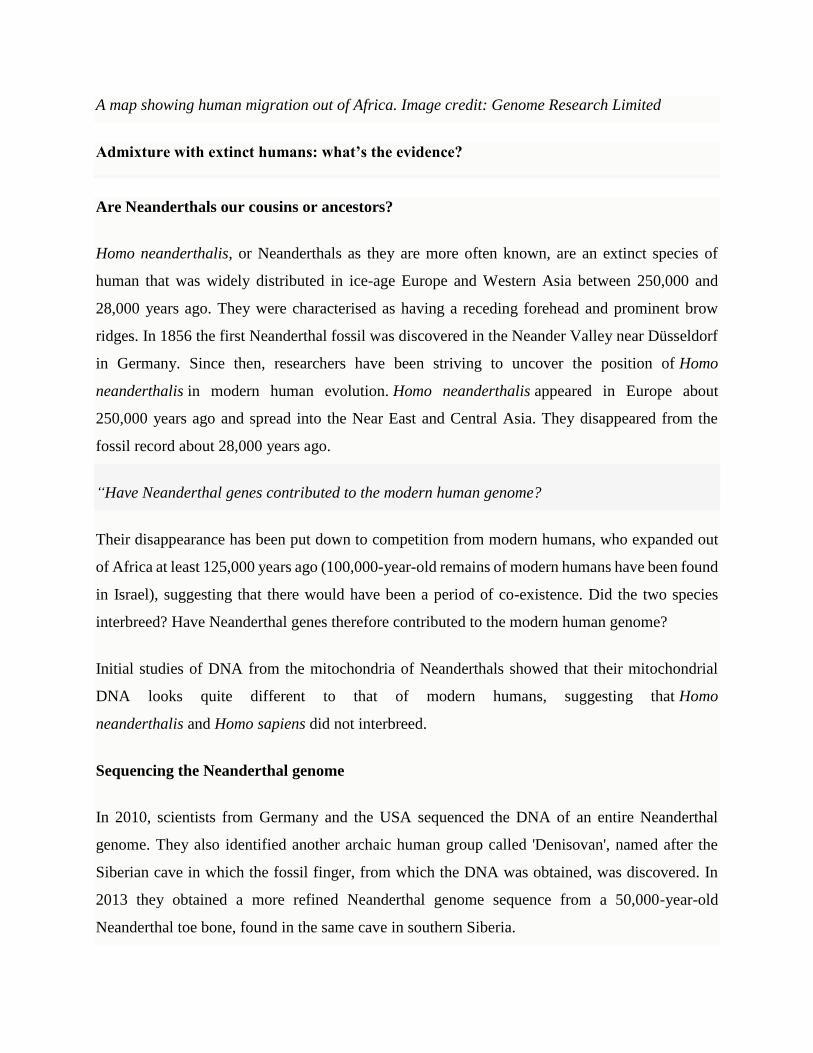
A map showing human migration out of Africa. Image credit: Genome Research Limited
Admixture with extinct humans: what’s the evidence?
Are Neanderthals our cousins or ancestors?
Homo neanderthalis, or Neanderthals as they are more often known, are an extinct species of
human that was widely distributed in ice-age Europe and Western Asia between 250,000 and
28,000 years ago. They were characterised as having a receding forehead and prominent brow
ridges. In 1856 the first Neanderthal fossil was discovered in the Neander Valley near Düsseldorf
in Germany. Since then, researchers have been striving to uncover the position of Homo
neanderthalis in modern human evolution. Homo neanderthalis appeared in Europe about
250,000 years ago and spread into the Near East and Central Asia. They disappeared from the
fossil record about 28,000 years ago.
“Have Neanderthal genes contributed to the modern human genome?
Their disappearance has been put down to competition from modern humans, who expanded out
of Africa at least 125,000 years ago (100,000-year-old remains of modern humans have been found
in Israel), suggesting that there would have been a period of co-existence. Did the two species
interbreed? Have Neanderthal genes therefore contributed to the modern human genome?
Initial studies of DNA from the mitochondria of Neanderthals showed that their mitochondrial
DNA looks quite different to that of modern humans, suggesting that Homo
neanderthalis and Homo sapiens did not interbreed.
Sequencing the Neanderthal genome
In 2010, scientists from Germany and the USA sequenced the DNA of an entire Neanderthal
genome. They also identified another archaic human group called 'Denisovan', named after the
Siberian cave in which the fossil finger, from which the DNA was obtained, was discovered. In
2013 they obtained a more refined Neanderthal genome sequence from a 50,000-year-old
Neanderthal toe bone, found in the same cave in southern Siberia.

“The genome sequence suggested that early modern non-African humans interbred with their now
extinct ancient human cousins.
DNA can survive in bone long after an animal dies. Over time the DNA from various microbes
that encounter the skeleton will also invade the bone. As a result, the DNA can be contaminated
with microbe DNA. Scientists therefore have to ensure that they sequence only the Neanderthal
genome and get rid of any DNA material left behind by these microbes or resulting from
contamination by modern humans who handle these bones. As with the human genome sequence,
the Denisovan and Neanderthal genome sequences were made available online for free. The
genome sequence suggested that early modern non-African humans interbred with their now
extinct ancient human cousins as they journeyed along coastlines and over mountains.
KEY FACT
Inbreeding is generally bad for the genetic fitness of a species as it reduces the variation in a
population making it more susceptible to disease and illness.
Analysis of the Neanderthal genome revealed that the toe bone came from a woman as it had two
X chromosomes. Further analysis showed that each pair of chromosomes was similar in sequence.
This suggests that her parents were closely related, perhaps an uncle and a niece. Inbreeding is
generally bad for the genetic fitness of a species as it reduces the variation in a population making
it more susceptible to disease and illness. This reduced genetic variation could explain why
Neanderthals became extinct.
When comparing human genomes to the Neanderthal genome, human genomes resemble each
other more than any of them resemble the Neanderthal genome. Some Neanderthal DNA is similar
to DNA from people of European and Asian origin but these similarities are not seen in African
DNA. This suggests that modern humans evolved in Africa and then expanded out into Asia and
Europe, where Neanderthals lived. A degree of interbreeding between Neanderthals and
early Homo sapiens then occurred in these areas. A study carried out in 2012 estimated that this
interbreeding probably took place about 37,000-85,000 years ago and it is estimated that the
proportion of Neanderthal-derived DNA in people outside Africa is 1.5-2.1 per cent.
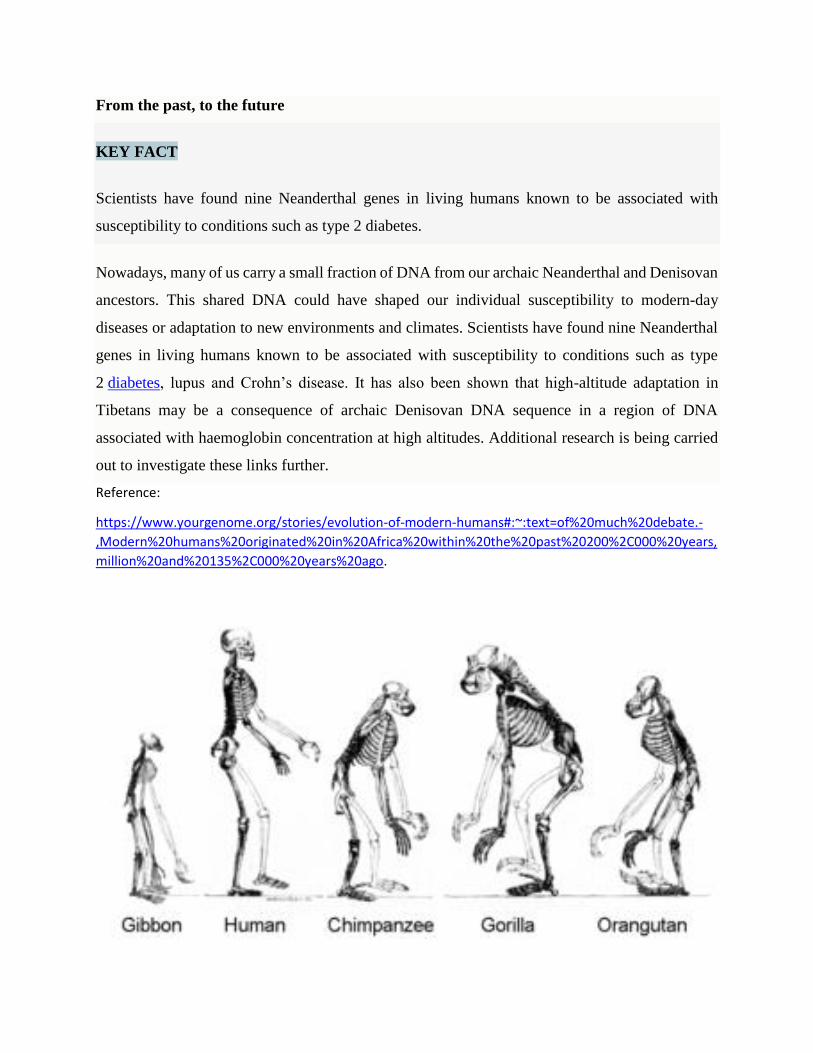
From the past, to the future
KEY FACT
Scientists have found nine Neanderthal genes in living humans known to be associated with
susceptibility to conditions such as type 2 diabetes.
Nowadays, many of us carry a small fraction of DNA from our archaic Neanderthal and Denisovan
ancestors. This shared DNA could have shaped our individual susceptibility to modern-day
diseases or adaptation to new environments and climates. Scientists have found nine Neanderthal
genes in living humans known to be associated with susceptibility to conditions such as type
2 diabetes, lupus and Crohn’s disease. It has also been shown that high-altitude adaptation in
Tibetans may be a consequence of archaic Denisovan DNA sequence in a region of DNA
associated with haemoglobin concentration at high altitudes. Additional research is being carried
out to investigate these links further.
Reference:
https://www.yourgenome.org/stories/evolution-of-modern-humans#:~:text=of%20much%20debate.-
,Modern%20humans%20originated%20in%20Africa%20within%20the%20past%20200%2C000%20years,
million%20and%20135%2C000%20years%20ago.
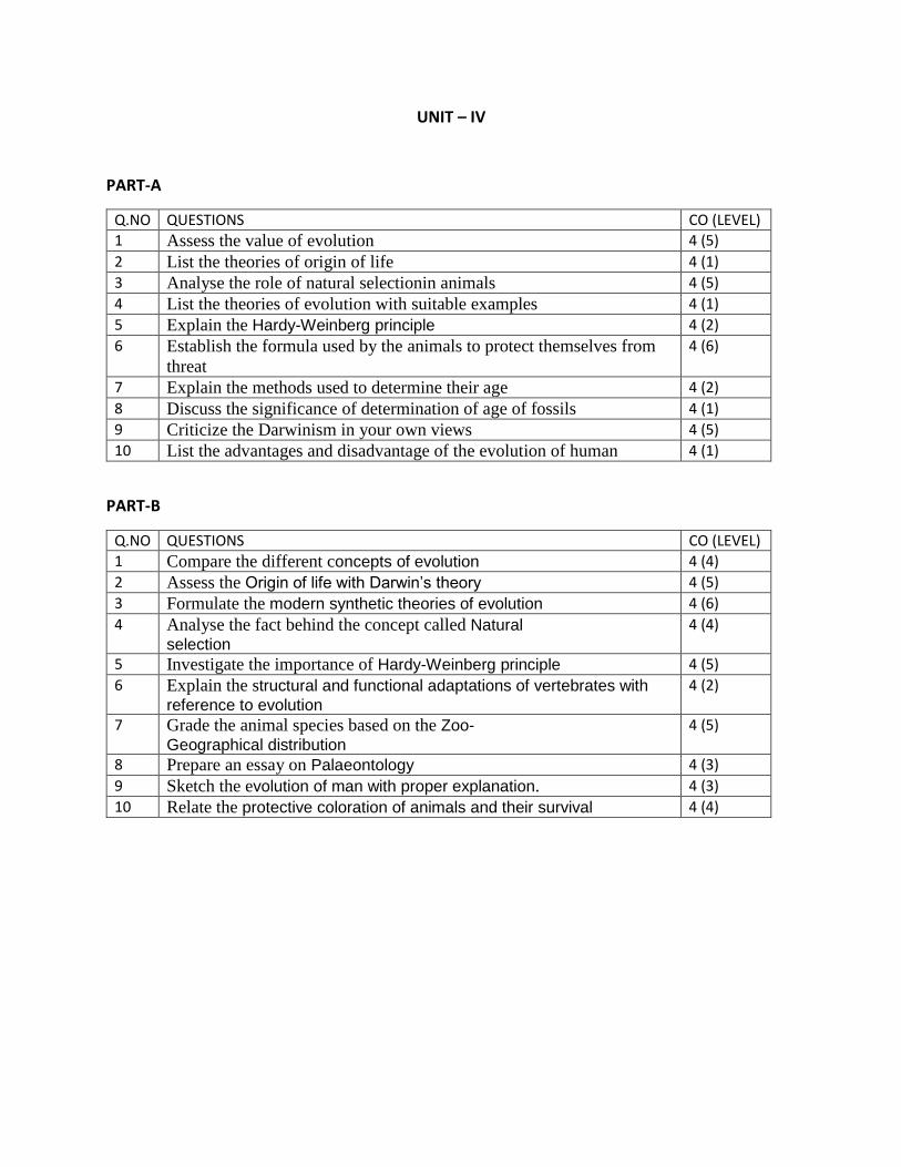
UNIT – IV
PART-A
Q.NO QUESTIONS CO (LEVEL)
1 Assess the value of evolution 4 (5)
2 List the theories of origin of life 4 (1)
3 Analyse the role of natural selectionin animals 4 (5)
4 List the theories of evolution with suitable examples 4 (1)
5 Explain the Hardy-Weinberg principle 4 (2)
6 Establish the formula used by the animals to protect themselves from
threat
4 (6)
7 Explain the methods used to determine their age 4 (2)
8 Discuss the significance of determination of age of fossils 4 (1)
9 Criticize the Darwinism in your own views 4 (5)
10 List the advantages and disadvantage of the evolution of human 4 (1)
PART-B
Q.NO QUESTIONS CO (LEVEL)
1 Compare the different concepts of evolution 4 (4)
2 Assess the Origin of life with Darwin’s theory 4 (5)
3 Formulate the modern synthetic theories of evolution 4 (6)
4 Analyse the fact behind the concept called Natural
selection 4 (4)
5 Investigate the importance of Hardy-Weinberg principle 4 (5)
6 Explain the structural and functional adaptations of vertebrates with
reference to evolution 4 (2)
7 Grade the animal species based on the Zoo-
Geographical distribution 4 (5)
8 Prepare an essay on Palaeontology 4 (3)
9 Sketch the evolution of man with proper explanation. 4 (3)
10 Relate the protective coloration of animals and their survival 4 (4)

SCHOOL OF BIO & CHEMICAL ENGINEERING
DEPARTMENT OF BIOTECHNOLOGY
SBC1201: ZOOLOGY
UNIT – V - APPLIED ZOOLOGY– SBC1201
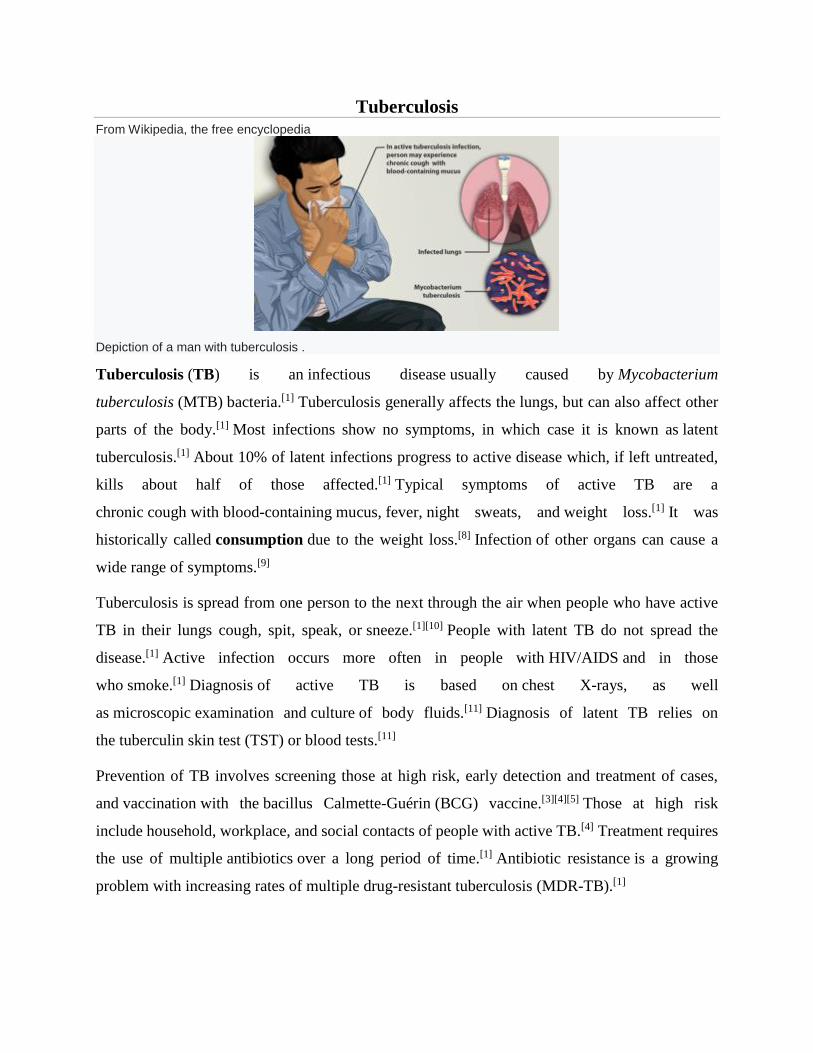
Tuberculosis
From Wikipedia, the free encyclopedia
Depiction of a man with tuberculosis .
Tuberculosis (TB) is an infectious disease usually caused by Mycobacterium
tuberculosis (MTB) bacteria.[1] Tuberculosis generally affects the lungs, but can also affect other
parts of the body.[1] Most infections show no symptoms, in which case it is known as latent
tuberculosis.[1] About 10% of latent infections progress to active disease which, if left untreated,
kills about half of those affected.[1] Typical symptoms of active TB are a
chronic cough with blood-containing mucus, fever, night sweats, and weight loss.[1] It was
historically called consumption due to the weight loss.[8] Infection of other organs can cause a
wide range of symptoms.[9]
Tuberculosis is spread from one person to the next through the air when people who have active
TB in their lungs cough, spit, speak, or sneeze.[1][10] People with latent TB do not spread the
disease.[1] Active infection occurs more often in people with HIV/AIDS and in those
who smoke.[1] Diagnosis of active TB is based on chest X-rays, as well
as microscopic examination and culture of body fluids.[11] Diagnosis of latent TB relies on
the tuberculin skin test (TST) or blood tests.[11]
Prevention of TB involves screening those at high risk, early detection and treatment of cases,
and vaccination with the bacillus Calmette-Guérin (BCG) vaccine.[3][4][5] Those at high risk
include household, workplace, and social contacts of people with active TB.[4] Treatment requires
the use of multiple antibiotics over a long period of time.[1] Antibiotic resistance is a growing
problem with increasing rates of multiple drug-resistant tuberculosis (MDR-TB).[1]
As of 2018 one quarter of the world's population is thought to have latent infection with TB.[6] New
infections occur in about 1% of the population each year.[12] In 2018, there were more than
10 million cases of active TB which resulted in 1.5 million deaths.[7] This makes it the number
one cause of death from an infectious disease.[13] As of 2018, most TB cases occurred in the
regions of South-East Asia (44%), Africa (24%) and the Western Pacific (18%), with more than
50% of cases being diagnosed in eight countries: India (27%), China (9%), Indonesia (8%), the
Philippines (6%), Pakistan (6%), Nigeria (4%) and Bangladesh (4%).[13] The number of new cases
each year has decreased since 2000.[1] About 80% of people in many Asian and African countries
test positive while 5–10% of people in the United States population test positive by the tuberculin
test.[
-------------------------------------------------------------------------------------------------------------------------------
Dengue fever
From Wikipedia, the free encyclopedia
Dengue fever is a mosquito-borne tropical disease caused by the dengue virus.[1] Symptoms
typically begin three to fourteen days after infection.[2] These may include a
high fever, headache, vomiting, muscle and joint pains, and a characteristic skin
rash.[1][2] Recovery generally takes two to seven days.[1] In a small proportion of cases, the disease
develops into a more severe dengue hemorrhagic fever, resulting in bleeding, low levels of blood
platelets and blood plasma leakage, or into dengue shock syndrome, where dangerously low
blood pressure occurs.[1][2]
Dengue is spread by several species of female mosquitoes of the Aedes genus, principally Aedes
aegypti.[1][2] The virus has five serotypes;[7][8] infection with one type usually gives
lifelong immunity to that type, but only short-term immunity to the others.[1] Subsequent infection
with a different type increases the risk of severe complications.[1] A number of tests are available
to confirm the diagnosis including detecting antibodies to the virus or its RNA.[2]
A vaccine for dengue fever has been approved and is commercially available in a number of
countries.[4][9] As of 2018, the vaccine is only recommended in individuals who have been
previously infected, or in populations with a high rate of prior infection by age nine.[10][5] Other
methods of prevention include reducing mosquito habitat and limiting exposure to bites.[1] This
may be done by getting rid of or covering standing water and wearing clothing that covers much
of the body.[1] Treatment of acute dengue is supportive and includes giving fluid either by mouth
or intravenously for mild or moderate disease.[2] For more severe cases, blood transfusion may be
required.[2] About half a million people require hospital admission every
year.[1] Paracetamol (acetaminophen) is recommended instead of nonsteroidal anti-inflammatory
drugs (NSAIDs) for fever reduction and pain relief in dengue due to an increased risk of bleeding
from NSAID use.[2][11][12]
Dengue has become a global problem since the Second World War and is common in more than
120 countries, mainly in Southeast Asia, South Asia and South America.[5][13][14] About
390 million people are infected a year and approximately 40,000 die.[5][6] In 2019 a significant
increase in the number of cases was seen.[15] The earliest descriptions of an outbreak date from
1779.[14] Its viral cause and spread were understood by the early 20th century.[16] Apart from
eliminating the mosquitos, work is ongoing for medication targeted directly at the virus.[17] It is
classified as a neglected tropical disease.[18]
--------------------------------------------------------------------------------------------------------------------------------
Malaria
From Wikipedia, the free encyclopedia
Malaria is a mosquito-borne infectious disease that affects humans and other
animals.[4][5][2] Malaria causes symptoms that typically include fever, tiredness, vomiting,
and headaches.[1] In severe cases, it can cause yellow skin, seizures, coma, or death.[1] Symptoms
usually begin ten to fifteen days after being bitten by an infected mosquito.[2] If not properly
treated, people may have recurrences of the disease months later.[2] In those who have recently
survived an infection, reinfection usually causes milder symptoms.[1] This
partial resistance disappears over months to years if the person has no continuing exposure to
malaria.[1]
Malaria is caused by single-celled microorganisms of the Plasmodium group.[2] The disease is
most commonly spread by an infected female Anopheles mosquito.[2] The mosquito bite
introduces the parasites from the mosquito's saliva into a person's blood.[2] The parasites travel to
the liver where they mature and reproduce.[1] Five species of Plasmodium can infect and be spread
by humans.[1] Most deaths are caused by P. falciparum, whereas P. vivax, P. ovale,
and P. malariae generally cause a milder form of malaria.[1][2] The species P. knowlesi rarely
causes disease in humans.[2] Malaria is typically diagnosed by the microscopic examination of
blood using blood films, or with antigen-based rapid diagnostic tests.[1] Methods that use
the polymerase chain reaction to detect the parasite's DNA have been developed, but are not
widely used in areas where malaria is common due to their cost and complexity.[6]
The risk of disease can be reduced by preventing mosquito bites through the use of mosquito
nets and insect repellents or with mosquito-control measures such as spraying insecticides and
draining standing water.[1] Several medications are available to prevent malaria in travellers to
areas where the disease is common.[2] Occasional doses of the combination
medication sulfadoxine/pyrimethamine are recommended in infants and after the first
trimester of pregnancy in areas with high rates of malaria.[2] As of 2020, there is
one vaccine which has been shown to reduce the risk of malaria by about 40% in children in
Africa.[7][8] Efforts to develop more effective vaccines are ongoing.[8] The recommended treatment
for malaria is a combination of antimalarial medications that includes artemisinin.[9][1][2] The
second medication may be either mefloquine, lumefantrine, or
sulfadoxine/pyrimethamine.[10] Quinine, along with doxycycline, may be used if artemisinin is not
available.[10] It is recommended that in areas where the disease is common, malaria is confirmed
if possible before treatment is started due to concerns of increasing drug resistance.[2] Resistance
among the parasites has developed to several antimalarial medications; for example, chloroquine-
resistant P. falciparum has spread to most malarial areas, and resistance to artemisinin has become
a problem in some parts of Southeast Asia.[2]
The disease is widespread in the tropical and subtropical regions that exist in a broad band around
the equator.[1] This includes much of sub-Saharan Africa, Asia, and Latin America.[2] In 2018
there were 228 million cases of malaria worldwide resulting in an estimated 405,000
deaths.[3] Approximately 93% of the cases and 94% of deaths occurred in Africa.[3] Rates of
disease have decreased from 2010 to 2014 but increased from 2015 to 2017, during which there
were 231 million cases.[3] Malaria is commonly associated with poverty and has a significant
negative effect on economic development.[11][12] In Africa, it is estimated to result in losses of
US$12 billion a year due to increased healthcare costs, lost ability to work, and adverse effects on
tourism.[13]
--------------------------------------------------------------------------------------------------------------------------------
Swine/Variant Influenza
Swine influenza is a respiratory disease of pigs caused by type A influenza viruses
that regularly cause outbreaks of influenza in pigs. Influenza viruses that commonly
circulate in swine are called “swine influenza viruses” or “swine flu viruses.” Like
human influenza viruses, there are different subtypes and strains of swine influenza
viruses. The main swine influenza viruses circulating in U.S. pigs in recent years
have been, swine triple reassortant (tr) H1N1 influenza virus, trH3N2 virus, and
trH1N2 virus.
------------------------------------------------------------------------------------------------------------------------
Integrated Pest Management (IPM)
BY JOLENE HANSEN
Even if you do everything right when building a healthy garden, pests inevitably show up. But
managing your garden with a thoughtful, proactive approach helps prevent pests from doing
serious damage. Integrated Pest Management (IPM) combines different types of controls — from
hands-on pest removal to traditional synthetic pesticides — in a sensible, long-term plan.
Designing your own program around proven IPM principles can help protect your garden and keep
it healthy.
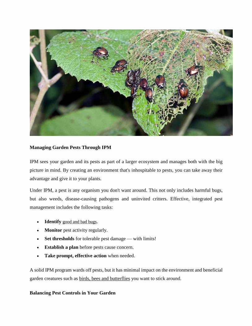
Managing Garden Pests Through IPM
IPM sees your garden and its pests as part of a larger ecosystem and manages both with the big
picture in mind. By creating an environment that's inhospitable to pests, you can take away their
advantage and give it to your plants.
Under IPM, a pest is any organism you don't want around. This not only includes harmful bugs,
but also weeds, disease-causing pathogens and uninvited critters. Effective, integrated pest
management includes the following tasks:
Identify good and bad bugs.
Monitor pest activity regularly.
Set thresholds for tolerable pest damage — with limits!
Establish a plan before pests cause concern.
Take prompt, effective action when needed.
A solid IPM program wards off pests, but it has minimal impact on the environment and beneficial
garden creatures such as birds, bees and butterflies you want to stick around.
Balancing Pest Controls in Your Garden
Four main categories of pest controls form IPM's foundation: cultural, biological,
mechanical/physical and pesticide controls. The four work hand in hand to provide targeted,
effective, long-term pest management, and each category plays a special role.
Cultural Controls
Cultural pest controls start with the decisions you make when choosing and caring for plants.
Prevention is your first line of defense; healthy, nurtured plants resist pests and diseases better than
weak, unhealthy plants. Cultural controls in good IPM programs include these simple
recommendations:
Choose plants suited to your area and its challenges. Arid, drought-prone regions, for
example, call for water-wise plants with low moisture needs.
Select disease- and pest-resistant plant varieties. Plants proven to withstand your
region's most common pests hold up better under attack.
Plant at appropriate times. In many regions, fall is prime planting time. Fall and winter
planting allow roots to establish before summer heat arrives. This is especially important
in southern or western regions. In far northern climates, spring planting is often best for
plants with less cold hardiness.
Choose proper sites. Plants have more problems and fail to thrive in inappropriate
conditions. For example, sun-loving plants are more vulnerable to pests and other problems
when planted in shady areas, and vice versa.
Maintain lawn and garden tools. Sharp mower blades and proper mowing heights lead
to healthier lawns. Sharp, sterile pruners help prevent the spread of disease.
Avoid overhead watering. Some leaf diseases, such as common garden fungal
diseases or black spot on roses, spread with the help of water. Water the soil at the base of
plants, instead of watering leaves.
Water in early morning. If leaves do get wet, they'll dry thoroughly before evening.
Test your soil pH. A simple soil test reveals adjustments that can help your soil's structure
and nutrients, so you can feed plants right.

Knowing what your plants need — and providing all they require — gives you the upper hand
over pests. Simple, common-sense cultural controls are integral to good IPM.
Biological Controls
All pests, from weeds and insects to diseases, have natural enemies. A balanced pest management
program conserves, supports and encourages those foes. Biological IPM controls include:
Predator insects: Adult lady beetles and their larvae are voracious aphid-eaters. Green
lacewing larvae feed on all kinds of pests, including mealybugs, whiteflies, mites and
thrips. These and other beneficial bugs are probably already in your garden.
Parasitic insects: Parasitic wasps lay their eggs on and in their living targets. Eggs hatch,
and then feed inside the pest. A mummified aphid with a round hole in its back is evidence
that parasitic wasps have been at work.
Biological pathogens: Bacillus thuringiensis, also known as Bt, is a soil-borne bacterium
that fights mosquitoes and insects in the larval, caterpillar stage. This and other pathogens
are effective biological pesticides for very specific pests.
(Published image, GTech media link in comments)
Knowing the difference between good and bad bugs is essential for IPM. You can buy beneficial
predators and parasites, but self-managing your garden's free, natural populations is effective.
Keep the good guys plentiful and they'll help keep bad bugs at bay, reducing the need for other
measures.
Mechanical and Physical Controls
Mechanical and physical IPM controls go directly after pests to capture or kill them and prevent
them from reaching their destinations. IPM recommends proactive lawn and garden controls and
actions, including:
Use mulch in garden areas. Mulch prevents weeds and weed seeds from getting light and
sprouting.
Hoe or pull weeds before they establish roots. If weeds escape the hoe, mow or cut them
before they set and drop their seeds.
Place collars in the soil around susceptible vegetable stems. Simple barriers prevent
hungry cutworms and other crawling pests from reaching their goal.
Stretch netting over your favorite berry bushes. This stops marauding birds from
settling in and helping themselves to your raspberry and blackberry harvest.
Stop destructive rodents with mechanical traps. Easy-to-use products such
as AMDRO® Gopher Traps provide control for troublesome pocket gophers.
Hand-pick pests off plants. This physical control puts an immediate end to pests' plant-
damaging days.
Using mechanical and physical controls in concert with other IPM methods keeps many types of
pest damage low.
Pesticide Controls
An effective IPM program includes pesticides for prevention and active treatment. Pesticides pack
necessary and powerful punches, especially when other IPM controls fall short. Invasive Japanese
beetles, for example, devastate gardens and skeletonize leaves and blossoms. In Japan, the beetle's
natural enemies control it, but its native predators don't exist in the United States. Pesticides help
fill that gap.
IPM-appropriate pesticides include the following types:
Traditional or synthetic pesticides: IPM programs include pesticides manufactured from
synthetic ingredients. These include products such as GardenTech® Sevin® brand insecticides,
trusted by gardeners for more than 50 years. Sevin® Insect Killer, available in ready-to-
use, ready-to-spray, concentrate and granular forms and Sevin®-5 Ready-To-Use Dust are
effective on ornamental and edible gardens, lawns and home perimeters to kill Japanese beetles
and a broad spectrum of other insect pests as part of a successful IPM program.
Natural or non-synthetic pesticides: Botanical-based pesticides fall into this IPM group.
Based on extracts from different types of plants, these natural insecticides include products
such as neem oil, based on extracts from the neem tree, or pyrethrins extracted from special
chrysanthemum blossoms. Pesticides in this category may or may not be organic. They
also require the same types of safety precautions as synthetic pesticides.
Preventive pesticides: IPM incorporates traditional fungicides to help treat fungal disease
and prevent it from spreading. The University of California Statewide Integrated Pest
Management Program recommends clorothalonil-based products, such
as GardenTech® Daconil® Fungicide, to protect healthy plants and prevent botrytis blight
and black spot in roses.1
By establishing a personalized Integrated Pest Management program for your garden, you can take
action — with the right controls — when pests show up, and then rest easy. With IPM principles
in mind and GardenTech® brands as your partners, you can build a healthy, harmonious garden.
Always read product labels thoroughly and follow instructions carefully, including guidelines for
pre-harvest intervals (PHI) on edible crops and frequency of applications.
Sevin is a registered trademark of Tessenderlo Kerley, Inc.
Amdro is a registered trademark of Central Garden & Pet Company.
Daconil is a registered trademark of GB Biosciences Corp.
GardenTech is a registered trademark of Gulfstream Home & Garden, Inc.
Sources:
1. S. T. Koike et al., “UC Pest Management Guidelines – Rose (Rosa spp.)," University of
California Agriculture & Natural Resources Statewide Integrated Pest Management Program,
March 2009.
2. https://www.gardentech.com/blog/pest-id-and-prevention/what-is-
integrated-pest-management
Genetic improvement in aquaculture Tuesday, 1 February 2005Jeffrey T. Silverstein, Ph.D.
Major challenge will be improving multiple traits in aquatic species
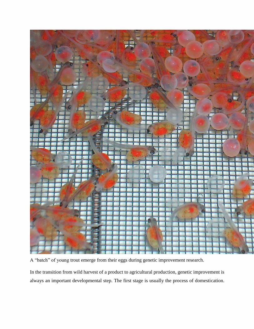
A “batch” of young trout emerge from their eggs during genetic improvement research.
In the transition from wild harvest of a product to agricultural production, genetic improvement is
always an important developmental step. The first stage is usually the process of domestication.
Further genetic improvement in agriculture or aquaculture typically has a human focus on
enhanced production characteristics.
Just as in terrestrial production scenarios, aquaculturists value faster growth, improved efficiency,
less loss to disease, and high quality. Several tools are available to accomplish such genetic
improvements.
Domestication
Most stocks of the aquatic organisms most widely grown in captivity are domesticated to some
degree, and a few have undergone further genetic improvement. Just as terrestrial livestock and
crops are bred to be not only productive, but productive in convenient proximity to humans, aquatic
plants and animals are now undergoing these same pressures.
Although the domestication process is arguably at a much earlier stage for aquatic organisms than
their terrestrial counterparts, aquaculture is undergoing the process at a time when the technologies
and tools available could remarkably speed up domestication and genetic improvement.
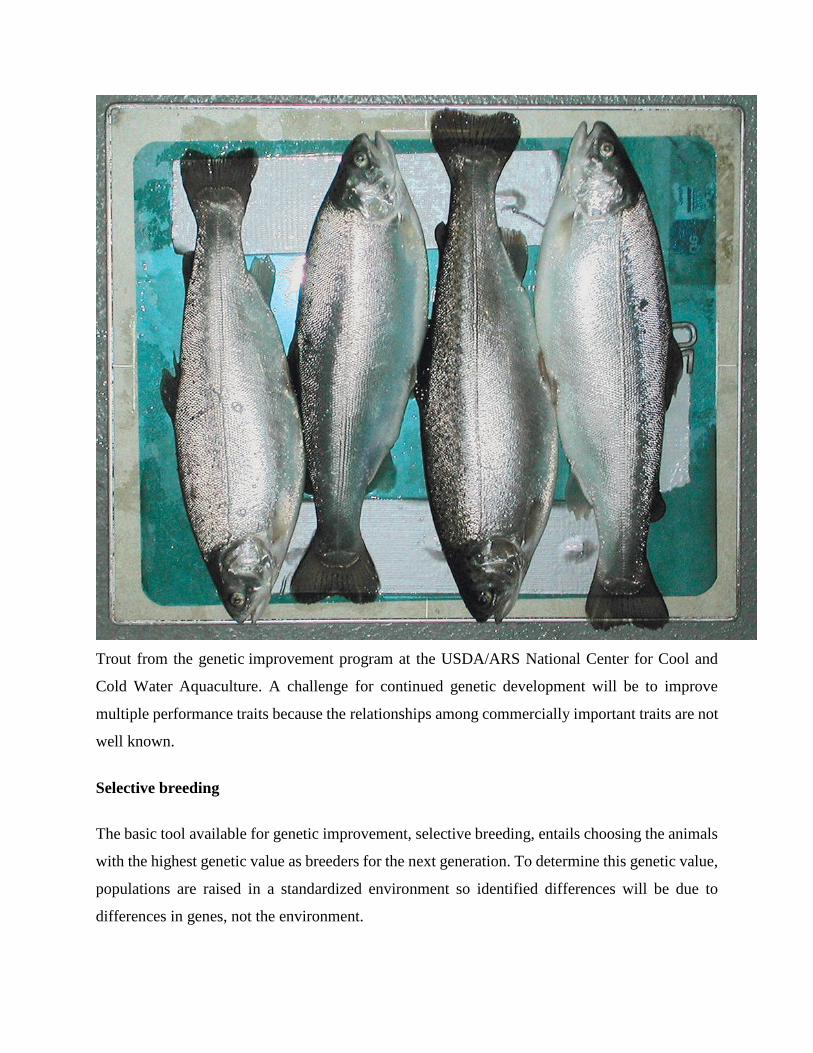
Trout from the genetic improvement program at the USDA/ARS National Center for Cool and
Cold Water Aquaculture. A challenge for continued genetic development will be to improve
multiple performance traits because the relationships among commercially important traits are not
well known.
Selective breeding
The basic tool available for genetic improvement, selective breeding, entails choosing the animals
with the highest genetic value as breeders for the next generation. To determine this genetic value,
populations are raised in a standardized environment so identified differences will be due to
differences in genes, not the environment.
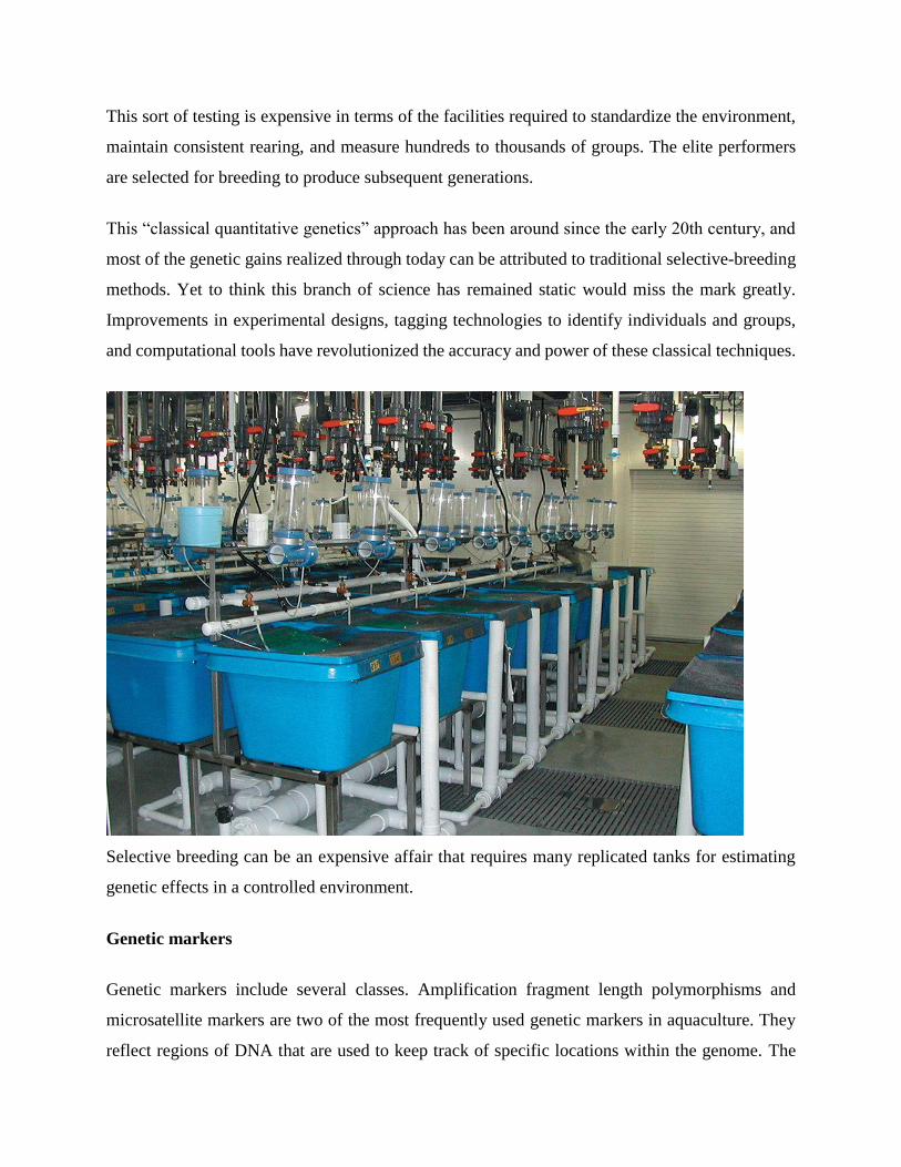
This sort of testing is expensive in terms of the facilities required to standardize the environment,
maintain consistent rearing, and measure hundreds to thousands of groups. The elite performers
are selected for breeding to produce subsequent generations.
This “classical quantitative genetics” approach has been around since the early 20th century, and
most of the genetic gains realized through today can be attributed to traditional selective-breeding
methods. Yet to think this branch of science has remained static would miss the mark greatly.
Improvements in experimental designs, tagging technologies to identify individuals and groups,
and computational tools have revolutionized the accuracy and power of these classical techniques.
Selective breeding can be an expensive affair that requires many replicated tanks for estimating
genetic effects in a controlled environment.
Genetic markers
Genetic markers include several classes. Amplification fragment length polymorphisms and
microsatellite markers are two of the most frequently used genetic markers in aquaculture. They
reflect regions of DNA that are used to keep track of specific locations within the genome. The
markers are distributed throughout the DNA of an organism and transmitted from parent to
offspring.
Genetic markers are useful for determining parentage because offspring share markers with their
parents. They are also expected to be useful for identifying regions of DNA that confer
performance advantages.
If a particular marker is always found associated with resistance to a coldwater bacterial disease,
for example, selection for breeding could be based on the presence of this marker without doing a
challenge trial under tightly controlled environmental conditions. This application of selective
breeding is referred to as marker-assisted selection.
An important consideration for marker-assisted selection is that traditional approaches relying on
direct trait measurements in organisms are expensive. Furthermore, elite performers in disease
challenges are usually not used for breeding for fear of transmitting disease, but their close relatives
could be used.
A similar situation occurs if traits of interest are measured after test animals are slaughtered.
Individuals with high meat quality or fillet yield may not be available for breeding after the traits
are measured.
Genetic engineering
Genetic engineering is a broad heading that includes chromosome set manipulations and
transgenesis. Chromosome set manipulation is a group of techniques in which sets of
chromosomes in organisms are modified.
Whereas normal organisms have two sets of chromosomes – one from each parent – one type of
chromosome set manipulation can result in triploids, organisms with three sets of chromosomes.
These animals are generally sterile, which can be advantageous under conditions where
reproduction is undesirable.
This technology has been applied in the United States to the nonindigenous weed-controlling grass
carp to prevent unwanted reproduction. Chromosome manipulation has also been used in oysters,
which typically undergo a loss of quality during the spawning season.
Transgenesis
Transgenesis refers to the technique of incorporating a gene or genes through biotechnological
methods, not breeding. Soybean plants that have been made resistant to the effects of herbicides
are an example of transgenic crops. With a gene from bacteria transferred into and expressed in
the plants, their use reduces labor costs for cultivating and weeding. Golden rice, a transgenic rice
plant, has three foreign genes inserted from bacteria and daffodils to allow the production of
vitamin A in the rice.
The Glofish, a fluorescent zebra-fish which came on the U.S. market in January 2004, is the first
transgenic aquatic organism to be marketed. The proteins responsible for making the zebrafish
fluoresce are transgenes derived from jellyfish and coral. These fish are not intended for food.
Atlantic salmon with a growth hormone gene from Chinook salmon and a promoter, or switch,
from the ocean pout are currently being reviewed by the United States Food and Drug
Administration for approval as food fish. Research on other aquatic transgenics with the potential
for greater disease resistance, enhanced nutritive value, and other characteristics is ongoing around
the world.
Ongoing challenges
Artificial fertilization of trout eggs.
The use of classical breeding techniques remains the foundation upon which genetic improvement
will be built for the foreseeable future. These techniques will continue to be refined to offer faster
rates of improvement in performance. Incorporating marker information into selective breeding is
a challenge, both in terms of the practicalities of identifying marker-trait associations and how to
incorporate the information.
Perhaps the major ongoing challenge will be to improve multiple traits in aquatic species. Fast,
efficient growth; disease resistance; and product quality are major traits of interest, but the
relationships among these traits are not well known. Since the genetic values for traits are not well
defined, or multiple traits of interest are not measured on the same individuals, the genetic
relationships have not been determined.
In limited cases, the relationships among a subset of these traits are known. For example, the
genetic relationship between growth and resistance to viral hemorrhagic septicemia (VHS) in one
farmed population of rainbow trout, Oncorhynchus mykiss, was shown to be negative. This
unfortunately suggested that simply selecting for improvement in growth would result in lowered
resistance to VHS.
Especially in cases like this, knowledge of markers associated with disease resistance and
improved estimates of genetic value for multiple traits will enable solutions. Current developments
in genetic improvement hold the promise that the relatively rare individuals with superior
performance for several traits of interest can be identified and selected for breeding.
Reference:
https://www.aquaculturealliance.org/advocate/genetic-improvement-in-
aquaculture/#:~:text=Further%20genetic%20improvement%20in%20agriculture,to%20disease%2C%20a
nd%20high%20quality.
--------------------------------------------------------------------------------------------------------------------------------------
Apiculture
Bees are economically important social insects. They not only provide us with honey and wax,
they are also responsible for pollination of flowers of the majority of damaged bee larvae, pollen
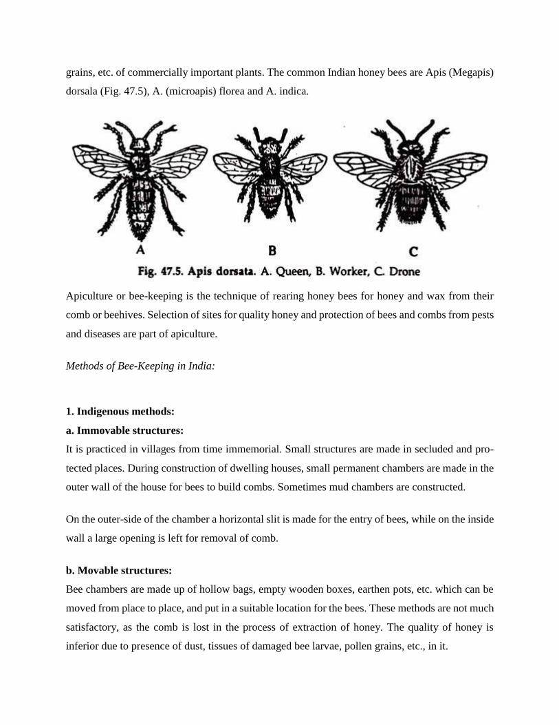
grains, etc. of commercially important plants. The common Indian honey bees are Apis (Megapis)
dorsala (Fig. 47.5), A. (microapis) florea and A. indica.
Apiculture or bee-keeping is the technique of rearing honey bees for honey and wax from their
comb or beehives. Selection of sites for quality honey and protection of bees and combs from pests
and diseases are part of apiculture.
Methods of Bee-Keeping in India:
1. Indigenous methods:
a. Immovable structures:
It is practiced in villages from time immemorial. Small structures are made in secluded and pro-
tected places. During construction of dwelling houses, small permanent chambers are made in the
outer wall of the house for bees to build combs. Sometimes mud chambers are constructed.
On the outer-side of the chamber a horizontal slit is made for the entry of bees, while on the inside
wall a large opening is left for removal of comb.
b. Movable structures:
Bee chambers are made up of hollow bags, empty wooden boxes, earthen pots, etc. which can be
moved from place to place, and put in a suitable location for the bees. These methods are not much
satisfactory, as the comb is lost in the process of extraction of honey. The quality of honey is
inferior due to presence of dust, tissues of damaged bee larvae, pollen grains, etc., in it.
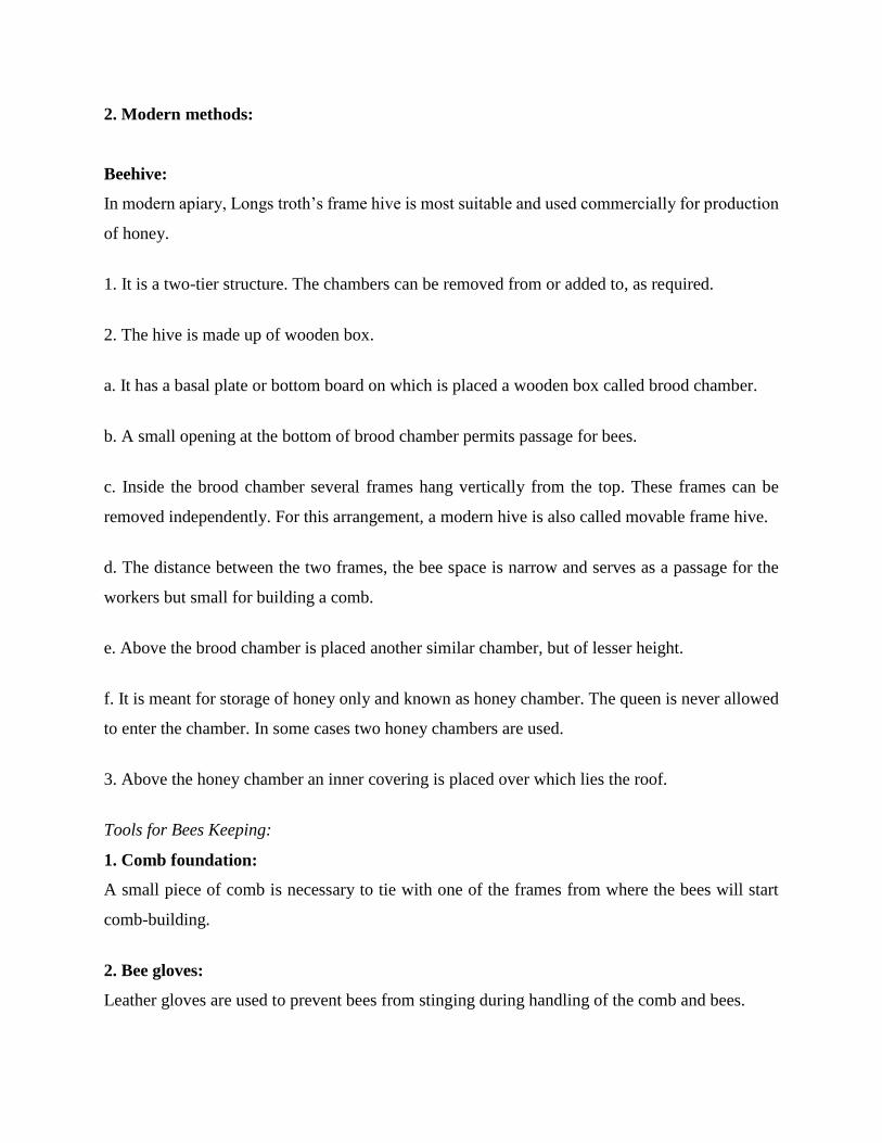
2. Modern methods:
Beehive:
In modern apiary, Longs troth’s frame hive is most suitable and used commercially for production
of honey.
1. It is a two-tier structure. The chambers can be removed from or added to, as required.
2. The hive is made up of wooden box.
a. It has a basal plate or bottom board on which is placed a wooden box called brood chamber.
b. A small opening at the bottom of brood chamber permits passage for bees.
c. Inside the brood chamber several frames hang vertically from the top. These frames can be
removed independently. For this arrangement, a modern hive is also called movable frame hive.
d. The distance between the two frames, the bee space is narrow and serves as a passage for the
workers but small for building a comb.
e. Above the brood chamber is placed another similar chamber, but of lesser height.
f. It is meant for storage of honey only and known as honey chamber. The queen is never allowed
to enter the chamber. In some cases two honey chambers are used.
3. Above the honey chamber an inner covering is placed over which lies the roof.
Tools for Bees Keeping:
1. Comb foundation:
A small piece of comb is necessary to tie with one of the frames from where the bees will start
comb-building.
2. Bee gloves:
Leather gloves are used to prevent bees from stinging during handling of the comb and bees.

3. Bee veil:
A bee veil is required to cover neck, face and head of the keeper during handling. Usually it is
made of linen.
4. Smoker:
A smoker must be used while capturing bees in a hive. Smoke from paper, wood and coconut cover
makes the bees inactive. There is fire box in a smoker in which smoke-producing materials and
fire are put. A bellow system is fitted to blow the smoke.
5. Hive tool:
It is a long, narrow and flat piece of steel with a slightly bent head to scrap away dirty materials
deposited by bees or some other factors.
6. Honey extractor:
It is used for extraction of honey from the frames without damaging the comb. It consists of a
metal drum with several pockets around a rotating wheel. The frames are hanged from the pockets
and the pockets are made to rotate round a central axis.
The centrifugal force created by rotation separates honey from the comb which is collected in the
drum. The honey is taken out from the drum through a hole at the bottom.- The combs and frames
are again placed in the hive.
Typical Location of Apiary:
A locality for apiary must have different varieties of pollen and nector-producing plants in
sufficient number within a distance of 1.5 to 2.5 km. area. Neem, Rita, Tamarind, Cheery, Apple,
and Citrus, Lily, Lotus, various wild plants and crops are good sources for both nectar and honey.
Diseases of the Bees:
Bees suffer from different contagious diseases and are very often subjected to curious organic
disorders. The organisms for contagious diseases are Aspergillus, viruses, mites and Protozoa.
a. Aspergillus, a fungus, causes paralysis in worker bees.
b. Virus cause paralysis and high percentage of mortality to all categories of bees.
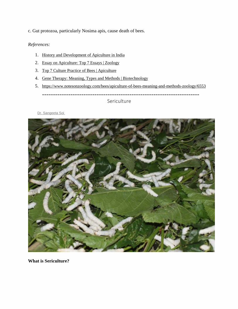
c. Gut protozoa, particularly Nosima apis, cause death of bees.
References:
1. History and Development of Apiculture in India
2. Essay on Apiculture: Top 7 Essays | Zoology
3. Top 7 Culture Practice of Bees | Apiculture
4. Gene Therapy: Meaning, Types and Methods | Biotechnology
5. https://www.notesonzoology.com/bees/apiculture-of-bees-meaning-and-methods-zoology/6553
------------------------------------------------------------------------ Sericulture
Dr. Sangeeta Soi
What is Sericulture?
Sericulture is an agro-based industry. It involves rearing of silkworms for the production of raw
silk, which is the yarn obtained out of cocoons spun by certain species of insects. Cultivation to
feed the silkworms that spin silk cocoons and reeling the cocoons to unwind the silk filament for
value added advantages like process and weaving are the major activities of sericulture. Silk has
been blended with the life and culture of the Indians.
India encompasses an upscale and sophisticated history in silk production and its silk trade dates
back to fifteenth century. Silk is the most elegant textile in the world with distinctive grandeur,
natural radiance, and inherent affinity for dyes, high absorbance, lightweight weight, soft touch
and high sturdiness and called the “Queen of Textiles” the world over Sericulture business
provides employment to roughly 8.25 million persons in rural and semi-urban areas in India
throughout 2015-16.
It stands for livelihood opportunity for millions owing to high employment oriented, low capital
intensive and remunerative nature of its production. From these, a sizeable number of workers
belong to the economically weaker sections of society, including girls. India’s ancient and culture
bound domestic market and a tremendous diversity of silk clothes that replicate geographic
specificity have helped the country to attain variety one position in silk business.
Silk production in India
India is the second largest producer of silk within the globe. Among the four kinds of silk created
in 2015-16, Mulberry accounts for 71.8% (20,434 MT), Tasar 9.9% (2,818 MT), Eri 17.8% (5,054
MT) and Muga 0.6% (166 MT) of the total raw silk production of 28,472 MT.
Mulberry sericulture is principally practised in 5 states i.e. Karnataka, Andhra Pradesh, Assam and
Bodoland, West Bengal, Jharkhand and Tamil Nadu are major silk producing states in the country.
North East has the distinctive distinction of being the sole region manufacturing four kinds of silk
like Mulberry, Oak Tasar, Muga and Eri. Overall NE region contributes eighteen of India's total
silk production.
Advantages of Sericulture:
High employment potential
60 lakh persons are engaged in various sericulture activities in the country.
It is calculable that Sericulture can generate employment @ 11 man days per kg of raw silk
production (in on-farm and off-farm activities) throughout the year. This potential ispar-
excellence and no other business generates this type of employment, especially in rural
areas, hence, sericulture is used as a tool for rural reconstruction.
Eco-friendly Activity
As a perennial crop with good foliage and root-spread, mulberry contributes to
conservation and provides green cover.
Waste from silkworm rearing will be recycled as inputs to garden.
Dried mulberry twigs and branches are used as fuel in place of firewood and therefore
reduce the pressure on vegetation/forest.
Being a labour intensive and predominantly agro-based activity, involvement of smoke-
emitting machinery is minimal.
Developmental programmes initiated for mulberry plantation are mainly in upland areas
where un-used cultivable land is made productive.
Intercropping can also be done with numerous plantations.
Being a deep-rooted perennial plant it can be raised in vacant lands, hill slopes and
watershed areas.
Currently, only concerning 0.1 % of the cultivable land in the country is beneath mulberry
cultivation.
Women friendly Occupation
Women constitutes over hour of those employed in down-stream activities of sericulture in the
country. This is the result of sericulture activities ranging from mulberry garden management, leaf
harvesting and silkworm rearing is more effectively taken up by the women folks. Even silk reeling
business together with weaving is largely supported by them.

Low Gestation, High Returns
Estimated investments of Rs.12,000 to 15,000 (excluding cost of land and rearing space)
is adequate for undertaking mulberry cultivation and silkworm rearing in one acre of
irrigated land.
It takes only six months for mulberry to grow for commencement of silkworm rearing.
Once planted, it will go on supporting silkworm rearing year after year for 15-20 years
depending on inputs and management provided.
Five crops can be taken in one year under tropical conditions.
By adopting stipulated package of practices, a farmer can attain net income levels up to
Rs.30000 per acre per annum.
Policy initiatives for the development of silk industry
Sericulture is the purposeful area under the Ministry of Textiles. Few of the recent policy initiatives
taken by the Ministry to promote sericulture unit are as follows.
Sericulture is considered as agriculture allied activity under RKVY. This enables the
sericulturists to avail the advantages of the theme for the complete sericulture activities up
to reeling.
The CSB (Amendment) Act, Rules and regulations are notified by the govt. of country to
bring quality standards in egg production.
Amendment in Forest Conservation Act to treat non mulberry sericulture as forest based
activity enabling the farmers to undertake Vanya silkworm rearing in the natural host
plantation in the forests.
Anti dumping duty on Chinese raw silk - The Director General of Antidumping & Allied
Duties (DGAD), New Delhi has recommended imposition of antidumping duty on Chinese
raw silk of 3A Grade & Below in the form of fixed duty of US$ 1.85 per kg on the landed
price of imported raw silk vide notification No.14/17/2014/DGAD dated 4-12-2015.
CDP-MGMREGA convergence guideline have been finalized and issued jointly by the
MOT and MORD. Farmers can avail assistance from MGNREGA scheme to get benefit
from these guidelines.
Reference:
https://krishijagran.com/agripedia/sericulture-an-introduction-to-silk-cultivation-and-
production-in-india-along-with-its-policy-initiatives/
-----------------------------------------------------------------------
Lac Culture
Article shared by :
Lac-insect for Lac Culture in India: Life Cycle of Lac-insect!
Nature has given much for welfare of human beings through animals and their products.
On the other hand human beings never seem to tire of discovering the mysteries of nature. But the
animals seem to be greater experimenters as some of them have astounded most human beings by
their complex, strange and at times bizarre performance. One of such performer known to man
from good old days is the tiny insect that has given a very valuable product in the form of lac, to
the civilization of man.
Lac is a natural resin of animal origin. It is secreted by an insect, known as lac-insect In order to
obtain lac, these insects are cultured and the technique is called lac-culture. It involves proper care
of host plants, regular pruning of host plants, propagation, collection and processing of lac.

History:
Lac has been used in India from time immemorial for several purposes, from the epic of
Mahabharat it has been recorded that Kauravas built a palace of lac for the destruction of Pandavas.
We come across references of lac in the Atharvaveda and Mahabharata, so it can be presumed that
ancient Hindus were quite familiar with lac and its uses.
Scientific study of lac started much later. In 1709 Father Tachard discovered the insect that
produced lac. First of all Kerr (1782) gave the name Coccus lacca which was also agreed by
Ratzeburi (1833) and Carter (1861). Later Green (1922) and Chatterjee (1915) called the ac- insect
as Tachardia lacca (kerr). Finally, the name was given as Laccifer lacca.
Systematic Position:
A number of species of lac insects are known, of this Laccifer lacca is by far the most important
and produces the bulk of the lac for commerce. It belongs to—
Phylam — Arthropoda
Class — Insecta
Order — Hemiptera
Super-family — Coccidae
Family — Lacciferidae
Genus — Laccifer
Species — Lacca
Food Plants:
The insects live as a parasite, feeding on the sap of certain trees and shrubs. The important trees
on which the lac insects breed and thrive well are –
Kusum (Schleichera trijuga)
Palas (Butea frondosa)
Ber (Zizyphus jujuba)
Babul (Acacia arabica)
Khair (Acacia catcchu)
Arhar (Ca]anus indicus)
Before coming to the actual mechanism of lac secretion and its processing, it is advisable for a lac-
culturist to have detailed knowledge of lac insect and its life cycle. The adult lac insect
Shows a marked phenomenon of sexual dimorphism. The male and female insect varies in shape,
size and also in presence or absence of certain body parts.
Structure of Male Lac-insect:
It is larger in size and red in colour. The body is typically divided into head, thorax and abdomen.
The head bears a pair of antennae and a pair of eyes. Mouth parts are absent so a male adult insect
is unable to feed. Thorax bears three pairs of legs. Wings may or may not be found. (Fig. 33 a,
b).Abdomen is the largest part of the body bearing a pair of caudal setae and sheath containing
penis at the posterior end.
Structure of Female lac-insect:
It is smaller in size. Head bears a pair of antennae and a single proboscis. Eyes are absent. Thorax
is devoid of wings and legs. (Fig. 34. b) The loss of eyes, wings, and legs are due to the fact that
the female larvae after settling down once never move again and thus these parts become useless
and ultimately atrophy. Abdomen bears a pair of caudal setae. It is female lac insect which secretes
the bulk of lac for commerce.
Fertilization:
After attaining the maturity, males emerge out from their cells and walk over the lac incrustations.
The male enters the female cell through anal tubular opening and inside female cell it fertilizes the
female.
After copulation, the male dies. One male is capable of fertilizing several females. Females
develop very rapidly after fertilization. They take more sap from plants and exude more resin and
wax.
Life Cycle:
The females after fertilization are capable of producing eggs. But it has been noticed in case of lac
insects that the post fertilization developments start when the eggs are still inside the ovary. These
developing eggs are oviposited into the incubating chambers (formed inside the female cell by the
body contraction of females). A female is capable of producing about one thousand eggs (average
200-500). Inside incubating chamber, the eggs hatch into larvae.
The larvae are minute, boat shaped, red coloured and measure little over half millimeter in length.
Larva consists of head, thorax and abdomen. Head bears a pair of antennae, a pair of simple eyes
and a single proboscis. All three thoracic segments are provided with a pair of walking legs. Thorax
also bears two pairs of spiracles for respiration. Abdomen is provided with a pair of caudal setae.
These larvae begin to wander in search of suitable centre to fix them. This mass movement of
larvae from female cell to the new off-shoots of host plant, is termed as “swarming”.
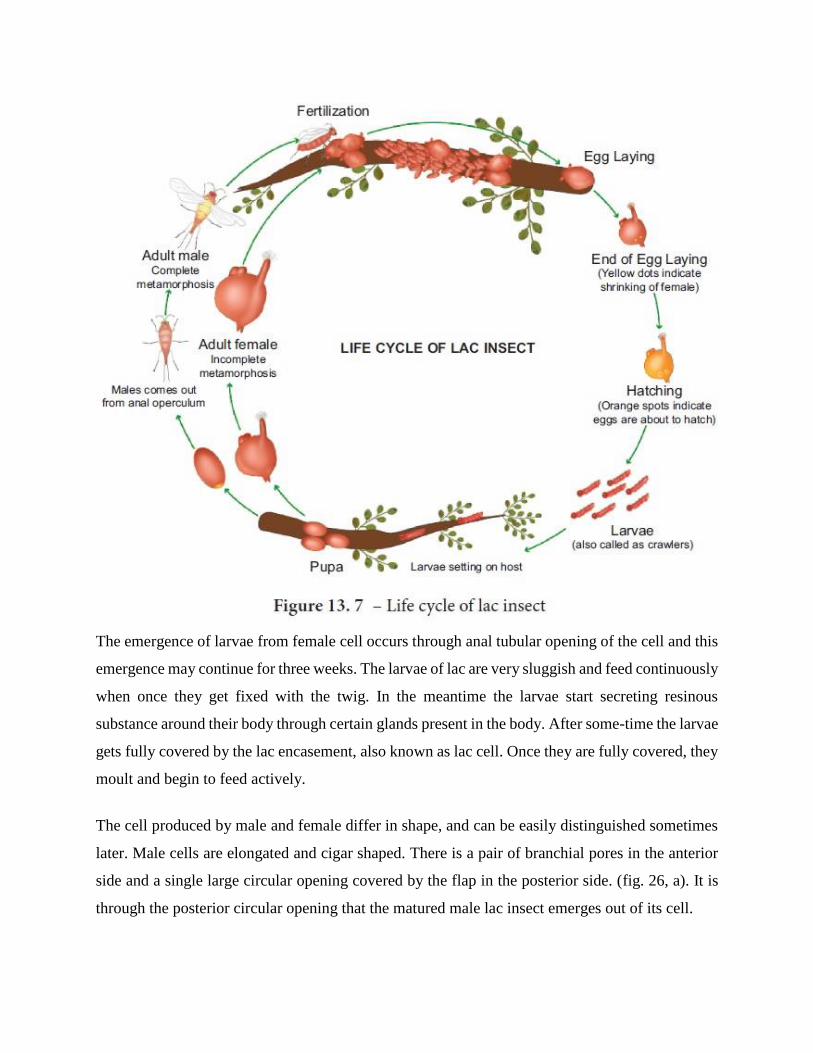
The emergence of larvae from female cell occurs through anal tubular opening of the cell and this
emergence may continue for three weeks. The larvae of lac are very sluggish and feed continuously
when once they get fixed with the twig. In the meantime the larvae start secreting resinous
substance around their body through certain glands present in the body. After some-time the larvae
gets fully covered by the lac encasement, also known as lac cell. Once they are fully covered, they
moult and begin to feed actively.
The cell produced by male and female differ in shape, and can be easily distinguished sometimes
later. Male cells are elongated and cigar shaped. There is a pair of branchial pores in the anterior
side and a single large circular opening covered by the flap in the posterior side. (fig. 26, a). It is
through the posterior circular opening that the matured male lac insect emerges out of its cell.
Female cell is oval, having a pair of small branchial pores in anterior side and a single round anal
tubular opening in posterior side. Through the anal tubular opening are protruding waxy white
filaments, secreted by the glands in the insects body, which is an indication that the insect inside
the cell is alive and is in healthy condition. These filaments also prevent the blocking of the pore
during excess secretion of lac.
Larvae moult in their respective cells. It is the second stage larva which undergoes pseudopupation
for a brief time, whereby it changes into adult stage. Now the male emerges out from its cell,
moves on lac incrustation and enters the female cell for fertilization. In this way the life cycle is
completed.
Lac Secretion:
Lac is a resinous substance secreted by certain glands present in the abdomen of the lac insects.
The secretion of lac begins immediately after the larval settlement on the new and tender shoots.
This secretion appears first as a shining layer which soon gets hardened after coming in contact
with air.
This makes a coating around the insect and the twig on which it is residing. As the secretion
continues the coating around one insect meet and fuses completely with the coating of another
insect. In this way a continuous or semi-contmuous incrustation of lac is formed on the tender
shoots.
Cultivation of Lac:
Cultivation of lac involves proper care of host plants, regular pruning of host plant, infection or
inoculation, crop-reaping, control of insect pests, and forecast of swarming, collection and
processing of lac.
The first and perhaps the most important prerequisite for cultivation of lac is the proper care of the
host plant. It is the host plants on which lac insects depend for their food, shelter and for completion
of their life cycle. There are two ways for the cultivation of host plants. One is that plants should
be allowed to grow in their natural way and the function of lac-culturist is only to protect and care
for the proper growth of plants.
Another way is that a particular piece of land is taken for the purpose and systematic plantation of
host plant is made there. Regular watch is necessary in this case by providing artificial manures,
irrigation facilities, ploughing and protecting the plants from cattle and human beings for which
the land should be fenced. The larvae of lac insects are inoculated on host plants only after the host
plants have reached a proper height.
The lac larvae feed on the cell sap by inserting their proboscis in the tender twigs. The proboscis
can only be inserted in the tender young off-shoots. For this before inoculation, prunning of lac
host plants is necessary. The branches less than an inch in diametre are selected for pruning.
Branches half inch of less in diametre should be cut from the very base of their origin. But the
branches more than half inch diametre should be cut at a distance of 1 ½ inch from the base.
Inoculation:
The method by which the lac insects are introduced to the new lac host plant is known as
inoculation. This may be of two types, namely “Natural infection” and “Artificial infection”. When
infection from one plant to other occurs by natural movements of insect, it is called natural
infection. This may be due to overcrowding of insect population and nonavailability of tender
shoots on a particular tree.
Artificial infection takes places through the agencies other than those of nature. Prior to about two
weeks of hatching, lac bearing sticks are cut to the size of six inches. They are called“Brood lac”.
Brood lacs are then kept for about two weeks in some cool place.
When the larvae start emerging from this brood lac, they are supposed to be ready for inoculation.
Strings со be used for tieding the brood lac with the host plant may be of different types in longitud
infection the brood lac is tied in close contact with host branches. In lateral infection the brood lac
is tied across the gaps between two branches. In interlaced method, brood lac is tied among the
branches of several new shoots.
Lac Crop:
The lac insects repeat its life cycle twice in a year. There are actually four lac crops since the lac
insects behave in two ways either they develop on Kusum plants or devlop on plants other than
Kusum. The lac which grows on Non-Kusum plants is called as “Ranjeem lac,” and which grows
on Kusum plant is called as “Kusumi lac. Four lac crops have been named after four Hindi months
in which they are cut from the tree. They are as follows:
Ranjeeni Crop:
(i) Katki:
Lac larvae are inoculated in June-July. Male insect emerges m August-September. Female give
rise to swarming larvae in October-November and the crop is reaped in Kartik (October and
November).
(ii) Baisakhi:
Larvae produced by Katki crop are inoculated in October-November, male insects emerges in
February-March, females give rise to swarming larvae m June-July, the crop is reaped in Baisakh
(April-May).
Kusumi Crop:
(i) Aghani:
Lac larvae are inoculated in June-July, male insect emerges m September, female give rise to
swarming larvae in January-February and crop is reaped in Aghan (December-January).
(ii) Jethoi:
The larvae produced by Aghani crop is inoculated in the month of January- February, male
emerges in March-April, female give rise to swarming larvae in June- July and the crop is reaped
in the month of Jeath (June-July).
The time of infection with swarming larvae, the time of emergence of male insects, the time of
reaping the crop, and the time of producing swarming larvae by female etc., are shown m tabular
form below
Scraping and Processing of lac:
Lac cut from the host plant is called as “stick lac”. Lac can be scraped from the twigs before or
after the emergence of larvae. If it is used for manufacturing before the emergence of larvae, the
type of lac produced is called as “Ari lac” and if it is used for manufacturing purpose after
swarming of larvae has occurred, the lac is said to be Phunki lac”.
The scraping of lac from twig is done by knife, after which they should not be exposed to sun. The
scraped lac is grinded in hard stone mills. The unnecessary materials are sorted out In order to
remove the finer particles of dirt and colour, this lac is washed repeatedly with cold water.
Now at this stage it is called as “Seed lac” and is exposed to sun for drying. Seed lac is now
subjected to the melting process. The melted lac is sieved through cloth and is given the final shape
by molding. The final form of lac is called “Shellac”. Colour or different chemicals may be mixed
during melting process for particular need.
Lac Enemies and Their Control:
A lac enemy imposes a challenge to the lac culturist, as they not only decreases the population of
lac insects, but also retard the production and quality of lac. Damage caused to lac insects may be
grouped under two heads, (a) damage caused by insects (b) damage caused by animals other than
insects. Insect enemies of lac crop may be predators and parasites.
The common parasites of lac insect are known as “Chalcid.” They are small, winged insects which
lay their eggs inside the lac coat either on the body of the lac insect or inside the body of the lac
insect. The larva which hatches from these eggs feed upon the lac insects, thereby causing
mortality of their host. Damage done by this parasite constitute about 5-10% of the total destruction
of the lac crop.
Damage done by the predators is of greater intensity (35% of the total destruction). The major
predators of lac insects are Eublemma amabilis (the white moth) and Holococera pulverea (the
blackish grey moth). They not only feed on lac insects but also destroy the lac produced by term.
Squirrels, monkey, rat, bat, birds (wood peckers), man etc., are the enemies other than insects
which destruct the lac crop in different ways. Damage is also done by climatic factors such as
excess heat, excess cold, heavy rain, and storm and partly by the faulty cultivation methods.
Control:
Damage caused by the above mentioned animals can be reduced to certain extent by the use of the
following methods.
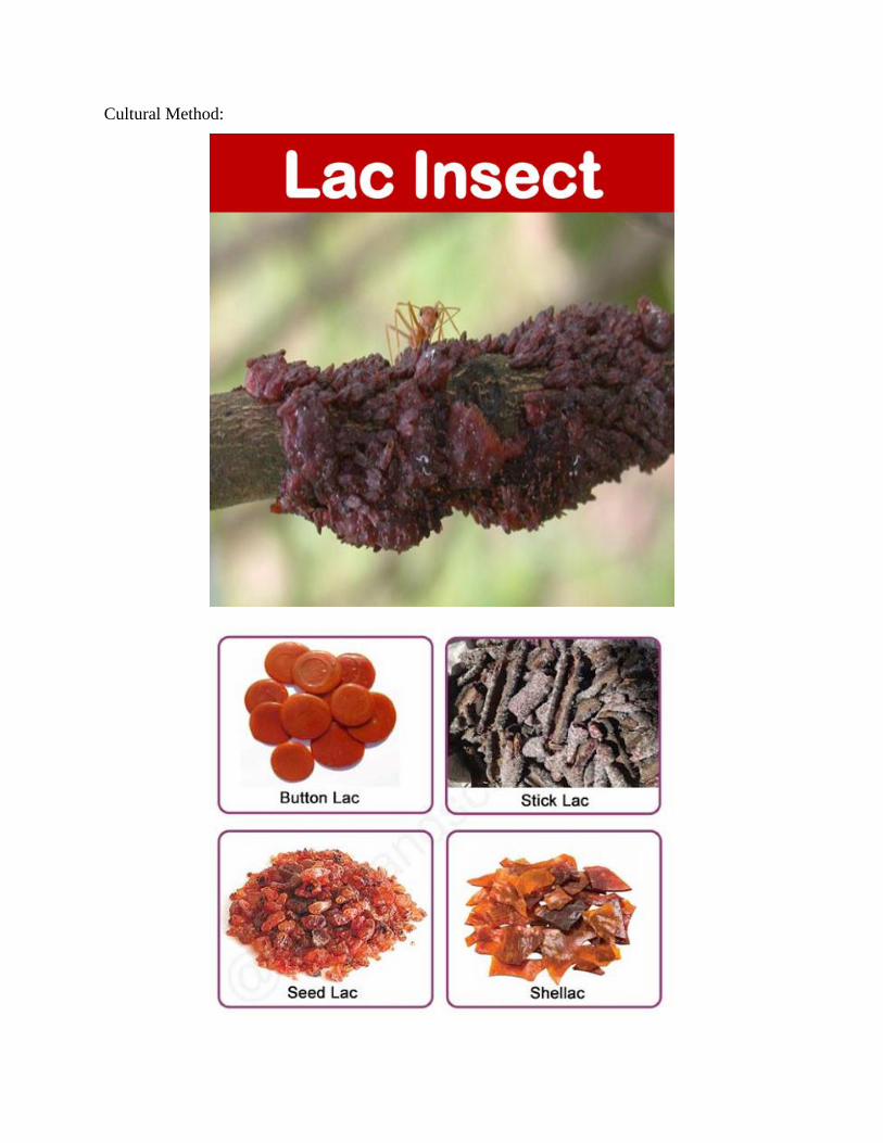
Cultural Method:

The amount of damage by infection can be reduced to a greater extent by taking care during the
culture of lac insects, especially at the time of inoculation. The brood lac showing the minimum
enemy attack should be selected for inoculation and should be cut from the host plant very near to
the time of emergence of larvae (about one week before the emergence). This will reduce the
chances of parasite attack on the emerging larvae at new place (host).
The brood lac used for inoculation should be removed from the new host’s branches as soon as the
emergence of larvae stops (approx. 3 weeks after inoculation). It reduces the chance of transference
of enemies to the new host plant from the brood lac. The infected brood lac not fit for inoculation
or the used up brood lac should not be retained for long. The lac should be scrapped at once and t
e rest may be crushed or dropped into fire in order to destroy the predators and parasites.
The delay m processing also gives chances to the enemy insects to escape into field. So the
manufacturers should try to convert stick lac into seed lac as soon as possible. By these cultural
methods the future production can be saved from infection to some extent.
Artificial Method:
During the crop reaping, it is not always possible for the manufacturers to convert the huge amount
of stick lac to seed lac at a time. To avoid the spreading of enemies at this time from stocked stick
lac simple artificial method can be used. Bundles of stick lac should be tied with stones and
immersed in fenced water (river or ponds) for about a week. This kills all the parasitic and predator
insects as they cannot survive in water.
Biological Method:
It is an indirect method for killing the parasitic and predator insects. For this purpose hyper-
parasitic insects are used which attacks the parasitic insects of lac and kill them. These hyper-
parasitic insects are however, not harmful for lac crop.
Use of Lac:
Lac has been used for the welfare of human beings from the great olden days No doubt the
development of many synthetic products have made its importance to a little lesser degree, but still
it can be included in the list of necessary articles. Lac is used in making toys, bracelets, sealing
wax, gramophone records etc.
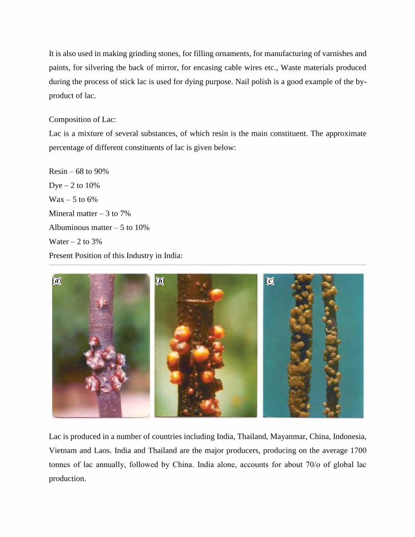
It is also used in making grinding stones, for filling ornaments, for manufacturing of varnishes and
paints, for silvering the back of mirror, for encasing cable wires etc., Waste materials produced
during the process of stick lac is used for dying purpose. Nail polish is a good example of the by-
product of lac.
Composition of Lac:
Lac is a mixture of several substances, of which resin is the main constituent. The approximate
percentage of different constituents of lac is given below:
Resin – 68 to 90%
Dye – 2 to 10%
Wax – 5 to 6%
Mineral matter – 3 to 7%
Albuminous matter – 5 to 10%
Water – 2 to 3%
Present Position of this Industry in India:
Lac is produced in a number of countries including India, Thailand, Mayanmar, China, Indonesia,
Vietnam and Laos. India and Thailand are the major producers, producing on the average 1700
tonnes of lac annually, followed by China. India alone, accounts for about 70/о of global lac
production.
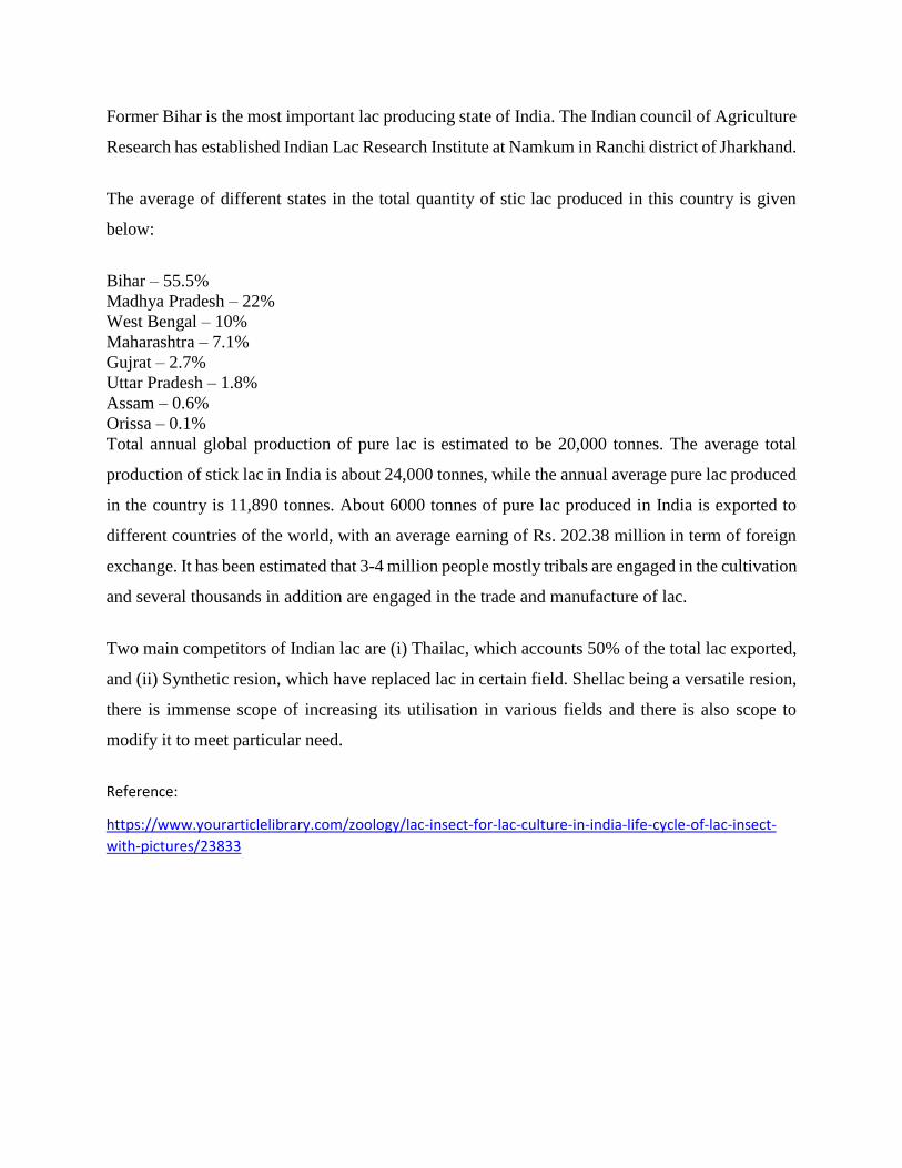
Former Bihar is the most important lac producing state of India. The Indian council of Agriculture
Research has established Indian Lac Research Institute at Namkum in Ranchi district of Jharkhand.
The average of different states in the total quantity of stic lac produced in this country is given
below:
Bihar – 55.5%
Madhya Pradesh – 22%
West Bengal – 10%
Maharashtra – 7.1%
Gujrat – 2.7%
Uttar Pradesh – 1.8%
Assam – 0.6%
Orissa – 0.1%
Total annual global production of pure lac is estimated to be 20,000 tonnes. The average total
production of stick lac in India is about 24,000 tonnes, while the annual average pure lac produced
in the country is 11,890 tonnes. About 6000 tonnes of pure lac produced in India is exported to
different countries of the world, with an average earning of Rs. 202.38 million in term of foreign
exchange. It has been estimated that 3-4 million people mostly tribals are engaged in the cultivation
and several thousands in addition are engaged in the trade and manufacture of lac.
Two main competitors of Indian lac are (i) Thailac, which accounts 50% of the total lac exported,
and (ii) Synthetic resion, which have replaced lac in certain field. Shellac being a versatile resion,
there is immense scope of increasing its utilisation in various fields and there is also scope to
modify it to meet particular need.
Reference:
https://www.yourarticlelibrary.com/zoology/lac-insect-for-lac-culture-in-india-life-cycle-of-lac-insect-
with-pictures/23833
UNIT – V
PART-A
Q.NO QUESTIONS CO (LEVEL)
1 List the infectious diseases caused by animals 5 (1)
2 Summarize the epidemiology of Tuberculosis 5 (2)
3 Investigate the epidemiology of Dengue 5 (5)
4 Investigate the epidemiology of Malaria 5 (5)
5 Investigate the epidemiology of Swine flu 5 (5)
6 Assess the current status of Integrated Pest Management 5 (5)
7 Review on the genetic improvements in aquaculture 5 (5)
8 Design an apiculture setup for your start up business 5 (6)
9 Design a sericulture model for your future business 5 (6)
10 Develop a Lac culture for doing business. 5 (1)
PART-B
Q.NO QUESTIONS CO (LEVEL)
1 Categorizethe infectious agents causing diseases to humans and
explain how to control it 5 (6)
2 Explain the epidemiology of Tuberculosis 5 (2)
3 Write a review on Dengue fever 5 (5)
4 Discuss the epidemiology of Malaria 5 (1)
5 Discuss the epidemiology of Swine flu 5 (1)
6 Investigate how best agricultural productivity can be improved through Integrated Pest Management
5 (5)
7 Judge the effect of Genetic improvements in aquaculture industry 5 (5)
8 Apiculture will improve the crop yield – Justify. 5 (5)
9 Review in present status of sericulture 5 (5)
10 Assess the different methods followed in Lac culture. 5 (5)