Tuberous sclerosis clinical factors in long term outcome
316
University of Bath DOCTOR OF MEDICINE Tuberous sclerosis clinical factors in long term outcome Hancock, Eleanor Award date: 2003 Awarding institution: University of Bath Link to publication Alternative formats If you require this document in an alternative format, please contact: [email protected] Copyright of this thesis rests with the author. Access is subject to the above licence, if given. If no licence is specified above, original content in this thesis is licensed under the terms of the Creative Commons Attribution-NonCommercial 4.0 International (CC BY-NC-ND 4.0) Licence (https://creativecommons.org/licenses/by-nc-nd/4.0/). Any third-party copyright material present remains the property of its respective owner(s) and is licensed under its existing terms. Take down policy If you consider content within Bath's Research Portal to be in breach of UK law, please contact: [email protected] with the details. Your claim will be investigated and, where appropriate, the item will be removed from public view as soon as possible. Download date: 26. Aug. 2022
-
Upload
khangminh22 -
Category
Documents
-
view
2 -
download
0
Transcript of Tuberous sclerosis clinical factors in long term outcome

University of Bath
DOCTOR OF MEDICINE
Tuberous sclerosis clinical factors in long term outcome
Hancock, Eleanor
Award date:2003
Awarding institution:University of Bath
Link to publication
Alternative formatsIf you require this document in an alternative format, please contact:[email protected]
Copyright of this thesis rests with the author. Access is subject to the above licence, if given. If no licence is specified above,original content in this thesis is licensed under the terms of the Creative Commons Attribution-NonCommercial 4.0International (CC BY-NC-ND 4.0) Licence (https://creativecommons.org/licenses/by-nc-nd/4.0/). Any third-party copyrightmaterial present remains the property of its respective owner(s) and is licensed under its existing terms.
Take down policyIf you consider content within Bath's Research Portal to be in breach of UK law, please contact: [email protected] with the details.Your claim will be investigated and, where appropriate, the item will be removed from public view as soon as possible.
Download date: 26. Aug. 2022

TUBEROUS SCLEROSIS
CLINICAL FACTORS IN LONG TERM OUTCOME
Submitted by Dr Eleanor Hancock
For the degree of MD of the University of Bath 2003
COPYRIGHT
Attention is drawn to the fact that copyright of this thesis rests with its author. This
copy of the thesis has been supplied on condition that anyone who consults it is
understood to recognise that its copyright rests with the author and that no quotation
from the thesis and no information derived from it may be published without prior
consent of the author.
This thesis may be made available for consultation within the University Library and
may be photocopied or lent to other libraries for the purpose of consultation.

UMI Number: U163210
All rights reserved
INFORMATION TO ALL USERS The quality of this reproduction is dependent upon the quality of the copy submitted.
In the unlikely event that the author did not send a complete manuscript and there are missing pages, these will be noted. Also, if material had to be removed,
a note will indicate the deletion.
Dissertation Publishing
UMI U163210Published by ProQuest LLC 2014. Copyright in the Dissertation held by the Author.
Microform Edition © ProQuest LLC.All rights reserved. This work is protected against
unauthorized copying under Title 17, United States Code.
ProQuest LLC 789 East Eisenhower Parkway
P.O. Box 1346 Ann Arbor, Ml 48106-1346


SUMMARY
Tuberous sclerosis (TSC) is a dominantly inherited disorder with a high spontaneous
mutation rate and a birth incidence of 1 in 10,000. It is a systemic disorder characterised
by the growth of hamartomas, which in turn give rise to the clinical manifestations, for
example, epilepsy and learning difficulties. Although some patients with TSC are only
mildly affected and lead a normal life with typical life expectancy, there is an immense
amount of morbidity associated with this disease. In addition, for the majority of patients
there is reduced life expectancy. The purpose of this thesis is to look at the clinical
factors that contribute to the morbidity and mortality seen in TSC and examine means of
reducing the impact of these factors on long-term outcome. It reports a longitudinal
population study of a small but defined group of patients looking at the epidemiology and
natural history (the morbidity and mortality) suffered by in this population. It investigates
the current treatment regimes for the types of epilepsy (infantile spasms and non-
convulsive status epilepticus) known to be associated with the highest risk of learning
difficulties in order to determine the most efficacious treatment for the seizures
potentially reducing long-term psychomotor delay. A cochrane review of the treatment of
infantile spasms was performed. This thesis also examines the effect of exogenous
melatonin on sleep disorders (one of the major causes of morbidity) in tuberous sclerosis
and the natural circadian rhythms in patients with sleep disorder in TSC and compares
them with the normal population. Two important causes of premature mortality in TSC
patients are respiratory and renal failure. This thesis examines the prevalence and
underlying causes of end stage renal failure in adults with TSC and reviews the literature
of LAM (in patients both with and without TSC) investigating further the natural history
and treatment of LAM in TSC.

TUBEROUS SCLEROSIS
CLINICAL FACTORS IN LONG TERM OUTCOME
INDEX
Statement of Authors Contribution -
Acknowledgements
Chapter One -
Chapter Two -
IntroductionDiagnostic criteria for tuberous sclerosis The genetics of tuberous sclerosis The prevalence of tuberous sclerosis Clinical manifestations of tuberous sclerosis EpilepsyMorbidity and mortality in tuberous sclerosis Aims of thesis
A ten year review of the epidemiology, morbidity and mortality of the tuberous sclerosis population in the Bath Health District
Page 3
Page 5
11
Page Page Page Page 12 Page 13 Page 22 Page 26 Page 29
Page 31
Chapter Three Epilepsy and Learning Difficulties Page 50Infantile spasms: recognition, treatment Page 50and prognosis.The treatment of infantile spasms with high dose Page 66prednisolone: a retrospective review.A review of vigabatrin in the treatment of infantile Page 72spasms in tuberous sclerosis.
Chapter Four
Chapter Five ■
The United Kingdom Infantile Spasm Study. Page 78Report on study status Page 88
The Cochrane collaborative: A systematic review Page 89comparing the medical treatments of infantile spasms (West Syndrome) in terms of long term developmental outcome, seizure control and side effects.
Chapter Six
Chapter Seven -
Oral treatment of non-convulsive status epilepticus in tuberous sclerosis.
Lymphangioleiomyomatosis and tuberous Sclerosis.Patients ascertainment.Literature search.
1
Page 120
Page 125
Page 126 Page 139

Discussion. Page 143
Chapter Eight -
Chapter Nine -
Renal disease. PageEnd stage renal failure in adults with the tuberous Page sclerosis complex
Sleep disorder in the tuberous sclerosis complex. The effect of Melatonin dosage on the sleep disorder in tuberous sclerosis complex. Melatonin excretion in normal children and tuberous sclerosis with sleep disorder responsive to melatonin.
Conclusions.Chapter Ten -
References
Glossary
Abbreviations -
Tables -
Figures -
Appendices - Appendix one - UKISS trial pack
Appendix two - Core group
Appendix three - Additional references for Cochrane review
Appendix four - Additional references for literature review of LAM
Appendix five - Sleep and seizure diaries
Publications and presentations
PagePage
158158
167171
Page 177
Page 184
Page 194
Page 201
Page 206
Page 207
Page 210
See suppliment
Page 211
Page 212
Page 215
Page 225
Page 250
2

Statement of Authors Contribution
This thesis contains a combination of research techniques including the collection of
epidemiology data, systematic reviews and original research.
Epidemiological data collection
Chapter two looked at the epidemiology, mortality and morbidity of the tuberous sclerosis
(TSC) population in the Bath Health District. It involved the retrieval and examination of
the relevant medical records and reports, scrutiny of the Bath TSC data base and
examination of the individual patients when they attended clinic or were visited for other
research projects. It was undertaken solely by the author (ECH).
Systematic reviews
There are several systematic reviews included in this thesis.
The first review undertaken by the author was looking at the use of steroids and ACTH in
the treatment of infantile spasms. This was a simple review using a single database and
only considering studies published in English, the second review looking at vigabatrin in
the treatment of infantile spasms in patients with TSC used two databases but again only
considered studies published in English. A third review searched the English literature for
all cases of lymphangioleiomyomatosis again used two data bases. All three of these
reviews were performed solely by ECH.
A fourth review was the cochrane review looking at the treatment of infantile spasms. This
involved searching several databases, in all languages, corresponding with colleagues,
authors and drug companies for which ECH was fully responsible and appeals at
international conferences for which JPO was responsible. ECH was also responsible for
developing the protocol design, the database of the studies, data collection and extraction,
data analysis and presentation of the results. JPO and PM had joint responsibility for the
protocol, data extraction and agreement of results.
Original research
The following pieces of original research have been included in this thesis;
1. The treatment of infantile spasms with high dose oral prednisolone. ECH was
responsible for developing the study design, collecting eligible patients, retrieving
3

and reviewing the medical notes, extracting the relevant data, data analysis and
presentation of the results.
2. The United Kingdom Infantile Spasms Study. This is a multicenter randomised trial
looking at the treatment of infantile spasms. ECH was responsible, under JPO’s
supervision for developing the triai design, protocol and trial packs which were put
to the trial steering committee for comment and approval. ECH remains closely
involved with the trial as a member of the trial steering committee. Management of
data, developmental assessment and analysis are the responsibility of another
research fellow of Prof. Osborne, Dr Andrew Lux.
3. Oral treatment of non-convulsive status epilepticus in tuberous sclerosis. ECH was
responsible for developing the study design, collecting eligible patients, retrieving
and reviewing the medical notes, extracting the relevant data, data analysis and
presentation of the results.
4. Lymphangioleiomyomatosis and tuberous sclerosis: patient ascertainment. ECH
was responsible for developing the study design, recruiting patients, retrieving and
reviewing the medical notes and examining the patients (with the exception of three
patients (who were seen by Sue Thompson) extracting the relevant data, data
analysis and presentation of the results.
5. End stage renal failure in adults with the tuberous sclerosis complex. ECH was
responsible for reviewing the results and writing up the data for this study. The
study design, data collection and initial assessment were done by Dr Antonia
Clarke.
6. The effect of melatonin dosage on the sleep disorder in tuberous sclerosis. ECH
was responsible for developing the study design, including the diaries for data
collection, recruiting patients, data analysis and presentation of the results.
7. Melatonin excretion in normal children and tuberous sclerosis with sleep disorder
responsive to melatonin. ECH was responsible for developing the study design,
including data collection forms, recruiting eligible patients and controls, data
analysis and presentation of the results.
4

Acknowledgements
I would like to thank all the following people for their valued contributions, which have
made this research possible:
To all the patients with tuberous sclerosis who participated in the research projects without
whom this thesis could never have been started.
Chapter Two
Professor John Osborne and Mrs Julia Legh-Smith for allowing me to use their data-base
on the patients with Tuberous Sclerosis in the Bath region.
Chapter Four - The United Kingdom Infantile Spasm Study
Professor John Osborne, Dr Richard Appleton, Dr Colin Kennedy, Dr Richard Newton, Dr
Finbar O’Callaghan, Dr Christopher Verity and Dr Lisa Vickers who formed the core
group which was responsible for approving the design of the trial protocol and for
obtaining funding. Formal statistical input into the study design was given by, Dr Anthony
Johnson.
Professor John Osborne, Dr Stuart Edwards, Dr Anthony Johnson, Dr Colin Kennedy, Dr
Andrew Lux, Dr Richard Newton, Dr Finbar O’Callaghan, and Dr Christopher Verity who
formed the Trial Steering Committee which was responsible for approving the design of
the trial packs, recruiting centres to participate in the trial and obtaining ethical approval.
Chapter Five - Cochrane Review on the Treatment of Infantile Spasms
Professor John Osborne and Professor Phillip Milner who agreed and approved the review
at all stages and independently evaluated which studies should be included and excluded
from the review and also independently extracted data from the included studies.
Dr Tony Marson and the Cochrane Group for all their advice.
Chapter Six - Non-Convulsive Status Epilepticus
Professor Frank Besag for his idea of trying oral diazepam to terminate non-convulsive
status in patients with Tuberous Sclerosis.
5
Chapter Seven - Lvmphangioleiomatosis
Professor John Osborne, Professor Julian Sampson, Dr Sue Tompkins, Dr Christopher
Kingswood and the Tuberous Sclerosis Association who helped to recruit patients.
Dr Sue Tompkins for interviewing and collecting data on three of the patients.
Professor John Osborne, Professor Julian Sampson and Dr Sue Tompkins who reviewed
the writing up of the patient data.
Dr Noleen Foley for her advice.
Chapter Eight - Renal Disease
Dr Antonia Clarke, Dr Christopher Kingswood and Prof. John Osborne who designed the
questionnaire, identified the members of the European Dialysis and Transplant
Association, distributed and collected the questionnaires.
Chapter Nine - Sleep Disorder in Tuberous Sclerosis
Professor John Osborne and Dr Finbar O’Callaghan for helping to design and recruit
patients into the studies. The Tuberous Sclerosis Association for helping to recruit patients
into the study. Dr Judie English for performing the biochemical analysis on our samples.
Financial Support
The Bath Unit for Research into Paediatrics, Cow & Gate and the Tuberous Sclerosis
Association who between them funded my salary and support costs.
6

CHAPTER ONE
TUBEROUS SCLEROSIS
Introduction
Tuberous sclerosis (TSC) is a dominantly inherited disorder with a high spontaneous
mutation rate and a birth incidence that may be as high as 1 in 10,000\ It is now known to
be a systemic disorder characterised by the growth of benign tumours (hamartomas), which
may arise in almost any organ of the body with the possible exceptions of striated muscle,
the peripheral nerves, the meninges and the thymus gland. Although TSC is an inherited
disorder it may not always manifest in childhood and clinical features may develop at any
age. The severity of the disease varies considerably, not only between individuals but also
within families. This makes genetic counselling particularly difficult as a mildly affected
parent with normal intellect is at risk of having an affected child who may either be mildly
affected or have severe learning difficulties, extreme behavioural problems and refractory
epilepsy.
Over the past century and a half our knowledge of the clinical manifestations and the
natural history of this condition has increased substantially. Whereas TSC was thought to
be exceptionally rare, causing severe handicap and early demise, it is now known to be
relatively common, affecting patients in a wide variety of ways, although it is still
associated with a high incidence of morbidity and mortality. It is, therefore, imperative that
clinicians continue to improve their understanding and treatment of this condition, not only
to maximise longevity but also to allow individuals to achieve their maximum potential.
The very first reference to TSC is probably the illustration drawn by Pierre Rayer in 1835
representing the facial angiofibroma (see figure one) of TSC. Von Recklinghausen gave
the first description of TSC in 1862 when he described sclerotic areas in the brain and
cardiac rhabdomyomas in a stillborn child, who he thought had neurofibromatosis. In 1880
Boumeville named the condition after accurately describing the potato-like (tuberous) hard
(sclerosis) cerebral lesions pathognomonic of the disease. Over the following decade
dermatologists such as Balzer, Menetrier and Pringle made the association between the
7

facial rash and mental retardation in individuals found to have the cerebral lesions o f TSC
at post-mortem.
■ Figure one -
At the beginning of the 20th century pathologists had started to recognise other lesions
associated with these tubers, in the heart, kidneys and skin and Vogt described the clinical
triad o f angiofibroma, severe epilepsy and mental retardation as a means o f recognising
TSC clinically in patients whilst alive; previously the diagnosis had only been able to be
made posthumously. Unfortunately, this gave rise to the misconception that all three
features had to be present for the diagnosis to be made, which not only led to under
recognition o f this disease but failure to recognise (and therefore to treat) the other
complications o f TSC. However, over the latter half o f the past century it has gradually
become apparent that the disease is more complex than that originally recognised and can
affect almost any part o f the body.
In 1920 Berg noted the hereditary nature of TSC and it was soon realised that some
relatives had the skin lesions without the mental handicap and over the following decades,
other manifestations o f TSC have been documented. However it was not until Gomez’s
study in 1967 that it became clear that as many as half o f affected individuals had normal
intelligence and only one third had the complete Vogt’s triad. This led Gomez2 to publish a
set o f criteria in 1979, updated by Osborne in 19883, on which the disease could be
recognised. These diagnostic criteria were divided into three groups.

Diagnostic criteria for the diagnosis of TSC2,3
Primary criteriaThese are diagnostic of TSC even in the absence of any other stigmata of TSC1. Angiofibroma2. Retinal phakoma3. Multiple subependymal nodules4. Multiple cortical tubers5. Forehead fibrous plaque6. Shagreen patch
Diagnostic criteria with a first degree relative1. Subependymal giant cell astrocytoma2. Cardiac rhabdomyoma3. Single cortical tuber4. Single retinal phakoma
Secondary diagnostic criteriaThese can occur in unaffected individuals but a combination of two or more implies a diagnosis of TSC because of the low risk of more than one occurring in an unaffected individual.1. Hypomelanic patches2. Bilateral polycystic kidneys3. Lymphangioleiomyomatosis of the lung4. Cardiac rhabdomyoma5. Renal angiomyolipoma
In 1998 the clinical diagnostic criteria were revised once more by the Tuberous Sclerosis
Complex Consensus4, yet again reflecting the increasing knowledge and understanding of
this disorder. It had become apparent that some features previously thought to be
pathognomonic of the disease, for example cortical tubers could occur in patients without
TSC, as could renal angiomyolipoma and lymphangioleiomyomatosis. It was also known
that when these individuals had no other signs of TSC, they did not have children who
inherited the disorder. Studies of families with a known gene mutation also showed that
where an individual had a disease causing mutation, they always had convincing signs of
the disorder and that those with no signs were always unaffected. Penetrance was
therefore high. The only reclassification in UK families following more traditional
analysis involved individuals with one possible hamartoma - usually white patches or a
single ungula fibroma. Improving the criteria required for diagnosis, would also highlight
the potential complications of the disease to the clinicians at the time of diagnosis,
hopefully improving long-term follow up and reducing morbidity and mortality from the
disease. It was therefore decided;
“A definite diagnosis of TSC should be reserved for patients with two or more distinct types of hamartomas rather than multiple lesions of the same type in the same organ system” 4_____________________________
9

For the purpose of this definition renal angiomyolipoma and pulmonary
lymphangioleiomyomatosis are considered as one hamartoma since some individuals with
these two hamartomas but without other signs of TSC do not pass TSC onto their children.
It is likely that the clinical criteria for TSC will become less important with the discovery
of two genes for TSC: it is probable that diagnosis by genetic testing will be available for
the vast majority of individuals in the near future: it is already possible for up to 70% of
individuals in the best laboratories.
The diagnosis of TSC was also made easier (and is often made at an earlier age) as the
result of the introduction of new imaging techniques particularly computerised
tomography (CT) (1973) and magnetic resonance imaging (MRI) (1980’s). Before such
techniques were available TSC could often only be recognised clinically if the patient
developed the skin lesions that are pathognomonic of TSC, for example angiofibroma and
shagreen patches, or became symptomatic from the hamartomas, for example developed
seizures, haematuria, or pneumothoracies. With the advent of CT, MRI etc it has become
apparent that hamartomas may be present in many organs e.g. brain and kidney without
giving rise to symptoms. For example, it is now known that the vast majority of patients
with TSC have subependymal calcifications (within the brain) that are easily recognisable
on CT scan, are present from birth (but may only calcify in the first year of life) and rarely
occur in the absence of TSC. Before CT scanning was available, if an affected individual
had a child, or if for example a child developed infantile spasms, only a skull X-ray or air
encephalogram could be undertaken to look for intracranial lesions. It is also possible to
perform echocardiography to look for rhabdomyomas: a positive finding would help
confirm the diagnosis but a negative finding would not help exclude the diagnosis. Only
30 -50% of TSC patients have rhabdomyomas at birth and they often regress over the first
few months of life. Likewise ophthalmology confirmation of a retinal phakoma would
help substantiate the diagnosis, but they are not present in all patients. Nowadays a CT
brain scan can be undertaken; if subependymal nodules are present it is highly probable
that the child has TSC. Unfortunately, even with these more advanced imaging techniques,
it is not always possible to make a definitive diagnosis of TSC. Other conditions such as
neurofibromatosis type one and some congenital infections can give rise to calcification
on imaging and grey matter heterotopia (including an autosomal dominant variant
associated with epilepsy) can give an appearance similar to the non-calcified lesions seen
in TSC.
10

Similarly, many patients with TSC have cysts and/or angiomyolipomas in their kidneys
but only some have symptoms such as pain or bleeding. Ultrasound scan is a non-invasive
but useful tool for diagnosing these lesions and in combination with other scanning
techniques may help to make a diagnosis in a patient who has few clinical manifestations
of TSC, but it can mislead: “AML” may not be found on further investigation. Renal cysts
can also be found in normal individuals, it is estimated that approximately 1 in 40 people
at 40 years of age will have one or more renal cysts.
Note: the majority of the cutaneous manifestations of TSC are not present at birth and may not develop until adulthood or rarely may never develop at all.
Genetics
Berg first noted the hereditary nature of TSC in 1920 and it was then recognised to be an
autosomal disorder by Gunther and Penrose in 1935. However, the condition has a high
spontaneous mutation rate accounting for about 60% of affected individuals5. A gene
locus was discovered on chromosome 9q34 (TSC1) by Fryer6 in 1987, but it soon became
apparent that a second gene existed. This was located, by linkage studies, on chromosome
16pl3 (TSC2) in 1992 by Kandt7, and was cloned a year later by Nellist8 who found that
the TSC2 gene lies close to the PKD1 gene. The TSC1 gene was cloned in 1998 by
Young et al9. The exact functions of the two gene products, hamartin (TSC1) and tuberin
(TSC2) are still being researched but both are thought to act as tumour suppressor genes.
A number of tumour suppressor genes are now recognised and molecular studies have shown them to play an important role in the regulation of the normal cell cycle. For a tumour suppressor gene to exert a pathological influence on a cell, both alleles must be inactivated; the Knudsen10 two hit theory explains why inheriting a defective copy of a tumour suppressor gene leads to an individual with multiple tumours. Normally for a tumour to arise (in an individual without an inherited or early embryonic defect), two independent somatic mutations are required, explaining why sporadic tumours can occasionally arise in a single tissue later in life. But, individuals who have already inherited an inactivated tumour suppressor gene only need one further somatic mutation to lose both alleles explaining why in TSC the tumours often occur earlier (as early as in utero) and tend to be multiple. This may also explain why clinical expression is extremely variable even within families, if chance affects the number and timing of second hits. The hypothesis that TSC genes were tumour suppressor genes was developed following the demonstration of loss of part of the normal gene in the hamartomas of individuals with TSC - a phenomenon known as loss of heterozygosity.
The TSC1 gene is 8.6kb in length and codes for a protein of at least 1164 amino acids
called hamartin. The TSC2 gene is larger in size, 45kb in length and codes for a similar
sized protein, tuberin, 1807 amino acids in length. Both proteins have regions similar in
structure to the GTPase-activating proteins (GAP) human GAP3 and murine GAP. GTP is
an energy source required for normal cell growth. GTPase-activating proteins break down
GTP to GDP, an inactive form, thus inhibiting normal cell growth. It is therefore
11

postulated that hamartin and tuberin have a comparable effect to GAP proteins i.e. act as a
growth suppressor gene. Consequently, if either hamartin or tuberin are deficient or do not
function correctly there is uninhibited cell growth thus giving rise to the hamartomas seen
in TSC.
Defects in either gene produce a remarkably similar phenotype, so that at present it is not
possible to clinically detect which gene is affected in any affected individual, except in the
rare cases where there is also early onset polycystic kidney disease (PKD) due to a deletion
on chromosome 16 affecting both the TSC2 and the PKD gene7. However, there have been
recent suggestions that TSC2 mutations may be seen more commonly in sporadic cases of
TSC and may be associated with more severe handicap than TSC1 mutations, but further
research is required in order confirm this hypothesis.
The rapidly increasing knowledge and understanding of molecular genetics may be
beneficial in aiding diagnosis in this group of patients, but it also introduces new
dilemmas. Pre-natal diagnosis is now available for families where a disease causing
mutation has been identified but there is no reason to believe that the test will be able to
predict the severity of affected individuals. This can lead to increased anxiety when
choosing whether or not to opt for pre-natal testing and subsequent termination of an
affected foetus. If the family opt for pre-natal testing, the mutation must be identified
before the pregnancy and counselling given with preferably both partners together. Pre
natal testing can also be considered in subsequent pregnancies in individuals with apparent
new mutations because of the risk of gonadal mosaicism even though the recurrence risk is
less than about 2%. There is also then the dilemma of whether and when to test apparently
unaffected individuals within a family. If found to be affected, not only can this be a
constant source of worry in itself but it can also have financial implications, making it
difficult, for example to acquire life insurance or a mortgage. On the other hand the
individual may not want to risk having an affected child, and may opt to be tested as late in
adult life as possible.
Prevalence
Earliest studies (Critchley and Earl11 1935 and Gunther and Penrose12 1935) investigating
the prevalence and morbidity in TSC concentrated on small samples of institutionalised
patients estimating TSC to be a rare disorder affecting 1 in 30 mentally handicapped
12

patients. Gunther and Penrose12 also estimated 1 in 1,000 of the population to be
handicapped giving a prevalence of TSC around 1 in 30,000. By the 90’s, it was well
known that TSC could present in a multitude of guises and not all sufferers had moderate
or severe learning difficulties. Consequently several population-based prevalence studies
were performed in the attempt to provide more accurate information. Unfortunately four of
the studies involved only small numbers of patients: Wiederholt et al13 1985 gave a
prevalence of 1 in 10,000 based on only eight patients; Umpathy14 et al 1989 gave a
prevalence of 1 in 34,000 based on 14 patients, Shepherd15 et al 1991 a prevalence of 1 in
13,000 based on 12 patients and Ahlsen16 et al 1994 a prevalence of 1 in 13,000 based on
32 patients.
The first epidemiological study to look at prevalence in different age groups was
performed by Hunt and Lindenbaum17 in 1984 reporting on 68 patients in the Oxford
region giving an overall estimated prevalence of 1 in 30,000. They found decreasing
prevalence with increasing age; 1:10,000 at birth, 1:15.000 under 5 years and 1:20,000
under 30 years of age. A further study carried out by Sampson et al in Scotland 198918
looked at 101 patients giving a similar overall prevalence of 1 in 27,000. In 1996 the
largest population based study to date was published by Webb19 et al totalling 131 known
affected individuals from a population of 3,400,000 people, most of whom were examined
for the review, unlike previous studies. They estimated a birth incidence of 1 in 10,000.
They found only 40% of their patients to have learning difficulties giving a prevalence of 1
in 40,000 for TSC and severe learning difficulties, which is similar to that found by
Gunther and Penrose12.
Clinical manifestations
Cutaneous manifestations
1. Facial angiofibroma (figure one, page eight) affecting the cheeks, nasal folds and chin
but often sparing the upper lip rarely occur before 2 years, but most often become
apparent before adolescence and progressively increase in number and prominence.
They are reported to occur in about 90% of patients with TSC. They result from
proliferation of the normal dermal tissue with a vascular component giving rise to
telangectatic nodules, while more fibrous lesions appear skin coloured. Either type
may become pedunculated20 21.
13

2. Shagreen patches (figure two) are usually found in the lumber region of the trunk and
are a roughened raised area o f skin, often slightly pigmented with a leathery
consistency. They occur in 20-80%21 of patients and vary considerably in size from a
few inches to as much as a foot in diameter. They have an irregular edge. Some
patients wili have a number of smaller, so called “satellite” lesions either in isolation
or surrounding a shagreen patch.
Figure two - Shagreen patch
3. Hypomelanic macules (figure three) are best seen with an ultraviolet light (W ood’s
light), are the commonest and often earliest skin sign of TSC, but are not unique to
TSC and may occur in healthy individuals3 20. As a general rule the greater the number
o f hypopigmented patches the higher the risk o f TSC, and the USA consensus
conference4 felt three was a significant number. Depigmentation may also affect the
hair (known as poliosis - figure four) and iris, or occasionally causes a confetti type
pattern (figure five) usually seen on the proximal limbs. Pathologically, there is an
absence o f melanosomes within a normal number o f melanocytes (unlike vitiligo). It is
not clear that these lesions are hamartomas.
Figure three - Hypomelanic macule
14

F ig u re fo u r - Poliosis
F igure five - confetti dep igm enta tion
4. Forehead fibrous plaques (figure six) are raised reddish areas o f skin occurring on the
forehead or scalp and occasionally the face. Unlike the shagreen patch, which has a
roughened texture, these tend to have a smooth surface3 21. They may be the first skin
signs o f TSC. Histologically they are angiofibromatous.
5. Fibromas (figure seven) are flesh-coloured nodules commonly occurring in up to 70%
of patients and may occur around the nails, more commonly the feet than the hands
(ungual fibromas) or gums. They are a result o f over proliferation o f the dermal'X 0 1
connective tissue and tend to re-grow after excision
15

Figure six - Fibrous forehead plaque
Figure seven - Fibroma of nail
Pathological neurological manifestations
1. Cortical tubers (figure eight) are present in almost all cases o f TSC and are traditionally
shown on T2 weighted images but may be best visualised on flare sequence MRI
scanning. Macroscopically, the tubers are prominent thickened gyri with an appearance
not unlike a potato and they may feel hard (sclerotic), hence Boumville's description in
1880. Histologically they contain dysplastic cells resembling neurones and astrocytes21.
The number and location o f tubers varies widely and although it appears as though
there is a relationship between the number o f tubers, the risk o f epilepsy and learning
difficulties22 23 this does not hold true for an individual. There may also be a correlation
between position o f the tubers and the clinical manifestations; for example, it has been
suggested that there is a link between temporal lobe tubers and autistic tendencies24.
16

Figure eight - Cortical tuber (MRI
FLAIR)
2. Subependymal nodules (figure nine) are best seen on CT imaging although they may
be missed in the first few months of life before calcification takes place3. They range
from small nodules to larger or multiple lesions, which may produce a candle-guttering
effect on the ventricular surface, and are nearly always but not universally present in
all patients with TSC. Histologically they consist o f a collection o f giant astrocytes and
look like giant cell astrocytomas, but many have calcified and do not grow.
Figure nine - Subependymal nodules
(CT scan)
17

Symptomatic neurological manifestations
1. Epilepsy (see later) is a common presenting feature in TSC affecting approximately
60% 19 25 o f individuals most often beginning in the first two years of life, but can also
occur in aduits for the first time. Infantile spasms account for about half o f the initial
seizures with partial and generalised seizures making up the rest26.
2. Severe learning difficulties (SLD) are seen in about 40% o f patients19 25 but rarely in
the absence of fits1926. Many are also lacking in verbal skills.
3. Behavioural problems are also common in these patients with hyperactivity, associated
with poor sleep patterns and autistic behaviour predominating27. Self-injury is also
seen.
4. Giant cell astrocytomas (figure ten) are much more common in TSC patients than theORgeneral population and there is a continuing debate as to whether they arise de novo
or from pre-existing subependymal nodules. They most commonly arise near to the
Foramen o f Munro and are essentially slow growing and benign but may lead to
hydrocephalus and raised intracranial pressure if untreated. They commonly enhance
with contrast injection on CT scan, unlike subependymal nodules, which do not often3 • • •enhance . However, if raised intracranial pressure does occur some larger
subependymal nodules (usually >l-2cm ) may enhance, leading to arbitrary radiological
criteria to label such lesions as “GCA pre-symptomatically” .
Figure ten - Giant cell astrocytoma (CT scan)
The kidney
1. Polycystic kidney disease (PKD) (figure eleven) is more common in patients with TSC
than the general population. In these patients, the kidneys are bilaterally enlarged with
18

numerous cysts from birth and these patients require careful follow up through life as
they commonly develop hypertension and chronic renal failure. Early onset PKD is
linked to the TSC2 gene on chromosome 167 and has not been reported in patients
carrying mutation in the TSC1 gene.
Figure eleven - Polycystic kidney (USS)
2. Angiomyolipoma (AML) (figure twelve) are highly vascular benign tumours consisting
o f smooth muscle and adipose cells occurring in 40-80%29 o f patients with TSC. They
can occur at any age. Many patients remain asymptomatic but in others they may cause
frank haematuria, pain, give rise to life threatening haemorrhage or occasionally cause
renal failure.
Figure twelve - Single renal
angiomyolipoma (arrowed) (CT scan)
3. Cysts occur separately from PKD in approximately 30%29 of patients with either TSC1
or TSC2. They may occur independently or coexist with AML. They may arise at any
age and have been known to resolve spontaneously. They may give rise to similar
problems as AML.
4. Renal cell carcinomas may be more common than in the general population, and are
considered to arise de novo and not from pre-existing AML29. Their significance in
TSC was highlighted by the discovery that the Eker rat (an animal model for renal cell
carcinoma) has an inherited mutation in the rat TSC 2 gene30.19

The heart
1. Cardiac rhabdomyomas (figure thirteen) occur relatively frequently (30-50% of2 1 # #
cases ) but rarely cause symptoms. If they do, it is usually in infancy when they can
cause cardiac failure (and even death) from arrhythmia or obstruction. Unlike the other
clinical manifestations o f TSC that become more common with increasing age, these
seem to shrink in size and even disappear with time, often disappearing rapidly, within
weeks or months o f birth. They can be demonstrated by echocardiography3 and larger
lesions may even be visualised in utero, usually in the third trimester. They can arise
anywhere in the heart and may grow up to several centimetres in size as smooth but
firm benign tumours. Histologically they are composed o f large myocardial cells with
large intracytoplasmic spaces .
1 0 B B /n iJ
Figure thirteen - Cardiac 1 large square = 0.2 sec
Rhabdomyoma (Echo) (arrowed) in Figure Fourteen - WPW (ECG)
right ventricle. LV = left ventricle
2. W olff Parkinson White syndrome (WPW) can also occur in TSC, though it is rare. It has
a typical electrocardiogram ECG pattern with a short PR interval and a wide QRS
complex that begins as a slurred upwards deflection known as the delta wave (figure
fourteen). WPW may predispose to supraventricular tachycardias, which in turn may
lead to heart failure or sudden death.
20

The lung
Changes resembling pulmonary lymphangioleiomyomatosis (LAM) have been described
as occurring in about 1% o f TSC sufferers. LAM is a rare cystic lung disease (figure
fifteen) that is usually considered generalised and progressive. It can be extremely difficult
to treat and is generally considered to have a poor prognosis. It is almost exclusively
reported to occur in women o f childbearing age, the commonest presentation being
dyspnoea and pneumothorax, after which many patients are thought to follow a relentless
deterioration.
Figure fifteen-
Lymphangioleiomyomatosis (HRCT)
The eves
Retinal phakoma (figure sixteen) are benign astrocytomas arising from the retina. They
often calcify with age but rarely cause problems unless they occur in the macular region.
Figure sixteen - Retinal phakoma
(fundoscopy) (arrowed)
21

The bones
Bony changes include cysts and sclerosis, but the incidence is unknown as they are usually
asymptomatic.
Epilepsy
In its simplest form an epileptic seizure (fit or convulsion) can be defined as an abnormal
paroxysmal electrical discharge of cerebral neurones. However, in reality epilepsy
encompasses a vast array of clinical symptoms and signs with poorly understood
underlying pathophysiology. As a consequence the many attempts to classify epileptic
seizures and syndromes over the past century have often been confusing and misleading.
The current classification, Commission on Classification and terminology of the
International League Against Epilepsy31 (ILAE), is based on the clinical manifestations of
the seizures experienced together with findings from electroencephalogram (EEG)
investigation and, whilst this classification does assist in communication between
professionals in both the clinical and research setting, it is less helpful in determining the
cause, prognosis or how the patient is best treated. It is likely that with time, as our
understanding of the underlying pathophysiological processes that take place in epileptic
seizures improves, particularly with regard to the chemical substrates involved, the
classification will evolve and become more useful in determining which treatments are
most likely to benefit the patient.
A further sub-classification divides epileptic seizures into three main groups; partial,
generalised and unclassified. The first two groups are then further sub-divided. However
this classification fails to make use of all the information available to physicians to
describe epileptic seizures and a second classification has been devised by the ILAE31.
This defines syndromes that are epileptic disorders characterised by a cluster of signs and
symptoms occurring together, are often age related, may be inherited, and may have
characteristic histories and results on investigation.
Although most types of epileptic seizures can occur in patients with TSC the most common
are infantile spasms, partial seizures, tonic clonic seizures, and myoclonic seizures.
Akinetic and drop attacks, status epilepticus and non-convulsive status epilepticus also
occur in these patients. True three per second spike and wave absence seizures (previously
called petit mal epilepsy) are very rare, probably arising in those with TSC only by chance.
22

It is probable that in TSC all seizure types are partial in onset (with many becoming rapidly
secondarily generalised), with the cortical tubers acting as epileptigenic foci, whatever the• • 00seizure semiology. Even in infantile spasms it has been suggested by Curatolo that the
seizures may arise in one frontal lobe but spread so fast that ordinary EEG methods
suggest the seizure arose in both hemispheres simultaneously.
Infantile spasms (also known as West’s syndrome)
This is an age related epileptic syndrome, usually but not exclusively, comprising the triad;
clinical manifestation of spasms, psychomotor retardation and hypsarrhythmia (see chapter
three). The onset of spasms is almost exclusively during the first year of life with a peak
between four and seven months. The spasms consist of a brief and sudden stiffening of the
body. Most commonly there is flexion of the head, bending of the knees and abduction of
the arms. However extensor spasms also occur as well as more subtle spasms that might
for example affect the head only. Each individual spasm usually lasts clinically for less
than a second but they tend to occur in batches ranging from a few, to hundreds of spasms
occurring in quick succession. They often take place as the infant is waking or falling
asleep and may be associated with a cry. Hypsarrhythmia is the term given to a
characteristic highly chaotic, high voltage pattern seen on EEG pathognomonic for this
syndrome. There are many underlying etiological factors that may predispose to the
development of infantile spasms but in as many as 30% of cases no cause in found. The
vast majority of infants diagnosed as having infantile spasms will develop psychomotor
retardation. Treatment of the spasms has been notoriously difficult with steroids and
benzodiazepines forming the mainstay of treatment over the past fifty years or so. More
recently, vigabatrin, one of the new generation of anticonvulsants has been shown to be
efficacious in treating spasms in a large proportion of these patients. Refractory epilepsy of
other seizure types is common in this group and there is increasing interest in the role of
epileptic surgery in those patients shown to have an epileptic focus for the propagation of
their spasms.
Partial seizures
Partial seizures are those that arise from a focal area of the brain (as opposed to generalised
seizures which involve the whole brain simultaneously). It may be difficult to distinguish
between focal seizures that rapidly become secondarily generalised and primary
generalised seizures clinically but investigations such as EEG or telemetry (prolonged
23

video EEG) may help to differentiate between the two. Some partial seizures do not
generalise, or do so only slowly. Partial seizures have a range of manifestations; motor,
sensory, behavioural or autonomic (previously called psychomotor or temporal lobe
epilepsy). In simple partial seizures there is no significant loss of consciousness, whilst in
complex partial seizures there is. Some or all types of partial seizures may be present in
any one individual.
Typical simple partial motor seizures involve a limb or one half of the face and consist of a
clonic movement (a sudden contraction, followed by slow relaxation of the affected
muscle). The seizure may be confined to one area or appear to move along the limb - a
phenomenon known as a Jacksonian march. Weakness of the affected limb(s) may follow,
lasting minutes to hours - a phenomenon known as Todd’s paralysis.
Complex partial seizures are often (though not always) associated with a temporal lobe
focus. In TSC in particular they are often frontal and can be occipital or parietal in origin
(possibly reflecting the position of tubers). The clinical features are wide-ranging and often
specific to the individual. They may include olfactory, auditory and even visual
hallucinations, which are accompanied by automatisms such as chewing, swallowing,
fumbling or doing repetitive movements whilst mumbling in a confused state.
Response to treatment is variable and resistance is not uncommon particularly in complex
partial seizures. Surgery should be considered in those shown to have a single epileptigenic
focus that continue to have refractory epilepsy.
Primary generalised tonic clonic seizures
These seizures affect the whole brain simultaneously. Clinically, onset is sudden and
without warning, with a tonic phase during which the patient become rigid and may appear
to stop breathing. This is rapidly followed by the clonic phase; the patient falls to the
ground with jerking movements affecting the whole body symmetrically: there may be
associated urinary or faecal incontinence and an increase in secretions giving the\
appearance of foaming at the mouth. This phase can last a variable amount of time and
there is loss of consciousness throughout. The patient then regains consciousness but often
falls asleep for a period of time: the post-ictal phase. A large choice of drugs is now
available for this type of seizure, though many have adverse side effects. It is often a
matter of trial and error to discover which treatment best controls the fits without causing
unacceptable side effects in any individual patient. Surgery is not usually an option in these
24

patients. Secondary rapidly generalised partial seizures are probably the usual aetiology of
apparent generalised tonic clonic seizures in TSC and are best treated as such.
Myoclonic seizures
These are a type of generalised seizure (i.e. the whole brain is affected simultaneously).
They have a sudden onset without warning, tend to be very brief and may affect any
muscle group. Sometimes there is loss of consciousness associated with a sudden
contraction of the muscles resulting in the patient been thrown to the ground with some
considerable force. These are known as “drop attacks” and frequently result in injury,
especially to the face and head. Treatment is problematic and it is not uncommon for these
fits to be refractory to treatment. Akinetic attacks resemble drop attacks in as much as there
is loss of consciousness and the patient drops to the ground, but in these seizures there is
sudden and total loss of muscle tone rather than forceful contraction of the muscles.
(Convulsive) status epilepticus
This is usually defined either as convulsions lasting longer than a given time (most often
thirty minutes) or successive convulsions such that the patient does not recover
consciousness between them. This term encompasses any type of seizure but it most
commonly presents as generalised tonic clonic seizures. Treatment of status epilepticus is a
medical emergency requiring both resuscitation and support of the patient as well as
pharmacological control of the fits.
Non-convulsive status epilepticus (NCSE)
Non-convulsive status epilepticus is a rare but severe type of epileptic seizure (see chapter
six) in which there is ongoing seizure activity within the brain (this can be seen on the
EEG recording), but it may not be obvious that the patient is having a seizure. For
example, there may not be any visible jerking of the limbs and the patient may be partially
aware of their surroundings, however on closer examination the patient may have clinical
features such as drooling, dilating and/or constricting pupils, sweating and palpable jerks.
NCSE most often occurs in patients who already have epilepsy that is difficult to control. It
most commonly occurs in patients with other seizure types (past or current) such as
infantile spasms or Lennox-Gastaut syndrome or other neurological conditions such as
TSC. Long periods of NCSE can interfere with development, education and well-being.
25
The traditional treatment is the intravenous administration of diazepam, which requires
hospital admission.
Generalised 3/sec spike and wave
These are a type of generalised seizure consisting of momentary loss of awareness and
responsiveness with a corresponding cessation of ongoing activity. There may be
associated clonic movements of the eyelids (at three cycles per second) and more rarely of
the face or a change in postural tone with either rolling of the eyes (increased) or slumping
of the arms and shoulders (decreased). Automatisms are seen in about 60% of cases. They
are often associated with triggering factors such as hyperventilation, tiredness or emotion.
Recovery is quick and they are rarely associated with a post ictal phase. Diagnosis is often
delayed because the events are put down to ‘day dreaming’, but once it has been made
many respond to sodium valproate or ethosuximide. The vast majority also resolve with
time and it is rare for them to continue into adult life (although other types of epilepsy may
occur). They are usually due to childhood or juvenile absence epilepsy, both genetic
syndromes. Typical absence seizures rarely occur in patients with TSC, as they are genetic
in origin and are truly generalised seizures. They do not result from secondary
generalisation from a focal epileptic focus whether acquired, e.g. head injury, or inherited
or in TSC.
Morbidity and mortality in tuberous sclerosis
Although some patients with TSC are only mildly affected and lead a normal life with
typical life expectancy (a few may never even be aware that they are affected) there is an
immense amount of morbidity associated with this disease. In addition, for the majority of
patients there is reduced life expectancy. The purpose of this thesis is to look at the clinical
factors that contribute to the morbidity and mortality seen in TSC and examine means of
reducing the impact of these factors on the long-term outcome.
There have been few studies investigating the morbidity and mortality in TSC. The two
early studies by Critchley and Earl11 1932 and Gunther and Penrose12 in 1935 concentrated
on small populations of institutionalised patients giving biased results. Over the past 15
years population based studies have been performed but they have either been based on
small numbers of patients (less than twenty) or have concentrated on issues other than
morbidity and mortality. Two studies have been carried out looking at morbidity in a large
26

geographically defined area with a known population of TSC sufferers: Shepherd et al
1992 who looked at the prevalence of TSC in the West of Scotland and then more
specifically at the associated seizures and intellectual disability; Webb et al19 1994 who
looked at the prevalence of TSC in the Wessex region and the related morbidity,
neurological and other. The largest study looking at the causes of death in TSC is that of
the Mayo clinic (1991) who studied a series of 355 TSC patients32.
Both Shepherd26 and Webb19 25 found seizures and mental retardation to be the major
inextricably linked causes of morbidity affecting up to 80% of the TSC population. There
are only a few cases of patients who have learning difficulties without a history of epilepsy
reported in the literature. In addition, the age of seizure onset correlates with long-term
prognosis for psychomotor development. Children who have not developed epilepsy by the
age of five rarely regress and are more than likely to have normal intellectual outcome.
None of the patients studied by Shepherd26 or Webb19 25 who developed epilepsy over the
age of five had psychomotor delay.
Seizure type is also an important prognostic factor for long term psychomotor
development, with infantile spasms (IS) most commonly associated with a poor outcome.
Webb19 25 et al found that 94% of infants who developed IS subsequently were shown to
have severe learning difficulties. Likewise, Shepherd26 found that seventeen out of
eighteen infants presenting with IS had impaired intellect as compared with thirteen out of
thirty presenting with other seizure types. They also found that learning difficulty is more
common in those that have poor seizure control. IS also account for over half of the
primary seizure type in patients with epilepsy and TSC. Although it is not possible to
change the age of onset (peak 4-7 months of age) it might be possible to reduce the long
term psychomotor delay either by improving the treatment of these seizures (both in term
of number of patients in whom control is attained and the speed at which this control
occurs) or by preventing the onset of the spasms from occurring.
However, some believe that mental retardation and epilepsy are not so closely linked. They
believe that it is the number and possibly, more specifically, the location of tubers which
give rise to the cognitive defects in patients with TSC not the onset of epilepsy. One study
has shown that nine patients with tuberous sclerosis considered to have normal intelligence
(Average IQ = 99) were significantly more prone to having specific cognitive difficulties
27

on detailed neuropsychological testing than controls. Only three of their patients also
suffered from epilepsy33. Nonetheless, this does not explain the developmental regression
seen in those developing seizures although it is probable that in most cases it is the tubers
that give rise to the epileptic foci.
Psychomotor regression is rarely seen in patients developing epilepsy after the age of five.
This regression usually results from the development of NCSE that continues either
unrecognised or untreated for long periods of time leading to prolonged periods of
impaired consciousness that can interfere with development, education and well being. The
problem is that the attacks are often frequently recurring and require hospital admission for
treatment. Not only is there a delay whilst admission to hospital is arranged but the
admission itself may result in interruption to education. If the parents could be taught to
recognise the episodes of NCSE early and administer treatment at home that aborted these
attacks, further regression might be prevented and improved education and well-being
would result.
Also associated with TSC and learning difficulties is a high incidence of behavioural• 17 •problems coupled with poor sleeping patterns. In a study, carried out by Hunt in 1993,
60% of parents reported refusal to go to sleep, early wakening and nocturnal hyperactivity
in children with learning difficulties as the hardest problems to cope with. A study
performed in Sweden16 also found that most of the children had behavioural problems such
as autistic traits, hyperactivity, poor sleep, aggression and self-harm. The main difficulty is
that these children also respond poorly to traditional sedatives (often becoming even more
hyperactive) or to behaviour modification techniques. Melatonin, a natural hormone that is
available in synthetic form, has been shown to improve sleep in blind children with
developmental delay and may therefore be of benefit in children with TSC.
In Shepherd’s32 review of the causes of death in 355 patients with TSC, forty had died
from their disease (nine others died from unrelated causes). Renal disease was the
commonest cause of death (twelve patients); seven died of renal failure, three of carcinoma
and two of haemorrhage from angiomyolipoma. To date no study has been carried out in
the United Kingdom (UK) to look at the incidence of end stage renal failure (ESRF) in
TSC or its clinical presentation and no paper has reported the underlying pathology in
these patients. Ten of Shepherd’s patients died as a result of brain tumours, four died of
28

pulmonary complications (LAM) and two infants died as a result of cardiovascular disease.
Of thirteen patients with severe handicap, nine died as a result of status epilepticus, the
other four from bronchopneumonia. It is important, when interpreting these results to note
that approximately 60% of patients with TSC have renal complications compared to only 1
-3% of patients who are known to have pulmonary complications and as Shepherd
concluded in his paper; “the prognosis associated with pulmonary TSC is less favourable
than that associated with other organ involvement in TSC; a higher percentage of patients
died of TSC with lung disease than with heart, kidney or brain involvement”. Despite this,
there is very little in the literature about the pulmonary complications of TSC both in terms
of its clinical presentation and association and in terms of treatment and long-term
prognosis.
The causes of premature mortality also seem to be age related in TSC. Cardiovascular
disease, usually arrhythmia or cardiac failure resulting from rhabdomyomas, less
commonly WPW syndrome leads to death in the infant or young child. GCAs most
commonly occur between the ages of ten and twenty whilst deaths from lung disease rarely
occur under the age of forty. Death from renal disease is also rare under the age of ten and
is again age related with respect to the underlying pathology; infants with PKD are often
affected earliest with renal failure occurring in the first to second decade of life; AMLs
causing haemorrhage become more frequent with increasing age and renal carcinomas tend
to occur in older patients (though often at a younger age than the general population)34.
Aims
The aims of this thesis were:
1. To report a longitudinal population study in a defined group of patients looking at the
epidemiology and natural history as well as the morbidity and mortality suffered by
this population.
2. To investigate the current treatment regimes for the types of epilepsy (infantile spasms
and NCSE) known to be linked with the highest risk of associated learning difficulties
to try and determine which are most efficacious in treating the fits by;
> reviewing the treatment of infantile spasms with high dose prednisolone,
> reviewing the use of vigabatrin in the treatment of infantile spasms in TSC,
> setting up the United Kingdom Infantile Spasm Study (UKISS) to compare the efficacy
of steroids and vigabatrin in treating infantile spasms,29
> undertaking a Cochrane review comparing all medical treatments of infantile spasms in
terms of seizure control,
> looking at oral treatments of non-convulsive status epilepticus in TSC.
3. To investigate whether any treatment regime is more efficacious at reducing the
incidence of
long-term psychomotor delay in these patients by;
> setting up UKISS to compare the efficacy of steroids and vigabatrin in terms of long
term developmental outcome,
> undertaking a Cochrane review: comparing all medical treatments of infantile spasms
in terms of long-term developmental outcome.
4. To determine if any treatment regime might be used to prevent the onset of seizures
and so prevent subsequent psychomotor delay and to develop a possible model for a
prophylactic trial.
5. To investigate the effect of exogenous melatonin on sleep disorders in tuberous
sclerosis by;
> reviewing the literature on the use of melatonin in sleep disorders,
> looking at the use of melatonin in treating the sleep disorder of TSC.
6. To investigate the natural circadian rhythms in patients with sleep disorder in TSC and
compare them with the normal population by;
> examining at the rhythms of melatonin excretion in children with TSC and sleep
disorder and to determine the normal excretion pattern of melatonin in children without
TSC.
7. Two important causes of premature mortality in patients are respiratory and renal
failure;
> to examine the prevalence and underlying causes of end stage renal failure in adults
with TSC.
> to undertake a review of the literature of LAM in patients both with and without TSC.
> to investigate further the natural history and treatment of LAM in TSC.
30

CHAPTER TWO
A Ten Year Review of the Epidemiology, Morbidity and Mortality of the
Tuberous Sclerosis Population in the Bath Health District
In 1996 our group published the first large population based study19 of 3,400,000 people
finding 131 known individuals with TSC in the Wessex region, most of whom were
examined for the review, unlike previous prevalence studies. It was also the first review to
look at both the epidemiology of this condition and the morbidity associated with TSC,
other than seizures and learning disability, which had previously been described.
Aim
To report a longitudinal population study in a defined sub-group of the Wessex study to
improve information about the epidemiology and the natural history of this condition, in
particular the morbidity and mortality suffered by this population.
Patient selection
Index cases were identified as patients ordinarily resident, within the Bath Health District
Authority between the 31st August 1986 and 31st August 1996. Gomez’s criteria2, updated
by Osborne were used to confirm a diagnosis in those patients given a diagnosis of TSC
on 31st August 1986, whilst the newer criteria of two hamartomas in two distinct organs4
was used for those given a diagnosis of TSC on 31st August 1996 (see chapter one).
Method
Since 1985 patients suffering from TSC in the Bath district have been identified by one of
us (JPO) and followed either regularly in clinic, or if they preferred, followed only if they
developed symptoms. Those not seen in clinic have been reviewed by checking their notes,
writing to their General Practitioners (GPs) as well as involving them in research studies.
Initially in 1986 there was a systematic search as previously reported19: since then we have
relied on the existence of the TSC clinic, which is well known in the district, as a source of
referral. The patients’ progress is reported up to August 1996. Patient details include
findings from at least one detailed history and examination, and where possible results
from Woods light examination, direct fundoscopy, cranial CT or MRI scan, renal
31
ultrasound scan (USS) and echocardiography, supplemented by further investigation as
indicated.
Normal intellect was defined using Webb et al’s definition used in the original cohort of
patients defined in 1996: patients able to talk, read, write and undertake self care e.g.
dressing, feeding and toileting were assumed to have normal intellect19.
Results
Prevalence and incidence
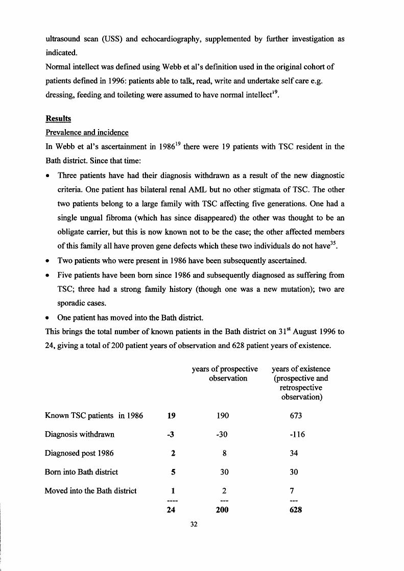
In Webb et al’s ascertainment in 198619 there were 19 patients with TSC resident in the
Bath district. Since that time:
• Three patients have had their diagnosis withdrawn as a result of the new diagnostic
criteria. One patient has bilateral renal AML but no other stigmata of TSC. The other
two patients belong to a large family with TSC affecting five generations. One had a
single ungual fibroma (which has since disappeared) the other was thought to be an
obligate carrier, but this is now known not to be the case; the other affected members
of this family all have proven gene defects which these two individuals do not have35.
• Two patients who were present in 1986 have been subsequently ascertained.
• Five patients have been bom since 1986 and subsequently diagnosed as suffering from
TSC; three had a strong family history (though one was a new mutation); two are
sporadic cases.
• One patient has moved into the Bath district.
This brings the total number of known patients in the Bath district on 31st August 1996 to
24, giving a total of 200 patient years of observation and 628 patient years of existence.
years of prospective years of existenceobservation (prospective and
retrospective observation)
Known TSC patients in 1986 19 190 673
Diagnosis withdrawn -3 -30 -116
Diagnosed post 1986 2 8 34
Bom into Bath district 5 30 30
Moved into the Bath district 1 2 7
24 200 628
32

Bath Health District covers a population of -417,000 people (1996) giving an estimated
prevalence of TSC of 1:17,000 (5.3 per 100,000).
In the Bath district there are approximately 5,500 live births per annum giving an estimated
birth incidence of 5:55,000 or 1:11000.
Population
The mean age has remained static at around 26 years over the ten-year period, but the age
at diagnosis has decreased from 18.8 years to 15.5 years. There were no deaths recorded
during the ten-year period.
In 1986 there was a predominance of affected females of 1.7:1 but in 1996 there were
equal numbers of affected males and females.
Genetics (table one)
In 1986 ten patients had an affected parent (although nine of these were from one family)
and nine were sporadic cases i.e. neither parent affected, this gives a spontaneous mutation
rate of 47%. In 1996 nine patients had an affected parent (again eight were from the one
family) and fifteen were sporadic cases, giving a spontaneous mutation rate of 63%.
Ideally for a diagnosis of TSC to be excluded in the parents, there should be absence of
skin and eye signs and a normal CT Brain. At one time, a renal ultrasound was also
performed but this is now thought to be unhelpful (due to regular normal occurrence of
renal cyst(s) in adults) and is no longer routinely performed36. 46% of parents chose or for
other reasons could not be examined and / or screened.
In Bath three patients have had the diagnosis of TSC withdrawn over the past ten years;
one had bilateral renal AML and according to Osborne’s contemporary criteria3 fitted the
criteria for TSC, but with the modification of these criteria, it is now recognised that
bilateral renal AML can occur independently of TSC37 and with no other stigmata present
this patient is no longer considered to have a diagnosis of TSC.
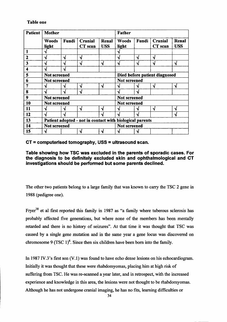
Table one
Patient Mother Father
Woodslight
Fundi Cranial CT scan
RenalUSS
Woodslight
Fundi Cranial CT scan
RenalUSS
1 V V2 V V V V V V3 V V V V V V V V4 V V5 Not screened Died before patient diagnosed6 Not screened Not screened7 V V V V V V V V8 V V V V V9 Not screened Not screened10 Not screened Not screened11 V V V V V V V V12 V V V V V V13 Patient adopted - not in contact with biological parents14 Not screened Not screened15 V V V V V
CT = computerised tomography, USS = ultrasound scan.
Table showing how TSC was excluded in the parents of sporadic cases. For the diagnosis to be definitely excluded skin and ophthalmological and CT investigations should be performed but some parents declined.
The other two patients belong to a large family that was known to carry the TSC 2 gene in
1988 (pedigree one).
Fryer et al first reported this family in 1987 as “a family where tuberous sclerosis has
probably affected five generations, but where none of the members has been mentally
retarded and there is no history of seizures”. At that time it was thought that TSC was
caused by a single gene mutation and in the same year a gene locus was discovered on
chromosome 9 (TSC l)6. Since then six children have been bom into the family.
In 1987 IV.3’s first son (V.l) was found to have echo dense lesions on his echocardiogram.
Initially it was thought that these were rhabdomyomas, placing him at high risk of
suffering from TSC. He was re-scanned a year later, and in retrospect, with the increased
experience and knowledge in this area, the lesions were not thought to be rhabdomyomas.
Although he has not undergone cranial imaging, he has no fits, learning difficulties or34

P ed ig ree on e
TSC 1 UnaffectedTSC 2
3 4
O c6 7 b I 8 9 10
6
3 5

neurocutaneous manifestations of TSC and Woods light examination, renal USS and
fundoscopy have all proved negative so far, making the diagnosis unlikely. He has now
been shown not to have the mutation in TSC1 which his mother and brother have (JPO
personal communication).
In 1988 IV.6 (previously thought to be unaffected) had her first child (V.4). At the age of
13 months V.4 developed epilepsy following his measles, mumps and rubella (MMR)
vaccination. He subsequently went on to develop severe learning and behavioural
difficulties. On investigation he was found to have subependymal nodules, bilateral renal
AML and retinal phakomas in both eyes. He has also developed angiofibroma and
hypomelanic patches leading to the unequivocal diagnosis of TSC. IV.6 was extensively
investigated: CT and MRI brain, renal USS, echocardiogram and examination of the skin,
including Woods light were all negative. It was therefore reported that this family
contained an individual (III.3) with minimal expression (under the diagnostic criteria in use
at that time III.3 was thought to have TSC as he had a first degree relative and a single
ungual fibroma: under the current criteria he would not be considered to suffer from TSC)
and an individual with non-penetrance39. IV.5 has also subsequently been investigated as
IV.6 and found to not be affected. V.4 was therefore the first member of this family now
spanning at least six generations to suffer from epilepsy and learning difficulties. However,
it had become apparent that a second gene existed and its location on chromosome 16pl37 ft(TSC2) was discovered in 1992 by Kandt, and was cloned a year later by Nellist. The
TSC1 gene was then cloned in 19979. This family has since undergone DNA testing and all
definitely affected members found to carry TSC1 mutation except V.4 who has been found
to have a TSC2 mutation, IV.6 and her father (III.3) tested negative for both gene defects.
We now believe V.4 to be a sporadic mutation occurring within this family carrying the
TSC gene. Interestingly, the hypothesis that the clinical phenotype of TSC1 is not as
severe as TSC2 is compatible with the information from this family.
IV.6 (in 1989) had another son (V.5) in 1989 who appears, as yet to be unaffected.
In 1990 V.2 was bom to IV.3. He was found to have three rhabdomyomas on
echocardiogram at two weeks of age, a repeat echocardiogram at seven weeks showed
them to have regressed without causing any symptoms, a phenomenon increasingly
recognised in this condition. He has also subsequently been shown to have tubers on MRI
36

imaging and AML on renal USS. He developed seizures and also has mild learning
difficulties and behavioural problems, though not as severe as V.4.
In 1993 IV.3 had her third son V.3. He was admitted to the neonatal intensive care unit on
the first day of life following a cyanotic episode on feeding. An ECG showed WPW
syndrome coupled with SVT; echocardiology showed a rhabdomyoma on the aortic valve
causing outflow obstruction. Initially he was successfully treated with diuretics and
digoxin, but by two months of age continuing attacks of aortic obstruction required
surgical removal of his rhabdomyoma which had not decreased in size. He has since gone
onto develop a shagreen patch, confirming the diagnosis of TSC.
The sixth boy to be bom into this family is V.6. At the time of IV.6 pregnancy with V.6 it
was still believed that she could be an obligate carrier. In the third trimester she
experienced some abnormal foetal movements, which she described as repetitive jerking
movements. Although fits starting in utero had not been previously described in TSC, with
the family history, this was the concern. After lengthy discussion with the parents it was
decided to undertake an antenatal MRI scan to try and establish whether or not the baby
was likely to be suffering from TSC. It was performed at 36 weeks gestation. The foetus
was sedated by giving IV.5, the mother, an oral sedative of lOmg diazepam approximately
20 minutes before the scan. T1 weighted images of the foetal brain were obtained in axial,
sagittal and coronal planes. Good quality images were obtained but (now unsurprisingly)
no abnormalities were detected. V.6 is now eighteen months old and has no stigmata of
TSC.
Referral patterns (table two)
The referral patterns are summarised in table two and are given for all 24 patients who
have resided in the Bath district over the past ten years. In the last ten years, seven new
patients have been diagnosed. Two were brothers bom into a family with a family history
of TSC spanning five generations; both had skin stigmata confirming the diagnosis. In
addition, the younger son also had a cardiac rhabdomyoma that was diagnosed on the first
day of life. Four others presented with epilepsy and were found to have either skin stigmata
and/or an abnormal brain scan (one also had a strong family history). The final patient was
diagnosed because of his facial angiofibroma.
37

Table two
Reason for:Other features Total (n=24)Referral Diagnosis
Affected relative 7 (29%) - - -
Incidental referral 1 (4%) - - -
Epilepsy 12 (50%) - 16 (67%) 16 (67%)Abnormal brain scan - 7 (29%) 4 (17%) 11(46%)Giant cell astrocytoma
- - 1 (4%) 1 (4%)
Developmental delay - - 10 (42%) 10 (42%)Behaviouralproblems
- - 8 (33%) 8 (33%)
Cardiac disease - 2 (8%) 2 (8%) 4 (17%)Renal disease 2 (2%) 2 (8%) 12 (50%) 14 (58%)Lung disease - 1 (4%) - 1 (4%)Retinal phakoma - 3 (12.5%) 4 (17%) 7 (29%)Skin signs 4 (17%) 23 (96%) 1 (4%) 24 (100%)
Table showing the referral patterns for the diagnosis of TSC (N.B. some patients may have had more than one reason for referral).
Skin stigmata (table three)
All 24 patients in Bath have skin signs, but this was not always true at presentation. The
most common dermatological features are facial angiofibroma, hypomelanic macules,
shagreen patches and ungual fibroma.
Table three
1986 (n=I9) 1996 (n=24)Facial angiofibroma 16 (84%) 22 (92%)Hypomelanic macules 10 (53%) 15 (63%)Forehead plaque 7 (37%) 10 (42%)Shagreen patch 9 (47%) 12 (50%)Confetti 2(11%) 3 (13%)Poliosis 2(11%) 4 (17%)Nail fibroma / ridging 12 (63%) 14 (58%)Gum fibroma 2(11%) 2 (8%)
Table showing the skin lesions experience by the 24 patients. All 24 patients had skin stigmata of TSC. The two patients who did not have angiofibroma both had a shagreen patch.
38

Retinal phakoma
Nineteen patients have had their retina examined by fundoscopy, eight (42%) of whom
were found to have retinal phakoma. One patient was also found to have amblyopia, which
may have arisen as a result of his phakoma overlying the macula and interfering with the
development of vision. Another patient is known to have a squint.
Seizures (table four)
In 1986 eight (42%) patients had suffered from epilepsy, by 1996 fourteen patients (58%)
had a diagnosis of epilepsy. Equal numbers of males and females are affected.
Type of seizure (table four)
Table four
Patientgender
Age of onset
Type at onset
Other types LD Outcome of seizures
Behaviouralproblems
F 12 Weeks GTC CPS, Absence, NCSE, Drop
Y Refractory Y
F 4 Months IS GTC, CPS Y ~ 2 / year YF 4 Months IS Focal N Stopped NF 11 Months GTC None Y Stopped NF 5 Years CPS GTC N Controlled NF 5 Years GTC None N Controlled NF 18 Years CPS None N Stopped NM 1 Month Focal IS, GTC, Absence,
Drop, NCSEY Refractory Y
M 6 Months IS GTC, Absence Y Refractory YM 7 Months IS Absence Y Controlled YM 8 Months GTC Focal N ~2 / year NM 9 Months IS None Y Stopped YM 13 Months GTC Absence Y Refractory YM 18 Months Absence None Y Refractory Y
LD = moderate I severe learning difficulties, IS = infantile spasms, GTC = generalised tonic clonic, CPS = complex partial seizures, NCSE + non- convulsive status epilepticus, Y = Yes, N = No.
Table showing patient gender, age at onset of seizures, the type of seizure at onset, other seizure types experienced, presence or absence of moderate or severe learning difficulties and behavioural problems and the outcome of the seizures.
39
There were many seizure types recorded with many patients experiencing more than one
type. In total five patients had infantile spasms (IS), five had primary generalised tonic
clonic seizures (GTC), one had atypical absence, two had complex partial (CPS), and one
focal seizures as their first seizure type.
Outcome of seizures (table four)
Five patients had IS as their primary seizure type with onset under one year of age. All
these patients developed further seizure types and all but one have severe learning
difficulties.
In total eleven patients had onset of seizures before the age of five. Nine have moderate to
severe learning difficulties. The three patients in whom the seizures started after the age of
five, and the ten patients who have never suffered from fits, all have normal intellect (as
defined by Webb).
Seven females have suffered from epilepsy, in five the fits have either stopped or are
completely controlled by anticonvulsants, one continues to suffer the occasional fit only
and the seventh patient continues to have poorly controlled fits with multiple seizure types.
Three have learning difficulties. Seven males have suffered from epilepsy. In one the fits
have stopped and in one they are controlled on vigabatrin. One other patient continues to
have occasional fits off anticonvulsants. The other four all continue to have poorly
controlled epilepsy despite anticonvulsant therapy. Six have learning difficulties.
Two children developed episodes of NCSE. Both have been successfully treated with oral
diazepam.
Learning difficulties and behavioural problems (table four)
In 1986, four patients had learning disabilities and three behavioural problems. In 1996
nine had learning disability and eight behavioural problems. All patients with behavioural
and/or learning disability suffered from epilepsy.
40

Other neurology
One patient known to have TSC developed symptoms of raised intracranial pressure and
was diagnosed as having a giant cell astrocytoma (GCA), which was successfully removed
without sequelae. One other patient has a mild hemiplegia.
Renal disease
Twenty-two patients have undergone at least one renal USS. Thirteen of these patients
were shown to have at least one renal lesion. Five patients had cysts; three unilaterally, one
bilaterally and one had adult type PKD. Eleven had renal AML, seven of which were
bilateral. One patient had a renal carcinoma.
Only three patients experienced symptoms of haematuria and pain; all three were known to
have large bilateral AML, and CT scanning confirmed renal haemorrhage arising from an
AML in each case. They were all successfully treated by selective renal artery
embolisation. The renal carcinoma was detected on a research scan; the patient had been
entirely asymptomatic. The diagnosis was confirmed by biopsy and the patient had a left
nephrectomy performed.
Cardiac disease
Twelve patients have had an echocardiogram. Three patients under the age of one and one
adult patient had evidence of rhabdomyomas. One had hypertrophy probably secondary to
his long-standing essential hypertension. The others were all normal. Two of the three
infants showed complete resolution of the rhabdomyomas, on repeat echocardiograms.
One patient (who also had a large cardiac rhabdomyoma causing outflow obstruction) was
found to have Wolff-Parkinson-White Syndrome.
Lung disease
Only one patient has been diagnosed as suffering from pulmonary
lymphangioleiomyomatosis (LAM) in the Bath district in the past ten years. She presented
with a pneumothorax at the age of 21 years treated by pleurectomy, which has not
recurred, but she continues to have exercise induced dyspnoea and episodes of
haemoptysis despite treatment with methyl-progesterone. Her spirometry shows a reduced
vital capacity of 35% predicted, an FEVj of 38% predicted and a peak flow of 95%
41

predicted. High Resonance CT (HRCT) scanning showed multiple thin walled cysts and
peripheral bullae confirming the diagnosis of LAM.
The chest X-rays of three other patients (performed for other reasons) were reported as
normal.
Discussion
Although patients with TSC may have a normal life span, and one patient in Bath is now in
his ninth decade and has suffered no serious complications, it is a disease associated with
high morbidity and early mortality. As discussed earlier it can affect almost any tissue in
the body, although the most troublesome complications arise from involvement of the
neurological, renal, cardiovascular and respiratory systems.
The disease also has important genetic implications. It is an autosomal dominant condition,
thus, any patient diagnosed with TSC will have a 1 in 2 risk of passing the disease to each
of their off spring. Although it may be possible that the TSC1 phenotype is not as severe as
the TSC2 phenotype, severity of disease does not run in families (there are many cases of
only mildly affected patients having children who are severely affected) and it is
impossible to predict who will be most affected. About half of patients with TSC have
severe learning difficulties and epilepsy25 26. Therefore, a patient with TSC will have
approximately a 1 in 4 chance of having a child with TSC and epilepsy and severe learning
difficulties.
It is important, to make the correct diagnosis, not only because of the possible
complications that might arise in the affected individual, but also so that accurate genetic
counselling may be given. Presently, it is not possible to routinely test DNA from
individuals with TSC (research has isolated TSC1 and TSC2 gene defects in approximately
half of affected families) but it is possible to detect TSC on chorionic villus sampling or
amniocentesis when a parent has a known mutation. Even though prenatal diagnosis will
be possible for the majority in the near future it is unlikely that a test will be available that
will predict the severity of the disease in an affected foetus. Therefore, the diagnosis is still
frequently a clinical one and the diagnostic criteria need to be modified accordingly as our
knowledge of the disease improves to allow both accurate diagnoses and exclusion to be
made.
42
Over the past ten years the age of diagnosis has gradually decreased. This may be
explained in part by the fact that three patients had a known family history, so that the
family and health professionals were aware of their risk of inheriting the disease. It is also
possible that greater awareness of this disease and improving technology have contributed
to earlier diagnosis. However, these figures must be treated with caution, as the number of
patients is small.
The most common causes of morbidity in the Bath TSC population over the last ten years
were seizures and learning difficulties coupled with behavioural problems. Both
Shepherd26 and Webb19 also found seizures and mental retardation to be the major causes
of morbidity affecting up to 80% of the TSC population. There also appears to be a close
relationship between seizures and severe learning difficulties. There are only a few cases of
patients who have learning difficulties without a history of epilepsy reported in the
literature and in those that do it may be some other mechanism other than the TSC that
accounts for their impaired intellect. None of our patients who were seizure free had
learning difficulties. In addition the age of seizure onset correlates with long-term
prognosis for psychomotor development. Children who have not developed epilepsy by the
age of five rarely regress and are more than likely to have normal intellectual outcome.
None of our patients who developed epilepsy over the age of five had psychomotor delay.
Seizure type at onset is also an important prognostic factor for long term psychomotor
development with infantile spasms (IS) associated with the highest risk of severe learning
difficulties (SLD). In our group the most common seizure type at onset was IS, followed
by generalised tonic clonic (GTC) and then partial seizures. Four out of five patients with
IS had SLD, and interestingly the one patient who had normal intellect was the only patient
in whom the spasms were rapidly controlled (although the electroencephalogram (EEG)
did not show hypsarrhythmia bringing the diagnosis into doubt, it was obtained after
treatment had been initiated which might have lead to resolution of the hypsarrhythmia).
Because of the long-term psychomotor implications of early onset epilepsy in this group of
patients, it is of paramount importance that fits are recognised quickly, accurately
diagnosed and treated promptly. Though no study has been undertaken to show that early
intervention will improve outcome in these children, many clinicians believe that the
quicker the fits (particularly when they are IS) are controlled the better the long-term
43
prognosis for development. Any infant who is known to suffer from TSC or is at high risk
(i.e. has a first-degree relative) must be closely monitored for the development of fits
especially IS. If any concerns arise they should be admitted to hospital the same day for
observation and EEG monitoring. If the diagnosis is confirmed, treatment should be started
immediately. The treatment of IS in the past has been notoriously difficult, but early
studies have suggested vigabatrin to be particularly efficacious in treating patients with
TSC, controlling the spasms in over 90% of patients40. Vigabatrin is also effective in
treating partial seizures (the second commonest type of seizure at onset) in these infants41.
Gender may also be an important prognostic factor for outcome of seizures. Although our
numbers are very small and the observation will have to be repeated on larger populations,
girls might have a more favourable outcome than boys. Equal numbers of males and
females suffered epilepsy but only one girl continues to have refractory epilepsy as
compared with four boys. Likewise only three females have moderate to severe learning
difficulties compared to six males.
Behavioural problems commonly occur in children with learning difficulties and/or
epilepsy. They often exhibit autistic traits and are commonly hyperactive with poor
sleeping patterns. In a survey carried out in 199427 parents complained that one of the most
difficult aspects to looking after their children with TSC was the hyperactivity and poor
sleeping. It is also known that these children respond badly to traditional sedatives (often
becoming more hyperactive) and behavioural therapies are often ineffective.
Giant cell astrocytomas are another recognised neurological complication of TSC and
occur more commonly than in the general population. There is controversy as to whether
they arise from subependymal nodules or occur de novo. They most commonly occur
around the Foramen of Munro, frequently occurring around adolescence and are rare over
the age of thirty years. They usually present with raised intracranial pressure: headache,
vomiting, ataxia, drowsiness, diplopia and papilloedema. Treatment is surgical removal.
Unlike other forms of brain tumours, the prognosis is excellent. The Bath patient was not
typical of patients with TSC who develop astrocytomas, as she was 37 years old. She had
long-standing epilepsy, though was taking no treatment. She complained of increasingly
severe intermittent headaches, episodes of loss of consciousness and weakness of her legs
on head movement. A CT brain scan confirmed a GCA obstructing the Foramen of Munro
44

causing obstructive hydrocephalus. The operation and post-operative period were
uneventful and she was discharged home on anticonvulsant medication. She remains well
and symptom free seven years later. This case illustrates the need for vigilance when
reviewing patients with neurological symptoms and a diagnosis of TSC.
The only other neurological complication recorded in this group was a hemiplegia. This
patient was diagnosed as having IS at the age of four months. Treatment with prednisolone
was commenced with resolution of the spasms, but then she developed right-sided fits that
continued until the patient was three years old when they resolved spontaneously. Over the
ensuing years it became apparent that she had a mild right-sided hemiplegia although in all
other aspects she had made excellent progress and had normal intellect. We can only
speculate as to whether the hemiplegia is secondary to the right-sided fits or is another
neurological manifestation of the TSC, possibly due to the location of a tuber.
Two thirds of the Bath population are known to have renal lesions, but the majority remain
asymptomatic. The patients in our survey represented the wide spectrum of renal disease
seen in TSC, most commonly AML and/or cysts with, more rarely PKD and
adenocarcinoma. As discussed above the TSC2 gene is close to the PKD1 gene. Although
PKD1 mutations do not underlie the cytogenesis in all patients (cysts can occur in TSC1
and TSC2), early onset bilateral PKD occurs with a contiguous deletion of both TSC2 and
PKD1. Only one patient in our survey has PKD - she is known to be mosaic for a deletion
on chromosome 16 affecting both genes. AMLs also occur in both TSC1 and TSC2. The
severity of renal disease between the individuals also varies widely. Many patients with
AML with or without cysts have remained asymptomatic with normal renal function
throughout their life, whilst others have suffered from haematuria, anaemia, pain and
haemorrhage (which can be life threatening)34.
Over the past ten years four of our patients (13%) have undergone treatment for renal
complications. Three suffered haemorrhage from large AMLs requiring embolisation and
one was found to have a renal carcinoma which was removed surgically. Selective arterial
embolisation should now be now the treatment of choice for patients suffering from
haemorrhage from renal lesions. In the past nephrectomy was frequently performed but, as
in our patients, both kidneys are often involved. When bleeding then occurs in the
remaining kidney, this may result in a second nephrectomy (either because of catastrophic
45

haemorrhage or if embolisation is not successful) leaving the patient reliant on dialysis
unless they are fortunate enough to receive a successful kidney transplant. Although none
of our patients suffer from ESRF, it does contribute a significant degree of morbidity and
mortality within the TSC population as a whole42. DMSA scanning and creatinine
excretion following selective embolisation does show the remaining part of the treated
kidney to be contributing significantly to renal excretion and therefore function.
Nephrectomy still remains the treatment of choice for renal carcinoma.
Cardiovascular complications can also occur in TSC and may be life threatening. Cardiac
rhabdomyomas are the commonest cardiac manifestation. Three patients in our survey
were found to have rhabdomyomas in early infancy, in two of which the lesions were
shown to regress and eventually disappear on echocardiology. The other patient was noted
to have a large rhabdomyoma in adult life. It is generally accepted that, unlike other
hamartomas in TSC which tend to become more numerous and larger with increasing age,
rhabdomyomas generally regress, becoming smaller and less common with age. However,
the size and position of the rhabdomyoma in this older patient makes it unlikely that it was
present in infancy, as she had experienced no symptoms. This raises the possibility that
some rhabdomyomas may increase in size after birth; whether arising de novo or from a
smaller congenital rhabdomyoma is not known. One possible explanation for the changing
size of rhabdomyomas is that they are under the influence of oestrogens. It has been
postulated that other manifestations of TSC such as AMLs and LAM are affected by
oestrogens (see chapter seven) and it is possible that rhabdomyomas are similarly affected.
It could be hypothesised that as oestrogen levels increase during pregnancy, growth of the
rhabdomyomas is stimulated so that they reach their maximum size at birth. After
parturition, as oestrogen levels fall, the growth of the rhabdomyomas is no longer
stimulated and their size regresses, so that they can no longer be detected on
echocardiology. Likewise if in later life a patient is exposed to high levels of oestrogens
(e.g. ovarian tumour) then a rhabdomyoma may either increase in size once again, or even
develop de novo. It is not known whether or not our patient was exposed to high levels of
oestrogens.
The majority of rhabdomyomas remain asymptomatic but they can cause heart failure if
they arise in a position that causes obstruction to the flow of blood within the heart, or as in
one of our patients, syncope due to acute but intermittent obstruction. A less common
46

problem is the development of WPW syndrome and associated SVTs. It nearly always
occurs in conjunction with rhabdomyomas and usually presents within the first year of life,
with approximately half presenting on the first day of life. It responds well to medical
treatment and will often resolve in later life43.
Only one patient had been symptomatic and therefore diagnosed as having LAM. She
presented at the age of 21 years with sudden onset of chest pain and shortness of breath as
a result of a pneumothorax. Pulmonary involvement in TSC is undoubtedly rare. It is
difficult to estimate the true incidence, partly because the few studies that have attempted
to do so have generally used small numbers of patients from biased groups and partly
because it is unknown how many patients with pulmonary complications become
symptomatic and therefore recognised. We now know of at least one other patient who
may have had asymptomatic LAM during this 10 year period, but only came to light later
when she became symptomatic. At present we have no proven method of screening our
TSC population for asymptomatic LAM. Previous studies have estimated 1 - 3 % of
patients to be affected44. It predominately affects females of child bearing age although
rare cases occurring in men and children have been reported45 46. The commonest
presenting symptoms are dyspnoea and pneumothorax, as in our patient. Other symptoms
include chronic dry cough, haemoptysis, wheeze, and chest pain. Development of cyanosis,
respiratory failure and cor-pulmonale can also occur44. Our patient is currently receiving
monthly injections of methyl progesterone and, though she remains symptomatic she has
not suffered any further pneumothoracies and has had a moderate improvement on
pulmonary function testing. This complication is reviewed in detail in chapter seven.
Skin lesions are nearly universally present in patients with TSC. All the patients in the
Bath district had skin lesions that are pathognomonic for TSC. All but two have facial
angiofibroma and they both have a shagreen patch. One of these patients is seven, the other
four years and it is well recognised that angiofibromas most commonly develop after the
age of five and may even arise in adult life. On the other hand hypomelanic patches (best
visualised by a Woods ultraviolet light) are often, though not always, present at birth. They
can arise later. One patient was examined at the age of 13 months with a Woods lamp; four
hypopigmented lesions were noted and documented. Two years later he was re-examined
with the Woods light and was found to have twelve new hypopigmented lesions (sixteen
lesions in all). The neurocutaneous lesions do not contribute to the potentially lethal
47
morbidity of this condition, but many of the lesions are unsightly and are of great cosmetic
concern to the patient. Treatment for the facial angiofibroma has progressed dramatically
over the past ten years due to the introduction of laser treatment. Though blistering of the
superficial layers of the skin does occur immediately after the laser treatment permanent
scarring is rare. It was also thought that if the treatment was carried out at a young age the
lesions would recur, but this does not appear to be the case so that the major limiting factor
for age at which treatment can be undertaken is the ability to lie still. A general anaesthetic
can be used, but several treatment sessions are required.
No deaths occurred in our population: this might be accounted by both the small numbers
and the relatively young age of the population.
Conclusion
Although there were no deaths in the Bath TSC population over the past ten years there is a
high rate of morbidity with seizures, associated learning difficulties and behavioural
problems affecting two thirds of the population. Symptomatic renal disease affected over
10% of our population necessitating either selective embolisation or nephrectomy.
Although renal failure requiring renal replacement therapy had not yet intervened in any of
our patients it remains an important cause of premature mortality in TSC. Lung
complications (LAM) do appear to be rare within the TSC population, becoming
symptomatic in only a small minority of patients. Nonetheless, it is not known how many
patients have asymptomatic lung involvement and whether screening patients might be
beneficial. Theoretically, at least, anaesthesia and flying may precipitate pneumothoracies,
respiratory failure and even death in some affected (but previously asymptomatic) patients.
There is very little information available about the effectiveness of treatment and long-term
outcome in this group of patients. Other complications contributing to morbidity within our
group of patients included cardiac manifestations, GCA and hemiplegia and amblyopia.
This study has also shown that it is possible to undertake long-term studies that give us
improved understanding of the natural history of a disease. However, despite the large
number of patient years observed (628), a larger study is required to examine lower risk
complications such as LAM in more detail. Nonetheless, this study helps confirm that
epilepsy, learning difficulties and behavioural problems are the most frequent problems
encountered in TSC and occur in as many as two thirds of patients. This type of study also
allows observation of time patterns of the complications: for example, although the skin48

manifestations of TSC are almost universal, most patients do not develop angiofibroma
until after the age of five and the number of hypomelanic patches may also increase with
age. Another way to present this data so that the effect of age, time and cohort can be
simply visualised would be the use of a modified Lexus diagram.
49

CHAPTER THREE
Epilepsy and Learning Difficulties
As discussed previously and shown in chapter two, seizures and the associated learning
difficulties are two of the most important causes of morbidity in TSC affecting over half of
the population. About two thirds of affected patients will develop their epilepsy within the
first year of life. The most common seizure type at onset is infantile spasms (IS)
accounting for about 40% of cases, followed by partial seizures and then secondarily
generalised seizures. Nearly all patients who experience IS will go on to have learning
difficulties, as will many of those suffering from partial seizures. Age at seizure onset as
well as type at onset and possibly gender are other important prognostic factors for long
term developmental outcome. It is also believed, though scientific evidence is lacking, that
early, good control of the seizures may also improve long term outcome.
Infantile spasms: recognition, treatment and prognosis
159 years ago Dr West wrote a letter to the Lancet describing “a peculiar type of
convulsion” occurring in his four month old son, so documenting the first case of IS or
what has become known as West’s Syndrome. IS are a clinical seizure type which include
a peculiar type of seizure and psychomotor retardation, usually with a characteristic
electroencephalogram (EEG) pattern known as hypsarrhythmia. There are numerous
causes of IS which do not share a known common underlying pathophysiological process,
although TSC accounts for 10-20% of cases. There are too many other causes to list them
all exhaustively but as a general rule any process that affects brain development or
metabolism can cause IS.
Examples of other underlying aetiological factors for infantile spasms include:• Abnormal brain development e.g.: Neurofibromatosis, Sturge Weber Syndrome, neuronal migration
disorders, agenesis corpus callusom, hemi-megalenencephaly.• Chromosomal disorders e.g.: Down Syndrome (-10%), Cri du Chat (trisomy 13).• Congenital infections e.g.: maternal rubella, CMV infection.• Infection e.g.: meningitis.• Hypoxic -Ischemic encephalopathy whether incurred at birth, antenatally, postnatally or due to pre-term
birth.• Metabolic e.g.: phenylketonuria, organic acidurias, pyridoxine dependency.• Degenerative brain disease, leukodystrophies (rare).• Trauma
50

Despite the huge advances that have been made in medicine IS still remain a poorly
understood entity. Although with newer imaging techniques we are more often able to
elicit the underlying causes of these spasms, still little is known about their
pathophysiological basis and treatment remains problematic.
Epidemiology
IS (from all causes) have an estimated incidence of 0.16 - 0.4247 48 49 50 per 1000 live
births. Little is known about the geographical distribution, but two studies from the USA47
48 , one from Denmark49 and one from Finland50 showed incidence rates of 0.16, 0.25,
0.25-0.33 and 0.42 per 1000 live births respectively. Studies have reported either equal sex
incidence or a slight preponderance of males ranging from 1:149 to 1:450 (only one has
found an excess of females51). Age of onset of the spasms has been reported to occur
between 1 day and 6 years of age52, however the vast majority begin within the first year of
life, after which alternative diagnoses including infantile myoclonic epilepsy, are more
likely. There is a peak age of onset between four and seven months. One of the main
difficulties in estimating the age of onset is the time taken to make the diagnosis. There is
often a significant delay between the start of the spasms and their eventual diagnosis:
parents may not recognise that there is something amiss; often if they do seek professional
advice the spasms are mistaken for colic or a natural startle reflex. In a review by Bobele
and Bodensteiner53 the average lead-time to diagnosis was 3.5 months, but it is well known
that this can vary greatly and it is possible that some cases are never recognised.
Genetics
It is rare to have more than one affected member in any family, unless there is another
inherited underlying cause for the spasms such as TSC, except in a small number of
families where it has been described as having X-linked inheritance54. It is more likely that
the spasms are a symptom of underlying disease (just as haematuria is a symptom of
disease, but can also be idiopathic).
Classification
IS can be classified into three groups;
Symptomatic group: the underlying cause is known prior to the onset of the spasms, for
example an infant diagnosed as having Down syndrome or TSC who then subsequently
develops IS.
51

Cryptogenic group: the underlying cause is only discovered following the onset of the
spasms for example, a normally developing infant who after the onset of IS, is investigated
and then found to have TSC.
Idiopathic group: no underlying cause is found despite investigation.
Unfortunately this classification has led to confusion, as it does not depend on the
underlying cause. For example one child with TSC may fall into the symptomatic group,
whilst another falls into the cryptogenic group. In addition, with better imaging techniques
and diagnostic tools, patients who would have previously been in the idiopathic group,
now fall into either the cryptogenic or symptomatic group. Some authors also cause
confusion by calling all infants in whom a cause is eventually found as symptomatic and
the rest either cryptogenic (assuming there must be a cause) or more correctly idiopathic.
The spasms
No good term has yet been given to this phenomenon that is brief yet fully descriptive. In
1841 West described the sudden forward jerking movement of his son as a Salaam spasm;
lightning major convulsions55, Blitz - Kramp and Jack Knife convulsions56 have also been
used. The problem with these terms is that they only describe the flexor type of spasm.c n
Other terms have been misleading for example, propulsive petit mal suggesting similarity
to benign petit mal epilepsy or Pallidum epilepsia58 falsely implying that the source of the
spasm is known. Names that fully describe the spasms such as that used by Gastaut in
195959 11 Encephalopathic myoclonique infantile avec hypsarrhythmie" are long and
cumbersome and are unlikely to be used in English speaking countries. Gibbs and Gibbs60
coined the phase “infantile spasms” in 1952. Although this is still the best and most
commonly used term it is not strictly accurate: not all cases start in “infancy” and a spasm
implies to some an abnormal muscle contraction rather than epileptic activity. It has
however fallen into general use over many years.
Infancy: this can be defined as “in the first year of life” as infancy is now usually alluded to e.g. infant mortality is death before one year of age, although infant is less precisely defined in the Oxford dictionary as a “child during earliest period of life” thus giving rise to terms such as “infant school”
The commonest type of spasm is the flexor spasm, consisting of sudden generally bilateral
and symmetrical contractions of the neck, trunk and extremities. The extent and degree of
muscle involvement varies between individuals so that in some, the whole body bends at
the waist, the arms are flung upwards and outwards and then brought down, hence the52
terms salaam and jack knife, whilst others have only involvement of the neck resulting in
head bobbing61. Extensor spasms have a predominance of extensor muscle contraction
producing an abrupt extension of the neck and trunk with extensor abduction or adduction
of the arms and legs62. Many infants will have a combination of flexor and extensor spasms
during the course of their epilepsy. Kellaway et al 1979 found that approximately 34% of
infants have flexor spasms, 23% extensor spasms and 42% mixed flexor-extensor spasms
Although the vast majority of infants have symmetrical spasms a small minority ~1% have
asymmetrical spasms often adopting a tonic neck reflex position.
Each spasm is only visible for a brief duration lasting a few seconds, though persisting
EEG changes for a single spasm may last as long as 106 seconds62. They tend to run in a
series or in batches or clusters ranging from just a few to as many as one hundred, with
many studies quoting averages of between 30 and 50 spasms in each batch. The spasms
may also be triggered by certain stimuli, the most common being waking or falling asleep63
though feeding64 has also been implicated. Some earlier studies suggested that external
stimuli such as noise and touch can provoke the spasms but this has only been reported
rarely since.
An association of a cry with the attacks is common and has been frequently reported,
usually during or after the attack, more rarely before. It may be this cry that leads to the
misdiagnosis of colic or intussusception, or the observer to believe the child is in pain64. A
few authors have reported other phenomena such as tears, hiccups, a laugh or a giggle63.
It is difficult to evaluate whether consciousness is lost or maintained during these events
because of the age of the patients, the brevity of the fit and the frequently associated
learning difficulties.
Electroencephalogram (EEG)
Hypsarrhythmia is the most important EEG pattern associated with IS (figure seventeen). It
is important to note that this EEG appearance is not present at all times and tends to be
modified by both the natural course of the disease and treatment therapy60. Synchrony
between the hemispheres and between spike and wave components often appear in the later
stages of the disease, a pattern termed modified hypsarrhythmia. Hypsarrhythmia is most
53

commonly seen as the child falls asleep and wakes, but interestingly the EEG often returns
to normal during rapid eye movement (REM) sleep65.
Figure seventeen - EEG showing hypsarrhythmia
Definition of hypsarrhythmia; Gibbs and Gibbs 195260 “Very high voltage, random, slow waves and spikes in all cortical areas. The spikes vary from moment to moment in duration and location; at times they seem to come from another focus, or possibly from multiple foci. Occasionally the spike discharge is generalised but it is never rhythmically repetitive or highly organised as in petit mal or petit mal variant. The chaotic appearance of this abnormality gives the impression of a nearly total disorganisation of cortical voltage regulation".
The presence of a focal abnormality within the hypsarrhythmia is not uncommon, and
when persistent may suggest a focal lesion within the brain. In patients with TSC such a
focus may be the result o f an underlying tuber. With the pioneering advances in epilepsy, it
may be that in the future this group of patients will be potential candidates for epilepsy
surgery.
Developmental delay
The most devastating association o f IS is with psychomotor development. Studies in the
past have estimated the risk o f mental retardation o f 80% - 90%66 67 68 69. Admittedly, many
of the affected infants already have an underlying disorder, which predisposes them to
having psychomotor delay, for example Down syndrome or cerebral palsy. Nonetheless,
even the children with these types o f problems will stop developing as quickly and even
regress after the onset of the spasms. Infants with TSC usually follow a pattern of
54
development within the normal range until such a time that they develop IS after which
they show signs of arrested development or regression.
Although it is impossible to predict which children will be most severely affected, good
prognostic factors include:
• idiopathic aetiology66 67 68
• later age of onset68d 'X• single spasms (rather than batches)
• no other fits67 68
• absence of focal abnormalities on EEG66
• short lag time between onset of spasms and treatment
• a good response to treatment67 68
It is not known whether any particular therapeutic agent is superior to the others with
regard to long-term outcome (seizure control or psychomotor development).
There is a definite pattern to the developmental delay. Social skills, such as smiling are lost
first. The infant then becomes lethargic, floppy and apathetic and no longer seems aware of
or interested in their surroundings: this sometimes leads to concerns about vision and
hearing. Motor skills such as grasping and reaching are usually the last to be affected, but
gross motor skills can be affected and a few children never learn to walk65.
In a minority of cases, development is not arrested or may return to normal after the
cessation of spasms.
Diagnosis and investigations
Diagnosis is by clinical observation and EEG. Clinical observation is not always easy and
can be missed by parents and less experienced nursing and medical staff. It is simple
enough when an infant presents with the typical salaam spasms occurring in long batches
many times a day, but in the infant with infrequent or single spasms they can readily be
mistaken for colic or a startle reflex. The younger the infant, the more difficult it is to
detect abnormal movement or delayed development.
It is important that whenever IS are suspected the child is admitted for close clinical
observation by experienced staff. An EEG should then be undertaken and if

hypsarrhythmia is not seen, a sleep EEG performed, since hypsarrhythmia may only be
present as the infant is falling asleep or waking. Where there is any doubt video and
polygraphic recordings may help. Occasionally, it may not be possible to confirm the
diagnosis with an abnormal EEG, however, if IS are clinically certain, treatment should be
considered because of the poor prognosis of this condition. Some of these infants, with a
normal interictal EEG will have a relapse later with severe psychomotor retardation but
their management at presentation is controversial, because benign infantile myoclonus
does not require treatment and is clinically similar, but without the risk of associated
psychomotor retardation: their EEG is normal. In all infants in whom there is no other
known cause for their spasms a diagnosis of TSC should be excluded. Brain imaging (CT
and/or MRI), examination of the skin with Woods light (for hypomelanic patches) and
fundoscopy (for retinal phakomas) should be undertaken as a minimum. Renal USS and
echocardiography should also be considered.
Treatment
At least 30 drugs have been tried since Dr West wrote his letter in 1841. Most
anticonvulsants have been tried, many with no effect and no treatment has been shown to
be successful in treating the spasms in more than about half of the patients, except steroids,
valproate and vigabatrin.
The first recorded treatment, after West, was by Gibbs et al in 195469, who reported
favourable results using chlortetracycline, 113 years after West's letter. It is unclear from
the literature whether this delay was due to the fact that IS were not being readily
recognised and therefore treated, or whether in fact no treatment had previously been found
to have any effect.
Steroids and hormonal treatments have been used in the treatment of seizure disorders
since the early 1950’s. In 1958 Sorel and Dusaucy Bauloye70 reported a dramatic response
to ACTH therapy in children suffering from IS. Not only did the spasms stop and the EEG
improve, often becoming normal, but they claimed that mental improvement occurred if
treatment was started within a few weeks of the onset of the spasms.
Following this first report there have been many studies which have frequently confirmed
the effect of steroid/hormonal therapy claiming to stop seizures in just over half of the
56
patients supporting the suggestion that steroid/hormonal therapy is effective in a significant
proportion of children with IS. A literature review of the English Language using Medline
between 1950 and 1995 found eleven studies and a further review of three studies (fourteen
in total) investigating the effect of ACTH and steroids on IS. There were three randomised
RCT trials71 72 73 , eight open prospective trials 74 73 76 77 78 79 80 and three retrospective trials
8182 (see table five). Out of a total of 784 patients treated there was complete cessation of
spasms in 449 (i.e. 57%).
57

Table fiveStudy Study
deelgnSelection Criteria Total No
o fp tsDrugs Dose Admin Duration n o or
patientssiezureestopped
siezureereduced
hyps, res loves
relapses Cross over No of patients
Baram 1996(71) RCT Clinical spasms Hypsarrythmia No prior steroid treatment
29 ACTHPrednisone
150U/m2/day2mg/kg/day
IMOrally
2 weeks (tap) 2 weeks (tap)
1514
14 II 13 N/AN/A
PrednisoneACTH
28
Hrachovy 1994 (72) RCT Clinical spasms Hypsarrythmia No prior steroid treatment
59 ACTH high dose
l50U/m2/day tor three weeks
IM 3 weeks (tap) 26 13 N/A 13 N/A N/A N/A
ACTH low dose
20 - 30 U/day tor two weeks
IM 2 weeks (tap) 24 14 N/A 14 N/A N/A N/A
Hrachovy 1983 (73) RCT Clinical spasms Hypsarrythmia No prior steroid therapy
24 ACTHPrednisone
20U/day2mg/kg/day
IMOrally
2 - 4 weeks (tap) 2 - 4 weeks (tap)
1212
54
N/AN/A
54 II Prednisone
ACTH78
Glaze 1988 (74) Prospective Clinical spasms Hypsarrythmia No prior steroid treatment
64 ACTHPrednisone
20 - 30U/ day 2mg/kg/day
IMOrally
2 - 4 weeks 2 - 6 weeks
64 in total
38 in total
Hrachovy 1979 (75) Prospective Clinical spasms Polygraph Ic-video No prior steroid therapy
12 Prednisone 2mg/kg/day Orally 4 - 6 weeks (tap) 12 2 1 2 0 N/A N/A
Hrachovy 1980 (76) Prospective Clinical spasms Modified hypsarrythmia No prior steroid therpsy
5 ACTH 20U/day IM 6 weeks (tap) 6 5 0 5 2 N/A N/A
Kuaaa 1993(77) Prospective Clinical spasms Hypsarrythmia Pychomotor delay
18 ACTH 0.8mg IM 4 weeks (tap) 18 6 5 6 N/A N/A N/A
Snead 1996 (78) Prospective Clinical spasms (modified) hypsarrythmia
15 ACTH l50U/m2/day IM 1 week (tap) 16 14 0 14 5 N/A N/A
Matsumoto 1987(79)
Prospective Clinical spasms 33 TRHACTH
0.5*10mQ/day 0.125-0 75mg IM
1-4 weeks 1-4 weeks
1320
715
11 312
N/AN/A
N/AN/A
Schlumberger 1994(80)
Prospective Clinical spasms hypsarrythmia No prior steroid therapy No serious malformations No known TSC
94 ValproateHC
40/mg/kg/da y 15/mg/kg/day
OrallyN/A
2 weeks 2 weeks
9494
70 N/A N/A 6 ACTH 15(9 exclude
Haines 1994 (81) Retrospective Clinical spasms Hypsarrythmia
191 ACTH 2-4U/kd/day IM 1 week (tap) 191 113 N/A 80 N/A N/A N/A
Retrospective Clinical spasms Hypsarrythmia
25 ACTH N/A N/A N/A 25 22 N/A 21 N/A N/A N/A
Prospective Clinical spasms Hypsarrythmia
52 ACTHPrednisone
i50U/m2/day3mg/kg/day
IMOrally
1 week (tap) 4 weeks (tap)
3022
3013
if 24 N/AN/A
11 WAN/A
Singer 1980 (82) Retrospective Clinical spasms 55 ACTH 40U/dsy IM 1 week (tap) 55 42 N/A 42 18 N/A N/A
Table showing the results of the literature search for the treatment of infantile spasms with steroids.
58

The mechanism of action of hormonal therapy in infantile spasms remains poorly
understood. Baram et al71 has proposed that “stress” increases adreno-corticotrophin
releasing hormone (CRH) activity causing infantile spasms during a time when brain
maturation is occurring, explaining why infantile spasms are age specific. They argue that
ACTH acts by suppressing ARH synthesis. ACTH may also act directly by membrane
stabilisation, improving the integrity of the blood-brain-barrier and stimulating myelin
formation. Steroids such as hydrocortisone and prednisolone have metabolic, anti
inflammatory and immunosuppressive effects that may play a role in spasm suppression
but also act directly by inhibiting anterior pituitary and hypothalamic secretion, including
ARH.
The problem with steroids are the side effects: potentially life threatening problems include
depression of the immune system (leading to overwhelming sepsis) and electrolyte
imbalances. There have been few studies investigating the side effects of steroids when
used for the treatment of IS. One such study , investigating the risk of infection with
ACTH therapy in 27 infants, identified 75 febrile episodes, four of which had confirmed
bacteraemia and three deaths occurred. However IS occur at an age when febrile episodes
are common. The deaths occurred in children who had received large doses (over 100 units
ACTH given 2 times a week over three weeks). Less serious side effects, for example
hypertension, are often transient but nevertheless cause a certain amount of morbidity. It is
difficult to estimate the frequency of these side effects as the study by Riikonen and
Donner shows. In their study only 17 out of 64 had their blood pressure routinely
measured. Minor side effects, estimated to occur in two thirds of patients, include
behavioural changes especially irritability, changes in appetite, weight gain and alteration
in sleep patterns. In addition some forms of steroids e.g. ACTH and synacthen involve
daily or alternate daily intramuscular injections.
In view of the devastating effect that IS can have on the patient, it is not surprising that
most anticonvulsants have been tried for their treatment at one time or another. Sodium
Valproate, used over the past 1 5 - 2 0 years84 85, has been one of the most successful.
Sodium valproate inhibits the breakdown of GABA, a neurotransmitter, thus increasing its
availability. It is also thought to have membrane stabilising properties. Siemes et al
conducted a trial of valproate therapy in 22 infants with IS. The dose ranged from 45mg to
lOOmg/kg i.e. much higher doses than those generally recommended in children for the
59

treatment of epilepsy (30-40mg/kg). Abolition of seizures was obtained in 65% of children
within three months of commencing therapy. There are potential problems with valproate
therapy and side effects are common. Hepatotoxicity and pancreatitis, although rare, can
occur and be life threatening. It may be that the higher than normal doses required and
fears of these serious complications, but particularly the length of time to obtain a response
(usually weeks or months rather than days), have limited the use of valproate in this
disorder.
Vigabatrin, one of the new generation anticonvulsants, is rapidly gaining popularity in
view of both its efficacy and its favourable drug profile. It also acts on the GABA
pathways, by inhibiting GABA transaminase (an enzyme involved in the metabolism of
GABA) thus increasing CSF concentration. It was first brought to clinicians’ attention in
1991 by Chiron87 et al who reported complete cessation of seizures in 43% of patients. The
effect was seen rapidly, often within one week. Subsequently there have been preliminary
trials, open and uncontrolled, on the use of vigabatrin in IS showing similar control to that
seen with steroids. There is also increasing evidence that vigabatrin is particularlyo o
efficacious in the treatment of IS in the group of children suffering from TSC. Acairdi
found in his review of 192 patients that out of the 28 patients with TSC, 27 (96%) hadO A
complete cessation of their spasms. There has also been one study comparing vigabatrin
with steroid therapy specifically in this subgroup of patients. They found that 11/11
responded to the vigabatrin and only 5/11 to hydrocortisone. Again they concluded that
vigabatrin might be particularly effective in treating children with both IS and TSC.
Although vigabatrin has only been used in the treatment of IS for a few years, it had been
thought to have few side effects. The most commonly reported problems were drowsiness,
vomiting and behavioural changes, all of which resolve on discontinuing the drug. There
had also been one reported event of myoclonus, though it is not clear whether this was ano o
effect of the vigabatrin or the child’s underlying neurological disorder .
However, more recently there have been reports of both asymptomatic and symptomatic visual field defects with loss of peripheral vision to varying degrees in adults treated with vigabatrin90 (see later).
Second line drugs include:
Benzodiazepines, for example, nitrazepam and clonazepam, have been shown to be
effective in treating IS but are rarely used as first line monotherapy, they are more
60

commonly used in addition to, or after failure of other drugs. They are thought to act by
acting directly on GABA receptors at chloride channels within the brain. Driefuss et al91
were the first to report the use of nitrazepam in a controlled study against steroids in 1986,
although it had been in use for many years before. They reported that both treatments
resulted in a significant reduction in seizures but that the difference between treatments
was not significant. The main problems with benzodiazepines that have limited their use
are; sedation, excess bronchopulmonary secretions and the development of tolerance in a
significant proportion of infants.
Intravenous gammaglobulins came into vogue in the early 1990s. With the knowledge of
the favourable effect of steroids it was postulated that there might be an immune
mechanism at work and IV gammaglobulins were tried92. But in view of their expense,
inconvenience and potential problems with infection (contamination with HIV and Hep C
was of great contemporary concern) as well as failure of any studies to prove their efficacy,
they are now rarely used.
Thyrotropin-releasing hormone (TRH) is widely distributed throughout the brain acting
as either a neurotransmitter or neuromodulator. A Japanese study, carried out in 198779
showed that just over half of the patients responded to TRH - similar results to those
achieved by ACTH. They also noted a lower incidence of side effects in the TRH group.
Despite these preliminary findings, it is not yet routinely used in the treatment of IS. The
mode of administration (intravenously) and cost (approximately seven times that of
ACTH) make the treatment unattractive.
Pyridoxine under went several studies93 94 during the late 80’s to assess it’s validity in
treating IS following the successful treatment of vitamin B6 - dependent seizures in the
new-born with high doses of pyridoxine and whilst there is no doubt that it is effective in a
small number of cases, none of the studies proved that it has such a benefit as to deserve
consideration as first-line monotherapy in this disorder. It is however used in Japan as first
line treatment.
Epilepsy surgery: Alongside the increased use of vigabatrin for IS there has also been a
steady rise in the number of patients undergoing epilepsy surgery. Although the numbers
have been small, early results have been promising. In one study95 of 23 patients only four
61

failed to respond to surgery. However, these patients were selected on strict criteria
suggesting a focal cause and in many children with TSC there are multiple active or
potential epileptic foci such that surgery is not thought to be an option. In addition long
term follow up studies have not been undertaken, but in surgery used for other forms of
epilepsy the relapse rate is high.
Table six - summary of the pharmacological treatment of infantile spasms
Drug Comments
First line drugsVigabatrin If the infant is known to have TSC, or is at high risk i.e. has a first-
degree relative with TSC, at the time of onset of the spasms then vigabatrin should be the drug of choice.
Prednisolone
ACTH
(Synacthen)
Infants who are commenced on steroid therapy should be admitted for the initial 48 hours of treatment. During this admission their blood pressure should be measured twice daily and their urine dipsticked to exclude the development of hypertension and diabetes respectively. They should then be reviewed on a weekly basis to monitor their blood pressure, urine and weight.
Second line drugsNitrazepam
Clonazepam
Valproate BEWARE of thrombocytopenia at high dosage.
Trial of
Pyridoxine
A test dose should be given intravenously alongside EEG monitoring. If the hypsarrhythmia resolves then oral pyridoxine should be tried to see if the spasms resolve.
Prognosis
The long-term prognosis is poor. Few studies have specifically looked at long term out
come. In a study carried out by Riikonen96 looking at adults with a history of IS, only 17%
attended mainstream school, and less than 10% were married, had children or were
employed. One fifth had progressed to developing Lennox Gastaut syndrome with severe
epilepsy and a further third of patients had continued to have seizures in adult life.07Another study looking at the long term prognosis in 162 patients, found mental
retardation in 78% and physical handicap in 56%. Only 10% of their children attended
normal school.
62

Conclusion
Although the improvement of diagnostic techniques has led to better recognition of the
underlying causes and pathology of IS, response to treatment remains disappointing. Even
with the best therapies available abolishment of seizures only occurs in two thirds of
patients at best, with a high risk of relapse of spasms and other seizure types and the long
term prognosis is appalling. There are many questions that remain with regard to
determining the best treatment for IS.
• Is one treatment better than another in reducing the long-term morbidity of IS? It could be argued
that long-term psychomotor development is the most important outcome factor for these children.
Although the spasms may be distressing in themselves, in a significant minority they resolve
spontaneously if left untreated, though many children will go on to develop other types of epilepsy.
However, the vast majority will develop severe learning difficulties. Although it is thought that earlier
control of the spasms may improve outcome, no study has specifically considered this and no study has
set out to compare different therapies with regard to long-term outcome.
• Is there an optimum steroid dosage regime? There is currently no widely accepted dosage regime for
steroid dose, frequency, duration or type of steroid used in the treatment of IS. The studies in the
literature have used many different regimes but none have been large enough to give reliable results.
• Is vigabatrin really the drug of choice for patients with TSC or for those at a high risk of TSC at
the time of diagnosis their spasms? Only one study89 to date has considered the use of different drug
regimes in the treatment of the subgroup of patients who have IS as a result of TSC. Their results
suggested that vigabatrin was more efficacious in this group than steroids, however their numbers were
small (22 in total).
• Finally, if IS can be prevented from occurring in infants with TSC will this reduce the incidence of
severe learning difficulties and the development of other seizure types in this subgroup?
In order to try and answer the first question it was decided that a large trial needed to be set
up to look at the effect of treatment in terms of both seizure control and more importantly
long-term psychomotor outcome. It was decided that this would be a pragmatic drug trial
comparing the two drug treatments currently in widespread use in the UK; adreno-corticoid
or hormonal treatments which I will call “steroids” for convenience and vigabatrin.
However, it is possible that adrenocorticotrophin hormones work not by production of
steroid hormone through stimulation of the adrenal cortex but by direct negative feedback
on the hypothalamus reducing ARH products. This theory promoted by Dr Baram suggests
that ARH is epileptogenic at a critical period in brain development. This brought up the
next two important questions; which steroid dosage regime should be used in the trial? and
63

if there were enough evidence to suggest that patients with TSC had a better response to
vigabatrin, should this sub-group be excluded from the trial?
There are currently two preparations of steroids widely used in the treatment of IS in the
UK
• Prednisolone given orally.
• Adrenocorticotrophin hormone (synacthen) given as an intramuscular (IM) injection.
It was decided that both drugs would be used in the trial as many doctors who would be
recruiting into the trial expressed that they thought one type to be superior to the other,
although these opinions are not founded on evidence based medicine. There has only been
one double blind, randomised crossover trial comparing ACTH and prednisolone. This
study by Hrachovy73 used lower doses of both ACTH (20u/day) and oral prednisolone
(2mg/kg/day) than usually used in the UK and found no significant difference between the
two different types of steroids. Five out of twelve patients responded to the ACTH and four
out of twelve to the prednisolone, but the numbers of patients enrolled into this study were
small and will have been insufficient to detect a clinically important difference in response.
The next question to be addressed was the dosage to be used for each of the steroids. Again
only one controlled trail comparing high dose versus low dose ACTH had been reported72
and the numbers were small but no difference was found between the two groups. We
therefore had a dilemma; if we were going to conduct a large clinical trial we wanted to
ensure that we were using an adequate dose of ACTH; one which would give us the best
clinical response, but on the other hand high dose steroids may be associated with more
frequent serious side effects. In the one paper that had looked at the side effects of ACTH• • • f t luse m IS in detail all the deaths had occurred in children who had received very large
doses i.e. over 100 units ACTH given two times a week. It was therefore decided that a
reasonable dose of ACTH would be 40 units on alternate days increasing to 60 units if no
response was seen within a week, a dosage regime currently in widespread use in the UK,
but the duration of treatment was restricted to 2 weeks since length of treatment as well
dosage seemed to bear a significant relation to the incidence of side effects.
Deciding the dose of prednisolone to be used proved more problematic. Again we wanted
to ensure that we were using an adequate dose that would give us the best clinical response
but without causing serious side effects. Historically, large doses of prednisolone have
been used when treating children. In the 1970’s doses were reduced, often to 2mg/kg body
! 64
{

weight probably as a result of a wish to be logical over the treatment and because of
concerns over the systemic side effects, although there was no scientific data to suggest
that a reduction was necessary or that a dose on a per kg basis was logical. Large doses of
steroids have been given for many years to pregnant women with no known ill effect to the
foetus. High doses well above 2mg/kg are used in acute asthma attacks in children, albeit
for only a few days. There are several pathophysiological mechanisms to explain why it
might be safe to give relatively large doses of steroids to young children, including a
relatively large volume of distribution. In the adult and older child only the kidney
inactivates cortisol and prednisol. In the infant they are metabolised in every organ of the
body, thus giving an extensive first pass metabolism effect throughout the first year which
tails off after the first three months of life (personal communication). It was therefore
thought that although 2mg/kg was the most widely reported dose of prednisolone for
infantile spasms it might be inadequate. Baram71 et al in a single blinded study used a high
dose of ACTH (150u/m2/day) against a low dose of oral prednisolone (2mg/kg/day),
concluding that ACTH was more effective in terminating infantile spasms. Fourteen out of
fifteen patients responded to the ACTH whereas only four out of fourteen responded to the
relatively low dose prednisolone perhaps suggesting that when smaller doses of the
steroids were used fewer patients responded. We would suggest that if these studies were
to be repeated on a larger population using higher doses of prednisolone a greater
proportion of infants might show a response. Their response to ACTH also helped to
justify a high dosage regime.
Higher doses of oral prednisolone had been in use at the Royal United Hospital (RUH)
during the previous thirteen years. We decide therefore to look at the patients who had
been treated with prednisolone for infantile spasms during this time in order to try and
assess the efficacy and safety of high dose oral prednisolone (usually 20mg tds for two
weeks, regardless of weight or age) in the treatment of IS.
65

The treatment of infantile spasms with high dose oral prednisolone: a retrospective
review.
Aims
In this retrospective study, the aims were to identify the frequency and type of side effects
suffered by infants being treated with high dose oral prednisolone, in particular those that
could be considered to be potentially serious or life threatening, and to observe the effect
of the high dose prednisolone on the spasms themselves.
Patients and methods
The study group comprised all patients who had been admitted to the RUH between
January 1983 and August 1996 with a diagnosis of IS. Patients were collected by two
methods. Those who had been admitted between August 1987 and August 1996 were
located via a database at Bath Health Information Services and those admitted prior to
August 1987 were located via the EEG departmental records.
The medical notes were then retrieved and reviewed. Both a clinical description of the
spasms and an EEG recording compatible with West syndrome (i.e. hypsarrhythmia or
modified hypsarrhythmia) were required to fulfil the criteria for a diagnosis of IS. The
following information was then sought for each patient;
• predisposing factors,
• age at diagnosis of IS,
• the apparent time delay from onset of spasms to diagnosis,
• time to start of treatment,
• dose and duration of steroids and the effect of treatment on spasms.
The notes, including nursing profiles, observation charts etc. were then scrutinised for any
evidence of side effects suffered during the treatment, including;
• hypertension,
• glycosuria,
• intercurrent infections.
Results
21 patients in total were identified has having IS during the period of January 1983 to
August 1996. In our study there were 10 girls and 11 boys.
66

There were four sets of notes in which the nursing observation charts had been lost and one
further set only had the clinic letters available for the period of time that the patient
suffered the IS (patient 21).
The age at diagnosis of the spasms ranged from three to 18 months with a mean of eight
months and a median of seven and half months; no significant age difference was seen
between girls and boys. In seven of the patients, no underlying cause was found. There
was a wide range of predisposing factors in the remaining fourteen patients (table seven).
Table seven
Predisposing factors Number of patients
n = 21
Symbol denoting this
diagnosis in table 8
None (Idiopathic) 7 *
Neonatal hypoxic ischaemic
encephalopathy
5 A
Down Syndrome 2 B
Tuberous Sclerosis 1 C
Head Injury 1 D
Developmental delay
(unspecified)
1 E
Polymicrogyria 1 F
Lisencephaly 1 G
Unknown 1 H
Idiopathic but time related to
Pertussis Vaccination
1 I
One third of the patients had no cause underlying their IS i.e. they were
idiopathic in type. Five of the patients had a history of birth asphyxia and
one of developmental delay prior to the onset of spasms.
14/21 of the patients received 20mg prednisolone tds the other seven received slightly
lower doses (table eight).
67

Table eightAge at Diagnosis
Delay to Diagnosis
Dose of oral prednisolone
Time to Stop spasms
Relapse
Other drugs used
1.* 9 months 1 week 20mg tds 1 month then stopped
3 days None None
2. f 5 months 2 months 20mg tds 2 weeks then weaned off
8 days None None
3.* 5 months 5 days 20mg tds 2 weeks then weaned off
3 weeks None None
4 .b 5 months 6 weeks 20 mg tds 2 weeks then weaned off
2 weeks None None
5.* 8 months 5 weeks 20mg tds 2 weeks then stopped
1 day None None
6 .d 7 months 4 days lOmg tds 2 weeks then weaned off
5 days None None
7 .8 4 months 3 days 15mg tds 2 weeks then weaned off
2 weeks None None
8. * 8.5 months 3 weeks 10 mg tds 2 weeks then weaned off
2 weeks None None
9.* 9 months 9 months 10 mg bd 2 weeks then weaned off
reduced N/A Nitrazepam
10. * 6 months 3 months 20mg tds 2 weeks then weaned off
Reduced N/A Nitrazepam
11. h 6 months 2 months 20mg tds 2 weeks then stopped
Reduced N/A Vigabatrin, clonazepam, Valproate
12.* 15 months 8 months 20 mg bd 4 weeks then weaned off
Reduced N/A Nitrazepam, CMZ
13.* 8 months 2 months 20mg tds I month then weaned off
1 day Yes Vigabatrin, Nitrazepam, Valproate, Pyridoxine,
Biotin14.* 3 months 1 month 20mg tds 2 weeks
then weaned off10 days Yes Vigabatrin, Valproate
15.c 7 months 4 months 20mg tds 1 month then weaned off
2 days Yes Vigabatrin
16.* 10 months 6 weeks 20mg tds 2 weeks then stopped
3 days Yes Clonazepam, CMZ
17.c 6 months 1 month lOmg qds 1 month then weaned off
6 weeks Yes Nitrazepam
18.* 5 months 1 week 20mg tds 2 weeks then weaned off
4 days Yes Steroids repeated fits stopped next day, no further relapse
19. b 11 months 6 months 20mg tds 2 weeks then stopped
Didn’t N/A Vigabatrin, Valproate, Nitrazepam
20.* 11 months Unknown 20 mg a day 2 weeks then stopped
Didn’t N/A Valproate, Nitrazepam, CMZClonazepam, ethosuximide
21.e 18 months Unknown 20mg tds 2 weeks then weaned off
Reduced N/A Steroids repeated, Vigabatrin and Valproate
• idiopathic, a birth asphyxia, b Down syndrome, c tuberous sclerosis, d head injury, e developmental delay, f polymicrogyria, 9 lisencephaly, h unknown, 'pertussis vaccination. See table seven for diagnoses.
• In patient 17 it is not clear whether the cessation in spasms was directly due to the steroid therapy or nitrazepam but analysis was performed on an intention to treat basis.
Table to show the age at which the infantile spasms were diagnosed and the delay from onset of the spasms to their diagnosis. The dose and duration of oral prednisolone, the effect it had on the spasms and any other drugs used.
68
L
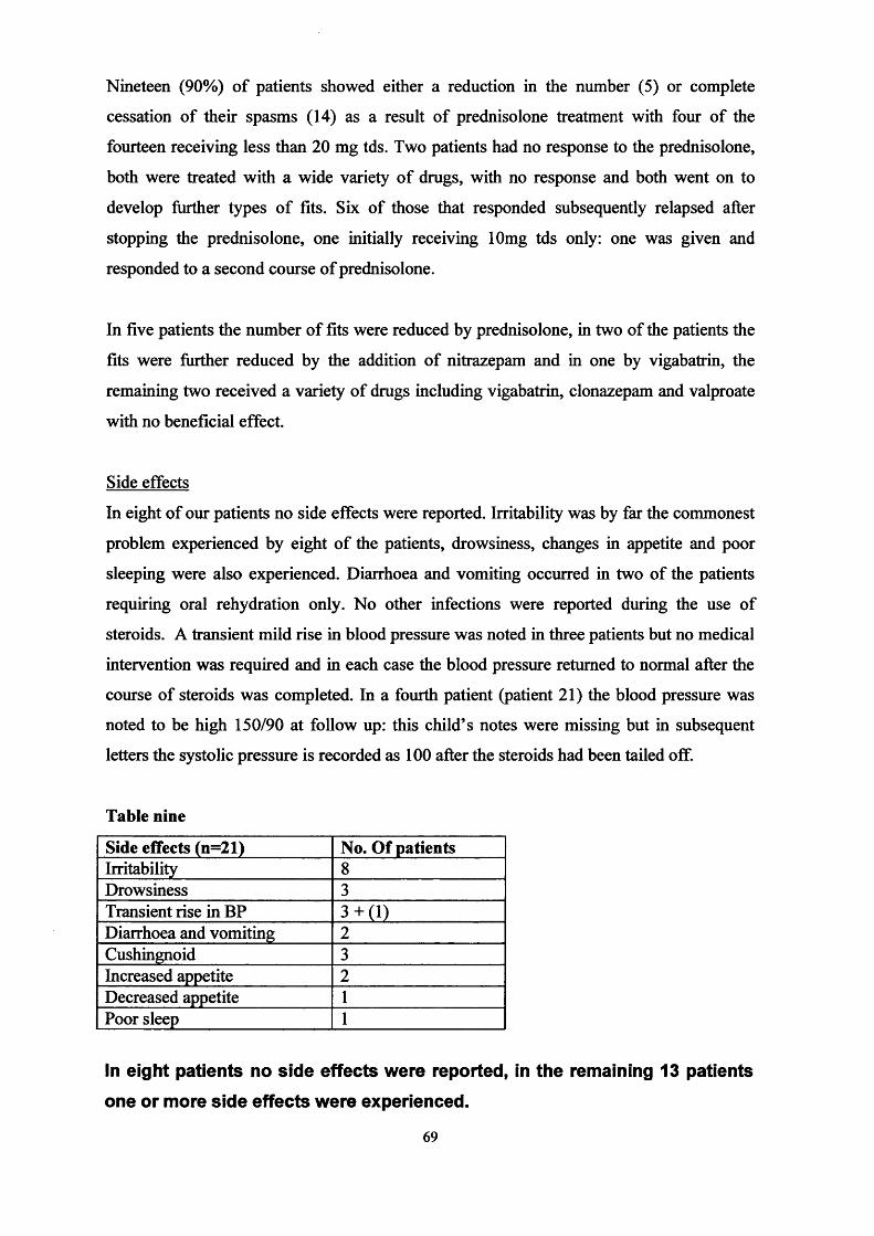
Nineteen (90%) of patients showed either a reduction in the number (5) or complete
cessation of their spasms (14) as a result of prednisolone treatment with four of the
fourteen receiving less than 20 mg tds. Two patients had no response to the prednisolone,
both were treated with a wide variety of drugs, with no response and both went on to
develop further types of fits. Six of those that responded subsequently relapsed after
stopping the prednisolone, one initially receiving lOmg tds only: one was given and
responded to a second course of prednisolone.
In five patients the number of fits were reduced by prednisolone, in two of the patients the
fits were further reduced by the addition of nitrazepam and in one by vigabatrin, the
remaining two received a variety of drugs including vigabatrin, clonazepam and valproate
with no beneficial effect.
Side effects
In eight of our patients no side effects were reported. Irritability was by far the commonest
problem experienced by eight of the patients, drowsiness, changes in appetite and poor
sleeping were also experienced. Diarrhoea and vomiting occurred in two of the patients
requiring oral rehydration only. No other infections were reported during the use of
steroids. A transient mild rise in blood pressure was noted in three patients but no medical
intervention was required and in each case the blood pressure returned to normal after the
course of steroids was completed. In a fourth patient (patient 21) the blood pressure was
noted to be high 150/90 at follow up: this child’s notes were missing but in subsequent
letters the systolic pressure is recorded as 100 after the steroids had been tailed off.
Table nine
Side effects (n=21) No. Of patientsIrritability 8Drowsiness 3Transient rise in BP 3 + (l)Diarrhoea and vomiting 2Cushingnoid 3Increased appetite 2Decreased appetite 1Poor sleep 1
In eight patients no side effects were reported, in the remaining 13 patients
one or more side effects were experienced.
69

No glycosuria was recorded at any time in any of the patients. Finally cushingnoid features
were noted in three of the infants, two of these patients had received two courses of
steroids (table nine).
Discussion
This was a small retrospective study and is therefore prone to bias. It is possible that cases
were missed the collection method and it is possible that those patients may have had a
worse outcome. The estimated incidence of IS ranges from 0.16 - 0.42 per 1000 live births
per year50 51 52 53. In the Bath district there are just over 5000 live births per annum. We
would therefore expect to see on average one to two cases a year (i.e. 13 -26 cases over the
13 year period). We believe that using our search strategy we are unlikely to have missed
many cases. In addition the study is prone to selection bias (see chapter five), that is
patients who might respond less well to steroids may have been given an alternative
treatment. Finally the numbers are small, thus the power of the study is small.
The side effects of steroids are well known. One of the aims of this study was to ascertain
the frequency of serious side effects with the use of high dose oral steroids. In our small
study of 21 patients none suffered serious complications of steroid therapy. There are the
less serious side effects that are often transient but nevertheless cause a certain amount of
morbidity such as hypertension. In our group we had three patients who had a mild
transient rise in blood pressure (95/50 to 155/80, 100/60 to 140/80 and 105/70 to 160/70),
fortunately with no serious sequele. The hypertension was usually seen after the patient
had been discharged from hospital, whilst being followed up in the out patient clinics.
Hypertension is often overlooked and as a result the amount of information available in the
literature is lacking. In the study carried out by Riikonen and Donner83 looking specifically
at the side effects of ACTH only 17 out of 162 patients had their blood pressure routinely
measured and in a review of the treatment by child neurologists of infantile spasms by
Bobele and Bodensteiner53 only two thirds measured blood pressure on a weekly basis
during treatment. In our study the blood pressure had been monitored in all patients for
whom the full medical notes and nursing observation charts were available. In our study
two thirds of the patients complained of minor side effects although none were sufficiently
severe to prevent continuation of the therapy.
70
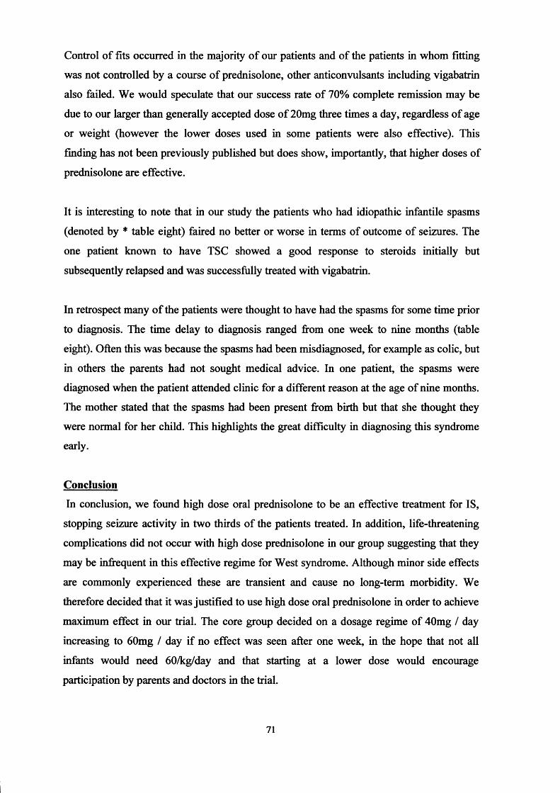
Control of fits occurred in the majority of our patients and of the patients in whom fitting
was not controlled by a course of prednisolone, other anticonvulsants including vigabatrin
also failed. We would speculate that our success rate of 70% complete remission may be
due to our larger than generally accepted dose of 20mg three times a day, regardless of age
or weight (however the lower doses used in some patients were also effective). This
finding has not been previously published but does show, importantly, that higher doses of
prednisolone are effective.
It is interesting to note that in our study the patients who had idiopathic infantile spasms
(denoted by * table eight) faired no better or worse in terms of outcome of seizures. The
one patient known to have TSC showed a good response to steroids initially but
subsequently relapsed and was successfully treated with vigabatrin.
In retrospect many of the patients were thought to have had the spasms for some time prior
to diagnosis. The time delay to diagnosis ranged from one week to nine months (table
eight). Often this was because the spasms had been misdiagnosed, for example as colic, but
in others the parents had not sought medical advice. In one patient, the spasms were
diagnosed when the patient attended clinic for a different reason at the age of nine months.
The mother stated that the spasms had been present from birth but that she thought they
were normal for her child. This highlights the great difficulty in diagnosing this syndrome
early.
Conclusion
In conclusion, we found high dose oral prednisolone to be an effective treatment for IS,
stopping seizure activity in two thirds of the patients treated. In addition, life-threatening
complications did not occur with high dose prednisolone in our group suggesting that they
may be infrequent in this effective regime for West syndrome. Although minor side effects
are commonly experienced these are transient and cause no long-term morbidity. We
therefore decided that it was justified to use high dose oral prednisolone in order to achieve
maximum effect in our trial. The core group decided on a dosage regime of 40mg / day
increasing to 60mg / day if no effect was seen after one week, in the hope that not all
infants would need 60/kg/day and that starting at a lower dose would encourage
participation by parents and doctors in the trial.
71

A review of vigabatrin in the treatment of infantile spasms in tuberous sclerosis
complex.
The next question to be addressed was whether patients known to have a diagnosis of TSC
at the onset of their spasms should be included or excluded from the trial. We therefore
undertook a review of the English literature to examine the efficacy and safety of
vigabatrin in the treatment of IS in infants suffering from TSC.
Methods
A Medline and Embase search dating back to 1990 of the English literature for all studies
investigating the effect of vigabatrin on IS was performed. Vigabatrin was first brought to
clinicians attentions by Chiron in 1991 and it is unlikely that there are any relevant trials
published prior to 1990. All references in the articles cited were then examined for further
references. Studies that did not distinguish which infants had an underlying diagnosis of
TSC were excluded.
Results
Fifteen studies investigating the effect of vigabatrin on IS were found in total. Of these, ten
studies gave a breakdown for the effect in patients with TSC in addition to the overall
results. Two of these studies98 99 were then rejected because the patients were all included
in a further publication100.
Table ten
Study Type of study No patients M:FChiron et al 19901W1 Single Blind 45 Not givenChiron et al 19918̂ Open add on 70 33:37Appleton et al 1993 100 Retrospective 21 10:11Vies et al 199310i Open 6 2:4Schmitt et al 1994103 Open 17 Not givenVigevano et al 1995104 Open randomised 21 Not givenAicardi et al 199688 Retrospective 192 109:83Chiron et al 199789 Open randomised 18 5:6
72
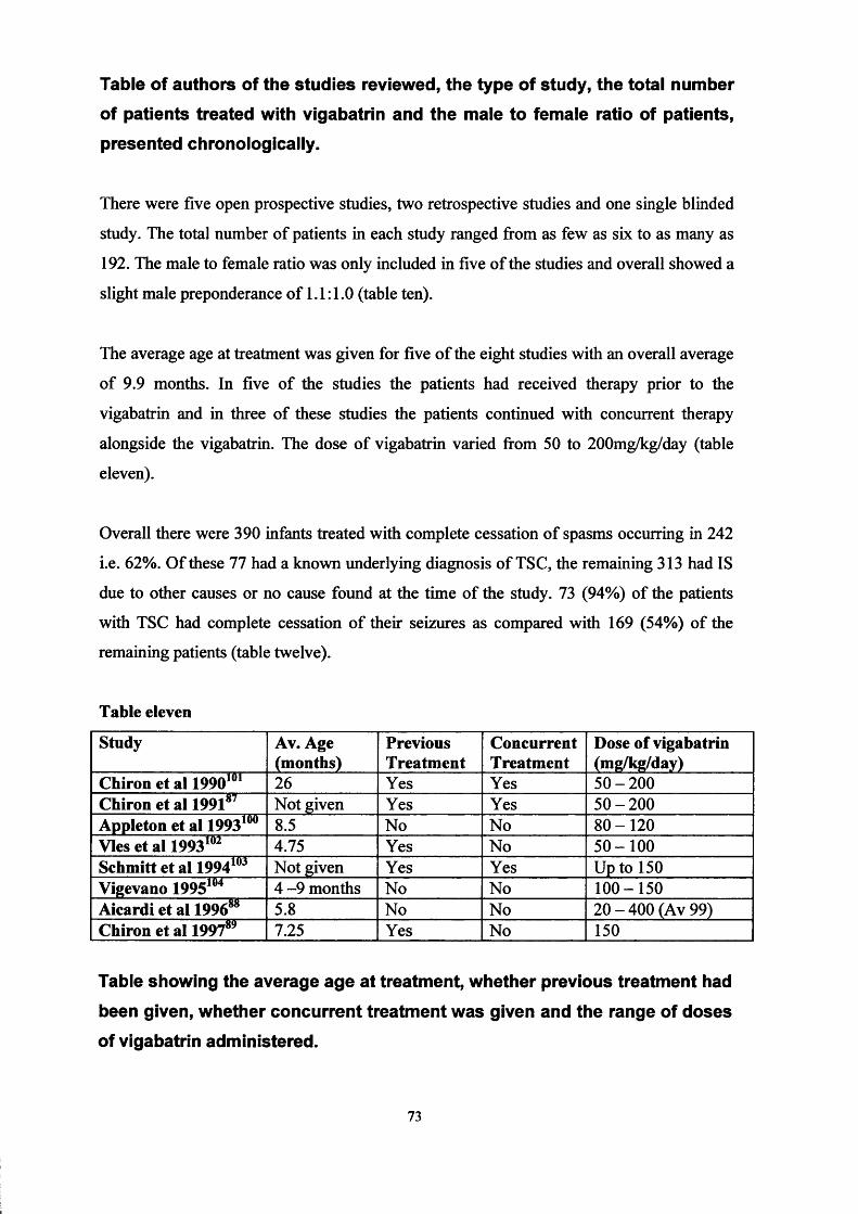
Table of authors of the studies reviewed, the type of study, the total number of patients treated with vigabatrin and the male to female ratio of patients, presented chronologically.
There were five open prospective studies, two retrospective studies and one single blinded
study. The total number of patients in each study ranged from as few as six to as many as
192. The male to female ratio was only included in five of the studies and overall showed a
slight male preponderance of 1.1:1.0 (table ten).
The average age at treatment was given for five of the eight studies with an overall average
of 9.9 months. In five of the studies the patients had received therapy prior to the
vigabatrin and in three of these studies the patients continued with concurrent therapy
alongside the vigabatrin. The dose of vigabatrin varied from 50 to 200mg/kg/day (table
eleven).
Overall there were 390 infants treated with complete cessation of spasms occurring in 242
i.e. 62%. Of these 77 had a known underlying diagnosis of TSC, the remaining 313 had IS
due to other causes or no cause found at the time of the study. 73 (94%) of the patients
with TSC had complete cessation of their seizures as compared with 169 (54%) of the
remaining patients (table twelve).
Table eleven
Study Av. Age (months)
PreviousTreatment
ConcurrentTreatment
Dose of vigabatrin (mg/kg/day)
Chiron et al 19901U1 26 Yes Yes 50-200Chiron et al 199187 Not given Yes Yes 50-200Appleton et al 1993100 8.5 No No 80-120Vies et al 199310i 4.75 Yes No 50-100Schmitt et al 1994103 Not given Yes Yes Up to 150Vigevano 1995104 4 -9 months No No 100-150Aicardi et al 199688 5.8 No No 20 - 400 (Av 99)Chiron et al 199789 7.25 Yes No 150
Table showing the average age at treatment, whether previous treatment had
been given, whether concurrent treatment was given and the range of doses
of vigabatrin administered.
73
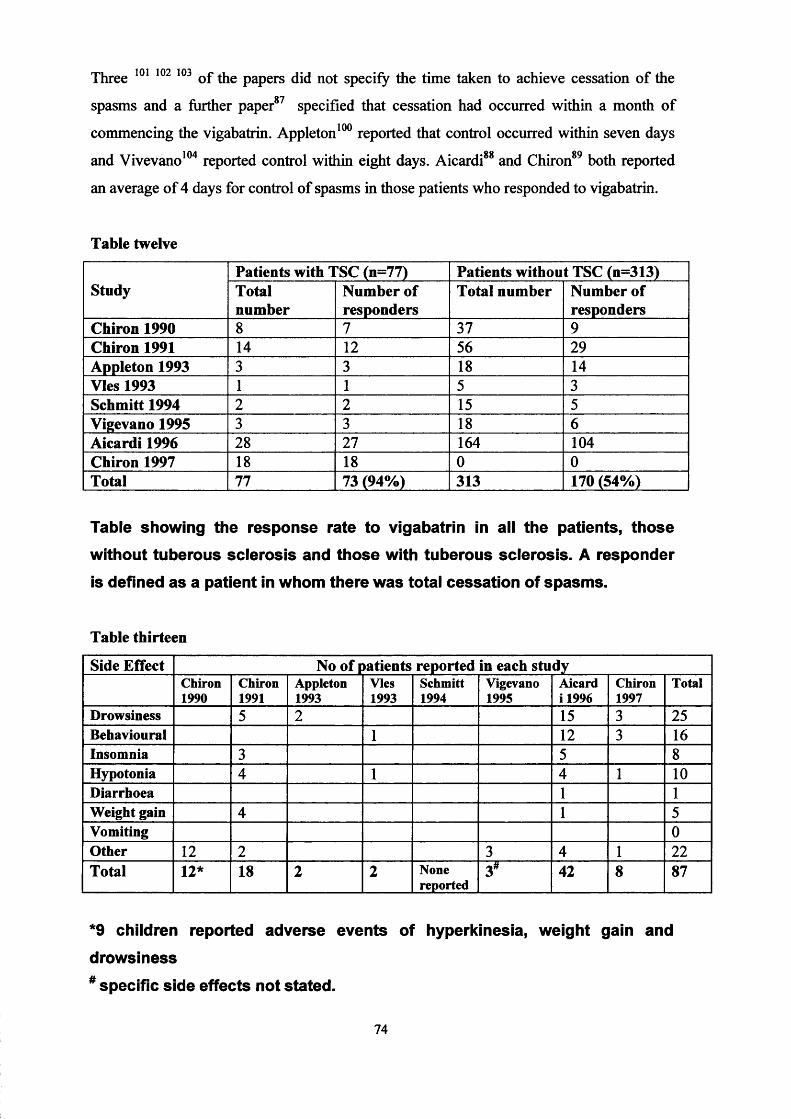
Three 101 102 103 of the papers did not specify the time taken to achieve cessation of the
spasms and a further paper87 specified that cessation had occurred within a month of
commencing the vigabatrin. Appleton100 reported that control occurred within seven days
and Vivevano104 reported control within eight days. Aicardi88 and Chiron89 both reported
an average of 4 days for control of spasms in those patients who responded to vigabatrin.
Table twelve
StudyPatients with TSC (n=77) Patients without TSC (n=313)Totalnumber
Number of responders
Total number Number of responders
Chiron 1990 8 7 37 9Chiron 1991 14 12 56 29Appleton 1993 3 3 18 14Vies 1993 1 1 5 3Schmitt 1994 2 2 15 5Vigevano 1995 3 3 18 6Aicardi1996 28 27 164 104Chiron 1997 18 18 0 0Total 77 73 (94%) 313 170 (54%)
Table showing the response rate to vigabatrin in all the patients, those
without tuberous sclerosis and those with tuberous sclerosis. A responder is defined as a patient in whom there was total cessation of spasms.
Table thirteen
Side Effect No of patients reported in each studyChiron1990
Chiron1991
Appleton1993
Vies1993
Schmitt1994
Vigevano1995
Aicard i 1996
Chiron1997
Total
Drowsiness 5 2 15 3 25Behavioural 1 12 3 16Insomnia 3 5 8Hypotonia 4 1 4 1 10Diarrhoea 1 1Weight gain 4 1 5Vomiting 0Other 12 2 3 4 1 22Total 12* 18 2 2 None
reported3* 42 8 87
*9 children reported adverse events of hyperkinesia, weight gain and
drowsiness
# specific side effects not stated.
74

Table showing the number and type of side effects I adverse reactions
reported in each study (there were 390 patients in total).In total 87, adverse events, from the 390 patients were noted (table thirteen). The majority
of these were transient and none were reported to continue after stopping the vigabatrin. In
all the studies only seven patients were reported as having the vigabatrin withdrawn due to
intolerability. The reasons for withdrawal were: hypertonia, hypotonia, hyperactivity,
irritability and myoclonus. There were no reported deaths attributable to vigabatrin.
Discussion
This review found a male : female ratio of 1.1:1. The average age of treatment was 9
months, which reflects that many of the patients had been tried on more traditional
therapies such as steroids before vigabatrin was used. Interestingly, in past studies TSC has
accounted for approximately 10% of patients with IS. In these studies it accounted for 20%
of the patients. This may be due to biased selection of patients (as most of the studies were
either open or retrospective) or alternatively with improving imaging and diagnostic
techniques, it may be that more patients are being diagnosed as having TSC. It may be a
chance finding.
The group of patients with TSC treated with vigabatrin had a 94% response rate. This
response rate was seen in spite of resistance to other treatment and delay in commencing
therapy. In contrast, in the group of patients in whom there was no history of TSC, the
overall response rate (i.e. total cessation of spasms) to vigabatrin was 54%, similar to
steroids but, it must be remembered that many of these patients treated with vigabatrin
were not newly presenting and had previously failed to have their spasms controlled by
other therapies.
Many children who suffer IS ultimately go on to have further seizures. The second
commonest seizure type in young children with TSC is partial seizures. Although partial
seizures are generally considered to be resistant to treatment with vigabatrin, a recent study
by Nabbout et al 1997105 suggested that in children with TSC vigabatrin may be effective
in treating partial seizures in up to 85% of patients.
75

Various dose regimes were used, but the core group decided to use the licensed dosage
regime:
50mg/kg/day orally in two divided doses for 24 hours (i.e. a total of two doses), increasing
to lOOmg/kg/day orally. If seizure control is not achieved then the dose can be further
increased to a maximum of 150mg/kg/day in two divided doses.
Although vigabatrin has only been in clinical use for a few years, it appeared to have few
serious side effects. Although a total of 87 adverse events were reported in the studies
reviewed, only seven patients had their vigabatrin stopped and no side effects were
reported to persist after withdrawal of the vigabatrin. (The reasons for withdrawing the
vigabatrin were only given for five of the seven patients and were: hypotonia, hypertonia,
hyperexcitability, irritability and myoclonus).
Earlier primate studies in dogs and rats had suggested a risk of intramyelinic oedema with
vigabatrin, but serial evoked potential studies and reports of brain histology in treated
patients have not supported this effect in humans106. However, since this work was
completed there have been reports of both asymptomatic and symptomatic visual field• 1H7defects with loss of peripheral vision to varying degrees in adults treated with vigabatrin
l08. It appears to occur most commonly in patients who have been treated with vigabatrin
for several years and more recent research, at least in adults, suggests that it is not
reversible on withdrawing the vigabatrin109. Out of approximately 140,000 adults treated
with vigabatrin between its introduction in 1989 and 1997 there had been 28 reports of
visual defects - a frequency of less than O.l90 107 108 109. The main difficulty in infants is that
they cannot be tested or monitored for visual field defects (a child needs to be at least
eleven to be able to co-operate and complete the test). However, on balance the devastating
effects of IS probably outweigh the risks of developing visual field defects (which may be
asymptomatic): a paediatric advisory group110 have recommended that vigabatrin should
still be prescribed for IS, and the medicine licence in the UK remains. In addition it must
be remembered that refractory epilepsy may cause visual defects itself, for example by
disseminated intravascular co-agulation (DIC) occurring with prolonged seizure activity111
and other anticonvulsants such as sodium valproate and phenytoin have also been110associated with visual defects . In some patients with infantile spasms, their underlying
disease may also cause visual field defects. Further research into the effects of vigabatrin
on visual fields in this group of children is required. Unfortunately, because of the
76

difficulty in testing visual fields in young children, it may be necessary to wait several
more years until infants treated with vigabatrin for infantile spasms become old enough to
co-operate with such testing. In addition, because of the devastating effect that the spasms
can have on development many of these children may never be able to perform visual field
testing.
Unfortunately only tw'o of the studies gave a breakdown for the effect of vigabatrin on
cessation of spasms due to other underlying aetiologies (e.g. cerebral dysgenesis, birth
asphyxia), a total of only 15 patients, so it is not possible to comment on the efficacy of
vigabatrin in these other subgroups. Neither is it possible to discuss the effect of vigabatrin
on EEG appearance in this review as the papers did not contain sufficient information. It is
also known that abolition of hypsarrhythmia on EEG can lag behind the clinical response,
and some of the studies only had a relatively short follow up period.
Conclusion
We conclude that vigabatrin should be considered the drug of choice for patients with IS
and either a known diagnosis of TSC, or at high risk of TSC before investigations are
complete i.e. those with a first degree relative with TSC because of its efficacy, speed of
onset and relatively safe drug profile. Paradoxically, steroids despite the side effects may
be a better choice for the remainder, but further information is required and a randomised
controlled trial clearly justified.
On the basis of this review it was decided that it would be unethical to randomise patients
with, or at high risk of TSC to steroids and they would therefore be excluded from the trial.
77

CHAPTER FOUR
The United Kingdom Infantile Spasms Study (UKISS)
Appendix one - protocol and trial pack
A core group (appendix two) was identified to propose a study design to investigate the use
of “steroids” and vigabatrin in the treatment of infantile spasms to try establish the most
effective initial treatment regime for this serious disorder. At the time of designing the
study the two “steroid” preparations in widespread use in the UK were ACTH
(adrenocorticotrophin hormone) and prednisolone. ACTH was a natural product derived
from a bovine source and as discussed above was administered as a daily intramuscular
injection with a commonly accepted dosage regime of 40-60iu/day. However, with the
existing concerns surrounding bovine spongiform encephalopathy (BSE) ACTH was
withdrawn from the market. It was replaced with synacthen depot, a synthetic analogue of
ACTH with a dose equivalent of 0.5-0.75mg/altemate days.
Infantile spasms are relatively uncommon and in order to be able to recruit the numbers
required to give the study reasonable power, the trial would need to recruit infants from a
large population such as the whole of the UK rather than just a few health districts. In order
to try and minimise selection bias it was decided that the trial should be a randomised
controlled trial with concealment of allocation (see chapter five). Ideally, in order to
minimise performance bias (see chapter five) the trial should also be blinded so that the
patients (parents/guardians) and medical staff administering the treatment and measuring
outcome would be unaware as to which treatment the subject had been randomised.
However, this would be problematic because synacthen depot can only be given by
intramuscular injection whilst prednisolone and vigabatrin are administered orally;
nowadays it is generally considered unethical to administer placebo injections to young
infants. In addition, the side effects of steroids are easily recognised by both clinicians and
parents so that it was likely that the treatment that had been assigned would become
apparent in a significant number of cases. The final dilemma was that few placebo
controlled trials have been performed and those that have contain insufficient number of
patients to provide adequate power to the study, so that we do not know if any one
treatment is more effective than no treatment at all. However, many would argue that
delaying treatment (which might worsen the outcome) by administering a placebo
78

treatment is also unethical. It was therefore decided that this should be a national,
multicentre, randomised, parallel-group, open clinical trial.
The trial centre would be in Bath and one Paediatric Neurologist in each of the old Health
Regions would be identified and recruited to help promote the study. It was hoped that
every health district within the UK would participate. Papers dealing with the
epidemiology of IS put the incidence between 1 in 2380 and 1 in 6250 births (see chapter
three). The average UK birth rate is approximately 800,000 per year. Therefore it should
be possible to recruit between 130 and 340 new cases per year. Taking into account
exclusions the scheduling of the study presumed a recruitment rate of approximately 200
per year. Assuming recruitment of 250 children over an 18 month period, with
approximately 125 randomised to vigabatrin and 125 to “steroids” and assuming
approximately 50% would achieve cessation of seizures in the “steroid” group, the trial
would have 90% power to detect an improvement in cessation of seizures to 70% in the
vigabatrin group. The consensus of the core group was that an improvement of at least
20% in cessation of spasms would be required to persuade clinicians to change their
clinical management of these patients. Central randomisation by telephone call would
register the child into the trial, report baseline information and allocate initial treatment
(concealed in sealed envelopes). Patients would be allocated to one of two treatment
groups: vigabatrin or steroids. The steroid group would then be further randomised to
receive synacthen or prednisolone. To try and reduce selection bias further, patients would
be stratified before entry according to sex, age and whether they belong to symptomatic or
non-symptomatic groups (see later).
We envisaged minimal problems with compliance and loss to follow up. This is a serious
disorder with profound sequele. The motivation for clinicians and parents of patients to
participate in treatment is high. The workload to be imposed on attending physicians
would be little more than constitutes good clinical practice. The workload to be imposed
on parents is small. Patients known to be likely to move outside the country during the trial
period would be excluded at entry. As this would be a national study, movement within
the UK should not lead to loss of contact. Patient's NHS number would be recorded on
entry to the trial thus making subsequent identification easier. In a previous multicentre
follow-up study conducted from Bath of 276 children, the investigators achieved 97%
79
follow-up at 3 years and 9 months. It was hoped that we could achieve a similar or
improved rate of follow-up in the UKISS trial.
To maximize the amount of information gathered during the trial it was decided to set the
drug trial within an epidemiological report of all cases of infantile spasms occurring within
the UK. This would allow us to collect further information on the epidemiology and
natural history of infantile spasms, including those infants who would be excluded from
the drug trial for whatever reason. It was decided therefore that we would ask that all cases
of infantile spasms would be reported to the trial centre even if the individual patient was
not to be recruited into the drug trial. This would also be helpful in allowing us to ascertain
how representative our trial population was compared to the whole UK population. Brief
clinical details would be requested.
In order to be recruited into the drug trial the patient would have to be between 2
completed calendar months and 1 year of age (up to but not including their first birthday)
at the onset of their spasms. Below the age of two months infantile spasms are rare and can
be difficult to diagnose. It was therefore decided that infants under the age of two months
would only be eligible for entry into the epidemiology part of the study. It was decided that
one year of age would be the upper age limit for drug trial entry for two reasons; firstly,
measurement of long-term psychomotor development (see later) would be undertaken
when the infant reached 12-14 months; secondly, after the age of one other possible
diagnosis should be considered, for example infantile myoclonic epilepsy, that can be
easily mistaken for infantile spasms. Infants would be excluded from the drug trial (but not
the epidemiology trial) if they were either known to suffer from tuberous sclerosis (TSC)
or who were at high risk i.e. a presumptive diagnosis of tuberous sclerosis because of;
known affected parent, previously diagnosed cardiac rhabdomyoma, hypomelanic macules,
a forehead fibrous plaque or shagreen patch noted, a retinal phakoma seen, or were known
to have polycystic kidneys. We recommended that first line therapy in these infants should
be vigabatrin (see chapter three). Infants would also be excluded from the drug trail if they
had received previous treatment for infantile spasms or had been treated with vigabatrin or
steroids within 28 days of the first diagnosis of infantile spasms for any other reason. This
was because the aim of the trial was to establish the most effective initial treatment for
infantile spasms. Other exclusion criteria included; infants with a contra-indication to
vigabatrin or steroids; infants with a lethal or potentially lethal other condition; inability of
80

parents or guardians to give informed, signed consent; inability of parents or guardians to
know when spasms stop - to the nearest whole day; infants expected to leave the UK
within one month of randomisation and finally any infants enrolled in a concurrent trial
that either uses therapy that might affect the outcome measures of the UKISS trial or one
that is time/effort consuming for the patients/guardians or the infants’ medical
practitioners.
The trial would then be discussed with the parents or guardians and their consent requested
for participation in the trial. If consent was refused for entry into the drug trial or the
infant was excluded for any reason given above then consent would be requested for entry
into the epidemiology trial only. If consent into the drug trial was successfully obtained
then an EEG (or video EEG) would be obtained as quickly as possible to confirm the
diagnosis of infantile spasms and allow randomisation into the drug trial. If the EEG was
normal the child would be excluded from the drug trial (but not the epidemiology trial).
Prior to randomisation for entry into the study the children would be stratified for; gender,
male or female; by age, 60 to 119 days, 120 to 179 days, 180 to 239 days, 240 days and
over; whether the infant belonged to a symptomatic or non-symptomatic group. The
symptomatic group consisted of those infants known to have an underlying diagnosis such
as a proven chromosomal abnormality, a syndrome diagnosis, cerebral palsy or a diagnosis
of neonatal encephalopathy with seizures, and those with a diagnosis of delayed
development having already been made by either a medical practitioner or health visitor
before the onset of the spasms. The non-symptomatic group includes all infants who had
no known underlying diagnosis at the time of diagnosis and who had normal development
prior to onset of the spasms. The purpose of pre-randomisation stratification was to balance
the treatment groups with respect to factors, which were identifiable at randomisation that
might affect the outcome measures.
All children eligible for the trial would then be randomised into the two treatment groups;
vigabatrin or “steroids” and all patients randomised to receive "steroids" would undergo a
second randomisation; to receive either synacthen depot or prednisolone. The only
pharmacological intervention for the infantile spasms for the initial trial period of two
weeks would be the drug to which each patient had been randomised. Those randomised to
receive vigabatrin would be given the dosage regime recommended by the manufacturers:
50mg/kg/day orally in two divided doses for 24 hours (i.e. a total of 2 doses); then to
81

increase to 100mg//kg/day orally in two divided doses in all patients; if at 96 hours seizure
control has not been achieved then the dose should be further increased to 150mg/kg/day
orally in two divided doses; they would then continue on this dose (100 or 150mg/kg/day)
until the time of their final developmental assessment around 12-14 months of age unless
no response or relapse occurred (see below). As ACTH gel was no longer available in the
UK, synacthen depot was substituted at the equivalent dosage; 0.5 mg on alternate days
(equivalent to 40iu/day of synacthen) intramuscularly (regardless of age or weight) for two
weeks. If at the end of the first week seizure control had not been achieved then the dose
would be further increased to 0.75 mg on alternate days (equivalent to 60iu/day of
synacthen) intramuscularly. This is a relatively high dosage regime, but one that is widely
accepted by clinicians in the UK. It is important that exogenous steroids are not stopped
suddenly, especially if they have been administered at high doses, as sudden withdrawal
can lead to an Addisonian crisis.
Addisonian CrisisAddisonian crisis is acute adrenocortical failure that may occur as the result of sudden withdrawal of exogenous steroids, and is a critical medical emergency. Delay in treatment may result in death. The patient usually presents with abdominal pain, vomiting, diarrhoea, pyrexia, dehydration and profound hypotension and later with collapse and coma. Laboratory parameters show low sodium, bicarbonate, glucose and cortisol levels and high potassium, urea and endogenous ACTH. Immediate management consists of resuscitation and intravenous hydrocortisone. All patients on steroid treatment should be advised not to alter their drug regime except on medical advice and should carry a medi-alert card.
This group would therefore tail on oral prednisolone over 15 days. Those on 0.5 mg on
alternate days of synacthen depot would receive 30mg of oral prednisolone for 5 days, then
20mg for 5 days then lOmg for five days and stop. Those on 0.75 mg on alternate days of
synacthen depot would receive 40mg of oral prednisolone for 5 days then 20 mg for 5 days
then lOmg for 5 days then stop.
Those randomised to prednisolone would be given lOmg qds. orally (regardless of age or
weight) for two weeks; if at the end of the first week seizure control had not been achieved
the dose should be increased to 20mg tds. orally. The dosage regime is higher than that
frequently used by clinicians in the UK but was chosen for the reasons outlined in chapter
three. This group would also tail on oral prednisolone over 15 days. Those on 40mg (10
mg qds.) would reduce to 30mg for 5 days, then 20mg for 5 days then lOmg for five days
and stop. Those on 60mg (20 mg tds.) would reduce to 40mg for 5 days then 20 mg for 5
days then 1 Omg for 5 days then stop.
82

Primary outcome measure
The primary outcome measure would be the number of patients who achieve complete
cessation of spasms for at least 48 hours up to and including the end of the 14th day of
treatment.
At enrolment the parents/guardians would be given their copy of a ‘fit diary’ and its use
fully explained to them. The parents/guardians would be asked to make a once daily entry
for the first 4 weeks of the trial and a once weekly entry thereafter, until the time of the
final developmental assessment (12 -14 months), with a final 7 day daily entry for those
still having seizures.
Responders are defined as those in whom there is total cessation of spasms for at least 48 hours up to and including the end of the 14th day of treatment.Relapse: a single spasm in a responder constitutes a relapse.
If a child relapses within the initial two week period then the lead clinician would be
expected to increase the dose of the initial therapy up to the maximum recommended dose
as per protocol.
The patient should be seen by the enrolling physician at two weeks post-trial entry to
confirm their progress. If the infant has responded the patient will continue on the same
therapy as above. The trial will be analysed by intention to treat at the time of
randomisation; in order to minimise the effects of a multitude of different subsequent
treatments if a response has not occurred after 2 weeks, the lead clinician will be asked to
change to the alternative therapy i.e. either steroids after vigabatrin (the lead clinician to
choose which steroid - synacthen depot or prednisolone) or vigabatrin after steroids. This
alternative treatment will continue for the third and fourth weeks (a full 14 days) at the
same dose as recommended for the first two weeks. If both treatments fail, the clinician
will be left to choose the most appropriate treatment.
If the patient relapses after the initial two week trial period we suggest that if the child is
on vigabatrin to increase the dose in parallel with the child’s weight gain (if increase is
more than 1 kg) or if on lOOmg/kg/day increase to 150mg/kg/day; If on steroids repeat the
course once even if tailing. If these measures fail after a full 14 days treatment change to
83

the alternative therapy i.e. either steroids (synacthen depot or prednisolone) after
vigabatrin, or vigabatrin after steroids.
Secondary outcome measures
ft was decided that the time taken to complete cessation of all spasms for at least 48 hours
would be measured as many clinicians believe that the quicker the spasms are brought
under control the better the long-term outcome. If one treatment does bring the spasms
under quicker control and also improves long-term psychomotor development then this
would support the above hypothesis. In addition the spasms may be distressing to the
infant and parents; if both treatments were found to be equally efficacious then it might be
preferable to use the treatment that works most quickly.
Any reduction in the number of spasms suffered by non-responders would also be
measured. If the duration of spasms is linked to long-term psychomotor development then
likewise the number of spasms may also be important in determining long-term
development. In addition if the spasms are distressing to the infant a treatment which
reduces their frequency may be preferable to one that has less effect.
Relapse rates; clearly even if one treatment stopped the spasms in a greater proportion than
the other, if remission of the spasms could not be maintained then if the number of patients
who remained in remission in the other group was higher it might be considered to be the
most efficacious treatment overall.
The precise relationship between clinical spasms and hypsarrhythmia is not clear
especially with regard to long-term outcome. To be eligible for entry into the drug trial
hypsarrhythmia had to be present on the entry EEG. An EEG was then to be repeated at the
end of the initial two week period. It was hoped this might allow us to determine whether
or not resolution of the hypsarrhythmia has any impact on the long term outcome. Patients
who did not have hypsarrhythmia on the entry EEG would be asked to participate in the
epidemiology trial, this would allow us to hypothesize whether patients without
hypsarrhythmia at time of diagnosis have a better/worse/similar outcome to those who do
have hypsarrhythmia.
It is known that in the majority of patients the natural history of the spasms is that they will
resolve with time, even if the patient is untreated. Nonetheless, many patients will continue84

on to develop other forms of seizures and epilepsy. It is not yet known whether controlling
the spasms effectively bears any relationship to the risk of developing other seizure types.
It was therefore decided that subsequent seizure types would be regarded as a long-term
outcome in our group of patients and would be measured at the time of the follow up for
psychomotor development. The type and frequency of seizures other than spasms suffered
by the infant in the month preceding the developmental assessment at 12-14 months would
be recorded.
Long term psychomotor development may be the most important outcome for this group of
patients. Many would argue that any treatment proven to be most beneficial with respect to
developmental progress should be used in preference to any other medication regardless of
its effect on the other outcomes listed above. Developmental progress would be assessed at
the time of trial entry and again at 12 -14 months of age. This would allow at least 2
months of recovery following treatment and is an age at which some language
development has occurred. It is believed that delaying the assessment to a greater age will
give little advantage and would have the disadvantage of increasing the impact of
environmental factors. The Vineland Adaptive Behavioural Scales were chosen after
lengthy consideration as the tool of choice for assessing developmental outcome in our
group of patients.
The Vineland Adaptive Behaviour Scales have been developed and validated in the U.S.A. to measure “adaptive behaviour” defined as the performance of the daily activities required for personal and social sufficiency. The Vineland has four domains (communication, daily living skills, socialisation and motor skills) and these combine to give an Adaptive Behaviour Composite: each of these has a standard score of 100 with a standard deviation of 15 and each can be expressed as an age equivalent (developmental age).
One of the main benefits of the Vineland is that it can be easily adapted to be performed as a questionnaire that can be administered over the phone by a trained researcher who would be based in Bath either to the enrolling physician or parent/guardian. In addition it concentrates on the usual response, not the best response to any task. It has been validated in greater detail on larger numbers of children (3000) and has been used in the U.K. for other developmental follow-up studies. It achieves construct validity, content validity and has been compared to other scales in both normal and handicapped children. It has been shown to have internal consistency, test-retest and inter-rater reliability. It has also been designed to minimise the influence of physical handicap.
Finally side effects and deaths in each group would be recorded as these are considerations
which in clinical practice often dictate the acceptability of a treatment to the
clinician/patient//parent /guardian. Should an infant suffer unacceptable side effects during
the trial then the treatment should be discontinued and the trial centre informed
immediately. The lead clinician may then choose his own alternative treatment, but we
85

suggest the lead clinician change to the alternative therapy i.e. either steroids (synacthen
depot or prednisolone) after vigabatrin, or vigabatrin after steroids.
Investigations
In order to understand more about the associations and aetiologies of infantile spasms we
choose to collect information on the results of any investigations undertaken on any patient
enrolled into the trial. As part of normal clinic practice it was anticipated that the following
investigations would be undertaken on all subjects.
1. MRI or CT of the brain.
2. Urine metabolic screen for amino acids.
3. Ophthalmoscopy (either direct or indirect).
4. Ultraviolet light examination (Woods light).
We requested that the results of these tests should be forwarded to the trial centre. The
following tests might also have be considered appropriate in some cases and, if performed,
the results of these and any other tests would be requested.
1. Urea and electrolytes.
2. Liver function tests.
3. Chromosomes.
4. Urine metabolic screen for organic acids.
5. Lactate.
6. Biotinidase assay.
7. Ammonia.
8. Thyroid function.
9. Pyridoxine response - if pyridoxine is to be given we would recommend that
pyridoxine is given one month after the start of the trial (after both the
randomised treatment and first alternate treatment have been given in a child
who continues fitting).
Finally, at present it is not thought that infantile spasms are genetically determined, but
there are rare reports of X-linked cases in the literature54. To investigate the possibility of a
genetic predisposition we requested that if venesection was performed for any clinical
reason during the course of the infant's illness, then a blood sample be taken for DNA
analysis.
86

Analysis
A Data Monitoring Group has been set up (appendix two) that will analyse and assess the
results independently of the trial investigators (the Trial Steering Committee, appendix
two) for the duration of the trial at a frequency to be recommended by the chair.
There will be post-randomisation stratification for potential confounding variables not
accounted for in pre-randomisation. These include EEG appearance (i.e. hypsarrhythmia vs
atypical vs normal awake, normal asleep) and diagnostic sub-groups (i.e. cerebral
dysgenesis on cranial scanning, chromosomal abnormalities or other syndromes and other
diagnosis such as metabolic disease). Social factors will include birth order and maternal
age at school leaving.
It may be possible to see if any trends in outcome are repeated within subgroups of the trial
population. The largest subgroup should be infants where development was delayed (as
shown by pre-randomisation stratification data and as shown separately by the
developmental history). Smaller subgroups, e.g. those diagnosed with cerebral palsy on
clinical examination, cerebral dysgenesis on cranial imaging, normal awake and sleep
EEGs, are unlikely to be large enough for separate analysis. However, the data will be
examined in case there is a dramatic difference in outcome, or a consistent trend.
We are aware that the pattern of developmental age in the two groups may be bimodal. A
direct comparison of the developmental age (total score and subsets) of the two groups will
be undertaken, but we will also determine the proportion of individuals in each group
whose development is one, two and three standard deviations below the mean for the
Vineland.
Ethical approval was then sought and granted from the Multicentre Research Ethics Committee (MREC). Investigators are continuing to be recruited and Local Research Ethics Committees (LREC) approached. Enrolment of patients into the trial began June 1999.
87

REPORT ON STUDY STATUS TO THE END OF SEPTEMBER 2002
Local ethical approval had been granted in 150 centres across the UK.
Enrollment
One hundred and five infants have been enrolled into the randomised controlled drug trial
and the epidemiology study.
Eighty eight infants have been enrolled into the epidemiology study only.
A further fourty four infants have been notified to the trial centre but were not eligible for
inclusion into the trial.
Duration of study
Due to unforeseen delays in recruiting centres and obtaining local ethical approval, the rate
of recruitment into the trial has failed to reach the above estimates. Additional funds have
been granted to allow the UKISS study to continue until the end of 2002 to allow us to aim
to reach an adjusted target recruitment of 125 patients into the randomised controlled drug
trial, this will give us an 80% power to see a 25% difference in cessation of spasms
between the two groups.
88

CHAPTER FIVE
The Cochrane Collaborative: a systematic review comparing the medical
treatments of infantile spasms (West syndrome) in terms of long term
developmental outcome, seizure control and treatment side effects.
Whilst searching the literature for information on trials and studies that had previously
investigated the effect of drug treatments on IS it became clear that although there was a
wealth of literature on this subject there were the following problems in trying to assess the
validity, applicability and implications of the results of these studies:
1. There were few randomised controlled trials (RCTs), the majority of reports were
either prospective non-randomised or retrospective studies. The main problem with
non-randomised controlled trials is that they are open to selection bias: i.e. systematic
differences between comparison groups in prognosis or responsiveness to treatment.
2. Many of the studies contained only small numbers of subjects with the result that they
have little or no power.
3. The studies were located by traditional methods only i.e. through the electronic
databases usually Medline only. The problem with this method is that only 30 - 80% of
published trials can be located via any one database. In addition unpublished trials were
not sought leading to publication bias: studies in which an intervention is not found to
be effective are far less likely to be published thus traditional reviews tend to
overestimate the true effect of an intervention.
4. Only studies published in the English language were considered, again studies in which
an intervention is not found to be effective are less likely to be published in English
leading to language bias.
In order to try and minimise the above problems and determine if any one treatment for IS
was better in terms of long-term outcome, seizure control and side effects a systematic
review in conjunction with the Cochrane Collaboration was undertaken. Although a
conventional review of the literature on the treatment of IS had shown that few studies had
considered the long-term outcomes (i.e. psychomotor development and progression to
other seizure types) when investigating the effect of medical therapies on IS, one of the
aims of this review was to set a “Gold Standard” for future studies.
89

It was decided that there are four important factors that must to be considered when
choosing a therapy for treatment of any infant diagnosed with IS;
1. Development
Any effect on long-term psychomotor development: arguably the most important
factor.
2. Subsequent epilepsy
Many patients continue to have other forms of epilepsy into adult life and about
20% will progress to Lennox Gastaut syndrome . These patients will therefore
require anticonvulsants and will continue to suffer the effects of epilepsy often for
life.
Definition: Lennox Gastaut syndrome;The Lennox-Gastaut syndrome is an age specific epileptic encephalopathy, characterised by epileptic seizures, slow spike-waves in the waking electroencephalogram (EEG) and fast rhythmic bursts during sleep, psychomotor delay and personality disorders. Its incidence is not known but it has been estimated to account for 1-10% of all childhood epilepsies. It occurs more frequently in males and onset is usually before the age of eight with a peak between three and five years
The following seizure types and EEG findings are associated with Lennox-Gastaut syndrome;• Atypical absence seizures: these occur in the majority of cases and are frequently subtle. Loss of consciousness may be incomplete allowing the patient to continue ongoing activities. However they are often accompanied by loss of muscle tone, myoclonic jerks and drooling. EEG shows irregular diffuse slow spike-wave activity at 2 - 2.5hz.• Tonic seizures: these are commonly seen and may be axial, appendicular or global, they may be symmetrical or unilateral. They consist of flexion of the neck and body, extension of the arms and legs and contraction of the facial muscles. There may be associated apnoea, eye rolling and facial flushing. Consciousness is usually impaired. They are usually brief, lasting seconds. The EEG shows discharges of fast bilateral bursts predominately anteriorly or on the vertex.• Myoclonic and atonic seizures: these are less common and consist of a sudden head drop or fall to the ground and are associated with polyspikes and slow waves, or diffuse spike waves, or fast rhythms with an anterior predominance on the EEG.• Status epilepticus and non-convulsive status occurs in approximately two thirds of patients and usually consists of continuous absence seizures punctuated by recurring tonic seizures and may be difficult to recognise.• The waking EEG is abnormal in the vast majority of cases showing 2 to 2.5hz slow spike-wave discharges over both hemispheres with multifocal spikes and spike waves predominating in the frontal and temporal areas.
Psychomotor delay and neuropsychiatric symptoms occur in 90% of patients with Lennox-Gastaut syndrome. Language is frequently affected with both slowness in ideation and expression in addition to difficulties of motor dysfunction. Severe behavioural disorders and personality disorders are nearly always present. There is also a tendency for psychosis to develop with time. The long-term prognosis is poor; although the epilepsy often improves, complete seizure freedom is rare and conversely the mental and psychiatric disorders tend to worsen with time.113
3. Response: control of the spasms
The spasms are certainly distressing for parents and may well be distressing to the
infant. It is also possible that the spasms are directly linked to the development of
SLD as it is generally thought by clinicians (anecdotal evidence only) that the90
earlier the spasms are brought under control the better the outcome. It may of
course be that those with a better prognosis have seizures that are easier to control.
It was therefore decided to look at control of spasms in terms of;
• complete cessation of spasm
• time taken to achieve cessation of spasms
• reduction in total number of spasms
• relapse rates
• resolution of hypsarrhythmia
The exact relationship between clinical spasms and hypsarrhythmia on the EEG is not
entirely clear. Not all infants with spasms have classical hypsarrhythmia on the EEG,
and in those in whom it is present it is not always present all of the time65. It is not
known whether presence or absence of hypsarrhythmia affects long-term outcome.
4. Side effects
These must always be taken into account. Any drug with unacceptable side effects will
not be used in clinical practice. Likewise if two drugs show similar beneficial effects
then the one with the safer drug profile should be used in preference.
The following review was undertaken under the aegis of the Cochrane Collaboration, it
strictly adheres to the guidelines set out in the Cochrane Collaboration Handbook and was
compiled and analysed using the Revman Package provided by the Cochrane collaboration.
ReviewersDr Eleanor Hancock (ECH), Prof. John Osbome(JPO), Prof. Phillip Milner (PLM).
Contribution of reviewers
ECH was primarily responsible for all aspects of this review including protocol design,
undertaking the searches, the database of studies, data collection and extraction, data
analysis and presentation of the results.
JPO and PLM agreed and approved the review at all stages and independently evaluated
which studies should be included and excluded from the review and also independently
extracted the data from the included studies.
Background
There have been numerous clinical trials looking at different treatment regimes in the past
(discussed in detail in chapter three). The main problem is that few of the trials have been
91

randomised controlled trials and that the drug under investigation has frequently been used
long after the onset of seizures and in addition to or after other anticonvulsants. There is
wide variation in the drug dosages used and the duration for which the drugs are used. As a
result it is confusing when looking at the literature to know which is the best treatment for
this disorder with the consequence that many different treatment modalities are currently in
use.
A conventional review of the literature on the treatment of infantile spasms carried out byo 1
Haines and Casto in 1994 concluded that the results of previous studies were widely
varying and that well-designed, blind, prospective clinical trials are needed to answer many
questions regarding the treatment of infantile spasms.
Objectives
The aim was to compare the effects of single pharmaceutical therapies used to treat
infantile spasms in terms of long-term psychomotor development, subsequent epilepsy
rates, control of spasms and side effects.
The following hypotheses were tested:
A. Therapy vs placebo1. Therapy A* will give improved long term psychomotor development compared with placebo treatment.2. Treatment of infantile spasms with therapy A* will reduce subsequent epilepsy rates compared with placebo treatment.3. Therapy A* is more effective in controlling infantile spasms (in terms of spasm cessation, reduction in total number of spasms and relapse) than placebo treatment.4. Therapy A* is more effective in resolving hypsarrhythmia than placebo treatment.
B. Therapy vs no treatment1. Therapy A* will give improved long term psychomotor development compared with no treatment.2. Treatment of infantile spasms with therapy A* will reduce subsequent epilepsy rates compared with no treatment.3. Therapy A* is more effective in controlling infantile spasms (in terms of spasm cessation, reduction in total number of spasms and relapse) than no treatment.4. Therapy A* is more effective in resolving hypsarrhythmia than no treatment.
C. Comparisons between therapies1. Therapy A* will give improved long term psychomotor development compared with any other single pharmaceutical therapy.2. Treatment of infantile spasms with therapy A* will reduce subsequent epilepsy rates compared with any other single pharmaceutical therapy.3. Therapy A* is more effective in controlling infantile spasms (in terms of spasm cessation, reduction in total number of spasms and relapse) compared with any other single pharmaceutical therapy.4. Therapy A* is more effective in resolving hypsarrhythmia compared with any other single pharmaceutical therapy.
♦Therapy A = ACTH** or Hydrocortisone or Prednisone / Prednisolone or
92

Carbamazepine or Ethosuximide or Gabapentin or Lamotrigine or Phenobarbitone or
Phenytoin or Topiramate or Vigabatrin or Valproate or Clonazepam or Diazepam or
Nitrazepam or Pyridoxine (Vitamin B6) or any other single therapeutic agent studied in the
literature.
** ACTH At the time of undertaking this review there were two "ACTH" preparations in
widespread use; ACTH (adrenocorticotrophin hormone) and tetracosactrin. ACTH is a
natural product derived from a bovine source administered as a daily intramuscular
injection. However, in the UK, with the existing concerns surrounding bovine spongiform
encephalopathy (BSE) ACTH has been withdrawn from the market.
Criteria for considering studies for this review
Types of studies
All randomised controlled trials (RCTs) of the administration of drug therapy to infants
and children with infantile spasms were included in this review, including trials which
compared a therapy with none or placebo and trials that compared one drug with another.
Definition of RCT: trials in which participants are prospectively allocated to treatment groups by a random (e.g. random number generation, coin flips) or quasi random (e.g. by date of birth) process.
If the study was not an RCT it was not included in the review: the existence of such studies
was documented.
We considered studies looking at drug therapy as second line therapy as well as those
studies looking at drug therapies as first line therapy.
Types of participants
Any infant or child treated for infantile spasms, regardless of whether or not EEGs were
performed or whether therapy had been given prior to trial entry.
As the majority of published studies on infantile spasms did not give a definition of infantile spasms used for their participants, for the purpose of this review, we assumed a clinical diagnosis had been made for any participant that was entered into a trial. We documented any definitions which were given, and considered whether differences in definition might account for between study variation.
93

Types of interventions
1. Any trial that compared at least one therapy against placebo treatment
2. Any trial that compared at least one therapy against no therapy
3. Any trial that compared at least one therapy against another therapy
Therapies included:
1. "Steroids"; ACTH, Tetracosactrin, Hydrocortisone, Prednisone/prednisolone.
2. Antiepileptic drugs; Carbamazepine, Clonazepam, Diazepam, Ethosuximide,
Gabapentin, Lamotrigine, Nitrazepam, Phenobarbitone, Phenytoin, Pyridoxine, Sodium
Valproate, Topiramate, Vigabatrin.
3. Methysergide and methylparatyrosine.
4. Any other single therapeutic agent studied in the literature.
Any dose regime of the above therapies was included.
Types of outcome measures
1. Long term psvchomotor development
This was to be analysed in months as a measure of the deviation from the chronological
age. It was to be measured at 3 months post entry into the trial and where possible at 5
years of age. It was measured as a continuous variable.
2. Subsequent epilepsy rates
The diagnosis of any other epileptic seizure type other than infantile spasms after the
commencement of therapy constituted subsequent epilepsy. It was to be measured at three
months post entry into the trial and where possible at five years of age. Any seizure type
plus each seizure type (e.g. primary generalised, partial etc.) was to be measured as a
dichotomous variable (i.e. present / absent) over the previous one month at three months
post entry and over the previous six months at five years of age.
Because of the difficulty of defining epileptic seizures in children we would use the
original authors1 diagnosis.
3. Cessation of spasms
This was defined as total cessation of spasms for at least 48 hours after commencing
therapy, but occurring within a month of commencement of therapy. It was measured as a
dichotomous variable (i.e. ceased / continuing). Time taken from commencement of
therapy to cessation of spasms was also to be measured as a continuous variable (measured
94

in days).
4. Quantitative reduction of spasms
This was measured as the number of spasms occurring before treatment was commenced
compared with the number occurring following treatment and was to be measured as a
dichotomous variable (i.e. greater than 50% reduction or less than 50% reduction in the
number of spasms occurring / day over the seven day period before trial entry and over a
seven day period at one month of commencing therapy).
5. Relapse rates of spasms
A single spasm occurring by one year of age or within the study period, but after cessation
of spasms constituted a relapse. It was measured as a dichotomous variable (i.e. relapse
occurred / no relapse occurred). Time taken from cessation of spasms to relapse was also to
be measured as a continuous variable (measured in days) in those patients in whom spasms
had ceased.
6. Resolution of hypsarrhythmia
Patients were divided into those who had a normal EEG prior to entry into a study and
those who did not. In the group that did not have a normal EEG on entry, the EEG
appearance after treatment was divided into those in whom the EEG remained abnormal
and those in whom it became normal. It was measured as a dichotomous variable (i.e.
normal / abnormal).
7. Side effects
Only side effects that were considered severe enough to warrant discontinuation of the test
treatment were measured. They were measured as a dichotomous variable i.e. therapy
stopped vs therapy not stopped. They were also qualitatively summarised.
8. Deaths
All deaths were measured. They were measured as a dichotomous variable (i.e. alive /
deceased).
Search strategy for identification of studies
In addition to searching the central trials register of the Cochrane Epilepsy Group (see
group module) we searched:
1. MEDLINE Database (1960 to 2001) according to the search strategy used by the
Cochrane Epilepsy Group.
2. EMBASE Database (1981 to 2001).
3. The reference lists of both the Randomised Controlled Trials and rejected articles were
95

scanned (by ECH) to identify possible articles missed by the computerised search.
4. Correspondence with colleagues to try and identify unpublished data. Correspondence
with authors of review articles has not yet been undertaken.
5. Correspondence with drug companies to try and identify unpublished data.
6. Appeals at international conferences to delegates to try and identify unpublished data
and studies not published in English.
Methods of the review
Relevant publications were reviewed independently by the three reviewers (Dr Hancock,
Professor Osborne and Professor Milner). Discrepancies were resolved by discussion.
There was no blinding of authorship or results. All randomised controlled trials were
considered. All non-English studies were also considered.
1. Exclusion criteria
• Any trial that was not a RCT was excluded from analysis but was documented.
• If a clinical definition for infantile spasms was given; in any trial in which there
was doubt about the clinical diagnosis, the trial was excluded from analysis, but
was documented.
2. Assessment of methodological quality
• Selection bias: the studies were assessed as to whether allocation concealment was
adequate, unclear or inadequate.
• Performance bias: The studies were assessed as to whether recipients and those
measuring outcome were unaware of the assigned therapy (However we accepted
that the majority of the studies in this review would not be double blinded trials)
• Attrition bias was assessed as to whether there was loss of patients to follow up.
Using the above criteria studies were then divided into (1) those with a low risk of bias (2)
those with a moderate risk of bias and (3) those with a high risk of bias. (See Cochrane
Handbook section 6.7.1)
In studies where it was unclear if the above criteria have been met then the reviewers are
endeavouring to obtain additional information by contacting the first author on up to three
occasions. This will be documented.
96

The following data was extracted independently by the three reviewers (Dr Hancock,
Professor Osborne and Professor Milner) and discrepancies resolved by discussion.
1. Participants (i.e. those characteristics of the population that may affect outcome
regardless of treatment):
• age at spasm onset, diagnosis and start of treatment (mean, median and range measured to
the nearest completed week of age).
• time taken from onset of spasms to initiation of treatment (mean, median and range
measured to the nearest completed week).
• sex (male, female).
• previous treatment (e.g. prednisolone, ACTH, vigabatrin, valproate).
2. Interventions:
• type of pharmaceutical agent used (e.g. prednisolone, ACTH, vigabatrin, valproate).
• dose (measured in the internationally accepted units i.e. mg for prednisolone, IU for
ACTH).
• frequency (measured as the number of times the pharmaceutical agent is given in a 24
hour
period).
• route of administration (i.e. oral, intramuscularly, intravenously).
• treatment length (measured to the nearest completed day).
3. Outcome measures:
• psychomotor development.
• subsequent epilepsy rates.
• cessation of spasms and the time taken to cessation of spasms.
• reduction in spasms.
• relapse rates and the time taken to relapse.
• resolution of hypsarrhythmia and the time until resolution.
• side effects.
• deaths.
Analysis plan
1. Study quality: this was done by a table of met / unmet criteria for selection, performance
and attrition bias.
2. Dichotomous data: for each item of data requiring dichotomous analysis the following
97

was recorded; no. of patients who experienced the event (or outcome) in each group for
each comparison and the total number in each group. They were analysed using Peto odds
ratios in the Cochrane RevMan Package.
3. Continuous data: for each item of data requiring continuous data analysis, except
psychomotor development the following were recorded; number of patients in each group,
the mean value for the outcome in each group and the standard deviation. They were
analysed using weighted means in the Cochrane RevMan Package
4. When data for the same outcome is presented in some studies as dichotomous and in
others as continuous data then we are endeavouring to obtain continuous data from the
investigators. If it is not possible to obtain continuous data, for example because it was not
recorded then the data will either be analysed as dichotomous data with a "cut off' point
agreed by the three reviewers or a mixture of dichotomous and continuous data will be
analysed using two separate tables.
5. We looked for sources of heterogeneity between trials of methodological and clinical
differences, including previous treatment for spasms, age at trial entry, single underlying
cause. We also looked for subgroup analysis of clinical features, for example;
developmental delay prior to onset of spasms, differences in drug dosages, timing and
length of treatment and specific underlying diagnosis such as tuberous sclerosis.
Description of studies
The literature search of the central trials register of the Cochrane Epilepsy Group found 13
potential studies. The literature search of MEDLINE (1960-2001) found 33 potential
studies, and the literature search of EMBASE (1981-2001) found 14 potential studies.
Some studies were identified from more than one database. From these three databases a
total of 43 potential studies were identified. The references of these 43 studies were then
scanned by Dr Hancock and a further 9 possible studies identified. Direct correspondence
with colleagues and appeals at international conferences (Seattle September 2000 and
Tokyo February 2001) also identified two of the above (Appleton 1999 and Vigevano 1997
pre-publication) randomised controlled trials (and two ongoing trials). Correspondence
with drug companies did not identify any further trials. This brought the total to 52 studies
to be evaluated for inclusion into this review (see appendix three for references). We
intend to continue correspondence with authors, colleagues and drug companies and to
regularly update our literature searches of the above databases, so that we may update this
review as future studies are completed. Hand searching of journals has not yet been
98

possib le .
Table fourteen
Study Methods Participants Interventions Outcomes
Appleton19991
Randomised controlled trial
40 patients with a clinical definition of infantile spasms and hypsarrhythmia on EEG.
VigabatrinPlacebo
Cessation of spasms Reduction in spasms Relapse rate Resolution of EEG
Baram19962
Randomised controlled trial
29 patients with a diagnosis confirmed on video EEG
ACTH Cessation of Spasms Prednisone Relapse rates
Resolution of EEG
Chiron19973
Randomised controlled trial
2 2 patients with a clinical diagnosis of infantile spasms or hypsarrhythmia on EEG
Vigabatrin Cessation of spasms Hydrocortisone Relapse rates
Resolution of EEG
Dreifuss19864
Randomised controlled trial
52 patients with a diagnosis ACTH Reduction in of hypsarrhythmia on EEG Nitrazepam seizures
Dyken19855
Randomised controlled trial
17 patients with a clinical definition of infantile spasms and hypsarrhythmia on EEG.
ValproatePlacebo
Reduction in spasms
Hrachovy19836
Randomised controlled trial
24 patients with a clinical definition of infantile spasms and hypsarrhythmia on EEG.
ACTHPrednisone
Cessation of spasms
Hrachovy19897
Randomised controlled trial
24 patients with a clinical definition of infantile spasms and hypsarrhythmia on EEG.
MethysergideAlpha-methylparatyrosine
Cessation of spasms Reduction in spasms
Hrachovy1 9 9 4 8
Randomised controlled trial
59 patients with a clinical definition of infantile spasms and hypsarrhythmia on EEG.
ACTH high Cessation of spasms dose Relapse rates ACTH low dose Resolution of EEG
Vigevano19979
Randomised controlled trial
42 patients with a diagnosis according to the I LAE classification
VigabatrinACTH
Cessation of spasms Relapse rates Subsequent epilepsy ratesResolution of Hypsarrhythmia
Yanagaki199910
Randomised controlled trial
26 patients with a diagnosis confirmed on video EEG
ACTH high doseACTH low dose
Cessation of spasms Relapse rates Resolution of hypsarrhythmia Developmental outcome
Table showing the characteristics of included studies
Review o f the studies eliminated 3611'46 studies because they were not randomised
controlled trials, a further four4 7 *5 0 because the subjects did not suffer from infantile spasms
and one paper51 was excluded because the data referred to the effects o f hormonal
treatment on the brain when imaged using computerised tomography, it did not report any99

other outcome measure. This left a total of eleven papers1'10 52, in one of these; Vigevano
199452 all the data was included in a further paper Vigevano 1997. This gave a total of ten
randomised controlled trials1"10 looking at the treatment of infantile spasms, with a total of
335 patients. These ten studies looked at eight different pharmacological agents for the
treatment of infantile spasms; vigabatrin, ACTH (including six different treatment
regimes), prednisone, hydrocortisone, nitrazepam, sodium valproate, methysergide and
alpha-methylparatyrosine (see table fourteen).
• Appleton 19991This was a short term randomised, double-blind, placebo-controlled multicentre trial of 40 patients. Inclusion criteria included patients aged between 1 and 20 months with newly diagnosed and previously untreated infantile spasms in whom the EEG demonstrated either classical or modified hypsarrhythmia. A spasm was defined as a sudden, generally bilateral and symmetric contraction of the muscles of the neck, trunk and extremities (flexor, extensor or mixed). The principal exclusion criteria was the use of any medication, including steroids, that could be considered to be an antiepileptic drug within a 2-month period before entry into the trail. The trial consisted of a 5-day double-blind phase during which patients received either vigabatrin or placebo. Twenty patients received vigabatrin and twenty placebo. The initial starting dose was 50 mg/kg/day for 24 hours, if spasms did not cease completely, the dose was increased to 100/mg/kg/day and maintained for a further 48 hours. The dose could then be increased further to 150/mg/day. Randomisation was pre-determined by a code held by the pharmacy department of each participating hospital. Both the recipients and assessors were blinded. Outcomes reported were cessation of spasms, reduction in spasms, resolution of hypsarrhythmia and relapse rates. Some patients were lost to follow up, but not during the double-blind phase.
• Baram 19962This was a randomised single-blinded single-centred trial of 29 patients. Infants with clinical infantile spasms were considered for the study and underwent 24-hour video-EEG to ascertain hypsarrhythmia and clinical spasms. The trial consisted of a two week period during which 15 patients were treated with ACTH, 150 units/m.squared/day, intramuscularly in two divided doses and 14 patients treated with prednisone 2 mg/kg/day given orally in two divided doses. A computer-generated random-number list determined treatment, and concealment of allocation was not clearly stated, but has been confirmed by Dr Baram (direct correspondence). Blinding of recipients was not done, but blinded assessment of outcomes was performed. Outcomes reported were, subsequent seizure rates (short term only), cessation of spasms, resolution of hypsarrhythmia, relapse rates and development of other seizure types within the follow-up period.
• Chiron 19973This was a randomised multicentre trial of 22 patients with infantile spasms all of whom had tuberous sclerosis. Inclusion criteria included epileptic spasms recorded on EEG or seen by an experienced clinician, diffuse interictal paroxysmal activity and age ranging from 1 month to 2 years. Patients were excluded if they had been previously treated with steroids or vigabatrin, but not other anticonvulsant medications. The trial consisted of a one month period during which 11 infants received vigabatrin at a dose of 150 mg/kg/day and 11 infants received hydrocortisone at a dose of 15 mg/kg/day. The method of
100

randomisation was not stated. Neither recipients nor assessors were blinded. Outcomes reported were cessation of spasms, resolution of hypsarrhythmia and relapse rates.
• Dreifuss 19864This was a randomised multicentre trial of 52 patients. Inclusion criteria included patients aged between 1 and 24 months of age with infantile spasms documented by a hypsarrhythmic or modified hypsarrhythmic pattern on EEG. None of the patients had received steroids or nitrazepam prior to entry into the trial. The trial consisted of a four week period during which 27 patients received nitrazepam and 25 patients ACTH. Nitrazepam was started at 0.2 mg/kg/day in two divided doses or 1 mg twice daily, whichever was greater and was adjusted twice weekly by increments of 0.3 to 0.4 mg/kg/day. Maintenance dosage ranged from 4.8 to 9 mg/day. ACTH was given as a once daily intramuscular injection of 40 units. A computer-generated randomisation code determined treatment, but concealment of allocation was not clearly stated. Blinding of recipients was not done, but blinded assessment of outcome was performed. The only outcome reported was reduction in spasm frequency. There was a discrepancy between the reviewers over the assessment of how many patients were lost to follow up in this trial, this discrepancy arose because the authors are unclear in their paper as to how many patients were lost to follow up. We are intending to contact the author in order to clarify this point so that it may be included in a future update of the review, in the meantime it remains unclear as to the number of patients lost to follow up.
• Dyken 1985sThis was a randomised, placebo-controlled-cross-over single-centre trial of 17 patients. Inclusion criteria included patients aged between 8 and 83 months of age with a clinical diagnosis of infantile spasms and hypsarrhythmia on EEG, who had previously failed to respond to steroids. Many patients had received other anticonvulsant medication. The trial compared sodium valproate against placebo but the length of the trial and dosages used were not given. A computer-generated random numbers table was used to allocate patients, but concealment of allocation was not clearly stated. Blinding of recipients and blinded assessment of outcome were performed. They only reported reduction in spasm frequency as an outcome. Four patients were lost to follow up.
• Hrachovy 19836This was a randomised single-centre trial (using a double-dummy technique) of 24 patients. Inclusion criteria included patients with infantile spasms and hypsarrhythmic EEG patterns on serial 24-hour video and polygraphic monitoring. No patient had been previously treated with steroids, but some had received other anticonvulsant medications. Twelve patients received ACTH 20-30 units/day for two weeks, which was then tapered over one week if a response was seen. If a response was not seen, they received a further four weeks before the dose was tapered. These patients also received a prednisone placebo. Twelve patients received prednisone, 2 mg/kg/day for two weeks, which was then tapered over one week if a response was seen. If a response was not seen, they received a further four weeks before the dose was tapered. These patients also received an ACTH gel placebo. The method of randomisation was not given. Blinding of recipients was done but it was unclear if blinded assessment of outcome was performed. Outcomes reported were complete cessation of spasms and relapse rates.
101

• Hrachovy 19897This was a randomised trial of 24 patients. Inclusion criteria included newly diagnosed patients with infantile spasms and hypsarrhythmic EEGs. The trial consisted of a three week period during which, twelve patients were treated with methysergide, 2 mg/m.squared/day for the first seven days, then 5 mg/m.squared/day for the next 14 days and twelve patients were treated with alpha-methylparatyrosine 500 mg/m.squared/day, increasing by 150 mg/m.squared/day on day two and thereafter until a maximum dosage of 1,250 mg/m.squared/day was reached. The method of randomisation was not given. It was not clear that recipients and assessors were blinded. They reported cessation of spasms, a reduction in the number of spasms (and developmental status at 3 weeks) as outcome measures.
• Hrachovy 19948This was a randomised controlled trial of 59 patients. Inclusion criteria included infants in whom a diagnosis of infantile spasms had recently been made, who had hypsarrhythmic EEG findings. They were excluded if they had previously received steroids. Thirty patients received 150 units/m.squared of ACTH per day for three weeks with a tapering dose over a further nine weeks, and twenty-nine patients received 20-30 units/day of ACTH for two to six weeks, tapering over one week. The method of randomisation was not stated. Blinding of recipients was not done, but blinded assessment of outcomes was performed. Outcomes reported were complete cessation of spasms, resolution of hypsarrhythmia and relapse rates. They had loss of follow up of nine patients.
• Vigevano 19979This was a randomised (response mediated, cross-over) single-centre trial of 42 patients. Inclusion criteria included patients with newly diagnosed and previously untreated infantile spasms diagnosed according to the ILAE classification. Twenty three patients received vigabatrin at 100-150 mg/kg/day and nineteen received ACTH at 10 units/day. The method of randomisation was not stated. Blinding of recipients was not done and it was not clear if the assessors of outcome were blinded. Outcomes reported were cessation of spasms, resolution of hypsarrhythmia, (and short term effects on subsequent epilepsy rates and cognitive function).
• Yanagaki 199910This was a randomised single-centre trial of 26 patients. Inclusion criteria included patients with infantile spasms and hypsarrhythmia on video EEG. Patients were excluded if they had received previous steroid or intravenous gamma globulin treatment. Thirteen patients received 1 unit/kg/day of synthetic ACTH for two weeks tapering over a further two weeks and thirteen patients received 0.2 units/kg/day of synthetic ACTH for two weeks tapering over a further two weeks. The method of randomisation was not stated. It was not clear whether recipients and assessors were blinded. Outcomes reported were cessation of spasms, resolution of hypsarrhythmia, relapse rates (and the short term effect on development). One patient was lost to follow up.
Methodological quality of included studies
Overall, methodological quality of the included studies was poor. All the studies had small• finumbers of participants, the largest having only 59 patients (although results were only
given for 50 of these patients), subsequently the power of each individual study was low.
102

There were only two placebo-controlled trials1 5 (one further trial used a double dummy
technique6). Although all the studies stated that they were randomised controlled trials,
only four gave the method o f randomisation, and only two stated that concealment of
allocation had been performed. Blinding o f recipients occurred in only three studies, did
not take place in five studies and was unclear in the remaining studies. Six studies clearly
stated that those measuring outcome were unaware o f the assigned therapy. At least four
studies had loss o f patients to follow up. Three o f the studies were multicentre trials, four
studies recruited patients from a single centre and it was unclear in the remaining three
studies how many centres were involved in recruiting patients. O f the ten RCTs considered
in this review, all but one had a high risk of bias (see table fifteen).
Table fifteen
Study IDNumberofpatients
PlaceboRCT
Allocationblinded
Recipientsblinded
Outcomeblinded
Loss to follow up Risk of
bias
Appleton1999' 40 Yes Yes Yes yes No Low
Baram19962 29 No Yes no yes no moderate
Chiron19973 2 2 No not clear No No no High
Dreifuss19864 52 No not clear no yes yes
high
Dyken19855 17 Yes not clear yes yes yes
high
Hrachovy19836 24 Yes not clear yes yes no high
Hrachovy19897 24 No not clear Not clear Not clear
nohigh
Hrachovy1994s 59 No not clear No yes yes
high
Vigevano19979
42 No not clear No Not clearNo
high
Yanagaki1 9 9 9 1 0 26 No not clear Not clear Not clear
yes high
Table showing methodological quality of included studies

Results
Participants, those characteristics of the population that may affect outcome regardless of
treatment (see table sixteen).
The male: female ratio was given for six of the studies and was 1:1.
The age at spasm onset was given in four studies and ranged from 20 to 30 weeks.
The age at which diagnosis of spasms was made was not given in any study.
The age at trial entry (i.e. the age at which the trial treatment was started) was given in six
studies and ranged from 22 to 41 weeks.
The time delay between onset of spasms and start of treatment was given in three studies
and ranged from 3 to 16 weeks.
In two studies other treatments had been previously tried, in two studies other treatments
had not been used, and in six studies it was unclear.
We had also planned to look for heterogeneity according to different diagnostic groupings
for example tuberous sclerosis and Down's syndrome and also looking at whether patients
had developmental delay prior to the onset of spasms. Unfortunately, for the majority of
the studies this information was not available and could not, therefore be analysed.
104
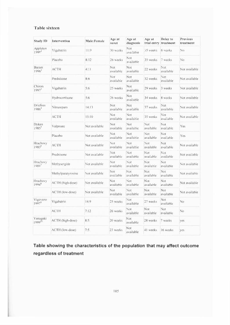
T a b le s ix teen
S tudy ID In terven tion M alerFem ale Age a t onset
Age a t diagnosis
Age a t tr ia l e n try
Delay to trea tm e n t
Previoustrea tm e n t
Appleton 1999 1
Vigabatrin 11:9 30 weeksNotavailable
35 weeks 6 weeks No
Placebo 8:12 26 weeksNotavailable
35 weeks 7 weeks No
Baram19962
ACTH 4:11 Notavailable
Notavailable
22 weeksNotavailable
N ot available
Prednisone 8:6 Notavailable
Notavailable
32 weeks Notavailable
N ot available
Chiron19973
Vigabatrin 5:6 25 weeks Notavailable
29 weeks 3 weeks Not available
Hydrocortisone 5:6 26 weeksNotavailable
34 weeks 8 weeks Not available
Driefiiss19864
Nitrazepam 14:13Notavailable
Notavailable
37 weeksNotavailable
N ot available
ACTH 15:10Notavailable
Notavailable
35 weeksNotavailable
Not available
Dyken19855
Valproate N ot available Notavailable
Notavailable
Notavailable
Notavailable
Yes
Placebo Not availableNotavailable
Notavailable
N otavailable
Notavailable
Yes
Hrachovy19836
ACTH Not available Notavailable
Notavailable
Notavailable
Notavailable
Not available
Prednisone Not availableNotavailable
Notavailable
Notavailable
Notavailable
Not available
Hrachovy19897 Methysergide Not available Not
availableNotavailable
Notavailable
Notavailable
Not available
M ethylparatyrosine Not availableNotavailable
Notavailable
Notavailable
Notavailable
Not available
Hrachovy19948 ACTH (high-dose) Not available
Notavailable
Notavailable
Notavailable
Notavailable
Not available
ACTH (low-dose) Not available N otavailable
Notavailable
Notavailable
Notavailable
Not available
Vigevano19979
Vigabatrin 14:9 25 weeksNotavailable
27 weeksNotavailable
No
ACTH 7:12 26 weeksNotavailable
Notavailable
Notavailable
No
Yanagaki199910
ACTH (high-dose) 8:5 20 weeksNotavailable
28 weeks 7 weeks yes
ACTH (low-dose) 7:5 23 weeksNotavailable
41 weeks 16 weeks yes
Table showing the characteristics of the population that may affect outcome
regardless of treatment
105
VIGABATRIN VERSUS PLACEBO
There was one study Appleton 19991.
• Effects on psychomotor development was not reported as an outcome in this study.
• Effects on subsequent seizure rates was not reported as an outcome in this study.
• Effects on cessation of spasms: Appleton 1999 had 40 patients and showed
complete cessation of spasms in seven of twenty (35%) of patients treated with
vigabatrin compared with two of twenty (10%) treated with placebo, Peto odds
ratio 4.1 (95% Cl 0.9 to 17.5).
• Effects on time taken to achieve cessation of spasms was not reported as an
outcome in this study.
• Effects on reduction in the number of spasms: Appleton 1999 showed an >70%
reduction in spasms in 40% of the group treated with vigabatrin compared with
15% in the group treated with placebo. However, it is not clear from the paper to
what proportion of the two groups of patients these figures apply, whether the
figures apply to the whole group or just those patients in whom complete cessation
of spasms was not achieved.
• Effects on relapse rates: Appleton 1999, four out of seven patients who responded
to vigabatrin relapsed and all the patients successfully treated with placebo
relapsed.
• Overall only three patients treated with vigabatrin and no patient treated with
placebo treatment remained spasm free within the four week study period, Peto
odds ratio 8.2 (95% Cl 0.8 to 84).
• Effects on time taken to relapse was not reported as an outcome in this study.
• Effects on resolution of hypsarrhythmia: Appleton 1999, five of the seven patients
who were spasm free with vigabatrin showed resolution of hypsarrhythmia on
EEG, compared with one of the two patients who had become spasm free on
placebo, Peto odds ratio 2.4 (95% Cl 0.1 to 54.6).
• Effects on time taken to achieve resolution of hypsarrhythmia was not reported as
an outcome in this study.
• No side effects severe enough to warrant stopping treatment were reported in this
study.
• No deaths were reported.

SODIUM VALPROATE VERSUS PLACEBO
There was one study Dyken 1985s
• Effects on psychomotor development was not reported as an outcome in this study.
• Effects on subsequent seizure rates was not reported as an outcome in this study.
• Effects on cessation of spasms was not reported as an outcome in this study.
• Effects on time taken to achieve cessation of spasms was not reported as an
outcome in this study.
• Effects on reduction in the number of spasms: Dyken 1985 used a spasm index: S.I.
= (spasm frequency x spasm duration / total observation time) x 1000 to calculate
the mean reduction in spasm frequency from the baseline. At the end of the four
week treatment period, the valproate treatment was found to have a lower mean
spasm index than placebo, when valproate was administered first (p<0.04). There
was no significant difference between placebo and valproate during the second
level of treatment. It is not possible to calculate a Peto odds ratio.
• Effects on relapse rates was not reported as an outcome in this study.
• Effects on time taken to relapse was not reported as an outcome in this study.
• Effects on resolution of hypsarrhythmia was not reported as an outcome in this
study.
• Effects on time taken to achieve resolution of hypsarrhythmia was not reported as
an outcome in this study.
• Side effects were not reported as an outcome in this study.
• Deaths were not reported as an outcome in this study.
VIGABATRIN VERSUS HYDROCORTISONE
There was one study Chiron 19973
• Effects on psychomotor development was not reported as an outcome in this study.
• Effects on subsequent seizure rates was not reported as an outcome in this study.
• Effects on cessation of spasms: Chiron 1997 comparing vigabatrin (150 mg/kg/day)
and hydrocortisone (15 mg/kg/day) in twenty-two infants with infantile spasms due
to tuberous sclerosis, found in the initial phase, eleven of the eleven patients
(100%) treated with vigabatrin to be spasm free as compared to five of eleven
patients (45%) treated with hydrocortisone. Peto odds ratio 13.8 (95% Cl 2.21 to
86.35).107
• Effects on time taken to achieve cessation of spasms: Chiron 1997; on average the
11 responders to vigabatrin took 4 days (range 0.5-14 days, median 2 days) to
achieve complete cessation of spasms, whilst the 5 responders to hydrocortisone
took an average of 13 days (range 3-30 days, median 23.5 days), WMD = -8.8 (-
19.2 to 1.6).
• Effects on reduction in the number of spasms was not reported as an outcome in
this study.
• Effects on relapse rates: Chiron 1997, ten of the eleven patients who responded to
vigabatrin remained spasm free, this information was not given for the five
responders to hydrocortisone.
• Effects on time taken to relapse was not reported as an outcome in this study.
• Effects on resolution of hypsarrhythmia was not reported as an outcome in this
study.
• Effects on time taken to achieve resolution of hypsarrhythmia was not reported as
an outcome in this study.
• Side effects: Chiron 1997 reported that hydrocortisone had to be stopped in one
patient, no reason was given.
• No deaths were reported in this study.
VIGABATRIN VERSUS ACTH
There was one study Vigevano 19979
• Effects on psychomotor development: Vigevano 1997 stated that there was no
apparent difference in cognitive development between the two groups during
follow up which ranged from 9 to 44 months. They did not state how they assessed
development or quantify it any further, and this comparison was confounded by the
fact that some infants initially randomised to receive vigabatrin went on to receive
ACTH, and vice versa, within the follow up period. They did not consider long
term developmental status at 5 years of age.
• Effects on subsequent seizure rates: Vigevano 1997 found an incidence of other
types of epileptic seizures to be 25% in both groups during follow up of 9 to 44
months. Again they did not consider subsequent epilepsy rates at 5 years of age.
Again, this comparison was confounded by the fact that some infants initially
randomised to receive vigabatrin went on to receive ACTH, and vice versa, within
the follow up period.
108
• Effects on cessation of spasms: Vigevano's 1997 trial of forty-two patients showed
cessation of spasms in eleven of the twenty-three patients randomised to vigabatrin
compared with fourteen of the nineteen patients randomised to ACTH. Peto odds
ratio 0.4 (95% Cl 0.1 to 1.2)
• Effects on time taken to achieve cessation of spasms: Vigevano 1997, the 11
responders to vigabatrin took between 1 and 14 days to achieve complete cessation
of spasms, whilst the 14 responders to ACTH took between 2 and 12 days.
• Effects on reduction in the number of spasms was not reported as an outcome in
this study.
• Effects on relapse rates was not reported as an outcome in this study.
• Effects on time taken to relapse was not reported as an outcome in this study.
• Effects on resolution of hypsarrhythmia: Vigevano 1997, resolution occurred in
four out of eleven patients responding to vigabatrin and eleven out fourteen patients
responding to ACTH. Peto odds ratio 0.2 (95% Cl 0.0 to 0.9).
• Effects on time taken to achieve resolution of hypsarrhythmia was not reported as
an outcome in this study.
• Side effects: Vigevano 1997 stopped vigabatrin in one patient because of excessive
irritability and ACTH in one patient, no reason given.
• Deaths: One patient in Vigevano's 1997 study died, but the cause was not given
and it is unclear to which treatment group they had been randomised.
ACTH VERSUS PREDNISONE
There were two studies Baram 19962 and Hrachovy 19836. The doses of ACTH used in
these two studies differed, so the results should be interpreted with caution.
• Effects on psychomotor development was not reported as an outcome in these
studies.
• Effects on subsequent seizure rates: Baram 1996 in their study comparing ACTH
with prednisone found seven (-50%) patients in both groups to have developed
other seizure types over the period of follow up of 2 to 48 months. However, this
comparison was confounded by the fact that some infants initially randomised to
receive prednisone went on to receive ACTH within the follow up period. They did
not report subsequent epilepsy rates at 5 years of age.
• Effects on cessation of spasms: Baram 1996 compared fifteen patients treated with
ACTH (150 units/m.squared/day) with fourteen treated with prednisone (2
109

mg/kg/day). and showed ACTH to be superior to prednisone with cessation of
spasms in thirteen of fifteen (87%) patients and four of fourteen (29%) patients
respectively. Hrachovy 1983 compared twelve patients treated with ACTH (20 -30
units/day) with twelve patients treated with prednisone (2 mg/kg/day). In the initial
phase of the trial five of twelve (42%) patients treated with ACTH had complete
cessation of spasms and resolution of hypsarrhythmia on their EEG compared with
four of twelve (33%) treated with prednisone. Combining the two studies, ACTH
stopped the spasms in 67.5% of patients compared with prednisone in 31% of
patients, Peto odds ratio 4.2 (95% Cl 1.4 to 12.4).
• Effects on time taken to achieve cessation: Baram 1996; on average the 13
responders to ACTH took 3.2 days (range 1-7 days, median 2 days) to achieve
complete cessation of spasms, whilst the 4 responders to prednisone took an
average of 4 days (range 2-7 days, median 3.5 days), WMD = -0.8 (95% Cl -3.3 to
1.7).
• Effects on reduction in the number of spasms was not reported as an outcome in
these studies.
• Effects on relapse rates: Baram 1996, two of the thirteen patients who responded to
ACTH relapsed and none of the four responders to prednisone relapsed. Hrachovy
1983, three of the five patients who responded to ACTH relapsed and one of the
four responders to prednisone also relapsed.
• Overall, Baram 1996, eleven patients who responded to ACTH remained spasm
free and the four responders to prednisone also remained spasm free. Hrachovy
1983, two patients successfully treated with ACTH remained spasm free and three
successfully treated with prednisone remained spasm free within the study period.
The combined Peto odds ratio for these two studies is 2.6 (95% Cl 0.8 to 8.1).
• Effects on time taken to relapse was not reported as an outcome in these studies.
• Effects on resolution of hypsarrhythmia: Baram 1996, this study showed ACTH to
be superior to prednisone with resolution of hypsarrhythmia in thirteen of fifteen
patients treated with ACTH compared to four of fourteen of patients treated with
prednisone, Peto odds ratio 10.1 (95% Cl 2.4 to 43.2). Hrachovy 1983, five of
twelve patients treated with ACTH had resolution of hypsarrhythmia but this was
not reported for the group treated with prednisone.
• Effects on time taken to achieve resolution of hypsarrhythmia was not reported as
an outcome in these studies.
110

• Side effects were not reported as an outcome in these studies.
• No deaths were reported in these studies.
HIGH DOSE ACTH VERSUS LOW DOSE ACTH
• There were two studies Hrachovy 19948 and Yanagaki 199910. Hrachovy used
natural ACTH and Yanagaki used synthetic ACTH. This makes direct comparison
difficult. Yanagaki states that the dose equivalent of 1 IU of natural ACTH is
0.025 IU of synthetic ACTH, but the results must be interpreted with caution.
• Effects on psychomotor development: Yanagaki 1999 in their study comparing
high-dose ACTH with low-dose ACTH used the Japanese Tsumori scale to
evaluate developmental status at entry into the trial and in 17 responders who were
followed up for more than one year. They found no significant difference in
developmental quotient between the two groups at the end of the follow up period.
However, they only considered psychomotor development in the responders which
gives rise to biased analysis and this should be interpreted with caution. They did
not consider long-term developmental status at 5 years of age.
• Effects on subsequent seizure rates was not reported as an outcome in these studies.
• Effects on cessation of spasms: Hrachovy 1994 randomised thirty patients to high-
dose ACTH (150 units/m.squared/day for three weeks with a tapering dose over a
further nine weeks), and twenty-nine patients to low-dose ACTH (20-30 units/day
for two to six weeks, tapering over one week). Of the thirty patients treated with
high dose therapy thirteen (50%) had complete cessation of spasms and of the
twenty-nine patients treated with low dose therapy fourteen (58%) had complete
cessation of spasms. Yanagaki 1999 randomised thirteen patients to receive 1 unit
/kg/day (equivalent to 40 units/ kg/ day of natural ACTH) for two weeks tapering
over a further two weeks and thirteen patients to receive 0.2 units/kg/day
(equivalent to 8 units/kg/day of natural ACTH) for two weeks tapering over a
further two weeks. Of the patients treated with high-dose ACTH, spasm cessation
occurred in eleven of thirteen patients compared with nine of thirteen treated with
low dose ACTH. Combining these two studies, "high-dose" ACTH stopped the
spasms in 79.5% patients compared with "low-dose" ACTH in 76.5% of patients,
Peto odds ratio 1.1 (95% Cl 0.4 to 2.6).
• Effects on time taken to achieve cessation of spasms was not reported as an
outcome in these studies.
I l l
|I'i

• Effects on reduction in the number of spasms was not considered as an outcome in
these studies.
• Effects on relapse rates: Hrachovy 1994, two of the thirteen patients who responded
to "high-dose ACTH" relapsed and two of the fourteen patients who responded to
"low-dose ACTH" also relapsed. Yanagaki 1999, three of the eleven patients who
responded to "high-dose ACTH" relapsed and three of the nine responders to "low-
dose ACTH" relapsed.
• Hrachovy 1994, eleven patients who responded to high-dose ACTH remained
spasm free and twelve patients who responded to low-dose ACTH also remained
spasm free. Yanagaki 1999, eight patients who responded to high-dose ACTH
remained spasm free and six patients who responded to low-dose ACTH remained
spasm free. Combining these two studies gives a Peto odds ratio 0.8 (95% Cl 0.3 to
1.9).
• Effects on time taken to relapse was not considered as an outcome reported in these
studies.
• Effects on resolution of hypsarrhythmia: Hrachovy 1994, three out of thirteen
responders to high-dose ACTH showed resolution of their EEG and three out of
fourteen responders to low-dose ACTH showed resolution of their EEG. Yanagaki
1999, eight out of thirteen treated with high-dose ACTH showed resolution of their
EEG and seven out of twelve treated with low-dose ACTH showed resolution of
their EEG. Combining these two studies gives a Peto odds ratio 1.9 (95% Cl 0.6 to
6.2)
• Effects on time taken to achieve resolution of hypsarrhythmia was not reported as
an outcome in these studies.
• Side effects were not reported as an outcome in these studies.
• No deaths were reported in these studies.
NITRAZEPAM VERSUS ACTH
There was one study Dreifuss 19864.
• Effects on psychomotor development was not reported as an outcome in this study.
• Effects on subsequent seizure rates was not reported as an outcome in this study.
• Effects on cessation of spasms was not reported as an outcome in this study.
• Effects on time taken to achieve cessation of spasms was not reported as an
outcome in this study.
112
• Effects on reduction in the number of spasms: Dreifuss 1986 showed an >50%
reduction in spasms in 66% of the group treated with nitrazepam compared with
50% in the group treated with ACTH, Peto odds ratio 2.0 (95% Cl 0.7 to 5.9).
• Effects on relapse rates was not reported as an outcome in this study.
• Effects on time taken to relapse was not reported as an outcome in this study.
• Effects on resolution of hypsarrhythmia was not reported as an outcome in this
study.
• Effects on time taken to achieve resolution of hypsarrhythmia was not reported as
an outcome in this study.
• Side effects: Dreifuss 1986 had to withdraw ACTH in six patients, hypertension
and maleana were given as two causes for withdrawal.
• Deaths: Dreifuss 1986 had one patient in their ACTH group who died, but no cause
was found at autopsy.
METHY SERGIDE VERSUS ALPHA-METHYLPARATYROSINE
There was one study Hrachovy 1989
• Effects on psychomotor development: Hrachovy 1989 used the Denver
Developmental Screening Test to evaluate developmental status at entry into the
trial and at 3 weeks post entry into the trial. They stated, in their results section, that
two patients in each group showed developmental improvement but did not
quantify this further. They did not report developmental status at 3 months post trial
entry or at 5 years of age.
• Effects on subsequent seizure rates was not reported as an outcome in this study.
• Effects on cessation of spasms: Hrachovy 1989 found only one patient (8%) treated
with methysergide and two (16%) treated with alpha-methylparatyrosine responded
to therapy, Peto odds ratio 0.5 (95% Cl 0.1 to 5.2).
• Effects on time taken to achieve cessation of spasms was not reported as an
outcome in this study.
• Effects on reduction in the number of spasms: Hrachovy 1989 showed a >50%
reduction in spasms in 25% of the group treated with methysergide compared with
17% in the group treated with alpha-methylparatyrosine, Peto odds ratio 1.6 (95%
Cl 0.2 to 11.2).
113

• Effects on relapse rates: no patient treated with methysergide remained spasm free
and only one of the two patients successfully treated with alpha-methylparatyrosine
remained spasm free. Peto odds ratio 0.14 (95% Cl 0.0 to 6.8).
• Effects on time taken to relapse was not reported as an outcome in this study.
• Effects on resolution of hypsarrhythmia was not reported as an outcome in this
study.
• Effects on time taken to achieve resolution of hypsarrhythmia was not reported as
an outcome in this study.
• No side effects severe enough to warrant stopping treatment were reported in this
study.
• No deaths were reported in this study.
Discussion
Infantile spasms were first described 160 years ago in a letter that Dr West wrote to the
Lancet. Since that time at least 30 drugs have been tried and reported in the literature, but
treatment remains problematic and the long-term prognosis remains poor for both
psychomotor development and subsequent development of other seizure types. Despite the
wealth of literature on this subject there have been only a few RCTs and the majority of
studies have been either open prospective or retrospective trials. The RCTs that have been
undertaken contain only small numbers of subjects with the result that they have little or no
power and have used widely varying dosage regimes and outcome measures, making meta
analysis difficult to perform. In addition, only eight drugs out of all those used in the
treatment of infantile spasms have been subjected to RCTs. We found no RCTs comparing
forms of therapies, such as phenobarbitone or pyridoxine, despite the fact that these are
often first line therapies in some countries. Some therapies (for example gamma-globulins,
pyridoxine, lamotrigine) have been reported as case reports or in uncontrolled trials. We
have not considered these therapies in this review. In addition many different "steroid"
preparations have been used in the available RCTs. This is partly due to the availability of
different compounds in different countries, for example in Europe and Japan synthetic
ACTH is usually available, whilst in the USA natural ACTH derivatives are normally
used. ACTH can only be given as an injection, intramuscularly, intravenously or into the
peritoneal cavity, infants find this distressing and many centres use oral preparations such
as prednisone or hydrocortisone in preference. This makes analysis of the efficacy of
individual "steroid" preparations difficult.
114
ijI

Despite an extensive literature search we found only ten RCTs and the overall reported
methodology was poor. Only one study had considered the number of patients required to
determine whether one treatment was more efficacious than another; they based their
calculations on the assumption that the trial drug would be 50% more efficacious in
stopping the spasms than the comparative drug; their trial did not find such a difference.
Although all the studies claimed to be randomised, less than half stated how randomisation
had been performed and only two studies stated that adequate concealment of
randomisation had taken place. Ideally to minimise performance bias trials should be
double-blinded, although perhaps it is not surprising that only two such trials have been
performed. Blinding the recipients (or parents) and staff administering the therapies
remains problematic. ACTH can only be given by intramuscular injection, whilst the other
drugs used in the above RCTs are administered orally and nowadays it is generally
considered to be unethical to administer placebo intramuscular injections to young infants.
In addition, many of the side effects of steroids are common and easily recognised, for
example weight gain and hypertension, making blinding difficult. Some would also argue
that it is possible that the spasms are directly linked to the development of severe learning
difficulties and it is generally thought (though not proven) that it may be possible that the
earlier the spasms are brought under control the better the outcome. Thus delaying
treatment (for example by administering placebo alone) is also usually thought to be
unethical. Finally five studies had loss of patients to follow up and the remaining studies
did not clearly state that all patients entered into the trials had been successfully followed
up for the duration of the study. We had also planned to look for possible sources of
clinical heterogeneity between the studies, for example, sex, age at spasm onset, age at trial
entry, delay to treatment and whether or not participants had received previous treatment.
Unfortunately, this information was not available for the majority of the included studies
and we are unable to draw any conclusions. However, it is possible that some of these
sources of heterogeneity are important in determining outcome. For example, the study
Baram 1996 found ACTH to be more efficacious than prednisolone but had three times as
many girls than boys in the ACTH group and roughly equal numbers in the prednisone
group. It is possible therefore, that if girls have a better outcome than boys that this may
have contributed to the differences seen in response between their two groups.
When developing our protocol we had tried to provide definitions for terms such as
infantile spasms, cessation of spasms, relapse etc. We had planned to try and establish
115
whether different definitions used by authors for infantile spasms might affect which
participants would be included into a trial, thereby affecting outcome. It became clear from
our review of the literature that few authors clearly define the term infantile spasms and it
was clear that some trials using entry criteria that defined the need for hypsarrhythmia in
fact entered infants who did not have this. It is theoretically possible that either some
patients entered into these trials did not have infantile spasms or that different trials were
investigating different populations. Therefore, for the purpose of this review we accepted
that any RCT stating that their participants had "infantile spasms" would be included, but
the results should be interpreted with caution. Likewise, we had also tried to define
outcome measures such as cessation of spasms and what constituted a relapse, so that we
could accurately compare the outcomes of each individual study. Again, the majority of
studies did not define such outcomes and meta-analysis of these outcomes should again be
interpreted with caution.
Infantile spasms are associated with a poor long-term prognosis. The majority will also
have severe learning difficulties, with high cost implications not only to the patients and
the families and their quality of life, but also on health services. Therefore, if any one
treatment was proven to improve long-term psychomotor development and to reduce the
risk of further seizure types then it would probably be considered to be more efficacious
than other treatments regardless of the effect on the spasms themselves or the
hypsarrhythmia. Although, with time the spasms will resolve, many patients continue to
have other forms of epilepsy into adult life and about one fifth will progress to Lennox
Gastaut syndrome. These patients will therefore require anticonvulsants and will continue
to suffer the effects of epilepsy, often for life. However, no study has considered these
factors as long term outcome measures beyond 44 months post trial entry, and currently
there is no standard agreement on the timing or means of measuring these outcomes.
The spasms themselves are certainly distressing to parents and carers. It is possible that
they are also distressing to the infant as many will cry immediately following a spasm or
appear to be in pain and distressed, so much so, it is not uncommon for the spasms to be
mistaken for colic. Whilst the long-term benefits of different therapies remain uncertain,
we tried to establish if any one therapy was more efficacious in stopping the spasms
themselves. One small study looking at patients with infantile spasms and an underlying
diagnosis of tuberous sclerosis found vigabatrin to be more efficacious than hydrocortisone
116

in stopping the spasms in this group of patients, a finding also found in some open
studies . However, it is not clear that vigabatrin is more efficacious than steroids in
treating infantile spasms in patients without tuberous sclerosis. One underpowered RCT
showed a trend that vigabatrin might be more efficacious than placebo. Two small studies,
Baram 1996 and Hrachovy 1993, did show ACTH to be more efficacious than prednisone
in stopping the spasms, however their dose of prednisone of 2mg/kg/day would be
considered too low a dose by some and it is therefore possible that higher doses of
prednisone may be as efficacious as ACTH54. Further trials are required. We found no
other RCTs comparing other forms of therapies, such as sodium valproate,
benzodiazepines, pyridoxine or phenobarbitone, in terms of cessation of spasms. Although
four studies did consider reduction in the number of spasms, it is not at all clear as to
whether a reduction in spasms is of major benefit to a patient. Complete control of spasms
has to be the main objective. Some open, prospective trials have suggested that vigabatrin
might control spasms more quickly than steroids. Only three RCTs considered the time
taken to cessation of spasms and there was no significant difference between vigabatrin
and hydrocortisone or ACTH.
The exact relationship between clinical spasms and typical or even modified (or atypical)
hypsarrhythmia on the EEG is not entirely clear. Not all infants with clinical evidence of
infantile spasms have hypsarrhythmia, and in those in whom it is present it is not always
present all of the time. It is not known whether the presence or absence of hypsarrhythmia
affects long term outcome. Again we had hoped that by collecting and combining data
from the studies, it might help clarify these areas of uncertainty. Unfortunately, few studies
contained sufficient data to draw any conclusions.
We had also planned to look at subgroup analysis of clinical features, but unfortunately,
because of lack of trials this was not possible. It was possible to consider patients with an
underlying diagnosis of tuberous sclerosis as a subgroup in one underpowered study. This
study, Chiron 19973 showed vigabatrin to be more efficacious than hydrocortisone in
stopping the spasms in this group of patients. All their patients treated with vigabatrin had
complete cessation of spasms and only one patient relapsed within the study period.
However, the numbers are small - only eleven patients in each group and these results
should be treated with caution, even though this result mimics the findings of open studies.
Further trials are required with larger numbers of patients to confirm (or refute) the
117

findings of this study.
Side effects must always be taken into account. Any drug with unacceptable side effects
will not be used in clinical practice. Likewise if two drugs show similar beneficial effects
then the one with the safer drug profile should be used in preference. Within these studies
few side effects or deaths were reported. However, both steroids and vigabatrin have
potentially serious side effects. The problem with steroids are potentially life threatening
problems which include depression of the immune system and modified response to
infection leading to overwhelming sepsis. Less serious side effects, for example
hypertension, are often transient but nevertheless have the potential to cause morbidity.
Minor side effects, estimated to occur in two thirds of patients, include behavioural
changes especially irritability, changes in appetite, weight gain and alteration in sleep
patterns. In addition some forms of steroids (e.g. ACTH) involve daily intramuscular
injections. However, there have been reports of both asymptomatic and symptomatic visual
field defects with loss of peripheral vision to varying degrees in adults and children treated
with vigabatrin. It appears to occur most commonly in patients who have been treated with
vigabatrin for more than six months but it does not appear to always be reversible on
withdrawing the vigabatrin. The main difficulty in infants is that they cannot be tested or
monitored for visual field defects (a child needs to be at least eleven to be able to co
operate and complete a reliable test). However, on balance the devastating effects of
infantile spasms probably outweigh the risks of developing the severe side effects of these
therapies.
Implications for practice
The optimum treatment for infantile spasms remains uncertain. No study to date has shown
any one drug to be more efficacious than any other in terms of long term psychomotor
development or subsequent epilepsy rates. Vigabatrin may be more efficacious than
placebo, and ACTH may be more efficacious than low doses of prednisone in stopping the
spasms. Vigabatrin may be more effective than hydrocortisone in stopping the spasms in
the group of patients who also have tuberous sclerosis. We found no one treatment to be
more efficacious than any other with regard to reduction in number of spasms, relapse rates
or resolution of hypsarrhythmia. Both steroids and vigabatrin have potentially serious side
effects but the risks of these should be considered against the potential benefits, when
considering treatment of infantile spasms.
118

Implications for research
There is still little evidence available on the optimum treatment for infantile spasms.
Further trials with improved methodology, standardised valid outcome measures, larger
numbers of participants and longer follow up are required. As stated previously infantile
spasms are a relatively rare disorder, so that in order to be able to recruit large numbers of
patients into randomised controlled trials, multicentre collaboration is likely to be required.
ReferencesSee appendix three
119

CHAPTER SIX
Oral treatment of non-convulsive status epilepticus in tuberous sclerosis
Age of seizure onset is one of the most important prognostic factors for long-term outcome
in patients suffering from TSC. It is rare for patients who have normal development and
intellect at the age of five who subsequently develop epilepsy to regress. In the review of
the patients in the Bath region over the past ten years no patient who developed fits over
the age of five had impaired intellect. One unusual cause of regression after the age of 5
years in TSC is the development of non-convulsive status epilepticus (NCSE). The
epilepsy in TSC is often refractory to treatment and NCSE is regularly seen in those whose
seizures are most difficult to treat. In addition these patients are liable to suffer recurrent
attacks of NCSE.
Dr Frank Besag, National Centre for Young People with Epilepsy, has treated his patients
with NCSE with oral diazepam for many years. He recommended this treatment to JPO
who also had success with oral diazepam treatment of NCSE in some of his TSC patients.
A literature search of the English Language failed to find any studies looking at this form
of treatment or comparing it with conventional intravenous benzodiazepine administration.
NCSE is an infrequent but severe epileptic seizure disorder. It is characterised by a patient who has continuous or almost continuous epileptic activity both clinically and on EEG but the clinical signs of an epileptic seizure disorder may be extremely subtle. Usually, but not always in a patient with NCSE, there is a history of a previous overt seizure disorder, frequently severe. This can include IS and other difficult to control epileptic syndromes including Lennox-Gastaut syndrome. Clinically there is no clonic jerking of the limbs and although the patient may have some awareness of their surroundings, impaired consciousness is the main clinical feature. The onset may either be so subtle as to suggest a dementia or may be abrupt. Lack of awareness may extend to loss of appetite with significant weight loss. Patients may cycle from periods of complete unresponsiveness to one of partial responsiveness. Frequent brief, but small, myoclonic jerks can often be felt but not seen: they are random in time and site and are effectively detected clinically by a light handshake. Often there are associated autonomic features such as excessive salivation, sweaty palms and pupils that are rapidly dilating and constricting for no obvious reason.
Aim
Prolonged periods of impaired consciousness can interfere with development, education
and well-being. The traditional treatment, effective in the majority of cases of idiopathic
primary generalised NCSE, is the intravenous administration of benzodiazepines 114, which
requires hospital admission and is inappropriate for those with frequently recurring attacks.
120

Therefore, the aim of this study was to evaluate the use of oral diazepam in aborting
attacks in those with recurrent episodes of NCSE.
Method
The notes of all the patients seen in the Bath Tuberous Sclerosis Clinic in 1996 (including
patients outside the Wessex region) were examined to identify those patients who had a
history of NCSE. The diagnosis of NCSE was made on clinical grounds at the time of
attending the clinic as defined above; palpable myoclonic jerks, evidence of autonomic
dysfunction and impaired consciousness were all required before NCSE was diagnosed.
Those notes were then examined further to ascertain the patient details, information about
their epilepsy and the treatment received. No patients were excluded from the study
because of lack of data in the medical notes.
Results
Eight patients had a history of NCSE, including the two patients reported in chapter two,
five males and three females. All eight patients had presented with IS by the age of seven
months. They all subsequently developed secondarily generalised tonic-clonic seizures,
absence seizures and drop attacks.
The age of onset of the NCSE varied from six to 18 years. The clinical nature of the
episodes varied greatly. For example the parents of case four described a picture of being
‘limp, lethargic, lying around, dribbling, often staring and gazing with pupils dilated, hands
sweating, unusual circulation and jerky movements of the limbs’. Case three had an
insidious onset over three to four months during which time he had changed from being
independently mobile to becoming wheelchair bound. On occasions when he did walk, he
would walk into doors. He also became disinterested in food during this period, lost weight
and was thought to be terminally ill when referred. He was given lOmg oral diazepam and
within an hour was alert and had a normal appetite. The dose was repeated when NCSE
recurred; on average every five days, with the same effect and speed of action.
Seven were treated successfully with oral diazepam (table seventeen) and in only one (case
number five) was drowsiness, lasting about three hours, noted to be a problem. No other
side effects were recorded. The dose of diazepam required varied considerably and bore no
relationship to either the child’s age or weight, but was the dose required to produce
121
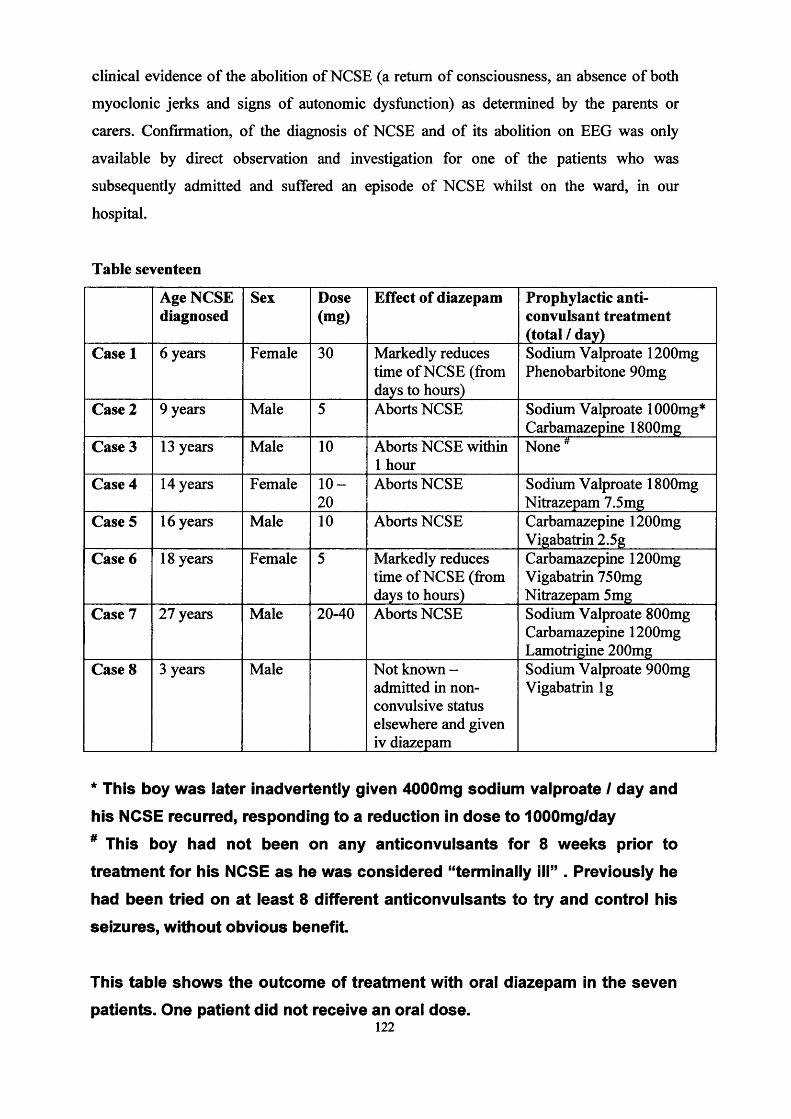
clinical evidence of the abolition of NCSE (a return of consciousness, an absence of both
myoclonic jerks and signs of autonomic dysfunction) as determined by the parents or
carers. Confirmation, of the diagnosis of NCSE and of its abolition on EEG was only
available by direct observation and investigation for one of the patients who was
subsequently admitted and suffered an episode of NCSE whilst on the ward, in our
hospital.
Table seventeen
Age NCSE diagnosed
Sex Dose(mg)
Effect of diazepam Prophylactic anticonvulsant treatment (total / day)
Case 1 6 years Female 30 Markedly reduces time of NCSE (from days to hours)
Sodium Valproate 1200mg Phenobarbitone 90mg
Case 2 9 years Male 5 Aborts NCSE Sodium Valproate lOOOmg* Carbamazepine 1800mg
Case 3 13 years Male 10 Aborts NCSE within 1 hour
None 4
Case 4 14 years Female 1 0 -20
Aborts NCSE Sodium Valproate 1800mg Nitrazepam 7.5mg
Case 5 16 years Male 10 Aborts NCSE Carbamazepine 1200mg Vigabatrin 2.5g
Case 6 18 years Female 5 Markedly reduces time of NCSE (from days to hours)
Carbamazepine 1200mg Vigabatrin 750mg Nitrazepam 5 mg
Case 7 27 years Male 20-40 Aborts NCSE Sodium Valproate 800mg Carbamazepine 1200mg Lamotrigine 200mg
Case 8 3 years Male Not known - admitted in non- convulsive status elsewhere and given iv diazepam
Sodium Valproate 900mg Vigabatrin lg
* This boy was later inadvertently given 4000mg sodium valproate I day and
his NCSE recurred, responding to a reduction in dose to 1000mg/day# This boy had not been on any anticonvulsants for 8 weeks prior to
treatment for his NCSE as he was considered “terminally ill” . Previously he
had been tried on at least 8 different anticonvulsants to try and control his
seizures, without obvious benefit.
This table shows the outcome of treatment with oral diazepam in the sevenpatients. One patient did not receive an oral dose.
122

The eighth patient diagnosed as having episodes of NCSE, from the parent’s description in
clinic, was prescribed 5 mg oral diazepam to be given at the onset of the next episode to
see if it would abort the attack. He was then admitted to his local hospital with NCSE
before the oral diazepam was given (he too had the clinical diagnosis of NCSE confirmed
by EEG). After successful treatment with IV diazepam he was discharged and to date
(three years later) has had no further episodes: his other seizures have also reduced in
frequency.
Discussion
These eight patients with TSC all had a severe recurrent epileptic seizure disorder and were
on regular prophylactic anticonvulsant treatment. We do not know if their EEG would have
supported a diagnosis of primary generalised or secondarily generalised NCSE except in
two patients. However we agree with Aicardi115 that a generalised EEG in these patients
who at other times have focal EEG changes and who have tubers causing focal structural
lesions makes it difficult to decide on classification. Our diagnoses were made on clinical
grounds and it was not possible to further validate this: there must therefore be some doubt
about the diagnosis of NCSE and of its abolition. However we believe these diagnoses are
usually reliable clinically and the response to treatment would support this. An EEG is
preferable but was usually not possible in our cases: it would be required if the diagnosis
were in doubt. Our patients were usually recognised as having episodes of NCSE between
attacks when in clinic either on history alone or history supported by examination at the
time. Carers were taught what to look for so that the diagnosis could be confirmed during a
telephone consultation. These latter are a common practice in our TSC clinic because of
the need for frequent feedback concerning the epilepsy and the great distance some people
travel to visit the clinic.
Little has been published about NCSE and many paediatricians are unfamiliar with its
clinical signs, diagnosis or management. In most publications intravenous diazepam is the
usual treatment for NCSE, effective in 80-100% cases of primary NCSE but only 15 - 59%
of secondary NCSE116: this is not convenient when the patient suffers recurrent attacks,
because of the severity of their epileptic seizure disorder, despite appropriate regular
anticonvulsant treatment. Oral diazepam is not normally considered for antiepileptic
treatment since it is not effective for the termination of primary generalised tonic-clonic
123

seizures or for prophylaxis because an inability to achieve adequate blood levels. Oral
diazepam has been used to prevent febrile convulsions and other agents, buccal or nasal,
midazolam most frequently, have more recently been used for primary generalised status
epilepticus. Why it might work in NCSE is unclear. We have found it to be effective in
patients with TSC for recurrent attacks of NCSE that the parents and carers can learn to
recognise, so allowing prompt treatment to be given without necessitating hospital
admission. It is likely to be equally or more effective in primary generalised NCSE.
Side effects were only seen in one patient. Effective management of NCSE in these
patients has allowed planned alteration to regular anticonvulsant treatment without
admission to hospital, making it easier for carers to consider trying alternative prophylactic
anticonvulsant management. In one patient the inadvertent administration of large doses of
sodium valproate (>100mg/kg) re-precipitated recurrent attacks of NCSE, which resolved
on return to the prescribed dose: no other associations between NCSE and prophylactic
treatment were noted. Precipitation of NCSE by a rapidly increasing dosage of sodium1 17valproate in an adult patient has been previously described
Conclusion
Oral diazepam is a safe and effective treatment for termination of NCSE in patients with
TSC. If parents and carers can be taught to recognise episodes of NCSE early in their
evolution so that early intervention with oral diazepam can be undertaken thus abolishing
the attacks then the outcome may be improved and regression avoided. Unfortunately all
the patients in this series had suffered severe fits from a young age and had severe learning
difficulties at the time of onset of their NCSE and it is difficult to ascertain whether further
regression has been avoided in these patients.
124
CHAPTER SEVEN
Lvmphangioleiomvomatosis and Tuberous Sclerosis
Lymphangioleiomyomatosis (LAM) is thought to occur as a complication in approximately
1% of patients with TSC. Although Lutembacher first described LAM in association with
TSC in 1881, he mistook the cystic and nodular changes for metastasis from renal
fibrosarcoma, in the lungs of a 36 year old woman who died from bilateral
pneumothoracies. It wasn’t until 1939 that Berg and Vejilens gave the first accurate and
clear description of the clinical and radiological features of pulmonary tuberous sclerosis.
It is difficult to estimate the true incidence of LAM in TSC, other than it is undoubtedly
rare, partly because the few studies that have attempted to do so have generally used small
numbers of patients from biased groups and partly because it is unknown how many
patients with pulmonary complications become symptomatic and are therefore recognised.1 i o
In 1971 Dwyer et. al. described three cases and reviewed the literature on a further 31
cases and in 1995 Castro et al.44 produced a retrospective study of nine patients seen at the
Mayo Clinic in the previous 43 years. The other reports have all been of only a few
patients.
LAM is a rare cystic lung disease that is usually generalised and progressive, can be extremely difficult to treat and has generally been considered to have a poor prognosis. It has almost exclusively been reported to occur in women of childbearing age, the commonest presentation being dyspnoea and pneumothorax, after which many patients follow a relentless deterioration. Although LAM is a rare complication of TSC it causes a significant amount of morbidity and mortality.LAM predominately affects females of childbearing age119. The commonest presenting symptoms are dyspnoea and pneumothorax but other symptoms include chronic cough, haemoptysis, chylothorax, wheeze and chest pain. Cyanosis, respiratory failure and cor- pulmonale also occur44118 . Asymptomatic cases havealso been described, with radiographic changes being the only clue to pulmonary involvement118. Pulmonary function tests tend to show an obstructive pattern although a restrictive pattern has been noted in some patients120.Treatment of LAM is difficult, and historically it has been treated symptomatically, for example, drainage of pneumothoracies, conventional inhalers for wheeze and oxygen therapy for respiratory failure. More recently hormone manipulation has been tried with variable success44,121 The long-term prognosis is considered poor with many patients following a relentless deterioration after the onset of their symptoms’18.
Aims
Despite the fact that LAM has been recognised as a complication of TSC for over 80 years
and is the third commonest cause of premature mortality, after renal disease and brain
tumours , it is probably under recognised, its epidemiology and natural history remains
poorly understood and there is little data on the effectiveness of treatment or the value of
125

screening. Therefore, the aim was to try and highlight those patients at greatest risk of
developing LAM in TSC and to try and provide further guidance for both the screening
and the treatment of this entity.
Methods - To ascertain patients with LAM in TSC
Patients suffering from the pulmonary complications of TSC were identified from several
sources;
1. The Bath TSC clinic, research and epidemiological studies: clinical information was
available on 145 patients known to suffer from TSC in the Wessex region and on
approximately 250 other TSC patients from outside the Wessex region.
2. By contacting all the respiratory physicians in the Wessex region asking them to notify
us of any patients with LAM who might also have TSC.
3. The Tuberous Sclerosis Association (TSA).
4. The medical genetics department in Cardiff.
5. The renal department in Brighton, where a TSC clinic is held.
Patients were classified as having LAM on the following criteria;1. A definite diagnosis
(a) Pathological confirmation by lung biopsy.(b) Pathological confirmation at post mortem.
2. A probable diagnosis(a) Typical findings on HRCT scan i.e. the presence of multiple thin walled cysts.(b) Honeycombing +/- pneumothorax on CXR.(c) Fine reticular shadowing +/- pneumothorax on CXR.
3. A possible diagnosis_______ (a) pneumothorax only on CXR._______________________________________________________
Patients were accepted as having TSC according to strict interpretation of the recently
revised criteria for clinical diagnosis: i.e. at least one hamartoma present in each of a
minimum of two different organs (but excluding the lung for the purpose of this study)4.
The following information was then collected by visiting each patient (other than those
deceased), studying their clinical notes and obtaining copies of the relevant investigations;
• Age of diagnosis of TSC.
• Diagnostic criteria for TSC.
• Family history of TSC.
• Age at presentation of lung disease.
• Age at diagnosis of lung disease.
126

• Mode of presentation.
• Pulmonary symptoms and complications.
• Chest X-ray (CXR), high resolution computerised tomography (HRCT) and biopsy
findings.
• Any disturbance in spirometry.
• Extrapulmonary manifestations (renal in particular).
• Treatment undertaken.
• Outcome.
Results
Ascertainment of patients
Three patients from Wessex had been diagnosed as having LAM, two of who were
deceased.
22 out of the 23 (96%) respiratory physicians in the Wessex region replied, but only one
had a patient with both LAM and TSC and this was the live patient that had been identified
from above.
The TSA was then contacted for the names of any patients suffering from the lung
complications of TSC and an advertisement placed in SCAN, the association’s publication.
Thirteen additional patients were contacted in this way including the names of two patients
who had died from their disease.
Three patients were known to the medical genetics department in Cardiff.
A renal physician in Brighton, who knew of our interest, then gave us the names of two
further patients bringing the total to twenty-one.
Confirmation of diagnosis
Definite LAM: We gave ten of our patients a definite diagnosis of LAM. These included
the four patients (patients 1-4) in whom a pathological diagnosis of LAM was confirmed at
post-mortem and the six patients (patients 5-10) who underwent open lung biopsy. In these
patients the histology was reported as consistent with LAM (i.e. proliferation of smooth
muscle), honeycomb lung or as having multiple cysts and bullae.
127

Probable LAM: Six patients (patients 11-16) had HRCT of the chest with findings
consistent with LAM. Three other patients had abnormal CXRs suggesting LAM, two
(patients 17 & 18) had a honeycomb appearance and one (patient 19) fine reticular
shadowing and on the grounds of this together with the clinical history were given a
probable diagnosis of LAM.
Possible LAM: The remaining two (patients 20 & 21) had evidence of pneumothoracies
but no other abnormality on CXR and therefore were given a diagnosis of possible LAM
only.
TSC: All our patients, other than patient 21, had facial angiofibroma (a hamartoma
considered pathognomonic of TSC) plus the presence of at least one hamartoma in another
organ (for example renal AML, retinal phakoma, giant cell astrocytoma, or subependymal
nodules) and had a definite diagnosis of TSC. Patient 21 had two hypomelanic macules
together with a strong family history of TSC: both her sister and son had been diagnosed as
having definite TSC (and her mother who had not been screened was known to have
ungual fibromata) thus making her a likely obligate carrier. Unfortunately the patient
herself had not undergone fundoscopy, renal or cranial imaging in order to prove the
diagnosis of TSC and was therefore given a diagnosis of probable, rather than definite
TSC.
Population characteristics (table eighteen)
There were 18 females and 3 males.
The median age of diagnosis of TSC was 17 years with a range of 6 weeks to 37 years.
Three (patients 7, 9 and 10) had a diagnosis of TSC made as a result of their respiratory
disease; the others had a variety of reasons. Seven had a family history of TSC, five of
whom had the diagnosis of TSC made as a result of a child being diagnosed. This gives a
spontaneous mutation rate of 66%.
128
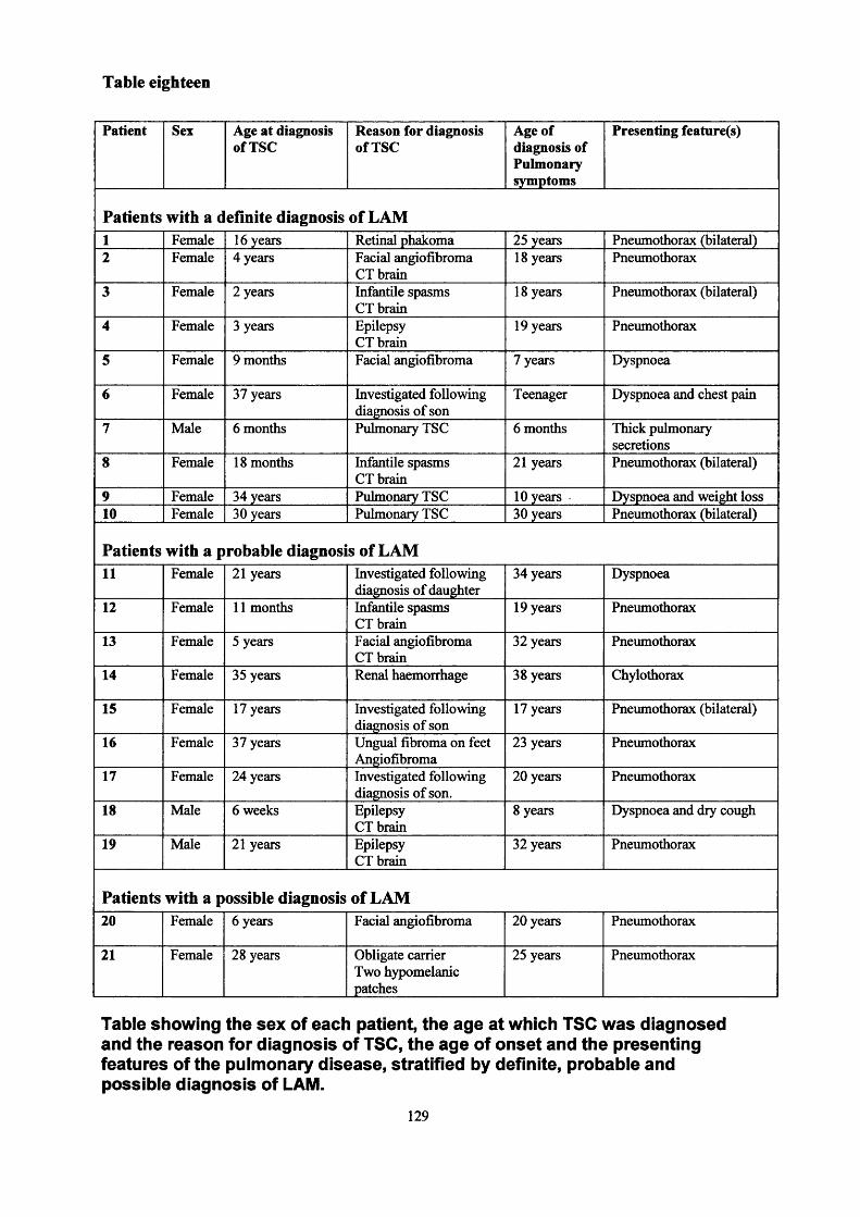
Table eighteen
Patient Sex Age at diagnosis of TSC
Reason for diagnosis of TSC
Age of diagnosis of Pulmonary symptoms
Presenting feature(s)
Patients with a definite diagnosis of LAM1 Female 16 years Retinal phakoma 25 years Pneumothorax (bilateral)2 Female 4 years Facial angiofibroma
CT brain18 years Pneumothorax
3 Female 2 years Infantile spasms CT brain
18 years Pneumothorax (bilateral)
4 Female 3 years Epilepsy CT brain
19 years Pneumothorax
5 Female 9 months Facial angiofibroma 7 years Dyspnoea
6 Female 37 years Investigated following diagnosis of son
Teenager Dyspnoea and chest pain
7 Male 6 months Pulmonary TSC 6 months Thick pulmonary secretions
8 Female 18 months Infantile spasms CT brain
21 years Pneumothorax (bilateral)
9 Female 34 years Pulmonary TSC 10 years Dyspnoea and weight loss10 Female 30 years Pulmonary TSC 30 years Pneumothorax (bilateral)
Patients with a probable diagnosis of LAM11 Female 21 years Investigated following
diagnosis of daughter34 years Dyspnoea
12 Female 11 months Infantile spasms CT brain
19 years Pneumothorax
13 Female 5 years Facial angiofibroma CT brain
32 years Pneumothorax
14 Female 35 years Renal haemorrhage 38 years Chylothorax
15 Female 17 years Investigated following diagnosis of son
17 years Pneumothorax (bilateral)
16 Female 37 years Ungual fibroma on feet Angiofibroma
23 years Pneumothorax
17 Female 24 years Investigated following diagnosis of son.
20 years Pneumothorax
18 Male 6 weeks Epilepsy CT brain
8 years Dyspnoea and dry cough
19 Male 21 years Epilepsy CT brain
32 years Pneumothorax
Patients with a possible diagnosis of LAM20 Female 6 years Facial angiofibroma 20 years Pneumothorax
21 Female 28 years Obligate carrier Two hypomelanic patches
25 years Pneumothorax
Table showing the sex of each patient, the age at which TSC was diagnosed and the reason for diagnosis of TSC, the age of onset and the presenting features of the pulmonary disease, stratified by definite, probable and possible diagnosis of LAM.
129

Pulmonary disease
The average age of onset of pulmonary symptoms was 19 years with a median of 20 years
and a range of birth to 34 years. The average age of diagnosis of pulmonary tuberous
sclerosis was 25 years with a median of 25 years and a range of six months to 38 years. On
average there was a delay of six years between the onset of respiratory symptoms and the
eventual diagnosis of LAM.
The commonest presenting feature was pneumothorax, which occurred in 14 of the
patients, five of whom presented with bilateral pneumothoracies. Five patients had a
history of gradual onset dyspnoea. One patient presented with a chylothorax. The final
patient had thick bronchopulmonary secretions requiring suction as an infant: a lung biopsy
at the age of six months gave the diagnosis of LAM (table eighteen).
Pulmonary symptoms: 19 patients complained of dyspnoea, 15 had a cough with six
having haemoptysis at times, 13 patients experienced pain usually, though not always, in
conjunction with proven pneumothoracies and 11 patients noticed wheezing at times (table
nineteen).
Complications
In total 17 of the 21 patients suffered pneumothoracies. There were 12 right sided, five left
sided and six bilateral pneumothoracies. Four patients suffered from chylothoraces, one
had a pericardial effusion and one died from pulmonary haemorrhage (table nineteen).
Four patients died as a result of their respiratory disease:
The first patient had sudden onset of pain and shortness of breath at the age of 25
years. She was diagnosed as having bilateral pneumothoracies requiring bilateral
pleurodesis. There was then a gradual deterioration over the following 20 years
with increasing shortness of breath, a productive cough and cyanosis. She was
initially treated with home oxygen but was later admitted to a long stay hospital
where she developed cor-pulmonale and died at the age of 47 years from her lung
disease. She was considered for a lung transplant but it was decided that, because of
her history of pleurodesis and her general medical condition by that stage, she
would be unable to tolerate the operation.
130

The second patient had had a history of a left pneumothorax, aged 18 years, which
was treated by pleurodesis. Three years later she had bilateral pneumothoracies,
which were again treated by pleurodesis. Approximately a year later she
complained of shortness of breath and started suffering from recurrent chest
infections. She then developed respiratory failure requiring increasing amounts of
oxygen therapy. Although hormonal treatment was tried it was ineffective and at
the age of 26 years she too was considered for a lung transplant. Unfortunately by
that time she was so weak it was felt that she would not be able to tolerate the
physiotherapy required. In addition there were technical concerns because of the
pleurodesis. She died a year later.
The third had been admitted for investigation of indigestion and vomiting and
underwent gastroscopy under general anaesthetic. During the procedure she
developed bilateral pneumothoracies and required admission to intensive care. The
pneumothorax on the right failed to resolve and chemical pleurodesis was
performed. Following this her urinary output deteriorated and she went into renal
and respiratory failure, dying one week later.
The fourth patient was a 19 year old girl with a history of severe renal involvement.
She had a one week history of dyspnoea and dry cough (following two recent
airplane flights) and had been treated by antibiotics before presenting to casualty
with a left-sided pneumothorax. She was treated conservatively and allowed home
to await HRCT and review. Two days before her review she suffered an acute fatal
pulmonary haemorrhage whilst asleep.
One other patient also had a history of respiratory failure with two respiratory
arrests as a result of developing pneumonia after a 10 year history of gradually
increasing shortness of breath. She was treated in hospital with oral steroids and
oxygen. She was not ventilated because there were concerns about the possibility of
not being able to wean her off a ventilator. She has since made a remarkable
recovery and just six months later has returned to work.
131
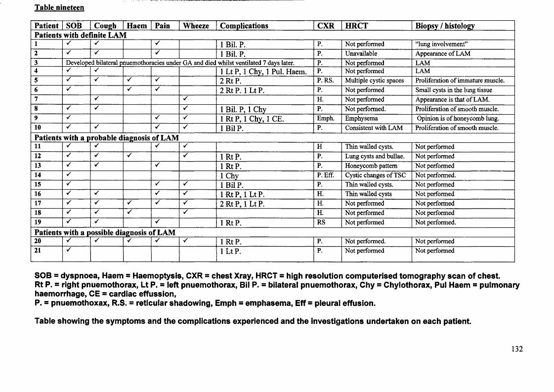
Table nineteen
Patient SOB Cough Haem Pain Wheeze Complications CXR HRCT Biopsy / histologyPatients with de inite LAM1 ✓ ✓ ✓ 1 Bil. P. P. Not performed “lung involvement”2 ✓ ✓ 1 Bil. P. P. Unavailable Appearance of LAM3 Developed bilateral pnuemothoracies under GA and diec whilst ventilated 7 days later. P. Not performed LAM4 V 1 Lt P, 1 Chy, 1 Pul. Haem. P. Not performed LAM5 S s ✓ ✓ 2 Rt P. P. RS. Multiple cystic spaces Proliferation of immature muscle.6 ✓ ✓ ✓ 2 Rt P. 1 Lt P. P. Not performed Small cysts in the lung tissue7 V ✓ H. Not performed Appearance is that of LAM.8 ✓ s s 1 Bil. P, 1 Chy P. Not performed. Proliferation of smooth muscle.9 ✓ s 1 RtP, 1 Chy, 1 CE. Emph. Emphysema Opinion is of honeycomb lung.10 ✓ ✓ 1 Bil P. P. Consistent with LAM Proliferation of smooth muscle.Patients with a probable diagnosis of LAM11 S ✓ s H Thin walled cysts. Not performed12 S ✓ s 1 RtP. P. Lung cysts and bullae. Not performed13 S s ✓ 1 RtP. P. Honeycomb pattern Not performed14 S 1 Chy P. Eff. Cystic changes of TSC Not performed.15 s ✓ s 1 Bil P. P. Thin walled cysts. Not performed16 s s ✓ s 1 Rt P, 1 Lt P. H. Thin walled cysts Not performed17 s V ✓ ✓ s 2 RtP, 1 Lt P. H. Not performed Not performed18 V s ✓ H. Not performed Not performed19 ✓ ✓ 1 RtP. RS Not performed Not performed.Patients with a possible diagnosis of LAM20 s 1 RtP. P. Not performed. Not performed21 1 LtP. P. Not performed Not performed
SOB = dyspnoea, Haem = Haemoptysis, CXR = chest Xray, HRCT = high resolution computerised tomography scan of chest. Rt P. = right pnuemothorax, Lt P. = left pnuemothorax, Bil P. = bilateral pnuemothorax, Chy = Chylothorax, Pul Haem = pulmonary haemorrhage, CE = cardiac effussion, P. = pnuemothoxax, R.S. = reticular shadowing, Emph = emphasema, Eff = pleural effusion.
Table showing the symptoms and the complications experienced and the investigations undertaken on each patient.
132
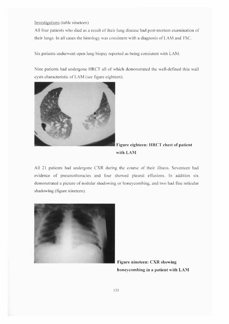
Investigations (table nineteen)
All four patients who died as a result o f their lung disease had post-mortem examination of
their lungs. In all cases the histology was consistent with a diagnosis of LAM and TSC.
Six patients underwent open lung biopsy reported as being consistent with LAM.
Nine patients had undergone HRCT all o f which demonstrated the well-defined thin wall
cysts characteristic o f LAM (see figure eighteen).
Figure eighteen: HRCT chest of patient
with LAM
All 21 patients had undergone CXR during the course o f their illness. Seventeen had
evidence of pneumothoracies and four showed pleural effusions. In addition six
demonstrated a picture of nodular shadowing or honeycombing, and two had fine reticular
shadowing (figure nineteen).
Figure nineteen: CXR showing
honeycombing in a patient with LAM
133

It was not possible to perform spirometry on ten of the patients; four who had had died,
five who had severe learning difficulties and were unable to co-operate with the test and
the spirometer was not available at the time of the visit of one further patient. Lung
function tests in the remaining patients were normal in two, showed an obstructive picture
in three and a restrictive pattern in the other six.
Treatment (table twenty)
Drainage of the pneumothoracies and chylothoraces was undertaken in the acute phase
when necessary. Nine patients underwent pleurodesis and four pleurectomy for recurrent or
severe pneumothoracies.
Five patients were prescribed conventional inhalers with little or no improvement in
symptoms.
Four patients received progesterone therapy;
Patient two suffered her first pneumothorax at the age of eighteen, which was treated
by pleurodesis, three years later she had bilateral pneumothoracies, again treated by
pleurodesis. A year later aged 22 years she began to suffer from increasing dyspnoea
and recurrent chest infections. She was commenced on progesterone tablets but her
condition continued to deteriorate and the progesterone was discontinued after one
year. She later died aged 27 years.
Patient ten had bilateral pneumothoracies aged 30 years requiring pleurodesis. She was
then started on depot provera, which she has continued to the present day (five years
follow up). Since starting the progesterone she has remained well with mild dyspnoea
and wheeze on exertion and an occasional cough (no haemoptysis) only.
Patient twelve suffered her first pneumothorax aged twenty one requiring pleurectomy.
Over the following 16 years she had slowly worsening dyspnoea with episodes of
haemoptysis. At the age of 35 years a HRCT of the chest confirmed the diagnosis of
LAM and depot provera was commenced. Over the following year her exercise
tolerance improved and she also showed an improvement in spirometry. She remains
well on the depot provera.
134

Table twenty
Acute / surgical treatment
Long term Medical treatment
Disease Course (follow up)
Patients with a definite diagnosis of LAM1 Drainage
PleurodesisOxygen therapy Died from cor-pulmonale whilst waiting
for transplant (~ 25 years after onset of symptoms)
2 DrainagePleurodesis
Oxygen therapy Progesterone
Died from cor-pulmonale, too ill for transplant (~ 10 years after onset of symptoms)
3 Drainage and ventilation None Died from respiratory and renal failure (~ one week after onset of symptoms)
4 None None Died from massive pulmonary haemorrhage (~ two weeks after onset of symptoms)
5 DrainagePleurodesis
None Stable (3 years)
6 DrainagePleurectomy
None Remains well (25 years)
7 None Inhalers (no benefit) Improved (23 years)8 Drainage
PleurectomyNone Remains well (6 months)
9 Lobectomy None Remains well (38 years)10 Drainage
PleurodesisProgesterone Remains well (6 years)
Patients with a probable diagnosis of LAM11 None Atrovent (little benefit) Stable (13 years)12 Drainage
PleurectomyProgesterone Improved (18 years)
13 DrainagePleurodesis
Atrovent (some benefit) Remains well (2 years)
14 Drainage Progesterone Remains well (1 year)15 Drainage
PleurodesisNone Progressive deterioration (10 years)
16 DrainagePleurodesis
Inhalers (no benefit) Progressive deterioration (30 years)
17 DrainagePleurodesis
Progressive deterioration (31 years)
18 None Inhalers (little benefit) Improved slowly (6 years)19 Aspiration None Remains well (2 years)Patients with a possible diagnosis of LAM20 Drainage
PleurodesisNone Remains well (17 years)
21 Drainage None Remains well (5 years)
Table showing acute surgical and medical treatment, long term medical treatment, disease course and time of follow up.
135
Patient fourteen suffered a renal haemorrhage at the age of 35 years, which was treated
conservatively by transfusion. She was then commenced on depot provera but four
years later suffered a pneumothorax and chylous effusion, treated by drainage. She
continues with hormonal treatment.
A course of steroids (dexamethasone / prednisolone) was prescribed for two patients with
no discernible effect on the progression of their disease. Though a third patient suffering
from acute respiratory failure made a remarkable recovery after treatment with oxygen and
steroids.
The two patients who died from cor-pulmonale received home oxygen therapy and courses
of antibiotics when appropriate. Both these patients were also considered for lung
transplantation.
Follow up (table twenty)
Two patients died within a few weeks of developing respiratory symptoms.
The remaining patients were followed up for an average of 14 years and a median of 10
years (range 6 months to 38 years).
Five patients have shown a progressive deterioration, a further two dying from their
disease. Three patients have improved slowly whilst two have remained stable. The other
nine patients have remained well with no further symptoms occurring after treatment of
their presenting symptoms.
Skin manifestations
All the patients had skin manifestations of TSC. Twenty patients had facial angiofibroma,
19 hypomelanic patches, 12 ungual fibromas, 11a shagreen patch, eight a fibrous forehead
plaque, eight nail ridging, five skin tags, three had poliosis and three confetti
depigmentation.
Renal manifestations (table twenty-one)
Twelve (57%) patients had a clinical history of renal involvement; of those who did not
only three had undergone imaging of the kidneys of whom one was found to have
136

angiomyolipomas (AML). Post mortem examination of a patient who had also been
entirely asymptomatic also showed evidence of renal disease. One patient died as a result
of a combination of renal and respiratory failure.
In total five patients suffered from proven haemorrhage, one underwent nephrectomy, one
had embolisation performed and one required both embolisation and then nephrectomy.
The other two patients were treated conservatively, one by transfusion the other with
analgesia and antibiotics.
In addition two patients had embolisation, and one a nephrectomy performed for pain
relief. One patient had a presumed renal carcinoma removed that was subsequently shown
to be an AML on histology.
Neurological manifestations
Epilepsy was the commonest neurological manifestation occurring in 18 (86%) of our
patients at some time. Ten (48%) patients had associated learning difficulties, but in three
it was very mild and they had been able to attend mainstream school. Six (29%) patients
had associated behavioural problems - autistic tendencies and hyperactivity. Two patients
had a GCA diagnosed with associated hydrocephalus requiring tumour excision and shunt
insertion. One patient had a mild hemiplegia.
Other clinical associations
Four patients (patients 7,11,18,19) had scoliosis, two were fixed and two were mobile,
none had undergone surgery or bracing. All four patients have had a relatively mild course
of LAM. Pulmonary function was only possible in one patient. He had a mild restrictive
defect, so although scoliosis is known to affect pulmonary function, it is difficult to assess
what part it has played in the respiratory disease of these four patients.
Four patients were also incidentally noted to have hepatic haemangioma on USS or CT
scan. None had a history of cardiac rhabdomyomas or WPW syndrome.
137

Table Twen tv-one
Renalcomplications
Age of onset of symptoms
Pain Haematuria Haemorrhage USS / CT result Treatment
Patients with a definite diagnosis of LAM1 Asymptomatic Not done2 Pain 11 years Yes Yes Yes Not known Embolisation and nephrectomy3 Pain, ARF 15 years Yes Multiple AML4 Pain 16 years Yes Yes Multiple AML Embolisation5 UTIs Unknown Yes Multiple AML6 Asymptomatic Not done7 Asymptomatic Normal8 Asymptomatic AML and cysts AML removed9 Asymptomatic Not done10 Asymptomatic Not donePatients with a probable diagnosis of LAM11 Pain, UTIs Childhood Yes AML & cysts12 Pain 20 years Yes Multiple AML Embolisation13 Asymptomatic Not done14 Pain 35 years Yes Yes Bilateral AML Transfusion15 Pain Not known AML & cysts16 Pain 21 years Yes Normal R kidney Nephrectomy17 Haemorrhage 14 years Yes Yes Yes Cysts R kidney Analgesia and antibiotics18 Asymptomatic Not done19 Pain 32 years Yes AML & cysts EmbolisationPatients with a possi )le diagnosis of LAM20 Pain, UTIs 11 years Yes Yes Yes AML & cysts Nephrectomy21 Asymptomatic Normal
Table showing the renal symptoms, complications and investigations of our group of patients.
Methods - Literature search (see appendix four for references)
A search of the English literature for all cases of LAM, both with and without associated
TSC, was performed from 1939 to 1997 using the Medline database and from 1981 to 1997
using the EMBASE database. The references of all retrieved papers (including those
discounted after review) were searched for additional references.
The cases were then divided into four groups on the following criteria:TSC: the paper either stated that a diagnosis of tuberous sclerosis had been made in the individual(s) or gave enough features in the individual(s) to fulfil the diagnostic criteria for tuberous sclerosis (for example the combination of epilepsy, mental retardation and renal angiomyolipoma).Possible: the paper did not state whether the individual(s) did or did not have tuberous sclerosis but described some features suggesting the possibility that they might have TSC (e.g. renal angiomyolipoma or mental retardation).Unlikely: the paper did not state that the diagnosis of tuberous sclerosis had been excluded and did not describe any features of tuberous sclerosis in the individual(s).Not TSC: The paper clearly stated that the individual(s) did not have any features of tuberous sclerosis. How this was done was not always stated.
Results
In total there were 183 papers citing a total of 445 individual cases of LAM.
83 cases had TSC, 119 were possible cases, 174 cases were unlikely and 69 definitely
were not TSC.
Population characteristics
There were 435 females, 9 males and in one case the gender was not given.
78 females and 5 males had TSC, 117 females and 2 males had possible TSC, 172 females,
1 male and one other patient (gender not stated) were unlikely to have TSC and 68 females
and 1 male were stated as not having TSC.
Pulmonary disease
The average age of onset of pulmonary symptoms was given for 365 / 445 (82%) cases and
was 32.7 years with a range of 4 years to 70 years.
TSC: Given for 75 patients, Average = 30.4 years (range = 8 - 5 7 years, SD = 9.2 years).
Possible: Given for 74 patients, Average = 27.5 years (range = 4 - 5 9 years, SD = 10.7
years).
Unlikely: Given for 148 patients, Average = 35.6 years (range = 4 - 7 0 years, SD = 11.5
years).139
Not TSC: Given for 68 cases, Average = 34.6years (range = 9 - 6 2 years, SD = 11.9
years).
The average age of diagnosis of LAM was given for 273 / 445 (61%) cases and was 36.2
years with a range of 4 - 72 years.
TSC: Given for 59 patients, Average = 36.6 years (range = 1 3 - 7 2 years, SD = 10.7
years).
Possible: Given for 50 patients, Average = 34.3 years (range = 4 -6 1 years, SD = 13.4
years).
Unlikely: Given for 135 patients, Average = 36.6 years (range = 1 4 - 7 2 years, SD = 10.7
years).
Not TSC: Given for 29 cases, Average = 36.6 years (range = 8 - 6 8 years, SD = 12.7
years).
Therefore on average there was a delay of 3.5 years between the onset of respiratory
symptoms and the eventual diagnosis of LAM.
The presenting features were given for 320 / 445 (72%) patients (see table twenty two) and
there was a wide range of pulmonary symptoms described in all groups (also summarised
in table twenty two). Pneumothorax and chylothorax were the commonest reported
complications. Pleural effusions were also reported but it was not always stated whether
these were serous, chylous or bloody.
Investigations and confirmation of diagnosis of TSC
Only two thirds of the papers stated how a diagnosis of LAM was confirmed. In the papers
that did, the majority were confirmed either histologically (post mortem or biopsy) or by
CT scan. Lung function testing was only performed on / given for approximately half of
the patients in the literature (table twenty three).
140
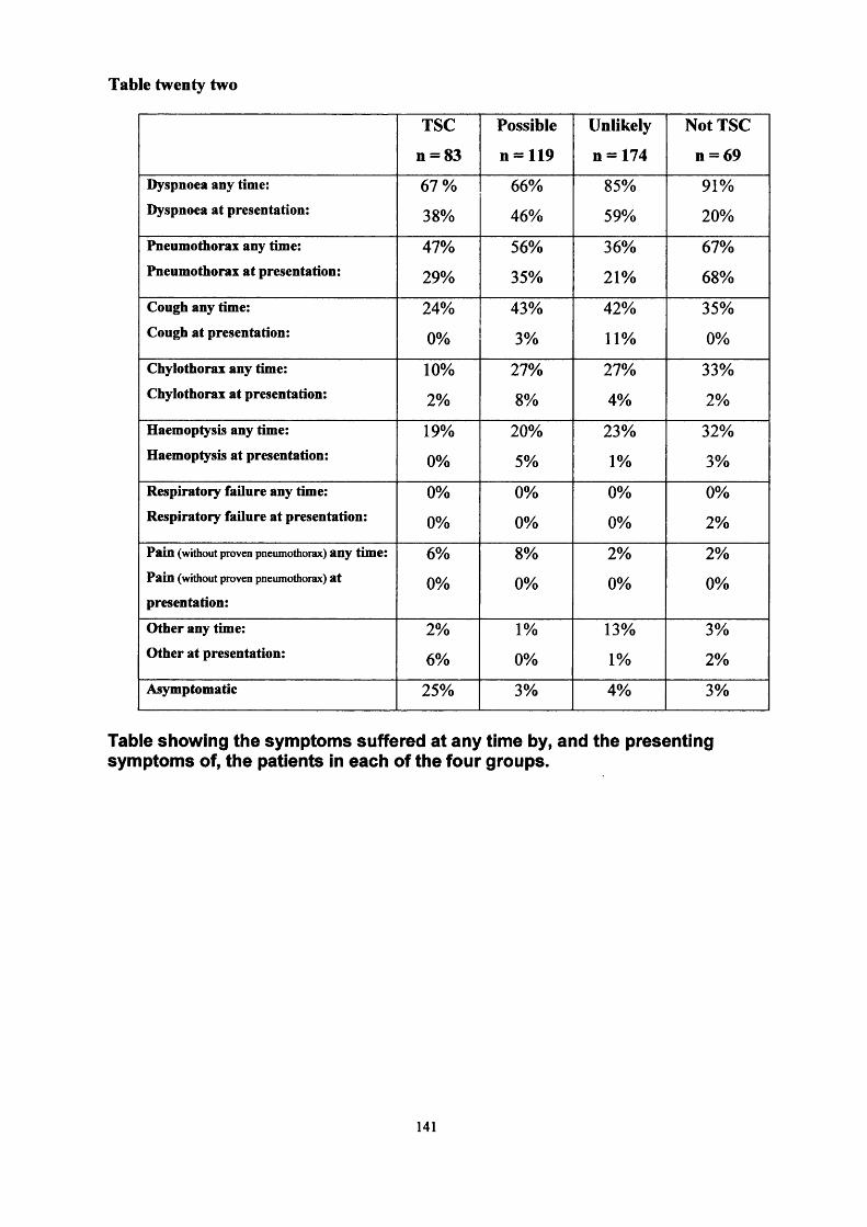
Table twenty two
TSC
n = 83
Possible
n = 119
Unlikely
n = 174
Not TSC
n = 69Dyspnoea any time: 67% 66% 85% 91%Dyspnoea at presentation: 38% 46% 59% 20%
Pneumothorax any time: 47% 56% 36% 67%Pneumothorax at presentation: 29% 35% 21% 68%
Cough any time: 24% 43% 42% 35%Cough at presentation: 0% 3% 11% 0%
Chylothorax any time: 10% 27% 27% 33%Chylothorax at presentation: 2% 8% 4% 2%Haemoptysis any time: 19% 20% 23% 32%Haemoptysis at presentation: 0% 5% 1% 3%Respiratory failure any time: 0% 0% 0% 0%Respiratory failure at presentation: 0% 0% 0% 2%Pain (without proven pneumothorax) any time: 6% 8% 2% 2%Pain (without proven pneumothorax) at
presentation:0% 0% 0% 0%
Other any time: 2% 1% 13% 3%Other at presentation: 6% 0% 1% 2%Asymptomatic 25% 3% 4% 3%
Table showing the symptoms suffered at any time by, and the presenting symptoms of, the patients in each of the four groups.
141

Table twenty three
TSC
n = 83
Possible
n =119
Unlikely
n = 174
Not TSC
n = 69
Investigations (for diagnosis)
Post mortem 9 (10%) 0 17(10%) 8 (12 %)
Biopsy 18 (21%) 48 (40%) 84 (48%) 16 (23%)
Chest CT 15(18%) 9 (8%) 61 (35%) 7 (10%)
CXR 9 (10%) 15 (13%) 2 (1%) 4 (6%)
Pulmonary function tests
Normal 5 (6%) 3 (3%) 9 (5%) 4 (6%)
Obstructive 18 (22%) 18 (15%) 46 (26%) 16 (23%)
Restrictive 3 (4%) 3 (3%) 19(11%) 1 (1%)
Mixed 0 0 10 (6%) 33 (48%)
Abnormal 8 (10%) 0 4 (2%) 2 (3%)
Table showing the investigations undertaken (and method of diagnosis) and the pulmonary function tests.
Treatment and outcome
Where stated most patients were treated symptomatically, pneumothoracies and effusions
were drained when clinically necessary and pleurodesis and pleurectomies were frequently
reported. Many patients received courses of antibiotics and those in respiratory failure
were treated by oxygen.
Hormonal manipulation was tried in a total of 187 / 445 (42%) of patients with variable
success (table twenty-four).
Unfortunately progression of the disease was only given for 95/258 (37%) of patients
who did not receive hormonal manipulation as treatment and even fewer gave an indication
of how long the patients had been followed up, but the findings are summarised in table
twenty-four.
142

Table twenty-four
Patients who did undergo hormonal manipulation
TSC
n = 83
Possible
n = 119
Unlikely
n = 174
Not TSC
n = 69
Total
n = 445
Improved 6 (33%) 2 (6%) 19 (20%) 2 (5%) 29 (16%)
Stable /
unchanged
5 (27%) 1 (3%) 27 (28%) 9 (21%) 42 (22%)
Worsened /
died
6 (33%) 26 (81%) 47 (50%) 23 (55%) 102 (55%)
Unknown 1 (5%) 3 (9%) 2 (2%) 8 (19%) 14 (7%)
Total 18 32 95 42 187
Average
follow up
5 years 10 years 4 years 8 years
Patients who did not undergo hormonal maniDulation
TSC
n = 83
Possible
n =119
Unlikely
n = 174
Not TSC
n = 69
Total
n = 445
Improved 0 2(11%) 1 (3%) 1 (7%) 4 (5%)
Stable /
unchanged
6 (24%) 3 (17%) 12 (32%) 3 (20%) 24 (25%)
Worsened /
died
19 (76%) 13 (72%) 24 (65%) 11 (73%) 67 (70%)
Total 25 18 37 15 95
Average follow up not given for this as data not available
Table showing the effects of hormonal manipulation on the course of the disease (compared with patients who did not receive hormonal manipulation).
Discussion
Diagnosis of LAM
We divided our group of patients into those who we felt had a definite (patients 1 -10), a
probable (patients 11- 19) and a possible (patients 20& 21) diagnosis of LAM. Although
our numbers were small there were no discernible differences between these three groups
143

in terms of presentation, clinical course and outcome. Unfortunately, when searching
through the literature it was not always clear as to how the diagnosis was reached or how
unequivocal the diagnosis was. For the purpose of our review we have assumed that the
diagnosis of LAM is correct in all the literature cases. However it must be borne in mind
that some cases may have been misdiagnosed and therefore suffer from other diseases such
as fibrosing alveolitis that have similar clinical and radiological appearances.
It is also interesting to note that papers have used the terms lymphangiomyomatosis and
lvmphangioleiomvomatosis synomonously to describe the changes in the lung.
Histologically, there is proliferation of irregularly arranged, spindle-shaped smooth muscle
cells surrounding the lymph channels and blood vessels within the lung parenchyma. There1/ i a 1 0 T 1 0 /1 1 0 < 1 0 /v
is no evidence of fat cells or collections in the lesions , therefore this
condition is most appropriately described as lvmphangioleiomvomatosis.
Diagnosis of TSC
We divided our group of patients into those who had a definite diagnosis of TSC,
according to the strict criteria given above, and those who had a probable diagnosis only.
For the purpose of the literature review we divided the patients on the information given by
the individual paper. For example, if a paper stated that the patient had TSC then they
would be classified as having TSC. Again it must be borne in mind that some cases may
have been misdiagnosed.
Prevalence
The Bath epidemiology study knew of 147 patients on 1st January 1997 with a diagnosis of
TSC in the Wessex region. The population of the Wessex region obtained from the Office
of Population Census and Surveys is 3,400,000. This gives a prevalence rate within the
Wessex region of 1:23,500 and this figure is similar to that found by other studies
estimating the prevalence of TSC. We know we are likely to have missed some cases of
TSC1 19, but within our population we are less likely to have missed symptomatic LAM.
On 1st January 1997 there was only one live TSC patient in the Wessex region, known to
have LAM, giving a prevalence rate of 0.66%, but patient four who was also alive at that1151time had asymptomatic LAM. This is similar to previous studies. Dwyer et. al. and
Torres et al127 (found 3 cases out of 403 patients reviewed) estimated that symptomatic
LAM occurs in less than 1% of patients with TSC. Castro et. al.44 believe their series
144
shows the incidence to be slightly higher at around 2.3%. However, incidental findings of
LAM have been described on the CXRs and CT scans of entirely asymptomatic patients118.
No study has been undertaken to screen TSC patients for LAM and it is likely that the true
prevalence is higher than 1%.
Population characteristics
LAM predominately affects females of child bearing age although cases occurring in1 ? o 1 0 0 n o i o imen and children have rarely been reported. In many cases symptoms start or
1 0 0 1 0 0are exacerbated during the menstruation period or in the course of a pregnancy
Patients have also reported onset of symptoms following the menopause, or after
commencing the oral contraceptive pill134 135.
We had 18 females and three males in our group of 21 patients. All the female patients
developed symptoms prior to the menopause. One (patient ten) had onset of respiratory
symptoms during her first pregnancy when she suffered bilateral pneumothoracies. Two
developed respiratory symptoms prepubertally at the age of seven (patient five), with
dyspnoea, pain haemoptysis and pneumothoracies, and at ten (patient nine) with dyspnoea,
pain, wheeze, pneumothorax and chylothorax (both started their menses in their teens).
Both these patients subsequently had open lung biopsy performed with a definite diagnosis
of LAM, so confirming that LAM in TSC is not confined to females of childbearing age.
In the literature out of 360 female cases in whom an age of onset was given there were five
cases of onset in childhood (4, 9, 9, 12 and 13 years). In total 7 / 378 cases presented in
childhood: approximately 2%.
We also had three phenotypic males in our group who exhibited signs of lung disease. Two
of the males developed symptoms at an early age: six months and eight years.
The first boy had a very unusual presentation; from birth he suffered from
thick pulmonary secretions requiring frequent suction and recurrent chest
infections. At the age of 6 months he underwent lung biopsy that was reported
as showing LAM. The combination of this result with the history of epilepsy
and developmental delay led to the diagnosis of TSC. During childhood he
continued to have recurrent chest infections requiring antibiotic treatment but
145
since going through puberty his infections have become far less frequent. He
has never suffered from pneumothoracies, chylothoraces or haemoptysis.
Unfortunately, the lung biopsy is no longer available for repeat analysis in
order to confirm the diagnosis and he is not sufficiently co-operative to
undergo HRCT. His unusual course must throw some doubt on the diagnosis.
The second male patient had suffered from episodes of dyspnoea and wheeze
from the age of eight years. He was originally diagnosed has having asthma,
but asthma medication did not relieve his symptoms and a CXR showing
honeycombing confirmed the diagnosis of LAM. He has never suffered from
pneumothoracies, chylothoraces or haemoptysis. He has never had
confirmation of LAM made by HRCT or open lung biopsy.
The third male had been entirely asymptomatic until the age of 32 years when
he suffered a spontaneous pneumothorax, which required treatment with
drainage only, he has had no recurrence (follow up 4 years), nor suffered any
other respiratory symptoms. His HRCT showed thin walled cysts and his CXR
reticular shadowing of the lung fields suggestive of LAM. Interestingly on
testing his chromosomes his genotype was 47XXY (Klienfelters syndrome).
This may give an explanation of his presentation of a lung disease that is
normally limited to females.
Looking through the literature there are five reported cases46 118 129 136 of LAM in
TSC males and a further possible two cases128 137’. Their clinical, radiographic
and histology details are summarised in table twenty- five.
Case one: had CXR findings compatible with a diagnosis of LAM and a lung
biopsy reported as “consistent with TSC”.
Case two: had CXR findings that were not typical of LAM and had not had CT
or biopsy performed. It is therefore possible that this patient did not have LAM.
Case three: had findings typical of LAM on his CXR, but the authors did not
state whether the biopsy showed smooth muscle proliferation.
Case four: although given a definite diagnosis of TSC, in the text the only
features described were skin tags and cafe au lait spots: we would not accept
146

these as confirming the diagnosis of TSC, so this diagnosis should be accepted
cautiously.
Case five: given that HRCT is highly sensitive at showing the thin walled cysts
associated with LAM, the presence of a normal result should make the diagnosis
unlikely.
In case six the authors stated that the biopsy was consistent with LAM but
described a proliferation of lymph vessels rather than smooth muscle hyperplasia.
These findings may be compatible with a diagnosis of lymphangioma rather than
lymphangioleiomyomatosis.
Case seven only had a clinical diagnosis of LAM.
Table twenty- five
ClinicalFindings
CXR CT Scan Biopsy
Patients diagnosed as having TSCCase l 118 Dyspnoea Extensive reticular
changesNot given Consistent with
TSCCase 211* Dyspnoea 111 defined opacity
plus a cystNot given Not given
Case 3138 DyspnoeaPneumothorax
Pneumothorax and cysts
Not given Thickened pleura and cysts
Case 4‘" Dyspnoea and cough
Not given Not given Cysts, empty or filled with fluid
CaseS1* Dyspnoea Pleural effusion Did not showpulmonaryalterations
Not given
Patients with a possible diagnosis of TSCCase 6128 Pleural effusion Pleural effusion Not given Abnormal
proliferation of lymph vessels Consistent with LAM
Case 7U7 Pleural effusion, pneumothorax
Not given Not given Not given
Table showing possible cases of LAM in males in the literature
It is therefore difficult to estimate the true frequency of LAM in TSC males,
other than it must be extremely rare. In any male in whom the diagnosis is
suspected, other pulmonary diagnoses must be considered and excluded and
147

chromosomal analysis undertaken. Patients should have HRCT at the very least
and preferably lung biopsy performed to confirm the diagnosis.
Although the numbers are small, the findings hint at the possibility that the
clinical course of LAM in TSC is milder in males than females.
The average age of diagnosis of TSC in our group of patients was 17 years, slightly older
than that usually seen for TSC. This may be a chance finding because of the small
numbers, but it may also back previous observations that patients with pulmonary TSC
generally suffer milder forms of TSC often delaying diagnosis. For example; Singerland et1 'Ifi • •al. found TSC patients without LAM to have intellectual impairment in 46% of cases
and seizures in 93%. In the patients with LAM 20% had intellectual impairment and 62%
had seizures.
Pulmonary disease
We found the average age to onset of pulmonary symptoms, and the subsequent diagnosis
of LAM to be earlier than that cited in the literature although the delay between onset andn o
diagnosis was similar. With regard to Dwyer’s data this difference may be explained, at
least in part, by improved diagnostic imaging and increased awareness over the past 25
years that lung complications do occur in TSC. However Castro44 et al had similar findings
to Dwyer118 in their review just two years ago (though they only had nine cases). The
average age of onset of pulmonary symptoms in our group of patients was 19 years and
diagnosis 25 years. This compares to an average of 30.4 and 36.6 years in the patients with
TSC in the literature. In both groups there is a delay of about 6 years between onset and
diagnosis. Therefore, the earlier age of diagnosis found in our small group of patients may
just reflect a chance finding of earlier age of onset.
Comparing LAM in patients with TSC to those without TSC there is a later age of onset of
symptoms (30.4 and 34.6 years respectively) and shorter period of delay between onset and
diagnosis in the group without TSC. This difference might be explained by the Knudson
two hit therory10: normally for a tumour to arise (in an individual without an inherited
defect), two independent somatic mutations are required, explaining why sporadic tumours
in those without TSC can rarely arise in a single tissue later in life. But individuals who
have already inherited an inactivated tumour suppressor gene only need one further148
somatic mutation to lose both alleles, explaining why in TSC the tumours often occur
earlier.
Histologically the lungs show perivascular and perilymphatic smooth muscle throughout
the lung fields. The proliferation of the smooth muscle gives rise to the different clinical
manifestations depending on its site. For example, around the airways it leads to
obstruction and air trapping contributing to the obstructive picture on lung function testing
and also causes bullae formation leading to pneumothoracies and a restrictive picture on
lung function testing. Around the pulmonary venules it leads to venous congestion with
consequent haemoptysis and around the arterioles it may precipitate pulmonary
hypertension and cor-pulmonale, whilst involvement of the lymphatics may lead to
chylothorax. In addition there is also proliferation of fibrous tissue which probably
contributes to the diffusion impairment seen in the most affected patients whilst, shunting
and V.Q. mismatching may increase the degree of hypoxia44 139.
As alluded to above, pulmonary tuberous sclerosis can present in a multitude of guises.
Presentation can range from mild exertional dyspnoea to sudden acute severe respiratory
distress with marked hypoxia and collapse. One common scenario is that described by
Slingerland138 in his case presentations of a 21 year old daughter and her 45 year old
mother. Progressive dyspnoea begins in the 2nd or 3rd decade of life accompanied by a
chronic non-productive cough and haemoptysis; this is then followed by the onset of
increasing number of lower respiratory tract infections associated with worsening
hypoxemia and an obstructive respiratory picture. Death can occur within years as a result
of respiratory failure and cor-pulmonale. In another case presentation of a 21 year old girl
by Uzzo et al140, the patient had been entirely asymptomatic from a respiratory point of
view until 2 weeks prior to scheduled surgery for renal angiomyolipoma. She then
presented as an emergency with a massive left pneumothorax and bilateral chylothorax
eventually requiring several chest drains and bilateral pleurectomies.
We also found a wide range of presentation from gradual onset of shortness of breath to
sudden bilateral pneumothoracies, thick pulmonary secretions to chylothorax. The
commonest presenting symptoms were pneumothorax and dyspnoea. Other symptoms
included; chronic dry cough, haemoptysis, wheeze, and chest pain. Development of
149

cyanosis, respiratory failure and cor-pulmonale was also common. These corresponded to
the findings in the literature.
Chylothoracies may occur less commonly in patients with TSC, 10% of cases with proven
TSC compared to 33% in patients in whom TSC has been definitely excluded. A possible
explanation may be differences found histologically as the smooth muscle proliferation in
LAM seen in TSC is more frequently described as being predominantly peri-vascular; in
LAM without TSC it is peri-lymphatic122 123 124 125 126. The reason for these differences is
not clear. Pneumothoracies seem to be equally common in both forms of the disease.
We also report two rare manifestations of LAM. The first is the occurrence of a pericardial
effusion (patient nine). This patient presented at the age of ten with dyspnoea and weight
loss. She subsequently developed recurrent pleural effusions, a persistent right
pneumothorax and a pericardial effusion. She was treated by lobectomy after which all her
symptoms resolved and she has remained well since. There has only been one other case141
reported in the literature in a patient without TSC. She had a history of dyspnoea and
recurrent pneumothoracies and had been diagnosed as having LAM on biopsy. She then
presented with cardiac tamponade and was found to have a haemopericardium, which was
successfully treated, by a subdiaphragmatic window. The second rare manifestation was
patient four who suffered a massive pulmonary haemorrhage resulting in sudden death. To
our knowledge this has not been previously reported in the literature.
Lymphangioleiomyomatosis is described as a generalised lung disease affecting all zones
of the lung equally142. However, we believe that, in patients with TSC at least, there is
evidence that it can occur as a focal entity. This is best demonstrated by the clinical history
of two of our patients. Patient nine (described above) had severe persistent disease for
several years until she underwent a lobectomy, following which her symptoms resolved.
She has received no other treatment and has remained asymptomatic for 38 years. Patient
six also had a two year history of severe persistent disease with dyspnoea, haemoptysis,
pain and recurrent pneumothoracies. She then underwent pleurectomy, has had no other
treatment, and has developed no further symptoms in 25 years of follow up.
150

Investigations
A CXR is the first investigation, showing any pneumothoracies, chylothoraces and
effusions and excluding other causes: it may show prominent interstitial markings with
obvious cysts sometimes forming a honeycomb pattern and in milder cases may show a
microreticulonodular pattern often greatest in the lower lobes118 139. Nevertheless, in the
early stages the CXR may be unhelpful.
In these cases, HRCT is more sensitive and more precise at demonstrating the well-defined
cysts that tend to have smooth thin walls. The architecture of the intervening lung tissue is
normal. Occasionally focal markings due to lymphatic oedema in the alveoli and
intralobular septa secondary to lymphatic involvement are also seen143 144. HRCT also has
the advantage of correlating better with lung function than plain radiography145; disease
extent as rated independently on a scale 0%-100% of involvement on HRCT, correlated
significantly with impairment in gas exchange (using carbon monoxide diffusion). There
was no significant correlation between the number of opacities seen on CXR and the
results of any of the pulmonary function tests. It is frequently stated that HRCT is highly
sensitive in diagnosing LAM and in combination with a clinical history and examination,
open lung biopsy to confirm the diagnosis may only rarely be required146. Also there have
been two reports147 148 of diagnosis made on cytology following aspiration of chylous
effusion. In both cases immature smooth muscle cells and surface cells from the
endothelium were demonstrated, suggesting that the LAM tissue had ruptured into the
thoracic cavity. In both cases the diagnoses were later confirmed by open lung biopsy.
Only one case has been cited in the literature on the use of magnetic resonance imaging
(MRI) in LAM. King149 reported on a 33-year-old woman who underwent MRI because of
the possibility of chest wall recurrence of breast cancer. An incidental finding was focal
areas of low signal intensity surrounded by thin walls. HRCT gave a diagnosis of LAM,
which was later confirmed by open lung biopsy. There is no reference in the literature as to
the use of MRI either in the diagnosis or as a marker of response to treatment in pulmonary
tuberous sclerosis.
Spirometry
Spirometry shows wide ranging of patterns from normal to obstructive through mixed to
restrictive. This variation is unsurprising when the underlying pathogenesis is considered.
151

Initially there is proliferation of smooth muscle cells around the bronchi leading to
narrowing of the lumen, as seen in asthma, giving rise to an obstructive picture. Unlike
asthma the narrowing is not due to bronchospasm but rather hyperplasia, and the
obstruction is not readily reversible. With time, air trapping in the alveoli leads to an
emphysematous picture with the formation of cysts and bullae giving rise to a restrictive
picture. As the disease progresses with more smooth muscle cells surrounding both the
airways and the blood vessels, diffusion becomes impaired, cor-pulmonale and respiratory
failure develop.
Clinical associations
Because very few studies (none of them large) of patients with LAM in TSC have been
described in the literature it is difficult to accurately summarise its associations with the
other clinical stigmata of TSC. Some authors have suggested that pulmonary TSC tends to
develop in patients with less severe disease, especially when considering seizures and
learning difficulties44 118 138. In Castro’s44 study eight out of nine of patients suffered from
severe renal disease with haemorrhage into AML prior to diagnosis of their lung
complications, suggesting a possible association between symptomatic renal disease and
LAM.
We carefully looked for other clinical associations of TSC in our 21 patients and compared
them with the 250 patients known to have TSC in the Wessex region to see if there was
any phenotype at high risk of developing LAM. If this proved to be the case it might form
a basis for screening high-risk patients. There was no significant difference between the
two groups in the prevalence of any of the following stigmata / clinical associations of
TSC;
• skin manifestations,
• epilepsy,
• learning difficulties,
• behavioural problems,
• renal disease,
• cardiac disease.
We also took blood from all our patients in order to analyse whether genotype correlates
with lung disease but this data will not be available for some time. We therefore propose,
that at the present time, there are no prognostic factors that assist in predicting which
152

individuals with TSC are at greatest risk of developing lung disease, but that it is rare in
men and children.
Treatment
Again, because of the rarity of this condition few studies have been performed to assess
long-term treatment and indeed few therapeutic measures have been undertaken in order to
ameliorate the course of the disease. Instead treatment is symptomatic, treating the
complications as they arise. Pneumothoracies and chylothoraces are treated in the acute
phase by drainage, though pleurodesis is often required, as they are frequently recurrent.
Chest infections are treated with the appropriate antibiotics and haemoptysis, if severe,
occasionally requires iron supplements. The cough and wheeze are difficult to control as
they are generally caused by the proliferation of the smooth muscle, (though undoubtedly
some patients do have an element of bronchospasm), with the result that conventional
inhalers are often ineffective. In those patients with a chronically deteriorating course,
continuous oxygen therapy will be required towards the terminal stages of the respiratory
failure and cor-pulmonale.
The presence of hormone receptors in the lung tissue of patients with LAM and TSC’150 151,
the suggestion that exacerbations can occur at times of hormonal fluctuation for example
during pregnancy and in association with the oral contraceptive pill, and the relative
absence of LAM in males have, not surprisingly, led to attempts to ameliorate the disease
by hormonal manipulation121 152 153 154 155.
In the literature review just under half of patients were tried on hormonal therapy. They
used a wide range of therapies including oophrectomy, radioablation of the ovaries,
progesterone administration and androgens with varying results. The best results are seen
in those patients with TSC. One third of patients with TSC tried on hormonal therapy
showed improvement compared with none showing improvement without hormonal
treatment, although the numbers are small and consist mainly of individual cases in open
studies.
1̂1In 1989 Eliasson et al undertook a meta-analysis of the hormonal treatment of LAM.
They identified 30 cases of LAM treated by hormonal manipulation. Unfortunately many
153

of the cases lacked information on dosage, timing and duration of therapy making them
ineligible for inclusion in the analysis, (table twenty-six).
Table twenty-six121
Hormonal therapy Claimedsuccess
Casesexcluded*
Objectivesuccess
Progesterone alone 8/13 5 5/9Oophrectomy alone 5/9 2 5/7Tamoxifen alone 2/7 4 1/3Androgen alone 0/1 0 0/1Oophrectomy + progesterone 3/5 3 2/2Oophrectomy and tamoxifen 0/1 1 0Progesterone + tamoxifen Vi 2 0Oophrectomy, progesterone + Tamoxifen
1/3 1 1/2
Reported regimens of treatment before and after meta-analysis.*ln sequential evaluation of each case, ten case were excluded because therapy was started too late in the course of disease; six additional cases were excluded due to lack of data on dosage; and of cases remaining, two were excluded due to insufficient data to judge outcome of therapy
The remaining studies showed oophrectomy alone or in combination with progesterone to
be the most successful therapeutic option with seven out of nine cases showing
stabilisation or improvement. Progesterone alone was successful in five out of nine
patients. None of the other regimes showed any significant benefit. Although this review
suggests that hormonal manipulation might be beneficial in some patients the results need
to be treated with caution. It is not clear if any of the thirty cases reviewed suffered from
TSC and it is not yet known whether LAM with TSC and LAM without TSC are similar in
their response to treatment. In addition it appears that the natural history of LAM in TSC is
extremely variable. Finally, we would not recommend radioablation of the ovaries as a
means of hormonal manipulation in patients with TSC: the genes involved in TSC are
tumour suppressor genes so there is a theoretical risk (at least) that exposure to
radiotherapy may increase the incidence and development of hamartomas. Our series
would not alter these findings.
Angel Ferdnandez-Gonzalez156 suggested in 1993 that lung transplantation should be
considered in patients in view of poor survival after the onset of respiratory symptoms.
Sadly, previous pleurectomies and the presence of systemic disease (cor-pulmonale,154

hypertension etc), not to mention lack of donors, limits the number of patients in whom
this is a viable option. It is also impossible to predict at the onset of symptoms, which
patients will follow a relentless deteriorating course, which patients will remain relatively
stable or even improve spontaneously and those who will respond to hormonal treatment.
In addition, there have been reports of LAM recurring in transplanted lungs157 158.
Outcome
LAM is an important cause of mortality in TSC. In the review undertaken by Charles
Shepherd et. al. at the Mayo Clinic in 1991 lung disease was the fourth commonest cause
of early mortality in TSC, accounting for four out of 49 deaths, despite it only affecting 1 -
2.5 % of all TSC sufferers.
It was previously thought that there was a relentless and severe deterioration after the onset110
of the disease. Average duration of survival was reported to be 4.8 years in 1971 . More
recent studies estimate survival times to be nearer to 10 years and some reports have even
told of spontaneous resolution of the disease. In our experience the clinical course in TSC
is extremely variable, with death occurring within days of the onset of symptoms in some
patients, whilst others may remain asymptomatic for years.
Screening
Screening for LAM in patients with TSC remains controversial. We had hoped to highlight
those patients at greatest risk of developing LAM and propose a screening model for this
complication; however this has not been possible. The criteria required for a good
screening programme include (as taken from Health For All Children edited by David Hall
- please see box below 203*204):
1. A recognisable latent or early symptomatic stage; this does not appear to be the
case with LAM. Fourteen of our patients presented acutely with pnuemothoracies
having been previously asymptomatic, two died within a short period of time.
2. That the natural history of the development of the disease from latent to
symptomatic disease should be understood; further research is required for this.
3. There should be a suitable test for detecting the disease at an early or latent stage,
which should be acceptable to the population; HRCT is likely to be the most
sensitive tool for recognising LAM but it is not clear that changes are seen on
HRCT before the development of symptoms and it may be unwise to screen all
155

females approaching childbearing age in view of the radiation dose administered in
patients with TSC who already have one mutation of their tumour suppressor gene;
we had considered if lung function testing, spirometry or oxygen saturation, could
be used for screening, these are simple non-invasive tests that can easily be
performed in a clinic setting, however even in our small cohort of patients with
symptomatic disease there was wide variation in lung-fimction suggesting that it is
unlikely to be helpful as a screening tool.
4. There should be an acceptable form of treatment available for the disease, which if
administered at the presymptomatic stage of the disease should favourably
influence its course and prognosis; as discussed above further research is required
to assess the efficacy of treatments such as progesterone in this group of patients,
before it could even be considered to be given in pre-symptomatic patients.
However, it may be worth considering assessment in female patients of child bearing age
for lung disease before they undergo anaesthesia or fly as these may precipitate onset of
symptoms.
Criteria for screening programmes203,204
1. The condition being sought should be an important health problem for the individual and the
community.
2. There should be an acceptable form of treatment for patients with recognizable disease or some
other form of useful intervention should be available (e.g. genetic counselling).
3. The natural history of the condition, including its development from latent to declared disease,
should be adequately understood.
4. There should be a recognisable latent or early symptomatic stage.
5. There should be a suitable test or examination for detecting the disease at an early or latent
stage, which should be acceptable to the population.
6. Facilities should be available for diagnosing and treating patients uncovered by the programme.
7. there should be an agreed policy on whom to treat as patients.
8. The treatment at the pre-symptomatic stage of the disease should favourably influence its
course and prognosis.
9. The cost of case finding which should include the cost of diagnosis and treatment, should be
economically balanced in relation to (a) possible expenditure on medical care as a whole and
(b) the cost of treatment if the patient does not present until the disease reaches a symptomatic
stage.
10. Case finding should be a continuing process, not a once and for all project.
156

Conclusion
LAM is a rare but important cause of morbidity and mortality in patients with TSC.
Importantly, the only risk factor we found both in our cohort of patients that it almost
exclusively occurs in female patients of childbearing age. It may rarely occur in
prepubertal girls, the diagnosis should strongly be questioned in males. We found no other
prognostic factors to identify those female patients at greatest risk of developing the
disease, but some authors have suggested an association between symptomatic renal
disease and LAM. We did not find any other clinical manifestations to be more common in
our group of TSC patients compared with the overall population of TSC patients. LAM is
usually generalised but may be focal in some patients and has a highly variable clinical
course and again we found no prognostic factors to predict how the disease will progress in
any individual, or those patients who will benefit from treatment. However, it does appear
that hormonal manipulation may be beneficial in as many as one third of patients and
further clinical trials are needed to fully evaluate the efficacy of such treatments. At
present there is no evidence that screening the whole TSC population would be beneficial;
nevertheless there is some evidence that flying and anaesthesia may precipitate
symptomatic LAM in some patients. It may therefore be prudent to undertake chest HRCT
before an anaesthetic is administered in high risk patients. Screening before flying is more
controversial and maybe should be discussed with each patient individually.
References
See appendix four.
157
CHAPTER EIGHT
Renal Disease
There were twenty-four patients with TSC in the Bath district; twenty-two underwent renal
ultrasound scanning. Nearly two thirds (13/22) were found to have renal involvement and
13% (3/22) had been symptomatic. AML were most frequently found lesions, occurring in
half of the patients screened. Five patients had cysts, one of whom had polycystic kidney
disease. One patient was found to have a renal carcinoma. None of the rarer complications
e.g. sarcoma were found in our group of patients.
Renal involvement is common in TSC with a wide diversity of renal disease most commonly angiomyolipoma (AML) and/or cysts with, more rarely adenocarcinoma, but oncocytomas, sarcomas, interstitial fibrosis and glomerulosclerosis have all been reported34. The severity of renal disease also varies widely. Many patients with AML with or without cysts will remain asymptomatic with normal renal function throughout their life, whilst others may suffer pain, haemorrhage, end stage renal failure (ESRF) and ultimately death from their lesions29.__________________________________________________________
Three patients who were all known to have large bilateral AML experienced symptoms of
pain and haematuria, requiring treatment with selective arterial embolisation. The patient
with the renal carcinoma had been asymptomatic and the lesion discovered on routine
scanning. The diagnosis was confirmed initially by CT scan and then by biopsy, the patient
then underwent nephrectomy and is currently being followed up. None of our patients
have died from their renal disease but renal complications are an important cause ofO'}
premature mortality in patients with TSC. In the study carried out at the Mayo Clinic
looking at the cause of death in 40 patients with TSC, renal disease was the leading cause
of mortality accounting for eleven of the deaths. Seven patients died of renal failure, two of
renal carcinoma and two of haemorrhage from AML. The paper was not clear about the
underlying pathology in the seven patients with renal failure.
End stage renal failure in adults with the tuberous sclerosis complex
To date no study has been carried out in the UK to look at the incidence or prevalence of
ESRF in TSC or its clinical presentation and no paper has reported the underlying
pathology in these patients. Therefore, the aim of this study was to examine both the
prevalence and underlying causes of ESRF in patients with TSC.
158
Acknowledgements
Dr Antonia Clarke, Dr Christopher Kingswood and Prof. John Osborne who designed the
questionnaire, identified the members of the European Dialysis and Transplant
Association, distributed and collected the questionnaires. I was responsible for collating
the results and writing up the data.
Method
This study was based on a questionnaire sent to the 170 UK members of the European
Dialysis and Transplant Association in September 1993. The questionnaire asked if they
currently had any patients with TSC in their renal replacement program. For those that did,
the following clinical information was requested:
• Sex.
• Age of diagnosis of TSC.
• Age of diagnosis of renal disease.
• Age of onset of ESRF.
• The reason for diagnosis of TSC and whether the patient was a familial or sporadic
case.
• They were also asked if the patient had ever suffered from epilepsy, learning
difficulties, giant cell astrocytomas or pulmonary lymphangioleiomyomatosis.
• In addition, information was collected on the presenting features of the renal disease,
including the presence or absence of hypertension, the underlying lesions, the
confirmation of diagnosis and renal function at presentation.
• Finally, we asked about the treatment the patient received for their renal disease
including the need for nephrectomy.
Results
Of the 170 members contacted, 142 (84%) replied with a total of ten patients being
identified. At least one reply was received from each unit.
There were eight females and two males. The average age at diagnosis of TSC was 21.2
years (range <1 year to 34 years). Three patients had TSC diagnosed as a result of then-
renal disease (table twenty seven), the average age of diagnosis of TSC in these patients
was 32 years compared to an average age of 16 .7 years in the other seven patients who
159

had TSC diagnosed on other criteria including skin lesions, epilepsy and family history
(table twenty-seven). Six patients were known to be sporadic cases of TSC and two were
known to be familial. Eight patients had a history of epilepsy and four had learning
difficulties (one mildly) but none had a history of giant cell astrocytomas or pulmonary
disease (table twenty-seven).
The average age of presentation of renal disease was 31 years (range 18 -50 years). Only
one patient (case 10) was known to have renal involvement prior to presentation of renal
disease as a result of screening at the age of 18 months when he was diagnosed as having
TSC. He had then remained asymptomatic until 26 years of age.
Table twenty-seven
Sex Age TSC Diagnosed
Reason TSC Diagnosed
Familial / Sporadic
LD
1 F 32 years Family history Familial No
2 F <1 year Infantile spasms Sporadic Yes
3 F About 12 years Epilepsy NK No
4 F 29 years Skin lesions Familial No
5 F NK Skin lesions Sporadic Yes
6 F 20 years Skin lesions, Epilepsy NK No
7 F 34 years NK Sporadic Yes
8 F 17 years Renal Haemorrhage Sporadic No
9 M 5 years Skin lesions, Epilepsy Sporadic No
10 M 18 months Infantile spasms Sporadic Yes
LD = learning difficulty, NK = not known
Table showing the age and sex of the patients, the age at which TSC was diagnosed and the reason for diagnosis, whether the inheritance was sporadic or familial and the presence I absence of learning difficulties.
160

The presenting features varied widely (table twenty-eight). Seven had impaired renal
function at the time of presentation, five had renal masses, four had raised blood pressure,
three had haematuria, three complained of pain and one had a urinary tract infection.
Table twenty-eight
Age at diagnosis of renal disease
Presentation Age ofESRF(years)
Lesions Diagnosis Renal function at diagnosis
Hypertension at diagnosis
1 29 years Renalmasses
38 Carcinoma Histology NK No
2 20 years ESRF 20 NK NK ESRF Yes
3 32 years Renalfailure
32 SK and cysts USS NK Yes
4 32 years Haematuria 48 AML and cysts
IVP Normal No
5 44 years Acute on chronic renal failure
49 PKD USS ARF No
6 26 years PainRenalmasses
26 SK and cysts USS CRF Yes
7 34 years Pain, renal masses and haematuria
40 PKD CT CRF Yes
8 18 years Acuteabdomen
19 AML CTHistology
Normal No
9 50 years UTIRenalmasses
55 PKD USS CRF No
10 26 years PainHaematuria
26 PKD USS ESRF Yes
ESRF = end stage renal failure, UTI = urinary tract infection, NK = not known, Cr = creatinine, SK = small kidneys, USS = ultrasound scan, PKD = polycystic kidney disease, CT = CT scan, CRF = Chronic renal failure (all had high creatinine at presentation).
Table showing the age of presentation of renal disease and age of ESRF, presentation of renal disease, the underlying lesions, method of confirmation of the diagnosis, renal function at time of presentation of renal disease and the presence I absence of hypertension.
There was a range of underlying pathology; four patients were noted to have PKD, one
patient had AMLs and cysts, two were noted to have small kidneys with cysts and one161
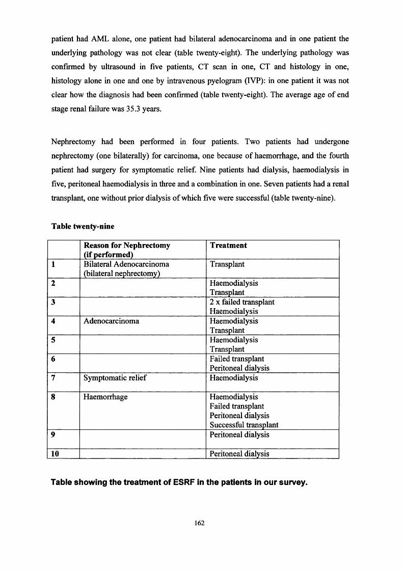
patient had AML alone, one patient had bilateral adenocarcinoma and in one patient the
underlying pathology was not clear (table twenty-eight). The underlying pathology was
confirmed by ultrasound in five patients, CT scan in one, CT and histology in one,
histology alone in one and one by intravenous pyelogram (IVP): in one patient it was not
clear how the diagnosis had been confirmed (table twenty-eight). The average age of end
stage renal failure was 35.3 years.
Nephrectomy had been performed in four patients. Two patients had undergone
nephrectomy (one bilaterally) for carcinoma, one because of haemorrhage, and the fourth
patient had surgery for symptomatic relief. Nine patients had dialysis, haemodialysis in
five, peritoneal haemodialysis in three and a combination in one. Seven patients had a renal
transplant, one without prior dialysis of which five were successful (table twenty-nine).
Table twenty-nine
Reason for Nephrectomy (if performed)
Treatment
1 Bilateral Adenocarcinoma (bilateral nephrectomy)
Transplant
2 HaemodialysisTransplant
3 2 x failed transplant Haemodialysis
4 Adenocarcinoma HaemodialysisTransplant
5 HaemodialysisTransplant
6 Failed transplant Peritoneal dialysis
7 Symptomatic relief Haemodialysis
8 Haemorrhage Haemodialysis Failed transplant Peritoneal dialysis Successful transplant
9 Peritoneal dialysis
10 Peritoneal dialysis
Table showing the treatment of ESRF in the patients in our survey.
162

Discussion
The prevalence of TSC is 1:23,000 between 15-45 years and 1: 97,000 over 45 years: we
would therefore expect 1,350 adults in the UK to have TSC of whom approximately half
will have normal intelligence. It is possible that in the UK patients with intellectual
disability (i.e. half of patients with TSC) are less frequently referred to a renal replacement
program. We have identified 10 adults, six with normal intellect who have end stage renal
failure and TSC in the UK giving a prevalence of 1% in those with normal intellect. There
are other possible inaccuracies. Firstly, the response rate to our questionnaire was only
84% and it is conceivable that there are patients with TSC who were not notified. It is also
likely that some patients with ESRF due to TSC are not recognised because either they do
not exhibit other overt signs of TSC or are only mildly affected. However recent analyses
suggest the prevalence of TSC1 may be higher than previously recognised which would
reduce the risk of ESRF in TSC. The approximate risk of ESRF in TSC may be different in
those with learning difficulties - recent studies have begun to suggest that TSC2 gene
defects may produce a more severe phenotype than TSC1 disease at least as far as intellect
and epilepsy is concerned with no data yet on renal disease, other than PKD. A study
carried out in France42, making different presumptions, also found the prevalence of ESRF
in adults with TSC to be 1 in 100. They do not state if this is the whole of the adult
population with TSC or those with normal intellect only. No mention is made of the
underlying causes.
In our survey there was a preponderance of females. A study carried out by Webb159 et al
suggested asymptomatic renal involvement to be as common in males as females. The
small numbers might account for this discrepancy, but it might reflect that females tend to
have more severe disease than males. It is well documented in the literature that females
have a higher probability of symptoms from the lung complications of TSC
(lymphangioleiomyomatosis - see chapter seven) than males. One explanation is that
female hormones play a part in influencing the disease. This theory has been backed up by
the finding of hormone receptors150 in affected lungs and that the course of the disease may
be affected in some patients by hormonal manipulation (e.g. oophrectomy) or by natural
changes in hormone levels (e.g. pregnancy132 and menopause134). It is therefore feasible
that oestrogens and progesterone similarly influence the renal lesions as the lung lesions.
The underlying histopathology is similar for most TSC lesions in that they arise as
163

hamartomas from over proliferation of otherwise normal tissue as a result of inactivation of
tumour suppressor genes.
Inheritance of TSC is autosomal dominant with a spontaneous mutation rate of about 60%.
Only two patients in our survey had parents with TSC, six were known to be sporadic
mutations and in two the mode of inheritance unknown.
The patients in our survey represented a wide spectrum of renal disease seen in TSC. As
discussed before, the TSC2 gene is close to the PKD1 gene, but PKD1 mutations do not
underlie the cytogenesis in all patients. Renal disease including cysts occurs in patients
with both TSC1 and TSC2. Although polycystic disease was more common in our survey
of TSC patients with renal failure than is found in the TSC population, PKD was the cause
of ESRF in only four of our patients; PKD is a known cause of ESRF, it is to be expected
that this cause would be significantly represented in our survey, the pathological causes of
ESRF in the other patients are more interesting.
The underlying pathology probably contributes to the renal failure by a variety of
mechanisms. There may be compression of the normal renal parenchyma by cysts,
angiomyolipoma or carcinoma. There may be a reduction in the amount of normal tissue
either as a result of direct invasion by tumour or resulting from surgery. In addition it has
been hypothesised that cysts in particular cause hyperfiltration of the remaining glomeruli,
creating focal glomerulosclerosis42.
Probably as a reflection of the range of underlying pathology our patients presented in
many different ways from simple haematuria to massive intra-abdominal haemorrhage;
from acute renal failure to insidious chronic renal failure. They all presented in adulthood
with a mean age of 31 years progressing to ESRF at an average of 35.3 years.
Confirmation of renal disease was made by ultrasound in the majority of cases.
Ultrasonography is particularly sensitive in detecting cysts and intramural or perinephric
haemorrhage160, but the echodense pattern seen on ultrasound although suggestive of AML
is not pathognomonic. Renal carcinoma tends to be echolucent on ultrasound. CT is highly
specific for AML when fatty tissue can be demonstrated within the lesion161. If any doubt
remains biopsy can be undertaken to exclude malignancy, but it can give very misleading
164

results. Histology at the edge of AML may look like renal carcinoma but recent studies
suggest that staining for HMB45, which is positive in AML and negative in carcinoma, can
differentiate it162. CT has the disadvantage of radiation and the theoretical risk of inducing
further hamartomas, but while MRI can demonstrate renal lesions it does not demonstrate
fatty tissue well.
Treatment of the renal lesions may contribute to the development of ESRF as discussed
above. Nephrectomy remains the treatment of choice for patients with carcinoma, but
transcatheter embolisation of distal branches of the renal arteiy for haemorrhage or pain
offers a more conservative form of treatment allowing greater preservation of parenchymal
tissue, thereby either delaying or preventing the onset of ESRF. Once ESRF has occurred
treatment consists of dialysis and transplantation. There is still controversy as to whether
nephrectomy should or should not be performed in patients undergoing transplant.
Advocates argue that the diseased kidney carries a risk of bleeding and malignancy and
should therefore be removed at the time of transplantation. Others argue that the disease
free part of the kidney may retain some function which will be of use to the patient should
rejection of the transplanted kidney occur.
Screening for renal disease in patients with TSC also remains controversial (please see
chapter seven for the criteria for a good screening programme).
Presymptomatic diagnosis of PKD is known to be advantageous, for control of
hypertension, prompt treatment of urine infections, close monitoring of renal function and
early treatment of renal bone disease, but other lesions are much more common. Unlike
LAM a suitable screening tool is available; ultrasound can be used to detect PKD in the
young infant. Ultrasound scanning is a safe non-invasive technique which is generally
acceptable to patients and parents. It is generally accepted that once a diagnosis of TSC has
been made in an infant or young child that a renal USS should be undertaken to allow
diagnosis of associated PKD. However, for the other lesions seen in TSC many of the
criteria for good screening discussed in the previous chapter can not be met.
Studies in the past have suggested that AML, which bleed, are likely to be greater than 3.51 f\*\- 4cm in diameter . Some would therefore advocate screening for AML so that when
they reach such a size prophylactic treatment could be undertaken. Ultrasound can be used
165

to detect small AML and to monitor their size. However, the natural history of progression
from latent to established disease remains unclear; size is not always predictive of
haemorrhage and some individuals with longstanding large AML have never suffered
haemorrhage. It might be the rate of expansion or some other factor which causes
haemorrhage; and whilst treatment (embolisation and / or surgery) is available it is not
without risk and complications, for example pain, infection, loss of normal renal tissue and
risk of embolisation elsewhere; neither is screening, for example it can force people to
know they have a disease, and may highlight the presence of disease other than that for
which they undertook screening164. Neither is it known that embolisation is curative.
Others would therefore wait until the lesions become symptomatic before initiating
treatment.
Likewise, screening for ESRF is not currently undertaken in the TSC population. At
present there is no screening test available that is simple, easy to interpret and acceptable to
the patients. In order to detect very early ESRF it is likely that regular renal function
testing involving venesection and blood pressure monitoring would be required. Whilst the
measurement of blood pressure in simple and non-invasive and should be routinely
performed as good clinical practice, many patients are not keen to have venesection
performed. In addition, at present it is not known how frequently renal function would
need to be undertaken to detect a decline in renal function before the onset of ESRF, nor
whether treatment will help. Further research is required to understand the natural history
and other factors to determine the risks and benefits of screening in these patients.
Conclusion
ESRF is rare in patients with TSC but does contribute significantly to the premature
mortality in this group. At present there are no clear prognostic factors that can be used to
determine which individuals are at greatest risk of developing ESRF, other than it appears
to be more common in females. Importantly it may be possible to delay, or even prevent
renal failure in some patients by undertaking more conservative forms of management,
such as embolisation rather than nephrectomy in the earlier stages of disease.
166
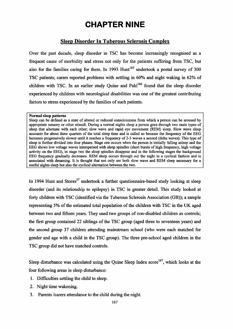
CHAPTER NINE
Sleep Disorder In Tuberous Sclerosis Complex
Over the past decade, sleep disorder in TSC has become increasingly recognised as a
frequent cause of morbidity and stress not only for the patients suffering from TSC, but
also for the families caring for them. In 1993 Hunt165 undertook a postal survey of 300
TSC patients; carers reported problems with settling in 60% and night waking in 62% of
children with TSC. In an earlier study Quine and Pahl166 found that the sleep disorder
experienced by children with neurological disabilities was one of the greatest contributing
factors to stress experienced by the families of such patients.
Normal sleep patternsSleep can be defined as a state of altered or reduced consciousness from which a person can be aroused by appropriate sensory or other stimuli. During a normal nights sleep a person goes through two main types of sleep that alternate with each other; slow wave and rapid eye movement (REM) sleep. Slow wave sleep accounts for about three quarters of the total sleep time and is called so because the frequency of the EEG becomes progressively slower until it reaches a frequency of 2-3 waves a second (delta waves). This type of sleep is further divided into four phases. Stage one occurs when the person is initially falling asleep and the EEG shows low voltage waves interspersed with sleep spindles (short bursts of high frequency, high voltage activity on the EEG), in stage two the sleep spindles disappear and in the following stages the background EEG frequency gradually decreases. REM sleep occurs through out the night in a cyclical fashion and is associated with dreaming. It is thought that not only are both slow wave and REM sleep necessary for a restful nights sleep but also the cyclical alternation between the two.
In 1994 Hunt and Stores27 undertook a further questionnaire-based study looking at sleep
disorder (and its relationship to epilepsy) in TSC in greater detail. This study looked at
forty children with TSC (identified via the Tuberous Sclerosis Association (GB)); a sample
representing 5% of the estimated total population of the children with TSC in the UK aged
between two and fifteen years. They used two groups of non-disabled children as controls;
the first group contained 22 siblings of the TSC group (aged three to seventeen years) and
the second group 37 children attending mainstream school (who were each matched for
gender and age with a child in the TSC group). The three pre-school aged children in the
TSC group did not have matched controls.
Sleep disturbance was calculated using the Quine Sleep Index score , which looks at the
four following areas in sleep disturbance:
1. Difficulties settling the child to sleep.
2. Night time wakening.
3. Parents /carers attendance to the child during the night.167

4. Parental sleep loss.
Each area is scored on a three point score (0 -2) dependant on the severity and frequency
of each problem giving a total score 0-8.
They found the degree of sleep disruption in their group of children with TSC to be
significantly greater (p < 0.001) than in the 22 siblings and 37 matched controls. The mean
sleep index for children with TSC was 4.9 (SD 3.1), compared with 0.59 (SD 1.02) for the
siblings and 0.41 (SD 0.68) for the matched controls, thus confirming that settling and
night time waking was more common in children with TSC than either their siblings or
other children attending mainstream school. Hunt and Stores do not state clearly how the
study subjects were recruited and it is possible that their group of children with TSC
represent the 5% with the greatest sleep difficulties and that the controls were chosen
because they were known not to have any sleep disturbances. Nevertheless, this study does
suggest that some children with TSC do suffer significant sleep disturbances.
Hunt and Stores also looked at the associations of sleep disturbance with epilepsy and
behavioural problems. Behaviour was rated using the Developmentally Delayed
Children’s Checklist (Einfeild and Tonge 1991), constructed specifically to include the
most prevalent behaviours observed in children with learning difficulties. Again they found
the scores for the children with TSC to be significantly higher (p < 0.001) than those
without. The children with TSC were further divided into two groups; those with current
seizures and those who had either been fit free for the six months preceding the study or
who had never had seizures. The mean Sleep Index of the former group was 6.24 (SD
2.44), and significantly higher than the mean 1.45 (SD 1.50) for the latter group (p <
0.001).
1 ££Seizures are known to disrupt sleep patterns in many ways including ;
• fragmentation by frequent awakenings,
• reduced REM sleep,
• increase in wakefulness after sleep onset,
• instability of sleep stages.
The sleep disorder in TSC is more complex than this with other multiple contributory
factors since the following are also known to affect sleep and occur in many patients with
TSC;168

• autism,
• hyperactivity,
• learning difficulties which in turn may affect the development of normal behavioural
zeitgebers (e.g. bed time routines which can be powerful enough to enforce the natural
circadian rhythms of melatonin secretion in the absence of light-dark cycles),
• anticonvulsant medications; often prescribed in patients with TSC may affect sleep.
Sedatives often prescribed for children with sleep disorders are generally ineffective and
clinical experience suggests that in children with TSC they are even less likely to be
effective. In fact they often worsen the situation by increasing hyperactivity. Behavioural
techniques are also of less benefit to many of these patients, because of the above
contributory factors and because of the extreme exhaustion of caring for these children day
and night.
Normal sleep-wake cyclesThe underlying physiological process for normal sleep-wake cycles in not yet fully understood and is likely to be complex, involving the circadian secretion o f neurotransmitters and learnt behavioural patterns (zeitgebers). Early theories suggested that the reticular activating system and other parts o f the brain became ‘fatigued’ over the period o f a waking day and therefore became inactive resulting in sleep. It was then thought that the raphe nuclei (situated in the brainstem), which produces serotonin, a neurotransmitter known to induce sleep was responsible. However, serotonin does not appear to be secreted in a circadian manner. It is now thought that Melatonin, a hormone secreted from the pineal gland plays a major role in the normal sleep-wake cycles. Melatonin is thought to synchronise sleep-wake patterns with the light-dark cycle o f the normal day, through pathways from the retina, via the hypothalamus, to the pineal gland. Decreased light intensity at the retina (i.e. darkness) leads to an increase in melatonin production, which in turn leads to drowsiness.
With the advancing interest in the treatment of sleep disorders in children, melatonin has
become one of the forerunners as a potential therapy for disruptive sleep patterns.
Melatonin was first used in its synthetic form in 1981 by Weitzman169 et al to regulate
sleep patterns in shift workers. It was then shown to be successful in alleviating jet-lag in
adults by Arendt et al170. First to show success in treating the sleep disorders of disabled171children (both with and without visual impairment) were Jan et al in 1994.
177A study of nine girls with Retts syndrome (but no visual impairment) was then
undertaken by McArthur et al 1998. Wrist actigraphs and sleep diaries were used to
monitor the sleep patterns of the nine patients. There was a one-week baseline period
followed by a double blind, placebo controlled, crossover trial lasting eight weeks with a
one-week washout period between the two crossover periods. The dose of melatonin given169
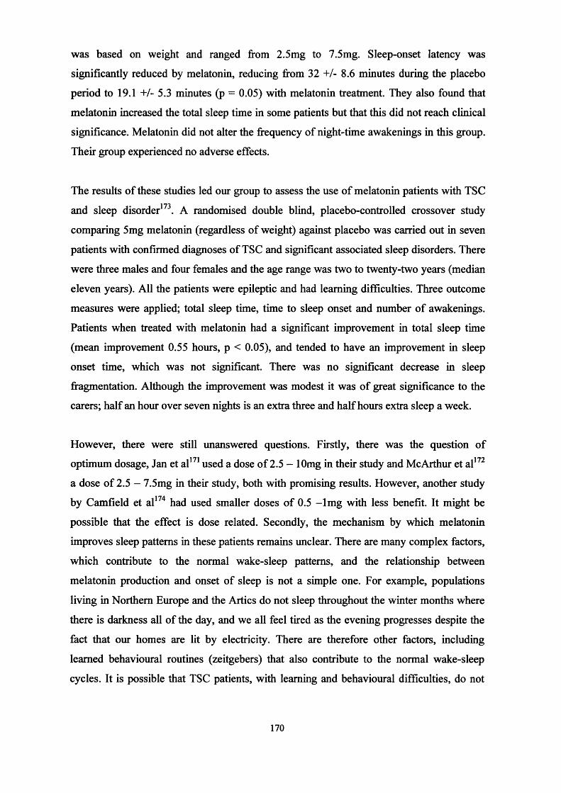
was based on weight and ranged from 2.5mg to 7.5mg. Sleep-onset latency was
significantly reduced by melatonin, reducing from 32 +/- 8.6 minutes during the placebo
period to 19.1 +/- 5.3 minutes (p = 0.05) with melatonin treatment. They also found that
melatonin increased the total sleep time in some patients but that this did not reach clinical
significance. Melatonin did not alter the frequency of night-time awakenings in this group.
Their group experienced no adverse effects.
The results of these studies led our group to assess the use of melatonin patients with TSC171and sleep disorder . A randomised double blind, placebo-controlled crossover study
comparing 5mg melatonin (regardless of weight) against placebo was carried out in seven
patients with confirmed diagnoses of TSC and significant associated sleep disorders. There
were three males and four females and the age range was two to twenty-two years (median
eleven years). All the patients were epileptic and had learning difficulties. Three outcome
measures were applied; total sleep time, time to sleep onset and number of awakenings.
Patients when treated with melatonin had a significant improvement in total sleep time
(mean improvement 0.55 hours, p < 0.05), and tended to have an improvement in sleep
onset time, which was not significant. There was no significant decrease in sleep
fragmentation. Although the improvement was modest it was of great significance to the
carers; half an hour over seven nights is an extra three and half hours extra sleep a week.
However, there were still unanswered questions. Firstly, there was the question of
optimum dosage, Jan et al171 used a dose of 2.5 - lOmg in their study and McArthur et al172
a dose of 2.5 - 7.5mg in their study, both with promising results. However, another study
by Camfield et al174 had used smaller doses of 0.5 -lm g with less benefit. It might be
possible that the effect is dose related. Secondly, the mechanism by which melatonin
improves sleep patterns in these patients remains unclear. There are many complex factors,
which contribute to the normal wake-sleep patterns, and the relationship between
melatonin production and onset of sleep is not a simple one. For example, populations
living in Northern Europe and the Arties do not sleep throughout the winter months where
there is darkness all of the day, and we all feel tired as the evening progresses despite the
fact that our homes are lit by electricity. There are therefore other factors, including
learned behavioural routines (zeitgebers) that also contribute to the normal wake-sleep
cycles. It is possible that TSC patients, with learning and behavioural difficulties, do not
170
acquire these normal social responses that aid the sleep-wake cycles. It is also possible that
TSC patients may not have a normal pattern of melatonin secretion.
The Effect of Melatonin Dosage on the Sleep Disorder in Tuberous
Sclerosis Complex
Aim
The aim of this study was to investigate whether there is a dose related response to
exogenous melatonin when using a dose higher than 5mg in patients suffering sleep
disorder associated with TSC.
Methods
Ethical approval for the study was obtained from the Bath Local Research Ethics
Committee. Patients with a confirmed diagnosis of TSC and severe sleep problems were
then identified through the Bath TSC clinic, research and epidemiological studies. A
diagnosis of TSC was confirmed by the presence of at least one hamartoma present in each
of a minimum of two different organs. A diagnosis of sleep disorder was confirmed by a
Quine sleep index score of at least six out of a possible eight167. The Quine index focuses
on four areas of sleep difficulties; difficulties settling the child to sleep, night time
wakening, parents /carers attendance to the child during the night and parental sleep loss.
Children were excluded from the trial if their sleep disorder proved to be situational (for
example a child that scored six on the Quine questionnaire whilst at home, but two whilst
at respite care would not be included in the trial).
This was a randomised double-blind controlled crossover trial comparing 5mg and lOmg
of melatonin. Melatonin was supplied by Penn Pharmaceuticals Ltd. Melatonin 5mg and
placebo (a sugar compound) were administered in identical capsules. The trial consisted of
four parts; an initial two week baseline period during which the patients received no
treatment, a two week period during which the patient received either 5mg or lOmg of
melatonin, a two week wash-out period during which the patient received no treatment and
a two week period during which the patient received the alternate dose of melatonin (i.e.
171

5mg or lOmg). The parents / carers completed both sleep and seizure diaries (see appendix
five) throughout to document the sleep disorder and seizure frequency.
Melatonin was administered approximately thirty minutes before the patients’ usual
bedtime; each patient received two identical capsules, either 2 x 5mg of melatonin (i.e.
lOmg in total) or 1 x 5mg plus 1 x placebo (i.e. 5mg in total). The patients, carers and
researchers did not know which weeks they received lOmg of melatonin and which weeks
they received 5mg of melatonin.
The sleep diaries were used to monitor the sleep latency (i.e. time taken to fall asleep), the
total sleep time and the number of awakenings each night. The seizure diaries were used to
monitor the frequency and type of seizures (if any) experienced by the patients during the
study period. The carers were also asked to record any illnesses the child had or any
possible side effects they suffered during the trial period.
Results were analysed using paired t-tests.
Results
Eight patients were recruited who satisfied the inclusion criteria. One patient completed the
study but the diaries were lost in the post and we had not thought to ask to have them
photocopied before they were posted to us. We present the results of the seven whose
diaries are available. There were four males and three females with an age range of 18
months to 31 years (median age 9 years). All patients were epileptic and on concurrent
anticonvulsants, two of whom were well controlled. All had severe learning and
behavioural difficulties.
The results are shown in table thirty and although there was a small improvement in sleep
latency time in the lOmg group compared to the 5 mg group, it does not reach conventional
levels of statistical significance (p = 0.4). Total sleep time was calculated for each of the
seven patients during each two-week phase of the trial. There was no statistical
significance between the two groups (p = 0.6). The mean number of awakenings was
recorded for five of the patients, but was not recorded by the carers in the other two
patients, again there was no statistical difference between the two groups in terms of sleep
fragmentation (p = 0.5).
172

Table Thirty
Patient Age Sleep latency
5mg
SleeD latency
lOmg
Total sleep time
5mg
Total sleep time
lOmg
Fragmentation
5mg
Fragmentation
lOmg
1 female 18 months 154 mins 105 mins 8 hours 51 mins 9 hours 25 mins 0.9 2.5
2 female 4 years 102 mins 128 mins 9 hours 7 hours 47 mins 0.9 1.0
3 female 9 years 53 mins 50 mins 8 hours 54 mins 8 hours 39 mins 0.2 .2
4 male 9 years 30 mins 46 mins 9 hours 28 mins 10 hours 29 mins 0.6 .2
5 male 11 years 11 mins 13 mins 9 hours 30 mins 9 hours 20 mins 1.2 1.2
6 male 19 years 208 mins 154 mins 8 hours 9 hours 36 mins
7 male 31 years 46 mins 36 mins 8 hours 55 mins 8 hours 38 mins
Mean 86 mins 76 mins 8 hours 57 mins 9 hours 4 mins 0.8 1.0
Table showing the mean sleep latency, total sleep time and sleep fragmentation after the administration of 5mg melatonin and10 mg melatonin.
173

In the five patients who had seizures during the trial, the seizure diary showed no change in
the frequency (or type) of seizures seen compared to the initial baseline period before
melatonin treatment. Three patients were well controlled on their anticonvulsants and
remained seizure free during the study (table thirty-one).
There were no adverse side effects reported by the carers during the trial period.
Table thirty-one
Patient Age No. of seizures
without melatonin
No. seizures on 5mg
melatonin
No. of seizures on
lOmg melatonin
1(f) 18 M None None None
2(f) 4 Y 23 / week 20 /week 20 / week
3(f) 9 Y None None None
4 (m) 9 Y 4 / week 3 / week 3 / week
5(m) 11 Y 5 / week 6 / week 6 / week
6 (m) 19 Y None None None
7 (m) 31 Y 73 / week 159 / week 40 / week
p = 0.38 p = 0.31
Table showing seizure frequency; without melatonin; after the
administration of 5mg melatonin; and after the administration of 10mg
melatonin.
Discussion
The best way to accurately monitor sleep and its disturbance is by polysomnography; this
was not possible in our group of patients. We chose to use sleep diaries, which is an
inexpensive method of monitoring sleep patterns. Although, reliant on the vigilance of
already overburdened caregivers, they demonstrate symptoms and any bias in minimised
by the double blind nature of the study. McArthur et al used wrist actigraphs in their
study172 and found them to be more accurate than sleep dairies but they too have
disadvantages; they are prone to mechanical failure and non-compliance, which may lead174

to total loss of data. The effect of nocturnal seizures on actigraphs is a further complication
in patients with TSC since the assumption is that movement is an awakening when it might
be due to a seizure.
Although only a few studies have been undertaken looking at the effect of melatonin on
sleep disturbance in children with severe neurological difficulties with mixed results, the
combined results suggest that the effect seen might be dose related. Studies using higher
doses (2.5mg - lOmg) demonstrated a response while those using lower doses (0.5mg -
1 .Omg) did not.
The results from our previous study173 comparing the effects of 5mg of melatonin versus
placebo, found increased total sleep time and a trend towards decreased sleep latency, but
with varying responses between subjects. We also found a trend towards increased total
sleep time and decreased sleep latency when comparing our baseline data (i.e. no
melatonin) with administration of 5 mg melatonin; however we have not reported these
findings since they were not blinded and are therefore prone to attrition bias. We have
shown that the administration of 10 mg of melatonin did not further reduce the sleep
latency, improve the total sleep time or reduce sleep fragmentation in our group compared
with 5mg. One possible explanation for this lack of dose related response is that 5mg is
already a supraphysiological dose.
The exact mechanism by which melatonin improves sleep patterns in these patients
remains unclear, many hypothesis have been suggested; the simplest that melatonin has a
straight forward sedative effect. This would explain the clinical effect seen in our patients
i.e. reduced sleep latency. It is also known that melatonin is metabolised rapidly and that
there is a large hepatic, first pass metabolism so that, in humans, peak plasma levels are
reached within one hour and there is a half life of approximately 45 minutes (personal
communication). This rapid metabolism may explain why total sleep time and sleep
fragmentation remained unimproved.
Another hypothesis is a link with epilepsy. All our patients were known to suffer from
seizures and were on concurrent anticonvulsants during the trial. It is known that epilepsy175plays an important role in the sleep disturbance of TSC by disrupting the normal sleep
physiology in a variety of ways. It has also been suggested that melatonin may influence
175

1 H(\epileptic activity in humans, though the exact relationship remains unclear. In 1974
melatonin was reported to improve seizure control in epileptic subjects, an observation also177 17ftnoted in mice when used in large doses (i.e. >200mg/kg). In 1985 Maurizi showed
melatonin to increase levels of serotonin in the brain thus raising the seizure threshold. In
1994 Molina et al1/9 and in 1995 Schapel180 both demonstrated that patients with epilepsy
had higher serum concentrations of melatonin than individuals without epilepsy suggesting
the possibility of a physiological attempt to reduce seizure threshold in these patients. Onlyl O I
Sheldon et al have suggested in an observational study reported m a letter to the Lancet
that oral administration of melatonin in disabled children might paradoxically result in an
increase in seizure frequency.
All our carers completed seizure dairies for the eight-week duration of the sleep trial.
There appeared to be no significant effect of melatonin administration on seizure activity
in any of our patients.
It is also possible that some patients with TSC have either abnormally low or disruptedi ftnmelatonin secretion. Although previous authors have suggested that patients with
uncontrolled epilepsy have increased nocturnal melatonin production, others have
suggested that the circadian rhythm is disrupted with peak levels occurring earlier in the170night than normal subjects . However, to our knowledge, no one has looked at the
circadian rhythms of melatonin production in children and comparisons have been made
against adult data. In addition no study has looked at melatonin production in patients with
TSC and epilepsy. We have addressed this in a separate study.
This study did not find any detrimental effect on seizures. However, there were only seven
patients and it is possible that we have insufficient power to detect a significant difference
in response, or that individual patients might benefit from a higher dose. Since none of our
patients reported any adverse effects whilst taking the melatonin, we would suggest when
treating this group of children for sleep disorder that 5mg melatonin is used and lOmg is
only considered if 5mg fails to be of any benefit. Further studies on the use of melatonin
would be worthwhile - looking further at dosage and tolerance over a prolonged treatment
period.
None of our patients reported any adverse effects whilst taking the melatonin.
176

Conclusion
In conclusion, whilst it seems likely that the administration of exogenous melatonin in
patients with TSC and sleep disorder does reduce sleep latency and increase total sleep
time, higher doses show no further benefit. The response to melatonin does seem to vary
between individuals and we would propose that a trial of 5mg of melatonin given 30
minutes before bedtime is worth considering in these patients and that the response should
be monitored by both sleep and seizure diaries. If no beneficial response is seen within two
weeks then, while there is no proven advantage in increasing the dose of melatonin to
lOmg, this is unlikely to cause significant side effects if it is considered. More studies are
required.
Melatonin excretion in normal children and tuberous sclerosis with sleep
disorder responsive to melatonin
Trials have established the beneficial effects of exogenous melatonin in the treatment of
sleep disorders in children, but the mechanism of action remains unclear. It has been
postulated that some children might have aberrant endogenous melatonin secretion that can
be corrected by giving exogenous melatonin.
Melatonin is excreted in the urine as 6-sulphatoxymelatonin (a relatively stable metabolite)
that can be easily measured. The pineal output of melatonin is believed to be very stable
from day to day so it is not necessary to collect urine over long periods of time. Total
excretion, the time of maximal excretion (acrophase time) and the wave form of excretion
over 24 hours (cosinor rhythm) can all be measured. A cosinor rhythm of less than 70%
can be due to inaccurate collection while if over 90% an accurate collection with normal
circadian rhythm can be inferred.
Aims
The aims of this study were firstly to measure the melatonin excretion in six children and
one adult suffering from sleep disorder responsive to melatonin (all these patients also
suffer from TSC) and secondly to measure melatonin excretion in healthy children to
obtain normal excretion data for age. The Bath Local Research Ethics Committee granted
ethical approval.177

Methods
Six children and one adult suffering from sleep disorder and TSC were recruited for the
study. All seven subsequently took part in a study comparing the effect of 5mg melatonin
versus placebo and were shown to have a significant improvement in total sleep time and a173tendency to decreased sleep latency when treated with melatonin . We also recruited 21
healthy children (between the ages of 5 and 15 years) who had no sleep problems. There
were two for each year of age, except for five years, where we were only able to recruit
one because of difficulties with compliance. We asked them to collect their urine over a
48-hour period in four hourly aliquots. The children were not woken in the night in order
to collect samples, but if they woke naturally to pass urine then a specimen was taken.
They were asked to pass urine, discard that first sample, note the time and then start the 48-
hour collection, collecting at least one sample four-hourly whenever possible. Each time
they subsequently passed urine they would note the time and the quantity passed. The
specimens were then frozen until the time of analysis.
All specimens were sent to Stockgrand Ltd, affiliated to the School of Biological Sciences
at the University of Surrey for analysis. Melatonin excretion was expressed as the total 6-
sulphatoxymelatonin secretion, cosinor analysis and the acrophase time over each of the
two consecutive 24-hour periods.
The results from the first 24-hours excretion of each healthy child were then analysed, both
including and excluding any results with a cosinor analysis of less than 70% and were then
compared with the second 24-hour excretion period to give an estimate of the repeatability
of the results. Only the excretion data from the first 24-hour period were used to determine
normal excretion.
The excretion data obtained from the patient group was then compared with the data
obtained from the healthy children and normal adult data.
Results
Tables thirty-two and thirty-three show the total daily excretion of 6-sulphatoxymelatonin,
the cosinor rhythm and the acrophase time for the healthy children and the patients
178

respectively. For each individual, two results are given, one for each of the 24 hours of the
48-hour collection. There were no significant differences or trends seen with age.
Table thirty-two
Controls Total 6SM Cosinor Acrophase(age years) (nanograms) % Time
5 11,112 99.9 02.3012,505 96.4 04.00
6 40,175 75.0 03.2430,574 99.7 03.48
6 17,148 99.1 01.3612,715 100.0 04.42
7 28,971 94.9 04.4228,374 96.2 05.00
7 24,386 57.8 08.3021,687 76.5 07.24
8 21,474 68.9 23.5416,167 73.6 01.54
8 28,820 95.7 02.1233,055 92.7 01.00
9 14,778 74.8 08.1214,630 81.1 09.42
9 11,911 94.9 03.0611,510 100.0 04.24
10 20,220 93.2 06.3620,529 56.9 09.42
10 11,184 92.7 04.0010,110 100.0 05.18
11 17,583 99.4 03.0621,074 96.9 04.48
11B 15,175 83.5 10.429,368 82.8 09.24
12 21,690 52.9 07.3618,644 100.0 03.36
12 16,820 99.9 05.4218,753 98.4 05.48
13 11,266 96.8 04.1211,915 100.0 05.36
13B 15,201 61.0 07.5413,209 75.4 09.42
14 24,785 97.7 05.1825,020 99.4 05.18
14 19,528 93.4 03.3018,020 99.0 05.00
15 10,760 99.3 03.3611,850 98.9 05.06
15 15,808 100.0 03.2416,307 96.9 02.24
Table showing the total 6-sulphatoxymelatonin excretion, the cosinor rhythms and acrophase times for the healthy controls. The first 24 hour excretion is given above the second 24 hour excretion for each child.
179
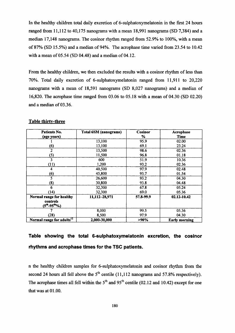
In the healthy children total daily excretion of 6-sulphatoxymelatonin in the first 24 hours
ranged from 11,112 to 40,175 nanograms with a mean 18,991 nanograms (SD 7,384) and a
median 17,148 nanograms. The cosinor rhythm ranged from 52.9% to 100%, with a mean
of 87% (SD 15.5%) and a median of 94%. The acrophase time varied from 23.54 to 10.42
with a mean of 05.54 (SD 04.48) and a median of 04.12.
From the healthy children, we then excluded the results with a cosinor rhythm of less than
70%. Total daily excretion of 6-sulphatoxymelatonin ranged from 11,911 to 20,220
nanograms with a mean of 18,591 nanograms (SD 8,027 nanograms) and a median of
16,820. The acrophase time ranged from 03.06 to 05.18 with a mean of 04.30 (SD 02.20)
and a median of 03.36.
Table thirty-three
Patients No. (age years)
Total 6SM (nanograms) Cosinor%
AcrophaseTime
1 13,100 95.9 02.00(6) 13,100 69.1 23.242 13,500 98.6 02.36
(5) 11,500 96.8 01.183 600 51.9 10.36
(ID 1,200 93.2 02.364 40,500 97.9 02.48
(6) 45,800 93.7 01.545 26,600 93.2 04.30
(8) 30,800 93.8 04.486 32,300 67.8 05.24
(14) 32,300 69.0 05.36Normal range for healthy
controls(5H,_95tfc0/o)
11,112-28,971 57.8-99.9 02.12-10.42
7 8,000 99.5 05.36(28) 8,500 97.9 04.30
Normal range for adults12 2,000-30,000 >90% Early morning
Table showing the total 6-sulphatoxymelatonin excretion, the cosinor
rhythms and acrophase times for the TSC patients.
n the healthy children samples for 6-sulphatoxymelatonin and cosinor rhythm from the
second 24 hours all fell above the 5th centile (11,112 nanograms and 57.8% respectively).
The acrophase times all fell within the 5th and 95th centile (02.12 and 10.42) except for one
that was at 01.00.
180

In our group of patients with tuberous sclerosis and sleep disorder, there were six children
and one adult. In the children, the total 6-sulphatoxymelatonin fell below the 5th centile in
one patient and above the 95th centile in one patient. The cosinor rhythm fell below the 5th
centile in one patient and the acrophase time fell before the 5th centile in three patients. The
adult patient showed excretion within the normal range for adults182.
Discussion
For our healthy controls, we chose five years as the lower limit, as we anticipated that
children below this age would be too young to co-operate. We took 15 years as the upper
limit as we felt that by 16 years excretion rates were likely to be similar to adult excretion
rates. We found no significant differences with age, although the very youngest children
tended to have slightly earlier acrophase time - perhaps because they go to bed earlier.
This finding is compatible with the results of Sivan184 et al who found a very early
acrophase time in normal infants. Adult data shows total daily excretion to be 2,000 to
30,000 nanograms, with a cosinor rhythm of >90% and an acrophase time within the early
hours of the morning . Our data is compatible with this and with the data of Sivan et al
suggesting that melatonin excretion is in a recognisable circadian rhythm from an early
age. This is also compatible with the data of Attanasio et al184 who showed a circadian
rhythm to melatonin excretion by measuring serum melatonin levels from infancy to
adolescence.
Melatonin excretion is not thought to vary greatly from day to day, but we chose to collect
our samples over two days to not only help assess how accurate the collections were, but
also to assess repeatability. Although a cosinor rhythm of 90% infers accurate collection, it
is also dependant on having a circadian rhythm. Our hypothesis was that patients with
sleep disorders might not have normal circadian rhythms as shown by a low (less than
70%) cosinor value, therefore we could not rely on this value alone. We therefore also
examined total 6- sulphatoxymelatonin excretion. Where the values from both 24-hour
collections were similar it suggests that all the urine passed in the 48-hour period was
measured and collected and no urine samples were “missed”. For example, it is possible
that the second eleven year old failed to collect at least some urine, perhaps on both days,
accounting for the low melatonin excretion and cosinor rhythms. A high cosinor value
correlates with a good circadian rhythm, however it was felt that ethically it was not
acceptable to wake the children in the middle of the night to ask them to pass urine, but
181

only to collect the urine if they woke naturally. Failing to collect the urine at frequent
intervals risks smoothing out the cosinor curve of a circadian rhythm and this might
explain why some of our children had low values. We also had to rely on the family to
correctly time and label the specimens, and any failures will have affected acrophase time
and cosinor rhythm. However, such failures will also occur in clinical practice, and
contribute to the “normal” range.
All our patients except patient three had 6-suphatoxymelatonin values that fell within the
normal range. Interestingly, patient three, as an individual also had a poor response to
exogenous melatonin. The fact that she did not respond to a “supraphysiological” dose of
melatonin strengthens the hypothesis that exogenous melatonin does not act by correcting
abnormal excretion patterns. This patient also had severe behavioural problems and this
may explain in part, her poor sleep patterns and poor response to melatonin. All but one of
the patients with TSC and sleep disorder had at least one cosinor value above 90%
indicating strong circadian rhythms. The one who did not, had responded to melatonin as
an individual with an improvement in total sleep time. The acrophase time is indicative of
the time at which maximal melatonin excretion occurs. In adults this occurs in the early
hours of the morning. In the majority of our controls the acrophase time also occurred in
the early hours of the morning. In two controls (1 IB and 13B), one or both collections, in
which the acrophase time was later than 9am, the cosinor values were also low suggesting
the possibility of inaccurate collections. However, the 6-SM excretion, although low was
better in the first 24 hours where the cosinor rhythm was no better. Full collection, but
inaccurate labelling of the time could explain this but normal variation is a more
reasonable explanation.
In the patients with sleep disorder, one patient had an acrophase time of 10.36am during
the first collection with a cosinor value of 51.9. The following collection had a cosinor
value of 93.2 and an acrophase time of 2.36am suggesting the possibility of inaccurate
collection or timing of samples during the first 24-hour period, but confirming the normal
excretion of melatonin in the second collection. Collecting two 24-hour samples from
patients is a sensible precaution when attempting to show normal excretion of melatonin.
Exogenous melatonin is usually administered shortly before the patient’s normal bedtime.
However we know that endogenous melatonin reaches its peak excretion in the early hours
of the morning; we also know that exogenous melatonin has a very short half life and is
182

quickly excreted in the urine. It is therefore possible that the advantageous effect of
exogenous melatonin occurs too early in the night to provide maximum benefit to the
patient. Perhaps by repeating the dose, should the patient wake or by administering a slow
release formula, the natural rhythm of endogenous melatonin secretion could be more
closely mimicked, allowing further improvement of the sleep patterns in these patients.
Conclusion
Although our numbers of controls and patients are small the total daily volume of 6-
sulphatoxymelatonin and acrophase time do not differ significantly between the two
groups or from adult data. In addition, all but one (who had responded to melatonin) of the
patients with sleep disorder showed a normal circadian rhythm of melatonin secretion, yet
showed an improvement in total sleep time and sleep latency when treated with exogenous
melatonin. These results suggest that the exogenous melatonin did not act by correcting
abnormal endogenous melatonin secretion. We would propose that exogenous melatonin
acts in these children by a simple sedative effect.
183
CHAPTER TEN
Summary of Thesis and Final Discussion
It is important to study TSC because potential treatment is available and there are a number
of unanswered questions about the natural history of the condition. It is likely that
resolving some of these issues could result in an improved quality of life and even life
expectancy. I chose to study this condition as the subject for my thesis because I have a
particular interest in epilepsy and neuro disability which are key issues for many patients
with TSC.
Epidemiology of the TSC population in the Bath Health District
The reason why I undertook this ten year review was to look at the natural history of TSC
in detail and to establish which clinical factors contributed most to the morbidity (and
mortality) of this population so that I might examine some of these problems in greater
detail. The advantage of undertaking a longitudinal study rather than a cross sectional
study is that it allowed me to examine the impact of such factors observed over time. A
database has now been set up at Bath to be updated regularly with information retrieved at
clinic appointments and from research studies, which I hope will allow a more detailed
picture of the disease to be compiled, giving greater understanding of the less common
complications such as LAM. I also hope that it will allow the impact of clinical
intervention in these patients to be more fully assessed.
In addition, this review gave us an opportunity to assess how the introduction of the
diagnostic criteria might change the overall prevalence of the disease4. There will be a
number of patients who previously had a diagnosis of TSC who are no longer considered
to do so. In our small population of just nineteen patients ascertained in 1986, three
patients had their diagnosis withdrawn; these are small numbers but if representative of the
whole population of TSC patients, this might mean that as many as 15% had an incorrect
diagnosis. This would significantly reduce the prevalence of the disease. It is therefore
important that with the introduction of new diagnostic criteria, large and detailed
epidemiology studies are repeated in order to provide a clear picture of the prevalence and
incidence of the disease.
184

Epilepsy, its association with learning difficulties and its treatment
The seizure disorders of TSC are closely associated with the learning difficulties
experienced by many of these patients. Infantile spasms are associated with the greatest
degree of mental handicap in patients with TSC but the optimum medical treatment for
infantile spasms remains uncertain. Steroids have been used for many years and my search
of the literature found them to be effective in stopping the spasms in just half of all
patients. One reason for the poor response to oral steroids might be that fears of serious
side effects have resulted in inadequate doses being administered, so I undertook a
retrospective review of the use of larger doses of oral prednisolone in patients with
infantile spasms in Bath. This review found it to be effective in stopping the spasms in
70% of patients with no serious adverse effects being reported; although small it provided
reassurance that larger doses of oral steroids could be used safely in the treatment of
infantile spasms and suggested that larger doses might be more effective in stopping the
spasms. I would suggest that larger study would be required to assess its efficacy more
fully. Assuming approximately 50% of patients would achieve cessation of seizures if
treated with 2mg/kg of prednisolone, such a trial would need to recruit at least 250
children, with approximately half randomised to “low dose (2mg/kg)” prednisolone and the
other half to “high dose (60mg/day)” to attain 90% power to detect an improvement in
cessation of seizures of 70% in the “high dose” group. Such a trial would require
collaboration between many centres over a long period of time.
Vigabatrin, a relatively new anticonvulsant has also been used in the treatment of infantile
spasms over the past 20 years, with similar results to steroids. However, recent studies had
suggested that it might be particularly efficacious in the treatment of infantile spasms in
tuberous sclerosis. I reviewed the English literature and found support for this theory,
although the majority of the studies undertaken were non-randomised and had little power.
Nonetheless, it suggested that vigabatrin should be the drug of choice for stopping the
spasms in infants either with a known diagnosis of TSC or at high risk of TSC.
Unfortunately although IS are associated with a high degree of mental handicap in patients
with TSC none of these trials assessed the effect of vigabatrin on either long term
psychomotor development or the onset of subsequent seizure types.
185

United Kingdom Infantile Spasms Study - A Stepping Stone to the Future
UKISS was a multicenter randomised trial set up to try and determine whether (if either)
steroids or vigabatrin was most effective at stopping infantile spasms and whether either
treatment was more efficacious with regard to the long-term outcome. I was responsible,
under JPO’s supervision for developing the trial design, protocol and trial packs which
were put to the trial steering committee for comment and approval. I also remain closely
involved with the trial as a member of the trial steering committee.
Unfortunately, patients with known TSC or at a high risk of TSC have been excluded from
this study since it was felt that it would be unethical to withhold vigabatrin as treatment
from this group of patients. The problem is that previous studies have not looked at long
term psychomotor development as an outcome. I therefore feel that it is important that
further studies are performed specifically looking at the treatment of infantile spasms in
this subgroup of patients, with particular respect to psychomotor development. If the
UKISS trial does find vigabatrin to be more effective in stopping the spasms and giving
improved development than steroids, it may be prudent to continue using vigabatrin as the
“gold standard” against which newer treatments can be considered. If however, the UKISS
trial finds no significant difference between the two groups or finds “steroids” to be more
efficacious then there is a greater dilemma. Should a randomised controlled trial be then
set up comparing steroids vs. vigabatrin in TSC - I feel that this would now be deemed
ethical (in light of the visual field defects associated with the use of vigabatrin), but others
might argue that a better trial would be one comparing treatment with vigabatrin alone say
against a combination of vigabatrin and ACTH? Side effects must be taken into
consideration. Although steroids are associated with a risk of mortality, especially if they
are used for long periods of time, their side effects are well known and can be closely
monitored. Unfortunately, although vigabatrin initially promised to be a relatively safe
drug there are increasing concerns about the potential visual field defects associated with
its use. At present it is unclear how frequently children will be affected, whether this
complication is related to dosage or length of administration or whether it is reversible on
withdrawing the drug.
Even if the UKISS trial does show one treatment to be beneficial overall as compared with
the others, I believe that there are still many other issues that need to be addressed. There is
often a delay in the presentation and/or diagnosis of the spasms so contributing to the delay
186

of initiating treatment. In the group of infants with TSC we hope that with improving
diagnostic techniques (especially the possibility of genetic testing) diagnosis will be made
at a younger age in many of these infants. It is important that both the parents (or carers)
and the professionals looking after these infants are taught to recognise the onset of
infantile spasms so that if they do occur treatment can be quickly initiated, preferably the
same day as onset. It is possible that one of the reasons that infantile spasms have such a
devastating affect on development is that their peak age of onset (4 -7 months) is also at a
time when the brain is maturing rapidly. Obviously, it is not possible to change the natural
history of the condition, but if the onset of infantile spasms could be delayed, or even
prevented then it is possible that these patients will have an improved outcome. I would
suggest that one way to prevent the spasms from occurring might be to treat young infants
(within a few weeks of birth) known to have TSC and therefore be at high risk of
developing the spasms from the time of diagnosis to say two years of age with a
prophylactic therapy. However, such a therapy would have to be known to be safe and
effective in treating the spasms (and even that would not guarantee that it would be
effective at preventing the spasms (or the onset of learning difficulties) and must be safe to
administer for long periods of time. At present there is no such treatment available.
A different approach to treatment of infantile spasms in infants with TSC might be
epilepsy surgery. Some clinicians have suggested that it is the tubers that are responsible
for the seizures and the learning difficulties suffered by these patients; others suggest that it
is the seizures that are responsible for the delay or regression in psychomotor development.
It is likely that the issue is more complex than this with many factors playing a role. In the
10 year review of the TSC patients in the Bath area, no patient without seizures had
moderate or severe learning difficulties, yet we know that many of our patients without
seizures have multiple tubers. It may be that in TSC tubers are responsible for acting as
foci for seizures and also cause subtle intellectual impairment, but that it is the effect of the
seizures on the developing brain that is associated with the greater loss of cognitive
function. Further research is required to define the exact relationship of the tubers with
both seizures and learning difficulties. Ongoing research into the treatment of epilepsy by
surgery may help in solving this conundrum, at least in part. Epilepsy surgery has been a
tool for treating some of those patients who have intractable epilepsy, resistant to
anticonvulsant medication. In the past it has usually been reserved as “a last resort” - to be
tried in those in whom all other treatments have “failed”, although it is often very
187

successful in selected patients in stopping seizures. It has been less noteworthy in its
impact on cognitive function, although there are anecdotal reports of patients improving
mentally following such surgery. One reason why cognitive function may not improve
after such surgery is that many patients are older and so the “damage” has already taken
place in the developing brain. If patients with severe seizures are treated at an earlier age
by surgery, it may be that their long term outcome may be improved. In the past, patients
with tuberous sclerosis have not been considered “good” candidates for epilepsy surgery
because they have multiple tubers each of which might act as a foci for seizures. As
investigative techniques improve, for example EEG techniques, SPECT and functional
scanning, may help in some patients. It might therefore be possible to set up a trial looking
at the role of epilepsy surgery in the treatment of infantile spasms including patients with
known TSC. Patients could be randomised to receiving either conventional treatment (i.e.
steroids or vigabatrin) alone or both conventional treatment and surgery. Long term
psychomotor development could then be measured. If patients treated with surgery had a
higher rate of cessation of spasms and improved long term outcome, this would lend
support to the theory that it is the affect of the seizure activity on the developing brain that
has the greatest impact on intellect.
A Cochrane Review: an alternative approach to the evaluation of the medical
treatments of infantile spasms.
The difficulty in setting up trials looking at the treatment of many of the complications
seen in TSC is their relative rarity. Often the number of patients seen at any one centre is
insufficient to give high enough power to an individual study, whilst collaboration relies on
co-operation and often more importantly agreement between different centres. The main
reason why lead clinicians declined to participate in recruiting patients into the UKISS trial
was because they felt one treatment to be superior or safer to the other in treating the
spasms and yet robust enquiry shows that there is no scientific data to substantiate these
claims. One alternative way of overcoming such problems is to combine smaller studies by
meta-analysis in order to increase the power of such studies. This was the reason that I
chose to undertaking a Cochrane review on the medical treatment of infantile spasms.
However, it is important to remember that the results that can be obtained from such a
meta analysis are only as good as the studies included within the review. Unfortunately, the
majority of studies that have previously been undertaken are not only small in number,
looking at a large number of different therapies, but also of poor methodological quality,
188

and more importantly did not use standardised definitions, dosage regimes or outcome
measures. Disappointingly, our review has not yet answered the question as to which
treatment is optimum for infantile spasms. However, the review is ongoing and will
continue to be updated on a six to twelve monthly basis, including newer (and hopefully
larger, better quality) studies as they are undertaken. This still does not solve the problem
of the different definitions or outcome measures used. Now might be the time to try and
agree an international consensus as to how terms should be defined and how outcome
should be measured, so that future studies can easily be compared.
A different approach to investigating the optimum treatment for infantile spasms is to
consider them as a symptom of many underlying aetiologies rather than as a single entity.
Past trials have produced inconsistent and varying results. One possible reason for this is
that trials have looked at patients with a wide range of underlying causes for their infantile
spasms. Only one trial40 to date has considered the treatment of infantile spasms due to a
single underlying cause (tuberous sclerosis), which suggested that vigabatrin was more
effective in stopping the spasms in this sub-group of patients. Again the problem in trying
to study single underlying pathophysiological causes of the spasms is the rarity of the
problem. In order to be able to study the treatment of infantile spasms in this way, large
collaborative trials will need to be set up. Other possible sources of heterogeneity also
need to be considered in future trials. At present it is unclear as to whether factors such as
sex or age of onset of spasms contribute to the outcome as there is little data on the long
term outcome of untreated spasms. It may be, for example, that girls have a better outcome
than boys regardless of treatment. In our small review of patients with TSC who suffered
from epilepsy in the Bath region (many of whom had infantile spasms) there was a
suggestion that the trend was for girls to have a better long-term outcome, both in terms of
seizure control and psychomotor development.
Can Oral treatment of Non-convulsive status epilepticus improve long term outcome?
Other seizure types (generalised tonic clonic seizures, complex partial seizures etc.) are
common in TSC but the precise relationship of these to the learning disabilities seen in
TSC remains unclear. The knowledge that some infants can develop normally to over a
year and even sometimes to two or three years of age before seizures commence when they
then dramatically lose skills does not deter some from believing that it is the continuing
seizure activity, which is usually present, which interferes with both the learning and the
189

expression of and the testing of ability; but that if the seizure activity ceased, the child
would resume learning. They believe that seizures do not cause permanent harm but that
our inability to control the seizures prevents us from seeing that. It has also been proposed
that the underlying brain lesions prevent further development at the same moment that the
seizures begin - a coincidence not likely to be explained by the site, or size, or number of
lesions. The loss of skills is explained by the theoretical possibility that cortical
development fails to take over from sub-cortical activity (akin, to the apparent loss of
vision in those with cortical blindness when sub-cortical fixation ceases). However, parents
and clinicians who have seen a perfectly normal child lose the majority of their skills often
believe that it is the seizures themselves that cause harm in some way and that controlling
the seizures (our only hope) might improve intellectual outcome. This is reflected in those
who suffer episodes of non-convulsive status. With the onset of continuous seizure activity
there is often a dramatic loss of skills, which are rapidly regained if the seizure activity is
successfully terminated. However, if the episodes recur regularly or are not treated quickly
then the patients do not always regain the skills they previously had. Also some adults do
stop seizing but do not “wake up” and recover their skills. Obtaining evidence of the
beneficial effect (if any) of seizure control or prevention is therefore an important issue for
which evidence is currently lacking, partly because of the lack of effective interventions
but also because of the difficulties of setting up clinical trials that are ethically sound in
these patients. Nevertheless it is an issue that urgently needs addressing. I therefore looked
at a small number of patients in order to assess the effectiveness of oral diazepam in the
treatment of NCSE, this observational study suggested that it was effective in terminating
such episodes. However, it did not address the issue of whether earlier abortion of the
episodes prevented or ameliorated subsequent intellectual regression. I would put forward
that a larger study needs to be undertaken with patients randomised to either receiving oral
treatment for each of their episodes of NCSE or more conventional intravenous
benzodiazepines. Again, because of the rarity of NCSE, such a trial would need to be
collaborative. Patients could be enrolled during their first episode of NCSE. On admission
to hospital an EEG could be performed to confirm the diagnosis of NCSE, the patient
would be randomised to receive either oral or intravenous treatment, following which a
second EEG could be performed to ensure that the NCSE has been successfully terminated.
Those patients randomised to receive oral treatment would then have subsequent episodes
treated by the carers at home, whilst those receiving intravenous treatment would be
readmitted to hospital. Cognitive function could then be performed at regular intervals, say
190

six monthly by a blinded assessor to try and establish whether or not there was a more
rapid or severe decline in function in either group. This would then need to be compared to
the amount of time spent in NCSE to see if there was a link between the two phenomena.
Lymphangioleiomyomatosis, a question of gender?
LAM is a rare complication of TSC. Our cohort of 21 patients is (to our knowledge) the
largest in the literature to date. We had hoped to highlight those patients at greatest risk of
developing LAM. We found that it does seem to be limited to females, mainly of child
bearing age. We only found one male with a definite diagnosis of LAM (both in our series
of patients and in the literature) and he was unusual because he also had a diagnosis of
Kleinfelters syndrome. However we found no other definite risk factors. We had hoped to
propose a model for screening those patients at high risk but further research is required to
ascertain if change on HRCT or abnormal lung function is present in the presymptomatic
stage of the disease. Our cohort of patients did provide useful information regarding the
natural history of LAM in TSC. In the literature LAM has been described as a relentless
disease with death occurring within years of the onset of symptoms. We found its course to
be extremely variable in our patients with TSC. We also found some anecdotal evidence
that it might occur as localised disease in patients with TSC rather than always being
generalised, and that these patients responded well to conventional treatments such as
lobectomy. We had hoped to study the effects of hormonal treatment on the course of
LAM, but unfortunately our numbers were too small to provide any useful information. A
search of the literature suggests that hormonal manipulation may well be beneficial in a
significant proportion of patients, however this is based mainly on case reports. The effects
of hormonal treatment on LAM needs to be investigated further. We would suggest that the
natural history of LAM may be different in patients with TSC than those without and that a
trial would need to be set up looking specifically at this group of patients. Because of its
rarity such a trial would need to be multicentre or even international in order to recruit the
numbers required. It would need to be a randomised blinded trial with one arm of the trial
receiving placebo treatment since the natural history of the condition remains uncertain.
One way forward would be to set up a central register, such as has been set up for patients
with renal complications, this would at least allow accumulation of information about the
natural history of the condition.
191
Screening for End Stage Renal Fialure - a Conundrum
Renal disease contributes a significant proportion of morbidity seen within the TSC
population. Fortunately we found ESRF to be rare in TSC, only affecting approximately
1% of the population. Although PKD (known to cause ESRF) was over-represented in the
population of TSC with ESRF as compared with the whole of the TSC population it
accounted for less than half of cases, making screening for the prevention of ESRF
problematic. Screening for PKD is no doubt useful, and all infants or young children
diagnosed with TSC should have renal USS performed to exclude the presence of PKD.
Screening for other causes requires further thought and until such time routine USS should
not be done, but patients with symptoms such as pain or bleeding should be thoroughly
investigated.
Sleep Disorders in TSC and the Use of Melatonin
There are many behavioural problems associated with tuberous sclerosis; they most
commonly arise in patients who also have epilepsy and learning difficulties. Many of these
patients have autistic tendencies, hyperactivity and self-harm. Many have associated sleep
disorders, which carers find the hardest to cope with. Melatonin had been shown to be
beneficial in treating the sleep disorders in other patients in small studies. Our group
looked at the effect of melatonin on the sleep disorders in TSC. In two small studies we
found it to be most helpful in increasing the total sleep time and sleep latency and is less
helpful in preventing the frequent night awakenings suffered by many of these patients. We
found 5mg to be a beneficial dose but that lOmg did not improve sleep patterns further. It
also looks as though melatonin has few side effects, does not interfere with the
pharmacokinetics of concurrent anticonvulsants and does not affect seizure frequency.
However, larger randomised controlled trials are required to confirm these findings and
also to assess long-term efficacy. A study undertaken over a longer time period might also
be helpful in determining why melatonin was more effective in some individuals than
others. We know that melatonin excretion in “normal individuals” is relatively stable from
day to day. We also know that as a group TSC patients with sleep disorder have melatonin
excretion patterns that fit within the normal distribution but we do not know how stable
melatonin secretion is day to day in this group of patients. It has been suggested that many
blind patients also have circadian rhythms, but that they have a different “time frame” i.e.
each co-sinor wave is longer/shorter than 24 hours with the result that on some days their
peak melatonin excretion will occur at the “normal” time i.e. the early hours of the
192

morning, but at other times it will occur at different periods of the day, e.g. mid-afternoon.
It has been suggested that in these patients timing of commencement of melatonin therapy
with respect to the individuals circadian rhythm is crucial in determining whether or not
melatonin therapy is likely to be successful or not.
We also attempted to examine the mechanism by which melatonin has its effect. It had
been suggested that exogenous melatonin worked by resetting the sleep-wake cycles.
However we found normal circadian rhythms in our small cohort of patients suggesting
that it works by a simple sedative effect. Again theses findings need to be confirmed by
larger studies. Interestingly endogenous melatonin reaches its peak excretion in the early
hours of the morning whilst exogenous melatonin is given late at night reaching its peak
levels earlier in the night. A slow release preparation of melatonin is now available which
might in theory prove to be more effective particularly with regard to total sleep time and
number of awakenings. Further studies need to be undertaken comparing slow release
preparations both against placebo and traditional preparations to establish whether they
improve sleep patterns further.
Conclusion
Even though great advances have been made in our understanding of tuberous sclerosis
since Von Recklinghausen first described it over a century ago, it is still associated with
high morbidity and mortality. It is unlikely, even with the advances in genetics that the
incidence of severely affected patients is likely to fall much in the near future. It is clear
that collaborative efforts, using the best protocols, need to be made if clinicians are to
make significant advances in reducing both the morbidity and mortality associated with
this disorder.
193
References
1 O’Callaghan FJK, Shiell AW, Osbome JP, Martyn CN. Capture-recapture Analysis to Estimate the Prevalence of Tuberous Sclerosis. Lancet 1998:351:1490.2 Gomez MR, ed. Tuberous Sclerosis. New York: Raven Press, 1979.3 Osbome JP. Diagnosis of Tuberous Sclerosis. Arch Dis Child 1988:63:1423-1425.4 Roach ES, Gomez MR, Northrup H. Tuberous Sclerosis Complex Consensus Conference: Revised Clinical Diagnostic Criteria. J Child Neurol 1998:12(12):624-628.5 Osbome JP, Fryer AE, Webb DW. Epidemiology of Tuberous Sclerosis. Ann NY Acad Sci 1991:615:125-7.6 Fryer AE, Chalmers AH, Conner JM et al. Evidence that the Gene for Tuberous Sclerosis is on Chromosome 9. Lancet 1987;v. 659-661.7 Kandt RS, Haines JL, Smith M et al. Linkage of an Important Gene Locus for Tuberous Sclerosis to a Chromosome 16 Marker for Polycystic Kidney Disease. Nature Genet 1992;2:37-41.8 Nellist M, Janssen B, Brook Carter P. Identification and Characterisation of the Tuberous Sclerosis Gene on Chromosome 16. Cell 1993;75:1305-1315.9 Young JM, Burley MW, Jeremiah SJ et al. A Mutation Screen of the TSC1 Gene Reveals 26 Protein Truncating Mutations and 1 Splice Site Mutation in a Panel of 79 Tuberous Sclerosis Patients. Ann Hum Genet 1998:62:203-213.10 Knudsen AG. Mutation and Cancer: Statistical Study of Retinoblastoma. Proc Nat Acad Sci 1971;68:820-823.11 Critchley M, Earl CJC. Tuberous Sclerosis and Allied Conditions. Brain 1932:55:311-344.12 Gunther M, Penrose LS. The Genetics of Epiloia. J Genetics 1935:31:413-30.13 Wiederhold TW, Gomez MR, Rurland LT. Incidence and Prevalence of Tuberous Sclerosis in Rochester, Minnesota, 1950 through 1982. Neurology 1985:35:600-603.14 Umpathy D, Johnson AW. Tuberous Sclerosis: Prevalence in the Grampian Region o f Scotland. J Ment DefRes 1989;33:348-355.15 Shepherd CW, Beard MB, Gomez MR et al. Tuberous Sclerosis Complex in Olmstead County, Minnesota, 1950-1989.Arch Neurol 1991:48:400-401.16 Ahlsen G, Gillberg I, Lindblom R et al. Tuberous Sclerosis in Western Sweden. A Population Study of Cases with Early Childhood Onset. Arch Neurol 1994:51:76-81.17 Hunt A, Lindenbaum RH. Tuberous Sclerosis: A New Estimate of Prevalence Within the Oxford Region. J Med Gen 1884;24:272-277.18 Sampson JR, Scahill SJ, Stephenson JBP et al. Genetic Aspects of Tuberous Sclerosis in the West of Scotland. J Med Genet 1989:26(1) :28-31.19 Webb DW, Fryer AE, Osbome JP. Morbidity Associated with Tuberous Sclerosis: a Population Study. Dev Med Child Neurol 1996;38:146-155.20 Janniger CK, Schwatz RA. Tuberous Sclerosis: Recent Advances for the Clinician. PedDerm 1993.March: 167-174.21 Bender BL, Yunis EJ. The Pathology of Tuberous Sclerosis. Pathol Biol Annual 1982:17(1):339 -382.22 Curatolo P. Epilepsy in Tuberous Sclerosis. New Trends PedEpilept. 1991:86-92.23 Stefansson K. Tuberous Sclerosis. Mayo Clin Proc 1991;66:868-878.24 Bolton PF, Griffiths PD. Association of Tuberous Sclerosis of Temporal Lobes with Autism and Atypical autism. Lancet 1997:349(9049):392-395.25 Webb DW, Fryer AE, Osbome JP. On the Incidence of Fits and Mental Retardation in Tuberous Sclerosis. J Med Gen 1991;28:395-397.26 Shepherd CW, Stephenson JBP. Seizures and Intellectual Disability Associated with Tuberous Sclerosis Complex in the West of Scotland. Dev Med Child Neurol 1992:34: 766-774.21 Hunt A, Stores G. Sleep Disorder and Epilepsy in Children with Tuberous Sclerosis; a Questionnaire-Based Study. Dev Med Child Neurol 1994;36:108-115.28 Shepherd CW, Scheithauer B, Gomez MR. Brain Tumours in Tuberous Sclerosis. A Clinicopathologic Study of the Mayo Clinic Experience. Ann NY Neurol 1991:615:378-379.29 O’Hagan AR, Ellsworth R, Secic M et al. Renal Manifestations of Tuberous Sclerosis Complex. Clin Ped 1996:35(10):483-489.
194
30 Satake N, Kobayashi T, Kobayashi E et al. Isolation and Characterisation of a Rat Homologue of the Human Tuberous Sclerosis Gene (TSC1) and Analysis of its Mutations in Rat Renal Carcinomas. Cancer Res 1999;59(4):849-855.31 Commission on Classification and Terminology of the International League Against Epilepsy. Proposal for Revised Classification of Epilepsies and Epileptic Syndromes. Epilepsia1989:30(4):389-399.32 Shepherd CW, Gomez MR, Lie JT, Crowson CS. Causes of Death in Patients with Tuberous Sclerosis. Mayo Clin Proc 1991:66:792-796.33 Harrison JE, O’Callaghan FJ, Hancock E et al. Cognitive Deficits in Normally Intelligent Patients with Tuberous Sclerosis. Am JM ed Genet 1999:88(6):642-646.34 Torres VE, King BF, Holley KE et al. The Kidney in the Tuberous Sclerosis Complex. In: Advances in Nephrology 23: 43-70. Mosby Year Book, Inc. 1994.35 Osbome JP, Jones AC, Burley MW et al. Non-Penetrance in Tuberous Sclerosis. Lancet 2000:355(9216):1698.36 Fryer AE, Chalmers AH, Osbome JP. The Value of Investigation for Genetic Councelling in Tuberous Sclerosis. JM ed Genet 1990:27(4):217-223.37 Van Ball JG, Smits NJ, Keeman JN. The Evolution of Renal Angiomyolipomas in Patients with Tuberous Sclerosis. J Urol 1994:152:35-38.38 Fryer AE, Osbome JP, Tan R, Siggers DC. Tuberous Sclerosis: A Large Family with no History of Seizures or Mental Retardation. JM ed Genet 1987:24:547-548.39 Webb DW, Osbome JP. Non-penetrance in Tuberous Sclerosis. JM ed Genet 1991;28:417-419.40 Chiron C, Dumas C, Jambaque I et al. Randomised Trial Comparing Vigabatrin and Hydrocortisone in Infantile Spasms due to Tuberous Sclerosis. Epilepsy Research 1997:26:389- 395.41 Appleton RE. Vigabatrin in the Management of Generalised Seizures in Children. Seizure 1995;4: 45-48.42 Schillinger F, Montagnac R. Chronic Renal Failure and Treatment in Tuberous Sclerosis. Nephrol Dial Transplant 1996:11:481-485.43 O’Callaghan FJ, Clarke AC, Joffe H et al. Tuberous Sclerosis Complex and Wolff-Parkinson- White syndrome. ArchDis Child 1998:78(2): 159-162.44 Castro M, Shepherd CW, Gomez MR et al. Pulmonary Tuberous Sclerosis. Chest 1995:107:189 -195.45 Bowen J, Beasley S. Rare Pulmonary Manifestations of Tuberous Sclerosis in Children. Ped Pulmonology 199:,23:114-115.46 Foresti V, Casati O, Zubani R, Villa A. Chylous Pleural Effusion in Tuberous Sclerosis. Respiration 1990:57:398-401.47 Van den Berg BJ, Yerushalmy J. Studies on Convulsive Disorders in Young Children. Incidence of Febrile and Non-febrile Convulsions by Age and Other Factors. Ped Res 1969;3:298-304.48 Nelson K. Discussion, Alter M, Hauser WA (eds). The Epidemiology of Epilepsy. US
Department o f Health, Education and Welfare Publication NIH 73-390, 1972 p78.49 Howitz P, Platz P. Infantile Spasms and HLA Antigens. Arch Dis Child 1978;53_ 680-682.50 Riikonen R, Donner M. Incidence and Aetiology of Infantile Spasms from 1960 to 1967: A Population Study in Finland. Dev Med Child Neurol 1979, 2^333-343.51 Jul-Jenson P, Foldspring A. Natural History of Epileptic Seizures. Epilepsia 1983:24:297-312.52 Kurokawa T. West Syndrome and the Lennox Gastaut Syndrome: A Survey of the Natural History. Pediatrics 1980:65:81-88.53 Bobele GB, Bodensteiner JB. Infantile Spasms. Neurol Clin 1990;8:633-645.54Bruyere H, Lewis S, Wood S et al. Confirmation of Linkage in X-linked Infantile Spasms (West Syndrome) and Refinement of the Gene Locus to Xp21.3-Xp22.1. Clin Genet 1999;55_: 173-18.55 Buchanon D. Convulsions in Infancy and Childhood. Med Clin NAmer 1964:30:163.56 Kellaway P. Myoclonic Phenomena in Infants. Electroenceph Clin Neurophysiol 1952;4:243.57 Janz D, Matthes A. Die propulsive-petit mal-epilepsie. Bibl Paediat Fasc 1955;60:suppl.58 Lederer M. Beitraig zur kenntnis der nickkramfe. Jb Kinderheilk:63:275.59 Gastaut et al. Apropos du traitement par l’ACTH des encephalites myocloniques de la premiere enfance avec dysarythmie majeure (hypsarrhythmia). Pediatrie 1959:14:35.
195
60 Gibbs FA, Gibbs EL. Atlas of Electroencephalography. Vol 2 Epilepsy 2nd Ed Addison-Wesley, Cambridge, Mass.61 Livingston S, Eisner V, Pauli L. Minor Motor Epilepsy: Diagnosis, Treatment and Prognosis. Pediatrics 1958:21:916.62 Kellaway P. Precise Characterisation and Quantification o f Infantile Spasms. Ann Neurol 1979;fr2l4-218.63 Druckman RD, Chao DH. Massive Spasms in Infancy and Childhood. Epilepsia 1955,4:61.64 Jeavons M, Bower B. Infantile spasms. Clin Dev Med 1964. The Laversham Press Ltd, Suffolk UK.65 Aicardi J. Infantile Spasms and Related Syndromes. Epilepsy in Children 2nd Ed. Procopis and Rapin. Raven Press. New York66 Dulac O, Plouin P, Jambaque I. Predicting Favourable Outcome in Idiopathic West Syndrome. Epilepsia 1993:34(4):747-756.67 Riikonen R. A Long-term Follow-up Study of 214 Children with the Syndrome of Infantile Spasms. Neuropediatrics 1982:13:14-23.68 Jeavons PM, Bower BD, Dimirakoudi M. Long Term Prognosis of 150 Cases of “West
Syndrome”. Epilepsia 1973:14:153-164.69 Gibbs FA, Gibbs EL, Flemming MM. Diagnosis and Prognosis of Hypsarrhythmia and Infantile Spasms. Pediatrics 1954:13:66.70 Sorel L, Dusaucy-Bauloye A. A propos de 21 cas d’hypsarythmia de Gibbs: Son traitement spectaculaire par l’ACTH. Acta Neurol Psychiat 1958:58:130.71 Baram TZ, Mitchell WG, Toumay A. High Dose Corticotrophin (ACTH) Versus Prednisone for Infantile Spasms. A Prospective Randomised Blinded Study. Pediatrics 1996:97(3):375-379.72 Hrachovy R, Frost JD, Glaze DG. High-dose, Long-duration Versus Low-dose, Short-duration Corticotropin Therapy for Infantile Spasms. J Pediatrics 1994:5(1):803-805.73 Hrachovy RA, Frost JD, Kellaway P, Zion T. Double-blind Study of ACTH Versus Prednisone Therapy in Infantile Spasms. J Pediatrics 1983:103(4) . 641-645.74 Glaze DG, Hrachovy RA, Frost JD et al. Prospective Study of Outcome of Infants with Infantile Spasms Treated During Controlled Studies of ACTH and Prednisone. J Pediatrics1988:112(3):389-396.75 Hrachovy RA, Frost JP, Kellaway P and Zion T. A Controlled Study of Prednisone in Infantile Spasms. Epilepsia 1979:20:403-407.7 Hrachovy RA, Frost JD, Kellaway P and Zion T. A Controlled Study o f ACTH Therapy in Infantile Spasms. Epilepsia 1980:21(6). 631-636.77 Kusse MC, van Nieuwenhuizen O, van Huttelen AC et al. The Effect of Non-Depot ACTH(l-24) on Infantile Spasms. Dev Med Child Neurol 1993:35(12):1067-1073.78 Snead OC. West Syndrome: Individulised ACTH Therapy. Brain Dev 1996:18(6):466
-468.79 Matsumoto A, Kumagai T, Takeuch T et al. Clinical Effects of Thyroropin-Releasing-Hormone for Severe Epilepsy in Childhood: a Comparative Study with ACTH Therapy. Epilepsia 1987; 28:1:49-55.80 Schlumberger E and Dulac O. A Simple, Effective and Well Tolerated Regime for West Syndrome. Dev Med Child Neurol. 1994:36(10):863-872.81 Haines ST, Casto DT. Treatment of Infantile Spasms. Ann Pharmacol 1994;28:779-791.82 Singer WD, Rabe EF, Haller JS. The Effect of ACTH Therapy upon Infantile Spasms. J Pediatrics 1980:96(3): 485-489.83 Riikonen R, Donner M. ACTH Therapy in Infantile Spasms; Side Effects. Arch Dis Child 1980;55:664-672.84 Ohtsuka O, Amano R, Mizukawa M et al. Treatment of Intractable Epilepsy with High Dose Valproate. Epilepsia 1992:33(1):158-164.85 Bachman D. Use o f Valproic Acid in the Treatment of Infantile Spasms. Arch Neurol
1982;39:49-52.86 Siemes H, Spohr H, Michael T, Nau H. Therapy of Infantile Spasms with Valproate: Results of a Prospective Study. Epilepsia 1988:29(5):553-560.87 Chiron C, Dulac O, Beaumont D et al: Therapeutic Trial of Vigabatrin in Refractory Infantile Spasms. Child Neurol. 1991;6{2):2552-2559.
196

88 Aicardi. Vigabatrin as Initial Therapy for Infantile Spasms; a European Retrospective Survey. Epilepsia 1996:37(7):638-642.8 Chiron C, Dumas C, Jambaque I et al: Randomised Trial Comparing Vigabatrin and Hydrocortisone in Infantile Spasms due to Tuberous Sclerosis. Epilepsy Research 1997:26;389 - 395.90 Eke T, Talbot J F, Lawden M C: Severe Persistent Visual Field Constriction Associated with Vigabatrin. BMJ 1997:314:180-181.91 Driefuss F, Farwell J, Holmes G. Infantile Spasms: Comparative Trial of Nitrazepam and Corticotropin. Arch Neurol 1986:43:1107-1110.92 Echenne B, Dulac O, Parayre-Chanez MJ et al. Treatment of Infantile Spasms with Intravenous Gamma-Globulins. Brain Dev 1991:13:313-319.93 Pietz J, Benninger C, Schafer H et al. Treatment of Infantile Spasms with High Dosage Vitamin B6. Epilepsia 1993:34(4):757-763.94 Masatoshi. Vitamin B6 and Valproaic Acid in the Treatment of Infantile Spasms. Ped Neurol 1990:7(2):49-55.95 Chagani H. Surgery for Intractable Infantile Spasms: Neuroimaging Perspectives. Epilepsia 1993;34:764-771.96 Riikonen R. Long-term Outcome of West Syndrome: A Study of Adults with a History of Infantile Spasms. Epilepsia 1985:37(4):367-362.97 Matsumoto A, Watanabe K, Negoro T et al. Long Term Prognosis after Infantile Spasms: A Statistical Study of Prognostic Factors in 200 cases. Dev Med Child Neurol 1981:23:51-6598 Appleton RE, Montiel-Viesca F: Vigabatrin in Infantile Spasms. Why Add On? Lancet 1993:341:962.99 Appleton RE. Vigabatrin in the Management of Generalised Seizures in Children. Seizure 1995; £ .4 5 -4 8 .100 Appleton RE. A Simple, Effective and Well-tolerated Treatment Regime for West syndrome. Dev Med Child Neurol 1995; 37^185-187.101 Chiron C, Dulac O, Luna L. Vigabatrin in Infantile Spasms. Lancet 1990:10:363-364.102 Vies JSH, van der Heyden AMHG, Ghijs A and Troost J. Vigabatrin in the Treatment of Infantile Spasms. Neuroped 1993:24:230-231.103 Schmitt B, Wohlrab G and Boltshauser E. Vigabatrin in Newly Diagnosed Infantile Spasms. Neuroped 1994:25:54.104 Vigevano F, Cilio MR, Faberi A et al. Vigabatrin Versus ACTH in the Treatment of Infantile Spasms. Epilepsia 1995:36(3) :s265.10 Nabbout RC, Chiron C, Mumford J et al. Vigabatrin in Partial Seizures in Children. J Child Neurol 199712;3:172-7.106 Grant SM, Heel RC: Vigabatrin. A Review of its Pharmacodynamic and Pharmacokinetic Properties, and Therapeutic Potential in Epilepsy and Disorders of Motor Control. Drugs 1991:41(6):889 -926107 Mackenzie R, Klistomer A. Asymptomatic as well as Symptomatic Defects Occur with Vigabatrin. BMJ 1998:316:233.108 Harding GFA. Severe Persistent Visual Field Constriction Associated with Vigabatrin. BMJ 1998:316:232.109 Harding GF, Wild JM, Robertson KA et al. Electro-oculography, electroretinography, visual evoked potentials, and multifocal electroretinography in patients with vigabatrin-attributed visual field constriction. Epilepsia 2000:41(11):1420-1431110 Appleton RE. Guideline may Help in Prescibing Vigabatrin. BMJ 1999:317:1322.111 Wilson EA, Brodie MJ. Chronic Refractory Epilepsy may have Role in Causing These Unusual Lesions. BMJ 1997:314:1693.112 Harding JFA. Benefit: Risk Ratio must be Calculated for Individual Patients. BMJ 1998; 316:232-233.113 Beaumanoir A and Dravet C. The Lennox-Gastaut Syndrome in Epileptic Syndromes in Infancy, Childhood and Adolescence. Ed Roger J, Bureau M, Dravet C, Dreifuss F, Perret A and Wolf P. John Libby 1992 2nd Edition.114 Livingston JH, Brown JK. Non-convulsive Status Epileptics Resistant to Benzodiazepines. Arch Dis Child; 1987;62:41-44.
197

115 J Aicardi. Status Epilepticus in Epilepsy in Children. International Review of Child Neurology Series. 2?* Edition, Raven Press New York. P284-309.1,6 Tassinari C. Benzodiazepines: Efficacy in Status Epilepticus. In: Advances in Neurology, Vol 34.Status Epilepticus, ed Delgado AV. 465-475 Raven Press, New York.117 Steinhoff J, Stodieck SRG. Temporary Abolition of Seizure Activity by Flumazenil in a Case of Valproate Induced Non-convulsive Status Epilepticus. Seizure 1993;2:261-265.118 Dwyer JM, Hickie JB and Garvin J. Pulmonary Tuberous Sclerosis. Report of Three Patients and a Review of the Literature. Q J Med New Series 1997:157:115-125.119 Miller WT, Comog JL, Sullivan MA: Lymphangiomyomatosis; Clinical-Roentgenologic- Pathologic Syndrome. Am J Roentgenol Rad TherNucl Med 1971; 111:565-572.120 Sobonya RE, Quan SF and Fleishman R. Pulmonary Lymphangioleiomyomatosis: Quantitative Analysis of Lesions Producing Airflow Limitation. Human Path 1985:16 (11): 1122-1128.121 Eliasson A, Phillips YY , Tenholder MF. Treatment of Lymphangioleiomyomatosis: a Meta - analysis. Chest 1989:196:1352-1355.122 Basset F, Soler P, Marsac J, Corrin B. Pulmonary Lymphangiomyomatosis. Three New Cases Studied with Electron Microscope. Cancer 1996:38:2357-2358.123 Steffelaar JW, Nijkamp DA, Hilvering C. Pulmonary Lymphangiomyomatosis. Demonstration of Smooth Muscle Antigens by Immunofluorence Technique. Scand J Res Dis 1977:58:103-109.124 Kane PB. Ultrastructure of the Proliferating Cells in Lymphangiomyomatosis. Arch Pathol Lab Med 1978:102:818-622.125 Peyrol S. Characterisation of the Smooth Muscle Cell Infiltrate and Associated Connective Matrix of Lymphangiomyomatosis. Immunohistochemical and Ultrastructural Study of Two Cases. J Pathol 1992:168:387-395.126 Vazquez JJ, Femandez-Cuervo L, Fidalgo B. Lymphangiomyomatosis. Morphogenic Study and Ultrastructeral Confirmation of the Histogenesis of the Lung Lesion. Cancer 1976:37;2321 -2328.127 Torres VE, Bjomsson J, King BF et al. Extrapulmonary Lymphangioleiomyomatosis and Lymphangiomatous Cysts in Tuberous Sclerosis Complex. Mayo Clin Proc 1995:70:641-648.128 Duckett JG, Lazarus A, White KM. Cutaneous Masses, Rib Lesions and Chylous Pleural Effusion in a 20-Year Old Man. Chest 1990:97(5):1227-8.129 Bowen J, Beasley SW. Rare Pulmonary Manifestations of Tuberous Sclerosis in Children. Ped Pulmonoolgy 1997:23:114-116.130 Nussbaum E. Early Onset of Childhood Lymphangiomyomatosis. Clinical Pediatrics 1988:27(6) .279-284.131 Sakano T. Pulmonary Lymphangiomyomatosis in Childhood. Hiroshima J Med Sci
1989:38(3):147-9.132 Hughes E, Hodder RV. Pulmonary Lymphangiomyomatosis Complicating Pregnancy. A Case Report. J Reprod Med 1987:32(7):553-557.133 Warren SE, Lee D, Martin V, Messink W. Pulmonary Lymphangiomyomatosis Causing Bilateral Pneumothorax During Pregnancy. Ann Thorac Surg 1993:55:998-1000.134 Sinclair W, Wright JL, Churg A. Lymphangioleiomyomatosis Presenting in a Postmenopausal Woman. Thorax 1985:40:465-476.135 Baldi S. Pulmonary Lymphangioleiomyomatosis in Potmenopausal Women. Eur Respir 1994;7:1013-1016.136 Dawson J. Pulmonary Tuberous Sclerosis. QuatJM ed 1954:54:113-145.137 Berberich. Lymphangiomyomatosis with Chylothorax. J Pediatrics 1975:87(6) :941-943.138 Slingerland JM, Grossman RF, Chamberlain D and Tremblay CE. Pulmonary Manifestations in First Degree Relatives. Thorax 1989:44: 212-214.139 Kerr L, Blute M, Ryu J et al. Renal Angiomyolipoma in Association with Pulmonary Lymphangioleiomyomatosis: Forme Fruste of Tuberous Sclerosis? Urology 1993, 41(5) :440- 444.140 Uzzo R, Libby D, Darracott VE and Levey S. Coexisting Lymphangioleiomatosis and Bilateral Angiomyolipoma in a Patient with Tuberous Sclerosis. J Urology 1994:June: 1612-1615.141 Fahy J, O’Sullivan, FitzGerald MX. Haemopericardium and Cardiac Tamponade Complicating Pulmonary Lymphangioleiomyoma. Thorax 1991:46:222.142 Kalassian KG. Lymphangioleiomyomatosis: New Insights. Am J Respir Crit Care Med 1997:155:1183-1186.
198

143 Lenoir S, Grenier P, Brauner MW et al. Pulmonary Lymphangioleiomyomatosis and Tuberous Sclerosis: Comparison of Radiographic and Thin Section CT Findings. Thoracic Radiology 1990:Mav: 329-334.144 Peng SF, Chang YC, Su CT et al. High Resolution Computed Tomography in Pulmonary Lymphangioleiomyomatosis and Pulmonary Tuberous Sclerosis. J Formos Assoc 1997; 95:399- 403.145 Muller NL, Chiles C, Kullnig P. Pulmonary Lymphangiomyomatosis: Correlation of CT with Radiographic and Functional Findings. Radiology 1990:175:335-339.146 Perez V and Shimkin P. Computed Tomography in Pulmonary Tuberous Sclerosis. Connecticut Medicine 1993; 57^13-14.147 Itami M. Pulmonary Lymphangiomyomatosis Diagnosed by Effusion Cytology. Acta Cytologica 1997:41(2):522-528.148 Berner A, Franzen S, Heilo A. Fine Needle Aspiration Cytology as a Diagnostic Approach to Lymphangioleiomyomatosis. Aca Cytologica 1997:41(3):877-879.149 King M A. MR Diagnosis of Lymphangioleiomyomatosis: Visability of Pulmonary Cysts on Spin-echo Images. Mag Res Imag 1996:14(4):361-363.1 Graham M, Spelsberg T, Dines D et al. Pulmonary Lymphangioleiomyomatosis: With Particular Reference to Steroid Receptor Assay Studies and Pathologic Correlation. Mayo Clin Proc 1984:59: 3-11.151 Capron F, Ameille J, Leclerk P et al. Pulmonary Lymphangioleiomatosis and Boumville’s Tuberous Sclerosis with Pulmonary Involvement; The Same Disease? Cancer 1983:52:851-855.152 Jourieaux V, Druelle S, Mayeux I et al. Progesterone Treatment in Chylothorax Associated with Pulmonary Tuberous Sclerosis. Eur Respir J 1996; 9; 2423-2425.153 Kriten A, Kitzsteiner and Mallen RG. Pulmonary Lymphangioleiomyomatosis: Treatment with Castration. Cancer 1980:46:2248-2249.154 Murray DP and O’Sullivan DJ. Pulmonary Tuberous Sclerosis Responds to Progesterone Treatment. Irish Med J 1988:157:196-197.155 Luna CM, Gene R and Jolly EC et al. Pulmonary Lymphangioleiomyomatosis Associated with Tuberous Sclerosis: Treatment with Tamoxifen and Tetracycline Pleurodesis. Chest 1985:88: 473- 475.156 Femandez-Gonzalez A, Llorens R, Lecumberri F. Pulmonary Tuberous Sclerosis: Is there a Place for Lung Transplantation? J Heart Lung Transpl 1992:12:534.157 Nine JS. Lymphangioleiomyomatosis: Recurrence after Lung Transplantation. J Heart Lung Transpl 1994:13(4):714-719.158 O’Brien J. Lymphangiomyomatosis Recurrence in the Allograft after Single-lung
Transplantation. Am J Respir Crit Care Med 1995:151:2033-2036.159 Webb DW, Kabala J, Osborne JP. A Population Study of Renal Disease in Tuberous Sclerosis. B rJU rol 1994;74:151-154.160 Hartman DS, Goldman SM, Friedman AC et al. Angiomyoipoma: Ultrasonic-patholologic Correlation. Radiology 1991:139:451-458.161Totty WG, McClennan BL, Melson GL. Relative Value of Computed Tomography and Ultrasonography in the Assessment of Renal Angiomyolipoma. J Comput Assist Tomogr 1981;5:173-178.162 Pea M, Bonetti F, Zamboni G et al. Melanocyte Marker HMB-45 is Regularly Expressed in Angiomyolipoma of the Kidney. Pathology 1991:23:185-188.163 Kennelly M J, Grossman HB, Cho KJ. Outcome Analysis of 42 Cases of Renal
Angiomyolipoma. J Urol 1994:152:1988-1991.164 Editorial. The Screening Muddle. The Lancet 1998:351:459.165 Hunt A. Development, Behaviour and Seizures in 300 Cases of Tuberous Sclerosis. J
Intellectual Disability Research 1993:37:323-339.166 Quine L, Pahl J. Stress and Coping in Families Caring for a Child with Severe Mental Handicap. Final report to the department o f health 1989. Institute o f Social and Applied Psychology,University o f Kent at Canterbury.167 Quine L. Sleep Problems in Children with Mental Handicap. J Mental Deficiency Research 1991;35:269-290.
199

168 Bruni O, Cortesi F, Giannotti F, Curatolo P. Sleep Disorders in Tuberous Sclerosis: a Polysomnographic Study. Brain and Development 1995; 17:52-56.169 Weitzman ED, Czeisler CA, Colman RM et al. Delayed Sleep-Phase Syndrome, a Chronobiological Disorder with Sleep-Onset Insomnia. Archives of General Psychaitry 198PM:737-746.170 Arendt J, Aldous M, English J et al. Some Effects of Jet-Lag and their Alleviation by Melatonin.Ergonomics 1987; 30:1379-93.171 Jan JE, Espezel H, Appleton RE. The treatment of Sleep Disorder with Melatonin. Dev Med Child Neurol 1994; 36;97-107.172 McArthur AJ, Budden SS. Sleep Dysfunction in Rett Syndrome: a Trail of Exogenous Melatonin Treatment. Dev Med Child Neurol 1998; 40:186-192.173 O’Callaghan FJ, Clarke AA, Hancock E et al. Use of Melatonin to Treat Sleep Disorders in the Tuberous Sclerosis Complex. Dev Med Child Neurol 1999; 41(21:123-6.174 Camfield P, Gordon K, Dooley J, Camfield C. Melatonin Appears Ineffective in Children with Intellectual Deficits and Fragmented Sleep: six “n of 1” trials. J Child Neurol 199;3 11:341-343.175 Stores G. Sleep Disorders in Children with Mental Handicap. J Child Psychol Psychiatry1992; 33.1303-131.176 Tay AF. Melatonin: Effects on Brain Function. Ad Biochem Psychopharmacology 1974;U;315-324.177 Sudgen D. Psychopharmocological Effects of Melatonin in Mouse and Rat. J Pharmacology Experimental Therapy 1983; 227:587-591.178Maurizi CP. Could Supplementry Dietary Tryptophan and Taurine Prevent Epileptic Seizures? Medical Hypothesis 1995;18:411-415.179 Molina-Carballo A, Acuna-Castroviejo D, Rodriguez-CabezasT, Monoz-Hoyos A. Effects of Febrile and Epileptic Convulsions on Daily Variations in Plasma Melatonin Concentration in Children. J Pineal Research 1994; 16:1-9.180 Schapel GJ, Beran RG, Kennaway DL et al. Melatonin Response in Active Epilepsy. Epilepsia 1995; 36(1):75-78.181 Sheldon SH. Pro-Convulsant Effects o f Oral Melatonin in Neurologically Disabled Children.The Lancet 1998; 351:1254.182 Personal Communication with Stockgrand Ltd, School of Biological Sciences, University of Surrey.183 Sivan Y, Laudon M, Kuint J, Zisapel N. Low Melatonin Production in Infants with a Life- Threatening Event. Dev Med Child Neurol 2000:42(7):487-491.184 Attansio A, Borrelli P, Gupta D. Circadian Rhythms in Serum Melatonin from Infancy to Adolescence. J Clin Endocrinol Metab 1985:61(1):388-390.
200
Glossary
Adrenocorticotrophin Hormone (ACTH): a synthetic steroid given as an intramuscular injection for the treatment of infantile spasms.
Allele: alternative form of a gene occupying the same locus on a particular chromosome.
Amblyopia: loss of the development of vision in one eye.
Amniocentesis: a needle is inserted through the abdominal wall into the amniotic sac to withdraw some of the amniotic fluid for diagnostic purposes: it may be carried out after fourteen weeks gestation.
Angiofibroma (AF): a characteristic facial rash pathonognomic for tuberous sclerosis see figure one.
Angiomyolipoma (AML): highly vascular benign tumours consisting of smooth muscle and adipose cells found in the kidneys, see figure twelve.
Autosomal dominant: a dominant gene is transmitted by an affected individual to half of his/her offspring.
Bath Health District Authority: covers a population of ~417,000 people and has ~ 5,500 live births per annum.
Chorionic Villus Sampling: involves taking chorionic tissue (part of the placenta) for diagnostic purposes; it may be carried out from eight weeks gestation.
Chromosome: a structure containing DNA and protein, situated in the nucleus of the cell, carrying genetic information.
Chylothorax: accumulation of chyle (fat laden fluid) within the lungs.
Cochrane Collaboration: A group that focuses on systematic reviews of randomised controlled trials because they are likely to provide more reliable information than other sources of evidence on the differential effects of alternative forms of health care.
Cochrane Controlled Trials Registrar (CCTR): a database of references to controlled trial in health care.
Cochrane Database of Systematic Reviews (CDSR): a database containing all the currently available Cochrane reviews.
Cochrane Review Library (CRL): is a collection of databases containing the Cochrane Database of Systematic reviews, the Cochrane Controlled Trial Register, the Database of Abstracts of Effectiveness, the Cochrane Review Methodology Database and information about the Cochrane Collaboration.
Complex partial seizures (CP): (previously called temporal lobe epilepsy) vary considerably in their clinical features. There is often an aura preceding the attack, during which the patient may be fully conscious. The patient will often exhibit extreme
?01
of emotion during an attack for example anger or fear. They often exhibit purposeful movements such as seeking close contact or pacing. Autonomic phenomena are also frequent. Some patients can recall the whole episode, others suffer amnesia. Computerised Tomography (CT): imaging of deep structures of the body by recording the echoes of pulses of electrons reflected by tissue planes where there is a change in density.
Cor pulmonale: right sided heart failure secondary to pulmonary hypertension.
Cyanosis: a bluish discolouration of the skin and mucus membranes due to decreased oxygenation of haemoglobin.
Deoxyribonucleic acid (DNA): Genetic information is stored in the form of double stranded DNA. Each strand of DNA is made up of a deoxyribose phosphate backbone and a series of purine (adenine and guanine) and pyrimidine (thymine and cytosine) bases that are held together by hydrogen bonds.
Disseminated Intravascular Co-agulation: activation of co-agualtion factors leading to platelet consumption and secondary fibrinolysis. It results in widespread spontaneous bleeding into the skin, from the mouth and nose and into the brain and other organs.
Drop attacks: These are typically violent and sudden in onset consisting of a sudden brief violent contraction of the muscles of the neck and trunk, often with a jerk of the arms. They cause the patient to fall and hurt himself as they make no attempt to save themselves by putting out their arms.
Dyspnoea: shortness of breath
Echocardiogram: recording of the position and motion of the heart walls, internal structures of the heart and neighbouring tissue by the echo obtained from beams of ultrasonic waves directed through the chest wall.
Electrocardiogram (ECG): a graphic record of the variations in electrical potential caused by the electrical activity of the heart muscle as detected at the body surface. See figure fourteen.
Electroencephalogram (EEG): a graphic record of changes in the electric potential in various areas of the brain by means of electrodes placed on the scalp. See figure seventeen.
Embase: an electronic database citing published scientific studies from 1981.
Fibrous Forehead Plaque (FFP): raised reddish area of skin occurring on the forehead or scalp. See figure six.
Gene: part of a DNA molecule which directs the synthesis of a specific polypeptide chain.
Generalised tonic-clonic seizures (GTC): (previously called grand mal seizures)These may be primary and generalised from the outset or occur secondarily from generalisation of a partial seizure. They consist of a tonic phase with generalised rigidity with sudden loss of consciousness, the patient falling if standing. This short phase is
202

followed by the clonic phase with rapid generalised jerking movements. After a variable time the clonic phase ends and the patient becomes post-ictal often sleeping for a period of time. There may be apnoea with cyanosis and incontinence of urine and faeces.
Genotype: the genetic constitution of an individual.
Giant Cell Astrocytoma (GCA): a brain tumour composed of astrocytes of varying differentiation. They usually follow a relatively benign course in tuberous sclerosis, but surgical removal is necessary.
Haemoptysis: blood stained sputum.
Hamartoma: a benign tumour composed of an overgrowth of tissue cells normally present in the affected part, but often with one element predominating.
Hemiplegia: weakness of one side of the body.
Heterozygosity: the possession of two different alleles at the corresponding loci on a pair of chromosomes.
Homozygosity: the possession of two identical alleles at the corresponding loci on a pair of chromosomes.
Hypertonia: increased tone.
Hypomelanic Patch or White Patch: Patches of skin that appear pale in colour as a result of a decrease in melanosomes in the skin. The are best visualised with ultraviolet light and are not unique to patients with tuberous sclerosis. See figure three.
Hypotonia: decreased tone.
Hypsarrhythmia: very high voltage, random, slow waves and spikes seen in all cortical areas giving a totally chaotic appearance on the EEG, see figure seventeen.
Incidence: the number of new cases of a specific disease occurring during a given period of time.
Infantile spasms (IS): Spasms may be flexor, extensor, lightning or nods, but most commonly are mixed. Each individual spasm last seconds only but they commonly occur in batches of up to 100s in one run. There is often an associated cry and they commonly occur on waking or falling asleep.
Language bias: studies in which an intervention is not found to be effective are less likely to published in English.
Lennox Gastaut syndrome: manifests itself in children aged 1 to 8 years. The most common seizure types are tonic-axial, atonic and absence seizures, but other types such as myoclonic, generalised tonic clonic or partial are frequently associated with this syndrome. Seizure frequency is high and status epilepticus frequent. In general there are severe learning difficulties and the seizures are difficult to control.
9.01

Locus: the site of a gene on a chromosome.
Lymphangioleiomyomatosis (LAM): a rare cystic disease of the lung that is usually generalised and progressive, can be extremely difficult to treat and is generally considered to have a poor prognosis. It is almost exclusively reported to occur in women of childbearing age. See figure fifteen.
Magnetic resonance imaging (MRI): imaging of deep structures of the body by recording electrons reflected by electrons as they pass through different tissue planes.
Medline: an electronic database citing published medical studies.
Mutation: a permanent transmissible change in the genetic material.
Non-convulsive status epilepticus (NCSE): characterised by a patient who has continuous or almost continuous epileptic activity both clinically and on the EEG but clinical signs of an overt seizure disorder may be extremely subtle. The onset may either be so subtle as to suggest dementia or may be abrupt. Lack of awareness may extend to loss of appetite with weight loss. Patients may cycle from periods of complete unresponsiveness to one of partial responsiveness. Frequent brief, but small, myoclonic jerks can often be felt but not seen. Often there are associated autonomic features such as excessive salivation and sweaty palms.
Penetrance: the proportion of individuals with a particular genotype who also have the corresponding phenotype.
Phenotype: the appearance of an individual resulting from the effects of both environment and genes.
Pleural effusion: accumulation of fluid within the lungs.
Pleurectomy: excision of a portion of the pleura.
Pleurodesis: fusion of the two layers of the pleura together (usually chemically).
Pneumothorax: air or gas in the pleural space, i.e. within the membrane that surrounds the lung.
Polycystic Kidney Disease (PKD): Inherited in an autosomal dominant manner, it manifests in infancy as tiny cystic lesions distributed throughout both kidneys. With advancing age the cysts enlarge at a variable rate resulting in loss of renal function.
Prevalence: the total number of cases of a specific disease in existence in a given population at a given point in time.
Randomised controlled trial (RCT): Trials in which participants are prospectively allocated to treatment groups by random (e.g. random number generation, coin flips) or quasi random (e.g. by date of birth) process.
Selection bias: systematic differences between comparison groups in prognosis or responsiveness to treatment.
7 0 4

Selective arterial embolisation: therapeutic introduction of a substance into a vessel in order to occlude it.
Simple partial seizures or absence attacks: These generally consist of a sudden, brief, blank stare accompanied by unawareness and sometimes flickering of the eyelids or other automatism, lasting seconds or occasionally minutes only.
Suppressor gene: a gene that plays an important role in the regulation of the normal cell cycle. For a suppressor gene to exert its influence on a cell, both alleles must be affected i.e. loss of heterozygosity.
Ultrasound scan (USS): imaging of deep structures of the body by recording the echoes of pulses of ultrasound reflected by tissue planes where there is a change in density.
West syndrome: (also called infantile spasms) consists of a characteristic triad; infantile spasms, arrest of psychomotor development and hypsarrhythmia on the EEG, though one element may be missing.
Wolff Parkinson White Syndrome (WPW): This is caused be an abnormal myocardial connection between the atria and ventricles of the heart causing abnormal depolarisation of the ventricles. About half of those with WPW will suffer from tachycardias (abnormally fast heart rates), see figure fourteen.
Woods light: an ultraviolet light that is shone on the skin. Hypomelanic patches shine bright white under light.
9.05

Abbreviations
1. ACTH Adrenocorticotrophin Hormone2. AF Angiofibroma3. AML Angiomyolipoma4. Bd Twice Daily5. CMZ Carbamezepine6. CPS Complex Partial Seizures7. CRF Chronic Renal Failure8. CT Computerised Tomography9. CXR Chest Xray10. DIC Disseminated Intravascular Co-agulation11. DNA Deoxyribonucleic Acid12. ECG Electrocardiogram13. Echo Echocardiogram14. EEG Electroencephalogram15.ESRF End Stage Renal Failure16. FFP Fibrous Forehead Plaque17. GCA Giant Cell Astrocytoma18. GP General Practitioner19. GTC Generalised Tonic Clonic Seizures20.HMP Hypomelanic Patch or White Patch21.HRCT High Resolution Computerised Tomography22. IM Intra-muscularly23. IS Infantile Spasms24. Iv Intra-vascular25.IVP Intra-venous Pyelogram26. JPO Professor John P Osborne27. LAM Lymphangiomyomatosis28. MMR Measles, Mumps and Rubella Vaccination29. MR Mental Retardation30.MRI Magnetic Resonance Imaging31. N/A Not Available / Applicable32.NCSE Non-Convulsive Status Epilepticus33. Od Once Daily34. PKD Polycystic Kidney Disease35. Qds Four Times Daily36. RCT Randomised Controlled Trial37. REM Rapid Eye Movement (sleep)38. RUH Royal United Hospital39. SLD Severe Learning Difficulties40. SVT Supraventricular Tachycardia41. Tds Three Times Daily42. TLE Temporal Lobe Epilepsy43.TRH Thyroid Releasing Hormone44. TSC Tuberous Sclerosis Complex45. UK United Kingdom46. USS Ultrasound scan47.UTI Urinary Tract Infection48. VGB Vigabatrin49. WPW Wolff Parkinson White Syndrome
7.06

Tables for thesis
Chapter two - A ten year review of the epidemiology, morbidity and mortality of the
tuberous sclerosis population in the Bath Health District.
Table one: page 34: Table showing how TSC was excluded in the parents of sporadic cases
ofTSC.
Table two: page 38: Table showing the referral patterns for diagnosis ofTSC.
Table three: page 38: Table showing the skin lesions of the patients in the epidemiology
study.
Table four: page 39: Table showing patient gender, age at onset of seizures, the type of
seizure at onset, other seizure types, presence or absence of learning difficulties and
behavioural problems and the outcome of seizures.
Chapter three - Epilepsy and learning difficulties.
Table five: page 58: Table showing the results of a literature review of the treatment of
infantile spasms with steroids.
Table six: page 62: Summary of the treatment of infantile spasms, first and second line
drugs.
Table seven: page 67: Table showing the underlying aetiologies for patients with infantile
spasms treated with high dose prednisolone.
Table eight: page 68: Table showing the age at which the infantile spasms were diagnosed
and the delay from onset of the spasms to their diagnosis. The dose and duration of oral
prednisolone, the effect it had on the spasms and any other drugs used.
Table nine: page 69: Table showing the side effects experienced with high dose
prednisolone.
Table ten: page 72: Table of authors of the studies reviewed for the treatment of infantile
spasms with vigabatrin in TSC, the type of study, the total number of patients treated with
vigabatrin and the male to female ratio of patient.
Table eleven: page 73: Table (of the studies reviewed for the treatment of infantile spasms
with vigabatrin in TSC) showing the average age at treatment, whether previous treatment
had been given, whether concurrent treatment was given and the range of doses of
vigabatrin administered.
Table twelve: page 74: Table showing the response rate to vigabatrin (in the studies
reviewed for the treatment of infantile spasms with vigabatrin in TSC) in all the patients,
those without tuberous sclerosis and those with tuberous sclerosis
707

Table thirteen: page 74: Table showing the number and type of side effects / adverse
reactions reported in each study (reviewed for the treatment of infantile spasms with
vigabatrin in TSC).
Chapter Five — Cochrane review
Table fourteen: page 99: Table of the characteristics of studies included in the cochrane
review
Table fifteen: page 103: Table showing the methodological quality of included studies in
the Cochrane review.
Table sixteen: page 105: Table showing details of the participants of the included studies
Chapter Six — Non-convulsive status epilepticus
Table seventeen: page 122: Table showing the outcome of treatment with oral diazepam
for NCSE
Chapter Seven - Lymphangioleiomyomatosis and tuberous sclerosis.
Table eighteen: page 129: Table showing the sex of each patient, the age at which TSC
was diagnosed and the reason for diagnosis ofTSC, the age of onset and the presenting
features of the pulmonary disease, stratified by definite, probable and possible diagnosis of
LAM.
Table nineteen: page 132: Table showing the pulmonary symptoms and the complications
experienced and the investigations undertaken on each patient.
Table twenty: page 135: Table showing acute surgical and medical treatment, long term
medical treatment, disease course and time of follow up of patients with LAM.
Table twenty-one: page 138: Table showing the renal symptoms, complications and
investigations in our group of patients with LAM.
Table twenty two: page 141: Table showing the symptoms suffered by the patients with
LAM in the literature (both with and without TSC).
Table twenty three: page 142: Table showing the investigations undertaken (and method of
diagnosis) and the pulmonary function tests in the patients with LAM in the literature (both
with and without TSC).
Table twenty-four: page 143: Table showing the effects of hormonal manipulation on the
course of LAM (compared with patients who did not receive hormonal manipulation).
Table twenty-five: page 147: Table showing possible cases of LAM in males in
the literature
208
Table twenty-six: page 154: Reported regimens of hormonal treatment for LAM
before and after meta-analysis.
Chapter Eight - Renal disease.
Table twenty-seven: page 160: Table showing the age and sex of the patients, the age at
which TSC was diagnosed and the reason for diagnosis, whether the inheritance was
sporadic or familial and the presence / absence of learning difficulties in patients with end
stage renal failure.
Table twenty-eight: page 161: Table showing the age of presentation of renal disease and
age of ESRF, presentation of renal disease, the underlying lesions, method of confirmation
of the diagnosis, renal function at time of presentation of renal disease and the presence /
absence of hypertension.
Table twenty-nine: page 162: Table showing the treatment of the patients in our ESRF
survey.
Chapter Nine - Sleep disorder in tuberous sclerosis complex.
Table thirty: page 173: Table showing the mean time to sleep onset, total sleep
time and sleep fragmentation; after the administration of 5mg and 10 mg.
Table thirtv-one: page 174: Table showing seizure frequency; without melatonin;
after the administration of 5mg melatonin; and after the administration of lOmg
melatonin.
Table thirty-two: page 179: Table showing total melatonin secretion, the cosinor values
and acrophase times in normal children.
Table thirty-three: page 180: Table showing total melatonin secretion, the cosinor
values and acrophase times in patients with the sleep disorder ofTSC.
709

Figures for thesis
Figure one Angiofibroma - page 8
Figure two Shagreen patch - page 14
Figure three - Hypopigmented lesion - page 14
Figure four Poliosis - page 15
Figure five Confetti depigmentation - page 15
Figure six Fibrous forehead plaque - page 16
Figure seven - Fibroma of finger - page 16
Figure eight - MRI scan of cortical tubers - page 17
Figure nine CT scan of subependymal nodules - page 17
Figure ten Astrocytoma - page 18
Figure eleven - USS of polycystic kidneys - page 19
Figure twelve - CT of angiomyolypoma of kidney - page 19
Figure thirteen Echo of cardiac rhabdomyoma - page 20
Figure fourteen - ECG of Wolff Parkinson White syndrome - page 20
Figure fifteen Lymphangioleiomyomatosis - page 21
Figure sixteen Retinal phakoma - page 21
Figure seventeen - EEG of hypsarrhythmia - page 54
Figure eighteen - Lymphangioleiomyomatosis (HRCT) - page 133
Figure nineteen - Lymphangioleiomyomatosis (CXR) - page 133
210

APPENDIX TWO
Professor John Osborne: a Consultant Paediatrician and Honorary Senior Lecturer at
the University of Bath. He has conducted research into Tuberous Sclerosis ( a
significant cause of infantile spasms) since 1985, leading the team which located the
TSC 1 gene. He was responsible for the follow-up and development assessment of 276
infants in the UK study of ethamsylate for the prevention of intraventricular
haemorrhage, which achieved a 97% follow-up rate.
Dr Richard Appleton: a Consultant Paediatric Neurologist in Liverpool with a special
interest in epilepsy. He has conducted studies of vigabatrin in infantile spasms.
Dr Eleanor Hancock.
Dr Tony Johnson: is a statistician for the Medical Research Council in Cambridge and
he has extensive experience of trials of epilepsy, although predominantly in older
children and adults.
Dr Colin Kennedy: is Consultant Paediatric Neurologist in Southampton and was
responsible for the multicentre study of post-haemorrhagic hydrocephalus in premature
infants.
Dr Richard Newton: is a Consultant Paediatric Neurologist in Manchester and a
member of the Cochrane Collaborative Group for Epilepsy. He has published a meta
analysis of the treatment of febrile convulsions.
Dr F O’Callaghan: is Research Registrar in Tuberous Sclerosis to Prof. Osborne, and
has recently won a Wellcome Research Epidemiology Training Fellowship to study
Tuberous Sclerosis, based in Prof. Osborne’s department, with Dr C Martyn of
Southampton University as joint supervisor.
Dr Christopher Verity: is a Consultant Paediatric Neurologist in Cambridge and has
published on the epidemiology of epilepsy. He is currently Chairman of the British
Paediatric Association’s Surveillance Unit.
Dr Lisa Vickers: is working for Prof. Osborne as a research co-ordinator on a one year
pump-priming grant to establish this study of infantile spasms. She is a pharmacologist
with expertise in research management.
711

APPENDIX THREE
References to studies for the Cochrane review on infantile spasms
'■ Appleton RE, Peters ACB, Mumford JP and Shaw DE. Randomised, Placebo- Controlled Study of Vigabatrin as First-Line Treatment of Infantile Spasms. Epilepsia 1999:40(1D.1627-1633.
2m Baram TZ, Mitchell WG, Toumay A, Carter-Snead O, Hanson RA, Horton EJ. High-dose Corticotrophin (ACTH) Versus Prednisone for Infantile Spasms: A Prospective, Randomised, Blinded Study. Pediatrics 1996:97(3):375-379.
3' Chiron C, Dumas C, Jambaque I, Mumford J, Dulac O. Randomized trial comparing vigabatrin and hydrocortisone in infantile spasms due to tuberous sclerosis. Epilepsy Research 1997^26:389-395.
4' Dreifuss F, Farwell J, Holmes G, Joseph C, Lockman L, Madsen J, Minarcik C, Rothner D, Shewmon A. Infantile Spasms. Comparative Trial of Nitrazepam and Corticotrophin. Archives of Neurology 1986:43:1107-1110.
5 Dyken PR, DuRant RH, Batts Minden D, King DW. Short Term Effects of Valproate on Infantile Spasms. Pediatric Neurology 1985:1(1):34-37’.
6‘ Hrachovy RA, Frost JD, Kellaway P, Zion TE.. Double-blind study of ACTH vs prednisone therapy in infantile spasms. Journal of Pediatrics 1983:103(4) :641-644.
7‘ Hrachovy RA, Frost JD, Glaze DG, Rose D. Treatment of Infantile Spasms with Methysergide and alpha-Methylparatyrosine. Epilepsia 1989;30f5):607-610.
5 Hrachovy RA, Frost JD, Glaze DG. High-dose, long-duration versus low-dose, short -duration corticotropin therapy for infantile spasms. The Journal of Pediatrics 1994:124(5(1)):803-805.
9 Vigevano F, Cilio MR. Vigabatrin Versus ACTH as First Line Treatment for Infantile Spasms: A Randomised Prospective Study. Epilepsia 1997:38(12): 1270- 1274.
10‘ Yanagaki S, Oguni H, Hayashi K, Imai K, Funatuka M, Tanaka T, Yanagaki M and Osawa M. A comparative study of high-dose and low-dose ACTH therapy for West syndrome. Brain and Development 1999:(21):461-467.
11‘ Appleton RE. The role of vigabatrin in the management of infantile epileptic syndromes. Neurology November 1993:43(Suppl 5):S21-S23.
72 Bachman DS. Use of Valproic Acid in Treatment of Infantile Spasms. Archives of Neurology 1982:39:49-52.
75 Blennow G. High Dose B6 Treatment in Infantile Spasms. Neuropediatrics 1986:17:7-10.
14' Bobele GB, Bodensteiner JB. The Treatment of Infantile Spasms by Child Neurologists. Journal of Child Neurology 1994:9(4):432-435.
75 Brodie MJ. Lamotrigine - An Update. The Canadian Journal of Neurological Sciences 1996:23(2) :S6-S9.
16' Browne TR. Clonazepam. The New England Journal of Medicine 1978;12:812-815.17‘ Chamberlain MC. Nitrazepam for Refractory Infantile Spasms and the Lennox-
Gastaut Syndrome. Journal of Child Neurology 1996:l l ( l ) :31-34.toChervrie JJ. Therapy of infantile spasms. The Journal of Pediatrics 1984:105(1):168.
l9‘ Connelly JF. Vigabatrin. The Annals of Pharmacotherapy 1993:27:197-204.
212

20' Duse M, Nortarangelo LD, Tiberti S, Menegati E, Plebani A, Ugazio AG.Intravenous immune globulin in the treatment of intractable childhood epilepsy. Clinical and Experimental Immunology 1996:104(Supvl. l):71-76.
21' Gibbs FA, Anderson EM. Treatment of hypsarhythmia and infantile spasms with a Lithium analogue. Neurology 1965:15(12): 1173-1176.
22' Glaze D, Zion T. Infantile spasms. In: Lockhart JD, editor(s). Current Problems in Pediatrics. 11 edition. Vol. 15. Chicargo: Year Book Medical Publisher, 1985:1-39.
21 Haines ST, Casto DT. Treatment of Infantile Spasms. The Annals of Pharmacotherapy 1994:28:779-791.
24‘ Hrachovy RA, Frost JD, Kellaway P, Zion T. A Controlled Study of Prednisone Therapy in Infantile Spasms. Epilepsia 1979:20:403-407.
25' Klein R. The Effects of ACTH and Corticosteroids on Eplieptiform Disorders. Progress in Brain Research 1970:32:263-269.*)f\ Lombroso CT. A Prospective Study of Infantile Spasms: Clinical and Therapeutic Correlations. Epilepsia 1983:24:135-158.
27 Lortie A, Chiron C, Dumas C, Mumford J, Dulac O. Optimizing the Indication of Vigabatrin in Children With Refractory Epilepsy. Journal of Child Neurology 1997:12(4):253-259.
28' Matsumoto A, Kumagai T, Takeuchi T, Miyazaki S and Watanabe K. Clinical Effects of Thyrotropin-Releasing Hormone for Severe Epilepsy in Childhood: A Comparative Study with ACTH Therapy. Epilepsia 1987:28(l):49-55.
29' Mimaki T. Clinical Pharmacology and Therapeutic Drug Monitoring of Zonisamide. Therapeutic Drug Monitoring 1998:20(6):593-597.Mumford JP, Dam M. Meta-analysis of European placebo controlled studies of vigabatrin in drug resistant epilepsy. British Journal of Clinical Pharmacology 1989:27(Suppl. ):S101-S107.
31' Mumford JP, Cannon DJ. Vigabatrin. Epilepsia 1994;35(Suppl. 5):S25-S28.31 Nolte R, Christen HJ, Doerrer J. Preliminary Report of a Multi-Center Study on the
West Syndrome. Brain and Development 1988:10(4) :236-242.33' O'Donohoe NV and Paes BA. A Trial of Clonazepam in the Treatment of Severe
Epilepsy in Infancy and Childhood. In: Penry J, editor(s). Epilepsy: The Eighth International Symposium.. New York: Raven Press, 1977:159-162.
34 Pietz J, Benninger C, Schafer H, Sontheimer D, Mittermaier G and Rating D. Treatment of Infantile Spasms with High-Dosage Vitamin B6. Epilepsia 1993:34(4) :757-763.
55 Ramsey RE. Controlled and Comparative Trials with Valproate: United States.Epilepsia 1984:25(suppl. 1):S40-S43.
36 Schlumberger E, Chavez F, Palacios L, Rey E, Pajot N, Dulac O. Lamotrigine in Treatment of 120 Children with Epilepsy. Epilepsia 1994:35(2):359-367.
37' Suzuki Y, Kita T, Mano T, Arai H, Matsuoka T, Kodaka R, Imai K, Nagai T, OkadaS. Outcome of Initial Treatment with High-Dose Vitamin B6, Valproate Sodium or Clonazepam in West Syndrome. No to Hattatsu (Brain and Development) 1996:28(5):398-402.
' Tada H, Morooka K, Arimoto K, Matsuo T. Clinical Effects of Allopurinol on Intractable Epilepsy. Epilepsia 1991;32{2):279-283.
39' Vassella F, Pavlincova E, Schneider HJ, Rudin HJ and Karbowski K. Treatment of Infantile Spasms and Lennox-Gastaut syndrome with Clonazepam (Rivotril). Epilepsia 1973;14:165-175.
213

40' Veggiotti P, Cieuta C, Rey E, Dulac O. Lamotrigine in infantile spasms. Lancet 1994:344:1375-1376.
4L Wallace SJ. Management issues in severe childhood epilepsies. Seizure 1995;4:215- 220.
41 Weinmann HM. Mogadon therapy of infantile spasms. Electroencephalopathy and Clinical Neurophysiology 1967:23(2):183.
43‘ Willig RP, Lagenstein I. Therapieversuch mit einem ACTH-Fragment (ACTH 4-10) bei Fruhkindlichen Anfallen [Therapeutic trial with a fragment of ACTH (ACTH 4- 10) in early childhood epilepsy]. Monatsschr Kinderheilkd 1980:128(2):100-103.
44 Wohlrab G, Bolthauser E, Schmitt B. Vigabatrin as a First -Line Drug in West Syndrome: linical and Electroencephalographic Outcome. Neuropediatrics 1998;29:133-136.
45 Yamamoto H, Asoh M, Murakami H, Kamiyama N, Ohta C. Liposteroid (Dexamethasone Palmitate) Therapy for West Syndrome: A Comparative Study With ACTH Therapy. Pediatric Neurology 1998:18(5):415-419.
46' Zafeiriou DI, Kontopoulos EE, Tsikoulas IG. Adrenocorticotropic hormone and vigabatrin treatment of children with infantile spasms underlying cerebral palsy. Brain and Development 1996:18:450-452.A7 Bensch J, Blennow G, Femgren H, Gamstorp I, Herrlin K-M, Kubista J, Arvidsson A, Dahlstrom H. A Double-blind Study of Clonazepam in the Treatment of Therapy-resistant Epilepsy in Children. Developmetal Medicine and Child Neurology 1977:19:335-342.
48' Inanaga K, Kumashiro H, Fukuyama Y, Ohtahara S, Shirouzu M. Clinical Study of Oral Administration of DN-1417, a TRH Analog, in Patients with Intractable Epilepsy. Epilepsia 1989:30(4):438-445.
49’ Luna D, Dulac O, Pajot N, Beamond D. Vigabatrin in the Treatment of Childhood Epilepsies: A Single-Blind Placebo-Controlled Study. Epilepsia 1989:30(4):430- 437.
50' Trompetter-van Woerden ML, van der Zwan A. Ervaringen met valproinezuur (Depakine) bij de behandeling van epilepsie [Experiences with sodium 2-propyl valproate in the treatment of epilepsy]. Nederlands Tijdschrifi voor Geneeskunde 1977:121(14):578-590.
51' Glaze DG, Hrachovy RA, Frost JD, Zion TE, Bryan RN. Computed Tomography in Infantile Spasms: Effects of Hormonal Therapy. Pediatric Neurology 1986:2(1):23- 27.
52“ Vigevano F, Cilio MR, Claps D, Faberi A and Gisondi A. Vigabatrin versus ACTH come terapia di prima scelta nella sindrome di West [Vigabatrin versus ACTH as first line therapy in West syndrome]. Boll LegaltEpil 1994:86/87:115-116.
53' Hancock E, Osborne JP. Vigabatrin in the treatment of infantile spasms in tuberous sclerosis: literature review. Journal of Child Neurology 1999:14(2): 71-4.
54' Hancock E, Osborne JP. Treatment of infantile spasms with high dose oral prednisolone. Developmental Medicine and Child Neurology 1998;40:500.
214

APPENDIX FOUR
List of references for literature search on lymphangioleiomyomatosis
Aberle DR, Hansell DM, Brown K et al. Lymphangiomyomatosis: CT, chest radiographic and functional correlation. Radiology 1990; 176:381-7.2‘ Adamson D, Heinrichs WL, Raybin DM, Raffin TA. Successful treatment ofpulmonary lymphangiomyomatosis with oophrectomy and progesterone. Am Rev Respir Dis 1985 Oct; 132(4):916-21.3' Aerts P, Van-Fraeyenhoven L, Verschakelen J. Pulmonarylymphangiomyomatosis. J Beige Radiol 1992 Oct; 75(5) 404-5.4‘ Aldeyturriaga JF, Gomez AG, Rodriguez EP. Lymphangioleiomyomatosis: theeffectiveness of radiotherapy induced ovarian function annulment. Respiration 1991;58:214-218.5' Awai K, Fujikawa K, Sato T et al. Pulmonary lymphangiomyomatosis.Radiation Med 1990; 8(4): 132-35.6‘ Babcock TL, Snyder BA. Spontaneous pneumothorax associated with tuberoussclerosis. J Thorac Cardiovasc Surg 1982:83:100-4.7 Baldi S, Papotti M, Valente ML et al. Pulmonary lymphangioleiomyomatosis inpostmenopausal women: report of two cases and review of the literature. Eur Respir J 1994;7:1013-1016.0
Banner AS, Carrington CB, Emory WB et al. Efficacy of oophrectomy in lymphangioleiomyomatosis and benign metastasizing leiomyoma. N Engl J Med 1981:305(4):204-9.9' Banner AS. Hormone receptors in lymphangioleiomyomatosis. Chest1984:85(1):3- 4.10' Banner MP. Progressive dyspnea and chylothorax in a young woman. Chest1977;7±:765-766.ll' Basset F, Soler P, Marsac J, Corrin B. Pulmonary lymphangiomyomatosis: threenew cases studied with electron microscopy. Cancer 1976;38(6):2357-66.12‘ Beck GJ, Sullivan EJ, Stoller JK, Peavy HH. Lymphangioleiomyomatosis: newinsights. Am J Respir Crit Care Med 1997 A us; 15 6(2ptl):670. l3‘ Berberich FR, Bernstein ID, Ochs HD and Schaller RT. Lymphangiomatosis with chylothorax. JPediatr 1975:87(6:1):941 -943.14' Berger JL, Shaff MI. Pulmonary lymphangioleiomyomatosis. J Comput AssistTomogr 1981; 5(4):565-567.l5' Berger U, Khaghani A, Pomerance A et al. Pulmonarylymphangioleiomyomatosis and steroid receptors: an immunocytochemical study. Am J Clin Pathol 1990:93:609-14.16' Berkman N, Bloom A, Cohen P et al. Bilateral spontaneous pneumothorax as thepresenting feature in lymphangioleiomyomatosis. Respir Med 1995;89:381-3.7‘ Berner A, Franzen S, Heilo A. Fine needle aspiration cytology as a diagnostic
approach to lymphangioleiomyomatosis: a case report. Acta Cytol 1997:41(3):877-9.1 ‘ Bernstein SM, Newell JD, Adamczyk D et al. How common are angiomyolipomas in patients with pulmonary lymphangiomyomatosis. Am J Respir Crit Care Med 1995 Dec; 152(6ntl):2138-43.19' Bevelaqua FA, Epstein H. Pulmonaiy lymphangiomyomatosis: long termsurvival in a patient with poor response to medroxyprogesterone. Chest 198587(4):552- 553.
Blute ML. How common are renal angiomyolipomas in patients with pulmonary lymphangiomyomatosis? Am J Respir Crit Care Med 1996:154(5):1580-1.
215
Boehler A, Speich R, Russi EW, Weder W. Lung transplantation for lymphangioleiomyomatosis. N Engl J Med 1996:335(17): 1275-80.22' Bonetti F, Chiodera P. Lymphangioleiomyomatosis and tuberous sclerosis: where is the border? Eur Respir J 1996:9(3):399-401.23' Bonetti F, Chiodera PL, Pea M et al. Transbronchial Biopsy inlymphangiomyomatosis of the lung: HMB45 for diagnosis. Am J Surgical Pathol 1993:17(11): 1092-1102.24 Bonnin M, Myers J, Roche P, Colby T. Pulmonary lymphangiomyomatosis: animmunohistochemical analysis of 22 cases. Mod Path 1992;5:112A
Borro JM, Morales P, Baamonde A. Tuberous sclerosis and pregnancy; report of a case with renal and pulmonary involvement. Eur J Obstet Gynecol Reprod Biol 1987:26(2):169-73.
Bowen J and Beasley SW. Rare Pulmonary Manifestations of Tuberous Sclerosis in Children. Ped Pulmonology 199:.23:114-116.21' Bradley SL, Dines DE, Soule EH, Muhm JR. Pulmonarylymphangiomyomatosis. Lung 1980:158(2):69-80.28‘ Braman SS, Mark EJ. Case records of the Massachusetts General Hospital: a 32year old woman with recurrent pneumothorax. N Engl J Med 1988:318(24):1601-1610. 9' Brentani MM, Carvalho CRR, Saldiva PH et al. Steroid receptors in pulmonary
lymphangiomyomatosis. Chest 1984 Jan;l_:96-9.30‘ Brock ET, Votto JJ. Lymphangioleiomyomatosis: treatment with hormonalmanipulation. N Y State J Med 1986:86:533-6. ̂1 • •Broughton RBK. Pulmonary tuberous sclerosis presenting with pleural effusion.BMJ 1970:E 447-478.32' Brusset A, Bonette P, Hatahet Z et al. Single lung transplantation for pulmonarylymphangiomyomatosis. Unexpected need for extracorporal circulation. Chest 1995; 107(1): 278-82.
Buckley KM, DeLuca SA. Pulmonary lymphangiomyomatosis. Am Fam Physician 1995; 51(4): 831-3.34’ Buhl L, Larsen KE, Bjom-Hanson L. Lymphangioleiomatosis: is fine needleaspiration cytodiagnosis possible? Acta Cytol 1988; 32(4): 559-562.35' Burger CD, Hyatt RE, Staats BA. Pulmonary mechanics inlymphangioleiomyomatosis. Am Rev Respir Dis 1991:143: 1030-1033.36' Burlew BP, Shames JM. Lymphangiomyomatosis: hormonal implications inetiology and therapy. South Med J 1991:84(10): 1247-9.37' Bush JK, McClean RL, Sieker HO. Diffuse lung disease due tolymphangiomyoma. Am J Med 1969:46:645-654.38‘ Cagnano M, Benharroch D, Geflfen D. Pulmonary Lymphangioleiomyomatosis:Report of a case with associated multiple soft tissue tumours. Ach Pathol Lab Med 1991:115:1257-1259.
Calabrese PR, Frank HD, Taubin HL. Lymphangiomyomatosis with chylous ascites, treatment with dietary fat restriction and medium chain triglycerides. Cancer 1977;40:895-897.40‘ Callol L, Fernandez FR, Alvarez JC et al. Pulmonary lymphangiomyomatosisand kidney angiomyolipomas. Respiration 1993:60(5):311.41' Capron F, Ameille J, Leclerk P et al. Pulmonary Lymphangioleiomatosis andBoumville’s Tuberous Sclerosis with Pulmonary Involvement; the Same Disease? Cancer 1983:52:851 - 855.42' Carrington BC, Cugell DW, Gaensler EA, et al. Lymphangioleiomyomatosis.Physiologic-pathologic-radiologic correlations. Am Rev Respir Dis 1977:116:977-995.
216

43’ Carvalho CRR, Saldiva PHN, Barbas Filho JV, Takagaki TY. Pulmonarylymphangiomyomatosis: two cases reports: considerations about diagnosis and treatment. Cancer Detect Prev 1982:5(1): 112.4A' Castro M, Shepherd CW, Gomez MR et al. Pulmonary Tuberous Sclerosis.Chest 1995:107:1:189-195.45 Chan JKC, Tsang WYW, Pau MY et al. Lymphangiomyomatosis andangiomyolipoma: closely related entities characterised by hamartomatous proliferation of HMB 45 positive smooth muscle. Histopathology 1993:22:445.46' Chew QT, Nouri MS. Pulmonary and retroperitoneal lymphangiomyomatosis. NY State J Med 1979;2J50-252.47' Chuang ML, Tsai YH, Pang LC. Early chylopneumothorax in a patient withpulmonary lymphangioleiomyomatosis. JFormos Med Assoc 1993:92(3):278-82.8' Clemm C Jehn U, Wolf-Homung B et al. Lymphangiomyomatosis: A report of
three cases treated with tamoxifen. Klin Wochenschr 1987:65(8):391-3.49' Collard M, Fievez M, Godart S et al. The contribution of lymphangiography inthe study of diffuse lymphangiomyomatosis. AJR 1968:102:466-470.50' Colley MH, Geppert E, Franklin WA. Immunohistochemical detection of steroidreceptors in a case of pulmonary Lymphangioleiomyomatosis. Am J Surg Pathol 1989;13j_803-80.51 Comog J, Enterline HT. Lymphangiomyoma: a benign lesion of chyliferouslymphatics synonymous with lymphangiopericytoma. Cancer 1966:19:1909-30.52' Corrin B, Liebow AA and Friedman PJ. Pulmonary Lymphangiomyomatosis: areview. Am J Pathol. 1975:75(2):348-382.53 Cramer SF. Pulmonary lymphangiomyomatosis and metastasizing leiomyoma.N-Engl-J-Med 1981:305(10):587-588.54' Dawson J. Pulmonary tuberous sclerosis. QuatJMed, 1954:90:113-145.55' De la Fuente, Paramo C, Roman F et al. Lymphangioleiomyomatosis:unsuccessful treatment with luteinizing hormone releasing hormone analogues. Eur J Med 1993:2(6):377-8.56‘ Delgrange E, Delgrange B, Wallon J, Rosoux P. Diagnostic approach topulmonary lymphangioleiomyomatosis. J Intern Med 1994; 236(4):461-4.7' Dishner W, Cordasco EM, Blackburn J, Demeter S et al Pulmonary
Lymphangiomyomatosis Chest 1984:85(6): 796-9.58' Duckett JG, Lazarus A, White KM. Cutaneous masses, rib lesions and chylouspleural effusion in a 20 year old man. Chest 1990:97(5): 1227-1228.9' Dweik RA, Arroliga AC. A 36 year old woman with progressive shortness of
breath and chylous pleural effusion. Cleve Clin J Med 1996; 63(2): 129-131.60‘ Dwyer JM, Hickie JB and Garvin J. Pulmonary Tuberous Sclerosis. Report ofthree patients and a review of the literature. Q J Med New Series 1971:157:115-125.61 ‘ Eisera NB, Venegoni E, Colombi R. Pulmonary Lymphangiomyomatosis. J R Soc Med 1988:81(10):607-9.62' El AllefD, Borlee G, Hadjoudj H et al Pulmonary Lymphangiomyomatosis EurJ Respir Dis 1984:65(2) :147-52.63' Eliasson AH, Phillips Y Y , Tenholder MF. Treatment ofLymphangioleiomyomatosis: a Meta-analysis. Chest 1989:196:1352-1355.64' Ernst JC, Sohaey R, Cary JM. Pelvic Lymphangioleiomyomatosis. Atypicalprecursor to pulmonary disease. Chest 1994:106(4): 1267-9.5' Estenne M, de Francquen P, Wellens F et al. Combined heart and lung
transplantation for lymphangioleiomyomatosis. Lancet 1984; 1_:275.66' Fahy J, Toner M, O’Sullivan J, FitzGerald MX. Haemopericardium and cardiactamponade complicating pulmonary lymphangioleiomyomatosis. Thorax 1991; 46:222.
217
dr*7
Fanti S, Monetti N, Schiavina M, Rimondi MR. Scintigraphic findings in a case of lymphangioleiomyomatosis. ClinNucl Med 1995 Nov:20(11):1034-5.68 Femandez-Gonzalez A, Llorens R and Lecumberri F. Pulmonary Tuberous Sclerosis: Is there a place for Lung Transplantation? J Heart Lung Transpl 1993, 12: 534.69 Firouz-badi A, Higgins JP. Pulmonary lymphangioleiomyomatosis. Pathology 1994 Apr:26(2): 138-40.70‘ Foresti V, Casati O, Zubani R and Villa A. Chylous Pleural Effusion inTuberous Sclerosis. Respiration 1990.57:398 - 401.71 ‘ Frack MD, Simon L, Dawson BH. The lymphangiomyomatosis syndrome.Cancer 1968; 22(2): 428-437.72‘ Freedman AP, Radocha RF, Shinnick JP. Bronchial angiofibromata in asuspected case of tuberous sclerosis. Chest 1979:76(4) .469-470.73 Gawad KA, Knoefel WT, Izbicki JR et al. GeneralisedLymphangioleiomyomatosis presenting as a retroperitoneal tumour. Eur J Surg. 1996:162(7):583-5.74‘ Graham ML, Spelsberg TC, Dines DE et al. Pulmonary lymphangiomyomatosis: With Particular Reference to Steroid Receptor Assay Studies and Pathologic Correlation. Mayo Clin Proc. 1984:59:3-11.75' Guinee DG, Feuerstein I, Koss MN, Travis WD. Pulmonarylymphangioleiomyomatosis. Arch Pathol Lab Med 1994:118:846-849.76‘ Guinee DG, Singh R, Azumi N et al. Multifocal Micronodular PneumocyteHyperplasia: A distinctive Pulmonary Manifestation of Tuberous Sclerosis. Modern Pathology 1995:8(9):902-906.11' Guinee DG, Thomberry DS, Azumi N et al. Unique pulmonary presentation ofan angiomyolipoma: analysis of clinical, radiographic and histopathologic features. Am J Surgical Path 1995:9(4):476-480.78 Gyure KA, Hart WR, Kennedy AW. Lymphangiomyomatosis of the uterusassociated with tuberous sclerosis and malignant neoplasia of the female genital tract: a report of two cases. IntJ Gynecol Pathol. 1995 Oct:14(4):344-51.79' Harris JO, Waltuck BL and Swenson EW. The pathophysiology of the lungs intuberous sclerosis. Am Review Resp Dis 1969:100: 379-387.80' Hauck RW, Konig G, Permanetter W et al. Tuberous Sclerosis with PulmonaryInvolvement. Respiration 1990, 57: 289 292.o I
Hickey BB, Evans CJ, Sharp ME and Ashley DJ. Renal and pulmonary tuberous sclerosis, the relationship of the renal lesion to haemangiopericytoma. Brit J Surg 1962;49:396-400.
Hirata T, Rashad K, Itoi K et al. Lipomas of the peripheral lung; a case report and a review of the literature. Thorac Cardiovasc Surg 1989:37:385-7.83' Hoon V, Thung SN, Kaneko M, Unger PD. HMD-45 reactivity in renalangiomyolipoma and lymphangioleiomyomatosis. Arch Pathol Lab Med 1994:18:732- 734.M’ Horstmann JP, Pietra GG, Harman JA et al. Spontaneous regression ofpulmonary leiomyomas during pregnancy. Cancer 1977:39:314-21.5' Hughes E, Hodder RV. Pulmonary Lymphangiomyomatosis complicating
pregnancy. A case report. JReprodMed 1987:32(7):553-7.6' Itami M, Teshima S, Asakuma Y et al. Pulmonary lymphangiomyomatosis
diagnosed by effusion cytology. A case report. Acta Cytol 1997:41(2):522-8.87' Jao J, Gilbert S, Messer R. Lymphangiomyoma and tuberous sclerosis. Cancer1972:29(5): 1188-1192.88' Jelihovsky T, Nicks R, Stephen D. Lymphangiomyomatosis with chylothorax.AustNZ Med 1971;E333.
218

O Q
Jih KS, Chen CL, Chen CY et al. Pulmonary Lymphangiomyomatosis: a report of a case. Taiwan I Hsueh Hui Tsa Chih 1988 Apr:87(4):490-4.90 Johnson SF, Davey DD, Cibull ML et al. Lymphangioleiomyomatosis. Am Surg 1993 Jul: 59(7):395-9.91 ‘ Joliat G, Stalder H, Kapanci Y. Lymphangiomyomatosis: a clinico-anatomical entity. Cancer 1973:31(2):455-61.
Jourieaux V, Druelle S, Mayeux I, Grimault E and Charet J C. Progesterone Treatment in Chylothorax associated with Pulmonary tuberous Sclerosis. Eur Respir J 1996;9:2423-2425.
Kaku T, Toyoshima S, Enjoji M. Tuberous sclerosis with pulmonary and lymph node involvement. Relationship to lymphangiomyomatosis. Acta Pathol Jpn 1983:33(2):395-401.94' Kalassian KG, Doyle R, Kao P et al. Lymphangioleiomyomatosis: new insights. Am J Respir Crit Care Med 1997:155(4):1183-6.95' Kane PB, Lane BP, Cordice JWV et al. Ultrastructure of the proliferating cellsin pulmonary lymphangiomyomatosis. Arch Pathol Lab Med 1978:102:618-622.96' Kerr L, Blute M, Ryu J et al. Renal Angiomyolipoma in Association withPulmonary Lymphangioleiomyomatosis: Forme Fruste of Tuberous Sclerosis? Urology 1993;41_(5):440 - 444.97' Khan SU, Mehta AC. Chyloptyses found in a patient withlymphangioleiomyomatosis. Chest 1995:108(6):1775.98' King MA. MR diagnosis of Lymphangioleiomyomatosis: visibility ofpulmonary cysts on spin echo images. Magn Reson Imaging. 1996:14(4):361-.3
King TE Jr. Restrictive lung disease in pregnancy. Clin Chest Med. 1992:13(4):607-22.100 Kingsley DPE, Kendall BE, Fitz CR. Tuberous sclerosis: a clinicoradiological evaluation of 110 cases with particular reference to atypical presentation. Neuroradiology 1986:28:38-46.101 Kinoshita M, Yokoyama T, Higuchi E et al. Hormone receptors in pulmonary lymphangiomyomatosis. Kurume Med J 1995;42:141-144.102' Kitaichi M, Nisshimura K, Itoh H, Izumi T. Pulmonarylymphangioleiomyomatosis: a report of 46 patients including a clinicopathologic study of prognostic factors. Am J Respir Crit Care Med 1995:151(2ptl):527-33. l03' Kitzsteiner K, Mallen RG. Pulmonary Lymphangiomyomatosis: treatment with Castration. Cancer 1980:46:2248-2249.l04' Klein M, Krieger O, Ruckser R et al. Treatment of lymphangioleiomyomatosis by ovariectomy, interferon alpha 2b and tamoxifen - a case report. Arch Gynecol Obstet 1992;252i 99-102.l05' Kreisman H, Robitaille Y, Dionne P, Palayew MJ. Lymphangiomyomatosissyndrome with hyperparathyroidism. Cancer1978:42:364-372.1 * Kuwabara H, Biyazima S, Osaka T et al. A case of pulmonarylymphangioleiomyomatosis diagnosed by TBLB and treated with progesterone and oophrectomy. Nippon Kyobu Shikkan Gakkaib Zasshi 1984:22:795-9.10 Lack EE, Dolan MF, Finisio J et al. Pulmonary and extrapulmonarylymphangioleiomyomatosis. Report of a case with bilateral renal angiomyolipomas, multifocal lymphangioleiomyomatosis and a glial polyp of the endocervix. AmerJ Surg Path 1986:10(9):650-657.108' Lantuejoul S, Ferretti G, Negoescu A et al. Multifocal alveolar hyperplasiaassociated with lymphangioleiomyomatosis in tuberous sclerosis. Histopathology 1997 Jun:30(6):570-5.109‘ Leeds SE, Benioff MA, Ortega P. Pulmonary lymphangiomyoma with renalangiomyolipomas. Calif Med 1973:119(2): 74-8.
219

110‘ Lenoir S, Grenier P, Brauner MW et al. Pulmonary Lymphangioleiomyomatosis and Tuberous Sclerosis: Comparison of Radiographic and Thin Section CT findings. Thoracic Radiology 1990, 175:329-334.In* Liberman BA, Chamberlain DW, Goldstein RS. Tuberous sclerosis withpulmonary involvement. Can Med Assoc J 1984:1 3 0 0 :287-9.12‘ Lie J T. Cardiac, pulmonary and vascular involvement in tuberous sclerosis. Ann
N Y Acad Sci 1991:615:58-70.t i t
Lie JT, Drew Miller R and Williams DE. Cystic disease in the lungs in tuberous sclerosis: clinicopathologic correlation, including body plethysmographic lung function tests. Mayo Clin Proc 1980:55:547—553.114' Lieberman J, Agliozzo CM. Intrapleural nitrogen mustard for treating chylous effusion of pulmonary lymphangioleiomyomatosis. Cancer 1974:33:1505-1511.115‘ Lipton JH, Fong TC, Burgess KR. Milliary pattern as presentation ofleiomyomatosis of the lung. Chest 1987:91:781-82.116' Lizotte PE, Whitlock WL, Prudhomme JC et al. Lymphangiomyomatosis. Chest 1990; 98(4):1044-5.117 Logan RF, Fawcett IW. Oophrectomy for pulmonary lymphangioleiomatosis: a case report. BrJDis Chest 1985:79:98-1001 1 o
Luna CM, Gene R and Jolly EC et al. Pulmonary lymphangiomyomatosis Associated with Tuberous Sclerosis. Treatment with Tamoxifen and Tetracycline Pleurodesis. Chest 1985;8{3£ 473-475.119* Maheux R, Samson Y, Farid NR, et al. Utilisation of luteinizing hormonereleasing hormone agonist in pulmonary leiomyomatosis. Fertil Steril 1987:48(2):315- 17.170 Malik SK, Pardee N, Martin CJ. Involvement of the lungs in tuberous sclerosis. Chest 1970; 58(5):S38-540.171 Master K, Ting EY, Katz M. Pathophysiology in pulmonary tuberous sclerosis. N Y State J Med 1974;74:2000-2003.I22' Maziak DE, Kesten S, Rappaport DC, Maurer J. Extra thoracic angiomyolipomas in lymphangioleiomyomatosis. Eur Respir J 1996,9;402-405.123 McCarty KS, Mossier JA, McLelland R, Sieker HO. Pulmonary lymphangiomyomatosis responsive to progesterone. N Engl J Med 1980:303(25): 1461- 1465.124' Merchant RN, Pearson MG, Rankin RN, Morgan WKC. Computerised tomography in the diagnosis of lymphangioleiomyomatosis. Am Rev Respir Dis 1985:131:295-297.17S •Mestres CA, Catalan M, Letang E et al. Tuberous sclerosis and associated pleuropulmonary lesions. Thorac Cardiovascular Surg. 1983:31:243-246.26‘ Meysman M, Noppen M, Herregodts P, Vincken W. A patient with seizures,
haemoptysis and dyspnoea. Eur Respir J 1995:8(8): 1433-4.127‘ Milledge RD, Gerald BE, Carter WJ. Pulmonary manifestations of tuberous sclerosis. 1996:98(3): 732-738128' Miller WT, Comog JL, Sullivan MA: Lymphangiomyomatosis; a clinical- roentgenologic-pathologic syndrome. Am J Roentgenol Rad Ther Nucl Med 1971:111:565-572.129' Monteforte WJ and Kohnen PW. Angiomyolipomas in a case of lymphangiomyomatosis syndrome: relationships to tuberous sclerosis. Cancer 1974;34:317-321.l30' Muller NL, Chiles C, Kullnig P: Pulmonary lymphangiomyomatosis: correlation of CT with radiological and functional findings. Radiology 1990:175:335-339.131' Murray DP and O’Sullivan DJ. Pulmonary Tuberous Sclerosis Responds to Progesterone Treatment. Irish Med J 1988:157:196-197.
220

1 *10 Nair LG, Kurtz CP. Lymphangiomyomatosis presenting with bronchial castformation. Thorax 1996;51(7): 765-6.1 Nine JS, Yousem SA, Paradis IL et al. Lymphangioleiomyomatosis: Recurrenceafter lung transplantation. J Heart Lung Transplant 1994:13(4):714-9.I34‘ Nussbaum E, Groncy P Finklestein J, Dickman P, Harrell JH. Early onsetchildhood pulmonary lymphangiomyomatosis. Clin Pediatr Phila 1988:27(6):279-84.1 O’Brien J D, Lium JH, Parosa JF et al. Lymphangiomyomatosis recurrence in the allograft after single lung transplantation. Am J Resp Crit Care Med 1995:151(6):2033-6.136‘ Ohori NP, Yousem SA, Sonmez-Alpan E, Colby TV1. Estrogen and progesterone receptors in lymphangioleiomatosis, epithelioid hemangioendothelioma and sclerosing hemangioma of the lung. Am J Clin Pathol 1991;6:529-535.137' Oxom DC, Landrgan P. Anaesthetic management for oophrectomy in pulmonary lymphangiomyomatosis. CanJAnaesth. 1987;34(51:512-4.38‘ Pear BL. Case of the day. Case 3. Pulmonary and retroperitoneal
lymphangiomyomatosis. AJR Am J Roentgenol 1980; 134(6):1288-1289.139* Peng SF, Chang YC, Su CT et al. High Resolution Computed Tomography in Pulmonary Lymphangioleiomyomatosis and Pulmonary Tuberous Sclerosis. JFormos Med Assoc 1996; 95(5):399-403.14°- Perez V and Shimkin P. Computed Tomography in Pulmonary tuberous Sclerosis. Connecticut Medicine 1993:57; 13-14.141' Peyrol S, Gindre D, Cordier JF et al. Characterisation of the smooth muscle cell infiltrate and associated connective matrix of lymphangiomyomatosis: immunohistochemical and ultrastructural study of two cases. J Pathol 1992;168:387-95. 142' Pieterse S, Henderson DW. Pulmonary lymphangiomyomatosis. Pathology 1980:12(2):168. 284-6.143‘ Polosa R, Magnano M, Crimi N et al. Pulmonary Tuberous Sclerosis in a Woman of Child Bearing Age with No Mental Retardation. Respir Med 1995:89:227- 231.144' Polosa R. Pulmonary lymphangioleiomyomatosis and tuberous sclerosis. Respir Med 1996 Feb:90(2): 121-2.145' Popper HH , Gamperi R, Pongratz MG et al. Chromosome typing in lymphangioleiomyomatosis of the lung with and without tuberous sclerosis. Eur Respir J 1993,6:753-759.146 Popper HH, Juettner-Smolle F M, Pongratz. Micronodular hyperplasia of type II pneumocytes - a new lung lesion associated with tuberous sclerosis. Histopathology 1991;18:347-354.147' Pouliadis GP, Wellauer J, Wirth W. Diffuse lymphangiomyomatosis. Lymphology 1976:9(1): 28-35.1 8' Radermecker M, Broux, Corhay JL et al. Failure of buserelin-induced medical castration to control lymphangiomyomatosis in two patients. Chest 1992:101(6):1724-6 149, Rajjoub S, Blatt MW, Ritterspach J. Response to treatment with progesterone in a patient with pulmonary lymphangioleiomyomatosis. W V Med J 1995:91(7):322-3.15 ' Rapperport DC et al. Pulmonary lymphangioleiomyomatosis: high resolution CT findings in four cases. Am J Pathol 1975:79:348-366.l51‘ Rossi GA, Balbi B, Oddera S et al. Response to treatment with an analogue ofthe leiutinizing-hormone-releasing hormone in a patient with pulmonarylymphangioleiomyomatosis. Am Rev Respir Dis 1991:143:174-6.l52‘ Rudolph RI. Pulmonary manifestations of Tuberous sclerosis. Cutis 1981:27:82-84.
221
1 Rumancik WM, Bosniak MA, Rosen RJ, Hulnick D. Atypical renal and pararenal hamartomas associated with lymphangiomyomatosis. AJR 1984:142(5):971- 2.154' Sakano T, Hamasaki T, Kawaguchi et al. Pulmonary lymphangiomyomatosis in childhood? Marked smooth muscle cell proliferation of the lung in a preadolescent girl with repeated pneumothorax and progressive dyspnoea. Hiroshima J Med Sci 1989:38(3): 147-9.155‘ Sawicka EH, Morris AJ. A report of two long-surviving cases of pulmonary lymphangioleiomyomatosis and the response to progesterone therapy. Br J Dis Chest 1985^79:400-406.156’ Schiaffino E, Tavani E, Dellafiore L, Schmid C. Pulmonary lymphangiomyomatosis. Report of a case with immunohistochemical and ultrastructural findings. Appl Pathol. 1989; ZW_:265-72.157' Schiavina M, Fabiani A, Comia B et al. Lymphangioleimyomatosis; clinical course. MonaldiArch Chest Dis 1994;49(1):6-14.
Shen A, Iseman MD, Waldron JA, and King TE. Exacerbation of pulmonary lymphangioleiomyomatosis by exogenous estrogens. Chest 1987:91:782-785.159 Shepherd CW, Gomez MR, Lie JT and Crowson CS. Causes of Death inPatients with Tuberous Sclerosis. Mayo Clin Proc 1991.66:792-796.160‘ Sherrier RH, Chiles C, Roggli V. Pulmonary lymphangioleiomatosis: CT findings. AJR 1989; 153: 937-940.161 Sheth RA, Greenberg SD, Jenkins DE, Beall AC. Lymphangiomyomatosis withchylous effusions. South Med J 1984:77(8): 1032-5.
Shuman RL, Engleman R, Kittle CF. Pulmonary Lymphangiomyomatosis Ann Thoracic Surg 1979 Jan 27(1):70-5.
Sieker HO, McCarty KS. Lymphangiomyomatosis: a respiratory illness with an endocrinlogical therapy. Trans Am Clin Climatol Assoc. 1987:99:57-67.164‘ Silverstein EF, Ellis K, Wolff M , Jaretzki A. Pulmonary lymphangiomyomatosis. Am J Roentgenol Radium Ther Nucl Med 1974:120(4):832-50. l65' Sinclair W, Wright JL, Churg A. lymphangioleiomyomatosis presenting in a post menopausal woman. Thorax 1985:40:475-476.66' Sleiman C, Mai H, Jebrac G et al. Pulmonary lymphangiomyomatosis treated by
single lung transplantation. Am Rev Respir Dis 1992:145(4ptl):964-6. l67‘ Slingerland JM, Grossman RF, Chamberlain D and Tremblay CE. Pulmonary Manifestations in First Degree Relatives. Thorax 1989,44: 21 -214.
Sobonya RE, Quan SF, Fleishman JS. Pulmonary lymphangioleiomyomatosis: Quantitative analysis of lesions producing airflow limitation. Hum path 1985:16(11):1122-1128.169' Steffelaar JW, Nijkamp DA, Hilvering C. Pulmonary lymphangiomyomatosis. Demonstration of smooth muscle antigens by immunoflourescence technique. Scand J Respir Dis 1977:58(2):103-9.170‘ Stem EJ, Webb WR, Golden JA and Gamsu G. Cystic lung disease associated with eosinophilic granuloma and tuberous sclerosis: air trapping at dynamic ultrafast high resolution CT. Radiology 1992:182:325-329.171' Stovin PG. Pulmonary lymphangiomyomatosis syndrome. J Pathol 1973 Jan 109(1) :Pvii.172' Stovin PGI, Lum LC, Flower CDR, Darke CS and Beeley M. The lungs in lymphangiomyomatosis and in tuberous sclerosis. Thorax 1975; 30: 497-509.1 3' Svendsen TL, Viskum K, Hansborg N et al. Pulmonary lymphangioleiomyomatosis: a case of progesterone receptor positivelymphangioleiomyomatosis treated with methoxyprogesterone, oophrectomy and tamoxifen. BrJDis Chest 1984:78:264-271.
222

174' Takahashi K, Takahashi H, Maeda K et al. An adult case oflymphangiomyomatosis of the mediastinum, pulmonary interstitium and retroperitoneum complicated by chronic disseminated intravascular co-agulation. Eur Respir J 1995:8(10): 1799-802.17S Tanaka H, Imada A, Morikawa T et al. Diagnosis of pulmonary lymphangioleiomyomatosis by HMB45 in surgically treated spontaneous pnuemothorax. Eur Respir J 1995; 8(11): 1879-82.76 Tawfik O, Austenfeld M, Persons D. Multicentric renal angiomyolipoma
associated with pulmonary lymphangioleiomyomatosis: case report, with histologic, immunohistochemical and DNA content analyses. Urology 1996:48(3):476-80.177' Taylor JR, Ryu J, Colby TV, Raffin TA. Lymphangioleiomyomatosis: clinicalcourse in 32 patients. N Engl J Med 1990:323(18):1254-60.178 Tazelaar HD, Kerr D, Yousem SA et al. Diffuse pulmonary lymphangiomatosis. Hum Path 1993:2402):1313-1322.179' Templeton PA, McLoud TC, Muller NL et al. Pulmonarylymphangioleiomyomatosis: CT and pathologic findings. J Comput assist Tomogr 1989; 13(l):54-57.180‘ Teplick JG. Tuberous sclerosis: extensive roentgen findings without the usualclinical picture: a case report. Radiology 1969;93:53-55.I81' Tomasian A, Greenberg MS, Rumerman H. Tamoxifen forlymphangioleiomyomatosis. N Engl J Med 1982; 306(12) :745-46.182' Torres VE, Bjomsson J, King BF et al. Extrapulmonarylymphangioleiomyomatosis and lymphangiomyomatous cysts in tuberous sclerosis complex. Mayo Clin Proc 1995; 70(7):641-8.| O O _ _ i
Trulock EP, Cooper JD, Kaiser LR et al. The Washington University-Bames Hospital experience with lung transplantation. JAMA 1991;266(14):1943-6 184 Urban T, Kuttenn F, Gompel A et al. Pulmonary lymphangiomyomatosis.Follow up and long term outcome with antiestrogen therapy; a report of eight cases. Chest 1992;102(21:472-6.185' Uzzo RG, Libby DM, Darracott VE, Levey S. CoexistingLymphangioleiomyomatosis and Bilateral Angiomyolipoma in a patient with Tuberous Sclerosis. J Urology 1994:1612-1615.i o r
Vadas G, Pare JAP, Thurlbeck WM: Pulmonary and lymph node myomatosis. Canad. Med. Ass J 1967:96:420-424.187' Valeski QJ. Pulmonary lymphangiomyolipoma: A probable forme fruste oftuberous sclerosis. A case report and a review of the literature. Am Rev Respir Dis 1973:108:1411-1415.188' Van de Stadt J, ten Seldam RE. Lymphangiomyoma. Pathology 1977:9(4):347-51.1 fiQ Van den Bosch JJ , Wagenaar SJ, Corrin B et al. Mesenchymona of the lung (so called hamartoma): a review of 154 parenchymal and endobronchial cases. Thorax 1987;42:790-793.190 Van Miligen de Wit and Meilof-Planteydt. Successful treatment of pulmonarylymphangioleiomyomatosis with oophrectomy and meddroxyprogesterone. Neth J Med 1990:36;247-251.191‘ Vazquez JJ, Femandez-Cuervo L, Fidalgo B. Lymphangiomyomatosis: Morphogenetic study and ultrastructural confirmation of the lung lesion. Cancer 1976;37:2321-2328.
Vejlens G. Specific pulmonary alterations in tuberous sclerosis. Acta Pathol Microbiol Scand 1941;18:115-125
Viscum K. Pulmonary lymphangiomyomatosis. Monaldi Arch Chest Dis 1993:48(3):233-6.
223

194' Warren SE, Lee D, Martin V, Messink W. Pulmonary lymphangiomyomatosis causing bilateral pnuemothorax during pregnancy. Ann Thorac Surg 1993;55(4):998- 1000.I95‘ Wellens F, Estenne M, de Fracquen P et al. Combined heart-lung transplantation for terminal pulmonary lymphangioleiomyomatosis. J Thorac Cardiovasc Surg 1985;89A872-6.196‘ Westermann CJJ, Oostveen ACM, Wagneaar SS et al. Pulmonary tuberous sclerosis treated with tamoxifen and progesterone. Thorax 1986:41:892-93.197' Williams DE and Rosenow E C. Exertion Dyspnea, Hemoptysis and skin lesions in a 34 year old woman. Chest 1993:104(2):587-589.10ft Wilson GC and Lo D. Tuberous Sclerosis; A case with pulmonary and lymph node involvement. The Medical Journal of Australia 1964:14:795-796.199‘ Winter J A. Oophrectomy in lymphangiomyomatosis and benign metastasizing leiomyoma. N Engl J Med 1981:305:1416-17200' Wolff M. Lymphangiomyoma: clinicopathologic study and ultra-structuralconfirmation of it’s histogenesis. Cancer 1973; 31:988-1007.9ni
Yockey CC, Riepe RE, Ryan K. Pulmonary lymphangioleiomyomatosis complicated by pregnancy. Kans Med 1986;87:277-93.202' Zanella A, Toppan P, Nitti D, Lise M. Pulmonary lymphangioleiomyomatosis; a case report in postmenopausal woman treated with pleurodesis and progesterone. Tumori 1996:82(1):96-8.
Wilson JMG, Junger G. Principles and practice of screening for disease. Public Health Papers No. 34 WHO Geneva 1968.204' Health for all children. Ed David M B Hall. 2nd Edition Oxford Medical Publications 1991.
224

APPENDIX FIVE
SLEEP DISTURBANCE IN TUBEROUS SCLEROSIS QUESTIONNAIRE
Name
Childs name
Childs d.o.b.
Address
Tel. No.
Please circle the appropriate number for questions 1-4
0 = no problem1 = occurs less than twice a week2 = occurs three or more times a week
1. Does your child take more than an hour to settle and finally fall asleep? Answer = 0 1 2
2. Does your child wake for more than a few minutes at night?Answer = 0 1 2
3. Do you have to get up to attend to your child at night?Answer = 0 1 2
4. Do you take your child into your bed at night?Answer = 0 1 2
5. At what time is your child put to bed?
6. Is your child in nappies? yes / no
7. We would like to participate in this study yes/no
8. We would be able to obtain a 48 hour urine collection yes/no
9. We would be able to obtain a urine sample approx. every 4 hours yes/no
Signature Date
SLEEP DIARY
WEEKS 1 & 2 (No treatment)
= time put to bed
NameD.o.b.Address

Day 1/ /
Day 2/ /
Day 3/ /
Day 4/ /
Day 5/ /
Day 6/ /
Day 7/ /
Midday
1 pm
2 pm
3 pm
4 pm
5 pm
6 pm
7 pm
8 pm
9 pm
10 pm
11 pm
Day 1/ /
Day 2/ /
Day 3/ /
Day 4/ /
Day 5/ /
Day 6/ /
Day 7/ /
Midnight
1 am
2 am
3 am
4 am
5 am
6 am
7 am
8 am
9 am
10 am
11 am
I l l

Day 8/ /
Day 9/ /
Day 10/ /
Day 11/ /
Day 12/ /
Day 13/ /
Day 14/ /
Midday
1 pm
2 pm
3 pm
4 pm
5 pm
6 pm
7 pm
8 pm
9 pm
10 pm
11 pm
Day 8/ /
Day 9/ /
Day 10/ /
Day 11/ /
Day 12/ /
Day 13/ /
Day 14/ /
Midnight
1 am
2 am
3 am
4 am
5 am
6 am
7 am
8 am
9 am
10 am
11 am
998

SLEEP DIARY
WEEKS 3 & 4 (Treatment A + B)
Give melatonin capsules half an hour Before time put to bed
= time put to bed
NameD.o.b.Address
??Q

Day 1/ /
Day 2/ /
Day 3/ /
Day 4/ /
Day 5/ /
Day 6/ /
Day 7/ /
Midday
1 pm
2 pm
3 pm
4 pm
5 pm
6 pm
7 pm
8 pm
9 pm
10 pm
11 pm
Day 1/ /
Day 2/ /
Day 3/ /
Day 4/ /
Day 5/ /
Day 6/ /
Day 7/ /
Midnight
1 am
2 am
3 am
4 am
5 am
6 am
7 am
8 am
9 am
10 am
11 am
930

Day 8/ /
Day 9/ /
Day 10/ /
Day 11/ /
Day 12/ /
Day 13/ /
Day 14/ /
Midday
1 pm
2 pm
3 pm
4 pm
5 pm
6 pm
7 pm
8 pm
9 pm
10 pm
11 pm
Day 8/ /
Day 9/ /
Day 10/ /
Day 11/ /
Day 12/ /
Day 13/ /
Day 14/ /
Midnight
1 am
2 am
3 am
4 am
5 am
6 am
7 am
8 am
9 am
10 am
11 am
?^1

SLEEP DIARY
WEEKS 5 & 6 (No treatment)
= time put to bed
NameD.o.b.Address
1M

Day 1/ /
Day 2/ /
Day 3/ /
Day 4/ /
Day 5/ /
Day 6/ /
Day 7/ /
Midday
1 pm
2 pm
3 pm
4 pm
5 pm
6 pm
7 pm
8 pm
9 pm
10 pm
11 pm
Day 1/ /
Day 2/ /
Day 3/ /
Day 4/ /
Day 5/ /
Day 6/ /
Day 7/ /
Midnight
1 am
2 am
3 am
4 am
5 am
6 am
7 am
8 am
9 am
10 am
11 am
933

Day 8/ /
Day 9/ /
Day 10/ /
Day 11/ /
Day 12/ /
Day 13/ /
Day 14/ /
Midday
1 pm
2 pm
3 pm
4 pm
5 pm
6 pm
7 pm
8 pm
9 pm
10 pm
11 pm
Day 8/ /
Day 9/ /
Day 10/ /
Day 11/ /
Day 12/ /
Day 13/ /
Day 14/ /
Midnight
1 am
2 am
3 am
4 am
5 am
6 am
7 am
8 am
9 am
10 am
11 am
934

SLEEP DIARY
WEEKS 7 & 8 (Treatment C + D)
Give melatonin capsules half an hour before time put to bed
= time put to bed
NameD.o.b.Address

Day 1/ /
Day 2/ /
Day 3/ /
Day 4/ /
Day 5/ /
Day 6/ /
Day 7/ /
Midday
1 pm
2 pm
3 pm
4 pm
5 pm
6 pm
7 pm
8 pm
9 pm
10 pm
11 pm
Day 1/ /
Day 2/ /
Day 3/ /
Day 4/ /
Day 5/ /
Day 6/ /
Day 7/ /
Midnight
1 am
2 am
3 am
4 am
5 am
6 am
7 am
8 am
9 am
10 am
11 am
I'Kfs

Day 8/ /
Day 9/ /
Day 10/ /
Day 11/ /
Day 12/ /
Day 13/ /
Day 14/ /
Midday
1 pm
2 pm
3 pm
4 pm
5 pm
6 pm
7 pm
8 pm
9 pm
10 pm
11 pm
Day 8/ /
Day 9/ /
Day 10/ /
Day 11/ /
Day 12/ /
Day 13/ /
Day 14/ /
Midnight
1 am
2 am
3 am
4 am
5 am
6 am
7 am
8 am
9 am
10 am
11 am
1M

FIT DIARY
WEEKS 1 & 2 (No treatment)
Symbols:
NameD.o.b.Address

Day 1/ /
Day 2/ /
Day 3/ /
Day 4/ /
Day 5/ /
Day 6/ /
Day 7/ /
Midday
1 pm
2 pm
3 pm
4 pm
5 pm
6 pm
7 pm
8 pm
9 pm
10 pm
11 pm
Day 1/ /
Day 2/ /
Day 3/ /
Day 4/ /
Day 5/ /
Day 6/ /
Day 7/ /
Midnight
1 am
2 am
3 am
4 am
5 am
6 am
7 am
8 am
9 am
10 am
11 am
73Q

Day 8/ /
Day 9/ /
Day 10/ /
Day 11/ /
Day 12/ /
Day 13/ /
Day 14/ /
Midday
1 pm
2 pm
3 pm
4 pm
5 pm
6 pm
7 pm
8 pm
9 pm
10 pm
11 pm
Day 8/ /
Day 9/ /
Day 10/ /
Day 11/ /
Day 12/ /
Day 13/ /
Day 14/ /
Midnight
1 am
2 am
3 am
4 am
5 am
6 am
7 am
8 am
9 am
10 am
11 am
?40

FIT DIARY
WEEKS 3 & 4 (Treatment A + B )
Symbols:1 .
2.3.4.5.
NameD.o.b.Address
?d i

Day 1/ /
Day 2/ /
Day 3/ /
Day 4/ /
Day 5/ /
Day 6/ /
Day 7/ /
Midday
1 pm
2 pm
3 pm
4 pm
5 pm
6 pm
7 pm
8 pm
9 pm
10 pm
11 pm
Day 1/ /
Day 2/ /
Day 3/ /
Day 4/ /
Day 5/ /
Day 6/ /
Day 7/ /
Midnight
1 am
2 am
3 am
4 am
5 am
6 am
7 am
8 am
9 am
10 am
11 am
94?

Day 8/ /
Day 9/ /
Day 10/ /
Day 11/ /
Day 12/ /
Day 13/ /
Day 14/ /
Midday
1 pm
2 pm
3 pm
4 pm
5 pm
6 pm
7 pm
8 pm
9 pm
10 pm
11 pm
Day 8/ /
Day 9/ /
Day 10/ /
Day 11/ /
Day 12/ /
Day 13/ /
Day 14/ /
Midnight
1 am
2 am
3 am
4 am
5 am
6 am
7 am
8 am
9 am
10 am
11 am
941
FIT DIARY
Symbols:1 .
WEEKS 5 & 6 (No treatment)
NameD.o.b.Address
?44

Day 1/ /
Day 2/ /
Day 3/ /
Day 4/ /
Day 5/ /
Day 6/ /
Day 7/ /
Midday
1 pm
2 pm
3 pm
4 pm
5 pm
6 pm
7 pm
8 pm
9 pm
10 pm
11 pm
Day 1/ /
Day 2/ /
Day 3/ /
Day 4/ /
Day 5/ /
Day 6/ /
Day 7/ /
Midnight
1 am
2 am
3 am
4 am
5 am
6 am
7 am
8 am
9 am
10 am
11 am
?4S

Day 8/ /
Day 9/ /
Day 10/ /
Day 11/ /
Day 12/ /
Day 13/ /
Day 14/ /
Midday
1 pm
2 pm
3 pm
4 pm
5 pm
6 pm
7 pm
8 pm
9 pm
10 pm
11 pm
Day 8/ /
Day 9/ /
Day 10/ /
Day 11/ /
Day 12/ /
Day 13/ /
Day 14/ /
Midnight
1 am
2 am
3 am
4 am
5 am
6 am
7 am
8 am
9 am
10 am
11 am

FIT DIARY
WEEKS 7& 8 (Treatment C + D)
Symbols:1 .
747
NameD.o.b.Address

Day 1/ /
Day 2/ /
Day 3/ /
Day 4/ /
Day 5/ /
Day 6/ /
Day 7/ /
Midday
1 pm
2 pm
3 pm
4 pm
5 pm
6 pm
7 pm
8 pm
9 pm
10 pm
11 pm
Day 1/ /
Day 2/ /
Day 3/ /
Day 4/ /
Day 5/ /
Day 6/ /
Day 7/ /
Midnight
1 am
2 am
3 am
4 am
5 am
6 am
7 am
8 am
9 am
10 am
11 am
948

Day 8/ /
Day 9/ /
Day 10/ /
Day 11/ /
Day 12/ /
Day 13/ /
Day 14/ /
Midday
1 pm
2 pm
3 pm
4 pm
5 pm
6 pm
7 pm
8 pm
9 pm
10 pm
11 pm
Day 8/ /
Day 9/ /
Day 10/ /
Day 11/ /
Day 12/ /
Day 13/ /
Day 14/ /
Midnight
1 am
2 am
3 am
4 am
5 am
6 am
7 am
8 am
9 am
10 am
11 am
?4Q

Publications
Peer Reviewed Articles:
Melatonin and sleep disorder in tuberous sclerosis.Dr FJK O’Callaghan, Dr A C Clarke, Dr E Hancock, A Hunt, Dr JP Osborne.Published: Developmental Medicine and Child Neurology 1999; 4L 123-126.
Vigabatrin in the treatment of infantile spasms in tuberous sclerosis: literature review.Dr Eleanor Hancock and Dr John Osborne.Published: Journal of Child Neurology 1999;14(2}:71-4.
End Stage Renal failure in Adults with Tuberous Sclerosis Complex.Dr Antonia Clark, Dr Eleanor Hancock, Dr Christopher Kingswood and Dr John Osborne. Published: Nephrology, Dialysis and Transplantation 1999; 14: 988-991.
Cognitive deficits in normally intelligent patients with Tuberous Sclerosis.Dr John Harrison, Dr Eleanor Hancock, Dr Finbar O’Callaghan, Dr John Osborne and Dr Patrick Bolton.Published: American Journal of medical Genetics 1999;88(6}:642-6.
The treatment of West syndrome: a Cochrane review of the literature to December 2000. Dr Eleanor Hancock, Prof John Osborne and Prof. Phillip Milner.Published: Brain and Development 2001:23:624-634.
Lymphangiomyomatosis in tuberous sclerosis.Dr Eleanor Hancock, Dr Sue Tompkins , Prof. Julian Samson and Prof. John Osborne. Published: Respiratory Medicine 2002:96:7-13.
Lymphangiomyomatosis: a literature review.Dr Eleanor Hancock and Prof John Osborne.Published: Respiratory Medicine 2002:96:1-6.
Cochrane Review: The treatment of infantile spasms.Dr Eleanor Hancock, Prof John Osborne and Prof. Phillip Milner.Published: Issue 2;2002: The Cochrane Library.
The effect of melatonin dosage in the Sleep disorder in tuberous sclerosis complex.Dr Eleanor Hancock, Dr Finbar O’Callaghan, Dr J English and Prof. Osborne.To be submitted.
Melatonin excretion in children.Dr Eleanor Hancock, Dr Finbar O’Callaghan, Dr J English and Prof. Osborne .To be submitted.
250

Invited Articles:
Infantile Spasms: recognition, diagnosis and treatment.Dr Eleanor Hancock and Dr Osborne.Published: Current Paediatrics 1998;8:48-54.
The treatment of infantile spasms.Dr Eleanor Hancock.Submitted: Clinical Evidence, BMJ.
Letters:
Treatment of infantile spasms with high dose oral prednisolone.Dr Eleanor Hancock and Dr John Osborne.Published: Developmental Medicine and Child Neurology 1998:40:500.
Infantile spasms and vigabatrin: Study will compare effects of drugs.John P Osborne, Stuart W Edwards, Eleanor Hancock, Andrew L Lux, Finbar O’Callaghan, Tony Johnson, Colin R Kennedy, Richard W Newton, Christopher Verity.Published: British Medical Journal 1998:318:56-57.
Revised guideline for prescribing vigabatrin in children.Dr A Lux, Dr S Edwards, Dr J Osborne, Dr E Hancock, Dr A Johnson, Dr C Kennedy, Dr F O’Callaghan, Dr R Newton and Dr C Verity.Letter BMJ 2001:322:236.
Presentations:
Treatment of infantile spasms with high dose oral prednisolone: a retrospective view.Poster presentation January 1997, BPNA Oxford.
The historic treatment of infantile spasms.Presented at a conference on infantile spasms February 1997, West Midlands.
Presentation of the UKISS trial.The Wessex Neurological Meeting June 1997, Bath.
The treatment of infantile spasms.The Wessex Paediatric Club July 1997, Winchester.
A review of vigabatrin in the treatment of infantile spasms in tuberous sclerosis.The Paediatric Research Society February 1998, Swansea .
The pulmonary complications of tuberous sclerosis.World Congress on Tuberous Sclerosis October 1998, Gothenburg .
Melatonin excretion in normal children and in the sleep disorder of tuberous sclerosis. Paediatric Research Society September 2000, Newcastle.
251

Cochrane review of the medical treatments of infantile spasms.International Symposium on the West Syndrome, Tokyo, Japan February 2001.
252

The United K ingdom Infanti le Spasm Study
T R I A L F O L D E R C O N T E N T S
Introduction
Summary Chart
Flow Diagram
Action Plan
Doctor's Information Sheet
GP's Information Sheet
Patient's Information Sheet
Patient's Information Sheet — Prednisolone
Patient's Information Sheet — Vigabatrin
Patient's Information Sheet — Synacthen Depot
Consent Checklist for Drug Trial
IV
v
7774
*5
*67
Form 'A': Consent for Drug Trial ~
Form 'B': Consent for Epidemiology Study ~~9
Form 1 Notification “ ToForm 2 Enrolment in Drug Trial —
Form 3 Enrolment in Epidemiology Study 12
Form 4 Assessment at 14 days (D rug Trial & Epidem io logy Study) 13
Form 5 Blood Samples (D rug Trial & E pidem io logy Study) "77Forms 6a: Three-monthly Assessment (Drug Trial at 3 m onths) " 7*5Forms 6b: Three-monthly Assessment (D rug Trial a t 6 m on ths) "76Forms 6c: Three-monthly Assessment (D rug Trial a t 9 m on ths)
—
Form 7: Final Assessment (D rug Trial & E pidem io logy Study)—
Trial Protocol
Patient's Diary
Envelopes for returning documents
Appendix
inside back cover
inside back cover
IN
Z *R H M E A E T T
N
CosENT
E N R O L M E
& N T
FOLLOW
up
TRI
AL
PROTOC
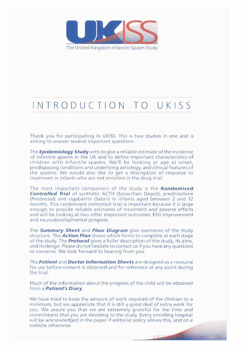
The United Kingdom Infantile Spasm Study
I N T R O D U C T I O N T O U K I S S
Thank you fo r partic ipating in UKISS. This is tw o studies in one and is aim ing to answer several im portan t questions.
The Epidem iology Study aims to give a reliable estimate o f the incidence o f in fan tile spasms in the UK and to define im portan t characteristics o f ch ild re n w ith in fa n t ile spasms. W e 'll be lo o k in g a t age a t onset, predisposing conditions and underlying aetiology, and clinical features o f the spasms. We w ou ld also like to ge t a descrip tion o f response to trea tm en t in infants w ho are not enrolled in the drug tria l.
The m ost im p o rta n t co m ponen t o f th e s tudy is the R an d o m ised C o n tro lled Trial o f synthetic ACTH (Synacthen Depot), prednisolone (Prednesol) and vigabatrin (Sabril) in infants aged between 2 and 12 months. This randomised contro lled tria l is im portan t because it is large enough to provide reliable estimates o f trea tm en t and adverse effects and w ill be looking at tw o o ther im portan t outcomes: EEG im provem ent and neurodevelopm ental progress.
The Sum m ary Sheet and F low D iagram give overviews o f the study structure. The A ction Plan shows which forms to complete at each stage o f the study. The Protocol gives a fu lle r description o f the study, its aims, and its design. Please do not hesitate to contact us if you have any questions or concerns. We look fo rw ard to hearing from you.
The P atien t and D octor In fo rm atio n Sheets are designed as a resource fo r use before consent is obtained and fo r reference at any po in t during the tria l.
Much o f the in fo rm ation about the progress o f the child w ill be obtained from a Patient's D iary
We have tried to keep the am ount o f w ork required o f the clinician to a m inim um , bu t we appreciate th a t it is still a good deal o f extra w ork fo r you. We assure you th a t we are extrem ely g ra te fu l fo r the tim e and com m itm ent th a t you are devoting to the study. Every enrolling hospital w ill be acknowledged in the paper if ed itoria l policy allows this, and on a website otherwise.
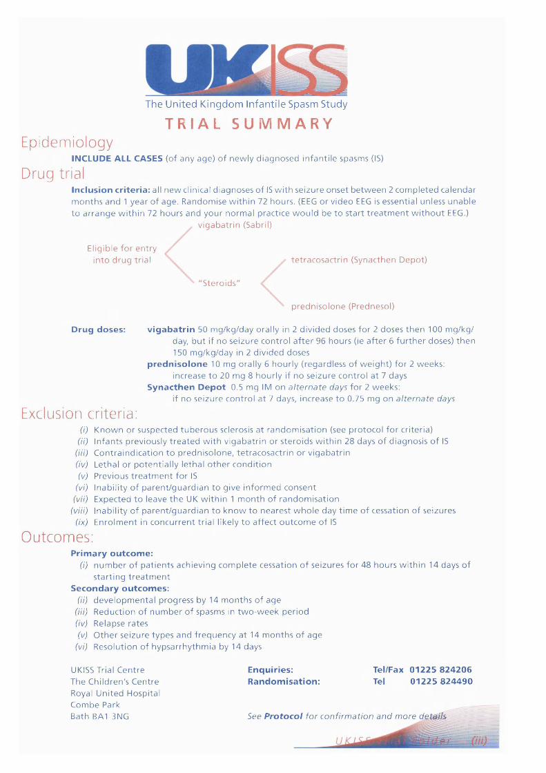
The United Kingdom Infantile Spasm Study
T R I A L S U M M A R YEpidemiology
INCLUDE ALL CASES (of any a g e ) o f n ew ly d ia g n o s e d in fan t i le spasm s (IS)
Drug trialInclusion criteria: all n e w clinical d ia g n o se s o f IS w i th se izu re o n s e t b e t w e e n 2 c o m p le t e d c a le n d a r m o n th s a n d 1 y ea r o f a g e . R a n d o m ise w i th in 72 hours . (EEG o r v id e o EEG is e s sen t ia l un less u n a b le t o a r r a n g e w i th in 72 h o u rs a n d y o u r n o rm a l p rac t ice w o u ld b e t o s t a r t t r e a t m e n t w i t h o u t EEG.)
Eligible fo r e n t ry in to d r u g trial
v ig a b a t r in (Sabril)
S tero ids
t e t r a c o s a c t r i n (S ynac then D epo t)
p r e d n i s o lo n e (P redneso l)
Drug doses: vigabatrin 50 m g /k g /d a y orally in 2 d iv id e d d o se s fo r 2 d o se s t h e n 100 m g /k g /day, b u t if n o s e izu re c o n t ro l a f t e r 96 h o u rs (ie a f t e r 6 f u r th e r doses) t h e n 150 m g /k g /d a y in 2 d iv ided d o se s
prednisolone 10 m g orally 6 h o u r ly ( re g a rd le s s o f w e ig h t ) fo r 2 w eeks : inc rease t o 20 m g 8 h ou r ly if n o se izu re c o n t ro l a t 7 days
Synacthen Depot 0.5 m g IM o n a lte rn a te days fo r 2 w eeks :if n o se izu re co n t ro l a t 7 days, in c re ase t o 0 .75 m g o n a lte rn a te days
Exclusion criteria:(i) K n o w n o r su sp e c te d tu b e r o u s sclerosis a t r a n d o m is a t io n (see p ro to co l fo r cr iteria)
(ii) In fa n ts p rev iously t r e a t e d w i th v ig a b a t r in o r s te ro id s w i th in 28 days o f d ia g n o s is o f IS (Hi) C o n t ra in d ic a t io n t o p re d n iso lo n e , te t r a c o s a c t r i n o r v ig a b a t r in(iv) Lethal o r p o te n t ia l ly le tha l o t h e r c o n d i t io n(v) P rev ious t r e a t m e n t fo r IS
(vi) Inabil i ty o f p a r e n t /g u a r d i a n t o g ive in fo rm e d c o n s e n t(vii) E xpec ted t o leave t h e UK w ith in 1 m o n th o f r a n d o m is a t io n
(viii) Inabil ity o f p a r e n t /g u a r d i a n t o k n o w t o n e a r e s t w h o le day t im e o f ce ssa t io n o f se izu res (ix) E n ro lm e n t in c o n c u r re n t tr ia l likely t o a f f e c t o u t c o m e o f IS
Outcomes:Prim ary outcome:
(i) n u m b e r o f p a t i e n t s ac h iev ing c o m p le t e ce ssa t ion o f se izu res fo r 48 h o u rs w i th in 14 days o f s t a r t in g t r e a t m e n t
Secondary outcomes:(ii) d e v e lo p m e n ta l p ro g re ss by 14 m o n th s o f a g e
(7/7) R e d u c t io n o f n u m b e r o f spasm s in tw o - w e e k p e r io d(iv) R e lapse ra te s(v) O th e r se iz u re ty p e s a n d f r e q u e n c y a t 14 m o n th s o f a g e
(vi) R eso lu t io n o f h y p s a r rh y th m ia by 14 days
UKISS Trial C e n tre The C hild ren 's C e n tre Royal U n ite d H ospita l C o m b e Park Bath BA1 3NG
Enquiries:Randomisation:
Tel/Fax 01225 824206 Tel 01225 824490
See Protocol fo r con firm a tion a n d m ore d e t^ M
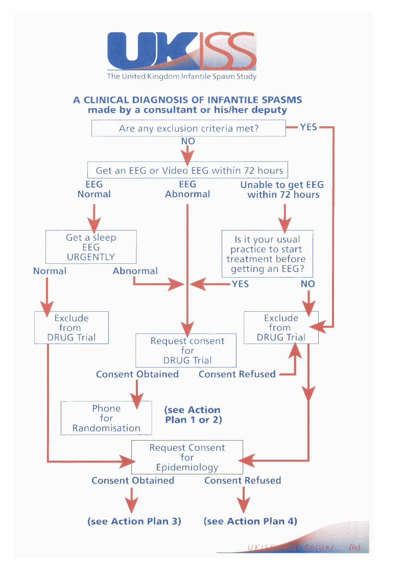
The United Kingdom Infantile Spasm Study
A CLINICAL DIAGNOSIS OF INFANTILE SPASMS made by a consultant or his/her deputy
Are any exclusion criteria met? — Y E S — i
NO
Get an EEG or Video EEG w ith in 72 hoursEEG
NormalEEG
AbnormalUnable to get EEG
within 72 hours
Get a sleep EEG
URGENTLYNormal Abnormal
i_______
Exclude from
DRUG Trial
iIs it your usual
practice to start trea tm ent before getting an EEG?
YES
tRequest consent
fo r DRUG Trial
Consent Obtained Consent Refused
Exclude from
DRUG Trial
Phone fo r
Randomisation
4s (see Action Plan 1 or 2)
Request Consent fo r
EpidemiologyConsent Obtained Consent Refused
I I(see Action Plan 3) (see Action Plan 4)

The United Kingdom Infantile Spasm Study
Ac t i o n Pl anPatient
Presentation
patient meets inclusion criteria forRCT (see Protocol section 1); no exclusion criteria for RCT (see Protocol section 2); paren ts consent to random isation and inclusion in trial.
Consent and Enrolment
1 com plete Consent Check List
j* com plete Consent Form A with parents
•co m p le te Form 1, then phone Trial Centre for random isation
• com plete Form 2'•a rra n g e trea tm en t to
be given and counsel parents in use of Patient's Diary
• return photocopies of the above form s to the Trial C entre
• i f taking blood during treatment period, please obtain sample for DNA analysis (see Form 5)
patient meets inclusion criteria for RCT; no exclusion criteria for RCT bu t EEG not ob tained within 72 hours; parents w ould consent to random isation and inclusion in trial.
patient has a clear exclusion criterion;
^ p a re n ts consent to inclusion in Epidemiology Studyorparents or clinician are not happy for child to be includedin RCT but are happy to consent to Epidemiology Studyorother, such as unable to a rrange EEG in time, or forgot to randomise
parents do not consent to inclusion in RCT orEpidemiology Study
• Please discuss with Trial Centre immediately
Forms as above
14-day Follow-up
• after 14 days of treatm ent, com plete Form 4
1 check pages D3 to D6 of Patient's Diary
• return photocopies of Forms 4 & 5 andpages D3 to D6 to Trial Centre
28-dayFollow-up
3-monthlyFollow-up
1 after 28 days o f • a t each 3-m onthly treatment, check page: follow-up, check D8 of Patient's Diary appropriate General
1 return photocopy of I [!ro.9res.sf s^ et *rom page D8 to Trial Patients D.aryC entre [• com plete Form(s) 6
• s e n d photocopies of i these to Trial Centre
> com plete Consent Form B with parents
» com plete Form 1• com plete Form 3» return photocopies of forms to Trial Centre
* counsel paren ts in use of Patient's Diary
1 i f taking blood during treatment period, please obtain sample Tor DNA analysis (see Form 5)
1 com plete Form 1 and return photocopy to Trial C entre
• after 14 days of treatm ent, com plete Form 4
• check pages D3 to D6 of Patient's Diaryreturn photocopies of Forms 4 & 5 andpages D3 to D6 to Trial Centre
1 after 28 days o f treatment, check page D8 of Patient's Diary
1 return photocopy of page D8 to Trial Centre
12 to 14 month Follow-up
when child is 12 to 14 months old, com plete Form 7
• check remaining pages of Patient's Diary
• send photocopies of Form 7 and remaining Patient's Diary pages to Trial Centre
• when child is 12 to 14 months old, com plete Form 7
• check remaining pages of Patient's Diary
• send photocopies of Form 7 and remaining Patient's Diarypages to Trial C entre

The United Kingdom Infantile Spasm Study
Doctors’ informatInfantile spasms (IS), also known as West syndrome were first described by Dr. West in 1841 who gave an account of them
occurring in his own 4 month old son. They are a refractory seizure disorder with a high risk of a poor prognosis, including intractable epilepsy and severe developmental delay. The condition is relatively common with an incidence which may be as high as 1 in 2500 births. There is an association with a number of disorders. Some of the disorders, such as cerebral palsy or Down syndrome are known at the disease onset, whilst others, such as Tuberous Sclerosis or neuronal migration disorders are discovered on investigation after the onset of spasms. However in a significant number of cases the aetiology remains unknown (i.e. idiopathic).
Two treatments are currently in widespread use i.e. vigabatrin and steroids. Steroids are either administered as ACTH or prednisolone. Steroids have been used in the trea tm ent of seizure disorders since the late 1950's. They undoubtedly have a significant effect in reducing seizure frequency but side effects such as increased appetite, irritability and hypertension are common. There is no clear evidence about whether ACTH is preferable to prednisolone. The effectiveness of trea tm ent has almost exclusively been measured in terms of reduction of seizure frequency and not in terms of improved psychomotor outcome. Vigabatrin has also recently been shown to reduce seizure frequency in patients with infantile spasms. Its side-effect profile appears better than tha t of steroids has been. It is gaining acceptance, although there are recent reports which suggest tha t it might affect visual fields in some adults. However it is not known w hether it is better than steroids in treating infantile spasms and we don 't know if either trea tm ent might improve developmental outcome.
This trial will compare steroids and vigabatrin in the treatm ent of infantile spasms. It will assess outcome primarily in terms of reduction of seizure frequency and secondarily in terms of psychomotor development.
Trial designUKISS is a national, multicentre, randomised, parallel-group open clinical trial. Patients will be allocated to one of two
trea tm ent groups:1. Vigabatrin (50mg/kg increasing to a maximum of 150mg/kg per day)2. Steroids. As ACTH gel is not available in the UK, we have substituted Synacthen Depot. Give either Synacthen Depot
(0.5mg on alternate days increasing to 0.75mg on alternate days if necessary) or Prednisolone (40mg increasing to 60mg per day if necessary).
Patients will be stratified before entry according to sex, age and whether, at the time of randomisation, there is identifiable prior risk of developmental delay. Exclusions include tuberous sclerosis, proven a t the time of randomisation.
Central randomisation will be by telephone call a t which time the child will be registered into the trial. We will collect baseline information and allocate initial treatment. The initial treatment period for all three groups will be two weeks. Responders are defined as those in whom there is a total cessation of seizures for at least 48 hours up to and including the end of the 14th day of treatment.
Responders to vigabatrin are recommended to continue on a maintenance dose of the drug until aged 1 2 - 1 4 months unless tha t is not your normal practice. Responders to steroids ( Synacthen Depot or prednisolone ) will undergo a tapered withdrawal over 15 days, using prednisolone only. Responders who subsequently relapse will be treated by increasing the dose of vigabatrin to a maximum of 150mg/kg/day or in the case of steroids by giving a repeated dose. If this fails we recommend tha t the alternative therapy is tried (i.e. vigabatrin if steroids fail and steroids if vigabatrin fails).
Please note th e precautions listed in the manufacturer's data sheet for Synacthen Depot. Synacthen Depot should only be administered under medical supervision. Previous hypersensitivity reaction (localised or generalised) to ACTH, Synacthen or Synacthen Depot is a contraindication to its use. It is also contraindicated in allergic disease. Hypersensitivity reactions generally occur within 30 minutes of administration (for which period direct observation of the child is recommended). However, the Committee on Safety of Medicines has not received any reports of anaphylaxis in children less than 5 years of age. Avoid live vaccines durings the period of treatment. Use with care in the presence of renal insufficiency and hypertension.
All patients will be seen as deemed necessary by their own consultant, but this must include a visit at two weeks to assess initial response to therapy and thereafter a t 3 monthly intervals. Referring consultants will be asked to fill in standardised progress report forms and send them back to the trial centre in Bath after each of these consultations. The patient's psychomotor development will be assessed by a te lephone questionnaire administered by the trial centre in Bath at 12 -14 months of age. Parents will be required to keep a diary until their child reaches 12-14 months of age. There should be a daily entry for 28 days after the start of trea tm ent and weekly after tha t until the 12-14 month development assessment. In those children still having any form of seizure at 12-14 months, a daily entry will be obtained for the final week.
If you have any questions or concerns, or would like any further information on this study please to not hesitate to contact:
The UKISS Trial Centre The Children's Centre Royal United Hospital Combe Park BATH BA1
U KISS/M DIF/1/399
3NG
Trial Centre Tel: 01225 824206Randomisation Tel: 01225 824490 (8.30am to 6pm)
(answer machine 6pm to 8.30am) Fax: 01225 824206 or 01225 824212
24 hour number for hospital: 01225 428331

The United Kingdom Infantile Spasm Study
<3P’s information sheet
Infantile spasms (IS), also known as West syndrome were first described by Dr. West in 1841 who gave an account of them occurring in his own 4 month old son. They are a refractory seizure disorder with a high risk of a poor prognosis, including intractable epilepsy and severe developmental delay. The condition is relatively common with an incidence which may be as high as 1 in 2500 births. There is an association with a number of disorders. Some of the disorders, such as cerebral palsy or Down syndrome are known at the disease onset, whilst others, such as Tuberous Sclerosis or neuronal migration disorders are discovered on investigation after the onset of spasms. However in a significant number of cases the aetiology remains unknown (i.e. idiopathic).
Two treatments are currently in widespread use i.e. vigabatrin and steroids. Steroids are either administered as ACTH or prednisolone. Steroids have been used in the trea tm ent of seizure disorders since the late 1950's. They undoubtedly have a significant effect in reducing seizure frequency but side effects such as increased appetite, irritability and hypertension are common. There is no clear evidence about w hether ACTH is preferable to prednisolone. The effectiveness of trea tm ent has almost exclusively been measured in terms of reduction of seizure frequency and not in terms of improved psychomotor outcome. Vigabatrin has also recently been shown to reduce seizure frequency in patients with infantile spasms. Its side-effect profile appears better than tha t of steroids has been. It is gaining acceptance, although there are recent reports which suggest tha t it might affect visual fields in some adults. However it is not known w hether it is better than steroids in treating infantile spasms and we don 't know if either treatm ent might improve developmental outcome.
This trial will compare steroids and vigabatrin in the trea tm ent of infantile spasms. It will assess outcome primarily in terms of reduction of seizure frequency and secondarily in terms of psychomotor development.
Trial designUKISS is a national, multicentre, randomised, parallel-group open clinical trial. Patients will be allocated to one of tw o
trea tm ent groups:1. Vigabatrin (50mg/kg increasing to a maximum of 150mg/kg per day)2. Steroids. As ACTH gel is not available in the UK, we have substituted Synacthen Depot. Give either Synacthen Depot
(0.5mg on alternate days increasing to 0.75mg on alternate days if necessary) or Prednisolone (40mg increasing to 60mg per day if necessary).
Patients will be stratified before entry according to sex, age and whether, a t the time of randomisation, there is identifiable prior risk of developmental delay. Exclusions include tuberous sclerosis, proven at the time of randomisation.
Central randomisation will be by te lephone call at which time the child will be registered into the trial. We will collect baseline information and allocate initial treatment. The initial treatment period for all three groups will be two weeks. Responders are defined as those in whom there is a total cessation of seizures for at least 48 hours up to and including the end of the 14th day of treatment.
Responders to vigabatrin are recommended to continue on a maintenance dose of the drug until aged 1 2 - 1 4 months unless th a t is not your normal practice. Responders to steroids ( Synacthen Depot or prednisolone ) will undergo a tapered withdrawal over 15 days, using prednisolone only. Responders who subsequently relapse will be treated by increasing the dose of vigabatrin to a maximum of 150mg/kg/day or in the case of steroids by giving a repeated dose. If this fails we recommend tha t the alternative therapy is tried (i.e. vigabatrin if steroids fail and steroids if vigabatrin fails).
Please note the precautions listed in the manufacturer's data sheet for Synacthen Depot. Synacthen Depot should only be administered under medical supervision. Previous hypersensitivity reaction (localised or generalised) to ACTH, Synacthen or Synacthen Depot is a contraindication to its use. It is also contraindicated in allergic disease. Hypersensitivity reactions generally occur within 30 minutes of administration (for which period direct observation of the child is recommended). However, the Committee on Safety of Medicines has not received any reports of anaphylaxis in children less than 5 years of age. Avoid live vaccines durings the period of treatment. Use with care in the presence of renal insufficiency and hypertension.
All patients will be seen as deemed necessary by their own consultant, but this must include a visit at two weeks to assess initial response to therapy and thereafter a t 3 monthly intervals. Referring consultants will be asked to fill in standardised progress report forms and send them back to the trial centre in Bath after each of these consultations. The patient's psychomotor development will be assessed by a te lephone questionnaire administered by the trial centre in Bath at 12 -14 months of age. Parents will be required to keep a diary until their child reaches 12 -14 months of age. There should be a daily entry for 28 days after the start of trea tm en t and weekly after tha t until the 12-14 month development assessment. In those children still having any form of seizure at 12 -14 months, a daily entry will be obtained for the final week.
If you have any questions or concerns, or would like any further information on this study please to not hesitate to contact:
The UKISS Trial Centre The Children's Centre Royal United Hospital Combe Park BATH BA13NG
Trial Centre Tel: 01225 824206Randomisation Tel: 01225 824490 (8.30am to 6pm)
(answer machine 6pm to 8.30am) Fax: 01225 824206 or 01225 824212
24 hour number for hospital: 01225 428331
Please send this sheet to the patient's general practitionerUKISS/GPIF/1/399
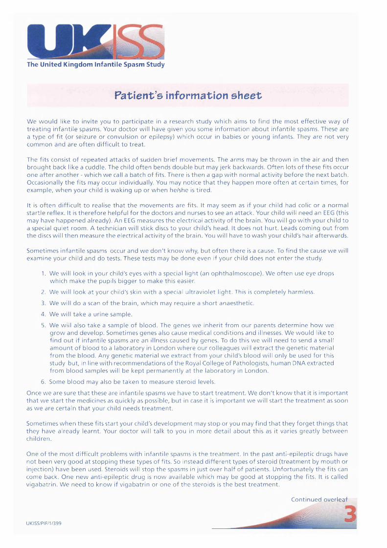
The United Kingdom In fantile Spasm Study
Patient’s information sheet
W e w o u ld like t o invite you t o p a r t i c ip a te in a re se a rch s tu d y w h ich aim s t o f ind t h e m o s t e f fec t iv e w a y o f t r e a t i n g in fa n t i le spasm s. Your d o c to r will h av e g iven you so m e in f o rm a t io n a b o u t in fan t i le spasm s. T h ese a re a t y p e o f f i t (or se izu re or convu ls ion o r epilepsy) w h ich occur in b a b ie s o r y o u n g in fan ts . They a r e n o t very c o m m o n a n d a r e o f t e n difficult t o t r e a t .
T he fits cons is t o f r e p e a t e d a t ta c k s o f s u d d e n b r ie f m o v e m e n ts . T he a rm s m ay b e t h r o w n in t h e air a n d t h e n b r o u g h t back like a cu d d le . T he child o f t e n b e n d s d o u b le b u t m ay je rk bac k w ard s . O f te n lots o f th e s e fits occur o n e a f t e r a n o t h e r - w h ich w e call a b a tc h o f fits. T h e re is t h e n a g a p w i th n o rm a l ac tivity b e f o r e t h e n e x t b a tch . O ccasionally t h e fits m ay occur individually. You m a y n o t ic e t h a t t h e y h a p p e n m o r e o f t e n a t ce r ta in t im es , fo r e x a m p le , w h e n y o u r child is w a k in g u p o r w h e n h e /sh e is t i r e d .
It is o f t e n d iff icu lt t o realise t h a t t h e m o v e m e n t s a re fits. It m ay s e e m as if y o u r child h a d colic o r a n o rm a l s ta r t le reflex. It is t h e r e f o r e h e lp fu l fo r t h e d o c to rs a n d n u rses t o se e a n a t tac k . Your child will n e e d a n EEG (this m ay h a v e h a p p e n e d a lready) . An EEG m e a s u re s t h e elec trica l activ ity o f t h e b ra in . You will g o w i th y o u r child t o a specia l q u i e t ro o m . A te ch n ic ia n will stick discs t o y o u r child's h e a d . It d o e s n o t h u r t . Leads co m in g o u t f ro m t h e discs will t h e n m e a s u r e t h e e lectrica l ac tivity o f t h e b ra in . You will h av e t o w a s h y o u r child's ha i r a f te rw a rd s .
S o m e t im e s in fa n t i le spasm s occur a n d w e d o n ' t k n o w why, b u t o f t e n t h e r e is a cause . To f ind t h e c a u se w e will e x a m in e y o u r child a n d d o tes ts . T hese te s ts m ay b e d o n e ev e n if y o u r child d o e s n o t e n t e r t h e study.
1. W e will look in you r child's eyes w i th a specia l l igh t (an o p h th a lm o s c o p e ) . W e o f t e n use eye d ro p s w h ich m a k e t h e pupils b ig g e r t o m a k e th is easier.
2. W e will look a t y o u r child's skin w i th a specia l u l t ra v io le t light. This is c o m p le te ly harm less .
3. W e will d o a scan o f t h e bra in , w h ich m ay r e q u i r e a s h o r t a n a e s th e t i c .
4. W e will t a k e a u r in e sam ple .
5. W e will a lso t a k e a s a m p le o f b lo o d . The g e n e s w e in h e r i t f ro m o u r p a r e n t s d e t e r m in e h o w w eg r o w a n d d ev e lo p . S o m e t im e s g e n e s also ca u se m ed ica l c o n d i t io n s a n d illnesses. W e w o u ld like t o f ind o u t if in fan t i le spasm s a r e an illness c a u se d by g e n e s . To d o th is w e will n e e d t o se n d a small a m o u n t o f b lo o d to a l a b o ra to r y in L ondon w h e r e o u r c o l le a g u e s will e x t ra c t t h e g e n e t i c m a te r ia l f ro m t h e b lo o d . Any g e n e t ic m a te r ia l w e e x t r a c t f ro m y o u r child's b lo o d will on ly be used fo r th is s tu d y bu t, in line w ith r e c o m m e n d a t io n s o f t h e Royal College o f Patho logists , h u m a n DNA e x t ra c te d f ro m b lo o d sam p les will b e k e p t p e r m a n e n t l y a t t h e l a b o ra to r y in L ondon .
6. S o m e b lo o d m ay also b e t a k e n t o m e a s u r e s te ro id levels.
O n ce w e a r e su re t h a t th e s e a r e in fan t i le spasm s w e h a v e t o s ta r t t r e a t m e n t . W e d o n ' t k n o w t h a t it is im p o r t a n t t h a t w e s t a r t t h e m ed ic ines as quickly as possib le , b u t in case it is im p o r t a n t w e will s ta r t t h e t r e a t m e n t as so o n as w e a re c e r ta in t h a t y o u r child n e e d s t r e a t m e n t .
S o m e t im e s w h e n th e s e fits s t a r t y o u r child 's d e v e lo p m e n t m ay s to p o r you m ay f ind t h a t th e y f o r g e t t h in g s t h a t th e y h av e a l r e a d y le a rn t . Your d o c to r will ta lk t o y ou in m o r e d e ta i l a b o u t th is as it varies g re a t ly b e t w e e n ch i ld ren .
O n e o f t h e m o s t d ifficu lt p ro b le m s w i th in fan t i le spasm s is t h e t r e a t m e n t . In t h e p a s t a n t i -e p i le p t ic d ru g s h av e n o t b e e n very g o o d a t s to p p in g th e s e ty p e s o f fits. So in s te a d d i f f e r e n t ty p e s o f s te ro id ( t r e a t m e n t by m o u th o r in jection) h a v e b e e n used. S tero ids will s to p t h e spa sm s in ju s t o v e r ha l f o f p a t ie n t s . U n fo r tu n a te ly t h e fits can c o m e back. O n e n e w an t i -e p i le p t ic d r u g is n o w av a i la b le w h ich m ay b e g o o d a t s to p p in g t h e fits. It is ca lled v ig a b a tr in . W e n e e d t o k n o w if v ig a b a t r in o r o n e o f t h e s te ro id s is t h e bes t t r e a t m e n t .
Co n t i n u e d o y ^ ^ ^ s
UKISS/PIF/1/399

W e w o u ld like t o ask y o u to t a k e p a r t in th is s tu d y t o f ind o u t w h ich o f t h e t r e a t m e n t s is bes t . If w e k n e w w hich w a s best, w e w o u ld n o t b e d o in g t h e study. If fo r an y r e a so n y ou w o u ld p re fe r n o t t o t a k e p a r t in t h e s tu d y w e will r e sp e c t t h a t dec is ion a n d t h e ca re t h a t y o u r child receives will n o t b e a f f e c te d .
If you d e c id e t o t a k e p a r t in th is s tu d y y o u r child will g iven o n e o f t h e fo l lo w in g t h r e e d rugs .
1. S y n a c th e n D e p o t . This is g iven by in jec t ion o n a l t e r n a t e days fo r t w o w ee k s . It te lls y o u r child's o w n b o d y t o m a k e m o r e n a tu ra l s te ro id s t h a n usual.
2. P re d n iso lo n e . This is a t y p e o f n a tu ra l s te ro id w h ich is t a k e n by m o u th 3 o r 4 t im e s a day fo r t w o w ee k s .
3. V ig ab a tr in . This is an a n t i -e p i le p t ic d ru g w h ich is t a k e n by m o u th 2 t im e s a d a y f o r t w o w eeks .
Your child will receive o n e o f th e s e d ru g s fo r tw o w ee k s . A f te r 2 w e e k s y o u r child will b e s e e n by y o u r docto r . If t h e fits h a v e s to p p e d t h e n y o u r child will:
• if o n v ig a b a tr in , c o n t in u e o n w i th t h e t r e a t m e n t until t h e y a r e a b o u t 12 -14 m o n th s o ld if t h a t is you r d o c to r 's n o rm a l p rac tice .
• if o n s y n a c th e n d e p o t o r p re d n iso lo n e , b e g iven o ral p r e d n i s o lo n e fo r 15 m o r e days a t a lo w e r dose .
A f te r 2 w ee k s , if t h e spasm s h a v e n o t s t o p p e d y o u r child will b e c h a n g e d t o a d i f f e r e n t t r e a t m e n t t o see if t h a t helps.
Your child will b e se e n by y o u r d o c to r a t leas t every t h r e e m o n th s (m o re o f t e n if necessary) t o check p rog ress . If t h e r e a re p ro b le m s w i th t h e spasm s o r t h e m e d ic in e p le a se te ll y o u r doc to r .
W h e n y o u r child is a b o u t 12 -14 m o n th s o f a g e t h e n w e will c o n ta c t y ou by p h o n e t o check o n y o u r child 's p rog ress . This will t a k e b e t w e e n 15 a n d 30 m in u te s t o c o m p le te .
D uring t h e s tu d y w e will also ask y ou to fill in a diary. This will t a k e ju s t a c o u p le o f m in u te s a day. W e will ask you t o m a rk if y o u r child has h a d an y fits t h a t day o r an y o t h e r illness. W e will ask you t o r e tu r n t h e d iary a t t h e e n d o f t h e study, b u t y ou m ay k e e p a copy if you w a n t . It will b e a p e r m a n e n t rec o rd o f y o u r child 's p rog ress .
If w e d o n o t d o th is s tu d y w e will n e v e r lea rn w h ich t r e a t m e n t is bes t . If w e k n e w n o w w h ic h t r e a t m e n t w as be s t t h e n w e w o u ld n o t b e d o in g t h e study. It will t a k e all, o r n ea r ly all, o f t h e ch i ld ren in t h e UK t o a g r e e t o t h e s tu d y fo r a b o u t 18 m o n th s b e f o r e w e e x p e c t t o k n o w w h ich is t h e b e s t t r e a t m e n t . T h e n w e can give t h e b es t t r e a t m e n t t o all ch i ld ren w i th in fan t i le spasm s. This is t h e usual w a y d o c to rs s tu d y d r u g s t o se e w h ich o n e s a re bes t.
The in f o rm a t io n co l lec ted o n y o u r child will b e k e p t by t h e d o c to rs in c h a r g e o f t h e s tudy. T hey will k e e p it s a fe in locked f il ing cab ine ts . S o m e in f o rm a t io n will b e k e p t o n c o m p u te r : it will b e p r o t e c t e d by a s e c re t p as sw o rd .
If you w a n t t o ask a b o u t t h e in f o rm a t io n o r t o ask a n y o t h e r q u e s t io n s ask y o u r local specia list . W e will se n d y ou a n e w s le t t e r every f e w m o n th s , th is will tell you h o w t h e s tu d y is g o in g .
If y o u n e e d to , you can c o n ta c t y o u r specialist:
His / Her n a m e is: ....................................................................................................................................
Address:......... ....................................................................................................................................
T e l e p h o n e No: ................................................................................................................
If y ou n e e d t o you can also c o n ta c t t h e d o c t o r in c h a r g e o f t h e s tudy:Prof J. O s b o rn e T he UKISS Trial C e n tre T he Child ren 's C e n tre Royal U n ited Hospital C o m b e Park BATH BA1 3NGTel: 01225 824206 b u t p le a s e ta lk to y o u r lo c a l s p e c ia l is t f ir s t .
UKISS/PIF/1/399

The United Kingdom In fantile Spasm Study
Patient information sheet — Prednisolone
D ear p a r e n t / g u a r d ia n ,
T h a n k y ou fo r p a r t i c ip a t in g in t h e UKISS study. Your child m ay b e c h o s e n t o rece ive p re d n is o lo n e e i th e r as th e i r f irst d r u g in th is s tu d y o r b e c a u s e v ig a b a tr in d id n o t s to p t h e i r spasm s. H opefu lly th is in fo rm a t io n s h e e t will a n s w e r a n y q u e s t io n s you h av e a b o u t p r e d n is o lo n e a n d its use. If y o u h a v e an y o t h e r q u e s t io n s o r t h e r e is an y th in g t h a t y ou d o n o t u n d e r s t a n d ask y o u r d o c to r now .
W hat is prednisolone?P re d n iso lo n e is t h e chem ica l n a m e fo r a ty p e o f s te ro id t h a t o u r b o d ie s p r o d u c e natura lly . It is also a v a i la b le as a m ed ic in e . It is m a d e by a d r u g c o m p a n y (Glaxo) w h o call t h e i r b r a n d o f t h e d r u g P redneso l (in t h e s a m e w a y t h a t N estle call t h e i r c o f fe e Nescafe).
How do I give it?It co m e s in t a b l e t fo rm . Each small p ink t a b l e t c o n ta in s 5m g p re d n is o lo n e . It is t a k e n by m o u th e i th e r 3 o r 4 t im e s a day. T hey can e i th e r b e c ru sh e d a n d sp r ink led o n f o o d o r can b e d isso lved in w a t e r (or squash).They m ay b e t a k e n b e fo re , d u r in g o r a f t e r meals .
How much do I give?For t h e first w e e k o f t r e a t m e n t you will g ive 10m g f o u r t im e s a d ay (2 t a b le t s f o u r t im e s a day). If a t t h e e n d o f t h e firs t w e e k y o u r child's spasm s h a v e n o t s to p p e d t h e n y o u r d o c to r will te ll you t o inc rease t h e d o s e t o 2 0 m g t h r e e t im e s a d ay (4 t a b le t s t h r e e t im e s a day). A f te r t h e t w o w e e k s you will n e e d to r e d u c e t h e d o se o f p re d n is o lo n e . Your d o c to r will exp la in h o w t o d o th is . The a m o u n t o f m e d ic in e n e e d e d d o e s n o t d e p e n d on y o u r child 's w e ig h t o r ag e .
If t h e spasm s s ta r t a g a in you d o c to r m ay ask you t o r e p e a t t h e c o u rse o f s te ro ids .
If y o u r child acc id en ta l ly ta k e s t o o m uch P redneso l , te ll y o u r d o c t o r im m e d ia te ly o r g o t o y o u r n e a r e s t ca sua lty d e p a r t m e n t .
If y ou f o r g e t t o g ive a d o se g ive a n o t h e r as so o n as y ou r e m e m b e r , un less it is a lm o s t t im e fo r th e i r n ex t d ose , t h e n g o o n as b e fo re .
Are there any side e f fec ts?As w ith all m e d ic in es so m e side e ffec ts m a y occur. T he m o s t c o m m o n s ide e f fec ts o f p r e d n i s o lo n e a re irri tabili ty a n d c h a n g e s in s leep in g . T hese will im p ro v e o n c e t h e s te ro id s a r e s to p p e d .
Your d o c to r ( e i th e r y o u r GP o r a d o c to r f ro m t h e hosp ita l) will n e e d t o see y o u r child o n c e a w e e k t o check t h e i r b lo o d p re s su re a n d urine.
B ecause w e on ly u se t h e s te ro id s fo r a s h o r t t im e , th e y d o n o t c a u se long las t ing effec ts . For ex a m p le , y o u r child will n o t s to p g r o w in g as an e f fec t o f t h e s te ro ids .
W hat do I do if my child has any side e ffects?C o n ta c t y o u r d o c t o r (GP) im m edia te ly . H e/she will adv ise y ou o n w h e t h e r t o c o n t in u e t h e p r e d n is o lo n e o r w h e t h e r y ou n e e d t o s to p t h e p re d n iso lo n e . If y ou h a v e t o s to p t h e p r e d n i s o lo n e you m u s t see y o u r child 's p a e d ia t r i c ia n as so o n as possib le so h e /sh e can g ive y ou s o m e d i f f e r e n t m e d ic in e t o s to p th e spasm s. You s h o u ld also w r i te d o w n t h e p r o b le m in y o u r ' f i t d ia ry ' .
W hat do I do if my child becomes ill?If y o u r child b e c o m e s unw ell in an y w a y o r if you h a v e an y c o n c e rn s you s h o u ld c o n ta c t y o u r d o c to r (GP) im m ed ia te ly . If y o u r child d e v e lo p s ch ick e n p o x o r is in c o n ta c t w i th a n o t h e r child w h o d e v e lo p s ch ick e n p o x y ou m u s t c o n ta c t y o u r d o c to r im m ed ia te ly .
Immunisations:Your child s h o u ld n o t h av e th e i r im m u n isa t io n s (vaccina tions, jabs) w hils t t h e y a r e on t h e i r s te ro ids . Theys h o u ld h a v e th e i r im m u n isa t io n s o n c e th e y h a v e c o m p le te ly f in ish e d th e i r co u rse o f s te ro id s .
UKISS/PDIF/1/399/P

The United Kingdom Infantile Spasm Study
Patient information sheet — Vigabatrin
Dear p a r e n t / g u a r d ia n ,
Thank y o u f o r p a r t i c ip a t in g in t h e UKISS study. Your child m ay b e c h o s e n t o rece ive v ig a b a t r in e i th e r as th e i r first d ru g in th is s tu d y o r b e c a u s e t h e s te ro id t r e a t m e n t d id n o t s to p th e i r spasm s. H opefu lly th is in fo rm a t io n sh e e t will a n s w e r a n y q u e s t io n s y ou h av e a b o u t v ig a b a tr in a n d its use. If you h a v e any o t h e r q u e s t io n s o r t h e r e is a n / t h i n g t h a t y ou d o n o t u n d e r s t a n d t h e n ask y o u r d o c to r now .
What is vigabatrin?V igaoa tr in is t h e chem ica l n a m e o f t h e d ru g . It is m a d e by a d r u g c o m p a n y (H oechs t M ario n Roussel) w h o call th e i r b r a n d o f t h e d r u g Sabril (in t h e s a m e w a y t h a t N estle call t h e i r c o f fe e Nescafe).
How do I give it?It com es in sache ts . Each s a c h e t c o n ta in s 500 m g v ig a b a t r in as a w h i t e p o w d e r . Do n o t o p e n t h e s a c h e t until it is t im e t o g ive t h e m ed ic in e . It is t a k e n by m o u th (orally) t w o t im e s a day. It can b e sp r ink led o n fo o d o r d isso lved in a d r in k (for e x a m p le w ate r , squash , milk, te a ) . It m ay b e t a k e n b e fo re , d u r in g o r a f t e r meals.
How much do I give?Your d o c t o r will w o rk o u t h o w m uch o f t h e m e d ic in e y o u r child has t o ta k e . T he a m o u n t n e e d e d d e p e n d s o n how m u c h y o u r child w e ig h s . W e will bu ild u p t h e a m o u n t o f v ig a b a t r in y o u r child t a k e s o v e r t h e first t w o days. A f t e r f o u r days w e m ay inc rease t h e d o se f u r th e r if y o u r child's spasm s h av e n o t s to p p e d . It m ay also oe n ecessa ry t o inc rease t h e d o s e la te r in t h e tr ia l if t h e spasm s s t a r t a g a in o r as y o u r child increases in w e igh t . You d o n o t n e e d t o w o rry a b o u t this , y o u r d o c to r will w o rk o u t h o w m u ch m e d ic in e is n e e d e d a n d will te ll y o u h o w m u c h t o g ive t o y o u r child.
If your child a c c id en ta l ly t a k e s t o o m uch v ig a b a tr in , te ll y o u r d o c to r im m e d ia te ly o r g o t o y o u r n e a r e s t casualty d e p a r t m e n t .
If you f o r g e t t o g ive a d o s e g ive a n o t h e r as so o n as you r e m e m b e r , un less it is a lm o s t t im e fo r th e i r n ex t d o s e t h e n g o on as b e fo re .
Are there any side e ffects?As w t h all m e d ic in es s o m e side e f fec ts m ay occur. T he m o s t c o m m o n side e f f e c t o f v ig a b a t r in is s leepiness. This usua lly w e a r s off a f t e r a f e w days. O ccasionally y o u r child m ay v o m it (b e sick). Recently, som e a d u l t s t r e a t e d w i th v ig a b a t r in h a v e b e e n f o u n d t o h a v e visual f ield d e fe c ts (difficulty in se e in g a ro u n d t h e e d g e o f t h e i r n o rm a l a r e a o f vision). This m ay b e m o r e likely if t r e a t m e n t c o n t in u e s fo r a long t im e O th e r s ide e f fec ts a r e very rare .
What do I do if my child has any side effects?Contac t y o u r d o c to r (GP) im m ed ia te ly . H e/she will adv ise y ou o n w h e t h e r t o c o n t in u e t h e v ig a b a t r in o r w h e th e r y ou n e e d t o s to p t h e v ig a b a tr in . If y ou h av e t o s to p t h e v ig a b a t r in y ou m u s t se e y o u r child 's p ae c ia t r ic ia n as so o n as possib le so h e /sh e can g ive y ou s o m e d i f f e r e n t m e d ic in e t o s to p t h e spasm s. You s h o u d a lso w r i te d o w n t h e p r o b le m in y o u r 'f i t d ia ry ' .
What do I do if my child becomes ill?If your child b e c o m e s u n w e ll in an y w a y o r if you h a v e any co n c e rn s y ou s h o u ld c o n ta c t y o u r d o c to r (GP) im m ediate ly .

The United Kingdom Infantile Spasm Study
Patient Information Sheet - Synacthen Depot
D e a r p a r e n t / g u a r d i a n ,
T h a n k y o u fo r p a r t ic ip a t in g in t h e UKISS study. Your child m ay b e ch o sen t o rece ive S y n a c th e n D e p o t e i th e r as t h e i r f irs t d r u g in th is s tu d y o r b e c a u s e v ig a b a tr in d id n o t s to p th e i r spasm s. H opefu lly th is in fo rm a t io n s h e e t will a n s w e r any q u e s t io n s y ou h av e a b o u t S y n ac th en D e p o t a n d its use. If y o u h a v e an y o t h e r q u e s t io n s o r t h e r e is a n y t h in g t h a t you d o n o t u n d e r s t a n d ask y o u r d o c to r now .
W hat is Synacthen Depot?S y n a c th e n D e p o t is t h e t r a d e n a m e fo r a m a n u f a c tu r e d m e d ic in e w h ich is a t y p e o f h o r m o n e o u r b od ie s p r o d u c e n a tu ra l ly (a t y p e o f a d r e n o c o r t ic o t ro p h ic h o r m o n e ) . This h o r m o n e causes t h e p ro d u c t io n o f n a tu ra l s te ro id s by t h e body. It is m a d e by a d r u g c o m p a n y w h o call t h e i r b r a n d o f t h e d ru g S y n ac th en D e p o t (in t h e s a m e w a y t h a t N estle call th e i r co f fee Nescafe).
How will i t be given?It is g iv e n as an in t ra m u sc u la r ( in to t h e m uscle - usually t h e t o p o f t h e leg) in jection , o n a l t e r n a t e days by a t r a i n e d m ed ica l p e r so n (your d o c to r o r nurse).
Are there any side effects?As w i th all m ed ic ines s o m e side e f fec ts m ay occur. T he m o s t c o m m o n s ide e f fec ts a re irritabili ty a n d c h a n g e s in s le e p in g . T hese will im p ro v e o n c e t h e s te ro id s a r e s to p p e d .
Your d o c t o r will ask y ou a b o u t an y k n o w n a l le rg ie s b e c a u s e S y n a c th e n D e p o t can u p s e t ch i ld ren w ith a l le rg ies .
Your d o c t o r (e i th e r y o u r GP o r a d o c to r f ro m t h e hosp i ta l) will n e e d t o see y o u r child o n c e a w e e k t o check th e i r b lo o d p re ssu re a n d urine .
B ecause w e only use t h e s te ro id s fo r a s h o r t t im e , th e y d o n o t c a u se long last ing effec ts . For ex a m p le , y o u r child will n o t s to p g r o w in g as an e f fec t o f t h e s te ro ids .
W hat do I do if my child has any side effects?C o n ta c t y o u r d o c to r (GP) im m edia te ly . If you h a v e t o s to p t h e S y n ac th en D e p o t you m u s t s e e y o u r child 's p a e d ia t r i c ia n as so o n as poss ib le so h e /sh e can g ive you s o m e d i f f e r e n t m e d ic in e t o try t o s to p t h e spasm s.You sh o u ld also w r i te d o w n t h e p r o b le m in y o u r 'f i t d ia ry '
W hat do I do if my child becomes ill?If y o u r child b e c o m e s u nw ell in an y w a y o r if you h a v e a n y co n c e rn s you s h o u ld c o n ta c t y o u r d o c to r (GP) im m ed ia te ly . If y o u r child d e v e lo p s c h ick e n p o x o r is in c o n ta c t w i th a n o t h e r child w h o d e v e lo p s ch ick e n p o x y o u m u s t c o n ta c t y o u r d o c t o r im m edia te ly .
Immunisations:Your child sh o u ld n o t h a v e th e i r im m u n isa t io n s (vaccina tions, jabs) w h ils t t h e y a re o n th e i r s te ro ids . They s h o u ld h a v e th e i r im m u n isa t io n s o n c e th e y h a v e c o m p le te ly f in ish e d t h e i r c o u rse o f s te ro ids .
U KISS/PDIF/1/399/SD
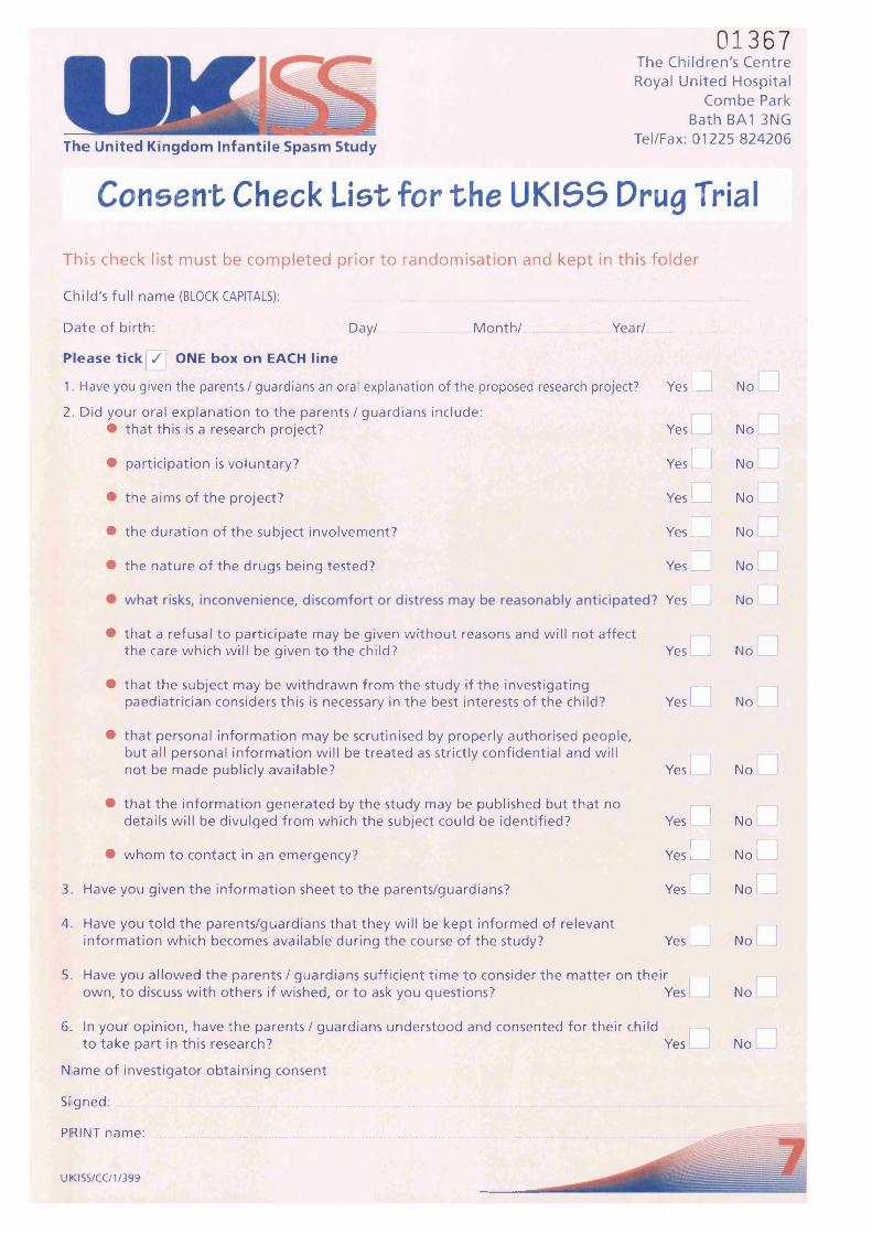
01367T h e C h i l d r e n ' s C e n t r e R o y a l U n i t e d H o s p i t a l
C o m b e P a r k B a th BA1 3N G
The United Kingdom In fan tile Spasm Study Tel/Fax: 0 1 2 2 5 8 2 4 2 0 6
Consent Check List for the UKISS Drug Trial
This check list must be com pleted prio r to random isation and kept in this fo lde r
C hild 's full n a m e (BLOCK CAPITALS):
D a t e o f b ir th : Day/ M o n th / Year/
Please tick ✓ ONE box on EACH line
1. Have you given th e parents / guardians an oral explanation of th e proposed research project?
2. Did y o u r oral e x p la n a t io n t o t h e p a r e n t s / g u a r d ia n s inc lude:• t h a t th is is a r e se a rch p ro je c t?
• p a r t i c ip a t io n is v o lu n ta r y ?
• t h e a im s o f t h e p r o je c t?
• t h e d u r a t i o n o f t h e s u b je c t in v o lv e m e n t?
• t h e n a t u r e o f t h e d r u g s b e in g t e s t e d ?
• t h a t a re fusa l t o p a r t i c ip a te m ay b e g iven w i t h o u t re a so n s a n d will n o t a f fe c t t h e ca re w h ich will b e g iven t o t h e child?
• t h a t t h e su b je c t m a y b e w i th d r a w n f ro m t h e s tu d y if t h e in v e s t ig a t in g p a e d ia t r i c ia n c o n s id e rs th is is necessary in t h e b e s t in te re s ts o f t h e child?
• t h a t p e r so n a l in f o rm a t io n m ay b e sc ru tin ised by p ro p e r ly a u th o r i s e d p e o p le , b u t all p e r so n a l in f o rm a t io n will b e t r e a t e d as strictly c o n f id e n t ia l a n d will n o t b e m a d e publicly av a i la b le ?
• t h a t t h e in f o rm a t io n g e n e r a t e d by t h e s tu d y m ay b e p u b l is h e d b u t t h a t no de ta i ls will b e d iv u lg e d f ro m w h ich t h e su b je c t cou ld b e id e n t i f ie d ?
• w h o m t o c o n ta c t in a n e m e r g e n c y ?
3- Have you g iven t h e in f o rm a t io n s h e e t t o t h e p a r e n t s /g u a rd ia n s ?
4- H ave y ou to ld t h e p a r e n t s /g u a r d i a n s t h a t t h e y will b e k e p t in fo rm e d o f r e le v a n t in f o rm a t io n w h ich b e c o m e s a v a i la b le d u r in g t h e co u r se o f t h e s tu d y ?
5- H ave y ou a l lo w e d t h e p a r e n t s / g u a r d ia n s su ff ic ien t t im e t o co n s id e r t h e m a t t e r o n t h e o w n , t o discuss w i th o th e r s if w ish e d , o r t o ask y ou q u e s t io n s ?
6. In y o u r o p in io n , h a v e t h e p a r e n t s / g u a r d ia n s u n d e r s to o d a n d c o n s e n te d f o r th e i r child t o t a k e p a r t in th is r e se a rc h ?
N a m e o f in v e s t ig a to r o b t a in in g c o n s e n t
S ig n ed : ....................................................................................................................
PRINT n a m e : ................................................................................................... ................................................
Yes L J No
Yes No
Yes LJ No
Yes □ No
Y e s Q No
Yes m t No
Yes CD No
Yes ] No
Yes Cl No
Yes l No
Yes ] No
Y e s D N o C H
Yes No [
Yes EH No
Ir nYes No
Yes No
U KISS/CC/1/399

01367
The United Kingdom In fantile Spasm Study
FORM A: CONSENT FOR DRU TRIALCONSENT SY THE PARENT / GUARDIAN
P a t ie n ts full n a m e (BLOCK CAPITALS):
D a te o f b ir th : Day/ M o n th / Year/
A ddress :
I hereby fu lly and freely consent to a llow my child to participate in the UKISS tria l.(P lease tick / a p p r o p r i a t e boxes)
H ave y ou r e a d t h e p a t i e n t in f o rm a t io n s h e e t? Yes No
Have y ou h a d a n o p p o r tu n i ty t o ask q u e s t io n s a n d discuss th is s tu d y ? Yes No □
Have y o u rece ived sa t is fac to ry a n s w e rs t o all y o u r q u e s t io n s ? Yes ! No □
Have y ou rece ived e n o u g h in f o rm a t io n a b o u t t h e s tu d y ? Yes No JI a g r e e t h a t my g e n e r a l p ra c t i t io n e r a n d hosp i ta l c o n s u l t a n t a r e n o t i f ie d o f my child 's p a r t ic ip a t io n in t h e tr ia l a n d t h a t t h e y m ay re le ase in f o rm a t io n o n my child 's p a s t m ed ica l history.
I u n d e r s t a n d a n d a c k n o w le d g e t h a t t h e tr ia l is d e s ig n e d t o p r o m o te m ed ica l k n o w le d g e .
I u n d e r s t a n d t h a t s o m e in fo rm a t io n will b e k e p t o n c o m p u te r in p a s sw o rd p r o t e c t e d files.
I u n d e r s t a n d t h a t I m ay w i th d r a w my c o n s e n t a t an y s t a g e in t h e tr ia l. I u n d e r s t a n d t h a t I d o n o t h a v e to g ive a r e a so n fo r w i th d r a w in g . I u n d e r s t a n d t h a t th is w o u ld n o t a f fe c t t h e ca re t h a t my child receives.
I a c k n o w le d g e t h e p u r p o s e o f t h e trial a n d t h e risks involved f ro m t h e p ro c e d u re s t h a t m ay b e u n d e r ta k e n . T h e n a t u r e a n d p u r p o s e o f t h e tr ia l has b e e n g iven t o m e in a n in f o rm a t io n s h e e t a n d has b e e n ex p la in e d t o m e by:
a n d I h a v e d iscussed th e s e m a t t e r s w i th h im/her.
S igned : Date :
N am e (PRINT):
W itn ess (signature):
N am e (PRINT):
P a r e n t / G u a rd ia n (Please ring as appropria te)D E C L A R A T I O N S Y T H E I N V E S T I G A T O R
I c o n f irm t h a t I h a v e p r o v id e d a n in fo rm a t io n s h e e t a n d e x p la in e d t h e n a t u r e a n d e f f e c t o f t h e tr ia l t o t h e p a r e n t / g u a r d ia n a n d t h a t c o n s e n t has b e e n g iven f ree ly a n d vo luntarily .
S igned :
N am e (PRINT):
S ta tus : Hospita l n a m e :
I u n d e r s t a n d t h a t s o m e in f o rm a t io n will b e k e p t o n c o m p u te r in p a s sw o rd p r o t e c t e d files.
P le a se r e tu r n a p h o t o c o p y o f th is fo r m to :
U KISS/DC/1/399
T he UKISS Trial C e n tre T he C h ild ren 's C e n tre Royal U n i te d Hospita l C o m b e Park BATH BA1 3NG

01367
The Unit ed Kingdom In fantile Spasm Study
FORM CONSENT FOR EPIDEMIOLOGY STUDYCONSENT BY THE PARENT / GUARDIAN
Patients fu ll n a m e (BLOCK CAPITALS):
Date o f b ir th : Day/ M o n th / Year/
Address:
I h a v e c h o s e n n ot t o a l lo w my child t o p a r t i c ip a te in t h e UKISS d r u g tr ial. H o w e v e r in o r d e r t o a l lo w t h e inves tiga to rs t o lea rn m o r e a b o u t in fa n t i le spa sm s I a g r e e t o a l lo w my clinician t o g ive t h e in v e s t ig a to rs t h e fo l low ing in f o rm a t io n a b o u t my child:
1. D e t a i l s a b o u t m y c h i l d ’s s p a s m s , f o r e x a m p l e w h a t a g e t h e y s t a r t e d , h o w m a n y o c c u r in a d a y a n d w h i c h m u s c l e s t h e y i n v o lv e .
2 . D e t a i l s a b o u t m y c h i l d ’s d e v e l o p m e n t .3 . D e t a i l s a b o u t m y c h i l d ’s p a s t m e d i c a l h i s t o r y .A-. D e t a i l s a b o u t m y c h i l d ’s e x a m i n a t i o n .5 . R e s u l t s o f a n y t e s t s p e r f o r m e d o n m y c h i l d .
I u n d e r s ta n d t h a t I d o n o t h a v e t o c o n s e n t t o re le a s e o f th is in f o rm a t io n a n d t h a t if I d o n o t c o n s e n t it will n o t a f f e c t th? c a re t h a t my child receives.
I l in d g rs ta n d t h a t s o m e in f o rm a t io n will b e k e p t o n c o m p u t e r in p a s sw o rd p r o t e c t e d files.
S igned : P a re n t /G u a rd ia n(d e le te as a p p ro p r ia te )
N a m e (PRINT): D ate :
W itn ess (S igna tu re ) :
N am e (PRINT):
DECLARATION SYTHE INVESTIGATOR
I c o n f i rn t h a t I h a v e p r o v id e d an in f o r m a t io n s h e e t a n d e x p la in e d t h e n a t u r e a n d e f f e c t o f t h e tr ia l t o t h e p a r e n t /g u a rd i a n a n d t h a t c o n s e n t h as b e e n g iv e n f re e ly a n d voluntarily .
S igned:
N am e (PRINT):
S ta tus : -.......... .... ...................................... ...............................................................
Hospita l n a m e :
I u n d e r s a n d t h a t s o m e in f o rm a t io n will b e k e p t o n c o m p u te r in p a s sw o rd p r o t e c t e d files.
P lle a se re tu rn a p h o t o c o p y o f th i s fo r m to : T he UKISS Trial C e n treT he C h ild ren 's C e n tre Royal U n ite d Hospita l C o m b e Park BATH BA1 3NG
UKIISS/EC1/399
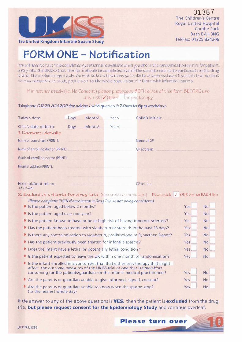
Tie United Kingdom In fantile Spasm Study
01367The Children's Centre Royal United Hospital
Combe Park Bath BA1 3NG
Tel/Fax: 01225 824206
FORM ONE - NotificationY?u will need to have this completed questionnaire available when you phone the randomisation centre for patient entry into the UKISS trial. This form should be completed even if the parents decline to participate in the drug t ia l or the epidemiology study. We wish to know how many patients have been excluded from this trial so th a t ws may compare our study population to the whole population of infants with infantile spasms.
If in neither s tudy (i.e. No Consent) please photocopy SOTH sides o f th is form SEFORE use and Tick { / ) here [ J on photocopy
Telephone 0 1 2 2 5 5 2 4 2 0 6 for advice / w ith q u eries 6 .3 0 a m t o 6p m w eek d ays
Today's d a t e : Day/ M o n th / Year/ Child's initials:
Child's d a t e o f b ir th : Day/ M o n th / Year/
1 D o c t o r s d e - t a i l s
Name of consultant (PRINT): Name o f GP: .....................................................
Name o f enrolling doctor (PRINT): GP address:
G'ade of enrolling doctor (PRINT): ........................................ ...........................................................................
Hospital address(PRINT): ...........................
Hospital/Dept tel no: GP tel no.:(if kn ow n )
2 . E x c l u s i o n c r i t e r i a f o r d r u g - tr ia l ( se e protocol for d eta ils) Please tick [ / J ONE box on EACH line
P /ease complete EVEN if enrolment in Drug Trial is not being considered t Is th e patient aged below 2 months? Yes No
• Is th e patient aged over o n e year? Yes No O
• Is th e patient known to have or be at high risk of having tuberous sclerosis? Yes No □
• Has th e patient been treated with vigabatrin or steroids in th e past 28 days? Yes No □
• Is there any contraindication to vigabatrin, prednisolone or Synacthen Depot? Yes No L ±• Has th e patient previously been treated for infantile spasms? Yes No □
• Does th e infant have a lethal or potentially lethal condition? YesL No □
• Is th e patient expected to leave th e UK within one m onth o f randomisation? Yes No □
• Is th e infant enrolled in a concurrent trial that either uses therapy that might affect th e outcom e m easures o f th e UKISS trial or on e that is tim e/effortconsum ing for the patients/guardians or th e infants' medical practitioners? Yes No
• Are th e parents or guardian unable to give informed, signed, consent? Yes No □
• Are th e parents or guardian unable to know w hen th e spasms stop? Yes No □(to th e nearest w hole day)
If tfe answer to any of the above questions is YES, then the patient is exc luded from the drug tria, b u t please request consen t fo r th e E p idem io logy S tudy and continue overleaf.
P l e a s eUKISVR1/1399

01367Please tick [ / one b o x o n ea ch line
3. Sex o f p a t i e n t
4. Does t h e child h a v e a k n o w n p ro v e n c h r o m o s o m a l a b n o r m a l i ty ?
5. Does t h e child h a v e a k n o w n p ro v e n s y n d r o m e ?
M ale Fem ale
□ □ 'Yes □ No
Yes No
6. Did th e child have a diagnosis o f cerebral palsy m a d e prior t o th e o n se t of th e spasms? Yes No
7. Did t h e child h a v e n e o n a ta l e n c e p h a lo p a th y w i th se izu res? Yes No
8. W as t h e child a l re a d y d ia g n o s e d as h av in g a b n o r m a l d e v e lo p m e n tb e f o r e t h e o n s e t o f sp a sm s (this d iagnosis m u s t have b een m a d e BEFORE th e o n se t o f spasms, b y a d o c to r or h ea lth visitor)! Yes No
9. C u r re n t m e d ic a t io n s
D rug
1
Total Daily Dose
□
10.C u r re n t w e ig h t ______ :_______ kg
If -this patient is -to be included in the O rug Trial, please c o m p lete Consent Form A
P le a se r e tu r n a p h o t o c o p y o f th is fo r m to : The UKISS Trial C e n treThe Child ren 's C e n tre Royal U n ited H ospita l C o m b e Park BATH BA1 3NG
Thank you very much for your help
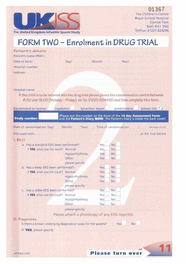
01367T he Children's C entre Royal U n ited H ospital
C om b e Park Bath BA1 3NG
Tel/Fax: 0 1 2 2 5 8 2 4 2 0 6
FORM TWO - Enrolment in DRUG TRIALP a t i e n t ' s d e t a i l s
P a t ie n t 's n a m e (PRINT):
D a te o f b ir th :
H ospita l n u m b e r
A ddress:
Day/ M o n th / Y ear/
The United Kingdom In fantile Spasm Study
H ospita l n a m e :
If the child is to be entered into the drug tria l please phone the randomisation centre between 3 .30 and 13.00 (Monday - Friday) on Tel: 01225 324490 and then complete this form.
'
■ h h h
D ate o f r a n d o m is a t io n : Day/ M o n th /
Discussed w ith :
Y ear/ T ime o f r a n d o m is a t i o n (24 h ou r clock)
a t t h e Trial C e n tre
1. E E Ga. Has a s t a n d a r d EEG b e e n p e r f o r m e d ?
If YES, w hat was th e result? N orm al
H y p sa r rh y th m ia
O th e r
please specify:b. Has a s le ep EEG b e e n p e r f o r m e d ?
If YES, w hat was the result? N orm al
H y p sa r rh y th m ia
O th e r
p lease specify:c. Has a v id e o EEG b e e n p e r f o r m e d ?
If YES, w hat was the result? N orm al
H y p sa r rh y th m ia
O th e r
p lease specify:
Please attach a photocopy o f any EEG report(s)2 ! . D i a g n o s i s
Is t h e r e a know n underly ing diagnosis o r cause fo r t h e spasms? Yes □ No □
If YES, p lease specify:
Yes No
Yes No
Yes No __,Yes No —
Yes No
Yes No
Yes No
Yes No
Yes No
Yes No _ ..Yes ..... No
Yes No
u«is/R2/i/399 P l e a s e tu rn o v e r

01367No D o n 't k n o w
. No ___ D o n 't k n o w
No D o n 't k n o w
No D o n 't k n o w
3 . S i n c e - t h e o n s e t - o f " th e s p a s m sa. (i) Has t h e r e b e e n a r r e s t o f d e v e lo p m e n t? Yes
(ii) If Yes, d id th is co inc ide w i th t h e s t a r t o f t h e spasm s? Yes
b. (i) Has t h e i r b e e n reg ress io n o f d e v e lo p m e n t? Yes
(ii) If Yes, d id th is co inc ide w i th t h e s t a r t o f t h e spasm s? Yes
c. UKISS D e v e lo p m e n t C h a r t
P lease fill in th is c h a r t w h e n t h e child is e n t e r e d in to t h e tr ial. In o r d e r t o g e t s o m e in d ic a t io n o f w h e t h e r t h e r e has b e e n a r r e s t o f d e v e lo p m e n t o r reg re ss io n b e f o r e t h e o n s e t o f t r e a t m e n t , p le a se r e s p o n d t o t h e q u e s t io n s f o r t h e best ever developm ent t o d a t e (in t h e c o lu m n h e a d e d 'Best ev e r ') a n d t h e current developm ental a tta in m en t (in t h e c o lu m n h e a d e d 'C u rre n t ') . (P lease tick / )
Date o f assessment Day/ M o n th / Year/
Developm ental m ilestone Best ever Current
Hand regard: Does t h e child fo cu s o n a n d look a t te n t iv e ly a t t h e h a n d s a n d f in g e rs?
Laugh: D oes t h e child r e s p o n d a t t im e s by la u g h in g loudly - a 'belly la u g h '?
Play w ith feet: Does t h e child fo cu s on , g rasp , a n d play w ith t h e f e e t ?
Grasps an object: Does t h e child g r a s p a small o b je c t w i th in rea ch w h e n s i t t ing w i th s u p p o r t (p a lm a r g r a s p is su ff ic ien t)?
Transfers an object: D oes t h e child t r a n s f e r a small o b je c t f ro m o n e h a n d t o a n o t h e r d e l ib e ra te ly ?
Sits unsupported: Does t h e child sit by itself o n a f la t su rface?
Permanency: Does t h e child k n o w w h e r e t o look fo r a n o b je c t t h a t has fa l len o u t o f v iew ?
Crawls: Can t h e child craw l a r o u n d o n all fo u rs?
Gives an object: Does t h e child h a n d a n o b je c t t o s o m e o n e o n r e q u e s t (w h e n p r o m p te d by g e s tu r e a n d voice)?
When compl et-edplease send a photocopy of thisform to the Trial Centre:
The UKISS Trial Centre The Children's Centre Royal United Hospital
Combe Park BATH BA1 3NG
(as in Action Plan 1 or 2)
Thank you very muc
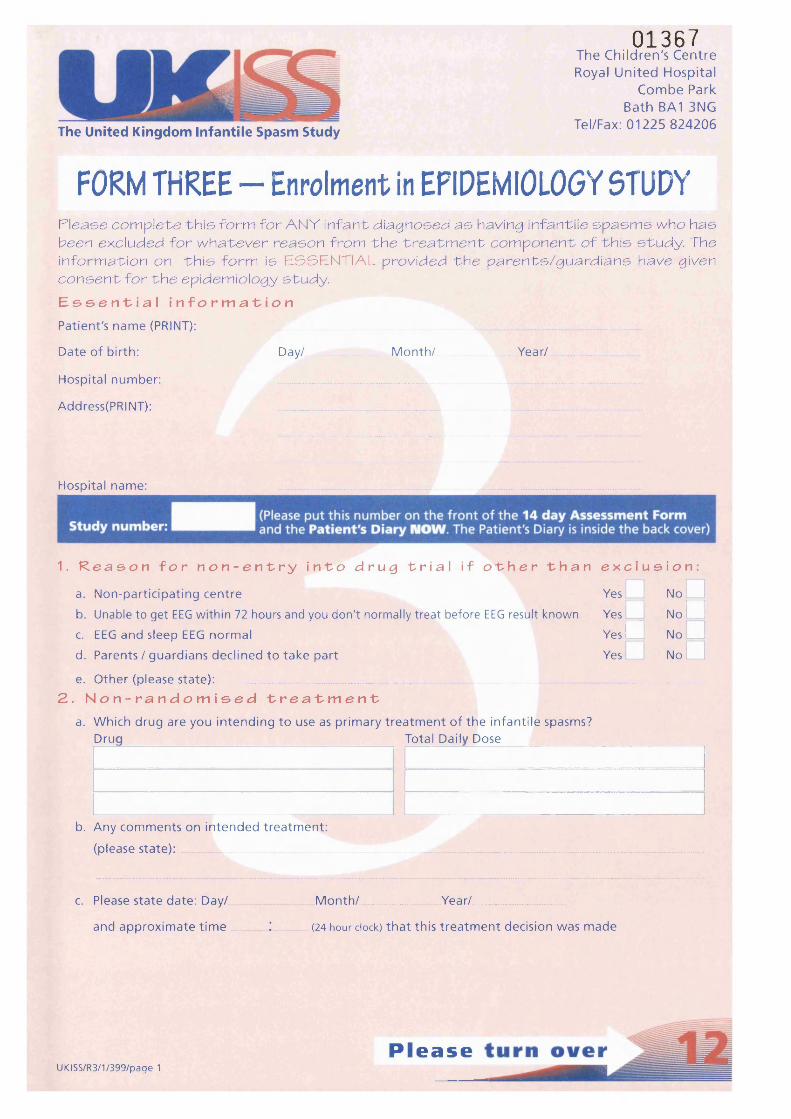
The United Kingdom In fan tile Spasm Study
01367The C hildren's C entre Royal U n ited H ospital
C om b e Park Bath BA1 3NG
Tel/Fax: 0 1 2 2 5 8 2 4 2 0 6
FORM THREE - Enrolment in EPIDEMIOLOGY STUDYPlease com plete t h is fo rm fo r A N Y in fan t: d iagno sed a s having in fa n tile s p a s m s who has been exc luded fo r w h a te v e r reason fro m th e t r e a tm e n t com ponent o f t h is s tu d y . The in fo rm a tio n on t h is fo rm is E S S E N T IA L provided t h e p a re n ts /g u a rd ia n s have given c o n s e n t f o r th e ep idem io logy s tu d y .
E s s e n t i a l i n f o r m a t i o n
P a t ie n t 's n a m e (PRINT):
D a te o f b ir th :
Hospita l n u m b e r :
Address(PRINT):
Day/ M o n th / Year/
Hospita l n a m e :
1. R e a s o n f o r n o n - e n t r y i n t o d r u g t r i a l i f o t h e r t h a n e x c l u s i o n :
a. N o n -p a r t ic ip a t in g c e n t r e Yes No
b. Unable to ge t EEG within 72 hours and you don 't normally trea t before EEG result known Yes No □
c. EEG a n d s le ep EEG n o r m a l
d. P a re n ts / g u a r d ia n s d e c l in e d t o t a k e p a r t
e. O th e r (p lease s ta te ) : ...............................................................................................
2 . N o n - r a n d o m i s e d t r e a t m e n t
a. W hich d r u g a r e y ou in te n d in g t o u se as p r im a ry t r e a t m e n t o f t h e in fan t i le spasm s?Drug Total Daily D o s e ____________
Y e sC H
Yes
No
No
b. Any c o m m e n t s o n i n t e n d e d t r e a t m e n t :
(p lease s ta te ) :
c. P lease s t a te d a te : Day/ M o n th / .............................Y ear/.................................
a n d a p p r o x im a te t im e ! (24 h ou r clock) t h a t th is t r e a t m e n t dec is ion w a s m a d e
P l e a s eU K ISS/R 3/1/399/page 1
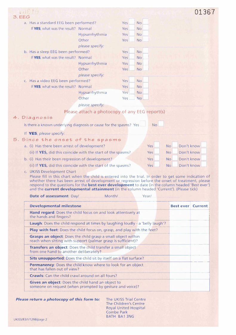
3 . E E Ga. Has a standard EEG been performed?
If YES, what was the result? NormalHypsarrhythmia Otherplease specify:
b. Has a sleep EEG been performed?If YES, what was the result? Normal
Hypsarrhythmia Otherplease specify:
c. Has a video EEG been performed?If YES, what was the result? Normal
Hypsarrhythmia Other
please specify:Please attach a photocopy of any EEG report(s)
A . D i a g n o s i s
Is there a known underlying diagnosis or cause for the spasms? Yes I
01367Yes NoYes NoYes NoYes No
Yes NoYes NoYes NoYes — No
Yes NoYes NoYes NoYes No
No
Yes
Yes
Yes
Yes :
No Don't know
No □ Don't know
No Don't know
If YES, please specify:5 . S i n c e - t h e o n s e t o f - t h e s p a s m s
a. (i) Has there been arrest o f developm ent?
(ii) If YES, did this coincide with the start o f th e spasms?
b. (i) Has their been regression o f developm ent?
(ii) If YES, did this coincide with th e start of the spasms?
c. UKISS D evelopm ent ChartPlease fill in this chart w hen th e child is entered into the trial. In order to g e t som e indication o f w hether there has been arrest o f developm ent or regression before th e onset o f treatm ent, please respond to th e questions for th e best ever developm ent to date (in th e column headed 'Best ever') and th e current developm ental a tta in m en t (in the column headed 'Current'). (Please tick)
Date of assessment Day/ M onth/ Year/
No Don't know
Developm ental m ilestone Best ever Current
Hand regard: Does the child focus on and look attentively at th e hands and fingers?
Laugh: Does th e child respond at tim es by laughing loudly - a 'belly laugh'?
Play w ith feet: Does th e child focus on, grasp, and play with th e feet?
Grasps an object: Does th e child grasp a small object within reach w hen sitting with support (palmar grasp is sufficient)?
Transfers an object: Does th e child transfer a small object from one hand to another deliberately?
Sits unsupported: Does th e child sit by itself on a flat surface?
Permanency: Does th e child know w here to look for an object that has fallen out o f view?
Crawls: Can th e child crawl around on all fours?
Gives an object: Does th e child hand an object to som eon e on request (when prom pted by gesture and voice)?
Please return a p h o to c o p y o f this form to: The UKISS Trial CentreThe Children's Centre Royal United Hospital Combe Park BATH BA1 3NG
U K IS S /R 3/1 /1298/page 2

01367The Children's Centre Royal United Hospital
Combe Park Bath BA1 3NG
The United Kingdom In fantile Spasm Study Tel/Fax: 01225 824206
FORM FOUR - DRUG TRIAL & EPIDEMIOLOGY Assessment a t 14 DaysThis form should be completed when the child is reviewed at the end of the initial two weeks of therapy. This assessment should take place as close to, but NOT BEFORE 14 completed days of the initial therapy.
D a te o f as se ssm en t:
P a t i e n t d e t a i l s :
Day/ M o n th / Year/
D a te o f b ir th : Day/ M o n th / Year/
H H H M H I1. T h e s p a s m s
a. (i) A g e a t onset o f t h e spasms:
(ii) If YES, specify d a te :
(iii) If NO, is t h e a g e a c c u r a te t o t h e n e a re s t :
b. D a te o f diagnosis o f t h e spasm s
Is t h e ex a c t d a t e k n o w n ? Yes No O
day / m o n t h / y ea r /
w e e k m o n th □
d ay / m o n t h / y ea r /
c. (i) Has the re been complete cessation of spasms for 48 hours since commencing trea tm e n t? Yes No □
(ii) If YES, date o f last spasm ? day /
d. W hat date did treatm ent com m ence? d ay /
e. (i) Has there been a relapse since?
(ii) If YES, have spasms stopped again for 48 hours ?
2 . P r e g n a n e y
a. (i) W ere there any maternal problems/illnesses during pregnancy?
(ii) If YES, what w e re they?
3 . I 3 i r t h h i s t o r y
a. Gestation at delivery: /40
b. (Please tick / boxes) (i) vaginal (ii) forceps
(iv) planned section (v) emergency section (vi) breech
c. Birth w eight: grams
d. Apgars (if known): (i) @1 (ii)
e. Head circum ference at birth: \ cm
f. History o f neonatal problems
m o n t h / yea r /
m o n th / y ea r /
Yes
Yes
No
No
Yes No
(iii) ventouse □
(vii) cephalic □
@5 (iii) @10
A- . A n y o t h e r p a s t m e d i c a l i l l n e s s e s / o p e r a t i o n s
P l e a s e tu rn o v e rUKISS/AS4/1/299/page 1
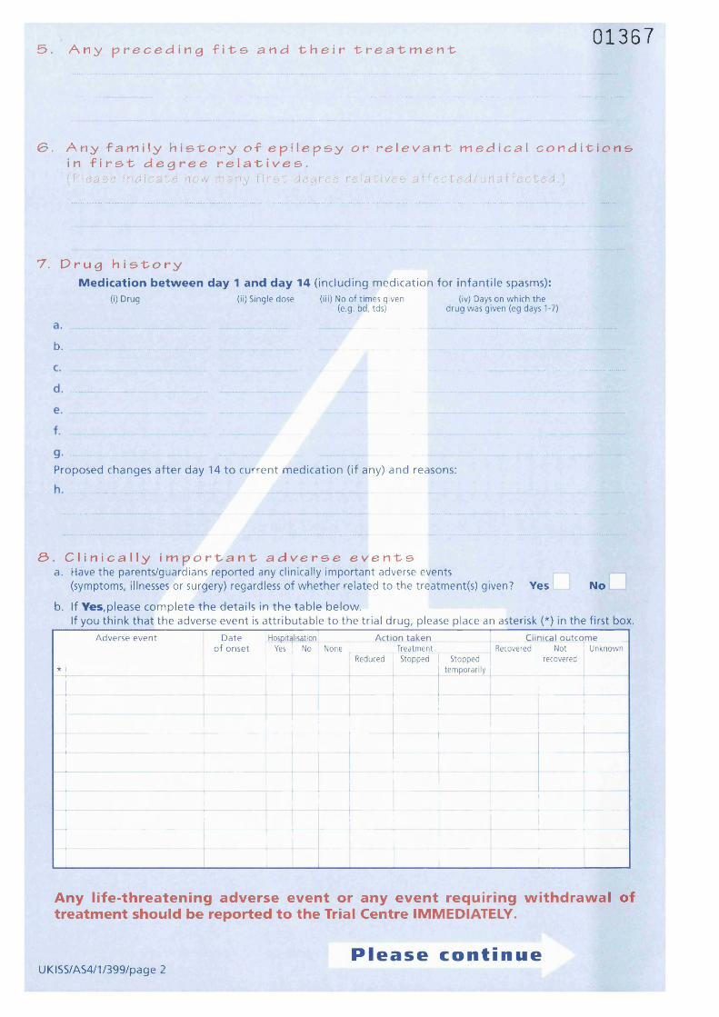
5 . A n y p r e c e d i n g f i t s a n d t h e i r t r e a t m e n t01367
0 . A n y f a m i l y h i s t o r y o f e p i l e p s y o r r e l e v a n t m e d i c a l c o n d i t i o n s i n f i r s t d e g r e e r e l a t i v e s .(Please i nd i ca te how many f i r s t degree r e l a t i ves a f f e c t e d / u n a f f e c t e d . )
7 . D r u g h i s t o r y
M edication betw een day 1 and day 14 (including m edication for infantile spasms):(i) Drug (ii) Single dose (iii) No o f tim es given (iv) Days on which the
(e.g. bd, tds) drug w as given (eg days 1-7)
b.
Proposed changes after day 14 to current m edication (if any) and reasons:
&. C l i n i c a l l y i m p o r t a n t * a d v e r s e e v e n t sa. Have the parents/guardians reported any clinically important adverse events
(symptoms, illnesses or surgery) regardless of whether related to the treatment(s) given? Yes No
b. If Yes,please com plete th e details in th e table below.If you think that the adverse event is attributable to the trial drug, please place an asterisk (*) in the first box.
A d verse e v e n t D a te o f o n s e t
HosDitalisation A ctio n ta k en __Cljjnical o u tc o m eYes [ No None I Treatment Recovered Not
recoveredUnknown
*Reduced Stopped Stopped
temporarily
I----|EH-|
Any life -th re a te n in g adverse event or any even t requ iring w ith d ra w a l of treatm ent should be reported to the Trial Centre IMMEDIATELY.
UKISS/AS4/1/399/page 2P l e a s e c o n t i n u e

013679 . N e u r o l o g i c a l a s s e s s m e n t
a. (i) W a s t h e n e u ro lo g ic a l e x a m in a t io n o f t h e in f a n t n o rm a l? Yes No(ii) If NO, p le ase d esc r ib e a n y f in d in g s b e lo w e .g . h y p o to n ia , spasticity:
b. (i) W e r e t h e fu n d i e x a m in e d w i th an o p th a lm o s c o p e ? (ii) If YES, w e r e t h e r e an y a b n o r m a l f in d in g s?D
CO
2
No
c. (i) W a s a W o o d s l igh t e x a m in a t io n p e r f o r m e d ? (ii) If YES, w e r e t h e r e an y a b n o r m a l f in d in g s?
Yes No
d. P lease list any o th e r n e u r o c u ta n o u s f in d in g s
e. (i) A re t h e r e an y d y sm o rp h ic f e a tu r e s ? Yes LJ No □
(ii) If YES, p lease list: .................................................. ................................................................................
f. (i) Current w eight: * kg (ii) Current head (occipitofrontal) circumference: * cm
1 O. 3 l o o d p r e s s u r ea. Has BP (either systolic or diastolic) risen above (Please tick: ^ one box only)? W 110/so ^ 12%o ^ No
b. Have antihypertensive drugs been started? Yes No
c. Did the drug treatment of infantile spasms have to be stopped as a result of the hypertension? Yes No □
1 1 . U r i n e a n a l y s i sa. (i) Has urine analysis been perform ed? Yes No
(ii) If YES, was glycosuria recorded? Yes No
(iii) If YES, was any treatm ent required? Yes No
b. Did treatm ent have to be stopped as a result o f diabetes? Yes No L J
1 2 . I n v e s t i g a t i o n s p e r f o r m e d
Please indicate whether or not the following investigations have been performed on the patient. If they have then where possible please can you either enclose copies of the reports or fill in the result on this form. If they have not had the investigation, please indicate whether it is planned in the fu ture or give the reason for it not being done.
a. (i) Was EEG perform ed at 14 days (the tim e o f th is assessm ent) Yes No □
(ii) If YES, Date:
(iii) Result (attach photocopy o f report if abnormal) Normal Hypsarrhythmia Other □
(iv) C o m m en ts ........................................................................ ...........................................................................
(v) If NOT done, is it planned for the future? Yes No O ;
(vi) If NOT planned, please specify reason:
b. (i) Was an MR I o f th e brain perform ed? Yes L J No □
(ii) If YES, result (a ttach p h o to c o p y o f rep o rt i f abnorm al) Normal Abnormal
(iii) C om m en ts .................................................................
(iv) If NOT done, is it planned for the future? Yes No □
(v) If NOT planned, please specify reason (e.g. CT done)
p i e a s e tu rn o v e rUKISS/AS4/1/299/page 3

01367The Children's Centre Royal United Hospital
Combe Park Bath BA1 3NG
Tel/Fax: 01225 824206
FORM 6(a) - THREE MONTHLY ASSESSMENT FORM {DRUG TRIAL)
This form should be completed when the child is reviewed THREE months after enrolment into the trial (unless preceded by final assessment).
P a t i e n t d e t a i l s :
Name (BLOCK CAPITALS):
Date o f birth: Day/ M onth/ Year/
The United Kingdom In fantile Spasm Study
Study number: Please now put Study Number on next A ssessm ent Form
Today's date:
Date o f last Assessment:
Day /
Day/
M onth/
M onth/
Year/
Year/
1 . E f f e c t o f t h e t h e r a p y o n t h e s p a s m s ( n o t o t h e r s e i zu r e t y p e s )
a. Has th e child had any spasms since th e last assessm ent? Yes No
b. Have an / changes been m ade to their treatm ent? Yes
c. If YES, please state those changes and w hen w ere they made?
No
2 .
3 .
A-.
B E GAny further EEG?
Date: Day / M onth/
Result:
(Please attach a photocopy o f any abnormal result)
E p i l e p t i c S e i z u r e s . Has th e :hiId had any other type o f epileptic seizure since last assessm ent?
D r u g h i s t o r y a t t i m e o f t h i s a s s e s s m e n t(p lease r e c o r d a II d r u g s c u r r e n t l y being t a k e n )
D rug (i) Total D aily D o se (ii)
Year/
Normal O
Yes EZI 8 No Q
Abnormal O
Yes No
U KISS/A S6a/1/399P l e a s e tu rn o v e r

013675 . C l i n i c a l l y i m p o r t a n t , a d v e r s e e v e n t s
a. Have the parents/guardians reported any clinically important adverse events (symptoms, illnesses or surgery) regardless o f w hether related to the treatment(s) given?
NoYes
b. If Y es,please com plete th e details in th e tab le below .(If you think that the adverse event is attributable to the trial drug, please place an asterisk (*) in the first box.)
A d verse e v e n t D ate Hosoitalisation A ctio n ta k e n Clinical o u t c o m eo f o n s e t Yes No None Treatment Recovered Not
recoveredUnknown
★Reduced Stopped Stopped
temporarily
0 . I n v e s t i g a t i o n s p e r f o r m e d t o f i n d u n d e r l y i n g c a u s e Have any further investigations been perform ed since the child's last review?
Please list and give th e results w here available
a)
b)
c)
d)
e)
7 . A n y o t h e r c o m m e n t s
— .
PI e a s e s an d a p h o to c o p y o f t h is c o m p le te d fo rm t o t h e Tria l C e n tre (a s in A c t io n P lan 1 o r 2 )
The UKISS Trial Centre The Children's Centre Royal United Hospital
Combe Park BATH BA1 3NG
Thank you very much for your help
UKISS/A S6a/1/1298

The United Kingdom In fan tile Spasm Study
01367The Children's Centre Royal United Hospital
Combe Park Bath BA1 3NG
Tel/Fax: 01225 824206
FORM 8(b) - THREE MONTHLY ASSESSMENT FORM TRIAL)
This form should be completed when the child is reviewed SIX months after enrolment into the tria l (unless preceded by final assessment).
P a t i e n t d e t a i l s :
N a m e (BLOCK CAPITALS):
D a te o f b ir th : Day/ M o n th / Y ear/
Study number:
Today 's d a t e :
D a te o f las t A ssessm ent:
Please now put Study Number on next A ssessm ent Form
Day/
Day/
M o n th /
M o n th /
Year/
Year/
1. E f f e c t o f t h e t h e r a p y o n t h e s p a s m s ( n o t o t h e r se i zu re t y p e s )
a. Has t h e child h a d a n y spasm s since t h e last a s s e s s m e n t? Yes No
b. H ave any c h a n g e s b e e n m a d e to th e i r t r e a t m e n t ?
c. If YES, p le a se s t a t e th o s e c h a n g e s a n d w h e n w e r e t h e y m a d e ?
Yes □ NolZ
B E GA ny f u r th e r EEG?
Date :
Result:
(P lease a t t a c h a p h o to c o p y o f an y a b n o r m a l result)
Day / M o n th / Y ear/
N orm al □
3 . E p i l e p t i c S e i z u r e sa. Has t h e child h a d a n y o t h e r t y p e o f ep i lep t ic se izu re since last a s se s s m e n t?
4 - . D r u g h i s t o r y a t t i m e o f t h i s a s s e s s m e n t(p l ease r e c o r d al l d r u g s c u r r e n t l y being t a k e n )
D rug (i) T otal D aily D o se (ii)
a ................
b.............
d ..............
Yes □ No
A b n o rm a l L J
Yes No
UK ISS/AS6b/1/399

013675 . C l i n i c a l l y i m p o r t a n t a d v e r s e e v e n t s
a. Have the parents/guardians reported any clinically important adverse events (symptoms, illnesses or surgery) regardless of w hether related to the treatm ent(s) given?
Yes No
b. If Yes,please com plete th e details in th e tab le below.(If you think that the adverse event is attributable to the trial drug, please place an asterisk (*) in the first box.)
A d verse e v e n t
*
D a te o f o n s e t
Hospitalisatior A ctio n ta k e n Clinical o u tc o m eYes No None
ReducedTreatmentStopped Stopped
temporarily
Recovered Notrecovered
Uncnown
0 . I n v e s t i g a t i o n s p e r f o r m e d t o f i n d u n d e r l y i n g c a u s e Have any further investigations been perform ed since th e child's last review?
Please list and give th e results w here available
a)
b)
c)
d)
e)
*7. A n y o t h e r c o m m e n t s
P le a s e s en d a p h o to c o p y o f t h is c o m p le te d fo rm t o th e Tria l C e n tre (a s in A c t io n Plan 1 o r 2 )
The UKISS Trial Centre The Children's Centre Royal United Hospital
Combe Park BATH BA1 3NG
Thank you very much for your help
U KISS/A S6b/1/399
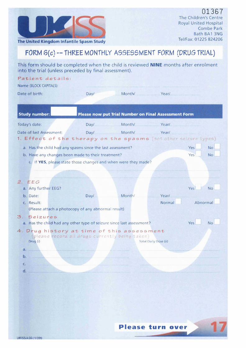
The United Kingdom In fan tile Spasm Study
01367The Children's Centre Royal United Hospital
Combe Park Bath BA1 3NG
Tel/Fax: 01225 824206
FORM 6(c) — THREE MONTHLY ASSESSMENT FORM (DRUG TRIAL)
This form should be completed when the child is reviewed NINE months after enrolment into the tria l (unless preceded by final assessment).
P a t i e n t d e - t a i l s :
Name (BLOCK CAPITALS):
Date o f birth: Day/ M onth/ Year/
Study number: Please now put Trial Number on Final A ssessm ent Form
Day/ M onth/
Day/ M onth/
Year/
Year/
Today's date:
Date o f last Assessment:
1 . E f f e c t o f - t h e - t h e r a p y o n - t h e s p a s m s ( n o t o t h e r se i zu re t y p e s )
a. Has th e child had any spasms since th e last assessm ent? Yes No
b. Have any changes been made to their treatm ent? Yes
c. If YES, please state those changes and w hen w ere they m ade?
No
Day/ M onth/
3 .
A-.
E E GAny further EEG?
Date:
Result:
(Please attach a photocopy o f any abnormal result)
S e i z u r e s. Has th e child had any other type o f seizure since last assessm ent?
D r u g h i s t o r y a t t i m e o f t h i s a s s e s s m e n t( p l ease r e c o r d al l d r u g s c u r r e n t l y being t a k e n )
D rug (i) Total Daily D o se (ii)
Year/............
Normal O
Yes Q g No □
Abnormal
Yes No
UKISS/AS6C/1/399
P l e a s e turn o v e r

013675 . C l i n i c a l l y i rr\ p o r t> a n t , a d v e r s e e v e n t s
a. Have the parents/guardians reported any clinically important adverse events (symptoms, illnesses or surgery),regardless of w hether related to the treatment(s) given?
Yes No
b. If Yes,please com plete th e details in th e table below.(If you think that the adverse event is attributable to the trial drug, please place an asterisk (*) in the first box.)
A d verse e v e n t D ate Hospitalisation A ction ta k en Clinical o u tc o m eo f o n s e t Yes No None Treatment Recovered Not
recoveredUrknown
*
Reduced Stopped Stopped
temporarily
-----------
0 . I n v e s t i g a t i o n s p e r f o r m e d t o f i n d u n d e r l y i n g c a u s e
Have a n y further investigations been perform ed since th e child's last review?
Please list and give th e results w here available
a)
b)
c)
d)
7 . A n y o t h e r c o m m e n t s
.
P le a s e s e n d a p h o to c o p y o f t h is c o m p le te d fo rm t o th e Tria l C e n tre (a s in A c t io n P lan 1 o r 2 )
The UKISS Trial Centre The Children's Centre Royal United Hospital
Combe Park BATH BA1 3NG
Thank you very much for your help
UKISS/AS6c/1/399
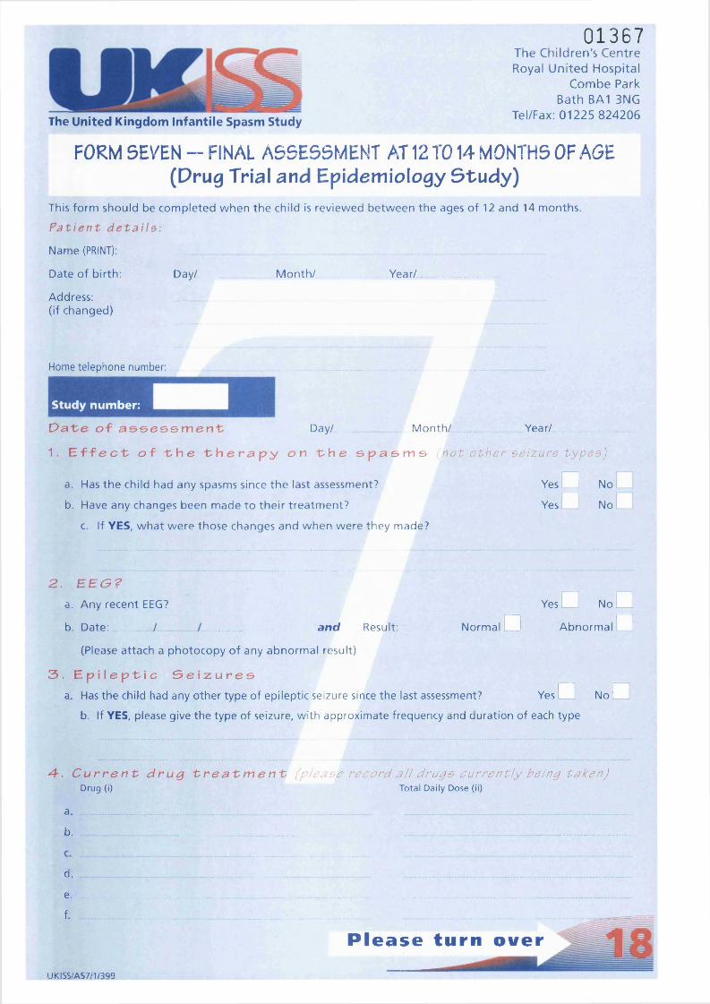
The United Kingdom In fan tile Spasm Study
01367The Children's Centre Royal United Hospital
Combe Park Bath BA1 3NG
Tel/Fax: 01225 824206
FORM SEVEN - FINAL ASSESSMENT AT 12 T014 MONTHS OF AGE (Drug Trial and Epidemiology Study)
This form should be com pleted w hen th e child is reviewed b etw een th e ages o f 12 and 14 months.
P a tie n t d e t a i l s :
Name (PRINT):
Date o f birth: Day/ M onth/ Year/
Address:(if changed)
Home telephone number:
Study number:
D a t e o f a s s e s s m e n t Day/ M onth/ Year/
1 . E f f e c t o f t h e t h e r a p y o n t h e s p a s m s (not o ther seizure types)
a. Has th e child had any spasms since th e last assessm ent?
b. Have any changes been m ade to their treatm ent?
c. If YES, w hat w ere th ose changes and w hen w ere they made?
Yes p i No
Yes No
2 . E E G ?a. Any recent EEG?
b. Date: / and Result: Normal
(Please attach a photocopy o f any abnormal result)
3 . E p i l e p t i c S e i z u r e s
a. Has the child had any other type of epileptic seizure since the last assessment?
Yes c i No U
□ Abnormal D
Yes L J No □
b. If YES, please give the type of seizure, with approximate frequency and duration of each type
A-. C u r r e n t d r u g t r e a t m e n t (please record all drugs currently being taken)D rug (i) T otal D aily D o se (ii)
a. ..............................
b.
d.
e ..................
f ..................................
P l e a s e tu rn o v e rUKISS/AS7/1/399

013675 . C l i n i c a l l y i m p o r t a n t a d v e r s e e v e n t s
a. Have the parents/guardians reported any clinically important adverse events (symptoms, illnesses or surgery) regardless of w hether related to the treatment(s) given?
Yes Nob. If Yes,please com plete th e details in th e table below.
(If you think that the adverse event is attributable to the trial drug, please place an asterisk (*) in the first box.)A d verse e v e n t D a te
o f o n s e tHospitalisation A ctio n ta k en Clinical o u tc o m e
*
Yes No NoneReduced
TreatmentStopped Stopped
temporarily
Recovered Notrecovered
Unknown
£ 3 . D e v e l o p m e n t a l h i s t o r y a t t i m e o f t h i s a s s e s s m e n t
In your opinion, is th e infant's developm ent appropriate for age? Yes No □
*7. N e u r o l o g i c a l a s s e s s m e n t ( e x c l u d i n g d e v e l o p m e n t )
a. Is th e neurological exam ination o f th e infant currently normal? Yes □ No Pb. If NO, please describe any positive findings below:
3 . (i) C u rre n t w e ig h t: kg (ii) C u rre n t h e a d ( o c c ip i to fr o n ta l) c irc u m fe re n c e : c m
9 . I n v e s t i g a t i o n s p e r f o r m e d t o f i n d u n d e r l y i n g c a u s e Have any further investigations been perform ed since th e child's last review?Please list and give th e results w here available
a) ..............................................
b)
c)
d)
e)1 0 . A n y o t h e r c o m m e n t s
11. I s t h e r e a k n o w n u n d e r l y i n g d i a g n o s i s / a e t i o l o g y f o r t h i s p a t i e n t ’s s p a s m s ?
Yes a No P
If YES, please com m ent: ....................................................................................................................................................
P le a se s e n d a p h o to c o p y o f t h i s c o m p le te d fo rm , t o g e t h e r w ith t h e rem ain ing p a g e s o f t h e P a t ie n t ’s D iary ;
t o t h e Trial C e n tr e (a s in A c tio n Plan 1, 2 o r 3 )The UKISS Trial Centre The Children's Centre Royal United Hospital
Combe Park BATH BA1 3NG
ISS/A S 7/1/399 Thank you very much for your help
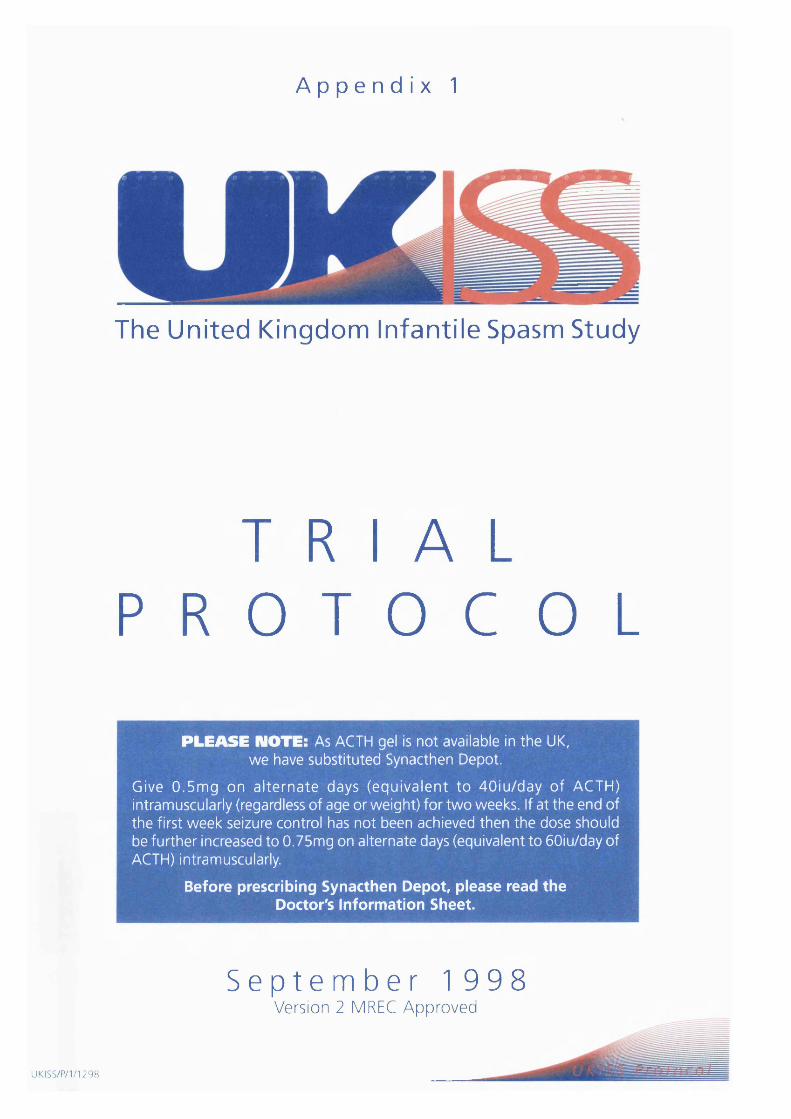
A p p e n d i x 1
The United Kingdom Infantile Spasm Study
T R I A L
P R O T O C O L
PLEASE IVIOTE: As ACTH gel is not available in the UK, we have substituted Synacthen Depot.
Give 0 .5m g on a lte rna te days (equ iva len t to 40 iu /day o f ACTH) intramuscularly (regardless o f age or weight) fo r tw o weeks. If at the end of the first week seizure control has not been achieved then the dose should be further increased to 0.75mg on alternate days (equivalent to 60iu/day of ACTH) intramuscularly.
Before prescribing Synacthen Depot, please read the Doctor's Information Sheet.
S e p t e m b e r 1 9 9 8Version 2 MREC Approved
UKISS/P/1/12 98

013675 . Cl inical ly i m p o r t a n t adverse even t s
a. Have t h e p a re n ts /g u a rd ia n s r e p o r te d any clinically im p o r ta n t adve rse ev en ts (symptoms, illnesses o r surgery) regard less o f w h e t h e r r e la te d to t h e t r e a tm e n t( s ) g iven?
Yes Nob. If Y e s ,p le a s e c o m p le t e t h e d e ta i l s in t h e t a b l e be low .
(If you think that the adverse event is attributable to the trial drug, please place an asterisk (*) in the first box.)A d verse e v e n t D ate Hospitalisation A ction ta k en C linical o u tc o m e
*
o f o n s e t Yes No NoneReduced
Treatment.Stopped Stopped
temporarily
Recovered Notrecovered
Unknown
0 . D e v e l o p m e n t a l h i s t o r y a t t i m e o f t h i s a s s e s s m e n t
In y o u r o p in io n , is the infant's developm ent appropriate for age? Yes
7 . N e u r o l o g i c a l a s s e s s m e n t ( exc l udi ng d e v e l o p me n t )a. Is t h e n e u ro lo g ic a l e x a m in a t io n o f t h e in fa n t cu r ren t ly n o rm a l? Yes □
b. If NO, p le a se d esc r ib e a n y posit ive f in d in g s b e lo w :
No
No□
3 . (i) C u rre n t w e ig h t: • kg (ii) C u rre n t h e a d (o c c ip i to fr o n ta l) c irc u m fe re n c e : •
9 . I n v e s t i g a t i o n s p e r f o r m e d t o f i n d u n d e r l y i n g c a u s e H ave an y f u r th e r in v e s t ig a t io n s b e e n p e r f o r m e d since t h e child 's last rev iew ?P lease list a n d give t h e resu lts w h e r e av a i lab le
a)
c m
b)
c)
d)
e)1 0 . A n y o t h e r c o m m e n t s
11. I s t h e r e a k n o w n u n d e r l y i n g d i a g n o s i s / a e t i o l o g y f o r t h i s p a t i e n t ’s s p a s m s ?
Yes No P
If YES, p lease c o m m e n t :
P le a se s e n d a p h o to c o p y o f th i s c o m p le te d form , t o g e t h e r w ith t h e rem aining p a g e s o f th e P a t ie n t ’s Diary,
t o th e Trial C e n tr e (a s in A c tio n Plan 1, 2 o r 3 )The UKISS Trial Centre The Children's Centre Royal United Hospital
Combe Park BATH BA1 3NG
U K ISS/A S7/1/399 Thank you very much for your help

A p p e n d i x 1
The United Kingdom Infantile Spasm Study
T R I A L P R O T O C O L
PLEASE MOTE: As ACTH gel is not available in the UK, we have substituted Synacthen Depot.
Give 0.5mg on alternate days (equivalent to 40iu/day of ACTH) intramuscularly (regardless of age or weight) for two weeks. If at the end of the first week seizure control has not been achieved then the dose should be further increased to 0.75mg on alternate days (equivalent to 60iu/day of ACTH) intramuscularly.
Before prescribing Synacthen Depot, please read the Doctor's Information Sheet.
S e p t e m b e r 1 9 9 8Version 2 MREC Approved
UKISS/P/1'1298
The United Kingdom Infantile Spasm Study
Trial C entre
The Children's Centre Royal United Hospital
Combe Park Bath BA1 3NG
Tel/Fax: 01225 824206
Trial S teer in g C o m m ittee
In Bath: Prof J P Osborne (Chairman)Dr S Edwards Dr E Hancock Dr A Lux Dr F O'Callaghan
Also: Dr T Johnson (MRC, Cambridge)Dr C Kennedy (Southampton)Dr R Newton (Manchester)Dr C Verity (Cambridge)
R A N D O M I S A T I O NTelephone: 01225 824490 / Monday to Friday between
or by Facsimile: 01225 824206[8 .30am and 6pm only
Please report all cases, even if not for randomisation
P r o t o c o l C o n t e n t sProtocol A1
Epidemiology A1
Notify all cases of infantile spasms A1
Drug trial A1
Inclusion criteria for drug trial A1
Exclusion criteria for drug trial A1
Consent A2
Electroencephalogram (EEG) A2
Pre-randomisation A2
Randomisation A2
Treatment groups A3
Initial treatment A3
Investigations A4
Initial observations A4
At enrolment A4
Before discharge A5
Following discharge, during the initial two week period A5
Responders and proposed treatment for relapse A5
Review at 2 weeks A5
Non - response at 2 weeks A6
Relapse after the initial two week period A6
Side effects A6
Subsequent follow up A6
Recruitment rate A6
Compliance A7
Likely rate of loss to follow-up A7
Post randomisation stratification A7
Post investigation diagnostic sub groups A7
Primary outcome measure A7
Secondary outcome measures
Analysis
The United Kingdom Infantile Spasm Study
P R O T O C O LThis is a pragmatic drug trial o f the current, most common treatments using the clinicians normal investigation plan.
E P I D E M I O L O G Y
NOTIFY ALL CASES OF INFANTILE SPASMS
Please notify all cases of infantile spasms to the tria l centre even if you do not intend to recruit the individual patient, fo r whatever reason, into the trial. Any patient w ith a clinical diagnosis o f infantile spasms should be included even if the EEG turns out to be normal. This would include patients younger than 2 months or older than 1 year. It would be very helpful if all cases can be notified so tha t we know how representative our tria l population is compared to the whole UK population. Brief clinical details w ill be requested.In order to carry out a capture-recapture analysis o f the UK population w ith IS (thus allowing a more complete ascertainment o f cases), we shall ask fo r information from pharmacists and EEG departments in collaborating hospitals. This information w ill be in itia ls, date o f birth and sex. The clinician will then be approached for pre-randomisation stratification data.
D R U G T R I A L
1. INCLUSION CRITERIA FOR DRUG TRIAL
All infants shall be included of either sex aged at seizure onset between 2 completed calendar months and 1 year o f age (up to but not including their first birthday) who are resident in the UK and who have a clinical diagnosis o f infantile spasms from any cause.
2. EXCLUSION CRITERIA FOR DRUG TRIAL
(i) Infants who are either known to suffer from tuberous sclerosis (TSC) or who are at high risk i.e. a presumptive diagnosis o f tuberous sclerosis because of;(a) known affected parent(b) previously diagnosed cardiac rhabdomyoma(c) hypomelanic macules, a forehead fibrous plaque or shagreen patch
noted(d) a retinal phakoma seen(e) polycystic kidneys in the infant

We recommend tha t first line therapy in these infants should be vigabatrin (see later fo r dosage. Please inform tria l centre even though they w ill not be entered into the tria l)
(ii) Infants previously treated w ith vigabatrin or steroids w ith in 28 days of the first diagnosis of infantile spasms.
(iii) Infants w ith a contra-indication to vigabatrin or steroids.(iv) Infants w ith a lethal or potentially lethal other condition.(v) Previous treatm ent for infantile spasms.(vi) Inability o f parents or guardians to give informed, signed consent.(vii) Infants expected to leave the UK w ith in one month o f randomisation.(viii) Inability o f parents or guardians to know when spasms stop - to the nearest
whole day.(ix) Infants enrolled in a concurrent tria l tha t either uses therapy tha t m ight affect
the outcome measures of the UKISS tria l or one tha t is tim e/effo rt consuming fo r the patients/guardians or the infants' medical practitioners.
All excluded infants should be reported to the tria l centre.
3. CONSENT
The tria l should then be discussed w ith the parents or guardians and the ir consent requested fo r participation in the tria l. Please report any infants where consent is not obtained to the tria l centre.
4. ELECTROENCEPHALOGRAM (EEG)
An EEG (or video EEG) should be obtained as quickly as possible so tha t treatm ent can start w ith in 72 hours o f diagnosis. Only if this is not possible and only if your normal practice is to start treatm ent prior to an EEG (or video EEG) may you randomise before an EEG (or video EEG) is obtained. If the EEG (or video EEG) is normal, a sleep EEG w ill be required prior to randomisation, if this has not already occurred. If the sleep EEG is normal the child w ill be excluded from the drug tria l (unless already enrolled) but please report the exclusion to the tria l centre fo r fu rther fo llow up.
If there w ill be a delay o f more than 72 hours before the EEG (or video EEG) can be performed and it is not your normal practice to start treatm ent prior to EEG (or video EEG), the child w ill be excluded from the drug tria l (but please report the exclusion to the tria l centre fo r further fo llow up).
5. PRE-RANDOMISATION
Prior to entry into the study the children w ill be stratified fo r randomisation by three variables:
(1) By gender: male, female
(2) By age: 60 to 119 days120 to 179 days 180 to 239 days 240 days and over
(3) By diagnosis know n a t tim e o f random isation (Yes / No).A: a proven chromosomal abnormality B: a proven syndrome diagnosisC: a diagnosis o f cerebral palsy made before the onset o f the
infantile spasms D: a diagnosis o f neonatal encephalopathy w ith seizures
U K I S S P r o t o c o l - A 2

E: a diagnosis of delayed development having already been made before the onset of the spasms. The diagnosis should have been made by either a medical practitioner or a health visitor, and must have been made before the onset o f the spasms.
Stratification is being done not to provide definitive groups fo r subanalysis but to balance the treatm ent groups w ith respect to factors which are identifiable at randomisation and which m ight affect our outcome measures.
6. RANDOMISATION
This w ill involve central randomisation by telephone call which w ill register the child into the trial, report base-line information and allocate initial treatment using the method of minimisation balancing over three factors.
7. TREATMENT GROUPS
All children eligible for the tria l w ill then be randomised into tw o treatm ent groups:
(1) Vigabatrin(2) "Steroids"
All patients randomised to receive steroids w ill undergo a second randomisation at that time to either Synacthen Depot or prednisolone.
The only pharmacological intervention, fo r the infantile spasms, in the first tw o weeks w ill be the use of the drug to which each patient has been randomised.
8. INITIAL TREATMENT
This w ill be a period of tw o weeks during which the child w ill receive the drug to which they are allocated as monotherapy. It is the local doctor's responsibility to read the manufacturer's drug data sheet before prescribing the fo llow ing drugs:
VigabatrinGive 50mg/kg/day orally in tw o divided doses fo r 24 hours (i.e. a to ta l o f 2 doses). Then increase to 100mg/kg/day orally in tw o divided doses in all patients.If at 96 hours seizure control has not been achieved then the dose should be further increased to 150mg/kg/day orally in tw o divided doses.They then continue on this dose (100 or 150mg/kg/day) until the tim e of the ir final developmental assessment around 12 -14 months o f age unless no response or relapse occurs (see below), or this is not your normal practice.
ACTH — USE SYNACTHEN DEPOTAs ACTH is not available in the UK, we have substituted Synacthen Depot. Give0.5mg on alternate days (equivalent to 40iu/day o f ACTH) intram usclarly (regardless of age or weight) fo r tw o weeks. If at the end of the first week seizure control has not been achieved then the dose should be fu rthe r increased to 0.75mg on alternate days (equivalent to 60iu/day of ACTH) intramuscularly. This group will then tail on oral prednisolone over 15 days. Those on 0.5mg on alternate days o f Synacthen Depot w ill receive 30mg of oral prednisolone fo r 5 days, then 20mg fo r 5 days then 10mg fo r 5 days and then stop. Those on 0.75mg of alternate days o f Synacthen Depot w ill receive 40mg of oral prednisolone for 5 days then 20mg fo r 5 days and then 10mg fo r 5 days and then stop.
PrednisoloneGive 10mg q.d.s. orally (regardless of age or weight) fo r tw o weeks.If at the end o f the first week seizure control has not been achieved the dose should be increased to 20mg t.d.s. orally.

This group will then tail on oral prednisolone over 15 days. Those on 40mg w ill reduce to 30mg fo r 5 days, then 20mg fo r 5 days then 10mg fo r five days and stop. Those on 60mg w ill reduce to 40mg fo r 5 days then 20 mg fo r 5 days then 10mg fo r 5 days then stop.
9. INVESTIGATIONS
(a) An investigation of the child's development w ill be made by history. A t enrolment the clinician w ill be asked to record the best development ever made on history by the child. It w ill not be necessary to record the age at which this performance was made. They w ill then be asked to record the current development achieved.
(b) As part o f normal practice in the investigation o f infants w ith infantile spasms, we assume the fo llow ing investigations w ill be undertaken:
1. MRI or CT of the brain2. Urine metabolic screen fo r amino acids3. Ophthalmoscopy (either direct or indirect)4. Ultraviolet light examination (Woods light)
The results o f these tests should be forwarded to the tria l centre. If fo r any reason one or more of these tests is not completed the reason should be stated.
(c) The fo llo w ing tests may be considered appropria te in some cases and, if performed, the results o f these and any other tests should be forwarded to the tria l centre.
1. Urea and electrolytes2. Liver function tests3. Chromosomes4. Urine metabolic screen fo r organic acids5. Lactate6. Biotinidase assay7. Ammonia8. Thyroid function9. Pyridoxine response - if pyridoxine is to be given we would recommend
tha t pyridoxine is given one month after the start of the tria l (after both the randomised treatm ent and first alternate treatm ent have been given in a child who continues fitting ).
(d) When venesection is performed fo r any clinical reason during the course of the infants illness, please take a blood sample fo r DNA analysis (bottle, form and addressed sample box included in patient pack). A subgroup of those on steroids w ill have blood taken to measure cortisol levels.
10. INITIAL OBSERVATIONS
The patient w ill be admitted if necessary fo r clinical confirm ation of the diagnosis o f in fantile spasms. If commencing steroid treatm ent, the patient w ill preferably be detained in hospital fo r 48 hours in order to have the ir blood pressure checked twice a day fo r tw o days and to have a urine analysis fo r glucose performed before discharge.
11. AT ENROLMENT
At enrolment the parents/guardians w ill be given the ir copy o f a 'f i t diary'. Its use, and how to complete it, must be explained and fu lly understood. In addition the importance o f this diary to interpreting the results o f the tria l should be explained. Details of the
U K I S S P r o t o c o l - A 4

child's name, date of birth, address, NHS number, tria l number, consultant and general practitioner should be completed. In addition we w ill ask all parents/guardians fo r a th ird party address (e.g. a grandparent) so tha t if they move and we lose contact w ith them we can try and track them through this th ird party.
As a minimum, the parents/guardians w ill be expected to make a once daily entry fo r the first 4 weeks of the tria l and a once weekly entry thereafter, until the time o f the final developmental assessment (12 -14 months), w ith a final 7 day daily entry fo r those still having seizures. They should bring the diary w ith them to all clinic appointments and they may use the diary to record more detailed information if the lead clinician so wishes.
12. BEFORE DISCHARGE
Before discharge please arrange fo r a fo llow up EEG as close to 14 days after the start of treatm ent as possible. Please also see the child in out-patients as close to 14 days after the start o f treatment as possible (but not less than 14 days after the start o f treatment).
13. FOLLOWING DISCHARGE, DURING THE INITIAL TWO WEEK PERIOD
Once the patient is discharged from hospital, if on steroids the blood pressure and urine analysis fo r glucose should be performed weekly whilst treatm ent continues, until the dose is reduced when this may be om itted if problems have not already been detected.
14. RESPONDERS A ND PROPOSED TREATMENT FOR RELAPSE
Responders: are those in whom there is to ta l cessation of spasms for at least 48 hours up to and including the end of the 14th day o f treatment.
Relapse: a single spasm in a responder constitutes a relapse.
If a child relapses w ith in the initial 2 week period then the lead clinician would be expected to increase the dose of the initial therapy up to the maximum recommended dose as per protocol.
If for any reason the lead clinician felt that it was not in the child's best interest to increase the dose of initial therapy (for example side effects experienced) then he should inform the trial centre as soon as possible, preferably but not necessarily before changing therapy.
15. REVIEW AT 2 WEEKS
Please review the patient as close to 14 days after the start o f treatm ent as possible but not less than 14 days after the start o f treatment.
The patient should be seen in order to confirm their progress and to complete the brief standardised progress report form. If the infant has responded the patient w ill continue on the same therapy as above.
16. NON - RESPONSE AT 2 WEEKS
The trial w ill be analysed by intention to treat at randomisation. However, in order to minimise the effects of a multitude o f different subsequent treatments, we will ask, but not require, clinicians to change to the alternative treatment if the primary treatment fails.
If a response has not occurred after 2 weeks, we expect the lead clinician to change to the alternative therapy i.e. either steroids after vigabatrin (the lead clinician to choose which steroid - Synacthen Depot or prednisolone) or vigabatrin after steroids. This alternative treatm ent w ill continue fo r the th ird and fourth weeks (a fu ll 14 days) at the same dose as recommended fo r the first tw o weeks.
If the lead clinician does not feel tha t this treatm ent is in the child's best interest they may change to a treatm ent of his/her own choice, w ithou t violating the tria l protocol, but they should notify the tria l centre o f this change in treatm ent plan using the 14 day standard report form.
If both treatments fail, the clinician will be left to choose the most appropriate treatment.
17. RELAPSE AFTER THE INITIAL TWO WEEK PERIOD
The lead clinician may choose his/her own treatment, but we suggest; if the child relapses after the initial tw o week period either: If on vigabatrin increase the dose in parallel w ith the child's weight gain (if increase is more than 1 kg) or if on 100mg/kg/day increase to 150mg/kg/day; If on steroids repeat the course once even if tailing. If these measures fail after a fu ll 14 days treatm ent change to the alternative therapy i.e. either steroids (Synacthen Depot or prednisolone) after vigabatrin, or vigabatrin after steroids.
18. SIDE EFFECTS
Should an in fan t suffer unacceptable side effects then the trea tm ent should be discontinued and the tria l centre informed immediately. The lead clinician may choose his own alternative treatment but we suggest, the lead clinician change to the alternative therapy i.e. either steroids (Synacthen Depot or prednisolone) a fte r vigabatrin, or vigabatrin after steroids.
19. SUBSEQUENT FOLLOW UP
1. All enrolled infants should be seen at 3 monthly intervals as a minimum although we realise tha t many w ill be seen more often because of the ir clinical need. The lead clinician w ill be asked to fill in a brief progress report form to send back to the tria l centre in Bath at each 3 monthly visit until the 12-14 month assessment.
2. Psychomotor development w ill also be assessed using a telephone questionnaire based on the "Vineland Adaptive Behaviour Scales" (see Section 26). This w ill be done by a trained researcher who w ill be based in Bath when the infant is between 12 and 14 months of age.
20. RECRUITMENT RATE
This is a national study and it is hoped to recruit all new cases of infantile spasms in the United Kingdom occurring w ith in the study period. Papers dealing w ith the epidemiology of infantile spasms put the incidence between 1 in 2380 and 1 in 6250 births. The average UK birth rate is approximately 800,000 per year. Therefore, it should be possible to recruit between 130 and 340 new cases per year. Taking into account exclusions, recruitment should be completed w ith in 18 months.
We intend to drive recruitment actively and have already sought to recruit one paediatric neurologist from each old health region to help promote the study. The tria l centre w ill take responsibility fo r ethics committee approval fo r each district in the UK and w ill recruit one paediatrician from each district to help promote the study. We w ill provide coasters and Post-it notes printed w ith contact arrangements at the commencement of
U K I S S P r o t o c o l - A 6
the tria l. A newsletter will be sent out to all recruiting clinicians and all promoters every tw o months.
We w ili notify all EEG departments o f the tria l w ith coasters, post-its and newsletters and ask them to remind clinicians of the trial whenever a request is made for an EEG in a child w ith possible infantile spasms, or whenever hypsarrhythmia is found.
21. COMPLIANCE
This is a serious disorder w ith profound sequelae. The motivation fo r clinicians and parents of patients to participate in treatm ent is high. The clinical workload to be imposed on attending physicians is little more than constitutes good clinical practice. (The workload to be imposed on parents is small.) The administrative work w ill be kept to a minimum. We w ill not collect unessential data and the trial centre w ill be responsible fo r documentation and fo r submissions to ethics committees.
22. LIKELY RATE OF LOSS TO FOLLOW UP
Loss to fo llow-up should be small. Patients known to be likely to move outside the country during the tria l period w ill be excluded at entry. As this is a national study, movement w ith in the UK should not lead to loss o f contact. Patient's NHS number, general practitioner and a th ird party address (e.g. grandparents) w ill be recorded on entry to the tria l thus making it easier to keep track of patients who move.
23. POST RANDOMISATION STRATIFICATION
There w ill be post-randomisation stratification fo r potential confounding variables not accounted fo r in pre-randomisation. These include EEG appearance (i.e. hypsarrhythmia vs atypical vs normal awake, normal asleep) and diagnostic sub-groups (i.e. cerebral dysgenesis on cranial scanning, chromosomal abnormalities or other syndromes and other diagnosis such as metabolic disease).
Social factors w ill include birth order and maternal age at school leaving.
24. POST INVESTIGATION DIAGNOSTIC SUB GROUPS
It may be possible to see if any trends in outcome are repeated w ith in subgroups of the tria l population.
The largest subgroup should be infants where development was delayed (as shown by pre-randomisation stratification data and, separately, by development history).
Smaller subgroups, e.g. those diagnosed w ith cerebral palsy on clinical examination, cerebral dysgenesis on cranial imaging, normal awake and asleep EEGs, are unlikely to be large enough fo r separate analysis. However, the data w ill be examined in case there is a dramatic difference in outcome, or a consistent trend.
25. PRIMARY OUTCOME MEASURE
The primary outcome measure w ill be the number o f patients who achieve complete cessation of spasms fo r at least 48 hours up to and including the end of the 14th day of treatment.

2 6 . S E C O N D A R Y OUTCOM E M E A SU R E S
1. Time taken to complete cessation o f all spasms fo r at least 48 hours.2. Partial response i.e. those patients tha t have a reduction in the number of spasms
during the tw o week period.3. Relapse rates.4. Other seizure types and frequency at 14 months, in those still having seizures.5. Resolution of hypsarrhythmia at 14 days.6. Developmental progress at 12 -14 months of age. This w ill allow at least 2 months
o f recovery fo llo w in g trea tm en t and is an age at which some language development has occurred. It is believed that delaying the assessment to a greater age w ill give little advantage and would have the disadvantage of increasing the impact of environmental factors.
The Vineland Adaptive Behaviour Scales have been developed and validated in the U.S.A. to measure "adaptive behaviour" defined as the performance of the daily activities required fo r personal and social sufficiency. It concentrates on the usual response, not the best response to any task. It has been validated in greater detail on larger numbers of children (3000) and has been used in the U.K. fo r other developmental fo llow-up studies. It achieves construct validity, content validity and has been compared to other scales in both normal and handicapped children. It has been shown to have internal consistency, test-retest and interrater reliability. It has been designed to minimise the influence of physical handicap.
The Vineland has four domains (communication, daily living skills, socialization and motor skills) and these combine to give an Adaptive Behaviour Composite: each of these has a standard score of 100 w ith a standard deviation of 15 and each can be expressed as an age equivalent (developmental age).
The Vineland scales can easily be adapted to be performed as a parental questionnaire that can be administered over the phone.
27. ANALYSIS
Assuming recruitment o f 250 children over the 18 month period, w ith approximately 125 randomised to vigabatrin and 125 to "steroids" and, assuming approximately 50% will achieve cessation of seizures in the "steroid" group, the tria l w ill have 90% power to detect an improvement in cessation of seizures to 70% in the vigabatrin group.
We are aware tha t the pattern of developmental age in the tw o groups may be bimodal. A direct comparison o f the developmental age (total score and subsets) o f the tw o groups w ill be undertaken, but we w ill also determine the proportion of individuals in each group whose development is one, tw o and three standard deviations below the mean fo r the Vineland.
U K I S S P r o t o c o l - A 8


The United Kingdom Infantile Spasm StudyThe Children's Centre Royal United Hospital
Combe Park Bath BA1 3NG
Tel/Fax: 01225 824206

01367
The United Kingdom Infantile Spasm Study
PATIENT’S D I A R Y
Name:
Address:
S tu d y number:
Your co n su lta n t d o c to r ’s team can be conX^acX^ed
by phoning
and asking for
N O TE TO DOCTOR: P le a se p h o to co p y co m p leted p a g es before p ostin g and keep original for yourself. The original will be needed if th e cop ies are lo s t in th e p o s t
In tr o d u c tio n t o t h e P a t ie n t ’s D iary
Thank you for agreeing to take p a r t in th e UKISS s tu d y which will help us findo u t more about infantile spasm s. This diary will be a record o f your child’s spasm sand o th er im portant information.
How to use your diary — for the first 2 weeks
1. Please fill in your child's n a m e and address on t h e fron t o f this diary. Please m ake sure th a t your d o c to r fills in t h e s tudy n u m b er and h o w t o c o n ta c t him or her.
2. Please fill in your child 's n a m e and s tu d y n u m b er on EACH PAGE o f this diary.
3. Please fill in p a g e s D3 t o D5 and start p a g e D6 a s soon as you are g iven t h e diary, w h ich m ust b e b e fo r e your child starts t r e a tm e n t .
4. During t h e first t w o w e e k s o f t r e a tm e n t w e n e e d t o k n o w w h a t h a p p e n s t o your child's spasms. Please fill in t h e deta ils a b o u t t h e n u m b er o f b a tch es and th e n u m ber o f spasm s per batch as best you can. Please w rite 'none' if your child has n o spasm s on a particular day. W e w a n t to f ind o u t w h e n t h e spasm s stop . W e also w a n t to k n o w if t h e spasm s c o m e back.
5. W e are in terested in s ide e f fec ts or p rob lem s w ith t h e trea tm en t; th in g s such as vom it in g , rashes, or excessive s leep in ess .
6. W e h ave left room for you to w r ite deta ils a b o u t o th e r illnesses th a t your child may h ave and a n yth in g e lse th a t y ou th ink m ig h t b e im portant. Please w rite as m uch as you like.
7. At t h e en d o f t h e first t w o w e e k s o f t r e a tm e n t y o u will b e s e e in g your d o cto r again . This sh ou ld b e as c lose t o 14 days after t h e start o f t r e a tm e n t as possib le - but n ot less th a n 14 d ays . A t th is visit, p le a s e ask y o u r d o c t o r t o p h o t o c o p y p a g e s D3f D4, D5, and D6 o f this diary and th e n forw ard th e m t o us.
8. R em em ber: it is im p o rta n t th a t YOUR CHILD'S NAME AND STUDY NUMBER ARE ON EACH PAGE. Please check this n ow .
9. If y ou r ch ild h as o n ly o n e sp a sm a t a t im e and never has b a tch es o f spasms, p lease ta k e g rea t care w h e n filling in t h e ta b le s on t h e fo l lo w in g p a g es . Put th e nu m b er o f s in g le spasm s occuring during o n e day (m id n igh t t o m idn ight) in th e co lu m n h e a d e d "number o f batches" and c h a n g e t h e w o rd "batches" in this h e a d in g t o "spasms". The correct resp on se t o t h e q u es t io n "maximum (largest) number o f spasms in one batch this day" w o u ld b e "one".
Thank yo u fo r helping u s To g e t th i s in form ation
Patient's Diary — Page D 2

01367Child’s Name: UKISS S tu d y Number:
Questionnaire for infantile spasmsFiease fill o u t pages D3, D4 and D5 a s soon a s you can. This will give us useful information about your child’s illness before th e trea tm en t started . Your doctor will ask to see th is when h e /sh e next se e s you. If you do n o t understand any o f th e questions then ask your doctor.
Y o u r c h i ld 's n a m e :
D a t e o f B ir th : / / ( d a y / m o n t h / y e a r )
NHS n u m b e r (if known).................. _....................... •............._........ ...........
P le a s e g iv e us t h e n a m e a n d a d d r e s s o f a f r i e n d , o r s o m e o n e in y o u r fam ily , w h o w o u l d b e a b l e t o f o r w a r d l e t t e r s t o y o u if y o u m o v e h o u s e o r if w e d o n o t k n o w w h e r e y o u a r e .
N a m e : „..... ............................. .................................................................
A d d r e s s : .........................................................................................................................................................................
GENERAL DESCRIPTION OF SPASMS1. Please tell us w h a t h a p p e n s w h e n your child has a spasm (please use your own words).
2. Do the spasms ever occur one at a time or do they occur in batches (ie several at one time)? (Please tick ( / ) o n e o f th e fo llow ing) only o n e spasm at a t im e
only in b a tch es o f spasm s
s o m e t im e s o n e spasm at a t im e , and s o m e t im e s b a tch es

01367Child’s Name: UKISS S tu d y Number:
HOW OFTEN IS YOUR CHILD HAVING INFANTILE SPASMS?A batch of spasms is when two or more spasms occur in a short space of time.
3. Number of BATCHES occurring in one day ...
On average, h o w m any BATCHES occur in a period lasting from m idnight o n e day to
m idnight th e fo llow in g day? Please tick ( / ) b e lo w for an a v era g e day during th e past
w eek , or since t h e spasms b egan if this is less th an 7 days ago . (please tick (S) one)
5 or fewer IZ j 6 to 10I J 11 to 20 21 to 40 More than 4 0 __]
A gain , but fo r th e w o r s t day, h o w m any BATCHES occur in a period lasting from
m id n ig h t o n e day t o m id n ig h t t h e n ex t day? Please tick ( / ) b e lo w for t h e w o r s t day (ie t h e day w ith t h e m ost batches) during th e past w e e k , (please tick (S) one)
5 or fewer1 Zl 6 to 10 i 11 to 20 21 to 40 More than 40 }Again, but for th e b est day, h o w m any BATCHES occur in a period lasting from midnight
o n e day to m idnight o f th e next day? Please tick ( / ) b e lo w for th e b e st day (ie th e day
w ith th e smallest num ber o f batches) during th e past w eek . (If th e spasms started less
than 7 days ago , only include days after th e spasms started.) (please tick (S) one)
5 or fewer 6 to l0 ] 11 to 2o\ 21to40 Morethan40' ’
4. Number of SPASMS occurring during any one batch
During th e past w e e k (or since t h e spasm s started if this is less th an 7 days ago),
h o w m any spasm s occurred, on a v era g e , during o n e batch? (please tick ( /) one)
5 or fewer 6 to 10 11 to 20 21 to 40 41 to 80 More than 80
During th e past w e e k , w h a t is t h e MAXIMUM (la rg est) n u m b er o f sp a sm s th a t
have occurred during o n e batch? (please tick (S) one)
5 or fewer [ 6 to 10 11 to 20 21 to 401 41 to 80 More than 80 □
During t h e past w e e k (or since t h e spasm s s tarted if th is is less th a n 7 days ago) , w h a t
is t h e MINIMUM (sm a lle st) n u m b er o f sp a sm s th a t h ave occurred during o n e
batch? (please tick (S) one)
5 or fewer I I 6 to 10! I 11 to 20 \ 21 to 40 41 to 80 More than 80 !
Patient's Diary — Page D 4

01367Child’s Name: UKISS Study Number:
M ovem ents o f th e b o d y during an a ttack
5. T h e s p a s m invo lves t h e f o l lo w in g m o v em e n ts o f parts o f th e body. (Please record the direction of the f a s t e s t or strongest movement) (please tick (S) 'Yes' or 'No')
T h e h ea d m o v i n g f o r w a r d s
T h e h ead m o v i n g b a c k w a r d s
T h e e y e s m o v i n g s i d e w a y s
if Yes,
T h e trunk ( b o d y ) m o v i n g f o r w a r d s
T h e trunk ( b o d y ) m o v i n g b a c k w a r d s
T h e arm s
if Yes, L ef t a r m o n ly or r i g h t a r m o n ly
T h e a r m s b e n d in t o w a r d s t h e c h e s t
T h e a r m s a r e f l u n g o u t , a w a y f r o m t h e b o d y
T h e le g s
if Yes, L ef t le g o n ly or r i g h t le g o n ly
Leg(s) b e n d a n d a r e b r o u g h t u p t o t h e b o d y
Leg(s) s t r a i g h t e n o u t , a w a y f r o m t h e b o d y
Timing of spasms
6. D o t h e s p a s m s o c c u r a t a n y p a r t i c u l a r t i m e ?
if Yes, (please tick (S) one or more)
m o r n i n g a f t e r n o o n J
o t h e r □ please specify:__________
Y e s : N o □
Yes N o □
Yes N o □
To o n e s id e o n ly L or t o b o t h s id e s gYes □ N o □
Y es E ll N o □
Yes □ N o o
or b o t h a r m s □
Y e s ' _ i N o □
Yes N o
Yes N o □
or b o t h l e g s □
Y es N o □
Yes □ N o □
e v e n i n g □Yes □ N o
n i g h t - t i m e □
H a v e y o u n o t i c e d a n y t h i n g t h a t s e t s t h e s p a s m s o f f ?
if Yes Please specify: __________________
Yes [ J/VoLJ
a n d d o e s it t r i g g e r t h e s p a s m ...
a l w a y s a: u s u a l ly □ s o m e t i m e s □ (please tick(S)one)

01367Child’sN am e: UKISS S tu d y Number:
INITIAL TREATMENT
Name of treatment:.....................................................................................................Please use this page of your diary to keep a daily record of your child's spasms during the first two weeks of the study.
D ate tr e a tm e n t started : / / YOU MUST FILL IN THIS DATE
DAILY Number o f batches* in a day. (m idnight to midnight)
Maximum (largest) number o f spasms in o n e batch this day
Please tick here if any treatm ent missed
Date
Date
Date
Date
Date
Date
Date
Date
Date
Date
Date
Date
Date
Date
Date
Date
Date
Date
* (or number of single spasms if they do not occur in batches)Please record in the table b e lo w anything else that you think may be important for the doctors running the trial to know: for example, unusual reactions that may be causedby the treatment, other illnesses, other medicines that have been started.Date
Date
Date
Date
Date
Date
Please ask your doctor to photocopy pages D3 to D6 when they are completed and he/ she will post the photocopy to us.CHECKED BY Dr.i ,_____ „_______ on: / /
Patient's Diary — Page D 6

How to use your diary — after the first two weeks o f the study
1. Please fill out the diary d aily for a further two weeks.2. For all children in the study, we will then need to keep a w e e k ly record of progress
until the time of the final study assessment at between 12 and 14 months of age.3. Each time you visit your doctor, please let them see the diary. This will give you a
chance to discuss it with them. Your doctor will send photocopies of the completed pages to us.
4. We will ask you to write down the number and type of fits for a week (7 days) after your child's first birthday. We want to know if your child continues to have spasms or develops any other form of fits.
I f you would like to keep a copy o f this diary, please ask your doctor to provideyou with a photocopy. Helshe w ill keep the original at the end o f the study.
01367Child’sName: UKISS Study Number:
SECOND TWO-WEEK PERIOD:Name of treatment:....................................................................................................Please use this page of the diary to keep a daily record of your child's spasms during the second two-week period of the study.______________________________________
D ate 2n d tr e a tm e n t started : / / YOU MUST FILL IN THIS DATEif your child has started a second t rea tm en t
DAILY Number o f batches* in a day. (m idnight to midnight)
Maximum (largest) number o f spasm s in on e batch this day
Please tick here if any treatm ent missed
Date
Date
Date
Date
Date
Date
Date
Date
Date
Date
Date
Date
Date
Date
Date
Date
Date
Date
* (or number of single spasms if they do not occur in batches)Please record in the table b e lo w anything else that you think may be important for the doctors running the trial to know: for example, unusual reactions that may be caused by the treatment, other illnesses, other medicines that have been started.
Date
Date
Date
Date
Date
Date
Please ask your doctor to photocopy this page at the end of two weeks and he/she will post the photocopy to us.
CHECKED BY Dr.: on: / /Patient's Diary — Page D 8

01367Child’s Name: UKISS S tu d y Number:
GENERAL PROGRESS:
Once your child has finished the two (or four) weeks of treatment, please fill in the diary below once w eekly until they have their final study assessment at between 12 and 14 months of age. Please record the number of batches during the worst day that week and the number of spasms in the longest batch during that week. As before, record in the second table any information that you think will be useful for the doctors conducting the study to know about____________ __________________________WEEKLY Number o f batches* during th e worst
day (m idnight to midnight) o f this w eekMaximum (largest) number o f spasms in o n e batch
Date
Date
Date
Date
Date
Date
Date
Date
Date
Date
Date
Date
Date
Date
Date
* (or number of single spasms if they do not occur in batches)Please record in the table b e lo w anything else that you think may be important for the doctors running the trial to know: for example, unusual reactions that may be caused by the treatment, other illnesses, other medicines that have been started.________Date
Date
Date
Date
Date
Date
Please ask your doctor to photocopy this page at the next visit and he/she will post the photocopy to us.
CHECKED BY Dr.:
01367Child’s Name: UKISS S tu d y Number:
GENERAL PROGRESS (continuation sheet)
If you have completed the table on page D9, please fill in the diary below once w eekly until your child has their final study assessment at between 12 and 14 months of age. Please record the number of batches during the worst day that week and the number of spasms in the longest batch during that week.
WEEKLY Number o f batches* during th e worst day (m idnight to midnight) o f this w eek
Maximum (largest) number o f spasm s in on e batch
Date
Date
Date
Date
Date
Date
Date
Date
Date
Date
Date
Date
Date
Date
Date
Date
Date
* (or number of single spasms if they do not occur in batches)Please record in the table be low anything else that you think may be important for the doctors running the trial to know: for example, unusual reactions that may be caused by the treatment, other illnesses, other medicines that have been started.Date
Date
Date
Date
Date
Date
Please ask your doctor to photocopy this page at the next visit and he/she will post the photocopy to us.
CHECKED BY Dr.: on: / /
Patient's Diary — Page D 10

01367Child’s Name: UKISS S tu d y Number:
CONTINUING FITS
Please complete this page after your child's 1st birthday.Please make a d a ily en try fo r o n e w e e k . Please fill in the types of fits that your child is having and the number they have each day. If they are having more than one type of fit, please record the number of each type that they are having. If you are not sure what type of fits your child is having you should ask your doctor.
Date Type o f f it N um ber o f fits this dayDay 1
Day 2
Day 3
Day 4
Day 5
Day 6
Day 7
or My child has not had any f it (epileptic seizure or spasm) during the past week. □
Signed:
Thank you very much for all your help.
If you have any questions about the stu dy please contact your consultant doctor’s team by telephoning the number
on the front of this diary.

The UKISS Trial Centre The Children's CentreRoyal United Hospital