Trousseau syndrome • Can we use direct oral anticoagulants ...
56
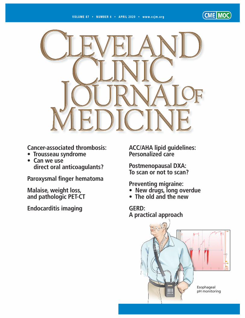
Esophageal pH monitoring Cancer-associated thrombosis: • Trousseau syndrome • Can we use direct oral anticoagulants? Paroxysmal finger hematoma Malaise, weight loss, and pathologic PET-CT Endocarditis imaging VOLUME 87 • NUMBER 4 • APRIL 2020 • www.ccjm.org ACC/AHA lipid guidelines: Personalized care Postmenopausal DXA: To scan or not to scan? Preventing migraine: • New drugs, long overdue • The old and the new GERD: A practical approach
-
Upload
khangminh22 -
Category
Documents
-
view
2 -
download
0
Transcript of Trousseau syndrome • Can we use direct oral anticoagulants ...
EsophagealpH monitoring
Cancer-associated thrombosis:• Trousseau syndrome• Can we use direct oral anticoagulants?
Paroxysmal fi nger hematoma
Malaise, weight loss, and pathologic PET-CT
Endocarditis imaging
V O L U M E 8 7 • N U M B E R 4 • A P R I L 2 0 2 0 • w w w. c c j m . o r g
ACC/AHA lipid guidelines:Personalized care
Postmenopausal DXA:To scan or not to scan?
Preventing migraine:• New drugs, long overdue• The old and the new
GERD:A practical approach
186 CLEVELAND CLINIC JOURNAL OF MEDICINE VOLUME 87 • NUMBER 4 APRIL 2020
EDITORIAL STAFFBrian F. Mandell, MD, PhD, Editor in ChiefPelin Batur, MD, Deputy EditorCraig Nielsen, MD, Deputy EditorKristi Thomsen, Executive EditorRay Borazanian, Managing EditorDavid A. Huddleston, Manuscript EditorAmy Slugg Moore, Manuscript Editor Ross Papalardo, CMI, Medical Art DirectorMary T. Cusick, Editorial Project LeaderPhilip Lammers, Editorial Project Leader
PUBLISHING OPERATIONSPeter G. Studer, Executive PublisherBruce M. Marich, Production ManagerKathy Dunasky, Production Manager, Special ProjectsIris Trivilino, Department CoordinatorLaurie Weiss, Accountant (Billing)
ASSOCIATE EDITORSJoseph Adewumi, MDAlejandro C. Arroliga, MDMoises Auron, MDDaniel J. Brotman, MDJacob Choi, MDAbhijit Duggal, MDRuth M. Farrell, MD, MAGary Francis, MDKathleen Franco, MDCarl Gillombardo, MD Steven M. Gordon, MDBrian Griffi n, MDDavid L. Keller, MDUmesh Khot, MDMandy C. Leonard, PharmDAndrew Lewis, DOAngelo A. Licata, MD, PhDAtul C. Mehta, MDChristian Nasr, MDRobert M. Palmer, MDDavid D.K. Rolston, MDGregory Rutecki, MDBernard J. Silver, MDTyler Stevens, MDTheodore Suh, MD, PhD, MHScMarc Williams, MDChristine Zayouna, MD
EDITORS EMERITIJohn D. Clough, MDHerbert P. Wiedemann, MDJames S. Taylor, MD
CLEVELAND CLINICTom Mihaljevic, MDPresident and Chief Executive Offi cer
CLEVELAND CLINIC EDUCATION INSTITUTEJames K. Stoller, MD, MS, ChairmanSteven Kawczak, PhD, Senior Director, Professional Development and Knowledge Resources
DISCLAIMERStatements and opinions expressed in the Cleveland Clinic Journal of Medicine are those of the authors and not necessarily of Cleveland Clinic or its Board of Trustees.
ADVERTISING Sima Sherman, Director of Sales and MarketingSHERMAN MEDICAL MARKETING GROUP 1628 John F. Kennedy Blvd., #2200, Philadelphia, PA 19103 (610) 529-0322 • [email protected]
SUBSCRIPTIONSU.S. and possessions: Personal $155; institutional $183; single copy/back issue $20
Foreign: $200; single copy/back issue $20
Institutional (multiple-reader rate) applies to libraries, schools, hospitals, and federal, commercial, and private institutions and organizations. Individual subscriptions must be in the names of, billed to, and paid by individuals.
Please make check payable to Cleveland Clinic Journal of Medicine and mail to: Cleveland Clinic Education Foundation, P.O. Box 373291, Cleveland, OH 44193-3291. To purchase a subscription with a credit card, please visit www.ccjm.org.
REPRINTS(610) 529-0322 • [email protected]
PHOTOCOPYINGAuthorization to photocopy items for internal or personal use is granted by Cleveland Clinic Journal of Medicine (ISSN 0891-1150 [print], ISSN 1939-2869 [online]), published by Cleveland Clinic, provided that the appropriate fee is paid directly to Copyright Clearance Center, 222 Rosewood Drive, Danvers, MA 01923 USA (978) 750-8400. Prior to photocopying items for educational classroom use, please contact Copyright Clearance Center, Inc., at the address above. For permission to reprint material, please fax your request with complete information to the Republication department at CCC, fax (978) 750-4470. For further information visit CCC online at www.copyright.com. To order bulk reprints, see above.
CHANGE OF ADDRESSTo report a change of address, send a recent mailing label along with new information to:
AMA, Data Verifi cation Unit, 330 N. Wabash Ave., Suite 39300, Chicago, IL 60611-5885 • Phone (800) 621-8335 • Fax (312) 464-4880 • [email protected]
Cleveland Clinic Journal of Medicine uses the AMA database of physician names and addresses. The database includes all US physicians and not just AMA members. Only the AMA can update changes of address and other data.
SUBSCRIPTIONS, EDITORIAL, BILLING, AND PRODUCTION 1950 Richmond Rd., TR404, Lyndhurst, OH 44124 • Phone (216) 444-2661 • Fax (216) 444-9385 • [email protected] • www.ccjm.org
Cleveland Clinic Journal of Medicine [ISSN 0891-1150 (print), ISSN 1939-2869 (online)] is published monthly by Cleveland Clinic at 1950 Richmond Rd., TR404, Lyndhurst, OH 44124.
COPYRIGHT© 2020 THE CLEVELAND CLINIC FOUNDATION.ALL RIGHTS RESERVED. PRINTED IN U.S.A.
CLEVELAND CLINIC JOURNAL OF MEDICINE VOLUME 87 • NUMBER 4 APRIL 2020 187
CONTINUED ON PAGE 188
TABLE OF CONTENTS
APRIL 2020
FROM THE EDITOR . . . . . . . . . . . . . . . . . . . . . . . . . . . . . . . . . . . . . . . . . . . . . . . . . . . . . . . . . . . . . . . . . . . . . . . . . . . .
The under- and overrecognized, 189and the elephant in the roomWe internists must recognize the pine cones without losing our vision of the forest.
Brian F. Mandell, MD, PhD
THE CLINICAL PICTURE . . . . . . . . . . . . . . . . . . . . . . . . . . . . . . . . . . . . . . . . . . . . . . . . . . . . . . . . . . . . . . . . . . . . . .
Paroxysmal fi nger hematoma 194This benign, self-limiting condition predominantly affects middle-aged women.
Daan J.L. van Twist, MD, PhD; Wim Hermans; Guy J.M. Mostard, MD
THE CLINICAL PICTURE . . . . . . . . . . . . . . . . . . . . . . . . . . . . . . . . . . . . . . . . . . . . . . . . . . . . . . . . . . . . . . . . . . . . . .
Constitutional symptoms, pathologic PET-CT 195PET-CT showed diffuse hypermetabolism of the great vessel walls, compatible with large-vessel giant cell arteritis.
Julia L. Riera, MD; Jorge L. Musuruana, MD; Fernando Faccio, MD; Javier A. Cavallasca, MD
THE CLINICAL PICTURE . . . . . . . . . . . . . . . . . . . . . . . . . . . . . . . . . . . . . . . . . . . . . . . . . . . . . . . . . . . . . . . . . . . . . .
Trousseau syndrome 199Examination revealed multiple bluish macules and 2 palpable thrombosed superfi cial veins in the right popliteal fossa.
Satish Maharaj, MBBS; Michael Omar, MBBS; Karan Seegobin, MBBS; Simone Chang, MBBS
1-MINUTE CONSULT . . . . . . . . . . . . . . . . . . . . . . . . . . . . . . . . . . . . . . . . . . . . . . . . . . . . . . . . . . . . . . . . . . . . . . . . . .
Can I use direct oral anticoagulants to treat 201cancer-associated venous thromboembolism?DOACs are increasingly replacing low-molecular-weight heparins for this purpose.
Oluwadunni Emiloju, MD; Sorab Gupta, MD; Claudia Dourado, MD
REVIEW . . . . . . . . . . . . . . . . . . . . . . . . . . . . . . . . . . . . . . . . . . . . . . . . . . . . . . . . . . . . . . . . . . . . . . . . . . . . . . . . . . . . . . . . . . .
To scan or not to scan? 205DXA in postmenopausal womenDXA is strongly recommended for women age 65 and older, but not so much for younger postmenopausal women.
Kristi Tough DeSapri, MD, CCD, NCMP; Rachel Brook, MD
OnlineFeaturesAccessCleveland Clinic Journal of Medicine content is readily available to all and is free of charge at www.ccjm.org.On your fi rst visit, we will ask you to take a few minutes to register, but subsequent visits will be unencumbered except for an occasional request for your e-mail address when you visit using another device (eg, mobile phone, tablet).
Services• Navigate quickly to articles in current and past issues via links on the home page and pull-down menus at the top of the page. Use the search function to fi nd a specifi c article, or browse by topic or article type.
• Continuing medical education activities are accessible from article links and from a CME pull-down menu on the home page. Participation is free.
• Follow links on the home page to news, summaries of recent scientifi c meetings, and interactive quizzes.
• Click on the PDF symbol at the top of any online article to read, download, or print the article in PDF format.
Social NetworkingTo post a CCJM article to Facebook or Twitter, just click on the icon at the top of any online article.
www.ccjm.org
CME MOC
■ Bronchoscopic lung reduction for emphysema
■ Steroid-associated bone loss
■ Perinatal depression
■ Cancer-associatedthrombosis:Predict, prevent, treat
■ ‘I want a doctor who looks like me’
■ Short QT and sudden cardiac death
■ Troponin testing in acute coronary syndrome
■ Cutaneous side effects of biologic medications
■ Pneumonia in alcohol use disorder
UpcomingFeatures
188 CLEVELAND CLINIC JOURNAL OF MEDICINE VOLUME 87 • NUMBER 4 APRIL 2020
APRIL 2020
CURRENT DRUG THERAPY . . . . . . . . . . . . . . . . . . . . . . . . . . . . . . . . . . . . . . . . . . . . . . . . . . . . . . . . . . . . . . . . . .
CGRP antagonists for decreasing migraine 211frequency: New options, long overdueCGRP drugs are an exciting frontier, but migraine management is still a combination of lifestyle changes plus treatment.
Julia Bucklan, DO; Zubair Ahmed, MD
EDITORIAL . . . . . . . . . . . . . . . . . . . . . . . . . . . . . . . . . . . . . . . . . . . . . . . . . . . . . . . . . . . . . . . . . . . . . . . . . . . . . . . . . . . . . . .
Preventing migraine: The old and the new 219Roughly half of patients taking any preventive medication have a 50% reduction in migraine frequency.
Glen D. Solomon, MD
REVIEW . . . . . . . . . . . . . . . . . . . . . . . . . . . . . . . . . . . . . . . . . . . . . . . . . . . . . . . . . . . . . . . . . . . . . . . . . . . . . . . . . . . . . . . . . . .
GERD: 223A practical approachPPIs are the fi rst-line medical therapy. Endoscopicand surgical options are pursued only if medicaltherapy fails.
Andrew Young, DO; Mythri Anil Kumar, MD; Prashanthi N. Thota, MD, FACG
REVIEW . . . . . . . . . . . . . . . . . . . . . . . . . . . . . . . . . . . . . . . . . . . . . . . . . . . . . . . . . . . . . . . . . . . . . . . . . . . . . . . . . . . . . . . . . . .
ACC/AHA lipid guidelines: Personalized care 231to prevent cardiovascular diseaseRisk assessment, drugs, patient subgroups, value of therapy, personalized plans, and shared decision-making.
Cara Reiter-Brennan; Albert D. Osei, MD, MPH; S.M. Iftekhar Uddin, MBBS, MSPH; Olusola A. Orimoloye, MD, MPH; Olufunmilayo H. Obisesan, MD, MPH; Mohammadhassan Mirbolouk, MD; Michael J. Blaha, MD, MPH; Omar Dzaye, MD, PhD
LETTERS TO THE EDITOR . . . . . . . . . . . . . . . . . . . . . . . . . . . . . . . . . . . . . . . . . . . . . . . . . . . . . . . . . . . . . . . . . . . .
Infective endocarditis: Don’t forget the ICE 191A recent article did not mention intracardiac echocardiography.
Faris G. Araj, MD; Michael Luna, MD
In reply: 192Infective endocarditis: Don’t forget the ICEEchocardiography relies on fi nding an anatomic abnormality, whereas 18FDG-PET is a functional examination.
Nkemdilim Mgbojikwe, MD; Steven R. Jones, MD; Thorsten M. Leucker, MD, PhD; Daniel J. Brotman, MD
DEPARTMENTS . . . . . . . . . . . . . . . . . . . . . . . . . . . . . . . . . . . . . . . . . . . . . . . . . . . . . . . . . . . . . . . . . . . . . . . . . . . . . . . . .
CME Calendar 190CME/MOC Instructions Inside back cover
CONTINUED FROM PAGE 187
CME MOC
CME MOC
CLEVELAND CLINIC JOURNAL OF MEDICINE VOLUME 87 • NUMBER 4 APRIL 2020 189
The under- and overrecognized,and the elephant in the room
FROM THE EDITOR
doi:10.3949/ccjm.87b.04020
In this issue of the Journal we have a paper reminding readers of an un-common clinical syndrome that is underrecognized, and a second paper
discussing a very common clinical syndrome that is likely overdiagnosed and over-reated.
Those of you who regularly read CCJM know of my preoccupation with the value of the patient’s history and the physical examination in directing the diagnostic evaluation as well as my enormous respect for clinicians who have honed those skills. The “Clinical Picture” section was born from my desire to remind us all of the power of observation by highlighting images from clinical and sometimes radiographic and other examinations.
In this issue, Van Twist et al (page 194) present a picture of a patient with recur-rent palmar surface fi nger hematomas (Achenbach syndrome). While I have seen and descriptively diagnosed this in 1 friend and several patients, ending the evaluation of their previously suspected vasculitis or Raynaud syndrome, I was not aware of its eponymous designation or of any literature describing small case series. I suspect that I may not be alone in this regard, and I thus appreciate the authors’ submission.
At the other end of the spectrum, Young et al (page 223) discuss gastroesophageal refl ux disease (GERD), an entity diagnosed by all of us in the clinic and at home and social gatherings. The disease is so common that we will usually be diagnosing it cor-rectly even without taking a careful history and pointedly revisiting the diagnosis after a pre-defi ned therapeutic trial with a proton pump inhibitor (PPI). But as the authors point out, there are specifi c features of the history that should direct us to considering an alternative approach to long-term PPI therapy or to recognizing when PPI therapy has failed, and why (eg, when exactly is the patient taking the medication).
As we are all in the midst of the amazingly jarring and outright scary COVID-19 pandemic, I realize how mundane a discussion of heartburn is. Yet in a way, it is the ability to recognize the pine cones without losing our vision of the forest that charac-terizes us as internists and keeps us professionally on course.
Hopefully, this pandemic will pass relatively soon, and our health systems and global connections will be stronger.
Be safe.
BRIAN F. MANDELL, MD, PhDEditor in Chief
190 CLEVELAND CLINIC JOURNAL OF MEDICINE VOLUME 87 • NUMBER 1 JANUARY 2020
2020APRIL
UVEITIS UPDATE [CANCELED]April 4Cleveland, OH
SOUTHWESTERN CONFERENCEON MEDICINE [CANCELED]April 23–26Tucson, AZ
MEDICAL DERMATOLOGY THERAPY UPDATE [CANCELED]April 29–May 1Cleveland, OH
MAY
DR. VICTOR FAZIO IBD SYMPOSIUM:INNOVATIONS IN MEDICALAND SURGICAL THERAPIESFOR INFLAMMATORY BOWEL DISEASE [CANCELED]May 4Chicago, IL
EMERGING CONCEPTSIN CARDIAC ELECTROPHYSIOLOGY:THE PRESENT AND THE FUTURE [CANCELED]May 5San Diego, CA
LEAD MANAGEMENT 2020:PREDICTING RISKS,STRENGTHS AND LIMITATIONS [CANCELED]May 6San Diego, CA
MULTIDISCIPLINARY MASTER CLASSIN ENDOCARDITIS AND OTHERCARDIOVASCULAR INFECTIONS [CANCELED]May 14–15Cleveland, OH
ANNUAL DIABETES DAY [CANCELED]May 20Cleveland, OH
JUNE
INTENSIVE REVIEWOF INTERNAL MEDICINEJune 1–5Cleveland, OH
INNOVATIONSIN CEREBROVASCULAR CAREJune 4–5Cleveland, OH
MELLEN CENTER UPDATEIN MULTIPLE SCLEROSISJune 12Cleveland, OH
INTERDISCIPLINARY APPROACHTO MANAGEMENT OF CRITICALLY ILLLIVER PATIENTSJune 15–16Cleveland, OH
INTERNAL MEDICINEBOARD REVIEW COURSEJune 16–20Plantation, FL
WASOG/AASOG 2020: MULTIDISCIPLINARY MEETING FOR SARCOIDOSIS AND ILDJune 24–27Hollywood, FL
JULY
MULTIDISCIPLINARY APPROACHTO THE CONTEMPORARY MANAGEMENTOF HEART FAILUREJuly 31Cleveland, OH
AUGUST
HOSPITAL MEDICINE 2020August 6–7Beachwood, OH
NEUROLOGY UPDATE: A COMPREHENSIVE REVIEW FOR THE CLINICIANAugust 7–9Washington, DC
INTENSIVE REVIEW OF CARDIOLOGYAugust 15–19Cleveland, OH
SEPTEMBER
CLEVELAND CLINIC EPILEPSY UPDATEAND REVIEW COURSESeptember 9–11Cleveland, OH
PRIMARY CARE WOMEN’S HEALTH: ESSENTIALS AND BEYONDSeptember 10–11Cleveland, OH
WOMEN IN HEALTHCARE FORUMSeptember 17Beachwood, OH
DIABETES, OBESITY,AND CARDIOVASCULAR DISEASE SUMMITSeptember 22–23Cleveland, OH, and Baton Rouge, LA
GENETICS EDUCATION SYMPOSIUM—GENETICS AND GENOMICS:APPLICATIONS FOR THE PREVENTION, DETECTION, AND TREATMENT OF CANCERSeptember 24Cleveland, OH
INTENSIVE REVIEW FOR THE GI BOARDSSeptember 25–28Las Vegas, NV
OCTOBER
STATE-OF-THE-ARTECHOCARDIOGRAPHY 2020October 2–4Cleveland, OH
INTENSIVE REVIEWOF ENDOCRINOLOGY AND METABOLISMOctober 9–11Cleveland, OH
ORTHOBIOLOGICS SUMMIT 2020:SCIENCE AND EVIDENCE BEHINDBIOLOGICS IN ORTHOPEDICSOctober 16–17Miami, FL
WAKE UP TO SLEEP DISORDERS 2020:A CLEVELAND CLINICSLEEP DISORDERS CENTER UPDATEOctober 16–17Beachwood, OH
FOR SCHEDULE UPDATES AND TO REGISTER, VISIT: WWW.CCFCME.ORG/LIVE
CME CALENDARCME MOC
190 CLEVELAND CLINIC JOURNAL OF MEDICINE VOLUME 87 • NUMBER 4 APRIL 2020
CLEVELAND CLINIC JOURNAL OF MEDICINE VOLUME 87 • NUMBER 4 APRIL 2020 191
Infective endocarditis:Don’t forget the ICEAUGUST 2019
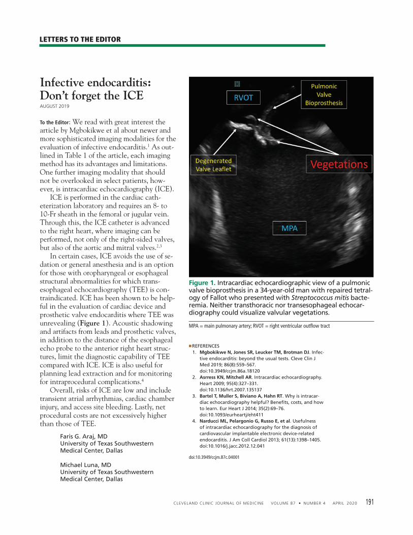
To the Editor: We read with great interest the article by Mgbokikwe et al about newer and more sophisticated imaging modalities for the evaluation of infective endocarditis.1 As out-lined in Table 1 of the article, each imaging method has its advantages and limitations. One further imaging modality that should not be overlooked in select patients, how-ever, is intracardiac echocardiography (ICE).
ICE is performed in the cardiac cath-eterization laboratory and requires an 8- to 10-Fr sheath in the femoral or jugular vein. Through this, the ICE catheter is advanced to the right heart, where imaging can be performed, not only of the right-sided valves, but also of the aortic and mitral valves.2,3
In certain cases, ICE avoids the use of se-dation or general anesthesia and is an option for those with oropharyngeal or esophageal structural abnormalities for which trans-esophageal echocardiography (TEE) is con-traindicated. ICE has been shown to be help-ful in the evaluation of cardiac device and prosthetic valve endocarditis where TEE was unrevealing (Figure 1). Acoustic shadowing and artifacts from leads and prosthetic valves, in addition to the distance of the esophageal echo probe to the anterior right heart struc-tures, limit the diagnostic capability of TEE compared with ICE. ICE is also useful for planning lead extraction and for monitoring for intraprocedural complications.4
Overall, risks of ICE are low and include transient atrial arrhythmias, cardiac chamber injury, and access site bleeding. Lastly, net procedural costs are not excessively higher than those of TEE.
Faris G. Araj, MDUniversity of Texas Southwestern Medical Center, Dallas
Michael Luna, MDUniversity of Texas Southwestern Medical Center, Dallas
◾REFERENCES 1. Mgbokikwe N, Jones SR, Leucker TM, Brotman DJ. Infec-
tive endocarditis: beyond the usual tests. Cleve Clin J Med 2019; 86(8):559–567. doi:10.3949/ccjm.86a.18120
2. Asrress KN, Mitchell AR. Intracardiac echocardiography. Heart 2009; 95(4):327–331. doi:10.1136/hrt.2007.135137
3. Bartel T, Muller S, Biviano A, Hahn RT. Why is intracar-diac echocardiography helpful? Benefi ts, costs, and how to learn. Eur Heart J 2014; 35(2):69–76. doi:10.1093/eurheartj/eht411
4. Narducci ML, Pelargonio G, Russo E, et al. Usefulness of intracardiac echocardiography for the diagnosis of cardiovascular implantable electronic device-related endocarditis. J Am Coll Cardiol 2013; 61(13):1398–1405. doi:10.1016/j.jacc.2012.12.041
doi:10.3949/ccjm.87c.04001
LETTERS TO THE EDITOR
Figure 1. Intracardiac echocardiographic view of a pulmonicvalve bioprosthesis in a 34-year-old man with repaired tetral-ogy of Fallot who presented with Streptococcus mitis bacte-remia. Neither transthoracic nor transesophageal echocar-diography could visualize valvular vegetations.
MPA = main pulmonary artery; RVOT = right ventricular outfl ow tract
192 CLEVELAND CLINIC JOURNAL OF MEDICINE VOLUME 87 • NUMBER 4 APRIL 2020
In reply: The letter from Drs. Araj and Luna regarding the utilization of intracardiac echo-cardiography (ICE) raises several interesting points. Indeed, for patients with infective endocarditis with inconclusive fi ndings on transthoracic echocardiography (TTE) and contraindications to use of contrast-mediated studies or transesophageal echocardiography (TEE), ICE does present another poten-tially useful diagnostic modality. However, it is an invasive procedure, and as such, the clinical team would need to weigh the risk of complications. Further, while the authors suggest that the cost is comparable to that of TEE, the likely higher cost relative to posi-tron emission tomography (PET) and other advanced imaging methodologies, as well as availability of institutional expertise, experi-ence, and availability, should also be consid-ered.
ICE, similar to TTE and TEE, relies upon the fi nding of an anatomic abnormality, in this case, the demonstration of a vegetation, for the diagnosis of infectious endocarditis. 18FDG-PET does not rely on anatomic iden-tifi cation of vegetations but is a functional
examination detecting infl ammation, which can be helpful in detecting microscopic veg-etations not identifi able by echocardiography.
Since the absence of an anatomically de-tected vegetation does not exclude infectious endocarditis, PET has potentially comple-mentary additive value to the various modali-ties based on demonstration of vegetation for the diagnosis of infectious endocarditis.
Nkemdilim Mgbojikwe, MDJohns Hopkins University School of Medicine, Baltimore, MD
Steven R. Jones, MDJohns Hopkins University School of Medicine, Baltimore, MD
Thorsten M. Leucker, MD, PhDJohns Hopkins University School of Medicine, Baltimore, MD
Daniel J. Brotman, MDJohns Hopkins University School of Medicine, Baltimore, MD
doi:10.3949/ccjm.87c.04002
LETTERS TO THE EDITOR

Daan J.L. van Twist, MD, PhDDepartment of Internal Medicine, Zuyderland Medical Centre, Sittard/Heerlen,The Netherlands
Paroxysmal fi nger hematomaA previously healthy 51-year-old woman report-
ed recurrent episodes of blue discoloration of one or more fi ngers associated with pain and swelling and mainly affecting the intermediate phalanges of the fourth fi nger of each hand. During some episodes, the second and third fi ngers were also involved. Each time, the symptoms resolved spontaneously within 3 days. She reported no other complaints and no recurrent trauma, spontaneous bleeding, palpitations, or discol-oration of the fi ngers when exposed to cold. She was a nonsmoker. Results of the physical examination were normal, including brachial and fi nger blood pressures, measured at the proximal phalanx of each fi nger, and Doppler studies of the brachial, radial, ulnar, and digital arteries. Other laboratory tests showed no signs of underlying infl ammatory, hematologic, or coagulation disorder. Based on the results of the evaluation, the patient’s condition was diagnosed as paroxysmal fi nger hematoma.
■ PAROXYSMAL FINGER HEMATOMA
Paroxysmal fi nger hematoma, also known as Achen-bach syndrome, is a benign, self-limiting condition that predominantly affects middle-aged women.1 It is characterized by recurrent spontaneous subcutane-ous bleeding in the fi ngers, typically on the palmar surface, mainly around the proximal interphalangeal joint creases. The cause is unknown, but local vascular fragility has been suggested. Although relapses may frequently occur, no treat-ment is indicated, as the symptoms resolve spontane-ously within a few days. The diagnosis is based on the typical clinical pre-sentation, as results of routine laboratory testing and Doppler studies of the arteries of the arm are usually normal.2 Therefore, it does not require further test-ing if the clinical presentation is typical and there are no clinical clues for an underlying disease such as Raynaud phenomenon, autoinfl ammatory disease, or thromboembolism.
Unfortunately, the typical symptoms are often not recognized, resulting in unnecessary and potentially harmful diagnostic procedures such as tissue biopsy and catheter-based angiography. Hence, awareness of this benign, self-limiting syndrome is important.
■ REFERENCES 1. Godoy A, Tabares AH. Achenbach syndrome (paroxysmal fi nger
hematoma). Vasc Med 2019; 24(4):361–366. doi:10.1177/1358863X19849627
2. Aida F, Kasimzade F. Analysis of 24 patients with Achenbach’s syn-drome. World J Clin Cases 2019; 7(10):1103–1110. doi:10.12998/wjcc.v7.i10.1103
Address: Daan J.L. van Twist, MD, PhD, Department of Internal Medicine, Zuyderland Medical Centre, PO-box 5500, 6130 MB, Sittard, The Nether-lands; [email protected]
THE CLINICAL PICTURE
doi:10.3949/ccjm.87a.19122
Wim HermansDepartment of Vascular Surgery, Zuyderland Medical Centre,Sittard/Heerlen, The Netherlands
Guy J.M. Mostard, MDDepartment of Internal Medicine, Zuyderland Medical Centre, Sittard/Heerlen, The Netherlands
Figure 1. The patient reported recurrent episodes of blue discoloration of the palmar surface of the fi ngers, associated with pain and swelling. The symptoms usually resolved within 3 days.
194 CLEVELAND CLINIC JOURNAL OF MEDICINE VOLUME 87 • NUMBER 4 APRIL 2020
Julia L. Riera, MDSection of Rheumatology and Autoimmune Diseases, Hospital JB Iturraspe, Santa Fe, Argentina
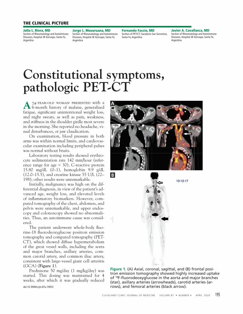
Constitutional symptoms,pathologic PET-CTA 74-year-old woman presented with a
6-month history of malaise, generalized fatigue, signifi cant unintentional weight loss, and night sweats, as well as pain, weakness, and stiffness in the shoulder girdle most severe in the morning. She reported no headache, vi-sual disturbances, or jaw claudication. On examination, blood pressure in both arms was within normal limits, and cardiovas-cular examination including peripheral pulses was normal without bruits. Laboratory testing results showed erythro-cyte sedimentation rate 142 mm/hour (refer-ence range for age < 30), C-reactive protein 15.80 mg/dL (0–1), hemoglobin 9.9 g/dL (12.0–15.5), and creatine kinase 55 U/L (22–198); other results were unremarkable. Initially, malignancy was high on the dif-ferential diagnosis, in view of the patient’s ad-vanced age, weight loss, and elevated levels of infl ammatory biomarkers. However, com-puted tomography of the chest, abdomen, and pelvis were unremarkable, and upper endos-copy and colonoscopy showed no abnormali-ties. Thus, an autoimmune cause was consid-ered. The patient underwent whole-body fl uo-rine-18 fl uorodeoxyglucose positron emission tomography and computed tomography (PET-CT), which showed diffuse hypermetabolism of the great vessel walls, including the aorta and major branches, axillary arteries, com-mon carotid artery, and common iliac artery, consistent with large-vessel giant cell arteritis (GCA) (Figure 1). Prednisone 50 mg/day (1 mg/kg/day) was started. This dosing was maintained for 4 weeks, after which it was gradually reduced
CLEVELAND CLINIC JOURNAL OF MEDICINE VOLUME 87 • NUMBER 4 APRIL 2020 195
THE CLINICAL PICTURE
doi:10.3949/ccjm.87a.19053
Jorge L. Musuruana, MDSection of Rheumatology and Autoimmune Diseases, Hospital JB Iturraspe, Santa Fe, Argentina
Fernando Faccio, MDSection of PET/CT, Sanatorio San Geronimo, Santa Fe, Argentina
Javier A. Cavallasca, MDSection of Rheumatology and Autoimmune Diseases, Hospital JB Iturraspe, Santa Fe, Argentina
Figure 1. (A) Axial, coronal, sagittal, and (B) frontal posi-tron emission tomography showed highly increased uptake of 18F-fl uoro deoxyglucose in the aorta and major branches (star), axillary arteries (arrowheads), carotid arteries (ar-rows), and femoral arteries (black arrow).
A
B
196 CLEVELAND CLINIC JOURNAL OF MEDICINE VOLUME 87 • NUMBER 4 APRIL 2020
PATHOLOGIC PET-CT
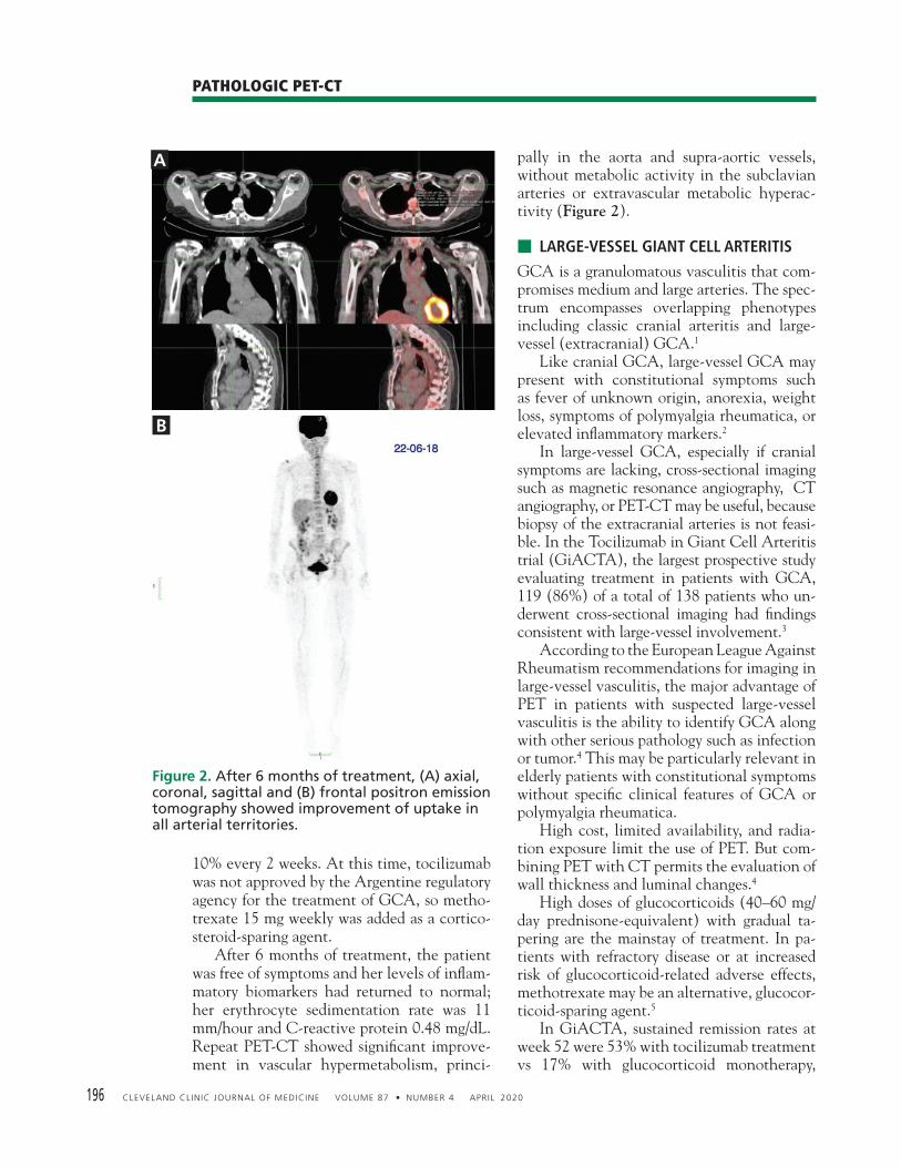
10% every 2 weeks. At this time, tocilizumab was not approved by the Argentine regulatory agency for the treatment of GCA, so metho-trexate 15 mg weekly was added as a cortico-steroid-sparing agent. After 6 months of treatment, the patient was free of symptoms and her levels of infl am-matory biomarkers had returned to normal; her erythrocyte sedimentation rate was 11 mm/hour and C-reactive protein 0.48 mg/dL. Repeat PET-CT showed signifi cant improve-ment in vascular hypermetabolism, princi-
pally in the aorta and supra-aortic vessels, without metabolic activity in the subclavian arteries or extravascular metabolic hyperac-tivity (Figure 2).
■ LARGE-VESSEL GIANT CELL ARTERITIS
GCA is a granulomatous vasculitis that com-promises medium and large arteries. The spec-trum encompasses overlapping phenotypes including classic cranial arteritis and large-vessel (extracranial) GCA.1
Like cranial GCA, large-vessel GCA may present with constitutional symptoms such as fever of unknown origin, anorexia, weight loss, symptoms of polymyalgia rheumatica, or elevated infl ammatory markers.2
In large-vessel GCA, especially if cranial symptoms are lacking, cross-sectional imaging such as magnetic resonance angiography, CT angiography, or PET-CT may be useful, because biopsy of the extracranial arteries is not feasi-ble. In the Tocilizumab in Giant Cell Arteritis trial (GiACTA), the largest prospective study evaluating treatment in patients with GCA, 119 (86%) of a total of 138 patients who un-derwent cross-sectional imaging had fi ndings consistent with large-vessel involvement.3 According to the European League Against Rheumatism recommendations for imaging in large-vessel vasculitis, the major advantage of PET in patients with suspected large-vessel vasculitis is the ability to identify GCA along with other serious pathology such as infection or tumor.4 This may be particularly relevant in elderly patients with constitutional symptoms without specifi c clinical features of GCA or polymyalgia rheumatica. High cost, limited availability, and radia-tion exposure limit the use of PET. But com-bining PET with CT permits the evaluation of wall thickness and luminal changes.4
High doses of glucocorticoids (40–60 mg/day prednisone-equivalent) with gradual ta-pering are the mainstay of treatment. In pa-tients with refractory disease or at increased risk of glucocorticoid-related adverse effects, methotrexate may be an alternative, glucocor-ticoid-sparing agent.5
In GiACTA, sustained remission rates at week 52 were 53% with tocilizumab treatment vs 17% with glucocorticoid monotherapy,
Figure 2. After 6 months of treatment, (A) axial, coronal, sagittal and (B) frontal positron emission tomography showed improvement of uptake in all arterial territories.
A
B
CLEVELAND CLINIC JOURNAL OF MEDICINE VOLUME 87 • NUMBER 4 APRIL 2020 197
RIERA AND COLLEAGUES
while the cumulative glucocorticoid dose was reduced by 50% in tocilizumab-treated pa-tients with fewer adverse events than those on glucocorticoids alone.6 These strikingly posi-tive results led to tocilizumab’s approval by the US Food and Drug Administration.
Encouraging results have also been reported with ustekinumab, an interleukin 12 and inter-leukin 23 antagonist, and with abatacept, a se-lective T cell costimulation modulator.2 A new era in the treatment of an old disease is com-ing. ■
■ REFERENCES 1. Dejaco C, Duftner C, Buttgereit F, Matteson EL, Dasgupta B. The
spectrum of giant cell arteritis and polymyalgia rheumatica: revisiting the concept of the disease. Rheumatology (Oxford) 2017; 56(4):506–515. doi:10.1093/rheumatology/kew273
2. Koster MJ, Matteson EL, Warrington KJ. Large-vessel giant cell arteritis: diagnosis, monitoring and management. Rheumatology (Oxford) 2018; 57(suppl 2):ii32–ii42. doi:10.1093/rheumatology/kex424
3. Tuckwell K, Collinson N, Klearman M, Dimonaco S, Stone JH, on behalf of GiACTA Investigators. FRIO377 classifi cation criteria for giant cell arteritis: data from Giacta informing the need for revision. Ann Rheum Dis 2016; 75(suppl 2):571. ard.bmj.com/content/75/Suppl_2/571.1. Accessed February 26, 2020.
4. Dejaco C, Ramiro S, Duftner C, et al. EULAR recommendations for the use of imaging in large vessel vasculitis in clinical practice. Ann Rheum Dis 2018; 77(5):636–643. doi:10.1136/annrheumdis-2017-212649
5. Hellmich B, Agueda A, Monti S, et al. 2018 Update of the EULAR recommendations for the management of large vessel vasculitis. Ann Rheum Dis 2020; 79(1):19–30. doi:10.1136/annrheumdis-2019-215672
6. Stone JH, Tuckwell K, Dimonaco S, et al. Trial of tocilizumab in giant-cell arteritis. N Engl J Med 2017; 377(4):317–328. doi:10.1056/NEJMoa1613849
Address: Javier Cavallasca, MD, Section of Rheumatology and Autoim-mune Diseases, Hospital JB Iturraspe, Bv. Pellegrini 3551, CP 3000, Santa Fe, Argentina; [email protected]; [email protected]

Satish Maharaj, MBBSJames Graham Brown Cancer Center, University of Louisville School of Medicine, Louisville, KY
Trousseau syndrome
A previously healthy 75-year-old man presented to the clinic with memory loss,
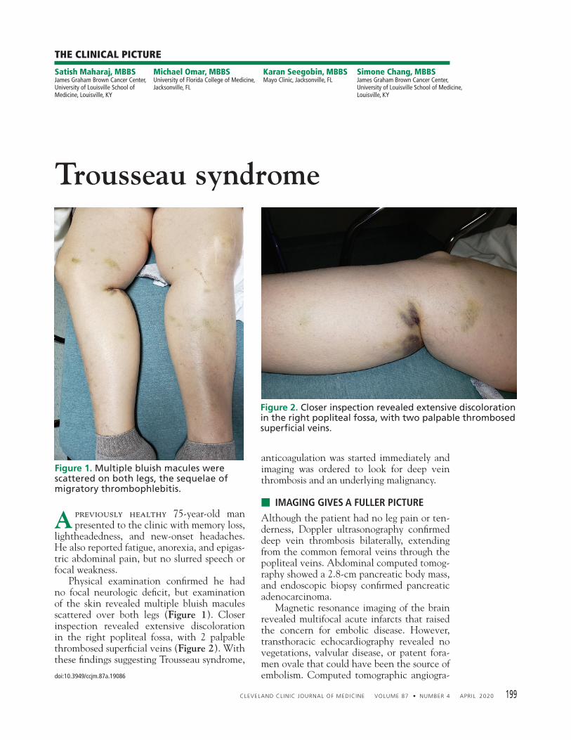
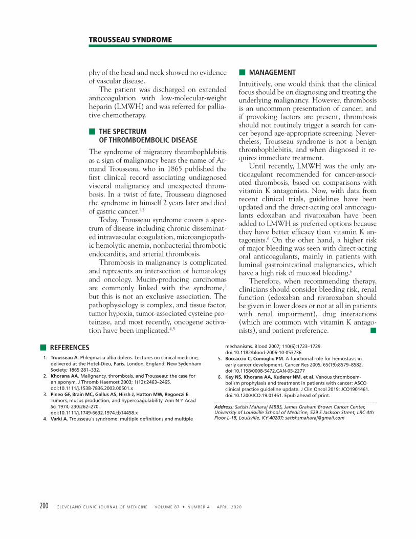
lightheadedness, and new-onset headaches. He also reported fatigue, anorexia, and epigas-tric abdominal pain, but no slurred speech or focal weakness. Physical examination confi rmed he had no focal neurologic defi cit, but examination of the skin revealed multiple bluish macules scattered over both legs (Figure 1). Closer inspection revealed extensive discoloration in the right popliteal fossa, with 2 palpable thrombosed superfi cial veins (Figure 2). With these fi ndings suggesting Trousseau syndrome,
CLEVELAND CLINIC JOURNAL OF MEDICINE VOLUME 87 • NUMBER 4 APRIL 2020 199
anticoagulation was started immediately and imaging was ordered to look for deep vein thrombosis and an underlying malignancy.
■ IMAGING GIVES A FULLER PICTURE
Although the patient had no leg pain or ten-derness, Doppler ultrasonography confi rmed deep vein thrombosis bilaterally, extending from the common femoral veins through the popliteal veins. Abdominal computed tomog-raphy showed a 2.8-cm pancreatic body mass, and endoscopic biopsy confi rmed pancreatic adenocarcinoma. Magnetic resonance imaging of the brain revealed multifocal acute infarcts that raised the concern for embolic disease. However, transthoracic echocardiography revealed no vegetations, valvular disease, or patent fora-men ovale that could have been the source of embolism. Computed tomographic angiogra-
THE CLINICAL PICTURE
doi:10.3949/ccjm.87a.19086
Michael Omar, MBBSUniversity of Florida College of Medicine,Jacksonville, FL
Karan Seegobin, MBBSMayo Clinic, Jacksonville, FL
Simone Chang, MBBSJames Graham Brown Cancer Center,University of Louisville School of Medicine, Louisville, KY
Figure 1. Multiple bluish macules were scattered on both legs, the sequelae of migratory thrombophlebitis.
Figure 2. Closer inspection revealed extensive discoloration in the right pop liteal fossa, with two palpable thrombosed superfi cial veins.
200 CLEVELAND CLINIC JOURNAL OF MEDICINE VOLUME 87 • NUMBER 4 APRIL 2020
TROUSSEAU SYNDROME
phy of the head and neck showed no evidence of vascular disease. The patient was discharged on extended anticoagulation with low-molecular-weight heparin (LMWH) and was referred for pallia-tive chemotherapy.
■ THE SPECTRUM OF THROMBOEMBOLIC DISEASE
The syndrome of migratory thrombophlebitis as a sign of malignancy bears the name of Ar-mand Trousseau, who in 1865 published the fi rst clinical record associating undiagnosed visceral malignancy and unexpected throm-bosis. In a twist of fate, Trousseau diagnosed the syndrome in himself 2 years later and died of gastric cancer.1,2 Today, Trousseau syndrome covers a spec-trum of disease including chronic disseminat-ed intravascular coagulation, microangiopath-ic hemolytic anemia, nonbacterial thrombotic endocarditis, and arterial thrombosis. Thrombosis in malignancy is complicated and represents an intersection of hematology and oncology. Mucin-producing carcinomas are commonly linked with the syndrome,3 but this is not an exclusive association. The pathophysiology is complex, and tissue factor, tumor hypoxia, tumor-associated cysteine pro-teinase, and most recently, oncogene activa-tion have been implicated.4,5
■ MANAGEMENT
Intuitively, one would think that the clinical focus should be on diagnosing and treating the underlying malignancy. However, thrombosis is an uncommon presentation of cancer, and if provoking factors are present, thrombosis should not routinely trigger a search for can-cer beyond age-appropriate screening. Never-theless, Trousseau syndrome is not a benign thrombophlebitis, and when diagnosed it re-quires immediate treatment. Until recently, LMWH was the only an-ticoagulant recommended for cancer-associ-ated thrombosis, based on comparisons with vitamin K antagonists. Now, with data from recent clinical trials, guidelines have been updated and the direct-acting oral anticoagu-lants edoxaban and rivaroxaban have been added to LMWH as preferred options because they have better effi cacy than vitamin K an-tagonists.6 On the other hand, a higher risk of major bleeding was seen with direct-acting oral anticoagulants, mainly in patients with luminal gastrointestinal malignancies, which have a high risk of mucosal bleeding.6
Therefore, when recommending therapy, clinicians should consider bleeding risk, renal function (edoxaban and rivaroxaban should be given in lower doses or not at all in patients with renal impairment), drug interactions (which are common with vitamin K antago-nists), and patient preference. ■
■ REFERENCES 1. Trousseau A. Phlegmasia alba dolens. Lectures on clinical medicine,
delivered at the Hotel-Dieu, Paris. London, England: New Sydenham Society; 1865:281–332.
2. Khorana AA. Malignancy, thrombosis, and Trousseau: the case for an eponym. J Thromb Haemost 2003; 1(12):2463–2465. doi:10.1111/j.1538-7836.2003.00501.x
3. Pineo GF, Brain MC, Gallus AS, Hirsh J, Hatton MW, Regoeczi E. Tumors, mucus production, and hypercoagulability. Ann N Y Acad Sci 1974; 230:262–270. doi:10.1111/j.1749-6632.1974.tb14458.x
4. Varki A. Trousseau's syndrome: multiple defi nitions and multiple
mechanisms. Blood 2007; 110(6):1723–1729. doi:10.1182/blood-2006-10-053736
5. Boccaccio C, Comoglio PM. A functional role for hemostasis in early cancer development. Cancer Res 2005; 65(19):8579–8582. doi:10.1158/0008-5472.CAN-05-2277
6. Key NS, Khorana AA, Kuderer NM, et al. Venous thromboem-bolism prophylaxis and treatment in patients with cancer: ASCO clinical practice guideline update. J Clin Oncol 2019: JCO1901461. doi:10.1200/JCO.19.01461. Epub ahead of print.
Address: Satish Maharaj MBBS, James Graham Brown Cancer Center, University of Louisville School of Medicine, 529 S Jackson Street, LRC 4th Floor L-18, Louisville, KY 40207; [email protected]

Oluwadunni Emiloju, MDInternal Medicine Resident, Department of Medi cine, Albert Einstein Medical Center, Philadelphia, PA
Yes. The direct oral anticoagulants ri-varoxaban, edoxaban, and apixaban
have been studied in cancer-associated ve-nous thromboembolism and are increasingly replacing low-molecular-weight heparins such as dalteparin and enoxaparin for this purpose. Individualizing care by balancing risks and benefi ts for each patient will help in choosing the right anticoagulant.
■ LOW-MOLECULAR-WEIGHT HEPARINS
The National Comprehensive Cancer Net-work guidelines previously recommended low-molecular-weight heparins as the preferred anticoagulants for cancer-associated venous thromboembolism, but now they are one of several fi rst-line options.1
Before the advent of direct oral anticoagu-lants, low-molecular-weight heparins were rec-ommended over vitamin K antagonists such as warfarin because they were more effective. This recommendation was supported by a large randomized trial,2 in which the recurrence rate was signifi cantly lower in patients treated with dalteparin than in those receiving vitamin K an-tagonists, with no signifi cant difference in major bleeding between the 2 treatment groups. The number needed to treat to prevent 1 recurrence of venous thromboembolism was 13.2 An important advantage of low-molecu-lar-weight heparins over vitamin K antago-nists is that their anticoagulant effect does not routinely need to be monitored, whereas vitamin K antagonists require monitoring of the international normalized ratio. Low-
molecular-weight heparins are, however, contraindicated in patients with severe kid-ney disease because these drugs are cleared renally.
■ RIVAROXABAN
Rivaroxaban, a direct-acting factor Xa inhibi-tor, is given twice daily for the fi rst 3 weeks and then once daily thereafter when used to treat venous thromboembolism.3 In this situa-tion, it should be taken with food, which im-proves its absorption.3 In a randomized clinical trial,4 rivaroxaban was more effective than dalteparin at reducing the recurrence of venous thromboembolism in cancer patients but was associated with higher rates of major bleeding and clinically relevant nonmajor bleeding. The number needed to treat to prevent 1 recurrence was 20, while the number needed to harm to cause 1 major bleed was 50.4
The risk of bleeding is higher with gastro-intestinal and genitourinary tract cancer, and this increased risk should be borne in mind when choosing a direct oral anticoagulant for venous thromboembolism.1
■ EDOXABAN
Edoxaban is an oral direct factor Xa inhibi-tor that has been studied for the treatment of cancer-associated venous thromboembo-lism. When initiating edoxaban therapy, a parenteral anticoagulant should be given for at least 5 days before transitioning to edoxa-ban.5 It is given as a once-daily dose and of-fers the convenience of oral route of admin-istration.5
CLEVELAND CLINIC JOURNAL OF MEDICINE VOLUME 87 • NUMBER 4 APRIL 2020 201
1-MINUTE CONSULT
BRIEF ANSWERS
TO SPECIFIC
CLINICAL
QUESTIONS
Rivaroxaban, edoxaban,and apixaban are increasingly replacinglow-molecular-weight heparins
Q: Can I use direct oral anticoagulants to treat cancer-associated venous thromboembolism?
A:
Sorab Gupta, MDHematology and Oncology Fellow, Department of Hematology and Medical Oncology, Albert Einstein Medical Center, Philadelphia, PA
Dr. Emiloju has disclosed consulting for GlaxoSmithKline.
doi:10.3949/ccjm.87a.19100
Claudia Dourado, MDHematologist and Medical Oncologist; Program Director, Hematology/Oncology Fellowship, Albert Einstein Medical Center, Philadelphia, PA
202 CLEVELAND CLINIC JOURNAL OF MEDICINE VOLUME 87 • NUMBER 4 APRIL 2020
CANCER-ASSOCIATED VENOUS THROMBOEMBOLISM
In the Hokusai trial,6 edoxaban was found to be noninferior to dalteparin for the composite end point of recurrent cancer-associated venous thromboembolism (hazard ratio 0.97, 95% con-fi dence interval 0.70–1.36, calculated number needed to treat 29). There was, however, a higher rate of major bleeding, especially from the upper gastrointestinal tract, with edoxaban than with dalteparin (calculated number needed to harm 34). Patients with gastrointestinal cancers were more likely to experience major gastrointestinal bleeding in the study. Thus, edoxaban should be used with caution in this patient group.
■ APIXABAN
Apixaban, another oral direct factor Xa in-hibitor, is taken twice a day when used to treat venous thromboembolism.7 It also offers the advantage of an oral route of administration. But its twice-a-day dosing makes it less conve-nient than rivaroxaban or edoxaban. A pilot randomized controlled trial com-pared apixaban with dalteparin in the treat-ment of cancer-associated venous thromboem-bolism and found that rates of recurrence and major bleeding were lower with apixaban.8 A larger trial called CARAVAGGIO (NCT03045406) comparing apixaban with dalteparin in cancer-associated venous throm-boembolism is under way, and trial results are awaited. The National Comprehensive Cancer Net-work guidelines already recommend apixaban for cancer-associated venous thromboembo-lism,1 but other societies such as the American Society for Clinical Oncology do not.9 It will be important to assess the safety of apixaban in patients with gastrointestinal and genitouri-nary cancers in light of what we already know from trials of other direct factor Xa inhibitors such as edoxaban and rivaroxaban.
■ DABIGATRAN
Dabigatran is a direct thrombin (factor IIa) inhibitor that has not been specifi cally stud-ied in cancer patients. There was, however, a subgroup analysis of cancer patients enrolled in a larger venous thromboembolism trial.10 Initial parenteral anticoagulation for at least 5 days was followed by either dabigatran or war-farin. In the analysis of the cancer population
within the study, there was no signifi cant dif-ference in recurrence and major bleeding rates between the dabigatran and warfarin groups.10 Major limitations of this study were that dabigatran was not compared with a low-mo-lecular-weight heparin, which is the standard of care, and the study was not prospectively designed to study cancer-associated venous thromboembolism.
■ CONTRAINDICATIONS TO DIRECT ORAL ANTICOAGULANTS
Renal impairmentThe direct factor Xa inhibitors are partially cleared by the kidneys, so renal function is important. Edoxaban requires a dose reduction in patients with creatinine clearance 15 to 50 mL/min and is contraindicated in patients with creatinine clearance below 15 mL/min.5 Rivaroxaban is contraindicated if creati-nine clearance is less than 30 mL/min, and the manufacturer recommends caution if cre-atinine clearance is 30 to 50 mL/min.3 Apixaban’s manufacturer does not recom-mend any dose reduction with renal impair-ment, but patients with creatinine clearance below 15 mL/min were not included in the randomized controlled trial of this drug.7
Liver impairmentGiven that coagulopathy is frequently associ-ated with liver disease and that some direct oral anticoagulants are partially cleared in the liver, hepatic impairment is an important con-traindication to their use. Apixaban requires no dose adjustment in mild hepatic impairment (Child-Pugh class A) and is contraindicated in severe hepatic impairment (Child-Pugh class C).7 Edoxaban and rivaroxaban are contrain-dicated in moderate and severe hepatic dys-function (Child-Pugh classes B and C).3,5 The guidelines recommend not giving apixaban and edoxaban if aminotransferase levels are more than twice the upper limit of normal, while rivaroxaban is contraindicated if they are more than 3 times the upper limit of normal.1
Other contraindicationsGastrointestinal lesions such as cancers, ul-cers, and varices and recent instrumentation
Gastrointestinallesionsare relative contraindi-cationsto the useof direct oralanticoagulants
CLEVELAND CLINIC JOURNAL OF MEDICINE VOLUME 87 • NUMBER 4 APRIL 2020 203
EMILOJU AND COLLEAGUES
are relative contraindications to direct oral anticoagulants in cancer-associated venous thromboembolism because of an increased risk of bleeding.4,6
Current guidelines do not recommend direct oral anticoagulants in patients whose body mass index is above 40 kg/m2 because the initial pharmacokinetic studies of these drugs did not include patients in this cat-egory.9
Other important considerations in the use of direct oral anticoagulants include potential drug interactions, especially with inducers and inhibitors of the cytochrome P450 3A4 enzymes and the potential nephrotoxicity and hepatotoxicity of concurrent anticancer agents.1 More frequent monitoring for adverse effects and organ dysfunction is warranted in these instances.
■ BLEEDING RATES
Compared with low-molecular-weight hepa-rins, rivaroxaban and edoxaban are associ-ated with higher rates of bleeding.4,6 The risk of bleeding is higher in patients with genito-urinary or gastrointestinal abnormalities (eg, cancers, ulcers, varices) and recent instru-mentation.4,6 In these scenarios, the Interna-tional Society on Thrombosis and Hemostasis recommends low-molecular-weight heparins instead of direct oral anticoagulants, and the choice of anticoagulant should be a shared one between the clinician and the patient.11 If life-threatening or uncontrollable bleed-ing develops in a patient on rivaroxaban or apixaban, andexanet alfa can potentially be used as an antidote, although it has not been studied specifi cally in patients with cancer-associated venous thromboembolism.12 ■
■ REFERENCES 1. Streiff MB, Holmstrom B, Angelini D, et al. NCCN guidelines insights.
Cancer-associated venous thromboembolic disease, version 2.2018. J Natl Compr Canc Netw 2018; 16(11):1289–1303.doi:10.6004/jnccn.2018.0084.
2. Lee AY, Levine MN, Baker RI, et al; Randomized Comparison of Low-Molecular-Weight Heparin versus Oral Anticoagulant Therapy for the Prevention of Recurrent Venous Thromboembolism in Patients with Cancer (CLOT) Investigators. Low-molecular-weight heparin versus a coumarin for the prevention of recurrent venous thrombo-embolism in patients with cancer. N Engl J Med 2003; 349(2):146–153. doi:10.1056/NEJMoa025313
3. Janssen Pharmaceuticals. XARELTO (rivaroxaban) package insert. www.janssenlabels.com/package-insert/product-monograph/prescribing-information/XARELTO-pi.pdf?sitelink=prescribing+info&gclid=EAIaIQobChMI9PyCo6jR5wIVgobACh2HiQ81EAAYASABEgJA1fD_BwE&gclsrc=aw.ds. Accessed March 13, 2020.
4. Young AM, Marshall A, Thirlwall J, et al. Comparison of an oral fac-tor Xa inhibitor with low molecular weight heparin in patients with cancer with venous thromboembolism: results of a randomized trial (SELECT-D). J Clin Oncol 2018; 36(20):2017–2023. doi:10.1200/JCO.2018.78.8034
5. Daiichi Sankyo Co. SAVAYSA (Edoxaban) package insert. www.accessdata.fda.gov/drugsatfda_docs/label/2015/206316lbl.pdf. Ac-cessed March 13, 2020.
6. Raskob GE, van Es N, Verhamme P, et al; Hokusai VTE Cancer Inves-
tigators. Edoxaban for the treatment of cancer-associated venous thromboembolism. N Engl J Med 2018; 378(7):615–624. doi:10.1056/NEJMoa1711948
7. Bristol-Myers Squibb Company. ELIQUIS (Apixaban) package insert. https://packageinserts.bms.com/pi/pi_eliquis.pdf. Accessed March 13, 2020.
8. McBane RD 2nd, Wysokinski WE, Le-Rademacher JG, et al. Apixaban and dalteparin in active malignancy-associated venous thromboem-bolism: The ADAM VTE trial. J Thromb Haemost 2020; 18(2):411–421. doi:10.1111/jth.14662
9. Key NS, Khorana AA, Kuderer NM, et al. Venous thromboembolism prophylaxis and treatment in patients with cancer: ASCO clini-cal practice guideline update. J Clin Oncol 2020; 38(5):496–520. doi:10.1200/JCO.19.01461
10. Schulman S, Goldhaber SZ, Kearon C, et al. Treatment with dabi-gatran or warfarin in patients with venous thromboembolism and cancer. Thromb Haemost 2015; 114(1):150–157. doi:10.1160/TH14-11-0977
11. Khorana AA, Noble S, Lee AYY. Role of direct oral anticoagulants in the treatment of cancer-associated venous thromboembolism: guid-ance from the SSC of the ISTH. J Thromb Haemost 2018; 16(9):1891–1894. doi:10.1111/jth.14219
12. Heo YA. Andexanet alfa: fi rst global approval. Drugs 2018; 78(10):1049–1055. doi:10.1007/s40265-018-0940-4
Address: Oluwadunni Emiloju, MD, Department of Medicine, Albert Einstein Medical Center, 5501 Old York Road, Philadelphia, PA 19141; [email protected]
To scan or not to scan?DXA in postmenopausal women
CLEVELAND CLINIC JOURNAL OF MEDICINE VOLUME 87 • NUMBER 4 APRIL 2020 205
A 56-year-old woman presents for a rou-tine physical examination. Her last
menstrual period was at age 51. She takes hydrochlorothiazide for hypertension and a multivitamin containing 400 mg of calcium carbonate plus 1,000 IU vitamin D3 daily. On most days, she eats 2 servings of calcium-rich foods (6 oz yogurt and 1 or 2 servings of cheese). She has no personal or family history of osteoporosis or fracture. She exercises 3 times a week and has had no falls or imbal-ance. She drinks about 5 alcoholic beverages per week. Her weight is 140 lb (63.5 kg) and height is 5 ft 2 in (157.5 cm), giving her a body mass index of 25.6 kg/m2, stable from last year. She asks whether she should get a dual-energy x-ray absorptiometry (DXA) scan to check her bone mineral density (BMD) because many of her postmenopausal friends have done so. Is DXA screening indicated in this patient?
■ BONE MINERAL DENSITY DECLINES WITH AGE AND MENOPAUSE
Most women achieve peak bone mass in their second or third decade of life, depending on skeletal site, with the most active bone forma-tion occurring during childhood, adolescence, and young adulthood. Bone is lost with age and with declining levels of estrogen and tes-tosterone, particularly after menopause, and low bone mineral density is associated with an increased risk of fracture. Estrogen plays a key role in maintaining the balance between bone formation and re-sorption. Estrogen defi ciency disrupts this bal-ance, resulting in decreased bone formation and increased bone resorption. The Study of Women Across Nations found that women may lose 5% to 10% of
REVIEW
Dr. Tough DeSapri has disclosed membership on advisory committees or review panels and teaching and speaking for Amgen, and consulting for Radius Health, Inc.
doi:10.3949/ccjm.87a.18136
ABSTRACTFracture is a major cause of morbidity and death in postmenopausal women. Dual-energy x-ray absorptiom-etry (DXA) measures bone mineral density, which helps in estimating fracture risk and in identifying those who may benefi t from treatment. Although screening guide-lines differ somewhat for postmenopausal women under age 65, in general, DXA is indicated if the patient has a high risk for fracture.
KEY POINTSBone is lost with aging and declining estrogen and tes-tosterone levels, particularly after menopause.
Advanced age, prior fragility fracture, and low T scores (< –3.0) are the greatest risks factors for fracture.
DXA is considered the therapeutic standard for measur-ing bone mineral density.
In younger postmenopausal women, guidelines recom-mend DXA only in those who have a substantial risk of fracture based on clinical factors.
Kristi Tough DeSapri, MD, CCD, NCMPAssistant Professor of Obstetrics and Gynecologyand Internal Medicine, Feinberg School of Medicine, Northwestern University, Chicago, IL
Rachel Brook, MDAssistant Professor, Hospitalist, Department of Medicine, Iris Cantor UCLA Women’s Health Center, University of California, Los Angeles
CME MOC
206 CLEVELAND CLINIC JOURNAL OF MEDICINE VOLUME 87 • NUMBER 4 APRIL 2020
DXA IN POSTMENOPAUSAL WOMEN
bone mineral density in both cortical and tra-becular bones during late perimenopause and the fi rst postmenopausal years.1 As women age, this bone loss slows but continues at an average rate of about 0.5% to 1% per year. Women with premature ovarian insuffi -ciency or early menopause from natural or sur-gical causes experience more profound bone loss and are at higher risk of fracture during their life.2
Several other medical, genetic, and surgi-cal conditions also either decrease peak bone mass or accelerate bone loss. These include medications such as glucocorticoids (> 5 mg for > 3 months) and lifestyle factors such as smoking and being underweight (ie, body mass index < 18 kg/m2). Rheumatoid arthritis and diabetes, particularly type 1 diabetes, also contribute to bone loss and increase the risk of fracture.3 The National Osteoporosis Foundation has published an extensive list of risk factors that can be shared with patients.4 Advancing age, prior fragility fracture, and a T score be-low –3.0 are the strongest risk factors predict-ing future fracture.
■ OSTEOPOROSIS IS COMMON
According to data from the third National Health and Nutrition Examination Sur-vey, more than 9.9 million Americans have osteoporosis (defi ned as a T score ≤ −2.5), and an additional 43.1 million have osteopenia (a T score between −1.0 and −2.5), leading to more than 2 million fractures per year.5,6 These osteoporosis-related fractures are a major cause of morbidity and death in post-menopausal women.
■ DXA SCREENING
DXA measures a patient’s bone mineral den-sity. Other screening tools exist, but DXA is considered the technical standard. Results are reported in absolute terms in g/cm2 and also as a T score (the difference, in standard de-viations, between the patient’s value and the mean value for healthy 30-year-olds of the same sex) and a Z score (the difference be-tween the patient’s value and the mean value of people the same age, race, and sex). The clinical purpose of a DXA scan is to
screen patients for low bone mass and osteo-porosis. It also provides a surrogate measure of bone strength to help estimate fracture risk. For example, a 10% loss of bone mass (equivalent to a 1 standard deviation decrease in the T score) in the vertebrae can double the risk of vertebral fractures. In the hip, a 10% loss of bone mass can cause a 2.5 times greater risk of hip fracture.7,8
For DXA to be an appropriate screening test, it must be able to detect disease (osteopo-rosis or osteopenia) at a stage when treatment (medication or lifestyle modifi cation) can ef-fectively reduce the serious consequences of the disease (eg, fracture). It must also be safe (this applies to both the test and the treat-ment), widely available, and inexpensive.
■ RECOMMENDATIONS FOR DXA SCREENING IN POSTMENOPAUSAL WOMEN
Several major medical societies strongly rec-ommend DXA testing for women age 65 and older,3,9,10 but the recommendations are not as clear for younger postmenopausal women, such as our patient. In general, however, women under age 65 should be screened if they have clinical risk factors for bone loss or fracture. The US Preventive Services Task Force (USPSTF)9 recommends DXA of the hip and spine if the 10-year predicted risk of major osteoporotic fracture according to the Frac-ture Risk Assessment Tool (FRAX)11 with-out bone mineral density is 8.4% or greater. This is equal to the fracture risk of a 65-year-old white woman of mean height and weight without major risk factors for fracture. The National Osteoporosis Foundation4 and the International Society of Clinical Densitometry10 both recommend DXA for postmenopausal women under age 65 and those in the menopausal transition who have clinical risk factors for fracture such as:• Low body weight • Prior fracture • A disease or condition associated with
bone loss • Use of medications that cause bone loss,
such as glucocorticoids. DXA is also recommended in women be-ing considered for pharmacologic treatment and to monitor treatment response.
Most women achieve peak bone mass in their second or third decade of life
CLEVELAND CLINIC JOURNAL OF MEDICINE VOLUME 87 • NUMBER 4 APRIL 2020 207
TOUGH DESAPRI AND BROOK
■ WHY NOT SCREEN ALL YOUNGER POSTMENOPAUSAL WOMEN?
Because recommendations differ regarding DXA screening of postmenopausal women under age 65, patients are selected on the basis of their clinical risk factors other than bone mineral density. The USPSTF, as noted above, recommends basing the decision on the FRAX score without bone mineral density. If a postmenopausal woman has a low clin-ical risk of fracture based on the FRAX score and the clinician’s determination, DXA will not add any information to determine if she needs treatment. Therefore, women who re-cently went through menopause who are at low risk do not need DXA screening. In addition, anyone who has already had
a fragility or low-trauma fracture (eg, fracture from falling from a standing height or less) as an adult should be evaluated for treatment of clinical osteoporosis. These patients do not need DXA screening because their risk of a subsequent fracture is 85%, regardless of bone mineral density.12
Does DXA have side effects? The USPSTF found only minimal harms from DXA screening.9 They reported that patients had no increased anxiety or decreased quality of life associated with screening. Radiation exposure from a DXA scan is low (typically 0.001 mSv, equivalent to 3 hours of background radiation). In compari-son, a mammogram releases 0.4 mSv.13 Overall, DXA is a low-cost screening test
Age, prior fragility fracture, and T scores below –3.0 are thestrongest risk factors for future fracture
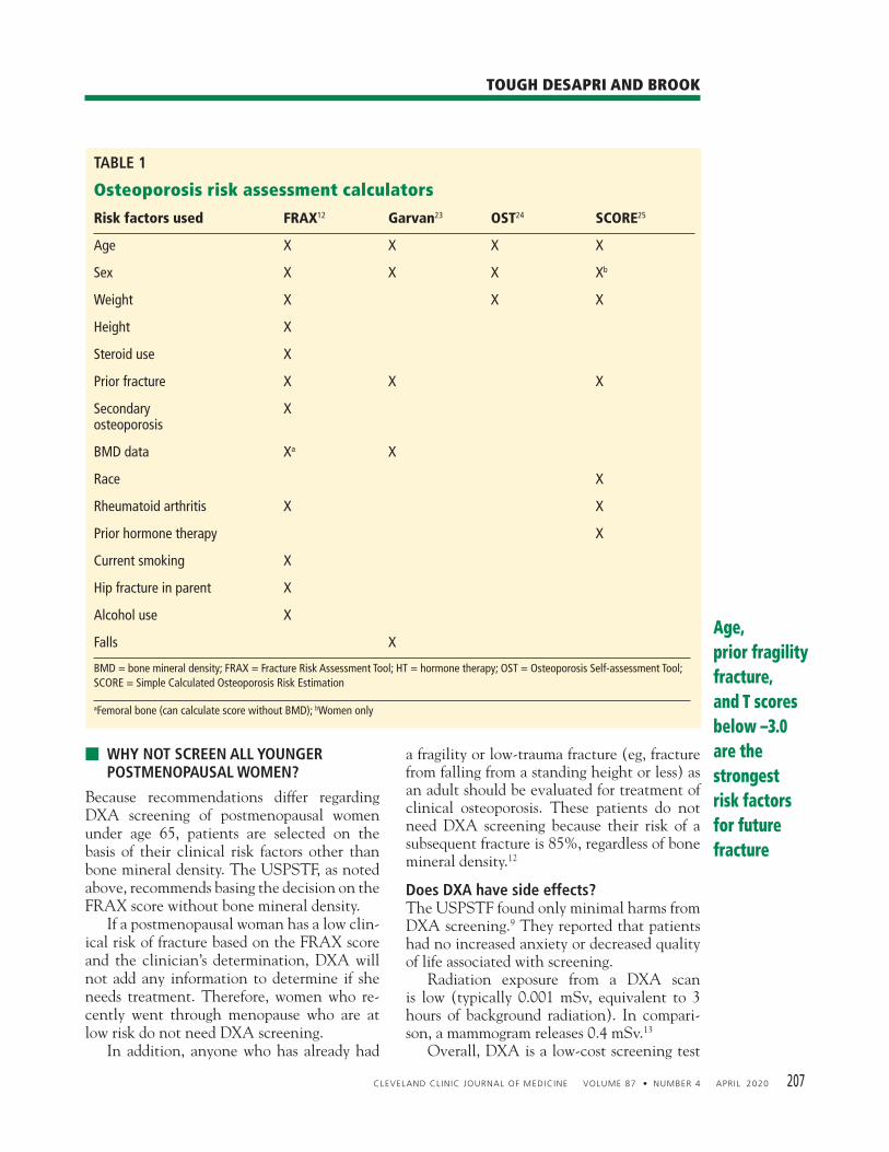
TABLE 1
Osteoporosis risk assessment calculators
Risk factors used FRAX12 Garvan23 OST24 SCORE25
Age X X X X
Sex X X X Xb
Weight X X X
Height X
Steroid use X
Prior fracture X X X
Secondary osteoporosis
X
BMD data Xa X
Race X
Rheumatoid arthritis X X
Prior hormone therapy X
Current smoking X
Hip fracture in parent X
Alcohol use X
Falls X
BMD = bone mineral density; FRAX = Fracture Risk Assessment Tool; HT = hormone therapy; OST = Osteoporosis Self-assessment Tool; SCORE = Simple Calculated Osteoporosis Risk Estimation
aFemoral bone (can calculate score without BMD); bWomen only
208 CLEVELAND CLINIC JOURNAL OF MEDICINE VOLUME 87 • NUMBER 4 APRIL 2020
DXA IN POSTMENOPAUSAL WOMEN
for those who meet the criteria to be screened, but it should not be done in all early post-menopausal women.
■ FRAX IS A VALIDATED TOOL
FRAX11 is a computer-based equation that uses clinical risk factors (and, if available, bone mineral density information) to esti-mate a patient’s 10-year fracture probability. Although it has been validated in the general population, it has some limitations that may cause it to underestimate the fracture risk in postmenopausal women. Its use of yes-or-no responses can limit its clinical application. For example, a patient who smoked cigarettes for 10 years but has quit is considered a non-smoker in FRAX, even though their smoking history could have a substantial effect on their peak bone deposition and rate of bone loss. Some experts suggest using one of the al-ternate risk calculators that include other vari-ables to determine the risk of fracture.14 Table 1 lists the risk variables used in each tool. The Simple Calculated Osteoporosis Risk Estimate (SCORE) tool, for example, ac-counts for hormone therapy and race in its calculation, whereas FRAX does not. In addi-tion, FRAX does not account for falls, which are a major contributor to fractures. Of note, except for FRAX, most of these risk calcula-tors have not been validated in diverse pop-ulations and are not in widespread use. We recommend FRAX because it is an easy-to-use clinical tool and is used around the world, but with caveats, as mentioned above.
■ SHOULD OUR PATIENT UNDERGO DXA?
Our patient is a postmenopausal woman who went through menopause at an average age, does not smoke, has a normal body mass index, and has no personal or family history of os-teoporosis or fracture. She consumes adequate calcium and vitamin D through supplements and diet. Based on her history and physical ex-amination, we assume she achieved a normal peak bone mass before menopause and, thus, has a low risk for fracture. Her FRAX score, calculated without DXA screening, shows a 6% 10-year risk of major osteoporotic fracture, which does not meet the 8.4% threshold for DXA screening.
If she continues to get enough calcium, vitamin D, and exercise, and without any offending agents or conditions that acceler-ate bone loss, she has a low risk of fracture and a very low probability of needing treat-ment. If her clinical situation remains the same, she should undergo DXA screening at age 65. In summary, clinicians can accurately as-sess the fracture risk in younger postmenopaus-al women (ie, before age 65) by performing a comprehensive history and physical examina-tion and combining it with the FRAX tool without a DXA scan.
■ MANAGING LOW BONE MASS
More fractures occur in women with osteope-nia than in those with osteoporosis because many more women have osteopenia, even though their fracture rate is lower.15 There-fore, it is important to judiciously treat low bone mass in patients who meet the criteria for treatment based on their FRAX score and the practitioners’ clinical judgment. The trabecular bone score is an indirect measure of trabecular microarchitecture de-rived from DXA images of the lumbar spine. It provides information about bone quality. A score below 1.200 indicates degraded microar-chitecture. Using a trabecular bone score indepen-dently or in conjunction with a DXA scan or FRAX score can improve fracture predic-tion.16,17 Also, FRAX can be adjusted for this score. More accurate evaluations of bone density and bone quality can help determine which patients with low bone mineral density need treatment. The effi cacy of treatment to reduce frac-ture rates in women at high risk of fracture but without a low T score (−2.5 or below) has not been established. Most FDA-approved thera-pies are indicated for treatment based on bone mineral density.
■ EFFECTIVE AND EMERGING THERAPIES
For postmenopausal women who are candi-dates for pharmacologic treatment based on their fracture risk assessment, there are safe and effective FDA-approved options.
If a womanis at low riskbased on clinical factors, DXA will not add relevant information
CLEVELAND CLINIC JOURNAL OF MEDICINE VOLUME 87 • NUMBER 4 APRIL 2020 209
TOUGH DESAPRI AND BROOK
Hormone therapyHormone therapy has been proven in the large Women’s Health Initiative18 and the Postmenopausal Estrogen/Progestone Inter-ventions trial19 to both prevent osteoporosis and reduce the incidence of fractures (such as vertebral and hip) compared with placebo. In the Million Women Study,20 women who received hormone therapy had a signifi cantly lower risk of any fracture than women who did not. Despite those results, hormone therapy is FDA-approved only for prevention of osteo-porosis, not treatment. It is also recommended for menopause-related vasomotor symptoms and the genitourinary syndrome of meno-pause. Candidates for hormone therapy are pri-marily women under age 60 who are fewer than 10 years past menopause; the risk-benefi t ratio for older women is less favorable because of higher risks of heart disease and stroke.21 It is important to engage the patient in an ac-curate, evidence-based discussion of the risks and benefi ts of hormone therapy. Tissue-selective estrogen complexes can be appropriate options to reduce the fracture risk and prevent osteoporosis. These pair estrogens with selective estrogen-receptor antagonists or agonists, such as conjugated estrogen and bazedoxifene. Selective estrogen-receptor modulators, such as raloxifene, are available in generic form. They may play a dual role of reducing risk of breast cancer and preventing or treat-ing osteoporosis.
AntiresorptivesThe antiresorptive class of medications in-cludes bisphosphonates (oral or intravenous) and denosumab, a subcutaneous monoclonal antibody; both are considered fi rst-line treat-ment for women with osteoporosis. Denosum-ab is indicated for women (and men) with a history of fracture or who are at increased risk of fracture and cannot tolerate other thera-pies. Although effective at reducing the inci-dence of fractures, antiresorptive therapies may increase the risk of osteonecrosis of the
jaw and atypical femoral fractures, especially with prolonged use. Fortunately, these are rare: the incidence rate with 10 years of de-nosumab use is 0.05%,22 and only 0.001% to 0.01% with more than 4 years of oral bisphos-phonate use.23,24
Anabolic drugs The anabolic drugs such as teriparatide, aba-loparatide, and romosozumab build bone mass by stimulating osteoblasts more than osteoclasts. Abaloparatide was studied head-to-head against placebo and teriparatide for 18 months, after which patients received alendronate for 2 years; sequential treatment with abaloparatide followed by alendronate reduced the risk of vertebral, nonvertrbral, clinical, and major osteoporotic fractures and increased bone mineral density.25 Romoso-zumab, a humanized monoclonal antibody to sclerostin, is FDA-approved to treat women at high risk of fracture. It has a dual effect, stimu-lating bone formation and reducing bone re-sorption.
■ CLINICAL BOTTOM LINE
Osteoporosis and osteopenia leading to frac-ture are major causes of morbidity and mortal-ity in postmenopausal women. A DXA scan is considered the best tool to measure bone mass, which can be used to determine the risk of fracture and who may benefi t from treat-ment. For younger postmenopausal women (age 50 to 65), the need for a DXA scan is deter-mined by a thorough history and physical ex-amination, noting any risk factors that con-tribute to bone loss. A DXA scan is indicated if their fracture risk is high (ie, equivalent to that of a woman age 65 or older) based on a FRAX calculation without a bone mineral density measurement. If DXA is not indicat-ed, clinicians should counsel women on ways to prevent bone loss and reduce fracture risks. Conversely, women at the highest risk of fracture are those with a prior adult fragility fracture, regardless of T score. Evaluation and pharmacologic therapy should be strongly rec-ommended in these cases. ■
Antiresorptive drugs may increase the risk of osteonecrosis of the jaw and atypical femoral fractures, especially with prolonged use,but these are rare
210 CLEVELAND CLINIC JOURNAL OF MEDICINE VOLUME 87 • NUMBER 4 APRIL 2020
DXA IN POSTMENOPAUSAL WOMEN
■ REFERENCES 1. Finkelstein JS, Brockwell SE, Mehta V, et al. Bone mineral density
changes during the menopause transition in a multiethnic cohort of women. J Clin Endocrinol Metab 2008; 93(3):861–868.doi:10.1210/jc.2007-1876
2. Anagnostis P, Siolos P, Gkekas NK, et al. Association between age at menopause and fracture risk: a systematic review and meta-analysis. Endocrine 2019; 63(2):213–224. doi:10.1007/s12020-018-1746-6
3. Fan Y, Wei F, Lang Y, Liu Y. Diabetes mellitus and risk of hip fractures: a meta-analysis. Osteoporos Int 2016; 27(1):219–228. doi:10.1007/s00198-015-3279-7
4. Cosman F, de Beur SJ, LeBoff MS, et al; National Osteoporosis Foundation. Clinician’s guide to prevention and treatment of osteo-porosis. Osteoporos Int 2014; 25(10):2359–2381. doi: 10.1007/s00198-014-2794-2. Erratum in: Osteoporos Int 2015l; 26(7):2045–2047. doi:10.1007/s00198-014-2794-2
5. Wright NC, Looker AC, Saag KG, et al. The recent prevalence of osteoporosis and low bone mass in the United States based on bone mineral density at the femoral neck or lumbar spine. J Bone Miner Res 2014; 29(11):2520–2526. doi:10.1002/jbmr.2269
6. Burge R, Dawson-Hughes B, Solomon DH, Wong JB, King A, Tosteson A. Incidence and economic burden of osteoporosis-related fractures in the United States, 2005–2025. J Bone Miner Res 2007; 22(3):465–475. doi:10.1359/jbmr.061113
7. Klotzbuecher CM, Ross PD, Landsman PB, Abbott TA 3rd, Berger M. Patients with prior fractures have an increased risk of future frac-tures: a summary of the literature and statistical synthesis. J Bone Miner Res 2000; 15(4):721–739. doi:10.1359/jbmr.2000.15.4.721
8. Johnell O, Kanis JA, Oden A, et al. Predictive value of BMD for hip and other fractures. J Bone Miner Res 2005; 20(7):1185–1194. doi:10.1359/JBMR.050304
9. US Preventive Services Task Force; Curry SJ, Krist AH, Owens DK, et al. Screening for osteoporosis to prevent fractures: US Preven-tive Services Task Force Recommendation Statement. JAMA 2018; 319(24):2521–2531. doi:10.1001/jama.2018.7498
10. The International Society for Clinical Densitometry. 2015 ISCD offi cial positions—adult. http://www.iscd.org/offi cial-positions/2015-iscd-offi cial-positions-adult/. Accessed October 18, 2019.
11. Centre for Metabolic Bone Diseases, University of Sheffi eld, UK. FRAX Fracture Risk Assessment Tool. https://www.sheffi eld.ac.uk/FRAX/. Accessed October 18, 2019.
12. Kanis JA, Johnell O, De Laet C, et al. A meta-analysis of previous fracture and subsequent fracture risk. Bone 2004; 35(2):375–382. doi:10.1016/j.bone.2004.03.024
13. Radiological Society of North America, Inc. (RSNA). Radiation dose in X-ray and CT exams. https://www.radiologyinfo.org/en/info.cfm?pg=safety-xray. Accessed January 23, 2020.
14. Crandall CJ, Larson J, Gourlay ML, et al. Osteoporosis screening in postmenopausal women 50 to 64 years old: comparison of US Pre-ventive Services Task Force strategy and two traditional strategies in
the Women’s Health Initiative. J Bone Miner Res 2014; 29(7):1661–1666. doi:10.1002/jbmr.2174
15. Siris ES, Chen YT, Abbott TA, et al. Bone mineral density thresholds for pharmacological intervention to prevent fractures. Arch Intern Med 2004; 164(10):1108–1112. doi:10.1001/archinte.164.10.1108
1 6. Hans D, Goertzen AL, Krieg MA, Leslie WD. Bone microarchitecture assessed by TBS predicts osteoporotic fractures independent of bone density: the Manitoba study. J Bone Miner Res 2011; 26(11):2762–2769. doi:10.1002/jbmr.499
17. Leslie WD, Johansson H, Kanis JA, et al. Lumbar spine texture en-hances 10-year fracture probability assessment. Osteoporos Int 2014; 25(9):2271–2277. doi:10.1007/s00198-014-2761-y
18. Rossouw JE, Anderson GL, Prentice RL, et al; Writing Group for the Women’s Health Initiative Investigators. Risks and benefi ts of es-trogen plus progestin in healthy postmenopausal women: principal results From the Women’s Health Initiative randomized controlled trial. JAMA 2002; 288(3):321–333. doi:10.1001/jama.288.3.321
19. Greendale GA, Wells B, Marcus R, Barrett-Connor E. How many women lose bone mineral density while taking hormone replace-ment therapy? Results from the Postmenopausal Estrogen/Proges-tin Interventions Trial. Arch Intern Med 2000; 160(20):3065–3071. doi:10.1001/archinte.160.20.3065
20. Banks E, Beral V, Reeves G, Balkwill A, Barnes I; Million Women Study Collaborators. Fracture incidence in relation to the pattern of use of hormone therapy in postmenopausal women. JAMA 2004; 291(18):2212–2220. doi:10.1001/jama.291.18.2212
21. Management of osteoporosis in postmenopausal women: 2010 position statement of the North American Menopause Society. Menopause 2010; 17(1):25–54. doi:10.1097/gme.0b013e3181c617e6
22. Watts NB, Grbic JT, Binkley N, et al. Invasive oral procedures and events in postmenopausal women with osteoporosis treated with denosumab for up to 10 years. J Clin Endocrinol Metab 2019; 104(6):2443–2452. doi:10.1210/jc.2018-01965
23. Khosla S, Burr D, Cauley J, et al; American Society for Bone and Mineral Research. Bisphosphonate-associated osteonecrosis of the jaw: report of a task force of the American Society for Bone and Mineral Research. J Bone Miner Res 2007; 22(10):1479–1491. doi:10.1359/jbmr.0707onj
24. Khan AA, Morrison A, Hanley DA, et al. Diagnosis and management of osteonecrosis of the jaw: a systematic review and international consensus. J Bone Miner Res 2015; 30(1):3–23. doi:10.1002/jbmr.2405
25. Bone HG, Cosman F, Miller PD, et al. ACTIVExtend: 24 Months of alendronate after 18 months of abaloparatide or placebo for postmenopausal osteoporosis. J Clin Endocrinol Metab 2018; 103(8):2949–2957. doi:10.1210/jc.2018-00163
Address: Kristi Tough DeSapri, MD, CCD, NCMP, Assistant Professor of Ob/GYN and Internal Medicine, Feinberg School of Medicine, Northwest-ern, 259 East Erie Street, Suite 2450 Chicago, IL 60611;[email protected]
CGRP antagonists for decreasingmigraine frequency:New options, long overdue
CLEVELAND CLINIC JOURNAL OF MEDICINE VOLUME 87 • NUMBER 4 APRIL 2020 211
T he cornerstone of preventive migraine treatment has long been drugs intended
for other diseases—epilepsy, depression, and hypertension. But in 2018, the US Food and Drug Administration (FDA) approved 3 new drugs—erenumab, galcanezumab, and freman-ezumab—specifi cally for decreasing the fre-quency of migraine attacks. A fourth, eptinez-umab, was approved February 22, 2020. These monoclonal antibodies against calcitonin gene-related peptide (CGRP) or its receptors are the fi rst preventive medications to target the pathophysiology of migraine.
See related editorial, page 219
The new drugs represent an exciting new frontier in headache medicine that is long over-due. Although they don’t seem to be more ef-fective than current drugs, they have long half-lives, permitting monthly or even quarterly dosing, and fewer adverse effects, which may improve adherence. In addition, they carry no contraindications for patients with liver disease, kidney disease, stroke, or coronary artery disease. They also have no known signifi cant drug-drug interactions. Their primary disadvantage is cost (about $700 per month), although insurance may pay for them, and the manufacturers have assistance programs (Table 1).
■ NEED FOR MORE OPTIONS
Headache disorders, treated as early as 1200 bce by the ancient Egyptians, affect nearly half of the world’s adult population.1,2 In the United States alone, migraine affects nearly 40 million people and is one of the most common complaints ad-
CURRENT DRUG THERAPY
doi:10.3949/ccjm.87a.19048
ABSTRACTThe cornerstone of preventive migraine treatment has long been drugs developed for other diseases such as epilepsy, depression, and hypertension. But a new set of drugs is available for preventing migraine attacks: ere-numab, galcanezumab, fremanezumab, and eptinezumab. These monoclonal antibodies target calcitonin gene-relat-ed peptide (CGRP) or its receptors, each a key molecule in the pathophysiology of migraine.
KEY POINTSMigraine is common, affecting nearly 40 million people in the United States.
In clinical trials, CGRP antagonists have been superior to placebo and similar in effi cacy to current prophylactic treatments in terms of reducing the frequency of head-aches.
These agents have long half-lives, permitting monthly or even quarterly dosing, and favorable side effect profi les compared with currently available oral therapies. This may improve adherence.
The new drugs are an exciting new frontier in headache medicine that is long overdue. However, the approach to migraine management must remain a combination of fi nding effective treatment and implementing patient-specifi c lifestyle changes for the best possible outcome.
Julia Bucklan, DOCenter for General Neurology, Department of Neurology, Cleveland Clinic; Clinical Instructor, Cleveland Clinic Lerner College of Medicine of Case Western Reserve University, Cleveland, OH
Zubair Ahmed, MDCenter for Neuro-Restoration, Department of Neurology, Cleveland Clinic; Assistant Professor, Cleveland Clinic Lerner College of Medicine of Case Western Reserve University, Cleveland, OH

212 CLEVELAND CLINIC JOURNAL OF MEDICINE VOLUME 87 • NUMBER 4 APRIL 2020
MIGRAINE MANAGEMENT
dressed by primary care physicians, emergency physicians, and neurologists. It is associated with decreased function in an otherwise healthy and productive demographic group3–5 and is the leading cause of healthy life-years lost as a result of disability from ages 15 to 49.6 Drugs that have long been used in mi-graine prophylaxis7 have many adverse effects and need to be taken daily, which can lead to nonadherence; more than 80% of patients stop taking them within 1 year.8
■ CGRP IS A KEY MOLECULE IN MIGRAINE
Migraine is a multifactorial disorder with complex interactions between multiple pre-disposing genetic and modulating nongenetic factors.9
The current understanding of migraine is that a wave of neuronal and glial depolariza-tion activates meningeal nociceptors inner-vated by the trigeminovascular system. When these perivascular afferent fi bers are activat-ed, the signal travels through the trigeminal ganglion to neurons in the trigeminocervical complex, using CGRP as the prominent neu-rotransmitter. This leads to symptoms such as cutaneous allodynia, neck pain, photophobia, phonophobia, and osmophobia. Once this sig-nal reaches the visual cortex, it alters visual perception, resulting in double vision, change in color saturation, and blurred vision.9
The discovery that using a peripherally ac-tive biologic, onabotulinumtoxinA, could be effective in migraine prophylaxis led to further investigation of the mechanism of action.10 It
TABLE 1
Current calcitonin gene-related peptide (CGRP) antagonists
Drug MechanismDosing and frequency a
Most common adverse effects a
Average wholesale price andPharmaceutical Assistance Program a
Erenumab CGRP receptor antagonist
Migraine: 70 or 140 mg subcutaneously, monthly
Injection site erythema or pain, 5%–6%
Constipation, 3%
$690 per month (regardless of dose)
If commercial insurance plan does not cover or requires prior authorization, patients are eligible for 12 doses over 24 months with a $5 copay card per month; maximum benefi t $2,700 annually
Fremanezumab CGRP ligand antagonist
Migraine: 225 mg monthly or 675 mg every 3 months subcutaneously
Injection site reaction, 45%
$690 per 225-mg syringe
Patients with commercial insurance plan are eligible for 12 months of treatment with a $0 copay card; there is no annual maximum benefi t; with electronic coupon, copay is $20
Galcanezumab CGRP ligand antagonist
Migraine:240 mg, then 120 mgper monthsubcutaneously
Cluster headache: 300 mg at onset of cluster period, then monthly until end of cluster headache
Injection sitereaction, 18%
$690 per 120-mg autoinjector
If commercial insurance plan does not cover or requires prior authorization, patients are eligible for a $0 copay card; maximum coverage is $4,900 annually
As of 2020, this benefi t is available only after prior authorization is approved by insurance
Eptinezumab CGRP ligand antagonist
100 mg/mL or 300 mg/mL via infusion every 90 days
Nausea, 1.6%Fatigue, 1.4%
$1,495 per infusion ($5,980 per year)
a Information from product package inserts and personal communication with Cleveland Clinic Adherence Specialty Pharmacy.
CLEVELAND CLINIC JOURNAL OF MEDICINE VOLUME 87 • NUMBER 4 APRIL 2020 213
BUCKLAN AND AHMED
is now understood that onabotulinumtoxinA inhibits CGRP release from peripheral neuro-nal C fi bers and does not cross the blood-brain barrier.11 CGRP, discovered in 1982, is a large mol-ecule.8 It binds 2 major receptors: calcitonin receptor-like receptor and receptor activity-modifying protein 1.12 This leads to signaling that can cause vasodilation or release of neu-rotransmitters or cytokines, in turn causing neurogenic infl ammation and increased neu-ronal excitability.12 CGRP receptors are found at all of the known central and peripheral sites involved in migraine pathogenesis, including the hypo-thalamus and parabrachial nucleus, and CGRP levels are elevated during migraine attacks and lower between attacks.12 Studies in animals fi rst showed that stimulation of the trigeminal ganglion was associated with increased blood fl ow and release of CGRP, which could be in-hibited by sumatriptan or dihydroergotamine.11 Studies in humans showed that sumatriptan, in addition to relieving migraine, lowered CGRP levels in the internal jugular vein.13 CGRP has also been shown to induce migraine-like symp-toms after intravenous infusion.14 These observations led researchers to de-velop drugs that target and block either the CGRP ligand itself or the receptors upon which it acts.
■ CGRP ANTAGONISTS: A NEW CLASS OF DRUGS
The fi rst CGRP antagonists to be studied were small molecules, with names ending in the suffi x “-gepant.” These so-called gepants block CGRP receptors, and 6 were found to be effective in acute treatment of episodic mi-graine.15–20 However, their development was discontinued due to reports of hepatotoxicity. Next to be developed were monoclonal antibodies targeting CGRP. These agents are metabolized by the reticuloendothelial system and, as a result, bypass hepatic metabolism; to date, no adverse effects on the liver have been reported.10,21 Further, the current inject-able antibodies are not thought to be con-traindicated in patients with coronary artery, cerebrovascular, peripheral vascular, or kidney disease.10
■ DEFINITIONS
Episodic migraine is defi ned as having fewer than 15 headache days per month fulfi lling di-agnostic criteria for migraine.22 Chronic migraine is defi ned as headaches on 15 or more days per month for 3 months or more in a patient with a preexisting diagnosis of migraine. Of the total headache days, at least 8 days per month should meet migraine criteria.22
■ EFFICACY OF CGRP ANTAGONISTS
Clinical trials of the monoclonal antibodies (Table 2)23–33 have found them to be supe-rior to placebo and similar in effi cacy to cur-rent prophylactic treatments for episodic and chronic migraine.34 Roughly half of patients receiving these drugs achieved at least a 50% reduction in the number of headache days per month, compared with roughly one-fourth of patients receiving placebo. The new drugs have also been shown to be tolerable and safe, with no signifi cant effects on blood pressure or peripheral vasoconstriction.35
ErenumabUnlike galcanezumab and fremanezumab, ere-numab targets the canonical CGRP receptor rather than the CGRP ligand itself. There are 2 available doses, 70 mg and 140 mg, which patients give themselves once a month at home using a preloaded subcutane-ous autoinjector.10 In episodic migraine. Three trials looked at 50% responder rates and mean decrease in monthly migraine days with use of erenu-mab in patients with episodic migraine (Table 2).23–25 Results were reliably better with ere-numab than with placebo, including in groups with so-called refractory migraine for whom 2 to 4 oral preventive therapies had failed.25
In chronic migraine, the results were simi-lar.26 Adverse effects noted included injection site pain (reported by 4% of patients receiving active treatment), constipation (4% of those on 140 mg), and muscle spasm (4% of those on 140 mg).26
Erenumab received FDA approval for pre-vention of migraine on May 17, 2018.
FremanezumabFremanezumab targets the CGRP ligand rather than the receptor. It can be taken as a
The CGRP antagonists are an exciting new frontier in headache medicine
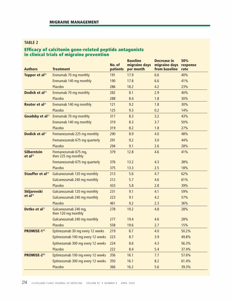
214 CLEVELAND CLINIC JOURNAL OF MEDICINE VOLUME 87 • NUMBER 4 APRIL 2020
MIGRAINE MANAGEMENT
TABLE 2
Effi cacy of calcitonin gene-related peptide antagonistsin clinical trials of migraine prevention
Authors TreatmentNo. of patients
Baseline migraine days per month
Decrease in migraine days from baseline
50% response rate
Tepper et al26 Erenumab 70 mg monthly 191 17.9 6.6 40%
Erenumab 140 mg monthly 190 17.8 6.6 41%
Placebo 286 18.2 4.2 23%
Dodick et al24 Erenumab 70 mg monthly 282 8.1 2.9 40%
Placebo 288 8.4 1.8 30%
Reuter et al25 Erenumab 140 mg monthly 121 9.2 1.8 30%
Placebo 125 9.3 0.2 14%
Goadsby et al23 Erenumab 70 mg monthly 317 8.3 3.2 43%
Erenumab 140 mg monthly 319 8.3 3.7 50%
Placebo 319 8.2 1.8 27%
Dodick et al27 Fremanezumab 225 mg monthly 290 8.9 4.0 48%
Fremanezumab 675 mg quarterly 291 9.2 3.0 44%
Placebo 294 9.1 2.6 28%
Silberstein et al28
Fremanezumab 675 mg, then 225 mg monthly
379 12.8 4.6 41%
Fremanezumab 675 mg quarterly 376 13.2 4.3 38%
Placebo 375 13.3 2.5 18%
Stauffer et al29 Galcanezumab 120 mg monthly 213 5.6 4.7 62%
Galcanezumab 240 mg monthly 212 5.7 4.6 61%
Placebo 433 5.8 2.8 39%
Skljarevski et al30
Galcanezumab 120 mg monthly 231 9.1 4.1 59%
Galcanezumab 240 mg monthly 223 9.1 4.2 57%
Placebo 461 9.2 2.3 36%
Detke et al31 Galcanezumab 240 mg, then 120 mg monthly
278 19.2 4.8 28%
Galcanezumab 240 mg monthly 277 19.4 4.6 28%
Placebo 558 19.6 2.7 15%
PROMISE-132 Eptinezumab 30 mg every 12 weeks 219 8.7 4.0 50.2%
Eptinezumab 100 mg every 12 weeks 223 8.7 3.9 49.8%
Eptinezumab 300 mg every 12 weeks 224 8.6 4.3 56.3%
Placebo 222 8.4 5.4 37.4%
PROMISE-233 Eptinezumab 100 mg every 12 weeks 356 16.1 7.7 57.6%
Eptinezumab 300 mg every 12 weeks 350 16.1 8.2 61.4%
Placebo 366 16.2 5.6 39.3%
CLEVELAND CLINIC JOURNAL OF MEDICINE VOLUME 87 • NUMBER 4 APRIL 2020 215
BUCKLAN AND AHMED
monthly subcutaneous injection of 225 mg or as a quarterly injection of 675 mg. In episodic migraine. A phase 3 trial in episodic migraine showed a decrease in mean monthly headache days and increases in the 50% responder rate and 75% responder rate with ei-ther dose compared with placebo (P < .0001).27
In chronic migraine. The same trial also compared fremanezumab and placebo in pa-tients with chronic migraine.36 The number of days with headache of moderate to severe in-tensity was reduced by 4.2 days in the placebo group and by 6 days in both a group receiving 225 mg monthly and a group receiving 675 mg quarterly.28 In a separate study,37 investigators found that patients noted an improvement as early as 1 week from initiation of therapy in both dose regimens. Fremanezumab received FDA approval for prevention of migraine on September 14, 2018.
GalcanezumabGalcanezumab also targets the CGRP ligand. It is given subcutaneously once a month with an autoinjector or prefi lled syringe in a rec-ommended monthly dose of 120 mg after an initial loading dose of 240 mg. In episodic migraine. Two 6-month trials compared galcanezumab monthly injections of galcanezumab 120 mg, galcanezumab 240 mg, and placebo.29,30 Both studies demonstrated a reduction of migraine days and an increase in 50% responder rate superior to placebo.36 In-terestingly, about 17% of patients had a 100% reduction in mean migraine days. This was seen most commonly in the last 3 months of the trials and was statistically signifi cant com-pared with placebo (P < .001).10
In chronic migraine. In a phase 3 trial, galcanezumab showed a signifi cant decrease in mean monthly migraine days compared with placebo. Also, differences in the 50% and 75% responder rates were statistically signifi cant in each treatment group compared with placebo (P < .001). Similar to the episodic migraine trial, 11.5% of galcanezumab recipients in the chronic migraine trial also noted 100% reduc-tion in mean migraine days, again noted most commonly in the last 3 months of the clinical trial (P < .001).31
This drug received FDA approval for pre-vention of migraine on September 27, 2018.
EptinezumabEptinezumab, a monoclonal antibody against the CGRP ligand, is given intravenously, where-as the other CGRP monoclonal antibodies are given subcutaneously, In episodic migraine. In a 3-month phase 3 trial,32 quarterly infusions of eptinezu mab 300 mg signifi cantly reduced the number of mean monthly migraine days. Secondary end points included the 75% responder rate at week 12 (49.8% in the 100-mg arm, P = .0085; and 56.3% in the 300-mg arm, P < .0001). The clinical trial also demonstrated rapid onset of effect with a reduction in the likelihood of migraine within 24 hours of infusion. Before treatment, 58% of the participants were likely to have a migraine on any given day. This de-clined by 27% in the placebo group, 51% in those who received 100 mg, and 53% in those who received 300 mg (P < .0001 for both doses). At a 300-mg dose given quarterly, the 75% responder rate was maintained for up to 1 year.10
In chronic migraine, a phase 3 clini-cal trial showed a signifi cant reduction in mean monthly migraine days compared with placebo at doses of 100 mg and 300 mg.33 This drug received FDA approval February 22, 2020.
■ A PRAGMATIC APPROACH TO ANTI-CGRP DRUG THERAPY
The approach to migraine management must remain a combination of cost-effective fi rst- and second-line treatments, generally reserv-ing CGRP monoclonal antibodies for patients for whom these options fail. All pharmaco-logic treatments should be accompanied by education and specifi c lifestyle changes for the best possible outcome. The Quality Standards Subcommittee of the American Academy of Neurology and the American Headache Society7 in 2012 re-viewed the evidence and ranked the migraine preventive therapies available in the United States at that time according to the evidence of their effi cacy. Level A medications, ie, those rated as having “established effi cacy,” were:• The antiepileptic drugs divalproex sodium,
sodium valproate, and topiramate
The fi rst CGRP antagonists to be studied were small molecules, termed gepants
216 CLEVELAND CLINIC JOURNAL OF MEDICINE VOLUME 87 • NUMBER 4 APRIL 2020
MIGRAINE MANAGEMENT
All drugtreatments should beaccompaniedby educationand specifi clifestylechanges
• The beta-blockers metoprolol, proprano-lol, and timolol
• The tripan frovatriptan (for short-term prophylaxis).
Level B medications, ie, those that are “probably effective,” were:• The antidepressants amitriptyline and
vanlafaxine• The beta-blockers atenolol and nadolol• The triptans naratriptan and zolmitriptan. Level C medications, ie, “possibly effec-tive,” were:• The angiotensin-converting enzyme in-
hibitor lisinopril• The angiotensin II receptor blocker can-
desartan• The alpha-agonists clonidine and guanfa-
cine• The antiepileptic drug carbamazepine• The beta-blockers nebivolol and pindolol• The antihistamine cryptoheptadine. While no formal guidelines exist for deciding whether anti-CGRP drugs would be appropriate for specifi c patients, the American Headache Society has offered general recommendations38,39 based on the frequency of migraine.
Patients in whom CGRP antagonists can be considered• Those with 4 to 7 migraine days per
month who have been unable to tolerate a 6-week trial of at least 2 oral preventive medications with level A or B evidence (see above).7,38,39 In addition, patients should also have at least moderate disabil-ity on the Migraine Disability Assessment Scale or the Headache Impact Test 6, both of which are used to assess functional im-pairment secondary to migraine.
• Those with 8 to 14 headaches per month who cannot tolerate a 6-week trial of at least 2 oral preventive drugs with level A or B evidence (no need to demonstrate functional impairment).
• Those with 15 or more headaches per month (ie, chronic migraine) if at least 2 preventive medications with level A or B evidence have failed or if onabotulinum-toxinA has produced an inadequate re-sponse after at least 2 administrations or has caused adverse effects precluding fur-ther use.
At this time, not enough data exist on the safety of this class of medications in pregnant patients or children. The fi ndings from clinical trials suggest that if a patient is going to respond to CGRP monoclonal antibody therapy, it should hap-pen within the fi rst 3 months, often as early as 1 month after starting. If migraines continue unabated in this period, it is reasonable to dis-continue the medication.
■ GEPANTS REVISITED
Gepants have been revisited in clinical trials over the past 5 years for both abortive and pre-ventive treatment.40
Ubrogepant for acute migraine treatmentA multicenter, randomized, double-blind, placebo-controlled clinical trial of ubrogepant for the acute treatment of migraine showed a statistically signifi cant improvement in rates of pain freedom 2 hours post-dose at 25 mg (P = .013), 50 mg (P = .020), and 100 mg (P = .003).41 Adverse effects were similar to those with placebo and included dry mouth, nausea, fatigue, dizziness, and somnolence. There were no observed liver function test elevations as were seen with previous gepant trials. Ubrogepant received FDA approval on December 23, 2019.
RimegepantRimegepant has also been studied for the acute treatment of migraine in a double-blind, ran-domized, placebo-controlled trial.20,21 Patients were randomized to receive placebo, sumatrip-tan, or rimegepant. The primary outcome was percentage of patients who were free of pain 2 hours post-dose. Sumatriptan 100 mg and rimegepant 75 mg, 150 mg, and 300 mg were all signifi cant-ly more effective than placebo (P < .007). Rimegepant was as effective as sumatriptan. No chest discomfort or paresthesias were re-ported with rimegepant as they were with su-matriptan. A prospective multicenter, open-label, long-term safety study is under way.
AtogepantAtogepant, another oral gepant, has been evalu-ated for prevention of episodic migraine. Mean headache days were reduced by 4.23 days per
CLEVELAND CLINIC JOURNAL OF MEDICINE VOLUME 87 • NUMBER 4 APRIL 2020 217
BUCKLAN AND AHMED
month with atogepant 40 mg twice daily, com-pared with 2.85 days with placebo (P = .0034). There was no evidence of hepatotoxicity.42
■ OTHER TYPES OF HEADACHE
Cluster headacheEpisodic cluster headache is defi ned as cluster headache attacks occurring in periods lasting from 7 days to 1 year, separated by pain-free periods lasting at least 3 months. Chronic cluster headache, in contrast, is defi ned as cluster headache attacks occurring for 1 year or longer without remission, or with remission periods lasting less than 3 months. In June 2019, galcanezumab received FDA approval for treatment of episodic cluster head-aches. For treatment, galcanezumab 300 mg is administered as 3 consecutive injections of 100 mg at the onset of a cluster period and then monthly until the end of the cluster period. In clinical trials,43–46 galcanezumab signifi -cantly reduced mean cluster attack frequency compared with placebo, with more than 70% of patients experiencing at least a 50% reduction in weekly cluster headache attack frequency by week 3. However, while trials showed galcan-ezumab to be effective in episodic cluster, this
was not true for chronic cluster. Fremanezumab was also not effective in the prevention of chronic cluster headache compared with placebo.10
Persistent posttraumatic headacheData from rodent models of concussion sug-gest that cephalic tactile pain hypersensitiv-ity improves with administration of murine CGRP antagonists.47 Fremanezumab is cur-rently being studied for the prevention of per-sistent posttraumatic headache.48
Medication-overuse headachePatients with medication-overuse headache may also benefi t from anti-CGRP monoclo-nal antibodies. Both erenumab and freman-ezumab have shown effi cacy in treating the subgroup of chronic migraine patients with medication-overuse headache.49–51 Erenumab 70 mg led to a reduction of 5.2 migraine days per month, and 140 mg had a reduction of 5.4 days, compared with a reduction of 3.5 days with placebo in patients with medication- overuse headache (P < .001).48 Erenumab is also being considered for eval-uation in pediatric patients with chronic mi-graine.52 ■
■ REFERENCES 1. Critchley M. Migraine from Cappadocia to Queen Square. In: Smith R, ed.
Background to Migraine. London, UK: Heinemann, 1967: 28–39. 2. Stovner LJ, Hagen K, Jensen R, et al. The global burden of headache: a
documentation of headache prevalence and disability worldwide. Cephalal-gia 2007; 27(3):193–210. doi:10.1111/j.1468-2982.2007.01288.x
3. Becker WJ, Findlay T, Moga C, Scott NA, Harstall C, Taenzer P. Guideline for primary care management of headache in adults. Can Fam Physician 2015 ;61(8):670–679. pmid:26273080
4. Cerbo R, Villani V, Bruti G, Di Stani F, Mostardini C. Primary headache in emergency department: prevalence, clinical features and therapeutical ap-proach. J Headache Pain 2005; 6(4):287–289. doi:10.1007/s10194-005-0210-1
5. Gracia-Naya M. The importance of headaches in neurology clinics. Study groups of neurologists of Aragon. Rev Neurol 1999; 29(5):393–396. Spanish. pmid:10584239
6. Steiner TJ, Stovner LJ, Vos T, Jensen R, Katsarava Z. Migraine is fi rst cause of disability in under 50s: will health politicians now take notice? J Headache Pain 2018; 19(1):17. doi:10.1186/s10194-018-0846-2
7. Silberstein SD, Holland S, Freitag F, et al; Quality Standards Subcommit-tee of the American Academy of Neurology and the American Headache Society. Neurology 2012; 78(17):1337–1345. Erratum in Neurology 2013; 80(9):871. doi:10.1212/WNL.0b013e3182535d20
8. Hepp Z, Dodick DW, Varon SF, Gillard P, Hansen RN, Devine EB. Adherence to oral migraine-preventive medications among patients with chronic migraine. Cephalalgia 2015; 35(6):478–488. doi:10.1177/0333102414547138
9. Ferrari MD, Klever RR, Terwindt GM, Ayata C, van den Maagdenberg AM. Migraine pathophysiology: lessons from mouse models and human genet-ics. Lancet Neurol 2015; 14(1):65–80. doi:10.1016/S1474-4422(14)70220-0
10. Tepper SJ. History and review of anti-calcitonin gene-related peptide (CGRP) therapies: from translational research to treatment. Headache 2018;
58(suppl 3):238–275. doi:10.1111/head.13379 11. Zhang X, Strassman AM, Novack V, Brin MF, Burstein R. Extracranial
injections of botulinum neurotoxin type A inhibit intracranial meningeal nociceptors’ responses to stimulation of TRPV1 and TRPA1 channels: are we getting closer to solving this puzzle? Cephalalgia 2016; 36(9):875–886. doi:10.1177/0333102416636843
12. Benarroch EE. CGRP: sensory neuropeptide with multiple neurologic impli-cations. Neurology 2011; 77(3):281–287. doi:10.1212/WNL.0b013e31822550e2
13. Goadsby PJ, Edvinsson L. Examination of the involvement of neuropeptide Y (NPY) in cerebral autoregulation using the novel NPY antagonist PP56. Neuropeptides 1993; 24(1):27–33. doi:10.1016/0143-4179(93)90037-b
14. Lassen LH, Haderslev PA, Jacobsen VB, Iversen HK, Sperling B, Olesen J. CGRP may play a causative role in migraine. Cephalalgia 2002; 22(1):54–61. doi:10.1046/j.1468-2982.2002.00310.x
15. Olesen J, Diener HC, Husstedt IW, et al; BIBN 4096 BS Clinical Proof of Concept Study Group. Calcitonin gene–related peptide receptor antagonist BIBN 4096 BS for the acute treatment of migraine. N Engl J Med 2004; 350(11):1104–1110. doi:10.1056/NEJMoa030505
16. Diener HC, Barbanti P, Dahlöf C, Reuter U, Habeck J, Podhorna J. BI 44370 TA, an oral CGRP antagonist for the treatment of acute migraine attacks: results from a phase II study. Cephalalgia 2011; 31(5):573–584. doi:10.1177/0333102410388435
17. Ho TW, Ferrari MD, Dodick DW, et al. Effi cacy and tolerability of MK-0974 (telcagepant), a new oral antagonist of calcitonin gene-related peptide receptor, compared with zolmitriptan for acute migraine: a randomised, placebo-controlled, parallel-treatment trial. Lancet 2008; 372(9656):2115–2123. doi:10.1016/S0140-6736(08)61626-8
18. Connor KM, Aurora SK, Loeys T, et al. Long-term tolerability of telcagepant for acute treatment of migraine in a randomized trial. Headache 2011; 51(1):73–84. doi:10.1111/j.1526-4610.2010.01799.x
218 CLEVELAND CLINIC JOURNAL OF MEDICINE VOLUME 87 • NUMBER 4 APRIL 2020
MIGRAINE MANAGEMENT
19. Hewitt DJ, Aurora SK, Dodick DW, et al. Randomized controlled trial of the CGRP receptor antagonist MK-3207 in the acute treatment of migraine. Cephalalgia 2011; 31(6):712–722. doi:10.1177/0333102411398399
20. Marcus R, Goadsby PJ, Dodick D, Stock D, Manos G, Fischer TZ. BMS-927711 for the acute treatment of migraine: a double-blind, randomized, placebo controlled, dose-ranging trial. Cephalalgia 2014; 34(2):114–125. doi:10.1177/0333102413500727
21. Dodick DW. CGRP ligand and receptor monoclonal antibodies for migraine prevention: evidence review and clinical implications. Cephalalgia 2019; 39(3):445–458. doi:10.1177/0333102418821662
22. Headache Classifi cation Committee of the International Headache Society (IHS). The International Classifi cation of Headache Disorders, 3rd edition. Cephalalgia 2018; 38(1):1–211. doi:10.1177/0333102417738202
23. Goadsby PJ, Reuter U, Hallström Y, et al. A controlled trial of erenumab for episodic migraine. N Engl J Med 2017; 377(22):2123–2132. doi:10.1056/NEJMoa1705848
24. Dodick DW, Ashina M, Brandes JL, et al. ARISE: a phase 3 randomized trial of erenumab for episodic migraine. Cephalalgia 2018; 38(6):1026–1037. doi:10.1177/0333102418759786
25. Reuter U, Goadsby PJ, Lanteri-Minet M, et al. Effi cacy and tolerability of erenumab in patients with episodic migraine in whom two-to-four previ-ous preventive treatments were unsuccessful: a randomised, double-blind, placebo-controlled, phase 3b study. Lancet 2018; 392(10161):2280–2287. doi:10.1016/S0140-6736(18)32534-0
26. Tepper S, Ashina M, Reuter U, et al. Safety and effi cacy of erenumab for preventive treatment of chronic migraine: a randomised, double-blind, placebo-controlled phase 2 trial. Lancet Neurol 2017; 16(6):425–434. doi:10.1016/S1474-4422(17)30083-2
27. Dodick DW, Silberstein SD, Bigal ME, et al. Effect of fremanezumab compared with placebo for prevention of episodic migraine: a randomized clinical trial. JAMA 2018; 319(19):1999–2008. doi:10.1001/jama.2018.4853
28. Silberstein SD, Dodick DW, Bigal ME, et al. Fremanezumab for the preven-tive treatment of chronic migraine. N Engl J Med 2017; 377(22):2113–2122. doi:10.1056/NEJMoa1709038
29. Stauffer VL, Dodick DW, Zhang Q, Carter JN, Ailani J, Conley RR. Evaluation of galcanezumab for the prevention of episodic migraine: the EVOLVE-1 randomized clinical trial. JAMA Neurol 2018; 75(9):1080–1088. doi:10.1001/jamaneurol.2018.1212
30. Skljarevski V, Matharu M, Millen BA, Ossipov MH, Kim BK, Yang JY. Effi cacy and safety of galcanezumab for the prevention of episodic migraine: results of the EVOLVE-2 phase 3 randomized controlled clinical trial. Cephalalgia 2018; 38(8):1442–1454. doi:10.1177/0333102418779543
31. Detke HC, Goadsby PJ, Wang S, Friedman DI, Selzler KJ, Aurora SK. Gal-canezumab in chronic migraine: the randomized, double-blind, placebo-controlled REGAIN study. Neurology 2018; 91(24):e2211–e2221. doi:10.1212/WNL.0000000000006640
32. Saper J, Lipton R, Kudrow D, et al. Primary results of PROMISE-1 (Preven-tion Of Migraine via Intravenous eptinezumab Safety and Effi cacy–1) trial: a phase 3, randomized, double-blind, placebo-controlled study to evaluate the effi cacy and safety of eptinezumab for prevention of frequent episodic migraines (S20.001). Neurology 2018; 90(suppl 15) S20.001.
33. Kudrow D, Lipton R, Silberstein S, et al. Eptinezumab for prevention of chronic migraine: results of 2 infusions in the phase 3 PROMISE-2 (Preven-tion of Migraine via Intravenous Eptinezumab Safety and Effi cacy02) trial (P2.10-006). Neurology 2019; 92(15 suppl)P2.10-006.
34. Deen M, Correnti E, Kamm K, et al; European Headache Federation School of Advanced Studies (EHF-SAS). Blocking CGRP in migraine patients – a review of pros and cons. J Headache Pain 2017; 18(1):96. doi:10.1186/s10194-017-0807-1
35. Bigal ME, Walter S, Rapoport AM. Calcitonin gene-related peptide (CGRP) and migraine current understanding and state of development. Headache 2013; 53(8):1230–1244. doi:10.1111/head.12179
36. Bigal ME, Dodick DW, Krymchantowski AV, et al. TEV-48125 for the preven-tive treatment of chronic migraine: effi cacy at early time points. Neurology 2016; 87(1):41–48. doi:10.1212/WNL.0000000000002801
37. Silberstein SD, Rapoport AM, Loupe PS, et al. The effect of beginning treatment with fremanezumab on headache and associated symptoms in the randomized phase 2 study of high frequency episodic migraine: post-hoc analyses on the fi rst 3 weeks of treatment: headache. Headache 2019; 59(3):383–393. doi:10.1111/head.13446
38. American Headache Society. The American Headache Society position statement on integrating new migraine treatments into clinical practice. Headache 2019; 59(1):1–18. doi:10.1111/head.13456
39. Loder E, Burch R, Rizzoli P. The 2012 AHS/AAN guidelines for prevention of episodic migraine: a summary and comparison with other recent clinical practice guidelines. Headache 2012; 52(6):930–945. doi:10.1111/j.1526-4610.2012.02185.x
40. Lambru G, Andreou AP, Guglielmetti M, Martelletti P. Emerging drugs for migraine treatment: an update. Expert Opin Emerg Drugs 2018; 23(4):301–318. doi:10.1080/14728214.2018.1552939
41. Voss T, Lipton RB, Dodick DW, et al. A phase IIb randomized, double-blind, placebo-controlled trial of ubrogepant for the acute treatment of migraine. Cephalalgia 2016; 36(9):887–898. doi:10.1177/0333102416653233
42. Cision. Allergan’s oral CGRP receptor antagonist atogepant demonstrates robust effi cacy and safety in episodic migraine prevention in a phase 2b/3 clinical trial. https://www.prnewswire.com/news-releases/allergans-oral-cgrp-receptor-antagonist-atogepant-demonstrates-robust-effi cacy-and-safety-in-episodic-migraine-prevention-in-a-phase-2b3-clinical-trial-300663770.html. Accessed March 2, 2020.
43. Goadsby PJ, Dodick DW, Leone M, et al. Trial of galcanezumab in preven-tion of episodic cluster headache. N Engl J Med 2019; 381(2):132–141. doi:10.1056/NEJMoa1813440
44. Cision. Lilly’s galcanezumab meets primary endpoint in phase 3 study evaluating galcanezumab for the prevention of episodic cluster headache. www.prnewswire.com/news-releases/lillys-galcanezumab-meets-primary-endpoint-in-phase-3-study-evaluating-galcanezumab-for-the-prevention-of-episodic-cluster-headache-300648022.html. Accessed March 2, 2020.
45. Williams GS. Galcanezumab reduces attack frequency in patients with cluster headache. Neurology Reviews 2018; 26(8):1,35. www.mdedge.com/neurology/migraineresourcecenter/article/169207/headache-migraine/galcanezumab-reduces-attack. Accessed March 2, 2020.
46. Cision. Lilly receives FDA priority review designation for emgality (galcanezumab-gnlm) injection for the preventive treatment of episodic cluster headache in adults. www.prnewswire.com/news-releases/lilly-receives-fda-priority-review-designation-for-emgality-galcanezumab-gnlm-injection-for-the-preventive-treatment-of-episodic-cluster-headache-in-adults-300805113.html Accessed March 2, 2020.
47. Bree D, Levy D. Development of CGRP-dependent pain and headache related behaviours in a rat model of concussion: implications for mecha-nisms of post-traumatic headache. Cephalalgia 2018; 38(2):246–258. doi:10.1177/0333102416681571
48. ClinicalTrials.gov. A study to test if fremanezumab reduces headache in patients with posttraumatic headache (PTH). https://clinicaltrials.gov/ct2/show/NCT03347188. Accessed March 2, 2020.
49. Tepper SJ, Diener H-C, Ashina M, et al. Erenumab in chronic migraine with medication overdose. Subgroup analysis of a randomized trial. Neurology 2019; 92:e2309–e2320. doi:10.1212/WNL.0000000000007497
50. Tepper SJ, Ashina M, Klatt J, Cheng S, Desai P, Mikol D. Erenumab impact on patient-reported outcomes in chronic migraine in presence of acute medication overuse. 60th Annual Scientifi c Meeting American Headache Society. Headache 2018; 58(S2):160–162. Abstract PS24.
51. Silberstein SD, Ashina S, Katsarava Z, et al. The impact of fremanezumab on medication overuse in patients with chronic migraine. 60th Annual Sci-entifi c Meeting American Headache Society. Headache 2018; 58(S2):76–77. Abstract IOR07 doi:10.1111/head.13306
52. ClinicalTrials.gov. Effi cacy and safety of erenumab in pediatric subjects with chronic migraine (OASIS (CM)). https://clinicaltrials.gov/ct2/show/NCT03832998. Accessed March 2, 2020.
Address: Julia Bucklan, DO, Department of Neurology, S90, Cleveland Clinic, 9500 Euclid Avenue, Cleveland, OH 44195; [email protected]
Preventing migraine:The old and the newM igraine is a highly prevalent and
debilitating condition that signifi cantly impairs quality of life. It affects people during their childbearing and most economically pro-ductive years.1 Preventing migraine by phar-macologic means has long been a goal of both physicians and the pharmaceutical industry.
See related article, page 211
The ideal preventive (prophylactic) mi-graine treatment will be effective, safe, and well tolerated, will have few or no contrain-dications and few or no drug interactions, will not be teratogenic, and will be dosed in a man-ner to ease adherence. Our attempts to meet these goals have so far been unsuccessful. The effi cacy of preventive medications for migraine has been consistent across all drug classes. Roughly half of patients taking a pre-ventive medication have a 50% reduction in migraine frequency. Whether in placebo-controlled trials or head-to-head studies, no medication has shown signifi cant superiority in effi cacy.
■ THE OLD
Methysergide was introduced into practice in the early 1960s.2 Its use was limited by both contraindications and safety issues. Contra-indications included pregnancy, peripheral vascular disorders, severe arteriosclerosis, coronary artery disease, severe hypertension, thrombophlebitis or cellulitis of the legs, pep-tic ulcer disease, fi brotic disorders, lung dis-eases, collagen disease, liver or renal function impairment, valvular heart disease, debilita-tion, and serious infection. Methysergide can
induce retroperitoneal fi brosis and pleural and heart valve fi brosis, with an estimated inci-dence of 1 in 5,000 treated patients. Sale of methysergide in the United States was discon-tinued in 2002.3
Antidepressants have long been used for migraine prophylaxis. Monoamine oxidase inhibitors were studied in the late 1960s, but their use was limited by drug and food inter-actions that could lead to hypertensive crises. Amitriptyline was shown to reduce migraine frequency in the mid-1970s.2 Side effects in-cluding weight gain and sedation limit its use-fulness. While selective serotonin reuptake in-hibitors have not been shown to be effective migraine preventives, serotonin-norepineph-rine reuptake inhibitors such as duloxetine and venlafaxine appear to provide benefi t. Side ef-fects include nausea, fatigue, and insomnia. Antihypertensive medications have been a mainstay of migraine prophylaxis. Beta-blockers such as propranolol and timolol are approved by the US Food and Drug Administration (FDA) for migraine prophylaxis. Propranolol was initially stud-ied for migraine in the late 1960s after the discovery that a patient with cardiac disease treated with the drug had an improvement in his migraines.2 The use of beta-blockers is limited by side effects including nausea, vom-iting, diarrhea, decreased sex drive, impo-tence, diffi culty having an orgasm, insomnia, and fatigue. Relative contraindications in-clude asthma, heart failure, sinus bradycardia, Wolff-Parkinson-White syndrome, second- or third-degree atrioventricular block, hyperthy-roidism, kidney disease, liver disease, Raynaud disease, pheochromocytoma, depression, and myasthenia gravis.
CLEVELAND CLINIC JOURNAL OF MEDICINE VOLUME 87 • NUMBER 4 APRIL 2020 219
EDITORIAL
Glen D. Solomon, MDProfessor and Chair, Department of Internal Medicine, Wright State University, Dayton, OH
Roughly half of patients taking any preventive medication have a 50% reduction in migraine frequency
doi:10.3949/ccjm.87a.19147
220 CLEVELAND CLINIC JOURNAL OF MEDICINE VOLUME 87 • NUMBER 4 APRIL 2020
PREVENTING MIGRAINE
Verapamil, a calcium channel blocker, was fi rst studied for migraine in the early 1980s.4 Its use is limited by drug interactions, consti-pation as a common side effect, and contra-indications including second- or third-degree atrioventricular block, sick sinus syndrome, Wolff-Parkinson-White syndrome, Lown-Ga-nong-Levine syndrome, heart failure, muscu-lar dystrophy, myasthenia gravis, liver disease, and kidney disease. Antiepileptic agents including valproic acid and topiramate are FDA-approved for migraine prophylaxis. Valproic acid has been used for migraine since 1983 and topiramate since 2004. Use of these agents is limited by teratogenicity and adverse effects: cognitive impairment, weight loss, paresthesia, and nephrolithiasis with topiramate, and weight gain, alopecia, and hepatic dysfunction with valproic acid.
■ THE NEW:CGRP ANTAGONISTS
The newest options for migraine prophy-laxis are the calcitonin gene-related peptide (CGRP) antagonists. The fi rst 3 of these drugs entered the market in 2018 and are mono-clonal antibodies to either the CGRP recep-tor (erenumab) or the CGRP ligand (galca-nezumab, fremanezumab). They are given by subcutaneous injection. A fourth CGRP antagonist (eptinezumab), given by infusion, and several oral CGRP antagonists (gepants) are likely to be approved soon; ubrogepant was approved on December 23, 2019. Unlike the drugs discussed earlier, these drugs were spe-cifi cally designed to treat migraine based on the currently proposed pathophysiology. That these drugs were specifi cally designed to treat migraine may be a valuable marketing slogan, but it has no clinical meaning. Drugs such as aspirin, a critical drug for acute coro-nary syndromes, was developed as an anti-in-fl ammatory agent for arthritis, and sildenafi l, the drug that revolutionized erectile dysfunc-tion treatment, was initially developed as an antihypertensive drug. Designing a drug spe-cifi cally for migraine has not improved the ef-fi cacy of this class of drugs compared with our older agents. The CGRP antagonists have some clear
advantages over existing therapies but also present new challenges for the prescribing cli-nician. These drugs have many of the char-acteristics of an ideal prophylactic migraine treatment. While they are only as effective as our current drugs, they are well tolerated, have few contraindications, have no drug in-teractions, and can be dosed either monthly or quarterly to improve adherence.
Concerns about safety, especially in pregnancyThe safety of blocking CGRP remains a concern. CGRP and its receptor are present throughout the vasculature and in the periph-eral and central nervous system. In addition to its role in cranial nociception, CGRP is a potent arterial vasodilator. Potential safety concerns include loss of vasodilation during ischemic events, diffi culties with wound heal-ing, problems with gastrointestinal motility and mucosal integrity, and dysregulation of pituitary function.5 While these issues have not been demonstrated in clinical trials, long-term use of these drugs and use in patients with signifi cant medical comorbidities have not yet been studied. There are no data on the safety of CGRP blockade in pregnancy. In pregnancy, levels of CGRP increase. CGRP levels are lower in women with preeclampsia than in women with normotensive pregnancies, suggesting that CGRP blockade during pregnancy might be harmful.6 This is a concern for a therapy aimed at a disease most prevalent in women during childbearing years. With many preg-nancies unplanned, the long half-life of these drugs may prove to be a disadvantage. Preclin-ical data7 have not shown fetal abnormalities or problems with organogenesis when CGRP antagonists were given during pregnancy in animal models. Data on humans are not yet available. With these concerns, clinicians will need to determine the appropriate place for CGRP an-tagonists in practice. These medications should be avoided in pregnant women or in women of childbearing potential not using contraception. They should be used with caution in patients with signifi cant risk of ischemic cardiovascular or cerebrovascular disease. Patients should be advised of the potential risks of CGRP block-
CGRP antagonists have some clearadvantages over existing therapies, but also present some new challenges
CLEVELAND CLINIC JOURNAL OF MEDICINE VOLUME 87 • NUMBER 4 APRIL 2020 221
SOLOMON
ade if they have gastrointestinal disease or are planning surgery. The CGRP antagonists are a welcome ad-dition, having many of the properties that we
desire for migraine prophylaxis. But as with any new class of medication, we need to be mindful of the potential safety risks and risks to the developing fetus. ■
■ REFERENCES 1. Solomon GD, Santanello N. Impact of migraine and migraine
therapy on productivity and quality of life. Neurology 2000; 55(9 suppl 2):S29–S35. pmid:11089517
2. Diamond ML, Solomon GD, eds. Diamond and Dalessio’s the Practic-ing Physician’s Approach to Headache. 6th ed. Philadelphia, PA; WB Saunders; 1999.
3. Silberstein SD. Methysergide. Cephalalgia 1998; 18(7):421–435. doi:10.1046/j.1468-2982.1998.1807421.x
4. Solomon GD, Steel JG, Spaccavento LJ. Verapamil prophylaxis of migraine. A double-blind, placebo-controlled study. JAMA 1983; 250(18):2500–2502. pmid:6355533
5. Deen M, Correnti E, Kamm K, et al; European Headache Federation
School of Advanced Studies (EHF-SAS). Blocking CGRP in migraine patients – a review of pros and cons. J Headache Pain 2017; 18(1):96. doi:10.1186/s10194-017-0807-1
6. Loder EW, Burch RC. Who should try new antibody treatments for migraine? JAMA Neurol 2018; 75(9):1039–1040. doi:10.1001/jamaneurol.2018.1268
7. Eli Lilly and Company, Indianapolis, IN. Emgality (galcanezumab-gnlm) prescribing information. https://pi.lilly.com/us/emgality-uspi.pdf. Accessed December 2, 2019.
Address: Glen D. Solomon, MD, Department of Internal Medicine, Wright State University, P.O. Box 927, Dayton, OH 45401-0927;[email protected]; [email protected]

GERD:A practical approach
CLEVELAND CLINIC JOURNAL OF MEDICINE VOLUME 87 • NUMBER 4 APRIL 2020 223
G astroesophageal reflux disease (GERD)is common, accounting for more than 5.6
million physician visits each year.1 From 10% to 20% of adults in Western countries and nearly 5% of those in Asia experience GERD symptoms at least weekly.2 The prevalence of GERD symptoms is increasing by about 4% per year, in parallel with increases in obesity rates and reduction in prevalence of Helicobacter py-lori over the past several decades.3 However, pa-tients may not have symptoms of GERD even if they have objective evidence of it such as ero-sive esophagitis or Barrett esophagus.4 In 2015, the total direct economic impact of GERD and its complications was estimat-ed to be over $18 billion, with use of proton pump inhibitors (PPIs) accounting for $12.4 billion, while the indirect costs driven by de-creased work productivity were as much as $75 billion.1,5
■ TROUBLESOME SYMPTOMS, COMPLICATIONS
An international consensus group has defi ned GERD as a condition that develops when re-fl ux of stomach contents causes troublesome symptoms with or without complications.6 Typical symptoms that lead to the diagnosis of GERD are regurgitation and heartburn. As much as 16% of the US population complains of regurgitation, and 6% report clinically troublesome heartburn.7 However, while these symptoms are specifi c for the disease, they are insensitive markers of refl ux. GERD symptoms can worsen with lying re-cumbent, especially after meals. Of note, dysphagia can be a symptom of un-complicated GERD, but its presence warrants more intensive examination and potential in-
REVIEW
doi:10.3949/ccjm.87a.19114
ABSTRACTGastroesophageal refl ux disease (GERD) is mainly a clinical diagnosis based on typical symptoms of heart-burn and acid regurgitation. Current guidelines indicate that patients with typical symptoms should fi rst try a proton pump inhibitor (PPI). If refl ux symptoms persist after 8 weeks on a PPI, endoscopy of the esophagus is recommended, with biopsies taken to rule out eosino-philic esophagitis. This review discusses the evidence for different medical, endoscopic, and surgical therapies and presents a management algorithm.
KEY POINTSThe diagnosis of GERD is mainly symptom-based and often does not require endoscopic confi rmation.
Endoscopy is warranted in patients with red-fl ag symp-toms such as dysphagia, anemia, weight loss, bleeding, and recurrent vomiting.
PPIs are the fi rst-line medical therapy. Histamine 2 recep-tor antagonists are mainly used to treat breakthrough nocturnal symptoms.
Endoscopic and surgical options exist but are pursued only if medical therapy fails.
Andrew Young, DODepartment of Internal Medicine, Cleveland Clinic
Mythri Anil Kumar, MDDepartment of Gastroenterology and Hepatology, Cleveland Clinic
Prashanthi N. Thota, MD, FACGMedical Director, Esophageal Center, Digestive Disease and Surgery Institute, Cleveland Clinic; Clinical Assistant Professor, Cleveland Clinic Lerner College of Medicine of Case Western Reserve University, Cleveland, OH
CME MOC
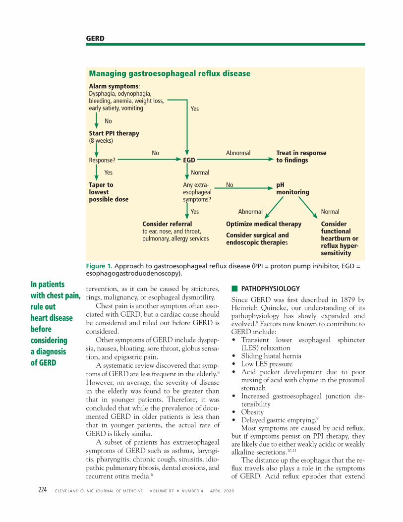
224 CLEVELAND CLINIC JOURNAL OF MEDICINE VOLUME 87 • NUMBER 4 APRIL 2020
GERD
tervention, as it can be caused by strictures, rings, malignancy, or esophageal dysmotility. Chest pain is another symptom often asso-ciated with GERD, but a cardiac cause should be considered and ruled out before GERD is considered. Other symptoms of GERD include dyspep-sia, nausea, bloating, sore throat, globus sensa-tion, and epigastric pain. A systematic review discovered that symp-toms of GERD are less frequent in the elderly.8 However, on average, the severity of disease in the elderly was found to be greater than that in younger patients. Therefore, it was concluded that while the prevalence of docu-mented GERD in older patients is less than that in younger patients, the actual rate of GERD is likely similar. A subset of patients has extraesophageal symptoms of GERD such as asthma, laryngi-tis, pharyngitis, chronic cough, sinusitis, idio-pathic pulmonary fi brosis, dental erosions, and recurrent otitis media.6
■ PATHOPHYSIOLOGY
Since GERD was fi rst described in 1879 by Heinrich Quincke, our understanding of its pathophysiology has slowly expanded and evolved.9 Factors now known to contribute to GERD include: • Transient lower esophageal sphincter
(LES) relaxation• Sliding hiatal hernia• Low LES pressure• Acid pocket development due to poor
mixing of acid with chyme in the proximal stomach
• Increased gastroesophageal junction dis-tensibility
• Obesity• Delayed gastric emptying.9
Most symptoms are caused by acid refl ux, but if symptoms persist on PPI therapy, they are likely due to either weakly acidic or weakly alkaline secretions.10,11 The distance up the esophagus that the re-fl ux travels also plays a role in the symptoms of GERD. Acid refl ux episodes that extend
In patients with chest pain, rule out heart diseasebefore considering a diagnosisof GERD
Figure 1. Approach to gastroesophageal refl ux disease (PPI = proton pump inhibitor, EGD = esophagogastroduodenoscopy).
Managing gastroesophageal refl ux disease
Alarm symptoms:Dysphagia, odynophagia,bleeding, anemia, weight loss,early satiety, vomiting
Yes
No
Start PPI therapy(8 weeks)
Response? No
EGDAbnormal Treat in response
to fi ndings
Yes Normal
Taper to lowest possible dose
Any extra-esophageal symptoms?
No pH monitoring
Yes Abnormal Normal
Consider referral to ear, nose, and throat, pulmonary, allergy services
Optimize medical therapy
Consider surgical and endoscopic therapies
Consider functional heartburn or refl ux hyper-sensitivity
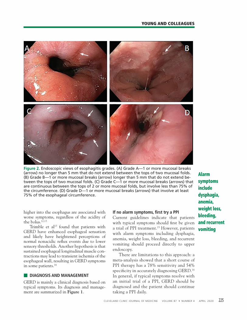
CLEVELAND CLINIC JOURNAL OF MEDICINE VOLUME 87 • NUMBER 4 APRIL 2020 225
YOUNG AND COLLEAGUES
higher into the esophagus are associated with worse symptoms, regardless of the acidity of the bolus.12,13 Trimble et al13 found that patients with GERD have enhanced esophageal sensation and likely have heightened perceptions of normal nonacidic refl ux events due to lower sensory thresholds. Another hypothesis is that sustained esophageal longitudinal muscle con-tractions may lead to transient ischemia of the esophageal wall, resulting in GERD symptoms in some patients.14
■ DIAGNOSIS AND MANAGEMENT
GERD is mainly a clinical diagnosis based on typical symptoms. Its diagnosis and manage-ment are summarized in Figure 1.
If no alarm symptoms, fi rst try a PPICurrent guidelines indicate that patients with typical symptoms should fi rst be given a trial of PPI treatment.15 However, patients with alarm symptoms including dysphagia, anemia, weight loss, bleeding, and recurrent vomiting should proceed directly to upper endoscopy. There are limitations to this approach: a meta-analysis showed that a short course of PPI therapy has a 78% sensitivity and 54% specifi city in accurately diagnosing GERD.16 In general, if typical symptoms resolve with an initial trial of a PPI, GERD should be diagnosed and the patient should continue taking a PPI daily.
Alarm symptoms include dysphagia, anemia, weight loss, bleeding, and recurrent vomiting
Figure 2. Endoscopic views of esophagitis grades. (A) Grade A—1 or more mucosal breaks (arrow) no longer than 5 mm that do not extend between the tops of two mucosal folds. (B) Grade B—1 or more mucosal breaks (arrow) longer than 5 mm that do not extend be-tween the tops of two mucosal folds. (C) Grade C—1 or more mucosal breaks (arrows) that are continuous between the tops of 2 or more mucosal folds, but involve less than 75% of the circumference. (D) Grade D—1 or more mucosal breaks (arrows) that involve at least 75% of the esophageal circumference.
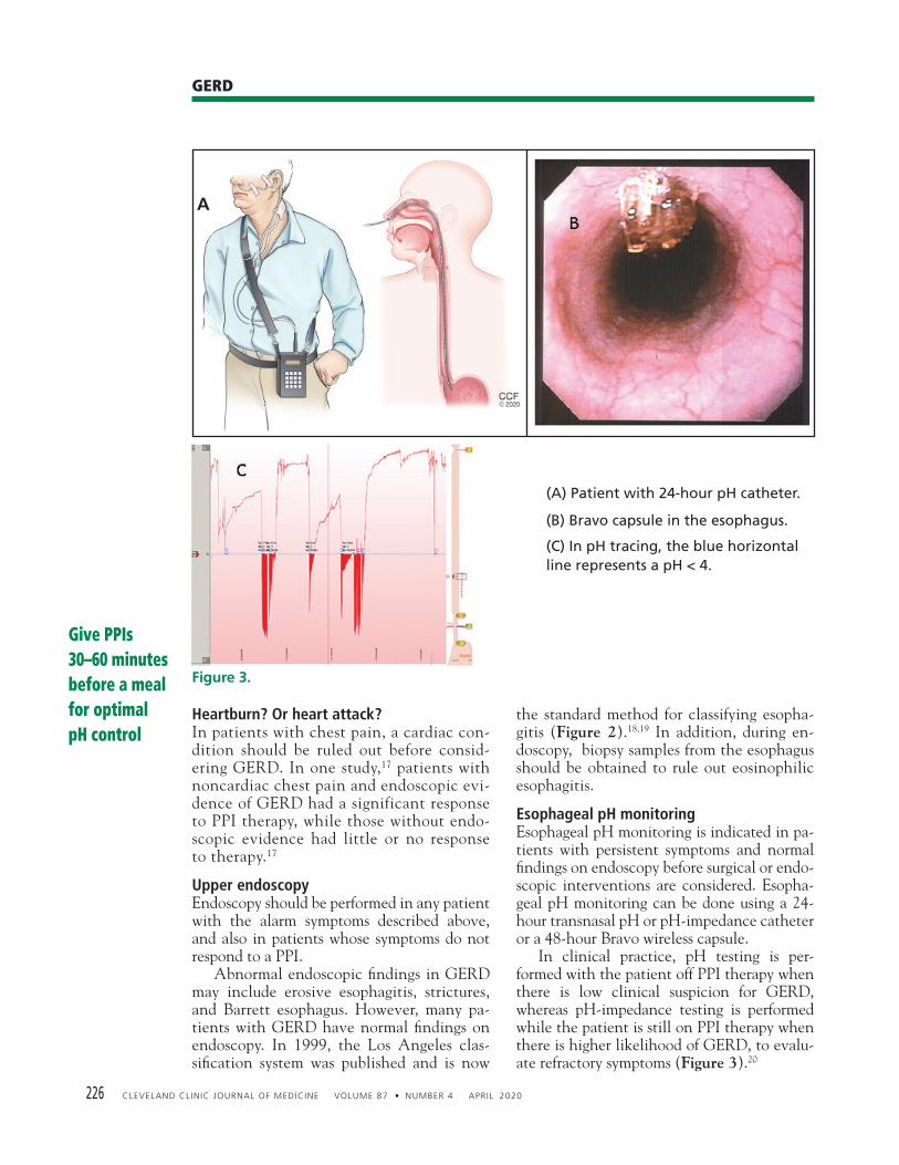
226 CLEVELAND CLINIC JOURNAL OF MEDICINE VOLUME 87 • NUMBER 4 APRIL 2020
GERD
Heartburn? Or heart attack?In patients with chest pain, a cardiac con-dition should be ruled out before consid-ering GERD. In one study,17 patients with noncardiac chest pain and endoscopic evi-dence of GERD had a significant response to PPI therapy, while those without endo-scopic evidence had little or no response to therapy.17
Upper endoscopy Endoscopy should be performed in any patient with the alarm symptoms described above, and also in patients whose symptoms do not respond to a PPI. Abnormal endoscopic fi ndings in GERD may include erosive esophagitis, strictures, and Barrett esophagus. However, many pa-tients with GERD have normal fi ndings on endoscopy. In 1999, the Los Angeles clas-sifi cation system was published and is now
the standard method for classifying esopha-gitis (Figure 2).18,19 In addition, during en-doscopy, biopsy samples from the esophagus should be obtained to rule out eosinophilic esophagitis.
Esophageal pH monitoringEsophageal pH monitoring is indicated in pa-tients with persistent symptoms and normal fi ndings on endoscopy before surgical or endo-scopic interventions are considered. Esopha-geal pH monitoring can be done using a 24-hour transnasal pH or pH-impedance catheter or a 48-hour Bravo wireless capsule. In clinical practice, pH testing is per-formed with the patient off PPI therapy when there is low clinical suspicion for GERD, whereas pH-impedance testing is performed while the patient is still on PPI therapy when there is higher likelihood of GERD, to evalu-ate refractory symptoms (Figure 3).20
Give PPIs 30–60 minutes before a meal for optimal pH control
(A) Patient with 24-hour pH catheter.
(B) Bravo capsule in the esophagus.
(C) In pH tracing, the blue horizontal line represents a pH < 4.
A
Figure 3.
CLEVELAND CLINIC JOURNAL OF MEDICINE VOLUME 87 • NUMBER 4 APRIL 2020 227
YOUNG AND COLLEAGUES
Barium esophagography is not indicated in the workup of refl ux disease as it has poor sen-sitivity and specifi city for GERD.
■ TREATMENT: LIFESTYLE,DRUG THERAPY, SURGERY
Lifestyle modifi cationsLifestyle modifi cations are the fi rst option for most patients. Weight loss can help reduce and eliminate GERD symptoms. A prospective cohort study found that 81% of obese patients who com-pleted a structured weight loss program had a reduction in symptoms, and 65% had com-plete resolution of symptoms.21 Another large retrospective study, with more than 15,000 patients, showed an association between im-provement in GERD symptoms and reduction in body mass index (BMI) in obese patients who lost at least 2 kg/m2 in BMI (odds ratio 2.34).22
Diet, smoking cessation, alcohol mod-eration. Numerous studies have aimed to fi nd foods that exacerbate refl ux symptoms. Historically, patients have been advised to avoid smoking, chocolate, carbonated bever-ages, spicy food, fatty food, alcohol, and large meals. Thus far, no study has found improve-ment in GERD symptoms with cessation of either smoking or alcohol. In terms of food consumption, no food has been conclusively linked with increased GERD symptoms. No consistent associations have been established between GERD symptoms and fatty food, spicy food, coffee, carbonated beverages, chocolate, citrus, or mint. Sleep position. Other studies have pro-moted elevating the head of the bed, sleeping in the left decubitus position, and, in those with nocturnal GERD symptoms, avoiding meals in the 2 to 3 hours before bedtime.23,24 A sleep positional therapy device has been shown to reduce acid exposure times and im-prove nocturnal refl ux symptoms.25,26 This de-vice places the user in the left decubitus posi-tion at an incline and has been an effective tool for those with nocturnal symptoms.
Drug therapyIf lifestyle interventions fail, drug treatment options are PPIs, histamine 2 receptor antago-nists (H2RAs), and antacids.
PPIs are considered the therapy of choice for symptomatic relief and healing of erosive esophagitis. Compared with H2RAs, PPIs have been shown to provide improved heal-ing rates and fewer relapses in patients with erosive esophagitis.27 To date, no study has shown a major difference in symptom control between the multiple PPIs. However, esome-prazole was shown, in a meta-analysis compar-ing it with other PPIs, to increase the prob-ability of healing erosive esophagitis at 4 and 8 weeks.28 PPIs inhibit gastric acid secretion by in-activating the hydrogen potassium ATPase molecules of the parietal cell. Optimal acid suppression occurs when the proton pumps are activated as the parietal cell is maximally stimulated after a meal. All PPIs should be taken 30 to 60 minutes before a meal for optimal pH control except dexlansoprazole, which employs dual delayed- release technology leading to sustained plasma drug concentrations; it can therefore be taken at any time of day. For patients with daytime symptoms, a PPI should be taken once daily in the morning, and for nighttime symptoms, the dose should be taken in the evening. After the initial 8-week course of therapy, most patients with GERD should attempt to take the lowest dose required to manage their symptoms. For some, this could mean only taking the medication when symptoms arise. However, patients with severe erosive esopha-gitis (grade C or D), Barrett esophagus, and peptic strictures need long-term PPI treat-ment. Adverse effects of PPIs. All patients need to be counseled about possible long-term ad-verse effects of PPIs.29 However, a recent ran-domized controlled trial found no association of PPIs with any adverse event when used for 3 years, with the possible exception of an in-creased risk of enteric infections.30
Vaezi et al29 reviewed the complications of PPI therapy and listed the relative risk and absolute excess risk in randomized controlled trials. From their data, we have calculated the number needed to harm, ie, the number of pa-tients who would need to be treated for 1 year to observe 1 adverse effect:• Chronic kidney disease, 333–1,000• Dementia, 67–1,429
If lifestyleinterventions fail, drugtreatmentoptions are PPIs, H2RAs,and antacids
228 CLEVELAND CLINIC JOURNAL OF MEDICINE VOLUME 87 • NUMBER 4 APRIL 2020
GERD
An 8-week course of PPIis needed to allow for heal-ing; patients should not be considered nonresponders until after this, unless alarm symptomsare present
• Bone fracture, 200–1,000• Campylobacter or Salmonella infection,
500–3,333• Spontaneous bacterial peritonitis (in pa-
tients with cirrhosis and ascites), 6–33• Clostridioides diffi cile infection, 1,111–no
association• Micronutrient defi ciencies, 250–333. The authors found no association between long-term PPI use and the following:• Myocardial infarction• Small intestinal bacterial overgrowth• Pneumonia• Gastrointestinal malignancies. Compared with earlier drugs, PPIs have been consistently shown to be superior at heal-ing erosive esophagitis and relieving symptoms. PPIs can maintain intragastric pH higher than 4 for 15 to 21 hours daily, compared with the 8 hours that H2RAs can achieve.31 In a ran-domized trial, endoscopic remission of erosive esophagitis was found in 80.2% of those taking omeprazole 20 mg daily vs 39.4% in those tak-ing ranitidine 150 mg daily.27 H2RAs appear useful in GERD for con-trolling nocturnal acid breakthrough. Howev-er, tachyphylaxis to these drugs develops rap-idly, and they may therefore have a role only if used intermittently.32
Antacids, especially when combined with alginate preparations, are effective for reduc-ing postprandial esophageal acid exposure.33
If fi rst-line therapy failsPPIs have immensely changed the landscape of treatment for GERD since their introduc-tion, but up to 40% of patients with GERD fi nd partial or no symptom relief with fi rst-line therapies.34 In these nonresponders, it is im-portant to determine compliance with PPIs, specifi cally the timing in relation to meals. An 8-week course of therapy is needed to allow for healing, and patients should not be considered nonresponders until after this pe-riod unless alarm symptoms are present. For these patients, upper endoscopy should be performed within 2 weeks. For those without alarm symptoms but continued refl ux in spite of therapy, endoscopy should be performed af-ter 8 weeks, with biopsies of the esophagus to evaluate for eosinophilic esophagitis. Esophageal impedance and pH testing are
performed on these non- and partial respond-ers while off PPIs to determine if there is per-sistent acidic or nonacidic refl ux. If results of pH and impedance testing are normal, the most common causes of contin-ued symptoms are refl ux hypersensitivity and functional heartburn. Refl ux hypersensitivity is a heightened response to nonpathologic refl ux, while functional heartburn is the pres-ence of symptoms without any evidence of abnormal exposure. These patients should be reassured that their condition is benign, and they can be started on a pain modulator such as a selective serotonin reuptake inhibitor, serotonin-norepinephrine reuptake inhibitor, or tricyclic antidepressant. If PPIs give partial relief, they should be continued, but they can be stopped for pa-tients who have no response to them. In patients found to have nonacid refl ux, a trial of baclofen should be offered, as it has been shown to reduce the rate of lower esoph-ageal sphincter transient relaxations.35
Alternative and investigational therapiesAlternative therapies are being investigated, but none have consistently shown signifi cant benefi ts over placebo. Therapies under investigation include re-fl ux inhibitors, prokinetics, acupuncture, and hypnotherapy. Prokinetics, including metoclo-pramide and domperidone, have shown benefi t in select patients with GERD but have been limited in their use due to associated central nervous system side effects and QT prolonga-tion. New medical treatments for GERD on the horizon include potassium competitive acid blockers (vonaprazan) and bile acid sequestrant (IW3718) that binds to bile in the refl uxate.
■ SURGICAL THERAPIES
Nissen fundoplication, fi rst performed by Dr. Rudolph Nissen in 1955, gained popularity in the 1970s and is now the most widely per-formed antirefl ux surgery. It involves reducing the hiatal hernia and wrapping the gastric fun-dus partially or completely around the lower esophagus to restore the LES barrier. Indications for the procedure are presence of a large hiatal hernia, refl ux esophagitis or GERD symptoms refractory to medical thera-py, or adverse effects of medical therapy.
CLEVELAND CLINIC JOURNAL OF MEDICINE VOLUME 87 • NUMBER 4 APRIL 2020 229
YOUNG AND COLLEAGUES
A trial comparing laparoscopic fundopli-cation with esomeprazole therapy found simi-lar remission rates after 3 years and a higher rate with esomeprazole after 5 years.36 While esomeprazole was associated with more symp-toms of refl ux compared with fundoplication, patients who underwent this surgery report-ed higher rates of dysphagia, fl atulence, and bloating. Antirefl ux surgery should be recommended with caution, as it can have severe side effects such as dysphagia, gas bloat syndrome, and fl atulence and the intended effect may only be temporary, as up to 60% of patients will re-quire antirefl ux medications regularly in the decade afterward.37 Esophageal manometry should be obtained before surgery to screen for esophageal aperistalsis, as this is an absolute contraindication to the procedure. Further-more, manometry will exclude other motility disorders that can present similarly to GERD as discussed. Of note, antirefl ux surgery is not recommended in PPI nonresponders.15
The Linx procedure (magnetic sphincter augmentation; Torax Medical Inc., Shore-view, MN) is a minimally invasive alternative. It involves laparoscopic insertion of a band of magnetic beads around the LES, which allows passage of food and then closes to prevent acid refl ux. The procedure is associated with improvement in symptom scores and reduced need for PPI therapy but not with consistent reduction in esophageal acid exposure.38
Roux-en-Y gastric bypass is a surgical op-tion for morbidly obese patients. A prospective study with 53 patients showed an improve-ment in GERD symptoms, refl ux esophagitis, and esophageal acid exposure for more than 3 years following bypass.39
■ ENDOSCOPIC THERAPIES
Alternatively, several endoscopic treatments for GERD have been developed over the last
2 decades.40 These include:• Transoral incisionless fundoplication (TIF)
using the Esophyx device (EndoGastric Solutions, Redmond, WA)
• Radiofrequency energy delivery to the LES (the Stretta procedure; Respiratory Tech-nology Corporation, Houston, TX)
• Endoscopic anterior fundoplication using the Medigus ultrasonic surgical endosta-pler (Medigus, Omer, Israel).
Of these, the fi rst 2 have the most evidence. TIF involves creation of a partial gastric wrap around the lower esophagus with an Eso-phyx device mounted on the endoscope. TIF is associated with symptom control and PPI reduction or cessation for at least 6 years and is a viable option for a select group of GERD patients with small hiatal hernias and pre-served esophageal function. A large randomized trial comparing TIF with PPIs showed symptomatic control in 67% vs 45% patients. TIF was associated with a reduction in esophageal acid exposure time from 9.3% to 6.4% and DeMeester score re-duction from 33.6 to 23.9.41 In 2018, a meta-analysis was performed to compare TIF with Nissen fundoplication, a sham procedure, and PPIs.42 TIF was asso-ciated with a larger increase in quality of life measures, while Nissen fundoplication had a greater ability to improve physiologic param-eters associated with GERD including LES pressure and the percentage of time the pH was less than 4. The Stretta device was developed in 2000 and works by delivering thermal energy to the LES, which is postulated to increase sphincter thickness through scar tissue depo-sition, thereby reducing refl ux. However in a meta-analysis of randomized controlled trials, Stretta treatment did not reduce percentage of time when pH is less than 4 or increase LES pressure or ability to stop PPIs.43 ■
Antirefl uxsurgery should be recommended with caution, as it can have severe side effects
■ REFERENCES 1. Peery AF, Crockett SD, Murphy CC, et al. Burden and cost of gastrointesti-
nal, liver, and pancreatic diseases in the United States: update 2018. Gastro-enterology 2019; 156(1):254–272.e11. doi:10.1053/j.gastro.2018.08.063
2. Dent J, El-Serag HB, Wallander MA, Johansson S. Epidemiology of gastro-oesophageal refl ux disease: a systematic review. Gut 2005; 54(5):710–717. doi:10.1136/gut.2004.051821
3. El–Serag HB. Time trends of gastroesophageal refl ux disease: a systematic review. Clin Gastroenterol Hepatol 2007; 5(1):17–26. doi:10.1016/j.cgh.2006.09.016
4. Zagari RM, Fuccio L, Wallander MA, et al. Gastro-oesophageal refl ux symp-toms, oesophagitis and Barrett’s oesophagus in the general population: the Loiano-Monghidoro study. Gut 2008; 57(10):1354–1359. doi:10.1136/gut.2007.145177
5. Wahlqvist P, Reilly MC, Barkun A. Systematic review: the impact of gastro-oesophageal refl ux disease on work productivity. Aliment Pharmacol Ther 2006; 24(2):259–272. doi:10.1111/j.1365-2036.2006.02996.x
6. Vakil N, van Zanten SV, Kahrilas P, Dent J, Jones R; Global Consensus Group. The Montreal defi nition and classifi cation of gastroesophageal refl ux disease: a global evidence-based consensus. Am J Gastroenterol 2006;
230 CLEVELAND CLINIC JOURNAL OF MEDICINE VOLUME 87 • NUMBER 4 APRIL 2020
GERD
101(8):1900–1920. doi:10.1111/j.1572-0241.2006.00630.x 7. Flegal KM, Carroll MD, Kit BK, Ogden CL. Prevalence of obesity and trends
in the distribution of body mass index among US adults, 1999–2010. JAMA 2012; 307(5):491–497. doi:10.1001/jama.2012.39
8. Becher A, Dent J. Systematic review: ageing and gastro-oesophageal refl ux disease symptoms, oesophageal function and refl ux oesophagitis. Aliment Pharmacol Ther 2011; 33(4):442–454. doi:10.1111/j.1365-2036.2010.04542.x
9. Herregods TV, Bredenoord AJ, Smout AJ. Pathophysiology of gastroesopha-geal refl ux disease: new understanding in a new era. Neurogastroenterol Motil 2015; 27(9):1202–1213. doi:10.1111/nmo.12611
10. Smith JL, Opekun AR, Larkai E, Graham DY. Sensitivity of the esophageal mucosa to pH in gastroesophageal refl ux disease. Gastroenterology 1989; 96(3):683–689. pmid:2914634
11. Sung HJ, Cho YK, Moon SJ, et al. Role of acid and weakly acidic refl ux in gastroesophageal refl ux disease off proton pump inhibitor therapy. J Neu-rogastroenterol Motil 2012; 18(3):291–297. doi:10.5056/jnm.2012.18.3.291
12. Weusten BL, Akkermans LM, vanBerge-Henegouwen GP, Smout AJ. Symp-tom perception in gastroesophageal refl ux disease is dependent on spatio-temporal refl ux characteristics. Gastroenterology 1995; 108(6):1739–1744. doi:10.1016/0016-5085(95)90135-3
13. Trimble KC, Pryde A, Heading RC. Lowered oesophageal sensory thresholds in patients with symptomatic but not excess gastro-oesophageal refl ux: evi-dence for a spectrum of visceral sensitivity in GORD. Gut 1995; 37(1):7–12. doi:10.1136/gut.37.1.7
14. Balaban DH, Yamamoto Y, Liu J, et al. Sustained esophageal contraction: a marker of esophageal chest pain identifi ed by intraluminal ultrasonogra-phy. Gastroenterology 1999; 116(1):29–37. doi:10.1016/s0016-5085(99)70225-8
15. Katz PO, Gerson LB, Vela MF. Guidelines for the diagnosis and manage-ment of gastroesophageal refl ux disease. Am J Gastroenterol 2013; 108(3):308–328. doi:10.1038/ajg.2012.444
16. Numans ME, Lau J, de Wit NJ, Bonis PA. Short-term treatment with proton-pump inhibitors as a test for gastroesophageal refl ux disease: a meta-anal-ysis of diagnostic test characteristics. Ann Intern Med 2004; 140(7):518–527. doi:10.7326/0003-4819-140-7-200404060-00011
17. Kahrilas PJ, Hughes N, Howden CW. Response of unexplained chest pain to proton pump inhibitor treatment in patients with and without objective evidence of gastro-oesophageal refl ux disease. Gut 2011; 60(11):1473–1478. doi:10.1136/gut.2011.241307
18. Sami SS, Ragunath K. The Los Angeles classifi cation of gastroesophageal refl ux disease. Video Journal and Encyclopedia of GI Endoscopy 2013; 1(1):103–104. doi:10.1016/S2212-0971(13)70046-3
19. Lundell LR, Dent J, Bennett JR, et al. Endoscopic assessment of oesophagitis: clinical and functional correlates and further validation of the Los Angeles classifi cation. Gut 1999; 45(2):172–180. doi:10.1136/gut.45.2.172
20. Gyawali CP, Kahrilas PJ, Savarino E, et al. Modern diagnosis of GERD: the Lyon consensus. Gut 2018; 67(7):1351–1362. doi:10.1136/gutjnl-2017-314722
21. Singh M, Lee J, Gupta N, et al. Weight loss can lead to resolution of gastroesophageal refl ux disease symptoms: a prospective intervention trial. Obesity (Silver Spring) 2013; 21(2):284–290. doi:10.1002/oby.20279
22. Park SK, Lee T, Yang HJ, et al. Weight loss and waist reduction is associated with improvement in gastroesophageal disease refl ux symptoms: a longitu-dinal study of 15,295 subjects undergoing health checkups. Neurogastroen-terol Motil 2017; 29(5). doi:10.1111/nmo.13009
23. Khan BA, Sodhi JS, Zargar SA, et al. Effect of bed head elevation during sleep in symptomatic patients of nocturnal gastroesophageal refl ux. J Gastroenterol Hepatol 2012; 27(6):1078–1082. doi:10.1111/j.1440-1746.2011.06968.x
24. Duroux P, Bauerfeind P, Emde C, Koelz HR, Blum AL. Early dinner reduces nocturnal gastric acidity. Gut 1989; 30(8):1063–1067. doi:10.1136/gut.30.8.1063
25. Person E, Rife C, Freeman J, Clark A, Castell DO. A novel sleep positioning device reduces gastroesophageal refl ux: a randomized controlled trial. J Clin Gastroenterol 2015; 49(8):655–659. doi:10.1097/MCG.0000000000000359
26. Allampati S, Lopez R, Thota PN, Ray M, Birgisson S, Gabbard SL. Use of a positional therapy device signifi cantly improves nocturnal gastroesophage-al refl ux symptoms. Dis Esophagus 2017; 30(3):1–7. doi:10.1111/dote.12495
27. Carlsson R, Galmiche JP, Dent J, Lundell L, Frison L. Prognostic factors infl uencing relapse of oesophagitis during maintenance therapy with an-tisecretory drugs: a meta-analysis of long-term omeprazole trials. Aliment Pharmacol Ther 1997; 11(3):473–482. doi:10.1046/j.1365-2036.1997.00167.x
28. Gralnek IM, Dulai GS, Fennerty MB, Spiegel BM. Esomeprazole versus other proton pump inhibitors in erosive esophagitis: a meta-analysis of random-ized clinical trials. Clin Gastroenterol Hepatol 2006; 4(12):1452–1458. doi:10.1016/j.cgh.2006.09.013
29. Vaezi MF, Yang YX, Howden CW. Complications of proton pump inhibitor therapy. Gastroenterology 2017; 153(1):35–48. doi:10.1053/j.gastro.2017.04.047
30. Moayyedi P, Eikelboom JW, Bosch J, et al; COMPASS Investigators. Safety of proton pump inhibitors based on a large, multi-year, randomized trial of patients receiving rivaroxaban or aspirin. Gastroenterology 2019; 157(3):682–691.e2. doi:10.1053/j.gastro.2019.05.056
31. Wolfe MM, Sachs G. Acid suppression: optimizing therapy for gastroduo-denal ulcer healing, gastroesophageal refl ux disease, and stress-related erosive syndrome. Gastroenterology 2000; 118(2 suppl 1):S9–S31. doi:10.1016/s0016-5085(00)70004-7
32. Komazawa Y, Adachi K, Mihara T, et al. Tolerance to famotidine and raniti-dine treatment after 14 days of administration in healthy subjects without Helicobacter pylori infection. J Gastroenterol Hepatol 2003; 18(6):678–682. doi:10.1046/j.1440-1746.2003.03041.x
33. De Ruigh A, Roman S, Chen J, Pandolfi no JE, Kahrilas PJ. Gaviscon double action liquid (antacid & alginate) is more effective than antacid in controlling post-prandial oesophageal acid exposure in GERD patients: a double-blind crossover study. Aliment Pharmacol Ther 2014; 40(5):531–537. doi:10.1111/apt.12857
34. Johnson T, Gerson L, Hershcovici T, Stave C, Fass R. Systematic review: the ef-fects of carbonated beverages on gastro-oesophageal refl ux disease. Aliment Pharmacol Ther 2010; 31(6):607–614. doi:10.1111/j.1365-2036.2010.04232.x
35. Grossi L, Spezzaferro M, Sacco LF, Marzio L. Effect of baclofen on oesopha-geal motility and transient lower oesophageal sphincter relaxations in GORD patients: a 48-h manometric study. Neurogastroenterol Motil 2008; 20(7):760–766. doi:10.1111/j.1365-2982.2008.01115.x
36. Galmiche JP, Hatlebakk J, Attwood S, et al; LOTUS Trial Collaborators. Laparoscopic antirefl ux surgery vs esomeprazole treatment for chronic GERD: the LOTUS randomized clinical trial. JAMA 2011; 305(19):1969–1977. doi:10.1001/jama.2011.626
37. Spechler SJ, Lee E, Ahnen D, et al. Long-term outcome of medical and surgical therapies for gastroesophageal refl ux disease: follow-up of a randomized controlled trial. JAMA 2001; 285(18):2331–2338. doi:10.1001/jama.285.18.2331
38. Bell R, Lipham J, Louie B, et al. Laparoscopic magnetic sphincter augmen-tation versus double-dose proton pump inhibitors for management of moderate-to-severe regurgitation in GERD: a randomized controlled trial. Gastrointest Endosc 2019; 89(1):14–22.e1. doi:10.1016/j.gie.2018.07.007
39. Madalosso CA, Gurski RR, Callegari-Jacques SM, Navarini D, Mazzini G, Pereira Mda S. The impact of gastric bypass on gastroesophageal refl ux disease in morbidly obese patients. Ann Surg 2016; 263(1):110–116. doi:10.1097/SLA.0000000000001139
40. Rouphael C, Padival R, Sanaka MR, Thota PN. Endoscopic treatments of GERD. Curr Treat Options Gastroenterol 2018; 16(1):58–71. doi:10.1007/s11938-018-0170-6
41. Hunter JG, Kahrilas PJ, Bell RC, et al. Effi cacy of transoral fundoplication vs omeprazole for treatment of regurgitation in a randomized controlled trial. Gastroenterology 2015; 148(2):324–333.e5. doi:10.1053/j.gastro.2014.10.009
42. Richter JE, Kumar A, Lipka S, Miladinovic B, Velanovich V. Effi cacy of laparoscopic Nissen fundoplication vs transoral incisionless fundoplication or proton pump inhibitors in patients with gastroesophageal refl ux disease: a systematic review and network meta-analysis. Gastroenterology 2018; 154(5):1298–1308.e7. doi:10.1053/j.gastro.2017.12.021
43. Lipka S, Kumar A, Richter JE. No evidence for effi cacy of radiofrequency ablation for treatment of gastroesophageal refl ux disease: a systematic re-view and meta-analysis. Clin Gastroenterol Hepatol 2015;1 3(6):1058–1067.e1. doi:10.1016/j.cgh.2014.10.013
Address: Prashanthi N. Thota, MD, FACG, Esophageal Center, Digestive Disease and Surgery Institute, A31, Cleveland Clinic, 9500 Euclid Avenue, Cleveland, OH 44195; [email protected]
ACC/AHA lipid guidelines:Personalized care to prevent cardiovascular disease
CLEVELAND CLINIC JOURNAL OF MEDICINE VOLUME 87 • NUMBER 4 APRIL 2020 231
The American College of Cardiology (ACC) and American Heart Associa-
tion (AHA) Task Force on Clinical Practice Guidelines published its most recent guide-lines for cholesterol management in 2018,1 and followed it with guidelines for primary prevention of cardiovascular disease in 2019.2
The new guidelines have updated patient risk assessment and treatment options in pri-mary and secondary prevention. In primary prevention, the guidelines provide clarity re-garding decision-making in patients at inter-mediate risk of atherosclerotic cardiovascular disease (“intermediate” meaning a 7.5%–20% 10-year risk). In secondary prevention, the guidelines group patients according to their risk (high risk vs very high risk) and incorporate new nonstatin therapies as add-on, evidence-based treatment options when low-density lipopro-tein (LDL-C) remains above the 70 mg/dL threshold. The guidelines also discuss the cost and value of each treatment option for each treatment group.
REVIEW
doi:10.3949/ccjm.87a.19078
ABSTRACTThe 2018 and 2019 guidelines from the American College of Cardiology and American Heart Association refl ect the complexity of individualized cholesterol management. The documents address more detailed risk as-sessment, newer nonstatin cholesterol-lowering drugs, special attention to patient subgroups, and consideration of the value of therapy, all with the aim of creating personalized treatment plans for each patient. Overall, the guidelines recommend shared decision-making to meet the individual needs of each patient.
Cara Reiter-BrennanJohns Hopkins Ciccarone Center for Prevention of Cardiovascular Disease, Baltimore, MD; Department of Radiology and Neuroradiology, Charité, Berlin, Germany
Albert D. Osei, MD, MPHJohns Hopkins Ciccarone Center for Prevention of Cardiovascular Disease, Baltimore, MD
S. M. Iftekhar Uddin, MBBS, MSPHJohns Hopkins Ciccarone Center for Prevention of Cardiovascular Disease, Baltimore, MD
Olusola A. Orimoloye, MD, MPHJohns Hopkins Ciccarone Center for Prevention of Cardiovascular Disease, Baltimore, MD; Depart-ment of Medicine, Vanderbilt University School of Medicine, Nashville, TN
Olufunmilayo H. Obisesan, MD, MPHJohns Hopkins Ciccarone Center for Prevention of Cardiovascular Disease, Baltimore, MD
Mohammadhassan Mirbolouk, MDJohns Hopkins Ciccarone Center for Prevention of Cardiovascular Disease, Baltimore, MD; Depart-ment of Medicine, Yale School of Medicine, New Haven, CT
Michael J. Blaha, MD, MPHJohns Hopkins Ciccarone Center for Prevention of Cardiovascular Disease, Baltimore, MD
Omar Dzaye, MD, PhDJohns Hopkins Ciccarone Center for Prevention of Cardiovascular Disease, Baltimore, MD; Russell H. Morgan Department of Radiology and Radiological Science, Johns Hopkins University School of Medi-cine, Baltimore, MD; Department of Radiology and Neuroradiology, Charité, Berlin, Germany
CME MOC
KEY POINTSEmphasize a heart-healthy lifestyle for all patients across their life span.
A discussion with the patient is the cornerstone of shared decision-making and should include the patient’s 10-year risk of atherosclerotic cardiovascular disease according to the Pooled Cohort Equations, as well as risk-enhancing factors.
Statins are the foundation of pharmacologic therapy, to which ezetimibe and, if necessary, a proprotein con-vertase subtilisin/kexin type 9 inhibitor can be added to achieve lipid goals.
Special treatment algorithms are outlined for certain patient subgroups, such as certain ethnic groups, adults with chronic kidney disease, those with human immuno-defi ciency virus infection, and women.
232 CLEVELAND CLINIC JOURNAL OF MEDICINE VOLUME 87 • NUMBER 4 APRIL 2020
LIPID GUIDELINES
Here, we review the recent guidelines and discuss the most important changes for clinical practice.1–3
■ CLASSES OF RECOMMENDATION, LEVELS OF EVIDENCE
The guidelines award classes of recommenda-tions, signifying the certainty of benefi t com-pared with the estimated risk and the strength of the recommendation.• Class I (strong)—benefi t greatly exceeds
risk; treatment is recommended• Class IIa (moderate)—benefi t exceeds
risk; treatment is reasonable• Class IIb (weak)—benefi t equals or ex-
ceeds risk; treatment might be reasonable• Class III: No benefi t (moderate)—benefi t
equals risk; treatment is not recommended• Class III: Harm (strong)—risk exceeds
benefi t. The guidelines also award levels of evi-dence to their recommendations:• Level A—high-quality evidence • Level B-R—moderate-quality evidence
from randomized controlled trials• Level B-NR—moderate quality evidence
from nonrandomized trials• Level C-LD—limited data• Level C-EO—expert opinion.
■ STATINS AND OTHER OPTIONS
In addition to a heart-healthy lifestyle (which should be encouraged for all patients across their life course), statins are the foundation of lipid management. Statin therapy is divided into 3 categories of intensity: High-intensity, aiming for at least a 50% reduction in LDL-C. Examples:• Atorvastatin 40–80 mg daily• Rosuvastatin 20–40 mg daily. Moderate-intensity, aiming at a 30% to 49% reduction in LDL-C. Examples:• Atorvastatin 10–20 mg• Fluvastatin 80 mg daily• Lovastatin 40–80 mg• Pitavastatin 1–4 mg daily• Pravastatin 40–80 mg daily• Rosuvastatin 5–10 mg• Simvastatin 20–40 mg daily. Low-intensity, aiming at a LDL-C reduc-tion of less than 30%. Examples:
• Fluvastatin 20–40 mg daily• Lovastatin 20 mg daily• Pravastatin 10–20 mg daily• Simvastatin 10 mg daily.
Nonstatin drugsThe nonstatin LDL-lowering drugs such as ezetimibe and proprotein convertase subtili-sin/kexin type 9 (PCSK9) inhibitors can be added to statin therapy, as recent randomized clinical trials found them to improve cardio-vascular outcomes in patients with atheroscle-rotic cardiovascular disease.4–7 Ezetimibe decreases cholesterol absorp-tion and consequently lowers LDL-C levels by about 20%. A large randomized trial in patients who recently had acute coronary syndromes showed that ezetimibe modestly reduced cardiovascular risk over 7 years of follow-up when added to their regimen of moderate-intensity statin therapy.4,5
PCSK9 inhibitors lower LDL-C by 50% to 60% by binding to PCSK9, inhibiting la-beling of LDL receptors for degradation, thus prolonging LDL receptor activity at the cell membrane. Several trials showed that PCSK9 inhibitors reduce cardiovascular risk in pa-tients with stable atherosclerotic cardiovascu-lar disease or recent acute coronary syndromes who are already on moderate- or high-intensi-ty statin therapy.4,6,7
■ PRIMARY PREVENTION
The new guidelines advocate a multifaceted approach to primary prevention of atheroscle-rotic cardiovascular disease through cholester-ol management. As the risk due to high cho-lesterol levels is cumulative over the life span, the guidelines encourage lifestyle therapy for primary prevention at all ages and in all patient categories. Additionally, they outline decision algorithms to create a therapy that suits the in-dividual needs of each patient (Table 1).
Statin benefi t groupsThe new guidelines keep the same statin benefi t groups defi ned in the previous (2013) ACC/AHA guidelines.8 Statin therapy recommendations are specifi cally given for the following groups:
Adults with severe hypercholesterolemiaIf a patient age 20 to 75 has LDL-C levels of 190 mg/dL or higher, you do not need to cal-
In addition toa heart-healthylifestyle, statins are the founda-tion of lipid management
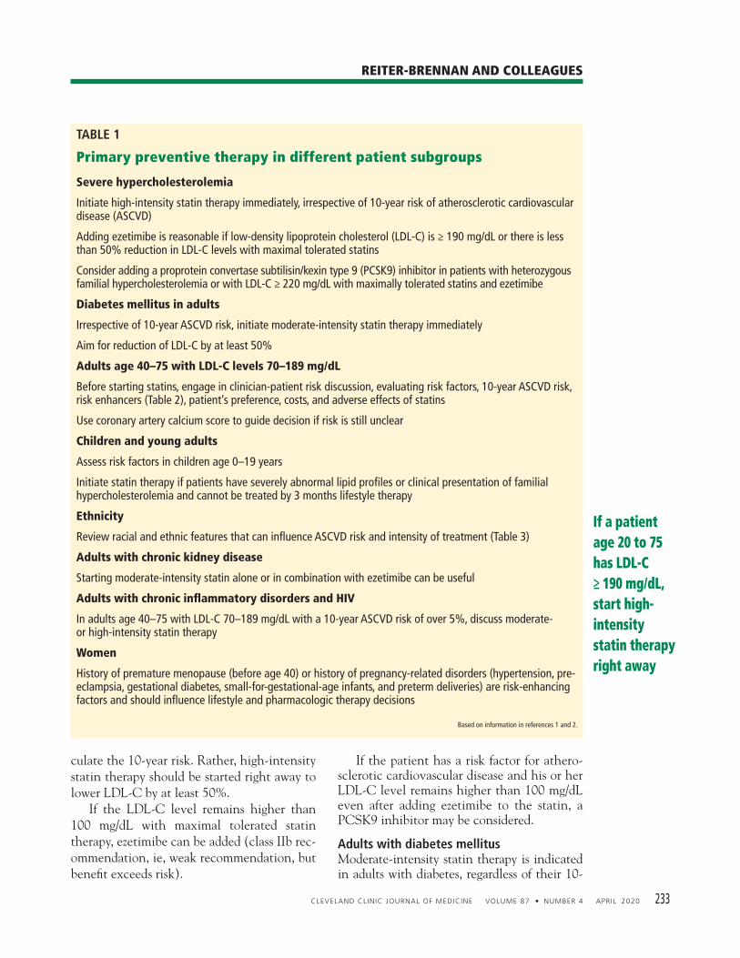
CLEVELAND CLINIC JOURNAL OF MEDICINE VOLUME 87 • NUMBER 4 APRIL 2020 233
REITER-BRENNAN AND COLLEAGUES
culate the 10-year risk. Rather, high-intensity statin therapy should be started right away to lower LDL-C by at least 50%. If the LDL-C level remains higher than 100 mg/dL with maximal tolerated statin therapy, ezetimibe can be added (class IIb rec-ommendation, ie, weak recommendation, but benefi t exceeds risk).
If the patient has a risk factor for athero-sclerotic cardiovascular disease and his or her LDL-C level remains higher than 100 mg/dL even after adding ezetimibe to the statin, a PCSK9 inhibitor may be considered.
Adults with diabetes mellitusModerate-intensity statin therapy is indicated in adults with diabetes, regardless of their 10-
TABLE 1
Primary preventive therapy in different patient subgroups
Severe hypercholesterolemia
Initiate high-intensity statin therapy immediately, irrespective of 10-year risk of atherosclerotic cardiovascular disease (ASCVD)
Adding ezetimibe is reasonable if low-density lipoprotein cholesterol (LDL-C) is ≥ 190 mg/dL or there is less than 50% reduction in LDL-C levels with maximal tolerated statins
Consider adding a proprotein convertase subtilisin/kexin type 9 (PCSK9) inhibitor in patients with heterozygous familial hypercholesterolemia or with LDL-C ≥ 220 mg/dL with maximally tolerated statins and ezetimibe
Diabetes mellitus in adults
Irrespective of 10-year ASCVD risk, initiate moderate-intensity statin therapy immediately
Aim for reduction of LDL-C by at least 50%
Adults age 40–75 with LDL-C levels 70–189 mg/dL
Before starting statins, engage in clinician-patient risk discussion, evaluating risk factors, 10-year ASCVD risk, risk enhancers (Table 2), patient’s preference, costs, and adverse effects of statins
Use coronary artery calcium score to guide decision if risk is still unclear
Children and young adults
Assess risk factors in children age 0–19 years
Initiate statin therapy if patients have severely abnormal lipid profi les or clinical presentation of familial hypercholesterolemia and cannot be treated by 3 months lifestyle therapy
Ethnicity
Review racial and ethnic features that can infl uence ASCVD risk and intensity of treatment (Table 3)
Adults with chronic kidney disease
Starting moderate-intensity statin alone or in combination with ezetimibe can be useful
Adults with chronic infl ammatory disorders and HIV
In adults age 40–75 with LDL-C 70–189 mg/dL with a 10-year ASCVD risk of over 5%, discuss moderate-or high-intensity statin therapy
Women
History of premature menopause (before age 40) or history of pregnancy-related disorders (hypertension, pre-eclampsia, gestational diabetes, small-for-gestational-age infants, and preterm deliveries) are risk-enhancing factors and should infl uence lifestyle and pharmacologic therapy decisions
Based on information in references 1 and 2.
If a patient age 20 to 75 has LDL-C ≥ 190 mg/dL, start high-intensity statin therapy right away
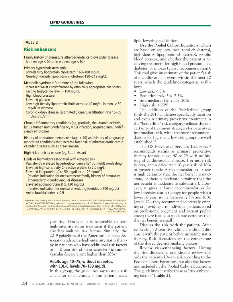
234 CLEVELAND CLINIC JOURNAL OF MEDICINE VOLUME 87 • NUMBER 4 APRIL 2020
LIPID GUIDELINES
year risk. However, it is reasonable to start high-intensity statin treatment if the patient also has multiple risk factors. Similarly, the 2019 guidelines of the American Diabetes As-sociation advocate high-intensity statin thera-py in patients who have additional risk factors or a 10-year risk of an atherosclerotic cardio-vascular disease event higher than 20%.9
Adults age 40–75, without diabetes, with LDL-C levels 70–189 mg/dLIn this group, the guidelines say to use a risk calculator to determine if the patient needs
lipid-lowering medication. Use the Pooled Cohort Equations, which are based on age, sex, race, total cholesterol, high-density lipoprotein cholesterol, systolic blood pressure, and whether the patient is re-ceiving treatment for high blood pressure, has diabetes, or smokes (class I recommendation). This tool gives an estimate of the patient’s risk of a cardiovascular event within the next 10 years, which the guidelines categorize as fol-lows: • Low risk: < 5%• Borderline risk: 5%–7.5%• Intermediate risk: 7.5%–20%• High risk: > 20%. The addition of the “borderline” group (only the 2018 guidelines specifi cally mention and explain primary preventive treatment in the “borderline” risk category) refl ects the un-certainty of treatment strategies for patients at intermediate risk, while treatment recommen-dations for high- and low-risk groups are well established.10
The US Preventive Services Task Force11
recommends statins as primary preventive therapy for adults age 40 to 75 with no his-tory of cardiovascular disease, 1 or more risk factors, and a calculated 10-year risk of 10% or greater (grade A recommendation—there is high certainty that the net benefi t is mod-erate, or there is moderate certainty that the net benefi t is moderate to substantial). How-ever, it gives a lower recommendation for low-intensity statin therapy for people with a lower 10-year risk, ie, between 7.5% and 10%. (grade C—they recommend selectively offer-ing or providing it to individual patients based on professional judgment and patient prefer-ences; there is at least moderate certainty that the net benefi t is small). Discuss the risk with the patient. After evaluating 10-year risk, clinicians should dis-cuss it with the patient before initiating statin therapy. Risk discussions are the cornerstone of the shared decision-making process. Review risk-enhancing factors. During the risk discussion, one should review not only the patient’s 10-year risk according to the Pooled Cohort Equations, but also risk factors not included in the Pooled Cohort Equations. The guidelines describe these as “risk-enhanc-ing factors” (Table 2).
TABLE 2
Risk enhancers
Family history of premature atherosclerotic cardiovascular disease (in men age < 55 or in women age < 65)
Primary hypercholesterolemia Low-density lipoprotein cholesterol 160–180 mg/dL Non-high-density lipoprotein cholesterol 190–219 mg/dL
Metabolic syndrome: 3 or more of the following: Increased waist circumference by ethnically appropriate cut points Fasting triglyceride level > 150 mg/dL High blood pressure Elevated glucose Low high-density lipoprotein cholesterol (< 40 mg/dL in men, < 50 mg/dL in women) Chronic kidney disease (estimated glomerular fi ltration rate 15–59 mL/min/1.73 m2)
Chronic infl ammatory conditions (eg, psoriasis, rheumatoid arthritis, lupus, human immunodefi ciency virus infection, acquired immunodefi -ciency syndrome)
History of premature menopause (age < 40) and history of pregnancy-associated conditions that increase later risk of atherosclerotic cardio-vascular disease such as preeclampsia
High-risk ethnicity or race (eg, South Asian)
Lipids or biomarkers associated with elevated risk Persistently elevated hypertriglyceridemia (≥ 175 mg/dL nonfasting) Elevated high-sensitivity C-reactive protein (≥ 2.0 mg/L) Elevated lipoprotein (a) (≥ 50 mg/dL or ≥ 125 nmol/L) (relative indication for measurement: family history of premature atherosclerotic cardiovascular disease) Elevated apolipoprotein B (≥ 130 mg/dL) (relative indication for measurement: triglycerides ≥ 200 mg/dL) Ankle-brachial index < 0.9
Reprinted from Grundy SM, Stone NJ, Bailey AL, et al. 2018 AHA/ACC/AACVPR/AAPA/ABC/ACPM/ADA/AGS/APhA/ASPC/NLA/PCNA guideline on the management of blood cholesterol: executive summary: a
report of the American College of Cardiology/American Heart Association Task Force on Clinical Practice Guidelines. J Am Coll Cardiol 2019; 73(24):3168–3209. doi:10.1016/j.jacc.2018.11.002, with permis-
sion from Elsevier.
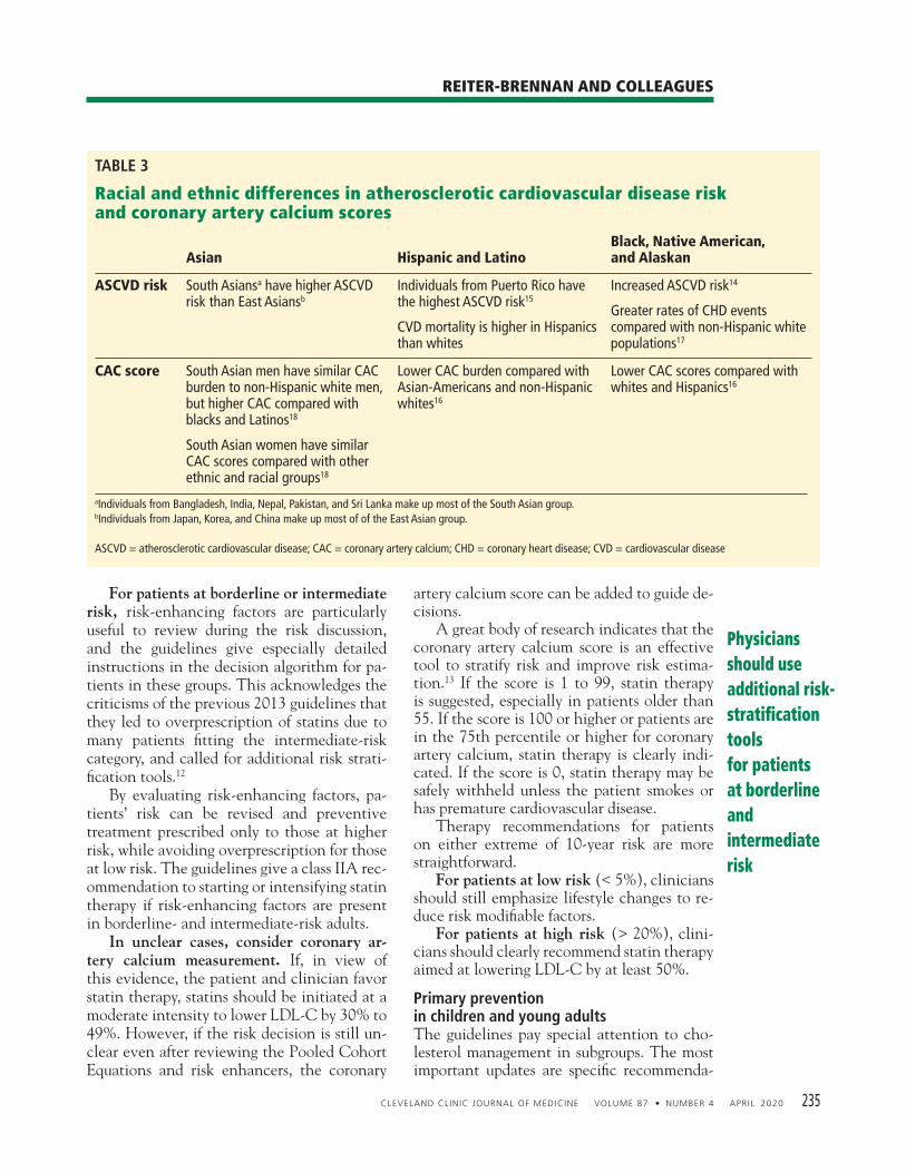
CLEVELAND CLINIC JOURNAL OF MEDICINE VOLUME 87 • NUMBER 4 APRIL 2020 235
REITER-BRENNAN AND COLLEAGUES
For patients at borderline or intermediate risk, risk-enhancing factors are particularly useful to review during the risk discussion, and the guidelines give especially detailed instructions in the decision algorithm for pa-tients in these groups. This acknowledges the criticisms of the previous 2013 guidelines that they led to overprescription of statins due to many patients fi tting the intermediate-risk category, and called for additional risk strati-fi cation tools.12 By evaluating risk-enhancing factors, pa-tients’ risk can be revised and preventive treatment prescribed only to those at higher risk, while avoiding overprescription for those at low risk. The guidelines give a class IIA rec-ommendation to starting or intensifying statin therapy if risk-enhancing factors are present in borderline- and intermediate-risk adults. In unclear cases, consider coronary ar-tery calcium measurement. If, in view of this evidence, the patient and clinician favor statin therapy, statins should be initiated at a moderate intensity to lower LDL-C by 30% to 49%. However, if the risk decision is still un-clear even after reviewing the Pooled Cohort Equations and risk enhancers, the coronary
artery calcium score can be added to guide de-cisions. A great body of research indicates that the coronary artery calcium score is an effective tool to stratify risk and improve risk estima-tion.13 If the score is 1 to 99, statin therapy is suggested, especially in patients older than 55. If the score is 100 or higher or patients are in the 75th percentile or higher for coronary artery calcium, statin therapy is clearly indi-cated. If the score is 0, statin therapy may be safely withheld unless the patient smokes or has premature cardiovascular disease. Therapy recommendations for patients on either extreme of 10-year risk are more straightforward. For patients at low risk (< 5%), clinicians should still emphasize lifestyle changes to re-duce risk modifi able factors. For patients at high risk (> 20%), clini-cians should clearly recommend statin therapy aimed at lowering LDL-C by at least 50%.
Primary prevention in children and young adultsThe guidelines pay special attention to cho-lesterol management in subgroups. The most important updates are specifi c recommenda-
Physicians should useadditional risk-stratifi cation toolsfor patientsat borderline andintermediate risk
TABLE 3
Racial and ethnic differences in atherosclerotic cardiovascular disease riskand coronary artery calcium scores
Asian Hispanic and LatinoBlack, Native American, and Alaskan
ASCVD risk South Asiansa have higher ASCVD risk than East Asiansb
Individuals from Puerto Rico have the highest ASCVD risk15
CVD mortality is higher in Hispanics than whites
Increased ASCVD risk14
Greater rates of CHD events compared with non-Hispanic white populations17
CAC score South Asian men have similar CAC burden to non-Hispanic white men, but higher CAC compared with blacks and Latinos18
South Asian women have similar CAC scores compared with other ethnic and racial groups18
Lower CAC burden compared with Asian-Americans and non-Hispanic whites16
Lower CAC scores compared with whites and Hispanics16
aIndividuals from Bangladesh, India, Nepal, Pakistan, and Sri Lanka make up most of the South Asian group.bIndividuals from Japan, Korea, and China make up most of of the East Asian group.
ASCVD = atherosclerotic cardiovascular disease; CAC = coronary artery calcium; CHD = coronary heart disease; CVD = cardiovascular disease
236 CLEVELAND CLINIC JOURNAL OF MEDICINE VOLUME 87 • NUMBER 4 APRIL 2020
LIPID GUIDELINES
Blackpopulations have higher rates of coronary heart disease even though they have lower coronary artery calcium scores
tions for children and young adults. The guidelines acknowledge that athero-sclerosis is a lifelong process and that the ef-fects of high cholesterol levels accumulate across an entire lifetime. This is why, unlike previous guidelines, the 2018/2019 guidelines recommend primary preventive therapy for children and young adults. Risk factor assessment and identifi cation of family history of hypercholesterolemia or inherited dyslipidemia should already occur for children age 0 to 19 years. Also, if children have severely elevated lipid levels related to obesity, intensive lifestyle therapy should be implemented.
Primary prevention for other populations at riskThe current recommendations also make spe-cifi c recommendations for cholesterol treat-ment algorithms for specifi c patient subgroups, in which treatment decisions were previously unclear.
Primary prevention: EthnicityThe ACC/AHA guidelines state in a class IIA recommendation that race and ethnicity in-fl uence the risk of atherosclerotic cardiovas-cular disease and the choice of treatment. Risk varies widely among and within ethnic groups, affecting treatment decisions (Table 3).14–18 In particular, the guidelines point out that South Asian individuals have higher risk, as do those who identify as Native American or Alaskan native compared with non-Hispanic white populations.14 Socioeconomic status and acculturation level (extent of assimilation to the dominant culture—in this case American culture) can affect the burden of atherosclerotic cardiovas-cular disease. For instance, a cross-sectional study showed that acculturation was associat-ed with higher cardiovascular risk in Hispanic participants.15
Moreover, ethnicity also affects other as-pects of risk classifi cation, such as coronary artery calcium scores. Studies suggest that eth-nicity infl uences the pathobiologic processes of vessel atherogenesis.19 Hispanic patients have a lower coronary artery calcium burden than Asian-Americans and non-Hispanic whites.16 However, cardiovascular mortality rates are higher in Hispanics than in whites
and Asians. Black populations also have high-er rates of coronary heart disease even though they have lower coronary artery calcium scores compared with whites.14,17 Variabilities in risk of atherosclerotic cardiovascular disease in different populations call for different clinical management of cholesterol levels. The guidelines remark specifi cally on the heightened statin sensitivity of East Asian populations,20 and suggest that Japanese pa-tients might benefi t from similar risk reduc-tions with lower statin doses instead of the higher dosages used for other ethnic groups. A secondary prevention trial showed that mod-erate-intensity pitavastatin therapy was ben-efi cial for Japanese individuals with clinically stable coronary artery disease.21 Metabolism of statins also seems to be affected by ethnicity. Higher rosuvastatin plasma levels were observed in Asian Indian, Chinese, Malay, and Japanese people than in white patients.22 Thus, lower starting doses of rosuvastatin are recommended for these populations, and clinicians should be cautious when up-titrating rosuvastatin.
Primary prevention in adults with chronic kidney diseaseChronic kidney disease is a risk-enhancing factor. Moderate-intensity statin therapy in combination with ezetimibe can be useful in adults age 40 to 75 with chronic kidney dis-ease who have greater than a 7.5% risk of ath-erosclerotic cardiovascular disease risk and are not treated with dialysis or kidney transplant (class of recommendation IIa). If patients are currently undergoing dialysis and already re-ceiving a statin, it is reasonable to continue statin therapy despite potential decreased ef-fi cacy in this population.
Primary prevention in adults with chronic infl ammatory disorders and HIVHuman immunodefi ciency virus infection and other chronic infl ammatory disorders are risk-enhancing factors. In a class IIA recom-mendation, the guidelines state that in this subgroup of patients, adults age 40 to 75 with LDL-C 70 to 189 mg/dL with a 10-year ath-erosclerotic cardiovascular disease risk of over 7.5%, moderate or high-intensity statin therapy should be discussed. In addition to evaluating risk factors, a fasting lipid profi le
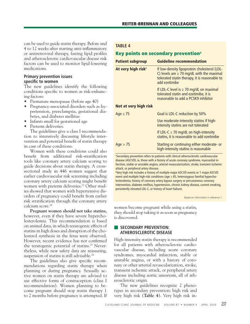
CLEVELAND CLINIC JOURNAL OF MEDICINE VOLUME 87 • NUMBER 4 APRIL 2020 237
REITER-BRENNAN AND COLLEAGUES
can be used to guide statin therapy. Before and 4 to 12 weeks after starting anti-infl ammatory or antiretroviral therapy, fasting lipid profi les and atherosclerotic cardiovascular disease risk factors can be used to monitor lipid-lowering medications.
Primary prevention issues specifi c to womenThe new guidelines identify the following conditions specifi c to women as risk-enhanc-ing factors: • Premature menopause (before age 40)• Pregnancy-associated disorders such as hy-
pertension, preeclampsia, gestational dia-betes, and diabetes mellitus
• Infants small for gestational age• Preterm deliveries. The guidelines give a class I recommenda-tion to intensively discussing lifestyle inter-vention and potential benefi t of statin therapy in case of these conditions. Women with these conditions could also benefi t from additional risk-stratifi cation tools like coronary artery calcium scoring to guide decisions about statin therapy. A cross-sectional study in 446 women suggest that earlier cardiovascular risk screening including coronary artery calcium scoring might benefi t women with preterm deliveries.23 Other stud-ies showed that women with hypertensive dis-orders of pregnancy could benefi t from earlier risk stratifi cation through the coronary artery calcium score.24 Pregnant women should not take statins, however, even if they have severe hypercho-lesterolemia. This recommendation is based on animal data, in which teratogenic effects of statins in high doses and disruption of the cho-lesterol synthesis in the fetus were observed. However, recent evidence has not confi rmed the teratogenic potential of statins.25 Never-theless, while new safety data are reassuring, suspension of statins is still advisable.26
The guidelines also give specifi c recom-mendations regarding statin therapy when planning or during pregnancy. Sexually ac-tive women on statin therapy are advised to use effective forms of contraception (class I recommendation). Women planning to be-come pregnant should stop statin therapy 1 to 2 months before pregnancy is attempted. If
women become pregnant while using a statin, they should stop taking it as soon as pregnancy is discovered.
■ SECONDARY PREVENTION: ATHEROSCLEROTIC DISEASE
High-intensity statin therapy is recommended for all patients with atherosclerotic cardio-vascular disease, including acute coronary syndromes, myocardial infarction, stable or unstable angina, or with a history of coro-nary or other arterial revascularization, stroke, transient ischemic attack, or peripheral artery disease including aortic aneurysm, all of ath-erosclerotic origin. The new guidelines recognize 2 pheno-types in secondary prevention: high risk and very high risk (Table 4). Very high risk in-
TABLE 4
Key points on secondary preventiona
Patient subgroup Guideline recommendation
At very high riskb If low-density lipoprotein cholesterol (LDL-C) levels are ≥ 70 mg/dL with the maximal tolerated statin therapy, it is reasonable to add ezetimibe
If LDL-C level is ≥ 70 mg/dL on maximal tolerated statin and ezetimibe, it isreasonable to add a PCSK9 inhibitor
Not at very high risk
Age ≤ 75 Goal is LDL-C reduction by 50%
Use moderate-intensity statins if high-intensity statins are not tolerated
If LDL-C ≥ 70 mg/dL on high-intensity statins, it is reasonable to add ezetimibe
Age > 75 Starting or continuing either moderate- or high-intensity statins is reasonable
aSecondary prevention refers to patients with clinical atherosclerotic cardiovascular disease (ASCVD), ie, those with a history of acute coronary syndrome, myocardial in-farction, stable or unstable angina, arterial revascularization, stroke, transient ischemic attack, or peripheral artery disease.bVery high risk includes a history of multiple major ASCVD events or 1 major ASCVD event and multiple high-risk conditions (age ≥ 65, heterozygous familial hypercho-lesterolemia, history of coronary artery bypass surgery or percutaneous coronary intervention, diabetes mellitus, hypertension, chronic kidney disease, current smoking, persistently elevated LDL-C, or history of heart failure).
Based on information in reference 1.
238 CLEVELAND CLINIC JOURNAL OF MEDICINE VOLUME 87 • NUMBER 4 APRIL 2020
LIPID GUIDELINES
cludes a history of multiple major atheroscle-rotic cardiovascular disease events or 1 major event and multiple high-risk conditions. The reduction in risk is proportional to the decrease of LDL-C levels. The authors also provide instructions on the use of nonstatin medications as part of secondary prevention. In patients with a very high risk and LDL-C levels higher than 70 mg/dL on maximal tol-erated statin therapy, it is reasonable to add ezetimibe. Further, in patients at very high risk whose LDL-C level remains higher than 70 mg/dL on maximally tolerated statin and ezetimibe therapy, adding a PCSK9 inhibitor is reasonable.
■ MONITORING RESPONSE TO LDL-C-LOWERING THERAPY
As in the last guidelines, the current ones suggest assessing adherence and percentage response after initiating or changing the dose of LDL-C-lowering medications and lifestyle changes, with repeat lipid measurements 4 to 12 weeks after therapy is started. This can be repeated every 3 to 12 months as needed.
■ COST AND VALUE CONSIDERATIONS
The 2018 guidelines comment on the impor-tance of considering the value of treatment in therapy decisions.1 The authors reviewed the cost-effective-ness of PCSK9 inhibitors using simulation models. These revealed that, to be cost-ef-fective, the prices of PCSK9 inhibitors will have to be reduced by at least 70% in the United States from 2018 levels. However, since PCSK9 inhibitors have an incremental cost-effective ratio of $141,800 to $450,000 per quality-adjusted life-year added, the cost-effectiveness of these drugs improves only if used for very high-risk patients. This is refl ect-ed in the current guidelines, which suggest adding PCSK9 inhibitors only after maximal tolerated doses of statins and ezetimibe have not improved LDL-C levels signifi cantly in very high-risk atherosclerotic cardiovascular disease patients or those with a family his-tory of premature atherosclerotic cardiovascu-
lar disease. However, in mid-2018, when the 2018 guidelines were written, the US list pric-es of PCSK9 inhibitors were roughly $14,000 a year; now (in 2019) costs have been reduced to a little more than $6,000 a year.
■ STATIN ADVERSE EFFECTS
The new guidelines additionally address pa-tients’ and clinicians’ fears of adverse effects of statins. They specifi cally recommend that the clinician-patient risk discussion also review possible adverse events and how these can be managed. The guidelines advocate reviewing the net clinical benefi t of statins and comparing the potential for reduction in risk of athero-sclerotic cardiovascular disease with the risk of statin-associated side effects and drug in-teractions (class I recommendation, level of evidence A). Observed adverse effects include myalgias, elevation of creatine kinase, and transaminitis.8 When adverse effects occur, clinicians should lower the dose or dosing frequency, prescribe an alternate statin, or combine statin with nonstatin therapy. If symptoms persist despite these measures, nonstatin therapies with proven effi cacy in randomized controlled trials are recommended. In recent clinical trials, evolocumab27 as well as alirocumab28 performed well in lowering LDL-C in statin-intolerant patients. Muscle symptoms are the most common statin-related adverse effects. Subjective my-algia occurred in 1% to 15% of participants in randomized controlled trials but in 5% to 20% of patients in observational studies. In a class I recommendation, the authors write that pa-tients with statin-associated muscle symptoms should undergo a detailed assessment of symp-toms, and nonstatin causes and predisposing factors should be taken into consideration. Further, statins slightly increase the risk of diabetes mellitus in patients with prediabetes. However, the guidelines clearly state that ther-apy should not be discontinued because of this, as the advantages of statins are much greater than the risk of diabetes mellitus.29,30 ■
Sexually active women on statin therapy should use effective forms of contraception
CLEVELAND CLINIC JOURNAL OF MEDICINE VOLUME 87 • NUMBER 4 APRIL 2020 239
REITER-BRENNAN AND COLLEAGUES
■ REFERENCES 1. Grundy SM, Stone NJ, Bailey AL, et al. 2018 AHA/ACC/AACVPR/AAPA/ABC/
ACPM/ADA/AGS/APhA/ASPC/NLA/PCNA guideline on the management of blood cholesterol: executive summary: a report of the American College of Cardiology/American Heart Association Task Force on Clinical Practice Guidelines. J Am Coll Cardiol 2019; 73(24):3168–3209. doi:10.1016/j.jacc.2018.11.002
2. Arnett DK, Blumenthal RS, Albert MA, et al. 2019 ACC/AHA guideline on the primary prevention of cardiovascular disease: a report of the American College of Cardiology/American Heart Association Task Force on Clinical Practice Guidelines. Circulation 2019; 140(11):e596–e646.doi:10.1161/CIR.0000000000000678
3. Michos ED, McEvoy JW, Blumenthal RS. Lipid management for the prevention of atherosclerotic cardiovascular disease. N Engl J Med 2019; 381(16):1557–1567. doi:10.1056/NEJMra1806939
4. Wilson PWF, Polonsky TS, Miedema MD, Khera A, Kosinski AS, Kuvin JT. Systematic review for the 2018 AHA/ACC/AACVPR/AAPA/ABC/ACPM/ADA/AGS/APhA/ASPC/NLA/PCNA guideline on the management of blood cholesterol: a report of the American College of Cardiology/American Heart Association Task Force on Clinical Practice Guidelines. J Am Coll Cardiol 2019; 73(24):3210–3227. doi:10.1016/j.jacc.2018.11.004
5. Cannon CP, Blazing MA, Giugliano RP, et al; IMPROVE-IT Investigators. Ezetimibe added to statin therapy after acute coronary syndromes. N Engl J Med 2015; 372(25):2387–2397. doi:10.1056/NEJMoa1410489
6. Sabatine MS, Giugliano RP, Keech AC, et al; FOURIER Steering Commit-tee and Investigators. Evolocumab and clinical outcomes in patients with cardiovascular disease. N Engl J Med 2017; 376(18):1713–1722.doi:10.1056/NEJMoa1615664
7. Schwartz GG, Steg PG, Szarek M, et al; ODYSSEY OUTCOMES Committees and Investigators. Alirocumab and cardiovascular outcomes after acute coronary syndrome. N Engl J Med 2018; 379(22):2097–2107.doi:10.1056/NEJMoa1801174
8. Kohli P, Whelton SP, Hsu S, et al. Clinician’s guide to the updated ABCs of cardiovascular disease prevention. J Am Heart Assoc 2014; 3(5):e001098. doi:10.1161/JAHA.114.001098
9. American Diabetes Association. 10. Cardiovascular disease and risk man-agement: standards of medical care in diabetes-2019. Diabetes Care 2019; 42(suppl 1):S103–S123. doi:10.2337/dc19-S010
10. Bittencourt MS, Blaha MJ, Nasir K. Coronary artery calcium—from screen-ing to a personalized shared decision-making tool: the new American prevention guidelines. Arq Bras Cardiol 2019; 112(1):1–2.doi:10.5935/abc.20190008
11. US Preventive Services Task Force; Bibbins-Domingo K, Grossman DC, Curry SJ, et al. Statin use for the primary prevention of cardiovascular disease in adults: US Preventive Services Task Force recommendation statement. JAMA 2016; 316(19):1997–2007. doi:10.1001/jama.2016.15450
12. DeFilippis AP, Young R, Carrubba CJ, et al. An analysis of calibration and discrimination among multiple cardiovascular risk scores in a modern multi-ethnic cohort. Ann Intern Med 2015; 162(4):266–275. doi:10.7326/M14-1281
13. Nasir K, Bittencourt MS, Blaha MJ, et al. Implications of coronary artery calcium testing among statin candidates according to American College of Cardiology/American Heart Association cholesterol management guide-lines: MESA (Multi-Ethnic Study of Atherosclerosis). J Am Coll Cardiol 2015; 66(15):1657–1668. doi:10.1016/j.jacc.2015.07.066
14. McClelland RL, Chung H, Detrano R, Post W, Kronmal RA. Distribution of coronary artery calcium by race, gender, and age: results from the Multi-Ethnic Study of Atherosclerosis (MESA). Circulation 2006; 113(1):30–37. doi:10.1161/CIRCULATIONAHA.105.580696
15. Daviglus ML, Pirzada A, Talavera GA. Cardiovascular disease risk factors
in the Hispanic/Latino population: lessons from the Hispanic Community Health Study/Study of Latinos (HCHS/SOL). Prog Cardiovasc Dis 2014; 57(3):230–236. doi:10.1016/j.pcad.2014.07.006
16. Bild DE, Detrano R, Peterson D, et al. Ethnic differences in coronary calcifi -cation: the Multi-Ethnic Study of Atherosclerosis (MESA). Circulation 2005; 111(10):1313–1320. doi:10.1161/01.CIR.0000157730.94423.4B
17. Doherty TM, Tang W, Detrano RC. Racial differences in the signifi cance of coronary calcium in asymptomatic black and white subjects with coronary risk factors. J Am Coll Cardiol 1999; 34(3):787–794.doi:10.1016/s0735-1097(99)00258-2
18. Kanaya AM, Kandula NR, Ewing SK, et al. Comparing coronary artery cal-cium among U.S. South Asians with four racial/ethnic groups: the MASALA and MESA studies. Atherosclerosis 2014; 234(1):102–107.doi:10.1016/j.atherosclerosis.2014.02.017
19. Orimoloye OA, Budoff MJ, Dardari ZA, et al. Race/ethnicity and the prog-nostic implications of coronary artery calcium for all-cause and cardiovascu-lar disease mortality: the Coronary Artery Calcium Consortium. J Am Heart Assoc 2018; 7(20):e010471. doi:10.1161/JAHA.118.010471
20. Liao JK. Safety and effi cacy of statins in Asians. Am J Cardiol 2007; 99(3):410–414. doi:10.1016/j.amjcard.2006.08.051
21. Taguchi I, Iimuro S, Iwata H, et al. High-dose versus low-dose pitavastatin in Japanese patients with stable coronary artery disease (REAL-CAD): a randomized superiority trial. Circulation 2018; 137(19):1997–2009. doi:10.1161/CIRCULATIONAHA.117.032615
22. Wu HF, Hristeva N, Chang J, et al. Rosuvastatin pharmacokinetics in Asian and white subjects wild type for both OATP1B1 and BCRP under control and inhibited conditions. J Pharm Sci 2017; 106(9):2751–2757.doi:10.1016/j.xphs.2017.03.027
23. Catov JM, Newman AB, Roberts JM, et al; Health ABC Study. Preterm delivery and later maternal cardiovascular disease risk. Epidemiology 2007; 18(6):733–739. doi:10.1097/EDE.0b013e3181567f96
24. Grandi SM, Vallee-Pouliot K, Reynier P, et al. Hypertensive disorders in preg-nancy and the risk of subsequent cardiovascular disease. Paediatr Perinat Epidemiol 2017; 31(5):412–421. doi:10.1111/ppe.12388
25. Bateman BT, Hernandez-Diaz S, Fischer MA, et al. Statins and congenital malformations: cohort study. BMJ 2015; 350:h1035. doi:10.1136/bmj.h1035
26. Haramburu F, Daveluy A, Miremont-Salame G. Statins in pregnancy: new safety data are reassuring, but suspension of treatment is still advisable. BMJ 2015; 350:h1484. doi:10.1136/bmj.h1484
27. Nissen SE, Stroes E, Dent-Acosta RE, et al; GAUSS-3 Investigators. Effi cacy and tolerability of evolocumab vs ezetimibe in patients with muscle-related statin intolerance: the GAUSS-3 randomized clinical trial. JAMA 2016; 315(15):1580–1590. doi:10.1001/jama.2016.3608
28. Moriarty PM, Thompson PD, Cannon CP, et al; ODYSSEY ALTERNATIVE Investigators. Effi cacy and safety of alirocumab vs ezetimibe in statin-intol-erant patients, with a statin rechallenge arm: The ODYSSEY ALTERNATIVE randomized trial. J Clin Lipidol 2015; 9(6):758–769.doi:10.1016/j.jacl.2015.08.006
29. Crandall JP, Mather K, Rajpathak SN, et al, on behalf of the Diabetes Prevention Program (DPP Research Group. Statin use and risk of developing diabetes: results from the Diabetes Prevention Program. BMJ Open Diabe-tes Research and Care 2017;5:e000438. doi:10.1136/bmjdrc-2017-000438
30. Chogtu B, Magazine R, Bairy KL. Statin use and risk of diabetes mellitus. World J. Diabetes 2015; 6(2):352–357. doi:10.4239/wjd.v6.i2.352
Address: Omar Dzaye MD PhD, Johns Hopkins University School of Medi-cine, 600 N. Wolfe Street, Blalock 524, Baltimore, MD 21287; [email protected]
CONTINUING MEDICAL EDUCATION
AMA/PRA Category 1 Credit™To read articles as CME activities and claim credit, go to www.ccjm.org, click on the “CME/MOC” menu, and then “Articles.” Find the articles that you want to read as CME activities and click on the appropriate links. After reading an article, click on the link to complete the activity. You will be asked to log in to your MyCME account (or to create an account). Upon logging in, select “CME,” complete the activity evaluation, and print your certifi cate.Maintenance of Certifi cation (MOC) Points
All Cleveland Clinic Journal of Medicine CME activities are now eligible for MOC points. Physicians may claim MOC points in addition to CME credit.
Follow the instructions for completing and claiming credit for CME activities.
When you log into your MyCME account, select “CME & MOC” and enter your ABIM identifi cation number and your date of birth. The system will store this information after you enter it the fi rst time.
Complete the quiz and evaluation and print your CME certifi cate.
GERD: A practical approach
To scan or not to scan?DXA in postmenopausal women
ACC/AHA lipid guidelines:Personalized care to prevent cardiovascular disease
Release date: April 1, 2020Expiration date: March 31, 2021
April 2020 CME/MOC activitiesEstimated time to complete each activity: up to 1 hour
CME MOC
FINANCIAL DISCLOSURES: In accordance with the Standards for Com-mercial Support issued by the Accreditation Council for Continuing Medical Education (ACCME), the Cleveland Clinic Foundation Center for Continuing Education requires resolution of all faculty confl icts of interest to ensure CME activities are free of commercial bias.
How to earn AMA PRA Category 1 Credit™and Maintenance of Certifi cation Points
AUTHOR AND STAFF DISCLOSURES: Authors’ potential confl icts of inter-est are disclosed within their articles. Cleveland Clinic Journal of Medicine’sstaff disclose the following fi nancial relationships that may be relevant to their editorial roles: Dr. Brian F. Mandell (Editor in Chief) reports consulting, teaching, and speaking for Genentech; consulting for Horizon Pharma; and consulting and membership on advisory committee or review panels for Takeda Pharmaceuticals. Dr. Christian Nasr (Associate Editor) reports service on advisory committees and review panels for Exelixis and Nevro. Dr. Bernard Silver (Associate Editor) reports board membership for Takeda Pharmaceuticals.
DISCLAIMER: The information in these educational activities is provided for general medical education purposes only and is not meant to substitute for the independent medical judgment of a physician relative to diagnostic and treatment options of a specifi c patient’s medical condition. The viewpoints expressed in these CME activities are those of the authors. They do not represent an endorsement by The Cleveland Clinic Foundation. In no event will The Cleveland Clinic Foundation be liable for any decision made or action taken in reliance upon the information provided through these CME activities.
ACCREDITATION: The Cleveland Clinic Foundation Center for Continuing Education is accredited by the Accreditation Council for Continuing Medical Education to provide continuing medical education for physicians.
The Cleveland Clinic Foundation Center for Continuing Education designates each Journal-based online CME activity for a maximum of 1 AMA PRA Category 1 Credit™. Physicians should claim only the credit commensurate with the extent of their participation in the activity.
Each activity may be submitted for American Osteopathic Association Continuing Medi-cal Education credit in Category 2.
ABA MOC: This activity contributes to the CME component of the American Board of Anesthesiology’s redesigned Maintenance of Certifi cation in AnesthesiologyTM (MOCA®) program, known as MOCA 2.0®. Please consult the ABA website, www.theABA.org, for a list of all MOCA 2.0 requirements.Successful completion of this activity enables the participant to earn up to 1.0 MOCA 2.0 points; points earned will be equivalent to the amount of CME credit claimed for the activity. Please note: It is the CME activity provider’s responsibility to submit participant completion information to ACCME for the purpose of granting MOCA 2.0TM points. Maintenance of Certifi cation in AnesthesiologyTM program and MOCA® are registered trademarks of The American Board of Anesthesiology®. MOCA 2.0® is a trademark of The American Board of Anesthesiology®.Your credit will be reported to the ABA within 60 days of claiming credit after the course.
ABIM MOC: Successful completion of this CME activity, which includes participation in the evaluation component, enables the participant to earn up to 1.0 MOC points in the American Board of Internal Medicine’s (ABIM) Maintenance of Certifi cation (MOC) program. Participants will earn MOC points equivalent to the amount of CME credits claimed for the activity. It is the CME activity provider’s responsibility to submit participant completion information to ACCME for the purpose of granting ABIM MOC credit.Your credit will be reported to the ABIM within 60 days of claiming credit after the course.
ABPath MOC: Successful completion of this CME activity, which includes participation in the evaluation component, enables the participant to earn up to 1.0 Lifelong Learning (Part II) / Self-Assessment Module (SAM) credits in the American Board of Pathology’s Maintenance of Certifi cation Program. Participants will earn MOC points equivalent to the number of CME credits claimed for the activity. It is the CME activity provider’s responsibility to submit learner completion information to ACCME for the purpose of granting MOC credit.Your credit will be reported to the ABPath within 60 days of claiming credit after the course.
ABP MOC: Successful completion of this CME activity, which includes participation in the activity and individual assessment of and feedback to the learner, enables the learner to earn up to 1.0 MOC points in the American Board of Pediatrics’ (ABP) Maintenance of Certifi cation (MOC) program. It is the CME activity provider’s responsibility to submit learner completion information to ACCME for the purpose of granting ABP MOC credit. Your credit will be reported to the ABP within 60 days of claiming credit after the course.
ABS MOC: This activity qualifi es for 1.0 self-assessment credit toward Part 2 of the American Board of Surgery (ABS) Maintenance of Certifi cation (MOC) Program.
Please note: It is the participant’s responsibility to self-report their participation to the American Board of Surgery, per board policy.