Transforming Growth Factor-β in Normal Nociceptive Processing and Pathological Pain Models
13
1 23 Molecular Neurobiology ISSN 0893-7648 Volume 45 Number 1 Mol Neurobiol (2012) 45:76-86 DOI 10.1007/s12035-011-8221-1 Transforming Growth Factor-β in Normal Nociceptive Processing and Pathological Pain Models Aquilino Lantero, Mónica Tramullas, Alvaro Díaz & María A. Hurlé
-
Upload
independent -
Category
Documents
-
view
4 -
download
0
Transcript of Transforming Growth Factor-β in Normal Nociceptive Processing and Pathological Pain Models
1 23
Molecular Neurobiology ISSN 0893-7648Volume 45Number 1 Mol Neurobiol (2012) 45:76-86DOI 10.1007/s12035-011-8221-1
Transforming Growth Factor-β in NormalNociceptive Processing and PathologicalPain Models
Aquilino Lantero, Mónica Tramullas,Alvaro Díaz & María A. Hurlé

1 23
Your article is protected by copyright and
all rights are held exclusively by Springer
Science+Business Media, LLC. This e-offprint
is for personal use only and shall not be self-
archived in electronic repositories. If you
wish to self-archive your work, please use the
accepted author’s version for posting to your
own website or your institution’s repository.
You may further deposit the accepted author’s
version on a funder’s repository at a funder’s
request, provided it is not made publicly
available until 12 months after publication.
Transforming Growth Factor-β in Normal NociceptiveProcessing and Pathological Pain Models
Aquilino Lantero & Mónica Tramullas & Alvaro Díaz &
María A. Hurlé
Received: 17 September 2011 /Accepted: 9 November 2011 /Published online: 29 November 2011# Springer Science+Business Media, LLC 2011
Summary The transforming growth factor-β (TGF-β)superfamily is a multifunctional, contextually acting familyof cytokines that participate in the regulation of develop-ment, disease and tissue repair in the nervous system. TheTGF-β family is composed of several members, includingTGF-βs, bone morphogenetic proteins (BMPs) and activins.In this review, we discuss recent findings that suggest TGF-βfunction as important pleiotropic modulators of nociceptiveprocessing both physiologically and under pathologicalpainful conditions. The strategy of increasing TGF-β signal-ing by deleting “BMP and activin membrane-bound inhibitor”(BAMBI), a TGF-β pseudoreceptor, has demonstrated theinhibitory role of TGF-β signaling pathways in normalnociception and in inflammatory and neuropathic painmodels. In particular, strong evidence suggests that TGF-β1is a relevant mediator of nociception and has protective effectsagainst the development of chronic neuropathic pain by
inhibiting the neuroimmune responses of neurons and gliaand promoting the expression of endogenous opioids withinthe spinal cord. In the peripheral nervous system, activins andBMPs function as target-derived differentiation factors thatdetermine and maintain the phenotypic identity and circuitassembly of peptidergic nociceptors. In this context, activin isinvolved in the complex events of neuroinflammation thatmodulate the expression of pain during wound healing. Thesefindings have provided new insights into the physiopathologyof nociception. Moreover, specific members of the TGF-βfamily and their signaling effectors and modulator moleculesmay be promising molecular targets for novel therapeuticagents for pain management.
Keywords TGF-β . Activin . BMP. BAMBI . Nociception .
Neuropathic pain . Pain
Introduction
Pain is an unpleasant but indispensable sensation that warnsthe body to protect itself from severe damage. However,prolonged suffering from pain becomes a seriousburden, and it is a major reason for which patientsseek medical care and pharmacological treatment.Chronic pain is remarkably prevalent; it affects millionsof people worldwide. Epidemiological surveys haverevealed the staggering extent of chronic pain, itshuman costs and socio-economic impact, and also thepaucity of effective methods for pain control [1, 2]. Poorpain control is the result of a deficit in our understandingof the mechanisms of chronic pain, which has limited ourarsenal of pathogenesis-based analgesic therapies. How-ever, there is considerable hope for the development of
A. Lantero :M. Tramullas :A. Díaz :M. A. HurléDepartamento de Fisiología y Farmacología,Facultad de Medicina, Universidad de Cantabria,39011 Santander, Spain
A. Lantero :M. Tramullas :M. A. Hurlé (*)Instituto de Formación e InvestigaciónMarqués de Valdecilla (IFIMAV),39011 Santander, Spaine-mail: [email protected]
A. DíazInstituto de Biomedicina y Biotecnologíade Cantabria IBBTEC (UC-CSIC-IDICAN),Santander, Spain
A. DíazCentro de Investigación Biomédica en Red deSalud Mental (CIBERSAM), Instituto de Salud Carlos III,Madrid, Spain
Mol Neurobiol (2012) 45:76–86DOI 10.1007/s12035-011-8221-1
Author's personal copy
new classes of analgesic drugs that target novel processesthat contribute to clinically relevant pain. In this review,we highlighted recent results that identified TGF-βs asimportant pleiotropic modulators of nociceptive process-ing in physiological and pathological pain. The evidenceindicates that specific members of the TGF-β family andtheir signaling effectors and modulators may be promisingmolecular targets for novel therapeutic agents for painmanagement.
The transforming Growth Factor-β Superfamilyof Cytokines
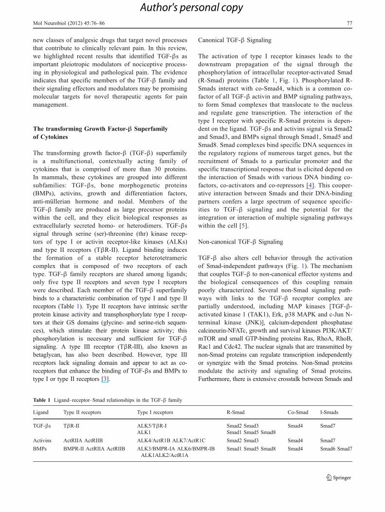
The transforming growth factor-β (TGF-β) superfamilyis a multifunctional, contextually acting family ofcytokines that is comprised of more than 30 proteins.In mammals, these cytokines are grouped into differentsubfamilies: TGF-βs, bone morphogenetic proteins(BMPs), activins, growth and differentiation factors,anti-müllerian hormone and nodal. Members of theTGF-β family are produced as large precursor proteinswithin the cell, and they elicit biological responses asextracellularly secreted homo- or heterodimers. TGF-βssignal through serine (ser)-threonine (thr) kinase recep-tors of type I or activin receptor-like kinases (ALKs)and type II receptors (TβR-II). Ligand binding inducesthe formation of a stable receptor heterotetramericcomplex that is composed of two receptors of eachtype. TGF-β family receptors are shared among ligands;only five type II receptors and seven type I receptorswere described. Each member of the TGF-β superfamilybinds to a characteristic combination of type I and type IIreceptors (Table 1). Type II receptors have intrinsic ser/thrprotein kinase activity and transphosphorylate type I recep-tors at their GS domains (glycine- and serine-rich sequen-ces), which stimulate their protein kinase activity; thisphosphorylation is necessary and sufficient for TGF-βsignaling. A type III receptor (TβR-III), also known asbetaglycan, has also been described. However, type IIIreceptors lack signaling domain and appear to act as co-receptors that enhance the binding of TGF-βs and BMPs totype I or type II receptors [3].
Canonical TGF-β Signaling
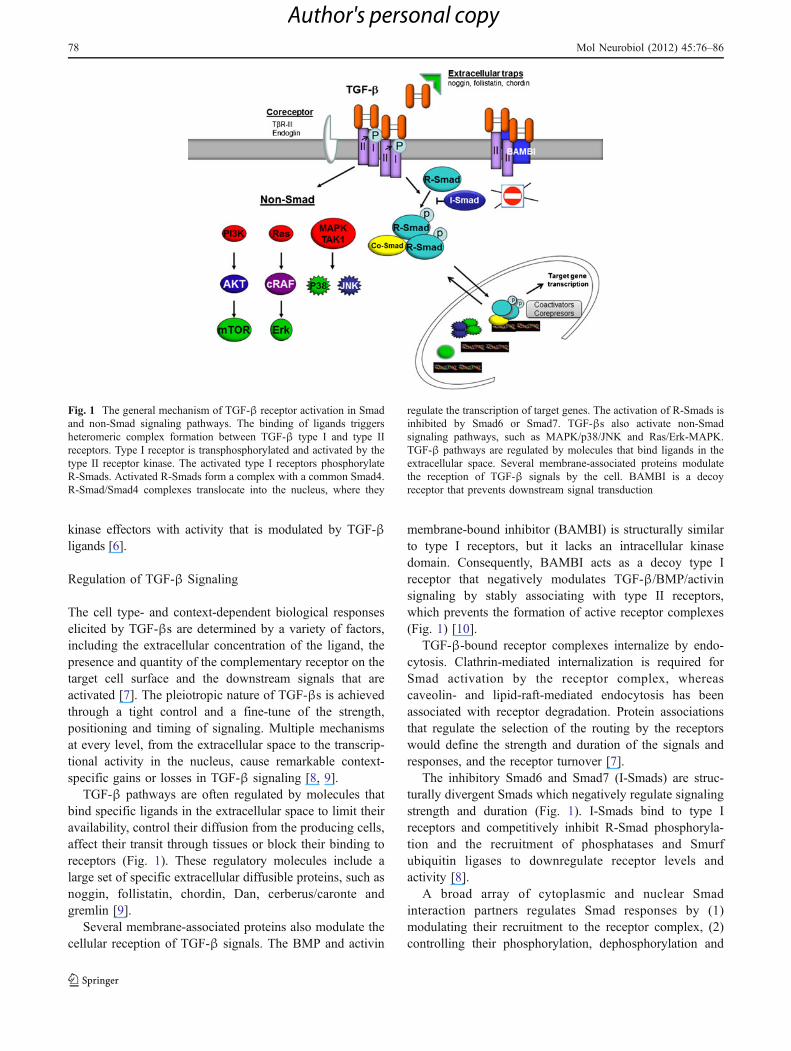
The activation of type I receptor kinases leads to thedownstream propagation of the signal through thephosphorylation of intracellular receptor-activated Smad(R-Smad) proteins (Table 1, Fig. 1). Phosphorylated R-Smads interact with co-Smad4, which is a common co-factor of all TGF-β activin and BMP signaling pathways,to form Smad complexes that translocate to the nucleusand regulate gene transcription. The interaction of thetype I receptor with specific R-Smad proteins is depen-dent on the ligand. TGF-βs and activins signal via Smad2and Smad3, and BMPs signal through Smad1, Smad5 andSmad8. Smad complexes bind specific DNA sequences inthe regulatory regions of numerous target genes, but therecruitment of Smads to a particular promoter and thespecific transcriptional response that is elicited depend onthe interaction of Smads with various DNA binding co-factors, co-activators and co-repressors [4]. This cooper-ative interaction between Smads and their DNA-bindingpartners confers a large spectrum of sequence specific-ities to TGF-β signaling and the potential for theintegration or interaction of multiple signaling pathwayswithin the cell [5].
Non-canonical TGF-β Signaling
TGF-β also alters cell behavior through the activationof Smad-independent pathways (Fig. 1). The mechanismthat couples TGF-β to non-canonical effector systems andthe biological consequences of this coupling remainpoorly characterized. Several non-Smad signaling path-ways with links to the TGF-β receptor complex arepartially understood, including MAP kinases [TGF-β-activated kinase 1 (TAK1), Erk, p38 MAPK and c-Jun N-terminal kinase (JNK)], calcium-dependent phosphatasecalcineurin-NFATc, growth and survival kinases PI3K/AKT/mTOR and small GTP-binding proteins Ras, RhoA, RhoB,Rac1 and Cdc42. The nuclear signals that are transmitted bynon-Smad proteins can regulate transcription independentlyor synergize with the Smad proteins. Non-Smad proteinsmodulate the activity and signaling of Smad proteins.Furthermore, there is extensive crosstalk between Smads and
Table 1 Ligand–receptor–Smad relationships in the TGF-β family
Ligand Type II receptors Type I receptors R-Smad Co-Smad I-Smads
TGF-βs TβR-II ALK5/TβR-I Smad2 Smad3 Smad4 Smad7ALK1 Smad1 Smad5 Smad8
Activins ActRIIA ActRIIB ALK4/ActR1B ALK7/ActR1C Smad2 Smad3 Smad4 Smad7
BMPs BMPR-II ActRIIA ActRIIB ALK3/BMPR-IA ALK6/BMPR-IBALK1ALK2/ActR1A
Smad1 Smad5 Smad8 Smad4 Smad6 Smad7
Mol Neurobiol (2012) 45:76–86 77
Author's personal copy
kinase effectors with activity that is modulated by TGF-βligands [6].
Regulation of TGF-β Signaling
The cell type- and context-dependent biological responseselicited by TGF-βs are determined by a variety of factors,including the extracellular concentration of the ligand, thepresence and quantity of the complementary receptor on thetarget cell surface and the downstream signals that areactivated [7]. The pleiotropic nature of TGF-βs is achievedthrough a tight control and a fine-tune of the strength,positioning and timing of signaling. Multiple mechanismsat every level, from the extracellular space to the transcrip-tional activity in the nucleus, cause remarkable context-specific gains or losses in TGF-β signaling [8, 9].
TGF-β pathways are often regulated by molecules thatbind specific ligands in the extracellular space to limit theiravailability, control their diffusion from the producing cells,affect their transit through tissues or block their binding toreceptors (Fig. 1). These regulatory molecules include alarge set of specific extracellular diffusible proteins, such asnoggin, follistatin, chordin, Dan, cerberus/caronte andgremlin [9].
Several membrane-associated proteins also modulate thecellular reception of TGF-β signals. The BMP and activin
membrane-bound inhibitor (BAMBI) is structurally similarto type I receptors, but it lacks an intracellular kinasedomain. Consequently, BAMBI acts as a decoy type Ireceptor that negatively modulates TGF-β/BMP/activinsignaling by stably associating with type II receptors,which prevents the formation of active receptor complexes(Fig. 1) [10].
TGF-β-bound receptor complexes internalize by endo-cytosis. Clathrin-mediated internalization is required forSmad activation by the receptor complex, whereascaveolin- and lipid-raft-mediated endocytosis has beenassociated with receptor degradation. Protein associationsthat regulate the selection of the routing by the receptorswould define the strength and duration of the signals andresponses, and the receptor turnover [7].
The inhibitory Smad6 and Smad7 (I-Smads) are struc-turally divergent Smads which negatively regulate signalingstrength and duration (Fig. 1). I-Smads bind to type Ireceptors and competitively inhibit R-Smad phosphoryla-tion and the recruitment of phosphatases and Smurfubiquitin ligases to downregulate receptor levels andactivity [8].
A broad array of cytoplasmic and nuclear Smadinteraction partners regulates Smad responses by (1)modulating their recruitment to the receptor complex, (2)controlling their phosphorylation, dephosphorylation and
Fig. 1 The general mechanism of TGF-β receptor activation in Smadand non-Smad signaling pathways. The binding of ligands triggersheteromeric complex formation between TGF-β type I and type IIreceptors. Type I receptor is transphosphorylated and activated by thetype II receptor kinase. The activated type I receptors phosphorylateR-Smads. Activated R-Smads form a complex with a common Smad4.R-Smad/Smad4 complexes translocate into the nucleus, where they
regulate the transcription of target genes. The activation of R-Smads isinhibited by Smad6 or Smad7. TGF-βs also activate non-Smadsignaling pathways, such as MAPK/p38/JNK and Ras/Erk-MAPK.TGF-β pathways are regulated by molecules that bind ligands in theextracellular space. Several membrane-associated proteins modulatethe reception of TGF-β signals by the cell. BAMBI is a decoyreceptor that prevents downstream signal transduction
78 Mol Neurobiol (2012) 45:76–86
Author's personal copy

sumoylation, (3) sequestering Smads from active signalingparticipation or (4) modulating the association of the R-Smad/Smad4 complex with transcription factors, co-activators andco-repressors and, subsequently, the Smad-dependent tran-scription [11], among other mechanisms.
Regulatory Roles of the TGF-β Family in NormalNociceptive Processing and Pathological Pain Models
Nociception Overview
Nociception is a specialized form of sensory signaling thatconveys information about potentially damaging stimuli tothe central nervous system (CNS). The transduction ofnoxious stimuli originates at the peripheral axon terminalsof high-threshold unmyelinated C or thinly myelinated Aδprimary sensory neurons that innervate the target tissue.Functional and molecular nociceptor heterogeneity isassociated with the specific detection of distinct painmodalities. Nociceptor cell bodies reside in the dorsal rootganglia (DRG) of spinal nerves and the sensory ganglia ofcranial nerves. The primary afferent projections fromnociceptors transmit the signal from the periphery to thespinal cord dorsal horn, where they form synapses withsecond-order neurons in laminae I and V. The ascendingfibers of second-order neurons project to third-orderneurons in the thalamus and brainstem, which transmit thenociceptive information to higher brain structures thatinterpret the sensory-discriminative, affective-emotional oraversive dimensions of pain [12].
The release of inflammatory mediators within the woundedarea amplifies peripheral nociceptor transduction. This “pe-ripheral sensitization” is a form of stimulus-evoked functionalplasticity, which normally occurs during the healing process,in which tissue damage enhances the excitability of nocicep-tors to protect the injured area by increasing pain sensitivity[13] .This physiological “nociceptive” pain normally dis-appears upon healing. However, pain persists in a state ofchronic neuropathic pain after nervous system injuries in asmall but significant percentage of the population [14].
In chronic neuropathic pain, a shift in the balancebetween the excitatory and inhibitory mechanisms thatmodulate spinal cord excitability heightens the response ofdorsal horn neurons to incoming afferent signals andincreases the output to the brain. This heightened sensitivityof spinal neurons is called “central sensitization” [15].Sensitization of spinal cord nociceptive neurons contributesto hypersensitive pain behaviors such as allodynia (painproduced by normally innocuous stimuli), hyperalgesia(heightened response to noxious stimuli) and spontaneouspain [16]. However, no direct link between the experimen-tal observation of spinal cord neuron hyperexcitability and
the underlying mechanism of chronic pathological pain hasbeen demonstrated [17]. Increasing evidence in the lastdecade has strengthened that chronic pain is a neuro-immune disorder that is caused by complex interactionsbetween neurons, activated glial cells and inflammatoryimmune cells in the peripheral and CNS [18, 19]. Thishypothesis suggests that the restoration of the balancebetween pro- and anti-inflammatory mechanisms may beused as a novel therapeutic approach to disrupt thedevelopment of chronic pain. Recent findings that supporta relevant regulatory role for the TGF-β family of cytokinesin acute physiological nociception, neuroprotection and anti-inflammation in models of chronic pathological pain arediscussed in the following.
The Influence of BAMBI Deletion in Normal Nociceptionand in Models of Inflammatory and Neuropathic Pain
As mentioned previously, BAMBI is a kinase-deficientpseudoreceptor for TGF-βs which prevents downstreamsignal transduction [10]. BAMBI, together with severaltype I TGF-β receptors, is highly expressed in anti-nociception-relevant areas, such as the cingulate cortex,mesencephalic periaqueductal gray, spinal cord dorsal hornand DRG (Fig. 2) [20].
The induction of a gain in TGF-β signaling through thedeletion of the BAMBI gene has been a valuable strategy forunraveling the involvement of the TGF-β family in paincontrol [20]. BAMBI-KO mice display attenuated nocifensiveresponses in acute pain models, regardless of the modality ofthe noxious stimuli (e.g. thermal, mechanical and chemical/inflammatory). Moreover, BAMBI-KO mice develop lessmechanical allodynia in models of chronic neuropathic pain(Fig. 2). The hypoalgesic phenotype of BAMBI-KO mice canbe reversed by the opioid antagonist naltrexone, which isindicative of an enhanced activity of the endogenous opioidsystem. Spinal cords from BAMBI-KO mice display higherexpression levels of endogenous opioid peptide precursors,including β-endorphins (proopiomelanocortin: POMC) andenkephalins (proenkephalin: PENK), compared to wild-typemice. Opioid peptide precursors are under the transcriptionalcontrol of TGF-β family members in cultured cells [21, 22].Furthermore, exogenous TGF-βs induce the expression ofPOMC and PENK in cultured spinal cord explants [20].Therefore, an increase in TGF-β signaling activity may leadto an increase in the transcription, expression and synapticrelease of endogenous opioids and a hypoalgesic phenotype.
The TGF-β Subfamily Protects Against Nerve-Injury-InducedNeuropathic Pain
Pharmacological approaches have contributed to knowing thespecific contribution of individual TGF-β family members to
Mol Neurobiol (2012) 45:76–86 79
Author's personal copy
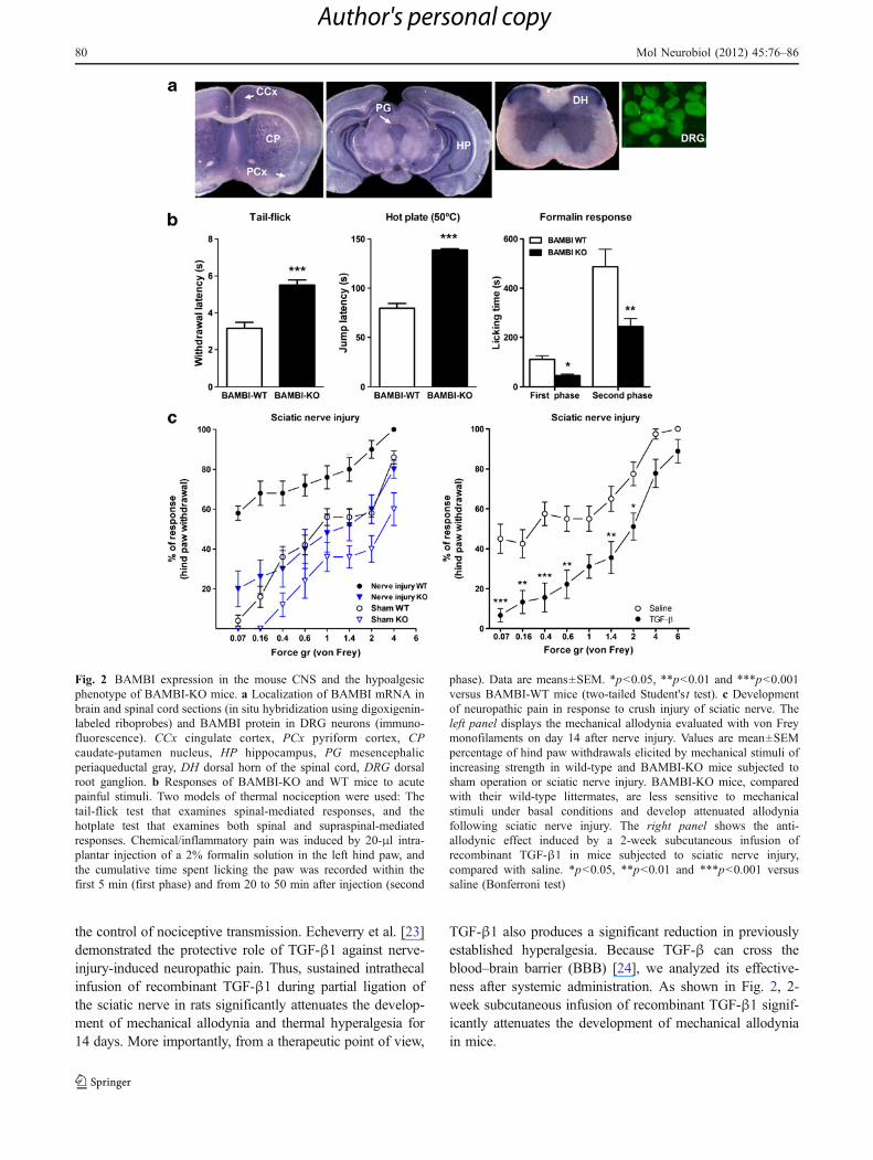
the control of nociceptive transmission. Echeverry et al. [23]demonstrated the protective role of TGF-β1 against nerve-injury-induced neuropathic pain. Thus, sustained intrathecalinfusion of recombinant TGF-β1 during partial ligation ofthe sciatic nerve in rats significantly attenuates the develop-ment of mechanical allodynia and thermal hyperalgesia for14 days. More importantly, from a therapeutic point of view,
TGF-β1 also produces a significant reduction in previouslyestablished hyperalgesia. Because TGF-β can cross theblood–brain barrier (BBB) [24], we analyzed its effective-ness after systemic administration. As shown in Fig. 2, 2-week subcutaneous infusion of recombinant TGF-β1 signif-icantly attenuates the development of mechanical allodyniain mice.
Fig. 2 BAMBI expression in the mouse CNS and the hypoalgesicphenotype of BAMBI-KO mice. a Localization of BAMBI mRNA inbrain and spinal cord sections (in situ hybridization using digoxigenin-labeled riboprobes) and BAMBI protein in DRG neurons (immuno-fluorescence). CCx cingulate cortex, PCx pyriform cortex, CPcaudate-putamen nucleus, HP hippocampus, PG mesencephalicperiaqueductal gray, DH dorsal horn of the spinal cord, DRG dorsalroot ganglion. b Responses of BAMBI-KO and WT mice to acutepainful stimuli. Two models of thermal nociception were used: Thetail-flick test that examines spinal-mediated responses, and thehotplate test that examines both spinal and supraspinal-mediatedresponses. Chemical/inflammatory pain was induced by 20-μl intra-plantar injection of a 2% formalin solution in the left hind paw, andthe cumulative time spent licking the paw was recorded within thefirst 5 min (first phase) and from 20 to 50 min after injection (second
phase). Data are means±SEM. *p<0.05, **p<0.01 and ***p<0.001versus BAMBI-WT mice (two-tailed Student's t test). c Developmentof neuropathic pain in response to crush injury of sciatic nerve. Theleft panel displays the mechanical allodynia evaluated with von Freymonofilaments on day 14 after nerve injury. Values are mean±SEMpercentage of hind paw withdrawals elicited by mechanical stimuli ofincreasing strength in wild-type and BAMBI-KO mice subjected tosham operation or sciatic nerve injury. BAMBI-KO mice, comparedwith their wild-type littermates, are less sensitive to mechanicalstimuli under basal conditions and develop attenuated allodyniafollowing sciatic nerve injury. The right panel shows the anti-allodynic effect induced by a 2-week subcutaneous infusion ofrecombinant TGF-β1 in mice subjected to sciatic nerve injury,compared with saline. *p<0.05, **p<0.01 and ***p<0.001 versussaline (Bonferroni test)
80 Mol Neurobiol (2012) 45:76–86
Author's personal copy
Echeverry et al. [23] attributed the biological effects ofTGF-β1 to a milder neuroinflammatory response to injuryin the spinal cord. These authors provided evidence thatTGF-β1 exerted neuroprotective effects, inhibited theactivation of spinal microglia and astrocytes and decreasedthe upregulation of pro-inflammatory cytokines, such as IL-1β and IL-6, within the spinal cord. These results areconsistent with the anti-inflammatory properties of thiscytokine [25, 26].
The contribution of peripheral mechanisms to the effects ofTGF-β is also important because several TGF-β familymembers (mainly activin and BMPs) exert pro-nociceptiveeffects on peripheral nociceptors. Our results indicate that2 weeks of systemic administration of a TGF-β neutralizingantibody, which does not cross the BBB, significantlyenhanced the development of mechanical allodynia in miceafter sciatic nerve injury (our unpublished observations).These findings support the idea that TGF-β also exertsperipheral anti-nociceptive effects and helps prevent periph-eral nociceptor hypersensitization following nerve injury.
The capability of TGF-β to maintain the integrity of theBBB has also been suggested as a protective mechanismagainst the development of pathological pain followingperipheral inflammatory [27, 28] or neural injuries [29].Systemic or intrathecal treatment with recombinant TGF-β1 prevents carrageenan- and nerve-injury-induced changesin endothelial tight junctions, as well as the functionaldisruption of the BBB. As a consequence, the extravasationof proteins, cytokines and a variety of inflammatorymediators and the infiltration of peripheral inflammatorycells in the spinal cord is significantly reduced. Themechanism involves ALK 5 receptors and the canonicalSmad2/3 signaling pathway [27, 29].
In summary, the TGF-β subfamily protects againstpathological pain development by pleiotropic mechanisms.TGF-β appears to promote the expression of endogenousopioids and inhibit the neuroimmune responses of glialcells and neurons in the spinal cord following peripheralinjuries. In addition, undetermined peripheral mechanismsalso contribute to its anti-allodynic effect. Even at this earlystage of investigation, the evidence suggests that themodulation of TGF-β signaling could be used as a novelpharmacological strategy for the control and treatment ofchronic pain.
Neuropathic Pain is Dissimilarly Modulated by IndividualMembers of the BMP Subfamily
The role of the BMP family in the processing ofnociceptive inputs within the CNS has been scarcelyaddressed, and different results were obtained dependingon the specific BMP analyzed. Recent studies indicate thatthe use of recombinant BMP2 as bone inductor during
surgical spinal fusion elicits a profound Smad-mediatedsignaling response in the spinal cord and local DRG [30].The direct access of BMP2 to the nervous system triggers aneuroinflammatory response that worsens the neurologicrecovery and produces postoperative allodynia in rats [31].This pro-algesic effect could have a clinical correlate aspatients that received recombinant BMP2 were more likelyto report postoperative radicular pain compared withpatients whose fusion surgery did not include use of theprotein [32] However, the presence of a dural tear thatwould favor BMP2 diffusion into the spinal cord paren-chyma did not increase the risk of radiculitis developmentor impair the neurologic recovery in a series of patients[33].
BMP4 has been reported to exert an indirect protectiveeffect against hyperalgesia development. Neuropathic painis a common harmful side effect of neuroepithelial stemcell transplantation therapy for spinal cord injury [34,35]. The pretreatment of glial precursor cells with BMP4(GDAsBMP) prevents the development of thermal hyper-algesia and mechanical allodynia in rats. Such protectionmay be related with the absence of dorsal horn sproutingof CGRP-immunoreactive C-fiber in mice treated withGDAsBMP [36]. In addition, the activation of BMPsignaling pathways in GDAsBMP promotes the expressionof glutamate transporter 1 and AKAP12 [37], which aregenes relevant to preventing inflammatory and neuropath-ic pain [38] and to maintaining the BBB integrity [39]. Bycontrast, intrathecal delivery of BMP4 cDNA incorporatedinto an adenovirus vector does not modify somatosensoryperceptions in mice subjected to spinal cord injury, whileit promotes sensory axon regeneration [40]. Similarly,BMP7 overexpression in the sciatic nerve, DRG andspinal cord neurons by adenoviral gene transfer improvesthe functional recovery after injury but does not affect thedevelopment of neuropathic pain in rats [41].
Activin and BMP Retrograde Signals Modulatethe Expression of the Neuropeptide CGRP by Nociceptorsand Contribute to Neuroinflammatory Pain
The terminal differentiation of many developing neuronsoccurs after the innervation of their target cells. Thisdifferentiation is triggered by extrinsic, target-derivedretrograde signals that are transduced by presynapticcognate receptors. Target tissues contain neurotrophicgrowth factors that support neuronal survival and differen-tiation factors that are critical for the maturation of axonalprojections and the acquisition of neuronal traits, such asthe expression of distinct combinations of neurotransmitters[42, 43]. Epidermal keratinocytes have a dynamic neuro-chemical organization that plays a direct role in themodulation of sensory ending functions [44]. Retrograde
Mol Neurobiol (2012) 45:76–86 81
Author's personal copy
communication between the skin and nociceptive neuronsby members of the TGF-β family regulates the phenotypicidentity of nociceptors and the circuit assembly. Thiscommunication also contributes to the plasticity that effectsdynamic changes in nociceptor sensitivity in physiologicaland pathological situations.
TGF-β receptors are broadly expressed by embryonicand adult neurons in trigeminal dorsal root sensory ganglia,which indicates the potential TGF-β sensitivity of thesecells [45–48]. In addition, members of the TGF-β familyare present in major nociceptor targets such as the skin [46,49, 50].
A well-documented consequence of target-derived TGF-βsignals is the specification of the neurotransmitter phenotypeof neuropeptide-containing DRG neurons. Glutamate is themajor neurotransmitter released by nociceptors, but manynociceptive neurons also contain neuropeptide co-transmittersthat help to transmit and modulate nociceptive inputs.Calcitonin gene-related peptide (CGRP) and substance P(SP) are the main neuropeptides that are expressed by animportant subset of nociceptors that innervate skin and viscera[51]. Embryonic nociceptors contain no detectable CGRPmRNA or protein until their axons contact target tissues andthe peripheral connections are functional, which indicatesthat peptidergic phenotype specification in nociceptiveneurons is dependent on retrograde signals from targets[52, 53].
Studies in vitro have shown that skin-cell-derived factorsinduce de novo CGRP expression in embryonic DRGneurons that are isolated before peripheral target contact,and this effect is antagonized by anti-activin A antibody orthe activin and BMP inhibitory protein, follistatin [45, 54–56]. Furthermore, the addition of recombinant activin A,BMP2, BMP4, BMP6 or BMP7 to cultured embryonicneurons that were dissociated from DRG induces a strongincrease in the expression of CGRP by the specific subsetof unmyelinated C-fiber peptidergic neurons. This effectinvolves the canonical Smad pathway [45, 56]. In postnatalDRG cultures, recombinant activin increases CGRP ex-pression [56–59], while TGF-β1 and TGF-β2 increase thelevels SP [59]. The effect of these cytokines on neuropep-tide expression is synergistic with NGF, which led theauthors to hypothesize that a cooperative interactionbetween Smads and NGF-dependent transcription factorsfurther increases neuropeptide transcription [59]. Overall,these data support a role for TGF-β family members astarget-derived differentiation factors that contribute to thespecification of the functional identity of neuropeptide-containing neurons during development. Moreover, sen-sory neurons remain sensitive to these cytokines duringadulthood [46, 54, 58, 59].
The contribution of TGF-β retrograde signals to themodulation of CGRP transcription by nociceptors in vivo is
highly relevant. CGRP conveys pain information fromnociceptors to second-order neurons in the spinal cord, andit is also an important mediator of normal inflammatorypain response to injuries [60, 61]. CGRP is also involved inthe development of hyperalgesia in inflammation- or nerve-injury-induced chronic pain [62]. Studies in vivo suggestthat the conditional overexpression of BMP4 in mouse skinkeratinocytes reduces the number of non-peptidergic neu-rons in sensory ganglia and the density of target innerva-tion, but the nerve endings from CGRP peptidergic neuronswere markedly increased [63].This phenotype is consistentwith the aforementioned effect of BMP signaling on CGRPexpression in vitro [45]. The phenotypic changes that areinduced by BMP4 overexpression are more prominent withincreasing age, which suggests that the same retrogradecommunication between skin cells and sensory nerveendings is maintained in adults [62].
Activin is involved in the inflammatory response that isassociated with the healing and repair of injured tissues inexperimental adult animal models [56, 64–67]. Althoughthe basal level of activin A in the skin is notably low, itsexpression is strongly increased following cutaneousexcisional wound injury or under inflammation producedby complete Freund's adjuvant [56, 68, 69]. Under theseexperimental conditions, the CGRP-containing sensoryneurons that innervate the skin wound region receive animportant trophic influence from activin released by kerati-nocytes and inflammatory cells, which strongly enhances theexpression of CGRP and other neuropeptides [56]. Further-more, activin injection in the skin of adult naïve ratsproduces a tactile allodynia to mechanical stimulation thatis associated with an increase in the proportion of CGRP-immunoreactive DRG neurons innervating the area ofinjection [57]. In addition, β-CGRP is released by skinkeratinocytes, and its expression is regulated by autocrine/paracrine BMP signaling [62]. Importantly, β-CGRP ismarkedly increased in the skin of patients and mice withchronic inflammatory or neuropathic pain, which suggests anadditional contribution of keratinocyte-dependent release ofβ-CGRP in regulating nociceptor plasticity under conditionsof pathological pain [62].
The CGRP released by peripheral nerve endings andkeratinocytes causes neurogenic vasodilatation, extravasa-tion of serum factors important for wound healing, mastcell degranulation and platelet activation. The resultingaccumulation of pro-inflammatory factors in the injuredarea plays an important role in the development andmaintenance of peripheral sensitization of nociceptors andthe phenotypic changes of DRG neurons that augmentcentral transmission and peripheral sensitization. In addi-tion, CGRP released by primary afferents contributes to thecentral sensitization of second-order neurons [70, 71].Thesensitization of the nociceptive system contributes to pain
82 Mol Neurobiol (2012) 45:76–86
Author's personal copy
hypersensitivity at the site of tissue damage and inflamma-tion. Therefore, the modulation of CGRP-containing noci-ceptors by retrograde activin signaling promotes theheightened nocifensive behaviors that protect againstfurther damage during the normal tissue healing process.An understanding of the role of activin and other TGF-βfamily members in the CGRP-dependent sensitization ofnociceptive signaling could provide further insights into thepathophysiology of highly prevalent pain diseases thatinvolve this neuropeptide, such as migraine and otherprimary headaches [72].
Activin Sensitizes TRPV1 Ionic Currents in DRG Neuronsand Produces Thermal Hyperalgesia in Mice
Transient receptor potential vanilloid 1 (TRPV1) is a non-selective cation channel that is expressed by polymodalnociceptors from somatic and visceral afferents, which aregated by noxious heat and irritant vanilloids, such as capsaicinand extracellular protons, and these nociceptors play a criticalrole in the development of heat hyperalgesia after inflamma-tion [73] and visceral hyperalgesia in inflammatory boweldisease and rectal hypersensitivity [74]. Electrophysiologicalrecordings of cultured DRG neurons suggest that recombi-nant activin A acutely potentiates capsaicin-induced ioniccurrents through TRPV1 by a mechanism that involvesALK4 receptors [48]. The epidermal injection of activininduces a rapid and short-lasting thermal hyperalgesia inwild-type mice, but this response is absent in TRPV1 null-mutant mice. Therefore, the positive modulation of TRPV1by activin may be a novel mechanism underlying sustainedinflammatory pain. Functionally, TRPV1 activation dependson a complicated balance of phosphorylation and dephos-phorylation signals. Pro-nociceptive inflammatory mediatorsincrease TRPV1 phosphorylation by the activation ofmultiple kinases, which sensitizes the TRPV1 response tonoxious heat and regulates inflammatory hyperalgesia [75].The non-canonical activation of PKCε probably mediatesacute TRPV1 sensitization by activin. In cultured DRGneurons, activin induces the translocation of PKCε from thecytosol to the plasma membrane, and this effect is preventedby the blockade of ALK4 receptors. Moreover, a PKCεtranslocation inhibitor peptide prevents activin-A-inducedTRPV1 sensitization. Other protein kinases, such as PKA,MAPK, PI3K, c-src and other PKC isoforms, are not likelyto play a role in TRPV1 sensitization because selectiveinhibitors of these kinases are ineffective [48].
From a therapeutic point of view, pharmacologicalapproaches for the modulation of TRPV1 receptor functionoffers a promising means of pain relief at the nociceptorlevel. TRPV1 agonists produce a long-lasting desensitiza-tion of peripheral nerve terminals to noxious stimuli(nociceptor “defunctionalization”), which is the mechanism
that underlies the proven efficacy of TRPV1 agonists, suchas capsaicin for neuropathic pain relief. TRPV1 antagonistsalso alleviate hyperalgesia associated with inflammatorypain [76]. Because activin has a sensitizing effect onTRPV1 channels, it would be useful to assess whether theinhibition of peripheral activin signaling interferes with thedevelopment of hyperalgesia under painful pathologicalconditions by reducing TRPV1 currents and whetherexogenous activin induces nociceptor defunctionalizationand long-lasting anti-nociception.
Conclusions and Perspectives
Members of the TGF-β family play crucial roles in thepathophysiology of nociception, both at the peripheral sensoryneurons and in the CNS. In models of inflammatory andneuropathic pain, the TGF-β subfamily provides protectionagainst the neuroinflammatory responses, prevents the dis-ruption of the BBB integrity and favors the release of opioidanalgesic mediators. The effects of the BMP family in thecentral processing of nociceptive inputs are largely unknown,and different results have been reported depending on thespecific BMP analyzed. Recombinant BMP2 triggers aneuroinflammatory response that produces allodynia. Con-versely, BMP4 could exert an indirect protective effect againsthyperalgesia development. In the peripheral nervous system,activin and, presumably, BMPs promote heightened nocifen-sive behaviors that protect against further damage during theprocess of tissue healing. Activin also has a sensitizing effecton TRPV1 channels in DRG neurons, which elicits thermalhyperalgesia.
Further work is needed to identify the most promisingcomponents of the TGF-β signaling pathways which mightserve as viable therapeutic targets for the treatment ofpathological pain. Also, due to the pleiotropic effects ofthese cytokines, a detailed investigation of the off-targeteffects that can potentially lead to unwanted toxicities isnecessary before successful development of TGF-β-basedtherapeutic strategies.
Acknowledgments This work was supported by grants fromInstituto de Salud Carlos III (RD06/001/1016), Ministerio de Cienciae Innovación (SAF2010-16894) and Fundación La Marató de TV3(Grant 072131).
Disclosures None.
REFERENCES
1. Elliott AM, Smith BH, Penny KI, Smith WC, Chambers WA(1999) The epidemiology of chronic pain in the community.Lancet 354:1248–1252. doi:10.1016/S0140-6736(99)03057-3
Mol Neurobiol (2012) 45:76–86 83
Author's personal copy
2. Breivik H, Collett B, Ventafridda V, Cohen R, Gallacher D (2006)Survey of chronic pain in Europe: prevalence, impact on dailylife, and treatment. Eur J Pain 10:287–333. doi:10.1016/j.ejpain.2005.06.009
3. Wharton K, Derynck R (2009) TGFbeta family signaling: novelinsights in development and disease. Development 136:3691–3697. doi:10.1242/dev.040584
4. Schmierer B, Hill CS (2007) TGFβ-Smad signal transduction:molecular specificity and functional flexibility. Nat Rev Mol CellBiol 8:970–982. doi:10.1038/nrm2297
5. Miyazono K, Maeda S, Imamura T (2005) BMP receptorsignaling: transcriptional targets, regulation of signals, andsignaling cross-talk. Cytokine Growth Factor Rev 16:251–263.doi:10.1016/j.cytogfr.2005.01.009
6. Zhang YE (2009) Non-Smad pathways in TGF-beta signaling.Cell Res 19:128–139. doi:10.1038/cr.2008.328
7. Kang JS, Liu C, Derynck R (2009) New regulatory mechanismsof TGF-beta receptor function. Trends Cell Biol 19:385–394.doi:10.1016/j.tcb.2009.05.008
8. Moustakas A, Heldin CH (2009) The regulation of TGFbeta signaltransduction. Development 136(22):3699–3714. doi:10.1242/dev.030338
9. Umulis D, O'Connor MB, Blair SS (2009) The extracellularregulation of bone morphogenetic protein signaling. Development136:3715–3728. doi:10.1242/dev.031534
10. Onichtchouk D, Chen YG, Dosch R, Gawantka V, Delius H,Massagué J, Niehrs C (1999) Silencing of TGF-beta signalling bythe pseudoreceptor BAMBI. Nature 401:480–485. doi:10.1038/46794
11. Itoh S, ten Dijke P (2007) Negative regulation of TGF-b receptor/Smad signal transduction. Curr Opin Cell Biol 19:176–184.doi:10.1016/j.ceb.2007.02.015
12. Basbaum AI, Bautista DM, Scherrer G, Julius D (2009) Cellularandmolecular mechanisms of pain. Cell 139:267–284. doi:10.1016/j.cell.2009.09.028
13. Cervero F (2009) Pain: friend or foe? A neurobiologic perspec-tive: the 2008 Bonica Award Lecture. Reg Anesth Pain Med34:569–574. doi:10.1097/AAP.0b013e3181b4c517
14. Bouhassira D, Lantéri-Minet M, Attal N, Laurent B, Touboul C(2008) Prevalence of chronic pain with neuropathic characteristicsin the general population. Pain 136:380–387. doi:10.1016/j.pain.2007.08.013
15. D'Mello R, Dickenson AH (2008) Spinal cord mechanisms ofpain. Br J Anaesth 101:8–16. doi:10.1093/bja/aen088
16. Costigan M, Scholz J, Woolf CJ (2009) Neuropathic pain: amaladaptive response of the nervous system to damage. Annu RevNeurosci 32:1–32. doi:10.1146/annurev.neuro.051508.135531
17. Cervero F (2009) Spinal cord hyperexcitability and its role in painand hyperalgesia. Exp Brain Res 196:129–137. doi:10.1007/s00221-009-1789-2
18. Milligan ED, Watkins LR (2009) Pathological and protective rolesof glia in chronic pain. Nat Rev Neurosci 10:23–36. doi:10.1038/nrn2533
19. Austin PJ, Moalem-Taylor G (2010) The neuro-immune balance inneuropathic pain: involvement of inflammatory immune cells,immune-like glial cells and cytokines. J Neuroimmunol 229:26–50.doi:10.1016/j.jneuroim.2010.08.013
20. Tramullas M, Lantero A, Díaz A, Morchón N, Merino D, Villar A,Buscher D, Merino R, Hurlé JM, Izpisúa-Belmonte JC, Hurlé MA(2010) BAMBI (bone morphogenetic protein and activinmembrane-bound inhibitor) reveals the involvement of the trans-forming growth factor-beta family in pain modulation. J Neurosci30:1502–1511. doi:10.1523/JNEUROSCI.2584-09.2010
21. Kamphuis S, Kavelaars A, Brooimans R, KuisW, Zegers BJ, HeijnenCJ (1997) T helper 2 cytokines induce preproenkephalin mRNAexpression and proenkephalin A in human peripheral blood
mononuclear cells. J Neuroimmunol 79:91–99. doi:10.1016/S0165-5728(97)00113-6
22. Nudi M, Ouimette JF, Drouin J (2005) Bone morphogenic protein(Smad)-mediated repression of proopiomelanocortin transcriptionby interference with Pitx/Tpit activity. Mol Endocrinol 19:1329–1342. doi:10.1210/me.2004-0425
23. Echeverry S, Shi XQ, Haw A, Liu H, Zhang ZW, Zhang J (2009)Transforming growth factor-beta1 impairs neuropathic painthrough pleiotropic effects. Mol Pain 5:16. doi:10.1186/1744-8069-5-16
24. McLennan IS, Weible MW 2nd, Hendry IA, Koishi K (2005)Transport of transforming growth factor-beta 2 across the blood–brain barrier. Neuropharmacology 48:274–282. doi:10.1016/j.neuropharm.2004.10.005
25. Bottner M, Krieglstein K, Unsicker K (2000) The TGF-βs:structure, signalling and roles in nervous system development andfunctions. J Neurochem 75:2227–2240. doi:10.1046/j.1471-4159.2000.0752227.x
26. Brionne TC, Tesseur I, Masliah E, Wyss-Coray T (2003) Loss ofTGF-beta 1 leads to increased neuronal cell death and micro-gliosis in mouse brain. Neuron 40:1133–1145. doi:10.1016/S0896-6273(03)00766-9
27. Ronaldson PT, Demarco KM, Sanchez-Covarrubias L, SolinskyCM, Davis TP (2009) Transforming growth factor-beta signalingalters substrate permeability and tight junction protein expressionat the blood–brain barrier during inflammatory pain. J CerebBlood Flow Metab 29:1084–1098. doi:10.1038/jcbfm.2009.32
28. Ronaldson PT, Finch JD, Demarco KM, Quigley CE, Davis TP(2011) Inflammatory pain signals an increase in functionalexpression of organic anion transporting polypeptide 1a4 at theblood–brain barrier. J Pharmacol Exp Ther 336:827–839.doi:10.1124/jpet.110.174151
29. Echeverry S, Shi XQ, Rivest S, Zhang J (2011) Peripheral nerveinjury alters blood-spinal cord barrier functional and molecularintegrity through a selective inflammatory pathway. J Neurosci31:10819–11028. doi:10.1523/JNEUROSCI.1642-11.2011
30. Dmitriev AE, Farhang S, Lehman RA Jr, Ling GS, Symes AJ (2010)Bone morphogenetic protein-2 used in spinal fusion with spinal cordinjury penetrates intrathecally and elicits a functional signalingcascade. Spine J 10:16–25. doi:10.1016/j.spinee.2009.10.003
31. Dmitriev AE, Lehman RA Jr, Symes AJ (2011) Bone morphoge-netic protein-2 and spinal arthrodesis: the basic science perspec-tive on protein interaction with the nervous system. Spine J11:500–505. doi:10.1016/j.spinee.2011.05.002
32. Carragee EJ, Hurwitz EL, Weiner BK (2011) A critical review ofrecombinant human bone morphogenetic protein-2 trials in spinalsurgery: emerging safety concerns and lessons learned. Spine J11:471–491. doi:10.1016/j.spinee.2011.04.023
33. Glassman SD, Gum JL, Crawford CH 3rd, Shields CB, CarreonLY (2011) Complications with recombinant human bone morpho-genetic protein-2 in posterolateral spine fusion associated with adural tear. Spine J 11:522–526. doi:10.1016/j.spinee.2010.05.016
34. Hofstetter CP, Holmström NA, Lilja JA, Schweinhardt P, Hao J,Spenger C, Wiesenfeld-Hallin Z, Kurpad SN, Frisén J, Olson L(2005) Allodynia limits the usefulness of intraspinal neural stem cellgrafts; directed differentiation improves outcome. Nat Neurosci8:346–353. doi:10.1038/nn1405
35. Macias MY, Syring MB, Pizzi MA, Crowe MJ, Alexanian AR,Kurpad SN (2006) Pain with no gain: allodynia following neuralstem cell transplantation in spinal cord injury. Exp Neurol201:335–348. doi:10.1016/j.expneurol.2006.04.035
36. Davies JE, Pröschel C, Zhang N, Noble M, Mayer-Pröschel M,Davies SJ (2008) Transplanted astrocytes derived from BMP- orCNTF-treated glial-restricted precursors have opposite effects onrecovery and allodynia after spinal cord injury. J Biol 7:24.doi:10.1186/jbiol85
84 Mol Neurobiol (2012) 45:76–86
Author's personal copy
37. Davies SJ, Shih CH, Noble M, Mayer-Proschel M, Davies JE,Proschel C (2011) Transplantation of specific human astrocytespromotes functional recovery after spinal cord injury. PLoS One6:e17328. doi:10.1371/journal.pone.0017328
38. Maeda S, Kawamoto A, Yatani Y, Shirakawa H, Nakagawa T,Kaneko S (2008) Gene transfer of GLT-1, a glial glutamatetransporter, into the spinal cord by recombinant adenovirusattenuates inflammatory and neuropathic pain in rats. Mol Pain4:65. doi:10.1186/1744-8069-4-65
39. Choi YK, Kim JH, Kim WJ, Lee HY, Park JA, Lee SW, YoonDK, Kim HH, Chung H, Yu YS (2007) Kim KW (2007) AKAP12regulates human blood-retinal barrier formation by downregula-tion of hypoxia-inducible factor-1alpha. J Neurosci 27:4472–4481. doi:10.1523/JNEUROSCI.5368-06.2007
40. Parikh P, Hao Y, Hosseinkhani M, Patil SB, Huntley GW, Tessier-Lavigne M, Zou H (2011) Regeneration of axons in injured spinalcord by activation of bone morphogenetic protein/Smad1 signalingpathway in adult neurons. Proc Natl Acad Sci U S A 108:E99–E107.doi:10.1073/pnas.1100426108
41. Tsai MJ, Pan HA, Liou DY, Weng CF, Hoffer BJ, Cheng H (2010)Adenoviral gene transfer of bone morphogenetic protein-7enhances functional recovery after sciatic nerve injury in rats.Gene Ther 17:1214–1224. doi:10.1038/gt.2010.72
42. Hippenmeyer S, Kramer I, Arber S (2004) Control of neuronalphenotype: what targets tell the cell bodies. Trends Neurosci27:482–488. doi:10.1016/j.tins.2004.05.012
43. Sanyal S, Kim SM, Ramaswami M (2004) Retrograde regulationin the CNS; neuron-specific interpretations of TGF-beta signaling.Neuron 41:845–848. doi:10.1016/S0896-6273(04)00152-7
44. Lumpkin EA, Caterina MJ (2007) Mechanisms of sensorytransduction in the skin. Nature 445:858–865. doi:10.1038/nature05662
45. Ai X, Cappuzzello J, Hall AK (1999) Activin and bonemorphogenetic proteins induce calcitonin gene-related peptide inembryonic sensory neurons in vitro. Mol Cell Neurosci 14:506–518. doi:10.1006/mcne.1999.0798
46. Hall AK, Burke RM, Anand M, Dinsio KJ (2002) Activin andbone morphogenetic proteins are present in perinatal sensoryneuron target tissues that induce neuropeptides. J Neurobiol52:52–60. doi:10.1002/neu.10068
47. Zhang D, Mehler MF, Song Q, Kessler JA (1998) Development ofbone morphogenetic protein receptors in the nervous system andpossible roles in regulating trkC expression. J Neurosci 18:3314–3326. doi:10.1002/neu.10068
48. Zhu W, Xu P, Cuascut FX, Hall AK, Oxford GS (2007) Activinacutely sensitizes dorsal root ganglion neurons and induceshyperalgesia via PKC-mediated potentiation of transient receptorpotential vanilloid I. J Neurosci 27:13770–13780. doi:10.1523/JNEUROSCI.3822-07.2007
49. Winnier G, Blessing M, Labosky PA, Hogan BL (1995) Bonemorphogenetic protein-4 is required for mesoderm formation andpatterning in the mouse. Genes Dev 9:2105–2116. doi:10.1101/gad.9.17.2105
50. Takahashi H, Ikeda T (1996) Transcripts for two members of thetransforming growth factor-beta superfamily BMP-3 and BMP-7are expressed in developing rat embryos. Dev Dyn 207:439–449.doi:10.1002/(SICI)1097-0177(199612
51. Yu LC, Hou JF, Fu FH, Zhang YX (2009) Roles of calcitoningene-related peptide and its receptors in pain-related behavioralresponses in the central nervous system. Neurosci Biobehav Rev33:1185–1191. doi:10.1016/j.neubiorev.2009.03.009
52. Hall AK, Ai X, Hickman GE, MacPhedran SE, Nduaguba CO,Robertson CP (1997) The generation of neuronal heterogeneity ina rat sensory ganglion. J Neurosci 17:2775–2784
53. Marti E, Gibson SJ, Polak JM, Facer P, Springall DR, VanAswegen G, Aitchison M, Koltzenburg M (1987) Ontogeny of
peptide- and amine-containing neurones in motor, sensory, andautonomic regions of rat and human spinal cord, dorsal rootganglia, and rat skin. J Comp Neurol 266:332–359
54. Hall AK, Dinsio KJ, Cappuzzello J (2001) Skin cell induction ofcalcitonin gene-related peptide in embryonic sensory neurons in vitroinvolves activin. Dev Biol 229:263–270. doi:10.1006/dbio.2000.9966
55. Hamza MA, Higgins DM, Ruyechan WT (2007) Two alphaher-pesvirus latency-associated gene products influence calcitoningene-related peptide levels in rat trigeminal neurons. NeurobiolDis 25:553–560. doi:10.1016/j.nbd.2006.10.016
56. Cruise BA, Xu P, Hall AK (2004) Wounds increase activin in skinand a vasoactive neuropeptide in sensory ganglia. Dev Biol271:1–10. doi:10.1016/j.ydbio.2004.04.003
57. Xu P, Van Slambrouck C, Berti-Mattera L, Hall AK (2005)Activin induces tactile allodynia and increases calcitonin gene-related peptide after peripheral inflammation. J Neurosci 25:9227–9235. doi:10.1523/JNEUROSCI.3051-05.2005
58. Xu P, Hall AK (2006) The role of activin in neuropeptideinduction and pain sensation. Dev Biol 299:303–309.doi:10.1016/j.ydbio.2006.08.026
59. Xu P, Hall AK (2007) Activin acts with nerve growth factor toregulate calcitonin gene-related peptide mRNA in sensoryneurons. Neuroscience 150:665–674. doi:10.1016/j.neurosci-ence.2007.09.041
60. Salmon AM, Damaj MI, Marubio LM, Epping-Jordan MP, Merlo-Pich E, Changeux JP (2001) Altered neuroadaptation in opiatedependence and neurogenic inflammatory nociception in alphaCGRP-deficient mice. Nat Neurosci 4:357–358. doi:10.1038/86001
61. Zhang Z, Winborn CS, Marquez de Prado B, Russo AF (2007)Sensitization of calcitonin gene-related peptide receptors by receptoractivity-modifying protein-1 in the trigeminal ganglion. J Neurosci27:2693–2703. doi:10.1523/JNEUROSCI.4542-06.2007
62. Hou Q, Barr T, Gee L, Vickers J, Wymer J, Borsani E, Rodella L,Getsios S, Burdo T, Eisenberg E, Guha U, Lavker R, Kessler J,Chittur S, Fiorino D, Rice F, Albrecht P. Keratinocyte expressionof calcitonin gene-related peptide β: implications for neuropathicand inflammatory pain mechanisms. Pain 152:2036–2051.doi:10.1016/j.pain.2011.04.033
63. Guha U, Gomes WA, Samanta J, Gupta M, Rice FL, Kessler JA(2004) Target-derived BMP signaling limits sensory neuronnumber and the extent of peripheral innervation in vivo.Development 131:1175–1186. doi:10.1242/dev.01013
64. Hübner G, Hu Q, Smola H, Werner S (1996) Strong induction ofactivin expression after injury suggests an important role ofactivin in wound repair. Dev Biol 173:490–498. doi:10.1006/dbio.1996.0042
65. Wankell M, Munz B, Hübner G, Hans W, Wolf E, Goppelt A,Werner S (2001) Impaired wound healing in transgenic miceoverexpressing the activin antagonist follistatin in the epidermis.EMBO J 20:5361–5372. doi:10.1093/emboj/20.19.5361
66. Bamberger C, Schärer A, Antsiferova M, Tychsen B, Pankow S,Müller M, Rülicke T, Paus R, Werner S (2005) Activin controlsskin morphogenesis and wound repair predominantly via stromalcells and in a concentration-dependent manner via keratinocytes.Am J Pathol 167:733–747. doi:10.1016/S0002-9440(10)62047-0
67. Antsiferova M, Klatte JE, Bodó E, Paus R, Jorcano JL, MatzukMM, Werner S, Kögel H (2009) Keratinocyte-derived follistatinregulates epidermal homeostasis and wound repair. Lab Invest89:131–141. doi:10.1038/labinvest.2008.120
68. Hübner G, Hu Q, Smola H (1996) Werner S (1996) Stronginduction of activin expression after injury suggests an importantrole of activin in wound repair. Dev Biol 173(2):490–498.doi:10.1006/dbio.1996.0042
69. Sulyok S, Wankell M, Alzheimer C, Werner S (2004) Activin: animportant regulator of wound repair, fibrosis, and neuroprotection.Mol Cell Endocrinol 225:127–132. doi:10.1016/j.mce.2004.07.011
Mol Neurobiol (2012) 45:76–86 85
Author's personal copy

70. Zhang L, Hoff AO, Wimalawansa SJ, Cote GJ, Gagel RF, WestlundKN (2001) Arthritic calcitonin/alpha calcitonin gene-related peptideknockout mice have reduced nociceptive hypersensitivity. Pain89:265–273. doi:10.1016/S0304-3959(00)00378-X
71. Benemei S, Nicoletti P, Capone JG, Geppetti P (2009) CGRPreceptors in the control of pain and inflammation. Curr OpinPharmacol 9:9–14. doi:10.1016/j.coph.2008.12.007
72. Ho TW, Edvinsson L, Goadsby PJ (2010) CGRP and its receptorsprovide new insights into migraine pathophysiology. Nat RevNeurol 6:573–582. doi:10.1038/nrneurol.2010.127
73. Davis JB, Gray J, Gunthorpe MJ, Hatcher JP, Davey PT, Overend P,Harries MH, Latcham J, Clapham C, Atkinson K, Hughes SA, RanceK, Grau E, Harper AJ, Pugh PL, Rogers DC, Bingham S, Randall A,
Sheardown SA (2000) Vanilloid receptor-1 is essential for inflammatorythermal hyperalgesia. Nature 405:183–187. doi:10.1038/35012076
74. Cervero F, Laird JMA (2004) Understanding the signaling andtransmission of visceral nociceptive events. J Neurobiol 61:45–54.doi:10.1002/neu.20084
75. Ma W, Quirion R (2007) Inflammatory mediators modulating thetransient receptor potential vanilloid 1 receptor: therapeutic targetsto treat inflammatory and neuropathic pain. Expert Opin TherTargets 11:307–320. doi:10.1517/14728222.11.3.307
76. Gunthorpe MJ, Chizh BA (2009) Clinical development ofTRPV1 antagonists: targeting a pivotal point in the painpathway. Drug Discov Today 14:56–67. doi:10.1016/j.drudis.2008.11.005
86 Mol Neurobiol (2012) 45:76–86
Author's personal copy