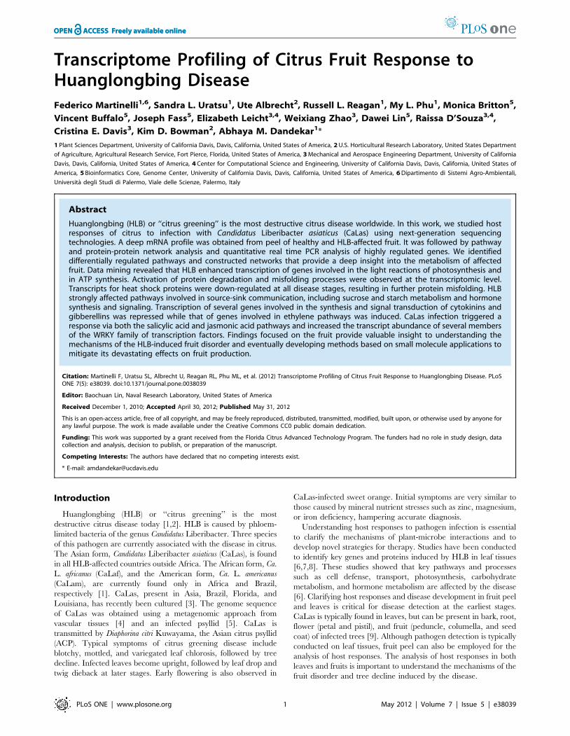
Transcriptome Profiling of Citrus Fruit Response to Huanglongbing Disease
16
Transcriptome Profiling of Citrus Fruit Response to Huanglongbing Disease Federico Martinelli 1,6 , Sandra L. Uratsu 1 , Ute Albrecht 2 , Russell L. Reagan 1 , My L. Phu 1 , Monica Britton 5 , Vincent Buffalo 5 , Joseph Fass 5 , Elizabeth Leicht 3,4 , Weixiang Zhao 3 , Dawei Lin 5 , Raissa D’Souza 3,4 , Cristina E. Davis 3 , Kim D. Bowman 2 , Abhaya M. Dandekar 1 * 1 Plant Sciences Department, University of California Davis, Davis, California, United States of America, 2 U.S. Horticultural Research Laboratory, United States Department of Agriculture, Agricultural Research Service, Fort Pierce, Florida, United States of America, 3 Mechanical and Aerospace Engineering Department, University of California Davis, Davis, California, United States of America, 4 Center for Computational Science and Engineering, University of California Davis, Davis, California, United States of America, 5 Bioinformatics Core, Genome Center, University of California Davis, Davis, California, United States of America, 6 Dipartimento di Sistemi Agro-Ambientali, Universita ` degli Studi di Palermo, Viale delle Scienze, Palermo, Italy Abstract Huanglongbing (HLB) or ‘‘citrus greening’’ is the most destructive citrus disease worldwide. In this work, we studied host responses of citrus to infection with Candidatus Liberibacter asiaticus (CaLas) using next-generation sequencing technologies. A deep mRNA profile was obtained from peel of healthy and HLB-affected fruit. It was followed by pathway and protein-protein network analysis and quantitative real time PCR analysis of highly regulated genes. We identified differentially regulated pathways and constructed networks that provide a deep insight into the metabolism of affected fruit. Data mining revealed that HLB enhanced transcription of genes involved in the light reactions of photosynthesis and in ATP synthesis. Activation of protein degradation and misfolding processes were observed at the transcriptomic level. Transcripts for heat shock proteins were down-regulated at all disease stages, resulting in further protein misfolding. HLB strongly affected pathways involved in source-sink communication, including sucrose and starch metabolism and hormone synthesis and signaling. Transcription of several genes involved in the synthesis and signal transduction of cytokinins and gibberellins was repressed while that of genes involved in ethylene pathways was induced. CaLas infection triggered a response via both the salicylic acid and jasmonic acid pathways and increased the transcript abundance of several members of the WRKY family of transcription factors. Findings focused on the fruit provide valuable insight to understanding the mechanisms of the HLB-induced fruit disorder and eventually developing methods based on small molecule applications to mitigate its devastating effects on fruit production. Citation: Martinelli F, Uratsu SL, Albrecht U, Reagan RL, Phu ML, et al. (2012) Transcriptome Profiling of Citrus Fruit Response to Huanglongbing Disease. PLoS ONE 7(5): e38039. doi:10.1371/journal.pone.0038039 Editor: Baochuan Lin, Naval Research Laboratory, United States of America Received December 1, 2010; Accepted April 30, 2012; Published May 31, 2012 This is an open-access article, free of all copyright, and may be freely reproduced, distributed, transmitted, modified, built upon, or otherwise used by anyone for any lawful purpose. The work is made available under the Creative Commons CC0 public domain dedication. Funding: This work was supported by a grant received from the Florida Citrus Advanced Technology Program. The funders had no role in study design, data collection and analysis, decision to publish, or preparation of the manuscript. Competing Interests: The authors have declared that no competing interests exist. * E-mail: [email protected] Introduction Huanglongbing (HLB) or ‘‘citrus greening’’ is the most destructive citrus disease today [1,2]. HLB is caused by phloem- limited bacteria of the genus Candidatus Liberibacter. Three species of this pathogen are currently associated with the disease in citrus. The Asian form, Candidatus Liberibacter asiaticus (CaLas), is found in all HLB-affected countries outside Africa. The African form, Ca. L. africanus (CaLaf), and the American form, Ca. L. americanus (CaLam), are currently found only in Africa and Brazil, respectively [1]. CaLas, present in Asia, Brazil, Florida, and Louisiana, has recently been cultured [3]. The genome sequence of CaLas was obtained using a metagenomic approach from vascular tissues [4] and an infected psyllid [5]. CaLas is transmitted by Diaphorina citri Kuwayama, the Asian citrus psyllid (ACP). Typical symptoms of citrus greening disease include blotchy, mottled, and variegated leaf chlorosis, followed by tree decline. Infected leaves become upright, followed by leaf drop and twig dieback at later stages. Early flowering is also observed in CaLas-infected sweet orange. Initial symptoms are very similar to those caused by mineral nutrient stresses such as zinc, magnesium, or iron deficiency, hampering accurate diagnosis. Understanding host responses to pathogen infection is essential to clarify the mechanisms of plant-microbe interactions and to develop novel strategies for therapy. Studies have been conducted to identify key genes and proteins induced by HLB in leaf tissues [6,7,8]. These studies showed that key pathways and processes such as cell defense, transport, photosynthesis, carbohydrate metabolism, and hormone metabolism are affected by the disease [6]. Clarifying host responses and disease development in fruit peel and leaves is critical for disease detection at the earliest stages. CaLas is typically found in leaves, but can be present in bark, root, flower (petal and pistil), and fruit (peduncle, columella, and seed coat) of infected trees [9]. Although pathogen detection is typically conducted on leaf tissues, fruit peel can also be employed for the analysis of host responses. The analysis of host responses in both leaves and fruits is important to understand the mechanisms of the fruit disorder and tree decline induced by the disease. PLoS ONE | www.plosone.org 1 May 2012 | Volume 7 | Issue 5 | e38039
-
Upload
independent -
Category
Documents
-
view
0 -
download
0
Transcript of Transcriptome Profiling of Citrus Fruit Response to Huanglongbing Disease
Transcriptome Profiling of Citrus Fruit Response toHuanglongbing DiseaseFederico Martinelli1,6, Sandra L. Uratsu1, Ute Albrecht2, Russell L. Reagan1, My L. Phu1, Monica Britton5,
Vincent Buffalo5, Joseph Fass5, Elizabeth Leicht3,4, Weixiang Zhao3, Dawei Lin5, Raissa D’Souza3,4,
Cristina E. Davis3, Kim D. Bowman2, Abhaya M. Dandekar1*
1 Plant Sciences Department, University of California Davis, Davis, California, United States of America, 2 U.S. Horticultural Research Laboratory, United States Department
of Agriculture, Agricultural Research Service, Fort Pierce, Florida, United States of America, 3 Mechanical and Aerospace Engineering Department, University of California
Davis, Davis, California, United States of America, 4 Center for Computational Science and Engineering, University of California Davis, Davis, California, United States of
America, 5 Bioinformatics Core, Genome Center, University of California Davis, Davis, California, United States of America, 6 Dipartimento di Sistemi Agro-Ambientali,
Universita degli Studi di Palermo, Viale delle Scienze, Palermo, Italy
Abstract
Huanglongbing (HLB) or ‘‘citrus greening’’ is the most destructive citrus disease worldwide. In this work, we studied hostresponses of citrus to infection with Candidatus Liberibacter asiaticus (CaLas) using next-generation sequencingtechnologies. A deep mRNA profile was obtained from peel of healthy and HLB-affected fruit. It was followed by pathwayand protein-protein network analysis and quantitative real time PCR analysis of highly regulated genes. We identifieddifferentially regulated pathways and constructed networks that provide a deep insight into the metabolism of affectedfruit. Data mining revealed that HLB enhanced transcription of genes involved in the light reactions of photosynthesis andin ATP synthesis. Activation of protein degradation and misfolding processes were observed at the transcriptomic level.Transcripts for heat shock proteins were down-regulated at all disease stages, resulting in further protein misfolding. HLBstrongly affected pathways involved in source-sink communication, including sucrose and starch metabolism and hormonesynthesis and signaling. Transcription of several genes involved in the synthesis and signal transduction of cytokinins andgibberellins was repressed while that of genes involved in ethylene pathways was induced. CaLas infection triggered aresponse via both the salicylic acid and jasmonic acid pathways and increased the transcript abundance of several membersof the WRKY family of transcription factors. Findings focused on the fruit provide valuable insight to understanding themechanisms of the HLB-induced fruit disorder and eventually developing methods based on small molecule applications tomitigate its devastating effects on fruit production.
Citation: Martinelli F, Uratsu SL, Albrecht U, Reagan RL, Phu ML, et al. (2012) Transcriptome Profiling of Citrus Fruit Response to Huanglongbing Disease. PLoSONE 7(5): e38039. doi:10.1371/journal.pone.0038039
Editor: Baochuan Lin, Naval Research Laboratory, United States of America
Received December 1, 2010; Accepted April 30, 2012; Published May 31, 2012
This is an open-access article, free of all copyright, and may be freely reproduced, distributed, transmitted, modified, built upon, or otherwise used by anyone forany lawful purpose. The work is made available under the Creative Commons CC0 public domain dedication.
Funding: This work was supported by a grant received from the Florida Citrus Advanced Technology Program. The funders had no role in study design, datacollection and analysis, decision to publish, or preparation of the manuscript.
Competing Interests: The authors have declared that no competing interests exist.
* E-mail: [email protected]
Introduction
Huanglongbing (HLB) or ‘‘citrus greening’’ is the most
destructive citrus disease today [1,2]. HLB is caused by phloem-
limited bacteria of the genus Candidatus Liberibacter. Three species
of this pathogen are currently associated with the disease in citrus.
The Asian form, Candidatus Liberibacter asiaticus (CaLas), is found
in all HLB-affected countries outside Africa. The African form, Ca.
L. africanus (CaLaf), and the American form, Ca. L. americanus
(CaLam), are currently found only in Africa and Brazil,
respectively [1]. CaLas, present in Asia, Brazil, Florida, and
Louisiana, has recently been cultured [3]. The genome sequence
of CaLas was obtained using a metagenomic approach from
vascular tissues [4] and an infected psyllid [5]. CaLas is
transmitted by Diaphorina citri Kuwayama, the Asian citrus psyllid
(ACP). Typical symptoms of citrus greening disease include
blotchy, mottled, and variegated leaf chlorosis, followed by tree
decline. Infected leaves become upright, followed by leaf drop and
twig dieback at later stages. Early flowering is also observed in
CaLas-infected sweet orange. Initial symptoms are very similar to
those caused by mineral nutrient stresses such as zinc, magnesium,
or iron deficiency, hampering accurate diagnosis.
Understanding host responses to pathogen infection is essential
to clarify the mechanisms of plant-microbe interactions and to
develop novel strategies for therapy. Studies have been conducted
to identify key genes and proteins induced by HLB in leaf tissues
[6,7,8]. These studies showed that key pathways and processes
such as cell defense, transport, photosynthesis, carbohydrate
metabolism, and hormone metabolism are affected by the disease
[6]. Clarifying host responses and disease development in fruit peel
and leaves is critical for disease detection at the earliest stages.
CaLas is typically found in leaves, but can be present in bark, root,
flower (petal and pistil), and fruit (peduncle, columella, and seed
coat) of infected trees [9]. Although pathogen detection is typically
conducted on leaf tissues, fruit peel can also be employed for the
analysis of host responses. The analysis of host responses in both
leaves and fruits is important to understand the mechanisms of the
fruit disorder and tree decline induced by the disease.
PLoS ONE | www.plosone.org 1 May 2012 | Volume 7 | Issue 5 | e38039
CaLas-infected peel tissues often show a characteristic color
inversion as the fruit changes from green to yellow/orange [1].
HLB also results in fruits that are small, asymmetric, and lopsided,
with a bent fruit axis, small or aborted seeds, and a strong yellow
to brown stain in vascular bundles within the axis at the
peduncular end [2].
Microarray technology has been used in numerous studies of
host response to infection by pathogens including bacteria, viruses,
and fungi [10,11,12]. However, this technology can reveal the
expression of only those genes represented on the array. Possible
misleading interpretations of microarray results can occur due to
non-specific hybridization. Next-generation DNA sequencing
technology can reveal very rare and unknown transcripts, offering
a more precise and accurate picture of the transcriptome. These
tools, already applied to plants [12,13,14,15], assume extensive
prior knowledge of the organism under investigation. For plant
species that lack whole-genome sequence information, an exten-
sive EST database is required. Misleading results due to multiple
mapping locations for the same sequence might occur. Indeed,
data obtained are usually confirmed with qRT-PCR analysis or
integrated with proteomic and metabolomic analyses. In addition,
analysis of the deep transcriptome profile using biological network
theory can help define gene regulatory networks. Protein networks
are increasingly used to describe the molecular basis of disease-
related subnetworks [16] and to define protein-protein interaction
networks (PPI) that regulate disease resistance in plants and plant-
pathogen interactions [17]. Bioinformatic tools are now available
for visualizing and characterizing statistical properties of these
networks (e.g. the BNArray, GeneReg and igraph packages of R,
and other software such as Centibin, Graphviz, Pajek, and
Cytoscape).
At present, no therapeutic treatments are available for HLB,
and removal of infected trees and insect control are the main
management strategies to limit or prevent its spread. Traditional
diagnostic approaches rely on symptom recognition in the field,
confirmed by PCR based on primers developed for individual
Candidatus Liberibacter species. [18,19,20,21]. These practices
were very useful to speed up pathogen detection and accelerate
management procedures, although the pathogen may elude
detection at asymptomatic stages [21,22,23]. This is probably
due to the fact that the pathogen is phloem-limited and not
uniformly distributed within the tissues of infected trees [9].
This study examines global changes in host gene expression due
to CaLas infection in fruit peel. It aims to elucidate metabolic
changes induced by the disease in the fruit. Using next generation
sequencing technology, mRNA transcripts from fruit peel sample
types representing various stages of disease were compared. Fruits
displaying symptoms were compared to asymptomatic fruits from
the same tree and to apparently healthy fruits taken from trees free
of HLB symptoms in the same orchard. Fruits were categorized
into one of the three stages based on qRT-PCR in addition to
commonly observed symptoms.
Results
Transcriptome profiling using RNA-SeqBetween 24 and 41 million 85-nt paired-end reads were
obtained from each of four cDNA libraries derived from mature
fruit peel at different disease stages: fruit peel from uninfected trees
in an orchard with no HLB present; fruit peel from apparently
healthy trees (PCR-positive at the time of sample collection,
healthy without any symptoms) in an orchard with HLB;
symptomatic and asymptomatic fruit peel from the same trees
infected with CaLas (PCR-positive with HLB symptoms). These
reads were aligned to the NCBI citrus unigene set, with 42 to 46%
of reads per sample mapping to a unigene. Six pairwise
comparisons were made between symptomatic, asymptomatic,
apparently healthy, and healthy control fruit peel to calculate
changes in expression (log fold ratio) of individual genes (Tables
S1, S2, S3, S4, S5, S6). The comparison between asymptomatic
and symptomatic stages of the disease identifies genes related to
the appearance of symptoms. Interestingly, the overall expression
profiles of apparently healthy and asymptomatic fruit peel were
very similar, indicated by fewer differentially expressed transcripts
in this comparison.
The four fruit types were also tested for the presence of Citrus
Tristeza Virus (CTV) using CTV CP reference sequence T36
(M76485) to show cross-responses to multiple pathogens. CTV
was detected in all four sample types. No significant differences
were observed among the three fruit categories from the infected
orchard (Figure S1).
Functional analysis of RNA-Seq datawas performed to determine how the four fruit types can be
separated based on their overall transcriptome profile (Figure S2).
Linear combination of transcriptomic data generated vectors or
groups to best explain overall variance in the data set without prior
assumptions about whether and how clusters might form. It was
clearly evident that apparently healthy and asymptomatic fruits
showed close similarities and were separated from the other two
fruit types. Healthy fruits and symptomatic fruits were clearly
distinguished from each other. The 21 target genes most specific to
each category are listed in Table S7. Three complementary
methods were used for functional analysis of the transcriptomic
data: Fisher’s Exact Test and Gene Ontology (GO) descriptions,
PageMan gene set enrichment analysis, and pathway enrichment
analysis using Pathexpress.
The Fischer Exact Test as provided in Blast2GO [24] is useful
to determine the specific GO terms affected by the disease. Some
GO terms were significantly over-represented among differentially
expressed genes obtained from pairwise comparisons (Table S8).
Among these, several GO terms associated with cell wall
biogenesis, modification, and organization and related metabolic
processes were over-represented among more abundant transcripts
in healthy control fruit than in apparently healthy, asymptomatic,
or symptomatic fruit. Interestingly, GO terms related to photo-
synthetic reactions (Photosystem I and II, rubisco activity,
thylakoid localization) were over-represented in fruits showing
the typical symptoms of HLB infection. Over-represented GO
terms in four pairwise comparisons (symptomatic vs. apparently
healthy, symptomatic vs. asymptomatic, symptomatic vs. appar-
ently healthy and asymptomatic, and asymptomatic vs. symptom-
atic) were analyzed to determine which GO terms correlated
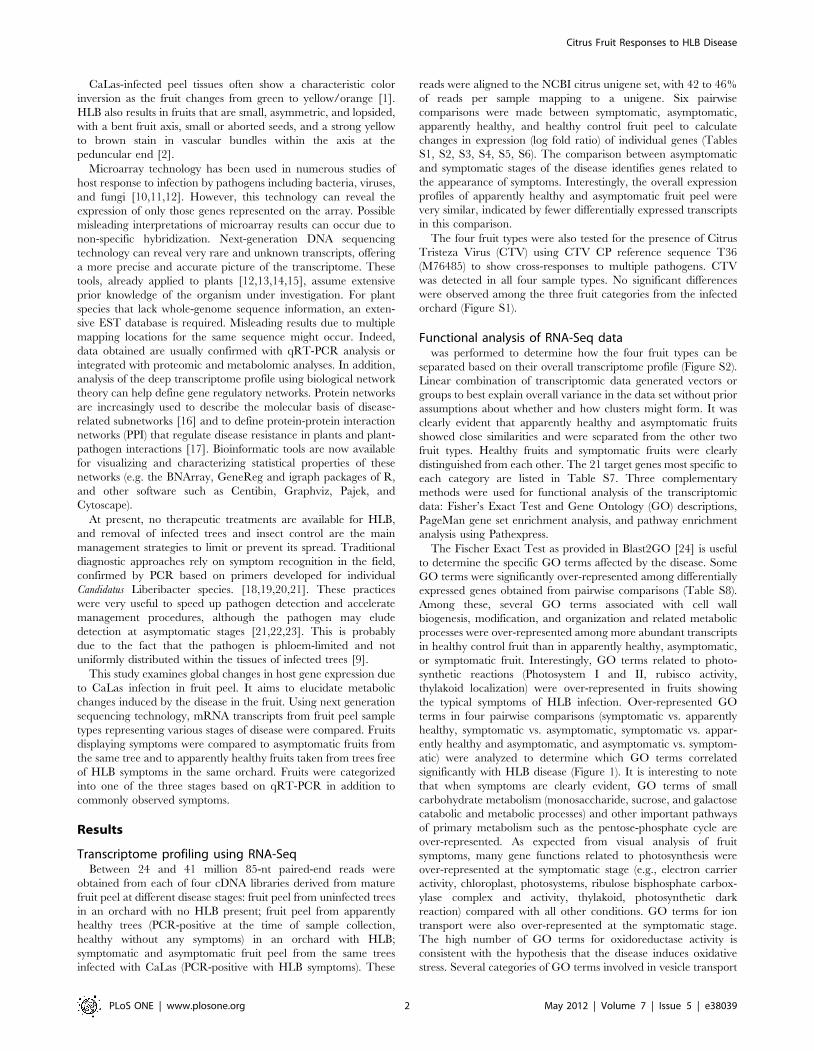
significantly with HLB disease (Figure 1). It is interesting to note
that when symptoms are clearly evident, GO terms of small
carbohydrate metabolism (monosaccharide, sucrose, and galactose
catabolic and metabolic processes) and other important pathways
of primary metabolism such as the pentose-phosphate cycle are
over-represented. As expected from visual analysis of fruit
symptoms, many gene functions related to photosynthesis were
over-represented at the symptomatic stage (e.g., electron carrier
activity, chloroplast, photosystems, ribulose bisphosphate carbox-
ylase complex and activity, thylakoid, photosynthetic dark
reaction) compared with all other conditions. GO terms for ion
transport were also over-represented at the symptomatic stage.
The high number of GO terms for oxidoreductase activity is
consistent with the hypothesis that the disease induces oxidative
stress. Several categories of GO terms involved in vesicle transport
Citrus Fruit Responses to HLB Disease
PLoS ONE | www.plosone.org 2 May 2012 | Volume 7 | Issue 5 | e38039
and cell wall biogenesis, metabolism, and organization were over-
represented among genes expressed at the symptomatic stage.
Interesting defense-related and lipid transport GO terms were
over-represented at the asymptomatic stage.
PageMan software [25] was used to visualize functional classes
that were significantly affected by HLB disease (Figure S3). This
method pinpoints which subcategory of genes were upregulated
and downregulated in each main gene category based not only on
metabolic pathways but also on cell functions. Increased expres-
sion in diseased samples is seen in photosynthesis, N-metabolism,
amino acid synthesis, isoprenoids, jasmonate and salicylic acid,
and several transcription factors. Functional classes with decreased
expression include sucrose and starch biosynthesis, glycolysis,
gibberellins, DNA and protein synthesis, and flavonoids metabo-
lism. Additional changes in expression can be seen in the
comparison between samples from the disease-free location and
apparently healthy fruits (asymptomatic but from the infected
orchard). These changes, eventually affected by environmental
variability, probably were induced in early stages of HLB disease.
Differential expression of transcripts comparing different stages of
HLB infection and their functions were visualized using MapMan
software [26]. This provided more specific information on
pathways and functions identified by Fisher’s Exact Test and
PageMan. For MapMan data analyses, a mapping file composed
of NCBI Citrus sinensis unigenes was used.
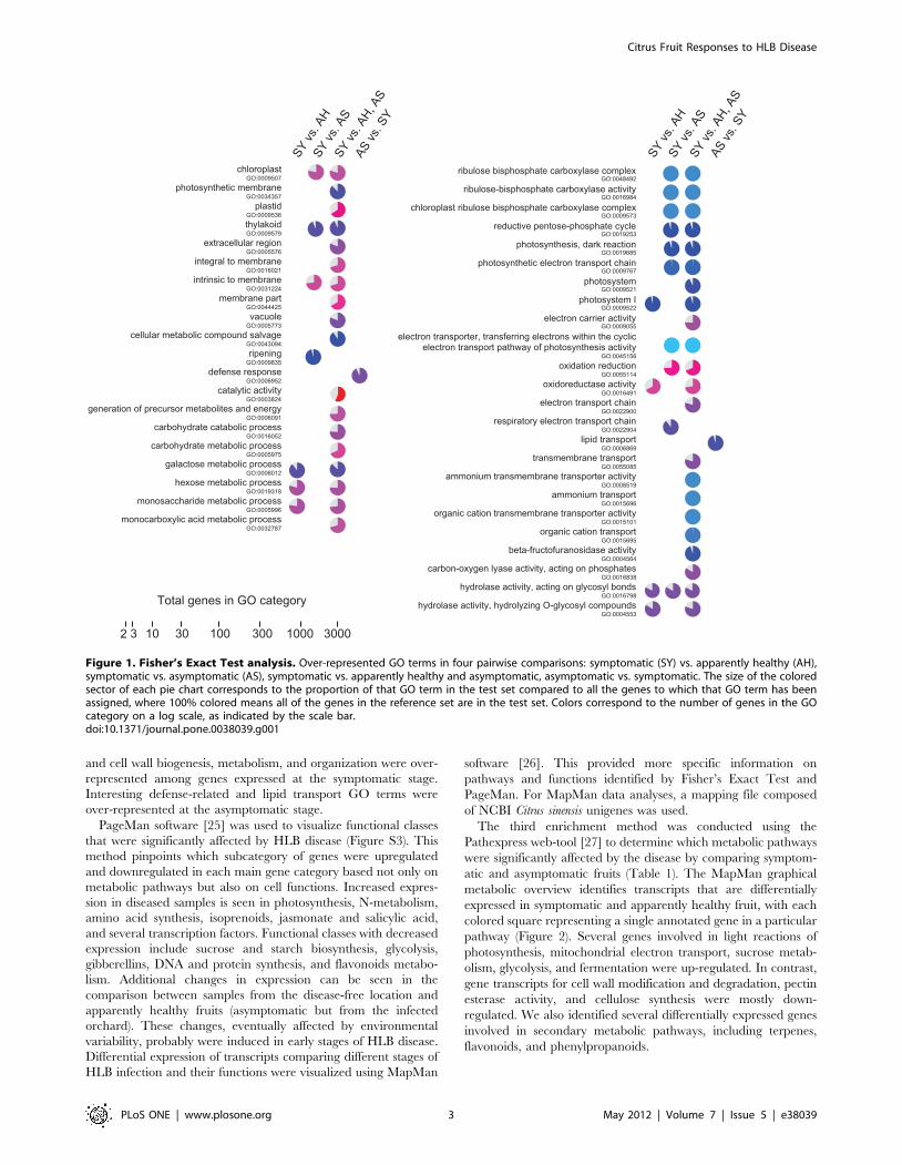
The third enrichment method was conducted using the
Pathexpress web-tool [27] to determine which metabolic pathways
were significantly affected by the disease by comparing symptom-
atic and asymptomatic fruits (Table 1). The MapMan graphical
metabolic overview identifies transcripts that are differentially
expressed in symptomatic and apparently healthy fruit, with each
colored square representing a single annotated gene in a particular
pathway (Figure 2). Several genes involved in light reactions of
photosynthesis, mitochondrial electron transport, sucrose metab-
olism, glycolysis, and fermentation were up-regulated. In contrast,
gene transcripts for cell wall modification and degradation, pectin
esterase activity, and cellulose synthesis were mostly down-
regulated. We also identified several differentially expressed genes
involved in secondary metabolic pathways, including terpenes,
flavonoids, and phenylpropanoids.
Figure 1. Fisher’s Exact Test analysis. Over-represented GO terms in four pairwise comparisons: symptomatic (SY) vs. apparently healthy (AH),symptomatic vs. asymptomatic (AS), symptomatic vs. apparently healthy and asymptomatic, asymptomatic vs. symptomatic. The size of the coloredsector of each pie chart corresponds to the proportion of that GO term in the test set compared to all the genes to which that GO term has beenassigned, where 100% colored means all of the genes in the reference set are in the test set. Colors correspond to the number of genes in the GOcategory on a log scale, as indicated by the scale bar.doi:10.1371/journal.pone.0038039.g001
Citrus Fruit Responses to HLB Disease
PLoS ONE | www.plosone.org 3 May 2012 | Volume 7 | Issue 5 | e38039
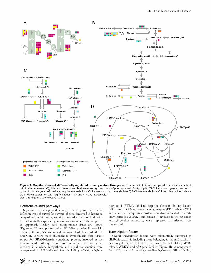
Photosynthesis and carbohydrate metabolismThe expression of several genes involved in photosynthesis and
carbohydrate metabolism increased significantly in symptomatic
fruits, when compared to asymptomatic fruit from the same tree or
different trees. Transcripts for oxygen-evolving enhancer 3 (PsbQ),
photosystem II subunit Q-2, photosystem II reaction center
protein J, and other genes encoding different subunits of
photosystem II were highly abundant in symptomatic fruit. In
addition, genes encoding subunits of cytochrome b6/f (complex
subunit 8, subunit IV) and genes encoding ATP synthase subunits
(ATPase subunit III, alpha, beta, F, and complex CF0) were
induced (Figure 3A). Several transcripts encoding subunits of
photosystem I increased, including chlorophyll A apoprotein
subunit G, photosystem I reaction center subunit (PSI-N), and D1
subunit.
Several genes for enzymes involved in the first steps of glycolysis
were differentially expressed. Up-regulation was observed for
genes encoding protein serine/threonine kinase and fructose-2,6-
biphosphatase (Figure 3B). There were significant changes in
transcripts related to carbohydrate metabolism in symptomatic
and asymptomatic fruit. The citrus orthologs of two Arabidopsis
genes encoding different isoforms of invertase (AB276108 and
EY662586) were induced in symptomatic fruit. In starch
metabolism, transcription of glucose-1-phosphate adenylyltrans-
ferase was diminished, while several genes involved in starch
degradation were significantly differentially expressed (Figure 3C).
Expression of genes involved in raffinose synthesis were differen-
tially regulated, while transcripts involved in galactinol metabolism
were abundant in symptomatic fruits (Figure 3D).
Figure 2. Functional categorization of differentially regulated genes in symptomatic fruits. Metabolism overview in MapMan depictingdifferential gene expression in symptomatic and apparently healthy fruits from the infected orchard. Log fold ratios are indicated as a gradientbetween red (up-regulated) and green (down-regulated).doi:10.1371/journal.pone.0038039.g002
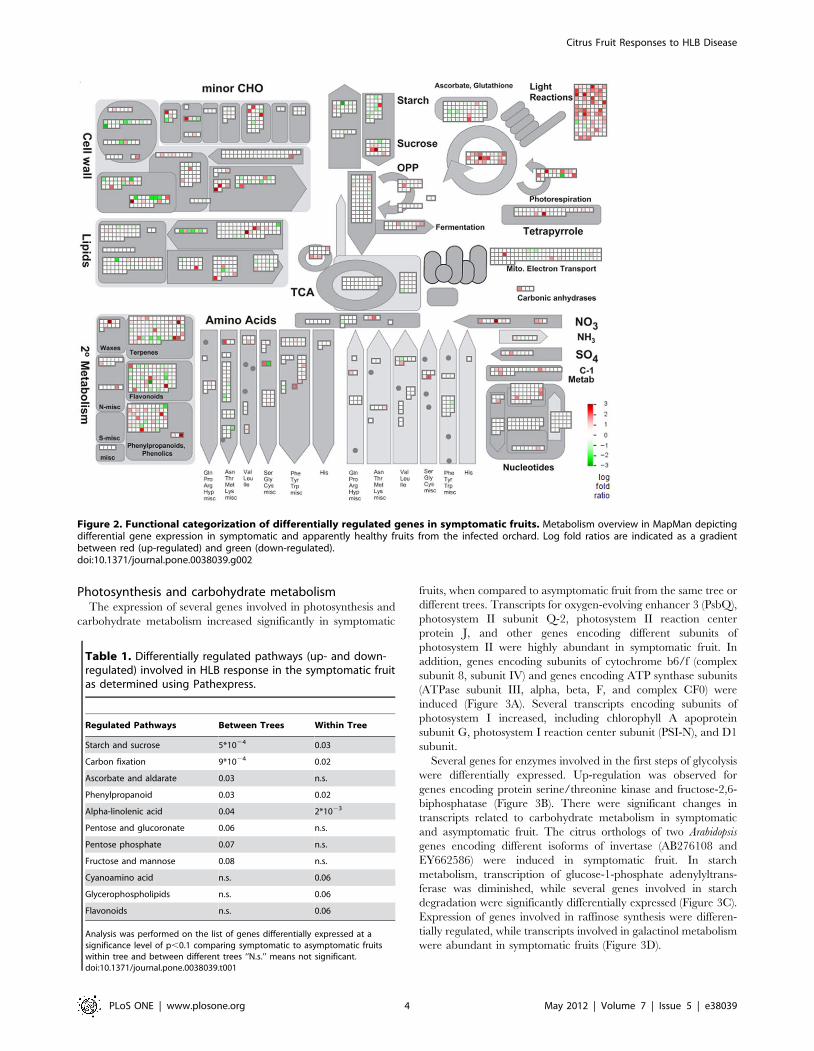
Table 1. Differentially regulated pathways (up- and down-regulated) involved in HLB response in the symptomatic fruitas determined using Pathexpress.
Regulated Pathways Between Trees Within Tree
Starch and sucrose 5*1024 0.03
Carbon fixation 9*1024 0.02
Ascorbate and aldarate 0.03 n.s.
Phenylpropanoid 0.03 0.02
Alpha-linolenic acid 0.04 2*1023
Pentose and glucoronate 0.06 n.s.
Pentose phosphate 0.07 n.s.
Fructose and mannose 0.08 n.s.
Cyanoamino acid n.s. 0.06
Glycerophospholipids n.s. 0.06
Flavonoids n.s. 0.06
Analysis was performed on the list of genes differentially expressed at asignificance level of p,0.1 comparing symptomatic to asymptomatic fruitswithin tree and between different trees ‘‘N.s.’’ means not significant.doi:10.1371/journal.pone.0038039.t001
Citrus Fruit Responses to HLB Disease
PLoS ONE | www.plosone.org 4 May 2012 | Volume 7 | Issue 5 | e38039
Hormone-related pathwaysSignificant transcriptional changes in response to CaLas
infection were observed for a group of genes involved in hormone
biosynthesis, mobilization, and signal transduction. Log fold ratios
for differentially expressed genes in symptomatic fruits compared
to apparently healthy and asymptomatic fruits are shown
(Figure 4). Transcripts related to GH3-like proteins involved in
auxin synthesis (IAA-amino acid conjugate hydrolase and GH3.1
and GH3.4) were more abundant in symptomatic fruit. Tran-
scripts for GRAM-domain containing protein, involved in the
abscisic acid pathway, were more abundant. Several genes
involved in ethylene biosynthesis and signal transduction were
upregulated in HLB-affected fruit including ACO4, ethylene
receptor 1 (ETR1), ethylene response element binding factors
(ERF1 and ERF2), ethylene forming enzyme (EFE), while ACO1
and an ethylene-responsive protein were downregulated. Interest-
ingly, genes for ATHK1 and Snakin-1, involved in the cytokinin
and gibberellin pathways, were repressed in infected fruit
(Figure 4A).
Transcription factorsSeveral transcription factors were differentially expressed in
HLB-infected fruit, including those belonging to the AP2-EREBP,
helix-loop-helix, bZIP, C2H2 zinc finger, C2C2-CO-like, MYB-
related, WRKY, and AS2 gene families (Figure 4B). Among genes
for bZIP, haloacid dehalogenase-like hydrolase, GBox binding
Figure 3. MapMan views of differentially regulated primary metabolism genes. Symptomatic fruit was compared to asymptomatic fruitwithin the same tree (AS), different tree (AH) and both trees. A) Light reactions of photosynthesis. B) Glycolysis. ‘‘CB’’ block shows gene expression incytosolic branch genes of small carbohydrate metabolism. C) Sucrose and starch metabolism D) Raffinose metabolism. Colored data points indicateup or down expression with log fold ratios .0.5 and ,20.5, respectively.doi:10.1371/journal.pone.0038039.g003
Citrus Fruit Responses to HLB Disease
PLoS ONE | www.plosone.org 5 May 2012 | Volume 7 | Issue 5 | e38039
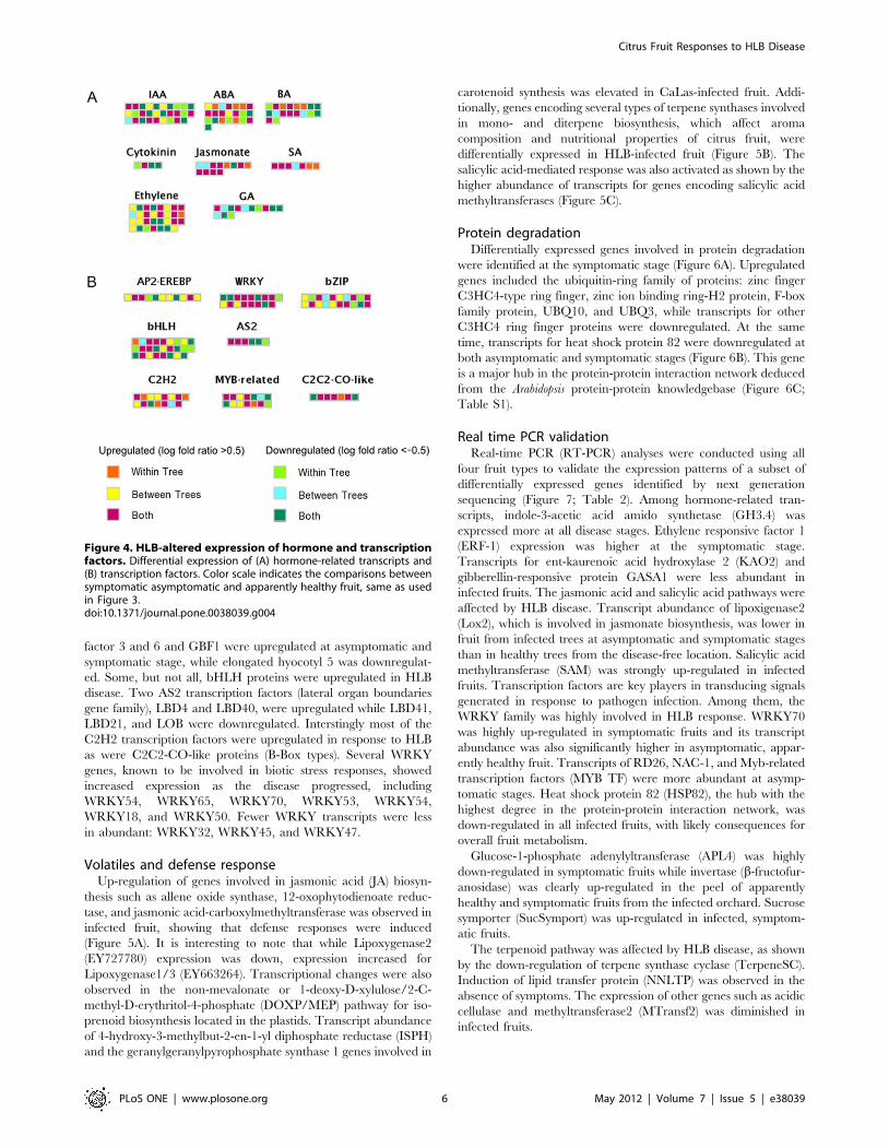
factor 3 and 6 and GBF1 were upregulated at asymptomatic and
symptomatic stage, while elongated hyocotyl 5 was downregulat-
ed. Some, but not all, bHLH proteins were upregulated in HLB
disease. Two AS2 transcription factors (lateral organ boundaries
gene family), LBD4 and LBD40, were upregulated while LBD41,
LBD21, and LOB were downregulated. Interstingly most of the
C2H2 transcription factors were upregulated in response to HLB
as were C2C2-CO-like proteins (B-Box types). Several WRKY
genes, known to be involved in biotic stress responses, showed
increased expression as the disease progressed, including
WRKY54, WRKY65, WRKY70, WRKY53, WRKY54,
WRKY18, and WRKY50. Fewer WRKY transcripts were less
in abundant: WRKY32, WRKY45, and WRKY47.
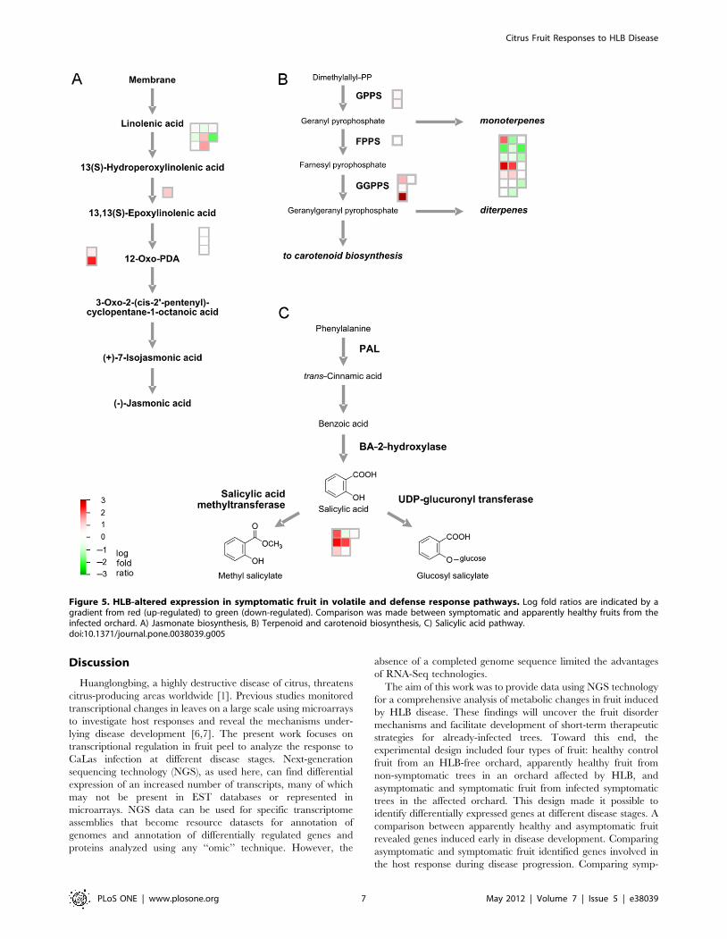
Volatiles and defense responseUp-regulation of genes involved in jasmonic acid (JA) biosyn-
thesis such as allene oxide synthase, 12-oxophytodienoate reduc-
tase, and jasmonic acid-carboxylmethyltransferase was observed in
infected fruit, showing that defense responses were induced
(Figure 5A). It is interesting to note that while Lipoxygenase2
(EY727780) expression was down, expression increased for
Lipoxygenase1/3 (EY663264). Transcriptional changes were also
observed in the non-mevalonate or 1-deoxy-D-xylulose/2-C-
methyl-D-erythritol-4-phosphate (DOXP/MEP) pathway for iso-
prenoid biosynthesis located in the plastids. Transcript abundance
of 4-hydroxy-3-methylbut-2-en-1-yl diphosphate reductase (ISPH)
and the geranylgeranylpyrophosphate synthase 1 genes involved in
carotenoid synthesis was elevated in CaLas-infected fruit. Addi-
tionally, genes encoding several types of terpene synthases involved
in mono- and diterpene biosynthesis, which affect aroma
composition and nutritional properties of citrus fruit, were
differentially expressed in HLB-infected fruit (Figure 5B). The
salicylic acid-mediated response was also activated as shown by the
higher abundance of transcripts for genes encoding salicylic acid
methyltransferases (Figure 5C).
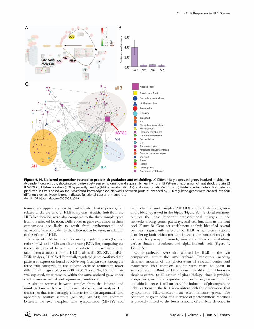
Protein degradationDifferentially expressed genes involved in protein degradation
were identified at the symptomatic stage (Figure 6A). Upregulated
genes included the ubiquitin-ring family of proteins: zinc finger
C3HC4-type ring finger, zinc ion binding ring-H2 protein, F-box
family protein, UBQ10, and UBQ3, while transcripts for other
C3HC4 ring finger proteins were downregulated. At the same
time, transcripts for heat shock protein 82 were downregulated at
both asymptomatic and symptomatic stages (Figure 6B). This gene
is a major hub in the protein-protein interaction network deduced
from the Arabidopsis protein-protein knowledgebase (Figure 6C;
Table S1).
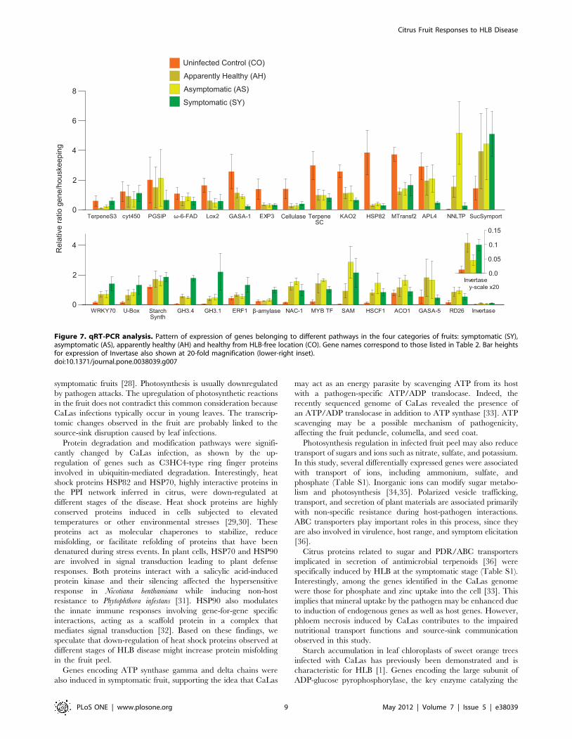
Real time PCR validationReal-time PCR (RT-PCR) analyses were conducted using all
four fruit types to validate the expression patterns of a subset of
differentially expressed genes identified by next generation
sequencing (Figure 7; Table 2). Among hormone-related tran-
scripts, indole-3-acetic acid amido synthetase (GH3.4) was
expressed more at all disease stages. Ethylene responsive factor 1
(ERF-1) expression was higher at the symptomatic stage.
Transcripts for ent-kaurenoic acid hydroxylase 2 (KAO2) and
gibberellin-responsive protein GASA1 were less abundant in
infected fruits. The jasmonic acid and salicylic acid pathways were
affected by HLB disease. Transcript abundance of lipoxigenase2
(Lox2), which is involved in jasmonate biosynthesis, was lower in
fruit from infected trees at asymptomatic and symptomatic stages
than in healthy trees from the disease-free location. Salicylic acid
methyltransferase (SAM) was strongly up-regulated in infected
fruits. Transcription factors are key players in transducing signals
generated in response to pathogen infection. Among them, the
WRKY family was highly involved in HLB response. WRKY70
was highly up-regulated in symptomatic fruits and its transcript
abundance was also significantly higher in asymptomatic, appar-
ently healthy fruit. Transcripts of RD26, NAC-1, and Myb-related
transcription factors (MYB TF) were more abundant at asymp-
tomatic stages. Heat shock protein 82 (HSP82), the hub with the
highest degree in the protein-protein interaction network, was
down-regulated in all infected fruits, with likely consequences for
overall fruit metabolism.
Glucose-1-phosphate adenylyltransferase (APL4) was highly
down-regulated in symptomatic fruits while invertase (b-fructofur-
anosidase) was clearly up-regulated in the peel of apparently
healthy and symptomatic fruits from the infected orchard. Sucrose
symporter (SucSymport) was up-regulated in infected, symptom-
atic fruits.
The terpenoid pathway was affected by HLB disease, as shown
by the down-regulation of terpene synthase cyclase (TerpeneSC).
Induction of lipid transfer protein (NNLTP) was observed in the
absence of symptoms. The expression of other genes such as acidic
cellulase and methyltransferase2 (MTransf2) was diminished in
infected fruits.
Figure 4. HLB-altered expression of hormone and transcriptionfactors. Differential expression of (A) hormone-related transcripts and(B) transcription factors. Color scale indicates the comparisons betweensymptomatic asymptomatic and apparently healthy fruit, same as usedin Figure 3.doi:10.1371/journal.pone.0038039.g004
Citrus Fruit Responses to HLB Disease
PLoS ONE | www.plosone.org 6 May 2012 | Volume 7 | Issue 5 | e38039
Discussion
Huanglongbing, a highly destructive disease of citrus, threatens
citrus-producing areas worldwide [1]. Previous studies monitored
transcriptional changes in leaves on a large scale using microarrays
to investigate host responses and reveal the mechanisms under-
lying disease development [6,7]. The present work focuses on
transcriptional regulation in fruit peel to analyze the response to
CaLas infection at different disease stages. Next-generation
sequencing technology (NGS), as used here, can find differential
expression of an increased number of transcripts, many of which
may not be present in EST databases or represented in
microarrays. NGS data can be used for specific transcriptome
assemblies that become resource datasets for annotation of
genomes and annotation of differentially regulated genes and
proteins analyzed using any ‘‘omic’’ technique. However, the
absence of a completed genome sequence limited the advantages
of RNA-Seq technologies.
The aim of this work was to provide data using NGS technology
for a comprehensive analysis of metabolic changes in fruit induced
by HLB disease. These findings will uncover the fruit disorder
mechanisms and facilitate development of short-term therapeutic
strategies for already-infected trees. Toward this end, the
experimental design included four types of fruit: healthy control
fruit from an HLB-free orchard, apparently healthy fruit from
non-symptomatic trees in an orchard affected by HLB, and
asymptomatic and symptomatic fruit from infected symptomatic
trees in the affected orchard. This design made it possible to
identify differentially expressed genes at different disease stages. A
comparison between apparently healthy and asymptomatic fruit
revealed genes induced early in disease development. Comparing
asymptomatic and symptomatic fruit identified genes involved in
the host response during disease progression. Comparing symp-
Figure 5. HLB-altered expression in symptomatic fruit in volatile and defense response pathways. Log fold ratios are indicated by agradient from red (up-regulated) to green (down-regulated). Comparison was made between symptomatic and apparently healthy fruits from theinfected orchard. A) Jasmonate biosynthesis, B) Terpenoid and carotenoid biosynthesis, C) Salicylic acid pathway.doi:10.1371/journal.pone.0038039.g005
Citrus Fruit Responses to HLB Disease
PLoS ONE | www.plosone.org 7 May 2012 | Volume 7 | Issue 5 | e38039
tomatic and apparently healthy fruit revealed host response genes
related to the presence of HLB symptoms. Healthy fruit from the
HLB-free location were also compared to the three sample types
from the infected location. Differences in gene expression in these
comparisons are likely to result from environmental and
agronomic variability due to the difference in location, in addition
to the effects of HLB.
A range of 1154 to 1762 differentially regulated genes (log fold
ratio ,21.5 and .1.5) were found using RNA-Seq comparing the
three categories of fruits from the infected orchard with those
taken from a location free of HLB (Tables S1, S2, S3). In qRT-
PCR analysis, 31 of 33 differentially regulated genes confirmed the
pattern of expression found by RNA-Seq. Comparisons among the
three fruit categories in the infected orchard resulted in fewer
differentially regulated genes (301–780; Tables S4, S5, S6). This
was expected, since samples within the same orchard grew under
similar environmental and agronomic conditions.
A similar contrast between samples from the infected and
uninfected orchards is seen in principal component analysis. The
transcripts that most strongly characterize the asymptomatic and
apparently healthy samples (MF-AS, MF-AH) are common
between the two samples. The symptomatic (MF-SY) and
uninfected orchard samples (MF-CO) are both distinct groups
and widely separated in the biplot (Figure S2). A visual summary
outlines the most important transcriptional changes in the
networks among genes, pathways, and cell functions in the fruit
peel (Figure 8). Gene set enrichment analysis identified several
pathways significantly affected by HLB as symptoms appear,
considering both within-tree and between-tree comparisons, such
as those for phenylpropanoids, starch and sucrose metabolism,
carbon fixation, ascorbate, and alpha-linolenic acid (Figure 1,
Figure S3).
Other pathways were also affected by HLB in the two
comparisons within the same orchard. Transcripts encoding
different subunits of the photosystem II reaction center and
cytochrome b6-f complex subunit were more abundant in
symptomatic HLB-infected fruit than in healthy fruit. Photosyn-
thesis is central to all aspects of plant biology, since it provides
energy for growth and reproduction, but its regulation by biotic
and abiotic stresses is still unclear. The induction of photosynthetic
light reactions in the fruit is consistent with the observation that
symptomatic HLB-infected fruit often remains green. The
retention of green color and increase of photosynthesis reactions
is probably linked to the lower amount of ethylene detected in
Figure 6. HLB-altered expression related to protein degradation and misfolding. A) Differentially expressed genes involved in ubiquitin-dependent degradation, showing comparison between symptomatic and apparently healthy fruits. B) Pattern of expression of heat shock protein 82(HSP82) in HLB-free location (CO), apparently healthy (AH), asymptomatic (AS), and symptomatic (SY) fruits. C) Protein-protein interaction networkpredicted in Citrus based on the Arabidopsis knowledgebase. Networks between proteins encoded by HLB-regulated genes were divided into fourdifferent clusters. Node legend indicates functional classes of transcripts.doi:10.1371/journal.pone.0038039.g006
Citrus Fruit Responses to HLB Disease
PLoS ONE | www.plosone.org 8 May 2012 | Volume 7 | Issue 5 | e38039
symptomatic fruits [28]. Photosynthesis is usually downregulated
by pathogen attacks. The upregulation of photosynthetic reactions
in the fruit does not contradict this common consideration because
CaLas infections typically occur in young leaves. The transcrip-
tomic changes observed in the fruit are probably linked to the
source-sink disruption caused by leaf infections.
Protein degradation and modification pathways were signifi-
cantly changed by CaLas infection, as shown by the up-
regulation of genes such as C3HC4-type ring finger proteins
involved in ubiquitin-mediated degradation. Interestingly, heat
shock proteins HSP82 and HSP70, highly interactive proteins in
the PPI network inferred in citrus, were down-regulated at
different stages of the disease. Heat shock proteins are highly
conserved proteins induced in cells subjected to elevated
temperatures or other environmental stresses [29,30]. These
proteins act as molecular chaperones to stabilize, reduce
misfolding, or facilitate refolding of proteins that have been
denatured during stress events. In plant cells, HSP70 and HSP90
are involved in signal transduction leading to plant defense
responses. Both proteins interact with a salicylic acid-induced
protein kinase and their silencing affected the hypersensitive
response in Nicotiana benthamiana while inducing non-host
resistance to Phytophthora infestans [31]. HSP90 also modulates
the innate immune responses involving gene-for-gene specific
interactions, acting as a scaffold protein in a complex that
mediates signal transduction [32]. Based on these findings, we
speculate that down-regulation of heat shock proteins observed at
different stages of HLB disease might increase protein misfolding
in the fruit peel.
Genes encoding ATP synthase gamma and delta chains were
also induced in symptomatic fruit, supporting the idea that CaLas
may act as an energy parasite by scavenging ATP from its host
with a pathogen-specific ATP/ADP translocase. Indeed, the
recently sequenced genome of CaLas revealed the presence of
an ATP/ADP translocase in addition to ATP synthase [33]. ATP
scavenging may be a possible mechanism of pathogenicity,
affecting the fruit peduncle, columella, and seed coat.
Photosynthesis regulation in infected fruit peel may also reduce
transport of sugars and ions such as nitrate, sulfate, and potassium.
In this study, several differentially expressed genes were associated
with transport of ions, including ammonium, sulfate, and
phosphate (Table S1). Inorganic ions can modify sugar metabo-
lism and photosynthesis [34,35]. Polarized vesicle trafficking,
transport, and secretion of plant materials are associated primarily
with non-specific resistance during host-pathogen interactions.
ABC transporters play important roles in this process, since they
are also involved in virulence, host range, and symptom elicitation
[36].
Citrus proteins related to sugar and PDR/ABC transporters
implicated in secretion of antimicrobial terpenoids [36] were
specifically induced by HLB at the symptomatic stage (Table S1).
Interestingly, among the genes identified in the CaLas genome
were those for phosphate and zinc uptake into the cell [33]. This
implies that mineral uptake by the pathogen may be enhanced due
to induction of endogenous genes as well as host genes. However,
phloem necrosis induced by CaLas contributes to the impaired
nutritional transport functions and source-sink communication
observed in this study.
Starch accumulation in leaf chloroplasts of sweet orange trees
infected with CaLas has previously been demonstrated and is
characteristic for HLB [1]. Genes encoding the large subunit of
ADP-glucose pyrophosphorylase, the key enzyme catalyzing the
Figure 7. qRT-PCR analysis. Pattern of expression of genes belonging to different pathways in the four categories of fruits: symptomatic (SY),asymptomatic (AS), apparently healthy (AH) and healthy from HLB-free location (CO). Gene names correspond to those listed in Table 2. Bar heightsfor expression of Invertase also shown at 20-fold magnification (lower-right inset).doi:10.1371/journal.pone.0038039.g007
Citrus Fruit Responses to HLB Disease
PLoS ONE | www.plosone.org 9 May 2012 | Volume 7 | Issue 5 | e38039
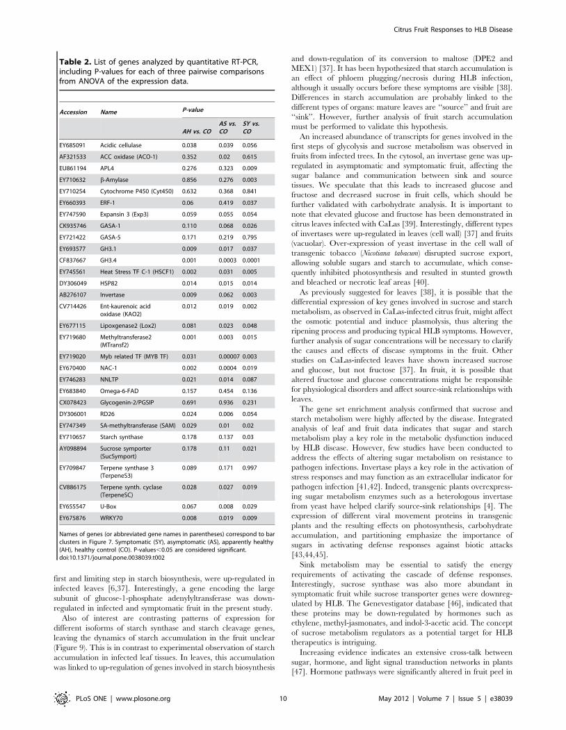
first and limiting step in starch biosynthesis, were up-regulated in
infected leaves [6,37]. Interestingly, a gene encoding the large
subunit of glucose-1-phosphate adenylyltransferase was down-
regulated in infected and symptomatic fruit in the present study.
Also of interest are contrasting patterns of expression for
different isoforms of starch synthase and starch cleavage genes,
leaving the dynamics of starch accumulation in the fruit unclear
(Figure 9). This is in contrast to experimental observation of starch
accumulation in infected leaf tissues. In leaves, this accumulation
was linked to up-regulation of genes involved in starch biosynthesis
and down-regulation of its conversion to maltose (DPE2 and
MEX1) [37]. It has been hypothesized that starch accumulation is
an effect of phloem plugging/necrosis during HLB infection,
although it usually occurs before these symptoms are visible [38].
Differences in starch accumulation are probably linked to the
different types of organs: mature leaves are ‘‘source’’ and fruit are
‘‘sink’’. However, further analysis of fruit starch accumulation
must be performed to validate this hypothesis.
An increased abundance of transcripts for genes involved in the
first steps of glycolysis and sucrose metabolism was observed in
fruits from infected trees. In the cytosol, an invertase gene was up-
regulated in asymptomatic and symptomatic fruit, affecting the
sugar balance and communication between sink and source
tissues. We speculate that this leads to increased glucose and
fructose and decreased sucrose in fruit cells, which should be
further validated with carbohydrate analysis. It is important to
note that elevated glucose and fructose has been demonstrated in
citrus leaves infected with CaLas [39]. Interestingly, different types
of invertases were up-regulated in leaves (cell wall) [37] and fruits
(vacuolar). Over-expression of yeast invertase in the cell wall of
transgenic tobacco (Nicotiana tabacum) disrupted sucrose export,
allowing soluble sugars and starch to accumulate, which conse-
quently inhibited photosynthesis and resulted in stunted growth
and bleached or necrotic leaf areas [40].
As previously suggested for leaves [38], it is possible that the
differential expression of key genes involved in sucrose and starch
metabolism, as observed in CaLas-infected citrus fruit, might affect
the osmotic potential and induce plasmolysis, thus altering the
ripening process and producing typical HLB symptoms. However,
further analysis of sugar concentrations will be necessary to clarify
the causes and effects of disease symptoms in the fruit. Other
studies on CaLas-infected leaves have shown increased sucrose
and glucose, but not fructose [37]. In fruit, it is possible that
altered fructose and glucose concentrations might be responsible
for physiological disorders and affect source-sink relationships with
leaves.
The gene set enrichment analysis confirmed that sucrose and
starch metabolism were highly affected by the disease. Integrated
analysis of leaf and fruit data indicates that sugar and starch
metabolism play a key role in the metabolic dysfunction induced
by HLB disease. However, few studies have been conducted to
address the effects of altering sugar metabolism on resistance to
pathogen infections. Invertase plays a key role in the activation of
stress responses and may function as an extracellular indicator for
pathogen infection [41,42]. Indeed, transgenic plants overexpress-
ing sugar metabolism enzymes such as a heterologous invertase
from yeast have helped clarify source-sink relationships [4]. The
expression of different viral movement proteins in transgenic
plants and the resulting effects on photosynthesis, carbohydrate
accumulation, and partitioning emphasize the importance of
sugars in activating defense responses against biotic attacks
[43,44,45].
Sink metabolism may be essential to satisfy the energy
requirements of activating the cascade of defense responses.
Interestingly, sucrose synthase was also more abundant in
symptomatic fruit while sucrose transporter genes were downreg-
ulated by HLB. The Genevestigator database [46], indicated that
these proteins may be down-regulated by hormones such as
ethylene, methyl-jasmonates, and indol-3-acetic acid. The concept
of sucrose metabolism regulators as a potential target for HLB
therapeutics is intriguing.
Increasing evidence indicates an extensive cross-talk between
sugar, hormone, and light signal transduction networks in plants
[47]. Hormone pathways were significantly altered in fruit peel in
Table 2. List of genes analyzed by quantitative RT-PCR,including P-values for each of three pairwise comparisonsfrom ANOVA of the expression data.
Accession Name P-value
AH vs. COAS vs.CO
SY vs.CO
EY685091 Acidic cellulase 0.038 0.039 0.056
AF321533 ACC oxidase (ACO-1) 0.352 0.02 0.615
EU861194 APL4 0.276 0.323 0.009
EY710632 b-Amylase 0.856 0.276 0.003
EY710254 Cytochrome P450 (Cyt450) 0.632 0.368 0.841
EY660393 ERF-1 0.06 0.419 0.037
EY747590 Expansin 3 (Exp3) 0.059 0.055 0.054
CK935746 GASA-1 0.110 0.068 0.026
EY721422 GASA-5 0.171 0.219 0.795
EY693577 GH3.1 0.009 0.017 0.037
CF837667 GH3.4 0.001 0.0003 0.0001
EY745561 Heat Stress TF C-1 (HSCF1) 0.002 0.031 0.005
DY306049 HSP82 0.014 0.015 0.014
AB276107 Invertase 0.009 0.062 0.003
CV714426 Ent-kaurenoic acidoxidase (KAO2)
0.012 0.019 0.002
EY677115 Lipoxgenase2 (Lox2) 0.081 0.023 0.048
EY719680 Methyltransferase2(MTransf2)
0.001 0.003 0.015
EY719020 Myb related TF (MYB TF) 0.031 0.00007 0.003
EY670400 NAC-1 0.002 0.0004 0.019
EY746283 NNLTP 0.021 0.014 0.087
EY683840 Omega-6-FAD 0.157 0.454 0.136
CX078423 Glycogenin-2/PGSIP 0.691 0.936 0.231
DY306001 RD26 0.024 0.006 0.054
EY747349 SA-methyltransferase (SAM) 0.029 0.01 0.02
EY710657 Starch synthase 0.178 0.137 0.03
AY098894 Sucrose symporter(SucSymport)
0.178 0.11 0.021
EY709847 Terpene synthase 3(TerpeneS3)
0.089 0.171 0.997
CV886175 Terpene synth. cyclase(TerpeneSC)
0.028 0.027 0.019
EY655547 U-Box 0.067 0.008 0.029
EY675876 WRKY70 0.008 0.019 0.009
Names of genes (or abbreviated gene names in parentheses) correspond to barclusters in Figure 7. Symptomatic (SY), asymptomatic (AS), apparently healthy(AH), healthy control (CO). P-values,0.05 are considered significant.doi:10.1371/journal.pone.0038039.t002
Citrus Fruit Responses to HLB Disease
PLoS ONE | www.plosone.org 10 May 2012 | Volume 7 | Issue 5 | e38039

response to CaLas infection. Two genes involved in auxin
synthesis, GH3.1 and GH3.4, were induced in affected fruit.
Gibberellin and cytokinin-related genes were mostly down-
regulated in symptomatic fruits. Gibberellin regulation has been
observed in other fruit disorders such as albedo breakdown
disorder (unpublished data) and applications of GA3 before fruit
color break can reduce the occurrence of some fruit disorders. It is
possible that sugar metabolism changes observed in the fruit might
be linked with the down-regulation of gibberellins that regulate
energy and carbohydrate metabolism. Previous studies have
demonstrated that regulation of gibberellic acid-induced gene
expression is affected by sugar and hormone signaling [48]. That
cytokinins play a role in sugar regulation has been demonstrated
[49]. Therapeutic approaches using small-molecule hormones
such as cytokinins and gibberellins may allow modification of fruit
metabolism to mitigate the negative impact of HLB on fruit quality
and productivity.
Ethylene regulates a variety of developmental processes and
stress responses in plants, including seed germination, cell
elongation, senescence, fruit ripening, and defense. Nonetheless,
ethylene can promote either disease resistance or susceptibility,
depending on the host–pathogen interaction [50,51]. In our study,
considerable changes were observed in the transcriptional profiles
of genes related to ethylene biosynthesis and signal transduction.
ACC synthase and ACC oxidase play pivotal roles in ethylene
biosynthesis and their expression is often affected by pathogen
attack [50]. It was unclear how ethylene concentration changes in
fruit in response to HLB. Different isoforms of ACC synthase and
ACC oxidase were up- or down-regulated. In addition, several
transcripts involved in ethylene signaling and response were more
abundant in symptomatic fruits, suggesting that HLB may have a
stronger effect on ethylene signaling than ethylene biosynthesis.
The general up-regulation of ethylene-related genes does not agree
with the lower ethylene concentration previously reported in HLB
symptomatic fruits [28]. This may be due to differences in fruit
Figure 8. Fruit metabolism and regulatory pathways in CaLas-infected fruits. Genes, pathways, and cell functions that were differentiallyexpressed are indicated with a square (red for up-regulated, green for down-regulated). Significantly differentially regulated pathways in gene setenrichment analysis are indicated in yellow.doi:10.1371/journal.pone.0038039.g008
Citrus Fruit Responses to HLB Disease
PLoS ONE | www.plosone.org 11 May 2012 | Volume 7 | Issue 5 | e38039
developmental stages and health status. Furthermore, gene
networks responsible for ethylene biosynthesis and perception
are still not fully elucidated. A large number of genes annotated as
ethylene-related occur as parologs playing tissue-specific roles, and
many play additional roles in biotic stress response. Their
expression may be drastically affected by the complex gene
regulatory network involved in immune responses without directly
affecting ethylene levels. The increased number of ERF- and AP2/
EREBP-related genes modulated by HLB supports this notion.
These factors control expression of many PR proteins and defense
response effectors [50]. In addition, the induction of ethylene
biosynthesis and signal transduction could profoundly modify fruit
metabolism by accelerating senescence linked to the typical fruit
malformations caused by HLB.
Salicylic acid and jasmonates are hormones involved in
activating defense responses to pests and pathogens. Pathways
for both hormones were activated in CaLas-infected citrus. It is
known that different hormone-regulated defense pathways are
activated depending on whether the pathogen is a necrotroph or
biotroph [4]. The mechanisms of CaLas pathogenesis are poorly
understood. The putative bacterial pathogen is closely related to
bacterial families with symbiotic properties (i.e., Rhizobiaceae) [1].
However, the gene expression in challenged fruit showed an up-
regulation of jasmonate-induced defense responses, typical of a
host response to localized necrotroph invasion. This pathway is
also stimulated by long distance signaling involving volatile
compounds. Salicylic acid methyl-transferase was up-regulated in
early, asymptomatic disease stages, potentially leading to produc-
tion of volatile methylsalicylate. Nonexpressor of pathogenesis-
related genes1 (NPR1) was up-regulated in symptomatic fruits
(Table S1). In Arabidopsis, NPR1 is required for SA-mediated
suppression of JA-dependent defenses [52]. Also, ethylene
modulates the NPR1 dependency of SA-JA antagonism, compen-
sating for enhanced allocation of NPR1 to functions in SA-
dependent activation of PR genes [53]. In plants, IRE1 (inositol-
requiring 1) gene is considered to be involved in unfolded protein
response (UPR) mediated by SAR with the engagement of BiP
[54]. HLB upregulation was observed for IRE1a (EY679744), a
gene closely involved in the UPR in plants [55]. This gene
expression change suggests that UPR may be activated in the
infected fruit.
Plants employ a network of intertwined mechanisms to counter
infection by pathogens and parasites. One line of defense is based
on dominant disease resistance (R) genes encoding nucleotide-
binding, leucine-rich repeat (NB-LRR) proteins that mediate
resistance to pathogens possessing corresponding avirulence (Avr)
genes [56]. Several leucine-rich repeat (LRR) receptor kinases
were up-regulated in symptomatic fruit, implying that they may be
receptors triggering a futile defense response against CaLas.
Innate immunity can be regulated by transcription factors.
Several genes belonging to the WRKY family of transcription
factors such as WRKY70, WRKY13, WRKY30, WRKY40,
WRKY65, and WRKY31 were up-regulated at both asymptom-
atic and symptomatic stages of HLB. WRKY70 acts as a
convergence point, determining the balance between SA- and
JA-dependent defense pathways in addition to being required for
R gene-mediated resistance. Its role in JA and SA signaling,
however, has recently been questioned [57]. Similarly,
Figure 9. HLB-regulation of primary metabolism in symptomatic fruits. Differentially regulated genes and pathways involved in primarymetabolism are indicated with a square (red for up-regulated, green for down-regulated).doi:10.1371/journal.pone.0038039.g009
Citrus Fruit Responses to HLB Disease
PLoS ONE | www.plosone.org 12 May 2012 | Volume 7 | Issue 5 | e38039
AtWRKY53 positively modulates SAR. Members of the WRKY
family were implicated in regulating the transcriptional repro-
gramming associated with plant immune responses and they may
act as positive and negative regulators of disease resistance [58].
Other genes encoding bZIP and C2H2 zinc finger transcription
factors were up-regulated in CaLas-infected fruits. The regulation
of bZIPs in HLB-affected citrus may be associated with reported
modifications observed in energy metabolism [59].
In the cytosol, up-regulation of 3-hydroxy-3-methyl-glutaryl
CoA reductase (HMG1), 12-oxophytodienoate reductase 2, and
allene oxide synthase may induce an increase in jasmonate-derived
volatiles. Several genes involved in terpenoid metabolism, such as
terpene synthase 3 and terpene synthase cyclase, were down-
regulated in symptomatic fruits. These enzymes are involved in the
synthesis and transport of a variety of terpenes, gibberellins,
brassinosteroids, alkaloids, and plant volatiles that play diverse
roles in plant development and defense [60]. The regulation of
genes involved in these pathways has important implications for
volatile emissions and is associated with a variety of responses.
Further studies on the volatile emission profiles of CaLas-infected
fruits will clarify the role of these enzymes.
Next generation sequencing enabled us to identify genes
differentially expressed in citrus fruit peel in response to CaLas
infection, leading to a better understanding of the processes
involved in HLB disease development. This study identified
several genes that were differentially expressed at the asymptom-
atic stage that may aid disease detection at primary stages of
infection, before the pathogen can be detected by PCR. However,
their usefulness as HLB-specific induced genes cannot be
determined until a similar expression analysis is conducted on
the same tissues infected by other citrus pathogens such as Citrus
tristeza virus, Xylella fastidiosa, and Xanthomonas axonopodis. In the
fruit peel, HLB induced transcriptional changes in important
pathways such as sucrose and starch metabolism, hormone
signaling, and isoprenoid synthesis. WRKY transcription factors
appear to help regulate defense responses to CaLas in the fruit.
The induction of genes involved in the light reactions of
photosynthesis might increase the occurrence of reactive oxygen
species, leading to protein degradation and misfolding. The
application of small-molecule hormones is another promising
short-term strategy to mitigate the devastating negative physio-
logical effects of HLB.
Materials and Methods
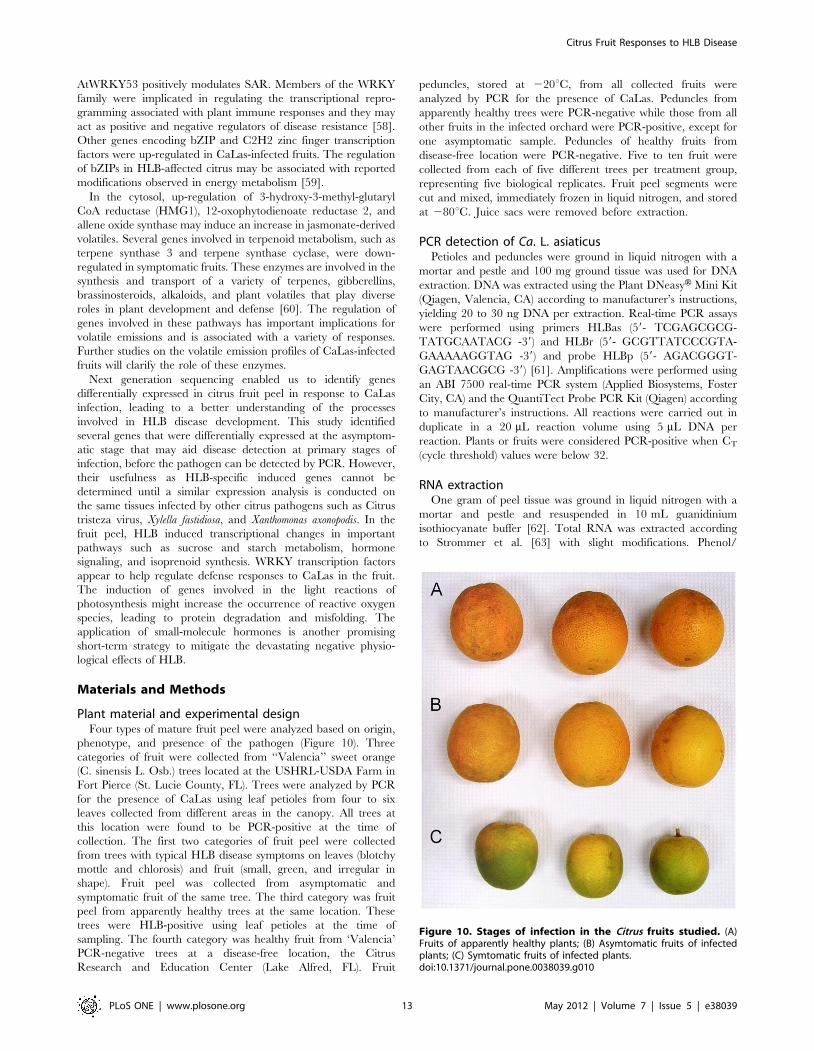
Plant material and experimental designFour types of mature fruit peel were analyzed based on origin,
phenotype, and presence of the pathogen (Figure 10). Three
categories of fruit were collected from ‘‘Valencia’’ sweet orange
(C. sinensis L. Osb.) trees located at the USHRL-USDA Farm in
Fort Pierce (St. Lucie County, FL). Trees were analyzed by PCR
for the presence of CaLas using leaf petioles from four to six
leaves collected from different areas in the canopy. All trees at
this location were found to be PCR-positive at the time of
collection. The first two categories of fruit peel were collected
from trees with typical HLB disease symptoms on leaves (blotchy
mottle and chlorosis) and fruit (small, green, and irregular in
shape). Fruit peel was collected from asymptomatic and
symptomatic fruit of the same tree. The third category was fruit
peel from apparently healthy trees at the same location. These
trees were HLB-positive using leaf petioles at the time of
sampling. The fourth category was healthy fruit from ‘Valencia’
PCR-negative trees at a disease-free location, the Citrus
Research and Education Center (Lake Alfred, FL). Fruit
peduncles, stored at 220uC, from all collected fruits were
analyzed by PCR for the presence of CaLas. Peduncles from
apparently healthy trees were PCR-negative while those from all
other fruits in the infected orchard were PCR-positive, except for
one asymptomatic sample. Peduncles of healthy fruits from
disease-free location were PCR-negative. Five to ten fruit were
collected from each of five different trees per treatment group,
representing five biological replicates. Fruit peel segments were
cut and mixed, immediately frozen in liquid nitrogen, and stored
at 280uC. Juice sacs were removed before extraction.
PCR detection of Ca. L. asiaticusPetioles and peduncles were ground in liquid nitrogen with a
mortar and pestle and 100 mg ground tissue was used for DNA
extraction. DNA was extracted using the Plant DNeasyH Mini Kit
(Qiagen, Valencia, CA) according to manufacturer’s instructions,
yielding 20 to 30 ng DNA per extraction. Real-time PCR assays
were performed using primers HLBas (59- TCGAGCGCG-
TATGCAATACG -39) and HLBr (59- GCGTTATCCCGTA-
GAAAAAGGTAG -39) and probe HLBp (59- AGACGGGT-
GAGTAACGCG -39) [61]. Amplifications were performed using
an ABI 7500 real-time PCR system (Applied Biosystems, Foster
City, CA) and the QuantiTect Probe PCR Kit (Qiagen) according
to manufacturer’s instructions. All reactions were carried out in
duplicate in a 20 mL reaction volume using 5 mL DNA per
reaction. Plants or fruits were considered PCR-positive when CT
(cycle threshold) values were below 32.
RNA extractionOne gram of peel tissue was ground in liquid nitrogen with a
mortar and pestle and resuspended in 10 mL guanidinium
isothiocyanate buffer [62]. Total RNA was extracted according
to Strommer et al. [63] with slight modifications. Phenol/
Figure 10. Stages of infection in the Citrus fruits studied. (A)Fruits of apparently healthy plants; (B) Asymtomatic fruits of infectedplants; (C) Symtomatic fruits of infected plants.doi:10.1371/journal.pone.0038039.g010
Citrus Fruit Responses to HLB Disease
PLoS ONE | www.plosone.org 13 May 2012 | Volume 7 | Issue 5 | e38039
chloroform/isoamylalcohol (25:24:1) extraction was followed by
two extractions with chloroform/isoamylalcohol and precipitation
of RNA with isopropanol at 220uC overnight. RNA was pelleted
by centrifugation at 10,000 g and 4uC for 1 h, resuspended in
5 mL water and precipitated overnight at 0uC with an equal
volume of 8 M LiCl. After centrifugation at 10,000 g and 4uC for
1 h, RNA was washed twice with 70% ethanol, air-dried and
dissolved in 500 mL of water. RNA was further purified using the
RNeasyH MinElute Cleanup kit (Qiagen, Valencia, CA, USA)
according to the manufacturer’s instructions. RNA quality and
purity were assessed using an Agilent Bionalyzer (Folsom, CA).
cDNA library construction and high throughputsequencing
RNA from the five biological replicates was equally pooled to
10 mg and then used to construct a cDNA library for each of the
four fruit types. The cDNA libraries were constructed following
the Illumina mRNA-sequencing sample preparation protocol
(Illumina Inc., San Diego, CA). Final elution was performed with
16 mL RNase-free water. The quality of each library was
determined using a BioRad Experion (BioRad, Hercules, CA).
Each library was run as an independent lane on a Genome
Analyzer II (Illumina, San Diego, CA) to obtain read lengths of up
to 85 bp per paired end.
Sequence data processing and analysisThe raw Illumina reads were trimmed to remove low quality
reads using custom scripts. Individual reads from each sample
were aligned to the Citrus sinensis unigene set (15,808 sequences;
NCBI Unigene Build #11, 4/20/09) with Burrows-Wheeler
Transform (BWA) [64]. Read counts were generated with
SAMtools [65] and custom scripts.
Six pairwise comparisons were made between the read counts
for the four samples. For each pairwise comparison, the raw count
data was normalized to control for different sequencing depths
across samples with the DESeq Bioconductor package [66].
DESeq was developed for the statistical analysis of experiments
with few or no replicates, accommodating this by treating samples
from differing treatments or phenotypes as replicates in the
estimation process. The results of this pooling are more
conservative than in an experiment with more replicates.
Principal component analysisData analysis was performed to alleviate possible bias caused by
the amount of collected material for each class or other
confounding factors. A within-sample normalization process was
applied to each sample to calculate the ratio of each target gene.
This way, the sum of the ratios of all target genes was 1 while each
target gene had its own count ratio within (0 to 1). For each
sample, all target genes were first sorted in order from high to low.
Then the top target genes with their accumulated ratio counting
for 25% of the total were retained for further analysis, to make the
differentially regulated gene selection more robust and focused on
a relatively small number of strong target genes. By integrating all
selected target genes for the four classes, we generated a complete
list of potential target genes. In this list, some genes are shared
among all four classes, while some are only observed in one class.
Principal component analysis was then applied to the ratio matrix
of this gene list to examine the contribution of each target gene to
the separation of the classes. A biplot was constructed based on the
first two principal components. The length of the loading vector
for each target gene indicates the contribution of that gene to the
separation based on the first two principal components. To
examine which genes contribute most to each class, two criteria for
screening were employed. First, the mean value of the loading
vector lengths (strengths) of all potential target genes was
calculated and 80% of that value was set as the threshold for
strength screening. Target genes with loading vector lengths larger
than this threshold were considered strong. Secondly, the
directional similarity was calculated between each target gene
and each class and then 0.98 (i.e, the cosine of 10u) was set as the
threshold value for similarity screening. Larger similarities indicate
bigger contributions of a gene to a class.
Functional categorization and protein-protein networkanalysis
The Citrus sinensis unigene set was annotated using Blast2GO
[67] to assign Gene Ontology (GO) terms to each gene. Lists of
transcripts that were differentially expressed at a significant level
(p,0.01, absolute value of Log Fold Change .1.5) in the pairwise
comparisons were used as input for one-tailed Fisher’s Exact Test
in Blast2GO to identify GO terms that were significantly over-
represented.
In addition, the differentially expressed genes were also
functionally analyzed using the MapMan knowledgebase blasting
to the TAIR database [25]. Gene set analysis was also performed
using Pathexpress [26] for the highest Arabidopsis hit per Citrus
sinensis unigene set (considering as a cutoff Log Fold Ratio .1/
21). A protein-protein interaction network (PPI) was deduced for
Citrus based on PPIs in Arabidopsis [68] using blast analysis.
Networks were identified and visualized using Graphviz software.
Real time TaqMan PCR systemReal Time Taqman PCR analysis was conducted to validate
RNA-seq data. Three biological replicates (a pool of 5 to 10 fruits
from different plants) were used for each of the four types of fruit
peel (healthy, apparently healthy, asymptomatic, and symptomat-
ic). For each target gene, PCR primers and a TaqManH probe
were purchased as an assay mix from Applied Biosystems (Foster
City, CA). DNase treatment and cDNA synthesis were performed
in a combined protocol following instructions of the Quantitect
Reverse Transcription Kit (Qiagen). A standard curve to
determine the linearity of amplicon quantity vs. initial cDNA
quantity was generated for each gene. Amplifications used 25 ng
cDNA in a 20 mL final volume with TaqMan Universal PCR
Master Mix and Taqman Assay ABI mixes (Applied Biosystems).
Amplifications were performed on a StepOne Real Time PCR
system (Applied Biosystems) using standard amplification condi-
tions: 1 cycle of 2 min at 50uC, 10 min at 95uC, 40 cycles of 15 s
at 95uC, and 60 s at 60uC. All PCR reactions were performed in
duplicate. Fluorescent signals were collected during the annealing
temperature and CT values extracted with a threshold of 0.04 and
baseline values of 3 to 10. Citrus sinensis elongation factor 1 alpha
(EF-1a, accession AY498567) was used as an endogenous
reference and DDCT was calculated by subtracting the average
EF-1a CT from the average CT of the gene of interest.
Real Time Taqman PCR analysis was also conducted for CTV
(Citrus Tristeza Virus) detection using the same RNA to determine
the presence of the virus in the samples analyzed for HLB
response. Primers were designed based on CTV CP reference
sequence T36 (M76485) and the same protocol was followed as
previously described.
ANOVA was performed considering the three biological
replicates for each of the four fruit categories. P-values were
determined for each of the six pairwise comparisons and values
lower than 0.05 were considered significant.
Citrus Fruit Responses to HLB Disease
PLoS ONE | www.plosone.org 14 May 2012 | Volume 7 | Issue 5 | e38039
Supporting Information
Table S1 Differentially expressed genes in symptomat-ic fruit in comparison to control (healthy in disease-freelocation), annotations and number of protein-proteininteractions deduced from Arabidopsis knowledgebase.(HTM)
Table S2 Differentially expressed genes in asymptom-atic fruit in comparison to control (healthy in disease-free location), annotations and number of protein-protein interactions deduced from Arabidopsis knowl-edgebase.(HTM)
Table S3 Differentially expressed genes in apparentlyhealthy fruit in comparison to control (healthy indisease-free location), annotations and number ofprotein-protein interactions deduced from Arabidopsisknowledgebase.(HTM)
Table S4 Differentially expressed genes in apparentlyhealthy fruits in comparison to asymptomatic, annota-tions and number of protein-protein interactions de-duced from Arabidopsis knowledgebase.(HTM)
Table S5 Differentially expressed genes in symptomat-ic fruits in comparison to asymptomatic, annotationsand number of protein-protein interactions deducedfrom Arabidopsis knowledgebase.(HTM)
Table S6 Differentially expressed genes in symptomat-ic fruit in comparison to apparently healthy, annota-tions and number of protein-protein interactions de-duced from Arabidopsis knowledgebase.(HTM)
Table S7 21 genes making the strongest contribution toeach of four classes. Five genes with count and log2foldchange
values also appear in two or more of Tables S1, S2, S3 and S6 as
indicated.
(HTM)
Table S8 GO categories of transcripts found in signif-icantly higher levels in pairwise comparisons (indicatedas ‘‘up’’ and ‘‘down’’ groups), using Fisher’s Exact Test.(HTM)
Figure S1 CTV detection in Citrus using qRT-PCR.Analysis were conducted for the four categories of fruits:
symptomatic (SY), asymptomatic (AS), apparently healthy (AH)
and healthy from HLB-free location (CO).
(EPS)
Figure S2 Principal component analysis of differentialgene expression in four HLB sample types. The length of
the loading vector of each target gene indicates that gene’s
contribution to the separation of four categories of mature fruit
peel by the first two principal components (C1, C2). MF-SY,
symptomatic; MF-AS, asymptomatic; MF-AH, apparently
healthy; MF-CO, HLB-free orchard.
(EPS)
Figure S3 Gene set enrichment analysis of Citrus fruits.PageMan analysis using Wilcoxon Rank Sum test, without
multiple testing correction and cutoff = 1.0 Comparisons are
shown between AS (asymptomatic) and SY (symptomatic), AH
(apparently healthy) and SY (symptomatic), CO (HLB-free
orchard) and AH (apparently healthy).
(EPS)
Author Contributions
Conceived and designed the experiments: AMD FM KDB. Performed the
experiments: FM SLU UA MLP. Analyzed the data: FM RLR MB VB JF
EL WZ DL RD CED AMD. Contributed reagents/materials/analysis
tools: FM SLU UA MLP KDB AMD. Wrote the paper: FM AMD.
Conceived and designed the experiments: AMD FM KDB. Performed the
experiments: FM SLU UA MLP. Analyzed the data: FM RLR MB VB JF
EL WZ DL RD CED AMD. Contributed reagents/materials/analysis
tools: FM SLU UA MLP KDB AMD. Wrote the paper: FM AMD.
References
1. Bove JM (2006) Huanglongbing: A destructive, newly-emerging, century-old
disease of citrus. Journal of Plant Pathology 88: 7–37.
2. Bove JM, Ayres AJ (2007) Etiology of three recent diseases of citrus in Sao Paulo
State: Sudden death, variegated chlorosis and huanglongbing. Iubmb Life 59:
346–354.
3. Sechler A, Schuenzel EL, Cooke P, Donnua S, Thaveechai N, et al. (2009)
Cultivation of ‘Candidatus Liberibacter asiaticus’, ‘Ca. L. africanus’, and ‘Ca. L.
americanus’ Associated with Huanglongbing. Phytopathology 99: 480–486.
4. Tyler HL, Roesch LFW, Gowda S, Dawson WO, Triplett EW (2009)
Confirmation of the Sequence of ‘Candidatus Liberibacter asiaticus’ and
Assessment of Microbial Diversity in Huanglongbing-Infected Citrus Phloem
Using a Metagenomic Approach. Molecular Plant-Microbe Interactions 22:
1624–1634.
5. Duan YP, Zhou LJ, Hall DG, Li WB, Doddapanenj H, et al. (2009) Complete
Genome Sequence of Citrus Huanglongbing Bacterium, ‘Candidatus Liberi-
bacter asiaticus’ Obtained Through Metagenomics. Molecular Plant-Microbe
Interactions 22: 1011–1020.
6. Albrecht U, Bowman KD (2008) Gene expression in Citrus sinensis (L.) Osbeck
following infection with the bacterial pathogen Candidatus Liberibacter asiaticus
causing Huanglongbing in Florida. Plant Science 175: 291–306.
7. Kim JS, Sagaram US, Burns JK, Li JL, Wang N (2009) Response of Sweet
Orange (Citrus sinensis) to ‘Candidatus Liberibacter asiaticus’ Infection:
Microscopy and Microarray Analyses. Phytopathology 99: 50–57.
8. Fan J, Chen C, Yu Q, Brlansky RH, Li Z-G, et al. (2011) Comparative iTRAQ
proteome and transcriptome analyses of sweet orange infected by Candidatus
liberibacter asiaticus. Physiologia Plantarum DOI: 10.1111/j.1399-
3054.2011.01502.x.
9. Tatineni S, Sagaram US, Gowda S, Robertson CJ, Dawson WO, et al. (2008) In
planta distribution of ‘Candidatus Liberibacter asiaticus’ as revealed by
polymerase chain reaction (PCR) and real-time PCR. Phytopathology 98:
592–599.
10. Gibly A, Bonshtien A, Balaji V, Debbie P, Martin GB, et al. (2004) Identification
and expression profiling of tomato genes differentially regulated during a
resistance response to Xanthomonas campestris pv. vesicatoria. Molecular Plant-
Microbe Interactions 17: 1212–1222.
11. Espinoza C, Medina C, Somerville S, Arce-Johnson P (2007) Senescence-
associated genes induced during compatible viral interactions with grapevine
and Arabidopsis. Journal of Experimental Botany 58: 3197–3212.
12. Panthee DR, Yuan JS, Wright DL, Marois JJ, Mailhot D, et al. (2007) Gene
expression analysis in soybean in response to the causal agent of Asian soybean
rust (Phakopsora pachyrhizi Sydow) in an early growth stage. Functional &
Integrative Genomics 7: 291–301.
13. Boccara M, Sarazin A, Billoud B, Jolly V, Martienssen R, et al. (2007) New
approaches for the analysis of Arabidopsis thaliana small RNAs. Biochimie 89:
1252–1256.
14. Navarro B, Pantaleo V, Gisel A, Moxon S, Dalmay T, et al. (2009) Deep
Sequencing of Viroid-Derived Small RNAs from Grapevine Provides New
Insights on the Role of RNA Silencing in Plant-Viroid Interaction. Plos One 4.
15. Donaire L, Wang Y, Gonzalez-Ibeas D, Mayer KF, Aranda MA, et al. (2009)
Deep-sequencing of plant viral small RNAs reveals effective and widespread
targeting of viral genomes. Virology 392: 203–214.
16. Ideker T, Sharan R (2008) Protein networks in disease. Genome Research 18:
644–652.
17. Holzmuller P, Grebaut P, Brizard JP, Berthier D, Chantal I, et al. (2008)
‘‘Pathogeno-Proteomics’’ Toward a New Approach of Host-Vector-Pathogen
Interactions. In: Sparagano OAE, Maillard JC, Figueroa JV, eds. Animal
Biodiversity and Emerging Diseases: Prediction and Prevention. Oxford:
Blackwell Publishing. pp 66–70.
Citrus Fruit Responses to HLB Disease
PLoS ONE | www.plosone.org 15 May 2012 | Volume 7 | Issue 5 | e38039
18. Jagoueix S, Bove JM, Garnier M (1996) PCR detection of the two ‘Candidatus’
liberobacter species associated with greening disease of citrus. Molecular andCellular Probes 10: 43–50.
19. Hocquellet A, Toorawa P, Bove JM, Garnier M (1999) Detection and
identification of the two Candidatus Liberobacter species associated with citrushuanglongbing by PCR amplification of ribosomal protein genes of the beta
operon. Molecular and Cellular Probes 13: 373–379.20. Teixeira DD, Danet JL, Eveillard S, Martins EC, Junior WCJ, et al. (2005)
Citrus huanglongbing in Sao Paulo State, Brazil: PCR detection of the
‘Candidatus’ Liberibacter species associated with the disease. Molecular andCellular Probes 19: 173–179.
21. Teixeira DC, Saillard C, Couture C, Martins EC, Wulff NA, et al. (2008)Distribution and quantification of Candidatus Liberibacter americanus, agent of
huanglongbing disease of citrus in Sao Paulo State, Brasil, in leaves of an affectedsweet orange tree as determined by PCR. Molecular and Cellular Probes 22:
139–150.
22. Lin H, Doddapaneni H, Bai XJ, Yao JQ, Zhao XL, et al. (2008) Acquisition ofuncharacterized sequences from Candidatus Liberibacter, an unculturable
bacterium, using an improved genomic walking method. Molecular and CellularProbes 22: 30–37.
23. Wang Z, Yin Y, Hu H, Yuan Q, Peng G, et al. (2006) Development and
application of molecular-based diagnosis for ‘Candidatus Liberibacter asiaticus’,the causal pathogen of citrus huanglongbing. Plant Pathology 55: 630–638.
24. Conesa A, Gotz S (2008) Blast2GO: A comprehensive suite for functionalanalysis in plant genomics. Int J Plant Genomics 619832.
25. Usadel B, Nagel A, Steinhauser D, Gibon Y, Blasing OE, et al. (2006) PageMan:An interactive ontology tool to generate, display, and annotate overview graphs
for profiling experiments. BMC Bioinformatics 7: 535.
26. Thimm O, Blasing O, Gibon Y, Nagel A, Meyer S, et al. (2004) MAPMAN: auser-driven tool to display genomics data sets onto diagrams of metabolic
pathways and other biological processes. Plant Journal 37: 914–939.27. Goffard N, Weiller G (2007) PathExpress: a web-based tool to identify relevant
pathways in gene expression data. Nucleic Acids Research 35: W176–W181.
28. Rosales R, Burns JK (2011) Phytohormone Changes and Carbohydrate Status inSweet Orange Fruit from Huanglongbing-infected Trees. Journal of Plant
Growth Regulation 30;3): 312–321.29. Almoguera C, Coca MA, Jordano J (1995) Differential accumulation of
sunflower tetraubiquitin messenger-RNAs during zygotic embryogenesis anddevelopmental regulation of their heat-shock response. Plant Physiology 107:
765–773.
30. Sabehat A, Lurie S, Weiss D (1998) Expression of small heat-shock proteins atlow temperatures - A possible role in protecting against chilling injuries. Plant
Physiology 117: 651–658.31. Kanzaki H, Saitoh H, Takahashi Y, Berberich T, Ito A, et al. (2008) NbLRK1, a
lectin-like receptor kinase protein of Nicotiana benthamiana, interacts with
Phytophthora infestans INF1 elicitin and mediates INF1-induced cell death.Planta 228: 977–987.
32. Schulze-Lefert P (2004) Plant immunity: The origami of receptor activation.Current Biology 14: R22–R24.
33. Duan Y, Zhou L, Gottwald T (2009) Genome sequencing of ‘‘Ca. Liberibacterasiaticus’’. Phytopathology 99: S157–S157.
34. Sadka A, Dewald DB, May GD, Park WD, Mullet JE (1994) Phosphate
modulates transcription of soybean vspb and other sugar-inducible genes. PlantCell 6: 737–749.
35. Nielsen KK, Bojsen K, Roepstorff P, Mikkelsen JD (1994) A hydroxyproline-containing class-IV chitinase of sugar-beet is glycosylated with xylose. Plant
Molecular Biology 25: 241–257.
36. van den Brule S, Smart CC (2002) The plant PDR family of ABC transporters.Planta 216: 95–106.
37. Fan J, Chen C, Brlansky RH, Gmitter Jr. FG, Li Z-G (2010) Changes incarbohydrate metabolism in Citrus sinensis infected with ‘Candidatus Liberi-
bacter asiaticus’. Plant Pathol 59: 1037–1043.
38. Etxeberria E, Gonzalez P, Achor D, Albrigo G (2009) Anatomical distribution ofabnormally high levels of starch in HLB-affected Valencia orange trees.
Physiological and Mol Plant Pathol 74: 76–83.39. Albrecht U, Bowman KM (2008) Transcriptional response and carbohydrate
metabolism of citrus infected with Candidatus Liberibacter asiaticus, the causalagent of huanglongbing in Florida. 11th Int. Citrus Congress. ICC, Wuhan,
China.
40. von Schaewen A, Stitt M, Schmidt R, Sonnewald U, Willmitzer L (1990)Expression of a yeast-derived invertase in the cell wall of tobacco and
Arabidopsis plants leads to accumulation of carbohydrate and inhibition ofphotosynthesis and strongly influences growth and phenotype of transgenic
tobacco plants. EMBO Journal 9: 3033–44.
41. Herbers K, Takahata Y, Melzer M, Mock HP, Hajirezaei M, et al. (2000)Regulation of carbohydrate partitioning during the interaction of potato virus Y
with tobacco. Mol Plant Pathol 1: 51–59.
42. Salzman RA, Tikhonova I, Bordelon BP, Hasegawa PM, Bressan RA (1998)
Coordinate accumulation of antifungal proteins and hexoses constitutes adevelopmentally controlled defense response during fruit ripening in grape.
Plant Physiology 117: 465–472.
43. Stitt M, Sonnewald U (1995) Regulation of metabolism in transgenic plants.Annual Review of Plant Physiology and Plant Molecular Biology 46: 341–368.
44. Roitsch T (1999) Source-sink regulation by sugar and stress. Current Opinion inPlant Biology 2: 198–206.
45. Lucas WJ, Wolf S (1999) Connections between virus movement, macromolec-
ular signaling and assimilate allocation. Current Opinion in Plant Biology 2:192–197.
46. Zimmermann P, Hirsch-Hoffmann M, Hennig L, Gruissem W (2004)GENEVESTIGATOR. Arabidopsis microarray database and analysis toolbox.
Plant Physiology 136: 2621–2632.47. Gibson SI (2005) Control of plant development and gene expression by sugar
signaling. Current Opinion in Plant Biology 8: 93–102.
48. Perata P, Matsukura C, Vernieri P, Yamaguchi J (1997) Sugar repression of agibberellin-dependent signaling pathway in barley embryos. Plant Cell 9:
2197–2208.49. Sakamoto T, Sakakibara H, Kojima M, Yamamoto Y, Nagasaki H, et al. (2006)
Ectopic expression of KNOTTED1-like homeobox protein induces expression
of cytokinin biosynthesis genes in rice. Plant Physiology 142: 54–62.50. Broekaert WF, Delaure SL, De Bolle MFC, Cammuel BPA (2006) The role of
ethylene in host-pathoven interactions. Annual Review of Phytopathology 44:393–416.
51. Pieterse CMJ, Leon-Reyes A, Van der Ent S, Van Wees SCM (2009)Networking by small-molecule hormones in plant immunity. Nature Chemical
Biology 5: 308–316.
52. Spoel SH, Koornneef A, Claessens SMC, Korzelius JP, Van Pelt JA, et al. (2003)NPR1 modulates cross-talk between salicylate- and jasmonate-dependent
defense pathways through a novel function in the cytosol. Plant Cell 15:760–770.
53. Leon-Reyes A, Spoel SH, De Lange ES, Abe H, Kobayashi M, et al. (2009)
Ethylene Modulates the Role of Nonexpressor of pathogenesis-related genes1 inCross Talk between Salicylate and Jasmonate Signaling. Plant Physiology 149:
1797–1809.54. Wang D, Weaver ND, Kesarwani M, Dong X (2005) Induction of protein
secretory pathway is required for systemic acquired resistance. Science 308:1036–1040.
55. Moreno AA, Mukhtar MS, Blanco F, Boatwright JL, Moreno I, et al. (2012)
IRE1/bZIP60-mediated unfolded protein response plays distinct roles in plantimmunity and abiotic stress responses. Plos One 7: e31944.
56. Postel S, Kemmerling B (2009) Plant systems for recognition of pathogen-associated molecular patterns. Semin Cell Dev Biol 20: 1025–1031.
57. Ren DT, Liu YD, Yang KY, Han L, Mao GH, et al. (2008) A fungal-responsive
MAPK cascade regulates phytoalexin biosynthesis in Arabidopsis. Proceedingsof the National Academy of Sciences of the United States of America 105:
5638–5643.58. Eulgem T, Somssich IE (2007) Networks of WRKY transcription factors in
defense signaling. Current Opinion in Plant Biology 10: 366–371.59. Baena-Gonzalez E, Rolland F, Thevelein JM, Sheen J (2007) A central
integrator of transcription networks in plant stress and energy signalling. Nature
448: 938–U910.60. Mercke P, Kappers IF, Verstappen FWA, Vorst O, Dicke M, et al. (2004)
Combined transcript and metabolite analysis reveals genes involved in spidermite induced volatile formation in cucumber plants. Plant Physiology 135:
2012–2024.
61. Li WB, Hartung JS, Levy L (2006) Quantitative real-time PCR for detection andidentification of Candidatus Liberibacter species associated with citrus
huanglongbing. Journal of Microbiological Methods 66: 104–115.62. Chomczynski P, Sacci N (1987) Single-step method of RNA isolation by acid
guanidinium hiocyanate-phenol-chloroform extraction, Analytical Biochemistry
162: 156–159.63. Strommer J, Gregerson R, Vayda M (1993) Isolation and characterization of
plant mRNA. In: Greenberg BM, Glick BR, eds. Methods in Plant MolecularBiology and Biotechnology. Boca Raton: CRC Press Inc. pp 49–65.
64. Li H, Durbin R (2009) Fast and accurate short read alignment with Burrows-Wheeler Transform. Bioinformatics 25: 1754–1760.
65. Li H, Handsaker B, Wysoker A, Fennell T, Ruan J, et al. (2009) The Sequence
Alignment/Map format and SAMtools. Bioinformatics 25: 2078–2079.66. Anders S, Huber W (2010) Differential expression analysis for sequence count
data. Genome Biology 11: R106.67. Gotz S, Garcia-Gomez J, Terol J, Williams T, Nueda M, et al. (2008) High-
throughput functional annotation and data mining with the Blast2GO suite.
Nucleic Acids Res 36: 3420–3435.68. Geisler-Lee J, O’Toole N, Ammar R, Provart NJ, Millar AH, et al. (2007) A
predicted interactome for Arabidopsis. Plant Physiology 145: 317–329.
Citrus Fruit Responses to HLB Disease
PLoS ONE | www.plosone.org 16 May 2012 | Volume 7 | Issue 5 | e38039