Mitral valve disruption following percutaneous balloon valvuloplasty
Upload
independentCategory
view
2download
0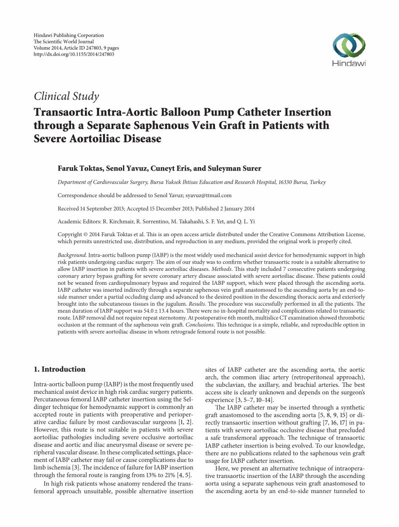
Clinical StudyTransaortic Intra-Aortic Balloon Pump Catheter Insertionthrough a Separate Saphenous Vein Graft in Patients withSevere Aortoiliac Disease
Faruk Toktas, Senol Yavuz, Cuneyt Eris, and Suleyman Surer
Department of Cardiovascular Surgery, Bursa Yuksek Ihtisas Education and Research Hospital, 16330 Bursa, Turkey
Correspondence should be addressed to Senol Yavuz; [email protected]
Received 14 September 2013; Accepted 15 December 2013; Published 2 January 2014
Academic Editors: R. Kirchmair, R. Sorrentino, M. Takahashi, S. F. Yet, and Q. L. Yi
Copyright © 2014 Faruk Toktas et al. This is an open access article distributed under the Creative Commons Attribution License,which permits unrestricted use, distribution, and reproduction in any medium, provided the original work is properly cited.
Background. Intra-aortic balloon pump (IABP) is the most widely used mechanical assist device for hemodynamic support in highrisk patients undergoing cardiac surgery. The aim of our study was to confirm whether transaortic route is a suitable alternative toallow IABP insertion in patients with severe aortoiliac diseases. Methods. This study included 7 consecutive patients undergoingcoronary artery bypass grafting for severe coronary artery disease associated with severe aortoiliac disease. These patients couldnot be weaned from cardiopulmonary bypass and required the IABP support, which were placed through the ascending aorta.IABP catheter was inserted indirectly through a separate saphenous vein graft anastomosed to the ascending aorta by an end-to-side manner under a partial occluding clamp and advanced to the desired position in the descending thoracic aorta and exteriorlybrought into the subcutaneous tissues in the jugulum. Results. The procedure was successfully performed in all the patients. Themean duration of IABP support was 54.0±13.4 hours.There were no in-hospital mortality and complications related to transaorticroute. IABP removal did not require repeat sternotomy. At postoperative 6thmonth,multislice CT examination showed thromboticocclusion at the remnant of the saphenous vein graft. Conclusions. This technique is a simple, reliable, and reproducible option inpatients with severe aortoiliac disease in whom retrograde femoral route is not possible.
1. Introduction
Intra-aortic balloon pump (IABP) is themost frequently usedmechanical assist device in high risk cardiac surgery patients.Percutaneous femoral IABP catheter insertion using the Sel-dinger technique for hemodynamic support is commonly anaccepted route in patients with preoperative and perioper-ative cardiac failure by most cardiovascular surgeons [1, 2].However, this route is not suitable in patients with severeaortoiliac pathologies including severe occlusive aortoiliacdisease and aortic and iliac aneurysmal disease or severe pe-ripheral vascular disease. In these complicated settings, place-ment of IABP catheter may fail or cause complications due tolimb ischemia [3].The incidence of failure for IABP insertionthrough the femoral route is ranging from 13% to 21% [4, 5].
In high risk patients whose anatomy rendered the trans-femoral approach unsuitable, possible alternative insertion
sites of IABP catheter are the ascending aorta, the aorticarch, the common iliac artery (retroperitoneal approach),the subclavian, the axillary, and brachial arteries. The bestaccess site is clearly unknown and depends on the surgeon’sexperience [3, 5–7, 10–14].
The IABP catheter may be inserted through a syntheticgraft anastomosed to the ascending aorta [5, 8, 9, 15] or di-rectly transaortic insertion without grafting [7, 16, 17] in pa-tients with severe aortoiliac occlusive disease that precludeda safe transfemoral approach. The technique of transaorticIABP catheter insertion is being evolved. To our knowledge,there are no publications related to the saphenous vein graftusage for IABP catheter insertion.
Here, we present an alternative technique of intraopera-tive transaortic insertion of the IABP through the ascendingaorta using a separate saphenous vein graft anastomosed tothe ascending aorta by an end-to-side manner tunneled to
Hindawi Publishing Corporatione Scientific World JournalVolume 2014, Article ID 247803, 9 pageshttp://dx.doi.org/10.1155/2014/247803
2 The Scientific World Journal
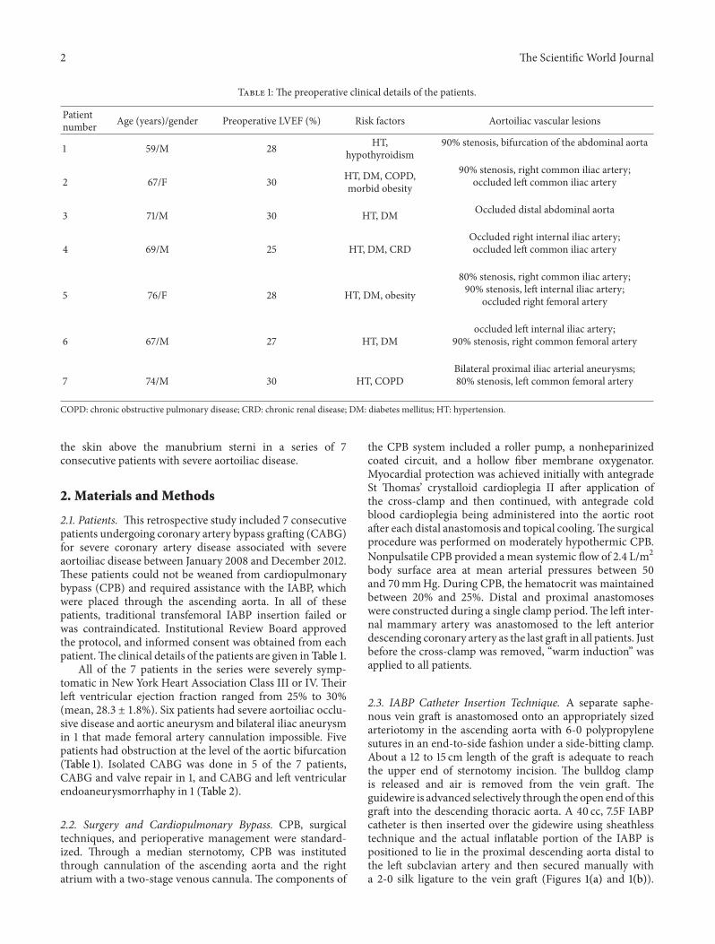
Table 1: The preoperative clinical details of the patients.
Patientnumber Age (years)/gender Preoperative LVEF (%) Risk factors Aortoiliac vascular lesions
1 59/M 28 HT,hypothyroidism
90% stenosis, bifurcation of the abdominal aorta
2 67/F 30 HT, DM, COPD,morbid obesity
90% stenosis, right common iliac artery;occluded left common iliac artery
3 71/M 30 HT, DM Occluded distal abdominal aorta
4 69/M 25 HT, DM, CRDOccluded right internal iliac artery;occluded left common iliac artery
5 76/F 28 HT, DM, obesity
80% stenosis, right common iliac artery;90% stenosis, left internal iliac artery;
occluded right femoral artery
6 67/M 27 HT, DMoccluded left internal iliac artery;
90% stenosis, right common femoral artery
7 74/M 30 HT, COPDBilateral proximal iliac arterial aneurysms;80% stenosis, left common femoral artery
COPD: chronic obstructive pulmonary disease; CRD: chronic renal disease; DM: diabetes mellitus; HT: hypertension.
the skin above the manubrium sterni in a series of 7consecutive patients with severe aortoiliac disease.
2. Materials and Methods
2.1. Patients. This retrospective study included 7 consecutivepatients undergoing coronary artery bypass grafting (CABG)for severe coronary artery disease associated with severeaortoiliac disease between January 2008 and December 2012.These patients could not be weaned from cardiopulmonarybypass (CPB) and required assistance with the IABP, whichwere placed through the ascending aorta. In all of thesepatients, traditional transfemoral IABP insertion failed orwas contraindicated. Institutional Review Board approvedthe protocol, and informed consent was obtained from eachpatient.The clinical details of the patients are given in Table 1.
All of the 7 patients in the series were severely symp-tomatic in New York Heart Association Class III or IV. Theirleft ventricular ejection fraction ranged from 25% to 30%(mean, 28.3 ± 1.8%). Six patients had severe aortoiliac occlu-sive disease and aortic aneurysm and bilateral iliac aneurysmin 1 that made femoral artery cannulation impossible. Fivepatients had obstruction at the level of the aortic bifurcation(Table 1). Isolated CABG was done in 5 of the 7 patients,CABG and valve repair in 1, and CABG and left ventricularendoaneurysmorrhaphy in 1 (Table 2).
2.2. Surgery and Cardiopulmonary Bypass. CPB, surgicaltechniques, and perioperative management were standard-ized. Through a median sternotomy, CPB was institutedthrough cannulation of the ascending aorta and the rightatrium with a two-stage venous cannula. The components of
the CPB system included a roller pump, a nonheparinizedcoated circuit, and a hollow fiber membrane oxygenator.Myocardial protection was achieved initially with antegradeSt Thomas’ crystalloid cardioplegia II after application ofthe cross-clamp and then continued, with antegrade coldblood cardioplegia being administered into the aortic rootafter each distal anastomosis and topical cooling.The surgicalprocedure was performed on moderately hypothermic CPB.Nonpulsatile CPB provided a mean systemic flow of 2.4 L/m2body surface area at mean arterial pressures between 50and 70mmHg. During CPB, the hematocrit was maintainedbetween 20% and 25%. Distal and proximal anastomoseswere constructed during a single clamp period.The left inter-nal mammary artery was anastomosed to the left anteriordescending coronary artery as the last graft in all patients. Justbefore the cross-clamp was removed, “warm induction” wasapplied to all patients.
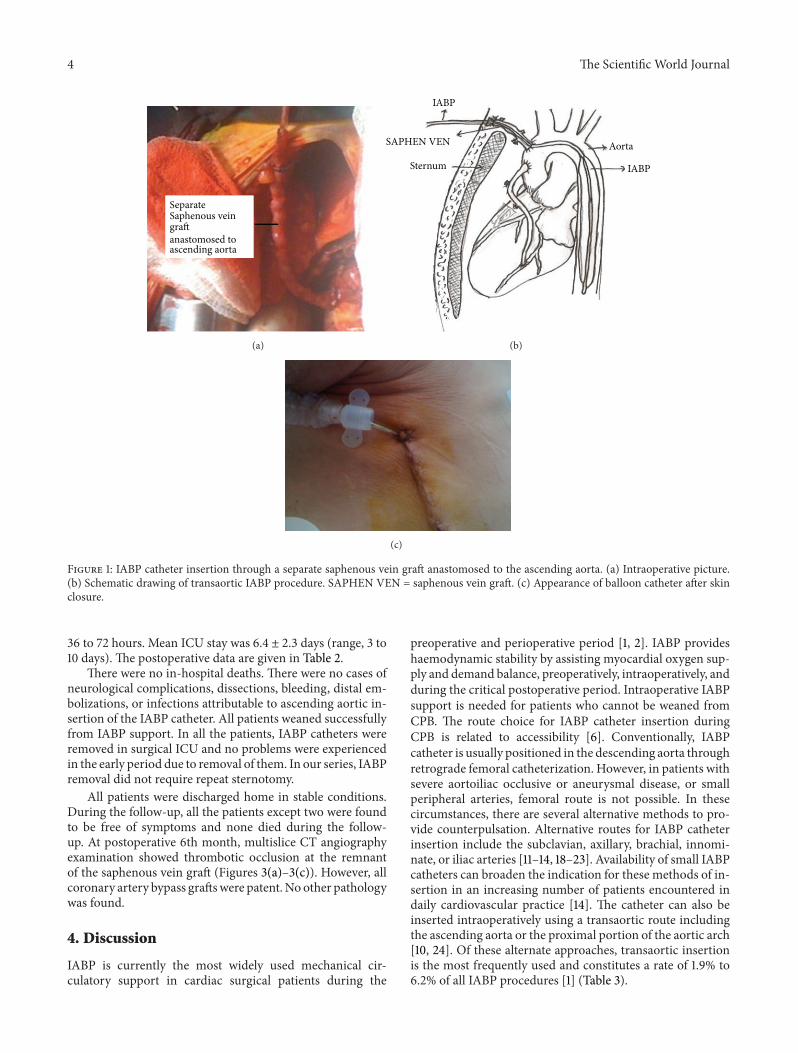
2.3. IABP Catheter Insertion Technique. A separate saphe-nous vein graft is anastomosed onto an appropriately sizedarteriotomy in the ascending aorta with 6-0 polypropylenesutures in an end-to-side fashion under a side-bitting clamp.About a 12 to 15 cm length of the graft is adequate to reachthe upper end of sternotomy incision. The bulldog clampis released and air is removed from the vein graft. Theguidewire is advanced selectively through the open endof thisgraft into the descending thoracic aorta. A 40 cc, 7.5F IABPcatheter is then inserted over the gidewire using sheathlesstechnique and the actual inflatable portion of the IABP ispositioned to lie in the proximal descending aorta distal tothe left subclavian artery and then secured manually witha 2-0 silk ligature to the vein graft (Figures 1(a) and 1(b)).
The Scientific World Journal 3
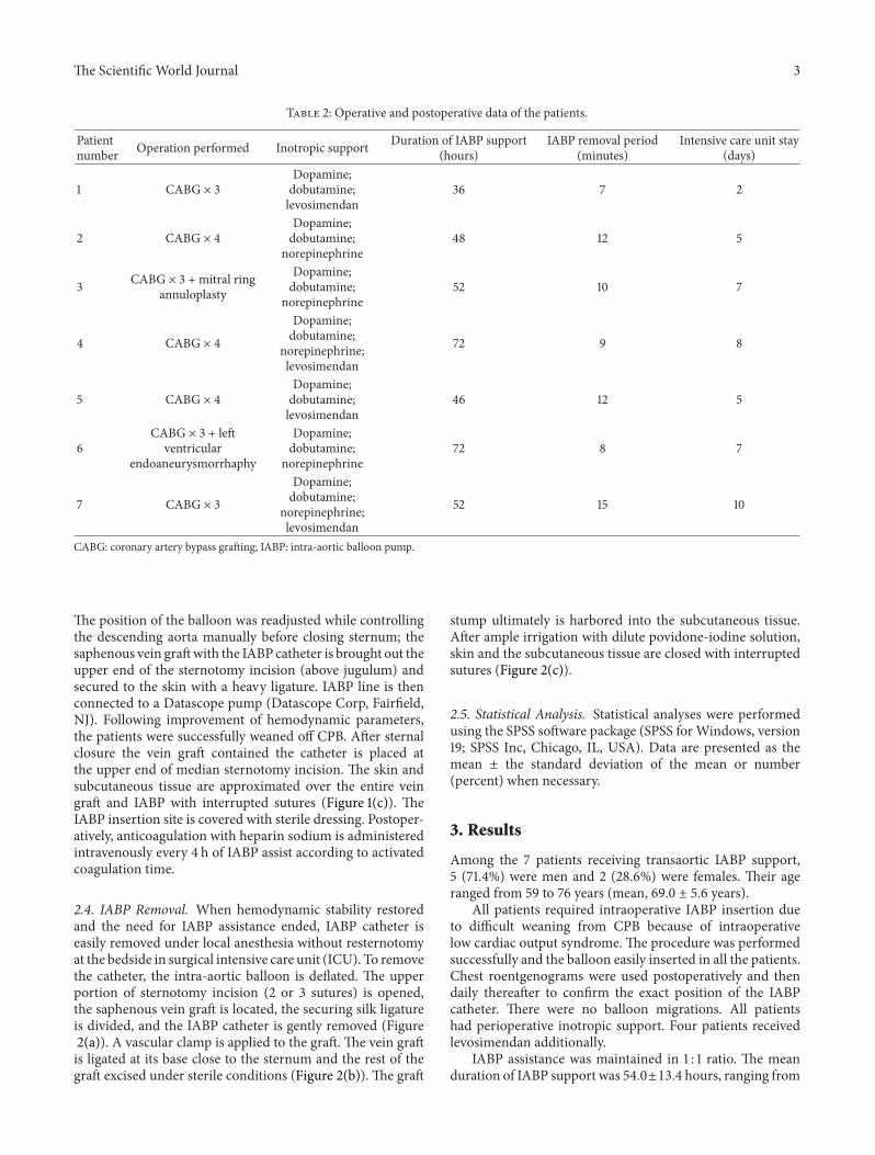
Table 2: Operative and postoperative data of the patients.
Patientnumber Operation performed Inotropic support Duration of IABP support
(hours)IABP removal period
(minutes)Intensive care unit stay
(days)
1 CABG × 3Dopamine;dobutamine;levosimendan
36 7 2
2 CABG × 4Dopamine;dobutamine;
norepinephrine48 12 5
3 CABG × 3 + mitral ringannuloplasty
Dopamine;dobutamine;
norepinephrine52 10 7
4 CABG × 4
Dopamine;dobutamine;
norepinephrine;levosimendan
72 9 8
5 CABG × 4Dopamine;dobutamine;levosimendan
46 12 5
6CABG × 3 + left
ventricularendoaneurysmorrhaphy
Dopamine;dobutamine;
norepinephrine72 8 7
7 CABG × 3
Dopamine;dobutamine;
norepinephrine;levosimendan
52 15 10
CABG: coronary artery bypass grafting; IABP: intra-aortic balloon pump.
The position of the balloon was readjusted while controllingthe descending aorta manually before closing sternum; thesaphenous vein graftwith the IABP catheter is brought out theupper end of the sternotomy incision (above jugulum) andsecured to the skin with a heavy ligature. IABP line is thenconnected to a Datascope pump (Datascope Corp, Fairfield,NJ). Following improvement of hemodynamic parameters,the patients were successfully weaned off CPB. After sternalclosure the vein graft contained the catheter is placed atthe upper end of median sternotomy incision. The skin andsubcutaneous tissue are approximated over the entire veingraft and IABP with interrupted sutures (Figure 1(c)). TheIABP insertion site is covered with sterile dressing. Postoper-atively, anticoagulation with heparin sodium is administeredintravenously every 4 h of IABP assist according to activatedcoagulation time.
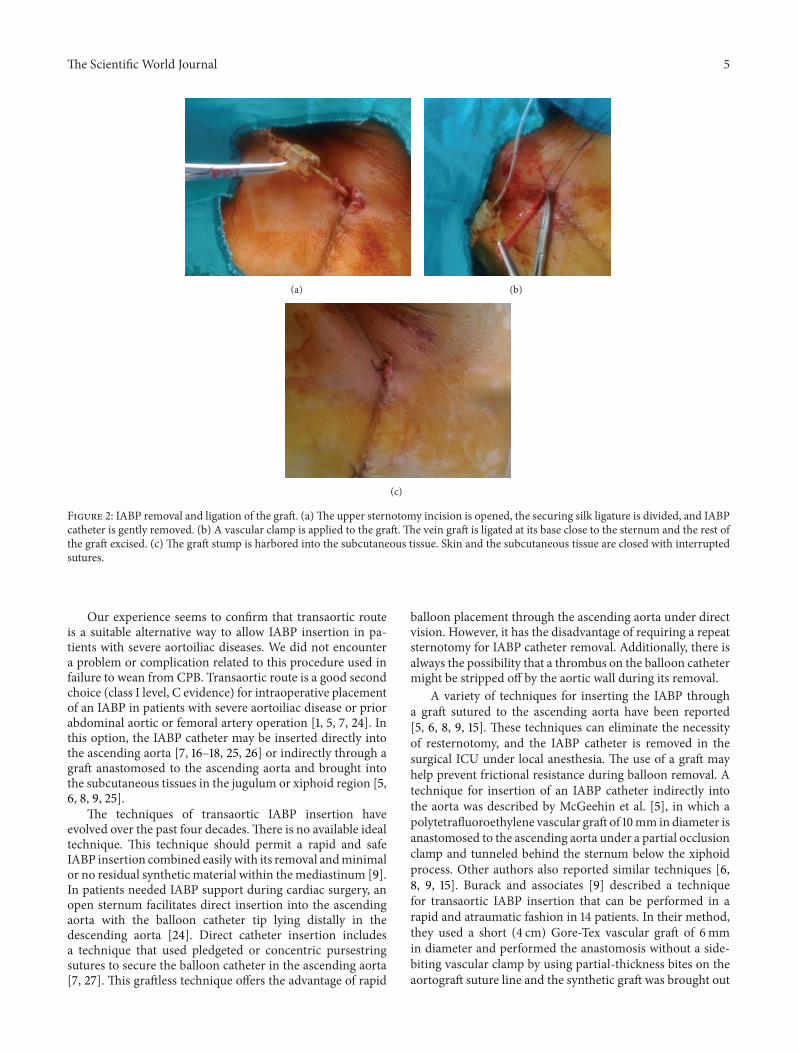
2.4. IABP Removal. When hemodynamic stability restoredand the need for IABP assistance ended, IABP catheter iseasily removed under local anesthesia without resternotomyat the bedside in surgical intensive care unit (ICU). To removethe catheter, the intra-aortic balloon is deflated. The upperportion of sternotomy incision (2 or 3 sutures) is opened,the saphenous vein graft is located, the securing silk ligatureis divided, and the IABP catheter is gently removed (Figure2(a)). A vascular clamp is applied to the graft. The vein graftis ligated at its base close to the sternum and the rest of thegraft excised under sterile conditions (Figure 2(b)). The graft
stump ultimately is harbored into the subcutaneous tissue.After ample irrigation with dilute povidone-iodine solution,skin and the subcutaneous tissue are closed with interruptedsutures (Figure 2(c)).
2.5. Statistical Analysis. Statistical analyses were performedusing the SPSS software package (SPSS for Windows, version19; SPSS Inc, Chicago, IL, USA). Data are presented as themean ± the standard deviation of the mean or number(percent) when necessary.
3. Results
Among the 7 patients receiving transaortic IABP support,5 (71.4%) were men and 2 (28.6%) were females. Their ageranged from 59 to 76 years (mean, 69.0 ± 5.6 years).
All patients required intraoperative IABP insertion dueto difficult weaning from CPB because of intraoperativelow cardiac output syndrome. The procedure was performedsuccessfully and the balloon easily inserted in all the patients.Chest roentgenograms were used postoperatively and thendaily thereafter to confirm the exact position of the IABPcatheter. There were no balloon migrations. All patientshad perioperative inotropic support. Four patients receivedlevosimendan additionally.
IABP assistance was maintained in 1 : 1 ratio. The meanduration of IABP support was 54.0±13.4 hours, ranging from
4 The Scientific World Journal
SeparateSaphenous veingraftanastomosed toascending aorta
(a)
IABP
SAPHEN VEN
Sternum
Aorta
IABP
(b)
(c)
Figure 1: IABP catheter insertion through a separate saphenous vein graft anastomosed to the ascending aorta. (a) Intraoperative picture.(b) Schematic drawing of transaortic IABP procedure. SAPHEN VEN = saphenous vein graft. (c) Appearance of balloon catheter after skinclosure.
36 to 72 hours. Mean ICU stay was 6.4 ± 2.3 days (range, 3 to10 days). The postoperative data are given in Table 2.
There were no in-hospital deaths. There were no cases ofneurological complications, dissections, bleeding, distal em-bolizations, or infections attributable to ascending aortic in-sertion of the IABP catheter. All patients weaned successfullyfrom IABP support. In all the patients, IABP catheters wereremoved in surgical ICU and no problems were experiencedin the early period due to removal of them. In our series, IABPremoval did not require repeat sternotomy.
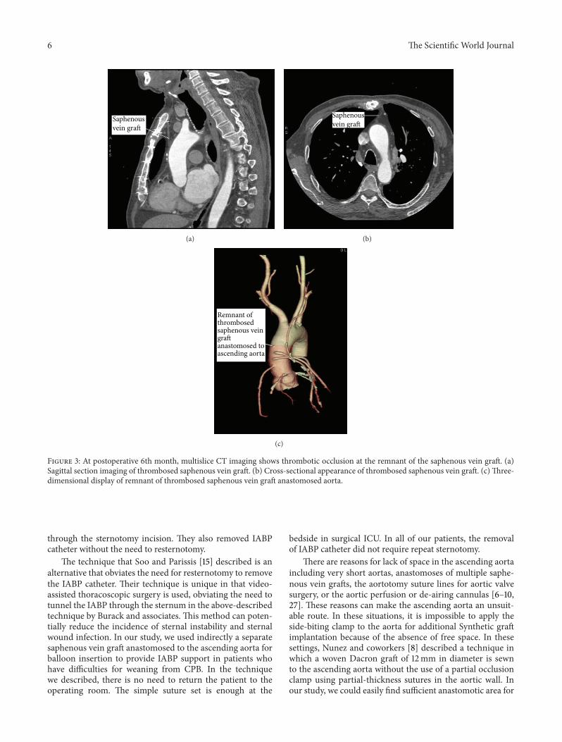
All patients were discharged home in stable conditions.During the follow-up, all the patients except two were foundto be free of symptoms and none died during the follow-up. At postoperative 6th month, multislice CT angiographyexamination showed thrombotic occlusion at the remnantof the saphenous vein graft (Figures 3(a)–3(c)). However, allcoronary artery bypass graftswere patent.No other pathologywas found.
4. Discussion
IABP is currently the most widely used mechanical cir-culatory support in cardiac surgical patients during the
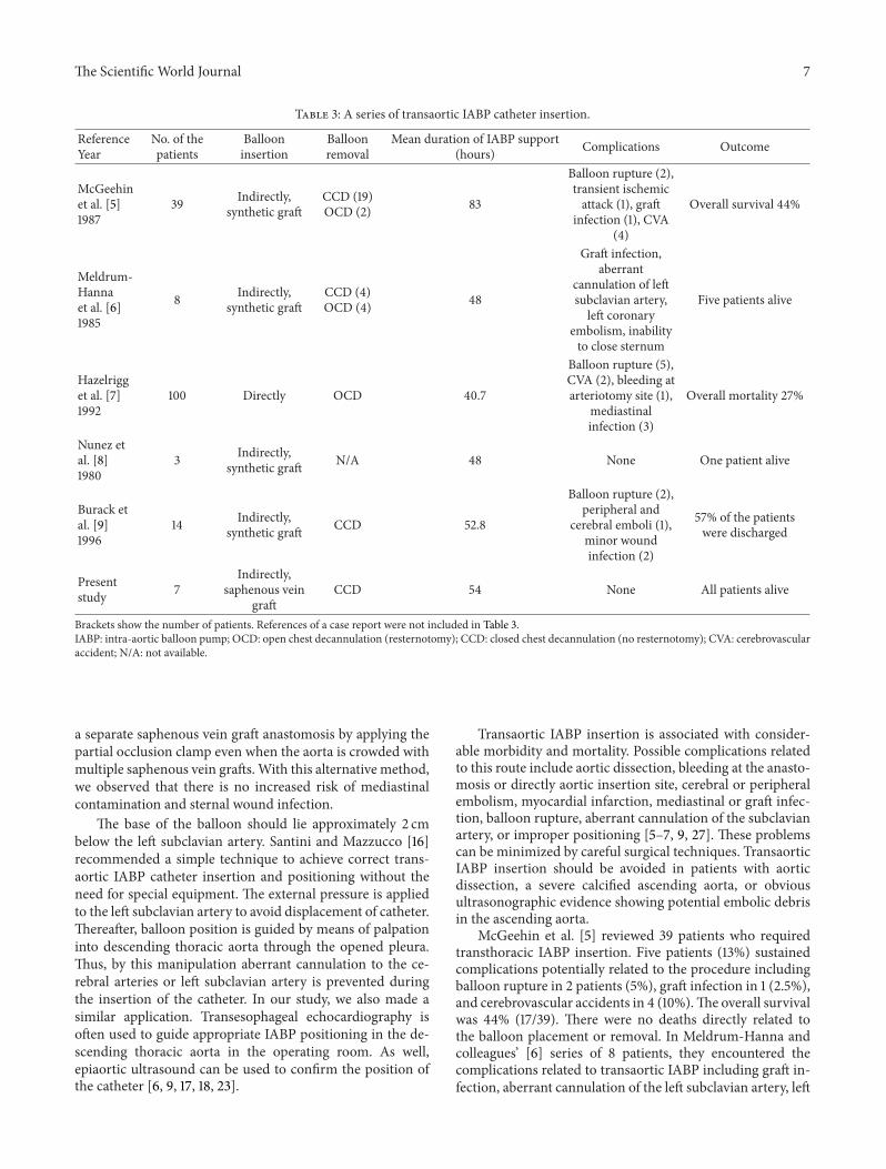
preoperative and perioperative period [1, 2]. IABP provideshaemodynamic stability by assisting myocardial oxygen sup-ply and demand balance, preoperatively, intraoperatively, andduring the critical postoperative period. Intraoperative IABPsupport is needed for patients who cannot be weaned fromCPB. The route choice for IABP catheter insertion duringCPB is related to accessibility [6]. Conventionally, IABPcatheter is usually positioned in the descending aorta throughretrograde femoral catheterization. However, in patients withsevere aortoiliac occlusive or aneurysmal disease, or smallperipheral arteries, femoral route is not possible. In thesecircumstances, there are several alternative methods to pro-vide counterpulsation. Alternative routes for IABP catheterinsertion include the subclavian, axillary, brachial, innomi-nate, or iliac arteries [11–14, 18–23]. Availability of small IABPcatheters can broaden the indication for these methods of in-sertion in an increasing number of patients encountered indaily cardiovascular practice [14]. The catheter can also beinserted intraoperatively using a transaortic route includingthe ascending aorta or the proximal portion of the aortic arch[10, 24]. Of these alternate approaches, transaortic insertionis the most frequently used and constitutes a rate of 1.9% to6.2% of all IABP procedures [1] (Table 3).
The Scientific World Journal 5
(a) (b)
(c)
Figure 2: IABP removal and ligation of the graft. (a)The upper sternotomy incision is opened, the securing silk ligature is divided, and IABPcatheter is gently removed. (b) A vascular clamp is applied to the graft. The vein graft is ligated at its base close to the sternum and the rest ofthe graft excised. (c) The graft stump is harbored into the subcutaneous tissue. Skin and the subcutaneous tissue are closed with interruptedsutures.
Our experience seems to confirm that transaortic routeis a suitable alternative way to allow IABP insertion in pa-tients with severe aortoiliac diseases. We did not encountera problem or complication related to this procedure used infailure to wean from CPB. Transaortic route is a good secondchoice (class I level, C evidence) for intraoperative placementof an IABP in patients with severe aortoiliac disease or priorabdominal aortic or femoral artery operation [1, 5, 7, 24]. Inthis option, the IABP catheter may be inserted directly intothe ascending aorta [7, 16–18, 25, 26] or indirectly through agraft anastomosed to the ascending aorta and brought intothe subcutaneous tissues in the jugulum or xiphoid region [5,6, 8, 9, 25].
The techniques of transaortic IABP insertion haveevolved over the past four decades.There is no available idealtechnique. This technique should permit a rapid and safeIABP insertion combined easilywith its removal andminimalor no residual synthetic material within themediastinum [9].In patients needed IABP support during cardiac surgery, anopen sternum facilitates direct insertion into the ascendingaorta with the balloon catheter tip lying distally in thedescending aorta [24]. Direct catheter insertion includesa technique that used pledgeted or concentric pursestringsutures to secure the balloon catheter in the ascending aorta[7, 27]. This graftless technique offers the advantage of rapid
balloon placement through the ascending aorta under directvision. However, it has the disadvantage of requiring a repeatsternotomy for IABP catheter removal. Additionally, there isalways the possibility that a thrombus on the balloon cathetermight be stripped off by the aortic wall during its removal.
A variety of techniques for inserting the IABP througha graft sutured to the ascending aorta have been reported[5, 6, 8, 9, 15]. These techniques can eliminate the necessityof resternotomy, and the IABP catheter is removed in thesurgical ICU under local anesthesia. The use of a graft mayhelp prevent frictional resistance during balloon removal. Atechnique for insertion of an IABP catheter indirectly intothe aorta was described by McGeehin et al. [5], in which apolytetrafluoroethylene vascular graft of 10mm in diameter isanastomosed to the ascending aorta under a partial occlusionclamp and tunneled behind the sternum below the xiphoidprocess. Other authors also reported similar techniques [6,8, 9, 15]. Burack and associates [9] described a techniquefor transaortic IABP insertion that can be performed in arapid and atraumatic fashion in 14 patients. In their method,they used a short (4 cm) Gore-Tex vascular graft of 6mmin diameter and performed the anastomosis without a side-biting vascular clamp by using partial-thickness bites on theaortograft suture line and the synthetic graft was brought out
6 The Scientific World Journal
Saphenousvein graft
(a)
Saphenousvein graft
(b)
Remnant ofthrombosedsaphenous vein
anastomosed toascending aorta
graft
(c)
Figure 3: At postoperative 6th month, multislice CT imaging shows thrombotic occlusion at the remnant of the saphenous vein graft. (a)Sagittal section imaging of thrombosed saphenous vein graft. (b) Cross-sectional appearance of thrombosed saphenous vein graft. (c)Three-dimensional display of remnant of thrombosed saphenous vein graft anastomosed aorta.
through the sternotomy incision. They also removed IABPcatheter without the need to resternotomy.
The technique that Soo and Parissis [15] described is analternative that obviates the need for resternotomy to removethe IABP catheter. Their technique is unique in that video-assisted thoracoscopic surgery is used, obviating the need totunnel the IABP through the sternum in the above-describedtechnique by Burack and associates. This method can poten-tially reduce the incidence of sternal instability and sternalwound infection. In our study, we used indirectly a separatesaphenous vein graft anastomosed to the ascending aorta forballoon insertion to provide IABP support in patients whohave difficulties for weaning from CPB. In the techniquewe described, there is no need to return the patient to theoperating room. The simple suture set is enough at the
bedside in surgical ICU. In all of our patients, the removalof IABP catheter did not require repeat sternotomy.
There are reasons for lack of space in the ascending aortaincluding very short aortas, anastomoses of multiple saphe-nous vein grafts, the aortotomy suture lines for aortic valvesurgery, or the aortic perfusion or de-airing cannulas [6–10,27]. These reasons can make the ascending aorta an unsuit-able route. In these situations, it is impossible to apply theside-biting clamp to the aorta for additional Synthetic graftimplantation because of the absence of free space. In thesesettings, Nunez and coworkers [8] described a technique inwhich a woven Dacron graft of 12mm in diameter is sewnto the ascending aorta without the use of a partial occlusionclamp using partial-thickness sutures in the aortic wall. Inour study, we could easily find sufficient anastomotic area for
The Scientific World Journal 7
Table 3: A series of transaortic IABP catheter insertion.
ReferenceYear
No. of thepatients
Ballooninsertion
Balloonremoval
Mean duration of IABP support(hours) Complications Outcome
McGeehinet al. [5]1987
39 Indirectly,synthetic graft
CCD (19)OCD (2) 83
Balloon rupture (2),transient ischemicattack (1), graft
infection (1), CVA(4)
Overall survival 44%
Meldrum-Hannaet al. [6]1985
8 Indirectly,synthetic graft
CCD (4)OCD (4) 48
Graft infection,aberrant
cannulation of leftsubclavian artery,left coronary
embolism, inabilityto close sternum
Five patients alive
Hazelrigget al. [7]1992
100 Directly OCD 40.7
Balloon rupture (5),CVA (2), bleeding atarteriotomy site (1),
mediastinalinfection (3)
Overall mortality 27%
Nunez etal. [8]1980
3 Indirectly,synthetic graft N/A 48 None One patient alive
Burack etal. [9]1996
14 Indirectly,synthetic graft CCD 52.8
Balloon rupture (2),peripheral and
cerebral emboli (1),minor woundinfection (2)
57% of the patientswere discharged
Presentstudy 7
Indirectly,saphenous vein
graftCCD 54 None All patients alive
Brackets show the number of patients. References of a case report were not included in Table 3.IABP: intra-aortic balloon pump; OCD: open chest decannulation (resternotomy); CCD: closed chest decannulation (no resternotomy); CVA: cerebrovascularaccident; N/A: not available.
a separate saphenous vein graft anastomosis by applying thepartial occlusion clamp even when the aorta is crowded withmultiple saphenous vein grafts.With this alternative method,we observed that there is no increased risk of mediastinalcontamination and sternal wound infection.
The base of the balloon should lie approximately 2 cmbelow the left subclavian artery. Santini and Mazzucco [16]recommended a simple technique to achieve correct trans-aortic IABP catheter insertion and positioning without theneed for special equipment. The external pressure is appliedto the left subclavian artery to avoid displacement of catheter.Thereafter, balloon position is guided by means of palpationinto descending thoracic aorta through the opened pleura.Thus, by this manipulation aberrant cannulation to the ce-rebral arteries or left subclavian artery is prevented duringthe insertion of the catheter. In our study, we also made asimilar application. Transesophageal echocardiography isoften used to guide appropriate IABP positioning in the de-scending thoracic aorta in the operating room. As well,epiaortic ultrasound can be used to confirm the position ofthe catheter [6, 9, 17, 18, 23].
Transaortic IABP insertion is associated with consider-able morbidity and mortality. Possible complications relatedto this route include aortic dissection, bleeding at the anasto-mosis or directly aortic insertion site, cerebral or peripheralembolism, myocardial infarction, mediastinal or graft infec-tion, balloon rupture, aberrant cannulation of the subclavianartery, or improper positioning [5–7, 9, 27]. These problemscan be minimized by careful surgical techniques. TransaorticIABP insertion should be avoided in patients with aorticdissection, a severe calcified ascending aorta, or obviousultrasonographic evidence showing potential embolic debrisin the ascending aorta.
McGeehin et al. [5] reviewed 39 patients who requiredtransthoracic IABP insertion. Five patients (13%) sustainedcomplications potentially related to the procedure includingballoon rupture in 2 patients (5%), graft infection in 1 (2.5%),and cerebrovascular accidents in 4 (10%).The overall survivalwas 44% (17/39). There were no deaths directly related tothe balloon placement or removal. In Meldrum-Hanna andcolleagues’ [6] series of 8 patients, they encountered thecomplications related to transaortic IABP including graft in-fection, aberrant cannulation of the left subclavian artery, left
8 The Scientific World Journal
coronary artery embolism, and inability to close the sternumdue to mechanical tamponade. In our study, there were nocomplications and mortality related to transaortic IABP.
In a retrospective large series of 100 cases of transthoracicIABP insertion without graft, Hazelrigg and coworkers [7]evaluated the complications in 81 patients who survived tohave their IABP removed. They demonstrated no increasedmortality and complication rates similar to those of femoralinsertion.The authors also reported that complications relat-ed to transaortic route included balloon rupture in 5 patients(6.2%), cerebral vascular accident in 2 (2.5%), transient is-chemic attack in 1 (1.2%), bleeding at the IABP arteriotomysite in 3 (3.7%), and mediastinal infection in 3 (3.7%). In thishigh risk group of patients, the rate of balloon rupture andmediastinal bleeding and infection has increased because ofthe direct transaortic IABP catheter insertion. However, neu-rologic events do not appear to be increased. In their series,overall mortality was 27% [7]. Santini and associates [28]also described balloon migration as an unexpected compli-cation of IABP support via the ascending aorta. They ex-plained that possible reason for this complication may be thelooping of the balloon catheter into the aortic lumen possiblyprompted by a too proximal location.
Pinkard and associates [29] found in analyzing a series of123 IABPs inserted for weaning from CBP in CABG patients(42 transaortic and 81 femoral) that the increased mortalityin the arch insertion group was a result of the greater comor-bidities rather than the route of insertion. Additionally, theyremarked no increase in complications in the aortic insertiongroup, in spite of a higher incidence of leg complications inthe patients with femoral insertion.
In our experience, the transaortic route appears as an ex-cellent intraoperative solution to provide IABP support in pa-tients who are difficult to wean from CPB due to low cardiacoutput syndrome and who are unsuitable femoral access. Wedid not observe any problems or complications related tothe placement or use of the transaortic IABP. An importantadvantage of our technique is technically easier and safer tosew the saphenous vein graft to the ascending aorta com-pared with the synthetic vascular grafts. An another advan-tage is that the separate saphenous vein graft which anasto-mosed the ascending aorta runs for a short distance behindthe manubrium and does not pass through or behind thesternotomy, as described by other authors.These benefits canresult in a decreased risk of sternal wound infections. Fur-thermore, the use of a sheathless balloon inserted through thesaphenous vein graft makes balloon removal easy and safe atthe bedside under local anesthesia.
Limitation of the Study. Herein, we reported our institutionalexperience with small case series, even if larger studies areneeded to definitely verify the advantages of transaortic route.This route to provide IABP assistance is lifesaving in patientswith severe comorbidity inwhom transfemoral route failed orcontraindicated. Transaortic IABP insertion has the followingdrawbacks: (a) it requires the environment of operating room;(b) it is more time consuming than the femoral route; and (c)it has several potential serious complications, including therisk of cerebral emboli, aortic dissection, and haemorrhage
from the graft sutured to ascending aorta during catheterremoval because of overstretch.
5. Conclusions
IABP catheter insertion through a separate saphenous veingraft anastomosed to the ascending aorta for IABP coun-terpulsation is a simple, reliable, and reproducible option inpatients with severe aortoiliac disease in whom retrogradefemoral route is not possible. Although possible severe com-plications are associated with ascending aortic cannulation,transaortic route should be considered for all patients requir-ing intra- and postoperative IABP support in whom otheraccess routes are not feasible. We also recommend that thisaccess site should be added to the surgical armamentariumin these complicated pathologies.
Conflict of Interests
All authors have no conflict of interests to disclose. Theauthors do not have a direct financial relation with anycommercial identities mentioned in the paper.
Authors’ Contribution
Faruk Toktas and Senol Yavuz are co-first authors for equalcontribution for the present study.
References
[1] R. J. F. Baskett,W. A. Ghali, A.Maitland, andG.M.Hirsch, “Theintraaortic balloon pump in cardiac surgery,”Annals ofThoracicSurgery, vol. 74, no. 4, pp. 1276–1287, 2002.
[2] J. T. Christenson, F. Simonet, P. Badel, and M. Schmuziger,“Optimal timing of preoperative intraaortic balloon pumpsupport in high-risk coronary patients,” Annals of ThoracicSurgery, vol. 68, no. 3, pp. 934–939, 1999.
[3] S. Yavuz, “eComment: right axillary artery as an alternativeroute for intraaortic balloon pump catheter insertion in severeaortoiliac pathologies,” Interactive CardioVascular andThoracicSurgery, vol. 9, no. 2, pp. 370–371, 2009.
[4] H. Parissis, A. Soo, and B. Al-Alao, “Intra aortic balloon pump:literature review of risk factors related to complications of theintraaortic balloon pump,” Journal of Cardiothoracic Surgery,vol. 6, no. 1, article 147, 2011.
[5] W. McGeehin, F. Sheikh, J. S. Donahoo, M. J. Lechman, andH. MacVaugh III, “Transthoracic intraaortic balloon pumpsupport: experience in 39 patients,” Annals of Thoracic Surgery,vol. 44, no. 1, pp. 26–30, 1987.
[6] W. G. Meldrum-Hanna, C. W. Deal, and D. E. Ross, “Compli-cations of ascending aortic intraaortic balloon pump cannula-tion,” Annals of Thoracic Surgery, vol. 40, no. 3, pp. 241–244,1985.
[7] S. R. Hazelrigg, J. E. Auer, and P. E. Seifert, “Experience in 100transthoracic balloon pumps,” Annals of Thoracic Surgery, vol.54, no. 3, pp. 528–532, 1992.
[8] L. Nunez,M. G. Aguado, A. Iglesias, and J. L. Larrea, “Transaor-tic cannulation for balloon pumping in a ‘crowded aorta’,”Annals of Thoracic Surgery, vol. 30, no. 4, pp. 400–402, 1980.
The Scientific World Journal 9
[9] J. H. Burack, P. Uceda, and J. N. Cunningham Jr., “Transthoracicintraaortic balloon pump: a simplified technique,” Annals ofThoracic Surgery, vol. 62, no. 1, pp. 299–301, 1996.
[10] S. C. Balderman, J. N. Bhayana, and R. Pifarre, “Technique forinsertion of the intra-aortic balloon through the aortic arch,”Journal of Cardiovascular Surgery, vol. 21, no. 5, pp. 614–616,1980.
[11] K. Garrett and K. L. Grady, “Intraaortic balloon pumpingthrough the common iliac artery: management of the ambu-latory intraaortic balloon pump patient,” Progress in Cardiovas-cular Nursing, vol. 15, no. 1, pp. 14–20, 2000.
[12] C. B. Marcu, T. J. Donohue, A. Ferneini, and A. E. Ghantous,“Intraaortic balloon pump insertion through the subclavianartery. Subclavian artery insertion of IABP,” Heart Lung andCirculation, vol. 15, no. 2, pp. 148–150, 2006.
[13] S. Yavuz, C. Eris, M. Sezen, and T. Turk, “Axillary arteryapproach for insertion of an intraaortic balloon pump catheterin a patient with severe occlusive aortoiliac disease,” InteractiveCardioVascular and Thoracic Surgery, vol. 7, supplement 1, p.S118, 2008.
[14] A. S. Rubino, F. Onorati, F. Serraino, and A. Renzulli, “Safetyand efficacy of transbrachial intra-aortic balloon pumping withthe use of 7-Fr catheters in patients undergoing coronary bypasssurgery,” Interactive CardioVascular andThoracic Surgery, vol. 9,no. 1, pp. 135–137, 2009.
[15] A. W. Soo and H. Parissis, “Transthoracic intra-aortic balloonpump removal without repeat sternotomy,” Asian Cardiovascu-lar and Thoracic Annals, vol. 20, no. 5, pp. 623–624, 2012.
[16] F. Santini and A. Mazzucco, “Transthoracic intraaortic coun-terpulsation: a simplemethod for balloon catheter positioning,”Annals of Thoracic Surgery, vol. 64, no. 3, pp. 859–860, 1997.
[17] L. J. Kaplan, D. S. Weiman, N. Langan, A. B. Sokil, and G. J. R.Whitman, “Safe intraaortic balloon pump placement throughthe ascending aorta using transesophageal ultrasound,” Annalsof Thoracic Surgery, vol. 54, no. 2, pp. 374–375, 1992.
[18] M. J. Russo, V. Jeevanandam, J. Stepney et al., “Intra-aortic bal-loon pump inserted through the subclavian artery: a minimallyinvasive approach to mechanical support in the ambulatoryend-stage heart failure patient,” Journal of Thoracic and Cardio-vascular Surgery, vol. 144, no. 4, pp. 951–955, 2012.
[19] G. Zattera, P. Totaro, A. M. D’Armini, and M. Vigano, “Intraaortic balloon pump insertion through left axillary artery inpatients with severe peripheral arterial disease,” InteractiveCardioVascular andThoracic Surgery, vol. 9, no. 2, pp. 369–370,2009.
[20] F. Onorati, B. Impiombato, A. Ferraro et al., “Transbrachialintraaortic balloon pumping in severe peripheral atherosclero-sis,”Annals ofThoracic Surgery, vol. 84, no. 1, pp. 264–266, 2007.
[21] S. Bundhoo, P. A. O’Keefe, H. Luckraz, and N. Ossei-Gerning,“Extended duration of brachially inserted intra-aortic balloonpump for myocardial protection in two patients undergoingurgent coronary artery bypass grafting,” Interactive CardioVas-cular andThoracic Surgery, vol. 7, no. 1, pp. 42–44, 2008.
[22] B. Datt and S. Miner, “Anatomical advantage to percutaneousinsertion of the intra-aortic balloon through the left brachialartery over the right brachial artery,” Journal of ExtracorporealTechnology, vol. 45, no. 1, pp. 51–54, 2013.
[23] D. Calcaterra, K. Karam, J. B. Babajanov, and J. E. Davis, “Anoption for intraoperative placement of an intra-aortic balloonpump in patients with occlusive peripheral vascular disease,”Journal of Thoracic and Cardiovascular Surgery, vol. 141, no. 2,pp. 586–587, 2011.
[24] B. Datt, L. Hutchison, and C. Peniston, “Trans-aortic coun-terpulsation: a viable alternative?” Journal of Extra-CorporealTechnology, vol. 39, no. 2, pp. 91–95, 2007.
[25] T. L. Gueldner and G. H. Lawrence, “Intraaortic balloon assistthrough cannulation of the ascending aorta,”Annals ofThoracicSurgery, vol. 19, no. 1, pp. 88–91, 1975.
[26] F. Robicsek, “Closed-chest decannulation of transthoracicallyinserted aortic balloon catheter without grafting,” Journal ofCardiac Surgery, vol. 2, no. 2, pp. 327–329, 1987.
[27] L. I. Bonchek and G. N. Olinger, “Direct ascending aorticinsertion of the “percutaneous” intraaortic balloon catheter inthe open chest: advantages and precautions,” Annals ofThoracicSurgery, vol. 32, no. 5, pp. 512–514, 1981.
[28] F. Santini, P. Bertolini, A. Rossi, G. Montalbano, and A.Mazzucco, “Unexpected complication of intra-aortic ballooncounterpulsation via the ascending aorta: balloon migration,”European Journal of Cardio-Thoracic Surgery, vol. 11, no. 3, pp.579–581, 1997.
[29] J. Pinkard, J. R. Utley, S. A. Leyland, M. Morgan, and H.Johnson, “Relative risk of aortic and femoral insertion ofintraaortic balloon pump after coronary artery bypass graftingprocedures,” Journal of Thoracic and Cardiovascular Surgery,vol. 105, no. 4, pp. 721–728, 1993.

Submit your manuscripts athttp://www.hindawi.com
Hindawi Publishing Corporationhttp://www.hindawi.com Volume 2013
Oxidative Medicine and Cellular Longevity
Hindawi Publishing Corporation http://www.hindawi.com Volume 2013Hindawi Publishing Corporation http://www.hindawi.com Volume 2013
The Scientific World Journal
International Journal of
EndocrinologyHindawi Publishing Corporationhttp://www.hindawi.com
Volume 2013
ISRN Anesthesiology
Hindawi Publishing Corporationhttp://www.hindawi.com Volume 2013
OncologyJournal of
Hindawi Publishing Corporationhttp://www.hindawi.com Volume 2013
PPARRe sea rch
Hindawi Publishing Corporationhttp://www.hindawi.com Volume 2013
OphthalmologyJournal of
Hindawi Publishing Corporationhttp://www.hindawi.com Volume 2013
ISRN Allergy
Hindawi Publishing Corporationhttp://www.hindawi.com Volume 2013
BioMed Research International
Hindawi Publishing Corporationhttp://www.hindawi.com Volume 2013
ObesityJournal of
Hindawi Publishing Corporationhttp://www.hindawi.com Volume 2013
ISRN Addiction
Hindawi Publishing Corporationhttp://www.hindawi.com Volume 2013
Hindawi Publishing Corporationhttp://www.hindawi.com Volume 2013
Computational and Mathematical Methods in Medicine
ISRN AIDS
Hindawi Publishing Corporationhttp://www.hindawi.com Volume 2013
Clinical &DevelopmentalImmunology
Hindawi Publishing Corporationhttp://www.hindawi.com
Volume 2013
Diabetes ResearchJournal of
Hindawi Publishing Corporationhttp://www.hindawi.com Volume 2013
Evidence-Based Complementary and Alternative Medicine
Volume 2013Hindawi Publishing Corporationhttp://www.hindawi.com
Hindawi Publishing Corporationhttp://www.hindawi.com Volume 2013
Gastroenterology Research and Practice
Hindawi Publishing Corporationhttp://www.hindawi.com Volume 2013
ISRN Biomarkers
Hindawi Publishing Corporationhttp://www.hindawi.com Volume 2013
MEDIATORSINFLAMMATION
of
Copyright © 2022 FDOKUMEN