Alternative substrates as a native oyster (Crassostrea virginica ...
Upload
independentCategory
view
0download
0
Elsevier Editorial System(tm) for Neuropsychologia Manuscript Draft Manuscript Number: NSY-D-06-00285R2 Title: Toward an understanding of the cerebral substrates of woman's orgasm Article Type: Reviews and Perspectives Section/Category: Reviews and Perspectives Keywords: women; sexual function; orgasm; functional neuroimaging; cognitive functions; neuropsychology. Corresponding Author: Dr. Stephanie Ortigue, PhD Corresponding Author's Institution: Sage Center for the Study of the Mind, University of California Santa Barbara First Author: Francesco Bianchi-Demicheli, MD Order of Authors: Francesco Bianchi-Demicheli, MD; Stephanie Ortigue, PhD Abstract: The way women experience orgasm is of interest to scientists, clinicians, and laypeople. Whereas the origin and the function of a woman's orgasm remains controversial, the current models of sexual function acknowledge a combined role of central (spinal and cerebral) and peripheral processes during orgasm experience. At the central level, although it is accepted that the spinal cord drives orgasm, the cerebral involvement and cognitive representation of a woman's orgasm has not been extensively investigated. Important gaps in our knowledge remain. Recently, the astonishing advances of neuroimaging techniques applied in parallel with a neuropsychological approach allowed the unravelling of specific functional neuroanatomy of a woman's orgasm. Here, clinical and experimental findings on the cortico-subcortical pathway of a woman's orgasm are reviewed. By defining the specific brain areas that sustain the assumed higher-order representation of a woman's orgasm, this review provides a foundation for future studies. The next challenge of functional imaging and neuropsychological studies is to understand the hierarchical interactions between these multiple cortical areas, not only with a correlation analysis but also with high

spatio-temporal resolution techniques demonstrating the causal necessity, the temporal time course and the direction of the causality. Further studies using a multi-disciplinary approach are needed to identify the spatio-temporal dynamic of a woman's orgasm, its dysfunctions and possible new treatments.

Bianchi-Demicheli & Ortigue
1
1
Toward an understanding of the cerebral substrates of woman’s orgasm
Francesco Bianchi-Demicheli1,2
Stephanie Ortigue3,4
1. Psychosomatic Gynaecology and Sexology Unit Emergency and Liaison Services,
Geneva University Psychiatric Centre, Switzerland.
2. Division of Reproductive Endocrinology and Infertility
Dept. of Obstetrics and Gynaecology University Hospital of Geneva
Switzerland.
3. Dartmouth Brain Imaging Center, Department of Psychology and Brain Sciences, Center for Cognitive Neuroscience, Dartmouth College, NH, USA
4. Sage Center for the Study of the Mind, Dpt of Psychology, University of California Santa Barbara, Santa
Barbara, CA, USA
*************************************************************************** Title word count: 10 Abstract word count: 217 Text word count: 8717 (including references in the text). Number of Pages: 35 Number of references: 113 *************************************************************************** Correspondence should be addressed to: [email protected]
ABSTRACT
The way women experience orgasm is of interest to scientists, clinicians, and laypeople. Whereas the origin and the function of a woman’s orgasm remains controversial, the current models of sexual function acknowledge a combined role of central (spinal and cerebral) and peripheral processes during orgasm experience. At the central level, although it is accepted that the spinal cord drives orgasm, the cerebral involvement and cognitive representation of a woman’s orgasm has not been extensively investigated. Important gaps in our knowledge remain. Recently, the astonishing advances of neuroimaging techniques applied in parallel with a neuropsychological approach allowed the unravelling of specific functional neuroanatomy of a woman’s orgasm. Here, clinical and experimental findings on the cortico-subcortical pathway of a woman’s orgasm are reviewed. By defining the specific brain areas that sustain the assumed higher-order representation of a woman’s orgasm, this review provides a foundation for future studies. The next challenge of functional imaging and neuropsychological studies is to understand the hierarchical interactions between these multiple cortical areas, not only with a correlation analysis but also with high spatio-temporal resolution techniques demonstrating the causal necessity, the temporal time course and the direction of the causality. Further studies using a multi-disciplinary approach are needed to identify the spatio-temporal dynamic of a woman’s orgasm, its dysfunctions and possible new treatments. Keywords: women, sexual function, orgasm, functional imaging, cognitive functions.
Manuscript

Bianchi-Demicheli & Ortigue
2
2
1. Introduction
Attitudes towards women's sexuality in general, and orgasm in particular, have
varied throughout recorded history (Muchembled, 2005; Symons, 1979; Lloyd, 2005).
Though repression of both was not uncommon, and continues in some cultures and
religions to this day, there have been times when it was viewed positively, and even
celebrated (Muchembled, 2005). Even with this complex historical background, several
philosophers, anthropologists, writers, anatomists, psychiatrists and sexologists have
nevertheless tried to decipher the sense, the mechanisms and the function of a woman’s
orgasm (e.g., Abraham, 2002; Basson, 2000; Buss, 2003; Kaplan, 1974, 1979; Kinsey,
Pomeroy, Martin, & Gebbard, 1953; Hite, 1976; Levin, 1981; Lloyd, 2005; Masters &
Johnson, 1966; Meston, Hull, Levin, & Sipski, 2004; Mould, 1980; Symons, 1979; Whipple,
2002). The greatest advances have been made in identifying the anatomy and physiology of
the organs that are involved during a woman’s orgasm (e.g., Alzate, 1985; Giuliano, Rampin,
& Allard, 2002; Kaplan, 1974; Levin & Wagner, 1985; Masters & Johnson, 1966; Mould,
1980). By highlighting spinal- and cerebral-driven mechanisms on the peripheral response,
great insights have been made in understanding the crucial role of the central nervous system
during orgasm.
Because the neuropharmacology, neuro-endocrinology and the spinal-driven components
of orgasm have been addressed in depth previously (e.g., Argiolas & Melis, 2003; Giuliano,
Rampin, & Allard, 2002; Heaton & Adams, 2003; Levin, 1981; Mah & Binik, 2001;
McKenna, 1999, 2002; Meston, Hull, Levin, & Sipski, 2004; Rowland, 2006), we will not
review them in the present article. Rather, our article aims to examine and provide a critical
review of the current knowledge about the cerebral network of woman’s orgasm. To our
knowledge, although neurophysiological research focusing on the spinal cord driven
mechanisms during orgasm does not exclude the role of higher-order cognitive functions, the
cerebral network underlying a woman’s orgasm remains poorly known and understood.
Indeed, although cerebral correlates of sexual function has been widely investigated and
Bianchi-Demicheli & Ortigue
3
3
reviewed for male orgasm (e.g., Allard, Truitt, McKenna, & Coolen, 2005; Bancroft, 1999;
Cohen, Rosen, & Goldstein, 1976; Coolen, 2005; Heath, 1972; Holstege, 2005; Mosovitch &
Tallafero, 1954; Tiihonen, Kuikka, Kupila, Partanen, Vainio, Airaksinen et al., 1994; Truitt &
Coolen, 2002), the functional cerebral organization of a woman’s orgasm has been
comparatively underrepresented in neuropsychological science. Forty years after Kinsey’s
and Master's and Johnson's pioneering research, there is still great potential for
advancement in the understanding of women’s orgasms by studying neuroimaging and
neuropsychological data in the framework of current models of sexual function. Thus, our
review presents: i) the definition of a woman’s orgasm; ii) the manifestations of a woman’s
orgasm, iii) the inter- and intra-individual variations in the normative response of a woman’s
orgasm; iv) the spinal and supra-spinal influences on a woman’s orgasm; and v) the
recent clinical and neuroimaging studies unraveling the neural basis of a woman’s
orgasm. This review also informs clinicians about assessing potential orgasm disorders
that might occur after brain damage. Finally, we discuss future empirical directions and
clinical applications.
2. Definition of a woman’s orgasm
A major problem in defining orgasm1 is the discrepancy between subjective descriptions and
objective physiological signs, forcing most researchers to describe only the observed physical
changes (e.g., Glenn & Kaplan, 1968; Kaplan, 1974, 1979, 1987; Kinsey, Pomeroy, Martin, &
Gebbard, 1953; Ladas, Whipple, & Perry, 1982; Levin, 1981; Levin & Wagner, 1985; Mah &
Binik, 2001, 2005; Masters & Johnson, 1966; Vance & Wagner, 1976). The change of state of
consciousness also makes it difficult for a woman experiencing orgasm to describe the
experience with precision until she returns to a "normal" state of consciousness (e.g., Levin,
1981).
1 The word "orgasm" is derived from the Greek word orgasmos, from orgâo and organ, which means: ‘to swell up’, ‘to be excited’. Its fundamental etymological root ‘varg’ means ‘to move’, ‘to act’. Derived from the Sanskrit ûrg’as, from the verbal root urj, orgasm also means strength and energy.

Bianchi-Demicheli & Ortigue
4
4
Despite all these difficulties, Meston and colleagues recently offered the following
effective definition: “A woman’s orgasm is a variable transient peak sensation of intense
pleasure, creating an altered state of consciousness, usually accompanied by involuntary,
rhythmic contractions of the pelvic striated circumvaginal musculature, often with concomitant
uterine and anal contractions and myotonia that resolves the sexually-induced vasocongestion,
usually with an induction of well-being and contentment” (Meston, Hull, Levin, & Sipski,
2004). This definition is adopted for this paper.
3. Manifestations of a woman’s orgasm
There is consensus that a woman's orgasm involves a transient peak of intense
sexual pleasure associated with rhythmic contractions of the pelvic circumvaginal musculature,
often with concomitant uterine and anal contractions (e.g., Kaplan, 1974, 1979, 1987; Kinsey,
Pomeroy, Martin, & Gebbard, 1953; Levin, 1981; Mah & Binik, 2001; Masters & Johnson,
1966; Meston, Hull, Levin, & Sipski, 2004; Mould, 1980; Vance & Wagner, 1976). In the
tradition of Masters and Johnson, who described a four-stages model of sexual response,
orgasm follows the phases of excitement and plateau; and precedes a resolution phase (Masters
& Johnson, 1966). In 1979, Kaplan added the concept of desire to the Masters and Johnson’s
linear model and condensed the sexual response into three phases: desire; arousal; and orgasm
(Kaplan, 1979). In brief, a key postulate of Kaplan’s model is that these three phases are
mediated by separate and interconnected neurophysiological mechanisms; desire is generated
by limbic activation, whereas arousal and orgasm is mostly connected with the stimulation of
reflex pathways in the spinal cord (Kaplan, 1974). The orgasmic stage happens when
excitement seems to go over the edge and a crescendo is reached (Masters & Johnson, 1966;
Kaplan, 1974). A woman’s orgasm is accompanied by 3-15 involuntary rhythmic contractions
of the pelvic circumvaginal musculature with concomitant uterine and anal contractions
(Kaplan, 1974, 1979; Ladas, Whipple, & Perry, 1982; Levin, 1981; Levin & Wagner, 1985;
Mah & Binik, 2001; Masters & Johnson, 1966; Meston, Hull, Levin, & Sipski, 2004).

Bianchi-Demicheli & Ortigue
5
5
These contractions usually occur at 0.8-second intervals (Masters & Johnson, 1966). According
to Masters and Johnson, “the orgasmic stage is limited to those few seconds during which the
vasoconstriction (such as sex flush) and myotonia (such as carpopedal spasm, a spastic
contraction of the striated musculature of the hands and the feet) developed from sexual stimuli
are experienced” (Masters & Johnson, 1966). More precisely, orgasm may last several seconds
(from 3 to 26sec) or longer (~2mn) (Kaplan, 1974; Kratochvil, 1993; Levin, 1981; Levin &
Wagner, 1985; Masters & Johnson, 1966). Other physiological manifestations also occur during
orgasm, such as tachycardia, hyperventilation (with an orgasm lasting more than 5 seconds),
blood pressure rise, and involuntary vocalizations (cries, exclamations, screams) and/or
involuntary spoken self-report, and perspiratory reaction in the immediately post-orgasmic time
sequence (e.g., Kaplan, 1974, 1979, 1987; Kinsey, Pomeroy, Martin, & Gebbard, 1953; Levin,
1981; Masters & Johnson, 1966). For instance, according to Masters and Johnson, “tachycardia
is a constant accompaniment of orgasmic experience, with cardiac rates running from 110 to
beyond 180 beats per minute. Increase of blood pressure also is a constant finding. The systolic
pressures are elevated by 30-80 mm. and diastolic pressures by 20-40 mm.Hg (Masters &
Johnson, 1966). In addition, a woman’s orgasm is also associated with a slight clouding of
consciousness (e.g., Levin, 1981; Masters & Johnson, 1966).

Bianchi-Demicheli & Ortigue
6
6
4. Inter- and intra-individual differences of a woman’s orgasm: a cognitive
differentiation?
When women are sexually stimulated, and if the stimulus is maintained and adequate
in intensity and duration, it can lead to a culmination of induced sexual arousal that causes a
broad variety of mental and physical manifestations that are normally described as the
experience of an orgasm. Nevertheless, there is enormous variation in the ease with which
women can achieve orgasm (e.g., Kaplan, 1974; Kinsey, Pomeroy, Martin, & Gebbard, 1953;
Levin, 1981; Masters & Johnson, 1966;). For instance, woman’s orgasm can be triggered by
various erotic stimulations of different genital sites that induce many different types of
orgasms (such as clitoral, vaginal, or blended; Alzate, 1985; Glenn & Kaplan, 1968; Ladas,
Whipple, & Perry, 1982; Levin, 1981; Levin & van Berlo, 2004; O'Hare, 1951; Masters &
Johnson, 1966). Clitoral stimulation is a primary source of sensory input for triggering
woman’s orgasm; even during coitus alone, indirect or direct clitoral stimulation may occur
(e.g., Kaplan, 1974, 1987; Kinsey, Pomeroy, Martin, & Gebbard, 1953; Masters & Johnson,
1966; Meston, Hull, Levin, & Sipski, 2004). Digital stimulations of the upper vaginal wall
(which includes the so-called Grafenberg or “G” spot) can also induce orgasm in women who
are especially sensitive to such stimulation (e.g., Ladas, Whipple, & Perry, 1982). Other
sources also argue that orgasm may result from a combined clitoral and vaginal stimulation
(blended orgasm; Alzate, 1985).
Some women may also experience orgasms after erotic stimulations of non-genital sites
(Kaplan, 1974, 1987; Kinsey, Pomeroy, Martin, & Gebbard, 1953; Ladas, Whipple, & Perry,
1982; Levin, 1981; Levin & van Berlo, 2004; Masters & Johnson, 1966; Whipple, Ogden, &
Komisaruk, 1992). For instance, different types of tactile stimulation, such as stimulation of
the breast/nipple can induce a woman’s orgasm (e.g., Kaplan, 1987; Masters & Johnson,
1966). This suggests a general orgasmic principle of building up pleasurable excitation
from different parts of the body.

Bianchi-Demicheli & Ortigue
7
7
Women may also sometimes achieve orgasm through sexual fantasies, mental
imagery or during sleep without any tactile stimulation (Henton, 1976; Kaplan, 1987; Money,
1960; Whipple, Ogden, & Komisaruk, 1992). For example, Whipple and colleagues
observed in 10 women that orgasms after mental imagery induced physiological changes
(heart rate, pupil diameter, systolic blood pressure) of a magnitude comparable to those
obtained after genital self-stimulation (Whipple, Ogden, & Komisaruk, 1992). Nevertheless,
these results have to be interpreted with caution because the small sample of participants may
have limited utility in explaining orgasm in the population of women as a whole. For a better
understanding of orgasms without genital stimulation, further studies combining
measurements of perineal contractions and self-reports would be useful.
There is great variation in the frequency and type of orgasm (Darling, Davidson,
& Jennings, 1991; Kaplan, 1974, 1979; Masters & Johnson, 1966; Whipple, 2002). Five to
ten percent of women in the United States have never experienced orgasm by any means of
self or partner stimulation" (Spector & Carey, 1990). On the other hand, some women can
have one orgasm right after another (serial multiple orgasms) and/or a series of orgasms that
come close together (2-10 minutes apart; sequential multiple orgasms), although under other
circumstances the same women may be totally satisfied after one orgasm (Darling, Davidson,
& Jennings, 1991; Ladas, Whipple, & Perry, 1982; Masters & Johnson, 1966; Mah & Binik,
2001; Meston, Hull, Levin, & Sipski, 2004). According to Masters and Johnson, women with
multiple orgasms return to the plateau phase of excitement after each orgasm, and do not
progress into the resolution phase until after the last orgasm (Masters & Johnson, 1966). In
order to explain the inter-individual differences of a woman’s orgasm, Kaplan also suggested
that orgasms, like all reflexes, have a range and distribution of different thresholds (Kaplan,
1974, 1987). According to Kaplan, the female orgasm seems to be distributed more or less
along a bell-shaped curve. Near the upper range are women who require only intense genital
stimulation to reach orgasm and at the very extreme are women who can achieve an orgasm
via fantasy and/or breast stimulation alone (Kaplan, 1987).
Bianchi-Demicheli & Ortigue
8
8
Recently, the variation in ability to orgasm has been assumed to be modulated by
genetic factors (at 34-45%; Dunn, Cherkas, & Spector, 2005). Whereas this study,
investigating mono- and dizygotic twins, does not rule out cultural, environmental or
psychological influences on a woman’s sexual dysfunction, it does suggest that heredity
might influence both masturbation and intercourse outcomes, particularly among identical
twins. However, the authors did not look at sex frequency or satisfaction. They also did not
examine whether genetic predispositions to certain illnesses or biological disorders and also
to certain similar cerebral correlates between twins could be involved. Further studies have to
be performed to confirm these results.
Because several inter- and intra-individual differences exist regarding these various
physiological manifestations, one might wonder what is the role of cognition during orgasm.
Several studies have demonstrated that physiological manifestations of orgasms can be
functions of a woman’s age, her comfort level (with partner and with surroundings), her
energy level (level of stress and fatigue), the context, her partner, her education, her
experience, self-esteem, body-image, pleasure, satisfaction, or her culture (e.g., Bancroft,
Loftus, & Long, 2003; Basson, 2000, 2001; Kaplan, 1974, 1987; Kinsey, Pomeroy, Martin,
& Gebbard, 1953; Levin, 1981; Mah & Binik, 2001, 2005; Masters & Johnson, 1966; Meston,
Hull, Levin, & Sipski, 2004; Sholty, Ephross, Plaut, Fischman, Charnas, & Cody, 1984;
Whipple & Brash-McGreer, 1997).
For example, in a survey of national survey of 987 women in 1999-2000, Bancroft,
Loftus and Long (2003) found that emotional well-being and the quality of a relationship
with a partner had more effects on sexuality than aging. Clinical reports also show that a
woman can reach the phase of plateau without having orgasms during intercourse with a
specific partner, while she can reach the orgasmic phase without difficulty with another
partner with whom she is in love. These inter- and intra-individual differences speak to the
critical role of cognition during orgasm.
Bianchi-Demicheli & Ortigue
9
9
Although Kaplan’s and Masters and Johnson’s models remain the currently accepted
models of sexual response by acknowledging also a potential role of psychological factors
during orgasm (“orgasm is a psychophysiologic experience occurring within, and made
meaningful by, a context of psychosocial influence; Masters & Johnson, 1966; “the thresholds
of reflexes are also influenced by other factors such as psychological inhibition, drugs and
emotional states”; Kaplan, 1987), these models have been recently called into question for a
number of reasons. Notably, many women do not move progressively and sequentially
through the phases as described, and some women may not even experience all of the
phases—for example, they may move from sexual arousal to orgasm and satisfaction without
experiencing sexual desire, or they can experience desire, arousal, and satisfaction but not
orgasm (e.g., Basson, 2000, 2001; Levin & van Berlo, 2004; Whipple, 2002). Thus, efforts
have been made to further describe orgasm with multidimensional psychological and
biological data. For instance, Whipple and Brash-McGreer proposed a circular sexual
response pattern demonstrating that pleasure and satisfaction play a crucial role in a woman’s
sexual experience, especially in the initiation of the seduction phase of the next sexual
experience (Bentler & Peeler, 1979; Whipple & Brash-McGreer, 1997). This model suggests
that if the orgasm experience was pleasant and satisfying, it would lead to another experience,
but if it was not, the woman may not want to repeat the experience. This model of female
sexual function highlights that woman’s sexual response does not conform to a linear model
that describes only one type of sexual response. A woman's sexual experience encompasses
different components, such as self-esteem, body image, relationship factors, pleasure,
satisfaction (Whipple & Brash-McGreer, 1997).
More recently, Basson also proposed a circular model of female sexual response that
incorporates the importance of emotional intimacy, sexual stimuli, and relationship
satisfaction in the perception of different sexual stimuli (Basson, 2000, 2001, 2002). This
model acknowledges that numerous psychosocial issues, including satisfaction with the
relationship, Self-image, and past emotional sexual experiences, have a striking impact on
female sexual response. It also suggests that female sexual functioning proceeds in a more
Bianchi-Demicheli & Ortigue
10
10
complex and circuitous manner than male sexual functioning, because the goal of sexual
activity for women may not be necessarily orgasm but rather personal satisfaction (Basson,
2000, 2001, 2002). Thus, Basson’s model characterizes orgasm as a physical satisfaction (a
pleasant feeling due to a combination of various orgasm-related physiological
manifestations), a conceptual satisfaction (a feeling of intimacy and connection with a
partner), or both (Basson, 2001).
Mah and Binik (2001) proposed another three-dimensional model of the subjective
orgasm experience including the sensory, evaluative and affective dimension. According to
Mah and Binik, the sensory dimension corresponds to sensations arising from physiological
events (such as muscle tension or thermal sensation). The evaluative (nonphysical) dimension
(“How does the orgasm feel?) represents the “subjective overall intensity of the orgasm
experience” (such as intensity) and negative appraisals (such as pleasure, satisfaction, or
pain). The affective dimension (“How does the person feel during orgasm?”) represents both
the positive and negative feelings at orgasm and immediately after orgasm (such as, well-
being, intimacy, or love; Mah & Binik, 2001).
5. Spinal and supraspinal influences on the mediation of a woman’s orgasm.
Female orgasm is mediated by complex interactions of somatic and autonomic
nervous systems, operating at a central (spinal and cerebral) and a peripheral level. The
neurobiological research of the physiological control of orgasm provides a great opportunity
to better understand the interaction between peripheral and central mechanisms of female
orgasm in a meaningful way. Because the role of the spinal cord in orgasm and its related
neuropharmacology has already been described in depth in previous elegant studies and
reviews, we will not review them in the present article (e.g, Giuliano, Rampin, & Allard,
2002; Mah & Binik, 2001; McKenna, 1999, 2002; Meston, Hull, Levin, & Sipski, 2004;
Rowland, 2006). Nevertheless, a brief summary will be provided to highlight the most
important findings.
Bianchi-Demicheli & Ortigue
11
11
Modulation of woman’s orgasm response by the spinal cord
The crucial role of the spinal cord in woman’s orgasm response has been mostly
emphasized by studies of spinal-injured women. Findings from these studies demonstrate
that female orgasm implicates different neural pathways within a reflexive neuromuscular
negative-feedback loop, many of which are similar to those in men (e.g., Komisaruk &
Whipple, 1995; Mah & Binik, 2001; Sipski, Alexander, & Rosen, 1995; Sipski & Arenas,
2006). A large body of substantial evidence has demonstrated the impact of specific spinal
injuries (mostly lumbosacral) on orgasmic potential (e.g., Sipski, Alexander, & Rosen, 1995;
Sipski & Arenas, 2006). On the other hand, there is also a strong level of evidence for the
occurrence of orgasm in spinal-injured women (Mah & Binik, 2001; Meston, Hull, Levin, &
Sipski, 2004). However, assessment of spinal lesions is typically indirect, and more precise
measures of neuropathy and lesion characteristics are desirable. Women with orgasm
disorders (such as secondary or primary anorgasmy) without spinal injury would also be
appropriate to study.
Modulation of spinal reflexes by supraspinal sites
The control of the urethrogenital reflex, a spinal sexual reflex consisting of
autonomic and somatic nerve activity and vaginal, uterine, and anal sphincter
contractions, has been demonstrated to be also modulated by inhibitory and excitatory
influence of supraspinal sites (e.g., Mah & Binik, 2001; Meston et al., 2004; Sipski,
Alexander, & Rosen, 1995; Sipski & Arenas, 2006). Overall, supraspinal sites of female
orgasm have been mainly localized in the nucleus paragigantocellularis and the limbic system
(hypothalamus and its paraventricular nucleus, the medial preoptic area, nucleus accumbens,
amygdala, hippocampus, etc.; Mah & Binik for review). Because both the urethrogenital
reflex in rats and orgasm in humans are controlled in part by a spinal pattern
generator, studies on animals have provided great insights on the understanding of this
supraspinal modulation of female orgasm (Meston, Hull, Levin, & Sipski, 2004). For
example, the urethrogenital reflex elicited by mechanical stimulation of the urethra or
Bianchi-Demicheli & Ortigue
12
12
by electric stimulation of certain brain areas of female rats is characterized by a series
of muscle contractions similar to those of orgasm in humans (Mah & Binik, 2001;
Meston, Hull, Levin, & Sipski, 2004). Electric stimulations of the medial preoptic area of
female rats elicited the urethrogenital reflex even in the absence of genital stimulation (e.g.,
Giuliano, Allard, Compagnie, Alexandre, Droupy, & Barnabe, 2001). Other animal studies
have also demonstrated that both sympathetic and parasympathetic influences may be
produced in female rats by electric stimulations of the medial preoptic area.
Interestingly, studies in female rats have identified reciprocal connections between the
medial preoptic area/anterior hypothalamus and the lateral septum, bed nucleus of the stria
terminalis, the medial amygdala, several hypothalamic nuclei (including the lateral,
paraventricular, ventromedial, arcuate), central gray, nucleus paragigantocellularis, raphe
nuclei (dorsalis and medianus), ventral tegmental area (area supplying dopamine to the
medial preoptic area and in part regulating the reward associated with sexual behavior), and
the nucleus of the solitary tract (Mah & Binik, 2001; Meston, Hull, Levin, & Sipski, 2004).
Genital reflexes are also under tonic inhibitory control by the neurons originating in the
nucleus paragigantocellularis and terminating in the lumbosacral spinal cord neurons, which
innerve pelvic viscera.
Bianchi-Demicheli & Ortigue
13
13
6. Neuroendocrine influences on a woman’s orgasm
The periaqueductal gray matter of the midbrain subserves autonomic functions, and
receives input from the medial preoptic area and from the area of the spinal cord in which the
pudendal and pelvic nerves terminate, and send outputs reaching, among others area, the
clitoris (e.g., McKenna, 1999, 2002; Meston, Hull, Levin, & Sipski, 2004). Interestingly, the
paraventricular nucleus is an integrative site of the sympathetic and neuroendocrine systems
(e.g., McKenna, 1999, 2002; Meston, Hull, Levin, & Sipski, 2004). For instance, at the level
of the paraventricular nucleus, a group of oxytocinergic neurons projecting to extra-
hypothalamic brain areas, including the spinal cord (at a lumbosacral level), have been
identified to facilitate erectile function, muscle contraction (including those of an orgasm) and
copulation. Releases of oxytocin, the peptide hormone known to be involved in psychological
bonding, attachment and love, have been demonstrated to positively correlate with the
intensity of the human female orgasmic contractions (Campbell & Petersen, 1953;
Carmichael, Warburton, Dixen, & Davidson, 1994; Meston, Hull, Levin, & Sipski, 2004;
Mah & Binik, 2001).
Levels of prolactin, another peptide hormone related to attachment, are also
consistently raised for 60min in women after orgasm as a neuro-hormonal index of sexual
satiety (Exton, Bindert, Krüger, Scheller, Hartmann, & Schedlowski, 1999; Meston, Hull,
Levin, & Sipski, 2004). Nevertheless, changes in prolactin in the circulation are difficult to
interpret, although they may be “a useful epiphenomenon acting as a marker of other changes
in the neuroendocrine system” (Bancroft, 1999; Meston, Hull, Levin, & Sipski, 2004).
Because this central control of prolactin release from the anterior pituitary gland involves
dopaminergic and serotoninergic activity, prolactin increases could reflect a decrease of
hypothalamic dopamine or an increase in hypothalamic serotonin, either or both of which
could explain loss of post-orgasmic arousability. For instance, a growing body of research
suggests that neuro-pharmalogical and neuro-endocrinal changes following human female
orgasm are involved in a loop that serves to decrease arousal through inhibitory central
Bianchi-Demicheli & Ortigue
14
14
mechanisms (e.g., Bancroft, 1999; Exton, Bindert, Krüger, Scheller, Hartmann, &
Schedlowski, 1999; Meston, Hull, Levin, & Sipski, 2004). However, the functional role of
prolactin increases remains unclear. If prolactin increases correspond to an “orgasmic linked
“off” switch for sexual arousal in men”, why do they not induce similar effects in women
who do not have a refractory period after one orgasm (Meston, Hull, Levin, & Sipski, 2004)?
It might be hypothesized that prolactin increases are just a retrospective indicator that orgasm
has indeed taken place (Meston, Hull, Levin, & Sipski, 2004). On the other hand, a recent
study in both men and women has demonstrated that the magnitude of prolactin increase
following intercourse is 400% greater than that following masturbation (Brody, & Kruger,
2006). Thus intercourse may be more physiologically and psychologically satisfying than
masturbation (Brody, & Kruger, 2006). This supports the assumption that not only is
cognition tightly involved in orgasm, but also that orgasm may play a crucial role in the
cognitive ability to maintain healthy interpersonal relationships and healthy psychological
boundaries with other people (e.g., Basson, 2001; Carmichael, Warburton, Dixen, &
Davidson, 1994; Lloyd, 2005; Symons, 1979; Wipple & Brash-McGreer, 1997).
Several studies have reviewed the influence of other endocrine factors (such as
estradiol) and neurotransmitters on human sexual function, and notably on women orgasm
(e.g., Ellison, 1998; Mah & Binik, 2001; Meston, Hull, Levin, & Sipski, 2004; Rowland,
2006). However, the role of particular hormones in orgasm is discussed. For example, a
positive association found between testosterone and orgasm in both women and men
may be mediated by increased sexual desire and sexual activity (Mah & Binik, 2001).
Moreover, female orgasm frequency is not systematically related to fluctuations in
androgen levels throughout the menstrual cycle (Mah & Binik, 2001). Increases of
orgasm frequencies have been reported just prior to ovulation, but different measures of
menstrual phases do not always correspond (Loyd, 2005; Mah & Binik, 2001). Potential
confounds (e.g., mood fluctuations, stress, love intensity) must also be considered in
interpreting a pre-ovulatory peak in orgasm frequency (Mah & Binik, 2001).
Bianchi-Demicheli & Ortigue
15
15
Research on neurotransmitters has looked at the facilitator role of the cholinergic,
adrenergic, and dopaminergic systems. The dopaminergic system facilitates sexual
response, and adrenergic, cholinergic, nitrergic, gamma-aminobutyric acidergic (GABA) play
important roles as well (Duncan, Blacklaw, Beastall, & Brodie, 1997; Mah & Binik, 2001;
Meston, Hull, Levin, & Sipski, 2004). Although some conflicting evidence exists, the central
serotoninergic system may play an inhibitory role (McKenna, 1999, 2002; Meston, Hull,
Levin, & Sipski, 2004). These findings suggest that any disruption of endocrine, neural, or
vascular response --caused by aging, disease, surgery, or medication --has the potential to
lead to changes in female sexual responses. Nevertheless, pharmacological data have to be
interpreted with caution because most drugs thought to primarily impact one neurotransmitter
system more likely affect multiple, interconnected systems through complex, non-linear
actions that are not yet completely understood. Although much progress has been made in the
past decades to understand the fundamental neurobiology and neuroendocrinology of the
nervous system and the complex pathways involved during woman’ s orgasm, much remains
to be learned.
7. The cortex and limbic system
7.1. Neurological case reports
Although physiological factors are clearly involved during orgasm, emotional, psychosocial and
cognitive factors are also strongly implicated as components of sexual motivation that are
crucial to sexual behavior. This standpoint has been acknowledged in several studies (e.g.,
Bancroft, Loftus, & Long 2003; Basson, 2000; Kaplan, 1974, 1979; Mah & Binik, 2001, 2005;
Masters & Johnson, 1966; Whipple & Brash-McGreer, 1997). Nevertheless, the specific
cerebral network underlying a woman’s subjective experience of orgasm is not understood.
The systematic study of patient with brain damage has provided critical insights into the
cognitive function of relevant brain areas (e.g., Broca, 1861; Corkin, 2002; Penfield & Jaspers,
1954). This approach is of particular interest in the study of orgasm because this phenomenon is

Bianchi-Demicheli & Ortigue
16
16
difficult to test under laboratory conditions in healthy volunteers (e.g., Levin & Wagner, 1985).
The study of the anatomical correlates sustaining the experience of unexpected orgasms in
patients without any psychiatric, gynecological, or hormonal disorders has suggested that
spontaneous orgasms are associated with cortical discharges, as indicated by monitoring
brain waves. [Spontaneous orgasms are mainly caused by epileptic discharges (Janszky, Ebner,
Szupera, Schultz, Hollo, Szucs, et al., 2004; Janszky, Szucs, Halasz, Borbely, Hollo, Barsi, et
al. 2002)]. Nevertheless, reports of unexpected orgasms are rare in the clinic. This paucity may
be due to the subjective and intimate nature of orgasms that makes spontaneous orgasms
delicate to express. Most of the patients are usually hesitant to provide information about the
sexual signs that may occur before their epileptic crisis (signs also known as sexual and/or
orgasmic aura, or warning; Gastaut & Collomb, 1954; Janszky, Ebner, Szupera, Schultz, Hollo,
Szucs, et al., 2004; Janszky, Szucs, Halasz, Borbely, Hollo, Barsi, et al. 2002).
For a better understanding of this phenomenon, we focused our review on female
patients who experienced spontaneous orgasms (Bancaud, Favel, Bonis, Bordas-Ferrer,
Miravet, & Talairach, 1970; Calleja, Carpizo, & Berciano, 1988; Chuang, Lin, Lui, Chen, &
Chang, 2004; Crevenna, Homann, Feichtinger, Ott, & Korner, 2000; Currier, Little, Suess, &
Andy, 1971; Erickson, 1945; Fadul, Stommel, Dragnev, Eskey, & Dalmau, 2005; Freemon &
Nevis, 1969; Gautier-Smith, 1980; Heath, 1972; Janszky, Ebner, Szupera, Schultz, Hollo,
Szucs, et al., 2004; Janszky, Szucs, Halasz, Borbely, Hollo, Barsi, et al. 2002; Reading & Will,
1997; Remillard, Andremann, Testa, et al., 1983; Ruff, 1980; Torelli & Bosna, 1958; Table 1).
This approach allowed us to find 20 patients, aged 20—57 years, who experienced spontaneous
orgasms subsequent to the recruitment of specific brain areas. Using Surface and/or Depth
EEG, CT-Scan MRI and/or SPECT techniques, the neuro-anatomical investigation of these
patients suggested that 80% of them had a temporal lobe epilepsy (70% focal and 11% non-
focal), 16% had an epileptic focus involving the frontal lobe and 21% had some parietal
discharges. Moreover we observed that orgasmic aura mostly originated from the right
hemisphere (70%) vs. the left hemisphere (21%). Note that 11% had epileptic discharges in
both hemispheres.

Bianchi-Demicheli & Ortigue
17
17
Taken together, these cases illustrate the central role of the brain in a woman’s sexual function.
Particularly, we found that the temporal lobe plays a crucial role in the generation of an orgasm,
even if some parietal, para-sagittal, post-central, and/or frontal activities have also been
reported (Table 1). Ictal somatosensory sensations in the genitalia are exceptional and seem to
be correlated to epileptic discharges in the centro-parietal region (case#1, case#7, case#10;
Table 1). This is congruent with the cortical sensory representation of the genitalia within the
paracentral lobule (Allison, McCarthy, Luby, Puce, & Spencer, 1996; Baird, Wilson, Bladin,
Saling, Reutens, 2006; Janszky, Ebner, Szupera, Schultz, Hollo, Szucs, et al., 2004; Janszky,
Szucs, Halasz, Borbely, Hollo, Barsi, et al. 2002).
INSERT TABLE 1
Phenomenologically, these findings are consistent with the fact that the medial
temporal lobe is involved in various aspects of sexuality (Ledoux, 1996; MacLean, 1952, 1990).
The present dominant involvement of the medial part of the temporal lobe, including the
amygdala, in generating spontaneous woman’s orgasms corresponds to the well-known
“positive” manifestations of sexual function (hypersexuality) that is observed after the
removal/damage of bilateral temporal lobes (Kluver-Bucy syndrom; in animals: Kluver &
Bucy, 1939; in humans: e.g., Ghika-Schmid, Assal, De Tribolet, & Regli, 1995; Lilly,
Cummings, Benson, & Frankel, 1983). However, these results are also inconsistent with some
clinical studies including patients with temporal lobe epilepsy that demonstrate “negative”
manifestations of sexual function, such as hyposexuality (e.g., Gastaut & Collomb, 1954).
Although these discrepancies might be due to the fact that most of the studies studying
hypersexuality do not specifically assess orgasm, but rather other phases of woman’s sexual
response (sexual desire and sexual arousal), one could wonder whether orgasmic aura are
related to cerebral changes due to epileptic discharges or to seizure-free status after epilepsy
surgery (Janszky, Szucs, Halasz, Borbely, Hollo, Barsi, et al. 2002). In the future, studies

Bianchi-Demicheli & Ortigue
18
18
should investigate this issue in the specific framework of orgasm in order to understand whether
this phenomenon is related to inhibitory and/or excitatory mechanisms.
Although inter-individual differences for medical and epilepsy history have to be taken into
account before drawing any definitive conclusion about the hemispheric lateralization of a
woman’s orgasm, the present review suggests that orgasmic sensations might be the result of
the spread of focal activity within the right hemisphere that might then be generalized to the
whole brain. Although orgasm is not a pathological symptom of human sexual response, it can
be assumed that epileptic orgasmic aura are caused by electric discharges at the same brain
regions, which produce the physiological orgasm in healthy subjects. This assumption is
reinforced by the fact that most of the present patients attributed their orgasmic aura to a feeling
very similar to one which they experienced during sexual intercourse or masturbation (Bancaud,
Favel, Bonis, Bordas-Ferrer, Miravet, & Talairach, 1970; Calleja, Carpizo, & Berciano, 1988;
Chuang, Lin, Lui, Chen, & Chang, 2004; Crevenna, Homann, Feichtinger, Ott, & Korner, 2000;
Erickson, 1945; Fadul, Stommel, Dragnev, Eskey, & Dalmau, 2005; Freemon & Nevis, 1969;
Gautier-Smith, 1980; Heath, 1972; Janszky, Ebner, Szupera, Schultz, Hollo, Szucs, et al., 2004;
Janszky, Szucs, Halasz, Borbely, Hollo, Barsi, et al. 2002; Reading & Will, 1997; Remillard,
Andremann, Testa, et al., 1983; Ruff, 1980; Torelli & Bosna, 1958).
On the other hand, because the medial temporal lobe is also known to be involved in a
broad variety of cognitive functions, such as autobiographical and semantic memory, perceptual
and motivational functioning, facial recognition, emotion, including feeding, fighting, fleeing,
fear reactions (e.g., Ledoux, 1996; MacLean, 1952, 1990; Moscovitch, Nadel, Winocur, Gilboa,
& Rosenbaum, 2006; Moscovitch, Rosenbaum, Gilboa, Addis, Westmacott, Grady, et al.,
2005), one might also wonder what is the functional role of a woman’s orgasm. To elucidate
this question, systematic assessment of this phenomenon using neuropsychological assessments
and functional neuroimaging techniques might be useful.
Bianchi-Demicheli & Ortigue
19
19
7.2. Functional brain imaging in healthy participants
Recently, the first brain imaging studies (PET and BOLD-fMRI) during orgasm in
women have been reported ( Komisaruk & Whipple, 2005; Komisaruk, Whipple, Crawford,
Liu, Kalnin, & Mosier, 2004; Whipple & Komisaruk, 2002). Whipple, Komisaruk et al’s study
was conducted on the basis of a large number of studies demonstrating that a significant
number of women with spinal cord injuries are still able to experience orgasm (e.g., Meston,
Hull, Levin, & Sipski, 2004; Sipski, Alexander, & Rosen, 1995; Sipski & Arenas, 2006;
Whipple & Komisaruk, 2002). Whipple, Komisaruk and colleagues recorded changes in BOLD
signal in three women experiencing orgasms (AP: 54, 1; EL: 40, 2; ED: 51, 21; subject
identifiers, age in years, number of years since spinal cord injury). For statistical analyses, a
contrast differentiating BOLD responses obtained in response to vaginal cervical self
stimulations (CSS) with and without orgasm were calculated (Komisaruk, Whipple, Crawford,
Liu, Kalnin, & Mosier, 2004). Statistical results revealed higher overall activity during orgasm
than during cervical self-stimulation prior to orgasm (Komisaruk, Whipple, Crawford, Liu,
Kalnin, & Mosier, 2004). Critically, the orgasmic response activated the following brain
regions: insula, limbic system (medial amygdala, hippocampus, cingulate cortex, preoptic area
and hypothalamus), nucleus accumbens, basal ganglia (especially putamen), superior parietal
cortex (post-central sulcus), dorsolateral prefrontal cortex, and cerebellum, in addition to
lower brainstem (central gray, mesencephalic reticular formation, and Nucleus Tractus
Solitarus). Interestingly, recordings of brain activity during continuous CSS over an 8-12 min
period allowed the authors to show the buildup of activity of these different brain areas
involved in orgasm (Figure 1). First, the medial amygdala, basal ganglia (especially the
putamen), and insula showed the earliest activation, then the cingulate cortex added to this
activation, and at orgasm, the nucleus accumbens, paraventricular nucleus of the hypothalamus,
and hippocampus became activated. Finally, the orgasmic response was characterized by an
overwhelmingly strong pattern of activation over a broad and distributed neural network
(Figure 1). These findings support the hypothesis that an orgasm results from a spread of neural
activation all over the brain, as suggested by the epileptic data we previously described.

Bianchi-Demicheli & Ortigue
20
20
Nevertheless, whereas these data illustrate the spatio-temporal dynamic of orgasm, it is
important to note that BOLD-fMRI does not have a very high temporal resolution as 3D-EEG
does with its millisecond temporal resolution. Brain activation observed in BOLD-fMRI data
might sum up a succession of several brain activations that occur over tens or hundreds of
milliseconds. Future studies should thus specifically address this issue in order to better
understand the temporal dynamic of a woman’s orgasm.
Despite this temporal limitation, the present high-spatial resolution fMRI results shed
some light on the current models of sexual function by specifying the brain areas that are
involved during a woman’s subjective orgasm experience. Based on previous functional studies,
these results highlight the relationship between this orgasm-related cerebral network and some
specific cognitive functions. First, the key location of this orgasm-related cerebral network
within or near the limbic system, which is known to be crucial in human emotional processing
(e.g., Ledoux, 1996; Papez, 1937; MacLean, 1952, 1990), is consistent with the intense emotion
felt during orgasm. For instance, the nucleus accumbens is also activated during the intense
pleasure felt during the opiate drug or nicotine "rush" induced by an injection of nicotine (e.g.,
Stein, Pankiewicz, Harsch, Cho, Fuller, Hoffmann, Hawkins, Rao, Bandettini, & Bloom, 1998).
Furthermore, the nucleus accumbens is involved rather in incentive motivation or reward
expectation than in basic affective responses related to reward (e.g., Kampe, Frith, Dolan, &
Frith, 2001; O’Doherty, Winston, Critchley, Perrett, Burt, & Dolan, 2003). Similarly, the insula
recently took a pivotal position in reward processing and especially in prediction of future
reward (e.g., O'Doherty, 2004; Tanaka, Doya, Okada, Ueda, Okamoto, Yamawaki, 2004). Thus,
modulation of neural activation in these brain areas could explain inter and intra-individual
differences for “motive for sexual intercourse” (Komisaruk, Whipple, Crawford, Liu, Kalnin, &
Mosier, 2004). This assumption is in line with current psychosocial models of sexual function,
which suggest that women often apprehend a novel sexual experience with a partner as a result
of contextual and other reward and motivational factors that are encoded with that partner
(Bancroft, Loftus, & Long, 2003; Basson, 2000; Mah & Binik, 2001, 2005; Whipple & Brash-
McGreer, 1997).
Bianchi-Demicheli & Ortigue
21
21
In addition, the present activation of the cingulate cortex and medial amygdala during
orgasm corresponds to their well-known activation during various aspects of sexuality (e.g.,
sexual arousal), regulation of the emotional life, reactivity to emotional stimuli, perceptual and
motivational functioning, memory and facial recognition (e.g., Ledoux, 1996; MacLean, 1952,
1990). The activation of cingulate cortex and medial amygdala could also correspond to an
oxytocin release, as we described above (e.g., Carmichael, Warburton, Dixen, & Davidson,
1994; Komisaruk & Whipple, 1998). The implication of the amygdala is also consistent with
previous clinical studies that showed a positive correlation between the size of the amygdala
contralateral to the epileptic focus and the sexual outcome of patients after temporal lobe
resection (i.e., larger contralateral amygdala contributing to an increase or improvement of
sexual behaviour).
The involvement of the amygdala during a woman’s orgasm has been recently
questioned by Holstege, Georgiadis and colleagues (Georgiadis, Kortekaas, Kulpers,
Nieuwenburg, et al., 2006). In their Positron Emission Tomography (PET) scan study, 12
women 21-47 years of age were submitted to four experimental conditions: a nonsexual
resting state, faking an orgasm, having their clitoris stimulated by their partner’s fingers
(sexual arousal control), and clitoral stimulation to the point of orgasm. Results revealed that
there is a 'shut down' of some areas in the brain associated with anxiety and fear, such as the
left amygdala. As the women were stimulated, activity rose in the primary somato-sensory
cortex, but fell in the amygdala, hippocampus. During orgasm, activity fell in other areas of
the brain, including the ventromedial prefrontal cortex, compared with the resting state. When
compared with sexual arousal, orgasm was mainly associated with profound regional cerebral
blood flow decreases in the left lateral orbitofrontal cortex, inferior temporal gyrus and
anterior temporal pole. The authors propose that decreased blood flow in the left lateral
orbitofrontal cortex implies behavioral disinhibition during orgasm, and that deactivation of
the temporal lobe is directly related to high sexual arousal (Georgiadis, Kortekaas, Kulpers,
Nieuwenburg, et al.; 2006). During faked orgasms, the absence of similar cerebral
deactivation led the authors to assume that a basic part of a real orgasm is letting go. In
Bianchi-Demicheli & Ortigue
22
22
parallel, a salient feature of brain regions activated during orgasm was an activation of the
cerebellum, dorsal primary motor cortex, paracentral lobule (when compared with the resting
state); and the left deep cerebellar nuclei (when compared with sexual arousal). According to
Georgiadis and colleagues, the key to a woman’s orgasm seems to be a deep relaxation and a
lack of anxiety, with direct sensory input from the genitals playing a less critical role.
On the other hand, the importance of cognition in woman’s orgasm is also
strengthened in Komisaruk et al.’s study by the activation of other brain areas, such as the
hippocampus. The present activation of the hippocampus, an area known to be needed for
encoding and re-experiencing detailed episodic and spatial memories and also to contribute
to the formation and assimilation of semantic memories (e.g., Moscovitch, Nadel, Winocur,
Gilboa, & Rosenbaum, 2006; Moscovitch, Rosenbaum, Gilboa, Addis, Westmacott, Grady, et
al., 2005), emphasizes that higher-order functional mechanisms take place during woman’s
orgasm (Bancroft, Loftus, & Long, 2003; Basson, 2000; Kaplan, 1974, 1979; Masters &
Johnson, 1966; Whipple & Brash-McGreer, 1997). Similarly, the activation of the cingulate
cortex and the insula (Komisaruk et al., 2004), two brain areas also known to be implicated in
pleasure, pain, empathy, craving, partner selection, recognition of social signal in other
people’s faces and also both the self's and the other's perspective taking (Carr, Iacoboni,
Dubeau, Mazziotta, & Lenzi, 2003; Singer, Seymour, O'Doherty, Kaube, Dolan & Frith,
2004; Turk, Banfield, Walling, Heatherton, Grafton, Handy, et al., 2004) reinforces the role
of cognition during a woman’s orgasm (Komisaruk et al., 2004). For instance, the
involvement of the insula and the cingulate cortex in human empathy (e.g., Carr, Iacoboni,
Dubeau, Mazziotta, & Lenzi, 2003; Singer, Seymour, O'Doherty, Kaube, Dolan & Frith,
2004; Singer, Seymour, O'Doherty, Stephan, Dolan, & Frith, 2006) may suggest that women
having an orgasm imagine how the other perceives the situation and feels as a result.
The importance of higher-order cognitive functions in woman’s orgasm is also emphasized in
Komisaruk et al.’s study by the activation of the dorsolateral prefrontal and the superior
parietal lobe, which are known to play an important role in a variety of cognitive functions,
such as decision making, risk-taking, body image, motor imagery, integration of abstract
Bianchi-Demicheli & Ortigue
23
23
representations, cognitive time management, and perspective taking (e.g., Krain, Hefton,
Pine, Erst, et al., 2006; Jackson, Brunet, Meltzoff, & Decety, 2006; Rorie & Newsome, 2005;
Rubia & Smith, 2004).
Taken together, these results provide additional information towards a better
understanding of a woman’s orgasm, which is partly a result of context and other cognitive
factors, i.e., how they feel about themselves (i.e., body image, abstract representation of the
self) and their partner, how safe they feel emotionally and socially, their closeness and their
attachment, etc (e.g., Bancroft, Loftus, & Long, 2003; Basson, 2000; Heaton & Adams, 2003;
Kaplan, 1974, 1979; Mah & Binik, 2005; Masters & Johnson, 1966; Meston, Hull, Levin, &
Sipski, 2004; Whipple & Brash-McGreer, 1997). By specifying the brain areas involved in
these mechanisms, contemporary neuroimaging studies throw considerable light on brain
activity related to woman’s orgasm, as it has been done previously in men (such as Graber,
Rohrbaugh, Newlin, Varner & Ellingson, 1985; Heath, 1972; Heaton & Adam, 2003;
Hosltege, 2005; Holstege & Georgiadis, 2004; Holstege, Georgiadis, Paans, Meiners, van der
Graaf, & Reinders, 2003; McKenna, 1999; Mosovitch and Tallafero, 1954; Rowland, 2006;
Tiihonen, Kuikka, Kupila, Partanen, Vainio, Airaksinen et al., 1994). This is of particular
importance in the field of neuropsychology where patients who suffer from a brain injury may
have impaired or affected orgasms. Further studies should keep investigating this issue to
reinforce these results.

Bianchi-Demicheli & Ortigue
24
24
8. Gender differences in human orgasm
Since the pioneering research of Kinsey and then of Master and Johnson, there
has been considerable discussion about the differences between female and male
orgasm. While orgasms are physiologically the same in males and females, it has often
been assumed that there are two distinct and easily distinguishable kinds of subjective
experiences (Vance & Wagner, 1976). This assumption is mostly based on the basic
physical disparities between male and female orgasm concerning the orgasm duration.
For example, it is agreed that a male orgasm is often more sudden and explosive in
nature while a female orgasm is more prolonged and less violent (Meston, Hull, Levin,
& Sipski, 2004; Vance & Wagner, 1976). However, a study investigating the basic
differences between male and female orgasm experience by submitting 48 written
descriptions of orgasm (24 male and 24 female) to 70 judges, demonstrated that
subjective experience of orgasm do not differ by gender (Vance & Wagner, 1976).
In this study, the judges (obstetrician-gynecologists, psychologists, and medical
students) had to sex-identify the descriptions and to discover whether sex differences
could be detected. The judges could not correctly identify the sex of the person
describing an orgasm. Furthermore, male judges did no better than female judges and
vice versa. This suggests that men and women share common mental [cognitive]
experiences during orgasm. Whether this is the case at the neurological level is a matter
for current neuroimaging data.
Many neurophysiological studies in male animals showed that the spinal
ejaculation generator is under inhibitory and excitatory influence of supraspinal sites,
including the nucleus paragigantocellularis, the paraventricular nucleus of the
hypothalamus, and the medial preoptic area (e.g., Coolen Allard, Truitt &McKenna,
2004; Giuliano & Clement, 2005; Heaton & Adam, 2003; Holstege, 2005; Mah & Binik,
2001; McKenna, 1999; Rowland, 2006).

Bianchi-Demicheli & Ortigue
25
25
Neuroimaging studies carried out in healthy heterosexual men have
demonstrated the significance of brain phenomena in sexual orgasm. Tiihonen et al.,
(1994), using single photon emission tomography (SPECT), reported ejaculation-related
decreased activation in all cortical areas, except for a significant increase in the right
prefrontal cortex (Tiihonen et al., 1994). Using positron emission tomography (PET),
Holstege and colleagues (2003) showed ejaculation-related activations in the meso-
diencephalic region (including the ventral tegmental area, ventroposterior and
intralaminar thalamic nuclei), putamen, insula, and cerebellum. Activations in the right
inferior frontal gyrus (Brodmann area (BA) 47), parietal (BA 7 and 40) and inferior
temporal (BA 20 and 21) cortex were also observed. Increased activations were also
present in the precuneus (BA 23/31). In the left hemisphere, increased regional cerebral
blood flow (rCBF) was only found in a small portion of the superior frontal gyrus (BA
6). The visual cortex (BA 18) showed increased rCBF bilaterally despite the fact that the
volunteers had their eyes closed. An important decrease of activation was found in the
medial amygdala (Holstege, Georgiadis, Paans, Meiners, van der Graaf, & Reinders,
2003). Surprisingly, no increased activation was observed in the hypothalamus or
preoptic area, while activations of these brain structures were observed in women
(Komisaruk et al., 2004). The absence of hypothalamic involvement in men may be
explained in the following way: the temporal resolution of PET experiment may be too
limited (in comparison with other neuroimaging techniques used in women, such as
fMRI) to detect short-lasting events occurring in the hypothalamus. This assumption is
reinforced by the absence of hypothalamus activation even in women during a recent
PET study carried out by the same authors (see above, Georgiadis et al., 2006).
In a more traditional methodology, EEG studies of men have also attempted to
register brain activations during ejaculation (e.g., Cohen, Rosen, & Goldstein, 1976;
Graber et al., 1985; Heath, 1972; Mosovich & Tallafferro, 1954). For example, Mosovich
and Tallafferro (1954) recorded EEG activity during the process of masturbation-
induced sexual orgasm without conclusive findings, although visual inspection of the
Bianchi-Demicheli & Ortigue
26
26
EEG records indicated a generalized slowing of electrical activity with concomitant
voltage increases. Heath (1972) recorded deep and surface2 EEG activity in a man with
a temporal epilepsy. EEG recordings were obtained at two occasions when the patient’s
arousal culminated in orgasm: once, as a consequence of masturbation and once
through heterosexual intercourse. Deep electrodes were implanted into a variety of deep
sites (i.e., septal region, right hippocampus, bilateral amygdalae, right anterior
hypothalamus, right posterior ventral lateral thalamus, left caudate nucleus, two
subcortical sites within the left lobe of the cerebellum), and also in bilateral frontal and
parietal lobe, and right temporal lobe. During orgasm, deep EEG recordings revealed
changes of activity in the septal region (more pronounced in the right than in the left), a
brain site most consistently implicated in the pleasure response (Heath, 1972). These
changes in the septal region were concomitant with high-amplitude delta waves in the
amygdalae (more pronounced in the right than in the left) and similar delta activity in
the left caudate nucleus. With onset of orgasm, the septal and thalamic recordings
evolved into spike and slow-wave activity. Interestingly, deep EEG recordings in an
epileptic woman showed similar changes in the septal region during orgasm (Heath,
1972). However, the presence of result discrepancies found in the EEG literature
moderates Heath’s findings. Recently, Graber and colleagues showed no remarkable
changes in brain activity recorded from four men during orgasm (compared to sexual
arousal; Graber et al., 1985), whereas Cohen and colleagues (1976), who recorded only
two parietal derivations on four men, demonstrated changes in left and right parietal
EEG activity. These changes were mainly characterized by a large increase in the
amplitudes in the right hemisphere (Cohen, Rosen, & Goldstein, 1976). Why parietal
sites were chosen was not indicated. Interestingly, Cohen et al. found the same pattern
of response in two right-handed women and not in a left-handed woman (Cohen, Rosen,
& Goldstein, 1976).
2 Surface EEG showed only artifact (Heath, 1972, p. 10).

Bianchi-Demicheli & Ortigue
27
27
EEG evaluations of rare male epileptic patients with orgasmic aura showed that
paroxysmal orgasmic feelings combined with abdominal aura are predominantly
represented in the right hemisphere (Janszky et al., 2002). More precisely, two male
patients have been reported with a right temporal lobe epilepsy and one male patient
with a right parasagittal-postcentral parietal lobe epilepsy (Janszky et al., 2002, 2004).
In comparison with studies carried out on women, activations in insula,
putamen, cerebellum, parietal, prefrontal cortex, and septal region have been described
in both genders. Yet, slight gender differences have also been observed. For example, an
activation of the medial amygdala has been reported in female orgasm (Komisaruk et
al., 2004) but not in men’s (except in Heath’s study). However, this gender difference is
challenged by a large number of clinical studies demonstrating a clear involvement of
the mesial temporal lobe and particularly the amygdala in the mediation of human
sexual function (Baird, Wilson, Bladin, Saling, Reutens, 2006). Further studies should be
done to: i) clarify and better understand the present gender difference; ii) and also to
discern whether activation of amygdala is a cause or an effect of orgasm.
On the other hand, a woman’s orgasm specifically involves the nucleus
accumbens, anterior cingulate, hippocampus, hypothalamus, and preoptic area,
although a male orgasm specifically involves the ventral tegmental area (a brain
structure connected to the nucleus accumbens via the mesolimbic pathway and involved
in a wide variety of rewarding behaviors), thalamus, and visual cortex. Nevertheless,
these gender differences have to be interpreted with caution due to some methodological
differences between male and female studies (such as orgasm-related motor confounds
that may have been induced during online ejaculation in men but not in women).
Understanding the inter-relationships of the brain mechanisms of human orgasm
without methodological issues will be an important challenge for future neuroimaging
studies that aim to assess this question. As a matter of fact, neuroimaging studies on
male orgasm report brain mechanisms sustaining ejaculation as an indicator of orgasm.

Bianchi-Demicheli & Ortigue
28
28
However, although men usually experience orgasm and ejaculation in conjunction with
each other, ejaculation does not always occur at the time of the subjective experience of
orgasm. Thus, a new methodological twist is needed in the study of the neural basis of
human orgasm based on some reports of men who ejaculate without experiencing any
orgasm pleasure during intercourse (orgasmic anhedonia; Ralph & Wylie, 2005), men
who subjectively experience orgasm without ejaculation (such as men suffering from
spinal cord injury, e.g., McMahon et al., 2004; Ralph & Wylie, 2005; Sipski & Arenas,
2006; or men with retrograde ejaculation, McMahon et al., 2004), and also on numerous
reports of women faking orgasm during intercourse with their partner. For instance,
further neuroimaging studies using the same experimental set up for both men and
women and focusing rather on the subjective pleasurable satisfaction than the
peripheral manifestations of orgasm could provide new insights in the understanding of
the neural basis of gender differences in orgasm.
9. Conclusions
The discovery of central (spinal- and cerebral-driven) effects during orgasm has
implications regarding the mechanisms underlying the physiological responses and also the
way to integrate women’s orgasm in part as a cognitive function. By demonstrating which
brain areas within and outside the limbic system sustain a woman’s orgasm, the present
review highlights how the heterogeneity of each brain area’s architecture and their respective
multifaceted functional connections imbue the notable complexity of women’s orgasm
experience. We hope that the development of neuroimaging techniques applied in parallel
with a neuropsychological approach will encourage new work on the neuropsychology of
orgasm. This new work can then be integrated into multidimensional models of sexual
function. Moving this research forward involves to better understand the hierarchical
interactions between the various orgasm components, not only with a correlation analysis but

Bianchi-Demicheli & Ortigue
29
29
also with combined high spatio-temporal resolution techniques (such as, 3D-EEG, fMRI,
Transcranial Magnetic Stimulation, and Diffusion Tensor Imaging) demonstrating the causal
necessity, and the direction of the causality of orgasm mechanisms. Mapping each step of
human sexual response with a high spatio-temporal resolution will have also significant
clinical benefits aside from making excellent educational material. Further multi-disciplinary
studies are thus needed to identify the spatio-temporal dynamic of woman’s orgasm, its
dysfunctions, and possible new treatments.
Bianchi-Demicheli & Ortigue
30
30
Acknowledgments
The Swiss National Foundation for research in Biology and Medicine (grant #1223/PASMA
111563/1 to SO) provided financial support. We thank Professor Don Symons for
discussions, and Professor Scott T. Grafton for his helpful comments on the manuscript. We
also thank Sarah S. Lee, and Suparna Choudhury for editing help. A particular thank also to
C. Cox. Finally, we are extremely grateful for the detailed and helpful comments of the two
anonymous reviewers and the action editor.
References
Abraham, G. (2002). The psychodynamics of orgasm. International Journal of Psychoanalysis, 83, 325-338. Allison, T., McCarthy, G., Luby, M., Puce, A., & Spencer, D. D. (1996). Localization of
functional regions of human mesial cortex by somatosensory evoked potential recording and by cortical stimulation. Electroencephalogry and Clinical Neurophysiology, 100, 126-140.
Alzate, H. (1985). Vaginal eroticism and female orgasm: a current appraisal. Journal of Sex and Marital Therapy, 11, 271 – 284. Baird, A.D., Wilson, S.J., Bladin, P.F., Saling, M.M., & Reutens, D.C (2006). Neurological
control of human sexual behaviour: Insights from lesions studies. J Neurol Neurosurg Psychiatry, Dec. 22.
Bancaud, J., Favel, P., Bonis, A., Bordas–Ferrer, M., Miravet, J., & Talairach, J. (1970). Manifestations sexuelles paroxytiques et epilepsie temporale. Revue Neurologique, 123, 217–230. Bancroft, J. (1999). Cardiovascular and endocrine changes during sexual arousal and
orgasm. Psychosomatic Medicine, 199, 61, 290-291 Bancroft, J. , Loftus, J. & Long J. S. (2003). Distress about sex: a national survey of
women in heterosexual relationships. Archives of Sexual Behavior, 32, 193-208. Basson, R. (2000). The female sexual response: a different model. Journal of Sex and
Marital Therapy, 26, 51-65. Basson, R. (2001). Female sexual response: the role of drugs in the management of
sexual dysfunction. Obstetrics and Gynaecology, 98, 350-353. Basson, R. (2002). A model of women's sexual arousal. J Sex Marital Ther., 28, 1-10. Bentler, P. M., & Peeler, W. H. Jr. (1979). Models of female orgasm. Archives of Sexual Behavior, 8, 405–423. Broca, P. P. (1861). Perte de la parole; ramolissement chronique et destruction partielle du lobe antérieur gauche de cerveau. Bulletins de la Société d'Anthropologie de Paris, 2, 235-238. Brody, S., & Kruger, T. H. (2006). The post-orgasmic prolactin increase following intercourse is greater than following masturbation and suggests greater satiety. Biological Psychology, 71, 312-315. Calleja, J., Carpizo, R., & Berciano, J. (1988). Orgasmic epilepsy. Epilepsia, 29, 635-639. Campbell, B., & Petersen, W. E. (1953). Milk ‘‘let-down’’ and the orgasm in the human female. Human Biology, 25, 165 – 168.
Bianchi-Demicheli & Ortigue
31
31
Carmichael, M. S., Warburton, V. L., Dixen, J., & Davidson, J. M. (1994). Relationships among cardiovascular, muscular, and oxytocin responses during human sexual activity. Archives of Sexual Behavior, 23, 59-79.
Carr, L., Iacoboni, M., Dubeau, M. C., Mazziotta, J. C., & Lenzi, G. L. (2003). Neural mechanisms of empathy in humans: a relay from neural systems for imitation to limbic areas. The Proceedings of the National Academy of Sciences U S A, 100, 5497-5502.
Chuang, Y. C., Lin, T. K., Lui, C. C., Chen, S. D., & Chang, C. S. (2004). Tooth-brushing epilepsy with ictal orgasms. Seizure, 13, 179-182.
Cohen, H. D., Rosen, R. C., & Goldstein, L. (1976). Electroencephalographic laterality changes during human sexual orgasm. Archives of Sexual Behavior, 5, 189–199. Coolen, L. M. (2005). Neural control of ejaculation. Journal of Comparative Neurology,
493, 39-45. Corkin, S. (2002). What's new with the amnesic patient H.M.? Nature Reviews Neuroscience,
3, 153-160. Crevenna, R., Homann, N., Feichtinger, M., Ott, E., & Korner, E. (2000). Spontaneous
orgasm - an epileptic case without structural correlate. British Journal of Psychiatry, 176, 100.
Currier, R. D., Little, S. C., Suess, J. F., & Andy, O. J. (1971). Sexual seizures. Archives of Neurology, 25, 260-264.
Darling, C. A., Davidson, J.K. Sr., & Jennings, D. A. (1991). The female sexual response revisited: understanding the multiorgasmic experience in women. Archives of Sexual Behavior, 20, 527-540.
Dunn, K. M., Cherkas, L. F., & Spector, T. D. (2005). Genetic influences on variation in female orgasmic function: a twin study. Biology Letters, 1, 260-263.
Duncan, S., Blacklaw, J., Beastall, G. H., & Brodie, M. J. (1997). Sexual function in women with epilepsy. Epilepsia, 38, 1074-1081.
Ellison, J. M. (1998). Antidepressant-induced sexual dysfunction: review, classification, and suggestions for treatment. Harvard Review of Psychiatry, 6, 177–189. Erickson TC. (1945). Erotomania (nymphomania) as an expression of cortical epileptiform
discharge. Archives of Neurology and Psychatriatry, 53, 226-231. Exton, M. S, Bindert, A., Krüger, T., Scheller, F., Hartmann, U., & Schedlowski, M. (1999).
Cardiovascular and endocrine alterations after masturbation-induced orgasm in women. Psychosomatic Medicine, 61, 280–289
Fadul, C. E., Stommel, E. W., Dragnev, K. H., Eskey, C. J., & Dalmau, J. O. (2005). Focal paraneoplastic limbic encephalitis presenting as orgasmic epilepsy. Journal of Neurooncology, 72, 195-198.
Freemon, F. R., & Nevis, A. H. (1969). Temporal lobe sexual seizures. Neurology, 19, 87-90. Gastaut, H., & Collomb, H. (1954). Etude du comportement sexuel chez les epileptiques
psychomoteurs. Annals of Medicopsychology, 112, 657-696. Gautier-Smith, P. C. (1980). Atteinte des fonctions cécébrales et troubles du
comportement sexuel. Revue Neurologique, 136, 311-319. Georgiadis, J. R., Kortekaas, R., Kulpers, R., Nieuwenburg, A., et al. (2006). Regional
cerebral blood flow changes associated with clitorally induced orgasm in healthy women. European Journal of Neuroscience, 24, 3305-3316.
Ghika-Schmid, F., Assal, G., De Tribolet, N., & Regli, F. (1995). Kluver-Bucy syndrome after left anterior temporal resection. Neuropsychologia, 33, 101-113.
Giuliano, F., Allard, J., Compagnie, S., Alexandre, L., Droupy, S., & Barnabe, J. (2001). Vaginal physiological changes in a model of sexual arousal in anesthesized rats. American Journal of Physiology - Regulatory, Integrative and Comparative Physiology, 281, 4140-4149. Glenn, J., & Kaplan, E. H. (1968). Types of orgasm in women: a critical review and redefinition. Journal of the American Psychoanalytic Association, 16, 549– 564.
Bianchi-Demicheli & Ortigue
32
32
Graber, B., Rohrbaugh, J. W., Newlin, D. B., Varner, J. L., & Ellingson, R. J. (1985). EEG during masturbation and ejaculation. Archives of Sexual Behavior, 14,
491–503. Heath, R.G. (1972). Pleasure and brain activity in man. J Nerv Ment Dis, 154, 3–18. Heaton, J. P. W., & Adams, M. A. (2003). Update on central function relevant to sex: remodeling the basis of durg treatments for sex and the brain. International Journal of impotence research, 15, S25-S32. Hite, S. (1976). The Hite report: a nationwide study of female sexuality. New York: Dell. Henton, C. L. (1976). Nocturnal orgasm in college women: its relation to dreams and anxiety associated with sexual factors. The Journal of Genetic Psychology, 129, 245 – 251. Holstege, G. (2005). Central nervous system control of ejaculation. World Journal of
Urology, 23,109-114. Holstege, G., & Georgiardis, J. R. (2004). The emotional brain : neural correlates of cat sexual behavior and human male ejaculation. Progress in Brain Research, 143, 39-45. Holstege, G., Geopardis, J. R., Paans, A. M., Meiners, L. C., van der Graaf, F. H., & Reinders, A. A. (2003). Brain activation during human male ejaculation. Journal of Neuroscience, 23, 9185-9193. Jackson, P. L., Brunet, E., Meltzoff, A. N., & Decety, J. (2006). Empathy examined through
the neural mechanisms involved in imagining how I feel versus how you feel pain. Neuroimage, 44, 752-761.
Janszky, J., Ebner, A., Szupera, Z., Schulz, R., Hollo, A., Szucs, A., et al. (2004). Orgasmic aura--a report of seven cases. Seizure, 13, 441-444.
Janszky, J., Szucs, A., Halasz, P., Borbely, C., Hollo, A., Barsi, P., et al. (2002). Orgasmic aura originates from the right hemisphere. Neurology, 58, 302-304.
Kampe, K.K.W., Frith, C.D., Dolan, R.J., & Frith, U. (2001). Reward value of attractiveness and gaze reinforcing stimuli that serve basic survival. Nature, 413, 589-602.
Kaplan, H. S. (1974). The new sex therapy: active treatment of sexual dysfunctions. New York.
Kaplan, H. S. (1979). Disorders of sexual desire and other new concepts and techniques in sex therapy. New York : Brunner/Hazel.
Kaplan, H. S. (1987). The illustrated manual of sex therapy. New York: Brunner- Routledge.
Kinsey, A. C., Pomeroy, W. B., Martin, C. E., & Gebbard, O. H. (1953). Sexual Behaviour in the human female. Philadelphia.
Kluver, H., & Bucy, P.C. (1939). Preliminary analysis of the functions of the temporal lobes in monkeys. Archives of Neurology and Psychiatry, 42, 979-1000.
Komisaruk, B. R., & Whipple, B. (1995). The suppression of pain by genital stimulation in females. Annual Review of Sex Research, 6, 151 – 186.
Komisaruk, B.R., & Whipple, B. (2005). Functional MRI of the brain during orgasm in women. Annual Review of Sex Research, 16, 62-86.
Komisaruk, B. R., Whipple, B., Crawford, A., Liu, W. C., Kalnin, A., & Mosier, K. (2004). Brain activation during vaginocervical self-stimulation and orgasm in women with complete spinal cord injury: fMRI evidence of mediation by the vagus nerves. Brain Research, 1024, 77-88.
Kratochvil, S. (1993). The duration of female orgasm. Cesk Psychiatry, 89, 296-299. Krain, A. L., Hefton, S., Pine, D. S., Xavier Castellanos, M., Klein, R. G., Milham, M. P.
(2006). An fMRI examination of developmental differences in the neural correlates of uncertainty and decision-making. J Child Psychol Psychiatry, 47, 1023-1030.
Ladas, A., Whipple, B., & Perry, J. D. (1982). The G spot and other recent discoveries about human sexuality. New York City.
Ledoux, J. E. (1996). The emotional brain. New York.
Bianchi-Demicheli & Ortigue
33
33
Levin, R. J. (1981). The female orgasm--a current appraisal. Journal of Psychosomatic Research, 25, 119-133.
Levin, R. J., & van Berlo, W. (2004). Sexual arousal and orgasm in subjects who experience forced or non-consensual sexual stimulation - a review. Journal of Clinical Forensic Medicine, 11, 82-88.
Levin, R. J., & Wagner, G. (1985). Orgasm in women in the laboratory--quantitative studies on duration, intensity, latency, and vaginal blood flow. Archives of Sexual Behavior, 14, 439-449.
Lilly, R., Cummings, J. L., Benson, F, & Frankel, M. (1983). The human Kluver–Bucy syndrome. Neurology, 33, 1141–1145.
Lloyd, E. A. (2005). The case of the female orgasm: bias in science of evolution. Yale: Harvard University Press.
Maclean, P.D. (1952). Some psychiatric implications of physiological studies on frontotemporal portion of limbic system (visceral brain). Electroencephalography and Clinical Neurophysiology Supplement, 4, 407-418.
MacLean, P. (1990). The triune brain in evolution. New York. Mah, K., & Binik, Y. M. (2001). The nature of human orgasm: a critical review of major
trends. Clinical Psychology Review, 21, 823-856. Mah, K., & Binik, Y. M. (2005). Are orgasms in the mind or the body? Psychosocial versus
physiological correlates of orgasmic pleasure and satisfaction. Journal of Sex and Marital Therapy, 31, 187-200.
Masters, W. H., & Johnson, V. (1966). Human sexual response. Boston: Little, Brown and company.
McKenna, K. E. (1999). The brain is the master organ in sexual function: central nervous system control of male and female sexual function. International Journal of Impotence Research, 11, S48-S55.
McKenna, K. E. (2002). The neurophysiology of female sexual function. World Journal of Urology, 20, 93-100.
McMahon, C. G., Abdo, C., Incrocci, L., Perelman, M., Rowland, D., Waldinger, M., & Xin, Z. C. (2004). Disorders of orgasm and ejaculation in men. J Sex Med, 1, 58-65.
Meston, C. M., Hull, E., Levin, R. J., & Sipski, M. (2004). Women's orgasm. In T. F. Lue, R. Basson, R. Rosen, F. Giuliano, S. Khoury & F. Montorsi (Eds.), Sexual Medicine: sexual dysfunctions in men and women. (pp. 783-850). Paris: Health Publications.
Money, J. (1960). Phantom orgasm in the dreams of paraplegic men and women. Archives of General Psychiatry, 3, 372-383.
Moscovitch, M., Nadel, L., Winocur, G., Gilboa, A., & Rosenbaum, R. S. (2006). The cognitive neuroscience of remote episodic, semantic and spatial memories. Current Opinion in Neurobiology, 16, 179-190.
Moscovitch, M., Rosenbaum, R. S., Gilboa, A., Addis, D. R., Westmacott, R., Grady, C., et al. (2005). Functional neuroanatomy of remote episodic, semantic and spatial memory: a unified account based on multiple trace theory. Journal of Anatomy, 207, 35-66.
Mosovich, A., & Tallaferro, A. (1954). Studies of EEG and sex function orgasm. Diseases of the Nervous System, 15, 218–220. Mould, D. E. (1980). Neuromuscular aspects of women’s orgasms. The Journal of Sex Research, 16, 193–201. Muchembled, R. (2005). L'orgasme et l'Occident. O'Connell, H. E., Sanjeevan, K. V., & Hutson, J. M. (2005). Anatomy of the clitoris. J Urol, 174, 1189-1195. O'Doherty, J. P. (2004). Reward representations and reward-related learning in the human
brain: insights from neuroimaging. Current Opinion in Neurobiology, 14, 769-776. O’Doherty, J.,Winston, J., Critchley, H., Perrett, D., Burt, D.M., & Dolan, R.J. (2003).
Beauty in a smile: the role of medial orbitofrontal cortex in facial attractiveness. Neuropsychologia, 41, 147–155.

Bianchi-Demicheli & Ortigue
34
34
O'Hare, H. (1951). Vaginal versus clitoral orgasm. International Journal of Sexology, 4, 243-244.
Papez, J.W. (1995) A proposed mechanism of emotion. (Original work in 1937)Journal of Neuropsychiatry and Clinical Neurosciences, 7,103-112.
Penfield, W., & Jaspers, H. (1954). The functional anatomy of the brain. Ralph, D. J., & Wylie, K. R. (2005). Ejaculatory disorders and sexual function. BJU Int. 95, 1181-1186. Reading, P. J., & Will, R. G., (1997). Unwelcome orgasms. The Lancet, 350,1746. Remillard, G. M., Andremann, F., Testa, G. F., et al. (1983). Sexual ictal manifestations predominate in women with temporal lobe epilepsy: a finding suggesting sexual dimorphism in the human brain. Neurology, 33, 323–330. Rorie, A. E., & Newsome, W. T. (2005). A general mechanism for decision-making in human brain? Trends in Cognitive Science, 9, 41-43. Rowland, D. L. (2006). Neurobiology of sexual response in men and women. CNS Spectrums,
11, 6-12. Rubia, K., & Smith, A. (2004). The neural correlates of cognitive time management: a
review. Acta Neurobiol Exp (Wars), 64, 329-340. Ruff, R. L. (1980). Orgasmic epilepsy. Neurology, 30, 1252. Sholty, M. J., Ephross, P. H., Plaut, S. M., Fischman, S. H., Charnas, J. F., & Cody, C. A.
(1984). Female orgasmic experience: a subjective study. Archives of Sexual Behavior, 13, 155-164.
Singer, T., Seymour, B., O'Doherty, J., Kaube, H., Dolan, R. J., & Frith, C. D. (2004). Empathy for pain involves the affective but not sensory components of pain. Science, 303, 1157-1162.
Singer, T., Seymour, B., O'Doherty, J., Stephan, K. E., Dolan, R. J., & Frith, C. D. (2006). Empathic neural responses are modulated by the perceived fairness of others. Nature, 439, 466-469.
Sipski, M. L., Alexander, C. J., & Rosen, R. C. (1995). Orgasm in women with spinal cord injuries: a laboratory-based assessment. Archives of Physical Medicine and Rehabilitation, 76, 1097–1102. Sipski, M. L., & Arenas, A. (2006). Female sexual function after spinal cord injury.
Progress in Brain Research, 152, 441-447. Spector, I. P., & Carey, M. P. (1990). Incidence and prevalence of the sexual
dysfunctions: a critical review of the empirical literature. Archives of Sexual Behavior, 19, 389-408.
Stein, E. A., Pankiewicz, J., Harsch, H. H., Cho, J. K., Fuller, S. A., Hoffmann, R. G., Hawkins, M., Rao, S. M., Bandettini, P. A., & Bloom, A. (1998). Nicotine- induced limbic cortical activation in the human brain: a functional MRI study, American Journal of Psychiatry, 155, 1009–1015.
Symons, D. (1979). The evolution of human sexuality. New York: Oxford University. Tanaka, S. C., Doya, K., Okada, G., Ueda, K., Okamoto, Y., & Yamawaki, S. (2004).
Prediction of immediate and future rewards differentially recruits cortico-basal ganglia loops. Nature Neuroscience, 7, 887-893.
Tiihonen, J., Kuikka, J., Kupila, J., Partanen, K., Vainio, P., Airaksinen, J., et al., (1994). Increase in cerebral blood flow of right prefrontal cortex in man during orgasm. Neuroscience Letters, 170, 241 – 243. Torelli, D., & Bosna, F. (1958). Alterazioni del’erotismo nell’epilessia e nei tumori cerebrali. Acta Neurologica, 23, 154-165. Truitt, W. A., & Coolen, L. M. (2002). Identification of a potential ejaculation generator
in the spinal cord. Science. 297, 1566-1569. Turk, D. J., Banfield, J. F., Walling, B. R., Heatherton, T. F., Grafton, S. T., Handy, T. C., et
al. (2004). From facial cue to dinner for two: the neural substrates of personal choice. Neuroimage, 22, 1281-1290.
Vance, E. B., & Wagner, N. N. (1976). Written descriptions of orgasms : a study of sex differences. Archives of Sexual Behavior. 5, 87-89.
Bianchi-Demicheli & Ortigue
35
35
Whipple, B., Ogden, G., & Komisaruk, B. R. (1992). Physiological correlates of imagery-induced orgasm in women. Archives of Sexual Behavior, 21, 121-133.
Whipple, B., & Brash-McGreer, K. (1997). Management of female sexual dysfunction. In: Sexual function in people with disability and chronic illness. A health professional’s guide. Gaithersburg, MD: Aspen Publishers, Inc., 509-534.
Whipple, B. (2002). Women’s sexual pleasure and satisfaction. A new view of female sexual function. The Female Patient, 27, 39-44.
Whipple, B., & Komisaruk, B. R. (2002). Brain (PET) responses to vaginal-cervical self-stimulation in women with complete spinal cord injury: preliminary findings. Journal of Sex and Marital Therapy, 28, 79-86.
Legend
Figure 1. Chronological increase of BOLD-fMRI signal intensity in brain regions
activated during cervical self-stimulation leading up to, and during, orgasm. Data are shown
for one woman. At the beginning of the stimulation, none of the seven brain regions showed
activation (all “-“), whereas at orgasm each of these seven areas was activated (all “+”).
Adapted from Komisaruk et al., 2004.

1
Stephanie Ortigue, PhD Department of Psychology and Brain Sciences Dartmouth Functional Brain Imaging Center Dartmouth College 6162 Moore Hall Room 432 Hanover, NH 03755 USA Tel: +(1) 603. 646.64.36 Fax: +(1) 603 646 11.81 E-mail: [email protected]
January 21th, 2007 Dear Professor Moscovitch, We were pleased to receive your reviews for our manuscript entitled: “Toward an understanding of the cerebral substrates of woman's orgasm.” We were also glad that you deemed it potentially publishable in Neuropsychologia subject to some revisions.
We would like to thank you very much for your constructive comments, which really helped us to improve our paper.
We provide a detailed response to each of your comments below, highlighting the page and line numbers where we have made changes to the previous revised version of our manuscript. Our revised manuscript is attached, along with our responses (in bold here after, and in the main text) to your comments (in italic here after). Both authors have seen and agreed with the content of the revised manuscript. We guarantee the accuracy of our references. Thanks for your consideration, Yours Sincerely, Stephanie Ortigue & Francesco Bianchi-Demicheli
* Response to Reviews
2
Responses to the Editor
3. Thank you for revising your paper on the female orgasm to address the reviewers' comments and my own. I apologize for the delay in reviewing your paper. Rather than wait for the reviewers' replies and prolong the delay, I have decided to review your paper on myself. I think you did a fine job in revising your paper. I think you addressed the major issues noted by the reviewer. […] I have made many other comments and suggestions (see below) which you need to take into account in revising your paper. I think one more revision is all you need to make the paper acceptable for publication. We are glad that you appreciated our revised version of our manuscript. As you
suggested, we now carefully address each comment you made in the second revised version of our manuscript (please see below for further details).
2. There is still more editorializing than I would like, but that can be rectified easily (see my comments below). What is of more concern to me is that as your paper is written now, only about a third of it (P18-25) deals with brain mechanisms involved in the female orgasm. If you re-name some of the sections on supra-spinal influences, I think that you can increase that proportion to about half. I cannot tell, however, whether those earlier sections (pages 15-18) are concerned with the human, female orgasm.
As suggested, we re-named some of the sections on supra-spinal influences in order
to increase the proportion of our paper that is dedicated to brain mechanisms (Page 3, lines 11-14; Page 10, line 16; Page 11, line 1 and 16).
For a better understanding, we also deleted the studies on males that were described
in these sections. Now, these sections include human female studies only. When rat studies are reported in these sections (Page 11, line 24-28; Page 12), please be sure that they only refer to mechanisms that are also believed to operate in humans. As you suggested, this is now carefully indicated in the revised version of our manuscript (Page 11, lines 24-28).
3. The part about which you said very little, and only mentioned in passing at the end of the paper, is that you hope that there will be as much neuroimaging research on female orgasm as there is on male. I think your paper would benefit from an additional couple of pages in which you compare the results of neuroimaging studies on females and males. I know that such comparisons are fraught with difficulty, but it may still be worthwhile. Certainly, the interested reader would want to know if there is anything special in the brain about female, as compared to male, orgasm. Certainly the peripheral physiological responses seem very similar. As suggested, we added a section in which we compare the results of neuroimaging
studies on females and males (Pages 24-27, Page 28, lines 1-13). This section is entitled “gender differences in human orgasm”. In this section, we also point out what is special in brain about female orgasm (Page 27, lines 6-26; Page 28, line 1-13). Please note that this additional part does not increase the total number of pages of our revised manuscript (initial number of pages: 36; now: 35), mainly because we also deleted half of the references as you suggested (please see your last comment below).
3
4. Other comments: . Abstract: Sentence 1. Rewrite as follows: The way women experience orgasm is of interest to scientists, clinicians, and laypeople. This sentence has been rewritten as you suggested. . Abstract, line 6. Should read "…usually drives orgasm,…": This is now corrected. . P 2. Please delete. It is not necessary to have this outline. As suggested, the outline has been deleted. . P 3. I don't think it is necessary to provide an editorializing history that may be challenged by other interpretations, especially since this is a review of scientific rather than historical evidence. Moreover, the historical part seems to refer only to Western culture. I suggest the following, more neutral introduction. I leave it to you to insert the proper references. Attitudes towards women's sexuality in general, and orgasm in particular, have varied throughout recorded history. Though repression of both was not uncommon, and continues in some cultures and religions to this day, there have been times when it was viewed positively, and even celebrated. Even with this complex historical background …. As suggested, we modified the Introduction including your suggestions in the revised version of our manuscript (Page 2, lines 2-6). . P 3. Please delete lines 18-19. As suggested, we deleted these two lines. . P 4, line 13. Should read "remains poorly known and understood". This is now corrected (Page 2, line 26). . P 4, line 14. Should read, "male orgasm". We corrected this error (Page 3, line 1). . P 4, line 21. Delete "sexual revolution" and substitute "Master's and Johnson's pioneering research". If you wish, you also can refer to Kinsey. As suggested, we substitute “sexual revolution” with “Kinsey’s and Master’s and Johnson’s pioneering research” (Page 3, line 6). . P 5, line 5-8. Please delete. You may put it in a footnote, if you wish. As suggested, we deleted line 5-8 from the main text and put it in a footnote (Page 3). . P 5, line 19. Should read. "until she returns to a "normal" state of consciousness…". We corrected this sentence (Page 3, line 24). . P 5, line 21. Instead of "devised" used "offered" or "proposed". This change has been made in the revised version of the manuscript (Page 4, line 1). . P 5, line 27. Please add. This definition is adopted for this paper. As suggested, we added this sentence in the revised version of the manuscript (Page 4, line 7). . P 6. Please delete line 2 and state instead, "There is consensus that a woman's orgasm…". We deleted line 2 and replaced it by the suggested statement (Page 4, line 10). . P 6, line 9. Should read "…four-stage..". As suggested, we corrected “four-stage” (Page 4, line 15).

4
. P 9, lines 5-7. Should read " For example, Whipple and colleagues observed in 10 woman that orgasms after mental imgagery…". We corrected this sentence as suggested (Page 7, lines 3-4). . P 9, lines 14-20. Delete and substitute, "There is great variation in the frequency and type of orgasm. Five to ten percent of women….". We made the suggested modification (Page 7, line 11). . P 10, line 8. Begin a new paragraph with "Recently". DONE (please see Page 8, line 1) . P 10, line 16. Delete "Moreover". Start a new paragraph with line 17. As suggested, we deleted “Moreover” and started a new paragraph in the revised version of our manuscript (Page 8, line 10). . P 11, line 1. Rewrite as follows. "For example, in a survey of national survey of 987 women in 1999-2000, Bancroft, Loftus and Long (2003) found that …". We have rewritten this sentence as you suggested (Page 8, lines 20-21). . P 11, line 3. Delete the reference as it already appears earlier. DONE (Page 8, line 22). . P 11, line 6. Substitute "speak to" for "enhance". DONE (Page 8, line 25). . P 11, line 7. Begin a new paragraph with "Although". As suggested, we started a new paragraph with “Although” (Page 9, line 1). . P 12, line 1. Begin a new paragraph with "More recently". DONE (Page 9, line 23). . P 12, lines 12-13. Delete "In 2001". Start a new paragraph with "Mah and Binik (2001) proposed another three-dimensional…" Delete the reference at the end of the sentence. We made the suggested changes (Page 10, line 7). . P 12, last sentence. Please delete it. As suggested the last sentence has been deleted (Page 10, line 15). . P 13, line 18. Last sentence should read. "Nevertheless, a brief summary will be provided to highlight the most important findings." As suggested we integrated this sentence in the revised version of our manuscript (Page 10, lines 25-26). . P 13-17. It is hard for the non-expert to distinguish those studies conducted on rats from those conducted on humans, and those conducted on males from those conducted on females. Please be very clear about that. I would delete the studies on males unless you indicate clearly that the same mechanisms are believed to operate in females. If you include rat studies, please indicate clearly whether these mechanisms also are believed to operate in humans.
As suggested, we clarified this point all over the present version of our revised manuscript. We deleted the studies on males that were described all over the previous version of our manuscript (Pages 11-15) and now summarize them in the section now entitled “Gender differences in human orgasm” (Pages 24-27) . In addition, as we explained above (please see our response to your comment #2), we now present only rat studies reporting mechanisms that are believed to operate in women as well.

5
. P 14, line 6. Have a subheading: "Modulation of spinal reflexes by supraspinal sites" Begin a new paragraph. As suggested, we added this subheading and started a new paragraph (Page 11, line 16). . P 15, line 2. Have a new heading: "Neuroendocrine influences" Begin a new paragraph with "The periaqueductal gray.." We added the suggested new heading (Page 13, line 1) and started a new paragraph with “The periaqueductal gray matter” (Page 13, line 2). . P 15, line 18. Begin a new paragraph. DONE (Page 13, line 16). . P 18, line 1. I would not use this heading because it implies that what came before was not based on women. I would call this section "The cortex and limbic system". The revised version of our manuscript now includes the suggested heading (Page 15, line 16). . P 18, line 9. Instead of "unclear", say "is not well known or understood". As suggested, we replace “ unclear” with “ is not well understood” (Page 15, line 23). . P 18, line. Should read "…patients with brain damage…". We made the suggested correction (Page 15, line 24). . P 18, line 10. Delete "throughout the ages". DONE (Page 15, line 25). . P 18, line 16. Orgasms do not take place in the brain. Instead, say "are associated with cortical discharges.". We made the suggested correction (Page 16, line 4). . P 18, line 19." … in the clinic..," DONE (Page 16, line 7). . P 21, line 21. Change "amount" to "number" . We made the suggested change (Page 19, line 5). . P 22, line 20. Change "ten-millisecond range" to "over tens or hundreds of milliseconds" This change has been done (Page 20, line 4). . P 22, line 22. Begin a new paragraph with "Despite" DONE (Page 20, line 7). . P 23, line 11. Change "coherent" to "consistent". We did the suggested change (Page 21, line 7). . P 23, line 15. Delete "Nevertheless" and begin a new paragraph with "The involvement of the amygdala…" As suggested, we started a new paragraph (Page 21, line 12). . P 23, line 22. Delete "in one sensory part of the brain, called" DONE (Page 21, line 19). . P 24, line 5. "…hippocampus, an area known to be needed for encoding and re-experiencing…" We made the suggested modification (Page 22, lines 8-9).

6
. P 24, last few sentences. Your mentioning the frontal and parietal lobes is important, but you need to be more specific as they are very large structures. Which regions exactly were implicated? Your conclusion about empathy and perspective taking depends on knowing the regions. As suggested, we clarified these points in the revised version of our manuscript (Page 19, lines 17-18; Page 22, line 16, lines 20-24, and lines 26-27) . P 25. You already referred to some of these regions earlier. You can delete most of P 25, retaining only those points you have not made before. As suggested, we retained only the points we did not made before (Page 23, lines 4-19). . Conclusions: Most of what you say here was already mentioned in different parts of the paper. Please cut it down to a small paragraph. As suggested, we cut it down to a small paragraph (Page 28, lines 16-22; Page 29, line 1-7). . Table 1. Delete the entire table. As suggested, we deleted the entire table. . There are far too many references in many parts of the paper - long lists of them. I suggest you cite one or two recent reviews, and perhaps one or two outstanding or pioneering papers. Of course, for the table in which you cite all the available lesions studies on orgasm, you have to keep all the references. As suggested, we decreased the number of references. Our revised manuscript now includes 113 references instead of 203.
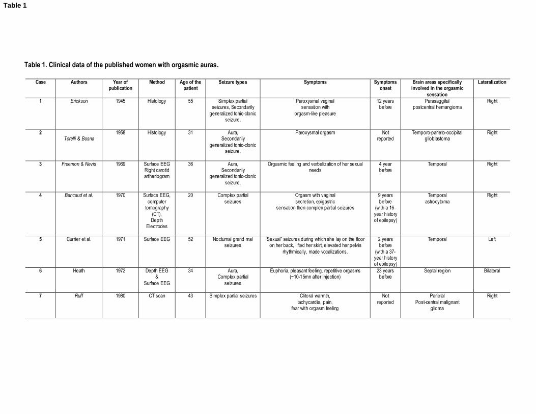
Table 1. Clinical data of the published women with orgasmic auras.
Case Authors Year of publication
Method Age of the patient
Seizure types Symptoms Symptoms onset
Brain areas specifically involved in the orgasmic
sensation
Lateralization
1 Erickson
1945
Histology
55 Simplex partial seizures, Secondarily
generalized tonic-clonic seizure.
Paroxysmal vaginal sensation with
orgasm-like pleasure
12 years before
Parasaggital postcentral hemangioma
Right
2 Torelli & Bosna
1958
Histology
31 Aura, Secondarily
generalized tonic-clonic seizure.
Paroxysmal orgasm
Not reported
Temporo-parieto-occipital glioblastoma
Right
3 Freemon & Nevis 1969 Surface EEG Right carotid artheriogram
36 Aura, Secondarily
generalized tonic-clonic seizure.
Orgasmic feeling and verbalization of her sexual needs
4 year before
Temporal Right
4 Bancaud et al.
1970
Surface EEG, computer
tomography (CT), Depth
Electrodes
20 Complex partial seizures
Orgasm with vaginal secretion, epigastric
sensation then complex partial seizures
9 years before
(with a 16-year history of epilepsy)
Temporal astrocytoma
Right
5 Currier et al. 1971 Surface EEG 52 Nocturnal grand mal seizures
‘Sexual” seizures during which she lay on the floor on her back, lifted her skirt, elevated her pelvis
rhythmically, made vocalizations.
2 years before
(with a 37-year history of epilepsy)
Temporal Left
6 Heath 1972 Depth EEG &
Surface EEG
34 Aura, Complex partial
seizures
Euphoria, pleasant feeling, repetitive orgasms (~10-15mn after injection)
23 years before
Septal region Bilateral
7 Ruff 1980 CT scan 43 Simplex partial seizures Clitoral warmth, tachycardia, pain,
fear with orgasm feeling
Not reported
Parietal Post-central malignant
glioma
Right
Table 1
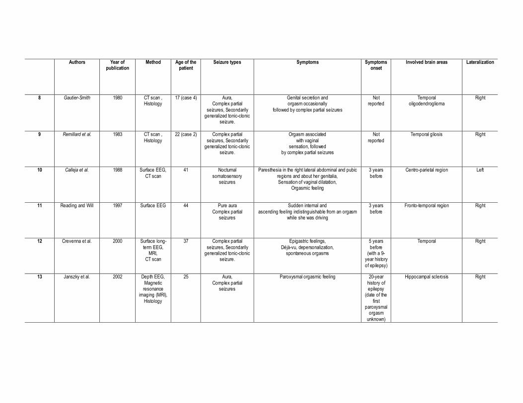
Authors Year of publication
Method Age of the patient
Seizure types Symptoms Symptoms onset
Involved brain areas Lateralization
8 Gautier-Smith 1980 CT scan , Histology
17 (case 4) Aura, Complex partial
seizures, Secondarily generalized tonic-clonic
seizure.
Genital secretion and orgasm occasionally
followed by complex partial seizures
Not reported
Temporal oligodendroglioma
Right
9 Remillard et al. 1983 CT scan , Histology
22 (case 2) Complex partial seizures, Secondarily
generalized tonic-clonic seizure.
Orgasm associated with vaginal
sensation, followed by complex partial seizures
Not reported
Temporal gliosis
Right
10 Calleja et al. 1988 Surface EEG, CT scan
41 Nocturnal somatosensory
seizures
Paresthesia in the right lateral abdominal and pubic regions and about her genitalia, Sensation of vaginal dilatation,
Orgasmic feeling
3 years before
Centro-parietal region Left
11 Reading and Will 1997 Surface EEG 44 Pure aura Complex partial
seizures
Sudden internal and ascending feeling indistinguishable from an orgasm
while she was driving
3 years before
Fronto-temporal region Right
12 Crevenna et al. 2000 Surface long-term EEG,
MRI, CT scan
37 Complex partial seizures, Secondarily
generalized tonic-clonic seizure.
Epigastric feelings, Déjà-vu, depersonalization,
spontaneous orgasms
5 years before
(with a 9-year history of epilepsy)
Temporal Right
13 Janszky et al.
2002
Depth EEG, Magnetic
resonance imaging (MRI),
Histology
25 Aura, Complex partial
seizures
Paroxysmal orgasmic feeling
20-year history of epilepsy
(date of the first
paroxysmal orgasm
unknown)
Hippocampal sclerosis
Right
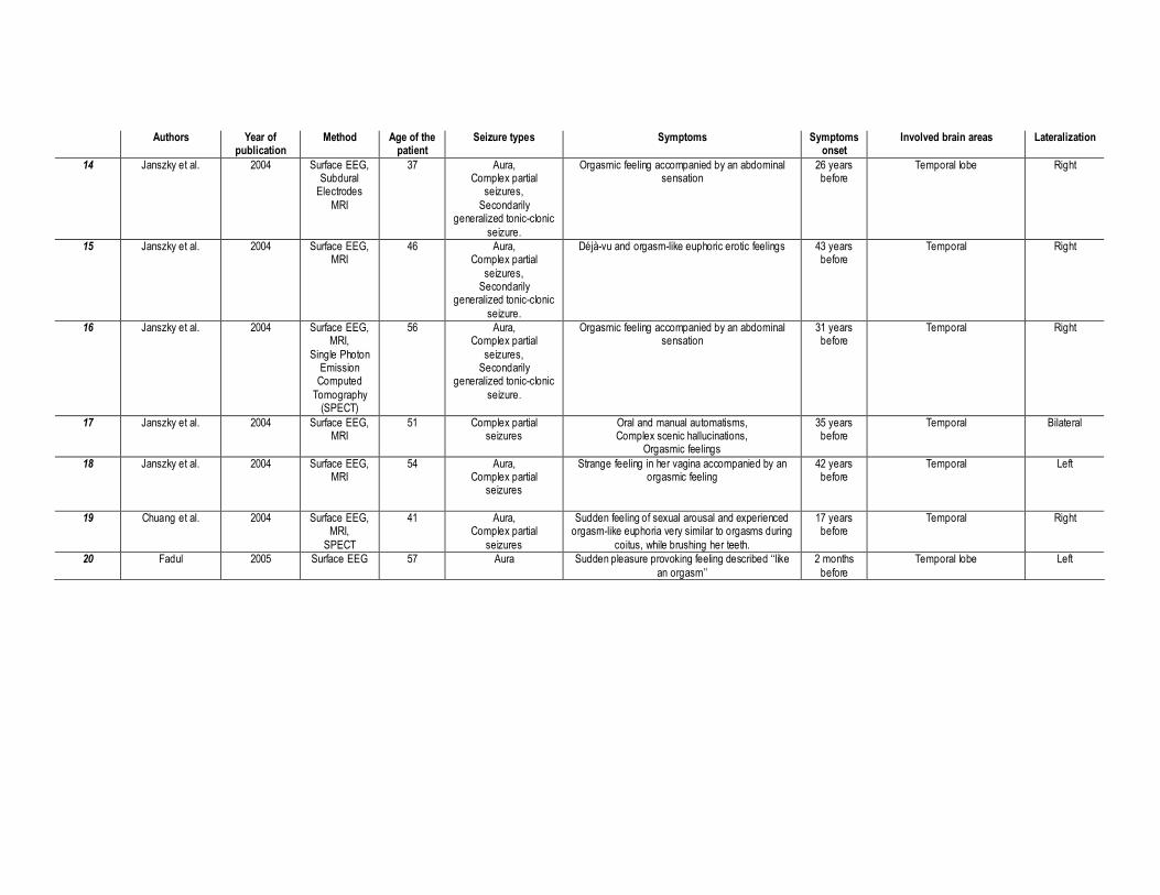
Authors Year of publication
Method Age of the patient
Seizure types Symptoms Symptoms onset
Involved brain areas Lateralization
14 Janszky et al. 2004 Surface EEG, Subdural
Electrodes MRI
37 Aura, Complex partial
seizures, Secondarily
generalized tonic-clonic seizure.
Orgasmic feeling accompanied by an abdominal sensation
26 years before
Temporal lobe Right
15 Janszky et al. 2004 Surface EEG, MRI
46 Aura, Complex partial
seizures, Secondarily
generalized tonic-clonic seizure.
Déjà-vu and orgasm-like euphoric erotic feelings 43 years before
Temporal Right
16 Janszky et al. 2004 Surface EEG, MRI,
Single Photon Emission
Computed Tomography
(SPECT)
56 Aura, Complex partial
seizures, Secondarily
generalized tonic-clonic seizure.
Orgasmic feeling accompanied by an abdominal sensation
31 years before
Temporal Right
17 Janszky et al. 2004 Surface EEG, MRI
51 Complex partial seizures
Oral and manual automatisms, Complex scenic hallucinations,
Orgasmic feelings
35 years before
Temporal Bilateral
18 Janszky et al. 2004 Surface EEG, MRI
54 Aura, Complex partial
seizures
Strange feeling in her vagina accompanied by an orgasmic feeling
42 years before
Temporal Left
19 Chuang et al. 2004 Surface EEG, MRI,
SPECT
41 Aura, Complex partial
seizures
Sudden feeling of sexual arousal and experienced orgasm-like euphoria very similar to orgasms during
coitus, while brushing her teeth.
17 years before
Temporal Right
20 Fadul 2005 Surface EEG 57 Aura Sudden pleasure provoking feeling described ‘‘like an orgasm’’
2 months before
Temporal lobe Left

FigureClick here to download high resolution image
Copyright © 2022 FDOKUMEN