Tlo0301 0001
12
Targeting p53 for Novel Anticancer Therapy 1 Zhen Wang* and Yi Sun † *Institute of Medicinal Biotechnology, PUMC&CAMS, Beijing, People’s Republic of China, 100050; † Division of Radiation and Cancer Biology, Department of Radiation Oncology, University of Michigan Comprehensive Cancer Center, Ann Arbor, MI 48109, USA Abstract Carcinogenesis is a multistage process, involving oncogene activation and tumor suppressor gene inactivation as well as complex interactions between tumor and host tissues, leading ultimately to an aggressive metastatic phenotype. Among many genetic lesions, mutational inactivation of p53 tumor suppressor, the “guardian of the genome,” is the most frequent event found in 50% of human cancers. p53 plays a critical role in tumor suppression mainly by inducing growth arrest, apoptosis, and senescence, as well as by blocking angiogenesis. In addition, p53 generally confers the cancer cell sensitivity to chemoradiation. Thus, p53 becomes the most appealing target for mechanism-driven anti- cancer drug discovery. This review will focus on the approaches currently undertaken to target p53 and its regulators with an overall goal either to activate p53 in cancer cells for killing or to inactivate p53 temporarily in normal cells for chemoradiation protection. The compounds that activate wild type (wt) p53 would have an application for the treat- ment of wt p53-containing human cancer. Likewise, the compounds that change p53 conformation from mutant to wt p53 (p53 reactivation) or that kill the cancer cells with mutant p53 using a synthetic lethal mechanism can be used to selectively treat human cancer harboring a mutant p53. The inhibitors of wt p53 can be used on a temporary basis to reduce the normal cell toxicity derived from p53 activation. Thus, successful development of these three classes of p53 modulators, to be used alone or in combination with chemoradiation, will revolutionize current anticancer thera- pies and benefit cancer patients. Translational Oncology (2010) 3, 1–12 Introduction Cancer is usually associated with aberrant cell cycle progression and defective apoptosis induction due to the activation of proto-oncogenes and/or inactivation of tumor suppressor genes [1]. The evolving mo- lecular events often provide the intervening candidate targets for the development of cancer therapy. One of the most promising targets is p53, a well-established and frequently mutated tumor suppressor in human cancer. Since its first discovery in 1979 as an oncogene [2,3], and particularly after its rediscovery as a tumor suppressor gene in 1989 [4,5], p53 has been the hot spot gene for cancer biologists seeking to elucidate the mechanisms of tumor formation and to vali- date it as a potential cancer therapy target [6–8]. It is well known now that p53 acts biochemically as a transcription factor and biologically as a powerful tumor suppressor. Under normal, unstressed conditions, p53 protein remains undetectable due to its short half-life. The p53 instability is primarily controlled by its nega- tive regulator Mdm2, which, as an E3 ubiquitin ligase, targets p53 for proteasome-mediated degradation [9,10]. Other E3 ubiquitin ligases, which are also implicated in p53 degradation, are Pirh2 and COP1 [11,12]. Another source of p53 instability comes from its own physical property with a melting temperature slightly above body temperature [13]. p53 responds to a wide variety of cellular stresses including geno- toxic damages, oncogene activation, and hypoxia [14,15] and is activated on posttranslational modifications by phosphorylation, acetylation, ubiquitination, and methylation [16–18]. Activated p53 then performs its two well-known biological functions: inducing apoptosis or inducing growth arrest [15,19]. The p53-induced apoptosis is mediated by the Address all correspondence to: Yi Sun, Division of Radiation and Cancer Biology, De- partment of Radiation Oncology, University of Michigan Comprehensive Cancer Center, 4424B Medical Science Bldg-I, 1301 Catherine St, Ann Arbor, MI 48109. E-mail: [email protected] 1 This work was supported by the National Cancer Institute grants (CA111554 and CA118762) and Department of Defense concept award (W81XWH-08-1-0539) to Y.S. Received 26 August 2009; Revised 26 August 2009; Accepted 21 September 2009 Copyright © 2010 Neoplasia Press, Inc. All rights reserved 1944-7124/10/$25.00 DOI 10.1593/tlo.09250 www.transonc.com Translational Oncology Volume 3 Number 1 February 2010 pp. 1–12 1
Transcript of Tlo0301 0001

Trans la t iona l Onco logy Volume 3 Number 1 February 2010 pp. 1–12 1
www.transonc.com
Targeting p53 for NovelAnticancer Therapy1
Zhen Wang* and Yi Sun†
*Institute of Medicinal Biotechnology, PUMC&CAMS,Beijing, People’s Republic of China, 100050;†Division of Radiation and Cancer Biology, Department ofRadiation Oncology, University of Michigan ComprehensiveCancer Center, Ann Arbor, MI 48109, USA
AbstractCarcinogenesis is a multistage process, involving oncogene activation and tumor suppressor gene inactivation as wellas complex interactions between tumor and host tissues, leading ultimately to an aggressive metastatic phenotype.Among many genetic lesions, mutational inactivation of p53 tumor suppressor, the “guardian of the genome,” is themost frequent event found in 50% of human cancers. p53 plays a critical role in tumor suppressionmainly by inducinggrowth arrest, apoptosis, and senescence, as well as by blocking angiogenesis. In addition, p53 generally confers thecancer cell sensitivity to chemoradiation. Thus, p53 becomes the most appealing target for mechanism-driven anti-cancer drug discovery. This review will focus on the approaches currently undertaken to target p53 and its regulatorswith an overall goal either to activate p53 in cancer cells for killing or to inactivate p53 temporarily in normal cells forchemoradiation protection. The compounds that activate wild type (wt) p53 would have an application for the treat-ment of wt p53-containing human cancer. Likewise, the compounds that change p53 conformation frommutant to wtp53 (p53 reactivation) or that kill the cancer cells with mutant p53 using a synthetic lethal mechanism can be used toselectively treat human cancer harboring a mutant p53. The inhibitors of wt p53 can be used on a temporary basis toreduce the normal cell toxicity derived from p53 activation. Thus, successful development of these three classes ofp53 modulators, to be used alone or in combination with chemoradiation, will revolutionize current anticancer thera-pies and benefit cancer patients.
Translational Oncology (2010) 3, 1–12
Address all correspondence to: Yi Sun, Division of Radiation and Cancer Biology, De-partment of Radiation Oncology, University of Michigan Comprehensive CancerCenter, 4424B Medical Science Bldg-I, 1301 Catherine St, Ann Arbor, MI 48109.E-mail: [email protected] work was supported by the National Cancer Institute grants (CA111554 andCA118762) and Department of Defense concept award (W81XWH-08-1-0539) to Y.S.Received 26 August 2009; Revised 26 August 2009; Accepted 21 September 2009
Copyright © 2010 Neoplasia Press, Inc. All rights reserved 1944-7124/10/$25.00DOI 10.1593/tlo.09250
IntroductionCancer is usually associated with aberrant cell cycle progression anddefective apoptosis induction due to the activation of proto-oncogenesand/or inactivation of tumor suppressor genes [1]. The evolving mo-lecular events often provide the intervening candidate targets for thedevelopment of cancer therapy. One of the most promising targetsis p53, a well-established and frequently mutated tumor suppressorin human cancer. Since its first discovery in 1979 as an oncogene[2,3], and particularly after its rediscovery as a tumor suppressor genein 1989 [4,5], p53 has been the hot spot gene for cancer biologistsseeking to elucidate the mechanisms of tumor formation and to vali-date it as a potential cancer therapy target [6–8].It is well known now that p53 acts biochemically as a transcription
factor and biologically as a powerful tumor suppressor. Under normal,unstressed conditions, p53 protein remains undetectable due to itsshort half-life. The p53 instability is primarily controlled by its nega-tive regulator Mdm2, which, as an E3 ubiquitin ligase, targets p53 forproteasome-mediated degradation [9,10]. Other E3 ubiquitin ligases,
which are also implicated in p53 degradation, are Pirh2 and COP1[11,12]. Another source of p53 instability comes from its own physicalproperty with a melting temperature slightly above body temperature[13]. p53 responds to a wide variety of cellular stresses including geno-toxic damages, oncogene activation, and hypoxia [14,15] and is activatedon posttranslational modifications by phosphorylation, acetylation,ubiquitination, and methylation [16–18]. Activated p53 then performsits two well-known biological functions: inducing apoptosis or inducinggrowth arrest [15,19]. The p53-induced apoptosis is mediated by the

2 p53 as a Cancer Target Wang and Sun Translational Oncology Vol. 3, No. 1, 2010
mitochondrial pathway through transcription-dependent or transcription-independent mechanisms and by the death receptor pathway throughtranscriptional activation of FAS and KILLER/DR5 [8,19,20]. p53also transcriptionally represses cell survival genes such as Bcl-2, survivin,IGFR, Mcl-1, and PIK3CA [21–24] through multiple mechanisms[25]. Conversely, p53-induced growth arrest is mainly mediatedthrough up-regulation of p21, Gadd45, 14-3-3σ, and PTGFβ amongothers, through a direct DNA binding and transactivation [8,26].Other p53-involved anticancer mechanisms include induction of cellu-lar senescence [27,28], inhibition of angiogenesis [29,30], and regula-tion of autophagy [31]. Although the major function of p53 is the“killer,” p53 is also implicated in some cases as a “healer” to enhancethe cell survival [21,32].
Given the central role of p53 in cancer prevention and suppressionand in chemosensitization or radiosensitization, p53 has to be abro-gated during carcinogenesis for most cancers to arise. Indeed, p53 isinactivated by point mutations in more than 50% of human cancers(see http://www.iarc.fr/p53) with a majority of mutations occurring inthe DNA binding domain, which either change wt p53 conformation(conformation mutants, e.g., 175H, 249S, 281G) or abolish its DNAcontact (contactmutants, e.g., 248W, 273H) [33]. Furthermore, in can-cer carrying a wt p53, p53 is often nonfunctional as a result of eitherbeing degraded by overexpressed Mdm2 [9,10] or being excluded fromthe nucleus where p53 acts as a transcriptional factor [19,34,35]. In this
Figure 1. Current approaches for p53 targeting: p53, the “guardian of thdomains. The transactivation domain (TD) and proline-rich domain (PD)domain at the central of themolecule, whereas the oligomerization domp53 plays a pivotal role in tumor suppression by inducing growth arresWild-typep53 also confers the sensitivity of cancer cells to chemoradiatidrug discovery. As illustrated in the figure, three classes of p53 targetinare the compounds that activate or restorewild-type p53 function and ccompounds reactivates and rescues themutant p53with an applicationof inhibiting wt p53 and can be used during chemoradiation to block p
review, we aimed to discuss various approaches 1) to activate wt p53, 2)to reactivate mutant p53 or selectively kill cancer cells with mutant p53,and 3) to temporarily inhibit wt p53 for normal cell protection (see Fig-ure 1 and Table 1). Successful clinical development of these three classesof novel compounds would eventually revolutionize the current cancertherapies to benefit a majority of cancer patients.
Targeting p53 Itself
Targeting wt p53 — To ActivateThe approaches include the use of chemoradiation to activate endoge-
nous wt p53, of gene therapy to introduce wt p53 or modified adeno-virus to kill cancer cells with mutant p53, and of synthetic peptides ornongenotoxic small molecules to activate endogenous wt p53.
Chemoradiation. Conventional anticancer therapies target p53 be-cause almost all genotoxic anticancer drugs as well as ionizing radiation(IR) cause the substantial DNA damage, which triggers p53 activationand stabilization [36]. Early preclinical studies using both in vitro celland in vivo tumor models showed that cells or tumors with a wt p53are more sensitive to chemoradiation [37,38]. The early clinical studiesfurther showed that mutant p53 confers chemoresistance in patientswith ovarian cancer [39,40], breast cancer [41,42], gastric and colorectal
e genome,” consists of 393 amino acidswith four distinct functionalis located at the N-terminus, the DNA binding andmutation hot spotsain (OD) and regulatory domain (RD) at the C-terminus. On activation,t, apoptosis, and senescence, as well as by blocking angiogenesis.on. Thus, p53becomesanappealing therapeutic target for anticancerg compounds have been identified and characterized. The first classan be used in human cancers harboring awt p53. The second class ofin human cancers carrying a p53mutation. The third class is capable53 activation in normal cells, thus reducing cytotoxicity.

Translational Oncology Vol. 3, No. 1, 2010 p53 as a Cancer Target Wang and Sun 3
cancer [43], and hematological malignancies [42]. Extensive follow-upstudies in both preclinical and clinical settings showed that in general,cancer cells with a wt p53 aremore sensitive to chemoradiation [44–47],but there are quite few exceptions. For example, breast cancer patientswith a transcription-deficient mutant p53 had a better response to che-motherapy than patients with a wt p53 [48]. The same was true forMCF7 breast cancer cells and HCT116 colon cancer cells with p53either abrogated or deleted, as a result of increased cellular vulnerabilityto G2 checkpoint abrogators [49,50]. In multiple head and neck cancercell lines, the absence of p53 appeared to be associated with radiosensi-tivity [51]. Furthermore, the p53 status determined the cellular responseto chemotherapy in an anticancer drug–dependent manner. Colon can-cer cells with the p53 gene deleted was found to be more sensitive to theDNA-damaging agent, doxorubicin, but wasmuchmore resistant to theantimetabolite 5-fluorouracil [52]. Taken together, these data indicatethat because of the nature of tumor heterogeneity, the cellular responseto chemoradiation is not solely determined by wt p53. However, under-standing such responses in conjunction with p53 status would help therational design of anticancer therapies to maximize their efficacy.
Gene therapy
To reintroduce wild-type p53. Because p53 function is lost inmany cancers, it is logical to restore p53 function by reintroducing wtp53.One common gene therapy approach is the use of viruses to deliverp53. An early study, using retrovirus-mediated gene transfer of wt p53into human lung cancer cells, showed the inhibition of tumor growthboth in vitro and in vivo [53]. Gene therapy using human wt p53,delivered by replication-defective adenovirus (Ad-p53) for better trans-duction efficiency and lower toxicity, has been extensively studied in the
preclinical and clinical settings with an impressive anticancer activity,resulting from p53-induced growth arrest and apoptosis [54]. TheAd-p53, under the brand name of Gendicine, or Advexin, has beencurrently in clinical use inChina since 2003 [55] or in phase 1 to 3 clini-cal trial in the United States, respectively [7]. The results showed thatGendicine/Advexin is well tolerated in patients and efficacious in thetreatment of numerous cancers, particularly head and neck cancer andlung cancer, as a single agent or in combination with chemotherapy orradiation therapy [7,56,57].
To eliminate mutant p53–containing cancer cells by adeno-virus. Another p53-related gene therapy is the use of an E1B-deletedadenovirus, designated as ONYX-015, which selectively replicates inp53-deficient cancer cells and subsequently lyse the cells [58]. Preclini-cal studies showed that ONYX-015 has antitumor activity both in vitroand in vivo, particularly in combination with chemotherapy or radia-tion therapy [59,60]. Clinical trials revealed that ONYX-015 had amarginal antitumor activity when administrated alone, but a significanteffect when combined with standard chemotherapies in a number ofhuman cancers [56,61,62].
Small molecules. Reactivation of p53 and induction of tumor cellapoptosis (RITA) was identified through a cell proliferation assay usinga pair of isogenic cancer cell lines differing in p53 status [63]. Biochemi-cally, RITA bound to p53 at the N-terminal domain with a high affinity.Although an initial study showed that RITA could block p53-Mdm2binding [63], a subsequent in vitro study using nuclear magnetic reso-nance suggested that it might not disrupt p53-Mdm2 binding [64]. Bio-logically, RITA suppressed tumor cell growth both in vitro and in vivoby inducing massive apoptosis in a p53-dependent manner in multiplecancer cell models [63]. A recent mechanistic study revealed that RITA,through activating p53, abrogates key oncogenic pathways. Specifically,RITA-activated p53 causes the transcriptional repression of antiapop-totic proteins, including Mcl-1, Bcl-2, survivin, and MAP4, downregu-lates oncogenic proteins, c-Myc, cyclin E, and β-catenin, and blocks theAKT pathway at multiple levels [65]. Thus, RITA can be further devel-oped as a single agent or used in combination with chemoradiation foreffective cancer therapy through p53-mediated abrogation of cancer cellsurvival and oncogenic pathways.
Targeting wt p53 — To InhibitChemotherapy or radiation therapy kills cancer cells, but at the same
time causesmany adverse effects because of normal cell toxicity, resultingat least in part from p53 activation and apoptosis induction in normalproliferating cells/tissues, such as bone marrow, lymphoid organs, hairfollicles, and epithelium lining of the small intestine. Temporary block-age of p53 activation in normal cells during the treatment of p53-deficient tumors should reduce these adverse effects [66]. Because p53induces apoptosis through transcription-dependent and transcription-independent, but mitochondria-dependent mechanisms [20], twoclasses of small molecules have been identified, which target eitherp53 transcriptional activity or p53-mitochondrial binding activity, re-spectively. The first class of compound, designated as Pifithrin (PFT)-α,reversibly inhibits p53-dependent transcriptional activation and apoptosisand protects mice from the lethal dose of IR without promoting tumorformation [67]. The second class of compound, designated as PFT-μ, in-hibits p53 binding to mitochondria by reducing p53-binding affinity toBcl-xL and Bcl-2 without affecting p53 transactivation. Similar to PFT-α,PFT-μ also protects primary mouse thymocytes from p53-mediated
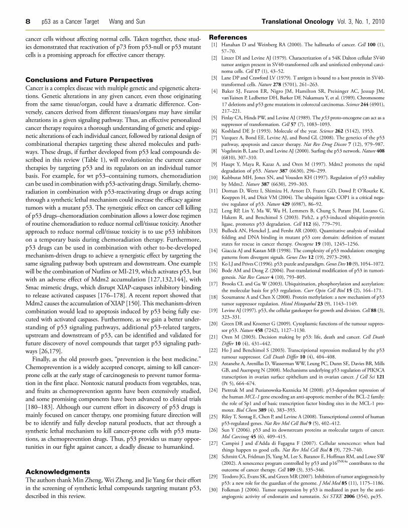
Table 1. Structure of Small-Molecule p53 Activators, Reactivators, and Inhibitors.

4 p53 as a Cancer Target Wang and Sun Translational Oncology Vol. 3, No. 1, 2010
apoptosis on exposure to radiation and protects mice from radiation-induced lethal hematopoietic syndrome [68]. Thus, these two classes ofcompounds could be further developed for clinical use in combination ofchemoradiation during the treatment of cancers, particularly those withp53 mutation or deletion for normal cell protection.
Targeting Mutant p53 – To RescueBecause p53mutation occurs in ∼50% of human cancers, an effective
cancer therapy would be the reactivation of p53 mutants. The rescuestrategies vary dependent on the mutation types. In tumors harboringa DNA-contact mutant, the attempts were to introduce the functionalgroups that create new contacts or to stabilize the scaffolding positioningin the remaining contact sites [69,70]. The functions of conformationmutants can be restored by specific small peptides or small moleculesthat aid the proper folding of the unfolded p53 conformation [70].
Synthetic peptides
CDB3. CDB3 is a short synthetic nine-residue peptide (REDE-DEIEW), derived from p53-binding protein 2 (53BP2 or ASPP), aknown p53-binding protein that interacts with the p53 core domainand upregulates p53-dependent transactivation [71]. It was identifiedthrough a rational approach searching for small molecules that bindto p53 core domain and stabilize p53 [72]. The notion behind thisapproach is that a peptide binding with higher affinity to a properlyfolded state than to an unfolded form of the mutant will shift the equi-librium toward the active wild-type conformation. The in vitro studiesshowed that CDB3 indeed restored the sequence-specific DNA bind-ing to various p53 mutants by stabilizing them in a bioactive confor-mation [72]. CDB3 also restored the transcriptional activity of two hotspot p53 mutants, R273H and R175H, for transactivation of two p53target genes,Mdm2, p21 [73]. Furthermore, CDB3 induced the accu-mulation of wt p53 and sensitized cancer cells to radiation [73].Although as a small peptide, CDB3 will likely have poor bioavailability,it can serve as a lead for the further development of p53-reactivatingsmall molecules.
p53 C-terminal peptide. In addition to CDB3, several otherstudies demonstrated that short synthetic peptides derived from the p53C-terminal region (peptide 46) can reactivate mutant p53 through stabi-lization of the core domain folding and/or establishment of novel DNAcontacts [74,75]. Although the exact mechanism of action is still unclear,these peptides were indeed shown to rescue the function of endogenousmutant p53 proteins, leading to growth inhibition, apoptosis, and sup-pression of solid tumor growth in an in vivo animal tumor model [76].
Small molecules
CP-31398. CP-31398 was the first small molecule that has ac-tivity to change p53 conformation from mutant to wild type. It wasidentified through a chemical library screening based on an in vitro bio-chemical assay that detects wt or mutant p53 conformation throughtwo specific antibodies [77]. CP-31398 was found to stabilize the coredomain and enhance transcriptional activity of p53 in living cells [77],but the detailed mechanism of action of CP-31398 remains elusive.Nuclear magnetic resonance studies failed to detect any binding ofCP-31398 to the p53 core domain [78]. CP-31398 had no effect onp53-Mdm2 binding, did not cause DNA damage response, or inducep53 phosphorylation at Ser 15 or 20, but rather it increasedwt p53 levels
by reducing p53 ubiquitination [79]. Biologically, CP-31398 inducedgrowth arrest and apoptosis in a number of human cancer cell lines bothin vitro and in vivo [77,80] and inhibited UVB-induced skin carcino-genesis [81]. Furthermore, CP-31398 seems to have other targets in ad-dition to p53 because it could induce cell death in both p53-dependentand -independent manners [70].
PRIMA-1 and MIRA-1. p53 reactivation and induction ofmassive apoptosis (PRIMA-1) and mutant p53 reactivation and induc-tion of rapid apoptosis (MIRA-1) are two classes of compounds withunique chemical structures that were identified through a cell-basedscreening for compounds that suppress the growth of tumor cellsexpressing an exogenous mutant p53 [82,83]. The compounds werefound to restore the sequence-specific DNA binding and change themutant p53 conformation to wild type, leading to transactivation ofp53 target genes [82,83]. In contrast to both CP-31398 and CDB3,PRIMA-1 does not activate wild-type p53. A recent mechanistic studyrevealed that PRIMA-1 is converted to a decomposition product thatforms covalent thiol adducts on mutant p53 to restore its tumor sup-pressor activity [84]. Biologically, both compounds showed antitumoractivity inmultiple cancer cell lines and in vivo xenograft tumors harbor-ing a mutant p53 without apparent normal tissue toxicity [82–84]. Fur-thermore, PRIMA-1 was found to be active against p53-null cancer celllines through a mechanism involving the JNK pathway and heat shockprotein 90 [85,86]. Finally, PRIMA-1 or its analog, PRIMA-1(met),sensitized lung cancer cells or prostate cancer cells to adriamycin orradiation, respectively [87,88] and inhibited in vivo xenograft tumorgrowth [89].
Ellipticine. Ellipticine and its derivatives were initially identifiedin the drug sensitivity profiling of theNCI-60 cancer cell line panel to bemore effective against tumor cells with a mutant p53 [90]. An ellipticinederivative, 9-hydroxyellipticine was found to increase the transcriptionof p21 and Bax and to induce G1 arrest in a mutant p53-dependentmanner [91]. The detailed follow-up work showed that ellipticinechanged the p53 conformation from mutant to wild type, restoredthe sequence-specific DNA binding and transactivation of p53-drivenluciferase reporter, and activatedmutant p53 to induce p53 target genes,p21 and Mdm2 in mouse xenograft tumor tissues [92]. Furthermore,ellipticine was recently identified to increase nuclear localization ofboth wt and mutant p53 in a manner independent of DNA damage[93]. Because the ellipticine series of compounds has many off-targetactivities including promoting p27 degradation [94], it is unlikely tobe further developed for the clinic use.
WR-1065. WR-1065 (aminothiol) is an active metabolite ofthe cytoprotector amifostine and acts as a classic scavenger of reactiveoxygen species [95]. WR-1065 was found to activate both wt and mu-tant p53 and increase the expression of p53 target genes in a mannerindependent of DNA damage [96,97].
P53R3. p53R3 is a recently identified small molecule that re-stores the sequence-specific DNA binding of p53 mutants (R175Hand R273H) after screening a small library of compounds using anin vitro gel shift assay [98]. The compound was found to enhance therecruitment of both wt and mutant p53 to target promoters and to in-duce the expression of a number of p53 target genes. Biologically, thecompound induces mutant p53-dependent growth arrest and sensitizesTRAIL-induced cell death in multiple glioma cell lines [98].

Translational Oncology Vol. 3, No. 1, 2010 p53 as a Cancer Target Wang and Sun 5
Targeting Mutant p53 – To Kill
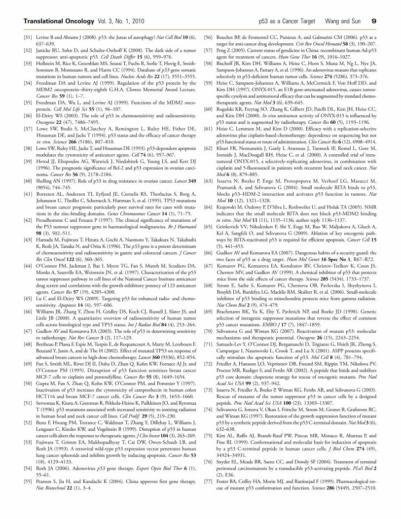
Synthetic lethality for p53 mutation. Synthetic lethality is a situa-tion where a cancer-associated mutation itself is nonlethal but renderscancer cells susceptible to a second hit that becomes lethal on inactiva-tion [99,100]. In the case of p53, which is mutated in 50% of humancancer, application of a synthetic lethality strategy to identify chemicalcompounds that selectively kill human cancer cells harboring a mu-tant p53 will be of significant importance for the discovery of a novelclass of anticancer drugs (Figure 2). Synthetic lethality has been shownin a number of human cancer cells with deletion of PTEN or DPC4,mutation of K-Ras or B-Raf, and overexpression of c-Myc [101–105],demonstrating the feasibility of using synthetic lethality strategy foranticancer drug discovery. The p53 synthetic lethal drugs, if identifiedand developed, can be used 1) for cancer treatment to selectively killmutant p53-containing cancer cells and 2) for chemoprevention to elimi-nate mutant p53-containing cancer-prone cells, at the early stage ofcarcinogenesis. Furthermore, p53 synthetic lethal drugs should have,in theory, minimal adverse effects, because normal cells do not containa p53 mutation.A “synthetic lethal-like” screeningwas conducted in a panel of 60NCI
cancer cell lines in an attempt to identify compounds that selectivelyinhibited the growth of cancer cells devoid of p53 [106]. Few classesof compounds were identified, and the only “popular” hit was paclitaxel,an inhibitor of microtubule polymerization [106]. It was speculated thatmicrotubule-associated protein 4, a p53 transcriptional repressed target,may mediate the sensitivity of cells lacking p53 activity to paclitaxel-
Figure 2. Synthetic lethality for p53 mutation: Synthetic lethalityrefers to the situation in which the cancer-associated mutation itselfis nonlethal but renders cancer cells susceptible to the second hit,which results in lethal phenotype. Because p53 is most frequentlymutated inmore thanhalf of humancancer cells, it is feasible in theoryto use this strategy to identify drug candidates that preferentially killcancer cells with a p53 mutation. The p53 synthetic lethal drugs, ifidentified and developed, should have a minimal toxicity to normalcells and can be used for cancer chemoprevention and treatment ofmutant p53–containing cancers.
induced cell killing [107]. Another drug that selectively inhibitedp53-deficient tumor cell growth is metformin, a diabetic drug, withthe mechanism associated with activation of AMP kinase and inhibitionof oxidative phosphorylation, which created an environment more vul-nerable to mutant p53 cells [108].
Small molecules that abrogate the G2/M checkpoint control can beconsidered to act through the synthetic lethal mechanism against p53mutation. The hypothesis is based on the fact that p53-deficient cellshave abrogated G1 checkpoint control (lack of p53-mediated p21 in-duction in response to DNA damage), and thus, further abrogation ofG2 checkpoint control will selectively kill p53-deficient cancer cellsthrough the induction of mitotic catastrophe. This turns out to be thecase. UCN01, a potent G2 checkpoint abrogator sensitized p53 mutantcancer cells to IR by abrogating G2 checkpoint control, whereas cancercells with wt p53 were much more resistant [109]. Similarly, our pre-vious work showed that PD0166285, a potent Wee-1 kinase inhibitorthat abrogated G2 checkpoint control, selectively sensitized p53 mutantcancer cells to radiation [110]. A recent study from Vogelstein’s groupfurther supported this notion of synthetic lethality. Transcription pro-files of four paired colon cancer cell lines, isogenic for p53 deletion ormutation, revealed a consistent up-regulation of polo-like kinase 1(PLK-1), a well-known protein that regulates G2/Mcheckpoint control,in p53-deficient lines after exposure to IR. Consistently, p53-deficientcells are much more sensitive to PLK-1 inhibitors when combined withp53 activators, such as radiation and Nutlin-3. Furthermore, in vivoxenograft tumor studies showed that a PLK-1 inhibitor used as a singleagent caused significant regression of p53-null tumors [111]. Thesestudies highlight the feasibility of using synthetic lethal mechanismfor effective cancer therapy.
We have recently attempted to identify novel small molecules thatact through synthetic lethal mechanism to selectively kill cancer cellswith a mutant p53 mutation. We conducted this p53 synthetic lethalscreening using a well-characterized p53 temperature-sensitive modelin which p53-null H1299 cells were transfected with a temperature-sensitive mutant p53A138V (H1299-p53ts). The p53 in these cellsadopts a mutant conformation when grown at 39°C and a wild typeconformation at 32°C. Temperature shifting from 39 to 32°C restoresthe wild type p53 conformation that induces growth arrest but notapoptosis [112,113]. Screening of both chemical and natural productlibraries was conducted at both temperatures, and the compounds thatselectively killed cells at 39°C (mutant p53 status), but not at 32°C (wtp53 status), were subjected to the secondary counterscreening. In thecounterscreening, we used H1299-neo vector control cells at bothtemperatures to filter out potential false-positives derived merely fromtemperature shifting. Candidates that showed selective killing ofH1299-p53ts at 39°C only were identified, and they are being furthercharacterized (unpublished data). A disadvantage of this type of screen-ing is the lack of mechanism of drug action, which will need follow-upstudy to identify the “second” target that is synthetically lethal to p53mutation. The ideal situation would be to conduct simultaneouslythe same screening using a small interfering RNA (siRNA) library, lead-ing to identification of the “second” target.
Elimination of cancer cells with the gain-of-function p53 mu-tants. During the validation of our candidate hits, identified fromthis p53 synthetic lethal screening, we serendipitously found that car-diac glycoside drugs, such as digoxin and ouabain, reduced the p53levels in a time- and dose-dependent manner in a subset of cancer celllines but not in immortalized normal cell lines. The drug sensitivity to

6 p53 as a Cancer Target Wang and Sun Translational Oncology Vol. 3, No. 1, 2010
p53 reduction is cancer cell line–dependent but independent of p53status of a wild type or mutants. A mechanistic study revealed that thedrug-induced p53 decrease neither occurs at the messenger RNA levelnor is due to enhanced degradation, which is a commonmechanism forp53 regulation. Rather, it occurs at the protein level as a result of reducedde novo synthesis of p53 protein. The drug-induced p53 reduction canbe rescued by the inhibitors of Src andMEK, suggesting an involvementof Src/mitogen-activated protein kinase signaling pathways, initiated onthe drug binding to Na+/K+-ATPase [114]. Given the fact that glyco-side drugs are being used in clinic for the treatment of congestive heartfailure [115] and that cardiac glycosides are inactive against wt p53 innormal cells, but potently active in the elimination of mutant p53 insome cancer cells, the drugs may have utility in the treatment of humancancers harboring a gain-of-function p53 mutant [116,117].
Targeting p53 Regulators – To Activate p53
Mdm-2 (Murine Double Minute-2, Hdm2 in Human)Another effective approach to activate wt p53 is to inhibit its nega-
tive regulators. The best-known naturally occurring p53 inhibitor is itsdownstream target, Mdm2 [118]. Mdm2 encodes a 90-kDa proteinthat was first identified as the gene responsible for the spontaneoustransformation of immortalized murine BALB/c 3T3 cells [119]. Itcontains a p53 binding domain at the N-terminus and a RING domainat the C-terminus. Mdm2 inhibits p53 through two well-characterizedmechanisms: 1) Mdm2 binds to p53 at the N-terminus and blocks p53transactivation activity and 2) Mdm2 acts as an E3 ubiquitin ligase topromote p53 degradation. In addition, Mdm2 also exports p53 out ofnucleus where p53 acts as a transcription factor [34,120]. Mdm2 isoverexpressed in approximately 7% of human cancers with a muchhigher incidence in soft tissue tumors, such as osteosarcoma [121]. Bothin vitro and in vivo studies indicated that oncogenic activity of Mdm2ismainly attributable to its binding and degrading p53 [122,123]. Thus,inhibition of Mdm2 in this subset of human cancer should reactivatep53 to induce cell killing. Indeed, this notion was supported by manyproof-of-concept studies, including 1) the blockage of the interactionbetween Mdm2 and p53 with synthetic peptides or monoclonal anti-bodies [124] and 2) reduction of Mdm2 levels with antisense oligo-nucleotides or siRNA [125]. So far, two classes of small moleculeshave been identified, which either disruptMdm2-p53 binding or inhibitMdm2 E3 ubiquitin ligase activity to reactivate wt p53.
Small Molecules that Disrupt Mdm2-p53 InteractionHistorically, it has been difficult to develop small-molecule inhibitors
that disrupt large protein-protein interactions. However, the crystalstructure of the Mdm2-p53 peptide binding revealed that the bindingrelies on the contact of the p53 peptide side chains of Phe19, Trp23,and Leu26 with the N-terminus of Mdm2 (amino acids 17-125) ina deep hydrophobic pocket [126], which made it possible for smallmolecules to disrupt binding. Several classes of structurally distinctivecompounds have been reported to disrupt Mdm2-p53 binding[127,128]. These compounds include Nutlins, benzodiazepinediones(BDAs), and an Mdm2 inhibitor (MI) series of spiro-oxindoles deriva-tives, including MI-63, MI-219, and MI-43 [127,129–133].
Nutlins. The nutlin series was the first class of small molecules,identified through a screening of a diverse library that disruptedMdm2-p53 peptide binding. This series of compounds effectively ab-rogatesMdm2-p53 interaction through a high-affinity binding toMdm2
[129] with a high selectivity for Mdm2 over Mdmx, a homologue ofMdm2 [127]. Nutlin-3, an analog of the series, induced p53 levels, ac-tivated p53 transcription activity, and has a broad activity against cancercells harboring a wt p53 both in vitro and in vivo [129]. Examples includecolon and breast cancer cell lines and osteosarcoma cells [129], lympho-blastic leukemia [134], and retinoblastoma [135]. In combination withchemoradiation, Nutlin-3 showed synergistic activity against prostatecancer [136], lung cancer [137], lymphocytic leukemia [138,139], andneuroblastoma [140]. Nutlin-3 also showed some normal cell protectiveactivity against chemotherapy by inducing cell cycle arrest [141,142].Furthermore,Nutlins showed a direct antiangiogenic and antiosteoclasticactivity, which may have an application for tumors harboring a mutantp53 [143].
Benzodiazepinedione. Benzodiazepinedione (BDA) and its deriv-atives were identified through a chemical library screening using a mini-aturized thermal denaturation-based assay. X-ray crystal structureconfirmed that the BDA interacts with the p53-binding pocket ofMdm2 [130]. The compounds increased the p53 transcriptional activity,inhibited the proliferation of cancer cells in a wt p53–dependentmanner,and synergized with doxorubicin to inhibit tumor cell growth bothin vitro and in vivo [130,133].
MI series. The MI series of spiro-oxindole compounds, includingMI-219,MI-63, andMI-43, were discovered through structure-based de-sign byDing et al. [131] bymimicking all fourMdm2-contacting residues(Phe19, Trp 23, Leu 22, and Leu 26) on p53. The MI series of com-pounds, with MI-219 being the most potent, bind toMdm2 with a highaffinity. The drug-induced disruption ofMdm2-p53 binding caused p53accumulation leading to up-regulation of many p53 target genes and toinduction of apoptosis in a number of human cancer cell lines derivedfrom breast, colon and prostate cancers in vitro in a wt p53–dependentmanner. MI-219 as a single agent also caused a complete inhibition ofxenograft tumor growth in vivo at a dose that has no obvious toxicity toanimals [127,144,145].
We tested the efficacy of MI-43 against human lung cancer cells andfound that the compound preferentially inhibited the growth of wtp53-containing cells over p53-null cells. MI-43 induced the growtharrest at both G1 and G2 phases of the cell cycle, at the low concen-tration as a result of p21 induction and apoptosis at the high concen-tration due to Puma/Noxa induction. Importantly, MI-43 was muchless toxic to normal lung cells than cancer cells. Finally, when used incombination, MI-43 sensitized chemoresistant A549 cells to etoposide-induced apoptosis [132].
Mdm2 E3 ubiquitin ligase inhibitors
HLI98. The HLI98 series of compounds was identified throughthe high-throughput screening of a chemical library using an in vitroMdm2 autoubiquitination assay [146]. The follow-up experimentshowed that HLI98C, an analog, indeed inhibited Mdm2 E3 ligaseactivity. In the cell-based assays, the compound stabilized p53 andMdm2 and activated p53-dependent transcription and apoptosis. Thecompound was much more efficacious against cancer cells with wt p53,although it demonstrated some p53-independent cytotoxicity [146]. Al-though the evidence of in vivo antitumor activity for HLI98C was lack-ing [146], this proof-of-concept study did indicate that an Mdm2 E3ligase inhibitor, which has selective activity against wt p53–containingcancer cells, can be identified.

Translational Oncology Vol. 3, No. 1, 2010 p53 as a Cancer Target Wang and Sun 7
Two concerns should be borne in mind in the clinical developmentof Mdm2-based inhibitors for anticancer therapy. The first is the thera-peutic window because the compounds would also induce p53 innormal cells to cause some adverse effects. Second is the Mdm2 itself,which is accumulated after compound treatment as a result of p53activation. In addition to targeting p53, Mdm2 interacts with a num-ber of other proteins, such as p73, p63, E2F1, and HIF1α [147–149].A recent study also showed that Mdm2 caused the accumulationof XIAP to inhibit apoptosis [150]. These p53-independent butMdm2-dependent effects would eventually affect the efficacy of thisclass of compounds.
SirT1/SirT2SirT1 and SirT2 are two members of the NAD+-dependent class III
histone deacetylases with a total of seven members in humans[151,152]. SirT1 and SirT2 catalyze the reaction between an acetylatedlysine with NAD+, leading to the production of deacetylated lysine, 2′-O-acetyl-ADP-ribose, and nicotinamide [153]. It is well establishedthat acetylation of p53 at lysine 382 enhances p53 DNA bindingactivity [154]. Thus, SirT1-catalyzed deacetylation of p53 at lysine382 would deactivate and destabilize p53 [155–157]. These studiesestablished that SirT1 is yet another negative regulator of p53.
Tenovin-1 and Tenovin-6. This series of compounds was identifiedin a cell-based screening for compounds that activate p53, using a p53-driven transactivation assay [158]. Tenovins stabilized wt p53, inducedp53-dependent cell cycle arrest and apoptosis, and suppressed xenografttumor growth in vivo as a single agent [158]. Mechanistically, Tenovinsare nongenotoxic agents and do not induce DNA damage. Rather, theyinhibited NAD-dependent deacetylase SirT1/T2, thus restabilizing andreactivating p53. Tenovins, therefore, are a novel class of p53-activatingagents that can be further developed for clinical use as well as for thestudy of sirtuin function as biological tools.
JJ78:12. This series of compounds was also identified by Lain et al.[158] in the same reporter assay used for p53 activator screening. TheJJ78:12 series contains much more potent p53 activators with a nano-molar dose range and showed clear anticancer activity both in vitro andin vivo. Mechanistically, this series of compounds caused tubulindepolymerization and may not be further developed for clinical use be-cause of their high cytotoxicity, resulting from tubulin poison [159].Interestingly, our effort in screening for compounds that change p53conformation from mutant to wt using a luciferase reporter–basedH1299-p53 temperature-sensitive model [112,113] led to the identifi-cation of a series of microtubulin poison compounds, including noco-dazole, albendazole, parbendazole, and mebendazole (unpublisheddata), suggesting a nonspecific or indirect nature of p53 activation bythis class of compounds.
Targeting Other p53 Family Membersp53 has two family members, p73 and p63 with significant sequencehomology among them [160,161]. Like p53, both p73 and p63 bindto the p53-specific DNA binding motif, transactivate p53 downstreamtarget genes, and suppress tumor cell growth by inducing growth arrestand apoptosis [160,161]. The tumor suppression activity of p73 andp63 was further demonstrated in a mouse knockout study, whichshowed various tumor predispositions on heterozygous deletion of eithergene [162]. Unlike p53, p73 or p63 is rarely mutated in human cancers[163] but could be inactivated by binding to a subset of p53 mutants
[164]. Targeting p73 and p63 has been proposed for cancer therapy[165,166] with approaches including gene therapy, small peptides,and small molecules.
Gene TherapyLike the gene therapy approach using wt p53, the same approach has
been extended to its family members, p73 and p63. The adenovirus-mediated transduction of p63 and p73 into tumor cells was found to bean alternatively efficient gene therapy approach both in vivo and in vitro[167,168], especially when tumors are resistant to p53-mediated genetherapy [168,169]. Ad-p73 demonstrated a significant cytotoxicityagainst multiple cancer cell lines tested by inducing both growth arrestand apoptosis [169]. Ad-p73 also sensitized p53 mutant–containingcells to adriamycin with a higher efficiency than Ad-p53 [169]. Theapoptosis-inducing effects of Ad-p63γ were also found to be greaterthan those of wild-type p53 in osteosarcoma cells with Mdm2 ampli-fication [168].
Small Peptides
Short-interfering mutant p53 peptides. One activity of gain-of-function p53 mutants is to physically interact with, sequester, and in-activate p73 [170,171]. Short-interfering mutant p53 (SIMP) peptideswere designed based onmutant p53/p73 binding regions. Indeed, SIMPeffectively disrupted the protein complex of mutant p53/p73 and re-stored the p73 function. Biologically, SIMP sensitized mutant p53-containing tumor cells to adriamycin and cisplatin. Notably, the effectsof SIMPs aremutant p53-specific and had no effect on cancer cells eitherwith a wt p53 or with a p53-null [172].
37AA. This peptide consists of 37 amino acids from p53 after a non-contiguous fusion of evolutionarily conserved boxes II and III on theDNA binding domain of p53. The 37AA induced cell death throughactivation of p53/p73 target genes in p53-null cells. Further studiesrevealed that 37AA was able to bind to iASPP, an inhibitory memberof the ASPP family, resulting in the release TA-p73 (a p73 isoform)from iASSP/TA-p73 complex. Anticancer activity of 37AA was furtherdemonstrated in a colon cancer xenograft model, in which 37AA wasdriven by an expression vector and delivered in dendrimer-based nano-particles [165,173].
Small MoleculesWang et al. [174] recently identified a number of small molecules in a
screening for activating p53 target genes and apoptosis in p53 mutant–containing cells. The follow-up studies using an siRNA-silencingapproach confirmed that some of these compounds mediated theiractivity through transactivating p73. Although the detailed mechanismis unknown, these compounds had anticancer activity when assayedin vivo using the p53-null HCT116 xenograft tumor model [174].
Reactivate transcriptional activity. Reactivate transcriptional activity(RETRA) was identified in a cell-based screening for compounds thatreactivate the transcriptional activity of p53 in mutant p53–containingcells [175]. Follow-up studies showed that RETRA activates a numberof p53 target genes and selectively inhibited the growth of mutant p53-containing cancer cells both in vitro and in mouse xenografts. Mechanis-tic studies in mutant p53–containing cells revealed that the compoundincreased the p73 levels and released p73 from complexing with mutantp53. Importantly, RETRA is active against mutant p53–containing
8 p53 as a Cancer Target Wang and Sun Translational Oncology Vol. 3, No. 1, 2010
cancer cells without affecting normal cells. Taken together, these stud-ies demonstrated that reactivation of p73 from p53-null or p53 mutantcells is a promising approach for effective cancer therapy.
Conclusions and Future PerspectivesCancer is a complex disease with multiple genetic and epigenetic altera-tions. Genetic alterations in any given cancer, even those originatingfrom the same tissue/organ, could have a dramatic difference. Con-versely, cancers derived from different tissues/organs may have similaralterations in a given signaling pathway. Thus, an effective personalizedcancer therapy requires a thorough understanding of genetic and epige-netic alterations of each individual cancer, followed by rational design ofcombinational therapies targeting these altered molecules and path-ways. These drugs, if further developed from p53 lead compounds de-scribed in this review (Table 1), will revolutionize the current cancertherapies by targeting p53 and its regulators on an individual tumorbasis. For example, for wt p53–containing tumors, chemoradiationcan be used in combinationwith p53-activating drugs. Similarly, chemo-radiation in combination with p53-reactivating drugs or drugs actingthrough a synthetic lethal mechanism could increase the efficacy againsttumors with a mutant p53. The synergistic effect on cancer cell killingof p53 drugs–chemoradiation combination allows a lower dose regimenof routine chemoradiation to reduce normal cell/tissue toxicity. Anotherapproach to reduce normal cell/tissue toxicity is to use p53 inhibitorson a temporary basis during chemoradiation therapy. Furthermore,p53 drugs can be used in combination with other to-be-developedmechanism-driven drugs to achieve a synergistic effect by targeting thesame signaling pathway both upstream and downstream. One examplewill be the combination of Nutlins or MI-219, which activates p53, butwith an adverse effect of Mdm2 accumulation [127,132,144], withSmac mimetic drugs, which disrupt XIAP-caspases inhibitory bindingto release activated caspases [176–178]. A recent report showed thatMdm2 causes the accumulation of XIAP [150]. This mechanism-drivencombination would lead to apoptosis induced by p53 being fully exe-cuted with activated caspases. Furthermore, as we gain a better under-standing of p53 signaling pathways, additional p53-related targets,upstream and downstream of p53, can be identified and validated forfuture discovery of novel compounds that target p53 signaling path-ways [26,179].
Finally, as the old proverb goes, “prevention is the best medicine.”Chemoprevention is a widely accepted concept, aiming to kill cancer-prone cells at the early stage of carcinogenesis to prevent tumor forma-tion in the first place. Nontoxic natural products from vegetables, teas,and fruits as chemoprevention agents have been extensively studied,and some promising components have been advanced to clinical trials[180–183]. Although our current effort in discovery of p53 drugs ismainly focused on cancer therapy, one promising future direction willbe to identify and fully develop natural products, that act through asynthetic lethal mechanism to kill cancer-prone cells with p53 muta-tions, as chemoprevention drugs. Thus, p53 provides us many oppor-tunities in our fight against cancer, a deadly disease to humankind.
AcknowledgmentsThe authors thankMin Zheng,Wei Zheng, and Jie Yang for their effortin the screening of synthetic lethal compounds targeting mutant p53,described in this review.
References[1] Hanahan D and Weinberg RA (2000). The hallmarks of cancer. Cell 100 (1),
57–70.[2] Linzer DI and Levine AJ (1979). Characterization of a 54K Dalton cellular SV40
tumor antigen present in SV40-transformed cells and uninfected embryonal carci-noma cells. Cell 17 (1), 43–52.
[3] Lane DP and Crawford LV (1979). T antigen is bound to a host protein in SV40-transformed cells. Nature 278 (5701), 261–263.
[4] Baker SJ, Fearon ER, Nigro JM, Hamilton SR, Preisinger AC, Jessup JM,vanTuinen P, Ledbetter DH, Barker DF, Nakamura Y, et al. (1989). Chromosome17 deletions and p53 gene mutations in colorectal carcinomas. Science 244 (4901),217–221.
[5] Finlay CA, Hinds PW, and Levine AJ (1989). The p53 proto-oncogene can act as asuppressor of transformation. Cell 57 (7), 1083–1093.
[6] Koshland DE Jr (1993). Molecule of the year. Science 262 (5142), 1953.[7] Vazquez A, Bond EE, Levine AJ, and Bond GL (2008). The genetics of the p53
pathway, apoptosis and cancer therapy. Nat Rev Drug Discov 7 (12), 979–987.[8] Vogelstein B, Lane D, and Levine AJ (2000). Surfing the p53 network.Nature 408
(6810), 307–310.[9] Haupt Y, Maya R, Kazaz A, and Oren M (1997). Mdm2 promotes the rapid
degradation of p53. Nature 387 (6630), 296–299.[10] Kubbutat MH, Jones SN, and Vousden KH (1997). Regulation of p53 stability
by Mdm2. Nature 387 (6630), 299–303.[11] Dornan D, Wertz I, Shimizu H, Arnott D, Frantz GD, Dowd P, O’Rourke K,
Koeppen H, and Dixit VM (2004). The ubiquitin ligase COP1 is a critical nega-tive regulator of p53. Nature 429 (6987), 86–92.
[12] Leng RP, Lin Y, Ma W, Wu H, Lemmers B, Chung S, Parant JM, Lozano G,Hakem R, and Benchimol S (2003). Pirh2, a p53-induced ubiquitin-proteinligase, promotes p53 degradation. Cell 112 (6), 779–791.
[13] Bullock AN, Henckel J, and Fersht AR (2000). Quantitative analysis of residualfolding and DNA binding in mutant p53 core domain: definition of mutantstates for rescue in cancer therapy. Oncogene 19 (10), 1245–1256.
[14] Giaccia AJ and Kastan MB (1998). The complexity of p53 modulation: emergingpatterns from divergent signals. Genes Dev 12 (19), 2973–2983.
[15] KoLJ and PrivesC (1996). p53: puzzle and paradigm.GenesDev 10 (9), 1054–1072.[16] Bode AM and Dong Z (2004). Post-translational modification of p53 in tumori-
genesis. Nat Rev Cancer 4 (10), 793–805.[17] Brooks CL and Gu W (2003). Ubiquitination, phosphorylation and acetylation:
the molecular basis for p53 regulation. Curr Opin Cell Biol 15 (2), 164–171.[18] Scoumanne A and Chen X (2008). Protein methylation: a new mechanism of p53
tumor suppressor regulation. Histol Histopathol 23 (9), 1143–1149.[19] Levine AJ (1997). p53, the cellular gatekeeper for growth and division. Cell 88 (3),
323–331.[20] Green DR and Kroemer G (2009). Cytoplasmic functions of the tumour suppres-
sor p53. Nature 458 (7242), 1127–1130.[21] Oren M (2003). Decision making by p53: life, death and cancer. Cell Death
Differ 10 (4), 431–442.[22] Ho J and Benchimol S (2003). Transcriptional repression mediated by the p53
tumour suppressor. Cell Death Differ 10 (4), 404–408.[23] Astanehe A, Arenillas D,WassermanWW, Leung PC, Dunn SE, Davies BR, Mills
GB, and Auersperg N (2008). Mechanisms underlying p53 regulation of PIK3CAtranscription in ovarian surface epithelium and in ovarian cancer. J Cell Sci 121(Pt 5), 664–674.
[24] Pietrzak M and Puzianowska-Kuznicka M (2008). p53-dependent repression ofthe humanMCL-1 gene encoding an anti-apoptotic member of the BCL-2 family:the role of Sp1 and of basic transcription factor binding sites in the MCL-1 pro-moter. Biol Chem 389 (4), 383–393.
[25] Riley T, Sontag E, Chen P, and Levine A (2008). Transcriptional control of humanp53-regulated genes. Nat Rev Mol Cell Biol 9 (5), 402–412.
[26] Sun Y (2006). p53 and its downstream proteins as molecular targets of cancer.Mol Carcinog 45 (6), 409–415.
[27] Campisi J and d’Adda di Fagagna F (2007). Cellular senescence: when badthings happen to good cells. Nat Rev Mol Cell Biol 8 (9), 729–740.
[28] Schmitt CA, Fridman JS, YangM, Lee S, Baranov E, Hoffman RM, and Lowe SW(2002). A senescence program controlled by p53 and p16INK4a contributes to theoutcome of cancer therapy. Cell 109 (3), 335–346.
[29] Teodoro JG, Evans SK, andGreenMR (2007). Inhibition of tumor angiogenesis byp53: a new role for the guardian of the genome. J Mol Med 85 (11), 1175–1186.
[30] Folkman J (2006). Tumor suppression by p53 is mediated in part by the anti-angiogenic activity of endostatin and tumstatin. Sci STKE 2006 (354), pe35.
Translational Oncology Vol. 3, No. 1, 2010 p53 as a Cancer Target Wang and Sun 9
[31] Levine B and Abrams J (2008). p53: the Janus of autophagy? Nat Cell Biol 10 (6),637–639.
[32] Janicke RU, Sohn D, and Schulze-Osthoff K (2008). The dark side of a tumorsuppressor: anti-apoptotic p53. Cell Death Differ 15 (6), 959–976.
[33] Hollstein M, Rice K, Greenblatt MS, Soussi T, Fuchs R, Sorlie T, Hovig E, Smith-Sorensen B, Montesano R, and Harris CC (1994). Database of p53 gene somaticmutations in human tumors and cell lines. Nucleic Acids Res 22 (17), 3551–3555.
[34] Freedman DA and Levine AJ (1999). Regulation of the p53 protein by theMDM2 oncoprotein–thirty-eighth G.H.A. Clowes Memorial Award Lecture.Cancer Res 59 (1), 1–7.
[35] Freedman DA, Wu L, and Levine AJ (1999). Functions of the MDM2 onco-protein. Cell Mol Life Sci 55 (1), 96–107.
[36] El-Deiry WS (2003). The role of p53 in chemosensitivity and radiosensitivity.Oncogene 22 (47), 7486–7495.
[37] Lowe SW, Bodis S, McClatchey A, Remington L, Ruley HE, Fisher DE,Housman DE, and Jacks T (1994). p53 status and the efficacy of cancer therapyin vivo. Science 266 (5186), 807–810.
[38] Lowe SW, RuleyHE, Jacks T, andHousmanDE (1993). p53-dependent apoptosismodulates the cytotoxicity of anticancer agents. Cell 74 (6), 957–967.
[39] Herod JJ, Eliopoulos AG, Warwick J, Niedobitek G, Young LS, and Kerr DJ(1996). The prognostic significance of Bcl-2 and p53 expression in ovarian carci-noma. Cancer Res 56 (9), 2178–2184.
[40] Shelling AN (1997). Role of p53 in drug resistance in ovarian cancer. Lancet 349(9054), 744–745.
[41] Borresen AL, Andersen TI, Eyfjord JE, Cornelis RS, Thorlacius S, Borg A,Johansson U, Theillet C, Scherneck S, Hartman S, et al. (1995). TP53 mutationsand breast cancer prognosis: particularly poor survival rates for cases with muta-tions in the zinc-binding domains. Genes Chromosomes Cancer 14 (1), 71–75.
[42] Preudhomme C and Fenaux P (1997). The clinical significance of mutations ofthe P53 tumour suppressor gene in haematological malignancies. Br J Haematol98 (3), 502–511.
[43] Hamada M, Fujiwara T, Hizuta A, Gochi A, Naomoto Y, Takakura N, TakahashiK, Roth JA, Tanaka N, and Orita K (1996). The p53 gene is a potent determinantof chemosensitivity and radiosensitivity in gastric and colorectal cancers. J CancerRes Clin Oncol 122 (6), 360–365.
[44] O’Connor PM, Jackman J, Bae I, Myers TG, Fan S, Mutoh M, Scudiero DA,Monks A, Sausville EA, Weinstein JN, et al. (1997). Characterization of the p53tumor suppressor pathway in cell lines of the National Cancer Institute anticancerdrug screen and correlations with the growth-inhibitory potency of 123 anticanceragents. Cancer Res 57 (19), 4285–4300.
[45] Lu C and El-Deiry WS (2009). Targeting p53 for enhanced radio- and chemo-sensitivity. Apoptosis 14 (4), 597–606.
[46] Williams JR, Zhang Y, Zhou H, Gridley DS, Koch CJ, Russell J, Slater JS, andLittle JB (2008). A quantitative overview of radiosensitivity of human tumorcells across histological type and TP53 status. Int J Radiat Biol 84 (4), 253–264.
[47] Gudkov AV and Komarova EA (2003). The role of p53 in determining sensitivityto radiotherapy. Nat Rev Cancer 3 (2), 117–129.
[48] Bertheau P, Plassa F, Espie M, Turpin E, de Roquancourt A,MartyM, Lerebours F,Beuzard Y, Janin A, and de The H (2002). Effect of mutated TP53 on response ofadvanced breast cancers to high-dose chemotherapy. Lancet 360 (9336), 852–854.
[49] Fan S, Smith ML, Rivet DJ II, Duba D, Zhan Q, Kohn KW, Fornace AJ Jr, andO’Connor PM (1995). Disruption of p53 function sensitizes breast cancerMCF-7 cells to cisplatin and pentoxifylline. Cancer Res 55 (8), 1649–1654.
[50] Gupta M, Fan S, Zhan Q, Kohn KW, O’Connor PM, and Pommier Y (1997).Inactivation of p53 increases the cytotoxicity of camptothecin in human colonHCT116 and breast MCF-7 cancer cells. Clin Cancer Res 3 (9), 1653–1660.
[51] Servomaa K, Kiuru A, Grenman R, Pekkola-Heino K, Pulkkinen JO, and RytomaaT (1996). p53 mutations associated with increased sensitivity to ionizing radiationin human head and neck cancer cell lines. Cell Prolif 29 (5), 219–230.
[52] Bunz F, Hwang PM, Torrance C, Waldman T, Zhang Y, Dillehay L, Williams J,Lengauer C, Kinzler KW, and Vogelstein B (1999). Disruption of p53 in humancancer cells alters the responses to therapeutic agents. J Clin Invest 104 (3), 263–269.
[53] Fujiwara T, Grimm EA, Mukhopadhyay T, Cai DW, Owen-Schaub LB, andRoth JA (1993). A retroviral wild-type p53 expression vector penetrates humanlung cancer spheroids and inhibits growth by inducing apoptosis. Cancer Res 53(18), 4129–4133.
[54] Roth JA (2006). Adenovirus p53 gene therapy. Expert Opin Biol Ther 6 (1),55–61.
[55] Pearson S, Jia H, and Kandachi K (2004). China approves first gene therapy.Nat Biotechnol 22 (1), 3–4.
[56] Bouchet BP, de Fromentel CC, Puisieux A, and Galmarini CM (2006). p53 as atarget for anti-cancer drug development. Crit Rev Oncol Hematol 58 (3), 190–207.
[57] Peng Z (2005). Current status of gendicine in China: recombinant human Ad-p53agent for treatment of cancers. Hum Gene Ther 16 (9), 1016–1027.
[58] Bischoff JR, Kirn DH, Williams A, Heise C, Horn S, Muna M, Ng L, Nye JA,Sampson-Johannes A, Fattaey A, et al. (1996). An adenovirus mutant that replicatesselectively in p53-deficient human tumor cells. Science 274 (5286), 373–376.
[59] Heise C, Sampson-Johannes A, Williams A, McCormick F, Von Hoff DD, andKirn DH (1997). ONYX-015, an E1B gene-attenuated adenovirus, causes tumor-specific cytolysis and antitumoral efficacy that can be augmented by standard chemo-therapeutic agents. Nat Med 3 (6), 639–645.
[60] Rogulski KR, Freytag SO, Zhang K, Gilbert JD, Paielli DL, Kim JH, Heise CC,and Kirn DH (2000). In vivo antitumor activity of ONYX-015 is influenced byp53 status and is augmented by radiotherapy. Cancer Res 60 (5), 1193–1196.
[61] Heise C, Lemmon M, and Kirn D (2000). Efficacy with a replication-selectiveadenovirus plus cisplatin-based chemotherapy: dependence on sequencing but notp53 functional status or route of administration.Clin Cancer Res 6 (12), 4908–4914.
[62] Khuri FR, Nemunaitis J, Ganly I, Arseneau J, Tannock IF, Romel L, Gore M,Ironside J, MacDougall RH, Heise C, et al. (2000). A controlled trial of intra-tumoral ONYX-015, a selectively-replicating adenovirus, in combination withcisplatin and 5-fluorouracil in patients with recurrent head and neck cancer. NatMed 6 (8), 879–885.
[63] Issaeva N, Bozko P, Enge M, Protopopova M, Verhoef LG, Masucci M,Pramanik A, and Selivanova G (2004). Small molecule RITA binds to p53,blocks p53–HDM-2 interaction and activates p53 function in tumors. NatMed 10 (12), 1321–1328.
[64] Krajewski M, Ozdowy P, D’Silva L, Rothweiler U, and Holak TA (2005). NMRindicates that the small molecule RITA does not block p53-MDM2 bindingin vitro. Nat Med 11 (11), 1135–1136; author reply 1136–1137.
[65] Grinkevich VV, Nikulenkov F, Shi Y, Enge M, Bao W, Maljukova A, Gluch A,Kel A, Sangfelt O, and Selivanova G (2009). Ablation of key oncogenic path-ways by RITA-reactivated p53 is required for efficient apoptosis. Cancer Cell 15(5), 441–453.
[66] Gudkov AV and Komarova EA (2007). Dangerous habits of a security guard: thetwo faces of p53 as a drug target. Hum Mol Genet 16 Spec No 1, R67–R72.
[67] Komarov PG, Komarova EA, Kondratov RV, Christov-Tselkov K, Coon JS,Chernov MV, and Gudkov AV (1999). A chemical inhibitor of p53 that protectsmice from the side effects of cancer therapy. Science 285 (5434), 1733–1737.
[68] Strom E, Sathe S, Komarov PG, Chernova OB, Pavlovska I, Shyshynova I,Bosykh DA, Burdelya LG, Macklis RM, Skaliter R, et al. (2006). Small-moleculeinhibitor of p53 binding to mitochondria protects mice from gamma radiation.Nat Chem Biol 2 (9), 474–479.
[69] Brachmann RK, Yu K, Eby Y, Pavletich NP, and Boeke JD (1998). Geneticselection of intragenic suppressor mutations that reverse the effect of commonp53 cancer mutations. EMBO J 17 (7), 1847–1859.
[70] Selivanova G and Wiman KG (2007). Reactivation of mutant p53: molecularmechanisms and therapeutic potential. Oncogene 26 (15), 2243–2254.
[71] Samuels-Lev Y, O’Connor DJ, Bergamaschi D, Trigiante G, Hsieh JK, Zhong S,Campargue I, Naumovski L, Crook T, and Lu X (2001). ASPP proteins specifi-cally stimulate the apoptotic function of p53. Mol Cell 8 (4), 781–794.
[72] Friedler A, Hansson LO, Veprintsev DB, Freund SM, Rippin TM, Nikolova PV,Proctor MR, Rudiger S, and Fersht AR (2002). A peptide that binds and stabilizesp53 core domain: chaperone strategy for rescue of oncogenic mutants. Proc NatlAcad Sci USA 99 (2), 937–942.
[73] Issaeva N, Friedler A, Bozko P, Wiman KG, Fersht AR, and Selivanova G (2003).Rescue of mutants of the tumor suppressor p53 in cancer cells by a designedpeptide. Proc Natl Acad Sci USA 100 (23), 13303–13307.
[74] Selivanova G, Iotsova V, Okan I, Fritsche M, Strom M, Groner B, Grafstrom RC,andWiman KG (1997). Restoration of the growth suppression function of mutantp53 by a synthetic peptide derived from the p53C-terminal domain.NatMed 3 (6),632–638.
[75] Kim AL, Raffo AJ, Brandt-Rauf PW, Pincus MR, Monaco R, Abarzua P, andFine RL (1999). Conformational and molecular basis for induction of apoptosisby a p53 C-terminal peptide in human cancer cells. J Biol Chem 274 (49),34924–34931.
[76] Snyder EL, Meade BR, Saenz CC, and Dowdy SF (2004). Treatment of terminalperitoneal carcinomatosis by a transducible p53-activating peptide. PLoS Biol 2(2), E36.
[77] Foster BA, Coffey HA, Morin MJ, and Rastinejad F (1999). Pharmacological res-cue of mutant p53 conformation and function. Science 286 (5449), 2507–2510.

10 p53 as a Cancer Target Wang and Sun Translational Oncology Vol. 3, No. 1, 2010
[78] Rippin TM, Bykov VJ, Freund SM, Selivanova G, Wiman KG, and Fersht AR(2002). Characterization of the p53-rescue drug CP-31398 in vitro and in livingcells. Oncogene 21 (14), 2119–2129.
[79] Wang W, Takimoto R, Rastinejad F, and El-Deiry WS (2003). Stabilization ofp53 by CP-31398 inhibits ubiquitination without altering phosphorylation atserine 15 or 20 or MDM2 binding. Mol Cell Biol 23 (6), 2171–2181.
[80] Takimoto R, Wang W, Dicker DT, Rastinejad F, Lyssikatos J, and El-Deiry WS(2002). The mutant p53-conformation modifying drug, CP-31398, can induceapoptosis of human cancer cells and can stabilize wild-type p53 protein.Cancer BiolTher 1 (1), 47–55.
[81] Tang X, Zhu Y, Han L, Kim AL, Kopelovich L, Bickers DR, and Athar M (2007).CP-31398 restores mutant p53 tumor suppressor function and inhibits UVB-induced skin carcinogenesis in mice. J Clin Invest 117 (12), 3753–3764.
[82] Bykov VJ, Issaeva N, Shilov A, Hultcrantz M, Pugacheva E, Chumakov P,Bergman J, Wiman KG, and Selivanova G (2002). Restoration of the tumor sup-pressor function to mutant p53 by a low-molecular-weight compound. Nat Med8 (3), 282–288.
[83] Bykov VJ, Issaeva N, Zache N, Shilov A, Hultcrantz M, Bergman J, Selivanova G,andWiman KG (2005). Reactivation of mutant p53 and induction of apoptosis inhuman tumor cells by maleimide analogs. J Biol Chem 280 (34), 30384–30391.
[84] Lambert JM, Gorzov P, Veprintsev DB, Soderqvist M, Segerback D, Bergman J,Fersht AR, Hainaut P, Wiman KG, and Bykov VJ (2009). PRIMA-1 reactivatesmutant p53 by covalent binding to the core domain. Cancer Cell 15 (5), 376–388.
[85] Li Y, Mao Y, Brandt-Rauf PW, Williams AC, and Fine RL (2005). Selectiveinduction of apoptosis in mutant p53 premalignant and malignant cancer cellsby PRIMA-1 through the c-Jun–NH2-kinase pathway. Mol Cancer Ther 4 (6),901–909.
[86] Rehman A, Chahal MS, Tang X, Bruce JE, Pommier Y, and Daoud SS (2005).Proteomic identification of heat shock protein 90 as a candidate target for p53mutation reactivation by PRIMA-1 in breast cancer cells. Breast Cancer Res 7 (5),R765–R774.
[87] Magrini R, Russo D, Ottaggio L, Fronza G, Inga A, and Menichini P (2008).PRIMA-1 synergizes with adriamycin to induce cell death in non–small cell lungcancer cells. J Cell Biochem 104 (6), 2363–2373.
[88] Supiot S, Zhao H, Wiman K, Hill RP, and Bristow RG (2008). PRIMA-1(met)radiosensitizes prostate cancer cells independent of their MTp53-status. RadiotherOncol 86 (3), 407–411.
[89] Bykov VJ, Zache N, Stridh H, Westman J, Bergman J, Selivanova G, andWiman KG (2005). PRIMA-1(MET) synergizes with cisplatin to induce tumorcell apoptosis. Oncogene 24 (21), 3484–3491.
[90] Shi LM,Myers TG, Fan Y,O’Connor PM, Paull KD, Friend SH, andWeinstein JN(1998). Mining the National Cancer Institute Anticancer Drug Discovery Data-base: cluster analysis of ellipticine analogs with p53-inverse and central nervoussystem-selective patterns of activity. Mol Pharmacol 53 (2), 241–251.
[91] Sugikawa E, Hosoi T, Yazaki N, Gamanuma M, Nakanishi N, and Ohashi M(1999). Mutant p53 mediated induction of cell cycle arrest and apoptosis at G1
phase by 9-hydroxyellipticine. Anticancer Res 19 (4B), 3099–3108.[92] Peng Y, Li C, Chen L, Sebti S, and Chen J (2003). Rescue of mutant p53 transcrip-
tion function by ellipticine. Oncogene 22 (29), 4478–4487.[93] Xu GW, Mawji IA, Macrae CJ, Koch CA, Datti A, Wrana JL, Dennis JW, and
Schimmer AD (2008). A high-content chemical screen identifies ellipticine as amodulator of p53 nuclear localization. Apoptosis 13 (3), 413–422.
[94] Pamarthy D, Tan M, Wu M, Chen J, Yang D, Wang S, Zhang H, and Sun Y(2007). p27 degradation by an ellipticinium series of compound via ubiquitin-proteasome pathway. Cancer Biol Ther 6 (3), 360–366.
[95] Grdina DJ, Shigematsu N, Dale P, Newton GL, Aguilera JA, and Fahey RC(1995). Thiol and disulfide metabolites of the radiation protector and potentialchemopreventive agent WR-2721 are linked to both its anti-cytotoxic and anti-mutagenic mechanisms of action. Carcinogenesis 16 (4), 767–774.
[96] North S, El-Ghissassi F, Pluquet O, Verhaegh G, and Hainaut P (2000). The cyto-protective aminothiol WR1065 activates p21waf-1 and down regulates cell cycleprogression through a p53-dependent pathway. Oncogene 19 (9), 1206–1214.
[97] North S, Pluquet O,Maurici D, El-Ghissassi F, and Hainaut P (2002). Restorationof wild-type conformation and activity of a temperature-sensitive mutant of p53(p53(V272M)) by the cytoprotective aminothiolWR1065 in the esophageal cancercell line TE-1. Mol Carcinog 33 (3), 181–188.
[98] Weinmann L,Wischhusen J, DemmaMJ, Naumann U, Roth P, Dasmahapatra B,and Weller M (2008). A novel p53 rescue compound induces p53-dependentgrowth arrest and sensitises glioma cells to Apo2L/TRAIL–induced apoptosis. CellDeath Differ 15 (4), 718–729.
[99] Kaelin WG (2005). The concept of synthetic lethality in the context of anticancertherapy. Nat Rev Cancer 5 (9), 689–698.
[100] Mizuarai S, Irie H, Schmatz DM, and Kotani H (2008). Integrated genomic andpharmacological approaches to identify synthetic lethal genes as cancer thera-peutic targets. Curr Mol Med 8 (8), 774–783.
[101] Neshat MS, Mellinghoff IK, Tran C, Stiles B, Thomas G, Petersen R, Frost P,Gibbons JJ, Wu H, and Sawyers CL (2001). Enhanced sensitivity of PTEN-deficient tumors to inhibition of FRAP/mTOR. Proc Natl Acad Sci USA 98(18), 10314–10319.
[102] Torrance CJ, Agrawal V, Vogelstein B, and Kinzler KW (2001). Use of isogenichuman cancer cells for high-throughput screening and drug discovery. Nat Bio-technol 19 (10), 940–945.
[103] Wang H, Han H, and VonHoff DD (2006). Identification of an agent selectivelytargeting DPC4 (deleted in pancreatic cancer locus 4)-deficient pancreatic cancercells. Cancer Res 66 (19), 9722–9730.
[104] Wang Y, Engels IH, Knee DA, Nasoff M, Deveraux QL, and Quon KC (2004).Synthetic lethal targeting of MYC by activation of the DR5 death receptor path-way. Cancer Cell 5 (5), 501–512.
[105] Guo W, Wu S, Liu J, and Fang B (2008). Identification of a small moleculewith synthetic lethality for K-ras and protein kinase Cι. Cancer Res 68 (18),7403–7408.
[106] Weinstein JN, Myers TG, O’Connor PM, Friend SH, Fornace AJ Jr, Kohn KW,Fojo T, Bates SE, Rubinstein LV, Anderson NL, et al. (1997). An information-intensive approach to the molecular pharmacology of cancer. Science 275 (5298),343–349.
[107] Zhang CC, Yang JM, Bash-Babula J, White E, Murphy M, Levine AJ, andHait WN (1999). DNA damage increases sensitivity to vinca alkaloids and de-creases sensitivity to taxanes through p53-dependent repression of microtubule-associated protein 4. Cancer Res 59 (15), 3663–3670.
[108] Buzzai M, Jones RG, Amaravadi RK, Lum JJ, DeBerardinis RJ, Zhao F, Viollet B,and Thompson CB (2007). Systemic treatment with the antidiabetic drug met-formin selectively impairs p53-deficient tumor cell growth. Cancer Res 67 (14),6745–6752.
[109] Wang Q, Fan S, Eastman A, Worland PJ, Sausville EA, and O’Connor PM(1996). UCN-01: a potent abrogator of G2 checkpoint function in cancer cellswith disrupted p53. J Natl Cancer Inst 88 (14), 956–965.
[110] Wang Y, Li J, Booher RN,Kraker A, LawrenceT, LeopoldWR, and SunY (2001).Radiosensitization of p53 mutant cells by PD0166285, a novel G(2) checkpointabrogator. Cancer Res 61 (22), 8211–8217.
[111] Sur S, Pagliarini R, Bunz F, Rago C, Diaz LA Jr, Kinzler KW, Vogelstein B, andPapadopoulos N (2009). A panel of isogenic human cancer cells suggests a ther-apeutic approach for cancers with inactivated p53. Proc Natl Acad Sci USA 106(10), 3964–3969.
[112] Pochampally R, Fodera B, Chen L, Lu W, and Chen J (1999). Activation of anMDM2-specific caspase by p53 in the absence of apoptosis. J Biol Chem 274 (21),15271–15277.
[113] Robinson M, Jiang P, Cui J, Li J, Wang Y, Swaroop M, Madore S, Lawrence TS,and Sun Y (2003). Global Genechip profiling to identify genes responsive to p53-induced growth arrest and apoptosis in human lung carcinomas. Cancer Biol Ther2, 406–415.
[114] Wang Z, Zheng M, Li Z, Li R, Jia L, Xiong X, Southall N, Wang S, Xia M,Austin CP, et al. (2009). Cardiac glycosides inhibit p53 protein synthesis by amechanism relieved by Src or MAPK inhibitors. Cancer Res 69, 6556–6564.
[115] Schoner W and Scheiner-Bobis G (2007). Endogenous and exogenous cardiacglycosides: their roles in hypertension, salt metabolism, and cell growth. Am JPhysiol Cell Physiol 293 (2), C509–C536.
[116] Di Agostino S, Strano S, Emiliozzi V, Zerbini V, Mottolese M, Sacchi A,Blandino G, and Piaggio G (2006). Gain of function of mutant p53: the mu-tant p53/NF-Y protein complex reveals an aberrant transcriptional mechanismof cell cycle regulation. Cancer Cell 10 (3), 191–202.
[117] Song H, Hollstein M, and Xu Y (2007). p53 gain-of-function cancer mu-tants induce genetic instability by inactivating ATM. Nat Cell Biol 9 (5),573–580.
[118] Wu X, Bayle JH, Olson D, and Levine AJ (1993). The p53–mdm-2 autoregu-latory feedback loop. Genes Dev 7 (7a), 1126–1132.
[119] Cahilly-Snyder L, Yang-Feng T, Francke U, and George DL (1987). Molecularanalysis and chromosomalmapping of amplified genes isolated from a transformedmouse 3T3 cell line. Somat Cell Mol Genet 13 (3), 235–244.
[120] Zhang Y and Xiong Y (2001). Control of p53 ubiquitination and nuclear exportby MDM2 and ARF. Cell Growth Differ 12 (4), 175–186.

Translational Oncology Vol. 3, No. 1, 2010 p53 as a Cancer Target Wang and Sun 11
[121] Momand J, Jung D, Wilczynski S, and Niland J (1998). The MDM2 geneamplification database. Nucleic Acids Res 26 (15), 3453–3459.
[122] Montes de Oca Luna R, Wagner DS, and Lozano G (1995). Rescue of early em-bryonic lethality in mdm2-deficient mice by deletion of p53.Nature 378 (6553),203–206.
[123] de Rozieres S, Maya R, Oren M, and Lozano G (2000). The loss of mdm2induces p53-mediated apoptosis. Oncogene 19 (13), 1691–1697.
[124] Wasylyk C, Salvi R, Argentini M, Dureuil C, Delumeau I, Abecassis J,Debussche L, and Wasylyk B (1999). p53 mediated death of cells overexpress-ing MDM2 by an inhibitor of MDM2 interaction with p53. Oncogene 18 (11),1921–1934.
[125] Wang H, Nan L, Yu D, Lindsey JR, Agrawal S, and Zhang R (2002). Anti-tumorefficacy of a novel antisense anti-MDM2 mixed-backbone oligonucleotide inhuman colon cancer models: p53-dependent and p53-independent mechanisms.Mol Med 8 (4), 185–199.
[126] Kussie PH, Gorina S, Marechal V, Elenbaas B, Moreau J, Levine AJ, andPavletich NP (1996). Structure of the MDM2 oncoprotein bound to the p53tumor suppressor transactivation domain. Science 274 (5289), 948–953.
[127] Shangary S, Qin D,McEachern D, LiuM,Miller RS, Qiu S, Nikolovska-ColeskaZ, Ding K, Wang G, Chen J, et al. (2008). Temporal activation of p53 bya specific MDM2 inhibitor is selectively toxic to tumors and leads to completetumor growth inhibition. Proc Natl Acad Sci USA 105 (10), 3933–3938.
[128] Bassett EA, Wang W, Rastinejad F, and El-Deiry WS (2008). Structural andfunctional basis for therapeutic modulation of p53 signaling. Clin Cancer Res 14(20), 6376–6386.
[129] Vassilev LT, Vu BT, Graves B, Carvajal D, Podlaski F, Filipovic Z, Kong N,Kammlott U, Lukacs C, Klein C, et al. (2004). In vivo activation of the p53 path-way by small-molecule antagonists of MDM2. Science 303 (5659), 844–848.
[130] Grasberger BL, Lu T, Schubert C, Parks DJ, Carver TE, Koblish HK, CummingsMD, LaFrance LV, Milkiewicz KL, Calvo RR, et al. (2005). Discovery and co-crystal structure of benzodiazepinedione HDM2 antagonists that activate p53 incells. J Med Chem 48 (4), 909–912.
[131] Ding K, Lu Y, Nikolovska-Coleska Z, Wang G, Qiu S, Shangary S, Gao W, QinD, Stuckey J, Krajewski K, et al. (2006). Structure-based design of spiro-oxindolesas potent, specific small-molecule inhibitors of theMDM2-p53 interaction. JMedChem 49 (12), 3432–3435.
[132] Sun SH, Zheng M, Ding K, Wang S, and Sun Y (2008). A small molecule thatdisrupts Mdm2-p53 binding activates p53, induces apoptosis, and sensitizes lungcancer cells to chemotherapy. Cancer Biol Ther 7 (6), 845–852.
[133] Koblish HK, Zhao S, Franks CF, Donatelli RR, Tominovich RM, LaFranceLV, Leonard KA, Gushue JM, Parks DJ, Calvo RR, et al. (2006). Benzodiaze-pinedione inhibitors of the Hdm2:p53 complex suppress human tumor cellproliferation in vitro and sensitize tumors to doxorubicin in vivo. Mol CancerTher 5 (1), 160–169.
[134] Gu L, Zhu N, Findley HW, and Zhou M (2008). MDM2 antagonist nutlin-3is a potent inducer of apoptosis in pediatric acute lymphoblastic leukemia cellswith wild-type p53 and overexpression of MDM2. Leukemia 22 (4), 730–739.
[135] Laurie NA, Donovan SL, Shih CS, Zhang J, Mills N, Fuller C, Teunisse A,Lam S, Ramos Y, Mohan A, et al. (2006). Inactivation of the p53 pathway inretinoblastoma. Nature 444 (7115), 61–66.
[136] Supiot S, Hill RP, and Bristow RG (2008). Nutlin-3 radiosensitizes hypoxicprostate cancer cells independent of p53. Mol Cancer Ther 7 (4), 993–999.
[137] Cao C, Shinohara ET, Subhawong TK, Geng L, Woon Kim K, Albert JM,Hallahan DE, and Lu B (2006). Radiosensitization of lung cancer by nutlin, aninhibitor of murine double minute 2. Mol Cancer Ther 5 (2), 411–417.
[138] Coll-Mulet L, Iglesias-Serret D, Santidrian AF, Cosialls AM, de Frias M, CastanoE, Campas C, Barragan M, de Sevilla AF, Domingo A, et al. (2006). MDM2antagonists activate p53 and synergize with genotoxic drugs in B-cell chronic lym-phocytic leukemia cells. Blood 107 (10), 4109–4114.
[139] Kojima K, Konopleva M, McQueen T, O’Brien S, Plunkett W, and AndreeffM (2006). Mdm2 inhibitor Nutlin-3a induces p53-mediated apoptosis bytranscription-dependent and transcription-independent mechanisms and mayovercome Atm-mediated resistance to fludarabine in chronic lymphocytic leu-kemia. Blood 108 (3), 993–1000.
[140] Barbieri E, Mehta P, Chen Z, Zhang L, Slack A, Berg S, and Shohet JM (2006).MDM2 inhibition sensitizes neuroblastoma to chemotherapy-induced apoptoticcell death. Mol Cancer Ther 5 (9), 2358–2365.
[141] Jiang M, Pabla N, Murphy RF, Yang T, Yin XM, Degenhardt K, White E, andDong Z (2007). Nutlin-3 protects kidney cells during cisplatin therapy by sup-pressing Bax/Bak activation. J Biol Chem 282 (4), 2636–2645.
[142] Kranz D and Dobbelstein M (2006). Nongenotoxic p53 activation protects cellsagainst S-phase–specific chemotherapy. Cancer Res 66 (21), 10274–10280.
[143] Secchiero P, di Iasio MG, Gonelli A, and Zauli G (2008). The MDM2 inhibitorNutlins as an innovative therapeutic tool for the treatment of haematologicalmalignancies. Curr Pharm Des 14 (21), 2100–2110.
[144] Shangary S, Ding K, Qiu S, Nikolovska-Coleska Z, Bauer JA, Liu M, Wang G,Lu Y, McEachern D, Bernard D, et al. (2008). Reactivation of p53 by a specificMDM2 antagonist (MI-43) leads to p21-mediated cell cycle arrest and selectivecell death in colon cancer. Mol Cancer Ther 7 (6), 1533–1542.
[145] Shangary S andWang S (2008). Targeting the MDM2-p53 interaction for cancertherapy. Clin Cancer Res 14 (17), 5318–5324.
[146] Yang Y, Ludwig RL, Jensen JP, Pierre SA, Medaglia MV, Davydov IV, SafiranYJ, Oberoi P, Kenten JH, Phillips AC, et al. (2005). Small molecule inhibitorsof HDM2 ubiquitin ligase activity stabilize and activate p53 in cells. CancerCell 7 (6), 547–559.
[147] Lau LM, Nugent JK, Zhao X, and Irwin MS (2008). HDM2 antagonistNutlin-3 disrupts p73-HDM2 binding and enhances p73 function. Oncogene27 (7), 997–1003.
[148] Ambrosini G, Sambol EB, Carvajal D, Vassilev LT, Singer S, and Schwartz GK(2007). Mouse double minute antagonist Nutlin-3a enhances chemotherapy-induced apoptosis in cancer cells with mutant p53 by activating E2F1. Oncogene26 (24), 3473–3481.
[149] LaRusch GA, JacksonMW, Dunbar JD, Warren RS, Donner DB, and Mayo LD(2007). Nutlin3 blocks vascular endothelial growth factor induction by prevent-ing the interaction between hypoxia inducible factor 1α and Hdm2. Cancer Res67 (2), 450–454.
[150] Gu L, Zhu N, Zhang H, Durden DL, Feng Y, and Zhou M (2009). Regulationof XIAP translation and induction by MDM2 following irradiation. Cancer Cell15 (5), 363–375.
[151] Haigis MC and Guarente LP (2006). Mammalian sirtuins—emerging roles inphysiology, aging, and calorie restriction. Genes Dev 20 (21), 2913–2921.
[152] Michan S and SinclairD (2007). Sirtuins inmammals: insights into their biologicalfunction. Biochem J 404 (1), 1–13.
[153] JacksonMDandDenu JM (2002). Structural identification of 2′- and 3′-O-acetyl-ADP-ribose as novel metabolites derived from the Sir2 family of β-NAD+–dependent histone/protein deacetylases. J Biol Chem 277 (21), 18535–18544.
[154] Luo J, Li M, Tang Y, Laszkowska M, Roeder RG, and GuW (2004). Acetylationof p53 augments its site-specific DNA binding both in vitro and in vivo. Proc NatlAcad Sci USA 101 (8), 2259–2264.
[155] Langley E, PearsonM, FarettaM, Bauer UM, Frye RA,Minucci S, Pelicci PG, andKouzarides T (2002). Human SIR2 deacetylates p53 and antagonizes PML/p53–induced cellular senescence. EMBO J 21 (10), 2383–2396.
[156] Luo J, Nikolaev AY, Imai S, Chen D, Su F, Shiloh A, Guarente L, and Gu W(2001). Negative control of p53 by Sir2α promotes cell survival under stress. Cell107 (2), 137–148.
[157] Vaziri H, Dessain SK, Ng Eaton E, Imai SI, Frye RA, Pandita TK, Guarente L,and Weinberg RA (2001). hSIR2(SIRT1) functions as an NAD-dependent p53deacetylase. Cell 107 (2), 149–159.
[158] Lain S,Hollick JJ, Campbell J, Staples OD,HigginsM, AoubalaM,McCarthy A,Appleyard V, Murray KE, Baker L, et al. (2008). Discovery, in vivo activity,and mechanism of action of a small-molecule p53 activator. Cancer Cell 13 (5),454–463.
[159] Staples OD, Hollick JJ, Campbell J, Higgins M, McCarthy AR, Appleyard V,Murray KE, Baker L, Thompson A, Ronseaux S, et al. (2008). Characterization,chemical optimization and anti-tumour activity of a tubulin poison identified by ap53-based phenotypic screen. Cell Cycle 7 (21), 3417–3427.
[160] Jost CA, Marin MC, and Kaelin WG Jr (1997). p73 is a simian [correction ofhuman] p53-related protein that can induce apoptosis [see comments] [publishederratum appears in Nature 1999 Jun 24;399(6738):817]. Nature 389 (6647),191–194.
[161] Yang A, Kaghad M, Wang Y, Gillett E, Fleming MD, Dotsch V, Andrews NC,Caput D, and McKeon F (1998). p63, a p53 homolog at 3q27-29, encodes mul-tiple products with transactivating, death-inducing, and dominant-negative ac-tivities. Mol Cell 2 (3), 305–316.
[162] Flores ER, Sengupta S, Miller JB, Newman JJ, Bronson R, Crowley D, Yang A,McKeon F, and Jacks T (2005). Tumor predisposition in mice mutant for p63and p73: evidence for broader tumor suppressor functions for the p53 family.Cancer Cell 7 (4), 363–373.
[163] Deyoung MP and Ellisen LW (2007). p63 and p73 in human cancer: definingthe network. Oncogene 26 (36), 5169–5183.

12 p53 as a Cancer Target Wang and Sun Translational Oncology Vol. 3, No. 1, 2010
[164] Li Y and Prives C (2007). Are interactions with p63 and p73 involved in mutantp53 gain of oncogenic function? Oncogene 26 (15), 2220–2225.
[165] Bell HS and Ryan KM (2007). Targeting the p53 family for cancer therapy:“big brother” joins the fight. Cell Cycle 6 (16), 1995–2000.
[166] Vilgelm A, El-Rifai W, and Zaika A (2008). Therapeutic prospects for p73 andp63: rising from the shadow of p53. Drug Resist Updat 11 (4-5), 152–163.
[167] Sasaki Y, Morimoto I, Ishida S, Yamashita T, Imai K, and Tokino T (2001).Adenovirus-mediated transfer of the p53 family genes, p73 and p51/p63 inducescell cycle arrest and apoptosis in colorectal cancer cell lines: potential applicationto gene therapy of colorectal cancer. Gene Ther 8 (18), 1401–1408.
[168] Oshima Y, Sasaki Y, Negishi H, Idogawa M, Toyota M, Yamashita T, Wada T,Nagoya S, Kawaguchi S, Yamashita T, et al. (2007). Antitumor effect of adenovirus-mediated p53 family gene transfer on osteosarcoma cell lines. Cancer Biol Ther6 (7).
[169] Das S, Nama S, Antony S, and Somasundaram K (2005). p73 β-expressing re-combinant adenovirus: a potential anticancer agent. Cancer Gene Ther 12 (4),417–426.
[170] Strano S, Munarriz E, Rossi M, Cristofanelli B, Shaul Y, Castagnoli L, Levine AJ,Sacchi A, Cesareni G, Oren M, et al. (2000). Physical and functional interactionbetween p53 mutants and different isoforms of p73. J Biol Chem 275 (38),29503–29512.
[171] Strano S, Fontemaggi G, Costanzo A, RizzoMG,Monti O, Baccarini A, Del Sal G,LevreroM, Sacchi A,OrenM, et al. (2002). Physical interactionwith human tumor-derived p53 mutants inhibits p63 activities. J Biol Chem 277 (21), 18817–18826.
[172] Di Agostino S,CorteseG,MontiO,Dell’Orso S, Sacchi A, EisensteinM,CitroG,Strano S, and Blandino G (2008). The disruption of the protein complex mutantp53/p73 increases selectively the response of tumor cells to anticancer drugs. CellCycle 7 (21), 3440–3447.
[173] Bell HS, Dufes C, O’Prey J, Crighton D, Bergamaschi D, Lu X, Schatzlein AG,
Vousden KH, and Ryan KM (2007). A p53-derived apoptotic peptide derepressesp73 to cause tumor regression in vivo. J Clin Invest 117 (4), 1008–1018.
[174] Wang W, Kim SH, and El-Deiry WS (2006). Small-molecule modulators of p53family signaling and antitumor effects in p53-deficient human colon tumor xeno-grafts. Proc Natl Acad Sci USA 103 (29), 11003–11008.
[175] Kravchenko JE, Ilyinskaya GV, Komarov PG, Agapova LS, Kochetkov DV, StromE, Frolova EI, Kovriga I, Gudkov AV, Feinstein E, et al. (2008). Small-moleculeRETRA suppresses mutant p53-bearing cancer cells through a p73-dependentsalvage pathway. Proc Natl Acad Sci USA 105 (17), 6302–6307.
[176] Bockbrader KM, Tan M, and Sun Y (2005). A small molecule Smac-mimiccompound induces apoptosis and sensitizes TRAIL- and etoposide-induced apop-tosis in breast cancer cells. Oncogene 24 (49), 7381–7388.
[177] Li L, Thomas RM, Suzuki H, De Brabander JK, Wang X, and Harran PG(2004). A small molecule Smac mimic potentiates TRAIL- and TNFα-mediatedcell death. Science 305 (5689), 1471–1474.
[178] Lu J, Bai L, Sun H, Nikolovska-Coleska Z, McEachern D, Qiu S, Miller RS, YiH, Shangary S, Sun Y, et al. (2008). SM-164: a novel, bivalent Smac mimeticthat induces apoptosis and tumor regression by concurrent removal of the block-ade of cIAP-1/2 and XIAP. Cancer Res 68 (22), 9384–9393.
[179] Hupp TR, Lane DP, and Ball KL (2000). Strategies for manipulating the p53pathway in the treatment of human cancer. Biochem J 352 (Pt 1), 1–17.
[180] Bode AM and Dong Z (2009). Cancer prevention research—then and now. NatRev Cancer 9 (7), 508–516.
[181] Clement Y (2009). Can green tea do that? A literature review of the clinicalevidence. Prev Med 49, 83–87.
[182] Khan N, Afaq F, and Mukhtar H (2008). Cancer chemoprevention throughdietary antioxidants: progress and promise. Antioxid Redox Signal 10 (3), 475–510.
[183] Nagini S (2008). Cancer chemoprevention by garlic and its organosulfurcompounds—panacea or promise? Anticancer Agents Med Chem 8 (3), 313–321.