Capturing the Temporal Sequence of Interaction in Young Siblings
Upload
johnshopkinsCategory
view
1download
0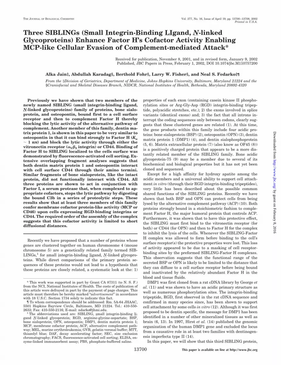
Three SIBLINGs (Small Integrin-Binding LIgand, N-linkedGlycoproteins) Enhance Factor H’s Cofactor Activity EnablingMCP-like Cellular Evasion of Complement-mediated Attack*
Received for publication, November 8, 2001, and in revised form, January 9, 2002Published, JBC Papers in Press, February 1, 2002, DOI 10.1074/jbc.M110757200
Alka Jain‡, Abdullah Karadag§, Berthold Fohr§, Larry W. Fisher§, and Neal S. Fedarko‡¶
From the ‡Division of Geriatrics, Department of Medicine, Johns Hopkins University, Baltimore, Maryland 21224 and the§Craniofacial and Skeletal Diseases Branch, NIDCR, National Institutes of Health, Bethesda, Maryland 20892-4320
Previously we have shown that two members of thenewly named SIBLING (small integrin-binding ligand,N-linked glycoproteins) family of proteins, bone sialo-protein, and osteopontin, bound first to a cell surfacereceptor and then to complement Factor H therebyblocking the lytic activity of the alternative pathway ofcomplement. Another member of this family, dentin ma-trix protein 1, is shown in this paper to be very similar toosteopontin in that it can bind strongly to Factor H (Ka� 1 nM) and block the lytic activity through either thevitronectin receptor (�V�3 integrin) or CD44. Binding ofFactor H to SIBLING localized to the cells surface wasdemonstrated by fluorescence-activated cell sorting. Ex-tensive overlapping fragment analyses suggests thatboth dentin matrix protein 1 and osteopontin interactwith cell surface CD44 through their amino termini.Similar fragments of bone sialoprotein, like the intactprotein, did not functionally interact with CD44. Allthree proteins are shown to act in conjunction withFactor I, a serum protease that, when complexed to ap-propriate cofactors, stops the lytic pathway by digestingthe bound C3b in a series of proteolytic steps. Theseresults show that at least three members of this familyconfer membrane cofactor protein-like activity (MCP orCD46) upon cells expressing RGD-binding integrins orCD44. The required order of the assembly of the complexsuggests that this cofactor activity is limited to shortdiffusional distances.
Recently we have proposed that a number of proteins whosegenes are clustered together on human chromosome 4 (mousechromosome 5) are a genetically related family termed SIB-LINGs,1 for small integrin-binding ligand, N-linked glycopro-teins. While direct comparisons of the primary protein se-quences of these proteins would not lead to a hypothesis thatthese proteins are closely related, a systematic look at the: 1)
properties of each exon (containing casein kinase II phospho-rylation sites or Arg-Gly-Asp (RGD) integrin-binding tripep-tide, polyacidic stretches, etc.); 2) the exons involved in splicevariants (identical exons) and; 3) the fact that all introns in-terrupt the coding sequences only between codons, clearly sug-gests that these clustered genes are related (1). At this time,the gene products within this family include four acidic pro-teins: bone sialoprotein (BSP) (2), osteopontin (OPN) (3), dentinmatrix protein 1 (DMP1) (4), and dentin sialophosphoprotein(5, 6). Matrix extracellular protein (7) (also know as OF45 (8))is a positively charged protein that appears to be a more dis-tantly related member of the SIBLING family. Bone acidicglycoprotein-75 (9) may be a member due to several of itsbiochemical and biological properties but it has not yet beencloned and sequenced.
Except for a high affinity for hydroxy apatite among theacidic members and a universal ability to support cell attach-ment in vitro (through their RGD integrin-binding tripeptides),very little has been described about the possible commonshared functions of the SIBLING proteins. Recently we haveshown that both BSP and OPN can protect cells from beinglysed by the alternative complement pathway (ACP) (10). Bothproteins strongly bound in a stoichiometric fashion to comple-ment Factor H, the major humoral protein that controls ACP.Furthermore, it was shown that to have this protective effect,the SIBLING must first bind to the vitronectin receptor (forboth) or CD44 (for OPN) and then to Factor H for the complexto inhibit the lysis of the cells. Whenever the SIBLING-FactorH complex was allowed to form before binding to the cell’ssurface receptor(s) the protective properties were lost. This lossof activity appeared to be due to a masking of cell receptor-binding sites by the preformed SIBLING-Factor H complexes.This observation suggests that the functional range of thesecreted BSP or OPN is likely to be limited to the distance thatthey can diffuse to a cell surface receptor before being boundand inactivated by the relatively abundant Factor H in theblood and tissue fluids.
DMP1 was first cloned from a rat cDNA library by George etal. (11) and was shown to have an acidic primary structure aswell as numerous phosphorylation sites. The integrin-bindingtripeptide, RGD, first observed in the rat cDNA sequence andconfirmed in many species since, has been shown to supportcell attachment by some cells in vitro (12). Although it was firstproposed to be dentin specific, the message for DMP1 has beenidentified in a number of other mineralized tissues as well asbrain (6, 13). In 1997, Hirst et al. (14) published the genomicorganization of the human DMP1 gene and excluded the locusfrom a causative role in at least two families with dentinogen-esis imperfecta type II (14).
In this paper, we will show that this third SIBLING protein,
* This work was supported in part by Grant CA 87311 (to N. S. F.)from the NCI, National Institutes of Health. The costs of publication ofthis article were defrayed in part by the payment of page charges. Thisarticle must therefore be hereby marked “advertisement” in accordancewith 18 U.S.C. Section 1734 solely to indicate this fact.
¶ To whom correspondence should be addressed: Rm. 5A-64 JHAAC,5501 Hopkins Bayview Circle, Baltimore, MD 21224. Tel.: 410-550-2632; Fax: 410-550-2116; E-mail: [email protected].
1 The abbreviations used are: SIBLING, small integrin-binding li-gand N-linked glycoprotein; RGD, arginine-glycine-aspartate; BSP,bone sialoprotein; OPN, osteopontin; DMP1, dentin matrix protein 1;MCP, membrane cofactor protein; ACP, alternative complement path-way; MEL, murine erythroleukemia; GVB, gelatin veronal buffer; MTT,thiazolyl blue; DAF, decay accelerating factor; SEC, size exclusionchromatography; FACS, fluorescence-activated cell sorting; ELISA, en-zyme-linked immunsorbent assay; PBS, phosphate-buffered saline.
THE JOURNAL OF BIOLOGICAL CHEMISTRY Vol. 277, No. 16, Issue of April 19, pp. 13700–13708, 2002Printed in U.S.A.
This paper is available on line at http://www.jbc.org13700
by guest on February 8, 2016http://w
ww
.jbc.org/D
ownloaded from
DMP1, can also protect tumor cells from attack by the ACP anddoes so by bridging Factor H to integrins and CD44. Further-more, the structural similarities and differences between theSIBLING family members are exploited to investigate themechanism and sequences involved in complementmodulation.
EXPERIMENTAL PROCEDURES
Reagents—Rabbit anti-DMP1 peptide-derived antibody, LF-148, wasraised against the human sequences (C)EHPSRKIFRKSRISE and(C)LKNIEIESRKLTVDAYH conjugated through the Cys to activatedhorseshoe crab hemocyanin (Pierce Chemical Co., Chicago, IL). Thisantiserum bound to fragments D6 and D8 (see Fig. 5 under “Results”) indirect ELISA suggesting that both peptides successfully raised usefulIgG components. Furthermore, this antiserum recognized recombinantmouse DMP1 made in Escherichia coli and full-length bovine DMP1described below. Normal human serum, purified human complementFactor H protein, and mouse monoclonal antibody against Factor Hwere obtained from Quidel Corp. (San Diego, CA). A monoclonal anti-body against human complement Factor I that blocks Factor I activity(cleavage of C3b) (catalog number A247) as well as a monoclonal anti-body to Factor I that binds but does not block Factor I function (catalognumber A231) were also obtained from Quidel Corp. Polyclonal anti-bodies against CD-44 and a “functional” antibody against �V�3 (catalognumber MAB1976) were obtained from Chemicon Co. (Temecula, CA).Synthetic purified glycine-arginine-aspartate-serine peptide (GRGDS)was obtained from Calbiochem-NovaBiochem Corp. (La Jolla, CA). Syn-thetic peptides corresponding to the sequences VKQADSGSSEEKQ(OPN exon 3) and LYNKYPDAVATWLNPDPSQKQNLLAPQ (OPNexon 4) were made and purified by the Peptide Laboratory of theFacility for Biotechnology Resources, Center for Biologics Evaluationand Research, Food and Drug Administration (Bethesda, MD). Preim-mune serum, human serum-adsorbed goat anti-rabbit IgG conjugatedto horseradish peroxidase (1 mg/ml IgG) as well as goat anti-mouse IgGconjugated to horseradish peroxidase (1 mg/ml IgG) were obtained fromKirkegaard & Perry (Gaithersburg, MD). Dulbecco’s modified essentialmedium, Hank’s balanced salt solution, and heat inactivated fetal bo-vine serum were obtained from BioFluids, Inc. (Rockville, MD).
Western Blotting—Samples diluted in gel sample buffer were re-solved by Tris glycine SDS-PAGE 4–20% gradient gels (Novex Corp.,San Diego, CA) and transferred to nitrocellulose following standardconditions (15). Nitrocellulose membranes were rinsed with Tris-buff-ered saline (0.05 M Tris-HCl, pH 7.5, 0.15 M NaCl) containing 0.05%Tween 20 (TBS-Tween). After a 1-h incubation in blocking solution(TBS-Tween � 5% non-fat powdered milk) at room temperature onrotary shaker, primary antibody was added (1:2,000) and incubatedovernight at 4 °C. The nitrocellulose sheet was washed in TBS-Tweenfour times for 5 min each time with TBS-Tween and then horseradishperoxidase-conjugated second antibody (1:50,000) in TBS-Tween � 5%milk was added and incubated for 2 h at room temperature. Followingremoval of the second antibody solution the membrane was washedthree times with TBS-Tween and rinsed a final time in enzyme sub-strate buffer for 5 min. Enhanced chemiluminescence reagents wereemployed for signal detection (Pierce Chemical Co., Chicago, IL) withx-ray film.
High Performance Liquid Chromatography—A Shimadzu LC10ASbinary gradient system was employed for chromatographic separations.Size exclusion chromatography utilized a 1.0 � 30-cm Superose 6 col-umn (Amersham Bioscience, Piscataway, NJ) equilibrated in 0.05 M
sodium phosphate, pH 7.4, containing 50% fresh formamide at a flowrate of 0.5 ml/min. The column was calibrated using commerciallyavailable protein standards of known molecular weight (AmershamBioscience).
Direct ELISA—Greiner high-binding 96-well plates (part number655061) were coated with 100 �l of high performance liquid chroma-tography fractions overnight at 4 °C. Plates were washed three times (5min each) with TBS-Tween and exposed to 100 �l of 1:2000 primaryantibody for 1 h at room temperature. Plates were washed three timesand exposed to 100 �l of 1:2000 horseradish peroxidase-conjugated goatanti-rabbit IgG. Following a 1-h incubation at room temperature, plateswere washed again three times with TBS-Tween and color was devel-oped using 3,3�,5,5�-tetramethylbenzidine and H2O2 for 10 min at roomtemperature. Color development was stopped by the addition of 25 �l of1 N H2SO4 and analyzed at 450 nm.
Production of Recombinant Intact SIBLINGs—Recombinant humanBSP and OPN were made and expressed as described previously (10).For DMP1 expression, an adenoviral construct was generated by sub-
cloning full-length bovine DMP1 cDNA (6, 13) into high expression,replication-deficient adenovirus (Ad5) using the cytomegalovirus pro-moter. The construct was selected, purified, and expressed following themethod described previously for BSP and OPN adenoviruses (10).Briefly, adenovirus was plaque-selected and propagated on HEK 293cells (ATCC number CRL1573). Viral particles were purified by twicebanding on CsCl and viral titers evaluated by plaque formation of virusdilutions on HEK293 cells (16). Recombinant DMP1 was generated byinfecting subconfluent normal human marrow stromal fibroblasts with10,000 pfu/cell. Harvested serum-free media was subjected to anionexchange chromatography. Native BSP, DMP1, and OPN proteins werepurified by diluting medium from normal human marrow stromal fi-broblast cells 1:1 with 40 mM phosphate buffer, pH 7.4, and loadingdirectly on a 5.0 � 2.0-cm column packed with ToyoPearl TSK QAEresin. A linear salt gradient to 2.0 M NaCl was employed to separatelypurify the three proteins to �95% purity as measured by SDS-PAGE.
Production of Recombinant SIBLING Fragments—A pET-15b vector(Novagen Inc., Madison, WI) which produces polypeptides as fusionproducts with an amino-terminal polyhistidine sequence followed by athrombin cleavage site (MGSSHHHHHHSSGLVPRGSH) was used forexpression and generation of most of the peptides. Peptides were puri-fied from isopropyl-1-thio-�-D-galactopyranoside-induced log phase E.coli by nickel affinity chromatography (Ni2� IMAC) following the man-ufacturer’s protocol. Sequences of primers used to insert the in-frameappropriate restriction sites (NdeI and BamHI) and generate the rele-vant SIBLING fragment by PCR for insertion into the vector are given.All OPN fragments were derived from the human sequence (3). The twoOPN fragments containing the RGD domain (O4 and O5) were engi-neered in a pET-22b vector with NcoI sites because of an internal NdeIsite. The use of pET-22b results in fusion polypeptides with the six Hisresidues at the carboxyl terminus. The first two DMP1 peptides (D1 andD2) were made using the bovine DMP1 cDNA as template (13) and allothers were made using human genomic DNA for the PCR templates.
Alternative Complement Mediated Cell Lysis Assay—Murine eryth-roleukemia (MEL) cells (a gift of Dr. Marilyn Farquhar, University ofCalifornia, San Diego, CA) grown in Dulbecco’s modified essential me-dium containing 10% fetal bovine serum and 4 mM glutamine wererinsed three times with gelatin veronal buffer (GVB, Sigma) containing2 mM Mg2� and 8 mM EGTA. Cells were resuspended in GVB-MgEGTA
FIG. 1. DMP1 in human serum. A, aliquots of normal human se-rum � heating and reduction with dithiothreitol were analyzed bySDS-polyacrylamide gradient gel electrophoresis. Proteins were trans-ferred to nitrocellulose and probed with anti-DMP1 antibody LF-148. B,unreduced normal human serum was fractionated by SEC and aliquotsof fractions from the SEC were transferred to 96-well microtiter platesand analyzed by direct ELISA for DMP1 (E). The DMP1 activity elutedat a location consistent with that in a complex with Factor H. Substi-tuting non-immune rabbit serum for the LF-148 antibody yielded im-munoreactive material corresponding to the void peak (‚) showing thatit is nonspecific. C, aliquots of the same column profile were analyzed bydirect ELISA for Factor H immunoreactivity using a monoclonal anti-body. D, serum heated at 100 °C and reduced with 2 mM dithiothreitolfor 10 min prior to SEC analysis was also analyzed for anti-DMP1immunoreactivity. The activity eluted at the expected location of freeDMP1.
SIBLINGs Enhance Factor H Activity 13701
by guest on February 8, 2016http://w
ww
.jbc.org/D
ownloaded from
(gelatin veronal buffer containing 2 mM magnesium and 8 mM EGTA) ata density of 5 � 106 cells/ml. Cells were preincubated with 10 �g ofDMP1, BSP, or OPN in 1 ml for 10 min at 37 °C followed by incubationat 37 °C with normal human serum diluted 1:10 in GVB-MgEGTA.After 2 h, cells were harvested for thiazolyl blue viability assay byincubating a 50-�l aliquot of the cell suspension in an equal volume of1 mg/ml thiazolyl blue (MTT) for 45 min. Cell viability was determinedspectrophotometrically by absorbance at 560 nm.
Fluorescence-activated Cell Sorting (FACS) Analysis—MEL cells(2 � 106/ml) were washed twice in PBS and incubated with either PBSor recombinant DMP1 (10 �g/ml) for 10 min at room temperature. Thecells were then washed twice and incubated with 5 �g/ml Alexa Fluor488 (Molecular probes, Eugene, OR) -labeled purified human comple-ment protein Factor H (Quidel, San Diego, CA) for 10 min at roomtemperature. The cells were washed twice, re-suspended in PBS, andthen analyzed by FACSCalibur cell sorter equipped with a 488-nmargon laser using Cellquest software (BD PharMingen, Bedford, MA).
RESULTS
DMP1 Exists as a Complex in Serum Bound to Factor H—Wehave proposed that, based on a shared chromosomal localiza-tion, similarities in exon structure and intron type, that DMP1belongs to the SIBLING family of proteins (1). We have previ-ously found that two other acidic SIBLING family members,BSP and OPN, are bound to complement Factor H in serumand that disruption of the complex requires heating and reduc-tion (10). The strong (nM) interaction between Factor H andeither BSP or OPN (which have no cysteine residues) is non-covalent and required reduction to disrupt due to the uniquestructure of Factor H. By NMR spectroscopy, BSP and OPNlack ordered structure and exist extended and flexible in solu-tion (1), while Factor H is a large and highly structured proteincontaining 20 repeated short consensus repeat motifs. Eachshort consensus repeat contains 4 cysteine residues that areinvolved in intra-repeat disulfide bonds forming a “sushi roll”-type structure. The status of DMP1 in human serum wasstudied by SDS-PAGE and Western blotting as well as by sizeexclusion chromatography (SEC). When aliquots of normal hu-man serum diluted 1:10 were subjected to SDS-PAGE followedby transfer to nitrocellulose and probing with a peptide-derivedantibody against DMP1, immunoreactive bands were readilyapparent (Fig. 1A). The migration position of immunoreactiveDMP1 shifted upon heating and reduction consistent with thedestruction of the DMP1-Factor H complex.
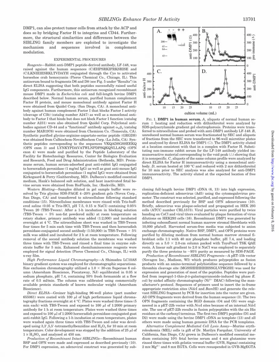
FIG. 2. Fluorescence titration. A, intrinsic tryptophan fluorescence was monitored by excitation at 295 nm and emission from 300 to 500 nmusing a Photon Technology International Series M fluorimeter. The initial Factor H concentration was 28 nM and DMP1 was added in nanomolaramounts. Both Factor H and DMP1 were dissolved in Hank’s balanced salt solution. B, the binding curve was determined following calculation offractional acceptor saturation. Factor H contains 25 tryptophan while DMP1 contains two. Given the abundance of tryptophans in Factor H andthe molecule’s robust fluorescent emission signal, the contribution of the fluorescent spectra arising from DMP1 was negligible. The fluorescentsignal for Factor H was progressively quenched until an equimolar amount of DMP1 was added showing a 1:1 saturable binding. The estimatedbinding of DMP1 to Factor H is in the nanomolar range.
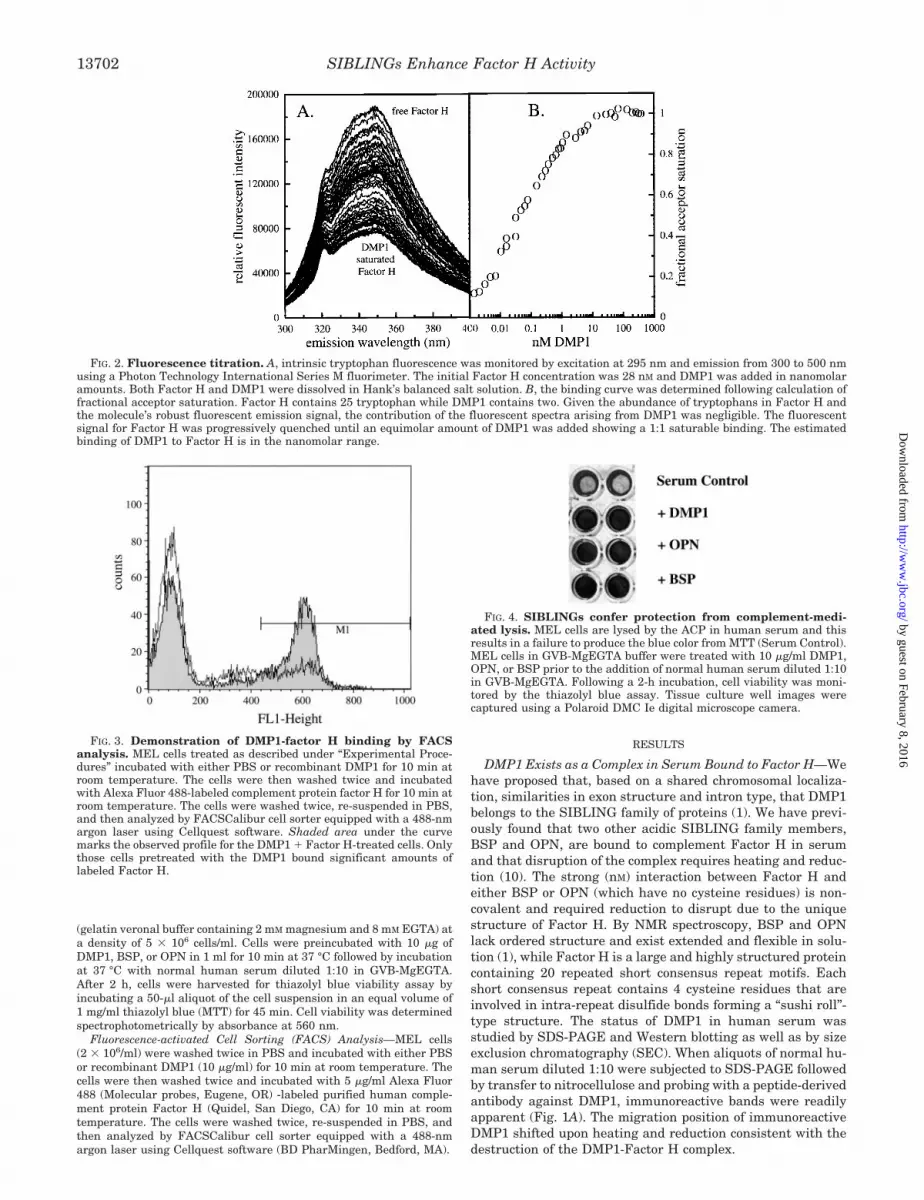
FIG. 3. Demonstration of DMP1-factor H binding by FACSanalysis. MEL cells treated as described under “Experimental Proce-dures” incubated with either PBS or recombinant DMP1 for 10 min atroom temperature. The cells were then washed twice and incubatedwith Alexa Fluor 488-labeled complement protein factor H for 10 min atroom temperature. The cells were washed twice, re-suspended in PBS,and then analyzed by FACSCalibur cell sorter equipped with a 488-nmargon laser using Cellquest software. Shaded area under the curvemarks the observed profile for the DMP1 � Factor H-treated cells. Onlythose cells pretreated with the DMP1 bound significant amounts oflabeled Factor H.
FIG. 4. SIBLINGs confer protection from complement-medi-ated lysis. MEL cells are lysed by the ACP in human serum and thisresults in a failure to produce the blue color from MTT (Serum Control).MEL cells in GVB-MgEGTA buffer were treated with 10 �g/ml DMP1,OPN, or BSP prior to the addition of normal human serum diluted 1:10in GVB-MgEGTA. Following a 2-h incubation, cell viability was moni-tored by the thiazolyl blue assay. Tissue culture well images werecaptured using a Polaroid DMC Ie digital microscope camera.
SIBLINGs Enhance Factor H Activity13702
by guest on February 8, 2016http://w
ww
.jbc.org/D
ownloaded from
Unreduced normal human serum was fractionated by sizeexclusion chromatography and aliquots of fractions from theSEC were transferred to 96-well microtiter plates and analyzedby direct ELISA for DMP1. Immunoreactive material was ev-ident in two peaks, a void volume peak and an included peak.Incubation of aliquots from the same SEC fractions with pre-immune serum (in place of anti-DMP1) yielded a single voidvolume peak. Thus the void volume peak appearing in theanti-DMP1 profile was most likely the result of nonspecificbinding. Direct ELISA of the same fractions for Factor H im-munoreactivity yielded a single peak that co-migrated with theincluded DMP1 immunoreactive material (Fig. 1C). SEC reso-lution of a separate aliquot of the same normal human serumthat had been incubated with reducing agent and heat to dis-sociate the binding complex yielded an immunoreactive profileupon direct ELISA analysis that shifted to �100 kDa, thelocation of authentic, free DMP1 (Fig. 1D). The immunoreac-tive material that eluted in the void volume in unreducedsamples (Fig. 1B) was absent in the profile of reduced serum,suggesting that the epitope recognized by the antibody wassensitive to denaturation.
DMP1 and Factor H Binding—Complement Factor H pos-sesses 25 tryptophan residues, while DMP1 contains 2. Thus,the binding between Factor H and DMP1 can be followed byintrinsic tryptophan fluorescence. Titration of purified humancomplement Factor H with DMP1 was followed by excitation at295 nm and monitoring emission between 300 and 450 nm. The
emission profile of Factor H alone yields a peak at 347 nm (Fig.2A). The addition of DMP1 in nanomolar increments causes arelative fluorescent intensity quenching. Conversion of the flu-orescent intensity titration into a binding curve by determiningthe fraction of binding sites occupied as the fractional change influorescence quenching at 347 nm yields a saturable bindingcurve (Fig. 2B). By steady state fluorescence, the binding ofDMP1 by Factor H is saturable, possess a 1:1 stoichiometry,and has a binding constant in the nanomolar range. This valueis similar to Factor H interactions with BSP and OPN reportedearlier.
DMP1 Bridges Factor H to the Cell Surface—The direct bind-ing of Factor H to DMP1, while DMP1 is engaged with its cellsurface receptor was studied by fluorescence-activated cellsorting. MEL cells incubated with DMP1, briefly washed, in-cubated with Alexa Fluor 488-conjugated purified human com-plement protein Factor H, and then analyzed by FACSCaliburcell sorter. The results indicate that a significant association ofcell surface Factor H was evident only in the DMP1-treatedcells (Fig. 3). Similar shifts were seen when cells were sub-jected to an initial incubation with BSP and OPN (data notshown).
DMP1 Protects Cells from Alternative Complement Pathway-mediated Lysis—DMP1 binding to Factor H suggests that thisSIBLING may also confer resistance to humoral complementsurveillance. The ability of rDMP1 to protect cells from com-plement activity was investigated using the MEL cell linewhich, when incubated with normal human serum, can bereadily assayed for ACP-mediated cell lysis (10, 17). Cell sur-vival was measured by MTT reduction by living mitochondria.Titration with dilutions of normal human serum and timecourses were carried out to define optimal incubation condi-tions. The addition of purified recombinant SIBLING to MELcells followed by treatment with normal human serum pro-tected the cells from lysis (Fig. 4). The protection of MEL cellsfrom ACP-mediated cell lysis by DMP1 addition exhibited adose response (data not shown).
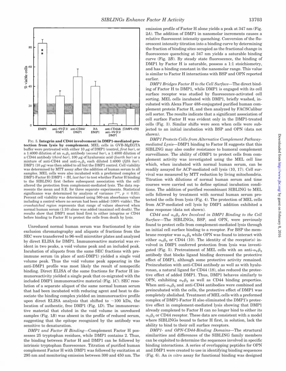
CD44 and �V�3 Are Involved in DMP1 Binding to the CellSurface—The SIBLINGs, BSP, and OPN, were previouslyfound to protect cells from complement-mediated lysis throughan initial cell surface binding to a receptor. For BSP the mem-brane receptor was �V�3 while OPN was found to interact witheither �V�3 or CD44 (10). The identity of the receptor(s) in-volved in DMP1 conferred protection from lysis was investi-gated (Fig. 5). Pretreatment of MEL cells with an anti-�V�3
antibody that blocks ligand binding decreased the protectiveeffect of DMP1, although some protective activity remained.Preincubation with anti-CD44 antibody as well as with hyalu-ronan, a natural ligand for CD44 (18), also reduced the protec-tive effect of added DMP1. Thus, DMP1 behaves similarly toOPN, exhibiting �V�3 as well as CD44 binding properties.When anti-�V�3 and anti-CD44 antibodies were combined andpreincubated with the cells, the protective effect of DMP1 wascompletely abolished. Treatment of MEL cells with a preformedcomplex of DMP1-Factor H also eliminated the DMP1’s protec-tive effect in complement-mediated lysis showing that DMP1already complexed to Factor H can no longer bind to either its�V�3 or CD44 receptor. These data are consistent with a modelwhere SIBLINGs bound to factor H first, in solution, lack theability to bind to their cell surface receptors.
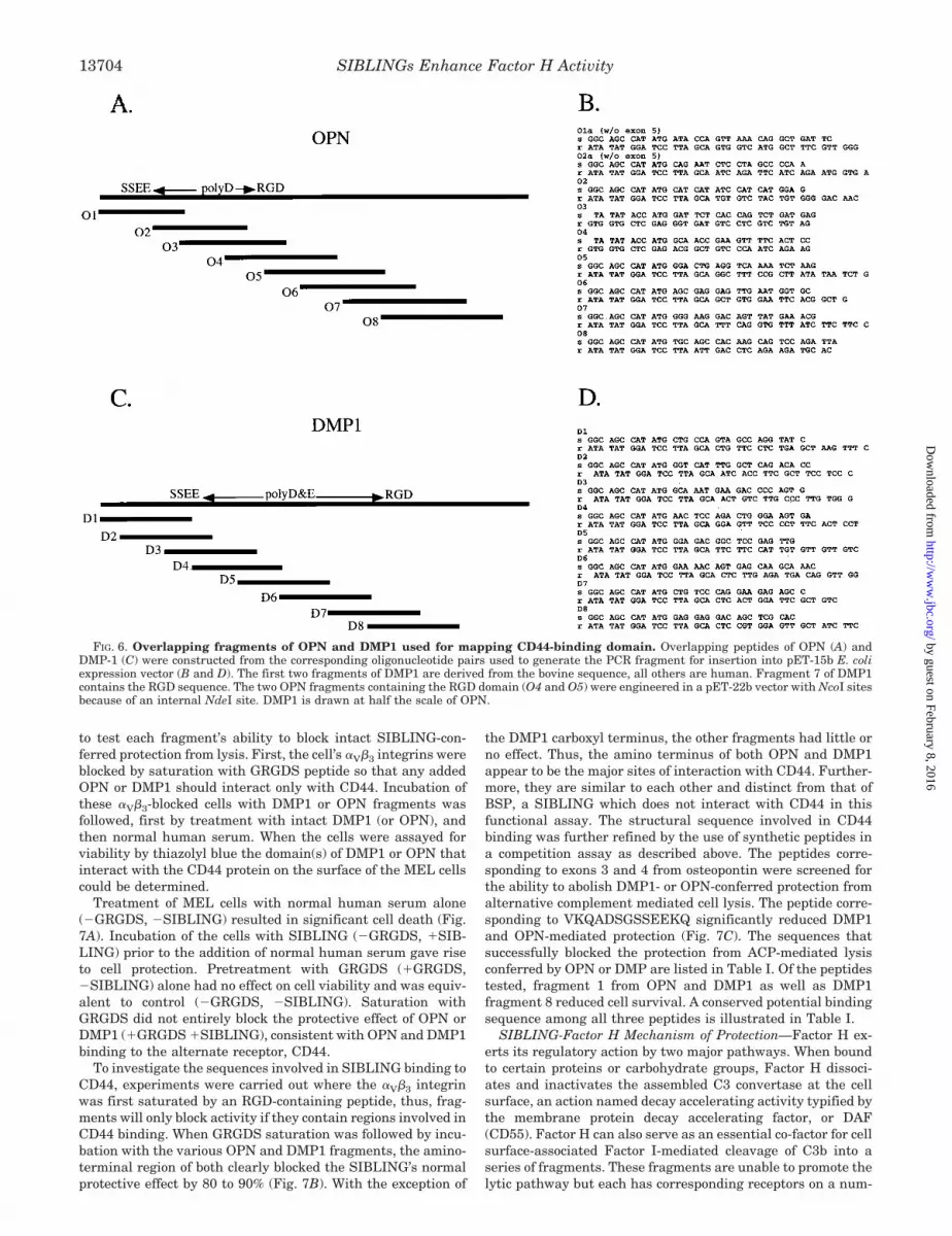
DMP1- and OPN-CD44-Binding Domains—The structuralsimilarities and differences of the SIBLING family memberscan be exploited to determine the sequences involved in specificbinding interactions. A series of overlapping peptides for OPNand DMP1 were created to use in identifying binding sequences(Fig. 6). An in vitro assay for functional binding was designed
FIG. 5. Integrin and CD44 involvement in DMP1-mediated pro-tection from lysis by complement. MEL cells in GVB-MgEGTAbuffer were pretreated with either 10 �g of DMP1 (control, first bar), ora 1:4000 dilution of an �V�3 antibody (second bar), a 1:4000 dilution ofa CD44 antibody (third bar), 100 �g of hyaluronic acid (fourth bar) or amixture of anti-CD44 and anti-�V�3 each diluted 1:4000 (fifth bar).DMP1 (10 �g) was then added to all but the DMP1 control. Cell viabilitywas determined by MTT assay after the addition of human serum to allsamples. MEL cells were also incubated with a preformed complex ofDMP1-Factor H (DMP1 � fH, last bar) to test whether Factor H bindingto the SIBLING first (before subsequent association with the cell)altered the protection from complement-mediated lysis. The data rep-resents the mean and S.E. for three separate experiments. Statisticalsignificance was determined by analysis of variance (**, p � 0.01).Percent cell viability was determined using 560-nm absorbance valuesincluding a control where no serum had been added (100% viable). Thecrosshatched region represents that range of values observed whennormal human serum (1:10) alone was added (maximal cell death). Theresults show that DMP1 must bind first to either integrins or CD44before binding to Factor H to protect the cells from death by lysis.
SIBLINGs Enhance Factor H Activity 13703
by guest on February 8, 2016http://w
ww
.jbc.org/D
ownloaded from
to test each fragment’s ability to block intact SIBLING-con-ferred protection from lysis. First, the cell’s �V�3 integrins wereblocked by saturation with GRGDS peptide so that any addedOPN or DMP1 should interact only with CD44. Incubation ofthese �V�3-blocked cells with DMP1 or OPN fragments wasfollowed, first by treatment with intact DMP1 (or OPN), andthen normal human serum. When the cells were assayed forviability by thiazolyl blue the domain(s) of DMP1 or OPN thatinteract with the CD44 protein on the surface of the MEL cellscould be determined.
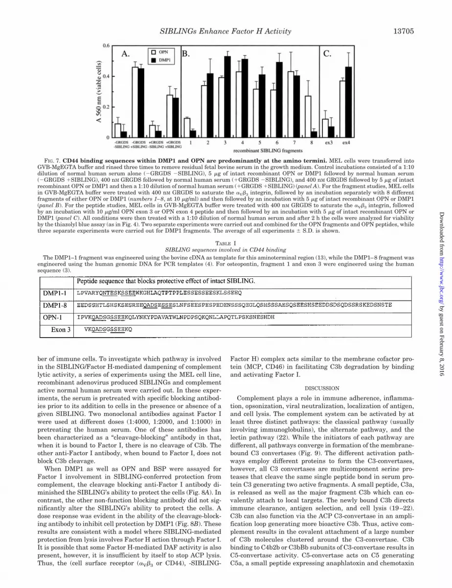
Treatment of MEL cells with normal human serum alone(�GRGDS, �SIBLING) resulted in significant cell death (Fig.7A). Incubation of the cells with SIBLING (�GRGDS, �SIB-LING) prior to the addition of normal human serum gave riseto cell protection. Pretreatment with GRGDS (�GRGDS,�SIBLING) alone had no effect on cell viability and was equiv-alent to control (�GRGDS, �SIBLING). Saturation withGRGDS did not entirely block the protective effect of OPN orDMP1 (�GRGDS �SIBLING), consistent with OPN and DMP1binding to the alternate receptor, CD44.
To investigate the sequences involved in SIBLING binding toCD44, experiments were carried out where the �V�3 integrinwas first saturated by an RGD-containing peptide, thus, frag-ments will only block activity if they contain regions involved inCD44 binding. When GRGDS saturation was followed by incu-bation with the various OPN and DMP1 fragments, the amino-terminal region of both clearly blocked the SIBLING’s normalprotective effect by 80 to 90% (Fig. 7B). With the exception of
the DMP1 carboxyl terminus, the other fragments had little orno effect. Thus, the amino terminus of both OPN and DMP1appear to be the major sites of interaction with CD44. Further-more, they are similar to each other and distinct from that ofBSP, a SIBLING which does not interact with CD44 in thisfunctional assay. The structural sequence involved in CD44binding was further refined by the use of synthetic peptides ina competition assay as described above. The peptides corre-sponding to exons 3 and 4 from osteopontin were screened forthe ability to abolish DMP1- or OPN-conferred protection fromalternative complement mediated cell lysis. The peptide corre-sponding to VKQADSGSSEEKQ significantly reduced DMP1and OPN-mediated protection (Fig. 7C). The sequences thatsuccessfully blocked the protection from ACP-mediated lysisconferred by OPN or DMP are listed in Table I. Of the peptidestested, fragment 1 from OPN and DMP1 as well as DMP1fragment 8 reduced cell survival. A conserved potential bindingsequence among all three peptides is illustrated in Table I.
SIBLING-Factor H Mechanism of Protection—Factor H ex-erts its regulatory action by two major pathways. When boundto certain proteins or carbohydrate groups, Factor H dissoci-ates and inactivates the assembled C3 convertase at the cellsurface, an action named decay accelerating activity typified bythe membrane protein decay accelerating factor, or DAF(CD55). Factor H can also serve as an essential co-factor for cellsurface-associated Factor I-mediated cleavage of C3b into aseries of fragments. These fragments are unable to promote thelytic pathway but each has corresponding receptors on a num-
FIG. 6. Overlapping fragments of OPN and DMP1 used for mapping CD44-binding domain. Overlapping peptides of OPN (A) andDMP-1 (C) were constructed from the corresponding oligonucleotide pairs used to generate the PCR fragment for insertion into pET-15b E. coliexpression vector (B and D). The first two fragments of DMP1 are derived from the bovine sequence, all others are human. Fragment 7 of DMP1contains the RGD sequence. The two OPN fragments containing the RGD domain (O4 and O5) were engineered in a pET-22b vector with NcoI sitesbecause of an internal NdeI site. DMP1 is drawn at half the scale of OPN.
SIBLINGs Enhance Factor H Activity13704
by guest on February 8, 2016http://w
ww
.jbc.org/D
ownloaded from
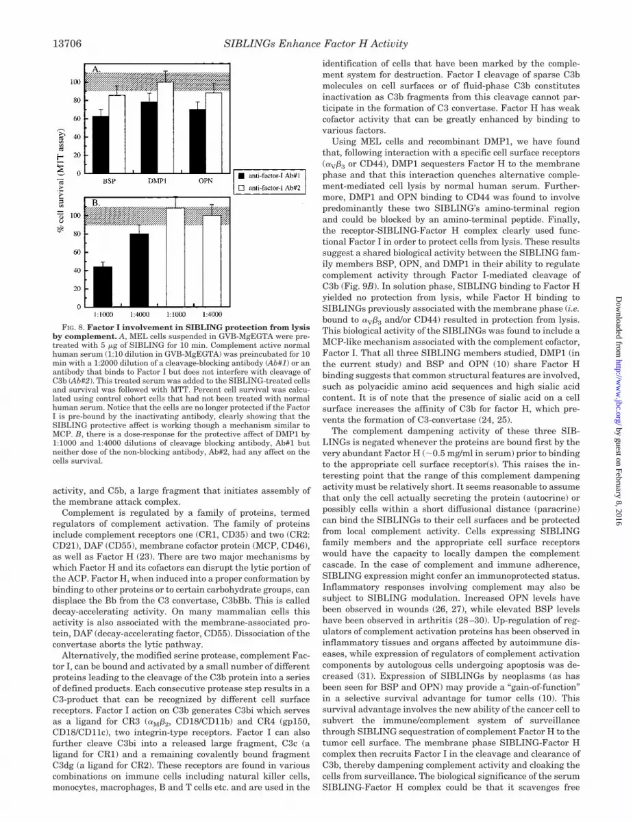
ber of immune cells. To investigate which pathway is involvedin the SIBLING/Factor H-mediated dampening of complementlytic activity, a series of experiments using the MEL cell line,recombinant adenovirus produced SIBLINGs and complementactive normal human serum were carried out. In these exper-iments, the serum is pretreated with specific blocking antibod-ies prior to its addition to cells in the presence or absence of agiven SIBLING. Two monoclonal antibodies against Factor Iwere used at different doses (1:4000, 1:2000, and 1:1000) inpretreating the human serum. One of these antibodies hasbeen characterized as a “cleavage-blocking” antibody in that,when it is bound to Factor I, there is no cleavage of C3b. Theother anti-Factor I antibody, when bound to Factor I, does notblock C3b cleavage.
When DMP1 as well as OPN and BSP were assayed forFactor I involvement in SIBLING-conferred protection fromcomplement, the cleavage blocking anti-Factor I antibody di-minished the SIBLING’s ability to protect the cells (Fig. 8A). Incontrast, the other non-function blocking antibody did not sig-nificantly alter the SIBLING’s ability to protect the cells. Adose response was evident in the ability of the cleavage-block-ing antibody to inhibit cell protection by DMP1 (Fig. 8B). Theseresults are consistent with a model where SIBLING-mediatedprotection from lysis involves Factor H action through Factor I.It is possible that some Factor H-mediated DAF activity is alsopresent, however, it is insufficient by itself to stop ACP lysis.Thus, the (cell surface receptor (�V�3 or CD44), -SIBLING-
Factor H) complex acts similar to the membrane cofactor pro-tein (MCP, CD46) in facilitating C3b degradation by bindingand activating Factor I.
DISCUSSION
Complement plays a role in immune adherence, inflamma-tion, opsonization, viral neutralization, localization of antigen,and cell lysis. The complement system can be activated by atleast three distinct pathways: the classical pathway (usuallyinvolving immunoglobulins), the alternate pathway, and thelectin pathway (22). While the initiators of each pathway aredifferent, all pathways converge in formation of the membrane-bound C3 convertases (Fig. 9). The different activation path-ways employ different proteins to form the C3-convertases,however, all C3 convertases are multicomponent serine pro-teases that cleave the same single peptide bond in serum pro-tein C3 generating two active fragments. A small peptide, C3a,is released as well as the major fragment C3b which can co-valently attach to local targets. The newly bound C3b directsimmune clearance, antigen selection, and cell lysis (19–22).C3b can also function via the ACP C3-convertase in an ampli-fication loop generating more bioactive C3b. Thus, active com-plement results in the covalent attachment of a large numberof C3b molecules clustered around the C3-convertase. C3bbinding to C4b2b or C3bBb subunits of C3-convertase results inC5-convertase activity. C5-convertase acts on C5 generatingC5a, a small peptide expressing anaphlatoxin and chemotaxin
FIG. 7. CD44 binding sequences within DMP1 and OPN are predominantly at the amino termini. MEL cells were transferred intoGVB-MgEGTA buffer and rinsed three times to remove residual fetal bovine serum in the growth medium. Control incubations consisted of a 1:10dilution of normal human serum alone (�GRGDS �SIBLING), 5 �g of intact recombinant OPN or DMP1 followed by normal human serum(�GRGDS �SIBLING), 400 nM GRGDS followed by normal human serum (�GRGDS �SIBLING), and 400 nM GRGDS followed by 5 �g of intactrecombinant OPN or DMP1 and then a 1:10 dilution of normal human serum (�GRGDS �SIBLING) (panel A). For the fragment studies, MEL cellsin GVB-MgEGTA buffer were treated with 400 nM GRGDS to saturate the �V�3 integrin, followed by an incubation separately with 8 differentfragments of either OPN or DMP1 (numbers 1–8, at 10 �g/ml) and then followed by an incubation with 5 �g of intact recombinant OPN or DMP1(panel B). For the peptide studies, MEL cells in GVB-MgEGTA buffer were treated with 400 nM GRGDS to saturate the �V�3 integrin, followedby an incubation with 10 �g/ml OPN exon 3 or OPN exon 4 peptide and then followed by an incubation with 5 �g of intact recombinant OPN orDMP1 (panel C). All conditions were then treated with a 1:10 dilution of normal human serum and after 2 h the cells were analyzed for viabilityby the thiazolyl blue assay (as in Fig. 4). Two separate experiments were carried out and combined for the OPN fragments and OPN peptides, whilethree separate experiments were carried out for DMP1 fragments. The average of all experiments � S.D. is shown.
TABLE ISIBLING sequences involved in CD44 binding
The DMP1–1 fragment was engineered using the bovine cDNA as template for this aminoterminal region (13), while the DMP1–8 fragment wasengineered using the human genomic DNA for PCR templates (4). For osteopontin, fragment 1 and exon 3 were engineered using the humansequence (3).
SIBLINGs Enhance Factor H Activity 13705
by guest on February 8, 2016http://w
ww
.jbc.org/D
ownloaded from
activity, and C5b, a large fragment that initiates assembly ofthe membrane attack complex.
Complement is regulated by a family of proteins, termedregulators of complement activation. The family of proteinsinclude complement receptors one (CR1, CD35) and two (CR2:CD21), DAF (CD55), membrane cofactor protein (MCP, CD46),as well as Factor H (23). There are two major mechanisms bywhich Factor H and its cofactors can disrupt the lytic portion ofthe ACP. Factor H, when induced into a proper conformation bybinding to other proteins or to certain carbohydrate groups, candisplace the Bb from the C3 convertase, C3bBb. This is calleddecay-accelerating activity. On many mammalian cells thisactivity is also associated with the membrane-associated pro-tein, DAF (decay-accelerating factor, CD55). Dissociation of theconvertase aborts the lytic pathway.
Alternatively, the modified serine protease, complement Fac-tor I, can be bound and activated by a small number of differentproteins leading to the cleavage of the C3b protein into a seriesof defined products. Each consecutive protease step results in aC3-product that can be recognized by different cell surfacereceptors. Factor I action on C3b generates C3bi which servesas a ligand for CR3 (�M�2, CD18/CD11b) and CR4 (gp150,CD18/CD11c), two integrin-type receptors. Factor I can alsofurther cleave C3bi into a released large fragment, C3c (aligand for CR1) and a remaining covalently bound fragmentC3dg (a ligand for CR2). These receptors are found in variouscombinations on immune cells including natural killer cells,monocytes, macrophages, B and T cells etc. and are used in the
identification of cells that have been marked by the comple-ment system for destruction. Factor I cleavage of sparse C3bmolecules on cell surfaces or of fluid-phase C3b constitutesinactivation as C3b fragments from this cleavage cannot par-ticipate in the formation of C3 convertase. Factor H has weakcofactor activity that can be greatly enhanced by binding tovarious factors.
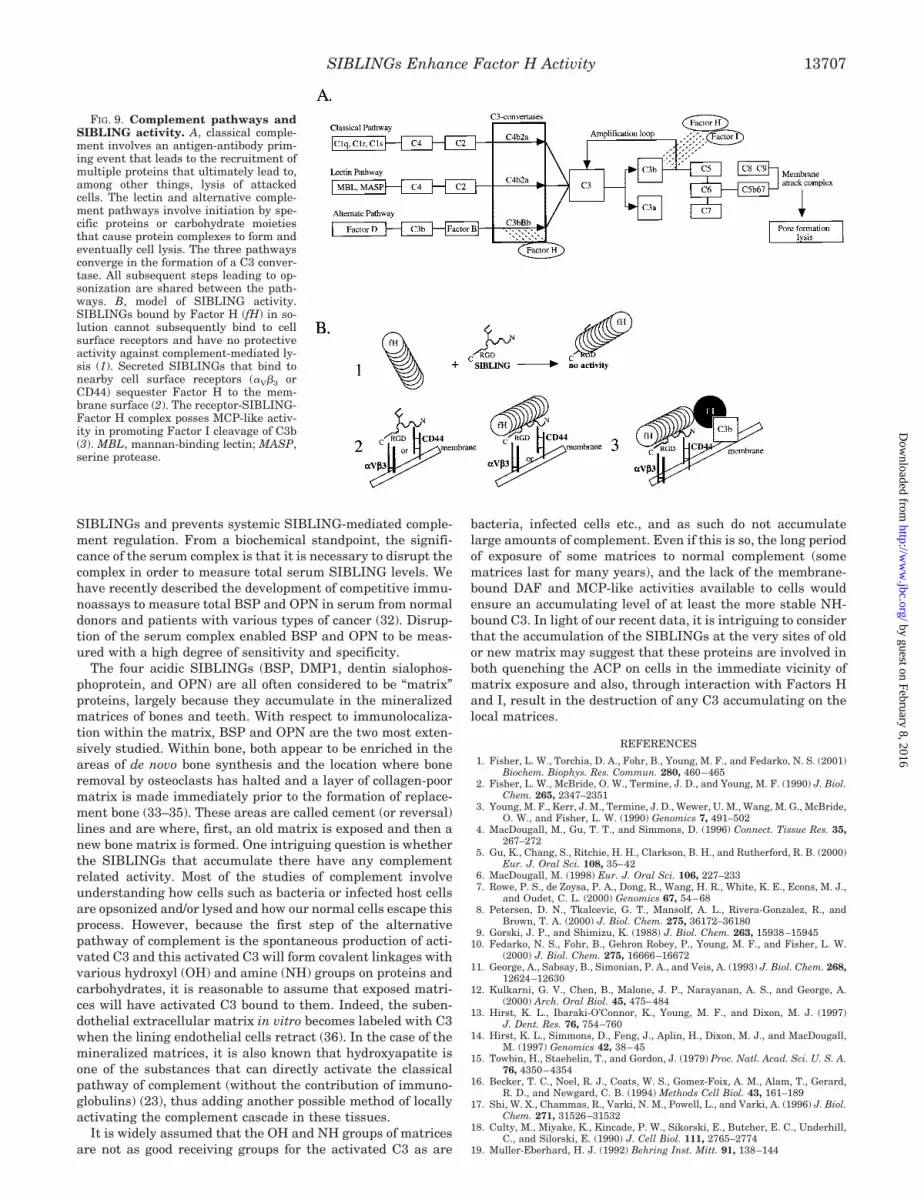
Using MEL cells and recombinant DMP1, we have foundthat, following interaction with a specific cell surface receptors(�V�3 or CD44), DMP1 sequesters Factor H to the membranephase and that this interaction quenches alternative comple-ment-mediated cell lysis by normal human serum. Further-more, DMP1 and OPN binding to CD44 was found to involvepredominantly these two SIBLING’s amino-terminal regionand could be blocked by an amino-terminal peptide. Finally,the receptor-SIBLING-Factor H complex clearly used func-tional Factor I in order to protect cells from lysis. These resultssuggest a shared biological activity between the SIBLING fam-ily members BSP, OPN, and DMP1 in their ability to regulatecomplement activity through Factor I-mediated cleavage ofC3b (Fig. 9B). In solution phase, SIBLING binding to Factor Hyielded no protection from lysis, while Factor H binding toSIBLINGs previously associated with the membrane phase (i.e.bound to �V�3 and/or CD44) resulted in protection from lysis.This biological activity of the SIBLINGs was found to include aMCP-like mechanism associated with the complement cofactor,Factor I. That all three SIBLING members studied, DMP1 (inthe current study) and BSP and OPN (10) share Factor Hbinding suggests that common structural features are involved,such as polyacidic amino acid sequences and high sialic acidcontent. It is of note that the presence of sialic acid on a cellsurface increases the affinity of C3b for factor H, which pre-vents the formation of C3-convertase (24, 25).
The complement dampening activity of these three SIB-LINGs is negated whenever the proteins are bound first by thevery abundant Factor H (�0.5 mg/ml in serum) prior to bindingto the appropriate cell surface receptor(s). This raises the in-teresting point that the range of this complement dampeningactivity must be relatively short. It seems reasonable to assumethat only the cell actually secreting the protein (autocrine) orpossibly cells within a short diffusional distance (paracrine)can bind the SIBLINGs to their cell surfaces and be protectedfrom local complement activity. Cells expressing SIBLINGfamily members and the appropriate cell surface receptorswould have the capacity to locally dampen the complementcascade. In the case of complement and immune adherence,SIBLING expression might confer an immunoprotected status.Inflammatory responses involving complement may also besubject to SIBLING modulation. Increased OPN levels havebeen observed in wounds (26, 27), while elevated BSP levelshave been observed in arthritis (28–30). Up-regulation of reg-ulators of complement activation proteins has been observed ininflammatory tissues and organs affected by autoimmune dis-eases, while expression of regulators of complement activationcomponents by autologous cells undergoing apoptosis was de-creased (31). Expression of SIBLINGs by neoplasms (as hasbeen seen for BSP and OPN) may provide a “gain-of-function”in a selective survival advantage for tumor cells (10). Thissurvival advantage involves the new ability of the cancer cell tosubvert the immune/complement system of surveillancethrough SIBLING sequestration of complement Factor H to thetumor cell surface. The membrane phase SIBLING-Factor Hcomplex then recruits Factor I in the cleavage and clearance ofC3b, thereby dampening complement activity and cloaking thecells from surveillance. The biological significance of the serumSIBLING-Factor H complex could be that it scavenges free
FIG. 8. Factor I involvement in SIBLING protection from lysisby complement. A, MEL cells suspended in GVB-MgEGTA were pre-treated with 5 �g of SIBLING for 10 min. Complement active normalhuman serum (1:10 dilution in GVB-MgEGTA) was preincubated for 10min with a 1:2000 dilution of a cleavage-blocking antibody (Ab#1) or anantibody that binds to Factor I but does not interfere with cleavage ofC3b (Ab#2). This treated serum was added to the SIBLING-treated cellsand survival was followed with MTT. Percent cell survival was calcu-lated using control cohort cells that had not been treated with normalhuman serum. Notice that the cells are no longer protected if the FactorI is pre-bound by the inactivating antibody, clearly showing that theSIBLING protective affect is working though a mechanism similar toMCP. B, there is a dose-response for the protective affect of DMP1 by1:1000 and 1:4000 dilutions of cleavage blocking antibody, Ab#1 butneither dose of the non-blocking antibody, Ab#2, had any affect on thecells survival.
SIBLINGs Enhance Factor H Activity13706
by guest on February 8, 2016http://w
ww
.jbc.org/D
ownloaded from
SIBLINGs and prevents systemic SIBLING-mediated comple-ment regulation. From a biochemical standpoint, the signifi-cance of the serum complex is that it is necessary to disrupt thecomplex in order to measure total serum SIBLING levels. Wehave recently described the development of competitive immu-noassays to measure total BSP and OPN in serum from normaldonors and patients with various types of cancer (32). Disrup-tion of the serum complex enabled BSP and OPN to be meas-ured with a high degree of sensitivity and specificity.
The four acidic SIBLINGs (BSP, DMP1, dentin sialophos-phoprotein, and OPN) are all often considered to be “matrix”proteins, largely because they accumulate in the mineralizedmatrices of bones and teeth. With respect to immunolocaliza-tion within the matrix, BSP and OPN are the two most exten-sively studied. Within bone, both appear to be enriched in theareas of de novo bone synthesis and the location where boneremoval by osteoclasts has halted and a layer of collagen-poormatrix is made immediately prior to the formation of replace-ment bone (33–35). These areas are called cement (or reversal)lines and are where, first, an old matrix is exposed and then anew bone matrix is formed. One intriguing question is whetherthe SIBLINGs that accumulate there have any complementrelated activity. Most of the studies of complement involveunderstanding how cells such as bacteria or infected host cellsare opsonized and/or lysed and how our normal cells escape thisprocess. However, because the first step of the alternativepathway of complement is the spontaneous production of acti-vated C3 and this activated C3 will form covalent linkages withvarious hydroxyl (OH) and amine (NH) groups on proteins andcarbohydrates, it is reasonable to assume that exposed matri-ces will have activated C3 bound to them. Indeed, the suben-dothelial extracellular matrix in vitro becomes labeled with C3when the lining endothelial cells retract (36). In the case of themineralized matrices, it is also known that hydroxyapatite isone of the substances that can directly activate the classicalpathway of complement (without the contribution of immuno-globulins) (23), thus adding another possible method of locallyactivating the complement cascade in these tissues.
It is widely assumed that the OH and NH groups of matricesare not as good receiving groups for the activated C3 as are
bacteria, infected cells etc., and as such do not accumulatelarge amounts of complement. Even if this is so, the long periodof exposure of some matrices to normal complement (somematrices last for many years), and the lack of the membrane-bound DAF and MCP-like activities available to cells wouldensure an accumulating level of at least the more stable NH-bound C3. In light of our recent data, it is intriguing to considerthat the accumulation of the SIBLINGs at the very sites of oldor new matrix may suggest that these proteins are involved inboth quenching the ACP on cells in the immediate vicinity ofmatrix exposure and also, through interaction with Factors Hand I, result in the destruction of any C3 accumulating on thelocal matrices.
REFERENCES
1. Fisher, L. W., Torchia, D. A., Fohr, B., Young, M. F., and Fedarko, N. S. (2001)Biochem. Biophys. Res. Commun. 280, 460–465
2. Fisher, L. W., McBride, O. W., Termine, J. D., and Young, M. F. (1990) J. Biol.Chem. 265, 2347–2351
3. Young, M. F., Kerr, J. M., Termine, J. D., Wewer, U. M., Wang, M. G., McBride,O. W., and Fisher, L. W. (1990) Genomics 7, 491–502
4. MacDougall, M., Gu, T. T., and Simmons, D. (1996) Connect. Tissue Res. 35,267–272
5. Gu, K., Chang, S., Ritchie, H. H., Clarkson, B. H., and Rutherford, R. B. (2000)Eur. J. Oral Sci. 108, 35–42
6. MacDougall, M. (1998) Eur. J. Oral Sci. 106, 227–2337. Rowe, P. S., de Zoysa, P. A., Dong, R., Wang, H. R., White, K. E., Econs, M. J.,
and Oudet, C. L. (2000) Genomics 67, 54–688. Petersen, D. N., Tkalcevic, G. T., Mansolf, A. L., Rivera-Gonzalez, R., and
Brown, T. A. (2000) J. Biol. Chem. 275, 36172–361809. Gorski, J. P., and Shimizu, K. (1988) J. Biol. Chem. 263, 15938–15945
10. Fedarko, N. S., Fohr, B., Gehron Robey, P., Young, M. F., and Fisher, L. W.(2000) J. Biol. Chem. 275, 16666–16672
11. George, A., Sabsay, B., Simonian, P. A., and Veis, A. (1993) J. Biol. Chem. 268,12624–12630
12. Kulkarni, G. V., Chen, B., Malone, J. P., Narayanan, A. S., and George, A.(2000) Arch. Oral Biol. 45, 475–484
13. Hirst, K. L., Ibaraki-O’Connor, K., Young, M. F., and Dixon, M. J. (1997)J. Dent. Res. 76, 754–760
14. Hirst, K. L., Simmons, D., Feng, J., Aplin, H., Dixon, M. J., and MacDougall,M. (1997) Genomics 42, 38–45
15. Towbin, H., Staehelin, T., and Gordon, J. (1979) Proc. Natl. Acad. Sci. U. S. A.76, 4350–4354
16. Becker, T. C., Noel, R. J., Coats, W. S., Gomez-Foix, A. M., Alam, T., Gerard,R. D., and Newgard, C. B. (1994) Methods Cell Biol. 43, 161–189
17. Shi, W. X., Chammas, R., Varki, N. M., Powell, L., and Varki, A. (1996) J. Biol.Chem. 271, 31526–31532
18. Culty, M., Miyake, K., Kincade, P. W., Sikorski, E., Butcher, E. C., Underhill,C., and Silorski, E. (1990) J. Cell Biol. 111, 2765–2774
19. Muller-Eberhard, H. J. (1992) Behring Inst. Mitt. 91, 138–144
FIG. 9. Complement pathways andSIBLING activity. A, classical comple-ment involves an antigen-antibody prim-ing event that leads to the recruitment ofmultiple proteins that ultimately lead to,among other things, lysis of attackedcells. The lectin and alternative comple-ment pathways involve initiation by spe-cific proteins or carbohydrate moietiesthat cause protein complexes to form andeventually cell lysis. The three pathwaysconverge in the formation of a C3 conver-tase. All subsequent steps leading to op-sonization are shared between the path-ways. B, model of SIBLING activity.SIBLINGs bound by Factor H (fH) in so-lution cannot subsequently bind to cellsurface receptors and have no protectiveactivity against complement-mediated ly-sis (1). Secreted SIBLINGs that bind tonearby cell surface receptors (�V�3 orCD44) sequester Factor H to the mem-brane surface (2). The receptor-SIBLING-Factor H complex posses MCP-like activ-ity in promoting Factor I cleavage of C3b(3). MBL, mannan-binding lectin; MASP,serine protease.
SIBLINGs Enhance Factor H Activity 13707
by guest on February 8, 2016http://w
ww
.jbc.org/D
ownloaded from

20. Fearon, D. T., and Locksley, R. M. (1996) Science 272, 50–5321. Carroll, M. C. (1998) Annu. Rev. Immunol. 16, 545–56822. Volanakis, J. E. (1998) in The Human Complement System in Health and
Disease (Volanakis, J. E., and Frank, M. M., eds) pp. 49–81, Marcel Dekker,New York
23. Volanakis, J. E. (1998) in The Human Complement System in Health andDisease (Volankis, J. E., and Frank, M. M., eds) pp. 9–32, Marcel Dekker,Inc., New York
24. Fearon, D. T. (1978) Proc. Natl. Acad. Sci. U. S. A. 75, 1971–197525. Kazatchkine, M. D., Fearon, D. T., and Austen, K. F. (1979) J. Immunol. 122,
75–8126. Giachelli, C. M., and Steitz, S. (2000) Matrix Biol. 19, 615–62227. McKee, M. D., and Nanci, A. (1996) Anat. Rec. 245, 394–40928. Larsson, E., Mussener, A., Heinegard, D., Klareskog, L., and Saxne, T. (1997)
Br. J. Rheumatol. 36, 1258–126129. Conrozier, T., Saxne, T., Fan, C. S., Mathieu, P., Tron, A. M., Heinegard, D.,
and Vignon, E. (1998) Ann. Rheum. Dis. 57, 527–53230. Lohmander, L. S., Saxne, T., and Heinegard, D. (1996) Ann. Rheum. Dis. 55,
622–62631. Kawano, M. (2000) Arch. Immunol. Ther. Exp. 48, 367–37232. Fedarko, N. S., Jain, A., Karadag, A., Van Eman, M. R., and Fisher, L. W.
(2001) Clin. Cancer Res. 7, 4060–406633. Riminucci, M., Silvestrini, G., Bonucci, E., Fisher, L. W., Gehron Robey, P.,
and Bianco, P. (1995) Calcif. Tissue Int. 57, 277–28434. Ingram, R. T., Clarke, B. L., Fisher, L. W., and Fitzpatrick, L. A. (1993) J. Bone
Miner. Res. 8, 1019–102935. McKee, M. D., and Nanci, A. (1996) J. Bone Miner. Res. 11, 873–87536. Hindmarsh, E. J., and Marks, R. M. (1998) J. Immunol. 160, 6128–6136
SIBLINGs Enhance Factor H Activity13708
by guest on February 8, 2016http://w
ww
.jbc.org/D
ownloaded from
Alka Jain, Abdullah Karadag, Berthold Fohr, Larry W. Fisher and Neal S. FedarkoComplement-mediated Attack
Factor H's Cofactor Activity Enabling MCP-like Cellular Evasion of -linked Glycoproteins) EnhanceNThree SIBLINGs (SmallIntegrin-Binding LIgand,
doi: 10.1074/jbc.M110757200 originally published online February 1, 20022002, 277:13700-13708.J. Biol. Chem.
10.1074/jbc.M110757200Access the most updated version of this article at doi:
Alerts:
When a correction for this article is posted•
When this article is cited•
to choose from all of JBC's e-mail alertsClick here
http://www.jbc.org/content/277/16/13700.full.html#ref-list-1
This article cites 34 references, 17 of which can be accessed free at
by guest on February 8, 2016http://w
ww
.jbc.org/D
ownloaded from
Copyright © 2022 FDOKUMEN