Three-dimensional CT scanning: a new diagnostic modality in congenital heart disease
6
REVIEW Three-dimensional CT scanning: a new diagnostic modality in congenital heart disease Phalla Ou, David S Celermajer, Giulio Calcagni, Francis Brunelle, Damien Bonnet, Daniel Sidi ................................................................................................................................... Heart 2007;93:908–913. doi: 10.1136/hrt.2006.101352 New generation multislice CT technology has changed the approach to non-invasive assessment of congenital heart disease, in both paediatric and adult patients. This is mainly because of rapid advances in spatial and temporal resolution and in post-processing capability. At Ho ˆpital Necker-Enfants Malades, CT with multiplanar and three-dimensional reconstruction has become a routine examination in the evaluation of congenital heart disease planning surgery, complex interventional catheterisations and for follow-up. It has proved to be an invaluable diagnostic and decision-aiding methodology in these situations, as a complement to echocardiography and, increasingly, as a substitute for diagnostic angiography (which is usually associated with higher-dose radiation and longer sedation times, as well as occasional morbidity). This review illustrates the current status of 64-slice CT in congenital heart diseases, including assessment of the aorta, the coronary arteries, the pulmonary arteries, the systemic and pulmonary veins, and other intra- and extracardiac malformations. ............................................................................. See end of article for authors’ affiliations ........................ Correspondence to: Dr P Ou, University Rene ´ Descartes–Paris 5, Department of Paediatric Radiology, Ho ˆpital Necker- Enfants Malades; 149, rue de Se `vres, 75743 Paris, Cedex 15, France; phalla. ou@ nck.ap-hop-paris.fr Accepted 8 August 2006 Published Online First 4 September 2006 ........................ M ultislice CT has seen rapid technological advances over the past 3–5 years, particu- larly with the recent emergence of 64-slice devices. Indeed since 2000 there has been a doubling of detector rows approximately every 2 years, with 4, 16, 32 and then 64 rows in 2004. This latest technology allows much faster acquisi- tion times, improved spatial resolution (with thinner slices) and with fewer motion artefacts, owing to improved ECG gating. CT scanning is thus becoming a leading modality for non-invasive imaging of cardiovascular diseases in adults, particularly with CT coronary angiography. 12 Indeed, in cardiovascular imaging in adults, multi- slice CT provides very high image quality equal or even better than that of conventional invasive angiography, for the proximal epicardial vessels. CT scanning is also a valuable diagnostic tool for the diagnosis of pulmonary embolism, complex arteriovenous malformations, aneurysms, aortic dissections and even coronary arteries in certain circumstances. In such clinical situations, CT scanning often obviates the need for conventional angiography. 3–6 Although many reports of cardiac CT imaging in adults have been published, there is little such information about imaging in paediatric cardiol- ogy. Only a few papers have examined the place of multislice CT scanning in the assessment of congenital heart disease. 7–9 Currently, cardiac ultrasound is the leading diagnostic modality for the interrogation of intracardiac structures in children. However, ultrasound is usually insuffi- cient for the study of certain structures such as the coronary arteries, the aorta and branches of the pulmonary artery, or for systemic and pulmonary venous structures. For visualising these structures, CT allows better diagnostic capability, as a result of the high-precision, three-dimensional imaging it provides. 9 Three-dimensional imaging is particu- larly useful for diagnostic investigation of complex forms of congenital heart disease, preparation of complex interventional procedures involving car- diac catheterisation, and the postoperative assess- ment of surgical reconstructions. We provide here some examples of the main forms of congenital heart disease where valuable diagnostic information can be obtained from 64-slice CT scanning with three-dimensional volume rendering, using commercially available equipment. TECHNICAL ASPECTS The examinations illustrated were carried out with a multislice CT-scan device (GE, LightSpeed VCT, Milwaukee, USA). The acquisition protocols take account of the patient’s age, the malformation to be studied, and the ratio of image quality to radiation dose. Children under 5 years This group includes neonates, infants and young children unable to achieve a 5-second breath hold and whose heart rates are high, generally above 100 beats/min. In this group of children, good image quality is difficult to obtain owing to respiratory and cardiac movement artefacts, which are all the more marked when the child is anxious or agitated. The rule therefore is to acquire images as quickly as possible while the child is calm or lightly sedated. This implies exploiting the speed of the CT scan along with optimal injection of contrast medium. ECG gating is avoided as it lengthens acquisition time. Contrast medium injection and image acquisition are carefully calculated to achieve optimal image quality. The acquisition delay is automatically determined by placing a region of interest in the aorta or pulmonary artery (depending on whether systemic or pulmonary arterial structures are to be exam- ined), with an automatic triggering threshold at 100 HU. The contrast medium bolus is then delivered by an automatic injector to ensure that all the contrast medium passes at a sufficiently high and uniform rate during the short image 908 www.heartjnl.com
Transcript of Three-dimensional CT scanning: a new diagnostic modality in congenital heart disease

REVIEW
Three-dimensional CT scanning: a new diagnostic modality incongenital heart diseasePhalla Ou, David S Celermajer, Giulio Calcagni, Francis Brunelle, Damien Bonnet, Daniel Sidi. . . . . . . . . . . . . . . . . . . . . . . . . . . . . . . . . . . . . . . . . . . . . . . . . . . . . . . . . . . . . . . . . . . . . . . . . . . . . . . . . . . . . . . . . . . . . . . . . . . . . . . . . . . . . . . . . . . . . . . . . . . . . . . . . . .
Heart 2007;93:908–913. doi: 10.1136/hrt.2006.101352
New generation multislice CT technology has changed theapproach to non-invasive assessment of congenital heartdisease, in both paediatric and adult patients. This is mainlybecause of rapid advances in spatial and temporal resolutionand in post-processing capability. At Hopital Necker-EnfantsMalades, CT with multiplanar and three-dimensionalreconstruction has become a routine examination in theevaluation of congenital heart disease planning surgery,complex interventional catheterisations and for follow-up. It hasproved to be an invaluable diagnostic and decision-aidingmethodology in these situations, as a complement toechocardiography and, increasingly, as a substitute fordiagnostic angiography (which is usually associated withhigher-dose radiation and longer sedation times, as well asoccasional morbidity). This review illustrates the current status of64-slice CT in congenital heart diseases, including assessmentof the aorta, the coronary arteries, the pulmonary arteries, thesystemic and pulmonary veins, and other intra- andextracardiac malformations.. . . . . . . . . . . . . . . . . . . . . . . . . . . . . . . . . . . . . . . . . . . . . . . . . . . . . . . . . . . . . . . . . . . . . . . . . . . . .
See end of article forauthors’ affiliations. . . . . . . . . . . . . . . . . . . . . . . .
Correspondence to:Dr P Ou, University ReneDescartes–Paris 5,Department of PaediatricRadiology, Hopital Necker-Enfants Malades; 149, ruede Sevres, 75743 Paris,Cedex 15, France; phalla.ou@ nck.ap-hop-paris.fr
Accepted 8 August 2006Published Online First4 September 2006. . . . . . . . . . . . . . . . . . . . . . . .
Multislice CT has seen rapid technologicaladvances over the past 3–5 years, particu-larly with the recent emergence of 64-slice
devices. Indeed since 2000 there has been adoubling of detector rows approximately every2 years, with 4, 16, 32 and then 64 rows in 2004.This latest technology allows much faster acquisi-tion times, improved spatial resolution (withthinner slices) and with fewer motion artefacts,owing to improved ECG gating. CT scanning isthus becoming a leading modality for non-invasiveimaging of cardiovascular diseases in adults,particularly with CT coronary angiography.1 2
Indeed, in cardiovascular imaging in adults, multi-slice CT provides very high image quality equal oreven better than that of conventional invasiveangiography, for the proximal epicardial vessels.CT scanning is also a valuable diagnostic tool forthe diagnosis of pulmonary embolism, complexarteriovenous malformations, aneurysms, aorticdissections and even coronary arteries in certaincircumstances. In such clinical situations, CTscanning often obviates the need for conventionalangiography.3–6
Although many reports of cardiac CT imaging inadults have been published, there is little suchinformation about imaging in paediatric cardiol-ogy. Only a few papers have examined the place of
multislice CT scanning in the assessment ofcongenital heart disease.7–9 Currently, cardiacultrasound is the leading diagnostic modality forthe interrogation of intracardiac structures inchildren. However, ultrasound is usually insuffi-cient for the study of certain structures such as thecoronary arteries, the aorta and branches of thepulmonary artery, or for systemic and pulmonaryvenous structures. For visualising these structures,CT allows better diagnostic capability, as a result ofthe high-precision, three-dimensional imaging itprovides.9 Three-dimensional imaging is particu-larly useful for diagnostic investigation of complexforms of congenital heart disease, preparation ofcomplex interventional procedures involving car-diac catheterisation, and the postoperative assess-ment of surgical reconstructions.
We provide here some examples of the main formsof congenital heart disease where valuable diagnosticinformation can be obtained from 64-slice CTscanning with three-dimensional volume rendering,using commercially available equipment.
TECHNICAL ASPECTSThe examinations illustrated were carried out witha multislice CT-scan device (GE, LightSpeed VCT,Milwaukee, USA). The acquisition protocols takeaccount of the patient’s age, the malformation tobe studied, and the ratio of image quality toradiation dose.
Children under 5 yearsThis group includes neonates, infants and youngchildren unable to achieve a 5-second breath holdand whose heart rates are high, generally above100 beats/min. In this group of children, goodimage quality is difficult to obtain owing torespiratory and cardiac movement artefacts, whichare all the more marked when the child is anxiousor agitated. The rule therefore is to acquire imagesas quickly as possible while the child is calm orlightly sedated. This implies exploiting the speed ofthe CT scan along with optimal injection ofcontrast medium. ECG gating is avoided as itlengthens acquisition time. Contrast mediuminjection and image acquisition are carefullycalculated to achieve optimal image quality. Theacquisition delay is automatically determined byplacing a region of interest in the aorta orpulmonary artery (depending on whether systemicor pulmonary arterial structures are to be exam-ined), with an automatic triggering threshold at100 HU. The contrast medium bolus is thendelivered by an automatic injector to ensure thatall the contrast medium passes at a sufficientlyhigh and uniform rate during the short image
908
www.heartjnl.com

acquisition period. Injection concludes with saline rinsing of thetubes, to avoid enhancement artefacts generated by the persis-tence of contrast medium in the superior vena cava. Finally,radiation dose must be minimised by continuous attention to thevoltage and current being applied, keeping these aspects reducedas far as possible without degrading image quality.For this group of children we use the following protocol:
N Patient preparation: sedation (oral or intrarectal pentobarbitol orchloral hydrate, or both, 30 minutes before the examination);preparation of the neonate or infant, wrapped comfortably inVelpeau strapping if necessary; insertion of a 22–24G peripheralvenous line in the elbow, back of the hand or foot.
N Scan parameters: rotation time 400 ms; high pitch 0.984, slicethickness 0.625 mm; low voltage 80 kV; current variedduring acquisition and according to the weight of the child.
N Injection parameters: volume of contrast medium (Iopamerol300 mg/ml; Schering SA, Berlin, Germany) 1 ml/kg, fol-lowed by 5 ml saline solution; flow rate 0.5–1 ml/s forneonates; 1–2 ml/s for older children.
Children over 5 yearsFor this older group of children, breath holding with appro-priate and sympathetic coaching is generally possible. Sedation
should be avoided if good cooperation from the child is requiredto ensure breath holding during the acquisition period. Choiceof acquisition protocol depends above all on the malformationto be explored. Study of the great vessels (aorta, pulmonaryarteries, systemic or pulmonary venous return) is relativelystraightforward: it is carried out by fast acquisition timeswithout ECG gating. Study of the coronary arteries, on theother hand, is more difficult. The best image quality is obtainedwith ECG-gated acquisition in a child whose heart rate ismaintained stable and slow by administration of a b blocker(,70 beats/min). In all cases, the same precautions should betaken for contrast medium injection parameters and radiationdose as already described.For this group of older children we use the following protocol:
N Patient preparation: no sedation; b blocker in the case of CTcoronary angiography (propranolol, 1–2 mg/kg orally2 hours before the examination); 20–22 G peripheral venousline in the elbow, back of the hand or foot.
N Scan parameters:
– Examination of the great arteries: rotation time 400 ms;high pitch 0.984; slice thickness 0.625 mm; voltage 80–100 kV depending on body weight; current varied duringacquisition and adapted to the weight of the child.
– Examination of the coronary arteries: rotation time350 ms; short pitch 0.2; slice thickness 0.625 mm; voltage80–100 kV depending on body weight; current variedduring acquisition and adapted to the weight of the child.
N Injection parameters: volume of contrast medium (Iopamerol300 mg/ml; Schering SA, Berlin, Germany) 1.5 ml/kg max-imum 70 ml, followed by 5–10 ml saline solution; high flowrate 2–3.5 ml/s.
IMAGE PROCESSINGThe acquisition volume is analysed on dedicated workstations.Routine analyses are carried out on standard slices in variousplanes, multiplanar and three-dimensional reconstructions.This post-processing stage is partly facilitated by vascularanalysis algorithms with an interface that is interactive andsimplified for the user. For CT coronary angiography, analysis ismore refined and requires reconstructions at different phases ofthe cardiac cycle: a first set of images is obtained systematicallyat 75% of the R–R interval (corresponding to late diastole);further image reconstructions are performed at every 10% of theR–R interval when artefacts are present.
Figure 1 Anomalous left coronary artery rising from the pulmonaryartery.
DA DA
Figure 2 Coarctation associated with hypoplasia of the aortic arch in a neonate. The coarctation can be located (arrow) and the degree and extent ofhypoplasia of the aortic arch defined. DA, ductus arteriosus.
3D-CT in congenital heart disease 909
www.heartjnl.com
CLINICAL APPLICATIONSCoronary arteriesIndications for CT coronary angiography in the diagnosis ofcongenital abnormalities of the coronary arteries are now wellestablished in adults.10 11 In congenital heart disease, thecoronary arteries are important if anomalous, or reimplantedafter surgery in certain circumstances. The main advantage ofCT scanning is to provide a precise description of the three-dimensional anatomy of the coronary arteries, in particulartheir origin and course (fig 1). CT coronary angiography isespecially useful in screening for complications after coronaryre-implantation in cases of anomalous left coronary arteryarising from the pulmonary artery or after the arterial switchoperation for transposition of the great arteries. With even four-slice CT scanning we demonstrated excellent negative pre-dictive value of this technique, allowing CT coronary angio-graphy to be used for screening for coronary lesions after thearterial switch procedure and reserving selective coronaryangiography for confirmation in cases of suspected abnormalitydetected by CT.12 The ability of 64-slice CT scan to provideaccurate imaging of the proximal coronary vessels is better than
four-slice scanning, and may even replace selective coronaryangiography. This still awaits prospective validation; ourpreliminary results, however, are extremely promising.13
Other non-irradiating techniques—for example, MRI—aretheoretically preferable for repeated screening examinations,particularly in children. Nevertheless, MRI is currently limitedin congenital heart imaging by relatively poor temporalresolution (particularly for assessment of the coronary arteries)and often-complex acquisition protocols requiring a longexamination so that general anaesthesia is often necessary inyoung children.14
Aortic arch abnormalitiesImaging methods using slices—CT and MRI—are the preferredtechniques for morphological study of the aorta—for example,in the investigation of coarctation and congenital degenerativediseases of the aorta. Being complementary to echocardiogra-phy, CT and MRI allow precise pre- and postoperativeassessment of the thoracic aorta.15 16 In our experience, CT scanis often extremely useful for neonates with isthmic coarctationassociated with hypoplasia of the aortic arch (fig 2). It precisely
Figure 3 Vascular ring. Left panel: double aortic arch. Right panel: severe compression of the trachea (arrow).
Figure 4 Mapping of pulmonary vascularisation in a child withpulmonary atresia with ventricular septal defect. Visualisation of majoraortopulmonary collateral arteries, which show a seagull shape (in blue).
Figure 5 Pulmonary arteries supplied by systemic pulmonaryanastomoses. Three-dimensional CT makes it possible to check the patencyof palliative anastomoses (arrows). Note the deformation of the origin ofthe right pulmonary artery, at the level of the implantation of the rightBlalock anastomosis (posterior view).
910 Ou, Celermajer, Calcagni, et al
www.heartjnl.com

identifies the site of the coarctation, determines the degree ofnarrowing and, above all, defines precisely the extent ofhypoplasia of the aortic arch, thereby assisting the choice ofsurgical technique (simple left posterolateral thoracotomy ormidline sternotomy). CT is preferable to MRI for this group ofchildren owing to the simplicity of the examination and therapidity of image acquisition, generally less than 2 seconds forneonates.
For vascular rings, multiplanar and three-dimensionalreconstruction clearly demonstrates the origin and course ofthe great vessels and the relationship of the vessels to theadjacent airway (fig 3).17
Some authors have suggested that MRI is the ‘‘goldstandard’’ for the evaluation of vascular rings,18 however, MRIquality is commonly affected by respiratory artefacts inneonates and infants. In our experience, MRI examination inchildren under 7 years requires profound sedation or generalanaesthesia. Thus, for practical reasons, MRI is only used tostudy older children, who can hold repeated breath during anexamination which often lasts for 30 minutes.19
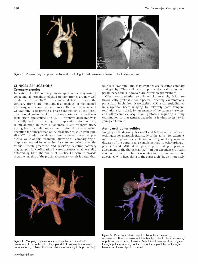
Pulmonary arteriesPrecise anatomical assessment of the right outflow tract andthe pulmonary arteries is essential in the investigation ofcertain types of cyanotic congenital heart diseases such asFallot’s tetralogy or pulmonary atresia with ventricular septaldefect (PA-VSD). In such cases, echocardiography is ofteninadequate for the study of the pulmonary arteries beyond theirproximal segments. CT scanning, by contrast, is well suited forprecise three-dimensional mapping of the pulmonary arterialtree and any aortopulmonary collateral arteries (fig 4). Wecurrently perform routine CT scanning in the preoperativeinvestigation of PA-VSD.20 21
In our experience, CT is also useful for monitoring the growthof the pulmonary arteries after successive palliative interven-tions in children (fig 5). Measurement of the size of thepulmonary arteries before dilatation or stenting procedures
allows accurate sizing of the devices to be used and oftenobviates the need for selective invasive angiography in suchpatients.
Venous returnPulmonary CT venography has been recently described inadults.22 In our practice, CT is used as a complement toechocardiography whenever there is a suspicion of anomaloussystemic or pulmonary venous return in the context of complexheart disease. The most common indication concerns thepreoperative investigation of a sinus venosus defect, which iscommonly associated with a partially anomalous pulmonaryvenous return, with the right superior pulmonary vein draining
LSPV
LIPVLA
RIPV
RSPV
SVC
Figure 6 Partial anomalous pulmonary venous return. Note theanomalous pulmonary venous return from the right superior pulmonaryvein, which drains into the superior vena cava. LA, left atrium; LIPV, leftinferior pulmonary vein; LSPV, left superior pulmonary vein; RIPV, rightinferior pulmonary vein; RSPV, right superior pulmonary vein; SVC,superior vena cava.
Figure 7 Supracardiac total anomalous pulmonary venous drainage.Volume rendered CT showing the supracardiac collecting vein draining allfour pulmonary veins.
Figure 8 Infradiaphragmatic partial anomalous pulmonary venousdrainage. Volume rendered CT showing the right superior pulmonary veindraining into the inferior vena cava.
3D-CT in congenital heart disease 911
www.heartjnl.com

abnormally into the superior vena cava (figs 6–8). Diagnosis ofpulmonary arteriovenous fistulae is also facilitated by CTscanning (fig 9). CT locates the malformation and identifiesthe feeding artery and receiving vein, thereby guiding treat-ment. Precise definition of the anomalous systemic venousreturn is often crucial in determining the appropriate approachfor cardiac catheterisation, particularly in cases of isomerism,and for the planning of cardiopulmonary bypass strategies.
LimitationsMultislice CT scanning undoubtedly represents a majoradvance in the imaging of congenital heart disease. Itsapplication to small children, in particular, is revolutionary.However, it is equally true that for a large number of simple andcommon malformations, such as atrial septal defect orventricular septal defect, echocardiography is sufficient forcomplete definition of the disease, and cardiac catheterisationprovides essential haemodynamic data. Experience with thisnew technology should be acquired prospectively and itsbenefits must be precisely evaluated through comparative trialsagainst the current standard methods.
Efforts should be made to minimise radiation dose,particularly in children for whom successive examinations arenecessary. In our institution, we strictly follow the ALARA (aslow as reasonably achievable) principles23 with regard toradiation exposure. Indeed, CT examinations in children areperformed at low kilovoltage (80–100 kV, instead of 120 kV,the standard kilovoltage setting). In adults, such precautionsassociated with strict examinations according to protocolallowed reduction of the radiation dose in comparison withconventional invasive angiography.24 Further studies arerequired to compare radiation exposure during paediatriccardiac CT scan with cardiac catheterisation; the latter isknown to be associated with high radiation doses because ofprolonged fluoroscopy time, multiple cine planes and thecomplexity of the procedures.25
It is too early yet to regard multislice CT as a substitute forinvasive angiography, although this may be demonstrable infuture. Its current use is as a complementary diagnosticmodality. In young adults with congenital heart disease, inwhom ultrasound windows are often suboptimal, CT scanningmay be particularly valuable.
CONCLUSIONThree-dimensional imaging obtained with the latest generationof CT-scan devices represents a real advance in the morpholo-gical study of cardiovascular malformations in children. In ourinstitution, CT now has a vital diagnostic and decision-aidingfunction in the assessment of congenital heart disease, as acomplement to echocardiography and, more and more often, asa substitute for invasive angiography.
Authors’ affiliations. . . . . . . . . . . . . . . . . . . . . . .
Phalla Ou, Francis Brunelle, University Rene Descartes–Paris 5,Department of Paediatric Radiology, Hopital Necker-Enfants Malades,AP-HP, Paris, FranceDavid S Celermajer, Department of Medicine, University of Sydney,AustraliaGiulio Calcagni, Damien Bonnet, Daniel Sidi, Department of PaediatricCardiology, Hopital Necker-Enfants Malades, AP-HP, Paris, France
Conflict of interest: None declared
REFERENCES1 Schoenhagen P, Halliburton SS, Stillman AE, et al. Noninvasive imaging of
coronary arteries: current and future role of multi-detector row CT. Radiology2004;232:7–17.
2 Pugliese F, Mollet NR, Runza G, et al. Diagnostic accuracy of non-invasive 64-slice CT coronary angiography in patients with stable angina pectoris. Eur Radiol2005;16:1–8.
3 Quiroz R, Kucher N, Zou KH, et al. Clinical validity of a negative computedtomography scan in patients with suspected pulmonary embolism: a systematicreview. JAMA 2005;293:2012–7.
4 Prestigiacomo CJ, Niimi Y, Setton A, et al. Three-dimensional rotational spinalangiography in the evaluation and treatment of vascular malformations. AJNRAm J Neuroradiol 2003;24:1429–35.
5 Kapustin AJ, Litt HI. Diagnostic imaging for aortic dissection. Semin ThoracCardiovasc Surg 2005;17:214–23.
6 Romeo G, Houyel L, Angel CY, et al. Coronary stenosis detection by 16-slicecomputed tomography in heart transplant patients: comparison with conventionalangiography and impact on clinical management. J Am Coll Cardiol2005;45:1826–31.
7 Bean MJ, Pannu H, Fishman EK. Three-dimensional computed tomographicimaging of complex congenital cardiovascular abnormalities. J Comput AssistTomogr 2005;29:721–4.
8 Kawano T, Ishii M, Takagi J, et al. Three-dimensional helical computedtomographic angiography in neonates and infants with complex congenital heartdisease. Am Heart J 2000;139:654–60.
9 Goo HW, Park IS, Ko JK, et al. CT of congenital heart disease: normal anatomyand typical pathologic conditions. Radiographics 2003;23:S147–65.
10 Shi H, Aschoff AJ, Brambs HJ, et al. Multislice CT imaging of anomalouscoronary arteries. Eur Radiol 2004;14:2172–81.
11 Datta J, White CS, Gilkeson RC, et al. Anomalous coronary arteries in adults:depiction at multi-detector row CT angiography. Radiology 2005;235:812–8.
12 Ou P, Mousseaux E, Azarine A, et al. Detection of coronary complications afterthe arterial switch operation for transposition of the great arteries: firstexperience with multislice computed tomogaphy in children. J Thorac CardiovascSurg 2006;131:639–43.
13 Ou P, Agnoletti G, Vouhe P, et al. Detection of coronary complications afterarterial switch operation for transposition of the great arteries with 64-slice CT.Pediatr Radiol 2006;36(Suppl 1):76.
14 Taylor AM, Dymarkowski S, Hamaekers P, et al. MR coronary angiography andlate-enhancement myocardial MR in children who underwent arterial switchsurgery for transposition of great arteries. Radiology 2005;234:542–7.
15 Prince MR, Narasimham DL, Jacoby WT, et al. Three-dimensional gadolinium-enhanced MR angiography of the thoracic aorta. AJR Am J Roentgenol1996;166:1387–97.
16 Lee EY, Siegel MJ, Hildebolt CF, et al. MDCT evaluation of thoracic aorticanomalies in pediatric patients and young adults: comparison of axial,multiplanar, and 3D images. AJR Am J Roentgenol 2004;182:777–84.
17 Lambert V, Sigal-Cinqualbre A, Belli E, et al. Preoperative and postoperativeevaluation of airways compression in pediatric patients with 3-dimensionalmultislice computed tomographic scanning: effect on surgical management.J Thorac Cardiovasc Surg 2005;129:1111–8.
18 van Son JA, Julsrud PR, Hagler DJ, et al. Imaging strategies for vascular rings.Ann Thorac Surg 1994;57:604–10.
19 Ou P, Bonnet D, Auriacombe L, et al. Late systemic hypertension and aortic archgeometry after successful repair of coarctation of the aorta. Eur Heart J2004;25:1853–9.
20 Paul JF, Lambert V, Losay J, et al. Scanner multidetecteur tridimensionnel: interetchez les patients atteints d’atresie pulmonaire a septum ouvert. Arch Mal CœurVaiss 2002;95:427–32.
21 Greil GF, Schoebinger M, Kuettner A, et al. Imaging of aortopulmonary collateralarteries with high-resolution multidetector CT. Pediatr Radiol 2006;36:502–9.
Figure 9 Pulmonary arteriovenous fistula. Volume rendered CT showingthe fistula (big arrow) and identifying the feeding artery and receiving vein.Please note other fistula that had been closed with coils (small arrows).
912 Ou, Celermajer, Calcagni, et al
www.heartjnl.com
22 Kim YH, Marom EM, Herndon JE 2nd, et al. Pulmonary vein diameter, cross-sectional area, and shape: CT analysis. Radiology 2005;235:43–9.
23 Anonymous. The ALARA (as low as reasonably achievable) concept in pediatricCT intelligent dose reduction. Multidisciplinary conference organized by theSociety of Pediatric Radiology. August 18–19, 2001. Pediatr Radiol2002;32:217–313.
24 Kuiper JW, Geleijns J, Matheijssen NA, et al. Radiation exposure ofmulti-row detector spiral computed tomography of the pulmonary arteries:comparison with digital subtraction pulmonary angiography. Eur Radiol2003;13:1496–500.
25 Bacher K, Bogaert E, Lapere R, et al. Patient-specific dose and radiation riskestimation in pediatric cardiac catheterization. Circulation 2005;111:83–9.
IMAGES IN CARDIOLOGY . . . . . . . . . . . . . . . . . . . . . . . . . . . . . . . . . . . . . . . . . . . . . . . . . . . . . . . . . . . . . . . . . . . . . . . . . . . . . . . .
doi: 10.1136/hrt.2006.089748
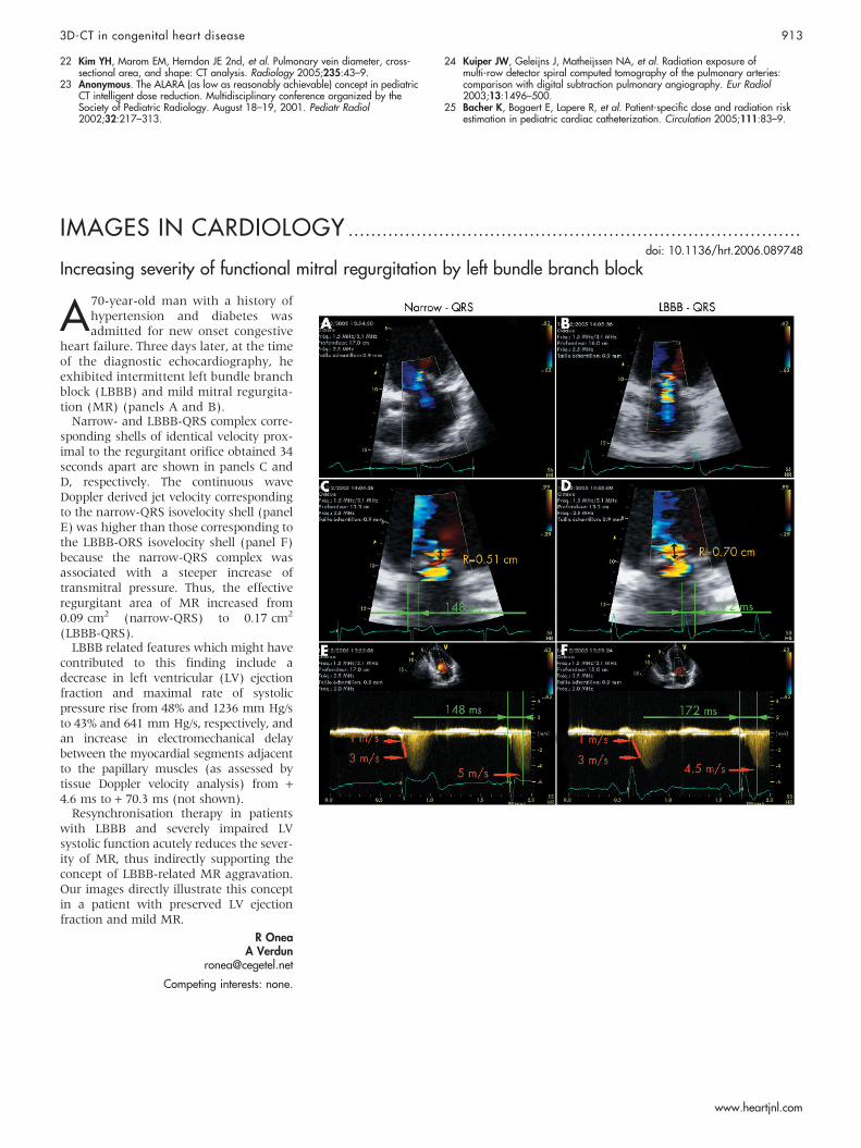
Increasing severity of functional mitral regurgitation by left bundle branch block
A70-year-old man with a history ofhypertension and diabetes wasadmitted for new onset congestive
heart failure. Three days later, at the timeof the diagnostic echocardiography, heexhibited intermittent left bundle branchblock (LBBB) and mild mitral regurgita-tion (MR) (panels A and B).
Narrow- and LBBB-QRS complex corre-sponding shells of identical velocity prox-imal to the regurgitant orifice obtained 34seconds apart are shown in panels C andD, respectively. The continuous waveDoppler derived jet velocity correspondingto the narrow-QRS isovelocity shell (panelE) was higher than those corresponding tothe LBBB-ORS isovelocity shell (panel F)because the narrow-QRS complex wasassociated with a steeper increase oftransmitral pressure. Thus, the effectiveregurgitant area of MR increased from0.09 cm2 (narrow-QRS) to 0.17 cm2
(LBBB-QRS).LBBB related features which might have
contributed to this finding include adecrease in left ventricular (LV) ejectionfraction and maximal rate of systolicpressure rise from 48% and 1236 mm Hg/sto 43% and 641 mm Hg/s, respectively, andan increase in electromechanical delaybetween the myocardial segments adjacentto the papillary muscles (as assessed bytissue Doppler velocity analysis) from +4.6 ms to + 70.3 ms (not shown).
Resynchronisation therapy in patientswith LBBB and severely impaired LVsystolic function acutely reduces the sever-ity of MR, thus indirectly supporting theconcept of LBBB-related MR aggravation.Our images directly illustrate this conceptin a patient with preserved LV ejectionfraction and mild MR.
R OneaA Verdun
Competing interests: none.
3D-CT in congenital heart disease 913
www.heartjnl.com