Thermophysiologic Aspects of the Three-Process-Model of Sleepiness Regulation
14
Thermophysiologic Aspects of the Three-Process-Model of Sleepiness Regulation Kurt Kr7uchi * , Christian Cajochen, PhD, Anna Wirz-Justice, PhD Psychiatric University Clinic, Centre for Chronobiology, Wilhelm Klein Strasse 27, CH-4025 Basel, Switzerland Sleepiness can be defined as a physiologic need for sleep and the behav- ioral measure of the subject’s tendency to fall asleep at a certain time (sleep propensity) [1,2]. Sleepiness, and its converse alertness, are regulated and im- portant determinants of vigilance and performance [3]. A better understanding of the mechanisms regulating sleepiness could lead to new strategies for sleepi- ness reduction and improved performance. Here we focus on the normal daily regulation of sleepiness and its relation to thermophysiologic processes. In the last century, Kleitman [4] proposed that body temperature represents the underlying mechanism regulating performance. The speed of thinking and performance depends on the level of metabolic processes in neurons in the cerebral cortex. Raising the speed of performance through an increase in core body temperature (CBT) causes indirect acceleration of thought processes. In studies that have manipulated CBT through external means (eg, altering ambient temperature, cold water immersion), cognitive function improved by increasing CBT slightly above the normal temperature of approximately 378C, whereas decreasing CBT below normal induced a decline in cognitive function [5–7]. In a forced desynchrony protocol, in which the contributions of cir- cadian phase and time awake can be assessed, a CBT increase of 0.158C was associated with increased subjective alertness and improved neurobehavioral performance [8]. Alertness and vigilance are a prerequisite for good perfor- mance. Thus, when alertness and vigilance are increased as a function of 0278-5919/05/$ – see front matter D 2005 Elsevier Inc. All rights reserved. doi:10.1016/j.csm.2004.12.009 sportsmed.theclinics.com * Corresponding author. E-mail address: [email protected] (K. Kr7uchi). Clin Sports Med 24 (2005) 287 – 300
-
Upload
independent -
Category
Documents
-
view
0 -
download
0
Transcript of Thermophysiologic Aspects of the Three-Process-Model of Sleepiness Regulation
Clin Sports Med 24 (2005) 287–300
Thermophysiologic Aspects of the
Three-Process-Model of Sleepiness Regulation
Kurt Kr7uchi*, Christian Cajochen, PhD,
Anna Wirz-Justice, PhD
Psychiatric University Clinic, Centre for Chronobiology, Wilhelm Klein Strasse 27,
CH-4025 Basel, Switzerland
Sleepiness can be defined as a physiologic need for sleep and the behav-
ioral measure of the subject’s tendency to fall asleep at a certain time (sleep
propensity) [1,2]. Sleepiness, and its converse alertness, are regulated and im-
portant determinants of vigilance and performance [3]. A better understanding
of the mechanisms regulating sleepiness could lead to new strategies for sleepi-
ness reduction and improved performance. Here we focus on the normal daily
regulation of sleepiness and its relation to thermophysiologic processes.
In the last century, Kleitman [4] proposed that body temperature represents
the underlying mechanism regulating performance. The speed of thinking and
performance depends on the level of metabolic processes in neurons in the
cerebral cortex. Raising the speed of performance through an increase in core
body temperature (CBT) causes indirect acceleration of thought processes.
In studies that have manipulated CBT through external means (eg, altering
ambient temperature, cold water immersion), cognitive function improved by
increasing CBT slightly above the normal temperature of approximately 378C,whereas decreasing CBT below normal induced a decline in cognitive function
[5–7]. In a forced desynchrony protocol, in which the contributions of cir-
cadian phase and time awake can be assessed, a CBT increase of 0.158C was
associated with increased subjective alertness and improved neurobehavioral
performance [8]. Alertness and vigilance are a prerequisite for good perfor-
mance. Thus, when alertness and vigilance are increased as a function of
0278-5919/05/$ – see front matter D 2005 Elsevier Inc. All rights reserved.
doi:10.1016/j.csm.2004.12.009 sportsmed.theclinics.com
* Corresponding author.
E-mail address: [email protected] (K. Kr7uchi).
krAuchi et al288
warmer CBT, performance is also indirectly improved. Thermoregulation is a
complex phenomenon and involves an intimate coupling between the central
(core) and peripheral (shell) body compartments that exchange body heat.
CBT and skin temperatures often change in parallel, depending on the location
where they are measured. This aspect, which is the focus of this article, has
not been sufficiently considered in studies dealing with the relationship between
thermophysiology, sleepiness, and sleep.
Circadian regulation of the core and the shell
A circadian rhythm of CBT was described in the middle of the nineteenth
century, when it was shown that oral temperature followed a daily rhythm that
included a maximum temperature in the early evening and a minimum in the
early morning hours, with a maximum–minimum range of 0.98C [9]. For a long
time, muscular activity (exercise) and digestive processes were considered the
most important factors for the generation of the CBT rhythm [10]. However,
Aschoff and his colleagues [11–13] systematically explored the underlying
causes and showed that the circadian rhythm of CBT is determined by changes
in heat production (measured by indirect calorimetry or indirectly by heart rate
[14]) and heat loss. They concluded that heat production undergoes a circa-
dian rhythm that is phase advanced with respect to the circadian rhythm of
heat loss (ie, when heat production surpasses heat loss, CBT increases) and that
the lag arises because of the body’s inertia and because transport of heat takes
time. That both heat production and heat loss are regulated results in a much finer
tuning of the CBT rhythm than if only one of these components was regulated.
Therefore, changes in CBT can only be explained by knowing the relation-
ship between heat production and heat loss. Under resting conditions, heat
production depends mainly on the metabolic activity of inner organs, such as the
liver, intestines, kidneys, the heart in the abdominal/thoracic cavity, and the
brain, which together produce about 70% of the entire resting metabolic rate
of the human body [13]. However, this heat is generated in only 8% of the
body mass, which is surrounded by a small proximal skin surface whose shape
is too flat for a good heat transfer to the environment. In other words, heat has
to be transferred from the core to more peripheral parts of the body (ie, the
extremities) with better heat transfer conditions [13]. These distal parts of the
body have ideal (round) surface shapes, which are good properties for heat
transfer to the environment. Under thermoneutral conditions, blood is the main
medium for transporting heat from the core to distal skin regions (convectively),
driven and distributed by the cardiovascular system. Therefore, in thermo-
physiologic terms, the human body consists of two compartments: the heat-
producing core, and the heat-loss regulating shell [13].
The core (especially the brain) is homeostatically regulated around a set
point of about 378C, whereas the shell is not. Shell temperature depends largely
on ambient temperature changes and can be considered poikilothermic, similar
sleepiness & thermophysiology 289
to the body of a lizard. From this point of view, humans have bodies that are
homeothermic and poikilothermic. In a hot environment the shell is small; in a
cold environment it is large, and thus acts as a buffer to protect the core from
dangerous cooling [13]. This regulation occurs very rapidly before CBT has
enough time to change. This so-called ‘‘feed-forward regulation’’ [15] with
respect to CBT is an important property of the thermophysiologic ‘‘core/shell’’
principle. Another feed-forward regulation serves the counter-current heat ex-
change in the extremities (ie, legs and arms). In a cold environment, venous
blood returns by way of inner blood vessels located near the arteries that pre-
warm the back-streaming blood, thereby efficiently protecting the core from
cooling out [16]. In contrast, in a warm environment the venous blood streams
back by way of outer veins near the skin surface, thereby enhancing additional
heat loss by way of the lower extremities [16]. Furthermore, it is known that
when shunts (ie, arteriovenous anastomoses [AVAs], exclusively found in distal
skin regions) between arterioles and venules are open, the blood streams back
also by way of these outer veins, thereby enhancing the heat-loss function of
opened AVAs [17]. Blood flows more rapidly through AVAs (about 10,000 times
more blood volume per second) than through capillary blood flow [17] directly
from arterioles to the dermal venous plexus, which enables an efficient heat
exchange. All these autonomically regulated mechanisms of shell size occur
through constriction or dilatation of blood vessels (arterioles and AVAs) in dis-
tal skin regions [17]. There is now substantial evidence indicating that homeo-
static control of CBT is mediated by a hierarchically organized set of neuronal
mechanisms, with the anterior hypothalamic-preoptic areas at the top of the
hierarchy [18]. In addition to the homeostatic principle, a rostral projection from
the circadian pacemaker (localized in the suprachiasmatic nuclei [SCN]) to the
preoptic areas serves the circadian modulation of CBT [19].
The mechanisms for changing shell size according to changing ambient
temperature also take place over 24 hours when the underlying endogenous
circadian CBT rhythm is regulated under constant ambient temperature. Distal
skin temperature rises in the evening, whereas heat production [14], proximal
skin temperature, and CBT decline; in the morning the inverse occurs [14], as is
described later. A crucial role for the circadian regulation of heat loss is played
by the nocturnally secreted pineal hormone melatonin. Melatonin selectively
augments distal skin blood flow, most likely through opening AVAs, either by
central or peripheral mechanisms (or both), while leaving proximal skin blood
flow and cerebral blood flow unaffected [20]. Until now, no direct evidence
exists for the existence of melatonin receptors (Mt1 or Mt2) in distal blood
vessels in humans. Melatonin initiates not only distal vasodilatation but also
sleepiness, acting therefore as the hormonal trigger between body heat loss and
induction of sleep in the evening (opening of the sleep gate) [21–25].
The following sections will discuss the relationship between thermophysi-
ology, sleepiness, and sleep, as elucidated in a series of studies performed under
constant routine conditions. This protocol provided the necessary controlled
environmental conditions to study the relationship between thermoregulation,
krAuchi et al290
sleepiness, and sleep [14,26] whereby external influences (‘‘masking effects’’)
are minimized (eg, constant room temperature, 228C; humidity, 60%; light, less
than 8 lux; constant bed rest in supine body position; no sleep allowed; food
and fluid intake in small isocaloric portions at hourly intervals).
Sleepiness in relation to skin temperatures and core body temperature
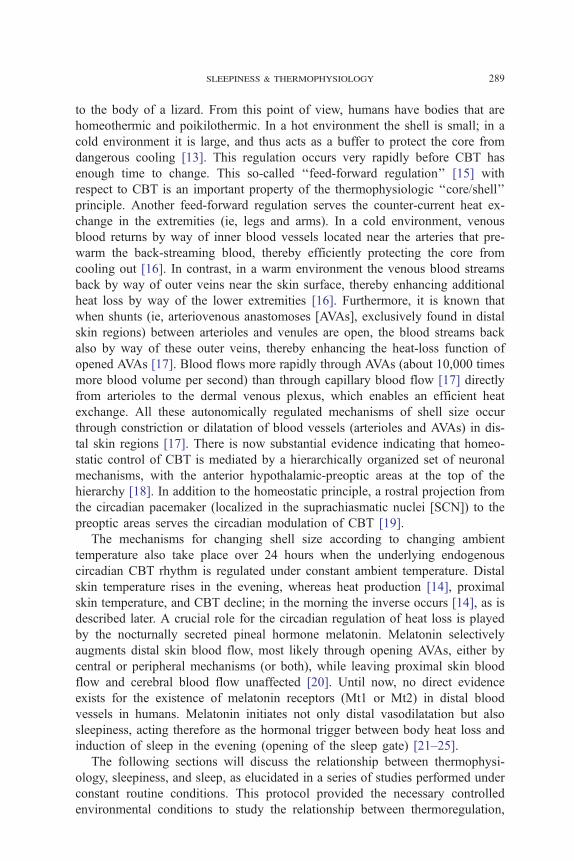
Three major processes are involved in the daily time course of sleepiness
(Fig. 1). First, there is a homeostatic increase of sleepiness (process H) depen-
dent on time spent awake which dissipates during sleep [27,28]. Neither the
central localization nor the neurobiologic mechanisms of the sleepiness/sleep
homeostat have been discovered. Second, a circadian process (C), driven by
the circadian pacemaker in the SCN, produces a maximal drive for sleepiness
during the subjective night in all diurnal species [27,28] and an alerting signal
during the subjective day. Third, a process of sleep inertia (I) describes the
H
C
IR
rela
ted
to th
erm
o-
regu
lato
ry c
hang
es
-R =
slee
pine
sssl
eepi
ness
slee
pine
ss
time (h)
2 2 2000
240
400
time (h)
elapsed time awake (h)
+
Fig. 1. Schematic illustration of the three-process model of sleepiness regulation. Process H represents
the homeostatic component of sleepiness which increases with time elapsed awake, regulated by a
sleep homeostat. H describes an exponential curve. The circadian component of sleepiness (C) is
driven by the SCN producing a waking signal (-) during the subjective day and a sleepiness signal (+)
during the subjective night. The sleep inertia process (I) describes a fast process that is active
immediately after sleep and disappears in the following 1 to 2 hours in an exponential manner (gray
area represents previous sleep duration). The exponential process R describes a relaxation-induced
sleepiness, which starts immediately after lights-off or lying down (gray area). -R represents the
inverse process of R occurring after lights-on or standing up (therefore, -R = I). Note: Processes C,
R, and -R (I) are coupled with thermophysiologic changes (distal vasodilatation), whereas H is
not coupled.
sleepiness & thermophysiology 291
phenomenon of low vigilance on awakening even though sleepiness should be
lowest at the end of a sleep episode [29,30]. The underlying neurobiologic and
physiologic mechanisms for sleep inertia are unknown. All three processes
have been mathematically described by the three-process model for sleepiness
[29,31], which is a further development of the two-process model of sleep
regulation [27,28].
The relationship between sleep and thermoregulation is tightly coupled to
the question: ‘‘Why do we sleep?’’ From a thermophysiologic point of view, the
proposed answer has been: ‘‘We sleep to conserve energy.’’ This answer was
derived from the observation that all living organisms, whether nocturnal or
diurnal, sleep or rest when their metabolism (heat production) is low. However,
this statement does not seem to be true, at least not for humans. In the past, one
important observation had not been taken into account: not only did heat pro-
duction decline in the evening, but also changes in heat loss occur via accel-
erated distal skin blood flow. Thus, the relationship between thermophysiology
and sleep is rather the opposite: we sleep at times when distal skin blood flow
starts to increase. The following summary of our studies concerning thermo-
physiology, sleepiness, and sleep deals with the question: ‘‘How are the three
processes involved in sleep regulation related to thermophysiologic changes?’’
Circadian and homeostatic aspects of sleepiness and its relation to changes in
body temperatures
Numerous studies have indicated that the circadian profile of subjective
and objective sleepiness is a mirror image of the endogenous CBT rhythm, with
maximum sleepiness occurring around the CBT minimum [7]. From a homeo-
static perspective, sleepiness should increase steadily with increased elapsed
time awake; however, this is not the case. In the evening humans are often in a
productive and alert state, especially evening chronotypes. From a homeostatic
point of view this is a paradoxical phenomenon, explained by the fact that
the circadian drive for alertness from the SCN is at its maximum right before
humans usually fall asleep, thereby counteracting the homeostatic drive for sleep
in the evening [32–34]. This so-called ‘‘wake maintenance zone’’ (or ‘‘forbidden
zone to sleep’’) occurs at the circadian phase where the inner heat conductance
is at its minimum: distal vasoconstriction is high in spite of the elevated CBT.
Thus, the sleepiness/sleep homeostat interacts with the circadian clock in an
additive or nonadditive way (which is still a matter of debate [35]), and deter-
mines the actual state of an individual’s sleepiness. Usually humans choose their
bedtimes shortly after the SCN-alerting signal has declined and the ‘‘sleep gate’’
has been opened [25], which occurs at the time of the maximum decline rate
of CBT and the maximum increase rate of body heat loss [24].
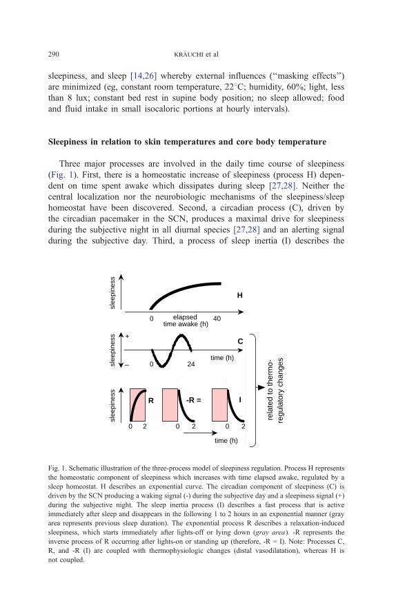
Fig. 2 shows the results of a constant routine study illustrating circadian
and homeostatic aspects of sleepiness together with the circadian patterns of
CBT, distal, and proximal skin temperatures [14]. To illustrate the homeostatic
component of sleepiness, an exponential curve (H) has been added to the
slee
pine
ss (
mm
VA
S)
skin
tem
pera
ture
(°C
)co
re b
ody
tem
pera
ture
(°C
)
time of day (hr)
36.5
36.6
36.7
36.8
36.9
37.0
30
50
70
90
32
33
34
35
H
proximal
distal
11 1717 23 5 11
C
H+C
Fig. 2. Mean time course of CBT (rectal) and skin temperatures are shown in addition to subjective
ratings of sleepiness (100 mm visual analog scale [VAS]). Seven men were studied in a constant
routine protocol for 35 hours (sleep not allowed). Proximal indicates weighted mean of following
skin regions: infraclavicular, stomach, forehead, and thigh, and distal indicates mean of hands and
feet. The vertical bar at the right of the curves indicates plus and minus averaged SEM of all time
points. To emphasize the homeostatic rise of sleepiness, an exponential curve (thick line H) has been
added. The residuals (gray areas) indicate the circadian component C. Vertical arrows indicate cir-
cadian phase positions of the parameters derived from the mean of up- and downwards midrange
crossing values. Note: The circadian rhythm of sleepiness is in phase with CBT; both are phase
delayed compared with distal skin temperature. (Data from Kr7uchi K, Wirz-Justice A. Circadian
rhythm of heat production, heart rate, and skin and core body temperature under unmasking condi-
tions in men. Am J Physiol 1994;267:R819–26.)
krAuchi et al292
original subjective sleepiness rating (H + C). The residuals (C) describe the
underlying circadian component of sleepiness (gray areas). The circadian
regulation of sleepiness is in phase with CBT and phase delayed with respect to
distal skin temperature, confirming other studies [36]. The fact that sleepiness is
phase delayed relative to distal skin temperature explains the finding that
increased distal skin temperature before nocturnal sleep is a better predictor for
sleepiness & thermophysiology 293
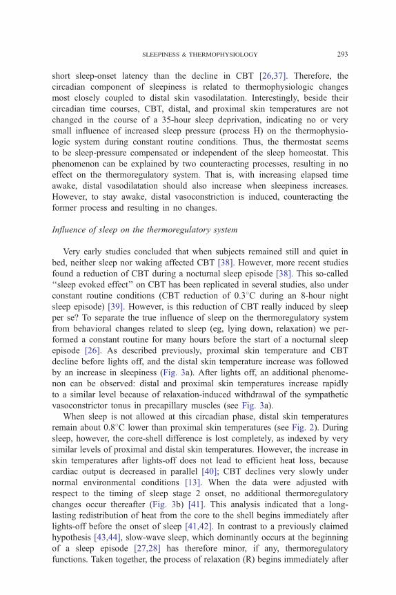
short sleep-onset latency than the decline in CBT [26,37]. Therefore, the
circadian component of sleepiness is related to thermophysiologic changes
most closely coupled to distal skin vasodilatation. Interestingly, beside their
circadian time courses, CBT, distal, and proximal skin temperatures are not
changed in the course of a 35-hour sleep deprivation, indicating no or very
small influence of increased sleep pressure (process H) on the thermophysio-
logic system during constant routine conditions. Thus, the thermostat seems
to be sleep-pressure compensated or independent of the sleep homeostat. This
phenomenon can be explained by two counteracting processes, resulting in no
effect on the thermoregulatory system. That is, with increasing elapsed time
awake, distal vasodilatation should also increase when sleepiness increases.
However, to stay awake, distal vasoconstriction is induced, counteracting the
former process and resulting in no changes.
Influence of sleep on the thermoregulatory system
Very early studies concluded that when subjects remained still and quiet in
bed, neither sleep nor waking affected CBT [38]. However, more recent studies
found a reduction of CBT during a nocturnal sleep episode [38]. This so-called
‘‘sleep evoked effect’’ on CBT has been replicated in several studies, also under
constant routine conditions (CBT reduction of 0.38C during an 8-hour night
sleep episode) [39]. However, is this reduction of CBT really induced by sleep
per se? To separate the true influence of sleep on the thermoregulatory system
from behavioral changes related to sleep (eg, lying down, relaxation) we per-
formed a constant routine for many hours before the start of a nocturnal sleep
episode [26]. As described previously, proximal skin temperature and CBT
decline before lights off, and the distal skin temperature increase was followed
by an increase in sleepiness (Fig. 3a). After lights off, an additional phenome-
non can be observed: distal and proximal skin temperatures increase rapidly
to a similar level because of relaxation-induced withdrawal of the sympathetic
vasoconstrictor tonus in precapillary muscles (see Fig. 3a).
When sleep is not allowed at this circadian phase, distal skin temperatures
remain about 0.88C lower than proximal skin temperatures (see Fig. 2). During
sleep, however, the core-shell difference is lost completely, as indexed by very
similar levels of proximal and distal skin temperatures. However, the increase in
skin temperatures after lights-off does not lead to efficient heat loss, because
cardiac output is decreased in parallel [40]; CBT declines very slowly under
normal environmental conditions [13]. When the data were adjusted with
respect to the timing of sleep stage 2 onset, no additional thermoregulatory
changes occur thereafter (Fig. 3b) [41]. This analysis indicated that a long-
lasting redistribution of heat from the core to the shell begins immediately after
lights-off before the onset of sleep [41,42]. In contrast to a previously claimed
hypothesis [43,44], slow-wave sleep, which dominantly occurs at the beginning
of a sleep episode [27,28] has therefore minor, if any, thermoregulatory
functions. Taken together, the process of relaxation (R) begins immediately after
3.5
4.0
4.5
5.0
5.5
6.0
slee
pine
ss (
KS
S u
nits
)
24222018 2 4 6
36.1
36.3
36.5
36.7
36.9
37.1
lights off CR co
re b
ody
tem
pera
ture
(°C
)
distal
skin
tem
pera
ture
(°C
)
32.0
32.5
33.0
33.5
34.0
34.5
proximal
time of day (hr)
a b
skin
tem
pera
ture
(°C
)
36.4
36.6
36.8
37.0
CB
T (
°C)
-120 -60 0 +60minutes after
sleep stage 2 onset
+120
33.0
33.4
34.2
33.8
32.6
proximal
distal
R
begin of sleep stage 2
Fig. 3. (a) Mean curves (F SEM; N = 18 men) of CBT, skin temperatures, and subjective ratings
of sleepiness before and during nocturnal sleep aligned to lights-off. Note: Distal and proximal
skin temperatures show an inverse time course before lights-off, but increase in parallel to a similar
level thereafter. (Data from Krauchi K, Cajochen C, Werth E, Wirz-Justice A. Functional link between
distal vasodilation and sleep-onset latency? Am J Physiol 2000;278:R741–8.) (b) Mean time courses
of CBT and skin temperatures (N = 36 men), adjusted to onset of sleep stage 2. Note: skin tem-
peratures have already increased before sleep stage 2 onset, indicating a relaxation-induced effect (R).
KSS, Karolinska sleepiness scale. (Data from Krauchi K, Cajochen C, Werth E, et al. Thermo-
regulatory changes begin after lights off and not after onset of sleep stages 2. Sleep 2001;24:165–6.)
krAuchi et al294
lights-off before sleep starts; it is not sleep that induces thermoregulatory
changes but rather the relaxation process per se. Because sleep is a very relaxed
state, especially deep sleep, a complete loss of the core/shell principle occurs
at this time. Therefore, a logical question is: ‘‘What happens to the core/shell
principle after waking up from a sleep episode?’’
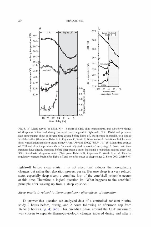
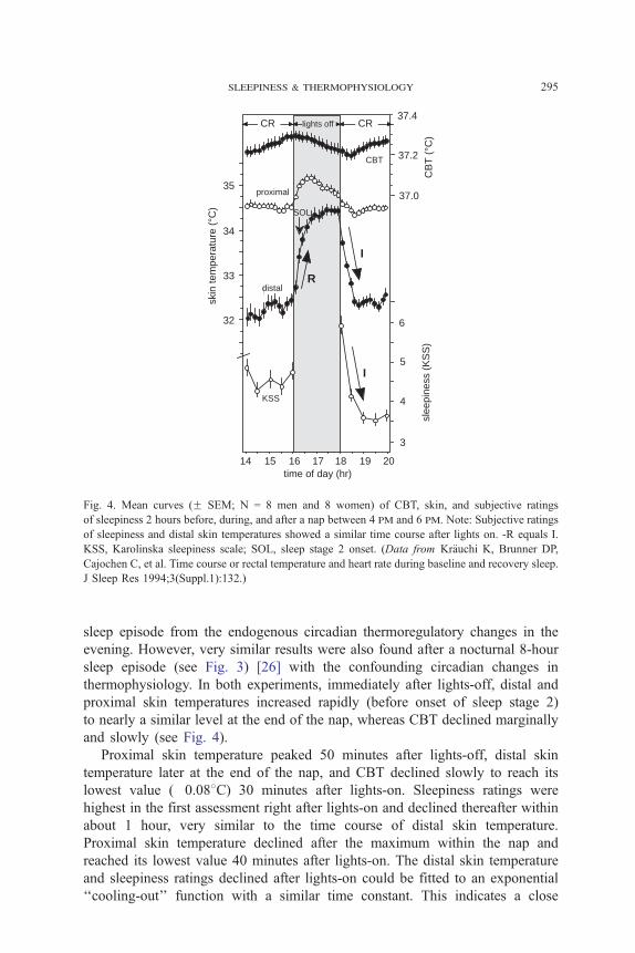
Sleep inertia is related to thermoregulatory after-effects of relaxation
To answer that question we analyzed data of a controlled constant routine
study 2 hours before, during, and 2 hours following an afternoon nap from
16 to18 hours (Fig. 4) [45]. This circadian phase around the CBT maximum
was chosen to separate thermophysiologic changes induced during and after a
time of day (hr)14 15 1716 18 19 20
3
slee
pine
ss (
KS
S)
distal
proximal
CBT
37.0
37.2
CB
T (
°C)
KSS 4
5
6
I
R
I
SOLsk
in te
mpe
ratu
re (
°C)
37.4
35
34
33
32
CR CR lights off
Fig. 4. Mean curves (F SEM; N = 8 men and 8 women) of CBT, skin, and subjective ratings
of sleepiness 2 hours before, during, and after a nap between 4 pm and 6 pm. Note: Subjective ratings
of sleepiness and distal skin temperatures showed a similar time course after lights on. -R equals I.
KSS, Karolinska sleepiness scale; SOL, sleep stage 2 onset. (Data from Kr7uchi K, Brunner DP,Cajochen C, et al. Time course or rectal temperature and heart rate during baseline and recovery sleep.
J Sleep Res 1994;3(Suppl.1):132.)
sleepiness & thermophysiology 295
sleep episode from the endogenous circadian thermoregulatory changes in the
evening. However, very similar results were also found after a nocturnal 8-hour
sleep episode (see Fig. 3) [26] with the confounding circadian changes in
thermophysiology. In both experiments, immediately after lights-off, distal and
proximal skin temperatures increased rapidly (before onset of sleep stage 2)
to nearly a similar level at the end of the nap, whereas CBT declined marginally
and slowly (see Fig. 4).
Proximal skin temperature peaked 50 minutes after lights-off, distal skin
temperature later at the end of the nap, and CBT declined slowly to reach its
lowest value (�0.088C) 30 minutes after lights-on. Sleepiness ratings were
highest in the first assessment right after lights-on and declined thereafter within
about 1 hour, very similar to the time course of distal skin temperature.
Proximal skin temperature declined after the maximum within the nap and
reached its lowest value 40 minutes after lights-on. The distal skin temperature
and sleepiness ratings declined after lights-on could be fitted to an exponential
‘‘cooling-out’’ function with a similar time constant. This indicates a close
krAuchi et al296
temporal association between subjective sleepiness ratings and distal vaso-
dilatation after lights-on. Sleep inertia on waking showed a similar rate of
dissipation to the cooling-out rate of the extremities, which had been warmed up
by the redistribution of blood during the dark period. Thus, as the readiness to
fall asleep is correlated with distal vasodilatation, so is the waking-up process
(or disappearance of sleep inertia) correlated with distal vasoconstriction. Taken
together, the symmetry between the thermoregulatory processes initiating sleepi-
ness [26,37] and those dissipating it is striking and provide a physiologic
rationale for a ‘‘power nap’’ being short: redistribution of blood to the extremi-
ties is incomplete after less than 20 minutes of scheduled sleep or relaxation.
This may lead to less distal vasodilatation on wake-up and less sleep inertia.
A simple test of our hypothesis would be cold water applied directly to the
extremities on waking, which should rapidly increase distal vasoconstriction
and, in turn, alertness. In the following section the simple behavior of lying
down to go to sleep will be revisited from a thermophysiologic point of view.
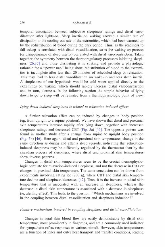
Lying down-induced sleepiness is related to relaxation-induced effects
A further relaxation effect can be induced by changes in body position
(eg, from upright to a supine position). We have shown that distal and proximal
skin temperatures increase rapidly after lying down, together with increased
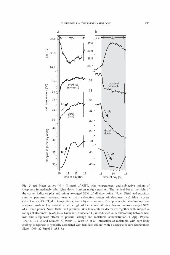
sleepiness ratings and decreased CBT (Fig. 5a) [46]. The opposite pattern was
found in another study after a change from supine to upright body position
(Fig. 5b) [46]. Here again, distal and proximal skin temperatures change in the
same direction as during and after a sleep episode, indicating that relaxation-
induced sleepiness may be differently regulated by the thermostat than by the
circadian process of sleepiness, where distal and proximal skin temperatures
show inverse patterns.
Changes in distal skin temperatures seem to be the crucial thermophysio-
logic correlate for relaxation-induced sleepiness, and not the decrease in CBT or
changes in proximal skin temperature. The same conclusion can be drawn from
experiments involving eating ice (200 g), where CBT and distal skin tempera-
ture decline and sleepiness decreases [47]. Thus, it is the increase in distal skin
temperature that is associated with an increase in sleepiness, whereas the
decrease in distal skin temperature is associated with a decrease in sleepiness
(ie, alerting effect). This leads to the question: ‘‘Which mechanisms are involved
in the coupling between distal vasodilatation and sleepiness induction?’’
Putative mechanisms involved in coupling sleepiness and distal vasodilatation
Changes in acral skin blood flow are easily demonstrable by distal skin
temperature, most prominently in fingertips, and are a commonly used indicator
for sympathetic reflex responses to various stimuli. However, skin temperatures
are a function of inner and outer heat transport and transfer conditions, leading
time of day (hr)15
time of day (hr)1310 11 12
45
30
35
40
30
31
32
CB
T (°
C)
34
35proximal
(stomach)
distal (foot)
13 14
40
45
28
29
30
31
33
34
32
36.7
36.8
36.9
37.0
proximal (stomach)
R
R
-R
-R
skin
tem
pera
ture
(°C
)sl
eepi
ness
(ar
bitr
ary
units
)
36.5
36.6
36.4
a b
distal (foot)
Fig. 5. (a) Mean curves (N = 8 men) of CBT, skin temperatures, and subjective ratings of
sleepiness immediately after lying down from an upright position. The vertical bar at the right of
the curves indicates plus and minus averaged SEM of all time points. Note: Distal and proximal
skin temperatures increased together with subjective ratings of sleepiness. (b) Mean curves
(N = 9 men) of CBT, skin temperatures, and subjective ratings of sleepiness after standing up from
a supine position. The vertical bar at the right of the curves indicates plus and minus averaged SEM
of all time points. Note: Distal and proximal skin temperatures decreased together with subjective
ratings of sleepiness. (Data from Kr7uchi K, Cajochen C, Wirz-Justice A. A relationship between heat
loss and sleepiness: effects of postural change and melatonin administration. J Appl Physiol
1997;83:134–9; and Kr7uchi K, Werth E, Wqst D, et al. Interaction of melatonin with core body
cooling: sleepiness is primarily associated with heat loss and not with a decrease in core temperature.
Sleep 1999; 22(Suppl 1):285–6.)
sleepiness & thermophysiology 297
krAuchi et al298
to a nonlinear relationship between skin blood flow and skin temperature; only
a certain range of blood flow skin temperatures can be used as a good indi-
cator for skin blood flow [13]. In many studies, the relationship between distal
skin blood flow and sleepiness has been challenged by diverse interventions
[48]. For example, distal skin temperature can be strongly affected during
hypnosis, when images used for suggesting cold and warmth include expe-
riences of physical temperature and physiologic stress or relaxation [49]. Fur-
thermore, thermal biofeedback training in raising hand temperature increases not
only finger-skin temperature [50] but also promotes a rapid sleep onset [51].
Therefore, sympathetic nerve activity seems to be the most important mecha-
nism determining blood flow through arterioles by way of adrenergic constric-
tor nerves, and is also a good indicator for the arousal system. There is now
emerging evidence from physiologic and neuroanatomic studies to indicate that
changes in body temperatures may trigger somnogenic brain areas (eg, medial
preoptic area [52], ventrolateral preoptic area [53]) to initiate sleep, either
indirectly through nerve afferents activated by cold and warm receptors located
in the dermis and in the core, or directly through changes in core blood tem-
perature leading to changed spinal cord and brain temperatures [54]. However,
the interrelationship between thermoregulatory and sleepiness/sleep regulatory
mechanisms is rather complex. Recent studies have indicated that the medial
preoptic area controls sleep and temperature through independent, but over-
lapping, neuronal circuits [53,55,56]. However, the disadvantage of these animal
studies is that the behavior of an animal cannot be independently controlled
for sleep, which is crucial to separate the influence of the three components of
sleepiness regulation (C, H, and I). There is good reason to believe that future
studies in humans using imaging techniques (eg, functional MRI) will shed
more light on how thermoregulatory and sleepiness/sleep regulatory mecha-
nisms are related in the human brain.
Acknowledgments
The studies reviewed here were supported by SNF Grants #32-4245.94
and #31-49254.96, and the Kneipp Stiftung Wqrzburg, Germany.
References
[1] Johns MW. A new method for measuring daytime sleepiness: the Epworth sleepiness scale.
Sleep 1991;14(6):540–5.
[2] Akerstedt T, Gillberg M. Subjective and objective sleepiness in the active individual. Intern
J Neurosci 1990;52:29–37.
[3] Folkard S, Akerstedt T. Trends in the risk of accidents and injuries and their implications
for models of fatigue and performance. Aviat Space Environ Med 2004;75(3 Suppl):A161–7.
[4] Kleitman N. Sleep and Wakefulness. Chicago7 The University of Chicago Press; 1987.
sleepiness & thermophysiology 299
[5] Coleshaw SR, van Someren RN, Wolff AH, et al. Impaired memory registration and speed
of reasoning caused by low body temperature. J Appl Physiol 1983;55:27–31.
[6] Giesbrecht GG, Arnett JL, Vela E, et al. Effect of task complexity on mental performance
during immersion hypothermia. Aviat Space Environ Med 1993;64:206–11.
[7] Johnson MP, Duffy JF, Dijk DJ, et al. Short-term memory, alertness and performance:
a reappraisal of their relationship to body temperature. J Sleep Res 1992;1:24–9.
[8] Wright Jr KP, Hull JT, Czeisler CA. Relationship between alertness, performance, and body
temperature in humans. Am J Physiol Regul Integr Comp Physiol 2002;283:R1370–7.
[9] Gierse A. Quaeniam sit ratio caloris organici [MD Thesis] [What is the cause of organic heat?].
Halle; 1842 [in German].
[10] Hardy JD. Physiology of temperature regulation. Physiol Rev 1961;41:521–606.
[11] Aschoff J. The circadian rhythm of body temperature as a function of body size. In: Taylor R,
Johanson K, Bolis L, editors. A Comparison to animal physiology. Cambridge, England7 Cam-
bridge Univ Press; 1982. p. 173–89.
[12] Aschoff J, Heise A. Thermal conductance in man: its dependence on time of day and of
ambient temperature. In: Itoh S, Ogata K, Yoshimura H, editors. Advances in climatic physi-
ology. Tokyo7 Igako Shoin; 1972. p. 334–48.
[13] Aschoff J. Temperaturregulation. In: Gauer OH, Kramer K, Jung R, editors. Energiehaushalt und
Temperaturregulation [Energy budget and temperature regulation]. Physiologie des Menschen.
Munchen7 Urban & Schwarzenberg; 1971. p. 43–112 [in German].
[14] Krauchi K, Wirz-Justice A. Circadian rhythm of heat production, heart rate, and skin and core
body temperature under unmasking conditions in men. Am J Physiol 1994;267:R819–26.
[15] Mrosovsky N. Rheostasis. The physiology of change. New York7 Oxford University Press; 1990.
[16] Aschoff J, Wever R. Kern und Schale im Warmehaushalt des Menschen [Core and shell in
human energy budget]. Naturwissenschaften 1958;45:477–85 [in German].
[17] Hales JRS. Skin arteriovenous anastomoses, their control and role in thermoregulation.
In: Johansen K, Burggren WW, editors. Cardiovascular shunts. Alfred Benzon Symposium 21.
Copenhagen, Denmark7 Munksgaard; 1985. p. 433–51.
[18] Satinoff E. Neural organization and evolution of thermal regulation in mammals. Science
1978;201:16–22.
[19] Moore RY, Danchenko RL. Paraventricular-subparaventricular hypothalamic lesions selectively
affect circadian function. Chronobiol Int 2002;19:345–60.
[20] van der Helm - Van Mil AH, van Someren EJ, van den Boom R, et al. No influence of mela-
tonin on cerebral blood flow in humans. J Clin Endocrinol Metabol 2003;88:5989–94.
[21] Cajochen C, Kr7uchi K, von Arx MA, et al. Daytime melatonin administration enhances
sleepiness and theta/alpha activity in the waking EEG. Neurosci Lett 1996;207:209–13.
[22] Cagnacci A, Kr7uchi K, Wirz-Justice A, et al. Homeostatic versus circadian effects of
melatonin on core body temperature in humans. J Biol Rhythms 1997;12:509–17.
[23] Cajochen C, Kr7uchi K, Wirz-Justice A. Role of melatonin in the regulation of human
circadian rhythms and sleep. J Neuroendocrinol 2003;15:432–7.
[24] Campbell SS, Broughton RJ. Rapid decline in body temperature before sleep: fluffing the
physiological pillow? Chronobiol Int 1994;11:126–31.
[25] Lavie P. Melatonin: role in gating nocturnal rise in sleep propensity. J Biol Rhythms 1997;
12(6):657–65.
[26] Kr7uchi K, Cajochen C, Werth E, et al. Functional link between distal vasodilation and
sleep-onset latency? Am J Physiol Regul Integr Comp Physiol 2000;278:R741–8.
[27] Daan S, Beersma DG, Borbely AA. Timing of human sleep: recovery process gated by
a circadian pacemaker. Am J Physiol 1984;246:R161–83.
[28] Borbely AA. A two-process model of sleep regulation. Hum Neurobiol 1982;1:195–204.
[29] Akerstedt T, Folkard S. A model of human sleepiness. In: Horne J, editor. Sleep ’90. Bochum,
Germany7 Pontenagel Press; 1990. p. 310–3.
[30] Dinges DF. Are you awake? Cognitive performance and reverie during the hypnotic state.
In: Bootzin R, Kihlstrom J, Schachter D, editors. Are you awake? Washington, DC7 Ameri-
can Psychological Association; 1990. p. 159–75.
krAuchi et al300
[31] Folkard S, Akerstedt T. A three-process model of the regulation of alertness-sleepiness.
In: Broughton RJ, Ogilvie RD, editors. Sleep, arousal, and performance. Boston7 Birkh7user;1992. p. 11–26.
[32] Dijk DJ, Duffy JF, Czeisler CA. Circadian and sleep/wake dependent aspects of subjective
alertness and cognitive performance. J Sleep Res 1992;1:112–7.
[33] Ebbecke U. Schqttelfrost in K7lte, Fieber und Affekt. Klin Wochenschr 1948;39/
40(15.Okt.):609–13.
[34] Edgar DM, Dement WC, Fuller CA. Effect of SCN lesions on sleep in squirrel monkeys:
evidence for opponent processes in sleep-wake regulation. J Neurosci 1993;13(3):1065–79.
[35] Achermann P, Borbely AA. Mathematical models of sleep regulation. Front Biosci
2003;8:s683–93.
[36] Gradisar M, Lack L. Relationships between the circadian rhythms of finger temperature,
core temperature, sleep latency, and subjective sleepiness. J Biol Rhythms 2004;19(2):157–63.
[37] Kr7uchi K, Cajochen C, Werth E, et al. Warm feet promote the rapid onset of sleep. Nature
1999;401:36–7.
[38] Aschoff J. Circadian control of body temperature. J Therm Biol 1983;8:143–7.
[39] Barrett J, Lack L, Morris M. The sleep-evoked decrease of body temperature. Sleep 1993;16:
93–9.
[40] Somers VK, Dyken ME, Mark AL, et al. Sympathetic-nerve activity during sleep in normal
subjects. N Engl J Med 1993;328(5):303–7.
[41] Kr7uchi K, Cajochen C, Werth E, et al. Thermoregulatory changes begin after lights off
and not after onset of sleep stage 2. Sleep 2001;24(Abstr Suppl):165–6.
[42] Lack L, Gradisar M. Acute finger temperature changes preceding sleep onsets over a 45-h period.
J Sleep Res 2002;11(4):275–82.
[43] Sewitch DE. Slow wave sleep deficiency insomnia: a problem in thermo-downregulation
at sleep onset. Psychophysiol 1987;24(2):200–15.
[44] McGinty D, Szymusiak R. Keeping cool: a hypothesis about the mechanisms and functions
of slow-wave sleep. Trends Neurosci 1990;13(12):480–7.
[45] Kr7uchi K, Cajochen C, Wirz-Justice A. Waking up properly: is there a role of thermoregu-
lation in sleep inertia? J Sleep Res 2004;13:121–7.
[46] Kr7uchi K, Cajochen C, Wirz-Justice A. A relationship between heat loss and sleepiness:
effects of postural change and melatonin administration. J Appl Physiol 1997;83:134–9.
[47] Kr7uchi K, Werth E, Wqst D, et al. Interaction of melatonin with core body cooling: sleepiness
is primarily associated with heat loss and not with a decrease in core temperature. Sleep
1999;22(Suppl 1):285–6.
[48] van Someren EJW. Sleep propensity is modulated by circadian and behavior-induced changes
in cutaneous temperature. J Therm Biol 2004;29:437–44.
[49] Kistler A, Mariauzouls C, Wyler F, et al. Autonomic responses to suggestions for cold
and warmth in hypnosis. Forsch Komplement7rmed 1999;6:10–4.
[50] Freedman RR, Morris M, Norton DA, et al. Physiological mechanism of digital vaso-
constriction training. Biofeedback Self Regul 1988;13(4):299–305.
[51] Lushington K, Greeneklee H, Veltmeyer M, et al. Biofeedback training in hand temperature
raising promotes sleep onset in young normals. J Sleep Res 2004;13(Suppl 1):460.
[52] Szymusiak R, Steininger T, Alam N, et al. Preoptic area sleep-regulating mechanisms. Arch Ital
Biol 2001;139(1–2):77–92.
[53] Saper CB. The central autonomic nervous system: conscious visceral perception and auto-
nomic pattern generation. Annu Rev Neurosci 2002;25:433–69.
[54] van Someren EJ. More than a marker: interaction between circadian regulation of temperature
and sleep, age-related changes, and treatment possibilities. Chronobiol Int 2000;17:313–54.
[55] van Someren EJ, Raymann RJ, Scherder EJ, et al. Circadian and age-related modulation
of thermoreception and temperature regulation: mechanisms and functional implications.
Ageing Res Rev 2002;1:721–78.
[56] Kumar VM. Body temperature and sleep: Are they controlled by the same mechanism? Sleep
Biol Rhythms 2004;2:103–24.