The utility of the minipig as an animal model in regulatory toxicology
25
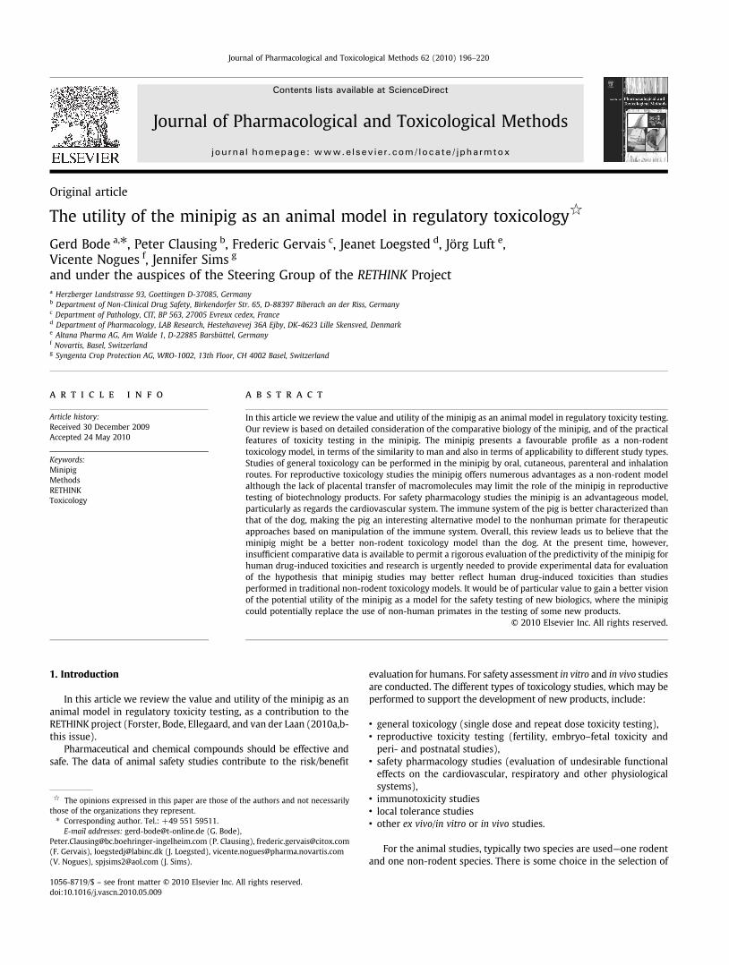
Original article The utility of the minipig as an animal model in regulatory toxicology ☆ Gerd Bode a, ⁎, Peter Clausing b , Frederic Gervais c , Jeanet Loegsted d , Jörg Luft e , Vicente Nogues f , Jennifer Sims g and under the auspices of the Steering Group of the RETHINK Project a Herzberger Landstrasse 93, Goettingen D-37085, Germany b Department of Non-Clinical Drug Safety, Birkendorfer Str. 65, D-88397 Biberach an der Riss, Germany c Department of Pathology, CIT, BP 563, 27005 Evreux cedex, France d Department of Pharmacology, LAB Research, Hestehavevej 36A Ejby, DK-4623 Lille Skensved, Denmark e Altana Pharma AG, Am Walde 1, D-22885 Barsbüttel, Germany f Novartis, Basel, Switzerland g Syngenta Crop Protection AG, WRO-1002, 13th Floor, CH 4002 Basel, Switzerland abstract article info Article history: Received 30 December 2009 Accepted 24 May 2010 Keywords: Minipig Methods RETHINK Toxicology In this article we review the value and utility of the minipig as an animal model in regulatory toxicity testing. Our review is based on detailed consideration of the comparative biology of the minipig, and of the practical features of toxicity testing in the minipig. The minipig presents a favourable profile as a non-rodent toxicology model, in terms of the similarity to man and also in terms of applicability to different study types. Studies of general toxicology can be performed in the minipig by oral, cutaneous, parenteral and inhalation routes. For reproductive toxicology studies the minipig offers numerous advantages as a non-rodent model although the lack of placental transfer of macromolecules may limit the role of the minipig in reproductive testing of biotechnology products. For safety pharmacology studies the minipig is an advantageous model, particularly as regards the cardiovascular system. The immune system of the pig is better characterized than that of the dog, making the pig an interesting alternative model to the nonhuman primate for therapeutic approaches based on manipulation of the immune system. Overall, this review leads us to believe that the minipig might be a better non-rodent toxicology model than the dog. At the present time, however, insufficient comparative data is available to permit a rigorous evaluation of the predictivity of the minipig for human drug-induced toxicities and research is urgently needed to provide experimental data for evaluation of the hypothesis that minipig studies may better reflect human drug-induced toxicities than studies performed in traditional non-rodent toxicology models. It would be of particular value to gain a better vision of the potential utility of the minipig as a model for the safety testing of new biologics, where the minipig could potentially replace the use of non-human primates in the testing of some new products. © 2010 Elsevier Inc. All rights reserved. 1. Introduction In this article we review the value and utility of the minipig as an animal model in regulatory toxicity testing, as a contribution to the RETHINK project (Forster, Bode, Ellegaard, and van der Laan (2010a,b- this issue). Pharmaceutical and chemical compounds should be effective and safe. The data of animal safety studies contribute to the risk/benefit evaluation for humans. For safety assessment in vitro and in vivo studies are conducted. The different types of toxicology studies, which may be performed to support the development of new products, include: • general toxicology (single dose and repeat dose toxicity testing), • reproductive toxicity testing (fertility, embryo–fetal toxicity and peri- and postnatal studies), • safety pharmacology studies (evaluation of undesirable functional effects on the cardiovascular, respiratory and other physiological systems), • immunotoxicity studies • local tolerance studies • other ex vivo/in vitro or in vivo studies. For the animal studies, typically two species are used—one rodent and one non-rodent species. There is some choice in the selection of Journal of Pharmacological and Toxicological Methods 62 (2010) 196–220 ☆ The opinions expressed in this paper are those of the authors and not necessarily those of the organizations they represent. ⁎ Corresponding author. Tel.: +49 551 59511. E-mail addresses: [email protected] (G. Bode), [email protected] (P. Clausing), [email protected] (F. Gervais), [email protected] (J. Loegsted), [email protected] (V. Nogues), [email protected] (J. Sims). 1056-8719/$ – see front matter © 2010 Elsevier Inc. All rights reserved. doi:10.1016/j.vascn.2010.05.009 Contents lists available at ScienceDirect Journal of Pharmacological and Toxicological Methods journal homepage: www.elsevier.com/locate/jpharmtox
-
Upload
independent -
Category
Documents
-
view
0 -
download
0
Transcript of The utility of the minipig as an animal model in regulatory toxicology
Journal of Pharmacological and Toxicological Methods 62 (2010) 196–220
Contents lists available at ScienceDirect
Journal of Pharmacological and Toxicological Methods
j ourna l homepage: www.e lsev ie r.com/ locate / jpharmtox
Original article
The utility of the minipig as an animal model in regulatory toxicology☆
Gerd Bode a,⁎, Peter Clausing b, Frederic Gervais c, Jeanet Loegsted d, Jörg Luft e,Vicente Nogues f, Jennifer Sims g
and under the auspices of the Steering Group of the RETHINK Projecta Herzberger Landstrasse 93, Goettingen D-37085, Germanyb Department of Non-Clinical Drug Safety, Birkendorfer Str. 65, D-88397 Biberach an der Riss, Germanyc Department of Pathology, CIT, BP 563, 27005 Evreux cedex, Franced Department of Pharmacology, LAB Research, Hestehavevej 36A Ejby, DK-4623 Lille Skensved, Denmarke Altana Pharma AG, Am Walde 1, D-22885 Barsbüttel, Germanyf Novartis, Basel, Switzerlandg Syngenta Crop Protection AG, WRO-1002, 13th Floor, CH 4002 Basel, Switzerland
☆ The opinions expressed in this paper are those of tthose of the organizations they represent.⁎ Corresponding author. Tel.: +49 551 59511.
E-mail addresses: [email protected] (G. Bode),[email protected] (P. Clausin(F. Gervais), [email protected] (J. Loegsted), vicente.n(V. Nogues), [email protected] (J. Sims).
1056-8719/$ – see front matter © 2010 Elsevier Inc. Aldoi:10.1016/j.vascn.2010.05.009
a b s t r a c t
In this article we review the value and utility of the minipig as an animal model in regulatory toxicity testing.
a r t i c l e i n f o
Article history:Received 30 December 2009Accepted 24 May 2010
Keywords:MinipigMethodsRETHINKToxicology
Our review is based on detailed consideration of the comparative biology of the minipig, and of the practicalfeatures of toxicity testing in the minipig. The minipig presents a favourable profile as a non-rodenttoxicology model, in terms of the similarity to man and also in terms of applicability to different study types.Studies of general toxicology can be performed in the minipig by oral, cutaneous, parenteral and inhalationroutes. For reproductive toxicology studies the minipig offers numerous advantages as a non-rodent modelalthough the lack of placental transfer of macromolecules may limit the role of the minipig in reproductivetesting of biotechnology products. For safety pharmacology studies the minipig is an advantageous model,particularly as regards the cardiovascular system. The immune system of the pig is better characterized thanthat of the dog, making the pig an interesting alternative model to the nonhuman primate for therapeuticapproaches based on manipulation of the immune system. Overall, this review leads us to believe that theminipig might be a better non-rodent toxicology model than the dog. At the present time, however,insufficient comparative data is available to permit a rigorous evaluation of the predictivity of the minipig forhuman drug-induced toxicities and research is urgently needed to provide experimental data for evaluationof the hypothesis that minipig studies may better reflect human drug-induced toxicities than studiesperformed in traditional non-rodent toxicology models. It would be of particular value to gain a better visionof the potential utility of the minipig as a model for the safety testing of new biologics, where the minipigcould potentially replace the use of non-human primates in the testing of some new products.
© 2010 Elsevier Inc. All rights reserved.
he authors and not necessarily
g), [email protected]@pharma.novartis.com
l rights reserved.
1. Introduction
In this article we review the value and utility of the minipig as ananimal model in regulatory toxicity testing, as a contribution to theRETHINK project (Forster, Bode, Ellegaard, and van der Laan (2010a,b-this issue).
Pharmaceutical and chemical compounds should be effective andsafe. The data of animal safety studies contribute to the risk/benefit
evaluation for humans. For safety assessment in vitro and in vivo studiesare conducted. The different types of toxicology studies, which may beperformed to support the development of new products, include:
• general toxicology (single dose and repeat dose toxicity testing),• reproductive toxicity testing (fertility, embryo–fetal toxicity andperi- and postnatal studies),
• safety pharmacology studies (evaluation of undesirable functionaleffects on the cardiovascular, respiratory and other physiologicalsystems),
• immunotoxicity studies• local tolerance studies• other ex vivo/in vitro or in vivo studies.
For the animal studies, typically two species are used—one rodentand one non-rodent species. There is some choice in the selection of

Table 1Organ blood flow distribution.
% of HMV ml/100 g tissue/min
Heart 4.5 118+/−45Brain 5.1 76+/−21Esophagus 0.1 16+/−11Stomach 2.6 59+/−44Small intestine 11.0 79+/−49Cecum 1.1 96+/−68Large intestine 3.4 43+/−30Pancreas 1.4 147+/−166Spleen 3.3 297+/−232Kidneys 17.0 361+/−86Adrenals 0.1 82+/−62Skin 5.0 8+/−4Muscles – 14+/−6Fat tissues – 11+/−6Liver (total) 26 167+/−74Liver (portal) 23 –
GI tract (total) 18 71+/−46
(16 Göttingen minipigs of 3 kg; Heart minute volume [HMV]=717+/−224 ml/min;Wyler, Käslin, & Hof, 1979).
Table 2Primary heart data.
Minipigs Dogs Cynomolgus Humans
Heart rate (BPM) 103±14 125±50 125±50 70±10Systolic blood pressure (mm Hg) 148±15 165±35 150±15 125±10Diastolic blood pressure (mm Hg) 96±14 95±25 91±8 70±10
(Data from 100 Göttingen minipigs of 20 kg; Glodek & Oldigs, 1981).
197G. Bode et al. / Journal of Pharmacological and Toxicological Methods 62 (2010) 196–220
the non-rodent species where dogs, non-human primates andminipigs are available. The extrapolation of animal toxicology datato humans is optimal when the most “human-like” species is used forthe studies. Criteria for the selection of the non-rodent toxicologymodel are:
• comparable pharmacodynamic activity of the compound in humansand the selected non-rodent species
• comparable pharmacokinetic and metabolic parameters• comparable sensitivity and profile of reactions following toxicinsults.
It is recognized that it is often difficult to identify themost relevantmodel for prediction of human reactions, especially when no humandata are available.
In this context it is clearly relevant that the pig is considered closeto humans in terms of anatomy, physiology and biochemistry. Moreavailable information from domestic pigs is readily applicable andvalid also for the minipig model (as discussed in more detail inForster, Ancian et al., 2010-this issue).
In this article current knowledge about the use of the minipig as alaboratory animal model for safety testing has been reviewed withrespect to:
• comparative biology of the pig and minipig and the implications fortheir use in safety testing
• current understanding of the value, reliability and predictivity of theminipig as a model for the toxicity of drugs and chemicals forhumans,
• the potential role of minipigs in non-rodent testing strategies,compared with dogs, non-human primates and others,
• technical gaps which impede the application and wider use of theminipig in each area
• identification of opportunities for the application of 3R's
2. Comparative biology of the minipig
2.1. Cardiovascular system
The heart of a minipig shows a similar coronary artery distributionto humans with a low coronary collateral circulation. The majoranatomic variation is the presence of the large left azygous vein,which drains the intercostal system into the coronary sinus whichends directly in the heart and not in the caval vein. The blood supplyfor the conduction system is delivered from the posterior septalartery, i.e. right side dominant as in humans (=auriculo-ventricular)(Table 1). In contrast to the pig the dog has a developed collateralcirculation; therefore the dog is less susceptible to myocardialinfarction. Furthermore, the pattern of infarction and repair of themyocardium in the pig is almost identical to humans and the woundhealing characteristics of the cardiovascular system mimics humanresponses (Bloor, White, & Roth, 1992; Gardener & Johnson, 1988;Swindle, 2007).
Histologically, Purkinje fibers are more prominent in pigscompared to humans or other species. The aortic wall of pigs containsa vasa vasorum like in humans (Swindle & Smith, 1998). In terms ofelectrophysiology, major ion channels except Ito are present in pigs.ICa2+,L, INa+ , IKs, and IKr were characterized in ventricular myocytes of 6–8 month old male Göttingen minipigs by Arlock, Mow, Lauersen, andGanderup (2007).
All these characteristics favor the use of the minipig forcardiovascular assessments in toxicological and pharmacologicalstudies, in particular in comparison to dog. In addition, the pig is arecognised model for several human cardiovascular diseases likeatherosclerosis (Jacobsen, Lundholm, & Wingern, 1984; Jacobsson,1989; Kobari, Koto, & Tanigawa, 1991) and myocardial infarction(Schuleri et al., 2008).
Studies which investigate the potential actions of compounds onphysiological functions are known as safety pharmacology studies.These physiological functions should be investigated before the firstadministration of candidate drugs to humans.While rodents can oftenbe used for the evaluation of respiratory and CNS functions,cardiovascular investigations generally use non-rodent species. Theionic mechanism of repolarization in adult rats and mice differ fromlarger species, including humans, rendering them less appropriate(also mentioned in van der Laan et al., 2010-this issue). The primaryion currents controlling repolarization in adult rats and mice is Ito andnot IKr as in humans and non-rodents (see ICH guideline S7B).
The most frequently selected species for cardiovascular safetypharmacology studies is the dog, but the minipig is increasingly usedfor novel therapeutic agents. The utility of the minipig in cardiovas-cular safety studies was documented by Abadie and Ackermann(2000), presenting blood pressure and heart rate data from minipigstreated with propranolol, isoproterenol, nifedipine and clonidine andQT interval prolongation data on terfenadine (Table 2). The authorsconclude that the similarities to humans in their cardiovascularphysiology, size, anatomy and the perfusion distribution of bloodflows make minipigs better subjects than other species.
One caveat must be kept in mind; minipigs can remain in a state ofexcitement for several hours after feeding, associated with a higherheart rate and this could lead to a misinterpretation of CV data.Openshaw, Purbrick, Peake, and Meecham (2008) therefore recom-mend that minipigs should be fed at least 4 h before starting ECGrecordings to allow return of the heart rate to normal resting values.
Stubhan et al. (2008) evaluated cardiovascular and ECG para-meters in the normal freely moving Göttingen minipig. Values areprovided for aortic blood pressure, left ventricular pressure, LV dP/dt,heart rate, body temperature and ECGs. The authors found extremelystable patterns for all hemodynamic parameters and a good signalquality. Because of the known effects of feeding (explained above),these experimenters recommend to not feed the animals in studies inwhich the Tmax of a given compound might occur very late (i.e. after

198 G. Bode et al. / Journal of Pharmacological and Toxicological Methods 62 (2010) 196–220
the feeding). In a cross-over telemetry study in six minipigs withmonitoring of aortic pressure, left ventricular pressure, ECG andbody temperature, Markert et al. (2009) demonstrated a dose-dependent QT prolongation when moxifloxacin was administeredorally at dose producing clinically relevant plasma drug concentra-tions. Expected propranolol-induced effects on heart rate andmyocardial contractility were also demonstrated. These and similarresults (Kano et al., 2005; Purbrick, Openshaw, Peake, & Meecham,2008; Skytte & Makin, 2008) support the use of the telemeteredminipig as a relevant and valid model for the detection of drug-induced cardiovascular changes.
It is recommended that ECG recordings from the minipig shoulduse the Nehb–Spoerri lead configuration. This system which takesaccount of the specificities of the heart in the pig, provides moreconsistent data, and is less influenced by the position of the animal(Nahas, Baneux, & Detweiler, 2002).
2.2. Respiratory system and inhalation toxicology
The anatomy of the airway system in the pig permits inhalationstudies and functional respiratory studies. Differences in the anatomybetween pigs and humans include apical, middle and diaphragmaticlobes as well as an additional accessory lobe of the right lung, and theinterlobular fissures are incomplete. The larynx is prominent, withlarge vestibule and lateral and middle ventricles which leads to acaudal narrowing (Brown & Terris, 1996; Guyton, 1947; Kuckelt et al.,1981; Maggriorini, Brimioulle, De Canniere, Delcroix, & Naeije, 1998;Swindle, 2007). However, the anatomy of the pig presents certaintechnical problems for intubation and clinical examination may bemore difficult due to its long oral cavity, angled larynx and often hypo-ventilated right apical lobe (Swindle, 2007).
Minipigs have been used as a model for acute respiratory distresssyndrome. Also, the development of the neonatal pig lung and airwaysis a useful model for the human immature lung. Minipigs have ahigher pulmonary vascular resistance than dogs.
The nasal cavity of the minipig is similar to humans. The twosides of the cavity are essentially separate and control and drugexposure can therefore take place in the same animals. Accordingly,minipigs are good candidates for nasal studies (Lacoste et al., 1999)although there may be some practical difficulties in administration(see Ellegaard et al., 2010-this issue). Bønløkke-Rankløve, Kaae-Thorhauge, Eriksen, and Glerup (2006) describe intranasal adminis-tration with an actuator. The aim of this study was to examine localand (because of the high nasal bioavailability) systemic effects. Theminipig was chosen (and considered as the preferable speciescompared to rabbits, guinea-pigs, and dogs), due to the possibilityof using the same device, dose and formulation as intended forhumans. In addition, the weight of the pigs in this study (25–30 kg)was closer to that of humans when compared to the other species.
Metabolic activity in the olfactory epithelium and respiratorymucosa has been described for normal pigs. The mucosa contains allthe components of the monooxygenase system as well as several non-oxidative enzymes (e.g. glutathione S-transferase, UDP-glucuronyltransferase, epoxide hydrolase, DT-diaphorase, benzaldehyde andpropionaldehyde dehydrogenases, and various esterases). The en-zyme activity is higher in the olfactory epithelium than in therespiratory mucosa (Marini, Longo, Mazzaccaro, & Gervasi, 1998;Reed, 1993).
Pigs are also characterized by the presence of a larger number ofintravascular pulmonarymacrophages (in addition to the alveolar andinterstitial macrophages) (Warner, Barry, & Brain, 1986). Intravascu-lar pulmonarymacrophages may be beneficial to the host (scavengingcell debris, bacteria, immune complexes and endotoxins from theblood circulation), but can also elicit acute lung injury when activatedby release of inflammatory mediators. Rodents, non-human primates
and humans do not have such an extensive resident pulmonaryintravascular component (Brain, Molina, DeCamp, & Warner, 1999).
Today, limited information is available with regard to thepulmonary administration of drugs and other chemicals in minipigs.A brief review of the three published uses of minipigs (all unrelated totoxicology) is presented by Koch, Windt, Walles, Borlak, and Clausing(2001); these authors are the first to describe methods for inhalationadministration in the minipig. They focused on validating the minipigfor inhalation administration by comparing the pharmacokinetics ofverapamil after intravenous and inhalation (snout mask) exposure.Recently another validation study has been presented of minipigschronically implanted with telemetry devices. The animals wereexposed to lactose (102 μg/kg) or dofetilide (target doses between 25and 300 μg/kg) using aerosols generated by a Wright Dust Feedmechanism (Purbrick, Openshaw et al., 2008; Purbrick, Moore et al.,2008).
One important gap concerning the necessary basic knowledge forusing the minipig in inhalation toxicology is the current lack of dataon the deposition efficiency (pulmonary distribution) of inhaledparticles including droplets of different sizes. Currently, only roughestimates can be made by comparison with the data existing for dogsand non-human primates (Koch et al., 2001).
The respiratory minute volume has been determined for minipigsof 8.2 kg weight (1.9 L/min) and earlier for those of 22.8 kg weight(5.1 L/min) by Koch et al. (2001) and earlier for those of 22.8 kgweight (5.1 L/min) by Denac, Spörri, and Begliner (1977).
For Yucatan minipigs Jones, Stuart, Greufe, and Landes (1999)observed a general minimal to slight rhinitis as a common finding inthe histopathological assessment, this finding might interfere withthe results of inhalation studies. Such spontaneous rhinitis as part ofgeneral background pathology, however, has never been described forGöttingen minipigs (Skydsgaard, 2005).
2.3. Liver and metabolism
The liver of the minipig is constituted of six lobes and a gallbladder. In contrast to humans, primates or dogs, porcine liverhistology is characterized by a prominent lobulation composed offibrous connective tissue connecting portal triads. This characteristicis well known and does not cause any problems for histopathologicalevaluations.
Cytochrome P450 (CYP) enzymes do not differ significantly intheir primary structure between normal pigs and minipigs (Souček,Zuber, Anzenbacherová, Anzenbacher, & Guengerich, 2001). All mainmetabolic activities typical for human CYP enzymes are found inporcine liver microsomes (Anzenbacher et al., 1998; Monshouwer etal., 1998; Skaanild & Friis, 1997).
An overview of specific CYP enzyme isoforms in normal pigs andtheir similarity to CYP isoforms identified in humans and rat is givenby Myers, Farrell, Howard, and Kawalek (2001). Although these datawere derived from domestic pigs the authors conclude that the CYPsfrom the Göttingen Minipig could be regarded as comparable (seeForster, Ancian, et al., 2010-this issue for the extent of geneticdifferences between common domestic pigs and minipigs).
Gender and breed-specific differences were shown by Skaanildand Friis (1997). CYP1A2 activity was 4-fold greater in female than inmale Göttingen minipigs or conventional domestic pigs. HepaticCYP3A4 activity in the Göttingen minipig was greater than inconventional pigs with a slight gender difference in both strains.
Myers et al. (2001) found constitutive levels of immuno-reactiveproteins in pigs analogous to those of human or rat CYP1A2, 2A6, 2B6,2E1 and 4A1/3.
A notable difference was seen in the molecular weight of pig CYPenzymes (1A1, 2B2, 2B1, and 2E1) when compared to rodent orhuman homologues.

Table 3P450 test reactions: human and porcine isoenzymes involved.
Substrate Human Porcine Reaction Refs
Methoxyresorufin CYP1A2 CYP1A O-demethylation 1Ethoxyresorufin CYP1A2 CYP1A O-deethylation 2, 1Coumarin CYP2A6 CYP2A6 7-hydroxylation 2–6Nicotine CYP2A6 CYP2A6/? O-oxidation 7Benzyloxyresulfin CYP2B6 CYP2B? O-dealkylation 8Coumarin 7-hydroxylation CYP2B6 CYP2B? Dealkylation 3Pentoxyresulfin CYP2B6 CYP2B? Demethylation 9–10Mephenytoin CYP2B6 ND O-debenzylation 2Diclofenac CYP2C8/9 CYP2C9/? 4-hydroxylation 3,8Tolbutamid CYP2C9 ? 4-hydroxylation 11s-mephenytoin CYP2C19 ND 4-hydroxylation 2–3, 8Debrisoquine CYP2D6 ND 4-hydroxylation 12Bufuralol CYP2D6 CYP2B 1-hydroxylation 3, 12Dextromethorphan CYP2D6 CYP2B O-demethylation 12Chlorzoxazone CYP2E1 CYP2A/3A 6-hydroxylation 2–3, 9Para-nitro-phenol CYP2E1 CYP2A/? 2-hydroxylation 1Analine CYP2E1 CYP2A/? 4-hydroxylation 1Dimethylnitrosamine CYP2E1 ? N-demethylationMidazolam CYP3A4 CYP3A 1 & 4-hydroxylation 13Testosterone CYP3A4 CYP3A 6β-hydroxylation 2–3Nifepidine CYP3A4 CYP3A N-oxidation 2, 9
References: (1) Nebbia et al. (2003), (2) Skaanild and Friis (1999), (3) Bogaards et al.(2000), (4) Skaanild and Friis (2000), (5) Shimada, Yamazaki, Mimura, and Guengerich(1994), (6) Pelkonen, Rautio, and Pasanen (2000), (7) Skaanild and Friis (2005), (8)Myerset al. (2001), (9) Desille et al. (1999), (10) Behnia et al. (2000), (11) Anzenbacher et al.(1998), (12) Skaanild and Friis (2002), (13) Lu and Li (2001).
199G. Bode et al. / Journal of Pharmacological and Toxicological Methods 62 (2010) 196–220
The induction of CYP enzymes with different inducers showedsimilar results to CYP induction in rats. Zuber, Anzenbacherova, andAnzenbacher (2002) compared the different cytochromes from rat,dog, pig, minipig and cynomolgus macaques.
The following details are provided to facilitate the assessment ofthe minipig as an early model for metabolic comparison to humans.
– The microsomal fraction of minipig liver homogenate has beenshown to contain the activities characteristic of human CYP3A4(nifedipine oxidation), 2A6 (coumarin 7-hydroxylation), 2D6(bufuralol 1′-hydroxylation), 2E1 (p-nitrophenol hydroxylation),and 2C9 (tolbutamide hydroxylation) (Anzenbacher et al., 1998).
– Souček et al. (2001) concluded that the N-terminus of minipigCYP2A is highly similar to human CYP2A6 (14 of 20 amino acidsidentical). Comparison of CYP2C family members may be difficultbecause of very high content of leucine at the N-terminus of thesequenced minipig protein (still about 50% sequence identity withthe human counterpart). Moreover, the human CYP2C subfamilyhas at least four highly homologous clones (sequence identity atthe N-terminus N90%), and therefore the existence of other CYP2C-related genes in the minipig may be anticipated.
– The pig/minipig CYP3A and human CYP3A4 share about 60%sequence similarity (12 of 20 amino acids identical). The presenceof a minipig liver CYP3A enzyme with similar activities to thehuman CYP3A4 has been reported earlier (Anzenbacher et al.,1998). Together with the data obtained from pig liver andintestinal microsomal systems (Bader et al., 2000; Lampen et al.,1995; Olsen, Hansen, & Friis, 1997), the results support thesuitability of pigs/minipigs for modelling the biotransformation ofdrugs in man.
– Souček et al. (2001) also showed that pigs and minipigs haveCYP2A, 2C, and 3A liver microsomal enzymes with N-terminalsequences very similar to the human enzymes.
– The observed high similarities of N-terminal sequences of minipigand human CYP2A and 2C confirm the published similarity inmarker activities (Anzenbacher et al., 1998; Skaanild & Friis, 1999).
In summary, the results presented on the comparability of hepaticP450 cytochromes between pigs and humans support the usefulness ofminipigs as experimental animals to predict biotransformation path-ways in man (Table 3). Minipig hepatocytes and microsomes arecommercially available (www.biopredic.com; http://www.celsis.com;www.3hbiomedical.com and others). More cross-species comparativedata on the metabolism of reference compounds would assist in theevaluation of the potential role of the minipig in safety testing. Forindividual products in development, inclusionofminipig hepatocytes ormicrosomes should routinely be considered. We recommend that theminipig should be routinely included in metabolic profiling to establishthe most suitable model predictive for humans.
2.4. Gastrointestinal system
Both humans and minipigs are true omnivores. This is reflected inthe anatomy, physiology and function of the gastrointestinal system,and despite several anatomical differences in the pig (see below) thephysiology of digestion remains similar to humans.
The buccal epithelium is unkeratinized, like in humans (Shojaei,1998), and domestic pigs have been used for studies on buccaladministration of peptides (Hoogstraate, Verhoef, Pijpers, et al., 1996;Hoogstraate, Verhoef, Tuk, et al., 1996).
Domestic pigs andminipigs have a pharyngeal diverticulumwhichis located dorsal to the larynx in the caudal portion of thenasopharynx. For oral administration it is important to rememberthis diverticulum, which is part of normal porcine anatomy, since oralgavage tubes can get caught in the diverticulum during intubation.Training in appropriate technique will allow avoidance of thisproblem (see Ellegaard et al., 2010-this issue).
Anatomically, the stomach is similar to other monogastric species,with the exceptionof themucosa. There are two types ofmucosa: a non-glandular keratinized mucosa (as in the rat) and a glandular mucosa.
Minipigs are less prone to emesis than dogs, reducing problems ofvariable or undefined exposure after oral administration of test items.For the dog, vomiting is often a limiting factor in the selection of thespecies. The minipig is more robust in this respect, but neverthelessthe pig is able to vomit and vomiting may be induced by some testagents. Some mycotoxins (in particular vomitoxin) are noted forprovoking emesis in pigs.
The small intestine is long, with a transit time and pH very similarto humans (McAnulty, 1997). In a recent study the mean retentiontime in the small intestine for 30 kg pigs was estimated to be 4 h(Wilfart, Montagne, Simmins, Noblet, & vanMilgen, 2007). In the dog,the small intestine is shorter, and the mean small intestinal transittime was determined as 3 h (Schulze et al., 2005). Bioavailability oforally administered drugs that are influenced by pH or transit timecan be expected to be comparable in humans and pigs.
The pig shows anatomical particularities when compared to dogs,monkeys and humans, especially in the large intestine where thececum and colon are larger. The cecum and colon are arranged in aseries of coils, known as the spiral colon. Themesenteric vasculature isalso different and characterized by a vascular arcade in the subserosa,rather than in the mesentery.
In summary, for safety testing, the gastrointestinal system of theminipig offers some anatomical advantages (in terms of similarity tohumans) and functional advantages (in terms of absorption, metab-olism and reduced tendency to vomit) when compared to dogs.
2.5. Kidneys and renal excretion
The kidneys of the pig have many similarities with those ofhumans in anatomy and function (McAnulty, 1997). They are multi-reniculate, multi-papillate and similar to human kidneys in size andstructure (including true calices) (Brown & Terris, 1996; Pennington,1992; Swindle, 2007; Terris, 1986). The blood supply dividestransversely between poles rather than longitudinally as in humans.This feature does not affect functional characteristics of the kidney butshould be remembered when performing surgery on the kidney.

Table 5Comparative sperm production.
Sperm produced (×106/g testis/day) Duration of spermatogenesis (days)
Rat 21⁎ 51.6Dog 20 54.4Rhesus 23 56Human 3.1⁎ 37Minipig No data 35Swine 21–25⁎⁎ 35
⁎ Source: Johnson, L, Petty C.S., and NeavesW.B. (1980) A Comparative Study of DailySperm Production and Testicular Composition in Humans and Rats, Biology ofReproduction, Vol 22, 1233–1243).⁎⁎ Source: Cupps PT. Reproduction in Domestic Animals (4th Ed.). New York, NY:Academic Press Inc.; 1991).
Table 6
200 G. Bode et al. / Journal of Pharmacological and Toxicological Methods 62 (2010) 196–220
The urinary bladder shows no anatomical peculiarities and isreadily catheterised in females. Catheterization of the male urinarytract is not possible, because of the sigmoid flexure of the penis.
The renal system of the minipig is well characterized because of itsuse in transplantation surgery and as an animal model for studyingurinary incontinence and renal hypertension (Conn, 2008).
2.6. Reproductive system and reproductive toxicology
Göttingen minipigs achieve sexual maturity before six months ofage as shown in the table below (Table 4).
For comparison, sexual maturity is reached at 7–12 months in thebeagle dog, and at 4–5 years in cynomolgus and rhesus monkeys.Sexually mature macaques are not often used in regulatory toxicologystudies, as a consequence of lack of availability. Beagle dogs (agedtypically 6 months at study outset) approach sexualmaturity at the endof the study when used for 13-week studies. At the outset of studies,minipigs are typically aged 3.5–4.5 months; in consequence at the endof 13-week toxicology studies minipigs of both sexes are fully sexuallymature and at the end of 4-week studies they are approaching sexualmaturity (Mitchell & Creasy, 2007).
2.6.1. MalesThe minipig shows in principle the same anatomical structures of
the testis as other mammals.Seminology data reveal similarities with humans, although there
are differences in the sperm volume and seminal vesicles and thebulbourethral glands are more developed than the prostate (unlikeman). The Göttingen minipig has proven to be a valid model forthe detection of adverse effects on male fertility (Svendsen, 2006).The different spermatogenesis stages in the minipig have been de-scribed by Damm Jorgensen, Kledal, Svendsen, and Skakkeboek(1998) but are not as well characterized as in the rat, dog or primate.Spontaneous histological lesions are often encountered in theminipig,such as tubular hypoplasia and Leydig cell hyperplasia, which maycomplicate the histopathological evaluation of subtle changes.
Spermatogenesis in the pig takes 35 days (Damm Jorgensen et al.,1998), a much shorter period than the dog or rat. This has directconsequences on the duration of studies where spermatogenesis is apotential target for toxicity or for the investigation of reversibility ofeffects on spermatogenesis.
More detailed information concerning spermatogenesis in theboar can be found in Wagner (2005).
2.6.2. FemalesThe female reproductive system of the pig has a bicornuate uterus
with tortuous fallopian tubes. The minipig is the closest species tohumans in terms of uterine histology and estrous cyclicity (Tables 5and 6). However females do not show menstruation, unlike thecynomolgus monkeys (Attia, 1998; Dukelow, 2005). Some comparativedata from dogs, monkeys and minipigs are summarized in the tablesbelow (further information is found in Ellegaard et al., 2010-this issue).
2.6.3. Embryonic developmentKey events in embryonic development of both minipig and human
embryos take place at approximately the same moment in thegestational period (Damm Jorgensen et al., 1998) (Tables 7 and 8).
Table 4Göttingen minipigs typical growth characteristics.
Sexual maturity, male 7–9 kg (14–16 lbs) 3–4 monthsSexual maturity, female 9–11 kg (18–22 lbs) 4–5 monthsClosure of growth lines 30–35 kg (60–70 lbs) 18–22 months
2.6.4. Placental transferIssues of placental transfer of test chemicalsmust also be considered
(see van der Laan et al., 2010-this issue) (Table 9). Transplacentaltransfer of anti-arrhythmics has been studied in the pig and shown to becloser to the transplacental transfer in humans than the (often used)pregnant ewe model (Wiest, Swindle, Garner, Smith, & Gillette, 1996).
In several species, maternal antibodies are transferred to the fetusacross the placenta during pregnancy. These species include forexample rats, mice, rabbits, guinea pigs, dogs, monkeys, apes andhumans. However, in utero antibody transfer does not occur in thosemammalian species with greater numbers of interhemal placentallayers and lacking a functional yolk sac placenta. In these animals (e.g.horses, pigs, cows, sheep, and goats), newborns obtain antibodies byingestion of immunoglobulin-rich colostrum (first milk). Antibodiesare absorbed by the intestinal epithelium of the newborn. (Faber &Thornburg, 1983; Rothkotter, Sowa, & Pabst, 2002).
The placenta of the pig has a six layered epitheliochorial structureand is impermeable to the passage of macromolecules such asimmunoglobulins, throughout the whole period of gestation(Table 10). There are no maternal-derived antibodies in newbornpiglet, and maternal antibodies are provided in the colostrum in thefirst 6 hours after farrowing (Wagstrom, Yoon, & Zimmerman, 2000)(Table 11). These aremainly IgG antibodies and other immunoglobulinsare transmitted in negligible amounts. After cessation of colostrumtransfer, mostly IgA is found inmilk, providing local mucosal protection(Table 12).
For these reasons, colostrum-deprived newborn piglets are aninteresting model for studies of the ontogeny of immunity, beingcompletely immuno-naïve. Because placental antibody transfer doesnot occur in theminipig, it is not expected tobean informativemodel forthe studyof antibody therapeutics (or vaccines) in embryo–fetal toxicitystudies.
2.6.5. Embryo–fetal toxicity studiesWhere rats and rabbits are unsuitable as the species of choice for
embryo–fetal toxicity studies, the selection of an alternative speciescan be difficult (for example, in the case of antibioticswhere the rabbitcannot be used). The relatively short gestation period (114 days) andlarge litter size (circa 5 fetuses/primiparous litter) make the minipigan advantageous choice, when compared with dogs or primates.
Comparative estrous cycle data.
Periodicity of estrousphase (days)
Estrous Ovulation
Dog Diestrous 180 7–13 days SpontaneousMinipig Polyestrous 21 47–56 h SpontaneousCynomolgus Polyestrous 28 – Spontaneous

Table 7Comparative gestation characteristics.
Days Offspring/litter Litter/year
Dog 58–63 4–8 1–2Minipig 114 6⁎ 2.2Cynomolgus 160–170 1 1
⁎ Primiparous minipigs: 4–6 piglets, multiparous minipigs: 5–9 piglets in litter.
Table 9Comparative placentation.
Species Placenta Yolk sac development
Human Discoidal, haemomonochorial (+)Monkey (Double) Discoidal, haemomonochorial (+)Minipig Diffuse, epitheliochorial (+)Dog Zonary, endotheliochorial +++Rabbit Discoidal, haemodichorial +++Rat Discoidal, haemotrichorial +++Mouse Discoidal, haemotrichorial +++
From: D.E. Atkinson, R.D.H. Boyd and C.P. Sibley, Placental Transfer, In: Jimmy D. Neill(Ed.): Knobil and Neill's physiology of reproduction, 3rd ed., 2006. Raven Press, Page2787ff; http://placentation.ucsd.edu/.
201G. Bode et al. / Journal of Pharmacological and Toxicological Methods 62 (2010) 196–220
The following study design for a minipig embryo–fetal study issuggested:
Table 10IgA-concentrations of immunoglobulins (mg/ml).
Species Serum Colostrum Milk Nasal secretion Saliva Tear fluid
Pig 2.0 10 5.0 – – –
Dog 0.5 15 4.0 – 0.2–1.3 –
Man 3.0 12 1.5 1.3 0.1 0.2
Minipig embryo–fetal study Suggested study design
• Group size: 14–18 Göttingen minipigs per group.• Primiparous pregnant females: age 6 to 8 months.• Exposure: Gestation Days (GD) 11–35 (organogenesis).• Route of administration as appropriate: (oral, subcutaneous,or intravenous etc.).
• Fetuses: Collected by caesarean section on GD 109–111.• Examination of fetuses: (follows that for rabbits)o external and visceral examination of fresh tissue at necropsyo skeletal examination of Alizarin stained boneso Heads from half of the fetuses are fixed in Bouin's fixative,
sectioned and examined for abnormalities and describedaccording to the terminology published byWise et al. (1997).
Background (historical) data on variations, anomalies and mal-formations are available (Berggren, Jensen, & Boegh, 2008; DammJorgensen et al., 1998).
A range of external variations and anomalies are observed in theminipig. Some of these changes appear spontaneously in 1 to 6% of thefetuses. The most common are fetuses with domed heads, exoph-thalmos (protruding eye), enlarged orbital, open or slightly open eye,hyperflexion of the limbs and polydactyly. More severe anomalies areseen more rarely.
A few visceral variations and anomalies are seen in the minipig.The most common findings are malpositioned testes (seen in approx.12% of the male fetuses) and dilatation of the ventricles of the brain(in approx. 5% of all fetuses).
A number of congenital skeletal anomalies have also beenobserved, including asymmetrical sternebrae, misshapen sternebrae,fused ribs, shortened ribs, a rudimentary 14th rib, cervical ribs,
Table 8Comparative embryonic development.
Minipigs Rabbit
Source (Becze & Smidt, 1968; Glodek &Oldigs, 1981)
DeSess
Event Gestation day Length (mm) Gestat
Two-cell stage 1 1Morula 3Blastula 5–6Somite appear 14–15 3 7.75–8Optic vesicle formed 16 3,5–4,5 9Neural groove closing 16–18 4,5–5,5 9–10.5Upper limb buds appear 18 5,5 10.5–1Hind limb buds appear 20 8,5 11–12Digital rays present 28/24 14–15 14.5Nipples formed 35/28 22.5Eyelids formed 35 30–35
Palate folds uniting 35 30–35 19.5End of gestation ∼114 30–32
rudimentary cervical rib, cervical rib fused with thoracic rib, fusedulna and radius and polydactyly and absent ossification site cranial ofproximal(s). There is a broad range of variations in the degree ofossification in a number of bones and more than 4% of the minipigfetuses display non-ossified or incomplete ossification of thefollowing bones: central hyoid, cervical arches, cervical centrum,cervical processus spinosus, thoracic arches, thoracic centrum,thoracic processus spinosus, lumbar centrum, ossification centerdistal of fibula, ossification center proximal of the humerus,ossification site(s) distal of metacarpal(s), and tarsal bones.
The pig has been shown susceptible to the teratogenic actions ofthalidomide, hydroxyurea, aminopterin, and ethanol (cited inJørgensen, 1998a); the teratogenicity of tretinoin (Jørgensen, 1998b)and pyrimethamine (Yamamoto et al., 1984) have been demonstratedin the Göttingen minipig.
In summary, features of the reproductive biology such as the rapidsexual maturity, short spermatogenic cycle, duration of gestation andlitter size make the minipig a promising alternative non-rodentspecies for use in reproductive toxicology studies.
2.7. Endocrine glands
The anatomy and histology of the endocrine organs in minipigs aresimilar to those in humans and other species.
s Rhesus monkeys Humans
o (2006) DeSesso (2006) (Sadler, 1995; DeSesso, 2006)
ion day Gestation day Gestation day Length (mm)
1 14 45–8 4–5
.25 20–21 20–21 2–323–25 24–25 3–4,525 24–27 3–5
1 25–26 26–27 3,5–528–30 28–30/32 4–634–35 36–42 9–14
43–49 13–2236–42 43–49
48–5113–2216–18
44–48 54–57 23–28166 ∼280/266

Table 11Immunoglobulin concentration in colostrum (mg/ml).
Species IgA IgM IgG
Pig 9.5–10.5 2.5–3.2 30–70Dog 5–22 0.14–0.57 1.2–3.0Man 12.2 0.6 0.1
202 G. Bode et al. / Journal of Pharmacological and Toxicological Methods 62 (2010) 196–220
With regard to endocrine function, some areas are very wellcharacterized, others are not well studied: Endocrine functionaffecting reproduction and growth is well characterized in thedomestic pig because of the importance for agricultural use. Thehormones of the hypothalamo-pituitary-gonadal axis are welldescribed in prenatal development as well as in adult animals(Chung, Etherton, & Wiggins, 1985; Danchin & Dubois, 1982;Molokwu & Wagner, 1973). For the minipig, endocrinology ofreproductive organs is also very well reported (Glodek & Oldigs,1981). Therefore, the minipig is a suitable model to study the impactof drugs and chemicals altering genital organ function in prenatalanimals (Choi & Jeung, 2003) as well as adult animals (Tellmann, inpress). The minipig may be a more suitable model for testing of drugsused for gynaecology therapy and male fertility than the dog. Theestrous cycle in the minipig is closer to that of the human. The dog hasbeen considered unsuitable for chronic toxicity studies of contracep-tive steroids, in particular because of the comparatively highincidence of spontaneous mammary tumors with the pig explicitlydiscussed as the possible better model (Owen & Briggs, 1976).Furthermore, the dog shows some differences in hormonal control ofthe mammary gland because of the importance of locally producedgrowth hormone (Lüdtke-Handjery, in press). Finally, because thedermal administration route is common for male sex steroids, theminipig presents some advantages because of the similarity of dermalabsorption and pharmacokinetics (Xing, Lin, & Chien, 1998).
The function of the endocrine pancreas is well described includingmethods tomonitor this function, since thepig and theminipig arewell-establishedmodels in diabetes and obesity research (Larsen et al., 2005,2006; Strauss et al., 2008; Wieczorek, Pospischil, & Perentes, 1998).
Another well-studied area is the impact of glucocorticoids, mainlyin the context of osteoporosis, but also with respect to impact of stress(Hizume et al., 2006; Pufe et al., 2003; Scholz-Ahrens et al., 2007).
At necropsy, the parathyroid glands are difficult to locate in the pig(and are often missed), since they are not next to the thyroid gland asinmany other species but are located by the cranial part of the thymusnext to the larynx.
2.8. Immune system
The porcine immune system has been the subject of substantialresearch efforts andmuch of this research has been related to infectiousdiseases of the domestic pig (by zoonotic pathogens such as Salmonellaand Campylobacter, and by animal-specific pathogens such as Foot-andMouth disease), driven by the economic importance of the pig inagriculture. As a result, the basic knowledge of the porcine immunesystem is certainly more extensive than that of the dog or old worldmonkeys.Multiple databases of pig immunology resources are available(http://eis.bris.ac.uk/~lvkh/welpig.htm, http://ars.usda.gov/Services/
Table 12Immunoglobulin concentration in milk (mg/ml).
Species IgA IgM IgG
Pig 3.0–7.0 0.3–0.9 1.0–3.0Dog 1.1–6.2 0.1–0.54 0.01–0.03Man 1.5 0.4 0.1
(Sources for Tables 10–12: Pastoret et al., 1998; Tizard, 2009).
docs.htm?docid=6065, http://www.vetschool.bris.ac.uk/research/infectimmun/pig_immunology/).
The basic structure of the porcine immune system resembles thatof other mammalian organisms, with some species-specific features(of the presence of chimaeric IgM/IgD (Zhao, Pan-Hammarstrom,Kacskovics, & Hammarstrom, 2003), composition of lymphocytesubsets (Saalmuller, Werner, & Fachinger, 2002), lymph nodestructure and lymphocyte trafficking (Binns & Pabst, 1994)). Thesedifferences have been studied in detail, and do not appear to representbiologically significant differences in the overall functioning of theimmune system.
2.8.1. General anatomyThe lymph nodes have inverted morphology when compared to
humans, dogs and primates but this difference does not affect thefunction of the lymph nodes. The inverted morphology means thatthere are hardly any lymphocytes in the efferent lymph system; theyenter efferent lymph node venules directly and hence the venoussystem.
There are about 20 individual Peyer's patches in the jejunum andone long Peyer's patch in the terminal ileum. (which may have acoordinating role in the development of the humoral immune systemand/or may function as a primary lymphoid organ).
2.8.2. ImmunoglobulinsPigs produce 4 major classes of immunoglobulins: IgM, IgG, IgA,
and IgE and five different IgG molecules: IgG1, IgG2a, IgG2b, IgG3, andIgG4.
IgD has not been found in pigs. Serum IgA is found as a dimer as inother animals with a molecular weight of MW 350 Kd (Table 13). Theratio of kappa and lambda light chains is 1:1.
2.8.3. Swine leukocyte antigen systemThe MHC system of pigs (swine leukocyte antigen: SLA system) is
well characterized. The SLA system has three expressed class I loci:SLA-A, SLA-B and SLA-C; two expressed class II loci SLA-DR and SLA-DQ as well as several pseudogenes in the MHC-area (Table 14). Thereare more than 40 class I allotypes, an unknown number of class IIallotypes as well as several class III genes (and products) which havebeen identified. The genomic sequence of the SLA complex has beenmapped (Renard et al., 2006).
In commercial pig breeds most SLA-I haplotypes (H01–H68) areeasily defined by serology using a panel of internationally standard-ized SLA-I alloantisera or mAbs. DNA profiles of 20 of these serotypeshave been established using a SLA-I cDNA probe and RFLP (Renardet al., 1988; Ruohonen-Lehto et al., 1998).
Pigs of defined specific haplotypes (Chardon, Renard, Gaillar, &Vaiman, 2000) and Göttingen minipigs of defined SLA haplotypes canbe obtained for research purposes. Haplotypes H04, H10 and DC80 canbe defined using SLA-I antisera in complement-dependent lympho-cytotoxicity testing, as described by Gronkjaer (2000). HaplotypeDC80 is very frequent (approximately 40%), but one or moreserologically blank haplotypes (i.e. for which no antisera areavailable) are present in these minipigs.
SLA-I serologically typed Ellegaard Göttingen minipigs have beensuccessfully used in research on experimental lung transplantations.
Table 13Serum concentrations of immunoglobulins (mg/ml).
Species IgG IgM IgA IgE IgD
Pig 17–29 1.0–5.0 0.5–5.0 ⁎⁎⁎
Dog 7–20 0.7–2.7 0.2–1.5 0.023–0.42 ⁎⁎⁎
Rabbit 5–20 1 3–4 ⁎⁎⁎
Man 8–16 0.5–2.0 1.5–4.0 2·10−5–5·10−4 0.1
⁎⁎⁎ Found but not quantified.
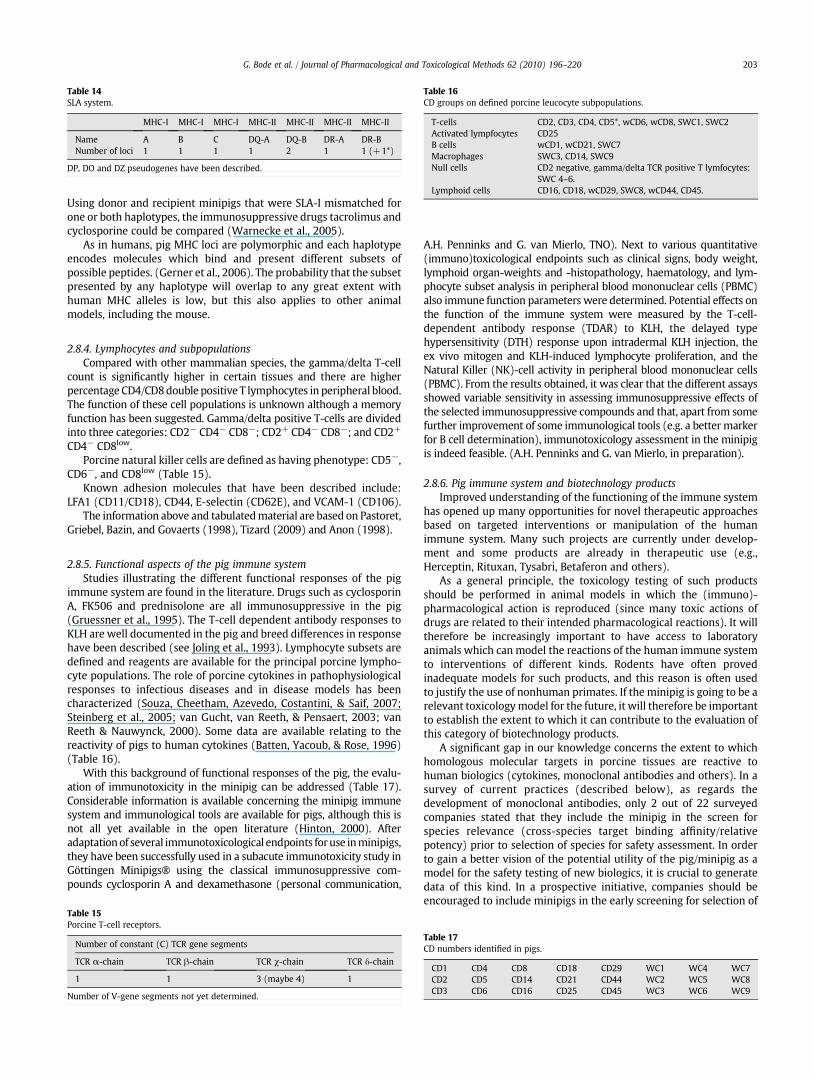
Table 14SLA system.
MHC-I MHC-I MHC-I MHC-II MHC-II MHC-II MHC-II
Name A B C DQ-A DQ-B DR-A DR-BNumber of loci 1 1 1 1 2 1 1 (+1*)
DP, DO and DZ pseudogenes have been described.
Table 16CD groups on defined porcine leucocyte subpopulations.
T-cells CD2, CD3, CD4, CD5*, wCD6, wCD8, SWC1, SWC2Activated lympfocytes CD25B cells wCD1, wCD21, SWC7Macrophages SWC3, CD14, SWC9Null cells CD2 negative, gamma/delta TCR positive T lymfocytes:
SWC 4–6.Lymphoid cells CD16, CD18, wCD29, SWC8, wCD44, CD45.
203G. Bode et al. / Journal of Pharmacological and Toxicological Methods 62 (2010) 196–220
Using donor and recipient minipigs that were SLA-I mismatched forone or both haplotypes, the immunosuppressive drugs tacrolimus andcyclosporine could be compared (Warnecke et al., 2005).
As in humans, pig MHC loci are polymorphic and each haplotypeencodes molecules which bind and present different subsets ofpossible peptides. (Gerner et al., 2006). The probability that the subsetpresented by any haplotype will overlap to any great extent withhuman MHC alleles is low, but this also applies to other animalmodels, including the mouse.
2.8.4. Lymphocytes and subpopulationsCompared with other mammalian species, the gamma/delta T-cell
count is significantly higher in certain tissues and there are higherpercentage CD4/CD8double positive T lymphocytes in peripheral blood.The function of these cell populations is unknown although a memoryfunction has been suggested. Gamma/delta positive T-cells are dividedinto three categories: CD2− CD4− CD8−; CD2+ CD4− CD8−; and CD2+
CD4− CD8low.Porcine natural killer cells are defined as having phenotype: CD5−,
CD6−, and CD8low (Table 15).Known adhesion molecules that have been described include:
LFA1 (CD11/CD18), CD44, E-selectin (CD62E), and VCAM-1 (CD106).The information above and tabulatedmaterial are based on Pastoret,
Griebel, Bazin, and Govaerts (1998), Tizard (2009) and Anon (1998).
2.8.5. Functional aspects of the pig immune systemStudies illustrating the different functional responses of the pig
immune system are found in the literature. Drugs such as cyclosporinA, FK506 and prednisolone are all immunosuppressive in the pig(Gruessner et al., 1995). The T-cell dependent antibody responses toKLH are well documented in the pig and breed differences in responsehave been described (see Joling et al., 1993). Lymphocyte subsets aredefined and reagents are available for the principal porcine lympho-cyte populations. The role of porcine cytokines in pathophysiologicalresponses to infectious diseases and in disease models has beencharacterized (Souza, Cheetham, Azevedo, Costantini, & Saif, 2007;Steinberg et al., 2005; van Gucht, van Reeth, & Pensaert, 2003; vanReeth & Nauwynck, 2000). Some data are available relating to thereactivity of pigs to human cytokines (Batten, Yacoub, & Rose, 1996)(Table 16).
With this background of functional responses of the pig, the evalu-ation of immunotoxicity in the minipig can be addressed (Table 17).Considerable information is available concerning the minipig immunesystem and immunological tools are available for pigs, although this isnot all yet available in the open literature (Hinton, 2000). Afteradaptationof several immunotoxicological endpoints foruse inminipigs,they have been successfully used in a subacute immunotoxicity study inGöttingen Minipigs® using the classical immunosuppressive com-pounds cyclosporin A and dexamethasone (personal communication,
Table 15Porcine T-cell receptors.
Number of constant (C) TCR gene segments
TCR α-chain TCR β-chain TCR χ-chain TCR δ-chain
1 1 3 (maybe 4) 1
Number of V-gene segments not yet determined.
A.H. Penninks and G. van Mierlo, TNO). Next to various quantitative(immuno)toxicological endpoints such as clinical signs, body weight,lymphoid organ-weights and -histopathology, haematology, and lym-phocyte subset analysis in peripheral blood mononuclear cells (PBMC)also immune function parameters were determined. Potential effects onthe function of the immune system were measured by the T-cell-dependent antibody response (TDAR) to KLH, the delayed typehypersensitivity (DTH) response upon intradermal KLH injection, theex vivo mitogen and KLH-induced lymphocyte proliferation, and theNatural Killer (NK)-cell activity in peripheral blood mononuclear cells(PBMC). From the results obtained, it was clear that the different assaysshowed variable sensitivity in assessing immunosuppressive effects ofthe selected immunosuppressive compounds and that, apart from somefurther improvement of some immunological tools (e.g. a better markerfor B cell determination), immunotoxicology assessment in the minipigis indeed feasible. (A.H. Penninks and G. van Mierlo, in preparation).
2.8.6. Pig immune system and biotechnology productsImproved understanding of the functioning of the immune system
has opened up many opportunities for novel therapeutic approachesbased on targeted interventions or manipulation of the humanimmune system. Many such projects are currently under develop-ment and some products are already in therapeutic use (e.g.,Herceptin, Rituxan, Tysabri, Betaferon and others).
As a general principle, the toxicology testing of such productsshould be performed in animal models in which the (immuno)-pharmacological action is reproduced (since many toxic actions ofdrugs are related to their intended pharmacological reactions). It willtherefore be increasingly important to have access to laboratoryanimals which can model the reactions of the human immune systemto interventions of different kinds. Rodents have often provedinadequate models for such products, and this reason is often usedto justify the use of nonhuman primates. If the minipig is going to be arelevant toxicologymodel for the future, it will therefore be importantto establish the extent to which it can contribute to the evaluation ofthis category of biotechnology products.
A significant gap in our knowledge concerns the extent to whichhomologous molecular targets in porcine tissues are reactive tohuman biologics (cytokines, monoclonal antibodies and others). In asurvey of current practices (described below), as regards thedevelopment of monoclonal antibodies, only 2 out of 22 surveyedcompanies stated that they include the minipig in the screen forspecies relevance (cross-species target binding affinity/relativepotency) prior to selection of species for safety assessment. In orderto gain a better vision of the potential utility of the pig/minipig as amodel for the safety testing of new biologics, it is crucial to generatedata of this kind. In a prospective initiative, companies should beencouraged to include minipigs in the early screening for selection of
Table 17CD numbers identified in pigs.
CD1 CD4 CD8 CD18 CD29 WC1 WC4 WC7CD2 CD5 CD14 CD21 CD44 WC2 WC5 WC8CD3 CD6 CD16 CD25 CD45 WC3 WC6 WC9

204 G. Bode et al. / Journal of Pharmacological and Toxicological Methods 62 (2010) 196–220
species responsive to the pharmacodynamic actions of new biologics.In a retrospective initiative, a survey of the responsiveness of porcinemolecular targets of existing biologics would be a valuable and infor-mative exercise which could make a useful contribution to our under-standing of the future role of the pig in safety assessment.
2.8.7. Immunology conclusionsPig immune system shows analogous structure and function to
human immune system. Some differences are known and have minorimpact (with possible exception of gamma delta cells and greater roleof innate immune system). The porcine immune system has been wellstudied and is probably better characterized than that of the dog orthe Macaque monkey. Nevertheless, little effort has been dedicated toevaluation of the relevance of the pig immune system specifically as amodel for therapeutic interventions (or toxic actions) in humans. Thiscertainly hinders the greater (informed) use of the pig for theevaluation of new therapeutic approaches based on immune systemmanipulation.
2.9. Central nervous system
Since the first use of minipigs as a model of MPTP-inducedParkinsonism (Mikkelsen et al., 1999) and in a model of diffuse braininjury with relevance for Alzheimer's disease (Smith et al., 1999), aconsiderable amount of knowledge has accumulated. In theirextensive review of the use of pigs in neuroscience, Lind et al.(2007) conclude that the gyrencephalic pig brain is more similar tothe human brain than the brains of common small laboratory animals.In addition they state that a considerable amount has been learnedabout pig brain anatomy and neurochemistry, but cortical function isnot yet sufficiently well described.
Various research tools concerning the central nervous system areavailable for pigs. This includes a number of stereotactic atlases (e.g.Anderson & Reiner, 1990; Felix et al., 1999; Salinas-Zeballos, Zeballos,& Gootman, 1986; Watanabe, Andersen, Evens, Gjedde, & Cumming,2001) and the increasing availability of pigs with transgenicmanipulations of neural genes (cf. Lind et al., 2007). The large sizeof the pig brain permits the detailed identification of cortical andsubcortical structures by imaging techniques such as positronemission tomography (PET) or magnetic resonance imaging (Lind etal., 2007). In addition, various behavioral testing methods are alsoestablished for pigs, including open field, anxiety and social behaviourtesting (reviewed by Lind et al., 2007), automated video tracking oflocomotor activity (Lind, Arnfred, et al., 2005; Lind, Gjedde, et al.,2005; Lind, Vinther, et al., 2005), and operant learning in a test batterysimilar to a battery already used for rats, primates and humans(Ferguson, Gopee, Paule, & Howard, 2009). Yet another learning test, aspatial delayed non-match to sample task, has recently beendescribed for the Göttingen minipig (Nielsen et al., 2009).
Although many of the neurobehavioral methods are not immedi-ately relevant for regulatory toxicity studies the accumulatingknowledge in this area has toxicological significance for two reasons.The first reason is the requirement that whenever possible theselected species should be responsive to the primary pharmacody-namic effect of the substance (Note for Guidance on Repeated DoseToxicity, CPMP/SWP/1042/99). In other words, a species whichrepresents a good pharmacological model for brain disorders (to beused for the development of drugs to treat them) is from thisperspective also good model for toxicity testing. The second reason isthe potential use of minipigs in postnatal reproductive toxicity andjuvenile toxicity studies. Behavioral testing is an important part ofsuch studies. Therefore it is very helpful that meanwhile a number ofbehavioral tests are well-established in the (mini)pig (see above). Inaddition, brain development of the neonatal pig appears in manyrespects more similar to the human newborn infant than that ofyoung rodents or dogs (Lind et al., 2007), including the important
“brain growth spurt” originally described by the group of Dobbing(Dickerson & Dobbing, 1966; Dobbing & Sands, 1979).
2.10. Anatomy and biology of the eye
After the primates, the porcine retina, pupil, and lens bear theclosest resemblance to human eyes and the porcine eye anatomyshares many similarities with the human eye. As in humans, themacula is composed only of cones in pigs. It is located in the visual axisof the bulb, thus enabling good visual acuity in daylight and excellentcolor vision in these species. The advantages of the porcine model arethe holangiotic retinal vasculature (fully vascularised, unlike therabbit), the choroidal blood flow and the absence of a tapetum. Inaddition, the thickness and surface area of the porcine sclera make theporcine eye a suitable model for studying transcleral drug delivery.Pigs do not have a truemacula, but they have a narrow horizontal areacentralis with photoreceptors, in a region that mimics the primatemacula (Olsen, Sanderson, Feng, & Hubbard, 2002; Prince, Diesem,Eglitis, & Riskell, 1960).
In spite of the smaller axial length, data on refractive error andcorneal power in the Göttingen minipig are comparable to humanvalues. A lack of accommodative reflex is probable. This informationmay be of importance when using minipigs as models for humanocular diseases or in research involving vision, e.g. in cognitive tasks(Nielsen & Lind, 2005).
Background data on ophthalmological findings and electroreti-nography in the minipig are available (Ausburger et al., 2009; Loget &Saint Macary, 2000).
2.11. Skin and dermal toxicity studies
Porcine skin resembles human skin. As in humans, the hair coat issparse. The skin is thicker in olderminipigs, and is less vascular than inhumans (Mortensen, Brinck, & Lichtenberg, 1998). The thickness inolder minipigs compared to humans is due to a thickening of thedermis. The thickness of the stratum corneum and epidermis remainsfairly constant after minipigs have reached three months of age.
Göttingen minipig skin is lightly pigmented as a result of the“dominant white” genetic status (see Simianer and Köhn, 2010-thisissue). In contrast to rodents, dogs, rabbits, porcine skin also has fineintersecting lines and rete ridge structures (it is closely attached to theunderlying structures). Apocrine sweat glands are absent in pigs(Swindle & Smith, 1998). Similar to humans, pigs have lipidbiophysical properties, epidermal turnover kinetics (1 month), andan aptitude for tanning (Meddahi, 1994).
Based on the similarities of the minipig and human skin theminipig is the species of choice in dermal studies to evaluate bothlocal tolerance and possible systemic toxicity after dermal application.Moreover, the abundant surface area of the minipigs allows multiple-site and long-term testing. Since visual assessment of skin reactionsmay not be fully consistent between studies and observers, non-invasive instrumental measurements (e.g. reflectance measurementswith a chromameter, high-resolution ultrasonography, transepider-mal water loss due to skin lesions by an evaporimeter and monitoringof skin blood flow and cutaneous vasoconstriction by laser Dopplerflowmetry) may be applied for a quantitative evaluation of localtreatment-related effects (Vogel, Kolopp, Singer, & Cordier, 1998);these types of examinations are more easily performed in minipigsthan in smaller species or animals with dense fur coat.
Chemical compounds, depending on their physicochemical prop-erties and the area of dermal administration, generally show a highpercutaneous penetration through the skin of densely haired animals,whereas permeability and metabolic properties of the porcine skinappear to be more comparable to man (Lin, Hou, Hsou, & Ye, 1998;Poet & McDougal, 2002; Ngawhirunpat, Opanasopit, & Prakongpan,2004). The skin penetration of several compounds was found to be

Table 19Relative distribution of skeletal muscle fiber types.
Fiber type Human Pig Minipig Dog Rat
I 50–65% 9–11% 20–45% 18–28% 5–28%II IIa: 30% IIa: 4–12%;
IIx: 18–20%;IIb: 60–71%
IIa: 15–40%; IIx:10–50%; IIb:25–55%
IIa: 43–67% IIa: 19–50%IIx: 10–20% IIx: 15% IIb: 8–75%IIb 0–44%
205G. Bode et al. / Journal of Pharmacological and Toxicological Methods 62 (2010) 196–220
similar between pig and human skin, whereas no significant correlationexisted between human skin and hairless dog or nude mouse skin(Reifenrath, Chellquist, Shipwash, & Jederberg, 1984). Furthermore, lowintra- and inter-variations were evident for different substances in invitro permeability studies using Göttingen minipig or domestic pig skinin comparison with human skin (Qvist, Hoeck, Kreilgaard, Madsen, &Fokjaer, 2000).
The similarities between human and minipig skin with regard toclinical, histological and immuno-histological findings further supportthe minipig as a model for the investigation of immuno-pathologicalmechanisms and pharmacological intervention (Vana & Meingassner,2000). A pig model has also been established for the investigationof inflammatory skin lesions and anti-inflammatory treatment (Nairet al., 1993). A more exotic administration route is on the upper snoutto investigate the neurotoxic potential of anticancer compounds onsubcutaneous cranial nerves (Mahl et al., 2006).
2.12. Musculoskeletal system
2.12.1. Skeletal muscleLike in all mammals, striated muscle of pigs is derived from three
different areas of mesoderm: The so-called myotomes of the somites,the branchial arches, and locally formed mesenchyme not derivedfrom somites (Table 18). The skeletal muscle fibers are postmitoticsyncitia derived from fusions of embryogenic precursor and thereforemultinucleated.
It is widely accepted, that mammalian skeletal muscle is composed ofdifferent fiber types, which are made up of a distinct set of structuralproteins (e.g. myosin) and metabolic (glycolytic and/or oxidative)enzymes (Schiaffino & Reggiani, 1996) (Table 19). At the end of fetaldevelopment, the first differences between muscle fibers are alreadyfound which allow to differentiate between (light-stained, slow-twitch)type Ifibers and (dark-stained, fast twitch) type IIfibers (Brooke&Kaiser,1970). Slow fibers acting for postural functions, are tonically active andfatigue resistant, and have an oxidative metabolism as the dominantsource of ATP synthesis. Fast fibers, on the other hand, are phasicallyactive, forwhich glycolyticmetabolism is functionally appropriate (Peter,Barnard, Edgerton, Gillespie, & Stempel, 1972). Most of the proteinsinvolved in contractile function, for example the myosin heavy chain(MyHC), and the sarco(endo)plasmic reticulum Ca2+-adenosine tripho-sphatase, exist as multiple isoforms (Schiaffino & Reggiani, 1996), whichare usually distributed in a myofiber type specific and coordinatedmanner (Hämäläinen & Pette, 1995). Fiber typing based on acid andalkaline denaturation of myosin molecules by using myofibrillaradenosine triphosphatase histochemistry leads to a further subdivisionof the myofibers (Peter et al., 1972): Type I fibers: SO (slow oxidative),Type IIa fibers: FOG (fast oxidative glycolytic) and type IIb fibers: FG (fastglycolytic). On basis of monoclonal antibodies and of mRNA content ofthe respective fiber type, a subdivision of type II fibers into types IIa, IIxand IIb can be made, without considering the existence of furthersubtypes, intermediate and mixed type variation or possible posttrans-lational shifts of fiber types (Schiaffino & Reggiani, 1994, 1996).
These different fibers contain different myosin heavy chain (MyHC)isoforms. So far, eight isoforms of myosin heavy chain (MyHC) inskeletal muscle of mammals are known (Weiss et al., 1999). In porcine
Table 18Principal characteristics of skeletal muscle fiber types.
Property Type I Type IIa
Morphology and color Slighted, red Huge, whiteOxidative capacity Low HighGlycolytic capacity Low HighContraction time Slow Moderately fastMetabolism Aerobic Anaerobic (long-tim
muscle, four myosin isoforms have been characterized: one clustercontains type I (or β) MyHC on chromosome 7, whereas the secondcluster contains the embryonic, IIa, IIx, IIb, neonatal, and extraocularMyHC on chromosome 12 (Davoli et al., 2002). In postnatal growingpigs, only four isoforms, type I, IIa, IIx, and IIb MyHC, are expressed inskeletal muscle (Lefaucheur, Ecolan, Plantard, & Gueguen, 2002).
In humans, striated muscle, fiber type and myosin heavy chain(MyHC) expression is extremely heterogenous (Schiaffino & Reggiani,1994), e.g. the relative distribution of type IIb fiber in the M. vastuslateralis can vary from 0 to 44% with a coefficient of variation of ∼66%within a sedentary population (Simoneau & Bouchard, 1989).
In pigs, MyHC composition differed between muscle types andbreed. In most breeds as well as in wild boar, the relative number ofwhite fibers (IIx and IIb types) was between 80 and 85% (Wimmerset al., 2008). Type IIb fibers, the isoform exhibiting themost rapid speedof contraction, are the predominantly isoform in fast-contractingmuscles of pig (Lefaucheur et al., 2002). The differences inhistochemicalproperties between breeds were, however, smaller in M. adductorthan in M. longissimus dorsi. In addition, the variation in muscle fibercomposition in pigs within the breeds was larger than the averagevariation between the breeds (Ruusunen & Puolanne, 2004). Only thenative Vietnamese Mongcai breed shows lower proportions of type IIfibers (47%) when compared to other conventional breeds (Wimmerset al., 2008). In the cross breed Duroc×Berlin Miniature pigs, type IImuscle fibers have contributions of 70% type IIb and IIx and 15% type Imeasured as relative MyHC isoform transcripts or as histochemicaldifferentiation (Wimmers et al., 2008).
Overall, there is a considerable correlation between contractile,metabolic, and morphological features in skeletal muscle of minipigs.This correlation allowsdiscriminationofmusclefibers according to theirMyHC content into a system of discrete types. In consequence, theminipig muscle does not exhibit dramatic variations in phenotypicexpression of their myofiber properties with regard to those previouslyreported in other small and large species ofmammals or inhumanswitheither similar or different expression of MyHCs (Schiaffino & Reggiani,1996).
Daily muscular training by treadmill-running in Yukatan Minipigsled to an increase in citrate synthetase enzyme activity in variousskeletal muscles. The number of type IIB fibers decreased after thetraining, whereas the type IIxX fibers increased in numbers, indicatinga transformation of fiber types and that skeletal muscle of Yucatanminipigs adapts to endurance exercise training in a manner similar tomuscle of humans with increased oxidative capacity (McAlister &Laughlin, 1997).
The calcium release channel (CRC) gene mutation responsible forhalothane-sensitivity and “porcine stress syndrome” in domestic pigshas not been demonstrated for Göttingen minipigs.
Type IIx Type IIb
Huge, white Huge, whiteHigh lowHigh HighFast Very fast
e) Anaerobic (short-time) Anaerorobic (short-time)

206 G. Bode et al. / Journal of Pharmacological and Toxicological Methods 62 (2010) 196–220
2.12.2. Bones and jointsBones consist of organic matrix (=osteoid) with incorporated
minerals (mainly Ca and P). During the pre- and postnatal period,bones are developed from connective tissue (desmal ossification, i.e.skull) or from preformed hyaline cartilage (peri- and enchondralossification, i.e. long bones) (Table 20). By birth most of the shafts ofthe bones are ossified but the ends are still cartilagenous. From birthto puberty, secondary ossification centres appear in the cartilagenousends of the bones. Eventually, the whole bone will be mineralizedexcept for the articular surfaces which remain covered by cartilage. Athin zone of cartilage – the growth or epiphyseal plates – persistsbetween the end of the bone and its shaft. Long bones growth by theinterstitial growth of the epihyseal plate in which chondrocytes areproduced by repeated cell division. The chondrocytes form palisadesand are growing towards the bones end and approach the diaphysealside of plate, where they enlarge and their matrix becomesmineralized. Cells next to the marrow cavity die, their matrixdisappear and leave “tunnels” invaded by vessels and osteoblasts.The osteoblasts build bony lamellae on the inner wall of those tunnelsuntil only a narrow channel (Harvesian channel) remains and theosteoblasts become osteocytes. Growth in width is achieved bydeposition (appositional growth) of new bone on peri- and endost(Hochberg, 2002). The skeleton ismaturewhen epiphysis, metaphysisand diaphysis are mineralized and fused (Gilli, 1996), that is at an ageof over 2.5 years in minipigs (Reinwald & Burr, 2008).
In minipigs, the peak bone mass is reached between 3 (Bouchardet al., 1995) and 6 years (Tsutsumi, Katagiri, Morimoto, et al., 2004), inhumans at an age of 30–40 years (Soyka, Fairfield, & Klibanski, 2000).Peak bone mass is the maximal bone mineral density present at theend of the skeletal maturation, is an important determinant ofosteoporotic fracture risk.
The histomorphometric examination of bones of miniature pigsshowed that the bone apposition rate and trabecular thickness inminipigs is similar to that in humans (Honig & Merten, 1993). Withthe help of freeze-fracture scanning electron microscopy (SEM) Kääb,Gwynn, and Notzli (1998) could demonstrate that the collagen fiberarrangement in pig and dog articular cartilage is very similar to theleaflike arrangement found in humans, whereas cow, sheep, rabbit orrat present a columnar pattern.
The presence of true osteonal remodelling is another desirablecharacteristic in the Göttingen minipig model. Remodelling means themechanism by which the basic multicellular unit, of old bone ordamaged bone is removed and replaced with new bone withoutalteration in the bone's shape. Modelling of bone means bone growthand shaping.
The remodelling and especially the forming of secondary osteonsis a characteristic of porcine bone that is relevant to humans and is animprovement on the rodent model with its lack of secondary osteons(Martiniakova, Grosskopf, Omelka, Vondrakova, & Bauerova, 2006).Animal models may be classified as either modelling (rats) or trueremodelling (i.e. pigs, dogs, ewes, and primates). In the rat, epiphysealgrowth in the long bones continues for 2 years and longer, and there isno plate closure, in the sense of remodelling and disappearance(Jowsey, 1968). Rat cortical bone is made of primary lamellar bone(periosteal and endosteal lamellae) which is less affected by boneremodelling compared to human or porcine bone which containssecondary osteonal bone (“harvesian bone”) (Hillier & Bell, 2007).Therefore, rat cortical bone is not necessarily a suitable model for
Table 20Basic bone data of different species.
Human Minipig Dog Rat
Skeletal maturity (years) 18–25 N2.5 1.3 N2/lifelongMaximum bone density (years) 30–40 6 6 1Remodelling Time (month) 8–10 3–6 3 35 days
studying the changes of mechanical property due to changes in boneremodelling.
Due to the similarity of skeletal parameters and bone-healing ratesthe minipig is a well-established model in bone and joint surgery. TheGMP is also used for studies on bone metabolism (Tsutsumi, Katagiri,Takeda, et al., 2004) dental and craniomaxillofacial surgery (Honig &Merten, 1993), osteoporosis (Ikeda et al., 2003), bone and fracturerepair (Merten, Wiltfang, Honig, Funke, & Luhr, 2000), implantfixation (Wong, Eulenberger, Schenk, & Hunziker, 1995), healing ofarticular cartilage defects (Hunziker & Rosenberg, 1996), chondraland osteochondral defect repair (Gotterbarm et al., 2008) and totalhip replacement (Thomsen et al., 1997) in orthopaedic research.
2.12.3. Musculoskeletal system: Gaps of knowledgeThere is limited knowledge on the occurrence of spontaneous
lesions of the musculoskeletal system known from swine, which areattributed to the enormous growth potential of commercial slaughterpigs and include exertional myopathies like the porcine stresssyndrome and skeletal disorders like “leg weakness” and osteochon-dritis. Such lesions should be less frequent or absent in minipigs, asthe body weight gain is much lower than in commercial pigs.
3. Regulatory toxicology testing
3.1. Species-specific toxicity
Species-specific toxic actions are occasionally encountered inregulatory safety testing and account must be taken of this species-specificity in extrapolating to man, and risk evaluation. Examples aregiven below to illustrate some advantages and disadvantages ofregulatory toxicology testing in the minipig as a consequence ofspecies-specific reactions.
As reviewed by Dogterom, Zbinden, and Reznik (1992) andLehmann (1998), dogs are particularly sensitive to sympathomimeticsand hypotensive drugs, showing cardiotoxicity at doses close to theED50. As a consequence, it is difficult to assess systemic toxicity otherthan exaggerated pharmacodynamic activity in this animal model(Dogterom et al., 1992). In the example presented by Lehmann(1998), a benzodiazepine piperidinyl derivative with sympathomi-metic activity caused tachycardia of similar extent in both dogs andminipigs. However, myocardial necroses were seen in the dog, but notin the minipig. The minipig was therefore considered more predictiveof human responses. Dogterom et al. (1992) proposed the use of moresuitable species like the pig as an alternative to dogs for the safetyevaluation of vasodilators and positive inotropic/vasodilating drugs.
Active renal clearance mechanisms for organic acids are ubiqui-tous in mammalian species, but the dog is known to have a muchlower capacity of this saturable carrier system for the excretion oforganic acids from the kidney than other species. This leads to anincreased sensitivity of dogs to organic anions, making the dog anunsuitable non-rodent model for human risk and safety assessmentsof organic acids (reviewed by Timchalk, 2004). It should be noted thatexcretion by this active transport mechanism is not affected byprotein binding, i.e. organic acids bound to plasma proteins are readilytransported across the proximal tubule epithelium. Consequently,comparison of the phamacokinetics of phenoxyacetic acids demon-strated that pigs (and rodents) were better models for the toxicityevaluation of these compounds than dogs (Timchalk, 2004).
Elliott, Purmalis, VanderMeer, and Denlinger (1988) suggestedthat gastrointestinal lesions provoked by proprionic acid nonsteroi-dal anti-inflammatory drugs (NSAIDs)–the most important (dose-limiting) adverse effect of this class of drugs are caused by systemicexposure rather than local irritation. This was concluded, becausesuch lesions have been seen after both parenteral and oral administra-tion. The order of sensitivity to NSAID'swas described as dogNratNnon-human primates. Minipigs were not mentioned in this paper. The
207G. Bode et al. / Journal of Pharmacological and Toxicological Methods 62 (2010) 196–220
question arises whether the dog-specific lower capacity to excreteorganic acids described above might be involved in the exaggeratedsensitivity of the dog towardsNSAIDs. This ideawas subsequently takenfurther by Lehmann,whoused theminipig as the non-rodent toxicologymodel for the development of the NSAIDmeloxicam (Yabe et al., 1997).
In contrast, proquazone, another NSAID compound which is not apropionic acid, was more toxic in minipigs than in dogs (van Ryzin &Trapold, 1980). In a 15-month oral (capsule) study using beagle dogs,proquazone induced gastrointestinal irritation causing ulceration,peritonitis and death at 40 and 80 mg/kg, whereas doses up to 75 mg/kg were tolerated without effects in a 14-week study. In a 26-weekstudy in Hormel minipigs, proquazone was administered with smallamounts of food at doses of 0, 6, 15, 38 and 94 mg/kg. The death rateincreased dose-dependently from 1/12 pigs at 6 mg/kg to 10/12 pigsat 94 mg/kg, and all deaths were attributed to gastric ulceration andits consequences.
Another example for pig specific toxicity was provided by Henne-Bruns, Arthwohl, Broelsch, and Kremer (1988). The attempt toestablish a pig model of acetaminophen-induced acute hepatic failurewas unsuccessful. The pigs developed methemoglobinaemia beforehepatic failure, because of acetaminophen's capability to oxidizehemoglobin. This observation is commensurate with the known lowcapacity of the pig's methemoglobin-reductase, (i.e. approximatelyone third of the human capacity), whereas the estimated spontaneousmethemoglobin-reductase in the dog is more than two-times higherin the pig (Smith, 1996).
Spontaneous (background) pathology must not be confused withspecies-specific reactions. Four types of background lesions in barrier-bred Göttingen minipigs should however be kept in mind during thehistopathological assessment of tissues from toxicity studies (Svendsen,Skydsgaard, Aarup, & Klastrup, 1998): (a) vasculitis, which may occurin different arteries or arterioles, (b) tubular hypoplasia in the testis,(c) serous atrophy with a decrease cellularity in the bone marrow(restricted feedingmay be involved, cf. Bollen & Skydsgaard, 2006), and(d) the so-called hemorrhagic syndrome. These spontaneous lesions arerare, most likely because of the high microbiological quality of barrier-bred Göttingen minipigs (Skydsgaard, 2005).
3.2. Juvenile toxicology studies
For non-rodent species such as the dog or nonhuman primate,juvenile toxicity studies present a number of practical and logisticalchallenges. Examples of these challenges include smaller litter sizes incomparison with rodents, long gestation periods, long pre-weaningperiods, space requirements and cost. Comparedwith other non-rodentanimal models such as the dog or nonhuman primate, the biologicalcharacteristics of the minipig are already favourable: the litter size isrelatively large (5 newborns per sow), the rate of growth is very rapid,sexual maturity is achieved at an early age and piglets are accessible forhandling. Weighing about 450 grams at birth, they are amenable tolaboratory procedures.
There are three basic approaches to the study design. One is to usethewhole litter as the basic group or experimental unit. The second is totreat animals within a litter differently from each other. The thirdapproach is to randomise the allocation of offspring to mothers at birth(cross-fostering) and then to treat all animals allocated to onemother asa group. All three approaches have advantages and disadvantages, butthe cross-fostering approach offers the most potential benefits.
Cross-fostering in minipigs is possible without difficulty and isroutinely done at the production facility. Randomised cross-fosteringof piglets may be performed on Day 1 after birth. The piglets may beborn on site or theymay be born at the breeders and transported, withtheir mothers, on the day after birth to the experimental laboratories.Oral gavage dosing can be performed from Day 1. Vascular accessports can be implanted in 5–7 day old minipigs and have been shownto remain functional for four weeks. The ports used were intended for
rats and despite the rapid growth during the first four–five weeksafter birth these ports remained functional. During the second weekafter birth, the piglets may be subjected to electrocardiographic andophthalmological examination. Blood and urine may routinely becollected fromweek 1 for haematology, clinical chemistry, bioanalysisand urinalysis examinations.
The data obtained for the various experimental parameters havebeen compared with background data from older minipigs. Therewere several notable findings. For instance, at one week of age, thevalues for red blood cell parameters (hemoglobin, red cell count andhaematocrit) were all lower than historical values for 4 to 6 month oldanimals. However, at 4 weeks of age the values for these parametershad moved into the same range as the older group. In comparisonwith older animals, serum values for alkaline phosphatase, bilirubin,gamma-glutamyl transferase, cholesterol, triglycerides and urea wereelevated. The mean values for several of these parameters at week 4were more in line with those of older animals. The reasons for thesedifferences are easily explained in terms of the animal age, diet, etc.However, they do underline the importance of having adequatebackground data for the age and strain of animals used in any study.
It is concluded that cross-fostering of minipigs for performance ofjuvenile toxicology studies is a practical option. However, there needsto be allowance in the number of animals allocated to the study in caseof early deaths; this complication is also well known from juvenileanimal studies in rats or dogs. Also, it is important that the mothersare allowed to give birth at the site where the studywill be conducted.The importance of having adequate background data for the animals ishighlighted.
The Göttingenminipig is a promising non-rodent animal model forjuvenile toxicity studies, because of advantageous characteristics suchas a large mean litter size (5 newborns per sow), high rate of growthand rapid achievement of sexual maturity. The piglets are accessibleand amenable to handing (unlike for example, nonhuman primates,where the neonates are carried by the mothers) and cross-fostering isfeasible. A number of behavioral tests are well-established in the pig(see Section 2.9). In comparison with other non-rodent animalmodels such as the dog or cynomolgus monkey, the minipig appearsto be an eminently practical choice for this kind of study.
3.3. Predictivity of minipig toxicity studies
In this article the potential benefits and disadvantages of theminipig as a suitablemodel predictive for humans have been analysedand the comparative anatomy, physiology and biochemistry of the pigand minipig have been reviewed. Some areas of species-specificity ofthe minipig and other non-rodent toxicology models have also beendiscussed.
Numerous examples illustrating the close similarities of porcineand human anatomy, physiology and biochemistry have beenidentified in almost every organ system or physiological processthat was considered (including the dermal, cardiovascular and renalsystems, functionality of the gastrointestinal and immune system,comparable hepatic biotransformation etc.).
– The cardiovascular conditions are similar, humans and minipigsshow comparable anatomy, comparable electrophysiological func-tions with the major ion channels and comparable reactions toharmful influences.
– The comparability of hepatic P450 cytochromes between pigs andhumans support the usefulness of minipigs as experimentalanimals to predict biotransformation pathways in man.
– The gastrointestinal system of the minipig offers some anatomicaland functional similarities in terms of absorption and metabolism.
– The dermal structure and dermal reactivity is similar; the minipigis also important for transdermal penetration studies.

208 G. Bode et al. / Journal of Pharmacological and Toxicological Methods 62 (2010) 196–220
– The basic structure of the porcine immune system resembles thatof other mammalian organisms including humans.
– Due to the similarity of skeletal parameters and bone-healing ratesthe minipig is a well-established model in bone and joint surgeryand has been used for studies on bone metabolism andosteoporosis. Minipig muscle and myofiber properties are compa-rable to other mammalian species.
These similarities suggest that theminipigwill be a usefulmodel insafety testing of new products. In addition to these broad similaritiesto humans, there are characteristics and features of the biology of theminipig that make it an attractive model in toxicology testing. A goodexample is given by reproductive toxicology testing, where the earlyachievement of sexual maturity, short estrous cycle length and largelitter size make the minipig an attractive alternative to existing (non-rodent) models for reproductive toxicity testing. In other areas, thesimilarity of the skin structure and large surface area make theminipig attractive for cutaneous studies, the reduced propensity forvomiting make the minipig a useful alternative for some classes oforally administered drugs.
A greater role for minipigs in testing could thus bring advantages.Wemay anticipate that the similarities of the minipig to man result ina similar profile of toxic reactions in the minipig and man. Hencetesting in minipigs may bring a more accurate prediction of adverseand harmful reactions in man. This is a conclusion that would bringsignificant economic and public health benefits.
We were not able to address and evaluate this issue of predictivityin the Safety Issues Working Group, since insufficient publishedmaterial and comparative safety testing data is available to permit arigorous evaluation of this hypothesis. Altogether our comparativebiology analysis supports the hypothesis that the pig or minipigshould be a good model for predicting human undesirable reactions.On the other hand, we have scoured the literature and could notidentify sufficient material for systematic or carefully conductedstudies to test this idea fully. It is therefore one of our conclusions thatresearch is need to allow evaluation of the hypothesis that minipigstudies may better reflect human drug-induced toxicities than studiesperformed in traditional non-rodent toxicology models (such as thedog).
There is a further issue in the application of the minipig toregulatory safety testing of biotechnology products (and hence to thereplacement, in part, of primate studies). A significant gap in ourknowledge concerns the extent to which homologous moleculartargets in porcine tissues are reactive to human biologics (cytokines,monoclonal antibodies and others). In a survey of current practices asregards the development of monoclonal antibodies, only 2 out of 22surveyed companies stated that they include theminipig in the screenfor species relevance (cross-species target binding affinity/relativepotency) prior to selection of species for safety assessment. Thescientific literature is equally devoid of information and examples onthis point. In order to gain a better vision of the potential utility of thepig/minipig as a model for the safety testing of new biologics, it iscrucial to generate data of this kind. In a prospective initiative,companies should be encouraged to include minipigs in the earlyscreening for selection of species responsive to the pharmacodynamicactions of new biologics. In a retrospective initiative, a survey of theresponsiveness of porcine molecular targets of existing biologicswould be a valuable and informative exercise which could make auseful contribution to our understanding of the future role of the pigin safety assessment.
In conclusion, insufficient comparative data is available to permit arigorous evaluation of the predictivity of the minipig for human drug-induced toxicities. In addition, for minipigs to play a more significantrole in the testing of biotechnology products and biologics, data isrequired on the pharmacodynamic responsiveness of minipig molec-ular targets to human biologic drugs.
3.4. Criteria for selection of non-rodent species
3.4.1. Individual company experienceA retrospective survey of the use of non-rodent species in 57
projects over the course of 10 years was performed by one of thepharmaceutical companies. Although drug classes are not disclosedhere, this survey reveals the rationale for species selection in the past.In addition, a strategy is described for the current approach essentiallyin line with the requirements/recommendations of CPMP and ICHguidelines.
According to the survey species selection was based on thefollowing considerations.
•Practicability (dog use) 30%
•Toxicology/pharmacodynamics 30% •Drug metabolism 12% •Inhalation (dog) 9%(primates)
1% •Proteins (use of primates) 4% •Unknown reasons 14%Currently non-rodent species selection at this company takes intoaccount the following aspects based on experience and on datacollected at an early stage of drug development:
• Species-specific inadequacies (e.g. exaggerated sensitivity of dogse.g. concerning cardiovascular effects, emesis and histaminerelease);
• Animal PK and Drug Metabolism (adequate exposure, presence ofhuman key metabolite[s], if any)
• Avoidance of primate use whenever possible with due regard ofscientific and ethical justification
• Technical considerations (e.g. exposure techniques in inhalationtoxicology)
• In case of similar suitability of dogs and minipigs various aspects ofresource availability are taken into account.
Ideally the following information would be available for speciesselection prior to the start of the first non-rodent toxicity studies:
• Pharmacodynamic activity in the different non-rodent species;• Exploratory toxicity data from the rat;• Cardiovascular data after low single doses in telemetered animals ofthe different species;
• Information from front runner compounds (if applicable);• in vitro metabolic stability in liver microsomes and/or hepatocytesof rat, dog, minipig, non-human primates, and man
• Active drug principle in vivo po/iv plasma profiles: rat, dog and/ormonkey and minipig
• iv/iv correlation for clearance early assessment of metabolite(s) ifidentified as major carrier of action.
As a conclusion we recommend early consideration of the inclusionof theminipigwhen looking for thebestmodel forpredicting side effectsin humans.
3.4.2. Current practice survey/results of a questionnaireIn order to better understand the way in which the minipig is
currently used in pharmaceutical safety assessment, the way in whichdecisions are made regarding its use and the perceived barriers to theuse of the minipig, a survey of current practice was undertaken. Aquestionnaire on “The use of minipigs in safety assessment” wasprepared and submitted to pharmaceutical industry companies; thisquestionnaire and the detailed results are found in the Appendix A tothis article. This questionnaire was distributed to the pharmaceuticalindustry via the following industry organizations: US PhRMA, BioSafe,EFPIA and ABPI. Responses to the questionnaire were received from23 organizations.

209G. Bode et al. / Journal of Pharmacological and Toxicological Methods 62 (2010) 196–220
The results of this survey showed that experience with safetyassessment studies using minipigs was in safety pharmacologystudies, in general toxicity studies (using dermal, oral and parenteralroutes) and also in reproductive and immunotoxicity studies. Theexperience in each individual company was limited.
Literature sources and communications via the questionnairerevealed that the minipig is used for dermal, oral and parenteraladministration. Studies by the intravenous route are possible, but asthe result of technical difficulties are less frequent. Dermal applicationin particular is considered to be advantageous in minipigs.
A minority of companies (6/22) routinely include the minipig inthe screen for pharmacological activity and/or metabolic clearanceprofiles used to aid selection of the non-rodent species for safetyassessment of low molecular weight products.
For monoclonal antibodies, only 2/22 companies stated that theyinclude the minipig in the screen for species relevance (cross-speciestarget binding affinity/relative potency) prior to selection of speciesfor safety assessment.
Six companies who used minipigs for safety assessment used thisspecies as the single non-rodent species (for the drug project), andfive companies used minipigs as a second (supporting) non-rodentspecies. Four companies had experience of using minipigs for safetyassessment both as the only non-rodent species and as a second non-rodent species. The reasons stated for conducting studies in minipigsas a second non-rodent species are outlined below:
• To try to reproduce a finding observed in humans but not inmonkeys
• Clarification of organ toxicity to determine species-specificity• Not tolerated in dogs• Clarification of effects• General tolerability issues• Metabolism issues• Consideration of possible metabolic differences• Poor exposure in dogs, special susceptibility of dog to vomitingcaused by test substance
• Occasionally for dermal programs to supply supporting datawhen another non-rodent species has been used
• Dermal toleration for dermal product/cutaneous patchprograms.
• GI tolerability of a new formulation• Pigs are used only if they offer a more relevant model of humanphysiology, disease or drug disposition/metabolism.
The majority of companies (14/22) perceived hurdles to the use ofminipigs as a more general non-rodent species in safety assessment.These perceived hurdles fell into 5 main categories:
1. the size of minipigs and corresponding high compound require-ment, particularly for chronic studies;
2. lack of experience in the use of minipigs and issues relating tohistorical control data;
3. animal husbandry, supply and logistics;4. technical aspects and staff training;5. lack of reagents and diagnostic kits.
About half of the companies stated that the lack of commercial kitsfor laboratory diagnosis and immune function testing for minipigsinfluenced their decision not to use minipigs more generally in safetyassessment.
In summary, the results of the survey illustrate that manycompanies are already using the minipig as a first or second linenon-rodent toxicology model in regulatory studies for the registrationof new drugs. The reasons for selecting the minipig are various, butscientific in nature. The “hurdles” that are cited to the greater use of
the minipig illustrate the need for further availability of training andscientific support for the model and development of reagents.
4. Gaps and research opportunities
The gaps in knowledge and/or research opportunities arepresented under two general headings of results that would bettercharacterise of the model, and results that would permit the properdeployment of the minipig in safety testing.
4.1. Characterisation of the minipig model
In reviewing the comparative biology of the minipig, many topicshave been described where further research would bring greaterunderstanding. In presenting these recommendations, we havelimited our proposals to areas where further understanding or moredetailed knowledge of the minipig would catalyse more informedand/or broader exploitation of theminipigmodel for regulatory safetytesting. These areas are as follows:
1. Inhalation toxicology and respiratory administration: this is currentlyan area of weakness for the minipig model. There are fewpublications and methods are not well-established for inhalationtoxicology or respiratory function studies. Until inhalationapproaches are better established, the minipig will not be seen asan alternative model for products administered by this route. Aprogram of work to provide basic information in this area (RMVvalues, deposition efficiency) and to consolidatemethods (inhalationadministration, respiratory functionmeasurements) would open theway to the use of the minipig for such products.
2. Reproductive toxicology: A number of actions would strengthen therole of the minipig in reproductive toxicology. These include (i)further characterisation of the different stages of spermatogenesisin the minipig boar, (ii) extended background data on spontaneousfetal variations and anomalies in the minipig and (iii) informationon the placental transfer of macromolecules. It is known thatantibodies do not cross the placental barrier in pigs, but very littlehard data is available regarding other large molecules andmacromolecules. This point requires clarification in order to betterunderstand the product categories where the minipig can be arelevant model for embryo–fetal toxicity testing.
4.2. Deployment of the minipig model
Proposals for research that will permit the proper deployment ofthe minipig model are as follows:
3. Collaborative programs: some issues are best addressed throughcollaborative programs rather than focussed research actions. Datathat could be collected in this way that would better define the roleof the minipig in regulatory toxicology testing includes:
a. Cross-species comparative data on metabolism providing a data-base and case-histories where the minipig does or does not pro-vide a closemodel of drugmetabolismanddisposition in humans.
b. It is generally believed that theminipig provides a bettermodel ofhuman cutaneous reactions than other animalmodels such as therabbit or the rat. For percutaneous absorption data is available todemonstrate the value of the mini(pig) model, but for skintolerance we do not have quantitative data. It is known that thecorrelation between rabbit and human irritant responses is poorforweak irritants (Steinberg, Akers,Weeks,McCreesh,&Maibach,1975), but the corresponding data is not available for the pig. Thisis an important point to establish, both in the interest of drugsafety and also since it could open the way for highly predictiveporcine skin-based ex vivomodels

210 G. Bode et al. / Journal of Pharmacological and Toxicological Methods 62 (2010) 196–220
4. Predictivity of the minipig for human toxicities (discussed in thefollowing section):
a. A predictivity testing program is urgently needed in which anadequate number of compounds or drugs (for example ten) ofwell characterized toxicity would be tested in both the minipig,the dog and the monkey in order to demonstrate thatpredictivity in the minipig is at least as good as the monkeyand better than the dog. Such a result would be a significantfinding and should provoke rethinking about the role ofmonkeys in the testing of new drugs.
5. Safety testing of new biologics: (discussed in the following section).In order to gain a better vision of the potential utility of the pig/minipig as amodel for the safety testing of new biologics, it is crucialto generate data on the responsiveness of porcine molecular targetsto human biologics. Two proposals are made to address this point:
a. In a prospective initiative, companies should be encouraged(by guidance documents or by research initiatives) to includeminipigs in the early screening for selection of species responsiveto the pharmacodynamic actions of new biologics.
b. In a retrospective initiative, a survey of the responsiveness ofporcinemolecular targets of existing biologics would be a valuableand informative exercisewhichcouldmakeauseful contribution toour understanding of the future role of the pig in safety assessment.
5. Discussion and conclusions
5.1. Current use of the minipig in toxicity testing
In this article current knowledge about the use of the minipig as alaboratory animal model for safety testing has been reviewed withrespect to the comparative biology of the pig and minipig and theimplications for their use in safety testing.
Minipigs are already routinely used for regulatory toxicity testing,typically for general toxicity studies. In Europe the Göttingen minipigis selected most frequently for such studies. There is some interestamongst toxicologists in the use of the minipig in reproductivetoxicity testing and in the performance of juvenile animal studies,although the routine use of the minipig for these studies is not yetwidespread. The current number of minipig regulatory toxicologystudies performed in Europe is estimated at circa 150 studies perannum (Estimate from Ellegaard Göttingen Minipigs ApS).
In general it can be stated that studies performed in minipigs areacceptable to regulatory authorities. This question is reviewed indetail in van der Laan et al. (2010-this issue), where examples ofproducts in commercewhichwere developed using theminipig as thenon-rodent toxicology model are also given.
Current use of the minipig in regulatory toxicity testing andattitudes of the toxicology community to the minipig were exploredthrough a questionnaire which was distributed to representativeorganizations including ABPI, EFPIA, PhRMA (DruSafe) and BioSafe.From the responses to the survey received from 22 companies, thefollowing general points emerged:
– Minipigs are used principally for toxicity studies with smallmolecules rather than biologics and macromolecules
– The minipigs are considered to be a useful model for cutaneoustoxicity studies, general toxicology studies and safety pharmacology.
– In terms of testing strategy, the minipig is used both as first line orsecond line non-rodent species. It is considered useful in caseswhere:
– species-specific toxicity is encountered in the non-rodent model– the test item is poorly tolerated (e.g. vomiting in dogs) or– other species have divergent metabolism from humans
– Handling ofminipigs requires an investment in training of technicians– As a consequence of the body weight of the minipig, compound
requirements for the studies are substantially greater than in theprimate or dog.
5.2. Review of the comparative biology of the minipig
A large part of the present article has been dedicated to a detailedreview of the comparative biology of the minipig, taking physiologicalsystems in turn. In particular the minipig was compared with the twonon-rodent species commonly used in toxicity testing, the dog andthe macaque monkey. This review not only demonstrates the generalutility of the minipig as a non-rodent model in toxicology. It alsodocuments numerous examples where the pig and/or minipig isconsidered to have close similarities with humans or to be anexcellent model for human physiology and responses. The review hasalso provided the opportunity to highlight some features of minipigthat facilitate toxicity testing or that make the minipig a particularlyconvenient or relevant model for toxicity testing.
Many of these points are presented in Table 21 below and may besummarized as follows:
– Studies of general toxicology can be performed in the minipig byoral, cutaneous, parenteral and inhalation routes
– For reproductive toxicology studies he minipig offers numerousadvantages as a non-rodent model. Nevertheless, the lack ofplacental transfer of antibodies (and of macromolecules withmolecular weight exceeding 5000) may limit the role of theminipig in reproductive testing of some categories of biotechnol-ogy products.
– For safety pharmacology studies the minipig is an advantageousmodel.
Overall the minipig presents a very favourable profile as a non-rodent toxicology model, in terms of the similarity to man and also interms of applicability to different study types.
5.3. Opportunities for the application of 3R's
The safety assessment of pharmaceutical or chemical products isbased today on the combination of in vitro and in vivo testing.Regarding Replacement, as long as there is no regulatory orscientifically justified chance to replace in vivo assays by in vitrostudies, there will be no option to replace animal studies. In suchcircumstances it does not matter which animal model one selects touse, rodents or non-rodents, as has been described in Webster et al.(2010-this issue).
Regarding the contribution for Refinement, the analysis presentedin this article stresses the importance of selection of the relevant andmost predictive animal species for extrapolation to humans. Theminipig has a number of advantages as outlined above. The selectionof appropriate species may contribute to improvement of thepredictability of toxic actions and adverse events in humans andtherefore improve the refinement of investigations by offering betteranatomical, functional, metabolic conditions and meaningful biomar-kers to monitor and evaluate drug-induced changes.
Regarding Reduction, there are options to reduce the number ofanimals used for the preclinical safety evaluations, if current strategiesare changed and improved. Today, minipigs are often the secondchoice when results from dog studies seem insufficiently predictivefor humans. In contrast, if the minipig is included in early orientating,dose-range finding and metabolic studies, as an optional species forselection of the best model for humans, it should contribute to theavoidance of unnecessary and/or irrelevant animal studies and in thisway support initiatives to reduce the conduct of irrelevant assays ininappropriate animal models.

Table 21Utility of the minipig in regulatory toxicology.
Study type Remarks on application and utility of the minipig
In vitro toxicology Pig organs and tissues for in vitro studies can beobtained from abattoirs, avoiding the need tosacrifice animals only to obtain biologicalmaterials.Minipig tissues can be obtained from theEllegaard Göttingen Minipigs.Minipig hepatocytes and microsomes arecommercially available, facilitating evaluation ofminipig as second (toxicology) species.
Pharmacokinetics,toxicokineticsand DMPK studies
There is a high degree of similarity betweenhuman and porcine hepatic CYP P450cytochromes (although there is a lack ofpublished examples or case-histories showingthe similar pharmacokinetic behaviour of drugsin pigs and humans).
General toxicology Broad similarity of humans and porcine digestivetracts. Both species are true omnivores, and GItract has many similarities to man includingintestinal pH values, transit times and drugabsorption. Drug administration can be achievedby oral gavage, capsule or dietary admixture.Technician training is required for gavageadministration. Minipigs are much less prone toemesis than dogs.
Oral administration
General toxicology Inhalation administration can be performed in theminipig. Stronger data is required for supportingbackground information (such as respiratoryminute volume and pulmonary distribution ofinhaled particles of different sizes).
Nasal and respiratory tractadministration (Inhalation)
Nasal administration can be performed in theminipig, but administered product may be lostthrough sneezing or contaminated by materialfrom “grubbing”.Pulmonary tissue reactions may be influenced bythe large number of intravascular pulmonarymacrophages present in the pig.
General toxicology Single dose intravenous administration of up to5 mL is achieved through the ear vein.
Parenteral administration Repeated intravenous administration requirescatheterisation (and as a consequence,individual housing). Cannulation of the deepvessels for more than a month can be performedon a routine basis.
General toxicology Porcine skin is an excellent model for humanskin in terms of anatomy, physiology,biochemistry and immunology.
Cutaneous administration There is a good correlation between transdermalabsorption and local tolerance in theminipig andin humans.The abundant surface area of skin and scarce hairfollicles of the Göttingen minipig permit long-term cutaneous testing and multiple-sitecutaneous testing. It is also adapted to thetesting of patches and depot preparations.The “dominant white” skin of the Göttingenminipig permits easy evaluation of cutaneousreactions.During toxicity studies by cutaneousadministration, minipigs will usually requireindividual housing.
Reproductive toxicology Minipigs offer a convenient model since theyrapidly achieve sexual maturity (before sixmonths of age) and the spermatogenic cycle is ofrelatively short duration (35 days). The differentspermatogenesis stages are not as wellcharacterized as in the rat, dog or primate.
Segment I (fertility) studies
Reproductive toxicology Compared with other “second species” theminipig has a short gestation period (113 days)and large litter size (5–6 fetuses/litter) making itan attractive model for Segment II studies.Initiatives are underway to increase the existingdatabase of fetal variations and anomalies.Previous studies have demonstrated thesensitivity of this model to drugs shown to be
Table 21 (continued)
Study type Remarks on application and utility of the minipig
teratogens in humans. Available data does notsupport concerns expressed in ICH guideline S5,relating to malformation clusters in pigs(Berggren, Jensen, & Boegh, 2008).
Segment II (embryofetaltoxicity) studies
Placentation in the pig is dissimilar to humans.Available data suggests that placental transfer of“small drugs” is similar in pigs and humans, butthere is less transfer of macromolecules, makingthe minipig an inappropriate model for SegmentII studies with macromolecule biotechnologyproducts. For monoclonal antibodies and vaccineproducts the minipig is not an appropriatemodel since there is no placental transfer ofantibodies in this species.
Juvenile animal toxicology andSegment III studies
Compared with other “second species”, theGöttingen minipig has a large mean litter size (5newborns per sow), and the piglets show a highrate of growth and rapid achievement of sexualmaturity. The piglets are accessible andamenable to handing (unlike for example, non-human primates, where the neonates are carriedby the mothers) and cross-fostering is feasible. Anumber of behavioral tests are well-establishedin the pig. The minipig appears to be aneminently practical choice for this kind of study.Some experience has already been gained in theperformance of juvenile studies in the Göttingenminipig.
Safety pharmacology The minipig has been shown to be a usefulmodel. Ion channels controlling repolarisationare similar to those in humans. ECG recordingsare best made using the Nehb–Spoerri leadsystem which takes account of pig anatomy.Post-prandial influences on cardiovascularparameters should be taken into account. Thelong QT interval of the pig does not influence theutility of the minipig as a model for QTprolongation. The minipig T-wave can showvariable pattern and polarity but using minipigspecific algorithms most beats can be capturedby available computerized ECG waveformanalysis programs(e.g. Notocord, EMKA).
Cardiovascular
Safety pharmacology Respiratory function can be evaluated in theminipig if a large animal is needed; forapproaches based on plethysmography rodentsare generally practicable and sufficient.
Respiratory function
Safety pharmacology studies A number of behavioral tests are well-established in the pig.CNS
Safety pharmacology For renal studies, urinary catheterisation is notpossible in male minipigs.Other
Bone studies Minipig reaches early skeletal maturity and is atrue bone remodelling species. As in humans,bone density is influenced by estrogens.
Immunotoxicity studies The immune system of the pig has anatomicaland organisational specificities, but isfunctionally similar to other mammalian species.
211G. Bode et al. / Journal of Pharmacological and Toxicological Methods 62 (2010) 196–220
5.4. Value, reliability and predictivity of the minipig in toxicity testingand the potential role of minipigs in non-rodent testing strategies
What conclusions can be drawn about the value, reliability andpredictivity of the minipig as a model for the toxicity of drugs andchemicals for humans? And the potential role of minipigs in non-rodenttesting strategies?
Everything that has been reviewed in this article leads us to believethat the minipig is a better non-rodent toxicology model than the dog.This is based on the numerous similarities toman thatmake theminipig arelevantmodel, and also on the characteristics of theminipig thatmake ita flexible and convenient animal model for the performance of generaltoxicology, reproductive toxicology and safety pharmacology studies.
Numerous anatomical, physiological and biochemical similaritiesbetween pigs and humans suggest that the minipig will be a usefulmodel in safety testing of new products, giving a profile of reactions to

212 G. Bode et al. / Journal of Pharmacological and Toxicological Methods 62 (2010) 196–220
toxic actions of drugs similar to those in humans. As a consequence itseems probable that testing inminipigs could provide amore accurateprediction of adverse and harmful reactions in man than rodents ordogs. If true, this is a conclusion that would bring significant economicand public health benefits, and therefore deserves furtherinvestigation.
At the present time, however, there is very little comparative datato evaluate this question and insufficient comparative data is availableto permits a rigorous evaluation of the predictivity of the minipig forhuman drug-induced toxicities. It is therefore a conclusion of thisproject that research is need to provide experimental data that allowsevaluation of the hypothesis that minipig studies may better reflecthuman drug-induced toxicities than studies performed in traditionalnon-rodent toxicology models (such as the dog).
(i) A testing program to provide this data could be proposed inwhich an adequate number of compounds or drugs (forexample ten) of well characterized toxicity would be testedin both the minipig, the dog and the monkey in order todemonstrate that predictivity in the minipig is at least as goodas themonkey and better than the dog. Such a result would be asignificant finding and should provoke rethinking about therole of monkeys in the testing of new drugs.
(ii) An alternative to a testing program would be the accumulationof data through a prospective validation exercise (“learning bydoing”).
There is a further issue in the application of the minipig toregulatory safety testing of biotechnology products. There would besignificant interest if the use of the minipig for safety testing ofbiologics permitted the replacement, even in part, of primate studies.In order to evaluate the utility of the minipig for biologics andbiotechnology products there is a significant gap in our knowledge;this concerns the extent to which homologous molecular targets inporcine tissues are reactive to human biologics (cytokines, monoclo-nal antibodies and others). In order to gain a better vision of thepotential utility of the pig/minipig as a model for the safety testing ofnew biologics, it is crucial to generate data of this kind.
Two proposals are made to address this point:
(i) In a prospective initiative, companies should be encouraged(by guidance documents or by research initiatives) to includeminipigs in the early screening for selection of speciesresponsive to the pharmacodynamic actions of new biologics.
(ii) In a retrospective initiative, a survey of the responsiveness ofporcine molecular targets of existing biologics would be avaluable and informative exercise which could make a usefulcontribution to our understanding of the future role of the pigin safety assessment.
6. Conclusions
It is therefore a major conclusion of this project that research isneed to allow evaluation of the hypothesis that minipig studies maybetter reflect human drug-induced toxicities than studies performedin traditional non-rodent toxicology models such as the dog and themacaque monkey.
In order to gain a better vision of the potential utility of the pig/minipig as a model for the safety testing of new biologics, it is crucial togenerate data on the responsiveness of porcine molecular targets tohuman biologics. It is a strong recommendation of this project that asurvey of the responsiveness of porcine molecular targets of existingbiologics should be undertaken. This would be a valuable andinformative exercise which could make a useful contribution to ourunderstanding of the future role of the pig in safety assessment.
Acknowledgment
Special thanks to our colleagues from Bayer Schering, Berlin:Claudia Stark, Rainer Ernst and Jakob Walter for all their excellentsupport. This work was supported by the RETHINK project, a SpecificSupport Action funded by the European Community 6th FrameworkProgramme under contract no.: PL01877.
Appendix A. Pharmaceutical Industry Questionnaire on the use ofminipigs in safety assessment
A.1. Introduction
A.1.1. Pharmaceutical Industry Questionnaire on the use of minipigs insafety assessment
At the beginning of the RETHINK project, the Working Groupmembers had little information on the use of minipigs for safetyassessment for human pharmaceuticals apart from general informa-tion from Ellegaard Göttingen Minipigs ApS, the suppliers ofminipigs. Working Group members also considered the potentialreasons that might be perceived as hurdles to the use of minipigs forsafety assessment and it was considered that there was value incollecting more information on the use of minipigs in safetyassessment directly from the pharmaceutical industry. This led tothe questionnaire in Appendix A being distributed to the pharmaindustry via the following industry organizations: US PhRMA,BioSafe, EFPIA and ABPI. Only pharma companies who are membersof one of more of these organizations would have received thequestionnaire but this represents most of the major pharma andbiotechnology-based companies.
Responses to the questionnaire were received from 23 organiza-tions, 22 pharma companies and one CRO. Only the general commentsreceived from the CRO were included in the analysis of responses topreclude the potential for double counting of responses. Responseswere received frommost of the big pharma companies. A summary ofthe questionnaire analysis is provided in Section 3 below.
Over half (13/22) of the respondents have experience of using theminipig in safety assessment for low molecular weight medicinalproducts, but only one company had experience of using the minipigfor the safety assessment of protein therapeutic/monoclonal anti-bodies. A second company responded that although there was noexperience of using minipigs for protein therapeutics/monoclonalantibodies, the respondent had “experience in a former life in a CRO ofusing domestic pigs extensively, mostly for transplant, dermal andcardiovascular products; most of the (many) mAbs encountered didnot cross to pigs—if they had, there would have been difficultyidentifying reagents and assays for the necessary immunophenotyp-ing for these immunomodulatory products”. When asked if therecompanies had experienced any problems in conducting studies inminipigs as a result of immunogenicity to a protein therapeuticproduct, one company responded YES, pigs (domestic) are permissivehosts for hAd-5 and form significant humoral and cellular immuneresponses when exposed. When testing hAd-5-related gene therapyproducts in pigs, immunogenicity was often encountered. Domesticpigs were also used for evaluation of PK properties of PEGylated IL-2administered SC and IV with lymphatic catheters for sampling. In thatstudy immunogenicity did not seem to be an issue, though that mayreflect the fact that dosing was limited.
The type of safety assessment studies in which minipigs have beenused is outlined in Table A1.
Although over half (13/22) of the pharma companies hadexperience if using minipigs for safety assessment, the experiencewithin each company was limited: only one company had experienceof performing more than 10 studies using minipigs, 3 companies hadexperience of performing 5–10 studies, and 9 companies had

213G. Bode et al. / Journal of Pharmacological and Toxicological Methods 62 (2010) 196–220
performed only several safety assessment studies (b5) usingminipigs.
Table A1Study types where minipigs used.
Type of study Number of companies
Safety Pharmacology—cardiovascular 5b3m dermal repeat dose toxicity 7N3m dermal repeat dose toxicity 4b3m oral repeat dose toxicity 12N3m oral repeat dose toxicity 5b3m parenteral repeat dose toxicity 4N3m parenteral repeat dose toxicity 0Reproductive toxicity 1Immunotoxicity 0Other 1—model for adverse event, Bridging
studies to compare formulations(requested from FDA if clinical dermalirritation study is positive)1—GI tolerability
Table A3Reasons stated for use of minipig.
• To try to reproduce a finding observed in humans but not in monkeys• Clarification of organ toxicity to determine species-specificity• Not tolerated in dogs• Clarification of effects• General tolerability issues• Metabolism issues• Consideration of possible metabolic differences• Poor exposure indogs, special susceptibility of dog to vomiting causedby test substance• Occasionally for dermal programs to supply supporting data when another non-rodent species has been used
• Dermal toleration for dermal product/cutaneous patch programs• GI tolerability of a new formulation• Pigs are used only if they offer a more relevant model of human physiology, disease ordrug disposition/metabolism
A minority (6/22) companies routinely include the minipig in thescreen for pharmacological activity/metabolic clearance/profiles usedto aid selection of the non-rodent species for safety assessment of lowmolecular weight products. An additional company did include thedomestic pig in such a screen but this was based on the nature ofproducts in the company portfolio. A further 2 companies sometimesincluded the minipig in such screening for low molecular weightproducts, depending on the nature of the product, for example fordermal products. Companies were asked to comment if they did notinclude the minipig in such screening and the comments received areoutlined below:
Table A2Comments on use of minipig.
• We include the minipig in our metabolite screening to aid the selection ofthe non-rodent species
• YES but not at the first pass• Dermal products only• The dog is the default second, non-rodent species• Our default has been dog or NHP for non-rodents• Dog and primate considered standard• Unlikely choice over dog or non-human primate• Normally we include dog, monkey and rabbit for metabolic clearance/profilesbut have so far not routinely included minipig. I guess this is based ratheron tradition than on any specific scientific rational
• We prefer the monkey• Other non-rodent species provide adequate coverage/data• Currently, not an accepted model• Has not been perceived as necessary.• No intention to use minipigs in DSE programs• Lack of understanding of species-specific pharmacology and gene function,lack of assays, experience and historical data
• No historical control data base—only used for 1 GI tolerability study once
testing of new drugs & chemicals
Questionnaire on the use of minipigs in safety assessmentThis is a simple questionnairewith the objective to obtain a rapid andhigh level response from the pharmaceutical industry regarding theuse of minipigs in safety assessment. Please return your ques-tionnaires either to your industry association (EFPIA, BioSafe orphRMA DruSafe) or directly to Dr Jenny Sims ([email protected]) who is a member of the EU RETHINK project. Pleasereply by May 18th.1. Has your company used minipigs for the safety assessment of
low molecular weight human pharmaceutical products?
NO YESIf no, please give reasons:
2. Has your company used minipigs for the safety assessment ofprotein therapeutics/monoclonal antibodies?
NO YESIf no, please give reasons:
For monoclonal antibodies, only 2/22 companies stated that theyinclude the minipig in the screen for species relevance (cross-species target binding affinity/relative potency) prior to selection ofspecies for safety assessment. Several comments were also receivedin relation to this question: 1) have routinely screened domesticpigs—they were typically not cross-reactive with the mAbs underevaluation; 2) it is assumed that there will be immunogenicityissues.
Six companies who used minipigs for safety assessment used thisspecies as the only non-rodent species, and 5 companies usedminipigs as a second non-rodent species. Four companies hadexperience of using minipigs for safety assessment both as the onlynon-rodent species and as a second non-rodent species. The reasonsstated for conducting studies in minipigs as a second non-rodentspecies are outlined below.
The majority of companies (14/22) perceived hurdles to the useof minipigs as a more general non-rodent species in safetyassessment. These perceived hurdles fell into 5 main categories:1) the size of minipigs and compound requirement, particularly forchronic studies; 2) lack of experience in the use of minipigs and issuesrelating to historical control data; 3) animal husbandry, supply andlogistics; 4) technical aspects and staff training; 5) lack of reagentsand diagnostic kits. About half of the companies stated that the lack ofcommercial kits for laboratory diagnosis and immune function testingfor minipigs influenced their decision not to use minipigs moregenerally in safety assessment. Further details about the perceivedhurdles can be found in the analysis of responses in Section 3.
A.2. Questionnaire
EU Framework 6 Project RETHINKMinipigs as models for toxicity

3. If you answered yes to one or both of Q1/2 please specify thetype of studies in which minipigs were used
Safety Phrmacology (specify nature of study)
General repeat dose toxicolgy study
Dermal b 3months duration N 3months durationOral b 3months duration N 3months durationParental b 3months duration N 3months durationReproductive toxicityImmunotoxicity studyOther (please specify)
4. If you answered yes to one or bothQ1/2 please give an estimateof the number of products for which the minipig has been usedasa non-rodent species for safety assessment (e.g. several, b5,5–10, N10 etc.)
5. For low molecular weight products, does you company includetheminipigs in the screen for pharmacological activity/metabolicclearance/profiles used to aid selection of non-rodent species forsafety assessment?
NO YESIf no, please give reasons:
6. For monoclonal antibodies does your company include theminipig in the screen for species relevance (cross-species targetbinding affinity/relative potency) prior to selection of species forsafety assessment?
NO YESIf no, please give reasons:
7. For productswhich usedminipigs for general repeat dose toxicitystudies and/or reproductive toxicity were minipigs the only non-rodent species used in the safety assessment programme or anadditional non-rodent species? Only non-rodent species secondnon-rodent species
NO YESIf no, please give reasons:
8. If minipigs were used as an additional non-rodent species todogs or non-human primates please specify the usual reasonsfor conducting studies also in minipigs
NO YESIf no, please give reasons:
9. If you answered Yes to Q2, have you experienced any problemsin conducting studies in minipigs as a result of immunogenicityto the product?
NO YESIf, yes, please provide more details, on an additional sheet ifnecessary
10. Do you perceive any hurdles to the use of minipigs as a moregeneral non-rodent species in safety assessment
NO YESIf, yes, please provide more details.
11. Does the lack of commercial kits for laboratory diagnosis andimmune function testing for minipigs influence your decisionnot to use minipigs more generally in safety assessment
NO YESIf, yes, please provide more details.
12. Are there any other reasons that have prevented you fromusingminipigs more generally in safety assessment?
NO YESIf, yes, please provide more details.
13. Any other comments?
214 G. Bode et al. / Journal of Pharmacological and Toxicological Methods 62 (2010) 196–220
A.3. Analysis of questionnaire responses
Responses were received from 23 organizations (22 Pharma compa-nies and one CRO)
• Abbott• Altana• Amgen• Archemix• AstraZeneca• Bayer• Boehringer Ingelheim• BMS• GSK• Lilly• Merck• Merck Serono• NMS• Novartis• Novo• Organon• Orion• Pfizer• Roche• Sanofi-Aventis• Solvay• Wyeth• CRO: HLS (The comments that were only received from the CROwere included in the questionnaire analysis to preclude doublecounting).
Question 1: Has your company used minipigs for the safetyassessment of low molecular weight human pharmaceuticalproducts?
• 13/22 respondents have used the minipig in safety assessment forlow molecular weight medicinal products
• Comments:– No, but we have used domestic (Yorkshire) pigs for testing the
pharmacologic properties of oligonucleotide products (aptamers)in animal models of human disease. In the US, many CROs offerdomestic pigs rather than minipigs. For relatively short-termstudies this is preferable because of their larger size (thatfacilitates surgical instrumentation).
Question 2: Has your company used minipigs for the safetyassessment of protein therapeutics/monoclonal antibodies?
• 1/22 respondents have used the minipig• Comments:– No. Inmy former life in a CROwe used pigs extensively, mostly for
transplant, dermal and cardiovascular products. Most of the(many) mAbs I have worked with did not cross to pigs. If theyhad, there would have been difficulty identifying reagents and

215G. Bode et al. / Journal of Pharmacological and Toxicological Methods 62 (2010) 196–220
assays for the necessary immunophenotyping for these immu-nomodulatory products.
• Yes 2/22• No 20/22• Comments:Yes, I have routinely screened domestic pigs. They were typically not cross-reactive with the mAbs I was evaluatingNo—because it's assumed that there will be immunogenicity issues
• Only non-rodent species 6 companiesUsed for reproWe used domestic pigs and rats to evaluate several different human adenovirus-5vector gene therapy products.
• Second non-rodent species 5 companies• Examples of both 4 companies
Safety pharmacology (specify nature of study)cardiovascular: 5 companies
General repeat dose toxicology studyDermal b3 months duration: 7 companies
3 months duration: 4 companiesOral b3 months duration: 12 companies
N3 months duration: 5 companiesParenteral b3 months duration: 4 companies
N3 months durationReproductive toxicity: 1 companyImmunotoxicity studyOther (please specify)• Model for adverse event, Bridging studies to compare formulations (requestedfrom FDA if clinical dermal irritation study is positive)
• GI tolerability
Question 3: If you answered yes to one or both of Q1/2 pleasespecify the type of studies in which minipigs were used
Question 4: If you answered yes to one or both Q1/2 please give anestimate of the number of products forwhich theminipig has been used
• 9 companies Zero• 9 companies b5 studies• 3 companies 5–10 studies• 1 company N10 studies
as anon-rodent species for safetyassessment (e.g. several,b5,5–10,N10etc.)
Question 5: For lowmolecular weight products, does your companyinclude the minipigs in the screen for pharmacological activity/
• Yes 6/22–1/6 screen domestic pigs (nature of products in company pipeline)
• No 14/22• Sometimes 2/22•We include theminipig in ourmetabolite screening to aid the selection of the non-rodent species
• YES but not at the first pass• Dermal products only• The dog is the default second, non-rodent species• Our default has been dog or NHP for non-rodents• Dog and primate considered standard• Unlikely choice over dog or non-human primate• Normally we include dog, monkey and rabbit for metabolic clearance/profiles buthave so far not routinely included minipig. I guess this is based rather on traditionthan on any specific scientific rational
• We prefer the monkey• Other non-rodent species provide adequate coverage/data• Currently, not an accepted model• Has not been perceived as necessary.• No intention to use minipigs in DSE programs• Lack of understanding of species-specific pharmacology and gene function, lack ofassays, experience and historical data
• No historical control data base — only used for 1 GI tolerability study once• Yes 14/22• No 5/22• Comments:No: Technicians are trained and can handle either dogs or minipigs. Clinicalpathology can also evaluate blood samples from both species under GLP
• Note: comments from question 12 have been incorporated into the analysis ofquestion 10Question 12: Are there any other reasons that have prevented you from usingminipigs more generally in safety assessment?
metabolic clearance/profiles used to aid selection of non-rodent speciesfor safety assessment?
Question 6: Formonoclonal antibodies does your company includethe minipig in the screen for species relevance (cross-species target
binding affinity/relative potency) prior to selection of species forsafety assessment?
Question 7: For products which used minipigs for general repeatdose toxicity studies and/or reproductive toxicity were minipigs the
only non-rodent species used in the safety assessment programme oran additional non-rodent species?
Question 8: If minipigs were used as an additional non-rodentspecies to dogs or non-human primates please specify the usualreasons for conducting studies also in minipigs.
• To try to reproduce a finding observed in humans but not inmonkeys• Clarification of organ toxicity to determine species-specificity• Not tolerated in dogs• Clarification of effects• General tolerability issues• Metabolism issues• Consideration of possible metabolic differences• Poor exposure in dogs, special susceptibility of dog to vomitingcaused by test substance
• Occasionally for dermal programs to supply supporting data whenanother non-rodent species has been used
• Dermal toleration for dermal product/cutaneous patch programs.• GI tolerability of a new formulation• Pigs are used only if they offer a more relevant model of humanphysiology, disease or drug disposition/metabolism
Question 9: If you answered Yes to Q2, have you experienced anyproblems in conducting studies in minipigs as a result of immuno-genicity to the product?
• No (but unsurewhether this response refers to a protein therapeuticor DNA-based product(s)
• Yes. Pigs are permissive hosts for hAd-5 and form significant humoraland cellular immune responses when exposed. When testing hAd-5-related gene therapy products in pigs, immunogenicity was oftenencountered. I have also used domestic pigs for evaluation of PKproperties of PEGylated IL-2 administered SC and IV with lymphaticcatheters for sampling. In that study immunogenicity didnot seem tobean issue, though that may reflect the fact that dosing was limited.

• Yes 9/22• No 10/22• Lack of kits for immune function testing (and lack of experience, data) wouldpresent significant issues for the use of this species in developingimmunomodulators, in addition to assessing unanticipated immunotoxicity forother molecules
• No reference values. Also, lack of commercial kits is already an issue forimmunotoxicity testing in dogs.
• Availability of pig kits in clin. path., historical database limited, lack of experiencein labs
• This is not the primary concern, since current use is primarily dermal programs. Itwill become a greater concern, if use of pigs is more prevalent for a greater varietyof programs (requiring many types of assays)
• For example, pigs are used extensively for evaluating the role of complementactivation with certain product-related hypersensitivity responses, yet there areessentially no reagents available to directly quantify the various components ofthe complement pathways. There are few reagents to allow traditionalimmunophenotyping also, assuming that one had a product (presumably abiologic) that crossed to a target expressed in pigs
• Problem for laboratory diagnosis—no experience with immune function testing
216 G. Bode et al. / Journal of Pharmacological and Toxicological Methods 62 (2010) 196–220
Question 10: Do you perceive any hurdles to the use of minipigs asa more general non-rodent species in safety assessment?
Comments relating to size:
• Size, compound requirement• Size• Their size• Amount of API• Need for larger amount of test material(s)• Compound needs (especially for long-term studies)• High weight (studies of N3 months duration)• Minipigs are larger than other species which will require more API• Compound requirements are greater than in primate (or dog inlonger term studies)
• Greater quantities of test compound needed for chronic studies• For some companies their relatively large size will be an issue, withrespect to test article requirements
• Excessive drug supplies for such a large animal.• More compounds are needed due to higher body weight (generallyin the range of 8–16 kg).
• In order to use sexually mature animals for development of biologics,this would represent a significant increase in requirements for drugsubstance (mature male cynos=5–9 kg, mature female cynos=3–6 kg) which would add significant delay to progress of novel bio-therapeutics to humans.
Comments relating to historical control database, experience, regula-tory acceptance etc.
• Lack of historical control database• Lack of background data• Historical background data for controls is not as extensive as otherspecies
• Availability of historical datasets• Historical database• The lack of (and expense) of obtaining historical control datamay bean issue- I have not seen published studies describing the use of pigs as aspecies for reproductive toxicity testing, though that seems useful
• General lack of familiarity resulting in project reluctance to use thespecies
• Limited historical data base for safety endpoints• Lack of vast historic controls• Gene function, pharmacology, historical data plus poor understand-ing of spontaneous disease/pathology in lab-based minipigs
• Lack of background data e.g. reproductive toxicology• Lack of familiarity with their use• Poor understanding of predictivity for humans• For biologics, a better understanding of immunological profile andhow it relates to humans is vital
• The dog is always the first choice; the minipig is only everconsidered as a fall-back if dog and monkey are unacceptable
• Sponsors are unsure of using minipigs for multiple reasons• Some sponsors generally only think in terms of dog and primate• Dog is a “more comfortable” species• Perceived lack of acceptability with regulators• Regulatory acceptance
- Request to duplicate the study(ies) with more conventionalpreclinical species (i.e. dogs, NHPs)
Comments relating to logistics etc.
• Logistics for housing and husbandry• Housing and husbandry are not routine in our lab• Housing and handling• Primarily husbandry• Facilities, facility planning—minipig would compete for dog housing
• In order to conduct pharmacology and early discovery/safety studiesin minipigs in-house, major re-configuration of vivaria andassociated management practices would be required
• Need to adapt in-house facilities or place studies at CRO• Limited number of CROs and no in-house capabilities• Availability of minipigs from vendors has been problematic onoccasion in the past
• Animal sources limited (limited breeders)• Facilities (non-spf) in the neighbourhood of pig farms
Comments relating to technical aspects, training
• Dosing procedure• Handling, dosing• Compared to dogs minipigs are more resource-consuming andchallenging regarding handling (blood sampling, test itemadministration)
• Repeat dose intravenous dosing is problematic in minipigs withcompounds affecting the coagulation
• Training is required to facilitate handling/dosing• Technical staff not trained to dose, evaluate for abnormal clin obs,bleed, etc.
• Veterinary staff not experienced to recognize common backgroundinfections or treating ailments
• Technically and regarding handling mini pigs prove to require moreattention and specialized skill, compared to beagle
• Animal handling- serial blood sampling for PK often results in quite some tissuedamage
- handling (requires more strength than most female biotechni-cians have)
Comments relating to diagnostic kits/reagents
• Lack of diagnostic reagents• Available assays• Immune reagents• Reagents not available for many of many parameters that arefrequently evaluated (i.e. immunotox and troponin)
• Availability of pig kits in clin. path., historical database limited, lackof experience in labs
Question 11: Does the lack of commercial kits for laboratorydiagnosis and immune function testing for minipigs influence yourdecision not to use minipigs more generally in safety assessment?
Question 13: Any other comments?
• For training reasons, we already performed a 4-week oral placebostudy in minipigs including clinical pathology and histopathology.

217G. Bode et al. / Journal of Pharmacological and Toxicological Methods 62 (2010) 196–220
• In the future, we plan to use minipigs for safety assessment in pilotand dose-range finding studies (up to 2 weeks) in case dogs are notan adequate species
• We see a great opportunity for using minipigs as a second non-rodent species and the number of enquiries and studies we haveconducted is currently burgeoning. This is generally for transdermalproducts and NCEs
• The immunological profile of the minipig requires further characterisa-tion before we can determine its value in developing biologics
• Wewoulddefinitely considerminipig as anoption for safety assessmentof dermal products and also for some products for cardiovasculartreatments
• Expense and availability• We would normally consider dog as the first option for oral smallmolecular weight compounds. Based on some pilot evaluation in one ofour projects the bioavailability of the compound was very poor in theminipig which probably discouraged us to consider minipig as a realoption
• Pigs are monogastric, omnivorous animals with GI and CV systemswhich are generally considered similar to humans. They are domesti-cated and will display clinical signs in the presence of humans whichhumans can recognize and interpret. Greedy pigs—a pig which doesn'teat all of its foodquickly is a sick pig. Can you say that of amarmoset or acyno?? Pigs ought to be considered prior to selecting NHP!
References
Abadie, C., & Ackermann, L. (2000). The minipig in cardiovascular safety pharmacology:QT interval prolongation. Poster presentation at 39th Annual Meeting of the Society ofToxicology, Philadelphia, 2000.
Anderson, K. D., & Reiner, A. (1990). Distribution and relative abundance of neurons inthe pigeon forebrain containing somatostatin, neuropeptide Y, or both. The Journalof Comparative Neurology, 299, 261−282.
Anzenbacher, P., Souček, P., Anzenbacherová, E., Gut, I., Hrubý, K., Svoboda, Z., et al.(1998). Presence and activity of cytochrome P450 isoforms in minipig livermicrosomes. Comparison with human liver samples. Drug Metabolism andDisposition, 26, 56−59.
Anon (1998). Proceedings of the 2nd International Workshop on Swine LeukocyteDifferentiation Antigens. Davis, California, USA (16 July 1995). VeterinaryImmunology and Immunopathology, 60, 205−446.
Arlock, P., Mow, T., Lauersen, M., & Ganderup, N. C. (2007). Major ion currents except Ito arepresent in the ventricle of the Göttingenminipig heart. 2007 Safety Pharmacology Society,Poster presented in Edinburgh, Scotland.
Attia, M. A. (1998, Novv). Cyclic changes in genital organs and vaginal cytology incynomolgus monkeys (Macaca fascicularis). Deutsche Tierärztliche Wochenschrift,105(11), 399−404.
Ausburger, A. S., et al. (2009). Electroretinogram recording on mini-pigs with amodified Societe Francaise d Ètudes et de Recherches en OphthalmologicVeterinaire STEROV`s protocol. Abstract No 41; Veterinary Ophthalmology 12,Abstracts from Annual Meeting. Denmark: Copenhagen June 2009.
Bader, A., Hansen, T., Kirchner, G., Allmeling, C., Haverich, A., & Borlak, J. T. (2000).Primary porcine enterocyte and hepatocyte cultures to study drug oxidationreactions. British Journal of Pharmacology, 129, 331−342.
Batten, P., Yacoub, M. H., & Rose, M. L. (1996). Effect of human cytokines (IFN-gamma,TNF-alpha, IL-1 beta, IL-4) on porcine endothelial cells: Induction of MHC andadhesion molecules and functional significance of these changes. Immunology, 87(1), 127−133.
Becze and Smidt (1968). Die Entwicklung der äusseren Gestalt beim GöttingerZwergschwein. Deutsche Tierärztliche Wochenschrift, 75, 352−356.
Behnia, K., Bhatia, S., Jastromb, N., Balis, U., Sullivan, S., Yarmush, M., et al. (2000).Xenobiotica metabolism by cultured primary porcine hepatocytes. Tissue Engineer-ing, 6, 467−479.
Berggren, K. F., Jensen, M. M. and Boegh, I. B., 2008, “Baggrundsincidensen af eksterne ogviscerale kongenitale misdannelser i Göttingen minigrise”. Koebenhavns Universitet,Institut for Produktionsdyr og Heste, Det Biovidenskabelige Fakultet for Fødevarer,Veterinærmedicin og Naturressourcer Veterinary thesis, January 2008.
Binns, R. M., & Pabst, R. (1994). Lymphoid-tissue structure and lymphocyte traffickingin the pig. Veterinary Immunology and Immunopathology, 43, 79−87.
Bloor, C. M., White, F. C., & Roth, D. M. (1992). The pig as a model for myocardialischemia and gradual coronary arterial occusion. In M. M. Swindle (Ed.), Swine asmodels in biomedical research (pp. 162−175). Ames: Iowa State University Press.
Bogaards, J. J. P., Bertrand,M., Jackson, P., Oudshoorn,M. J., Weaver, R. J., van Bladeren, P. J.,et al. (2000). Determining the best animal model for human cytochrome P450activities: A comparison of mouse, rat, rabbit, dog, micropig, monkey and man.Xenobiotica, 30, 1131−1152.
Bollen, P., & Skydsgaard, M. (2006). Restricted feeding may induce serous fat atrophy inmale Gottingen minipigs. Experimental and Toxicologic Pathology, 57, 347−349.
Bønløkke-Rankløve, L., Kaae-Thorhauge, M. M., Eriksen, C. S., & Glerup, P. (2006). Theuse of minipigs for testing the local intranasal toxicity of fentanyl. ScandinavianJournal of Laboratory Animal Science, 33, 1−5.
Bouchard, G. F., Durham, H. E., McOsker, J. E., Krause, G. F., Ellersieck, M. R., & Reddy, C. S.(1995). Determination of the peak bone mass and whole body composition inSinclair miniature swine. Journal of Bone and Mineral Research, 10, S476.
Brain, J. D., Molina, R. M., DeCamp, M. M., & Warner, A. E. (1999). Pulmonaryintravascular macrophages: Their contribution to the mononuclear phagocytesystem in 13 species. The American Journal of Physiology, 276(1 Pt 1), L146−L154.
Brooke, M. H., & Kaiser, K. K. (1970). Muscle fiber types: How many and what kind?Archives of Neurology, 23, 369−379.
Brown, D. R., & Terris, J. M. (1996). Swine in physiological and pathological research. InM. E. Tumbleson, & L. B. Schook (Eds.), Advances in swine in biomedical research,Vol. 1 (pp. 5−6). New York: Penum Press.
Chardon, P., Renard, C., Gaillard, C. R., & Vaiman, M. (2000). The porcine majorhistocompatibility complex and related paralogous regions: A review. Genetics,Selection, Evolution, 32, 109−128.
Choi, K. -C., & Jeung, E. -B. (2003). The biomarker and endocrine disruptors inmammals.The Journal of Reproduction and Development, 49(5), 337−345.
Chung, C. S., Etherton, T. D., & Wiggins, J. P. (1985). Stimulation of swine growth byporcine growth hormone. Journal of Animal Science, 60, 118−130.
Conn, P.M. (2008). Sourcebook of models for biomedical research. Springer.Damm Jorgensen, K., Kledal, T. S. A., Svendsen, O., & Skakkeboek, N. E. (1998). The
Göttingen minipig as a model for studying effects on male fertility. ScandinavianJournal of Laboratory Animal Science, 25(Suppl. 1).
Danchin, E., & Dubois, M. P. (1982). Immunocytological study of the chronology of pituitarycytogenesis in the domestic pig with special reference to the functioning of thehypothalamo-pituitary-gonadal axis. Reproduction Nutrition Development, 22(1A),135−151.
Davoli, R., Fontanesi, L., Zambonelli, P., Bigi, D., Gellin, J., Yerle, M., et al. (2002). Isolationof porcine expressed sequence tags for the construction of a first genomictranscript map of the skeletal muscle in pig. Animal Genetics, 33, 3−18.
Denac, M., Spörri, H., & Beglinger, R. (1977). Das Göttinger Minischwein alsVersuchstier. Research in Experimental Medicine, 170, 283−288.
Desille, M., Corcos, L., L'Helgoualc'h, A., Frémond, B., Campion, J. -P., Guillouzo, A., et al.(1999). Detoxifying activity in pig livers and hepatocytes intended for xenother-apy. Transplantation, 68(10), 1437−1443.
DeSesso, J. M. (2006). Comparative features of vertebrate embryology. In R. D. Hood(Ed.), Developmental and reproductive toxicology (pp. 147−197)., 2nd Edition : CRCTaylor& Francis.
Dickerson, J., & Dobbing, J. (1966). Prenatal and postnatal growth and development of thecentral nervous systemof thepigs. Proceedings. Royal Society of London, 166, 384−395.
Dobbing, J., & Sands, J. (1979). Comparative aspects of the brain growth spurt. EarlyHuman Development, 3, 79−83.
Dogterom, P., Zbinden, G., & Reznik, G. K. (1992). Cardiotoxicity of vasodilators andpositive inotropic/vasodilating drugs in dogs: an overview. Critical Reviews inToxicology, 22, 203−241.
Dukelow, W.R. (2005). Reproduction female. In S. Woolfe-Cote (Ed.), The laboratoryprimate. Academic in Press.
Ellegaard, L., Cunningham, A., Edwards, S., Grand, N., Nevalainen, T., Prescott, M., &Schuurman, T. under the auspices of the Steering Group of the RETHINK Project.(2010). Welfare of the minipig with special reference to use in regulatory toxicologystudies. Journal of Pharmacological and Toxicological Methods, 62, 167−183 (this issue).
Elliott, G. A., Purmalis, A., VanderMeer, D. A., & Denlinger, R. H. (1988). The propionic acids.Gastrointestinal toxicity in various species. Toxicologic Pathology, 16, 245−250.
Faber, J. J., & Thornburg, K. L. (1983). Placental physiology (pp. 97−114). USA: RavenPress Books.
Felix, B., Leger, M. E., Albe-Fessard, D., Marcilloux, J. C., Rampin, O., & Laplace, J. P.(1999). Stereotaxic atlas of the pig brain. Brain Research Bulletin, 49, 1−137.
Ferguson, S. A., Gopee, N. V., Paule, M. G., & Howard, P. C. (2009). Female mini-pigperformance of temporal response differentiation, incremental repeated acquisi-tion and progressive ratio operant tasks. Behavioural Processes, 80, 28−34.
Forster, R., Ancian, P., Fredholm, M., Simianer, H., & Whitelaw, B. under the auspices of theSteering Group of the RETHINK Project (2010). The minipig as a platform for newtechnologies in toxicology. Journal of Pharmacological and Toxicological Methods, 62,227−235 (this issue).
Forster, R., Bode,G., Ellegaard, L.,&vanderLaan, J. -W. (2010a). TheRETHINKproject,mini-pigsasmodels for the toxicity testing of newmedicines and chemicals: an impact assessment.Journal of Pharmacological and Toxicological Methods, 62, 158−1159 (this issue).
Forster, R., Bode, G., Ellegaard, L., & van der Laan, J. -W. (2010b). The RETHINK projecton minipigs in the toxicity testing of new medicines and chemicals: Conclusionsand recommendations. Journal of Pharmacological and Toxicological Methods, 62,236−242 (this issue).
Gardener, T. J., Johnson, D. L. (1988). Cardiovascular system. In: Induced animal modelsin human disease, 74–124. Swindle; Adams, eds, translator and editor Experimen-tal surgery and physiology.
Gerner, W., Denyer, M. S., Takamatsu, H. H., Wileman, T. E., Wiesmuller, K. H., Pfaff, E.,et al. (2006). Identification of novel foot-and-mouth disease virus specific T-cellepitopes in c/c and d/d haplotype miniature swine. Virus Research, 121, 223−228.
Gilli, G. (1996). The assessment of skeletal maturation. Hormone Research, 45(Suppl 2),49−52.
Glodek, P., & Oldigs, B. (1981). Das Göttinger Miniaturschwein.Berlin und Hamburg:Verlag Paul Parey 189 pp.
Gotterbarm, T., et al. (2008). The minipig model for experimental chondral andosteochondral defect repair in tissue engineering: retrospective analysis of 180defects. Lab Animal, 42, 71−82.

218 G. Bode et al. / Journal of Pharmacological and Toxicological Methods 62 (2010) 196–220
Gronkjaer, A. (2000) Serological Class I typing of Gottingen minipigs, Royal Veterinaryand Agricultural College of Copenhagen, Veterinary Thesis, June 2000.
Gruessner, R. W., Fryer, J. P., Fasola, C., Nakhleh, R. E., Gruessner, A. C., Kim, S., et al.(1995, Jan 27). A prospective study of FK506 versus CsA and pig ATG in a porcinemodel of small bowel transplantation. Transplantation, 59(2), 164−171.
Guyton, A. C. (1947). Measurement of the respiratory volumes of laboratory animals.The American Journal of Physiology, 150, 70−77.
Hämäläinen, N., & Pette, D. (1995). Patterns of myosin isoforms in mammalian skeletalmuscle fibres. Microscopy Research and Technique, 30, 381−389.
Henne-Bruns, D., Artwohl, J., Broelsch, C., & Kremer, B. (1988). Acetaminophen-inducedacute hepatic failure in pigs: Controversical results to other animal models.Research in Experimental Medicine, 188, 463−472.
Hillier, M. L., & Bell, L. S. (2007). Differentiating human bone from animal bone: Areview of histological methods. Journal of Forensic Sciences, 52, 249−263.
Hinton, D. M. (2000). US FDA “Redbook II” immunotoxicity testing guidelines andresearch in immunotoxicity evaluations of food chemicals and new food proteins.Toxicologic Pathology, 28, 467−478.
Hizume, T., Morikawa, K., Takaki, A., Abe, K., Sunagawa, K., Amano, M., et al. (2006).Sustained elevation of serum cortisol level causes sensitization of coronaryvasoconstricting responses in pigs in vivo. Circulation Research, 99, 767−775.
Hochberg, Z. (2002). Clinical physiology and pathology of the growth plate. Best Practice& Research. Clinical Endocrinology & Metabolism, 16, 399−419.
Honig, J. F., & Merten, H. A. (1993). Subperiosteal versus epiperiosteal foreheadaugmentation with hydroxylapatite for aesthetic facial contouring: Experimentalanimal investigation and clinical application. Aesthetic Plastic Surgery, 17, 93−98.
Hoogstraate, A. J., Verhoef, J. C. V., Pijpers, L. A. M. G., van Leengoed, J. H. M., Verheijden,J. H. M., Junginger, H. E., et al. (1996). In vivo of buccal delivery of the drug buserelinwith glycodeoxycholate as an absorption enhancer in pigs. Pharmacology Research,13, 1233−1237.
Hoogstraate, A. J., Verhoef, J. C. V., Tuk, B., Pijpers, L. A. M. G., van Leengoed, J. H. M.,Verheijden, J. H. M., et al. (1996). In vivo of buccal delivery of fluoresinisothiocyanate-dextran 4400 with glycodeoxycholate as an absorption enhancerin pigs. Journal of Pharmacological Sciences, 85, 457−460.
Hunziker, E. B., & Rosenberg, L. C. (1996). Repair of partial-thickness defects in articularcartilage: Cell recruitment from the synovial membrane. Journal of Bone and JointSurgery, 78, 721−733.
Ikeda, S., Morishita, Y., Tsutsumi, H., Ito, M., Shiraishi, A., Arita, S., et al. (2003).Reductions in bone turnover, mineral, and structure associated with mechanicalproperties of lumbar vertebra and femur in glucocorticoid-treated growingminipigs. Bone, 33, 779−787.
Jacobsen, L., Lundholm, L., & Wingren, G. (1984). Sudden death related to advancedcoronary atherosclerosis in minipigs; influence of some drugs. Acta Pharmacologicaet Toxicologica, 55, 174−182.
Jacobsson, L. (1989). Comparison of experimental hypercholesterolemia and athero-sclerosis in male and female mini-pigs of the Göttingen strain. Artery, 16(2),105−117.
Joling, P., Mok, K. S., de Vries Reilingh, G., Wever, P. J., et al. (1993). An evaluation of immunecompetence in different swine breed. The Veterinary Quarterly, 15(1), 9−15.
Jones, R. D., Stuart, B. P., Greufe, N. P., & Landes, A. M. (1999). Electrophysiology andpathology evaluation of the Yucatan pig as a non-rodent animal model forregulatory and mechanistic toxicology. Lab Animal, 33, 356−365.
Jowsey, J. (1968). Age and species differences in bone. The Cornell Veterinarian, 58,74−94.
Jørgensen, K. D. (1998a). Minipig in reproductive toxicology. Scandinavian Journal ofLaboratory Animal Science, 25(Supplement 1), 63−75.
Jørgensen, K. D. (1998b). Teratogenic activity of tretinoin in the Göttingen minipig.Scandinavian Journal of Laboratory Animal Science, 25(Supplement 1), 235−243.
Kääb, M. J., Gwynn, I. A., & Notzli, H. P. (1998). Collagen fibre arrangement in the tibialplateau articular cartilage of man and other mammalian species. Journal ofAnatomy, 193(Pt 1), 23−34.
Kano, M., Toyoshi, T., Iwasaki, S., Kato, M., Shimizu, M., & Ota, T. (2005). QT PRODACT:Usabilityofminiaturepigs in safetypharmacologystudies: assessment fordrug-inducedQT interval prolongation. Journal of Pharmacological Sciences (Jap), 99, 501−511.
Kobari, Y., Koto, M., & Tanigawa, M. (1991, Aprr). Regression of diet-inducedatherosclerosis in Göttingen miniature swine. Lab Animal, 25(2), 110−116.
Koch, W., Windt, H., Walles, M., Borlak, J., & Clausing, P. (2001). Inhalation studies withthe Göttingen minipig. Inhalation Toxicology, 13, 249−259.
Kuckelt, W., Dauberschmidt, R., Bender, V., Heironymi, U., Mrochen, H., Winsel, K., et al.(1981). Experimental investigation in adult respiratory distress syndrome (ards).Repeated pulmonary lavage in lewe-minipigs. I. Pulmonary mechanics, gasexchange, and pulmonary hemodynamics. Experimental Pathology, 20, 88−114.
Lacoste, L., Bouquet, S., Ingrand, P., Caritex, J. C., Carretier, J. P., &Debaene, B. (1999). Intranasalmidozalam in piglets: pharmaco-dynamics (0.2 vs 0.4 mg/kg) and pharmacokinetics(0.4 mg/kg) with bioavailability determination. Laboratory Animals, 34, 29−35.
Lampen, A., Christians, U., Guengerich, F. P., Watkins, P. B., Kolars, J. C., Bader, A., et al.(1995). Metabolism of the immunosuppressant tacrolimus in the small intestine:Cytochrome P450, drug interactions, and interindividual variability. Drug Metab-olism and Disposition, 12, 1315−1324.
Larsen, M. O., Juhl, C. B., Pørksen, N., Gotfredsen, C. F., Carr, R. D., Ribel, U., et al. (2005).β-cell function and islet morphology in normal, obese, and obese β-cell mass-reduced Göttingen minipigs. American Journal of Physiology. Endocrinology andMetabolism, 288, E412−E421.
Larsen, M. O., Rolin, B., Sturis, J., Wilken, M., Carr, R. D., Pørksen, N., et al. (2006).Measurements of insulin responses as predictive markers of pancreatic β-cell massin normal and β-cell-reduced lean and obese Göttingen minipigs in vivo. AmericanJournal of Physiology. Endocrinology and Metabolism, 290, E670−E677.
Lefaucheur, L., Ecolan, P., Plantard, L., & Gueguen, N. (2002). New insights into musclefiber types in the pig. The Journal of Histochemistry and Cytochemistry, 50, 719−730.
Lehmann, H. (1998). The minipig in general toxicology. Scandinavian Journal ofLaboratory Animal Science. Supplement, 1(25), 59−62.
Lin, S. -Y., Hou, S. -J., Hsou, T. H. -S., & Ye, F. L. (1998). Comparisons of different animalskins with human skin in drug percutaneous penetration studies. Methods andFindings in Experimental and Clinical Pharmacology, 14, 645−654.
Lind, N. M., Arnfred, S. M., Hemmingsen, R. P., & AK (2005). Open field behaviour andreaction to novelty in Göttingen minipigs: Effects of amphetamine and haloperidol.Scandinavian Journal of Laboratory Animal Science, 32, 103−112.
Lind, N. M., Gjedde, A., Moustgaard, A., Olsen, A. K., Jensen, S. B., Jakobsen, S., et al. (2005,Nov 7). Behavioral response to novelty correlates with dopamine receptoravailability in striatum of Göttingen minipigs. Behavioural Brain Research, 164(2),172−177.
Lind, N. M., Moustgaard, A., Jelsing, J., Vajta, G., Cumming, P., & Hansen, A. K. (2007). Theuse of pigs in neuroscience: Modeling brain disorders. Neuroscience andBiobehavioral Reviews, 31, 728−751.
Lind, N. M., Vinther, M., Hemmingsen, R. P., & Hansen, A. K. (2005). Validation of adigital video tracking system for recording pig locomotor behaviour. Journal ofNeuroscience Methods, 143, 123−132.
Loget, O., & Saint Macary, G. (2000). Electroretinographic and ophthalmologicexaminations in the Yucatan micropig and in the Göttingen minipig. Cutaneousand Ocular Toxicology, 19(1), 69−78.
Lu, C., & Li, A. P. (2001). Species comparison in P450 induction: Effects of dexamerhasone,omeprazole, and rifampicin onP450 isoforms1Aand3A inprimary culturedhepatocytesfromman, Sprague–Dawley rat, minipig, and beagle dog. Chemico-Biological Interactions,134, 271−281.
Lüdtke-Handjery, O. (in press). Gynäkomastie beim Beagle Hund nach Applikation vonsteroidalem Androgen. INAUGURAL-DISSERTATION, Freie Universität Berlin.
Maggriorini, M., Brimioulle, S., De Canniere, D., Delcroix, M., & Naeije, R. (1998). Effectsof pulmonary embolism on pulmonary vascular impedance in dogs and minipigs.Journal of Applied Physiology, 83, 815−821.
Mahl, J. A., Vogel, B. E., Court, M., Kolopp, M., Roman, R., & Nogues, V. (2006). Theminipig in dermatotoxicology: Methods and challenges. Experimental and Toxico-logic Pathology, 341−345.
Marini, S., Longo, V., Mazzaccaro, A., & Gervasi, P. G. (1998). Xenobiotic-metabolizingenzymes in pig nasal and hepatic tissues. Xenobiotica, 28, 923−935.
Markert, M., Stubhan, M., Mayer, K., Trautmann, T., Klumpp, A., Schuler-Metz, A.,Schumacher, K., & Guth, B. (2009). Validation of the normal, freely movingGöttingen minipig for pharmacological safety testing. Journal of Pharmacologicaland Toxicological Methods, 60, 79−87.
Martiniakova, M., Grosskopf, B., Omelka, R., Vondrakova, M., & Bauerova, M. (2006).Differences among species in compact bone tissue microstructure of mammalianskeleton: Use of a discriminant function analysis for species identification. Journalof Forensic Sciences, 51, 1235−1239.
McAlister, R. M., & Laughlin, M. H. (1997). Short term exercise training alters responses.Experimental Physiology, 85(1), 103−108.
McAnulty, P. (1997). The value of the minipig in toxicity and other studies supportingthe development of new pharmaceuticals. Satellite symposium Eurotox.
Mikkelsen, M., Møller, A., Jensen, L. H., Pedersen, A., Harajehi, J. B., & Pakkeberg, H.(1999). MPTP-induced Parkinsonism in minipigs: A behavioral, biochemical, andhistological study. Neurotoxicology and Teratology, 21, 169−175.
Mitchell, D. J., & Creasy, D. M. (2007). Sexual maturity and age of animals in preclinicalstudies to support “first in man” studies. Poster at 46th Annual Meeting of the Societyof Toxicology, Charlotte, North Carolina, 25-29 March 2007.
Meddahi, A. (1994). Leminiporc commemodèle expérimental. Sciences et Techniques deL Animal de Laboratoire, 19, 11−27.
Merten, H. A., Wiltfang, J., Honig, J. F., Funke, M., & Luhr, H. G. (2000). IntraindividuellerVergleich von alpha und beta-TCP-Keramik im Tierexperiment. Mund-, Kiefer- undGesichtschirurgie, 4(Suppl 2), S509−S515.
Molokwu, E. C., & Wagner, W. C. (1973). Endocrine physiology of the puerperal sow.Journal of Animal Science, 36(6), 1158−1163.
Monshouwer, M., van't Klooster, G. A. E., Nijmeijer, S. M., Witkamp, R. F., & van Miert,A. S. J. P. A. M. (1998). Characterization of cytochrome P450 isoenzymes inprimary cultures of pig hepatocytes. Toxicology In Vitro, 12, 715−723.
Mortensen, J. T., Brinck, P., & Lichtenberg, J. (1998). The minipig in dermal toxicology. Aliterature review. Scandinavian Journal of Laboratory Animal Science. Supplement, 1, 25.
Myers, M. J., Farrell, D. E., Howard, K. D., & Kawalek, J. C. (2001). Identification ofmultiple constitutive and inducible hepatic cytochrom P450 enzymes in marketweight swine. Drug Metabolism and Disposition, 29, 908−915.
Nair, X., Nettleton, D., Clever, D., Tramposch, K. M., Ghosh, S., & Franson, R. C. (1993).Swine as a model of skin inflammation. Phospholipase A2-induced inflammation.Inflammation, 17, 205−215.
Nahas, K., Baneux, Ph., & Detweiler, D. (2002). Electrocardiographic monitoring in theGöttingen minipig. Am. Ass. Lab. Anim. Science, 52, 258−264.
Nebbia, C., Dacasto, M., Rossetto, G. A., Guiliano, A. A., & Carletti, M. (2003). Comparativeexpression of liver cytochrome P-450 dependant monooxygenases in horses andother agricultural and laboratory species. The Veterinary Journal, 165, 53−64.
Nielsen, T. R., Kornum, B. R., Moustgaard, A., Gade, A., Lind, N. M., & Knudsen, G. M.(2009). A novel spatial delayed non-match to sample (DNMS) task in the Göttingenminipig. Behavioural Brain Research, 196, 93−98.
Ngawhirunpat, T., Opanasopit, P., & Prakongpan, S. (2004). Comparison of skintransport and metabolism of ethyl nicotinate in various species. European Journal ofPharmaceutics and Biopharmaceutics, 58, 645−651.
Nielsen, L. S., & Lind, N. (2005). Measurement of three ocular parameters in theGöttingen minipig. Scandinavian Journal of Laboratory Animal Science, 32, 9−16.

219G. Bode et al. / Journal of Pharmacological and Toxicological Methods 62 (2010) 196–220
Olsen, A., Hansen, K. T., & Friis, C. (1997). Pig hepatocytes as an in vitro model to studythe regulation of human CYP3A4: Prediction of drug–drug interactions with 17-alpha-ethynylestradiol. Chemico-Biological Interactions, 107, 93−108.
Olsen, Sanderson, & Feng, Hubbard (2002, Augustt). Porcine sclera: Thickness andsurface area. Investigative Ophthalmology and Visual Science, 43(8), 2529−2532.
Openshaw, M., Purbrick, S., Peake, G., & Meecham, K. (2008). Effects of feeding oncardiovascular parameters measured in the conscious telemetered minipig. Journalof Pharmacological and Toxicological Methods, 58(2), 171−172.
Owen, L. N., & Briggs, M. H. (1976). Contraceptive steroid toxicology in the beagle dogand its relevance to human carcinogenicity. Current Medical Research and Opinion,4, 309−329.
Pastoret, P. -P., Griebel, P., Bazin, H., & Govaerts, A. (Eds.). (1998). Handbook ofvertebrate immunology. San Diego: Academic Press 673 pp.
Pelkonen, O., Rautio, H., & Pasanen, M. (2000). CYP2A6: a human coumarin 7-hydroxulase.Toxicology, 144, 139−147.
Pennington, L. R. (1992). Renal transplantation in swine. In M. M. Swindle (Ed.), Swineas models in biomedical research (pp. 35−43). Ames, IA: Iowa State University Press.
Peter, J. B., Barnard, R. J., Edgerton, V. R., Gillespie, C. A., & Stempel, K. E. (1972).Metabolic profiles of three fiber types of skeletal muscle in guinea pigs and rabbits.Biochemistry, 11, 2627−2633.
Poet, T. S., & McDougal, J. N. (2002). Skin absorption and human risk assessment.Chemico-Biological Interactions, 140, 19−34.
Prince, J. H., Diesem, D. C., Eglitis, I., & Riskell, G. L. (1960). The pig. anatomy andhistology of the eye and orbit in domestic animals (pp. 210−233). Springfield, IL:Charles C Thomas.
Pufe, T., Scholz-Ahrens, K. E., Franke, A. T., Petersen, W., Mentlein, R., Varoga, D., et al.(2003). The role of vascular endothelial growth factor in glucocorticoid-inducedbone loss: Evaluation in a minipig model. Bone, 33, 869−876.
Purbrick, S., Moore, S., McPhie, G., Peake, E., Milner, V., Davies, P., et al. (2008).Inhalation safety pharmacology in the minipig. validation of procedures. Poster atthe Minipig research Forum, 05–06 November 2008, Copenhagen.
Purbrick, S., Openshaw, M., Peake, G., & Meecham, K. (2008). Effects of d,l sotalol andverapamil on cardiovascular parameters in the conscious telemetered minipig.Journal of Pharmacological and Toxicological Methods, 58(2), 171.
Qvist, M. H., Hoeck, U., Kreilgaard, B., Madsen, F., & Frokjaer, S. (2000). Evaluation ofGöttingen minipig skin for transdermal in vitro permeation studies. EuropeanJournal of Pharmacological Science, 11, 59−68.
Reed, C. J. (1993). Drug metabolism in the nasal cavity: Relevance to toxicology. DrugMetabolism Reviews, 25, 173−205.
Reifenrath, W. G., Chellquist, E. M., Shipwash, E. A., & Jederberg, W. W. (1984).Evaluation of animal models for predicting skin penetration in man. Fundamentaland Applied Toxicology, 4, S224−S230.
Reinwald, S., & Burr, D. (2008). Review of nonprimate, large animal models forosteoporosis research. Journal of Bone and Mineral Research, 23, 1353−1368.
Renard, C., Kristensen, B., Gautschi, C., Hruban, V., Fredholm, M., & Vaiman, M. (1988).Joint report of the first international comparison test on swine lymphocytealloantigens (SLA). Animal Genetics, 19(1), 63−72.
Renard, C., Hart, E., Sehra, H., Beasley, H., Coggill, P., Howe, K., et al. (2006, Jull). Thegenomic sequence and analysis of the swine major histocompatibility complex.Genomics, 88(1), 96−110.
Rothkotter, H. J., Sowa, E., & Pabst, R. (2002). The pig as a model of developmentalimmunology. Human & Experimental Toxicology, 21, 533−536.
Ruohonen-Lehto, M. K., Renard, C., Rothschild, M. F., Edfors-Lilja, I., Kristensen, B.,Gustafsson, U., et al. (1998). Variable number of pig MHC class I genes in differentserologically defined haplotypes identified by a 3′-untranslated region probe.Animal Genetics, 29, 178−184.
Ruusunen, M., & Puolanne, E. (2004). Histochemical properties of fibre types in musclesof wild and domestic pigs and the effect of growth rate on muscle fibre properties.Meat Science, 67, 533−539.
Saalmuller, A., Werner, T., & Fachinger, V. (2002). T-helper cells from naive tocommitted. Veterinary Immunology and Immunopathology, 87, 137−145.
Sadler, T. W. (1995). Langman's medical embryology, Seventh Edition : William &Wilkins.Salinas-Zeballos, M. E., Zeballos, G. A., & Gootman, P. M. (1986). A stereotaxic atlas of
the developing swine (Sus scrofa) forebrain. In M. E. Tumbleson (Ed.), Swine inbiomedical research (pp. 887−906). New York: Plenum Press.
Schiaffino, S., & Reggiani, C. (1994). Myosin isoforms in mammalian skeletal muscle.Journal of Applied Physiology, 77, 493−501.
Schiaffino, S., & Reggiani, C. (1996). Molecular diversity of myofibrillar proteins: Generegulation and functional significance. Physiological Reviews, 76, 371−423.
Scholz-Ahrens, K. -E., Delling, G., Stampa, B., Helfenstein, A., Hahne, H. -J., Acil, Y., et al.(2007). Glucocorticosteroid-induced osteoporosis in adult primiparous Göttingenminiature pigs: effects on bone mineral and mineral metabolism. American Journalof Physiology. Endocrinology and Metabolism, 293, E385−E395.
Schuleri, K. H., Boyle, A. J., Centola, M., Amado, L. C., Evers, R., Zimmet, J. M., et al. (2008,Decc). The adult Göttingen minipig as a model for chronic heart failure aftermyocardial infarction: Focus on cardiovascular imaging and regenerative therapies.Comparative Medicine, 58(6), 568−579.
Schulze, J. D. R., Peters, E. E., Vickers, A. W., Staton, J. S., Coffin, M. D., Parsons, G. E., et al.(2005). Excipient effects on gastrointestinal transit and drug absorption in beagledogs. International Journal of Pharmaceutics, 300, 67−75.
Shimada, T., Yamazaki, H., Mimura, M., & Guengerich, P. (1994). Interindividual variations inhuman liver cytochrome P450 enzyme involved in the oxidation of drugs, carcinogensand toxic chemicals: Studieswithmicrosomesof 30 Japanese and30Caucasian. Journal ofPharmacology and Experimental Therapy, 270, 414−423.
Shojaei, A. H. (1998). Buccal mucosa as a route for systemic drug delivery: A review.Journal of Pharmacy and Pharmaceutical Sciences, 1(1), 15−30.
Simianer, H., & Köhn, F. (2010). Genetic management of the Göttingen Minipig population.Journal of Pharmacological and Toxicological Methods, 62, 221−226 (this issue).
Simoneau, J. A., & Bouchard, C. (1989). Human variation in skeletal muscle fiber-typeproportion and enzyme activities. AJP—Endocrinology and Metabolism, 257, E567−E572.
Skaanild, M. T., & Friis, C. (1997). Characterization of the P450 system in Goettingenminipigs. Pharmacology & Toxicology, 80(Suppl. 2), 28.
Skaanild, M. T., & Friis, C. (1999). Cytochrome P450 sex differences in minipigs andconventional pigs. Pharmacology & Toxicology, 85, 174−180.
Skaanild, M. T., & Friis, C. (2000). Expression changes in CYP2A and CYP3A inmicrosomes from pig liver and cultured hepatocytes. Pharmacology & Toxicology,87, 174−178.
Skaanild, M. T., & Friis, C. (2002). Is cytochrome P450 CYP2D activity present in pigliver? Pharmacology & Toxicology, 91, 198−203.
Skaanild, M. T., & Friis, C. (2005). Porcine CYP2A polymorphisms and activity. Basic &Clinical Pharmacology & Toxicology, 2, 115−121.
Skydsgaard,M. (2005). Backgroundpathology in theGöttingenminipig. Presentationat the3rd Congress of the European Society of Toxicologic Pathology. Copenhagen, 29 Sept—1October 2005.
Skytte, C., & Makin, A. (2008). Measurement of cardiovascular effects in minipigs usingtelemetry. Journal of Pharmacological and Toxicological Methods, 58(Issue 2), 172.
Smith, R. P. (1996). Toxic responses of the blood. In C. D. KLaassen (Ed.), Casarett andDoull's Toxicology. The basic science of poisons (pp. 335−354)., 5th edition NewYork: McGraw-Hill Health Professions Division.
Smith, D. H., Chen, X. H., Nonaka, M., Trojanowski, J. Q., Lee, V. M., Saatman, K. E., et al.(1999). Accumulation of amyloid beta and tau and the formation of neurofilamentinclusions following diffuse brain injury in the pig. Journal of Neuropathology andExperimental Neurology, 58, 982−992.
Souček, P., Zuber, R., Anzenbacherová, E., Anzenbacher, P., & Guengerich, F. P. (2001).Minipig cytochrome P450 3A, 2A and 2C enzymes have similar properties to humananalogs C. BMC Pharmacology, 1, 11.
Souza, M., Cheetham, S. M., Azevedo, M. S. P., Costantini, V., & Saif, L. J. (2007). Cytokineand antibody responses in gnotobiotic pigs after infection with human norovirusgenogroup II.4 (HS66 strain). Journal of Virology, 81, 9183−9192.
Soyka, L. A., Fairfield, W. P., & Klibanski, A. (2000). Hormonal determinants anddisorders of peak bone mass in children. The Journal of Clinical Endocrinology andMetabolism, 85, 3951−3963.
Steinberg, M., Akers, W. A., Weeks, M. H., McCreesh, A. H., & Maibach, H. I. (1975). Acomparison of test techniques based on rabbit and human skin resp0nses toirritants with recommendations regarding the evaluation of mildly or moderatelyirritating compounds. In H. Maibach (Ed.), Animal Models in Dermatology (pp. I−II).Edinburgh: Churchill Livingstone.
Steinberg, J., Halter, J., Schiller, H., Gatto, L., Carney, D., Lee, H. M., et al. (2005).Chemically modified tetracycline prevents the development of septic shock andacute respiratory distress syndrome in a clinically applicable porcine model. Shock,24, 348−356.
Strauss, A., Tiurbe, C., Chodneyskaja, I., Thiede, A., Timm, S., Ulrichs, K., et al. (2008). Useof the continuous glucose monitoring system in Goettingenminipigs, with a specialfocus on the evaluation of insulin-dependent diabetes. Transplantation Proceedings,40, 536−539.
Stubhan, M., Markert, M., Mayer, K., Trautmann, T., Klumpp, A., Henke, J., et al. (2008).2008, Evaluation of cardiovascular and ECG parameters in the normal, freely movingGöttingenminipig. Journal of Pharmacological and ToxicologicalMethods, 57, 202−211.
Svendsen, O. (2006). The minipig in toxicology. Experimental and Toxicologic Pathology,57, 335−339.
Svendsen, O., Skydsgaard, M., Aarup, V., & Klastrup, S. (1998). Spontaneously occurringmicroscopic lesions in selected organs of the Göttingen minipig. ScandinavianJournal of Laboratory Animal Science. Supplement, 1, 25.
Swindle, M. M/ (2007). Swine in the laboratory: Surgery, anesthesia, imaging, andexperimental techniques, 2nd edition. CRC Press Taylor & Francis.
Swindle, M. M., & Smith, A. C. (1998). Comparative anatomy and physiology of the pig.Scandinavian Journal of Laboratory Animal Science, 25(Supplement 1), 11−21.
Tellmann, A. (in press). Wirkung von Levonorgestrel (LNG) auf den Ovarialzyklus vonGöttinger Minischweinen. Inaugural – Dissertation, Freien Universität Berlin.
Terris, J. M. (1986). Swine as a model in renal physiology and nephrology: An overview.In M. E. Tumbleson (Ed.), Swine in Biomedical Research, Vol. II (pp. 1673−1690).
Thomsen, M., von Strachwitz, B., Loew, M., Cotta, H., Kirsch, S., Schunk, O., et al. (1997).Das Göttinger Minipig als Tiermodell in der Hüftendoprothetil. Zeitschrift fürOrthopädie und Ihre Grenzgebiete, 135, 58−62.
Timchalk, C. (2004). Comparative inter-species pharmacokinetics of phenoxyaceticacid herbicides and related organic acids. Evidence that the dog is not a relevantspecies for evaluation of human health risk. Toxicology, 200, 1−19.
Tizard, I. R. (2009). Veterinary immunology: An introduction, 6th edition Philadelphia:Saunders 482 pp.
Tsutsumi, H., Katagiri, K., Morimoto, M., Nasu, T., Tanigawa, M., & Mamba, K. (2004).Diurnal variation and age-related changes of bone turnover markers in femaleGottingen minipigs. Laboratory Animals, 38, 439−446.
Tsutsumi, H., Katagiri, K., Takeda, S., Nasu, T., Igarashi, S., Tanigawa, M., et al. (2004).Standardized data and relationship between bone growth and bone metabolism infemale Gottingen minipigs. Experimental Animals, 53, 331−337.
Vana, G., & Meingassner, J. G. (2000). Morphologic and immunohistochemical featuresof experimentally induced allergic contact dermatitis in Göttingen minipigs.Veterinary Pathology, 37, 565−580.
van der Laan, J. -W., Brightwell, J., McAnulty, P., Ratky, J., & Stark, C. under the auspicesof the Steering Group of the RETHINK Project (2010). Regulatory acceptability ofthe minipig in the development of pharmaceuticals, chemicals and other products.Journal of Pharmacological and Toxicological Methods, 62, 184−195 (this issue).

220 G. Bode et al. / Journal of Pharmacological and Toxicological Methods 62 (2010) 196–220
van Gucht, S., van Reeth, K., & Pensaert, M. (2003, Marr). Interaction between porcinereproductive-respiratory syndrome virus and bacterial endotoxin in the lungs ofpigs: Potentiation of cytokine production and respiratory disease. Journal of ClinicalMicrobiology, 41(3), 960−966.
van Reeth, K., & Nauwynck, H. (2000, Mar–Apr). Proinflammatory cytokines and viralrespiratory disease in pigs. Veterinary Research, 31(2), 187−213.
van Ryzin, R. J., & Trapold, J. H. (1980). The toxicology profile of the anti-inflammatorydrug proquazone in animals. Drug and Chimical Toxicology, 3, 361−379.
Vogel, E., Kolopp, M., Singer, T., & Cordier, A. (1998). Dermal toxicity testing inminipigs:assessment of skin reactions by noninvasive examination techniques. ScandinavianJournal of Laboratory Science Supplement, 1, 25.
Wagner, A. (2005). Steuerungsmechanismen der Spermatogenese beim Eber: Auswir-kungen einer GnRH-Immunisierung und anschlieβender Estradiolinfusion. ThesisUniversity Hohenheim, Germany, Faculty for Agro Science.
Wagstrom, E. A., Yoon, K. J., & Zimmerman, J. J. (2000). Immune components in porcinemammary secretions. Viral Immunology, 13, 383−397.
Warnecke, G., Avsar, M., Steinkamp, T., Reinhard, R., Niedermeyer, J., Simon, A. R., et al.(2005). Tacrolimus versus cyclosporine induction therapy in pulmonary trans-plantation in miniature swine. European Journal of Cardio-Thoracic Surgery, 28(3),454−460.
Warner, A. E., Barry, B. E., & Brain, J. D. (1986). Pulmonary intravascular macrophages insheep: Morphology and function of a novel constituent of the mononuclearphagocyte system. Laboratory Investigation, 55, 276−288.
Watanabe, H., Andersen, F., Evens, S. M., Gjedde, A., & Cumming, P. (2001). The DaNeXStudy Group, 2001. MR-based statistical atlas of the Göttingen minipig brain.Neuroimage, 14, 1089−1096.
Webster, J., Bollen, P., Grimm, H., & Jennings, M. under the auspices of the SteeringGroup of the RETHINK Project (2010). Ethical implications of using the minipig inregulatory toxicology studies. Journal of Pharmacological and Toxicological Methods,62, 160−166 (this issue).
Weiss, A., McDonough, D., Wertman, B., Acakpo-Satchivi, L., Montgomery, K.,Kucherlapati, R., et al. (1999). Organization of human and mouse skeletal myosinheavy chain gene clusters is highly conserved. Proceedings of the National Academyof Sciences of the United States of America, 96, 2958−2963.
Wieczorek, G., Pospischil, A., & Perentes, E. (1998). A comparative immunohistochem-ical study of pancreatic islets in laboratory animals (rats, dogs, minipigs, nonhumanprimates). Experimental and Toxicologic Pathology, 50, 151−172.
Wiest, D. B., Swindle, M. M., Garner, S. S., Smith, A. C., & Gillette, P. C. (1996). PregnantYucatan miniature swine as a model for investigating fetal drug therapy. In M. E.Tumbleson, & L. B. Schook (Eds.), Advances in swine in biomedical research(pp. 629−635). New York: Plenum Press.
Wilfart, A., Montagne, L., Simmins, H., Noblet, J., & van Milgen, J. (2007). Effect of fibercontent in the diet on the mean retention time in different segments of thedigestive tract in growing pigs. Livestock Science, 109, 27−29.
Wimmers, K., Ngu, N. T., Jennen, D. G. J., Tesfaye, D., Murani, E., Schellander, K., et al.(2008). Relationship between myosin heavy chain isoform expression andmuscling in several diverse pig breeds. Journal of Animal Science, 86, 795−803.
Wise, L. D., Beck, S. L., Beltrame, D., Beyer, B. K., Chahoud, I., Clark, R. L., et al. (1997).Terminology of developmental abnormalities in common laboratory mammals(Version 1). Teratology, 55, 249−292.
Wong, M., Eulenberger, J., Schenk, R., & Hunziker, E. (1995). Effect of surface topologyon the osseointegration of implant materials in trabecular bone. Journal ofBiomedical Materials Research, 29, 1567−1575.
Wyler, F. M., Käslin, R., & Hof, R. (1979). Das Göttinger Miniaturschwein alsVersuchstier. 5. Mitt.: Das Herzminutenvolumen, seine prozentuale Verteilungauf den Organismus und die effektive Organdurchblutung. Research in ExperimentalMedicine, 175, 31−36.
Xing, Q. -F., Lin, S., & Chien, Y. W. (1998). Transdermal testosterone delivery in castratedYucatan minipigs: Pharmacokinetics and metabolism. Journal of Controlled Release,52, 89−98.
Yabe, T., Honma, M., Katsuki, S., Wiegleb, J., Luetzen, L., Pueschner, H., et al. (1997).Single and repeated dose toxicity studies of Meloxicam by oral administration inminipigs. Pharmacometrics, 53, 197−212.
Yamamoto, Y., Ohnishi, M., Kokue, E., Hayama, T., Aoyama, H., Kaneda, M., et al. (1984).Pyrimethamine teratogenesis by short term administration in Goettingen minia-ture pig. Congenital Anomalies, 24(2), 83−87.
Zhao, Y. F., Pan-Hammarstrom, Q., Kacskovics, I., & Hammarstrom, L. (2003). Theporcine Ig delta gene: Unique constant region domain in its chimeric splicing of thefirst heavy chain transcripts. Journal of Immunology, 171, 1312−1318.
Zuber, R., Anzenbacherova, E., & Anzenbacher, P. (2002). Cytochromes P450 andexperimental models of drug metabolism. Journal of Cellular and MolecularMedicine, 6, 189−198.