The transcriptome of Plasmodium vivax reveals divergence and diversity of transcriptional regulation...
6
The transcriptome of Plasmodium vivax reveals divergence and diversity of transcriptional regulation in malaria parasites Zbynek Bozdech*, Sachel Mok*, Guangan Hu*, Mallika Imwong † , Anchalee Jaidee ‡ , Bruce Russell § , Hagai Ginsburg ¶ , Francois Nosten ‡ , Nicholas P. J. Day † , Nicholas J. White † , Jane M. Carlton , and Peter R. Preiser* , ** *School of Biological Sciences, Nanyang Technological University, Singapore 637551; † Wellcome Trust Mahidol University Oxford Tropical Medicine Research Programme and ‡ Shoklo Malaria Research Unit, Faculty of Tropical Medicine Research, Mahidol University, Salaya, Nakhon Pathom 73170,Thailand; § Laboratory for Malaria Immunobiology, Singapore Immunology Network, Biopolis, Agency for Science, Technology and Research, Singapore 138632; ¶ Department of Biological Chemistry, Institute of Life Sciences, The Hebrew University of Jerusalem, Jerusalem 91904, Israel; and Department of Medical Parasitology, New York University Langone Medical Center, New York, NY 10010 Communicated by Louis H. Miller, National Institutes of Health, Rockville, MD, August 18, 2008 (received for review May 14, 2008) Plasmodium vivax causes over 100 million clinical infections each year. Primarily because of the lack of a suitable culture system, our under- standing of the biology of this parasite lags significantly behind that of the more deadly species P. falciparum. Here, we present the complete transcriptional profile throughout the 48-h intraerythro- cytic cycle of three distinct P. vivax isolates. This approach identifies strain specific patterns of expression for subsets of genes predicted to encode proteins associated with virulence and host pathogen inter- actions. Comparison to P. falciparum revealed significant differences in the expression of genes involved in crucial cellular functions that underpin the biological differences between the two parasite species. These data provide insights into the biology of P. vivax and constitute an important resource for the development of therapeutic approaches. comparative genomics Plasmodium falciparum I t is now increasingly recognized that P. vivax infections contribute significantly to the burden of malaria (1, 2). In all endemic areas except for Africa, P. vivax is often the dominant species, and at least 100 million cases are reported annually (2, 3). Although vivax malaria is clinically less likely than P. falciparum to develop into a life threatening disease, it exerts a substantial toll on the individual’s health and economic well being. The chronic, long-lasting nature of the infection contributes substantially to morbidity. Chronicity is because of hypnozoites, dormant liver stages from which fresh blood infection or relapses originate up to 2 years after the infectious bite (4). The presence of hypnozoites make infections by P. vivax difficult to cure radically and pose a serious obstacle to the control and eventual eradication of this parasite. The description of the P. falciparum genome (5) and staged erythrocytic transcriptome (6, 7) has provided an invaluable re- source for the study of this important species. It would be of fundamental and practical interest to do the same for P. vivax because there are important biological and clinical differences between this species and P. falciparum, whose basis is currently unknown (8). For example, the presence of circulating mature erythrocytic stages of P. vivax would suggest that multigene families and processes implicated in antigenic variation and immune evasion are quite different to P. falciparum, whose mature asexual red cell stages generally sequester. Unlike P. falciparum, P. vivax has a selective preference for infecting reticulocytes (9), strongly suggest- ing an alternate red cell attachment invasion mechanism. In con- trast to the rigid, sticky and knobby P. falciparum infected red cell, P. vivax remodels the host-cell membranes to produce a highly deformable erythrocyte characterized by numerous caveola-vesicle complexes (10–12). Finally, the kinetics of gametocyte production in P. vivax is also different than P. falciparum, with P. vivax gametocytes appearing much earlier and being relatively short lived (8). Aside from these notable interspecies differences, there are a number of important phenotypic differences within P. vivax relating to relapse periodicity and chloroquine sensitivity, which the mech- anisms behind these differences are still unknown. Investigations into the biology of P. vivax have been restricted by the lack of a continuous cultivation system. With advances of sequencing technology, the P. vivax genome is now available (13), allowing the construction of a representative microarray. This significant advance, coupled with the ability to mature ex vivo isolates, has opened the way to obtain a high-quality transcriptome of the blood stages. This study aimed to provide a stage-specific transcriptome of the intraerythrocytic developmental cycle (IDC) of P. vivax which can be compared with P. falciparum (6). Although the IDC represents only a relatively small portion of the Plasmo- dium life cycle, close to two thirds of Plasmodium genes are expressed and transcriptionally regulated during this 48-h develop- ment (6, 7). Thus, characterizing the P. vivax IDC transcriptome will provide broad insights into the P. vivax biology and gene function- alities of this parasite. In addition, three separate clinical isolates of P. vivax were used to provide some indication into the magnitude of intraspecies IDC transcriptome variation [see supporting infor- mation (SI) Dataset S1, Dataset S2, and Dataset S3]. Results and Discussion Transcriptional Regulation of P. vivax Genes During the Erythrocytic Stage. To study the IDC transcriptome of P. vivax, we collected three clinical isolates from acute vivax malaria patients before treatment on the Northwestern border of Thailand (Shoklo Malaria Research Unit, Mahidol University, Mae Sot, Thailand). These synchronous, monoclonal, erythrocytic isolates were cultured ex vivo from the early ring stage to the schizont stage (see Table S1) (12, 14). Transcriptional analysis using a genome-wide long oligo- nucleotide microarray designed by the recently established algo- rithm OligoRankPick (15) showed that during the P. vivax IDC, each gene is activated at a particular developmental stage analo- gous to P. falciparum (Fig. 1) (6). To evaluate the reproducibility and fidelity of the microarray results, one of the IDC transcriptome was replicated in a dye swap experiment, and expression profiles for 5 genes were verified by quantitative RT-PCR (Fig. S1). Similar to P. falciparum, the IDC transcriptome of P. vivax shows that func- Author contributions: Z.B., M.I., F.N., N.P.J.D., N.J.W., and P.R.P. designed research; S.M., M.I., and A.J. performed research; G.H., M.I., B.R., F.N., and J.M.C. contributed new reagents/analytic tools; Z.B., S.M., G.H., B.R., H.G., and P.R.P. analyzed data; and Z.B., B.R., and P.R.P. wrote the paper. The authors declare no conflict of interest. **To whom correspondence should be addressed at: School of Biological Science, Nanyang Technological University, 60 Nanyang Drive, Singapore 637551. E-mail: [email protected]. This article contains supporting information online at www.pnas.org/cgi/content/full/ 0807404105/DCSupplemental. © 2008 by The National Academy of Sciences of the USA 16290 –16295 PNAS October 21, 2008 vol. 105 no. 42 www.pnas.orgcgidoi10.1073pnas.0807404105
Transcript of The transcriptome of Plasmodium vivax reveals divergence and diversity of transcriptional regulation...
The transcriptome of Plasmodium vivax revealsdivergence and diversity of transcriptionalregulation in malaria parasitesZbynek Bozdech*, Sachel Mok*, Guangan Hu*, Mallika Imwong†, Anchalee Jaidee‡, Bruce Russell§, Hagai Ginsburg¶,Francois Nosten‡, Nicholas P. J. Day†, Nicholas J. White†, Jane M. Carlton�, and Peter R. Preiser*,**
*School of Biological Sciences, Nanyang Technological University, Singapore 637551; †Wellcome Trust Mahidol University Oxford Tropical Medicine ResearchProgramme and ‡Shoklo Malaria Research Unit, Faculty of Tropical Medicine Research, Mahidol University, Salaya, Nakhon Pathom 73170,Thailand;§Laboratory for Malaria Immunobiology, Singapore Immunology Network, Biopolis, Agency for Science, Technology and Research, Singapore 138632;¶Department of Biological Chemistry, Institute of Life Sciences, The Hebrew University of Jerusalem, Jerusalem 91904, Israel; and �Department of MedicalParasitology, New York University Langone Medical Center, New York, NY 10010
Communicated by Louis H. Miller, National Institutes of Health, Rockville, MD, August 18, 2008 (received for review May 14, 2008)
Plasmodium vivax causes over 100 million clinical infections each year.Primarily because of the lack of a suitable culture system, our under-standing of the biology of this parasite lags significantly behind thatof the more deadly species P. falciparum. Here, we present thecomplete transcriptional profile throughout the 48-h intraerythro-cytic cycle of three distinct P. vivax isolates. This approach identifiesstrain specific patterns of expression for subsets of genes predicted toencode proteins associated with virulence and host pathogen inter-actions. Comparison to P. falciparum revealed significant differencesin the expression of genes involved in crucial cellular functions thatunderpin the biological differences between the two parasite species.These data provide insights into the biology of P. vivax and constitute animportant resource for the development of therapeutic approaches.
comparative genomics � Plasmodium falciparum
I t is now increasingly recognized that P. vivax infections contributesignificantly to the burden of malaria (1, 2). In all endemic areas
except for Africa, P. vivax is often the dominant species, and at least100 million cases are reported annually (2, 3). Although vivaxmalaria is clinically less likely than P. falciparum to develop into alife threatening disease, it exerts a substantial toll on the individual’shealth and economic well being. The chronic, long-lasting nature ofthe infection contributes substantially to morbidity. Chronicity isbecause of hypnozoites, dormant liver stages from which freshblood infection or relapses originate up to 2 years after theinfectious bite (4). The presence of hypnozoites make infections byP. vivax difficult to cure radically and pose a serious obstacle to thecontrol and eventual eradication of this parasite.
The description of the P. falciparum genome (5) and stagederythrocytic transcriptome (6, 7) has provided an invaluable re-source for the study of this important species. It would be offundamental and practical interest to do the same for P. vivaxbecause there are important biological and clinical differencesbetween this species and P. falciparum, whose basis is currentlyunknown (8). For example, the presence of circulating matureerythrocytic stages of P. vivax would suggest that multigene familiesand processes implicated in antigenic variation and immune evasionare quite different to P. falciparum, whose mature asexual red cellstages generally sequester. Unlike P. falciparum, P. vivax has aselective preference for infecting reticulocytes (9), strongly suggest-ing an alternate red cell attachment invasion mechanism. In con-trast to the rigid, sticky and knobby P. falciparum infected red cell,P. vivax remodels the host-cell membranes to produce a highlydeformable erythrocyte characterized by numerous caveola-vesiclecomplexes (10–12). Finally, the kinetics of gametocyte productionin P. vivax is also different than P. falciparum, with P. vivaxgametocytes appearing much earlier and being relatively short lived(8). Aside from these notable interspecies differences, there are anumber of important phenotypic differences within P. vivax relating
to relapse periodicity and chloroquine sensitivity, which the mech-anisms behind these differences are still unknown.
Investigations into the biology of P. vivax have been restricted bythe lack of a continuous cultivation system. With advances ofsequencing technology, the P. vivax genome is now available (13),allowing the construction of a representative microarray. Thissignificant advance, coupled with the ability to mature ex vivoisolates, has opened the way to obtain a high-quality transcriptomeof the blood stages. This study aimed to provide a stage-specifictranscriptome of the intraerythrocytic developmental cycle (IDC)of P. vivax which can be compared with P. falciparum (6). Althoughthe IDC represents only a relatively small portion of the Plasmo-dium life cycle, close to two thirds of Plasmodium genes areexpressed and transcriptionally regulated during this 48-h develop-ment (6, 7). Thus, characterizing the P. vivax IDC transcriptome willprovide broad insights into the P. vivax biology and gene function-alities of this parasite. In addition, three separate clinical isolates ofP. vivax were used to provide some indication into the magnitudeof intraspecies IDC transcriptome variation [see supporting infor-mation (SI) Dataset S1, Dataset S2, and Dataset S3].
Results and DiscussionTranscriptional Regulation of P. vivax Genes During the ErythrocyticStage. To study the IDC transcriptome of P. vivax, we collectedthree clinical isolates from acute vivax malaria patients beforetreatment on the Northwestern border of Thailand (Shoklo MalariaResearch Unit, Mahidol University, Mae Sot, Thailand). Thesesynchronous, monoclonal, erythrocytic isolates were cultured exvivo from the early ring stage to the schizont stage (see Table S1)(12, 14). Transcriptional analysis using a genome-wide long oligo-nucleotide microarray designed by the recently established algo-rithm OligoRankPick (15) showed that during the P. vivax IDC,each gene is activated at a particular developmental stage analo-gous to P. falciparum (Fig. 1) (6). To evaluate the reproducibilityand fidelity of the microarray results, one of the IDC transcriptomewas replicated in a dye swap experiment, and expression profiles for5 genes were verified by quantitative RT-PCR (Fig. S1). Similar toP. falciparum, the IDC transcriptome of P. vivax shows that func-
Author contributions: Z.B., M.I., F.N., N.P.J.D., N.J.W., and P.R.P. designed research; S.M.,M.I., and A.J. performed research; G.H., M.I., B.R., F.N., and J.M.C. contributed newreagents/analytic tools; Z.B., S.M., G.H., B.R., H.G., and P.R.P. analyzed data; and Z.B., B.R.,and P.R.P. wrote the paper.
The authors declare no conflict of interest.
**To whom correspondence should be addressed at: School of Biological Science, NanyangTechnological University, 60 Nanyang Drive, Singapore 637551. E-mail: [email protected].
This article contains supporting information online at www.pnas.org/cgi/content/full/0807404105/DCSupplemental.
© 2008 by The National Academy of Sciences of the USA
16290–16295 � PNAS � October 21, 2008 � vol. 105 � no. 42 www.pnas.org�cgi�doi�10.1073�pnas.0807404105
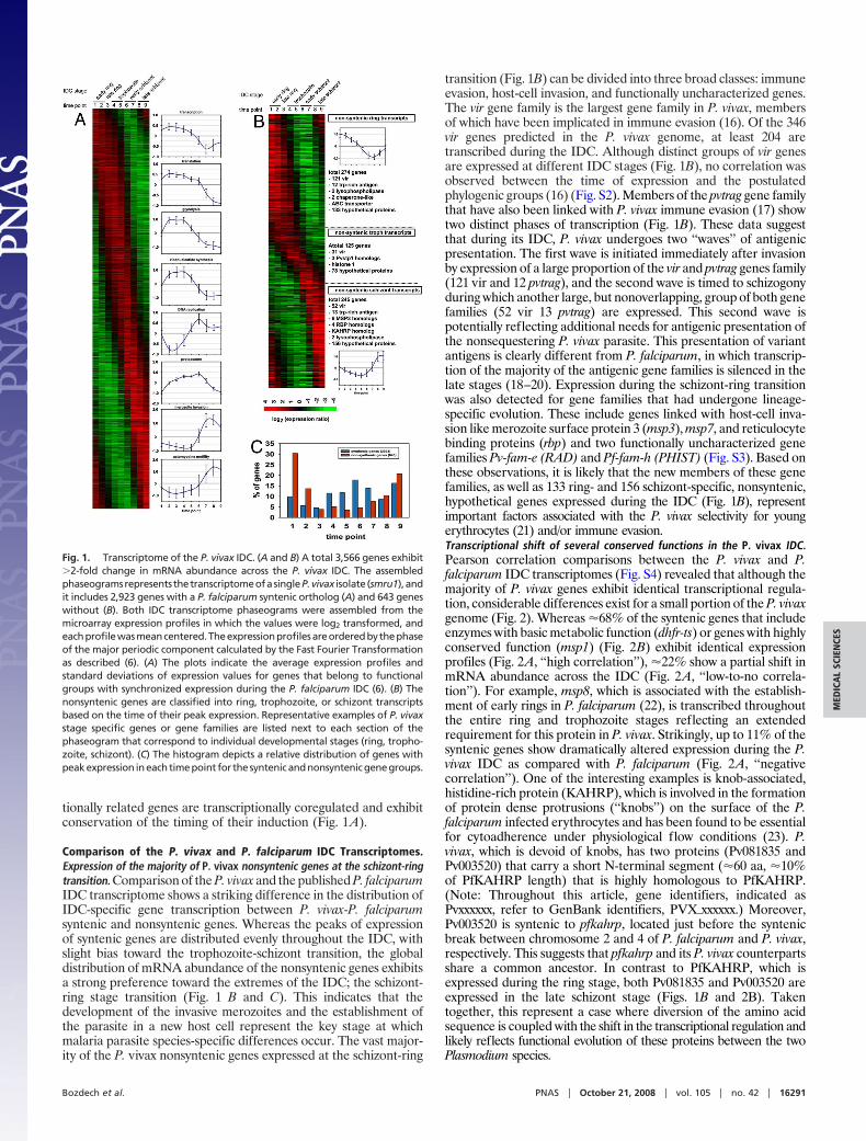
tionally related genes are transcriptionally coregulated and exhibitconservation of the timing of their induction (Fig. 1A).
Comparison of the P. vivax and P. falciparum IDC Transcriptomes.Expression of the majority of P. vivax nonsyntenic genes at the schizont-ringtransition. Comparison of the P. vivax and the published P. falciparumIDC transcriptome shows a striking difference in the distribution ofIDC-specific gene transcription between P. vivax-P. falciparumsyntenic and nonsyntenic genes. Whereas the peaks of expressionof syntenic genes are distributed evenly throughout the IDC, withslight bias toward the trophozoite-schizont transition, the globaldistribution of mRNA abundance of the nonsyntenic genes exhibitsa strong preference toward the extremes of the IDC; the schizont-ring stage transition (Fig. 1 B and C). This indicates that thedevelopment of the invasive merozoites and the establishment ofthe parasite in a new host cell represent the key stage at whichmalaria parasite species-specific differences occur. The vast major-ity of the P. vivax nonsyntenic genes expressed at the schizont-ring
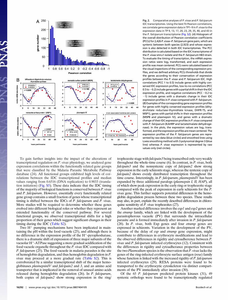
transition (Fig. 1B) can be divided into three broad classes: immuneevasion, host-cell invasion, and functionally uncharacterized genes.The vir gene family is the largest gene family in P. vivax, membersof which have been implicated in immune evasion (16). Of the 346vir genes predicted in the P. vivax genome, at least 204 aretranscribed during the IDC. Although distinct groups of vir genesare expressed at different IDC stages (Fig. 1B), no correlation wasobserved between the time of expression and the postulatedphylogenic groups (16) (Fig. S2). Members of the pvtrag gene familythat have also been linked with P. vivax immune evasion (17) showtwo distinct phases of transcription (Fig. 1B). These data suggestthat during its IDC, P. vivax undergoes two ‘‘waves’’ of antigenicpresentation. The first wave is initiated immediately after invasionby expression of a large proportion of the vir and pvtrag genes family(121 vir and 12 pvtrag), and the second wave is timed to schizogonyduring which another large, but nonoverlapping, group of both genefamilies (52 vir 13 pvtrag) are expressed. This second wave ispotentially reflecting additional needs for antigenic presentation ofthe nonsequestering P. vivax parasite. This presentation of variantantigens is clearly different from P. falciparum, in which transcrip-tion of the majority of the antigenic gene families is silenced in thelate stages (18–20). Expression during the schizont-ring transitionwas also detected for gene families that had undergone lineage-specific evolution. These include genes linked with host-cell inva-sion like merozoite surface protein 3 (msp3), msp7, and reticulocytebinding proteins (rbp) and two functionally uncharacterized genefamilies Pv-fam-e (RAD) and Pf-fam-h (PHIST) (Fig. S3). Based onthese observations, it is likely that the new members of these genefamilies, as well as 133 ring- and 156 schizont-specific, nonsyntenic,hypothetical genes expressed during the IDC (Fig. 1B), representimportant factors associated with the P. vivax selectivity for youngerythrocytes (21) and/or immune evasion.Transcriptional shift of several conserved functions in the P. vivax IDC.Pearson correlation comparisons between the P. vivax and P.falciparum IDC transcriptomes (Fig. S4) revealed that although themajority of P. vivax genes exhibit identical transcriptional regula-tion, considerable differences exist for a small portion of the P. vivaxgenome (Fig. 2). Whereas �68% of the syntenic genes that includeenzymes with basic metabolic function (dhfr-ts) or genes with highlyconserved function (msp1) (Fig. 2B) exhibit identical expressionprofiles (Fig. 2A, ‘‘high correlation’’), �22% show a partial shift inmRNA abundance across the IDC (Fig. 2A, ‘‘low-to-no correla-tion’’). For example, msp8, which is associated with the establish-ment of early rings in P. falciparum (22), is transcribed throughoutthe entire ring and trophozoite stages reflecting an extendedrequirement for this protein in P. vivax. Strikingly, up to 11% of thesyntenic genes show dramatically altered expression during the P.vivax IDC as compared with P. falciparum (Fig. 2A, ‘‘negativecorrelation’’). One of the interesting examples is knob-associated,histidine-rich protein (KAHRP), which is involved in the formationof protein dense protrusions (‘‘knobs’’) on the surface of the P.falciparum infected erythrocytes and has been found to be essentialfor cytoadherence under physiological flow conditions (23). P.vivax, which is devoid of knobs, has two proteins (Pv081835 andPv003520) that carry a short N-terminal segment (�60 aa, �10%of PfKAHRP length) that is highly homologous to PfKAHRP.(Note: Throughout this article, gene identifiers, indicated asPvxxxxxx, refer to GenBank identifiers, PVX�xxxxxx.) Moreover,Pv003520 is syntenic to pfkahrp, located just before the syntenicbreak between chromosome 2 and 4 of P. falciparum and P. vivax,respectively. This suggests that pfkahrp and its P. vivax counterpartsshare a common ancestor. In contrast to PfKAHRP, which isexpressed during the ring stage, both Pv081835 and Pv003520 areexpressed in the late schizont stage (Figs. 1B and 2B). Takentogether, this represent a case where diversion of the amino acidsequence is coupled with the shift in the transcriptional regulation andlikely reflects functional evolution of these proteins between the twoPlasmodium species.
Fig. 1. Transcriptome of the P. vivax IDC. (A and B) A total 3,566 genes exhibit�2-fold change in mRNA abundance across the P. vivax IDC. The assembledphaseogramsrepresents thetranscriptomeofasingleP.vivax isolate (smru1),andit includes 2,923 genes with a P. falciparum syntenic ortholog (A) and 643 geneswithout (B). Both IDC transcriptome phaseograms were assembled from themicroarray expression profiles in which the values were log2 transformed, andeachprofilewasmeancentered.Theexpressionprofilesareorderedbythephaseof the major periodic component calculated by the Fast Fourier Transformationas described (6). (A) The plots indicate the average expression profiles andstandard deviations of expression values for genes that belong to functionalgroups with synchronized expression during the P. falciparum IDC (6). (B) Thenonsyntenic genes are classified into ring, trophozoite, or schizont transcriptsbased on the time of their peak expression. Representative examples of P. vivaxstage specific genes or gene families are listed next to each section of thephaseogram that correspond to individual developmental stages (ring, tropho-zoite, schizont). (C) The histogram depicts a relative distribution of genes withpeakexpression ineachtimepoint for thesyntenicandnonsyntenicgenegroups.
Bozdech et al. PNAS � October 21, 2008 � vol. 105 � no. 42 � 16291
MED
ICA
LSC
IEN
CES
To gain further insights into the impact of the alterations oftranscriptional regulation on P. vivax physiology, we analyzed geneexpression correlations within the functionally related gene groupsthat were classified by the Malaria Parasite Metabolic Pathwaydatabase (24). All functional groups exhibited high levels of cor-relations between the IDC transcriptional profiles and medianvalues ranging from 0.6516 (DNA replication) to 0.9033 (transla-tion initiation) (Fig. S5). These data indicate that the IDC timingof the majority of biological functions is conserved between P. vivaxand P. falciparum. However, essentially every functionally relatedgene group contains a small fraction of genes whose transcriptionaltiming is shifted between the IDCs of P. falciparum and P. vivax.More studies will be required to determine whether these genesevolved into different biological roles or whether they represent anextended functionality of the conserved pathway. For severalfunctional groups, we observed transcriptional shifts for a highproportion of their genes which suggest significant changes in theirtiming during the IDC (Table S2).
Two H� pumping mechanisms have been implicated in main-taining the pH within the food vacuole (25), and although there isno difference in the expression profile of the H�-pyrophoshatase,there is a dramatic shift of expression of three major subunits of thevacuolar H�-ATPase suggesting a more gradual acidification of thefood vacuole organelle throughout the P. vivax IDC compared withP. falciparum (25). The food vacuole in malaria parasites is the siteof hemoglobin degradation, and thus hemoglobin degradation in P.vivax may proceed at a more gradual rate (Table S2). This iscorroborated by a similar transcriptional shift of the major hemo-globinases plasmepsinIV and falcipain2 and 3, and an amino acidtransporter that is implicated in the removal of unused amino acidsreleased during hemoglobin degradation (26). In P. falciparum,both copies of falcipain2 show strong expression in the ring/
trophozoite stage with falcipain3 being transcribed only very weaklythroughout the whole-time course (6). In contrast, in P. vivax, bothfalcipain3 and the nonsyntenic copy of falcipain2 show strongexpression in the early schizonts stage, whereas the syntenic copy offalcipain2 shows evenly distributed transcription throughout thetime course. Interestingly, in P. falciparum, plasmepsinIV has beenexpanded by three additional paralogs (plasmepsin I, II, HAP), allof which show peak expression in the early ring or trophozoite stagecompared with the peak of expression in early schizonts for the P.vivax gene. This further supports potential differences in the hemo-globin degradation process between these two species. These resultsmay also, in part, explain the recently described differences in chloro-quine sensitivity of P. vivax trophozoites (27).
Another marked difference involves the exp1 and exp2 genes andthe etramp family, which are linked with the development of theparasitophorous vacuole (PV) that surrounds the intracellularparasite and is formed immediately after invasion in P. falciparum(28). In P. vivax, both Exp genes and four etramp genes areexpressed in schizonts. Variation in the development of the PV,because of the delay of exp and etramp gene expression, mightcontribute to differences in erythrocyte modifications and lead tothe observed differences in rigidity and cytoadherence between P.vivax and P. falciparum infected erythrocytes (12). Consistent withthe differences in rigidity and cytoadherance properties betweenthe two Plasmodium species is the observation that P. vivax lacks thegenes of the ring-infected erythrocyte surface antigen (resa) familywhose function is linked with the increased rigidity of P. falciparuminfected erythrocytes (29). RESA proteins were found to betransported to the erythrocytic cytoskeleton via distinct compart-ments of the PV immediately after invasion (30).
Of the 65 P. falciparum predicted protein kinases (31), 49syntenic orthologs were found to be transcriptionally regulated
Fig. 2. Comparative analyses of P. vivax and P. falciparumIDC transcriptomes. Using the best fit Pearson correlations,wecorrelategeneexpressiondata inTP1–9 inP.vivax to theexpression data in TP 9, 13, 17, 20, 23, 29, 35, 40, and 43 inthe P. falciparum transcriptome (Fig. S2). (A) Histogram ofthe overall distribution of Pearson correlation coefficients(PCCs) for 2,426 P. vivax–P. falciparum gene pairs, which aresyntenic between both species (2,923) and whose expres-sion is also detected in both IDC transcriptomes. The PCCdistribution is calculated based on the IDC transcriptome ofthe P. vivax smru1 isolate and the P. falciparum HB3 strain.To evaluate the timing of transcription, the mRNA expres-sion ratios were log2 transformed, and each expressionprofile was mean centered. PCCs were calculated based onthe visual inspections of the corresponding expression pro-files, and we defined arbitrary PCCs thresholds that dividethe genes according to their conservation of expressionprofiles between the P. vivax and P. falciparum IDC. Highcorrelations (PCC 1 to 0.5) include genes with highly con-served IDC expression profiles, low-to-no correlations (PCC0.5to�0.2) includegeneswithapartial shift intheir the IDCexpression profile, and negative correlations (PCC �0.2 to�1) include genes with a dramatic change in their IDCexpression profiles in P. vivax compared with P. falciparum.(B) Examples of the corresponding gene expression profilesfor genes with highly conserved expression profiles (dihy-drofolate reductase-thymidilate kinase, DHFR-TS, andMSP1), genes with partial shifts in their expression profiles(MSP8 and plasmepsin IV), and genes with a dramaticchange of their IDC expression profiles in P. vivax comparedwith P. falciparum (KAHRP and lactate/malate dehydroge-nase). In the plots, the expression values are log2 trans-formed,andtheexpressionprofilesaremeancentered.Theexpression profiles of the P. falciparum genes are repre-sented by raw data (blue circles) and smoothed lines usingLoess smoothing method with 3 polynomial degree 3 (blueline) whereas P. vivax expression is represented by rawvalues only (red circles).
16292 � www.pnas.org�cgi�doi�10.1073�pnas.0807404105 Bozdech et al.
through the P. vivax IDC. Expression of 11 of these, however, isconsiderably delayed in P. vivax. Nine of these belong to the CMGC,NIMA, or CamK classes that are linked to developmental processesand/or regulation of cell proliferation (Table S2). One of these is awell characterized serine/threonine protein kinase PfNek1 (32)whose expression is shifted from early to late schizonts. The Nek1kinases were found to be involved in the onset of mitosis in otherorganisms (33). The transcriptional delays of PfNek1 and otherprotein kinases might signal a delayed onset and progression of P.vivax schizogony compared with P. falciparum. This is consistentwith the observed delay in expression of a number of DNAreplication factors during the P. vivax IDC (Table S2). Proteinscontaining the AP2-integrase, DNA-binding domain have beenrecently implicated in the global regulation of the Plasmodium lifecycle (34, 35). All 22 P. falciparum AP2 proteins have syntenichomologues in P. vivax, of which 19 exhibit identical IDC expressionprofiles between these two species; one homologue (Pv118015) isshifted from the trophozoite stage in P. falciparum to the lateschizont stage in P. vivax, and two homologues (Pv086035 andPv094580) were below the detection threshold of the P. vivaxmicroarray. These data indicate a conserved role of the AP2proteins in the regulation of the Plasmodium life cycle, but moreanalyses will be required to determine whether Pv118015, theoutlying ortholog, contributes significantly to the transcriptionaldiversion between the two plasmodium species.Transcriptional changes between P. vivax and P. falciparum are driven fromregulatory elements of individual genes. The distribution of transcrip-tional regulation along Plasmodium chromosomes shows no appar-ent association with any particular chromosomal region or domain,
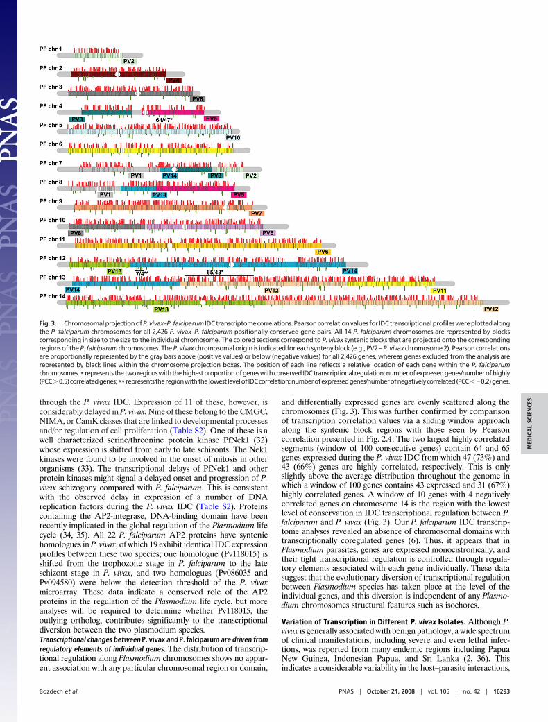
and differentially expressed genes are evenly scattered along thechromosomes (Fig. 3). This was further confirmed by comparisonof transcription correlation values via a sliding window approachalong the syntenic block regions with those seen by Pearsoncorrelation presented in Fig. 2A. The two largest highly correlatedsegments (window of 100 consecutive genes) contain 64 and 65genes expressed during the P. vivax IDC from which 47 (73%) and43 (66%) genes are highly correlated, respectively. This is onlyslightly above the average distribution throughout the genome inwhich a window of 100 genes contains 43 expressed and 31 (67%)highly correlated genes. A window of 10 genes with 4 negativelycorrelated genes on chromosome 14 is the region with the lowestlevel of conservation in IDC transcriptional regulation between P.falciparum and P. vivax (Fig. 3). Our P. falciparum IDC transcrip-tome analyses revealed an absence of chromosomal domains withtranscriptionally coregulated genes (6). Thus, it appears that inPlasmodium parasites, genes are expressed monocistronically, andtheir tight transcriptional regulation is controlled through regula-tory elements associated with each gene individually. These datasuggest that the evolutionary diversion of transcriptional regulationbetween Plasmodium species has taken place at the level of theindividual genes, and this diversion is independent of any Plasmo-dium chromosomes structural features such as isochores.
Variation of Transcription in Different P. vivax Isolates. Although P.vivax is generally associated with benign pathology, a wide spectrumof clinical manifestations, including severe and even lethal infec-tions, was reported from many endemic regions including PapuaNew Guinea, Indonesian Papua, and Sri Lanka (2, 36). Thisindicates a considerable variability in the host–parasite interactions,
Fig. 3. ChromosomalprojectionofP.vivax–P. falciparum IDCtranscriptomecorrelations.Pearsoncorrelationvalues for IDCtranscriptionalprofileswereplottedalongthe P. falciparum chromosomes for all 2,426 P. vivax–P. falciparum positionally conserved gene pairs. All 14 P. falciparum chromosomes are represented by blockscorresponding in size to the size to the individual chromosome. The colored sections correspond to P. vivax syntenic blocks that are projected onto the correspondingregions of the P. falciparum chromosomes. The P. vivax chromosomal origin is indicated for each synteny block (e.g., PV2 – P. vivax chromosome 2). Pearson correlationsare proportionally represented by the gray bars above (positive values) or below (negative values) for all 2,426 genes, whereas genes excluded from the analysis arerepresented by black lines within the chromosome projection boxes. The position of each line reflects a relative location of each gene within the P. falciparumchromosomes.* represents thetworegionswiththehighestproportionofgeneswithconserved IDCtranscriptional regulation:numberofexpressedgenes/numberofhighly(PCC�0.5)correlatedgenes;**representstheregionwiththelowestlevelofIDCcorrelation:numberofexpressedgenes/numberofnegativelycorrelated(PCC��0.2)genes.
Bozdech et al. PNAS � October 21, 2008 � vol. 105 � no. 42 � 16293
MED
ICA
LSC
IEN
CES
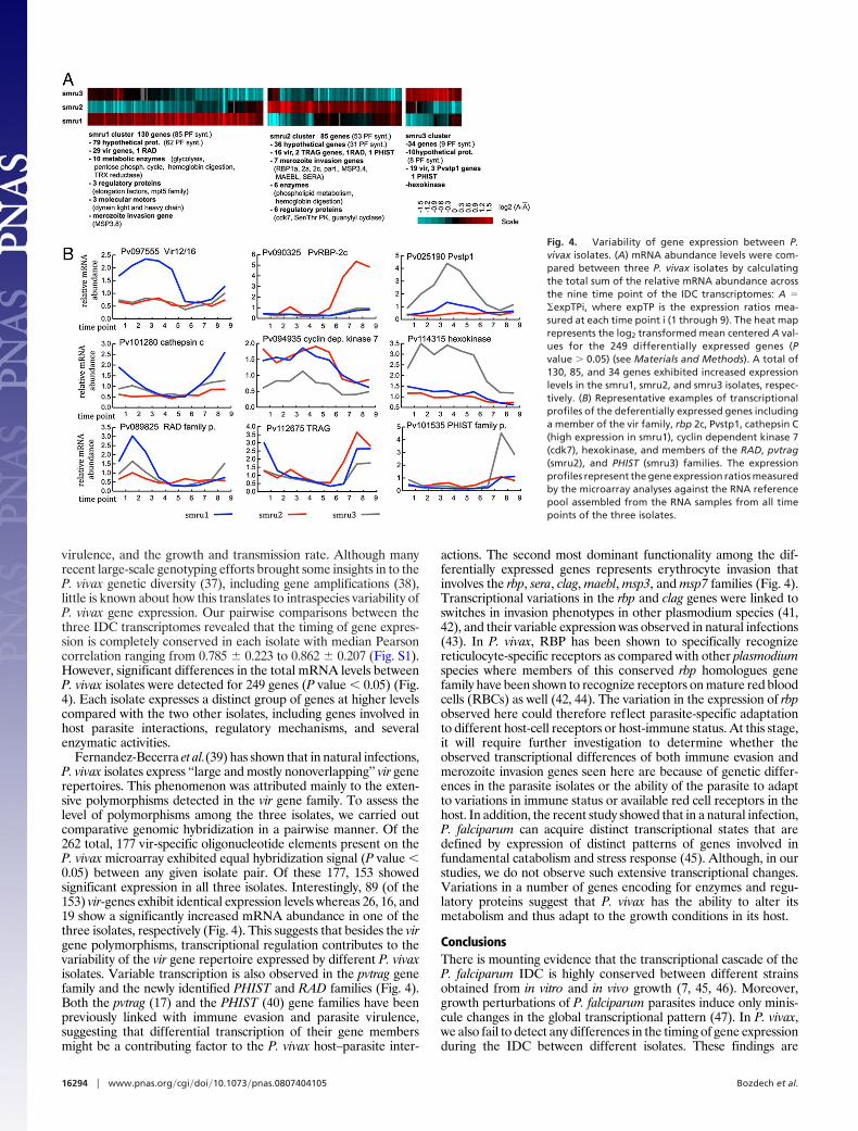
virulence, and the growth and transmission rate. Although manyrecent large-scale genotyping efforts brought some insights in to theP. vivax genetic diversity (37), including gene amplifications (38),little is known about how this translates to intraspecies variability ofP. vivax gene expression. Our pairwise comparisons between thethree IDC transcriptomes revealed that the timing of gene expres-sion is completely conserved in each isolate with median Pearsoncorrelation ranging from 0.785 � 0.223 to 0.862 � 0.207 (Fig. S1).However, significant differences in the total mRNA levels betweenP. vivax isolates were detected for 249 genes (P value � 0.05) (Fig.4). Each isolate expresses a distinct group of genes at higher levelscompared with the two other isolates, including genes involved inhost parasite interactions, regulatory mechanisms, and severalenzymatic activities.
Fernandez-Becerra et al.(39) has shown that in natural infections,P. vivax isolates express ‘‘large and mostly nonoverlapping’’ vir generepertoires. This phenomenon was attributed mainly to the exten-sive polymorphisms detected in the vir gene family. To assess thelevel of polymorphisms among the three isolates, we carried outcomparative genomic hybridization in a pairwise manner. Of the262 total, 177 vir-specific oligonucleotide elements present on theP. vivax microarray exhibited equal hybridization signal (P value �0.05) between any given isolate pair. Of these 177, 153 showedsignificant expression in all three isolates. Interestingly, 89 (of the153) vir-genes exhibit identical expression levels whereas 26, 16, and19 show a significantly increased mRNA abundance in one of thethree isolates, respectively (Fig. 4). This suggests that besides the virgene polymorphisms, transcriptional regulation contributes to thevariability of the vir gene repertoire expressed by different P. vivaxisolates. Variable transcription is also observed in the pvtrag genefamily and the newly identified PHIST and RAD families (Fig. 4).Both the pvtrag (17) and the PHIST (40) gene families have beenpreviously linked with immune evasion and parasite virulence,suggesting that differential transcription of their gene membersmight be a contributing factor to the P. vivax host–parasite inter-
actions. The second most dominant functionality among the dif-ferentially expressed genes represents erythrocyte invasion thatinvolves the rbp, sera, clag, maebl, msp3, and msp7 families (Fig. 4).Transcriptional variations in the rbp and clag genes were linked toswitches in invasion phenotypes in other plasmodium species (41,42), and their variable expression was observed in natural infections(43). In P. vivax, RBP has been shown to specifically recognizereticulocyte-specific receptors as compared with other plasmodiumspecies where members of this conserved rbp homologues genefamily have been shown to recognize receptors on mature red bloodcells (RBCs) as well (42, 44). The variation in the expression of rbpobserved here could therefore reflect parasite-specific adaptationto different host-cell receptors or host-immune status. At this stage,it will require further investigation to determine whether theobserved transcriptional differences of both immune evasion andmerozoite invasion genes seen here are because of genetic differ-ences in the parasite isolates or the ability of the parasite to adaptto variations in immune status or available red cell receptors in thehost. In addition, the recent study showed that in a natural infection,P. falciparum can acquire distinct transcriptional states that aredefined by expression of distinct patterns of genes involved infundamental catabolism and stress response (45). Although, in ourstudies, we do not observe such extensive transcriptional changes.Variations in a number of genes encoding for enzymes and regu-latory proteins suggest that P. vivax has the ability to alter itsmetabolism and thus adapt to the growth conditions in its host.
ConclusionsThere is mounting evidence that the transcriptional cascade of theP. falciparum IDC is highly conserved between different strainsobtained from in vitro and in vivo growth (7, 45, 46). Moreover,growth perturbations of P. falciparum parasites induce only minis-cule changes in the global transcriptional pattern (47). In P. vivax,we also fail to detect any differences in the timing of gene expressionduring the IDC between different isolates. These findings are
Fig. 4. Variability of gene expression between P.vivax isolates. (A) mRNA abundance levels were com-pared between three P. vivax isolates by calculatingthe total sum of the relative mRNA abundance acrossthe nine time point of the IDC transcriptomes: A ��expTPi, where expTP is the expression ratios mea-sured at each time point i (1 through 9). The heat maprepresents the log2 transformed mean centered A val-ues for the 249 differentially expressed genes (Pvalue � 0.05) (see Materials and Methods). A total of130, 85, and 34 genes exhibited increased expressionlevels in the smru1, smru2, and smru3 isolates, respec-tively. (B) Representative examples of transcriptionalprofiles of the deferentially expressed genes includinga member of the vir family, rbp 2c, Pvstp1, cathepsin C(high expression in smru1), cyclin dependent kinase 7(cdk7), hexokinase, and members of the RAD, pvtrag(smru2), and PHIST (smru3) families. The expressionprofiles represent the gene expression ratios measuredby the microarray analyses against the RNA referencepool assembled from the RNA samples from all timepoints of the three isolates.
16294 � www.pnas.org�cgi�doi�10.1073�pnas.0807404105 Bozdech et al.
consistent with predictions that the timing of gene expressionduring the IDC of Plasmodium species does not fluctuate betweendifferent strains or isolates (46). Thus, it is reasonable to speculatethat many (if not all) of the observed interspecies variationsrepresent conserved transcriptional differences that reflect evolu-tion of the individual Plasmodium species.
Similar to P. falciparum, the intraerythrocytic development of P.vivax is characterized by extensive transcriptional regulation inwhich each biological function is timed to a specific section of theIDC. Although most of the P. vivax genes exhibit an identicalexpression profile across the IDC compared with their P. falciparumsyntenic orthologs, there are partial shifts as well as dramaticalterations in the mRNA abundance profiles for 22% and 11%genes, respectively. In several cases, these changes represent sub-stantial alteration of the timing for an entire biological functionincluding hemoglobin degradation, host parasite interaction, pro-tein export to the host-cell cytoplasm, and DNA replication. Incontrast to the conserved portion of the P. vivax genome, thenonsyntenic P. vivax genes are activated predominantly at theschizont/ring interphase which suggests that the species differencesderive mainly from events occurring at red cell invasion and earlyintraerythrocytic development. Moreover, expression of the ma-jority of vir and pvtrag gene families throughout the IDC indicatemajor differences in antigenic presentation between P. falciparumand P. vivax. The even distribution of the differentially expressedgenes along the P. vivax-P. falciparum chromosomal synteny blockssuggests that the diversion of transcriptional control between thesetwo species occurred at the regulatory elements of individual genes.
The intraisolate diversity of gene expression indicates an extensivecapacity of P. vivax to adapt to its host and modify its virulence. Thisfundamental, biological information about P. vivax should facilitatethe development of innovative control measures against this re-markably resilient parasite.
Materials and MethodsSample Collection and RNA Isolation. Adult patients from Northwestern Thai-land (Shoklo Malaria Research Unit, Mahidol University, Thailand) with symp-tomatic vivax malaria gave informed consent to donate 10 ml of blood. The P.vivax infected blood samples were collected from three preselected patientsbefore receiving standard chloroquine treatment. For more details, see SI Text.
Target DNA Preparations and Microarray Analysis. The microarray contained5,727 60-mer oligonucleotides that represented a total of 5,335 predicted genesin the P. vivax genome by at least one unique probe. From these, 5,063 arerepresented by a single oligonucleotide, whereas 272 genes (typically long ORFs)are represented by two or more oligonucleotide elements. The P. vivax microar-ray oligonucleotide set was designed by OligoRankPick (15). Sequences of alloligonucleotides and further information are available online at http://zblab.sbs.ntu.edu.sg/vivax/index.html. The P. virax long-oligo array is also avail-able through the Pathogen Functional Genomics Resource Center (http://pfgrc.jcvi.org).
Data Processing and Analysis. Microarray data processing and analysis (includingthe Fast Fourier Transform) was carried out as described (6). For more details, seeSI Text.
ACKNOWLEDGMENTS. This work was supported by Biomedical Research Coun-cil Singapore Grants BMRC 04/1/22/19/364 and BMRC 05/1/22/19/398 and NationalMedical Research Council Singapore Grant NMRC/CPG/016/2005.
1. Mendis K, Sina BJ, Marchesini P, Carter R (2001) The neglected burden of Plasmodiumvivax malaria. Am J Trop Med Hyg 64:97–106.
2. Price RN, et al. (2007) Vivax malaria: Neglected and not benign. Am J Trop Med Hyg77:79–87.
3. Hay SI, et al. (2004) The global distribution and population at risk of malaria: Past,present, and future. Lancet Infect Dis 4:327–336.
4. Krotoski WA, et al. (1986) Observations on early and late post-sporozoite tissue stagesin primate malaria. IV. Pre-erythrocytic schizonts and/or hypnozoites of Chesson andNorth Korean strains of Plasmodium vivax in the chimpanzee. Am J Trop Med Hyg35:263–274.
5. Gardner MJ, et al. (2002) Genome sequence of the human malaria parasite Plasmodiumfalciparum. Nature 419:498–511.
6. Bozdech Z, et al. (2003) The transcriptome of the intraerythrocytic developmental cycleof Plasmodium falciparum. PLoS Biol 1:E5.
7. Le Roch KG, et al. (2003) Discovery of gene function by expression profiling of themalaria parasite life cycle. Science 301:1503–1508.
8. Grannham PCC (1966) Malaria Parasites and Haemosporidia (Blackwell ScientificPublications, Oxford).
9. Mons B (1990) Preferential invasion of malarial merozoites into young red blood cells.Blood Cells 16:299–312.
10. Aikawa M (1971) Parasitological review. Plasmodium: The fine structure of malarialparasites. Exp Parasitol 30:284–320.
11. Aikawa M, Miller LH, Rabbege J (1975) Caveola–vesicle complexes in the plasmalemmaof erythrocytes infected by Plasmodium vivax and P cynomolgi. Unique structuresrelated to Schuffner’s dots. Am J Pathol 79:285–300.
12. Suwanarusk R, et al. (2004) The deformability of red blood cells parasitized by Plas-modium falciparum and P. vivax. J Infect Dis 189:190–194.
13. Carlton J, et al. (2008) Comparative genomics of the neglected human malaria parasitePlasmodium vivax. Nature, in press.
14. Russell BM, et al. (2003) Simple in vitro assay for determining the sensitivity ofPlasmodium vivax isolates from fresh human blood to antimalarials in areas where P.vivax is endemic. Antimicrob Agents Chemother 47:170–173.
15. Hu G, et al. (2007) Selection of long oligonucleotides for gene expression microarraysusing weighted rank-sum strategy. BMC Bioinformatics 8:350–362.
16. del Portillo HA, et al. (2001) A superfamily of variant genes encoded in the subtelomericregion of Plasmodium vivax. Nature 410:839–842.
17. Jalah R, et al. (2005) Identification, expression, localization and serological character-ization of a tryptophan-rich antigen from the human malaria parasite Plasmodiumvivax. Mol Biochem Parasitol 142:158–169.
18. Duraisingh MT, et al. (2005) Heterochromatin silencing and locus repositioning linkedto regulation of virulence genes in Plasmodium falciparum. Cell 121:13–24.
19. Freitas-Junior LH, et al. (2005) Telomeric heterochromatin propagation and histoneacetylation control mutually exclusive expression of antigenic variation genes inmalaria parasites. Cell 121:25–36.
20. Tham WH, Payne PD, Brown GV, Rogerson SJ (2007) Identification of basic transcrip-tional elements required for rif gene transcription. Int J Parasitol 37:605–615.
21. Galinski MR, Medina CC, Ingravallo P, Barnwell JW (1992) A reticulocyte-bindingprotein complex of Plasmodium vivax merozoites. Cell 69:1213–1226.
22. Drew DR, Sanders PR, Crabb BS (2005) Plasmodium falciparum merozoite surfaceprotein 8 is a ring-stage membrane protein that localizes to the parasitophorousvacuole of infected erythrocytes. Infect Immun 73:3912–3922.
23. Rug M, et al. (2006) The role of KAHRP domains in knob formation and cytoadherenceof P falciparum-infected human erythrocytes. Blood 108:370–378.
24. Ginsburg H (2006) Progress in in silico functional genomics: The malaria MetabolicPathways database. Trends Parasitol 22:238–240.
25. Saliba KJ, et al. (2003) Acidification of the malaria parasite’s digestive vacuole by aH�-ATPase and a H�-pyrophosphatase. J Biol Chem 278:5605–5612.
26. Martin RE, et al. (2005) The ‘‘permeome’’ of the malaria parasite: An overview of themembrane transport proteins of Plasmodium falciparum. Genome Biol 6:R26.
27. Sharrock WW, et al. (2008) Plasmodium vivax trophozoites insensitive to chloroquine.Malar J 7:94–100.
28. Spielmann T, et al. (2006) Organization of ETRAMPs and EXP-1 at the parasite-host cellinterface of malaria parasites. Mol Microbiol 59:779–794.
29. Mills JP, et al. (2007) Effect of plasmodial RESA protein on deformability of human redblood cells harboring Plasmodium falciparum. Proc Natl Acad Sci USA 104:9213–9217.
30. Culvenor JG, Day KP, Anders RF (1991) Plasmodium falciparum ring-infected erythro-cyte surface antigen is released from merozoite dense granules after erythrocyteinvasion. Infect Immun 59:1183–1187.
31. Ward P, Equinet L, Packer J, Doerig C (2004) Protein kinases of the human malaria parasitePlasmodium falciparum: The kinome of a divergent eukaryote. BMC Genomics 5:79–97.
32. Dorin D, et al. (2005) PfPK7, an atypical MEK-related protein kinase, reflects theabsence of classical three-component MAPK pathways in the human malaria parasitePlasmodium falciparum. Mol Microbiol 55:184–196.
33. Osmani SA, Pu RT, Morris NR (1988) Mitotic induction and maintenance by overex-pression of a G2-specific gene that encodes a potential protein kinase. Cell 53:237–244.
34. Balaji S, Babu MM, Iyer LM, Aravind L (2005) Discovery of the principal specifictranscription factors of Apicomplexa and their implication for the evolution of theAP2-integrase DNA binding domains. Nucleic Acids Res 33:3994–4006.
35. De Silva EK, et al. (2008) Specific DNA-binding by apicomplexan AP2 transcriptionfactors. Proc Natl Acad Sci USA 105:8393–8398.
36. Tjitra E, et al. (2008) Multidrug-resistant Plasmodium vivax associated with severe andfatal malaria: A prospective study in Papua, Indonesia. PLoS Med 5:e128.
37. Karunaweera ND, et al. (2008) Extensive microsatellite diversity in the human malariaparasite Plasmodium vivax. Gene 410:105–112.
38. Suwanarusk R, et al. (2007) Chloroquine resistant Plasmodium vivax: In vitro charac-terisation and association with molecular polymorphisms. PLoS ONE 2:e1089.
39. Fernandez-Becerra C, et al. (2005) Variant proteins of Plasmodium vivax are notclonally expressed in natural infections. Mol Microbiol 58:648–658.
40. Sargeant TJ, et al. (2006) Lineage-specific expansion of proteins exported to erythro-cytes in malaria parasites. Genome Biol 7:R12.
41. Cortes A, et al. (2007) Epigenetic silencing of Plasmodium falciparum genes linked toerythrocyte invasion. PLoS Pathog 3:e107.
42. Iyer JK, Amaladoss A, Genesan S, Preiser PR (2007) Variable expression of the 235-kDarhoptry protein of Plasmodium yoelii mediate host cell adaptation and immuneevasion. Mol Microbiol 65:333–346.
43. Nery S, et al. (2006) Expression of Plasmodium falciparum genes involved in erythrocyteinvasion varies among isolates cultured directly from patients. Mol Biochem Parasitol149:208–215.
44. Gaur D, Mayer DC, Miller LH (2004) Parasite ligand-host receptor interactions duringinvasion of erythrocytes by Plasmodium merozoites. Int J Parasitol 34:1413–1429.
45. Daily JP, et al. (2007) Distinct physiological states of Plasmodium falciparum in malaria-infected patients. Nature 450:1091–1095.
46. Llinas M, et al. (2006) Comparative whole genome transcriptome analysis of threePlasmodium falciparum strains. Nucleic Acids Res 34:1166–1173.
47. Gunasekera AM, et al. (2007) Plasmodium falciparum: Genome wide perturbations intranscript profiles among mixed stage cultures after chloroquine treatment. ExpParasitol 117:87–92.
Bozdech et al. PNAS � October 21, 2008 � vol. 105 � no. 42 � 16295
MED
ICA
LSC
IEN
CES