The structure of Xis reveals the basis for filament formation and insight into DNA bending within a...
11
The Structure of Xis Reveals the Basis for Filament Formation and Insight into DNA Bending within a Mycobacteriophage Intasome Shweta Singh 1 , Joseph G. Plaks 1 , Nicholas J. Homa 1,3 , Christopher G. Amrich 1 , Annie Héroux 2 , Graham F. Hatfull 1 and Andrew P. VanDemark 1 1 - Department of Biological Sciences, University of Pittsburgh, Pittsburgh, PA 15260, USA 2 - Department of Biology, Brookhaven National Laboratory, Upton, NY 11973, USA 3 - Present address: N. J. Homa, 426 CARL Building, Duke University, Durham, NC 27710, USA Correspondence to Andrew P. VanDemark: Department of Biological Sciences, University of Pittsburgh, 4249 Fifth Avenue, 360A Langley Hall, Pittsburgh, PA 15260, USA. [email protected] http://dx.doi.org/10.1016/j.jmb.2013.10.002 Edited by G. Schulz Abstract The recombination directionality factor, Xis, is a DNA bending protein that determines the outcome of integrase-mediated site-specific recombination by redesign of higher-order protein–DNA architectures. Although the attachment site DNA of mycobacteriophage Pukovnik is likely to contain four sites for Xis binding, Xis crystals contain five subunits in the asymmetric unit, four of which align into a Xis filament and a fifth that is generated by an unusual domain swap. Extensive intersubunit contacts stabilize a bent filament-like arrangement with Xis monomers aligned head to tail. The structure implies a DNA bend of ~ 120°, which is in agreement with DNA bending measured in vitro. Formation of attR-containing intasomes requires only Int and Xis, distinguishing Pukovnik from lambda. Therefore, we conclude that, in Pukovnik, Xis-induced DNA bending is sufficient to promote intramolecular Int-mediated bridges during intasome formation. © 2013 Elsevier Ltd. All rights reserved. Introduction Prophage establishment by temperate bacterio- phages typically involves integrase-mediated site- specific recombination between the phage attP and chromosomal attB site [1]. Of the large family of phage-encoded integrases of the tyrosine recombi- nase type that mediate these events, phage lambda represents a well-studied prototype [1–3]. In lambda, integration requires integrase (Int), the host integra- tion factor (IHF), a large (250 bp) attP site that contains both core-type and arm-type integrase binding sites, and a smaller attB site (25 bp). Strand exchange occurs within the shared common core sequence and proceeds through a Holliday Junction intermediate [4,5]. Prophage excision, which occurs during induction of lytic growth, is also catalyzed by Int, requires IHF, but is strongly dependent on the recombination directionality factor Xis [6,7]. These Int-mediated reactions are strongly directional. In the absence of Xis, the only productive pair of substrates are attP and attB, whereas in the presence of Xis, only attL and attR recombine; Xis is also a strong inhibitor of integrative recombination [3]. The molecular basis of this directionality lies in the requirement for the formation of higher-order pro- tein–DNA architectures for synapsis and strand exchange to occur [6]. Int is a bivalent DNA binding protein that can bind simultaneously to core- and arm-type binding sites forming intra or intermolecular protein bridges [8]. Formation of recombinationally active complexes requires the introduction of DNA bends, and this is accomplished through the binding of IHF [9,10] to the H1, H2, and H′ sites in lambda attP and through the binding of Xis to the X1, X′, and X2 binding sites in attP (and attR) [6,11,12]. The 72-residue Lambda Xis bends DNA upon binding and also contributes cooperative interactions through the binding of its extreme C-terminus to the arm-type binding N-terminal domain of Int [13–15]. Structural analysis of lambda Xis shows that it binds DNA through a winged-helix motif of the MerR superfamily, 0022-2836/$ - see front matter © 2013 Elsevier Ltd. All rights reserved. J. Mol. Biol. (2013) 426, 412–422 Article
-
Upload
independent -
Category
Documents
-
view
10 -
download
0
Transcript of The structure of Xis reveals the basis for filament formation and insight into DNA bending within a...

Article
Shweta Singh1
2
0022-2836/$ - see front m
The Structure of Xis Reveals the Basis forFilament Formation and Insight into DNABending within aMycobacteriophage Intasome
, Joseph G. Plaks1, Nicho
las J. Homa1, 3, Christopher G. Amrich1,Annie Héroux , Graham F. Hatfull 1 and Andrew P. VanDemark11 - Department of Biological Sciences, University of Pittsburgh, Pittsburgh, PA 15260, USA2 - Department of Biology, Brookhaven National Laboratory, Upton, NY 11973, USA3 - Present address: N. J. Homa, 426 CARL Building, Duke University, Durham, NC 27710, USA
Correspondence to Andrew P. VanDemark: Department of Biological Sciences, University of Pittsburgh, 4249 FifthAvenue, 360A Langley Hall, Pittsburgh, PA 15260, USA. [email protected]://dx.doi.org/10.1016/j.jmb.2013.10.002Edited by G. Schulz
Abstract
The recombination directionality factor, Xis, is a DNA bending protein that determines the outcome ofintegrase-mediated site-specific recombination by redesign of higher-order protein–DNA architectures.Although the attachment site DNA of mycobacteriophage Pukovnik is likely to contain four sites for Xisbinding, Xis crystals contain five subunits in the asymmetric unit, four of which align into a Xis filament and afifth that is generated by an unusual domain swap. Extensive intersubunit contacts stabilize a bentfilament-like arrangement with Xis monomers aligned head to tail. The structure implies a DNA bend of ~120°,which is in agreement with DNA bending measured in vitro. Formation of attR-containing intasomes requiresonly Int and Xis, distinguishing Pukovnik from lambda. Therefore, we conclude that, in Pukovnik, Xis-inducedDNA bending is sufficient to promote intramolecular Int-mediated bridges during intasome formation.
© 2013 Elsevier Ltd. All rights reserved.
Introduction
Prophage establishment by temperate bacterio-phages typically involves integrase-mediated site-specific recombination between the phage attP andchromosomal attB site [1]. Of the large family ofphage-encoded integrases of the tyrosine recombi-nase type that mediate these events, phage lambdarepresents a well-studied prototype [1–3]. In lambda,integration requires integrase (Int), the host integra-tion factor (IHF), a large (250 bp) attP site thatcontains both core-type and arm-type integrasebinding sites, and a smaller attB site (25 bp). Strandexchange occurs within the shared common coresequence and proceeds through a Holliday Junctionintermediate [4,5]. Prophage excision, which occursduring induction of lytic growth, is also catalyzed byInt, requires IHF, but is strongly dependent on therecombination directionality factor Xis [6,7]. TheseInt-mediated reactions are strongly directional. In theabsence of Xis, the only productive pair of substrates
atter © 2013 Elsevier Ltd. All rights reserve
are attP and attB, whereas in the presence of Xis,only attL and attR recombine; Xis is also a stronginhibitor of integrative recombination [3].The molecular basis of this directionality lies in the
requirement for the formation of higher-order pro-tein–DNA architectures for synapsis and strandexchange to occur [6]. Int is a bivalent DNA bindingprotein that can bind simultaneously to core- andarm-type binding sites forming intra or intermolecularprotein bridges [8]. Formation of recombinationallyactive complexes requires the introduction of DNAbends, and this is accomplished through the bindingof IHF [9,10] to the H1, H2, and H′ sites in lambda attPand through the binding of Xis to the X1, X′, and X2binding sites in attP (and attR) [6,11,12]. The72-residue Lambda Xis bends DNA upon bindingand also contributes cooperative interactions throughthe binding of its extreme C-terminus to the arm-typebinding N-terminal domain of Int [13–15]. Structuralanalysis of lambda Xis shows that it binds DNAthrough a winged-helix motif of the MerR superfamily,
d. J. Mol. Biol. (2013) 426, 412–422

Fig. 1. Pukovnik Xis promotes integrase-mediated excisive recombination. (a) Organization of mycobacteriophageattachment sites. The phage and bacterial attachment sites, attP and attB, respectively, each contain core-type integrasebinding sites (C and C′ in attP; B and B′ in attB) flanking the sites of strand exchange; attP also contains arm-type integrasebinding sites P1–P5 (gray) and a region to which mycobacterial integration host (mIHF) is predicted to bind (light gray).Integrase (Int) mediates both integrative and excisive recombination, and both reactions typically require mIHF. Excisionrequires excise (Xis), which also inhibits integration. The Xis binding sequence is indicated with crosshatched box. (b) Xis–DNA complexes were formed using a 50-bp attR fragment containing X1–X4 sites or an equivalent fragment withmutations within the X1–X4 sites and were separated using native-gel electrophoresis. The Xis protein concentrationswere 0, 0.125, 0.25, 0.5, 1, 2, 4, 8, 16, and 32 μM. (c) Pukovnik gp37 is a functional Xis. Int, mIHF, and Xis were mixed withlinear attL (0.12 pmol) and plasmid attR (0.03 pmol) DNA substrates. The formation of a linear product is separated on0.8% agarose gel in 1× TBE running buffer and visualized by ethidium bromide staining. (d) Xis promotes Int binding. Int(250 nM) and Xis (100 nM) were mixed with a 365-bp attR fragment as indicated, and the resulting complexes wereseparated by native-gel electrophoresis. (e) Xis but not IHF is required for attR intasome formation. Xis (100 nM), mIHF(25 nM), and Int were mixed with attR DNA and separated as in (d). Int concentrations were 0, 0.3, 0.9, 2.7, 8.1, 24, 74,220, 670, 2000, and 6000 nM.
413Xis-Induced Bending Establishes Intasome Structure
using contacts between a central loop and the wingmotif to mediate interactions between Xis proteinswhen bound to DNA and presumably drive cooper-ative binding and the formation of a micronucleopro-tein filament with a modest bend [16,17].Mycobacteriophage L5 encodes a far distant
relative of lambda Int but shares many commonfeatures [18]. Its attP site contains arm- and
core-type integrase binding sites, although thespecific arrangements of arm-type sites are differentfrom those in lambda attP [19]. L5 also requires ahost factor for both integration and excision,although this mycobacterial integration host factor(mIHF) is unrelated at the sequence level toEscherichia coli IHF and only binds specifically toattP in the presence of L5 Int [20,21]. The L5 Xis
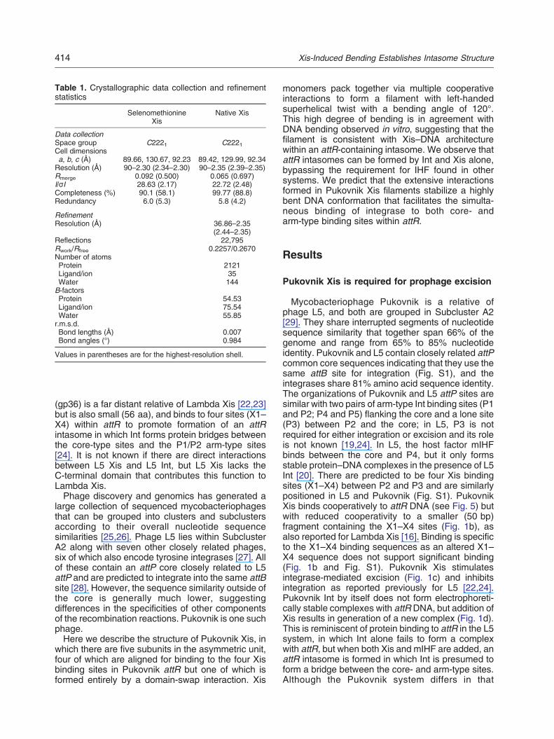
Table 1. Crystallographic data collection and refinementstatistics
SelenomethionineXis
Native Xis
Data collectionSpace group C2221 C2221Cell dimensionsa, b, c (Å) 89.66, 130.67, 92.23 89.42, 129.99, 92.34Resolution (Å) 90–2.30 (2.34–2.30) 90–2.35 (2.39–2.35)Rmerge 0.092 (0.500) 0.065 (0.697)I/σI 28.63 (2.17) 22.72 (2.48)Completeness (%) 90.1 (58.1) 99.77 (88.8)Redundancy 6.0 (5.3) 5.8 (4.2)
RefinementResolution (Å) 36.86–2.35
(2.44–2.35)Reflections 22,795Rwork/Rfree 0.2257/0.2670Number of atomsProtein 2121Ligand/ion 35Water 144B-factorsProtein 54.53Ligand/ion 75.54Water 55.85r.m.s.d.Bond lengths (Å) 0.007Bond angles (°) 0.984
Values in parentheses are for the highest-resolution shell.
414 Xis-Induced Bending Establishes Intasome Structure
(gp36) is a far distant relative of Lambda Xis [22,23]but is also small (56 aa), and binds to four sites (X1–X4) within attR to promote formation of an attRintasome in which Int forms protein bridges betweenthe core-type sites and the P1/P2 arm-type sites[24]. It is not known if there are direct interactionsbetween L5 Xis and L5 Int, but L5 Xis lacks theC-terminal domain that contributes this function toLambda Xis.Phage discovery and genomics has generated a
large collection of sequenced mycobacteriophagesthat can be grouped into clusters and subclustersaccording to their overall nucleotide sequencesimilarities [25,26]. Phage L5 lies within SubclusterA2 along with seven other closely related phages,six of which also encode tyrosine integrases [27]. Allof these contain an attP core closely related to L5attP and are predicted to integrate into the same attBsite [28]. However, the sequence similarity outside ofthe core is generally much lower, suggestingdifferences in the specificities of other componentsof the recombination reactions. Pukovnik is one suchphage.Here we describe the structure of Pukovnik Xis, in
which there are five subunits in the asymmetric unit,four of which are aligned for binding to the four Xisbinding sites in Pukovnik attR but one of which isformed entirely by a domain-swap interaction. Xis
monomers pack together via multiple cooperativeinteractions to form a filament with left-handedsuperhelical twist with a bending angle of 120°.This high degree of bending is in agreement withDNA bending observed in vitro, suggesting that thefilament is consistent with Xis–DNA architecturewithin an attR-containing intasome. We observe thatattR intasomes can be formed by Int and Xis alone,bypassing the requirement for IHF found in othersystems. We predict that the extensive interactionsformed in Pukovnik Xis filaments stabilize a highlybent DNA conformation that facilitates the simulta-neous binding of integrase to both core- andarm-type binding sites within attR.
Results
Pukovnik Xis is required for prophage excision
Mycobacteriophage Pukovnik is a relative ofphage L5, and both are grouped in Subcluster A2[29]. They share interrupted segments of nucleotidesequence similarity that together span 66% of thegenome and range from 65% to 85% nucleotideidentity. Pukovnik and L5 contain closely related attPcommon core sequences indicating that they use thesame attB site for integration (Fig. S1), and theintegrases share 81% amino acid sequence identity.The organizations of Pukovnik and L5 attP sites aresimilar with two pairs of arm-type Int binding sites (P1and P2; P4 and P5) flanking the core and a lone site(P3) between P2 and the core; in L5, P3 is notrequired for either integration or excision and its roleis not known [19,24]. In L5, the host factor mIHFbinds between the core and P4, but it only formsstable protein–DNA complexes in the presence of L5Int [20]. There are predicted to be four Xis bindingsites (X1–X4) between P2 and P3 and are similarlypositioned in L5 and Pukovnik (Fig. S1). PukovnikXis binds cooperatively to attR DNA (see Fig. 5) butwith reduced cooperativity to a smaller (50 bp)fragment containing the X1–X4 sites (Fig. 1b), asalso reported for Lambda Xis [16]. Binding is specificto the X1–X4 binding sequences as an altered X1–X4 sequence does not support significant binding(Fig. 1b and Fig. S1). Pukovnik Xis stimulatesintegrase-mediated excision (Fig. 1c) and inhibitsintegration as reported previously for L5 [22,24].Pukovnik Int by itself does not form electrophoreti-cally stable complexes with attR DNA, but addition ofXis results in generation of a new complex (Fig. 1d).This is reminiscent of protein binding to attR in the L5system, in which Int alone fails to form a complexwith attR, but when both Xis and mIHF are added, anattR intasome is formed in which Int is presumed toform a bridge between the core- and arm-type sites.Although the Pukovnik system differs in that

Fig. 2. Structure of Pukovnik Xis. (a) Xis protomer packing within the crystal forms left-handed superhelical filaments.Surface representation of Xis protomers (green, red, blue, yellow, and cyan) that pack together to form an Xis filament.Adjacent filaments are bridged by canonical packing of a domain-swapped protomer (*), while the white protomer is adomain-swapped Xis from an adjacent filament. (b) Cartoon representation of the asymmetric unit. The positions of thefour filament forming Xis protomers are indicated, as are the positions of the DNA recognition helices and the C-terminaltail that packs next to the wing, providing a platform for Xis–Xis interactions that drives cooperative DNA binding.
415Xis-Induced Bending Establishes Intasome Structure
formation of the attR–Int–Xis complex is mIHFindependent (Fig. 1d and e), it seems likely that italso includes a similar bridging interaction and acomplex—an attR intasome—with overall similarityto the L5.
Structure of the Pukovnik Xis
The structure of Pukovnik Xis was determined byX-ray crystallography. Phasing was accomplishedusing the single-wavelength anomalous dispersionmethod from crystals containing selenomethionine-substituted protein and refined against native data at2.35 Å resolution to a free R value of 26.7% (seeMaterials and Methods and Table 1 for a completedescription of the structure determination processand statistics). The asymmetric unit contains fiveindividual Xis subunits that stack onto each otherthrough an extensive array of protein–protein inter-actions forming a filament with left-handed superhe-lical twist (Fig. 2a and b). Stacking within the filamentis quite regular with each monomer exhibiting an~60° bending angle and a twist of 40°. Within theasymmetric unit, four Xis protomers are containedwithin the filament region with the fifth protomerbeing positioned adjacent to the filament andparticipating in stacking with a symmetry-relatedfilament. All protomers adopt a compact winged-helix fold with very little structural variation betweenthem (0.3 Å r.m.s.d. over all Cα atoms). Crystalliza-tion required the inclusion of either ammonium
sulfate or ammonium phosphate, many of whichwere well resolved particularly in the domain-swapinterface (Fig. S2). Despite limited sequence identi-ty, Pukovnik Xis is also highly similar structurally toother Xis proteins currently described in the struc-tural database, including phage lambda Xis (1LX8)(1.9 Å r.m.s.d. over 42 Cα atoms) [17,30,31]. Thedomain-swap Xis pair are formed by a dramaticrearrangement within the loop connecting β1 and β2(residues 36–39) allowing the C-terminal residues40–56 to interact with the N-terminal 35 residues of aneighboring Xis monomer, reforming two Xis mono-mers (Fig. S2). The resulting domain-swap Xisproteins retain the canonical fold but are linkedtogether by their wing regions.
Subunit interactions and direct DNA contactspromote excision
While the overall fold of Pukovnik gp37 is similar toother Xis proteins, there are few sequence motifsthat are conserved between lambda and mycobac-teriophage Xis proteins from the A2 subcluster ofmycobacteriophages (Fig. 3a). We therefore soughtto determine sequence elements responsible forDNA recognition. We began this exploration bygenerating a mutant with substitutions at positionsK19, R22, and N23 within the DNA recognition helixand with substitutions at R34 and R38 within thewing, positions that mediate direct contacts withDNA in lambda [16,32]. These putative DNA

Fig. 3. Both predicted DNA binding residues and observed Xis protomer contacts are important for DNA binding andexcision. (a) Observed secondary structure for Xis protomers with either the canonical or the domain-swapped fold.Multiple sequence alignment for related Xis proteins and lambda. Residues mediating Xis–Xis contacts in both Pukovnikand Lambda are indicated (green squares), as are the residues with base-specific (red) and phosphate-backbone (orange)DNA contacts. (b) Xis–Xis contacts in Pukovnik Xis are formed by packing of the C-terminal tail against the DNArecognition wing and a series of hydrophobic interactions centered on L35. (c) Xis–DNA complexes were formed using a365-bp attR fragment and the indicated Xis mutant predicted to impair Xis–Xis contacts. Complexes were separated usingnative-gel electrophoresis. The Xis protein concentrations were 0, 1.6 nM, 4.8 nM, 14.6 nM, 43 nM, 131 nM, 390 nM,1.2 μM, 3.5 μM, 10.6 μM, and 32 μM. (d) DNA recognition residues and the C-terminal tail are each required for excision.The indicated Xis mutants were mixed with mIHF, Int, linear attL, and plasmid attR as in Fig. 1. Reaction products areseparated using 0.8% agarose in 1× TBE running buffer and visualized using ethidium bromide staining.
416 Xis-Induced Bending Establishes Intasome Structure
recognition mutants were purified and tested for theirability to recognize attR X1–X4 DNA, and aspredicted, all show a strong reduction in the affinityfor attR DNA (Fig. S3). We conclude that Pukovnikand Lambda Xis proteins recognize DNA similarly.We next sought to determine if the protein–protein
interactions that are mediating filament formationwithin our structure are also important for DNArecognition and phage excision. These contacts arefeatures that differentiate Pukovnik and lambda Xis.The most important of these are residues 51–54 ofPukovnik Xis, which make extensive contacts withthe wing of the adjacent monomer. These contactsinclude several backbone hydrogen bonds, forminga three-stranded β sheet with the wing of an adjacentsubunit (Fig. 3b). This interaction provides a platformfor cooperative interactions positioning Xis subunitsin a head-to-tail arrangement that drives filamentformation. Deletion of the C-terminal tail through theintroduction of a stop codon after residue 50 greatly
diminishes DNA binding affinity and abolishesexcision (Fig. 3c and d). Structures of lambda Xis(residues 1–55) in complex with DNA [16] indicatethat the equivalent tail sequences make only minimalcontacts between protein subunits, with subunit–subunit contacts being driven primarily by electro-static interactions between acidic residues in thewing (D37 and E40) and basic residues on theadjacent subunit (R14, R16, and K49). We note,however, that the Lambda Xis protein used for X-raystructure determination lacks the C-terminal 17residues that are involved in Xis–Int interactions[15] and that, although this region is not required forcooperative Xis binding [17], the deletion couldinfluence the position of the extreme C-terminus inthe X-ray structure. In Pukovnik, this interactionsurface is maintained as an additional contact thatdrives filament formation, with L35 from the wingengaging the neighboring subunit through numeroushydrophobic interactions (Fig. 3b). An L35A

Fig. 4. DNA recognition and filament formation contribute to attR intasome assembly. Int (100 nM) and mIHF (25 nM)were mixed with a 365-bp attRDNA fragment and the indicated Xis variant. The Xis protein concentrations were 0, 1.6 nM,4.8 nM, 14.6 nM, 43 nM, 131 nM, 390 nM, 1.2 μM, 3.5 μM, 10.6 μM, and 32 μM. The formation of an attR intasomecontaining attR, Int, and Xis was monitored using native-gel electrophoresis.
Fig. 5. Pukovnik Xis-mediated bending in X1–X4. (Top)Diagram of DNA binding substrates used in the permuta-tion assay. Substrates are liberated from the pBEND2plasmid via digestion with the indicated restriction enzymeand contain the same length of DNA with differentiallypositioned X1–X4 binding cassette. (Bottom) MeasuringXis-mediated DNA bending in vitro. Twenty-five nano-grams of DNA substrates of equal length is liberated fromthe pBEND2 plasmid via digestion with the indicatedrestriction enzyme, mixed with 10 ng of an unrelatednon-specific DNA control and Xis (2 μM) and complexesseparated by native-gel electrophoresis and detected bySybrGreen. Differential migration of the resulting complexwas used to calculate the bending angle as previouslydescribed [33].
417Xis-Induced Bending Establishes Intasome Structure
substitution interferes with cooperative binding toattR DNA (Fig. 3c), indicating that the ability to form aXis filament is impaired even in the presence of theC-terminal tail. Surprisingly, the L35A substitution isstill capable of supporting excision (Fig. 3d), sug-gesting that the intersubunit interactions provided bythe C-terminal tail are sufficient to support afunctionally viable conformation even when DNAbinding affinity is diminished. Taken together, theseresults demonstrate that DNA binding of PukovnikXis is mediated by direct interactions with the wingand DNA recognition helices as expected but is alsofacilitated by an extensive series of interactions thatdrive filament formation and thereby provide thephysical basis for the cooperativity observed in Xisbinding.
Formation of attR intasomes
Since formation of attR intasomes in Pukovnikdoes not require mIHF, we predicted that formationof this higher-order structure would be stronglydependent on Xis filament formation. To test this,we examined the ability of various Xis mutants toform an attR intasomes, in the presence of Int(Fig. 4). In this assay, we find that Int alone isincapable of binding attR; however, the addition ofsmall concentrations of Xis (14.6 nM) stimulates theformation of a higher-order Int–Xis–DNA complex(Fig. 4). Xis mutants incapable of binding a 365-bpattR fragment (Δ51–56, K19A/R22A/N23A, andR34A/R38A) are inactive in attR intasome formation(Fig. 4), although an L35A mutant, which affectscontacts between Xis monomers, displays dimin-ished capacity to stimulate attR intasome formation.Together, these data are consistent with the inter-pretation that both direct DNA binding interactionsand intersubunit interactions promote filament
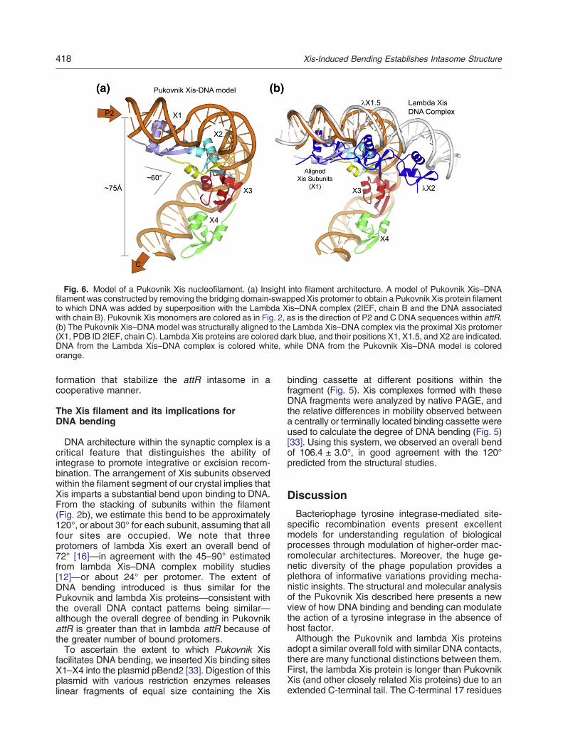
Fig. 6. Model of a Pukovnik Xis nucleofilament. (a) Insight into filament architecture. A model of Pukovnik Xis–DNAfilament was constructed by removing the bridging domain-swapped Xis protomer to obtain a Pukovnik Xis protein filamentto which DNA was added by superposition with the Lambda Xis–DNA complex (2IEF, chain B and the DNA associatedwith chain B). Pukovnik Xis monomers are colored as in Fig. 2, as is the direction of P2 and C DNA sequences within attR.(b) The Pukovnik Xis–DNAmodel was structurally aligned to the Lambda Xis–DNA complex via the proximal Xis protomer(X1, PDB ID 2IEF, chain C). Lambda Xis proteins are colored dark blue, and their positions X1, X1.5, and X2 are indicated.DNA from the Lambda Xis–DNA complex is colored white, while DNA from the Pukovnik Xis–DNA model is coloredorange.
418 Xis-Induced Bending Establishes Intasome Structure
formation that stabilize the attR intasome in acooperative manner.
The Xis filament and its implications forDNA bending
DNA architecture within the synaptic complex is acritical feature that distinguishes the ability ofintegrase to promote integrative or excision recom-bination. The arrangement of Xis subunits observedwithin the filament segment of our crystal implies thatXis imparts a substantial bend upon binding to DNA.From the stacking of subunits within the filament(Fig. 2b), we estimate this bend to be approximately120°, or about 30° for each subunit, assuming that allfour sites are occupied. We note that threeprotomers of lambda Xis exert an overall bend of72° [16]—in agreement with the 45–90° estimatedfrom lambda Xis–DNA complex mobility studies[12]—or about 24° per protomer. The extent ofDNA bending introduced is thus similar for thePukovnik and lambda Xis proteins—consistent withthe overall DNA contact patterns being similar—although the overall degree of bending in PukovnikattR is greater than that in lambda attR because ofthe greater number of bound protomers.To ascertain the extent to which Pukovnik Xis
facilitates DNA bending, we inserted Xis binding sitesX1–X4 into the plasmid pBend2 [33]. Digestion of thisplasmid with various restriction enzymes releaseslinear fragments of equal size containing the Xis
binding cassette at different positions within thefragment (Fig. 5). Xis complexes formed with theseDNA fragments were analyzed by native PAGE, andthe relative differences in mobility observed betweena centrally or terminally located binding cassette wereused to calculate the degree of DNA bending (Fig. 5)[33]. Using this system, we observed an overall bendof 106.4 ± 3.0°, in good agreement with the 120°predicted from the structural studies.
Discussion
Bacteriophage tyrosine integrase-mediated site-specific recombination events present excellentmodels for understanding regulation of biologicalprocesses through modulation of higher-order mac-romolecular architectures. Moreover, the huge ge-netic diversity of the phage population provides aplethora of informative variations providing mecha-nistic insights. The structural and molecular analysisof the Pukovnik Xis described here presents a newview of how DNA binding and bending can modulatethe action of a tyrosine integrase in the absence ofhost factor.Although the Pukovnik and lambda Xis proteins
adopt a similar overall fold with similar DNA contacts,there are many functional distinctions between them.First, the lambda Xis protein is longer than PukovnikXis (and other closely related Xis proteins) due to anextended C-terminal tail. The C-terminal 17 residues

419Xis-Induced Bending Establishes Intasome Structure
of lambda Xis and the related HK022 Xis are poorlydefined in NMR analyses [17,30] and are excludedfrom the crystallographic analysis [16]. The extremeC-terminus of Pukovnik Xis is, however, critical forinteraction with adjacent Xis protomers withinfilament-like structures and is thus required fornormal Xis function.A second difference is in the unusual domain-
swap interaction in the Pukovnik Xis filament forwhich there is no counterpart in lambda Xis. Althoughstructures for three other functionally related proteinshave been described (HK022 [30], transposon Tn916[31], and the prophage excise TorI [34]), all of thesewere determined by NMR spectroscopy in theabsence of DNA, and neither filament-like organiza-tions nor domain swaps are apparent.Lastly, we generated a model of a DNA-bound
Pukovnik Xis filament to examine the constraintssuch a filament might place on intasome architec-ture. To do this, we removed the domain-swapprotomer that bridges filaments within the crystal(residues 1–35 of the yellow subunit and residues36–56 of the cyan subunit in Fig. 2b), noting that theremaining swapped protomer is nearly identical withthe canonical fold (Fig. S2b). We then modeled DNAonto the Pukovnik filament through superposition ofindividual Pukovnik protomers with the structure of alambda Xis–DNA complex (PDB ID 2IEF, chain B).In the resulting model, the attR DNA sequenceleading toward core sequence C is separated fromthe beginning of the X1 site by ~75 Å (Fig. 6a). Themodel indicates that bending is not constrainedwithin a plane as was observed for the IHF–DNAcomplex [35] or the lambda Xis–DNA complex [16]but, instead, is twisted out of the plane by ~60°. Wecompared our model with the structure of the lambdaXis–DNA complex (PDB ID 2IEF, containing Xisproteins bound at X1, X1.5, and X2 positions) viasuperposition of Xis proteins at the X1 site (Fig. 6b).The resulting model clearly suggests that Xis bindingplaces a different set of constraints on DNAarchitecture than in lambda. As stated previously,this model of the Pukovnik nucleoprotein filamentpredicts a 120° bend in the DNA, which is close tothe 106° bend we observe in vitro; however, at ~30°of bend per monomer, our in vitro results suggestthat 3.5 monomers are binding per attR DNAfragment. The difference in bending angle couldsuggest that four Xis monomers bind but that somedegree of relaxation within the DNA-bound filamentis not described in this simple model. Alternatively, itcould suggest that only three Xis proteins arebinding. This is supported by the observation thatthere is somewhat lower sequence conservationbetween L5 and Pukovnik attR DNA sequences attheir X1 sites (Fig. S1c). Our data cannot distinguishbetween these possibilities.The substantial differences in DNA bending
between Pukovnik and lambda may be the result of
the need to perform excision from attR sites withdifferent organizations (Fig. S4). For example, wenote that there are two IHF binding sites withinLambda attR. In contrast, Pukovnik attR does notcontain IHF binding sites. Finally, in lambda attR,there are only three Xis binding sites (X1, X1.5, andX2) rather than the four Xis binding sites in Pukovnikand related phages. The overall distances betweenthe core sites and the outermost arm-type bindingsite (110–120 bp) are similar in lambda and Pukov-nik attR, and we presume that similar DNA bendingis required to form Int-mediated intramolecularbridges. However, Lambda accomplishes thisthrough the combined action of Xis and IHF,whereas Pukovnik does this by Xis alone.An attractive scenario explaining the evolutionary
relationships of the Lambda and Pukovnik integra-tion systems is one in which the Pukovnik system ismore similar to the ancestral state from whichLambda was derived. For example, loss of one ofthe Xis binding sites from the precursor state couldhave arisen through the acquisition of FIS bindingsites that provide dependence on FIS at low Xisconcentrations [36,37]. The reduction in Xis-asso-ciated DNA bending might have provided theselective pressure for the gain of IHF binding sites.
Materials and Methods
Plasmids and DNA substrates
Plasmid pMH57 (pattB) has been previously described[38]. DNA fragments (546 bp) containing attP site wereamplified from the Pukovnik genomic DNA and cloned intothe EcoR1 and BamH1 sites of the pUC19 plasmid toobtain pSS19. DNA fragments of 490 bp and 345 bpcontaining attL and attR were amplified by PCR from theproduct of in vitro integration (between pMH57 and pSS19)reactions and cloned into the HindIII and Nde1 sites ofpMOS Blue to obtain pSS21 and pSS22, respectively.
Preparation of radiolabeled DNA
The digestion of plasmid pSS21 with HindIII and Nde1generated a 490-bp DNA fragment containing the attL site.A 365-bp DNA fragment containing the attR site wasgenerated by PCR amplification from the plasmid pSS22.These DNA fragments containing the attL (490 bp) andattR (365 bp) sites were gel extracted and labeled byend-labeling with T4 polynucleotide kinase.
In vitro recombination assay
In vitro integrative recombination was carried outbetween pSS19 (attP) and pMH57 (attB) in a recombina-tion buffer containing 20 mM Tris (pH 7.5), 25 mM NaCl,10 mM ethylenediaminetetraacetic acid (EDTA), 10 mMspermidine, and 1 mM DTT in a final volume of 10 μl.

420 Xis-Induced Bending Establishes Intasome Structure
Wherever needed, pMH57 was cut by Sca1 to make linearattB substrate for reactions. Reactions using supercoiledor linear double-stranded attB DNA containing 0.03 pmolof pMH57 and supercoiled attP DNA added in ratioindicated in each experiment.In vitro excision assays were performed between pSS21
(attL) and pSS22 (attR) in a recombination buffercontaining 10 mM Tris (pH 7.5), 25 mM NaCl, 5 mMEDTA, 10 mM spermidine, 1 mM DTT, and 0.2 mg/mlbovine serum albumin in a final volume of 10 μl. Whereverneeded, pSS21was cut by Sca1 to make linear attLsubstrate for reactions. Reactions using supercoiled orlinear double-stranded attL DNA containing pSS21 andsupercoiled attR DNA added in indicated ratio.Both integration and excision reactions were incubated
at 37 °C for up to 4 h, and the product was treated with1 mg/ml proteinase K and 0.5% of SDS at 60 °C for 10–15 min. The products were separated on 0.8% agarose gelin 1× TBE (Tris–borate–EDTA) running buffer andvisualized by ethidium bromide staining.
DNA-binding assays
Approximately 4 ng of labeled DNA was incubated withthe indicated amounts of Int, mIHF, and Xis (wild type andmutants) in a buffer containing 10 mM Tris (pH 7.5),25 mM NaCl, 5 mM EDTA, 10 mM spermidine, 1 mMDTT, 0.2 mg/ml bovine serum albumin, and 1 μg calfthymus DNA, in a total volume of 10 μl. The reactions wereincubated at 37 °C for 1 h, and the protein–DNA com-plexes were separated from the free DNA on a non-denaturing 4% (unless otherwise stated) polyacrylamidegel at 4 °C.
Xis purification
The complete coding sequence for mycobacteriophagePukovnik excise (Xis) was PCR amplified from isolatedgenomic DNA and inserted into the bacterial expressionvector pLC3 (a kind gift of Dr. Jim Sacchettini) usingtradition cloning techniques. The resulting plasmid wascoded for an N-terminal His6-MBP fusion tag that isremovable via digestion with TEV (tobacco etch virus)protease, leaving the sequence GDITH attached to theN-terminus of the protein. The His6-MBP-Xis protein wasexpressed using ZY autoinduction media [39] and thebacterial strain BL21 DE3 Codon-Plus (RIPL). Cells wereharvested by centrifugation and lysed in 25 mM Tris(pH 8.0), 500 mM NaCl, 10% glycerol, 5 mM imidazole,and 1 mM β-mercaptoethanol, plus protease inhibitors.Lysate was cleared by centrifugation at 30,000g, and Xiswas purified by nickel affinity chromatography usingNi-NTA resin (Qiagen). The His6-MBP-tag was removedvia overnight digestion with TEV protease digestion,followed by a second round of nickel affinity chromatog-raphy to remove TEV and other contaminants. Theresulting sample was further purified using cation-exchange chromatography, and peak fractions weresubjected to gel filtration using a Sephacryl S-200 (GEHealthcare). Purified Xis was then dialyzed into 100 mMNaCl, 10 mM Hepes (pH 7.5), and 1 mM β-mercaptoetha-nol and was concentrated to ~25 mg/ml using a Vivaspinconcentrator (Millipore) prior to crystallization trials. Final
protein purity was N99% as verified by SDS-PAGE.Selenomethionine Xis was expressed using PASMmedia [39], and the purification was similar to the nativeprotein with addition of 5 mM β-mercaptoethanol in all thebuffers. The final selenomethionine Xis protein concentra-tion was 20.4 mg/ml in 25 mM NaCl and 10 mM Hepes(pH 7.5).
Xis mutants
To enhance the yield of Xis protein, we amplified andcloned the coding sequence for Xis into the bacterialexpression vector pMCSG7 (which contains a TEV-cleavable N-terminal polyhistidine tag but no MBP tag)via ligation-independent cloning. Single, double, and tripleamino acid mutations were introduced into Xis using theQuikChange protocol (Stratagene) utilizing pMCSG7-Xisplasmid as a template. The Δ51–56 C-terminal truncationwas also cloned into the pMCSG7 vector as with the wildtype. Expression and purification of the Xis mutants wereperformed in a similar manner as wild type.
Integrase and integration host factor purification
The coding regions for Pukovnik integrase (pInt) andMycobacterium smegmatis integration host factor (mIHF)were PCR amplified and cloned into pMCSG7 to expressN-terminal His-tagged fusions cleavable by TEV protease.IHF was expressed using ZY autoinduction media as withXis while pInt was expressed via IPTG induction in LB.Both proteins were purified using nickel and heparin affinitychromatography, and protein purity was judged at N99%by SDS-PAGE.
Xis crystallization and structure determination
Crystals were initially obtained using the sitting-dropvapor diffusion method at 20 °C against a reservoirsolution containing 0.2 M ammonium sulfate and 20%polyethylene glycol (PEG) 3350. Single crystals measur-ing approximately 0.2 mm × 0.2 mm × 0.05 mm formover the period of 2 weeks from drops containing 0.8 μlof protein and 1.0 μl of well solution (100 mM ammoniumsulfate and 13.5% PEG 3350). Crystals were cryopro-tected by a stepwise transition into well solution supple-mental with 30% methyl-2,4-pentanediol and 15% PEG3350. Selenomethionine crystals were grown in the samemanner.Crystals of Pukovnik Xis belong to space group C2221
with a = 89.5 Å, b = 130.5 Å, and c = 92.2 Å. Diffractiondata from crystals of selenomethionine-substituted Xiswere collected at beamline X25 at the National Synchro-tron Light Source. Integration, scaling, and merging ofdiffraction data were performed using HKL2000 [40].Phases were determined experimentally via single-wave-length anomalous dispersion using crystals containingselenomethionine-substituted protein. Phases were calcu-lated using the AutoSOL routine within PHENIX [41] and astarting model built into the resulting electron density. Thisinitial model was then refined against native data collectedin-house using a FR-E rotating anode with VariMax opticsand a Saturn944 CCD detector. The integration, scaling,
421Xis-Induced Bending Establishes Intasome Structure
and merging of diffraction data were performed usingHKL2000. The initial model was refined within PHENIXusing positional refinement, simulated annealing, isotropicB-factor refinement, and TLS refinement. Model qualitywas analyzed using MolProbity [42], and the final modelcontains no Ramachandran outliers.
Permutation assay
DNA sequence containing the Xis binding cassette(sites X1–X4) from Pukovnik attR was inserted into thepBend2 plasmid [43]. Digesting the resulting plasmid withEcoRV, MluI, or the other indicated restriction enzymesresulted in DNA fragments of equal length but with X1–X4either centrally or terminally located. Complexes of theseoligonucleotides with Xis were formed with 25 ng of theindicated DNA fragment, using 0.25 μM Xis and separatedby native-gel electrophoresis. The location of Xis–DNAcomplexes was determined by staining with SybrGreen,and mobilities for the various DNA fragments weremeasured directly using ImageJ software. The DNAbending angle was determined by the equation μM/μE =cos(α/2) where μM/μE is the relative mobility of the centrallybound to the terminally bound oligonucleotide and α is theangle of DNA bending [33].
Accession codes
Coordinates and structure factors for Pukovnik Xis havebeen deposited at the Protein Data Bank under accessionnumber 4J2N.
Acknowledgements
We thank M. Schmidt and J. Sacchettini forsharing resources and helpful advice. This workwas supported by National Institutes of Health grantAI59114 to G.F.H and A.P.V. were supported by aScience Education Grant from the Howard HughesMedical Institutes to the University of Pittsburgh.
Appendix A. Supplementary data
Supplementary data to this article can be foundonline at http://dx.doi.org/10.1016/j.jmb.2013.10.002.
Received 27 June 2013;Received in revised form 26 September 2013;
Accepted 1 October 2013Available online 7 October 2013
Keywords:DNA recombination;mycobacteriophage;
DNA bending;
structure;Xis
Abbreviations used:EDTA, ethylenediaminetetraacetic acid; PEG, polyethy-
lene glycol.
References
[1] Landy A. Dynamic, structural, and regulatory aspects oflambda site-specific recombination. Annu Rev Biochem1989;58:913–49.
[2] Van Duyne GD. Lambda integrase: armed for recombination.Curr Biol 2005;15:R658–60.
[3] AzaroMA,LandyA. λ Int and the λ Int family. In:CraigNL,CraigieR,GellertM, LambowitzAM,editors.MobileDNA II.Washington,DC: American Society Microbiology; 2002. p. 118–48.
[4] Franz B, Landy A. The Holliday junction intermediates oflambda integrative and excisive recombination responddifferently to the bending proteins integration host factorand excisionase. EMBO J 1995;14:397–406.
[5] Nunes-Duby SE, Azaro MA, Landy A. Swapping DNAstrands and sensing homology without branch migration inlambda site-specific recombination. Curr Biol 1995;5:139–48.
[6] Kim S, Landy A. Lambda Int protein bridges between higherorder complexes at two distant chromosomal loci attL andattR. Science 1992;256:198–203.
[7] Bushman W, Yin S, Thio LL, Landy A. Determinants ofdirectionality in lambda site-specific recombination. Cell1984;39:699–706.
[8] Moitoso de Vargas L, Kim S, Landy A. DNA loopinggenerated by DNA bending protein IHF and the two domainsof lambda integrase. Science 1989;244:1457–61.
[9] Ellenberger T, Landy A. A good turn for DNA: the structure ofintegration host factor bound to DNA. Structure 1997;5:153–7.
[10] Robertson CA, Nash HA. Bending of the bacteriophagelambda attachment site by Escherichia coli integration hostfactor. J Biol Chem 1988;263:3554–7.
[11] Kim S, Moitoso de Vargas L, Nunes-Duby SE, Landy A.Mapping of a higher order protein–DNA complex: two kinds oflong-range interactions in lambda attL. Cell 1990;63:773–81.
[12] Thompson JF, Landy A. Empirical estimation of protein-induced DNA bending angles: applications to lambda site-specific recombination complexes. Nucleic Acids Res1988;16:9687–705.
[13] Numrych TE, Gumport RI, Gardner JF. Characterization ofthe bacteriophage lambda excisionase (Xis) protein: the C-terminus is required for Xis-integrase cooperativity but not forDNA binding. EMBO J 1992;11:3797–806.
[14] Wu Z, Gumport RI, Gardner JF. Defining the structural andfunctional roles of the carboxyl region of the bacteriophagelambda exc is ionase (X is ) pro te in . J Mol B io l1998;281:651–61.
[15] Cho EH, Gumport RI, Gardner JF. Interactions betweenintegrase and excisionase in the phage lambda excisivenucleoprotein complex. J Bacteriol 2002;184:5200–3.
[16] Abbani MA, Papagiannis CV, Sam MD, Cascio D, JohnsonRC, Clubb RT. Structure of the cooperative Xis-DNA complexreveals a micronucleoprotein filament that regulates phagelambda intasome assembly. Proc Natl Acad Sci USA2007;104:2109–14.
422 Xis-Induced Bending Establishes Intasome Structure
[17] Sam MD, Papagiannis CV, Connolly KM, Corselli L, IwaharaJ, Lee J, et al. Regulation of directionality in bacteriophagelambda site-specific recombination: structure of the Xisprotein. J Mol Biol 2002;324:791–805.
[18] Lee MH, Hatfull GF. Mycobacteriophage L5 integrase-mediated site-specific integration in vitro. J Bacteriol1993;175:6836–41.
[19] Peña CE, Lee MH, Pedulla ML, Hatfull GF. Characterizationof the mycobacteriophage L5 attachment site, attP. J Mol Biol1997;266:76–92.
[20] Pedulla ML, Lee MH, Lever DC, Hatfull GF. A novel hostfactor for integration of mycobacteriophage L5. Proc NatlAcad Sci USA 1996;93:15411–6.
[21] Pena CE, Kahlenberg JM, Hatfull GF. Assembly andactivation of site-specific recombination complexes. ProcNatl Acad Sci USA 2000;97:7760–5.
[22] Lewis JA, Hatfull GF. Identification and characterization ofmycobacteriophage L5 excisionase. Mol Microbiol2000;35:350–60.
[23] Lewis JA, Hatfull GF. Control of directionality in integrase-mediated recombination: examination of recombinationdirectionality factors (RDFs) including Xis and Cox proteins.Nucleic Acids Res 2001;29:2205–16.
[24] Lewis JA, Hatfull GF. Control of directionality in L5 integrase-mediated site-specific recombination. J Mol Biol2003;326:805–21.
[25] Hatfull GF. Complete genome sequences of 138 mycobac-teriophages. J Virol 2012;86:2382–4.
[26] Hatfull GF, Pedulla ML, Jacobs-Sera D, Cichon PM, Foley A,Ford ME, et al. Exploring the mycobacteriophage metapro-teome: phage genomics as an educational platform. PLoSGenet 2006;2:e92.
[27] Hatfull GF. The secret lives of mycobacteriophages. AdvVirus Res 2012;82:179–288.
[28] Peña CE, Stoner JE, Hatfull GF. Positions of strandexchange in mycobacteriophage L5 integration and charac-terization of the attB site. J Bacteriol 1996;178:5533–6.
[29] Hatfull GF, Jacobs-Sera D, Lawrence JG, Pope WH, RussellDA, Ko CC, et al. Comparative genomic analysis of 60mycobacteriophage genomes: genome clustering, geneacquisition, and gene size. J Mol Biol 2010;397:119–43.
[30] Rogov VV, Lucke C, Muresanu L, Wienk H, Kleinhaus I,Werner K, et al. Solution structure and stability of the full-length excisionase from bacteriophage HK022. Eur JBiochem 2003;270:4846–58.
[31] Abbani M, Iwahara M, Clubb RT. The structure of theexcisionase (Xis) protein from conjugative transposon Tn916provides insights into the regulation of heterobivalent tyrosinerecombinases. J Mol Biol 2005;347:11–25.
[32] SamMD, Cascio D, Johnson RC, Clubb RT. Crystal structureof the excisionase-DNA complex from bacteriophage lamb-da. J Mol Biol 2004;338:229–40.
[33] Zwieb C, Adhya S. Plasmid vectors for the analysis ofprotein-induced DNA bending. Methods Mol Biol2009;543:547–62.
[34] Elantak L, Ansaldi M, Guerlesquin F, Mejean V, Morelli X.Structural and genetic analyses reveal a key role in prophageexcision for the TorI response regulator inhibitor. J Biol Chem2005;280:36802–8.
[35] Rice PA, Yang S, Mizuuchi K, Nash HA. Crystal structure ofan IHF-DNA complex: a protein-induced DNA U-turn. Cell1996;87:1295–306.
[36] Ball CA, Johnson RC. Efficient excision of phage lambdafrom the Escherichia coli chromosome requires the Fisprotein. J Bacteriol 1991;173:4027–31.
[37] Ball CA, Johnson RC. Multiple effects of Fis on integrationand the control of lysogeny in phage lambda. J Bacteriol1991;173:4032–8.
[38] Lee MH, Pascopella L, Jacobs WR, Hatfull GF. Site-specificintegration of mycobacteriophage L5: integration-proficientvectors for Mycobacterium smegmatis, Mycobacterium tu-berculosis, and bacille Calmette-Guerin. Proc Natl Acad SciUSA 1991;88:3111–5.
[39] Studier FW. Protein production by auto-induction in highdensity shaking cultures. Protein Expression Purif2005;41:207–34.
[40] Otwinowski Z, Minor W. Processing of X-ray diffraction datacollected in oscil lation mode. Methods Enzymol1997;276:307–26.
[41] Adams PD, Afonine PV, Bunkoczi G, Chen VB, Davis IW,Echols N, et al. PHENIX: a comprehensive Python-basedsystem for macromolecular structure solution. Acta Crystal-logr Sect D Biol Crystallogr 2010;66:213–21.
[42] Davis IW, Leaver-Fay A, Chen VB, Block JN, Kapral GJ,Wang X, et al. MolProbity: all-atom contacts and structurevalidation for proteins and nucleic acids. Nucleic Acids Res2007;35:W375–83.
[43] Kim J, Zwieb C, Wu C, Adhya S. Bending of DNA by gene-regulatory proteins: construction and use of a DNA bendingvector. Gene 1989;85:15–23.