Commuting fruit bats beneficially modulate their flight in relation to wind

10.1128/MCB.23.6.2068-2082.2003.
2003, 23(6):2068. DOI:Mol. Cell. Biol. JallalHowlett, James R. Bischoff, Kenneth E. Lipson and BahijaFlanagan, Sharon D. Buckley, David B. Whyte, Anthony R.
M.J. LaMere, Phuong Le, Shirley Zhu, Deepak Khatry, Peter Jocelyn H. Wright, Xueyan Wang, Gerard Manning, Brandon and AdhesionModulate Cellular Transformation, Invasion,Expressed in Human Tumor Cells and Can The STE20 Kinase HGK Is Broadly
http://mcb.asm.org/content/23/6/2068Updated information and services can be found at:
These include:
REFERENCEShttp://mcb.asm.org/content/23/6/2068#ref-list-1at:
This article cites 53 articles, 22 of which can be accessed free
CONTENT ALERTS more»articles cite this article),
Receive: RSS Feeds, eTOCs, free email alerts (when new
http://journals.asm.org/site/misc/reprints.xhtmlInformation about commercial reprint orders: http://journals.asm.org/site/subscriptions/To subscribe to to another ASM Journal go to:
on Decem
ber 11, 2013 by guesthttp://m
cb.asm.org/
Dow
nloaded from
on Decem
ber 11, 2013 by guesthttp://m
cb.asm.org/
Dow
nloaded from

MOLECULAR AND CELLULAR BIOLOGY, Mar. 2003, p. 2068–2082 Vol. 23, No. 60270-7306/03/$08.00�0 DOI: 10.1128/MCB.23.6.2068–2082.2003Copyright © 2003, American Society for Microbiology. All Rights Reserved.
The STE20 Kinase HGK Is Broadly Expressed in Human Tumor Cellsand Can Modulate Cellular Transformation, Invasion, and AdhesionJocelyn H. Wright,* Xueyan Wang, Gerard Manning, Brandon J. LaMere, Phuong Le, Shirley Zhu,
Deepak Khatry, Peter M. Flanagan, Sharon D. Buckley, David B. Whyte, Anthony R. Howlett,James R. Bischoff, Kenneth E. Lipson, and Bahija Jallal
Sugen, Inc., South San Francisco, California 94080
Received 15 July 2002/Returned for modification 17 September 2002/Accepted 6 November 2002
HGK (hepatocyte progenitor kinase-like/germinal center kinase-like kinase) is a member of the humanSTE20/mitogen-activated protein kinase kinase kinase kinase family of serine/threonine kinases and is theortholog of mouse NIK (Nck-interacting kinase). We have cloned a novel splice variant of HGK from a humantumor line and have further identified a complex family of HGK splice variants. We showed HGK to be highlyexpressed in most tumor cell lines relative to normal tissue. An active role for this kinase in transformationwas suggested by an inhibition of H-RasV12-induced focus formation by expression of inactive, dominant-negative mutants of HGK in both fibroblast and epithelial cell lines. Expression of an inactive mutant of HGKalso inhibited the anchorage-independent growth of cells yet had no effect on proliferation in monolayerculture. Expression of HGK mutants modulated integrin receptor expression and had a striking effect onhepatocyte growth factor-stimulated epithelial cell invasion. Together, these results suggest an important rolefor HGK in cell transformation and invasiveness.
The mammalian STE20/mitogen-activated protein kinase ki-nase kinase kinase (MAP4K) family consists of 28 serine/thre-onine kinases related in their catalytic domains (reviewed inreference 14). By analogy with the prototype STE20 kinase inSaccharomyces cerevisiae, mammalian MAP4K kinases arelikely to regulate changes in transcription, cytoskeletal organi-zation, and cell cycle progression in response to extracellularsignals (17). Comparison of the overall domain structureplaces these kinases into two structural classes, the p21-acti-vated protein kinases (PAKs) (1) and germinal center kinases(GCKs) (28). The GCK kinases lack the regulatory Cdc42/Rac-interacting domain found in the PAKs, having instead anN-terminal kinase domain and a C-terminal extension of vari-able length. Unlike PAKs, several GCKs appear to be activatedin the absence of stimuli when overexpressed (3, 10, 38), sug-gesting that these kinases are regulated either by oligomeriza-tion or by binding of negative regulatory factors.
GCK kinases show little sequence homology outside of thekinase domain and are further broken down into nine subfam-ilies (14, 31a). HGK (hepatocyte progenitor kinase-like/GCK-like kinase) is one of four members of the GCK group IV(recently renamed the MSN subfamily) that also includesTNIK, MINK, and NRK/NESK (13, 22, 27, 35, 51). HGK,TNIK, and MINK are highly homologous in their kinase andC-terminal domains (about 92 and 87% amino acid identity,respectively), with variability in the intervening region that isless conserved (53% between HGK and TNIK). NRK/NESK ismore divergent, with only 59% homology in the kinase domainand 37% homology in the C-terminal domain. The C-terminaldomain is a citron homology (CNH) domain, named for citronrho-interacting kinase (CRIK), where it was first described (16,
31). CNH domains are found not only in the GCK groupIV/MSN kinases but also in group I GCKs (KHS subfamily)(14, 31a), as well as in proteins other than kinases, includinghuman Vam6p homologs, involved in membrane vesicle dock-ing and fusion, and the S. cerevisiae Rho GDP-exchange factorRom1p (9). The CNH domain has been shown to be importantfor protein-protein interactions. In CRIK itself, this domainmediates association with rho family GTPases (30, 31). InVam6p, it mediates association with lysosomes (9). In GCK (agroup I/KHS kinase), this domain is important for homodimer-ization, leading to the activation of mitogen-activated proteinkinase/extracellular signal-regulated kinase MEKK1 (10). Inmouse HGK, the CNH domain has been shown to interact withMEKK1 and also recently shown to mediate interaction of thiskinase with the cytoplasmic domain of �1-integrin receptors(37, 43).
Orthologs of the HGK group are known in both Drosophilamelanogaster and Caenorhabditis elegans, called misshapen(msn) and mig-15, respectively (46, 53). msn and mig-15 showstriking sequence conservation with HGK in both the kinaseand CNH domains, with more than 80% amino acid identity inboth domains.
msn functions in multiple signaling pathways, includingthose downstream of frizzled (Wnt receptor) in determiningepithelial polarity, in the regulation of dorsal closure, and inneuronal targeting. In the Wnt pathway, msn bifurcates fromthe �-catenin/cadherin signaling pathway at the upstream sig-naling protein coded by disheveled, signaling to a separatepathway leading to the activation of both JNK and p38 (36). Indorsal closure, msn also signals through a JNK module thatmediates the spreading of epithelial sheets to close the embryothrough induction of transforming growth factor � (44, 46). Inphotoreceptor neuronal targeting, msn functions downstreamof dock, the Drosophila homolog of the Nck adapter protein, toguide axons to their target (40, 44). In nematodes, mig-15
* Corresponding author. Mailing address: Sugen, Inc., South SanFrancisco, CA 94080. Phone: (650) 837 3477. Fax: (650) 837 3313.E-mail: [email protected].
2068
on Decem
ber 11, 2013 by guesthttp://m
cb.asm.org/
Dow
nloaded from

functions in the processes of muscle arm targeting, where mus-cle cells extend cytoplasmic protrusions to synapse with motorneurons during development, as well as in neuroblast migra-tion (53). The common factor to both model systems is theinvolvement of an HGK homolog in processes requiring cellmigration or cell shape change.
The mouse ortholog of HGK, Nck-interacting kinase (NIK),was cloned in an interaction screen with the SH3 domain of theNck adapter protein (43). NIK has been shown to be activatedby stimulation of the ephrin receptor family (3). Ephrins arecell surface ligands shown to be involved in cell migration andtissue remodeling (52) and have been proposed to be involvedin inside-out integrin regulation (15, 33). Consistent with apositive function for NIK in cell migration and morphogenesis,NIK�/� knockout mice showed an early embryonic-lethal phe-notype, with defects in mesoderm differentiation and migration(50). This embryonic lethality indicates a lack of functionalredundancy between the GCK group IV family members dur-ing development.
Human HGK was previously cloned from a macrophagecDNA library (51). Yao et al. demonstrated that HGK acti-vated JNK in transient transfections and further suggested thatthis activation occurred through the mitogen-activated proteinkinase kinase kinase transforming growth factor �-activatedkinase (TAK1) rather than MEKK1.
We present the characterization of an alternatively splicedform of HGK identified from a human glioblastoma libraryand provide an analysis of a complex family of alternativelyspliced HGK isoforms. We found HGK mRNA to be broadlyoverexpressed in tumor cell lines relative to normal adult tis-sue. Ectopic expression of various mutants of the tumor-de-rived HGK isoform suggested an active role for this protein incell transformation and invasion as well as in reducing theadhesive properties of tissue culture cells. HGK appeared tofunction in these processes through modulation of growth fac-tor and integrin receptor signaling.
MATERIALS AND METHODS
Cloning of HGK gene. The complete HGK cDNA was constructed from twooverlapping PCR clones isolated with an SNB19 human glioblastoma single-stranded cDNA library as a template. The original PCR product containing thecatalytic domain was isolated with degenerate oligonucleotides corresponding toconserved motifs in kinase domains. Homology with the murine ortholog NIKand the C. elegans ZC504.4 contig were observed, and primers from the Nterminus of NIK were used in combination with primers derived from theoriginal PCR isolate to generate the N-terminal half of the gene. A Smith-Waterman search of the public expressed sequence tag (EST) database identifieda human EST whose open reading frame was related to the C. elegans gene(mig-15) and terminated in an identical Trp residue. A primer was designed tothe 3� end and used in a PCR assay with a primer derived from the 5� end of theHGK cDNA.
Bioinformatic analysis of HGK gene. A Blast search with our human HGKcDNA sequence was run against the Celera (version R26) and the public (releaseof 20 June 2001) genomic sequence databases to identify long contigs thatcovered the gene. Those contigs were then compared with EST databases fromthe public domain, Incyte (LifeSeqGold), and in-house sequence databases andwith the NCBI nonredundant nucleotide database. Sequences that were identi-fied by the Blast search were aligned to both the Celera and Human GenomeProject genomic sequences with the Sim4 program (18) and visualized with anin-house genome browser.
Manual inspection was used to identify ESTs and cDNA sequences whosemapping to the genome showed alternative exon usage, and each sequence wasaligned with a consensus long sequence (containing all alternative spliced mod-ules) in order to confirm the differences between alternative splice forms. When
there were sequence polymorphisms between genomic, cDNA, and EST datasources, the polymorphic regions were compared with Blast against all EST andpublic sources used and against raw genomic reads from Celera to determinewhich nucleotide was seen most frequently at the point of dispute.
Cloning HGK cDNA fragments from human tumor cell lines. Total RNA wasprepared from A549, H1299, and 293T cells with an RNeasy kit (Qiagen). Inaddition, Universal Reference RNA (containing a mixture of RNAs purifiedfrom 10 tumor cell lines) was purchased from Clontech. Reverse transcriptionreactions were performed with a Taqman kit (ABI); 2 �g of the cDNA was usedin PCRs with Taq-Platinum polymerase (Invitrogen) with two primer sets togenerate products between 1 and 3 kb. Products were size selected and clonedinto pCR-II with a TA cloning kit (Invitrogen). Between 13 and 17 clones werefully sequenced and analyzed from each of the four RNA sources.
Expression analysis. Northern blots were performed with standard tech-niques. The cells and tissues were described previously (5). Northern blots wereprepared by running 10 �g of total RNA isolated from 60 human tumor lines and21 adult tissues and 1 fetal tissue on a denaturing 1.2% formaldehyde–agarosegel and transferred to a nylon membrane. Filters were hybridized with a random-primed [32P]dCTP-labeled probe derived from a 404-bp NaeI-SmaI fragmentfrom a coding region of HGK conserved in all splice variants.
The multiple tissue blot from which the tumor versus normal data wereextracted was prepared as follows. cDNA libraries derived from 489 tissue or cellline sources were immobilized onto nylon membranes. The amplified cDNAlibrary was manually arrayed onto nylon membranes with a 384-pin replicator.DNA spotting on arrays was quantified by treating nondenatured arrays withSYBR Green (1:100,000 in 50 mM Tris, pH 8.0) for 2 min. After washing with 50mM Tris, pH 8.0, the fluorescent emission was quantified with a Phosphorimager(Molecular Dynamics). The amount of the arrayed DNA was used to normalizethe hybridization signal. The same 32P-labeled HGK probe used for the Northernblots was hybridized to the array blot. Blots were washed and quantified with aPhosphorimager (Molecular Dynamics). The data were standardized for statis-tical analysis across the different tissue types with range standardization toconvert measurements to a common scale starting at 0 and ending at 1. Allstatistical analysis was carried out with SYSTAT 9.01 (SPSS, Inc.).
Mutagenesis method and location of point mutations. Mutations in the full-length cDNA were generated to change threonine 187 to glutamate (T187E),threonine 191 to glutamate (T191E), and lysine 54 to arginine (K54R) with aQuickChange PCR mutagenesis kit (Stratagene) and oligonucleotides T187E(CTGTGGGGCGGAGAAATGAATTCATAGGCACTCCC, GGGAGTGCCTATGAATTCATTTCTCCGCCCCACAG), T191E (GAAATACGTTCATAGGCGAGCCCTACTGGATGGC, GCCATCCAGTAGGGCTCGCCTATGAACGTATTTC), and K54R (GTTGGCAGCCATCAGAGTTATGGATGTCACTGAGG, CCTCAGTGACATCCATAACTCTGATGGCTGCCAAC). Wild-typeand mutant forms of HGK were subcloned into pcDNA3myc(�)B vectors (In-vitrogen), removing all 3� untranslated sequence and generating a C-terminalMyc epitope tag.
In vitro HGK kinase assay. RIE-1 cells expressing Myc-tagged HGK werelysed with 50 mM HEPES (pH 7.0)–150 mM NaCl,–1.5 mM MgCl2–1.0 mMEGTA–10% glycerol–1% Triton–10 mM pyrophosphate–1 mM Na3VO5–1 mMdithiothreitol–0.1 mg of 4-(2-aminoethyl)benzenesulfonyl fluoride per ml–2 to 10�g each of aprotinin, pepstatin, leupeptin, and E-64 per ml as protease inhibi-tors. The lysates were spun at 14,000 rpm (15,800 � g) in an Eppendorf micro-centrifuge for 10 min at 4°C. The supernatants were immunoprecipitated withprotein G-Sepharose beads (Fisher) with an anti-Myc monoclonal antibody(mouse ascites fluid made with the 9E10 hybridoma cell line). The beads werewashed twice with the lysis buffer and twice with kinase assay buffer (20 mM Tris[pH 7.4], 200 mM NaCl, 0.5 mM dithiothreitol, 10 mM MgCl2). The washedbeads were resuspended in kinase assay buffer with 100 �M ATP and 0.5 �Ci of[�-32P]ATP per �l and incubated for 20 min in an Eppendorf Thermomixer at30°C at maximal mixing velocity. Then 4 �g of myelin basic protein (Sigma) wasadded, and the reaction was allowed to continue for 10 min. The reaction wasstopped by boiling in Laemmli sodium dodecyl sulfate (SDS) sample buffer for10 min.
Focus-forming assays. Focus formation assays were performed according tostandard procedures (12). NIH 3T3 cells, obtained from Tony Hunter, were usedat a low passage number, and the RIE-1 cells were obtained from Channing Der.NIH 3T3 cells were transfected with a total of 6 �g of DNA and 18 �l ofLipofectamine per 10-cm dish, and 0.3 to 0.5 �g of SV40sport-H-RasV12 alongwith 1 to 10 �g of pcDNA3-HGK mutants was transfected along with plasmidpBSK� as carrier DNA. Empty vector (pcDNA3 from Invitrogen) was includedas a negative control. RIE-1 cells were transfected with a total of 15 �g of DNAand 50 �l of Lipofectamine per 10-cm dish with the same plasmid ratios as for
VOL. 23, 2003 STE20 KINASE HGK 2069
on Decem
ber 11, 2013 by guesthttp://m
cb.asm.org/
Dow
nloaded from
NIH 3T3. Foci were stained with 0.1% crystal violet in 20% methanol andcounted from duplicate plates 12 to 13 days after transfection.
Generation of stable cell lines with retroviral infection. HGK full-lengthcDNAs including a Myc epitope tag on the C terminus were subcloned into thepLXSN or pBABE retroviral vector. pLXSN-HGK and pBABE-HGK constructswere cotransfected into 293T cells with an Ecotropic retroviral packaging vector(29) with Lipofectamine. Viral supernatants were used to infect RIE-1 (Eco-tropic) cells. Three successive rounds of infection were done approximately every8 to 12 h to maximize the rate of infection. Pools of cells resistant to G418(pLXSN) or puromycin (pBABE) were generated after 1 week of selection.These pools were used in some experiments, and individual clones were grownout of the pools as selected by screening for higher expression of the HGKproteins.
Monolayer cell growth rates. Stable pools or clones of RIE-1 cells expressingHGK mutants were plated at low cell density in 12-well trays in medium with10%, 1%, or 0.1% fetal bovine serum. Cell numbers were counted from two wellsper cell line per day on a Coulter particle counter (Beckman-Coulter).
Soft agar assays. Stable RIE-1 cell lines were plated in 0.3% Bactoagar(Gibco-BRL)–Dulbecco’s modified Eagle’s medium (DMEM)–10% fetal calfserum (Gibco-BRL) on top of a 0.6% Bactoagar–DMEM base layer. For eachclone, one six-well tray was plated with the following number of cells per well: 0,1.0 � 103, 5.0 � 103, 1.0 � 104, 5.0 � 104, and 1.0 � 105. Assays were photo-graphed after 4 weeks of growth. Photographs shown in Fig. 6 were of the wellswith 1.0 � 104 cells/well.
Cell spreading assay. RIE-1 clones expressing wild-type HGK, activated mu-tant (T187E), inactive mutant (T191E), and empty vector as controls wereremoved from the dish with trypsin-EDTA and counted, and equal numbers ofcells were replated on dishes coated with a 10-�g/ml solution of fibronectin(Chemicon FC014). Cells were then photographed at 10� magnification atvarious time points. The number of spread cells was quantified by counting thenumber of phase-dark cells.
Cell adhesion assay. Standard-curve wells were prepared by adding 50 �l ofpolylysine (Sigma; 100 �g/ml) per well to wells, incubating at room temperaturefor 5 min, washing with phosphate-buffered saline (PBS), and air drying. Forexperimental wells, fibronectin (Chemicon, FC104) was diluted in PBS to theappropriate working concentration, added at 100 �l/well to 96-well trays, andincubated for 60 min. Fibronectin solution was removed, and the wells wereblocked with bovine serum albumin (Sigma, tissue culture grade) for 30 min andwashed with PBS. RIE-1 cells were removed from plates with trypsin-EDTAsolution (Gibco-BRL), counted, and resuspended in 0.1% bovine serum albuminat 5 � 105 cells/ml. Then 50 �l of cells was added to each experimental well, andcells were allowed to adhere for 30 and 60 min. The medium was then removedfrom all wells, and nonadherent cells were gently washed away with PBS. Then5% glutaraldehyde was added for 20 min at room temperature. Wells werewashed with PBS. Crystal violet solution (0.1% crystal violet dye in 20% meth-anol) was added to each well and incubated for 60 min on a platform shaker.Stained cells were first photographed and then solubilized in 100 �l of 10% aceticacid for 1 to 2 h, and the absorbance at 550 nm was measured.
Fluorescence-activated cell sorting analysis. Cells were detached from disheswith trypsin-EDTA, counted, and resuspended at 2 � 107 cells/ml in PBS–1%fetal bovine serum. Anti-integrin receptor antibodies (Pharmingen) 553350 (�5),555003 (�1), 553715 (�1), 553343 (�3), 550344, and 553957 (isotype controls)were diluted to 20 �g/ml, mixed with cells in a 1:1 volume ratio, and incubatedin the dark for 20 min at 4°C. Cells were washed twice with PBS–1% fetal bovineserum (wash buffer). Then 100 �l of secondary antibody (Pharmingen), fluores-cein isothiocyanate-conjugated anti-rat immunoglobulin G2a 553896 or fluores-cein isothiocyanate-conjugated anti-hamster immunoglobulin M 554036) at 10�g/ml in PBS–1% fetal bovine serum was added to the cells and incubated for 20min in the dark at 4°C. Cells were washed twice and resuspended in 500 �l ofwash buffer. Cells were analyzed for relative FL1 intensity for cells stained withdifferent primary antibodies and compared with a secondary-antibody stainingonly as well as with a primary antibody isotype control with a Becton-Dickinsonfluorescence-activated cell sorter machine and the CellQuest program. Themean fluorescence intensity values shown in Fig. 8 represent the mean fluores-cence intensity values measured for the specific integrin antibody minus themean fluorescence intensity value for the corresponding isotype control anti-body.
Three-dimensional invasion-tubulogenesis assay. Matrigel (Matrigel base-ment membrane matrix, catalog no. 40234, Becton Dickinson Labware) wasthawed on ice. Then 50 �l of Matrigel was added per well in a 96-well plate andincubated for 20 min at 37°C to induce gel formation. RIE-1 cells were sus-pended at 2 � 104 cell/ml and mixed 1:1 with Matrigel on ice. Then 0.1 ml of themixture was added per well. The next day, 0.1 ml of DMEM with and without 80
ng/ml of hepatocyte growth factor (HGF) (R&D 294-hg-005) was added on topof each Matrigel cell plug. Fresh medium with and without HGF was added every3 days. Photographs were taken after 6 days in culture.
Boyden chamber (directional) invasion assay. The Boyden chamber (direc-tional) invasion assay was done with Biocoat Matrigel Invasion 24-well chambers(Becton Dickinson). Inserts were first rehydrated at room temperature. Withtrypsin-EDTA, cell suspensions were prepared in DMEM–1% bovine serumalbumin at 105 cells/ml. HGF was added to the bottom chamber at 50 to 100ng/ml. Then 0.5 ml of cell suspension was added to the inserts, and the chamberswere incubated for 48 h at 37°C in a 5% CO2 atmosphere. After incubation, thenoninvading cells were removed from the upper surface of the membrane with amoistened Q-tip swab. The inserts were transferred to crystal violet (0.2% crystalviolet in 10% buffered Formalin) stain solution (1 min) and then washed withwater. Inserts were allowed to air dry before being photographed.
Cell stimulation with HGF and analysis of cellular phosphoproteins. RIE cellswere grown until confluent and then serum starved overnight. After stimulationwith HGF (50 ng/ml; 294-HG, R&D Systems), cells were lysed with HNTG (50mM HEPES [pH 7.4], 150 mM NaCl, 10% glycerol, 0.5% Triton X-100,1 mMNaVO4, and protease inhibitors [1.4 �M E-64, 10 �M bestatin, 1 �M leupeptin,0.3 �M aprotinin, 1 �M pepstatin, and 1 mM phenylmethylsulfonyl fluoride]).For determination of HGF-stimulated phosphorylation of Met, the Met proteinwas isolated by immunoprecipitation with anti-Met antibody (SC-162; SantaCruz Biotechnology) and analyzed by Western blotting with antiphosphotyrosineantibody (SC-7020B; Santa Cruz Biotechnology). For analysis of phospho-STAT3, aliquots of whole-cell lysates were subjected to sodium dodecyl sulfate-polyacrylamide gel electrophoresis (SDS-PAGE) followed by Western blottingwith anti-phospho-STAT3 antibodies (anti-phospho-STAT3-Y705, 06658, Up-state Biotechnology; anti-phospho-STAT3-S727, SC-8001, anti-phospho-ERK,SC-7383, Santa Cruz Biotechnology; anti-phospho-AKT, 9271, Cell Signaling).
To ensure equal protein loading, membranes were stripped and reprobed withantibodies against STAT3 (anti-STAT3, 7179, anti-ERK1, SC93, and anti-ERK2,SC-154, Santa Cruz Biotechnology) or, in one case (AKT), a parallel gel was runfollowed by Western blotting with antibodies against the respective proteins(anti-AKT, 9272; Cell Signaling). For coimmunoprecipitation of HGK andSTAT3, confluent 293T cells transfected with HGK or RIE stable clones ex-pressing wild-type HGK with or without stimulation with HGF were lysed withradioimmunoprecipitation assay (RIPA) buffer (50 mM Tris [pH 7.2], 150 mMNaCl, 1% Triton X-100, 0.5% deoxycholic acid, 0.1% SDS, 1 mM NaVO4, andprotease inhibitors [1.4 �M E-64, 10 �M bestatin, 1 �M leupeptin, 0.3 �Maprotinin, 1 �M pepstatin, and 1 mM phenylmethylsulfonyl fluoride]). Solubleproteins were subjected to immunoprecipitation with anti-STAT3 antibody 9132(Cell Signaling) for 2 h at 4°C, and the immunoprecipitates were analyzed bySDS-PAGE followed by Western blotting with anti-Myc tag antibodies 9E10 andanti-STAT-3.
Nucleotide sequence accession number. The GenBank accession number forthe novel HGK splice variant is AY212247.
RESULTS
Cloning of HGK gene and identification of a family of splicevariants. We have identified a novel HGK (HPK-like/GCK-like kinase) gene product from a human SNB19 glioblastomacell line library. The cDNA isolated was 3,798 bp long, encod-ing a protein of 1,239 amino acids. No polyadenylation signalwas found in the original clone. This HGK gene product con-tained multiple splice variations compared with published hu-man HGK forms (26, 51) and the mouse ortholog, NIK (43).
In view of these differences, we performed a full bioinfor-matic analysis of the HGK gene. A family of HGK transcriptswere identified by comparison of the sequence of our clonedHGK cDNA with the genomic sequence as well as with publicand Incyte ESTs and public cDNAs mapping to that locus. TheHGK gene is located on chromosome 2 at 2q11.2 and contains33 exons, including nine alternatively spliced modules (exonsor partial exons created by use of alternative splice sites; Fig. 1and Table 1) within the coding region. All modules encode awhole number of codons, allowing any combination of modulesto be used while maintaining the open reading frame. In ad-
2070 WRIGHT ET AL. MOL. CELL. BIOL.
on Decem
ber 11, 2013 by guesthttp://m
cb.asm.org/
Dow
nloaded from

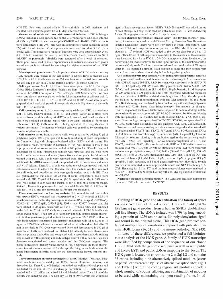
FIG. 1. (A) HGK amino acid sequence: predicted maximal HGK protein containing all possible alternatively spliced modules. The kinasedomain, the coiled-coil domain, and the CNH domain are underlined. Alternatively spliced modules M1 to M9 are shown in bold and labeled tothe right of the sequence. For adjacent modules M1/M2 and M3/M4, a vertical line defines their boundaries. (B) Schematic illustration of thedomain structure of known HGK splice variants compared with mouse NIK. Alternatively spliced modules are indicated with inverted V’s whenabsent and alternative patterning where present. The HGK gene product that we cloned from a tumor cell line (HGKT) contains alternativemodules M1, M2, M3, and M8. The two HGK cDNAs from a human macrophage library contain M1, M6, M8, and M9 (HGKS; short version)and M1, M4, M6, M8, and M9 (HGKL; long version). KIAA0687 isolated from brain has modules M1, M3, M5, M6, and M8. The mouse NIK clonecontains M1, M4, M5, M6, and M8.
VOL. 23, 2003 STE20 KINASE HGK 2071
on Decem
ber 11, 2013 by guesthttp://m
cb.asm.org/
Dow
nloaded from
dition, examination of HGK ESTs revealed a long 3� untrans-lated region of 3,442 additional base pairs. Within this regionare three alternative polyadenylation sites that would give tran-scripts of 5.0, 6.3, and 8.1 kb.
Figure 1A contains the predicted maximal HGK amino acidsequence from all 33 exons. The alternatively spliced modules,M1 to M9, are shown in bold. EST evidence indicated thateach alternative module was present in multiple but not alltranscripts (Table 1). All HGK splice forms contained theN-terminal kinase domain, a coiled-coil domain, and a C-terminal citron homology (CNH) domain. The close HGKfamily member TNIK also has three alternatively spliced ex-ons, two of which are equivalent to modules M1 and M3 (22).
The cDNAs encoding HGK isoforms isolated from differenttissue sources each contained a distinct subset of modules. Aschematic comparison of the domain structures of the fourcloned human HGK isoforms as well as the mouse orthologNIK is shown in Fig. 1B. The human HGK isoforms includeour tumor-derived HGK isoform (HGKT), the original HGKclones isolated from macrophages (HGKS, short version;HGKL, long version) (51), as well as another splice variantcloned from brain, published with the reference numberKIAA0687 (26).
There was no alternative splicing in the kinase domain, butthe coiled-coil region can be alternatively spliced into shortand long forms. The HGKT form contains both M1 and M2and therefore has a longer coiled coil than the other forms.The M3 and M4 modules show sequence similarity to eachother and contain PxxP motifs that can interact with SH3domain-containing proteins. Our tumor-derived HGK(HGKT) contains M3, while the short form (HGKS) isolatedfrom human macrophage lacks M3/M4, and the long form(HGKL) and NIK contain M4. M5 and M6 are small exonsencoding one and three amino acids, respectively. M7 is veryrich in serine and is not present in any of the isolated full-length HGK clones. The CNH domain contains two alterna-
tively spliced modules, of which the M8 but not the M9 moduleis conserved in CNH domains found in other protein families.
We extended our study to survey which isoforms of HGK areexpressed in tumor cells. With reverse transcription-PCR, wecloned multiple cDNA fragments of HGK from RNA isolatedfrom several tumor cell lines, including A549 and H1299 lungcarcinoma cells, 293T transformed kidney cells, as well as amixed pool of RNA from 10 tumor cell line sources (Clon-tech’s Universal Reference RNA). Table 1 shows the preva-lence of each module in cDNAs isolated from these tumor celllines; the results from the four tumor cell line sources werepooled and are shown together, as the distribution of modulesfrom each was similar. We identified at least five differentsplice isoforms from each tumor cell line source, indicatingthat the tumor cells possessed multiple forms of HGK. Themost striking difference observed when the tumor cell line datawas compared with EST data was that the tumor cell cloneslargely lacked the PxxP-containing module M4. Only 2% of thetumor-derived clones contained M4. Similarly, the M3 PxxPmotif was absent in most clones. In addition, the tumor celllines also generally lacked the serine-rich M7 module and theM9 module in the CNH domain.
HGK expression analysis. A 400-bp fragment from a con-served region of HGK present in all splice variants was used toprobe Northern blots containing RNA purified from variousnormal human tissues as well as from 60 cell lines from theNational Cancer Institute (NCI) tumor panel (Fig. 2A). Tran-scripts of 4.7 and 6.5 kb were detected, comparable in sizeto two of the transcripts predicted from the analysis of ESTs.In normal tissue, HGK mRNA was expressed at readily de-tectable levels only in tissue samples from brain and testis. Incontrast, HGK mRNA was expressed at high levels in 40 of the60 tumor cell lines derived from a variety of tissues (Fig. 2A).
The same probe was also hybridized to an array of cDNAsamplified from a broader array of tissue and tumor cell linesources. Quantification of the probed array by phosphorimag-
TABLE 1. Alternative sequence modules in HGK
Mod-ule Frame
Length(nucleotides/
aminoacids)
Position(amino acid no.
in max form)Origin
No. of ESTdatabase
sequenceswith/withoutthis module
No. of tumorcell line
cDNAs with/without this
module
Comments
M1 1 87/29 428 Exon 41/1 27/14 Encodes C-terminal extension of coiled-coildomain; similar module found in TNIK;adjacent to M2
M2 1 93/31 495 Alternative spliceacceptor
14/28 12/29 Adjacent to MI
M3 1 162/54 570 Exon 14/33 7/50 Similar module found in TNIK; contains 2PxxP motifs; adjacent to M4
M4 1 231/77 623 Exon 12/35 1/56 Contains 2 PxxP motifs; adjacent to M3M5 1 3/1 736 Alternative splice
acceptor23/9 39/18 Adds a serine residue
M6 1 9/3 819 Exon 20/20 10/46 Adds 3 residuesM7 1 192/64 921 Exon 21/16 9/47 Contains a serine-rich sequenceM8 1 57/19 1215 Intron readthrough 34/1 24/0 Encodes part of CNH domain; similar
sequence seen in other human GCK typeIV kinases
M9 1 24/8 1258 Alternative splicedonor
11/17 3/20 Encodes part of CNH domain; similarsequence not seen in other CNHdomains
2072 WRIGHT ET AL. MOL. CELL. BIOL.
on Decem
ber 11, 2013 by guesthttp://m
cb.asm.org/
Dow
nloaded from
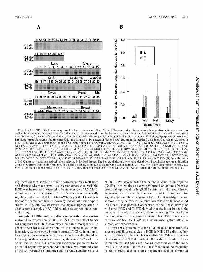
ing revealed that across all tumor-derived sources (cell linesand tissues) where a normal tissue comparison was available,HGK was increased in expression by an average of 7.7-fold intumor versus normal tissues. This difference was statisticallysignificant at P � 0.000001 (Mann-Whitney test). Quantifica-tion of the same data broken down by individual tumor type isshown in Fig. 2B. We observed the highest upregulation inglioblastoma samples (46.3-fold relative to expression in nor-mal brain).
Analysis of HGK mutants: effects on growth and transfor-mation. Overexpression of HGK mRNA in a variety of tumorcells suggests that HGK may play a role in tumorigenesis. Inorder to test for a causative role for this kinase in cell trans-formation, we constructed mutant forms of HGKT in mamma-lian expression vectors to test in tissue culture cells. Based onhomology with other related kinases, threonine 187 and thre-onine 191 in the HGK activation loop were predicted to bepotential regulatory phosphorylation sites. We mutated eachof the two residues to glutamic acid to create activating alleles
of HGK. We also mutated the catalytic lysine to an arginine(K54R). In vitro kinase assays performed on extracts from ratintestinal epithelial cells (RIE-1) infected with retrovirusesexpressing each of the HGK mutants used in subsequent bio-logical experiments are shown in Fig. 3. HGK wild-type kinaseshowed strong activity, while mutation of K54 to R inactivatedthe kinase, as expected. Comparison of the kinase activity ofwild-type HGK and T187E showed that the latter had a slightincrease in in vitro catalytic activity. Mutating T191 to E, incontrast, abolished the kinase activity. This T191E mutant wasused in addition to K54R as a dominant-negative allele insubsequent experiments.
To test for a possible role for HGK in focus formation, wecoexpressed different alleles of HGK in NIH 3T3 cells togetherwith an activated allele of H-Ras (valine 12). While expressionof wild-type and T187E mutant HGKs did not induce focusformation by itself (data not shown), coexpression of the inac-tive HGK K54R mutant with H-RasV12 reduced the frequencyof Ras-induced foci in a dose-dependent fashion compared
FIG. 2. (A) HGK mRNA is overexpressed in human tumor cell lines. Total RNA was purified from various human tissues (top two rows) aswell as from human tumor cell lines from the standard tumor panel from the National Cancer Institute. Abbreviations for normal tissues: (firstrow) Br, brain; Cx, cortex; Cb, cerebellum; Tm, thymus; SG, salivary gland; Lu, lung; Liv, liver; Pn, pancreas; Ki, kidney; Sp, spleen; St, stomach;Du, duodenum; Ut, uterus; Pr, prostate; SM, skeletal muscle; Pl, placenta; (second row) Br, brain; Te, testis; Bl, bladder; Co, colon; Ad, adiposetissue; fLi, fetal liver. Numbering for the NCI tumor panel: 1, HOP-92; 2, EKVXl 3, NCI-H23; 4, NCI-H226; 5, NCI-H322; 6, NCI-H460; 7,NCI-H522; 8, A549; 9, HOP-62; 10, OVCAR-3; 11, OVCAR-4; 12, OVCAR-5; 14, IGROV1; 15, SK-OV-3; 16, SNB-19; 17, SNB-75; 18, U251;19, SF-288; 20, SF-295; 21, SF-539; 22, CCRF-CEM; 23, K-562; 24, MOLT-4; 25, HL-60; 26, RPMI-8226; 27, SR; 28, DU-145; 29, PC-3; 30, HT-29;31, HCC-2998l; 32, HCT116; 33, SW620; 34, COLO-205; 35, HCT-15; 36, M-12; 37, UO-31; 38, SN12C; 39, A498; 40, Caki-1; 41, RXF-393; 42,ACHN; 43, 786-0; 44, TK-10; 45, LOXIMVI; 46, Malme-3 M; 47, SK-MEL-2; 48, SK-MEL-5; 49, SK-MEL-28; 50, UACC-62; 51, UACC-257; 52,M14; 53, MCF-7; 54, MCF-7/ADR; 55, Hs578T; 56, MDA-MB-231; 57, MDA-MB-431; 58, MDA-N; 59, BT-549, and 60, T-47D. (B) Quantificationof HGK in tumor versus normal cells from selected individual tissues. The bar graph shows the relative signal from PhosphorImager quantificationof dot blot arrays from tumor cell lines and normal tissues. From left to right: colon tumor-normal, 2.7-fold, P � 0.210; lung tumor-normal, 2.0,P � 0.034; brain tumor-normal, 46.3, P � 0.007; kidney tumor-normal, 5.5, P � 0.076. P values were calculated with the Mann-Whitney test.
VOL. 23, 2003 STE20 KINASE HGK 2073
on Decem
ber 11, 2013 by guesthttp://m
cb.asm.org/
Dow
nloaded from

with coexpression of wild-type HGK (Fig. 4). When the num-ber of foci was tabulated, the K54R mutant was found toreduce focus formation rates by approximately 50% at a DNAratio of 1:20 with respect to H-RasV12. In addition, coexpres-sion of wild-type HGK reproducibly increased the number offoci generated by H-RasV12, indicating some level of cooper-ation. A similar rate of inhibition of focus formation was ob-served in RIE-1 cells cotransfected with H-RasV12 and eitherinactive form of HGK (K54R or T191E; data not shown).These data indicate that HGK kinase activity is important fortransformation by Ras in this assay.
To further elucidate the role of HGK in cell growth andtransformation, we compared both monolayer and anchorage-independent growth of stable clones of RIE-1 cells expressingvarious alleles of HGK isolated from the retrovirally infectedpools. Relative expression levels of Myc-tagged HGK in dif-
FIG. 3. Kinase activity of HGK mutants in RIE-1 cells. HGK wild-type and mutant proteins were immunoprecipitated from stable poolsof RIE-1 cells infected with HGK retroviruses. (A) Comparison of theT187E and T191E activation loop mutations with wild-type kinase.The top panel shows an autoradiogram of samples analyzed by SDS-PAGE, showing the relative amount of 32P incorporated into myelinbasic protein (MBP) after incubation of the immunoprecipitate and[�-32P]ATP for 20 min at 30�C. The bottom panel shows the relativeamount of HGK proteins in the immunoprecipitate. (B) Comparisonof the T191E and K54R catalytically inactive HGK mutants with wild-type HGK. An autoradiogram shows the relative amount of 32P incor-poration both into HGK protein (autophosphorylation) and intoadded MBP in the immunoprecipitate.
FIG. 4. Dominant negative mutant HGK can suppress Ras-in-duced focus formation in NIH 3T3 cells. (Top) Images of tissue cultureplates stained with crystal violet to visualize the foci are shown from arepresentative experiment with NIH 3T3 cells. The relative number offoci per plate generated by H-RasV12 in the presence of a 10- or 20-foldmolar excess of plasmid encoding HGK wild-type, HGK K54R inactivemutant kinase, or vector control is shown. The panel on the top showsbackground levels of foci generated by transfection of a vector control(no H-RasV12). (Bottom) The bar graph shows the relative number offoci per plate (setting the number of foci in the Ras/vector control 1:10at 100%). The data represent an average of two experiments.
2074 WRIGHT ET AL. MOL. CELL. BIOL.
on Decem
ber 11, 2013 by guesthttp://m
cb.asm.org/
Dow
nloaded from
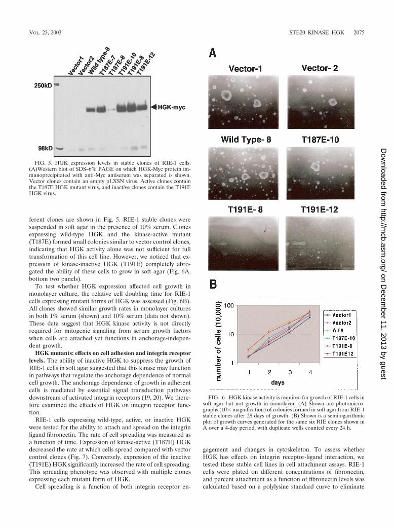
ferent clones are shown in Fig. 5. RIE-1 stable clones weresuspended in soft agar in the presence of 10% serum. Clonesexpressing wild-type HGK and the kinase-active mutant(T187E) formed small colonies similar to vector control clones,indicating that HGK activity alone was not sufficient for fulltransformation of this cell line. However, we noticed that ex-pression of kinase-inactive HGK (T191E) completely abro-gated the ability of these cells to grow in soft agar (Fig. 6A,bottom two panels).
To test whether HGK expression affected cell growth inmonolayer culture, the relative cell doubling time for RIE-1cells expressing mutant forms of HGK was assessed (Fig. 6B).All clones showed similar growth rates in monolayer culturesin both 1% serum (shown) and 10% serum (data not shown).These data suggest that HGK kinase activity is not directlyrequired for mitogenic signaling from serum growth factorswhen cells are attached yet functions in anchorage-indepen-dent growth.
HGK mutants: effects on cell adhesion and integrin receptorlevels. The ability of inactive HGK to suppress the growth ofRIE-1 cells in soft agar suggested that this kinase may functionin pathways that regulate the anchorage dependence of normalcell growth. The anchorage dependence of growth in adherentcells is mediated by essential signal transduction pathwaysdownstream of activated integrin receptors (19, 20). We there-fore examined the effects of HGK on integrin receptor func-tion.
RIE-1 cells expressing wild-type, active, or inactive HGKwere tested for the ability to attach and spread on the integrinligand fibronectin. The rate of cell spreading was measured asa function of time. Expression of kinase-active (T187E) HGKdecreased the rate at which cells spread compared with vectorcontrol clones (Fig. 7). Conversely, expression of the inactive(T191E) HGK significantly increased the rate of cell spreading.This spreading phenotype was observed with multiple clonesexpressing each mutant form of HGK.
Cell spreading is a function of both integrin receptor en-
gagement and changes in cytoskeleton. To assess whetherHGK has effects on integrin receptor-ligand interaction, wetested these stable cell lines in cell attachment assays. RIE-1cells were plated on different concentrations of fibronectin,and percent attachment as a function of fibronectin levels wascalculated based on a polylysine standard curve to eliminate
FIG. 5. HGK expression levels in stable clones of RIE-1 cells.(A)Western blot of SDS–6% PAGE on which HGK-Myc protein im-munoprecipitated with anti-Myc antiserum was separated is shown.Vector clones contain an empty pLXSN virus. Active clones containthe T187E HGK mutant virus, and inactive clones contain the T191EHGK virus.
FIG. 6. HGK kinase activity is required for growth of RIE-1 cells insoft agar but not growth in monolayer. (A) Shown are photomicro-graphs (10� magnification) of colonies formed in soft agar from RIE-1stable clones after 28 days of growth. (B) Shown is a semilogarithmicplot of growth curves generated for the same six RIE clones shown inA over a 4-day period, with duplicate wells counted every 24 h.
VOL. 23, 2003 STE20 KINASE HGK 2075
on Decem
ber 11, 2013 by guesthttp://m
cb.asm.org/
Dow
nloaded from

background due to differences in cell number and plating ef-ficiency. Compared with the vector control, the inactive-ki-nase-expressing clones demonstrated increased rates of adhe-sion, while the active-kinase-expressing clones (T187E)showed a reduced rate of adhesion (Fig. 8). Similar results
were obtained with multiple clones. Therefore, HGK appearsto negatively regulate RIE-1 cell adhesion to fibronectin.
Major fibronectin receptors in epithelial cells include �5�1and �v�3. Function-blocking integrin antibodies were used toshow that RIE-1 cell adhesion to fibronectin is mediated, atleast in part, by �5�1 receptors. Adhesion of these cells tofibronectin was completely blocked by treatment of cells withanti-�1 antibodies and partially blocked by treatment withanti-�5 antibodies, while anti-�3 antibodies had no effect (datanot shown). The level of �5 and �1 integrin receptor subunitson the cell surface of HGK-expressing RIE-1 clones was mea-sured by fluorescence-activated cell sorting. While there wasno effect on �1 levels, the �5 levels were somewhat higher inthe inactive-kinase-expressing clones and lower in the active-kinase-expressing clones relative to vector controls (Fig. 9).
HGK effect on cell invasion and morphogenesis. RIE-1 cellswere used to test the effects of HGK mutant expression on cellgrowth, morphogenesis, and invasion in Matrigel. In a three-dimensional invasion or tubulogenesis assay (2, 34), cells weresuspended in Matrigel and cultured in the presence and ab-sence of hepatocyte growth factor (HGF). As was seen previ-ously with kidney epithelial cells, HGF was both mitogenic andmorphogenic for RIE-1 cells in this assay, as demonstrated by
FIG. 7. Role for HGK in cell spreading. RIE-1 stable clones ex-pressing active and inactive alleles of HGK as well as vector controlswere detached with trypsin and plated on fibronectin. Photomicro-graphs (10� magnification) were taken at various time points there-after. The graph shows the relative number of spread (phase-dark)cells counted in three separate fields for each clone as a function oftime. Representative photographs from the 170-min time point areshown on top.
FIG. 8. HGK kinase activity interferes with cell attachment. RIE-1stable clones expressing active and inactive alleles of HGK as well asvector controls were detached with trypsin and plated on various con-centrations of fibronectin (FN). After 60 min, unattached cells werewashed away, and the remaining cells were stained with crystal violet.(A) Photomicrographs (2� magnification) show the presence of re-maining cells at different concentrations of fibronectin. (B) By com-parison with a standard curve of cells plated on polylysine, the relativeamount of cells was quantified by measuring the amount of solubilizeddye by optical density. The graph shows the relative optical density at550 nm as a function of fibronectin concentration for the differentRIE-1 clones.
2076 WRIGHT ET AL. MOL. CELL. BIOL.
on Decem
ber 11, 2013 by guesthttp://m
cb.asm.org/
Dow
nloaded from

inducing them to grow and invade the surrounding Matrigel,forming large lattice-like networks (Fig. 10A, compare vectorcontrol with and without HGF). In the absence of HGF, RIE-1cells survived in the Matrigel but showed a slower growth rate.Clones expressing either the wild-type or the T187E mutant ofHGK gave larger networks of invading cells than the vectorcontrols within the same time frame. However, cells expressingthe inactive T191E form of HGK were almost fully blocked intheir invasive response, forming spheroid colonies. In contrastto their inability to invade, the T191E mutant cells appeared tobe able to respond mitogenically to HGF and proliferate (Fig.10A, compare T191E clones with and without HGF). Similareffects were observed when cells expressing the mutant alleleK54R were tested (data not shown). These data suggest thatblocking HGK kinase-dependent signaling specifically blockedthe morphogenic response of the cells to HGF.
Next we asked whether HGK plays a role in directionalinvasion (chemotaxis toward HGF). To address this question,RIE-1 cells were seeded in a Boyden chamber coated withMatrigel, and the rate of invasion was measured. RIE-1 stablepools overexpressing wild-type HGK showed significantly in-creased migration through the Matrigel towards HGF com-
pared with either the vector control or parental cells (Fig.10B). In contrast, the inactive-HGK-expressing pools were sig-nificantly retarded in invasive response.
Effects of HGK mutants on HGF intracellular signaling. Inview of the ability of the HGK mutants to interfere with HGF-induced morphogenic and invasive responses, effects on imme-diate downstream signaling from the HGF receptor, Met, wereexamined in RIE-1 clones expressing HGK mutants. The levelof Met receptor expression as well as the level of receptorphosphorylation in response to HGF stimulation was not sig-nificantly affected (Fig. 11A and C). Next we examined three ofthe major signaling pathways that have been reported to func-tion in HGF-induced morphogenesis, the mitogen-activatedprotein kinase pathway, the phosphatidylinositol 3-kinase/AKT pathway, and STAT3 activation (11, 41, 42).
As shown in Fig. 11B, we observed little or no effect on bothERK1/ERK2 and AKT activation with HGF stimulation. Incontrast, we noted an attenuation of STAT3 phosphorylationon both of the regulatory residues, tyrosine 705 and serine 727,in the cells expressing the kinase-inactive HGK. To rule outthe possibility that this effect was due to clonal variation, weexamined HGF stimulation of STAT3 phosphorylation in two
FIG. 9. HGK mutant expression affects cell surface �5 integrin subunit levels. By fluorescence-activated cell sorting analysis, the level of cellsurface integrin receptors was compared for different RIE-1 stable clones. Histograms show the relative fluorescence intensity of cells stained withfluorescein isothiocyanate-labeled integrin receptor antibodies. The level of specific integrin receptor (�5 subunit on top and �1 subunit on thebottom) are shown as open green peaks. Isotype controls are shown as solid purple peaks. The relative shift of the green peak to the right relativeto the isotype control indicates the level of that particular receptor. Numbers in the upper right corner of each histogram are the mean fluorescenceintensity (normalized to the relevant isotype control).
VOL. 23, 2003 STE20 KINASE HGK 2077
on Decem
ber 11, 2013 by guesthttp://m
cb.asm.org/
Dow
nloaded from

independent clones expressing inactive HGK (T191E-8 andT191E-12). The panel shown in Fig. 11C illustrates thatSTAT3 tyrosine phosphorylation was inhibited in both of theseclones. In addition, a small increase in STAT3 Y705 phosphor-
ylation was observed in the clones expressing the active forms(wild type or T187E) of HGK in some experiments (Fig. 11C).We also observed STAT3 serine 727 phosphorylation to beinhibited in both kinase-inactive clones (data not shown). Weconfirmed the protein band observed as STAT3 by immuno-precipitation with a STAT3 antibody, followed by probing withthe phospho-STAT3 antibodies (data not shown). These datasuggest that HGK functions in the activation of STAT3 down-stream of the stimulated Met receptor. No increase in basalSTAT3 phosphorylation was observed in the RIE clones ex-pressing active HGK forms or in 293T cells transiently trans-fected with wild-type HGK, indicating that this kinase cannotactivate STAT3 on its own but rather modulated the stimula-tion of STAT3 phosphorylation with HGF stimulation (Fig. 11and data not shown).
To further investigate the involvement of HGK in STAT3activation, we designed experiments to test whether HGK andSTAT3 were associated in cells. As shown in Fig. 12A, wetransfected 293T cells with wild-type and kinase-inactive HGK,followed by immunoprecipitation of proteins with anti-STAT3.We observed that the transfected HGK coimmunoprecipitatedwith endogenous STAT3. The association between STAT3 andHGK was not dependent on HGK kinase activity, as the wild-type and kinase-inactive proteins coimmunoprecipitated withsimilar efficiency. We next preformed a similar coimmunopre-cipitation from the RIE stable clones with and without HGFstimulation (Fig. 12B). In this cell system as well, we observedthat HGK was retained in the STAT3 immunoprecipitation.These data suggest that HGK and STAT3 are part of the samesignaling complex and suggest a direct involvement of HGK inmodulation of STAT3 phosphorylation in response to HGF.
DISCUSSION
We identified a novel form of HGK in a human glioblastomacell line cDNA library. Compared with other forms of HGKisolated from macrophage and brain, and with the mouse or-tholog NIK, this form showed multiple different splice varia-tions. We analyzed the overall genomic structure of the HGKgene and found it to include 33 exons, with nine regions (mod-ules) of alternative splicing. EST data suggest that all of thesealternative modules are present in expressed proteins, result-ing in a complex family of HGK isoforms. While many genesare alternatively spliced, the HGK gene has an unusually largeamount of alternative splicing. At least five different isoformswere identified from each of four tumor cell line sources,indicating that multiple isoforms are present in the same cell.Alternative splicing modulated the length of the coiled-coildomain, the composition of the CNH domain, and an array ofPxxP motifs.
Given the variations in these protein-protein interaction do-mains, different splice forms are likely to be differentially lo-calized and/or regulated. For example, the HGK clone isolatedfrom glioblastoma cells has the PXXP-containing M3 module,while an alternative PXXP-containing module, M4, replacesM3 in the clones isolated from macrophage (51) and mouseadipocytes (43). The consensus sequences of the PXXP motifsin M3 and M4 differ so that HGK isoforms containing one orthe other may interact with different SH3-containing proteins.Two PXXP motifs in M4 from NIK were shown to bind to the
FIG. 10. (A) Role for HGK in RIE cell morphogenesis. HGKkinase expression affects RIE-1 cells in a tubulogenesis assay. Shownare photomicrographs (3.2� magnification) of colonies formed in Ma-trigel after 6 days of growth. Each clone is shown with and withoutadded HGF. Vector control is an RIE-1 clone with pLXSN alone.WT-8 is a clone expressing wild-type HGK. T187E-7 is a clone ex-pressing the active HGK mutant; T191E-12 and T191E-8 are twoclones expressing the inactive HGK mutant. (B) Role for HGK in RIEcell invasion. Boyden chambers in which the cells of the lower mem-brane are fixed and stained with crystal violet are shown. The amountof staining is proportional to the number of cells that successfullyinvaded through the Matrigel plug to colonize the lower membraneafter stimulation with 50 ng of human HGF per ml for 4 days. Tripli-cate wells are shown from an experiment with RIE-1 stable poolsexpressing vector alone, wild-type, or inactive (T191E) HGK.
2078 WRIGHT ET AL. MOL. CELL. BIOL.
on Decem
ber 11, 2013 by guesthttp://m
cb.asm.org/
Dow
nloaded from

adapter protein Nck (43). The binding partners for the PXXPmotifs in M3 are unknown and under investigation. The inter-actions mediated by these PXXP motifs may not be importantfor the function of HGK in tumor cells, as M3 and M4 areabsent from the majority (88% and 98%, respectively) of theisoforms identified in tumor cell lines.
With a probe that detects all HGK splice forms, we foundthis kinase to be broadly overexpressed at the RNA level in alarge number of tumor cell lines relative to most normal tis-sues. HGK does not map to a known tumor amplicon, yet theoverabundance of HGK RNA in tumor cell lines versus normaltissue provides correlative evidence for a role for this kinase intumorigenesis. An active role for HGK kinase activity in celltransformation is suggested by the fact that ectopic expressionof inactive forms blocked oncogenically relevant processes intissue culture cell models. It is important to note that both theNIH 3T3 and RIE-1 cells used in these studies express detect-able levels of HGK, suggesting that the inactive mutant acts ina dominant negative manner (data not shown).
Expression of the inactive dominant negative mutantblocked focus formation and anchorage-independent growth,increased cell adhesion and spreading rates, and blocked inva-sion and morphogenesis in response to HGF. In contrast, over-expression of wild-type or active alleles slightly increased focusformation, had no effect on anchorage-independent growth,decreased cell adhesion and spreading, and potentiated thecells’ invasive and morphogenic responses to HGF. Togetherthese results suggest that HGK catalytic activity is required foranchorage-independent growth, causes the reduction of celladhesion, and increases invasive properties.
The ability of dominant negative HGK to block the growthof RIE-1 cells in soft agar suggests that this kinase may func-tion in pathways that regulate the anchorage dependence ofnormal cell growth, implicating HGK in the so-called out-side-in signaling from integrin receptors (24). Focal adhesionkinase (FAK) has been shown to function in such pathways(21, 25, 49). There was no measurable difference in autophos-phorylation of FAK or phosphorylation of the FAK substratep130CAS during cell attachment in the stable cell lines express-ing the various HGK mutants (data not shown), indicating thatHGK may function either downstream or independently ofFAK.
The effects of HGK mutants on integrin function and onintegrin receptor expression indicate that this kinase does ap-pear to induce changes in cell adhesion or inside-out signaling.
FIG. 11. HGK mutant expression affects HGF-induced STAT3phosphorylation. Western blots of cell lysates prepared from starvedRIE-1 cells with and without stimulation with 100 ng of HGF per ml
are shown. (A) Western blots of anti-Met receptor immunoprecipi-tates (IP) probed with antiphosphotyrosine (�-pY) antibody in the toppanel and anti-Met receptor antibody in the bottom panel are shown.(B) A panel of Western blots prepared from total cell lysates stimu-lated for 0, 30, and 60 min with HGF are shown. From top to bottom:anti-phospho-mitogen-activated protein kinase, anti-ERK1 plus anti-ERK2, anti-phospho-AKT, anti-AKT anti-phospho-STAT3 (Y705),anti-phospho-STAT3 (S727), and anti-STAT3. (C) Total cell lysatesfrom unstimulated cells and cells 30 min after HGF stimulation. Thetop panel was probed with an anti-phospho-Y705 STAT3-specific an-tibody; the middle panel was probed with an anti-STAT3 antibody; thebottom panel shows anti-Met receptor immunoprecipitations from thesame lysates, probed with an anti-Met antibody. Data from RIE-1clones expressing empty vector (vector), wild-type HGK, T187E HGK(active mutant), and T191E HGK (inactive mutant) are shown.
VOL. 23, 2003 STE20 KINASE HGK 2079
on Decem
ber 11, 2013 by guesthttp://m
cb.asm.org/
Dow
nloaded from

Integrin mediated inside-out and outside-in signaling are oftendescribed as independent pathways, but there is evidence thateffects on one impinge on the other (reviewed in references 4and 23). We observed that cells with higher HGK kinase ac-tivity were less adherent to fibronectin. This effect may beexplained, at least in part, by the reduced surface expression ofintegrin receptor �5 subunit in cells expressing active HGK.While a comprehensive analysis of all integrin subunits levelscould not be done with available reagents for rat cells, we didnote that HGK also had similar effects on �2 subunit levels butnot on �1 subunit levels. Interestingly, NIK�/� mice show aphenotype similar to those of fibronectin�/� and �5 integrinreceptor�/� mice, suggesting that NIK may also be involved inthe �5 integrin receptor regulation during embryogenesis (50).
As the changes in �5 subunit cell surface expression weresmaller than might be expected from the phenotypic changesobserved, HGK may also affect integrin signaling by an addi-
tional mechanism. The recent report showing physical associ-ation, as well as colocalization in attaching cells of �1-integrinreceptor with NIK (mouse HGK) strongly suggests that HGKplays a direct role in integrin receptor signaling in addition toaffecting �-integrin receptor levels, as we have shown (37).Taken together, these observations point to both direct andindirect involvement of HGK in the regulation of integrinfunction and signaling.
This role for HGK in negative regulation of cell adhesionmay be conserved in other HGK family members, since TNIKalso appears to interfere with cell spreading in a kinase activity-dependent manner in transient-transfection assays (22). A re-duction in cell spreading rates was also observed previouslywith other STE20 kinases, including STE20-like kinase (SLK)and the p21-activated kinases �PAK and PAK4 (32, 39, 48). Inthese three reports, the effects on cell spreading were attrib-uted to effects on the cell cytoskeleton. Whether or not these
FIG. 12. HGK kinase coimmunoprecipitated with STAT3. (A) 293T cells were transfected with Myc-tagged wild-type K54R (inactive) HGKand the vector control. Lysates were immunoprecipitated with anti-STAT3, followed by analysis on Western blots probed with anti-STAT3 (left)and anti-Myc (middle). Total lysates were analyzed with anti-Myc in parallel. (B) RIE-1 clones expressing wild-type and kinase-inactive (T191E-12)HGK were stimulated with HGF for 0 or 30 min Lysates were immunoprecipitated with both anti-Myc and anti-STAT3. The middle panel showsthe STAT3 immunoprecipitate probed with anti-Myc. The left panel shows the same immunoprecipitate probed with anti-STAT3, and the rightpanel is an anti-Myc immunoprecipitate probed with anti-Myc for HGK. The HGK and STAT3 protein bands are indicated with arrows.
2080 WRIGHT ET AL. MOL. CELL. BIOL.
on Decem
ber 11, 2013 by guesthttp://m
cb.asm.org/
Dow
nloaded from

other STE20 kinases modulated integrin-ligand binding in ad-dition to effects on the cytoskeleton was not investigated.STE20 kinases show significant homology within the kinasedomain and may share some cellular substrates, yet the lack ofhomology in the regulatory domains of these kinases suggestthat they are not localized and/or regulated by the same mech-anism. For example, the PAK kinases are directly regulated byrho family G-proteins, while HGK is not regulated by Rac orCDC42 and furthermore has been implicated to function up-stream of Rac in the activation of JNK (45).
Increased HGK kinase activity through overexpressioncaused a striking increase in the rate of cell invasion andmorphogenesis, while expression of a kinase-inactive mutantblocked invasion and morphogenesis. Ectopic expression ofHGK was not sufficient to induce invasion on its own andinstead potentiated the invasive response of cells to HGF. Asimilar potentiation role in HGF-induced invasion was shownfor the cytoplasmic domain of �6�4 integrin receptors (47).Perhaps HGK also participates in the signaling complex withthe Met receptor and the �6�4 integrin receptor. In examina-tion of signaling downstream of the Met receptor, we observeda decrease in STAT3 phosphorylation on both Y705 and S727with dominant negative HGK mutant expression. Both of thesephosphorylation events have been shown to increase the activ-ity of STAT3 as a transcription factor (reviewed in reference8).
STAT3 does not appear to be a direct substrate of HGK inthat STAT3 protein isolated from cells was not phosphorylatedwhen active, recombinant glutathione S-transferase-HGK ki-nase domain was added in the presence of ATP (data notshown). However, HGK protein was found to copurify withSTAT3 in immunoprecipitations of STAT3 from cells, suggest-ing that they are part of the same protein complex. HGK couldmodulate STAT3 activation by affecting its localization in thecell or by regulating the kinases and phosphatases that directlyregulate STAT3. As STAT3 plays a role in HGF-induced in-vasion, blocking STAT3 activation may be one mechanism bywhich the inactive HGK mutants blocked RIE-1 cell invasionand morphogenesis (6). STAT3 has also been implicated intumorigenesis in a number of different cell systems (7) and thuscould also be important for the dominant negative effects ofinactive HGK on focus formation and anchorage-independentgrowth. A more detailed investigation of the function of HGKin STAT3 activation will be the subject of a future study.
In conclusion, we present an analysis of the HGK gene withits complex family of isoforms. We present evidence that thishuman homolog of the msn and mig-15 kinases from Drosoph-ila melanogaster and Caenorhabditis elegans functions in mor-phogenic pathways not only during embryogenesis (50) butalso during tumorigenesis and/or invasion. Based on our ob-servations and those of others, we suggest that this kinase mayfunction in the integration of extracellular signals from solublegrowth factors and from the extracellular matrix.
ACKNOWLEDGMENTS
We thank Rhea Pugliese, Greg Plowman, and Ricardo Martinez foridentification and isolation of the first HGK gene and Joseph Zach-wieja for preparation of the ascites 9E10 antibodies. We thank KongChow and Jennifer Goldstein for sequencing a large number of HGKclones. We thank Tony Hunter (by way of Martin Broome) for thelow-passage-number NIH 3T3 cells, Channing Der (by way of Sara
Courtneidge) for the RIE-1 cells, and Paul Hughes for advice on andprotocols for the integrin experiments.
This work was supported entirely by Sugen, Inc., a subsidiary ofPharmacia Corporation.
REFERENCES
1. Bagrodia, S., and R. A. Cerione. 1999. Pak to the future. Trends Cell Biol.9:350–355.
2. Bao, Q., and R. C. Hughes. 1995. Galectin-3 expression and effects on cystenlargement and tubulogenesis in kidney epithelial MDCK cells cultured inthree-dimensional matrices in vitro. J. Cell Sci. 108:2791–2800.
3. Becker, E., U. Huynh-Do, S. Holland, T. Pawson, T. O. Daniel, and E. Y.Skolnik. 2000. Nck-interacting Ste20 kinase couples Eph receptors to c-JunN-terminal kinase and integrin activation. Mol. Cell. Biol. 20:1537–1545.
4. Berman, A. E., and N. I. Kozlova. 2000. Integrins: structure and functions.Membrane Cell Biol. 13:207–244.
5. Bischoff, J. R., L. Anderson, Y. Zhu, K. Mossie, L. Ng, B. Souza, B. Schryver,P. Flanagan, F. Clairvoyant, C. Ginther, C. S. Chan, M. Novotny, D. J.Slamon, and G. D. Plowman. 1998. A homologue of Drosophila aurorakinase is oncogenic and amplified in human colorectal cancers. EMBO J.17:3052–3065.
6. Boccaccio, C., M. Ando, L. Tamagnone, A. Bardelli, P. Michieli, C. Battis-tini, and P. M. Comoglio. 1998. Induction of epithelial tubules by growthfactor HGF depends on the STAT pathway. Nature 391:285–288.
7. Bowman, T., R. Garcia, J. Turkson, and R. Jove. 2000. STATs in oncogen-esis. Oncogene 19:2474–2488.
8. Bromberg, J., and J. E. Darnell, Jr. 2000. The Role of STATs in transcrip-tional control and their impact on cellular function. Oncogene 19:2468–2473.
9. Caplan, S., L. M. Hartnell, R. C. Aguilar, N. Naslavsky, and J. S. Bonifacino.2001. Human Vam6p promotes lysosome clustering and fusion in vivo.J. Cell Biol. 154:109–122.
10. Chadee, D. N., T. Yuasa, and J. M. Kyriakis. 2002. Direct activation ofmitogen-activated protein kinase kinase kinase MEKK1 by the Ste20p ho-mologue GCK and the adapter protein TRAF2. Mol. Cell. Biol. 22:737–749.
11. Comoglio, P. M. 2001. Pathway specificity for Met signaling. Nat. Cell Biol.3:E161–E162.
12. Cox, A. D., and C. J. Der. 1994. Biological assays for cellular transformation.Methods Enzymol. 238:271–276.
13. Dan, I., N. M. Watanabe, T. Kobayashi, K. Yamashita-Suzuki, Y. Fukagaya,E. Kajikawa, W. K. Kimura, T. M. Nakashima, K. Matsumoto, J. Ninomiya-Tsuji, and A. Kusumi. 2000. Molecular cloning of MINK, a novel member ofmammalian GCK family kinases, which is up-regulated during postnatalmouse cerebral development. FEBS Lett. 469:19–23.
14. Dan, I., N. M. Watanabe, and A. Kusumi. 2001. The Ste20 group kinases asregulators of MAP kinase cascades. Trends Cell Biol. 11:220–230.
15. Davy, A., and S. M. Robbins. 2000. Ephrin-A5 modulates cell adhesion andmorphology in an integrin-dependent manner. EMBO J. 19:5396–5405.
16. Di Cunto, F., E. Calautti, J. Hsiao, L. Ong, G. Topley, E. Turco, and G. P.Dotto. 1998. Citron rho-interacting kinase, a novel tissue-specific ser/thrkinase encompassing the Rho-Rac-binding protein Citron. J. Biol. Chem.273:29706–29711.
17. Elion, E. A. 2000. Pheromone response, mating and cell biology. Curr. Opin.Microbiol. 3:573–581.
18. Florea, L., G. Hartzell, Z. Zhang, G. M. Rubin, and W. Miller. 1998. Acomputer program for aligning a cDNA sequence with a genomic DNAsequence. Genome Res. 8:967–974.
19. Frisch, S. M., and H. Francis. 1994. Disruption of epithelial cell-matrixinteractions induces apoptosis. J. Cell Biol. 124:619–626.
20. Frisch, S. M., and E. Ruoslahti. 1997. Integrins and anoikis. Curr. Opin. CellBiol. 9:701–706.
21. Frisch, S. M., K. Vuori, E. Ruoslahti, and P. Y. Chan-Hui. 1996. Control ofadhesion-dependent cell survival by focal adhesion kinase. J. Cell Biol. 134:793–799.
22. Fu, C. A., M. Shen, B. C. Huang, J. Lasaga, D. G. Payan, and Y. Luo. 1999.TNIK, a novel member of the germinal center kinase family that activatesthe c-Jun N-terminal kinase pathway and regulates the cytoskeleton. J. Biol.Chem. 274:30729–30737.
23. Giancotti, F. G., and E. Ruoslahti. 1990. Elevated levels of the alpha 5 beta1 fibronectin receptor suppress the transformed phenotype of Chinese ham-ster ovary cells. Cell 60:849–859.
24. Giancotti, F. G., and E. Ruoslahti. 1999. Integrin signaling. Science 285:1028–1032.
25. Hungerford, J. E., M. T. Compton, M. L. Matter, B. G. Hoffstrom, and C. A.Otey. 1996. Inhibition of pp125FAK in cultured fibroblasts results in apo-ptosis. J. Cell Biol. 135:1383–1390.
26. Ishikawa, K., T. Nagase, M. Suyama, N. Miyajima, A. Tanaka, H. Kotani, N.Nomura, and O. Ohara. 1998. Prediction of the coding sequence of uniden-tified human genes. X. The complete sequences of 100 new cDNA clonesfrom brain which can code for large proteins in vitro. DNA Res. 5:169–176.
27. Kanai-Azuma, M., Y. Kanai, M. Okamoto, Y. Hayashi, H. Yonekawa, and K.
VOL. 23, 2003 STE20 KINASE HGK 2081
on Decem
ber 11, 2013 by guesthttp://m
cb.asm.org/
Dow
nloaded from

Yazaki. 1999. Nrk: a murine X-linked NIK (Nck-interacting kinase)-relatedkinase gene expressed in skeletal muscle. Mech. Dev. 89:155–159.
28. Kyriakis, J. M., and J. Avruch. 1996. Protein kinase cascades activated bystress and inflammatory cytokines. Bioessays 18:567–577.
29. Landau, N. R., and D. R. Littman. 1992. Packaging system for rapid pro-duction of murine leukemia virus vectors with variable tropism. J. Virol.66:5110–5113.
30. Madaule, P., M. Eda, N. Watanabe, K. Fujisawa, T. Matsuoka, H. Bito, T.Ishizaki, and S. Narumiya. 1998. Role of citron kinase as a target of thesmall GTPase Rho in cytokinesis. Nature 394:491–494.
31. Madaule, P., T. Furuyashiki, T. Reid, T. Ishizaki, G. Watanabe, N. Morii,and S. Narumiya. 1995. A novel partner for the GTP-bound forms of rho andrac. FEBS Lett. 377:243–248.
31a.Manning, G., D. B. Whyte, R. Martinez, T. Hunter, and S. Sandarsanam.2002. The protein kinase complement of the human genome. Science 298:1912–1934.
32. Manser, E., H.-Y. Huang, T.-H. Loo, X.-Q. Chen, J.-M. Dong, T. Leung, andL. Lim. 1997. Expression of constitutive active alpha-PAK reveals effects ofthe kinase on actin and focal complexes. Mol. Cell. Biol. 17:1129–1143.
33. Miao, H., E. Burnett, M. Kinch, E. Simon, and B. Wang. 2000. Activation ofEphA2 kinase suppresses integrin function and causes focal-adhesion-kinasedephosphorylation. Nat. Cell Biol. 2:62–69.
34. Montesano, R., K. Matsumoto, T. Nakamura, and L. Orci. 1991. Identifica-tion of a fibroblast-derived epithelial morphogen as hepatocyte growth fac-tor. Cell 67:901–908.
35. Nakano, K., J. Yamauchi, K. Nakagawa, H. Itoh, and N. Kitamura. 2000.NESK, a member of the germinal center kinase family that activates thec-Jun N-terminal kinase pathway and is expressed during the late stages ofembryogenesis. J. Biol. Chem. 275:20533–20539.
36. Paricio, N., F. Feiguin, M. Boutros, S. Eaton, and M. Mlodzik. 1999. TheDrosophila STE20-like kinase misshapen is required downstream of theFrizzled receptor in planar polarity signaling. EMBO J. 18:4669–4678.
37. Poinat, P., A. De Arcangelis, S. Sookhareea, X. Zhu, E. M. Hedgecock, M.Labouesse, and E. Georges-Labouesse. 2002. A conserved interaction be-tween beta1 integrin/PAT-3 and Nck-interacting kinase/MIG-15 that medi-ates commissural axon navigation in C. elegans. Curr. Biol. 12:622–631.
38. Pombo, C. M., J. H. Kehrl, I. Sanchez, P. Katz, J. Avruch, L. I. Zon, J. R.Woodgett, T. Force, and J. M. Kyriakis. 1995. Activation of the SAPKpathway by the human STE20 homologue germinal centre kinase. Nature377:750–754.
39. Qu, J., M. S. Cammarano, Q. Shi, K. C. Ha, P. de Lanerolle, and A. Minden.2001. Activated PAK4 regulates cell adhesion and anchorage-independentgrowth. Mol. Cell. Biol. 21:3523–3533.
40. Ruan, W., P. Pang, and Y. Rao. 1999. The SH2/SH3 adaptor protein dockinteracts with the Ste20-like kinase misshapen in controlling growth conemotility. Neuron 24:595–605.
41. Stella, M. C., and P. M. Comoglio. 1999. HGF: a multipfunctional growthfactor controlling cell scattering. Int. J. Biochem. Cell Biol. 31:1357–1362.
42. Stuart, K. A., S. M. Riordan, S. Lidder, L. Crostella, R. Williams, and G. G.Skouteris. 2000. Hepatocyte growth factor/scatter factor-induced intracellu-lar signalling. Int. J. Exp. Pathol. 81:17–30.
43. Su, Y. C., J. Han, S. Xu, M. Cobb, and E. Y. Skolnik. 1997. NIK is a newSte20-related kinase that binds NCK and MEKK1 and activates the SAPK/JNK cascade via a conserved regulatory domain. EMBO J. 16:1279–1290.
44. Su, Y. C., C. Maurel-Zaffran, J. E. Treisman, and E. Y. Skolnik. 2000. TheSte20 kinase misshapen regulates both photoreceptor axon targeting anddorsal closure, acting downstream of distinct signals. Mol. Cell. Biol. 20:4736–4744.
45. Su, Y. C., J. E. Treisman, and E. Y. Skolnik. 1998. The Drosophila Ste20-related kinase misshapen is required for embryonic dorsal closure and actsthrough a JNK mitogen-activated protein kinase module on an evolutionarilyconserved signaling pathway. Genes Dev. 12:2371–2380.
46. Treisman, J. E., N. Ito, and G. M. Rubin. 1997. misshapen encodes a proteinkinase involved in cell shape control in Drosophila. Gene 186:119–125.
47. Trusolino, L., A. Bertotti, and P. M. Comoglio. 2001. A signaling adapterfunction for a6b4 integrin in the control of HGF-dependent invasive growth.Cell 107:643–654.
48. Wagner, S., T. A. Flood, P. O’Reilly, K. Hume, and L. A. Sabourin. 2002.Association of the Ste20-like kinase (SLK) with the microtubule. J. Biol.Chem. 277:37685–37692.
49. Xue, H., X. Chen, L. Benade, J. Owens, E. Shanbrom, W. Drohan, and L. H.Xu. 1995. Attenuation of the expression of the focal adhesion kinase inducesapoptosis in tumor cells. Blood 86:791–796.
50. Xue, Y., X. Wang, Z. Li, N. Gotoh, D. Chapman, and E. Y. Skolnik. 2001.Mesodermal patterning defect in mice lacking the Ste20 NCK interactingkinase (NIK). Development 128:1559–1572.
51. Yao, Z., G. Zhou, X. S. Wang, A. Brown, K. Diener, H. Gan, and T. H. Tan.1999. A novel human STE20-related protein kinase, HGK, that specificallyactivates the c-Jun N-terminal kinase signaling pathway. J. Biol. Chem.274:2118–2125.
52. Zhou, R. 1998. The Eph family of receptors and ligands. Pharmacol. Ther.77:151–181.
53. Zhu, X., and E. M. Hedgecock. 1996. mig-15 encodes a novel Ser/Thr proteinkinase of the Ste-20/p65PAK family. Worm Breeder’s Gazette 14:76.
2082 WRIGHT ET AL. MOL. CELL. BIOL.
on Decem
ber 11, 2013 by guesthttp://m
cb.asm.org/
Dow
nloaded from
Copyright © 2022 FDOKUMEN