The Sri Lankan endemic semi‐slug Ratnadvipia (Limacoidea: Ariophantidae) and a new species from...
28
Systematics and Biodiversity 4 (1): 99–126 Issued 6 March 2006 doi:10.1017/S1477200005001854 Printed in the United Kingdom C The Natural History Museum The Sri Lankan endemic semi-slug Ratnadvipia (Limacoidea: Ariophantidae) and a new species from southwestern Sri Lanka Dinarzarde Raheem 1,2 & Fred Naggs 1∗ 1 Department of Zoology, The Natural History Museum, London SW7 5BD, UK 2 Department of Zoology, University of Cambridge, Downing Street, Cambridge CB2 3EJ, UK submitted June 2004 accepted May 2005 *Corresponding author. Email: [email protected] Contents Abstract 99 Introduction 99 Genus Ratnadvipia 100 Species descriptions 107 Ratnadvipia irradians 107 Ratnadvipia karui 117 Acknowledgements 125 References 125 Appendix I Abbreviations used in figures 126 Appendix II List of forest areas 126 Abstract The endemic Sri Lankan land-snail genus Ratnadvipia is reviewed and a new species is described. Detailed de- scriptions of the shell, external morphology of the animal, reproductive anatomy, spermatophore and radula of Ratnadvipia irradians and the new species are provided. Distributional data collected to date for the two species are reviewed. Both species are associated with the highly fragmented rain forests of Sri Lanka’s wet southwestern quarter. R. irradians is the most wide-ranging of Sri Lanka’s endemic land snails. It extends into the drier parts of the island and occurs in synanthropic habitats such as home gardens. The new species is largely restricted to the lowland rain forests of the southern part of the wet lowlands. Both species appear to be at least partly arboreal, but little is known of their ecology. Key words Ratnadvipia, Ariophantidae, rainforest, land snail, Sri Lanka Introduction Sri Lanka is an integral part of the ancient Deccan Plate separ- ated from the Indian mainland by the narrow and shallow Palk Strait. It has been repeatedly joined to the mainland, most re- cently during the last glacial period when global sea levels were lowered. Nevertheless, the island possesses a rich endemic snail fauna. The endemic species are mostly concentrated in the small fragments of rain forest remaining in the south- west of the island. Our extensive surveys of the land molluscan fauna suggest that it is highly threatened by fragmentation and transformation of forest habitats. The taxonomic status of the snail fauna needs significant revision and taxonomic diversity is far greater than was previously recognised (Naggs et al., 2003, 2005). Five snail genera are currently considered to be endemic to Sri Lanka, the cyclophorid prosobranch Aulopoma and four stylommatophoran pulmonates. The stylommatophorans are the acavoids Acavus and Oligospira, and ariophantid limac- oids Ravana and Ratnadvipia. Ratnadvipia (Godwin-Austen, 1899a) is a striking semi-slug commonly encountered in the rain forests of southwestern Sri Lanka. Detailed investigations of the endemic snail fauna are needed in order to gain an understanding of what has given rise to this endemism and its significance for conservation. To this end we are currently engaged in several projects ranging from disseminating information in educational material such as faunal guides (Naggs & Raheem, 2000, 2002, 2003a, 2003b; Mordan et al. 2003) to the inclusion of Sri Lankan snails in molecular phylogenetic studies (Wade et al., 2001, in press). Here we investigate the morphology of Ratnadvipia concen- trating on features of the reproductive organs that have proved of value in developing stylommatophoran systematics. 99
-
Upload
independent -
Category
Documents
-
view
2 -
download
0
Transcript of The Sri Lankan endemic semi‐slug Ratnadvipia (Limacoidea: Ariophantidae) and a new species from...
Systematics and Biodiversity 4 (1): 99–126 Issued 6 March 2006
doi:10.1017/S1477200005001854 Printed in the United Kingdom C© The Natural History Museum
The Sri Lankan endemic semi-slug Ratnadvipia(Limacoidea: Ariophantidae) and a new species fromsouthwestern Sri LankaDinarzarde Raheem1,2 & Fred Naggs1∗1Department of Zoology, The Natural History Museum, London SW7 5BD, UK2Department of Zoology, University of Cambridge, Downing Street, Cambridge CB2 3EJ, UK
submitted June 2004accepted May 2005
*Corresponding author. Email: [email protected]
ContentsAbstract 99
Introduction 99
Genus Ratnadvipia 100
Species descriptions 107Ratnadvipia irradians 107Ratnadvipia karui 117
Acknowledgements 125
References 125
Appendix I Abbreviations used in figures 126
Appendix II List of forest areas 126
Abstract The endemic Sri Lankan land-snail genus Ratnadvipia is reviewed and a new species is described. Detailed de-scriptions of the shell, externalmorphology of the animal, reproductive anatomy, spermatophore and radula ofRatnadvipiairradians and the new species are provided. Distributional data collected to date for the two species are reviewed. Bothspecies are associated with the highly fragmented rain forests of Sri Lanka’s wet southwestern quarter. R. irradians is themostwide-ranging of Sri Lanka’s endemic land snails. It extends into the drier parts of the island and occurs in synanthropichabitats such as home gardens. The new species is largely restricted to the lowland rain forests of the southern part of thewet lowlands. Both species appear to be at least partly arboreal, but little is known of their ecology.
Key words Ratnadvipia, Ariophantidae, rainforest, land snail, Sri Lanka
IntroductionSri Lanka is an integral part of the ancient Deccan Plate separ-ated from the Indian mainland by the narrow and shallow PalkStrait. It has been repeatedly joined to the mainland, most re-cently during the last glacial period when global sea levels werelowered. Nevertheless, the island possesses a rich endemicsnail fauna. The endemic species are mostly concentrated inthe small fragments of rain forest remaining in the south-west of the island. Our extensive surveys of the land molluscanfauna suggest that it is highly threatened by fragmentation andtransformation of forest habitats. The taxonomic status of thesnail fauna needs significant revision and taxonomic diversityis far greater than was previously recognised (Naggs et al.,2003, 2005).
Five snail genera are currently considered to be endemicto Sri Lanka, the cyclophorid prosobranch Aulopoma and fourstylommatophoran pulmonates. The stylommatophorans arethe acavoids Acavus and Oligospira, and ariophantid limac-oids Ravana and Ratnadvipia. Ratnadvipia (Godwin-Austen,1899a) is a striking semi-slug commonly encountered in therain forests of southwestern Sri Lanka.
Detailed investigations of the endemic snail fauna areneeded in order to gain an understanding of what has givenrise to this endemism and its significance for conservation. Tothis end we are currently engaged in several projects rangingfrom disseminating information in educational material suchas faunal guides (Naggs & Raheem, 2000, 2002, 2003a, 2003b;Mordan et al. 2003) to the inclusion of Sri Lankan snails inmolecular phylogenetic studies (Wade et al., 2001, in press).Here we investigate the morphology of Ratnadvipia concen-trating on features of the reproductive organs that have provedof value in developing stylommatophoran systematics.
99
100 Dinarzarde Raheem & Fred Naggs
Godwin-Austen (1899a: 253) described Ratnadvipia inhis review of the anatomical foundations for recognising gen-era within the ‘Zonitidae’, in which he focused on the repro-ductive organs proximal to the genital orifice. However, Zon-itidae sensu Godwin-Austen was a broad Limacoidean groupand was not used in the currently restricted sense. Earlier workon Ratnadvipa was based entirely on shell characters. Godwin-Austen (1899b) and later Blanford & Godwin-Austen (1908)also considered the external appearance of the body, the jawand radula and were thus responsible for demonstrating ho-moplasy in similar shelled forms of limacoids from South andSouth-East Asia which represent different, local or regionalradiations. Hausdorf (1998) carried out a review of morpholo-gical relationships within all limacoid families but the mono-phyly of the Ariophantidae sensu Hausdorf, or even that ofgenera in the Ariophantinae sensu Zilch 1959, has not beenresolved.
The eastern geographical limit of the Ariophantinae hasnot been firmly established but it is primarily, if not entirely, aSouth Asian group and a significant component of the regionalfauna, representing 30% of Sri Lanka’s stylommatophoran spe-cies (Naggs & Raheem, 2000). From our investigations of theSri Lankan snail fauna it is clear that, in addition to systematicstudies on relationships, there is an urgent need for taxonomicrevisions and the publication of information on distributionsbefore conservation issues can be addressed adequately. Un-fortunately, Indian controls on the export of biological materialfor research currently hinder our investigating the relationshipsof the Sri Lankan snail fauna to that of India (Pethiyagoda,2004).
Genus Ratnadvipia
Genus Ratnadvipia Godwin-Austen, 1899
Type speciesVitrina irradians Pfeiffer 1853 (by monotypy)
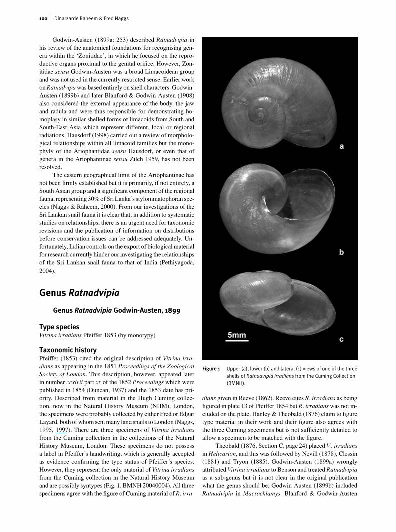
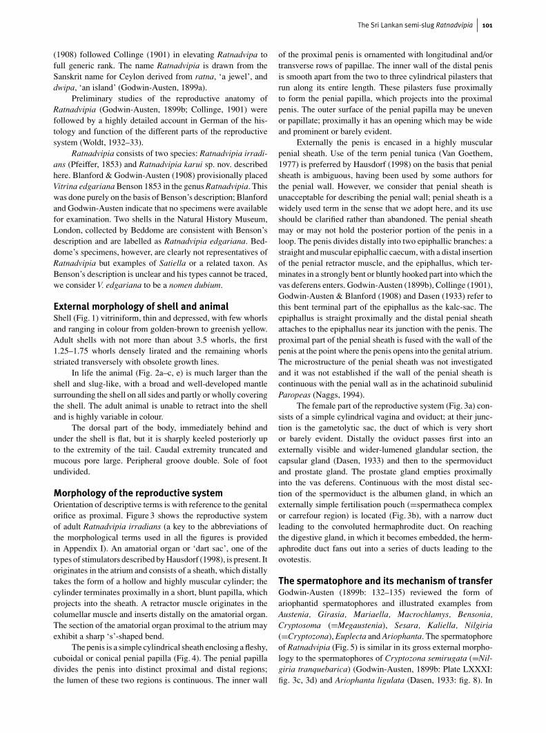
Taxonomic historyPfeiffer (1853) cited the original description of Vitrina irra-dians as appearing in the 1851 Proceedings of the ZoologicalSociety of London. This description, however, appeared laterin number ccxlvii part xx of the 1852 Proceedings which werepublished in 1854 (Duncan, 1937) and the 1853 date has pri-ority. Described from material in the Hugh Cuming collec-tion, now in the Natural History Museum (NHM), London,the specimens were probably collected by either Fred or EdgarLayard, both of whom sent many land snails to London (Naggs,1995, 1997). There are three specimens of Vitrina irradiansfrom the Cuming collection in the collections of the NaturalHistory Museum, London. These specimens do not possessa label in Pfeiffer’s handwriting, which is generally acceptedas evidence confirming the type status of Pfeiffer’s species.However, they represent the only material of Vitrina irradiansfrom the Cuming collection in the Natural History Museumand are possibly syntypes (Fig. 1, BMNH 20040004). All threespecimens agree with the figure of Cuming material of R. irra-
Figure 1 Upper (a), lower (b) and lateral (c) views of one of the threeshells of Ratnadvipia irradians from the Cuming Collection(BMNH).
dians given in Reeve (1862). Reeve cites R. irradians as beingfigured in plate 13 of Pfeiffer 1854 but R. irradians was not in-cluded on the plate. Hanley & Theobald (1876) claim to figuretype material in their work and their figure also agrees withthe three Cuming specimens but is not sufficiently detailed toallow a specimen to be matched with the figure.
Theobald (1876, Section C, page 24) placed V . irradiansin Helicarion, and this was followed by Nevill (1878), Clessin(1881) and Tryon (1885). Godwin-Austen (1899a) wronglyattributed Vitrina irradians to Benson and treated Ratnadvipiaas a sub-genus but it is not clear in the original publicationwhat the genus should be; Godwin-Austen (1899b) includedRatnadvipia in Macrochlamys. Blanford & Godwin-Austen
The Sri Lankan semi-slug Ratnadvipia 101
(1908) followed Collinge (1901) in elevating Ratnadvipa tofull generic rank. The name Ratnadvipia is drawn from theSanskrit name for Ceylon derived from ratna, ‘a jewel’, anddwipa, ‘an island’ (Godwin-Austen, 1899a).
Preliminary studies of the reproductive anatomy ofRatnadvipia (Godwin-Austen, 1899b; Collinge, 1901) werefollowed by a highly detailed account in German of the his-tology and function of the different parts of the reproductivesystem (Woldt, 1932–33).
Ratnadvipia consists of two species: Ratnadvipia irradi-ans (Pfeiffer, 1853) and Ratnadvipia karui sp. nov. describedhere. Blanford & Godwin-Austen (1908) provisionally placedVitrina edgariana Benson 1853 in the genus Ratnadvipia. Thiswas done purely on the basis of Benson’s description; Blanfordand Godwin-Austen indicate that no specimens were availablefor examination. Two shells in the Natural History Museum,London, collected by Beddome are consistent with Benson’sdescription and are labelled as Ratnadvipia edgariana. Bed-dome’s specimens, however, are clearly not representatives ofRatnadvipia but examples of Satiella or a related taxon. AsBenson’s description is unclear and his types cannot be traced,we consider V. edgariana to be a nomen dubium.
External morphology of shell and animalShell (Fig. 1) vitriniform, thin and depressed, with few whorlsand ranging in colour from golden-brown to greenish yellow.Adult shells with not more than about 3.5 whorls, the first1.25–1.75 whorls densely lirated and the remaining whorlsstriated transversely with obsolete growth lines.
In life the animal (Fig. 2a–c, e) is much larger than theshell and slug-like, with a broad and well-developed mantlesurrounding the shell on all sides and partly or wholly coveringthe shell. The adult animal is unable to retract into the shelland is highly variable in colour.
The dorsal part of the body, immediately behind andunder the shell is flat, but it is sharply keeled posteriorly upto the extremity of the tail. Caudal extremity truncated andmucous pore large. Peripheral groove double. Sole of footundivided.
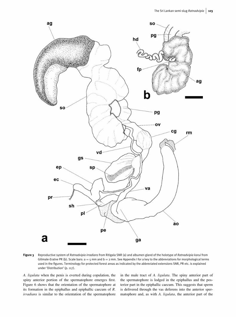
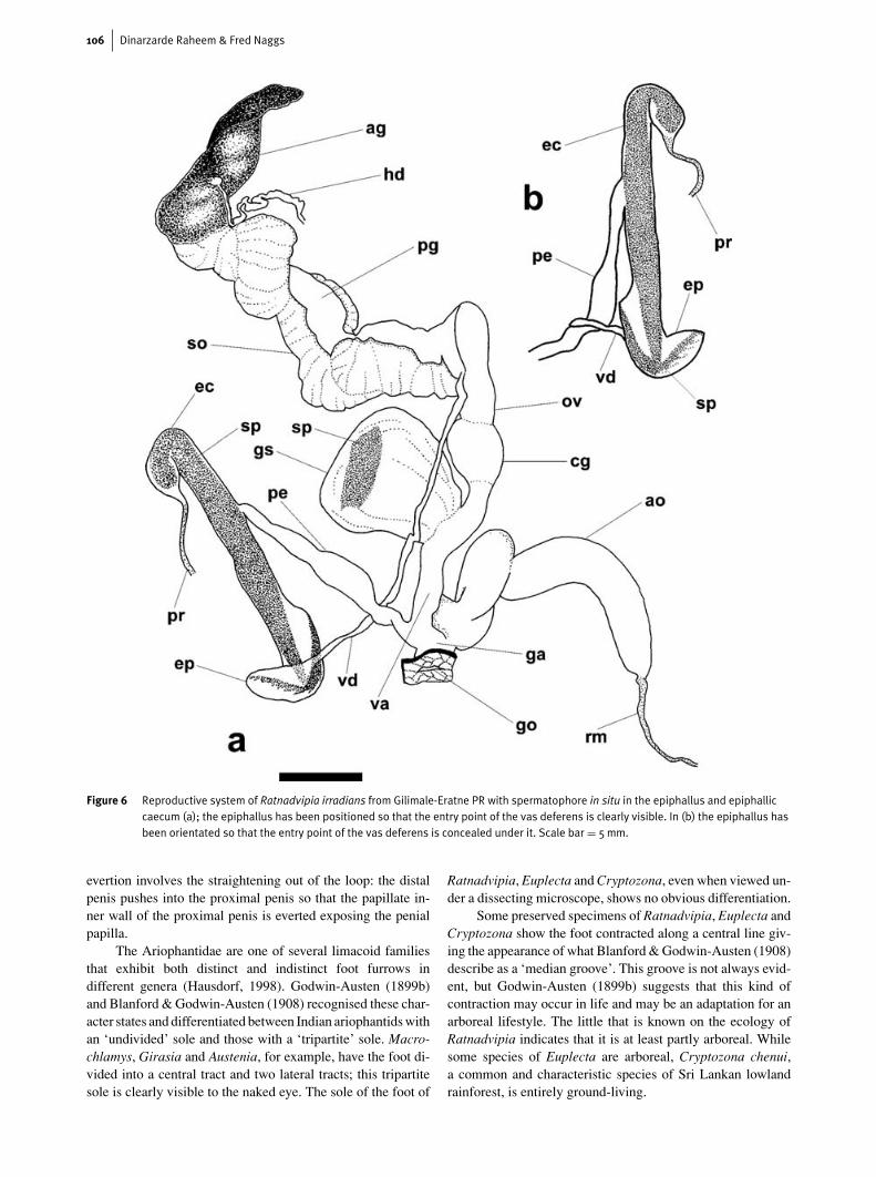
Morphology of the reproductive systemOrientation of descriptive terms is with reference to the genitalorifice as proximal. Figure 3 shows the reproductive systemof adult Ratnadvipia irradians (a key to the abbreviations ofthe morphological terms used in all the figures is providedin Appendix I). An amatorial organ or ‘dart sac’, one of thetypes of stimulators described by Hausdorf (1998), is present. Itoriginates in the atrium and consists of a sheath, which distallytakes the form of a hollow and highly muscular cylinder; thecylinder terminates proximally in a short, blunt papilla, whichprojects into the sheath. A retractor muscle originates in thecolumellar muscle and inserts distally on the amatorial organ.The section of the amatorial organ proximal to the atrium mayexhibit a sharp ‘s’-shaped bend.
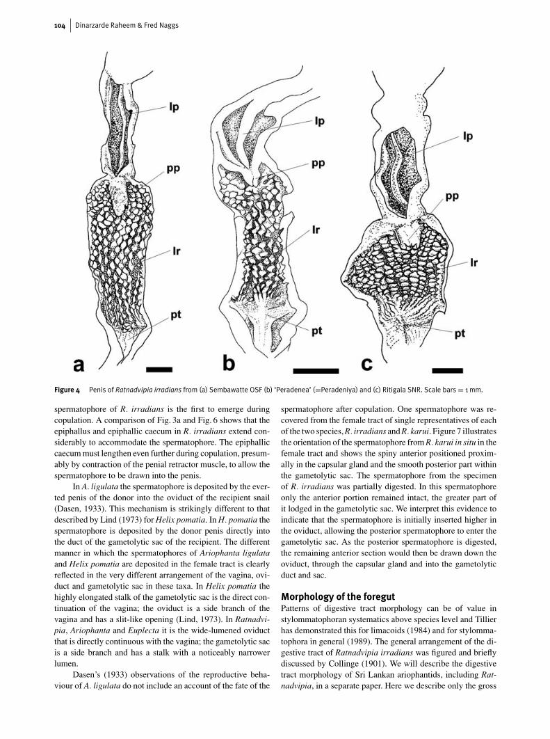
The penis is a simple cylindrical sheath enclosing a fleshy,cuboidal or conical penial papilla (Fig. 4). The penial papilladivides the penis into distinct proximal and distal regions;the lumen of these two regions is continuous. The inner wall
of the proximal penis is ornamented with longitudinal and/ortransverse rows of papillae. The inner wall of the distal penisis smooth apart from the two to three cylindrical pilasters thatrun along its entire length. These pilasters fuse proximallyto form the penial papilla, which projects into the proximalpenis. The outer surface of the penial papilla may be unevenor papillate; proximally it has an opening which may be wideand prominent or barely evident.
Externally the penis is encased in a highly muscularpenial sheath. Use of the term penial tunica (Van Goethem,1977) is preferred by Hausdorf (1998) on the basis that penialsheath is ambiguous, having been used by some authors forthe penial wall. However, we consider that penial sheath isunacceptable for describing the penial wall; penial sheath is awidely used term in the sense that we adopt here, and its useshould be clarified rather than abandoned. The penial sheathmay or may not hold the posterior portion of the penis in aloop. The penis divides distally into two epiphallic branches: astraight and muscular epiphallic caecum, with a distal insertionof the penial retractor muscle, and the epiphallus, which ter-minates in a strongly bent or bluntly hooked part into which thevas deferens enters. Godwin-Austen (1899b), Collinge (1901),Godwin-Austen & Blanford (1908) and Dasen (1933) refer tothis bent terminal part of the epiphallus as the kalc-sac. Theepiphallus is straight proximally and the distal penial sheathattaches to the epiphallus near its junction with the penis. Theproximal part of the penial sheath is fused with the wall of thepenis at the point where the penis opens into the genital atrium.The microstructure of the penial sheath was not investigatedand it was not established if the wall of the penial sheath iscontinuous with the penial wall as in the achatinoid subulinidParopeas (Naggs, 1994).
The female part of the reproductive system (Fig. 3a) con-sists of a simple cylindrical vagina and oviduct; at their junc-tion is the gametolytic sac, the duct of which is very shortor barely evident. Distally the oviduct passes first into anexternally visible and wider-lumened glandular section, thecapsular gland (Dasen, 1933) and then to the spermoviductand prostate gland. The prostate gland empties proximallyinto the vas deferens. Continuous with the most distal sec-tion of the spermoviduct is the albumen gland, in which anexternally simple fertilisation pouch (=spermatheca complexor carrefour region) is located (Fig. 3b), with a narrow ductleading to the convoluted hermaphrodite duct. On reachingthe digestive gland, in which it becomes embedded, the herm-aphrodite duct fans out into a series of ducts leading to theovotestis.
The spermatophore and its mechanism of transferGodwin-Austen (1899b: 132–135) reviewed the form ofariophantid spermatophores and illustrated examples fromAustenia, Girasia, Mariaella, Macrochlamys, Bensonia,Cryptosoma (=Megaustenia), Sesara, Kaliella, Nilgiria(=Cryptozona), Euplecta and Ariophanta. The spermatophoreof Ratnadvipia (Fig. 5) is similar in its gross external morpho-logy to the spermatophores of Cryptozona semirugata (=Nil-giria tranquebarica) (Godwin-Austen, 1899b: Plate LXXXI:fig. 3c, 3d) and Ariophanta ligulata (Dasen, 1933: fig. 8). In
102 Dinarzarde Raheem & Fred Naggs
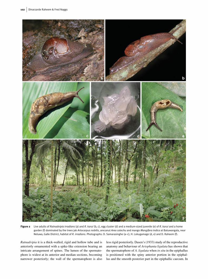
Figure 2 Live adults of Ratnadvipia irradians (a) and R. karui (b, c), egg cluster (d) and a medium-sized juvenile (e) of R. karui and a homegarden (f) dominated by the trees jak Artocarpus nobilis, arecanut Area catechu and mangoMangifera indica at Batuwangala, nearNeluwa, Galle District, habitat of R. irradians. Photographs: D. Samarasinghe (a–c), H. Lokugamage (d, e) and D. Raheem (f).
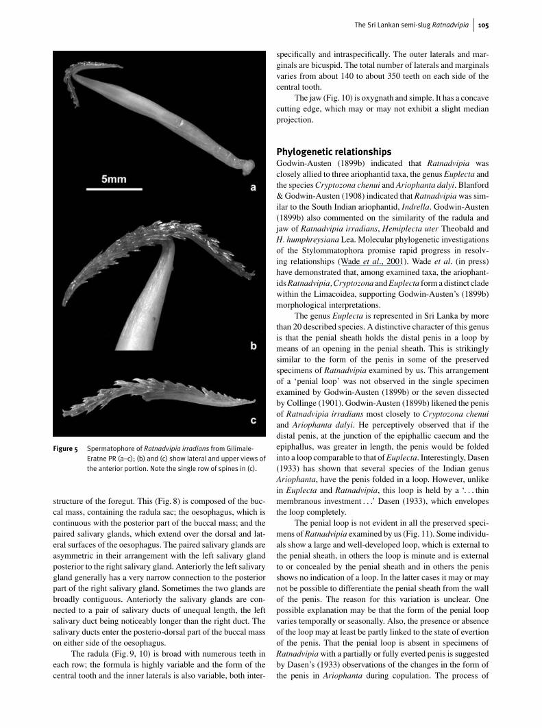
Ratnadvipia it is a thick-walled, rigid and hollow tube and isanteriorly ornamented with a spike-like extension bearing anintricate arrangement of spines. The lumen of the spermato-phore is widest at its anterior and median sections, becomingnarrower posteriorly; the wall of the spermatophore is also
less rigid posteriorly. Dasen’s (1933) study of the reproductiveanatomy and behaviour of Ariophanta ligulata has shown thatthe spermatophore of A. ligulata when in situ in the epiphallusis positioned with the spiny anterior portion in the epiphal-lus and the smooth posterior part in the epiphallic caecum. In
The Sri Lankan semi-slug Ratnadvipia 103
Figure 3 Reproductive system of Ratnadvipia irradians from Ritigala SNR (a) and albumen gland of the holotype of Ratnadvipia karui fromGilimale-Eratne PR (b). Scale bars: a = 5mm and b = 2mm. See Appendix I for a key to the abbreviations for morphological termsused in the figures. Terminology for protected forest areas as indicated by the abbreviated extensions SNR, PR etc. is explainedunder ‘Distribution’ (p. 117).
A. ligulata when the penis is everted during copulation, thespiny anterior portion of the spermatophore emerges first.Figure 6 shows that the orientation of the spermatophore atits formation in the epiphallus and epiphallic caecum of R.irradians is similar to the orientation of the spermatophore
in the male tract of A. ligulata. The spiny anterior part ofthe spermatophore is lodged in the epiphallus and the pos-terior part in the epiphallic caecum. This suggests that spermis delivered through the vas deferens into the anterior sper-matophore and, as with A. ligulata, the anterior part of the
104 Dinarzarde Raheem & Fred Naggs
Figure 4 Penis of Ratnadvipia irradians from (a) Sembawatte OSF (b) ‘Peradenea’ (=Peradeniya) and (c) Ritigala SNR. Scale bars = 1mm.
spermatophore of R. irradians is the first to emerge duringcopulation. A comparison of Fig. 3a and Fig. 6 shows that theepiphallus and epiphallic caecum in R. irradians extend con-siderably to accommodate the spermatophore. The epiphalliccaecum must lengthen even further during copulation, presum-ably by contraction of the penial retractor muscle, to allow thespermatophore to be drawn into the penis.
In A. ligulata the spermatophore is deposited by the ever-ted penis of the donor into the oviduct of the recipient snail(Dasen, 1933). This mechanism is strikingly different to thatdescribed by Lind (1973) for Helix pomatia. In H. pomatia thespermatophore is deposited by the donor penis directly intothe duct of the gametolytic sac of the recipient. The differentmanner in which the spermatophores of Ariophanta ligulataand Helix pomatia are deposited in the female tract is clearlyreflected in the very different arrangement of the vagina, ovi-duct and gametolytic sac in these taxa. In Helix pomatia thehighly elongated stalk of the gametolytic sac is the direct con-tinuation of the vagina; the oviduct is a side branch of thevagina and has a slit-like opening (Lind, 1973). In Ratnadvi-pia, Ariophanta and Euplecta it is the wide-lumened oviductthat is directly continuous with the vagina; the gametolytic sacis a side branch and has a stalk with a noticeably narrowerlumen.
Dasen’s (1933) observations of the reproductive beha-viour of A. ligulata do not include an account of the fate of the
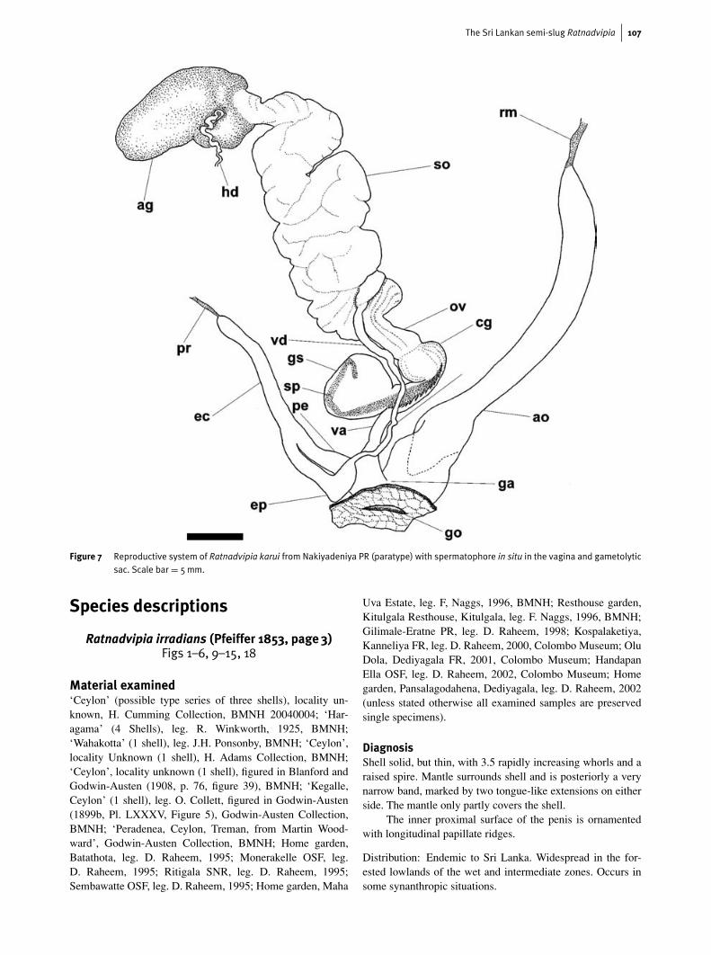
spermatophore after copulation. One spermatophore was re-covered from the female tract of single representatives of eachof the two species, R. irradians and R. karui. Figure 7 illustratesthe orientation of the spermatophore from R. karui in situ in thefemale tract and shows the spiny anterior positioned proxim-ally in the capsular gland and the smooth posterior part withinthe gametolytic sac. The spermatophore from the specimenof R. irradians was partially digested. In this spermatophoreonly the anterior portion remained intact, the greater part ofit lodged in the gametolytic sac. We interpret this evidence toindicate that the spermatophore is initially inserted higher inthe oviduct, allowing the posterior spermatophore to enter thegametolytic sac. As the posterior spermatophore is digested,the remaining anterior section would then be drawn down theoviduct, through the capsular gland and into the gametolyticduct and sac.
Morphology of the foregutPatterns of digestive tract morphology can be of value instylommatophoran systematics above species level and Tillierhas demonstrated this for limacoids (1984) and for stylomma-tophora in general (1989). The general arrangement of the di-gestive tract of Ratnadvipia irradians was figured and brieflydiscussed by Collinge (1901). We will describe the digestivetract morphology of Sri Lankan ariophantids, including Rat-nadvipia, in a separate paper. Here we describe only the gross
The Sri Lankan semi-slug Ratnadvipia 105
Figure 5 Spermatophore of Ratnadvipia irradians from Gilimale-Eratne PR (a–c); (b) and (c) show lateral and upper views ofthe anterior portion. Note the single row of spines in (c).
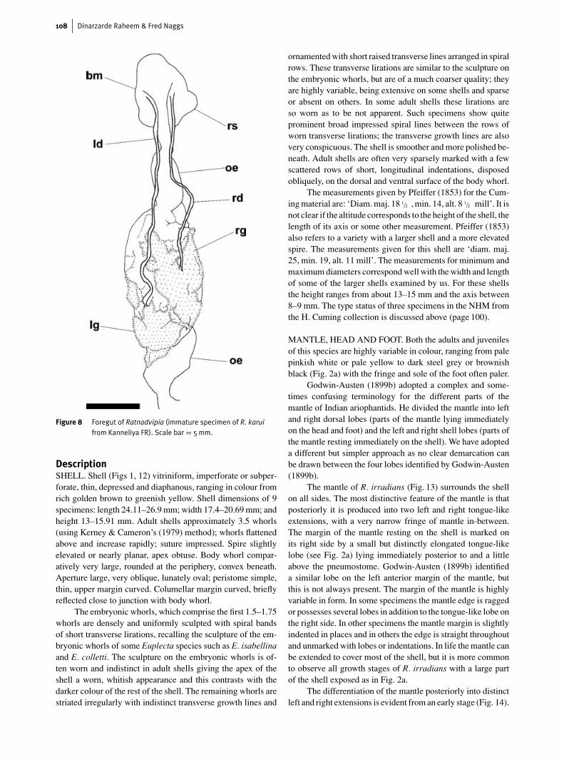
structure of the foregut. This (Fig. 8) is composed of the buc-cal mass, containing the radula sac; the oesophagus, which iscontinuous with the posterior part of the buccal mass; and thepaired salivary glands, which extend over the dorsal and lat-eral surfaces of the oesophagus. The paired salivary glands areasymmetric in their arrangement with the left salivary glandposterior to the right salivary gland. Anteriorly the left salivarygland generally has a very narrow connection to the posteriorpart of the right salivary gland. Sometimes the two glands arebroadly contiguous. Anteriorly the salivary glands are con-nected to a pair of salivary ducts of unequal length, the leftsalivary duct being noticeably longer than the right duct. Thesalivary ducts enter the posterio-dorsal part of the buccal masson either side of the oesophagus.
The radula (Fig. 9, 10) is broad with numerous teeth ineach row; the formula is highly variable and the form of thecentral tooth and the inner laterals is also variable, both inter-
specifically and intraspecifically. The outer laterals and mar-ginals are bicuspid. The total number of laterals and marginalsvaries from about 140 to about 350 teeth on each side of thecentral tooth.
The jaw (Fig. 10) is oxygnath and simple. It has a concavecutting edge, which may or may not exhibit a slight medianprojection.
Phylogenetic relationshipsGodwin-Austen (1899b) indicated that Ratnadvipia wasclosely allied to three ariophantid taxa, the genus Euplecta andthe species Cryptozona chenui and Ariophanta dalyi. Blanford& Godwin-Austen (1908) indicated that Ratnadvipia was sim-ilar to the South Indian ariophantid, Indrella. Godwin-Austen(1899b) also commented on the similarity of the radula andjaw of Ratnadvipia irradians, Hemiplecta uter Theobald andH. humphreysiana Lea. Molecular phylogenetic investigationsof the Stylommatophora promise rapid progress in resolv-ing relationships (Wade et al., 2001). Wade et al. (in press)have demonstrated that, among examined taxa, the ariophant-ids Ratnadvipia, Cryptozona and Euplecta form a distinct cladewithin the Limacoidea, supporting Godwin-Austen’s (1899b)morphological interpretations.
The genus Euplecta is represented in Sri Lanka by morethan 20 described species. A distinctive character of this genusis that the penial sheath holds the distal penis in a loop bymeans of an opening in the penial sheath. This is strikinglysimilar to the form of the penis in some of the preservedspecimens of Ratnadvipia examined by us. This arrangementof a ‘penial loop’ was not observed in the single specimenexamined by Godwin-Austen (1899b) or the seven dissectedby Collinge (1901). Godwin-Austen (1899b) likened the penisof Ratnadvipia irradians most closely to Cryptozona chenuiand Ariophanta dalyi. He perceptively observed that if thedistal penis, at the junction of the epiphallic caecum and theepiphallus, was greater in length, the penis would be foldedinto a loop comparable to that of Euplecta. Interestingly, Dasen(1933) has shown that several species of the Indian genusAriophanta, have the penis folded in a loop. However, unlikein Euplecta and Ratnadvipia, this loop is held by a ‘. . . thinmembranous investment . . .’ Dasen (1933), which envelopesthe loop completely.
The penial loop is not evident in all the preserved speci-mens of Ratnadvipia examined by us (Fig. 11). Some individu-als show a large and well-developed loop, which is external tothe penial sheath, in others the loop is minute and is externalto or concealed by the penial sheath and in others the penisshows no indication of a loop. In the latter cases it may or maynot be possible to differentiate the penial sheath from the wallof the penis. The reason for this variation is unclear. Onepossible explanation may be that the form of the penial loopvaries temporally or seasonally. Also, the presence or absenceof the loop may at least be partly linked to the state of evertionof the penis. That the penial loop is absent in specimens ofRatnadvipia with a partially or fully everted penis is suggestedby Dasen’s (1933) observations of the changes in the form ofthe penis in Ariophanta during copulation. The process of
106 Dinarzarde Raheem & Fred Naggs
Figure 6 Reproductive system of Ratnadvipia irradians from Gilimale-Eratne PR with spermatophore in situ in the epiphallus and epiphalliccaecum (a); the epiphallus has been positioned so that the entry point of the vas deferens is clearly visible. In (b) the epiphallus hasbeen orientated so that the entry point of the vas deferens is concealed under it. Scale bar = 5mm.
evertion involves the straightening out of the loop: the distalpenis pushes into the proximal penis so that the papillate in-ner wall of the proximal penis is everted exposing the penialpapilla.
The Ariophantidae are one of several limacoid familiesthat exhibit both distinct and indistinct foot furrows indifferent genera (Hausdorf, 1998). Godwin-Austen (1899b)and Blanford & Godwin-Austen (1908) recognised these char-acter states and differentiated between Indian ariophantids withan ‘undivided’ sole and those with a ‘tripartite’ sole. Macro-chlamys, Girasia and Austenia, for example, have the foot di-vided into a central tract and two lateral tracts; this tripartitesole is clearly visible to the naked eye. The sole of the foot of
Ratnadvipia, Euplecta and Cryptozona, even when viewed un-der a dissecting microscope, shows no obvious differentiation.
Some preserved specimens of Ratnadvipia, Euplecta andCryptozona show the foot contracted along a central line giv-ing the appearance of what Blanford & Godwin-Austen (1908)describe as a ‘median groove’. This groove is not always evid-ent, but Godwin-Austen (1899b) suggests that this kind ofcontraction may occur in life and may be an adaptation for anarboreal lifestyle. The little that is known on the ecology ofRatnadvipia indicates that it is at least partly arboreal. Whilesome species of Euplecta are arboreal, Cryptozona chenui,a common and characteristic species of Sri Lankan lowlandrainforest, is entirely ground-living.
The Sri Lankan semi-slug Ratnadvipia 107
Figure 7 Reproductive system of Ratnadvipia karui from Nakiyadeniya PR (paratype) with spermatophore in situ in the vagina and gametolyticsac. Scale bar = 5mm.
Species descriptions
Ratnadvipia irradians (Pfeiffer 1853, page 3)Figs 1–6, 9–15, 18
Material examined‘Ceylon’ (possible type series of three shells), locality un-known, H. Cumming Collection, BMNH 20040004; ‘Har-agama’ (4 Shells), leg. R. Winkworth, 1925, BMNH;‘Wahakotta’ (1 shell), leg. J.H. Ponsonby, BMNH; ‘Ceylon’,locality Unknown (1 shell), H. Adams Collection, BMNH;‘Ceylon’, locality unknown (1 shell), figured in Blanford andGodwin-Austen (1908, p. 76, figure 39), BMNH; ‘Kegalle,Ceylon’ (1 shell), leg. O. Collett, figured in Godwin-Austen(1899b, Pl. LXXXV, Figure 5), Godwin-Austen Collection,BMNH; ‘Peradenea, Ceylon, Treman, from Martin Wood-ward’, Godwin-Austen Collection, BMNH; Home garden,Batathota, leg. D. Raheem, 1995; Monerakelle OSF, leg.D. Raheem, 1995; Ritigala SNR, leg. D. Raheem, 1995;Sembawatte OSF, leg. D. Raheem, 1995; Home garden, Maha
Uva Estate, leg. F, Naggs, 1996, BMNH; Resthouse garden,Kitulgala Resthouse, Kitulgala, leg. F. Naggs, 1996, BMNH;Gilimale-Eratne PR, leg. D. Raheem, 1998; Kospalaketiya,Kanneliya FR, leg. D. Raheem, 2000, Colombo Museum; OluDola, Dediyagala FR, 2001, Colombo Museum; HandapanElla OSF, leg. D. Raheem, 2002, Colombo Museum; Homegarden, Pansalagodahena, Dediyagala, leg. D. Raheem, 2002(unless stated otherwise all examined samples are preservedsingle specimens).
DiagnosisShell solid, but thin, with 3.5 rapidly increasing whorls and araised spire. Mantle surrounds shell and is posteriorly a verynarrow band, marked by two tongue-like extensions on eitherside. The mantle only partly covers the shell.
The inner proximal surface of the penis is ornamentedwith longitudinal papillate ridges.
Distribution: Endemic to Sri Lanka. Widespread in the for-ested lowlands of the wet and intermediate zones. Occurs insome synanthropic situations.
108 Dinarzarde Raheem & Fred Naggs
Figure 8 Foregut of Ratnadvipia (immature specimen of R. karuifrom Kanneliya FR). Scale bar = 5mm.
DescriptionSHELL. Shell (Figs 1, 12) vitriniform, imperforate or subper-forate, thin, depressed and diaphanous, ranging in colour fromrich golden brown to greenish yellow. Shell dimensions of 9specimens: length 24.11–26.9 mm; width 17.4–20.69 mm; andheight 13–15.91 mm. Adult shells approximately 3.5 whorls(using Kerney & Cameron’s (1979) method); whorls flattenedabove and increase rapidly; suture impressed. Spire slightlyelevated or nearly planar, apex obtuse. Body whorl compar-atively very large, rounded at the periphery, convex beneath.Aperture large, very oblique, lunately oval; peristome simple,thin, upper margin curved. Columellar margin curved, brieflyreflected close to junction with body whorl.
The embryonic whorls, which comprise the first 1.5–1.75whorls are densely and uniformly sculpted with spiral bandsof short transverse lirations, recalling the sculpture of the em-bryonic whorls of some Euplecta species such as E. isabellinaand E. colletti. The sculpture on the embryonic whorls is of-ten worn and indistinct in adult shells giving the apex of theshell a worn, whitish appearance and this contrasts with thedarker colour of the rest of the shell. The remaining whorls arestriated irregularly with indistinct transverse growth lines and
ornamented with short raised transverse lines arranged in spiralrows. These transverse lirations are similar to the sculpture onthe embryonic whorls, but are of a much coarser quality; theyare highly variable, being extensive on some shells and sparseor absent on others. In some adult shells these lirations areso worn as to be not apparent. Such specimens show quiteprominent broad impressed spiral lines between the rows ofworn transverse lirations; the transverse growth lines are alsovery conspicuous. The shell is smoother and more polished be-neath. Adult shells are often very sparsely marked with a fewscattered rows of short, longitudinal indentations, disposedobliquely, on the dorsal and ventral surface of the body whorl.
The measurements given by Pfeiffer (1853) for the Cum-ing material are: ‘Diam. maj. 18 1/2 , min. 14, alt. 8 1/2 mill’. It isnot clear if the altitude corresponds to the height of the shell, thelength of its axis or some other measurement. Pfeiffer (1853)also refers to a variety with a larger shell and a more elevatedspire. The measurements given for this shell are ‘diam. maj.25, min. 19, alt. 11 mill’. The measurements for minimum andmaximum diameters correspond well with the width and lengthof some of the larger shells examined by us. For these shellsthe height ranges from about 13–15 mm and the axis between8–9 mm. The type status of three specimens in the NHM fromthe H. Cuming collection is discussed above (page 100).
MANTLE, HEAD AND FOOT. Both the adults and juvenilesof this species are highly variable in colour, ranging from palepinkish white or pale yellow to dark steel grey or brownishblack (Fig. 2a) with the fringe and sole of the foot often paler.
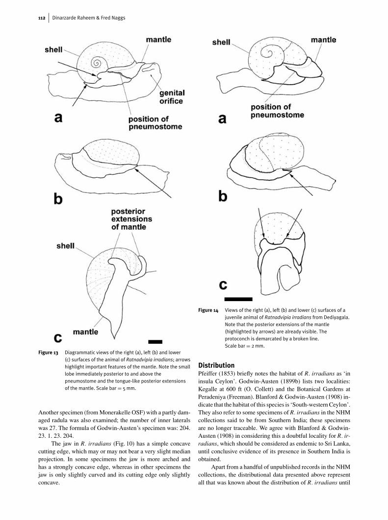
Godwin-Austen (1899b) adopted a complex and some-times confusing terminology for the different parts of themantle of Indian ariophantids. He divided the mantle into leftand right dorsal lobes (parts of the mantle lying immediatelyon the head and foot) and the left and right shell lobes (parts ofthe mantle resting immediately on the shell). We have adopteda different but simpler approach as no clear demarcation canbe drawn between the four lobes identified by Godwin-Austen(1899b).
The mantle of R. irradians (Fig. 13) surrounds the shellon all sides. The most distinctive feature of the mantle is thatposteriorly it is produced into two left and right tongue-likeextensions, with a very narrow fringe of mantle in-between.The margin of the mantle resting on the shell is marked onits right side by a small but distinctly elongated tongue-likelobe (see Fig. 2a) lying immediately posterior to and a littleabove the pneumostome. Godwin-Austen (1899b) identifieda similar lobe on the left anterior margin of the mantle, butthis is not always present. The margin of the mantle is highlyvariable in form. In some specimens the mantle edge is raggedor possesses several lobes in addition to the tongue-like lobe onthe right side. In other specimens the mantle margin is slightlyindented in places and in others the edge is straight throughoutand unmarked with lobes or indentations. In life the mantle canbe extended to cover most of the shell, but it is more commonto observe all growth stages of R. irradians with a large partof the shell exposed as in Fig. 2a.
The differentiation of the mantle posteriorly into distinctleft and right extensions is evident from an early stage (Fig. 14).
The Sri Lankan semi-slug Ratnadvipia 109
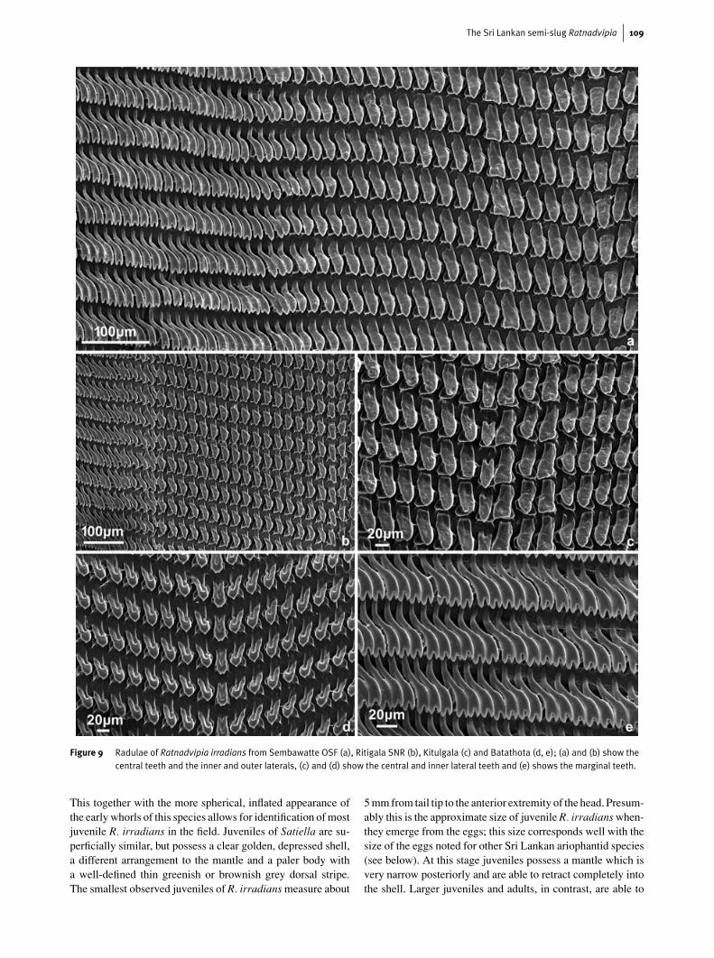
Figure 9 Radulae of Ratnadvipia irradians from Sembawatte OSF (a), Ritigala SNR (b), Kitulgala (c) and Batathota (d, e); (a) and (b) show thecentral teeth and the inner and outer laterals, (c) and (d) show the central and inner lateral teeth and (e) shows the marginal teeth.
This together with the more spherical, inflated appearance ofthe early whorls of this species allows for identification of mostjuvenile R. irradians in the field. Juveniles of Satiella are su-perficially similar, but possess a clear golden, depressed shell,a different arrangement to the mantle and a paler body witha well-defined thin greenish or brownish grey dorsal stripe.The smallest observed juveniles of R. irradians measure about
5 mm from tail tip to the anterior extremity of the head. Presum-ably this is the approximate size of juvenile R. irradians when-they emerge from the eggs; this size corresponds well with thesize of the eggs noted for other Sri Lankan ariophantid species(see below). At this stage juveniles possess a mantle which isvery narrow posteriorly and are able to retract completely intothe shell. Larger juveniles and adults, in contrast, are able to
110 Dinarzarde Raheem & Fred Naggs
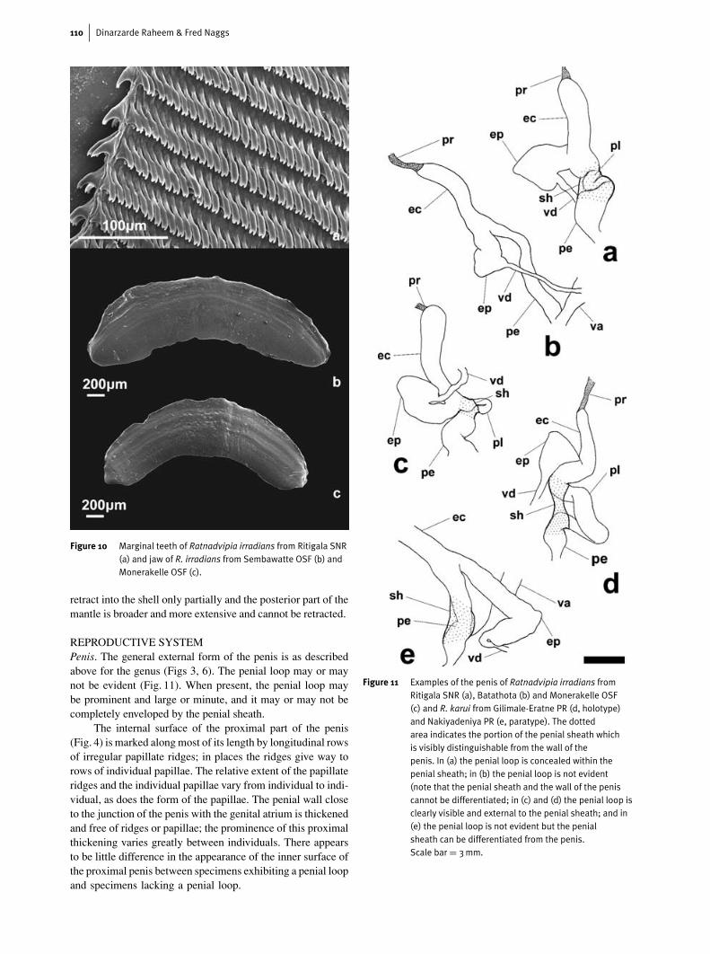
Figure 10 Marginal teeth of Ratnadvipia irradians from Ritigala SNR(a) and jaw of R. irradians from Sembawatte OSF (b) andMonerakelle OSF (c).
retract into the shell only partially and the posterior part of themantle is broader and more extensive and cannot be retracted.
REPRODUCTIVE SYSTEMPenis. The general external form of the penis is as describedabove for the genus (Figs 3, 6). The penial loop may or maynot be evident (Fig. 11). When present, the penial loop maybe prominent and large or minute, and it may or may not becompletely enveloped by the penial sheath.
The internal surface of the proximal part of the penis(Fig. 4) is marked along most of its length by longitudinal rowsof irregular papillate ridges; in places the ridges give way torows of individual papillae. The relative extent of the papillateridges and the individual papillae vary from individual to indi-vidual, as does the form of the papillae. The penial wall closeto the junction of the penis with the genital atrium is thickenedand free of ridges or papillae; the prominence of this proximalthickening varies greatly between individuals. There appearsto be little difference in the appearance of the inner surface ofthe proximal penis between specimens exhibiting a penial loopand specimens lacking a penial loop.
Figure 11 Examples of the penis of Ratnadvipia irradians fromRitigala SNR (a), Batathota (b) and Monerakelle OSF(c) and R. karui from Gilimale-Eratne PR (d, holotype)and Nakiyadeniya PR (e, paratype). The dottedarea indicates the portion of the penial sheath whichis visibly distinguishable from the wall of thepenis. In (a) the penial loop is concealed within thepenial sheath; in (b) the penial loop is not evident(note that the penial sheath and the wall of the peniscannot be differentiated; in (c) and (d) the penial loop isclearly visible and external to the penial sheath; and in(e) the penial loop is not evident but the penialsheath can be differentiated from the penis.Scale bar = 3mm.
The Sri Lankan semi-slug Ratnadvipia 111
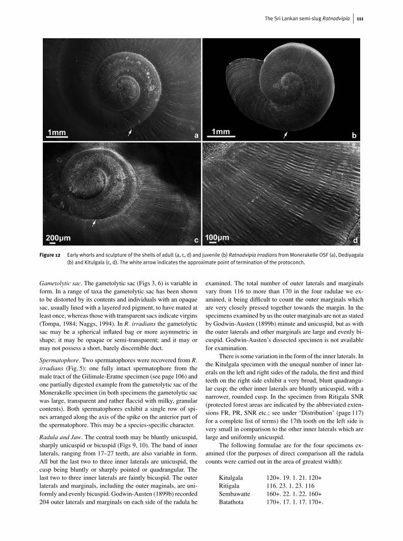
Figure 12 Early whorls and sculpture of the shells of adult (a, c, d) and juvenile (b) Ratnadvipia irradians fromMonerakelle OSF (a), Dediyagala(b) and Kitulgala (c, d). The white arrow indicates the approximate point of termination of the protoconch.
Gametolytic sac. The gametolytic sac (Figs 3, 6) is variable inform. In a range of taxa the gametolytic sac has been shownto be distorted by its contents and individuals with an opaquesac, usually lined with a layered red pigment, to have mated atleast once, whereas those with transparent sacs indicate virgins(Tompa, 1984; Naggs, 1994). In R. irradians the gametolyticsac may be a spherical inflated bag or more asymmetric inshape; it may be opaque or semi-transparent; and it may ormay not possess a short, barely discernible duct.
Spermatophore. Two spermatophores were recovered from R.irradians (Fig. 5): one fully intact spermatophore from themale tract of the Gilimale-Eratne specimen (see page 106) andone partially digested example from the gametolytic sac of theMonerakelle specimen (in both specimens the gametolytic sacwas large, transparent and rather flaccid with milky, granularcontents). Both spermatophores exhibit a single row of spi-nes arranged along the axis of the spike on the anterior part ofthe spermatophore. This may be a species-specific character.
Radula and Jaw. The central tooth may be bluntly unicuspid,sharply unicuspid or bicuspid (Figs 9, 10). The band of innerlaterals, ranging from 17–27 teeth, are also variable in form.All but the last two to three inner laterals are unicuspid, thecusp being bluntly or sharply pointed or quadrangular. Thelast two to three inner laterals are faintly bicuspid. The outerlaterals and marginals, including the outer maginals, are uni-formly and evenly bicuspid. Godwin-Austen (1899b) recorded204 outer laterals and marginals on each side of the radula he
examined. The total number of outer laterals and marginalsvary from 116 to more than 170 in the four radulae we ex-amined, it being difficult to count the outer marginals whichare very closely pressed together towards the margin. In thespecimens examined by us the outer marginals are not as statedby Godwin-Austen (1899b) minute and unicuspid, but as withthe outer laterals and other marginals are large and evenly bi-cuspid. Godwin-Austen’s dissected specimen is not availablefor examination.
There is some variation in the form of the inner laterals. Inthe Kitulgala specimen with the unequal number of inner lat-erals on the left and right sides of the radula, the first and thirdteeth on the right side exhibit a very broad, blunt quadrangu-lar cusp; the other inner laterals are bluntly unicuspid, with anarrower, rounded cusp. In the specimen from Ritigala SNR(protected forest areas are indicated by the abbreviated exten-sions FR, PR, SNR etc.; see under ‘Distribution’ (page 117)for a complete list of terms) the 17th tooth on the left side isvery small in comparison to the other inner laterals which arelarge and uniformly unicuspid.
The following formulae are for the four specimens ex-amined (for the purposes of direct comparison all the radulacounts were carried out in the area of greatest width):
Kitulgala 120+. 19. 1. 21. 120+Ritigala 116. 23. 1. 23. 116Sembawatte 160+. 22. 1. 22. 160+Batathota 170+. 17. 1. 17. 170+.
112 Dinarzarde Raheem & Fred Naggs
Figure 13 Diagrammatic views of the right (a), left (b) and lower(c) surfaces of the animal of Ratnadvipia irradians; arrowshighlight important features of the mantle. Note the smalllobe immediately posterior to and above thepneumostome and the tongue-like posterior extensionsof the mantle. Scale bar = 5mm.
Another specimen (from Monerakelle OSF) with a partly dam-aged radula was also examined; the number of inner lateralswas 27. The formula of Godwin-Austen’s specimen was: 204.23. 1. 23. 204.
The jaw in R. irradians (Fig. 10) has a simple concavecutting edge, which may or may not bear a very slight medianprojection. In some specimens the jaw is more arched andhas a strongly concave edge, whereas in other specimens thejaw is only slightly curved and its cutting edge only slightlyconcave.
Figure 14 Views of the right (a), left (b) and lower (c) surfaces of ajuvenile animal of Ratnadvipia irradians from Dediyagala.Note that the posterior extensions of the mantle(highlighted by arrows) are already visible. Theprotoconch is demarcated by a broken line.Scale bar = 2mm.
DistributionPfeiffer (1853) briefly notes the habitat of R. irradians as ‘ininsula Ceylon’. Godwin-Austen (1899b) lists two localities:Kegalle at 600 ft (O. Collett) and the Botanical Gardens atPeradeniya (Freeman). Blanford & Godwin-Austen (1908) in-dicate that the habitat of this species is ‘South-western Ceylon’.They also refer to some specimens of R. irradians in the NHMcollections said to be from Southern India; these specimensare no longer traceable. We agree with Blanford & Godwin-Austen (1908) in considering this a doubtful locality for R. ir-radians, which should be considered as endemic to Sri Lanka,until conclusive evidence of its presence in Southern India isobtained.
Apart from a handful of unpublished records in the NHMcollections, the distributional data presented above representall that was known about the distribution of R. irradians until
The Sri Lankan semi-slug Ratnadvipia 113
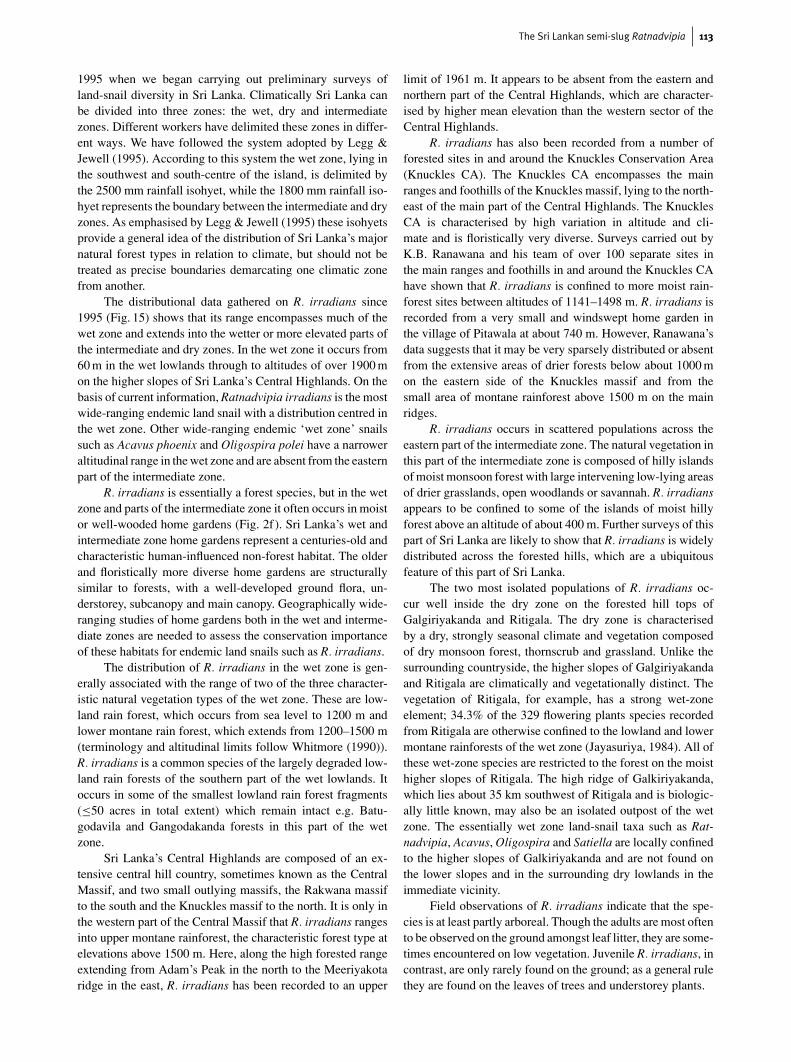
1995 when we began carrying out preliminary surveys ofland-snail diversity in Sri Lanka. Climatically Sri Lanka canbe divided into three zones: the wet, dry and intermediatezones. Different workers have delimited these zones in differ-ent ways. We have followed the system adopted by Legg &Jewell (1995). According to this system the wet zone, lying inthe southwest and south-centre of the island, is delimited bythe 2500 mm rainfall isohyet, while the 1800 mm rainfall iso-hyet represents the boundary between the intermediate and dryzones. As emphasised by Legg & Jewell (1995) these isohyetsprovide a general idea of the distribution of Sri Lanka’s majornatural forest types in relation to climate, but should not betreated as precise boundaries demarcating one climatic zonefrom another.
The distributional data gathered on R. irradians since1995 (Fig. 15) shows that its range encompasses much of thewet zone and extends into the wetter or more elevated parts ofthe intermediate and dry zones. In the wet zone it occurs from60 m in the wet lowlands through to altitudes of over 1900 mon the higher slopes of Sri Lanka’s Central Highlands. On thebasis of current information, Ratnadvipia irradians is the mostwide-ranging endemic land snail with a distribution centred inthe wet zone. Other wide-ranging endemic ‘wet zone’ snailssuch as Acavus phoenix and Oligospira polei have a narroweraltitudinal range in the wet zone and are absent from the easternpart of the intermediate zone.
R. irradians is essentially a forest species, but in the wetzone and parts of the intermediate zone it often occurs in moistor well-wooded home gardens (Fig. 2f). Sri Lanka’s wet andintermediate zone home gardens represent a centuries-old andcharacteristic human-influenced non-forest habitat. The olderand floristically more diverse home gardens are structurallysimilar to forests, with a well-developed ground flora, un-derstorey, subcanopy and main canopy. Geographically wide-ranging studies of home gardens both in the wet and interme-diate zones are needed to assess the conservation importanceof these habitats for endemic land snails such as R. irradians.
The distribution of R. irradians in the wet zone is gen-erally associated with the range of two of the three character-istic natural vegetation types of the wet zone. These are low-land rain forest, which occurs from sea level to 1200 m andlower montane rain forest, which extends from 1200–1500 m(terminology and altitudinal limits follow Whitmore (1990)).R. irradians is a common species of the largely degraded low-land rain forests of the southern part of the wet lowlands. Itoccurs in some of the smallest lowland rain forest fragments(≤50 acres in total extent) which remain intact e.g. Batu-godavila and Gangodakanda forests in this part of the wetzone.
Sri Lanka’s Central Highlands are composed of an ex-tensive central hill country, sometimes known as the CentralMassif, and two small outlying massifs, the Rakwana massifto the south and the Knuckles massif to the north. It is only inthe western part of the Central Massif that R. irradians rangesinto upper montane rainforest, the characteristic forest type atelevations above 1500 m. Here, along the high forested rangeextending from Adam’s Peak in the north to the Meeriyakotaridge in the east, R. irradians has been recorded to an upper
limit of 1961 m. It appears to be absent from the eastern andnorthern part of the Central Highlands, which are character-ised by higher mean elevation than the western sector of theCentral Highlands.
R. irradians has also been recorded from a number offorested sites in and around the Knuckles Conservation Area(Knuckles CA). The Knuckles CA encompasses the mainranges and foothills of the Knuckles massif, lying to the north-east of the main part of the Central Highlands. The KnucklesCA is characterised by high variation in altitude and cli-mate and is floristically very diverse. Surveys carried out byK.B. Ranawana and his team of over 100 separate sites inthe main ranges and foothills in and around the Knuckles CAhave shown that R. irradians is confined to more moist rain-forest sites between altitudes of 1141–1498 m. R. irradians isrecorded from a very small and windswept home garden inthe village of Pitawala at about 740 m. However, Ranawana’sdata suggests that it may be very sparsely distributed or absentfrom the extensive areas of drier forests below about 1000 mon the eastern side of the Knuckles massif and from thesmall area of montane rainforest above 1500 m on the mainridges.
R. irradians occurs in scattered populations across theeastern part of the intermediate zone. The natural vegetation inthis part of the intermediate zone is composed of hilly islandsof moist monsoon forest with large intervening low-lying areasof drier grasslands, open woodlands or savannah. R. irradiansappears to be confined to some of the islands of moist hillyforest above an altitude of about 400 m. Further surveys of thispart of Sri Lanka are likely to show that R. irradians is widelydistributed across the forested hills, which are a ubiquitousfeature of this part of Sri Lanka.
The two most isolated populations of R. irradians oc-cur well inside the dry zone on the forested hill tops ofGalgiriyakanda and Ritigala. The dry zone is characterisedby a dry, strongly seasonal climate and vegetation composedof dry monsoon forest, thornscrub and grassland. Unlike thesurrounding countryside, the higher slopes of Galgiriyakandaand Ritigala are climatically and vegetationally distinct. Thevegetation of Ritigala, for example, has a strong wet-zoneelement; 34.3% of the 329 flowering plants species recordedfrom Ritigala are otherwise confined to the lowland and lowermontane rainforests of the wet zone (Jayasuriya, 1984). All ofthese wet-zone species are restricted to the forest on the moisthigher slopes of Ritigala. The high ridge of Galkiriyakanda,which lies about 35 km southwest of Ritigala and is biologic-ally little known, may also be an isolated outpost of the wetzone. The essentially wet zone land-snail taxa such as Rat-nadvipia, Acavus, Oligospira and Satiella are locally confinedto the higher slopes of Galkiriyakanda and are not found onthe lower slopes and in the surrounding dry lowlands in theimmediate vicinity.
Field observations of R. irradians indicate that the spe-cies is at least partly arboreal. Though the adults are most oftento be observed on the ground amongst leaf litter, they are some-times encountered on low vegetation. Juvenile R. irradians, incontrast, are only rarely found on the ground; as a general rulethey are found on the leaves of trees and understorey plants.
114 Dinarzarde Raheem & Fred Naggs
Figure 15 Distribution of Ratnadvipia irradians. The wet zone (solid line) is delimited by the 2500mm isohyet and the 1800mm isohyet(broken line) represents the boundary between the intermediate and dry zones. Individual sites from which this species has beenrecorded are denoted by crosses and towns or villages by filled circles. Closed-canopy natural forest cover is shown in dark grey; thedistribution of forest cover is based on the 1: 50000-scale forest map by Legg & Jewell (1995). The current extent of occurrence ofthe species, extrapolated from the available distributional information is shown in pale grey.
Distributional records: Wahakotte (7◦43.0′N, 80◦
35.0′E), J.H. Ponsonby, BMNH; Nalanda (7◦39.5′N,80◦38.0′E), R. Winkworth, 1925, BMNH; 579 m, Haragama(7◦14.7′N, 80◦42.5′E), R. Winkworth, 1925, BMNH; 183 m,Kegalla (7◦15.0′N, 80◦20.5′E), O. Collett, BMNH; 488 m,
Royal Botanical Gardens, Peradeniya (7◦15.5′N, 80◦36.0′E), c.1898, BMNH; 1280 m, Maha Uva Estate (7◦3.8′N, 80◦52.5′E),F. Naggs, 1996; 457 m, Resthouse garden, Kitulgala (6◦59.7′N,80◦24.3′E), F. Naggs, 1996; 457 m, Kandy (1500 ft.)(6◦18.3′N, 80◦38.7′E), c. 1901, BMNH; 155 m, Mulatiyana FR
The Sri Lankan semi-slug Ratnadvipia 115
(6◦10.4′N, 80◦34.8′E), 1995; 288 m, Kudapana, MulatiyanaFR (6◦10.6′N, 80◦34.4′E), 1995; 195 m, Beraliya (Akuressa)PR (6◦4.1′N, 80◦27.3′E), 1995; 270 m, Home garden,Batathota (6◦49.6′N, 80◦22.4′E), 1995; 1175 m, Kurusigala,Handapan Ella OSF (6◦26.7′N, 80◦36.6′E), 1995; 550–1080 m, Monerakelle OSF (6◦54.5′N, 81◦22.7′E), 1995;510–695 m, Ritigala SNR (8◦6.8′N, 80◦39.4′E), 1995; 130 m,Bandarakelle forest, Kitulgala (6◦59.6′N, 80◦24.0′E), 1995;545 m, Sembawatte OSF (7◦2.0′N, 80◦26.2′E), 1995; 620 m,Uda Pawana Ella, Peak Wilderness Sanctuary (6◦47.6′N,80◦27.6′E), 1998; 1285 m, Kodiyadaputena, Peak WildernessSanctuary (6◦48.5′N, 80◦28.0′E), 1998; 240 m, Sudagala,Gilimale-Eratne PR (6◦48.5′N, 80◦23.1′E), 1998; 350 m,Palabaddala, Peak Wilderness Sanctuary (6◦46.8′N,80◦27.1′E), 1998; 285–380 m, Batadomba Lena, Gilimale-Eratne PR (6◦46.5′N, 80◦23.5′E), 1998; 158–355 m,Kospelaketiya, Kanneliya FR (6◦14.8′N, 80◦20.9′E),1998; 330 m, Pimburukanda, Nakiyadeniya PR (6◦12.9′N,80◦22.9′E), 1998; 155–495 m, Hangarankande, KanneliyaFR (6◦16.1′N, 80◦20.2′E), 1998; 125 m, Annasigetakanda,Kanneliya FR (6◦14.9′N, 80◦20.1′E), 1998; 75–330 m,Lavalvaruvakanda, Kanneliya FR (6◦16.4′N, 80◦20.9′E),1998; 165–265 m, Kodikande, Kanneliya FR (6◦15.2′N, 80◦
21.8′E), 1998; 195–210 m, Manamaladolakanda, KanneliyaFR (6◦15.3′N, 80◦22.4′E), 1998; 165 m, Galbendidolakanda,Nakiyadeniya PR (6◦10.4′N, 80◦23.1′E), 1998; 937–1067 m,Dotalugala, Bambarabotuwa FR (6◦41.4′N, 80◦36.2′E),1998; 799–980 m, Dehenakande, Peak Wilderness Sanctuary(6◦45.7′N, 80◦30.0′E), 1998, 1553 m, Ratkande, Peak Wilder-ness Sanctuary (6◦47.1′N, 80◦31.1′E), 1998; 918–1038 m,Kondurugala, Peak Wilderness Sanctuary (6◦44.7′N,80◦32.8′E), 1998; 763 m, Asamanakande, BambarabotuwaFR (6◦39.0′N, 80◦35.4′E), 1998; 1829 m, above Moray Estate,Peak Wilderness Sanctuary (6◦48.4′N, 80◦30.5′E), 1998;790–1025 m, Bathborakande, Peak Wilderness Sanctuary(6◦53.6′N, 80◦27.8′E), 1998; 465 m, Sevalagala, KelaniValley PR (6◦56.0′N, 80◦26.7′E), 1998; 850–1110 m,Kiripanagala, Kelani Valley PR (6◦55.7′N, 80◦27.6′E),1998; 1748–1961 m, Doturugala, Peak Wilderness Sanctuary(6◦46.5′N, 80◦32.6′E), 1998; 1795 m, Gawarawela Patana,Peak Wilderness Sanctuary, 6◦46.8′N, 80◦33.7′E), 1998;760 m, Gannoruwa forest (7◦17.2′N, 80◦35.8′E); 565 m,Udawattakele FR (7◦18.0′N, 80◦38.5′E); 753 m, HopewellOSF (7◦14.9′N, 80◦37.0′E); 1248 m, Hantane forest(7◦13.7′N,80◦38.3′E); 450 m, Viyanahela OSF (7◦17.5′N,81◦22.6′E); 738–844 m, Madigala OSF (7◦18.1′N, 81◦16.0′E);501 m, Velihela OSF (6◦52.9′N, 81◦26.9′E); 424 m, KitulhelaOSF (6◦59.6′N, 81◦27.8′E); 1053–1119 m, MonerakelleOSF (6◦53.2′N, 81◦23.4′E); 610 m, Doluwakanda PR(7◦36.9′N, 80◦ 24.7′E); 906 m, Neugalkanda PR (7◦36.9′N,80◦34.2′E); 1013 m, Opalagala OSF (7◦36.5′N, 80◦41.8′E);396–463 m, Galgiriyakanda PR (7◦56.2′N, 80◦22.9′E); 223 m,Malgalla, Kanneliya FR (6◦17.6′N, 80◦21.7′E); 623 m,Hinidunkande, Habarakada PR (6◦19.9′N, 80◦17.8′E); 232 m,Dediyagala FR (6◦11.2′N, 80◦24.6′E); 273 m, Galbendid-olakande, Nakiyadeniya PR (6◦10.3′N, 80◦23.0′E); 360 m,Malambure, Malambure FR (6◦14.5′N, 80◦18.8′E); 405 m,Pimburukanda, Nakiyadeniya PR (6◦12.3′N, 80◦22.8′E);
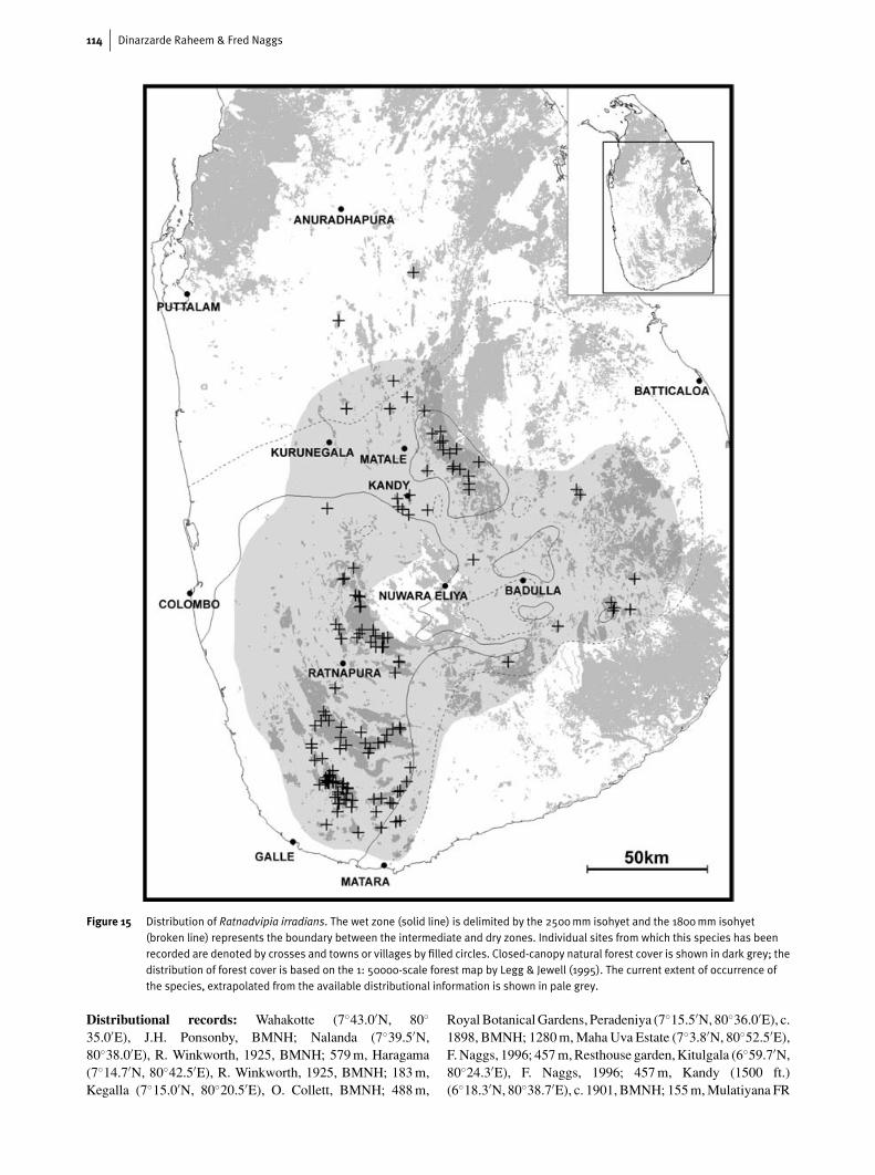
Figure 16 Upper (a), lower (b, d) and lateral (c) views of shells ofRatnadvipia karui from Gilimale-Eratne PR (holotype, a–c)and Nakiyadeniya PR (paratype, d); note the completelyand partly worn ostracum of (c) and (d) respectively.
380 m, Murutukanda OSF (6◦49.2′N, 81◦11.0′E); 1120 m,Handapan Ella OSF (6◦26.7′N, 80◦34.8′E); 183 m,Kospelaketiya, Kanneliya FR (6◦15.0′N, 80◦20.8′E);140 m, Kombala-Kottawa PR (6◦5.7′N, 80◦20.3′E); 165 m,
116 Dinarzarde Raheem & Fred Naggs
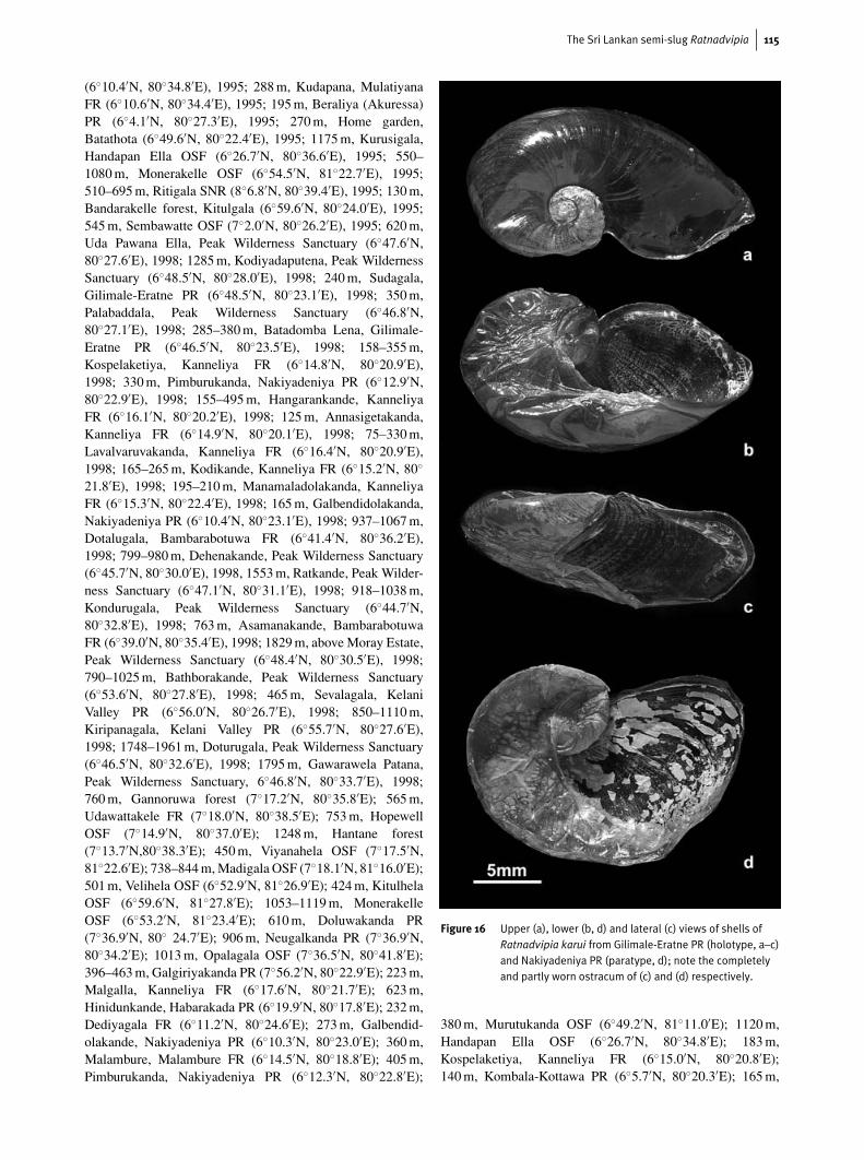
Figure 17 Early whorls and sculpture of the shells of adult Ratnadvipia karui from Gilimale-Eratne PR (a and c, paratype) and juvenile R. karui(b). The white arrow indicates the approximate point of termination of the protoconch. The white box in (a) indicates the area of theshell shown in (c); note the oblique rows of indentations in (c).
Lavalvaruvakanda, Kanneliya FR (6◦15.3′N, 80◦20.5′E);290 m, Manamaladolakanda, Kanneliya FR (6◦14.9′N,80◦22.4′E); 457 m, Kalukanda, Kanneliya FR (6◦16.7′N,80◦21.1′E); 219 m, Homadola, Nakiyadeniya PR (6◦11.8′N,80◦22.3′E); 232 m, Awudagalakanda, Auwegalakanda OSF(6◦22.5′N, 80◦17.1′E); 253 m, Kondagala, Kanneliya FR(6◦14.1′N, 80◦23.8′E); 170 m, Olu Dola, Dediyagala FR(6◦11.1′N, 80◦26.0′E); 236 m, Kekirikanda, Dediyagala FR(6◦12.6′N, 80◦24.7′E); 310 m, Viharakanda, MulatiyanaFR (6◦11.5′N, 80◦32.4′E); 220 m, Viharekele FR (6◦6.6′N,80◦36.5′E); 76 m, Home garden, Batuwangala, Neluwa(6◦22.4′N, 80◦23.3′E); 90 m, Home garden, Pansalagodahena,Dediyagala (6◦9.6′N, 80◦25.9′E); 780 m, Rammalakanda FR(6◦15.2′N, 80◦37.9′E); 220 m, Home garden, Ehelakanda,Mawarala (6◦13.0′N, 80◦36.0′E); 72 m, Gangodakanda forest(6◦8.6′N, 80◦31.2′E); 23 m, Getara forest, OliyagankeleFR (6◦5.0′N, 80◦32.4′E); 100 m, Kudaludolakanda forest(6◦11.3′N, 80◦30.7′E); 250 m, Home garden, Panapola(6◦26.0′N, 80◦27.9′E); 560 m, Wanduragala ridge, SinharajaNHWA (6◦23.6′N, 80◦28.9′E); 540 m, Wewagama, DelgodaPR (6◦27.1′N, 80◦23.6′E); 588 m, Illumbekanda, Sinha-raja NHWA (6◦25.4′N, 80◦33.6′); 394 m, Nawalakanda,Waratalgoda PR (6◦30.5′N, 80◦19.8′E); 728 m, Dothalugala,Delgoda PR (6◦24.9′N, 80◦23.4′E); 151 m, Pallekumbura,Morapitiya-Runakanda PR (6◦28.5′N,80◦19.2′E); 595 m,Denihena, Morapitiya-Runakanda PR (6◦28.5′N, 80◦20.8′E);461 m, Dothalankanda, Morapitiya-Runakanda PR (6◦29.8′N,80◦20.2′E); 210 m, Batugodavila forest, Hedigalla (6◦27.5′N,80◦17.9′E); 448 m, Giguruwa Ihala, Sinharaja NHWA
(6◦21.5′N, 80◦29.5′E); 520 m, Kohila Arambe, SinharajaNHWA (6◦22.5′N, 80◦29.9′E); 1065 m, Kurulugala,Sinharaja NHWA (6◦23.9′N, 80◦33.1′E); 1017 m, Hinipitigala,Sinharaja NHWA (6◦23.7′N, 80◦31.6′E); 160 m, Wattekelewaforest (6◦8.6′N, 80◦22.8′E); 60 m, Tawalama forest (6◦20.3′N,80◦19.4′E); 351 m, Warukandeniya, Sinharaja NHWA(6◦23.2′N, 80◦24.7′E); 1275 m, Between Nawanagala andLoolwatta, Knuckles CA (7◦19.2′N, 80◦51.6′E); 1141 m,Dankanda-Midland Top Division, Knuckles CA (7◦31.4′N,80◦43.5′E); 1498 m, Hunnasgiriya, Campbell′s Land FR(7◦23.3′N, 80◦42.4′E); 1380 m, Knuckles Peaks, Knuckles CA(7◦23.7′N, 80◦48.0′E); 1298 m, Walpalamulla-Kalupahana,Knuckles CA (7◦27.1′N, 80◦47.4′E); 741 m, Homegarden, Pitawela, near Illukkumbura, Knuckles CA(7◦32.0′N, 80◦45.3′E); 1203 m, Udagaldebokka-Buneluwa,Knuckles CA (7◦25.2′N, 80◦53.7′E); 1150 m, Walpalamulla-Bambaragalakanda, Knuckles CA (7◦29.8′N, 80◦45.8′E).
All records dated 1995 collected by D. Raheem and T. O.Butterworth and those dated 1998 collected by D. Raheem,T. O. Butterworth and C. Inglis. Distributional informationobtained from the reference collections of the Natural His-tory Museum, London are indicated as ‘BMNH’. The remain-ing distributional records, unless otherwise indicated, weregathered by D. Raheem largely during the Darwin InitiativeProject ‘Land-snail Diversity in Sri Lanka’ (1999–2002). Theonly exceptions are the records from the Knuckles CA andCampbell’s Land FR, which were collected by K.B. Ranawanaand his Darwin Initiative team from the University ofPeradeniya.
The Sri Lankan semi-slug Ratnadvipia 117
Forest names follow the Forest register of the ForestDepartment, Government of Sri Lanka (source: Green &Gunawardena, 1997). Forests are designated as FR = ForestReserve, PR = Proposed Reserve, OSF = Other State Forest,NHWA = National Heritage Wilderness Area, CA = Conser-vation Area or SNR = Strict Natural Reserve unless otherwisestated.
Ratnadvipia karui (sp. nov.)Figs 2–3, 7–8, 16–26
Material examinedUnless otherwise indicated all examined samples are preservedsingle specimens.
Type materialHOLOTYPE (BMNH 20040666), Induruwa, Gilimale-EratnePR, leg. D. Raheem 1998; PARATYPE (BMNH 20040667),Induruwa, Gilimale-Eratne PR, leg. D. Raheem, 1998; PARA-TYPE (BMNH 20040668), Holmanwala, Kanneliya FR, leg.D. Raheem, 1998; PARATYPE, Galbendidolakanda, Nakiy-adeniya PR, leg. D. Raheem, 2000; PARATYPE, Galbendid-olakanda, Nakiyadeniya PR, leg. D. Raheem, 2000, ColomboMuseum; PARATYPE, Pallekumbura, Morapitiya-RunakandaPR, leg. D. Raheem, 2002, Colombo Museum; PARATYPE,Hinidumkanda, Habarakada PR, leg. D. Raheem and K.G.L.K.Kariyawasam, 2000, Colombo Museum; PARATYPE, Be-raliya (Kudagala) PR, leg. D. Raheem and K.G.L.K. Kariy-awasam, 2001, Colombo Museum; PARATYPE, DediyagalaFR, leg. D. Raheem and K.G.L.K. Kariyawasam, ColomboMuseum, 2001.
Other materialGilimale-Eratne PR (1 shell), leg. D. Raheem, 1998; On Hum-boldtia laurifolia Vahl (4 specimens), small boggy forest,near Ratnapura Resthouse, Ratnapura, leg. F. Naggs and P.B.Karunaratne, 1996; BMNH; Kospalaketiya, Kanneliya FR,leg. D. Raheem, 1998; Kospalaketiya, Kanneliya F.R.,leg. D.Raheem, 2000, Colombo Museum; Hinidumkanda,Habarakada PR, leg. D. Raheem, 2002.
DiagnosisShell partly membranaceous, elongately oval, spire flat, 2.5rapidly increasing whorls. Mantle continues as a broad bandaround shell and generally covers all or nearly all of the shell.
Inner surface of the proximal penis ornamented in somespecimens with a proximal band of longitudinal papillateridges and a distal zone of dentate transverse ridges; somespecimens with an extensive proximal thickening and a distalzone of papillate and/or smooth longitudinal ridges.
Distribution: Endemic to Sri Lanka and restricted to the low-land rainforests of the southern part of the wet lowlands.
DescriptionSHELL. Shell (Figs 16, 17) vitriniform; thin, highly depressedand planar, varying in colour from golden yellow to deepgolden-brown. Shell dimensions of four specimens: length
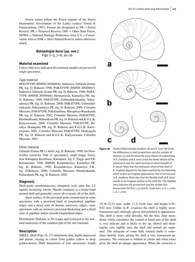
Figure 18 Shells of Ratnadvipia irradians (a) and R. karui (b) (notethe differences in shell proportions and the number ofwhorls); (c) and (d) show the early whorls of respectivelyof R. irradians and R. karui (note the fewer whorls of theprotoconch and the rapid increase in whorl breadth ofR. karui). Note that the embryonic whorl of the shell ofR. irradians figured (c) has been overlaid by the followingwhorl to give an irregular appearance; this is not unusualin R. irradians. Note also that the flexible shell of R. karuiresults in an irregular outline to the shell (d). The stippledarea indicates the protoconch and the broken linedemarcates the first 1.25 whorls. Scale bars: a, b = 5mm;c, d = 2mm.
19.16–22.21 mm; width 11.2–14.66 mm; and height 6.55–8.62 mm. Unlike in R. irradians the shell is highly mem-branaceous and strikingly glossy and polished in appearance.The shell is more solid dorsally, but the thin, limp mem-brane which constitutes the ventral or basal part of the shellis very delicate and is likely to dry up, shrivel and disin-tegrate very rapidly once the shell and animal are separ-ated. The ostracum of some fully formed shells is some-times heavily worn, giving the shell a very transparent ap-pearance. The ostracum is whitish in colour and when intactgives the shell an opaque appearance. When the ostracum is
118 Dinarzarde Raheem & Fred Naggs
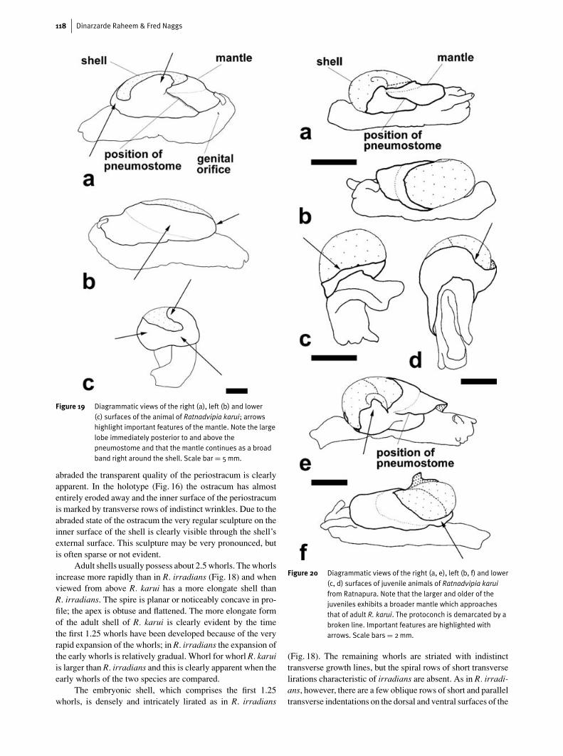
Figure 19 Diagrammatic views of the right (a), left (b) and lower(c) surfaces of the animal of Ratnadvipia karui; arrowshighlight important features of the mantle. Note the largelobe immediately posterior to and above thepneumostome and that the mantle continues as a broadband right around the shell. Scale bar = 5mm.
abraded the transparent quality of the periostracum is clearlyapparent. In the holotype (Fig. 16) the ostracum has almostentirely eroded away and the inner surface of the periostracumis marked by transverse rows of indistinct wrinkles. Due to theabraded state of the ostracum the very regular sculpture on theinner surface of the shell is clearly visible through the shell’sexternal surface. This sculpture may be very pronounced, butis often sparse or not evident.
Adult shells usually possess about 2.5 whorls. The whorlsincrease more rapidly than in R. irradians (Fig. 18) and whenviewed from above R. karui has a more elongate shell thanR. irradians. The spire is planar or noticeably concave in pro-file; the apex is obtuse and flattened. The more elongate formof the adult shell of R. karui is clearly evident by the timethe first 1.25 whorls have been developed because of the veryrapid expansion of the whorls; in R. irradians the expansion ofthe early whorls is relatively gradual. Whorl for whorl R. karuiis larger than R. irradians and this is clearly apparent when theearly whorls of the two species are compared.
The embryonic shell, which comprises the first 1.25whorls, is densely and intricately lirated as in R. irradians
Figure 20 Diagrammatic views of the right (a, e), left (b, f) and lower(c, d) surfaces of juvenile animals of Ratnadvipia karuifrom Ratnapura. Note that the larger and older of thejuveniles exhibits a broader mantle which approachesthat of adult R. karui. The protoconch is demarcated by abroken line. Important features are highlighted witharrows. Scale bars = 2mm.
(Fig. 18). The remaining whorls are striated with indistincttransverse growth lines, but the spiral rows of short transverselirations characteristic of irradians are absent. As in R. irradi-ans, however, there are a few oblique rows of short and paralleltransverse indentations on the dorsal and ventral surfaces of the
The Sri Lankan semi-slug Ratnadvipia 119
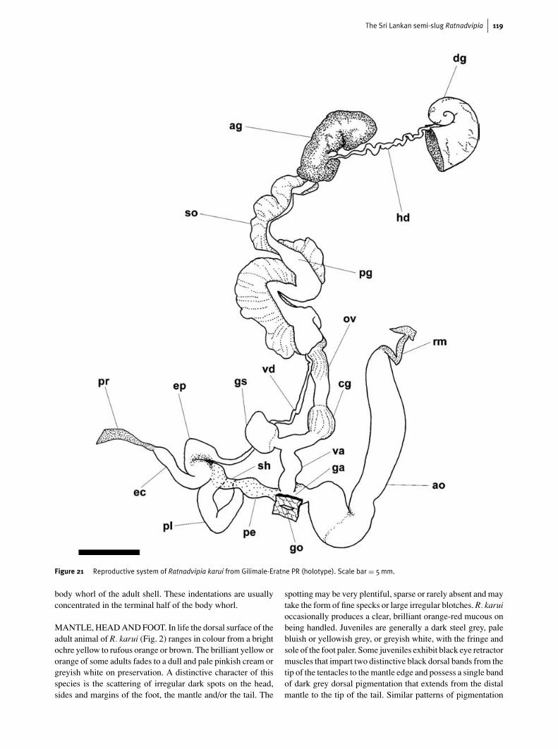
Figure 21 Reproductive system of Ratnadvipia karui from Gilimale-Eratne PR (holotype). Scale bar = 5mm.
body whorl of the adult shell. These indentations are usuallyconcentrated in the terminal half of the body whorl.
MANTLE, HEAD AND FOOT. In life the dorsal surface of theadult animal of R. karui (Fig. 2) ranges in colour from a brightochre yellow to rufous orange or brown. The brilliant yellow ororange of some adults fades to a dull and pale pinkish cream orgreyish white on preservation. A distinctive character of thisspecies is the scattering of irregular dark spots on the head,sides and margins of the foot, the mantle and/or the tail. The
spotting may be very plentiful, sparse or rarely absent and maytake the form of fine specks or large irregular blotches. R. karuioccasionally produces a clear, brilliant orange-red mucous onbeing handled. Juveniles are generally a dark steel grey, palebluish or yellowish grey, or greyish white, with the fringe andsole of the foot paler. Some juveniles exhibit black eye retractormuscles that impart two distinctive black dorsal bands from thetip of the tentacles to the mantle edge and possess a single bandof dark grey dorsal pigmentation that extends from the distalmantle to the tip of the tail. Similar patterns of pigmentation
120 Dinarzarde Raheem & Fred Naggs
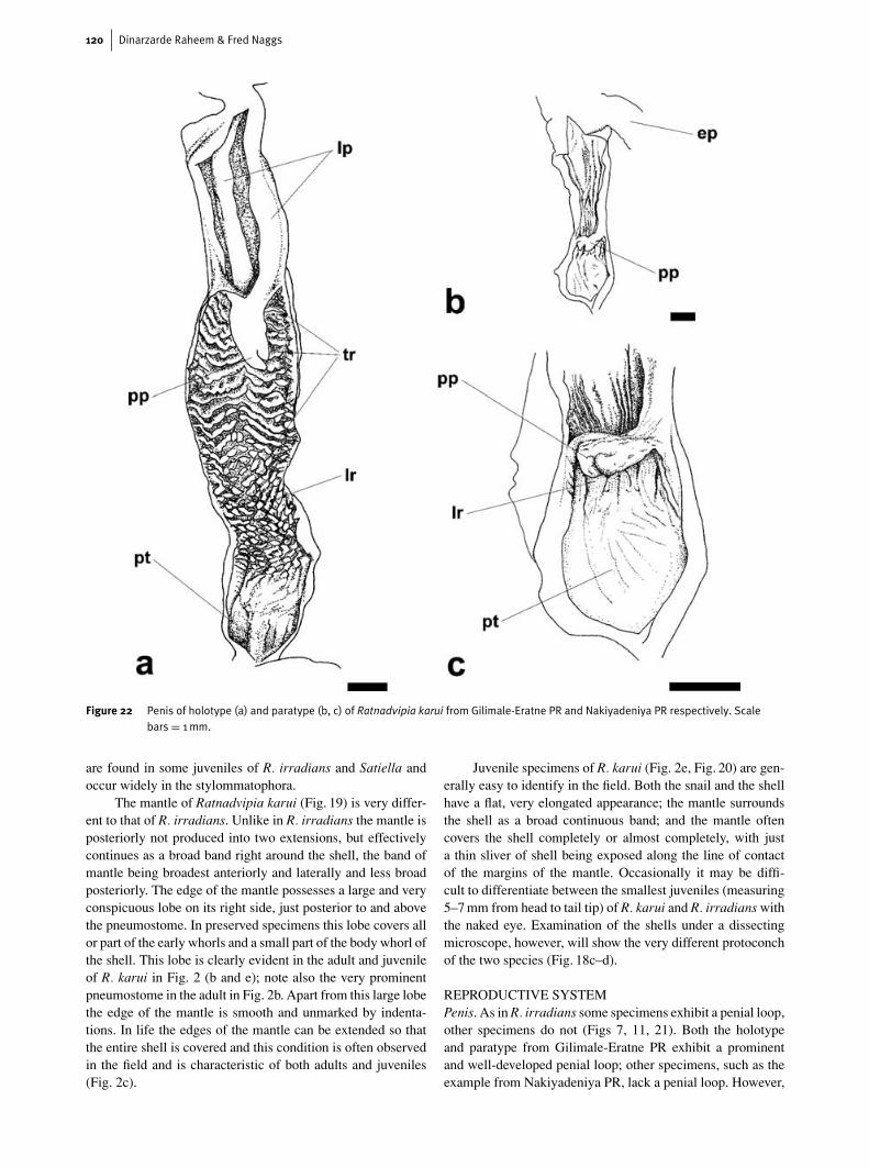
Figure 22 Penis of holotype (a) and paratype (b, c) of Ratnadvipia karui from Gilimale-Eratne PR and Nakiyadeniya PR respectively. Scalebars = 1mm.
are found in some juveniles of R. irradians and Satiella andoccur widely in the stylommatophora.
The mantle of Ratnadvipia karui (Fig. 19) is very differ-ent to that of R. irradians. Unlike in R. irradians the mantle isposteriorly not produced into two extensions, but effectivelycontinues as a broad band right around the shell, the band ofmantle being broadest anteriorly and laterally and less broadposteriorly. The edge of the mantle possesses a large and veryconspicuous lobe on its right side, just posterior to and abovethe pneumostome. In preserved specimens this lobe covers allor part of the early whorls and a small part of the body whorl ofthe shell. This lobe is clearly evident in the adult and juvenileof R. karui in Fig. 2 (b and e); note also the very prominentpneumostome in the adult in Fig. 2b. Apart from this large lobethe edge of the mantle is smooth and unmarked by indenta-tions. In life the edges of the mantle can be extended so thatthe entire shell is covered and this condition is often observedin the field and is characteristic of both adults and juveniles(Fig. 2c).
Juvenile specimens of R. karui (Fig. 2e, Fig. 20) are gen-erally easy to identify in the field. Both the snail and the shellhave a flat, very elongated appearance; the mantle surroundsthe shell as a broad continuous band; and the mantle oftencovers the shell completely or almost completely, with justa thin sliver of shell being exposed along the line of contactof the margins of the mantle. Occasionally it may be diffi-cult to differentiate between the smallest juveniles (measuring5–7 mm from head to tail tip) of R. karui and R. irradians withthe naked eye. Examination of the shells under a dissectingmicroscope, however, will show the very different protoconchof the two species (Fig. 18c–d).
REPRODUCTIVE SYSTEMPenis. As in R. irradians some specimens exhibit a penial loop,other specimens do not (Figs 7, 11, 21). Both the holotypeand paratype from Gilimale-Eratne PR exhibit a prominentand well-developed penial loop; other specimens, such as theexample from Nakiyadeniya PR, lack a penial loop. However,
The Sri Lankan semi-slug Ratnadvipia 121
unlike R. irradians, the internal surface of the proximal penis inR. karui specimens with a penial loop (Fig. 22a) is very dif-ferent to those without a penial loop. In the holotype andparatype of R. karui from Gilimale-Eartne PR the proximalpenis is distally ornamented with about five to twelve parallelirregular and wavy transverse ridges. Proximally, these trans-verse ridges give way to a broad zone of longitudinal papillateridges and/or longitudinal rows of papillae; these longitudinalridges and papillae are similar in form to those of R. irradians.As in R. irradians the part of the penis just proximal to itsjunction with the genital atrium is marked by a thickened orswollen penial wall. At the meeting point of the longitudinalridges with this proximal thickening, the ridges are smootherwith little or no papillae.
The internal structure of the penis of specimens lacking apenial loop (Fig. 22b–c) is very different. The inner surface ofthe proximal penis is composed largely of the plain proximalthickening of the penial wall bordered distally by a narrowband of longitudinal ridges. These ridges are smooth and cyl-indrical proximally and become markedly papillate distallyand immediately around the penial papilla.
In R. irradians the internal surface of the penis (Fig. 4)shows a uniform arrangement irrespective of the presence orabsence of a penial loop. R. karui, in contrast, exhibits twoclearly distinct arrangements to the inner surface of the penis,which our preliminary investigations have shown to be asso-ciated with the presence or absence of the penial loop. Furtherstudy is needed to establish if the two penial forms in R. karuicorrespond to different stages of growth and reproductive de-velopment or if they have taxonomic significance.
Gametolytic sac. As in R. irradians the gametolytic sac(Figs 7, 21) is variable in form. In the holotype and paratypefrom Gilimale-Eratne PR the gametolytic sac is an asymmet-ric and inflated hooked sac with an opaque appearance, whichis connected to the oviduct by a very short but discerniblenarrow-lumened duct. In other specimens, such as the onefrom Nakiyadeniya PR, the gametolytic sac is a symmetrical,transparent elongated bag; the duct is not discernible.
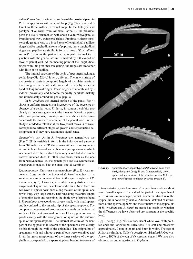
Spermatophore. Only one spermatophore (Fig. 23) was re-covered from the six specimens of R. karui examined. It issmaller but similar in general form to the spermatophore of R.irradians (Fig. 5). However, it exhibits a very distinctive ar-rangement of spines on the anterior spike. In R. karui there aretwo rows of spines positioned along the axis of the spike; onerow is long, with large spines. This runs along the entire lengthof the spike’s axis and resembles the single row of spines foundin R. irradians, the second row is very small, with small spinesand is confined to the anterior tip of the spermatophore. Thecomplex arrangement of grooves and channels on the internalsurface of the bent proximal portion of the epiphallus corres-ponds exactly with the arrangement of spines on the anteriorspike of the spermatophore. The internal structure of the bentpart of the epiphallus in several of the specimens was clearlyvisible through the wall of the epiphallus. The epiphallus ofspecimens with and without a penial loop were examined andin all the gross morphology of the inner surface of the epi-phallus corresponded to a spermatophore bearing two rows of
Figure 23 Spermatophore of paratype of Ratnadvipia karui fromNakiyadeniya PR (a–c); (b) and (c) respectively showupper and lateral views of the anterior portion. Note thetwo rows of spines in (shown by white arrow in b).
spines anteriorly, one long row of large spines and one shortrow of smaller spines. The wall of the part of the epiphallus ofR. irradians is more opaque, so that the internal structure of theepiphallus is not clearly visible. Additional detailed examina-tions of the spermatophores and the structure of the epiphallusof R. irradians and R. karui are needed to establish whetherthe differences we have observed are constant at the specificlevel.
Egg. The egg (Fig. 2d) is a translucent white, oval with poin-ted ends and longitudinal sulcations. It is soft and measuresapproximately 7 mm in length and 4 mm in width. The egg ofR. karui is similar to Collett’s description (Blanford & Godwin-Austen, 1908) of the egg of Cryptozona chenui. We have alsoobserved a similar egg-form in Euplecta.
122 Dinarzarde Raheem & Fred Naggs
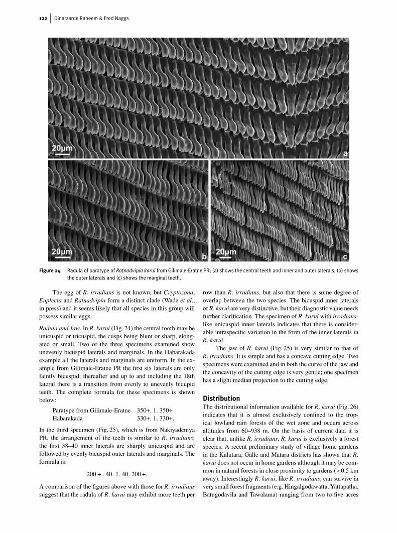
Figure 24 Radula of paratype of Ratnadvipia karui from Gilimale-Eratne PR; (a) shows the central teeth and inner and outer laterals, (b) showsthe outer laterals and (c) shows the marginal teeth.
The egg of R. irradians is not known, but Cryptozona,Euplecta and Ratnadvipia form a distinct clade (Wade et al.,in press) and it seems likely that all species in this group willpossess similar eggs.
Radula and Jaw. In R. karui (Fig. 24) the central tooth may beunicuspid or tricuspid, the cusps being blunt or sharp, elong-ated or small. Two of the three specimens examined showunevenly bicuspid laterals and marginals. In the Habarakadaexample all the laterals and marginals are uniform. In the ex-ample from Gilimale-Eratne PR the first six laterals are onlyfaintly bicuspid; thereafter and up to and including the 18thlateral there is a transition from evenly to unevenly bicupidteeth. The complete formula for these specimens is shownbelow:
Paratype from Gilimale-Eratne 350+. 1. 350+Habarakada 330+. 1. 330+.
In the third specimen (Fig. 25), which is from NakiyadeniyaPR, the arrangement of the teeth is similar to R. irradians;the first 38–40 inner laterals are sharply unicuspid and arefollowed by evenly bicuspid outer laterals and marginals. Theformula is:
200 + . 40. 1. 40. 200 +.
A comparison of the figures above with those for R. irradianssuggest that the radula of R. karui may exhibit more teeth per
row than R. irradians, but also that there is some degree ofoverlap between the two species. The bicuspid inner lateralsof R. karui are very distinctive, but their diagnostic value needsfurther clarification. The specimen of R. karui with irradians-like unicuspid inner laterals indicates that there is consider-able intraspecific variation in the form of the inner laterals inR. karui.
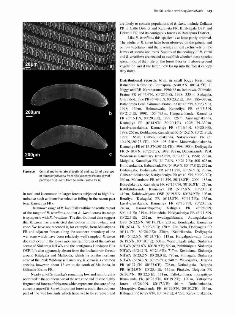
The jaw of R. karui (Fig. 25) is very similar to that ofR. irradians. It is simple and has a concave cutting edge. Twospecimens were examined and in both the curve of the jaw andthe concavity of the cutting edge is very gentle; one specimenhas a slight median projection to the cutting edge.
DistributionThe distributional information available for R. karui (Fig. 26)indicates that it is almost exclusively confined to the trop-ical lowland rain forests of the wet zone and occurs acrossaltitudes from 60–938 m. On the basis of current data it isclear that, unlike R. irradians, R. karui is exclusively a forestspecies. A recent preliminary study of village home gardensin the Kalutara, Galle and Matara districts has shown that R.karui does not occur in home gardens although it may be com-mon in natural forests in close proximity to gardens (<0.5 kmaway). Interestingly R. karui, like R. irradians, can survive invery small forest fragments (e.g. Hingalgodawatta, Yattapatha,Batugodavila and Tawalama) ranging from two to five acres
The Sri Lankan semi-slug Ratnadvipia 123
Figure 25 Central and inner lateral teeth (a) and jaw (b) of paratypeof Ratnadvipia karui from Nakiyadeniya PR and jaw ofparatype of R. karui from Gilimale-Eratne PR (c).
in total and is common in larger forests subjected to high dis-turbance such as intensive selective felling in the recent past(e.g. Kanneliya FR).
The known range of R. karui falls within the southern partof the range of R. irradians, so that R. karui across its rangeis sympatric with R. irradians. The distributional data suggestthat R. karui has a restricted distribution in the lowland wetzone. We have not recorded it, for example, from MulatiyanaFR and adjacent forests along the southern boundary of thewet zone which have been relatively well sampled. R. karuidoes not occur in the lower montane rain forests of the easternsector of Sinharaja NHWA and the contiguous Handapan EllaOSF. It is also apparently absent from the lowland rain forestsaround Kitulgala and Maliboda, which lie on the northernedge of the Peak Wilderness Sanctuary. R. karui is a commonspecies, however, about 12 km to the south of Maliboda, inGilimale-Eratne PR.
Nearly all of Sri Lanka’s remaining lowland rain forest isrestricted to the southern part of the wet zone and it is the highlyfragmented forests of this area which represents the core of thecurrent range of R. karui. Important forest areas in the southernpart of the wet lowlands which have yet to be surveyed and
are likely to contain populations of R. karui include DellawaPR in Galle District and Karawita PR, Kiribatgala OSF, andDelwela PR and its contiguous forests in Ratnapura District.
Like R. irradians this species is at least partly arboreal.The adults of R. karui have been observed on the ground andon low vegetation and the juveniles almost exclusively on theleaves of shrubs and trees. Studies of the ecology of R. karuiand R. irradians are needed to establish whether these speciesspend most of their life on the forest floor or in above-groundvegetation and if the latter, how far up into the forest canopythey move.
Distributional records: 61 m, in small boggy forest nearRatnapura Resthouse, Ratnapura (6◦40.9′N, 80◦24.2′E), F.Naggs and P.B. Karunaratne, 1996; 68 m, Induruwa, Gilimale-Eratne PR (6◦45.6′N, 80◦25.6′E), 1998; 333 m, Sudagala,Gilimale-Eratne PR (6◦48.3′N, 80◦23.2′E), 1998; 285–380 m,Batadomba Lena, Gilimale-Eratne PR (6◦46.5′N, 80◦23.5′E),1998; 130 m, Holmanwala, Kanneliya FR (6◦15.5′N,80◦21.3′E), 1998; 155–495 m, Hangarankande, KanneliyaFR (6◦16.1′N, 80◦20.2′E), 1998; 125 m, Annasigetakanda,Kanneliya FR (6◦14.9′N, 80◦20.1′E), 1998; 75–330 m,Lavalvaruvakanda, Kanneliya FR (6◦16.4′N, 80◦20.9′E),1998; 265 m, Kodikande, Kanneliya FR (6◦15.2′N, 80◦21.8′E),1998; 165 m, Galbendidolakanda, Nakiyadeniya PR (6◦
10.4′N, 80◦23.1′E), 1998; 195–210 m, Manamaladolakanda,Kanneliya FR (6◦15.3′N, 80◦22.4′E), 1998; 195 m, DediyagalaFR (6◦10.4′N, 80◦25.5′E), 1998; 938 m, Dehenakande, PeakWilderness Sanctuary (6◦45.6′N, 80◦30.3′E), 1998; 223 m,Malgalla, Kanneliya FR (6◦17.6′N, 80◦21.7′E); 400–623 m,Hinidumkanda, Habarakada PR (6◦19.9′N, 80◦17.8′E); 232 m,Dediyagala, Dediyagala FR (6◦11.2′N, 80◦24.6′E); 273 m,Galbendidolakande, Nakiyadeniya PR (6◦10.3′N, 80◦23.0′E);360 m, Malambure FR (6◦14.5′N, 80◦18.8′E), 2000; 183 m,Kospelaketiya, Kanneliya FR (6◦15.0′N, 80◦20.8′E); 210 m,Katukitulakanda, Kanneliya FR (6◦17.8′N, 80◦20.3′E);410 m, Kalubowitiyana OSF (6◦19.5′N, 80◦24.9′E); 103 m,Beraliya (Kudagala) PR (6◦15.8′N, 80◦11.7′E); 165 m,Lavalvaruvakanda, Kanneliya FR (6◦15.3′N, 80◦20.5′E);240 m, Harankahapatha, Kalugala PR (6◦28.6′N,80◦14.1′E); 219 m, Homadola, Nakiyadeniya PR (6◦11.8′N,80◦22.3′E); 232 m, Awudagalakanda, AuwegalakandaOSF (6◦22.5′N, 80◦17.1′E); 253 m, Kondagala, KanneliyaFR (6◦14.1′N, 80◦23.8′E); 170 m, Olu Dola, Dediyagala FR(6◦11.1′N, 80◦26.0′E); 236 m, Kekirikanda, DediyagalaFR (6◦12.6′N, 80◦24.7′E); 113 m, Hingalgodawatta forest(6◦19.5′N, 80◦19.7′E); 560 m, Wanduragala ridge, SinharajaNHWA (6◦23.6′N, 80◦28.9′E); 592 m, Paththinigala, SinharajaNHWA (6◦24.1′N, 80◦24.6′E); 717 m, Koskulana, SinharajaNHWA (6◦23.2′N, 80◦28.0′E); 780 m, Sinhagala, SinharajaNHWA (6◦24.3′N, 80◦26.6′E); 540 m, Wewagama, DelgodaPR (6◦27.1′N, 80◦23.6′E); 728 m, Dothalugala, DelgodaPR (6◦24.9′N, 80◦23.4′E); 441 m, Pitakele, Delgoda PR(6◦26.7′N, 80◦22.5′E); 151 m, Pallekumbura, morapitiya-Runakanda PR (6◦28.5′N, 80◦19.2′E); 120 m, Yattapathaforest, (6◦26.0′N, 80◦17.3′E); 461 m, Dothalankanda,Morapitiya-Runakanda PR (6◦29.8′N, 80◦20.2′E); 314 m,Kalugala PR (6◦27.8′N, 80◦14.2′E); 472 m, Katukitulakanda,
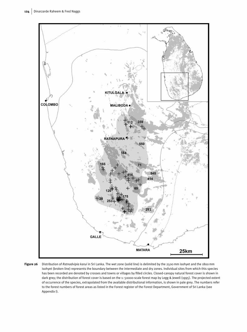
124 Dinarzarde Raheem & Fred Naggs
Figure 26 Distribution of Ratnadvipia karui in Sri Lanka. The wet zone (solid line) is delimited by the 2500mm isohyet and the 1800mmisohyet (broken line) represents the boundary between the intermediate and dry zones. Individual sites from which this specieshas been recorded are denoted by crosses and towns or villages by filled circles. Closed-canopy natural forest cover is shown indark grey; the distribution of forest cover is based on the 1: 50000-scale forest map by Legg & Jewell (1995). The projected extentof occurrence of the species, extrapolated from the available distributional information, is shown in pale grey. The numbers referto the forest numbers of forest areas as listed in the Forest register of the Forest Department, Government of Sri Lanka (seeAppendix I).
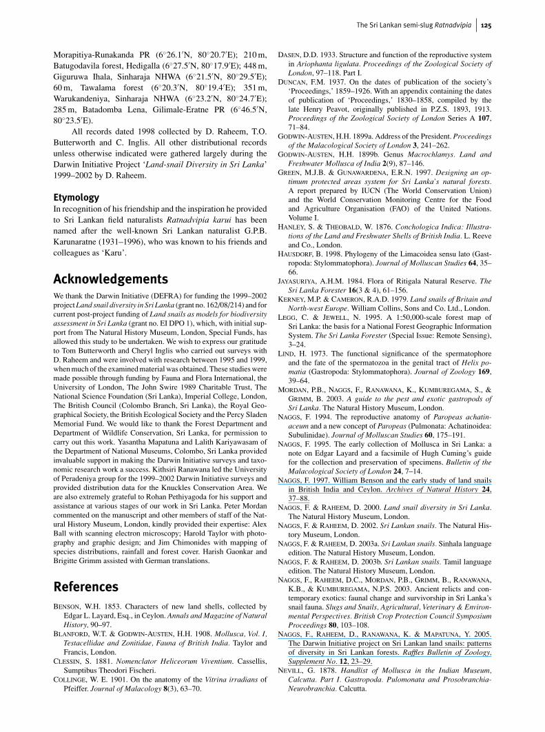
The Sri Lankan semi-slug Ratnadvipia 125
Morapitiya-Runakanda PR (6◦26.1′N, 80◦20.7′E); 210 m,Batugodavila forest, Hedigalla (6◦27.5′N, 80◦17.9′E); 448 m,Giguruwa Ihala, Sinharaja NHWA (6◦21.5′N, 80◦29.5′E);60 m, Tawalama forest (6◦20.3′N, 80◦19.4′E); 351 m,Warukandeniya, Sinharaja NHWA (6◦23.2′N, 80◦24.7′E);285 m, Batadomba Lena, Gilimale-Eratne PR (6◦46.5′N,80◦23.5′E).
All records dated 1998 collected by D. Raheem, T.O.Butterworth and C. Inglis. All other distributional recordsunless otherwise indicated were gathered largely during theDarwin Initiative Project ‘Land-snail Diversity in Sri Lanka’1999–2002 by D. Raheem.
EtymologyIn recognition of his friendship and the inspiration he providedto Sri Lankan field naturalists Ratnadvipia karui has beennamed after the well-known Sri Lankan naturalist G.P.B.Karunaratne (1931–1996), who was known to his friends andcolleagues as ‘Karu’.
AcknowledgementsWe thank the Darwin Initiative (DEFRA) for funding the 1999–2002project Land snail diversity in Sri Lanka (grant no. 162/08/214) and forcurrent post-project funding of Land snails as models for biodiversityassessment in Sri Lanka (grant no. EI DPO 1), which, with initial sup-port from The Natural History Museum, London, Special Funds, hasallowed this study to be undertaken. We wish to express our gratitudeto Tom Butterworth and Cheryl Inglis who carried out surveys withD. Raheem and were involved with research between 1995 and 1999,when much of the examined material was obtained. These studies weremade possible through funding by Fauna and Flora International, theUniversity of London, The John Swire 1989 Charitable Trust, TheNational Science Foundation (Sri Lanka), Imperial College, London,The British Council (Colombo Branch, Sri Lanka), the Royal Geo-graphical Society, the British Ecological Society and the Percy SladenMemorial Fund. We would like to thank the Forest Department andDepartment of Wildlife Conservation, Sri Lanka, for permission tocarry out this work. Yasantha Mapatuna and Lalith Kariyawasam ofthe Department of National Museums, Colombo, Sri Lanka providedinvaluable support in making the Darwin Initiative surveys and taxo-nomic research work a success. Kithsiri Ranawana led the Universityof Peradeniya group for the 1999–2002 Darwin Initiative surveys andprovided distribution data for the Knuckles Conservation Area. Weare also extremely grateful to Rohan Pethiyagoda for his support andassistance at various stages of our work in Sri Lanka. Peter Mordancommented on the manuscript and other members of staff of the Nat-ural History Museum, London, kindly provided their expertise: AlexBall with scanning electron microscopy; Harold Taylor with photo-graphy and graphic design; and Jim Chimonides with mapping ofspecies distributions, rainfall and forest cover. Harish Gaonkar andBrigitte Grimm assisted with German translations.
ReferencesBENSON, W.H. 1853. Characters of new land shells, collected by
Edgar L. Layard, Esq., in Ceylon. Annals and Magazine of NaturalHistory, 90–97.
BLANFORD, W.T. & GODWIN-AUSTEN, H.H. 1908. Mollusca, Vol. I,Testacellidae and Zonitidae, Fauna of British India. Taylor andFrancis, London.
CLESSIN, S. 1881. Nomenclator Heliceorum Viventium. Cassellis,Sumptibus Theodori Fischeri.
COLLINGE, W. E. 1901. On the anatomy of the Vitrina irradians ofPfeiffer. Journal of Malacology 8(3), 63–70.
DASEN, D.D. 1933. Structure and function of the reproductive systemin Ariophanta ligulata. Proceedings of the Zoological Society ofLondon, 97–118. Part I.
DUNCAN, F.M. 1937. On the dates of publication of the society’s‘Proceedings,’ 1859–1926. With an appendix containing the datesof publication of ‘Proceedings,’ 1830–1858, compiled by thelate Henry Peavot, originally published in P.Z.S. 1893, 1913.Proceedings of the Zoological Society of London Series A 107,71–84.
GODWIN-AUSTEN, H.H. 1899a. Address of the President. Proceedingsof the Malacological Society of London 3, 241–262.
GODWIN-AUSTEN, H.H. 1899b. Genus Macrochlamys. Land andFreshwater Mollusca of India 2(9), 87–146.
GREEN, M.J.B. & GUNAWARDENA, E.R.N. 1997. Designing an op-timum protected areas system for Sri Lanka’s natural forests.A report prepared by IUCN (The World Conservation Union)and the World Conservation Monitoring Centre for the Foodand Agriculture Organisation (FAO) of the United Nations.Volume I.
HANLEY, S. & THEOBALD, W. 1876. Conchologica Indica: Illustra-tions of the Land and Freshwater Shells of British India. L. Reeveand Co., London.
HAUSDORF, B. 1998. Phylogeny of the Limacoidea sensu lato (Gast-ropoda: Stylommatophora). Journal of Molluscan Studies 64, 35–66.
JAYASURIYA, A.H.M. 1984. Flora of Ritigala Natural Reserve. TheSri Lanka Forester 16(3 & 4), 61–156.
KERNEY, M.P. & CAMERON, R.A.D. 1979. Land snails of Britain andNorth-west Europe. William Collins, Sons and Co. Ltd., London.
LEGG, C. & JEWELL, N. 1995. A 1:50,000-scale forest map ofSri Lanka: the basis for a National Forest Geographic InformationSystem. The Sri Lanka Forester (Special Issue: Remote Sensing),3–24.
LIND, H. 1973. The functional significance of the spermatophoreand the fate of the spermatozoa in the genital tract of Helix po-matia (Gastropoda: Stylommatophora). Journal of Zoology 169,39–64.
MORDAN, P.B., NAGGS, F., RANAWANA, K., KUMBUREGAMA, S., &GRIMM, B. 2003. A guide to the pest and exotic gastropods ofSri Lanka. The Natural History Museum, London.
NAGGS, F. 1994. The reproductive anatomy of Paropeas achatin-aceum and a new concept of Paropeas (Pulmonata: Achatinoidea:Subulinidae). Journal of Molluscan Studies 60, 175–191.
NAGGS, F. 1995. The early collection of Mollusca in Sri Lanka: anote on Edgar Layard and a facsimile of Hugh Cuming’s guidefor the collection and preservation of specimens. Bulletin of theMalacological Society of London 24, 7–14.
NAGGS, F. 1997. William Benson and the early study of land snailsin British India and Ceylon. Archives of Natural History 24,37–88.
NAGGS, F. & RAHEEM, D. 2000. Land snail diversity in Sri Lanka.The Natural History Museum, London.
NAGGS, F. & RAHEEM, D. 2002. Sri Lankan snails. The Natural His-tory Museum, London.
NAGGS, F. & RAHEEM, D. 2003a. Sri Lankan snails. Sinhala languageedition. The Natural History Museum, London.
NAGGS, F. & RAHEEM, D. 2003b. Sri Lankan snails. Tamil languageedition. The Natural History Museum, London.
NAGGS, F., RAHEEM, D.C., MORDAN, P.B., GRIMM, B., RANAWANA,K.B., & KUMBUREGAMA, N.P.S. 2003. Ancient relicts and con-temporary exotics: faunal change and survivorship in Sri Lanka’ssnail fauna. Slugs and Snails, Agricultural, Veterinary & Environ-mental Perspectives. British Crop Protection Council SymposiumProceedings 80, 103–108.
NAGGS, F., RAHEEM, D., RANAWANA, K. & MAPATUNA, Y. 2005.The Darwin Initiative project on Sri Lankan land snails: patternsof diversity in Sri Lankan forests. Raffles Bulletin of Zoology,Supplement No. 12, 23–29.
NEVILL, G. 1878. Handlist of Mollusca in the Indian Museum,Calcutta. Part I. Gastropoda. Pulomonata and Prosobranchia-Neurobranchia. Calcutta.
126 Dinarzarde Raheem & Fred Naggs
PETHIYAGODA, R. 2004. Biodiversity law has had some unintendedeffects. Moves to prevent unfair exploitation of resources couldrestrict conservation research. Nature 429, 129.
PFEIFFER, L. 1853. Monographia Heliceorum Viventium. Volume 3.PFEIFFER, L. 1854. Descriptions of eighteen new species of land-
shells from the collection of H. Cuming, Esq. Proceedings of theZoological Society of London for 1852 XX, 156–160.
REEVE, L.A. 1862. Conchologica Iconica or Illustrations of the Shellsof Molluscous Animals, Vol. XIII. Lovell Reeve and Co., London.
THEOBALD, W. 1876. Catalogue of the land and fresh-water shells ofBritish India. Thacker, Spink & Co., Calcutta.
TILLIER, S. 1984. Patterns of digestive tract morphology in thelimacisation of helicarionid, succineid and athoracophorid snailsand slugs (Mollusca: Pulmonata). Malacologia 25, 173–192.
TILLIER, S. 1989. Comparative morphology, phylogeny and classific-ation of land snails and slugs (Gastropoda: Pulmonata: Stylomma-tophora). Malacologia 30, 1–303.
TOMPA, A.S. 1984. Land Snails (Stylommatophora). In: TOMPA, A.S.,VERDONIK, N.H. & VAN DEN BIGGELAAR, J.A.M., Eds., The Mol-lusca, Volume 7, Reproduction. Academic Press, Orlando, Florida,pp. 47–140.
Appendix IAbbreviations used in figures
ag albumen gland ov oviductao amatorial organ pe penisbm buccal mass pg prostate glandcg capsular gland pl penial loopdg digestive gland pp penial papillaep epiphallus pr penial retractor muscleec epiphallic caecum pt proximal thickening of penisfp fertilisation pouch rd right salivary ductga genital atrium rg right salivary glandgo genital orifice rm retractor muscle of the amatorial organgs gametolytic sac rs radular sachd hermaphrodite duct sh penial sheathld left salivary duct so spermoviductlg left salivary gland sp spermatophorelp smooth longitudinal pilasters tr transverse dentate ridgeslr longitudinal papillate ridges and papillae va vaginaoe oesophagus vd vas deferens
TRYON, G.W. 1885. Manual of Conchology. Vol. I. Academy of Nat-ural Sciences, Philadelphia.
VAN GOETHEM, L. 1977. Revision systematique des Urocyclinae(Mollusca, Pulmonata, Urocyclidae). Musee royal de l’AfriqueCentrale, Tervuren. Belgique, Annales, Sciences zoologiques218(I-XI), 1–355.
WADE, C.M., MORDAN, P.B. & CLARKE, B. 2001. A phylogeny ofthe land snails (Gastropoda: Pulmonata). Proceedings of the RoyalSociety London B 268, 413–422.
WADE, C.M., MORDAN, P.B. & NAGGS, F. In press. Evolutionaryrelationships among the Pulmonate land snails and slugs (Pul-monata, Stylommatophora). Biological Journal of the LinneanSociety.
WHITMORE, T.C. 1990. An Introduction to Tropical Rainforests. Clar-endon Press, Oxford.
WOLDT, K. 1932–33. Beitrage zur Anatomie der Pulmonate Rat-nadvipia irradians Godwin-Austen mit besonderer Berucksich-tigung des Genitalsystems. Naturwissenschaft LXVI, 263–322.
ZILCH, A. 1959. Gastropoda von Wilhelm Wenz. Lieferung mit Ab-bildung 702–1434. Handbuch der Palaozoologie 6.
Appendix IIList of forest areas
The following is a list of forest areas highlighted in Fig. 26.Forest names and numbers follow the Forest register ofthe Forest Department, Government of Sri Lanka (source:Green & Gunawardena, 1997). The forests are designated asFR = Forest Reserve, PR = Proposed Reserve, OSF = OtherState Forest or NHWA = National Heritage Wilderness Area,unless otherwise stated.
For the purposes of administration some of the largerindividual forest fragments were divided into two or moreprotected areas by the Forest Department during the early partof the twentieth century; such forest fragments are indicatedbelow by an asterisk. For example, the forest isolate of whichKanneliya FR forms the northern sector has its southern partdivided into western and eastern portions known respectivelyas Nakiyadeniya PR and Dediyagala FR. For convenience, inFig. 26 we have indicated forest fragments composed of several
such constituent protected areas with the number of one of theconstituent parts.
38 Beraliya (Kudagala) PR69 Dellawa PR71 Delwala PR∗
112 Gilimale–Eratne PR120 Habarakada PR∗
166 Kalugala PR175 Kanneliya FR∗
184 Karawita PR253 Malambure FR∗
293 Mulatiyana FR359 Peak Wilderness Sanctuary∗
414 Sinharaja NHWA∗
550 Kiribatgala OSF545 Handapan Ella