The Role of the Inflammatory Response in Mediating ... - MDPI
18
International Journal of Molecular Sciences Review The Role of the Inflammatory Response in Mediating Functional Recovery Following Composite Tissue Injuries Naveena B. Janakiram 1,2,3 , Michael S. Valerio 1,2,3 , Stephen M. Goldman 1,2,3 and Christopher L. Dearth 1,2,3, * Citation: Janakiram, N.B.; Valerio, M.S.; Goldman, S.M.; Dearth, C.L. The Role of the Inflammatory Response in Mediating Functional Recovery Following Composite Tissue Injuries. Int. J. Mol. Sci. 2021, 22, 13552. https://doi.org/10.3390/ ijms222413552 Academic Editor: Nguan Soon Tan Received: 20 October 2021 Accepted: 14 December 2021 Published: 17 December 2021 Publisher’s Note: MDPI stays neutral with regard to jurisdictional claims in published maps and institutional affil- iations. Copyright: © 2021 by the authors. Licensee MDPI, Basel, Switzerland. This article is an open access article distributed under the terms and conditions of the Creative Commons Attribution (CC BY) license (https:// creativecommons.org/licenses/by/ 4.0/). 1 Research & Surveillance Division, DoD-VA Extremity Trauma and Amputation Center of Excellence, Bethesda, MD 20889, USA; [email protected] (N.B.J.); [email protected] (M.S.V.); [email protected] (S.M.G.) 2 Department of Surgery, Uniformed Services University of the Health Sciences, Bethesda, MD 20889, USA 3 Walter Reed National Military Medical Center, Bethesda, MD 20889, USA * Correspondence: [email protected]; Tel.: +1-(301)-319-2461 Abstract: Composite tissue injuries (CTI) are common among US Military Service members during combat operations, and carry a high potential of morbidity. Furthermore, CTI are often complicated due to an altered wound healing response, resulting in part from a dysregulation of the innate and adaptive immune responses. Unlike normal wound healing, in CTI, disruptions occur in innate immune responses, altering neutrophil functions, macrophage activation and polarization, further impacting the functions of T regulatory cells. Additionally, the biological underpinnings of these unfavorable wound healing conditions are multifactorial, including various processes, such as: ischemia, hypoxia, low nutrient levels, and altered cell metabolic pathways, among others, all of which are thought to trigger anergy in immune cells and destabilize adaptive immune responses. As a result, impaired wound healing is common in CTI. Herein, we review the altered innate and adaptive immune cells and their metabolic status and responses following CTI, and discuss the role a multi-pronged immunomodulatory approach may play in facilitating improved outcomes for afflicted patients. Keywords: wound healing; tissue regeneration; inflammation; immunomodulation; military medicine; composite musculoskeletal trauma 1. Introduction Composite tissue injuries (CTI) owing to high energy wounding mechanisms (e.g., explosions, gunshot wounds) are an unfortunate reality of modern military combat opera- tions. In simple terms, CTI are those injuries that involve significant damage to multiple tissues, including skeletal muscle, nerve, bone, vasculature, and dermal tissues [1–4]. When such injuries affect the extremities, they often result in significant, long-term impairments in functional outcomes, and therefore delay or prevent the service member from returning to duty. As such, CTI are a critical concern of the Military Health System and a significant threat to the readiness of the joint force. Beyond military medicine, civilian populations are often impacted by CTI as a result of vehicular or industrial accidents [5], and these injuries represent a substantial economic burden in the order of billions of dollars annually (i.e., costs associated with hospitalizations, acute surgical services, long term rehabilitation costs, lost work hours) [6,7]. Surgical and rehabilitation costs, in particular, can be extraordinarily high owing to the inherent injury complexity and associated deleterious wound healing outcomes (e.g., delayed union, fibrosis); a key driver of such outcomes is the (dysregulated) endogenous immune-inflammatory response. The immune-inflammatory response to acute injury is a highly complex system com- prising multiple soluble (e.g., antibodies, complement) and cellular mediators (e.g., cy- tokines, prostaglandins) that serve to clear pathogens and cellular debris and stimulate Int. J. Mol. Sci. 2021, 22, 13552. https://doi.org/10.3390/ijms222413552 https://www.mdpi.com/journal/ijms
-
Upload
khangminh22 -
Category
Documents
-
view
2 -
download
0
Transcript of The Role of the Inflammatory Response in Mediating ... - MDPI

International Journal of
Molecular Sciences
Review
The Role of the Inflammatory Response in MediatingFunctional Recovery Following Composite Tissue Injuries
Naveena B. Janakiram 1,2,3, Michael S. Valerio 1,2,3, Stephen M. Goldman 1,2,3 and Christopher L. Dearth 1,2,3,*
�����������������
Citation: Janakiram, N.B.; Valerio,
M.S.; Goldman, S.M.; Dearth, C.L.
The Role of the Inflammatory
Response in Mediating Functional
Recovery Following Composite
Tissue Injuries. Int. J. Mol. Sci. 2021,
22, 13552. https://doi.org/10.3390/
ijms222413552
Academic Editor: Nguan Soon Tan
Received: 20 October 2021
Accepted: 14 December 2021
Published: 17 December 2021
Publisher’s Note: MDPI stays neutral
with regard to jurisdictional claims in
published maps and institutional affil-
iations.
Copyright: © 2021 by the authors.
Licensee MDPI, Basel, Switzerland.
This article is an open access article
distributed under the terms and
conditions of the Creative Commons
Attribution (CC BY) license (https://
creativecommons.org/licenses/by/
4.0/).
1 Research & Surveillance Division, DoD-VA Extremity Trauma and Amputation Center of Excellence,Bethesda, MD 20889, USA; [email protected] (N.B.J.); [email protected] (M.S.V.);[email protected] (S.M.G.)
2 Department of Surgery, Uniformed Services University of the Health Sciences, Bethesda, MD 20889, USA3 Walter Reed National Military Medical Center, Bethesda, MD 20889, USA* Correspondence: [email protected]; Tel.: +1-(301)-319-2461
Abstract: Composite tissue injuries (CTI) are common among US Military Service members duringcombat operations, and carry a high potential of morbidity. Furthermore, CTI are often complicateddue to an altered wound healing response, resulting in part from a dysregulation of the innate andadaptive immune responses. Unlike normal wound healing, in CTI, disruptions occur in innateimmune responses, altering neutrophil functions, macrophage activation and polarization, furtherimpacting the functions of T regulatory cells. Additionally, the biological underpinnings of theseunfavorable wound healing conditions are multifactorial, including various processes, such as:ischemia, hypoxia, low nutrient levels, and altered cell metabolic pathways, among others, all ofwhich are thought to trigger anergy in immune cells and destabilize adaptive immune responses.As a result, impaired wound healing is common in CTI. Herein, we review the altered innate andadaptive immune cells and their metabolic status and responses following CTI, and discuss therole a multi-pronged immunomodulatory approach may play in facilitating improved outcomes forafflicted patients.
Keywords: wound healing; tissue regeneration; inflammation; immunomodulation; military medicine;composite musculoskeletal trauma
1. Introduction
Composite tissue injuries (CTI) owing to high energy wounding mechanisms (e.g.,explosions, gunshot wounds) are an unfortunate reality of modern military combat opera-tions. In simple terms, CTI are those injuries that involve significant damage to multipletissues, including skeletal muscle, nerve, bone, vasculature, and dermal tissues [1–4]. Whensuch injuries affect the extremities, they often result in significant, long-term impairmentsin functional outcomes, and therefore delay or prevent the service member from returningto duty. As such, CTI are a critical concern of the Military Health System and a significantthreat to the readiness of the joint force. Beyond military medicine, civilian populations areoften impacted by CTI as a result of vehicular or industrial accidents [5], and these injuriesrepresent a substantial economic burden in the order of billions of dollars annually (i.e.,costs associated with hospitalizations, acute surgical services, long term rehabilitation costs,lost work hours) [6,7]. Surgical and rehabilitation costs, in particular, can be extraordinarilyhigh owing to the inherent injury complexity and associated deleterious wound healingoutcomes (e.g., delayed union, fibrosis); a key driver of such outcomes is the (dysregulated)endogenous immune-inflammatory response.
The immune-inflammatory response to acute injury is a highly complex system com-prising multiple soluble (e.g., antibodies, complement) and cellular mediators (e.g., cy-tokines, prostaglandins) that serve to clear pathogens and cellular debris and stimulate
Int. J. Mol. Sci. 2021, 22, 13552. https://doi.org/10.3390/ijms222413552 https://www.mdpi.com/journal/ijms
Int. J. Mol. Sci. 2021, 22, 13552 2 of 18
tissue repair. Generally speaking, this system is highly redundant with multiple compen-satory signaling pathways in place to ensure that the initial inciting stimulus is neutralizedand that the injury progresses towards an end-stage wound healing outcome from both ahistological and functional perspective. As with any complex system, however, substantialperturbations to the magnitude and/or spatiotemporal kinetics of this process—as is char-acteristic of CTI—can lead to impaired healing and a chronic pathology, even when woundhealing is buttressed by potent exogenous therapeutics.
Exogenous therapies have demonstrated a limited benefit towards wound healing inCTI due to knowledge gaps concerning how best to modulate this dysregulated immuneresponse as a means to promote improved wound healing outcomes. Therefore, a deeperunderstanding of the disrupted immune response following CTI will help to facilitatethe development of next-generation, rationally informed therapeutic strategies which aimto facilitate improvements in wound healing with functional recovery. To that end, thedisrupted inflammation and immune response resulting from CTI, and the associatedinteractions within the site of injury, are broadly discussed herein as a means to highlightpotential targets for immunomodulation to enhance wound healing.
2. An Immune Mediated Inflammatory Response to Injury
Inflammation is a fundamental physiological response to injury. Acute injury leads toa cascade of inflammation-related events consisting of four overlapping phases: hemosta-sis, an inflammatory phase (involves resolution), a proliferation phase, and a remodelingphase [8]. During the inflammatory phase, the complement system initiates the infiltrationof immune cells to the injury site [9]. Damaged cells release damage-associated molec-ular patterns (DAMPs) and trigger the secretion of attractant chemokines by residentmacrophages [10,11]. Neutrophils rapidly infiltrate the wound and secrete oxidants andproteases to enable phagocytosis of damaged cells and tissues [12,13] (Figure 1). Thelongevity of neutrophils’ presence within the site of injury and the tight regulation of theirfunction are required for a smooth transition to the proliferation phase of wound healing.For example, neutrophil-mediated production of inflammatory and chemotactic media-tors, such as interleukin (IL)-1, IL-8, IL-6, and CCL2, (chemokine ligand 2 or monocytechemoattractant protein (MCP)-1), initiate the second wave of inflammatory response,including the stimulation of resident macrophages [13,14] (Figure 1). Activated residentmacrophages trigger the extravasation of monocytes to the site of injury that further differ-entiate into macrophages to amplify the population of these local cells. Simultaneously,neutrophils support the additional recruitment of macrophages by upregulating MCP-1and chemokine ligand 3 (CCL3) [15]. Once within the injured tissue, macrophages secrete anumber of soluble factors, including tumor necrosis factor-alpha (TNF-α), IL-1β, IL-6, andCCL2, which initiate the recruitment of a variety of cell populations, including fibroblasts,mesenchymal stem cells (MSCs), and progenitor cells. Macrophages at this stage functionin a pro-inflammatory manner. These cells have been observed to reach peak numbers1–2 days after injury [16,17] and produce robust inflammatory, chemotactic mediators andgrowth factors that govern the proliferation of various cell populations (e.g., fibroblasts,MSCs) and the deposition of the extracellular matrix.

Int. J. Mol. Sci. 2021, 22, 13552 3 of 18Int. J. Mol. Sci. 2021, 22, x FOR PEER REVIEW 3 of 18
Figure 1. Composite tissue injuries involves multiple immune cells and endogenous immune pathways. Tissue-resident macrophages release chemoattractants triggering neutrophil infiltration and the simultaneous activation of complement pathway molecules in vessels. These events result in increased pro-inflammatory conditions with an increased number of M1-like macrophages. Pro-inflammatory arachidonic acid pathway metabolites, PGE2 and leukotrienes along with pro-inflammatory cytokines result in initiating precursor cells in various damaged tissues. An increase in Th2 immune re-sponse is established with an increase in IL10, IL4, and IL3, etc., which propagates M2 macrophages and Tregs that help in inducing cell differentiation and wound healing. A sustained pro-inflammatory reaction for a longer duration than needed results in a chronic wound or delayed wound closure.
Concurrently, but with slower kinetics, the adaptive immune system is likewise acti-vated. The innate and adaptive immune responses exhibit an interdependent relationship wherein cross-talk is observed to regulate the complex series of events that facilitate wound healing. T lymphocytes are part of the adaptive cell-mediated immune response and play an important role in this cross-talk, both as producers of growth factors and through differentiation into effector cells. T lymphocytes are categorized into subsets based on functionality, with cytotoxic T lymphocytes—identified by the expression of CD8 surface markers—and helper T lymphocytes—identified by the expression of the CD4 surface marker—being the highest-level bifurcation germane to the discussion herein. These cells typically infiltrate into wounds after those of the innate immune re-sponse (e.g., neutrophils and macrophages) and play seemingly opposing roles. Cytotoxic T cells are considered the early responders of the adaptive immune system in an injury, and play an important role in enhancing the innate immune responses through expression of interferon (IFN)-γ and transforming growth factor (TGF)-β1 [18], and by recruiting in-flammatory monocytes through T cell receptor signaling and secretion of MCP-1 [19]. Helper T lymphocytes, however, produce cytokines—specifically, high levels of IFN-γ, TGF-β1, IL-10, and IL-17—and affect neighboring cells through paracrine signaling to help regulate collagen deposition within the injured tissue. Secretion of IL-10 by helper T cells, in particular, regulates innate immune responses by mediating the transition of the inflammatory response as it drives the functional characteristics of the macrophage pop-ulation from a predominately pro-inflammatory phenotype towards one of remodeling and repair.
The transition of the inflammatory response is also associated with an increase in recruitment of regulatory T lymphocytes (Tregs). Tregs typically peak in numbers at 3–5 days post-injury, and regulate the repair of injured tissue by modulating the inflammatory response [20]. Tregs attenuate tissue injury by suppressing the activity of neutrophils and
Figure 1. Composite tissue injuries involves multiple immune cells and endogenous immunepathways. Tissue-resident macrophages release chemoattractants triggering neutrophil infiltrationand the simultaneous activation of complement pathway molecules in vessels. These events re-sult in increased pro-inflammatory conditions with an increased number of M1-like macrophages.Pro-inflammatory arachidonic acid pathway metabolites, PGE2 and leukotrienes along with pro-inflammatory cytokines result in initiating precursor cells in various damaged tissues. An increase inTh2 immune response is established with an increase in IL10, IL4, and IL3, etc., which propagates M2macrophages and Tregs that help in inducing cell differentiation and wound healing. A sustainedpro-inflammatory reaction for a longer duration than needed results in a chronic wound or delayedwound closure.
Concurrently, but with slower kinetics, the adaptive immune system is likewise acti-vated. The innate and adaptive immune responses exhibit an interdependent relationshipwherein cross-talk is observed to regulate the complex series of events that facilitate woundhealing. T lymphocytes are part of the adaptive cell-mediated immune response andplay an important role in this cross-talk, both as producers of growth factors and throughdifferentiation into effector cells. T lymphocytes are categorized into subsets based onfunctionality, with cytotoxic T lymphocytes—identified by the expression of CD8 surfacemarkers—and helper T lymphocytes—identified by the expression of the CD4 surfacemarker—being the highest-level bifurcation germane to the discussion herein. These cellstypically infiltrate into wounds after those of the innate immune response (e.g., neutrophilsand macrophages) and play seemingly opposing roles. Cytotoxic T cells are consideredthe early responders of the adaptive immune system in an injury, and play an importantrole in enhancing the innate immune responses through expression of interferon (IFN)-γand transforming growth factor (TGF)-β1 [18], and by recruiting inflammatory monocytesthrough T cell receptor signaling and secretion of MCP-1 [19]. Helper T lymphocytes, how-ever, produce cytokines—specifically, high levels of IFN-γ, TGF-β1, IL-10, and IL-17—andaffect neighboring cells through paracrine signaling to help regulate collagen depositionwithin the injured tissue. Secretion of IL-10 by helper T cells, in particular, regulates in-nate immune responses by mediating the transition of the inflammatory response as itdrives the functional characteristics of the macrophage population from a predominatelypro-inflammatory phenotype towards one of remodeling and repair.
The transition of the inflammatory response is also associated with an increase in re-cruitment of regulatory T lymphocytes (Tregs). Tregs typically peak in numbers at 3–5 dayspost-injury, and regulate the repair of injured tissue by modulating the inflammatory re-sponse [20]. Tregs attenuate tissue injury by suppressing the activity of neutrophils andpro-inflammatory macrophages and promoting the differentiation of MSCs. Furthermore,

Int. J. Mol. Sci. 2021, 22, 13552 4 of 18
as an overabundance of active conventional T cells is detrimental to wound healing, Tregscan dampen CD4+ and CD8+ T cells through the secretion of anti-inflammatory cytokinessuch as IL-10, TGF-β, and IL-35 [21]. Additional evidence of this role is depicted in stud-ies wherein the absence of Tregs was shown to enhance pro-inflammatory macrophageactivity and IFN-γ expression [22] and increase the accumulation of CD8+ T cells andfibrotic collagen deposition within the site of injury [23]. Thus, it is clear that Tregs arehighly involved in immune regulatory functions, and that the communication which existsbetween immune cells and MSCs orchestrate wound healing in several tissues (Figure 1).
During the later stages of the wound healing process, Tregs regulate the differen-tiation of a variety of cell populations, such as (Pax7+) satellite cells in skeletal muscleby continuously suppressing excessive inflammatory T cell responses (i.e., Type 1) whileconcomitantly being permissive to anti-inflammatory (i.e., Type 2) activity [23]. A varietyof soluble factors (e.g., IL-10, IL-4, TGF-β, and AMPK activity) coordinate to augmentanti-inflammatory conditions by resolving the inflammation and to induce stem cells tocommitted lineages regulating tissue composition, thus helping in the wound healingprocess [24]. Anti-inflammatory macrophages regulate extracellular matrix remodeling(which involves myofibroblasts and IL-13) and the proliferation of granulocytes and bloodvessels within the injured region [25]. During the resolution of the inflammation andproliferation phase, macrophages produce numerous growth factors, such as insulin-likegrowth factor 1 (IGF-1), platelet-derived growth factor (PDGF), and vascular endothelialgrowth factor α (VEGF-α), and additionally, macrophages repress host immune responsesby expressing programmed cell death ligands 1 (PDL1) and 2 (PDL2), which initiateTreg signaling and trigger anti-inflammatory responses, ensuring an enduring woundrepair process [26,27].
3. Dysregulation of the Immune-Inflammatory Response Associated with CompositeTissue Injuries Contributes to Poor Outcomes3.1. Altered Innate Inflammatory Response to Composite Tissue Injuries
As discussed above, inflammation is the initial biologic response observed in anyinjury [28]. For a successful wound healing outcome, the arrival, magnitude (or concentra-tion), and persistence time of cellular and soluble (e.g., cytokines, chemokines) componentsof the innate and adaptive immune responses must be precisely regulated so as to allow aresolution of inflammation and subsequently facilitate tissue repair. CTI, however, triggersa shift away from this normal homeostatic response, resulting in several alterations to thecanonical inflammatory cascade, and leading to a dysregulated response. Furthermore, themagnitude of the inflammatory stimuli emanating from multiple tissues within a CTI over-whelms regulatory mechanisms and leads to chronic pathologies in CTI patients [29,30].Adding to the complexity, each injured tissue type within a CTI responds distinctly tothe immune process concerning both the unique function of resident and infiltrating cellpopulations, as well as the spatiotemporal dynamics of when these actions occur. Here, wediscuss how various innate immune cells and their responses are altered in CTI conditions,and how this disrupted response affects the transition of the acute inflammatory phase tothe resolution of inflammation.
Within minutes of tissue injury, gradual accumulation and coagulation of blood willnaturally occur at the injury site, promptly forming a hematoma (Figure 2). As in the normalresponse to injury, hematoma and fibrin matrix formation signals an influx of inflammatorycells that are predominantly comprised of neutrophils [31]. Concomitantly, the complementsystem is activated and the production of various complement components is increasedin correlation with the severity of the critical injury. It is apparent in CTI patients thatthese active components interact with their specific central membrane-bound and solublecomplement regulatory proteins expressed on leukocytes (CD35, CD46, CD55, and CD59)and neutrophils (CD88) to trigger, exacerbate, and maintain the inflammatory reactions [32](Table 1). Sufficient expression of surface receptors, including the IL-8 chemokine receptor(CXCR)1 and CXCR2, FcγRIII (CD16), IL-6 receptor (IL-6R), and complement receptorC5aR1, is required for the normal egress and active recruitment of neutrophils from the

Int. J. Mol. Sci. 2021, 22, 13552 5 of 18
bone marrow to the injured tissue [15] (Table 1). Interestingly, the expression of thesereceptors is severely reduced in critical CTI injuries due to engulfment by neutrophilsor release in macrovesicles [33,34]. IL-6 receptor (IL-6R) is continuously shed from theneutrophil surface, inducing increased inflammatory reactions (Table 1). Some publishedstudies have reported that CTI typically induces functional changes in neutrophils, makingthem insensitive to persisting potential dangers [35,36]. Stepien et al. 2020 reported theprolonged presence of neutrophils and monocytes in a muscle injury and ischemia model,which exhibited a dampened wound healing response, reiterating the role of persistentpro-inflammatory conditions disrupting the necessary resolution of inflammation andregenerative regulatory pathways in wound healing [37]. Usually, neutrophil accumulationis mediated by chemo-attractants, such as C-X-C motif ligand (CXCL) 1, macrophageinflammatory protein-1α, the anaphylatoxin C5a, leukotriene B4 (LTB4), and IL-8 [13]. Oneof the harmful effects of neutrophils within a CTI is the uncontrolled release of IL-8, whichmobilizes immature, less deformable neutrophils from the bone marrow, increasing in-flammation and transcellular permeability, therefore compounding unfavorable conditionssuch as ischemia at the site of injury [38] (Table 1). The pre-existing ischemic environment,owing to disruption of the blood supply during injury, is exacerbated by the increasedmetabolic demand of the infiltrating immune cells in the zone of injury. Additionally, neu-trophils degranulate and release free radicals, elastase, collagenase, and arachidonic acidto carefully clear the dead cells that accumulate (Table 1). This process typically triggersthe induction of inflammatory response and increases edema and the shutting down of thelocal circulatory system, further exacerbating the ischemic condition [39].
Neutrophils express both intrinsic (naturally associated with mitochondria and B celllymphoma-2-Bcl proteins) and extrinsic (promptly initiated by death ligand and deathreceptor) apoptotic pathways. It is interesting that the half-lives of pro-apoptotic genesare relatively longer than those of anti-apoptotic genes in these cells. The cellular levelsof the anti-apoptotic gene-induced myeloid leukemia cell differentiation protein (Mcl-1)correlate with neutrophil survival and thus the half-life of neutrophils is governed bycellular levels of the anti-apoptotic gene Mcl-1 [40]. Pro-survival anti-apoptotic signalingpathways (e.g., Akt, PI3k, ERK, etc.) are activated to temporarily delay apoptosis to ensurethe viability of neutrophils as they infiltrate the wounded region. Major trauma mostlyconsists of CTI-triggered anti-apoptotic genes that are transiently upregulated, increasingthe persistence time of neutrophils in circulation [41]. Neutrophils instantly produce a sig-nificant amount of LTB4 and phagocytize damaged, dying cellular debris and subsequentlymay undergo NETosis, forming neutrophil extracellular traps (NETs) (Figure 2) (Table 1).These neutrophils likewise generate reactive oxygen species (ROS), antimicrobial peptides,serine proteases, and various cytokines and chemotaxins, including IL-1β, IL-6, IL-10,and MCP-1, driving macrophage/monocyte infiltrations [42,43] (Figure 2). The requiredmechanisms shown by neutrophils for the successful resolution of inflammation includesthe clearance of cellular debris, and notably the production of anti-inflammatory cytokines(e.g., IL-10, IL-1rα, lipoxins, lysophosphatidylserine) to alter macrophage polarizationfor wound healing [44,45]. However, in a CTI, neutrophils are over-activated, leading toan altered cytokine profile, increased neutrophil heterogenicity [36], and an increase inNETosis, which aggravates tissue damage, leading to a toxic microenvironment (Table 1). Asubtype of neutrophils, immunosuppressive low-density neutrophils (LDNs), observed inthe thoraco-abdominal porcine trauma surgery [46], play an important role in suppressingadaptive immune responses by severely inhibiting T cell functions [47]. LDNs are notonly activated, but also express high levels of arginase-1 activity, which regulates T cellfunction and thus impairs adaptive immunity in trauma patients afflicted with CTI anddelays the successful transition to the proliferative phase of wound healing [48] (Figure 2)(Table 1). Neutrophils, however, also display certain beneficial effects which help in re-pairing the initial trauma wound. For example, neutrophils also secrete VEGF, initiatingthe angiogenesis process to facilitate blood circulation and wound healing [49]. Mostly,neutrophil functions are typically limited to the inflammatory phase of the wound healing

Int. J. Mol. Sci. 2021, 22, 13552 6 of 18
unless prolonged persistent inflammation due to tissue degradation exists at the woundsite, inhibiting an effective wound healing process. It is very clear in the little availableliterature that neutrophils play a vital role in initial immune responses after CTI and theirfunctions largely affect wound healing responses. However, further in-depth research isnecessary to elucidate the neutrophil associated with complications in trauma.
Int. J. Mol. Sci. 2021, 22, x FOR PEER REVIEW 6 of 18
may undergo NETosis, forming neutrophil extracellular traps (NETs) (Figure 2) (Table 1). These neutrophils likewise generate reactive oxygen species (ROS), antimicrobial pep-tides, serine proteases, and various cytokines and chemotaxins, including IL-1β, IL-6, IL-10, and MCP-1, driving macrophage/monocyte infiltrations [42,43] (Figure 2). The re-quired mechanisms shown by neutrophils for the successful resolution of inflammation includes the clearance of cellular debris, and notably the production of anti-inflammatory cytokines (e.g., IL-10, IL-1rα, lipoxins, lysophosphatidylserine) to alter macrophage po-larization for wound healing [44,45]. However, in a CTI, neutrophils are over-activated, leading to an altered cytokine profile, increased neutrophil heterogenicity [36], and an increase in NETosis, which aggravates tissue damage, leading to a toxic microenviron-ment (Table 1). A subtype of neutrophils, immunosuppressive low-density neutrophils (LDNs), observed in the thoraco-abdominal porcine trauma surgery [46], play an im-portant role in suppressing adaptive immune responses by severely inhibiting T cell func-tions [47]. LDNs are not only activated, but also express high levels of arginase-1 activity, which regulates T cell function and thus impairs adaptive immunity in trauma patients afflicted with CTI and delays the successful transition to the proliferative phase of wound healing [48] (Figure 2) (Table 1). Neutrophils, however, also display certain beneficial ef-fects which help in repairing the initial trauma wound. For example, neutrophils also se-crete VEGF, initiating the angiogenesis process to facilitate blood circulation and wound healing [49]. Mostly, neutrophil functions are typically limited to the inflammatory phase of the wound healing unless prolonged persistent inflammation due to tissue degradation ex-ists at the wound site, inhibiting an effective wound healing process. It is very clear in the little available literature that neutrophils play a vital role in initial immune responses after CTI and their functions largely affect wound healing responses. However, further in-depth research is necessary to elucidate the neutrophil associated with complications in trauma.
Figure 2. Dysregulation of innate immune responses following CTI: (1) blast-induced trauma in extremities damages mul-tiple tissues, leading to hematoma. (2) Hematoma triggers neutrophil extravasation from the vasculature to the wound site. (3) A robust increase in pro-inflammatory cytokines and chemokines triggers neutrophils to clear the debris of dead cells. (4) Increased pro-inflammatory chemokines/interleukins induces the release of immature neutrophils, exacerbating the pro-inflammatory conditions. (5) An increase in immature neutrophils induces ischemia through the secretion of chem-okines and proteases at the wound site. (6) Increased neutrophil infiltration further causes the upregulation of interleukins, chemokines and reactive oxygen species. (7) Monocytes and macrophages are activated to allow the transition from the
Figure 2. Dysregulation of innate immune responses following CTI: (1) blast-induced trauma inextremities damages multiple tissues, leading to hematoma. (2) Hematoma triggers neutrophilextravasation from the vasculature to the wound site. (3) A robust increase in pro-inflammatorycytokines and chemokines triggers neutrophils to clear the debris of dead cells. (4) Increased pro-inflammatory chemokines/interleukins induces the release of immature neutrophils, exacerbating thepro-inflammatory conditions. (5) An increase in immature neutrophils induces ischemia through thesecretion of chemokines and proteases at the wound site. (6) Increased neutrophil infiltration furthercauses the upregulation of interleukins, chemokines and reactive oxygen species. (7) Monocytesand macrophages are activated to allow the transition from the acute inflammatory phase to theresolution phase. (8) Activated macrophages induce TGF-β. (9) Netosis of neutrophils induces robustpro-inflammatory responses, leading to excessive tissue damage. (10) Aberrant immune responsesrestrict the transition to adaptive immune responses. (11) Macrophages secrete arginase 1, thusinducing anergy in T cells. (12) These altered inflammatory responses maintain the presence oftriggers for chronic inflammation, thus delaying the wound healing in CTI. Dotted arrows indicatethe secondary induction of immune cells/cytokines, which inhibit normal healing responses byexacerbating pro-inflammatory responses. See text for further details.
Int. J. Mol. Sci. 2021, 22, 13552 7 of 18
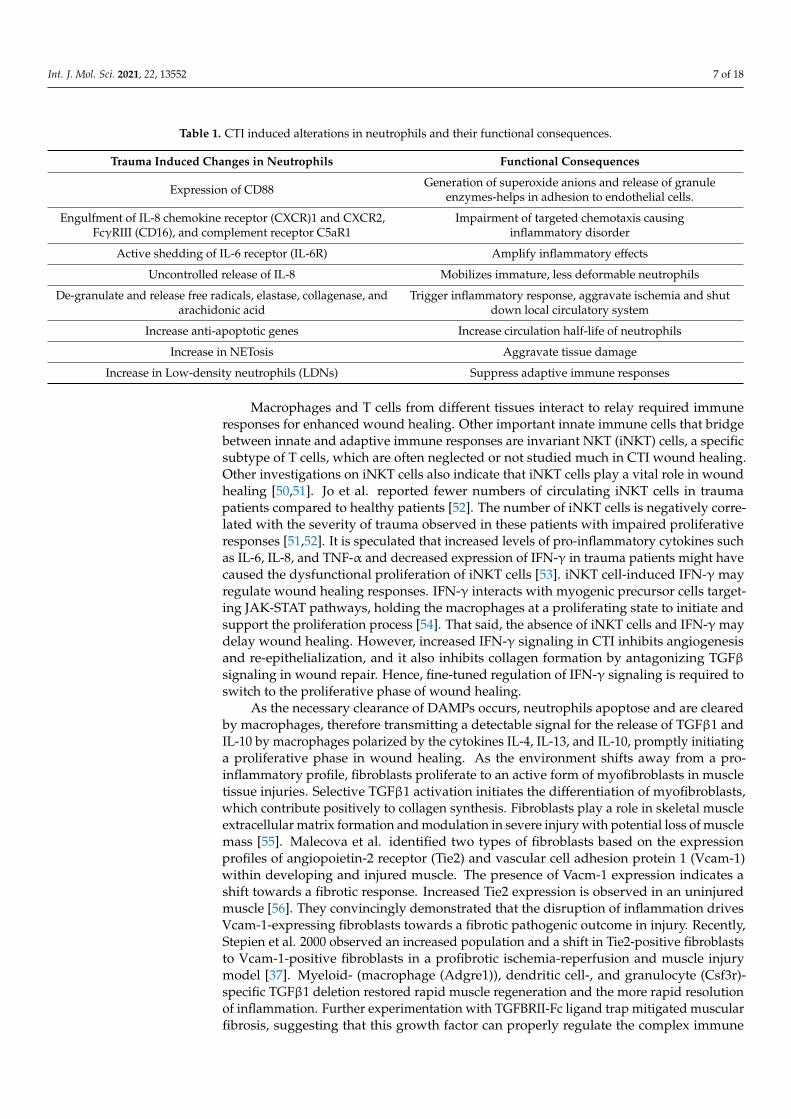
Table 1. CTI induced alterations in neutrophils and their functional consequences.
Trauma Induced Changes in Neutrophils Functional Consequences
Expression of CD88 Generation of superoxide anions and release of granuleenzymes-helps in adhesion to endothelial cells.
Engulfment of IL-8 chemokine receptor (CXCR)1 and CXCR2,FcγRIII (CD16), and complement receptor C5aR1
Impairment of targeted chemotaxis causinginflammatory disorder
Active shedding of IL-6 receptor (IL-6R) Amplify inflammatory effects
Uncontrolled release of IL-8 Mobilizes immature, less deformable neutrophils
De-granulate and release free radicals, elastase, collagenase, andarachidonic acid
Trigger inflammatory response, aggravate ischemia and shutdown local circulatory system
Increase anti-apoptotic genes Increase circulation half-life of neutrophils
Increase in NETosis Aggravate tissue damage
Increase in Low-density neutrophils (LDNs) Suppress adaptive immune responses
Macrophages and T cells from different tissues interact to relay required immuneresponses for enhanced wound healing. Other important innate immune cells that bridgebetween innate and adaptive immune responses are invariant NKT (iNKT) cells, a specificsubtype of T cells, which are often neglected or not studied much in CTI wound healing.Other investigations on iNKT cells also indicate that iNKT cells play a vital role in woundhealing [50,51]. Jo et al. reported fewer numbers of circulating iNKT cells in traumapatients compared to healthy patients [52]. The number of iNKT cells is negatively corre-lated with the severity of trauma observed in these patients with impaired proliferativeresponses [51,52]. It is speculated that increased levels of pro-inflammatory cytokines suchas IL-6, IL-8, and TNF-α and decreased expression of IFN-γ in trauma patients might havecaused the dysfunctional proliferation of iNKT cells [53]. iNKT cell-induced IFN-γ mayregulate wound healing responses. IFN-γ interacts with myogenic precursor cells target-ing JAK-STAT pathways, holding the macrophages at a proliferating state to initiate andsupport the proliferation process [54]. That said, the absence of iNKT cells and IFN-γ maydelay wound healing. However, increased IFN-γ signaling in CTI inhibits angiogenesisand re-epithelialization, and it also inhibits collagen formation by antagonizing TGFβsignaling in wound repair. Hence, fine-tuned regulation of IFN-γ signaling is required toswitch to the proliferative phase of wound healing.
As the necessary clearance of DAMPs occurs, neutrophils apoptose and are clearedby macrophages, therefore transmitting a detectable signal for the release of TGFβ1 andIL-10 by macrophages polarized by the cytokines IL-4, IL-13, and IL-10, promptly initiatinga proliferative phase in wound healing. As the environment shifts away from a pro-inflammatory profile, fibroblasts proliferate to an active form of myofibroblasts in muscletissue injuries. Selective TGFβ1 activation initiates the differentiation of myofibroblasts,which contribute positively to collagen synthesis. Fibroblasts play a role in skeletal muscleextracellular matrix formation and modulation in severe injury with potential loss of musclemass [55]. Malecova et al. identified two types of fibroblasts based on the expressionprofiles of angiopoietin-2 receptor (Tie2) and vascular cell adhesion protein 1 (Vcam-1)within developing and injured muscle. The presence of Vacm-1 expression indicates ashift towards a fibrotic response. Increased Tie2 expression is observed in an uninjuredmuscle [56]. They convincingly demonstrated that the disruption of inflammation drivesVcam-1-expressing fibroblasts towards a fibrotic pathogenic outcome in injury. Recently,Stepien et al. 2000 observed an increased population and a shift in Tie2-positive fibroblaststo Vcam-1-positive fibroblasts in a profibrotic ischemia-reperfusion and muscle injurymodel [37]. Myeloid- (macrophage (Adgre1)), dendritic cell-, and granulocyte (Csf3r)-specific TGFβ1 deletion restored rapid muscle regeneration and the more rapid resolutionof inflammation. Further experimentation with TGFBRII-Fc ligand trap mitigated muscularfibrosis, suggesting that this growth factor can properly regulate the complex immune

Int. J. Mol. Sci. 2021, 22, 13552 8 of 18
response and can modulate fibroblast function towards fibrosis or regeneration duringmuscle repair.
The resolution of inflammation typically favors the production of collagen and thecontraction of the lesion. Andermahr et al. made an insightful observation that in poly-trauma patients—both with and without concurrent traumatic brain injury—an increasein collagen degradation resulted in decreased osteogenesis and non-union fractures [57].They reported that pyridinoline cross-linked carboxy-terminal telopeptide (1CTP) wassignificantly higher in polytrauma patients. ICTP is secreted in response to collagen break-down, collagen synthesis and breakdown by-products, and is detected in the blood duringbone synthesis. Data indicate that in polytrauma, a lengthy pro-inflammatory conditionprevails, disrupting the number and activity of fibroblasts, resulting in decreased collagensynthesis, thus resulting in a delayed union. This study is referenced here because of itssignificant findings in polytrauma with bone fractures which are often associated withCTI. Additionally, the role of fibroblasts is critical, since its deficit will slow down woundclosure, or its uncontrolled activity can lead to excessive collagen deposition that may leadto fibrotic healing with impaired regeneration. This suggests that fibroblast proliferationand activity may decrease due to prolonged inflammation associated with CTI. Addi-tionally, fibroblasts depend on anti-inflammatory macrophages, traditionally associatedwith non-chronic wounds. Both pro-and anti-inflammatory cytokine signaling interactspositively with myogenic and osteogenic regulatory pathways, in which an appropriatetemporal conversion from type-1 (M1/Th1) to type-2 (M2/Th2) wound healing phenotypesis required for optimal regeneration. Along with anti-inflammatory macrophages, thereis an independent subset of monocytes/macrophages, reported in CTI patients whosefunctions in wound healing are yet to be adequately investigated. West et al. (2012) showedan upregulation of TGF-β and macrophage colony stimulating factor (M-CSF) after a pro-inflammatory phase in CTI patients that helped in the differentiation of CD14hiCD16+,monocytes/macrophages, activated through C-reactive protein [58]. They also reportedthat the presence of the CD163 receptor on these cells indicated an alternative activationmechanism and that CD163 induces anti-inflammatory cytokines such as IL-10 in these cells.Induction of this specific type of myeloid population may enhance the anti-inflammatoryenvironment, driving the wound healing regenerative phase. In a CTI, the integrity ofsurrounding tissue influences the rate of fracture healing. Severe traumas will lead to frac-tures along with extensive muscle tissue damage, delaying fracture healing. The profoundincrease in inflammation within an injured muscle can directly influence the immune cellinfiltration and selective activation within the adjacent fracture defect. At the fracture site,bone-specific osteal macrophages also play a role in fracture healing, as they are localizedclose/next to mature osteoblasts and adequately provide support in bone healing, [59–61].In vitro and in vivo removal of osteal macrophages from osteoblasts resulted in reducedosteoblast mineralization, supporting its role in bone formation [60,61]. Furthermore,osteoclasts originating from monocyte/macrophage precursors show increased activityduring bony callus formation, and osteoclast bone resorption of the callus is followed byosteoblast-mediated bone formation [62]. TGF-β also helps in osteoblast differentiationand proliferation. Increased expression of TGF-β by monocytes observed in immuno-suppressed patients indicate the presence of high levels of monocyte-derived TGF-β inblunt trauma patients [58]. Higher levels of TGF-β lead to an increased production ofprostaglandin-E2 that is known to suppress adaptive T cell functions [63]. Therefore, thissufficiently indicates that elevated TGF-β levels by monocytes may impair adaptive Tcell functions, which are needed for the successful proliferation and repair phases. It isapparent that innate immune responses naturally drive adaptive immune responses forthe long-lasting effects.
3.2. Disruptions of the Adaptive Immune System in Composite Tissue Injuries
T-cell populations are diverse and secrete various cytokines and proteins to carefullyregulate the proliferation and repair processes in a wound. Tregs, a specialized subpopula-

Int. J. Mol. Sci. 2021, 22, 13552 9 of 18
tion of T cells which typically contribute towards balancing of Th-1 type pro-inflammationand Th-2 type anti-inflammation during the process of wound healing, are mostly prevalentin high numbers during the proliferative phase. Several published reports suggest that thepresence of Tregs could confer faster wound healing [64] and may provide a survival advan-tage in CTI patients. Their increased number and activity were detected immediately aftertrauma, however, which resulted in an undesirable suppression of pro-inflammatory Th-1type cytokine activities and lead to immune suppression and delayed wound healing [65].Tregs attenuate the pro-inflammatory responses of monocytes [65] and regulate monocytesthrough the secretion of cytokines IL-10, IL-13, and IL-4. A marked increase in IL-10 con-centrations was observed in trauma patients, indicating its role in Th1/Th2 shift mediatedby Tregs [66]. This early increase in the expression of IL-10 with CTI could undoubtedlyaffect the healing process by suppressing vital pro-inflammatory responses. Moreover,Treg-derived IL-10 has a positive role in chondrocyte proliferation and differentiationin bone fractures. These roles are highlighted by observations that the loss of IL-10 inmice resulted in smaller proliferating zones in bones [67]. Mouse bone marrow-derivedMSCs possess immunosuppressive properties. They are capable of shifting from a pro-to anti-inflammatory phenotype, resulting in decreased production of pro-inflammatorycytokines, mediated by TNF-α stimulated gene/protein 6 (TSG-6) and prostaglandinE-2 [68–70]. Treg-derived IL-10 inhibits T cells and specifically suppresses Th-1-type cellsand their pro-inflammatory cytokines, such as IFN-γ. Tregs also play an important rolein controlling/extending pro-inflammatory conditions. There is a possibility that in thepresence of TGF-β and IL-6, these cells are susceptible to being efficiently converted intothe pro-inflammatory Th-17 cells [71]. Therefore, this naturally raises the need to properlyunderstand the altered cytokine profiles which may exist in CTI that lead to the activationof Tregs during initial phases/immediately after trauma. It is known that Tregs derivedfrom naïve CD4+ T cells positive for Foxp3+ and CTLA4+ suppressed the proliferation ofNKT and CD4 and CD8 T cells through contact inhibition facilitated by CTLA4 and CD36receptors [72]. Tregs mitigate this function through immature dendritic cells (DC), whichexpress low levels of co-stimulatory molecules. Tregs rigorously suppress the maturationof DCs, thus inducing anergy in T cells [73]. Tregs also regulate the macrophage phenotypeand functions [74]. Tregs suppress neutrophils and conventional T cell functions to initiatethe transition of the inflammatory phase to the regenerative and repair phase. Loss of Tregfunctions in a CTI might impair this transition. Co-culture of Tregs with muscle satellitecells enhanced the expansion of satellite cells, indicating cell to cell interaction [75]. Thissuggests that Tregs not only regulate other immune cells through the secretion of cytokinesbut also through cell–cell interactions. Furthermore, the absence of Tregs diminishedmyogenic activity in the acute injury of skeletal muscle [23]. These reports sufficientlyemphasize the role of Tregs in the proliferative/regenerative phase of wound repair. Inmuscle and adipose tissue, Tregs express amphiregulin (Areg), an epidermal growth factor(EGF)-like growth factor, involved in controlling muscle-homeostasis, function and repair.The administration of Areg decreased the expression of genes encoding proteins relatedto fibrosis (e.g., a battery of collagens, Adam12, Acta2), and enhanced the expression ofgene encoding molecules, such as Pfkfb1 and 3 and Myl2, highly represented in the healthymuscle [23]. Additionally, Areg promoted increased myogenic differentiation of satellitecells, and these cells expressed high levels of transcripts and protein of myosin heavy chain.Furthermore, emphasizing the significance of this pathway, accumulation, and functionsof injury-associated-Tregs that are stimulated by IL-33. Loss of IL-33 in stimulated Tregsresulted in impaired tissue repair [23]. Very little information exists on how amphiregulinregulates the Treg functions under CTI conditions presently. What is known about Tregs’altered functions in CTI is listed in Table 2. Research into the regulatory effects of thevarious growth factors on Tregs within the context of CTI represents an opportunity forfurther research, as knowledge is currently very limited at this stage.

Int. J. Mol. Sci. 2021, 22, 13552 10 of 18
Table 2. T regs display both pro and anti-inflammatory responses in CTI during initial phases/immediately after trauma.
T regs in CTI Functional Consequences
Increase in Tregs number and activity immediately after CTI Undesirable suppression of pro-inflammatory Th-1 typecytokines delaying wound healing
Early phase increased expression of IL-10 Suppression of pro-inflammatory responses (Th1 type andIFN-γ delaying wound healing
Presence of TGF-β and IL-6 convert Tregs to TH17 cells Increase unrequired inflammatory responses
T regs suppress DC maturation Inducing anergy in T cells
T regs loose regulatory function on neutrophils, andconventional T cell functions
Loss of transition of inflammatory phase to regenerative andrepair phase
Absence of Tregs Myogenic activity
Expression of amphiregulin by Tregs Controls muscle-homeostatis
Loss of IL-33 in stimulated Tregs Impaired tissue repair
3.3. Deleterious Effects of Immune-Inflammatory Dysregulation on Wound Healing
Wound healing involves immune cells, cytokines, growth factors, and progenitorcells, such as FAPs, which are mesenchymal in origin [76]. The physiologic response to aninjury triggers fibrogenic or adipogenic differentiation of FAPs in an immune-dependent(i.e., IL13 and IL4) fashion [76]. During the proliferation phase, IL-4/IL-13 signaling viasignal transducer and activator of transcription 6 suppresses the differentiation of FAPsinto adipocytes and generates tissue-specific cells supporting wound healing. Immedi-ately after injury, an acute repair response is triggered, which heals the damaged tissues,whereas in a CTI, a heightened inflammatory response leads to the death of cells and theinfiltration of immune cells. A transition from pro-inflammatory to anti-inflammatoryimmune cells drives the proliferation phase, which is a critical step for successful woundrepair. A prolonged inflammatory milieu of more than 3 days after injury in CTI disturbsthe well-orchestrated cellular choreography between inflammatory, FAP, and progenitorcells, resulting in the fibrotic and adipogenic degeneration of injured tissue, with an endresult of an impaired function of the afflicted tissues.
4. Consequences of an Altered Wound Environment on Metabolic Processes andDownstream Effects on Inflammatory Signaling Pathways
In CTI, hypoxic conditions, edema, low nutrient levels, and contributing factors down-stream of cell metabolic pathways effectively stimulate immune and vascular endothelialcells to release various cytokines and growth factors that impair angiogenesis and regen-eration. If neovascularization is successful, it allows the possible restoration of nutrientdelivery and oxygen, and cells use oxidative metabolism for their longer-term functionsand contribute positively to restoring the wound. Otherwise, the cells undergo stressfulconditions, leading to the release of altered metabolic stressors and sensors. The mam-malian target of rapamycin (mTOR), general control nonderepressible 2 (GCN2) kinase,hypoxic conditions, and adenosine, the metabolite of ATP, are known metabolic regulatorsand sensors that cause anergy in immune cells [77]. These pathways regulate the inductionof anergy, ensure the maintenance of anergy in T cells, and sense/respond to the presenceof hypoxia, changing nutrient levels and modulating metabolic processes, including glu-cose, lipid, energy, and metabolism. These metabolic pathways play a role in switchingcatabolic and anabolic pathways in T cells, regulating their activation, proliferation, anddifferentiation. T-cell receptor (TCR) and IL-2 promote the mTORC1 complex of mTORand suppress Tregs, enhancing effector T-cell molecules such as CTLA-4, which can causeanergy in T cells, as discussed above. Under inflammatory conditions, over activation ofmTORC1 leads to a loss of Tregs stability and the conversion of Tregs to effector T cells.Effector T cells producing cytokines such as IL-17 and IL-1β create a pro-inflammatory en-vironment [78]. An increased mTORC2 complex of mTOR activation due to the deficiency
Int. J. Mol. Sci. 2021, 22, 13552 11 of 18
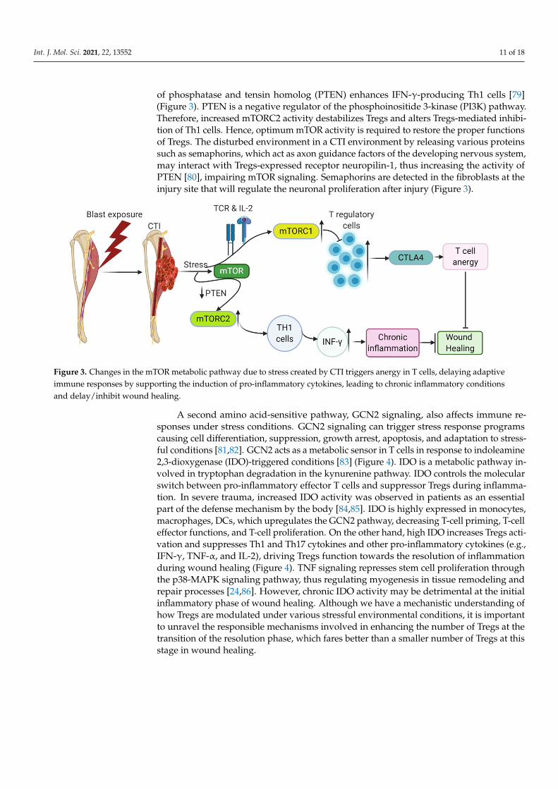
of phosphatase and tensin homolog (PTEN) enhances IFN-γ-producing Th1 cells [79](Figure 3). PTEN is a negative regulator of the phosphoinositide 3-kinase (PI3K) pathway.Therefore, increased mTORC2 activity destabilizes Tregs and alters Tregs-mediated inhibi-tion of Th1 cells. Hence, optimum mTOR activity is required to restore the proper functionsof Tregs. The disturbed environment in a CTI environment by releasing various proteinssuch as semaphorins, which act as axon guidance factors of the developing nervous system,may interact with Tregs-expressed receptor neuropilin-1, thus increasing the activity ofPTEN [80], impairing mTOR signaling. Semaphorins are detected in the fibroblasts at theinjury site that will regulate the neuronal proliferation after injury (Figure 3).
Int. J. Mol. Sci. 2021, 22, x FOR PEER REVIEW 11 of 18
Figure 3. Changes in the mTOR metabolic pathway due to stress created by CTI triggers anergy in T cells, delaying adap-tive immune responses by supporting the induction of pro-inflammatory cytokines, leading to chronic inflammatory con-ditions and delay/inhibit wound healing.
A second amino acid-sensitive pathway, GCN2 signaling, also affects immune re-sponses under stress conditions. GCN2 signaling can trigger stress response programs causing cell differentiation, suppression, growth arrest, apoptosis, and adaptation to stressful conditions [81,82]. GCN2 acts as a metabolic sensor in T cells in response to in-doleamine 2,3-dioxygenase (IDO)-triggered conditions [83] (Figure 4). IDO is a metabolic pathway involved in tryptophan degradation in the kynurenine pathway. IDO controls the molecular switch between pro-inflammatory effector T cells and suppressor Tregs during inflammation. In severe trauma, increased IDO activity was observed in patients as an essential part of the defense mechanism by the body [84,85]. IDO is highly expressed in monocytes, macrophages, DCs, which upregulates the GCN2 pathway, decreasing T-cell priming, T-cell effector functions, and T-cell proliferation. On the other hand, high IDO increases Tregs activation and suppresses Th1 and Th17 cytokines and other pro-inflammatory cytokines (e.g., IFN-γ, TNF-α, and IL-2), driving Tregs function towards the resolution of inflammation during wound healing (Figure 4). TNF signaling represses stem cell proliferation through the p38-MAPK signaling pathway, thus regulating myo-genesis in tissue remodeling and repair processes [24,86]. However, chronic IDO activity may be detrimental at the initial inflammatory phase of wound healing. Although we have a mechanistic understanding of how Tregs are modulated under various stressful environmental conditions, it is important to unravel the responsible mechanisms involved in enhancing the number of Tregs at the transition of the resolution phase, which fares better than a smaller number of Tregs at this stage in wound healing.
Figure 3. Changes in the mTOR metabolic pathway due to stress created by CTI triggers anergy in T cells, delaying adaptiveimmune responses by supporting the induction of pro-inflammatory cytokines, leading to chronic inflammatory conditionsand delay/inhibit wound healing.
A second amino acid-sensitive pathway, GCN2 signaling, also affects immune re-sponses under stress conditions. GCN2 signaling can trigger stress response programscausing cell differentiation, suppression, growth arrest, apoptosis, and adaptation to stress-ful conditions [81,82]. GCN2 acts as a metabolic sensor in T cells in response to indoleamine2,3-dioxygenase (IDO)-triggered conditions [83] (Figure 4). IDO is a metabolic pathway in-volved in tryptophan degradation in the kynurenine pathway. IDO controls the molecularswitch between pro-inflammatory effector T cells and suppressor Tregs during inflamma-tion. In severe trauma, increased IDO activity was observed in patients as an essentialpart of the defense mechanism by the body [84,85]. IDO is highly expressed in monocytes,macrophages, DCs, which upregulates the GCN2 pathway, decreasing T-cell priming, T-celleffector functions, and T-cell proliferation. On the other hand, high IDO increases Tregs acti-vation and suppresses Th1 and Th17 cytokines and other pro-inflammatory cytokines (e.g.,IFN-γ, TNF-α, and IL-2), driving Tregs function towards the resolution of inflammationduring wound healing (Figure 4). TNF signaling represses stem cell proliferation throughthe p38-MAPK signaling pathway, thus regulating myogenesis in tissue remodeling andrepair processes [24,86]. However, chronic IDO activity may be detrimental at the initialinflammatory phase of wound healing. Although we have a mechanistic understanding ofhow Tregs are modulated under various stressful environmental conditions, it is importantto unravel the responsible mechanisms involved in enhancing the number of Tregs at thetransition of the resolution phase, which fares better than a smaller number of Tregs at thisstage in wound healing.

Int. J. Mol. Sci. 2021, 22, 13552 12 of 18
Int. J. Mol. Sci. 2021, 22, x FOR PEER REVIEW 11 of 18
Figure 3. Changes in the mTOR metabolic pathway due to stress created by CTI triggers anergy in T cells, delaying adap-tive immune responses by supporting the induction of pro-inflammatory cytokines, leading to chronic inflammatory con-ditions and delay/inhibit wound healing.
A second amino acid-sensitive pathway, GCN2 signaling, also affects immune re-sponses under stress conditions. GCN2 signaling can trigger stress response programs causing cell differentiation, suppression, growth arrest, apoptosis, and adaptation to stressful conditions [81,82]. GCN2 acts as a metabolic sensor in T cells in response to in-doleamine 2,3-dioxygenase (IDO)-triggered conditions [83] (Figure 4). IDO is a metabolic pathway involved in tryptophan degradation in the kynurenine pathway. IDO controls the molecular switch between pro-inflammatory effector T cells and suppressor Tregs during inflammation. In severe trauma, increased IDO activity was observed in patients as an essential part of the defense mechanism by the body [84,85]. IDO is highly expressed in monocytes, macrophages, DCs, which upregulates the GCN2 pathway, decreasing T-cell priming, T-cell effector functions, and T-cell proliferation. On the other hand, high IDO increases Tregs activation and suppresses Th1 and Th17 cytokines and other pro-inflammatory cytokines (e.g., IFN-γ, TNF-α, and IL-2), driving Tregs function towards the resolution of inflammation during wound healing (Figure 4). TNF signaling represses stem cell proliferation through the p38-MAPK signaling pathway, thus regulating myo-genesis in tissue remodeling and repair processes [24,86]. However, chronic IDO activity may be detrimental at the initial inflammatory phase of wound healing. Although we have a mechanistic understanding of how Tregs are modulated under various stressful environmental conditions, it is important to unravel the responsible mechanisms involved in enhancing the number of Tregs at the transition of the resolution phase, which fares better than a smaller number of Tregs at this stage in wound healing.
Figure 4. Stress-induced amino acid-sensitive pathway, GCN2 signaling is activated in CTI conditions at the very earlystages, thus inducing Treg functions not necessary at that stage of wound healing, thus inhibiting/disrupting the naturalcourse of the acute pro-inflammatory phase. Increased expression of Tregs at early stages delays wound healing throughthe GCN2 pathway.
Tregs also have shown a complementary immunological arm under unfavorable con-ditions such as hypoxia. Hypoxia is generally observed in CTI patients due to low oxygenconditions and an inflamed microenvironment. Nanobashvili et al. (2003) reported that51% of the analyzed patient population showed distal ischemia/low oxygen levels dueto mangled injuries associated with damaged vessels [87]. Nitecki et al. reported limbischemia in 75% of CTI patients, who mostly had extremity vascular injuries [88]. Thesedata suggest the apparent prevalence of hypoxia/ischemia in CTI. Post-trauma edemafurther reduces the oxygen supply to the damaged tissues, thus impairing the neoangiogen-esis and required resupply of nutrients for regeneration and wound healing. The hypoxicmicroenvironment formed due to a lack of oxygen and nutrients drives the cells to pro-duce adenosine from extracellular ATP. The immunosuppressive extracellular metaboliteadenosine typically plays an immune regulatory role in Tregs. Ectonucleotidases CD39 andCD73 degrade ATP to produce adenosine. CD39 and CD73 are immune checkpoint media-tors expressed on Tregs [89]. A2aR and A2bR adenosine receptors stimulate intracellularadenylyl cyclase to synthesize cAMP. A2aR and A2bR adenosine receptors are present on Tcells. Increased levels of cAMP lead to immunosuppressive effects, such as a decrease inpro-inflammatory cytokine IFN-γ and an increase in the production of anti-inflammatorycytokines such as IL-10 and TGF-β. Adenosine produced by Tregs interacts with otherimmune-infiltrating cells through A2aR and A2bR adenosine receptors, thus activatingor inactivating the other immune cells [90]. Prolonged hypoxic conditions at the earlystages of CTI may trigger Tregs responses at very early phases, disturbing the requiredpro-inflammatory cytokine profile and thus hampering the wound healing process. Adeeper understanding of this phenomenon in CTI can provide us with possible clues formodifying the environmental conditions and immune responses for a faster-desired woundhealing process.
5. A Multi-Pronged Immunomodulatory Strategy as a Potential Opportunity toFacilitate Improved Outcomes Following Composite Tissue Injuries
In the above sections, a detailed description of how the immune-inflammatory re-sponse interacts throughout both the normative physiologic and CTI-related pathophysi-ologic wound healing processes was provided as a means to provide a foundation uponwhich next-generation, rationally informed, multi-pronged immunomodulatory strategiescan be developed as a means to facilitate improvements in outcomes following CTI. Further-more, the spatiotemporal application of these therapies should be conducted in coordination

Int. J. Mol. Sci. 2021, 22, 13552 13 of 18
with the known pathophysiology of CTI injury. Thus, a multi-pronged immunomodulatorystrategy is an attractive option to complement existing CTI treatment strategies.
We previously discussed in detail how uncoordinated interactions between variousimmune cells at the injury site contribute to a highly unsynchronized microenvironmentresulting in impaired wound healing. An example of how immune cells can be modulatedto coordinate successful wound healing is provided by macrophages, in which staggeredbolus release of IFN-γ and subsequent sustained release of IL-4 promote a shift from a pro-inflammatory to anti-inflammatory phenotype to mediate the scaffold vascularization [91].Pro-resolving mediators such as lipoxins and resolvins have shown their inhibitory ef-fects on neutrophils and macrophages. These mediators may have a benefit in CTI toreduce pro-inflammatory effects of neutrophils and macrophages diverting towards ananti-inflammatory response and regeneration of wound [92,93]. Implantation of chitosanscaffolds with lipoxins and resolvins in a mouse model polarized macrophages towards ananti-inflammatory phenotype and subsequently reduced fibrosis [92,93]. TGF-β is anotherpotent molecule that has both anti- and pro-inflammatory functions, and caution is advisedin wound repair if using this molecule. TGF-β has been reported to induce Tregs, whichdrive the regeneration process during wound healing [94]. A positive action of TGF-β3in decreasing post-operative scar formation in a clinical trial is encouraging, and also itsuse in the CTI model [95,96]. The action and interplay between pro-inflammatory andanti-inflammatory for positive wound regeneration and specifically the effects of timingstill need work.
Further revelation of the regulatory components involved in the development of in-flammatory reaction to CTI holds great potential in the development of effective treatmentstrategies to maximize immune elements critical for repair, while at the same time suppress-ing elements of the immune response responsible for further damage to injury progression.
6. Conclusions
Composite tissue injuries remain a major challenge to the US Military and pose thethreat of lifelong disability for those affected. Achieving improved clinical outcomes ofthese injuries will require restoration of normative inflammation and immune cell interac-tions at the site of injury and redressment of the hallmark inflammatory abnormalities ofCTI that disrupt normal wound healing mechanisms. Specifically, a prolonged or height-ened inflammatory phase, dysregulation or activation of pro-inflammatory immunologicalcells/factors, unwanted anti-inflammatory microenvironment at the very early phase ofwound healing, and undesired alteration in the cytokine profile at a very initial phasemust be addressed to achieve such clinical goals. Additionally, other stress factors suchas hypoxic conditions, ischemia, low nutrient levels and altered metabolic processes thatinfluence the immune functions must be managed concurrently. As such, CTI treatmentstrategies should aim to precisely address the cytokine network dysregulation so as tomodulate the secretory profile of various damaged tissues. Ostensibly, this can be achievedby modulating the cytokines themselves (e.g., exogenous supplementation) or by preciselycontrolling the complex spatiotemporal dynamics of different immune cell populations,of which neutrophils and Tregs seem to be promising targets. Practically, however, thisis a foreboding task, as very little is known of how the initial inflammation following aninjury interacts with local regenerative processes in the context of CTI, and much researchis needed to improve our understanding to the point of identifying precise targets for ther-apeutics which will not have deleterious effects elsewhere in the wound healing process.Specifically, ambiguity still exists as to which phases of immune-inflammatory response areneeded to promote successful wound healing outcomes, and the optimal timing for thesephases to resolve. These factors are critical, as both insufficient inflammation and chronicinflammation may lead to impaired wound healing and/or scar formation. Therefore, moredetailed experimental evidence is needed to help us accurately decide how we could care-fully manipulate inflammatory responses towards wound healing and to make informed

Int. J. Mol. Sci. 2021, 22, 13552 14 of 18
decisions on the dosing (e.g., magnitude, frequency, duration) of immunomodulatoryagents following composite musculoskeletal trauma.
Author Contributions: N.B.J.—Conceptualization, original draft preparation, writing and figures;M.S.V.—review and editing; S.M.G.—conceptualization, review and editing; C.L.D.—conceptualization,review and editing. All authors have read and agreed to the published version of the manuscript.
Funding: This work was supported by the DoD-VA Extremity Trauma and Amputation Center ofExcellence (Public Law 110–417, National Defense Authorization Act 2009, Section 723).
Acknowledgments: The contents of this publication are the sole responsibility of the author(s) and donot necessarily reflect the views, opinions or policies of Uniformed Services University of the HealthSciences (USUHS), the Department of Defense (DoD), the Departments of the Army, Navy, or AirForce. Mentions of trade names, commercial products, or organizations do not imply endorsementby the U.S. Government.
Conflicts of Interest: The authors declare that they have no competing interests.
Abbreviations
CTI Composite tissue injuries(DAMPs) Damage-associated molecular patternsTNF-α Tumor necrosis factor-alphaCCL2 Monocyte chemoattractant protein-1, MCP-1IL InterleukinCCL3 Chemokine ligand 3CD Cluster of differentiationIFN InterferonTGF Transforming growth factorTregs Regulatory T cellsAMPK 5′ Adenosine monophosphate-activated protein kinaseIGF-1 Insulin-like growth factor 1PDGF Platelet-derived growth factorVEGF-α Vascular endothelial growth factor αPDL1 Programmed cell death ligands 1FAP cells Fibro-adipogenic progenitor cellsCXCR Chemokine receptorBcl-2 B-cell lymphoma 2MCL1 Induced myeloid leukemia cell differentiation proteinLTB4 Leukotriene 4NETs Neutrophil extracellular trapsROS Reactive oxygen speciesMIP Macrophage inflammatory protein-1αLDNs Low-density neutrophilsiNKT Invariant NKTTie2 Angiopoietin-2 receptor1CTP Pyridinoline cross-linked carboxy-terminal telopeptideMCSF-1 Macrophage colony-stimulating factorDC Dendritic cellsAreg AmphiregulinEGF Epidermal growth factormTOR Mammalian target of rapamycinGCN2 General control nonderepressible 2TCR T-cell receptorPTEN Phosphatase and tensin homologPI3K Phosphoinositide 3-kinaseIDO Indoleamine 2,3-dioxygenase

Int. J. Mol. Sci. 2021, 22, 13552 15 of 18
References1. Eskridge, S.L.; Macera, C.A.; Galarneau, M.R.; Holbrook, T.L.; Woodruff, S.I.; MacGregor, A.J.; Morton, D.J.; Shaffer, R.A. Injuries
from combat explosions in Iraq: Injury type, location, and severity. Injury 2012, 43, 1678–1682. [CrossRef] [PubMed]2. Corona, B.T.; Rivera, J.C.; Owens, J.G.; Wenke, J.C.; Rathbone, C.R. Volumetric muscle loss leads to permanent disability following
extremity trauma. J. Rehabil. Res. Dev. 2015, 52, 785–792. [CrossRef] [PubMed]3. Sunderland, S. A classification of peripheral nerve injuries producing loss of function. Brain 1951, 74, 491–516. [CrossRef] [PubMed]4. Davis, K.M.; Griffin, K.S.; Chu, T.G.; Wenke, J.C.; Corona, B.T.; McKinley, T.O.; Kacena, M.A. Muscle-bone interactions during
fracture healing. J. Musculoskelet. Neuronal Interact. 2015, 15, 1–9.5. Holcomb, J.B. Major scientific lessons learned in the trauma field over the last two decades. PLoS Med. 2017, 14, e1002339. [CrossRef]6. von Rüden, C.; Woltmann, A.; Röse, M.; Wurm, S.; Rüger, M.; Hierholzer, C.; Bühren, V. Outcome after severe multiple trauma: A
retrospective analysis. J. Trauma Manag. Outcomes 2013, 7, 4. [CrossRef] [PubMed]7. Masini, B.D.; Waterman, S.M.; Wenke, J.C.; Owens, B.D.; Hsu, J.R.; Ficke, J.R. Resource utilization and disability outcome
assessment of combat casualties from Operation Iraqi Freedom and Operation Enduring Freedom. J. Orthop. Trauma 2009, 23,261–266. [CrossRef]
8. MacLeod, A.S.; Mansbridge, J.N. The Innate Immune System in Acute and Chronic Wounds. Adv. Wound Care 2015, 5,65–78. [CrossRef] [PubMed]
9. Neher, M.D.; Weckbach, S.; Flierl, M.A.; Huber-Lang, M.S.; Stahel, P.F. Molecular mechanisms of inflammation and tissue injuryafter major trauma—Is complement the “bad guy”? J. Biomed. Sci. 2011, 18, 90. [CrossRef] [PubMed]
10. Köhl, J. The role of complement in danger sensing and transmission. Immunol. Res. 2006, 34, 157–176. [CrossRef]11. Bianchi, M.E.; Manfredi, A.A. Dangers in and out. Science 2009, 323, 1683–1684. [CrossRef]12. Ley, K. Integration of inflammatory signals by rolling neutrophils. Immunol. Rev. 2002, 186, 8–18. [CrossRef]13. Soehnlein, O.; Lindbom, L. Phagocyte partnership during the onset and resolution of inflammation. Nat. Rev. Immunol. 2010, 10,
427–439. [CrossRef]14. Soehnlein, O.; Lindbom, L.; Weber, C. Mechanisms underlying neutrophil-mediated monocyte recruitment. Blood 2009, 114,
4613–4623. [CrossRef]15. Sadik, C.D.; Kim, N.D.; Luster, A.D. Neutrophils cascading their way to inflammation. Trends Immunol. 2011, 32, 452–460. [CrossRef]16. Claes, L.; Recknagel, S.; Ignatius, A. Fracture healing under healthy and inflammatory conditions. Nat. Rev. Rheumatol. 2012, 8,
133–143. [CrossRef]17. Arasapam, G.; Scherer, M.; Cool, J.C.; Foster, B.K.; Xian, C.J. Roles of COX-2 and iNOS in the bony repair of the injured growth
plate cartilage. J. Cell. Biochem. 2006, 99, 450–461. [CrossRef] [PubMed]18. Chen, L.; Mehta, N.D.; Zhao, Y.; DiPietro, L.A. Absence of CD4 or CD8 lymphocytes changes infiltration of inflammatory cells
and profiles of cytokine expression in skin wounds, but does not impair healing. Exp. Dermatol. 2014, 23, 189–194. [CrossRef]19. Zhang, J.; Xiao, Z.; Qu, C.; Cui, W.; Wang, X.; Du, J. CD8 T cells are involved in skeletal muscle regeneration through facilitating
MCP-1 secretion and Gr1(high) macrophage infiltration. J. Immunol. 2014, 193, 5149–5160. [CrossRef] [PubMed]20. Li, J.; Tan, J.; Martino, M.M.; Lui, K.O. Regulatory T-Cells: Potential Regulator of Tissue Repair and Regeneration. Front. Immunol.
2018, 9, 585. [CrossRef]21. Corthay, A. How do regulatory T cells work? Scand. J. Immunol. 2009, 70, 326–336. [CrossRef]22. Villalta, S.A.; Rosenthal, W.; Martinez, L.; Kaur, A.; Sparwasser, T.; Tidball, J.G.; Margeta, M.; Spencer, M.J.; Bluestone, J.A.
Regulatory T cells suppress muscle inflammation and injury in muscular dystrophy. Sci. Transl. Med. 2014, 6, 258ra142. [CrossRef]23. Burzyn, D.; Kuswanto, W.; Kolodin, D.; Shadrach, J.L.; Cerletti, M.; Jang, Y.; Sefik, E.; Tan, T.G.; Wagers, A.J.; Benoist, C.; et al. A
special population of regulatory T cells potentiates muscle repair. Cell 2013, 155, 1282–1295. [CrossRef]24. Sag, D.; Carling, D.; Stout, R.D.; Suttles, J. Adenosine 5′—Monophosphate—Activated protein kinase promotes macrophage
polarization to an anti-inflammatory functional phenotype. J. Immunol. 2008, 181, 8633–8641. [CrossRef] [PubMed]25. Wynn, T.A.; Vannella, K.M. Macrophages in Tissue Repair, Regeneration, and Fibrosis. Immunity 2016, 44, 450–462. [CrossRef] [PubMed]26. Zigmond, E.; Bernshtein, B.; Friedlander, G.; Walker, C.R.; Yona, S.; Kim, K.W.; Brenner, O.; Krauthgamer, R.; Varol, C.; Müller,
W.; et al. Macrophage-restricted interleukin-10 receptor deficiency, but not IL-10 deficiency, causes severe spontaneous colitis.Immunity 2014, 40, 720–733. [CrossRef]
27. Said, E.A.; Dupuy, F.P.; Trautmann, L.; Zhang, Y.; Shi, Y.; El-Far, M.; Hill, B.J.; Noto, A.; Ancuta, P.; Peretz, Y.; et al. Programmeddeath-1-induced interleukin-10 production by monocytes impairs CD4+ T cell activation during HIV infection. Nat. Med. 2010,16, 452–459. [CrossRef] [PubMed]
28. Marsell, R.; Einhorn, T.A. The biology of fracture healing. Injury 2011, 42, 551–555. [CrossRef]29. Gebhard, F.; Pfetsch, H.; Steinbach, G.; Strecker, W.; Kinzl, L.; Brückner, U.B. Is Interleukin 6 an Early Marker of Injury Severity
Following Major Trauma in Humans? Arch. Surg. 2000, 135, 291–295. [CrossRef] [PubMed]30. Jawa, R.S.; Anillo, S.; Huntoon, K.; Baumann, H.; Kulaylat, M. Interleukin-6 in surgery, trauma, and critical care part II: Clinical
implications. J. Intensive Care Med. 2011, 26, 73–87. [CrossRef]31. Gerstenfeld, L.C.; Cullinane, D.M.; Barnes, G.L.; Graves, D.T.; Einhorn, T.A. Fracture healing as a post-natal developmental
process: Molecular, spatial, and temporal aspects of its regulation. J. Cell. Biochem. 2003, 88, 873–884. [CrossRef] [PubMed]

Int. J. Mol. Sci. 2021, 22, 13552 16 of 18
32. Amara, U.; Kalbitz, M.; Perl, M.; Flierl, M.A.; Rittirsch, D.; Weiss, M.; Schneider, M.; Gebhard, F.; Huber-Lang, M. Early expressionchanges of complement regulatory proteins and C5A receptor (CD88) on leukocytes after multiple injury in humans. Shock 2010,33, 568–575. [CrossRef] [PubMed]
33. Unnewehr, H.; Rittirsch, D.; Sarma, J.V.; Zetoune, F.; Flierl, M.A.; Perl, M.; Denk, S.; Weiss, M.; Schneider, M.E.; Monk, P.N.;et al. Changes and regulation of the C5a receptor on neutrophils during septic shock in humans. J. Immunol. 2013, 190,4215–4225. [CrossRef] [PubMed]
34. Raghuwanshi, S.K.; Su, Y.; Singh, V.; Haynes, K.; Richmond, A.; Richardson, R.M. The chemokine receptors CXCR1 and CXCR2couple to distinct G protein-coupled receptor kinases to mediate and regulate leukocyte functions. J. Immunol. 2012, 189,2824–2832. [CrossRef] [PubMed]
35. Pillay, J.; Ramakers, B.P.; Kamp, V.M.; Loi, A.L.; Lam, S.W.; Hietbrink, F.; Leenen, L.P.; Tool, A.T.; Pickkers, P.; Koenderman, L.Functional heterogeneity and differential priming of circulating neutrophils in human experimental endotoxemia. J. Leukoc. Biol.2010, 88, 211–220. [CrossRef]
36. Bastian, O.W.; Kuijer, A.; Koenderman, L.; Stellato, R.K.; van Solinge, W.W.; Leenen, L.P.; Blokhuis, T.J. Impaired bone healing inmultitrauma patients is associated with altered leukocyte kinetics after major trauma. J. Inflamm. Res. 2016, 9, 69–78. [CrossRef] [PubMed]
37. Stepien, D.M.; Hwang, C.; Marini, S.; Pagani, C.A.; Sorkin, M.; Visser, N.D.; Huber, A.K.; Edwards, N.J.; Loder, S.J.; Vasquez, K.;et al. Tuning Macrophage Phenotype to Mitigate Skeletal Muscle Fibrosis. J. Immunol. 2020, 204, 2203–2215. [CrossRef] [PubMed]
38. van Eeden, S.F.; Terashima, T. Interleukin 8 (IL-8) and the release of leukocytes from the bone marrow. Leuk Lymphoma 2000, 37,259–271. [CrossRef] [PubMed]
39. Grøgaard, B.; Gerdin, B.; Reikerås, O. The polymorphonuclear leukocyte: Has it a role in fracture healing? Arch. Orthop. TraumaSurg. 1990, 109, 268–271. [CrossRef]
40. Dzhagalov, I.; St John, A.; He, Y.W. The antiapoptotic protein Mcl-1 is essential for the survival of neutrophils but not macrophages.Blood 2007, 109, 1620–1626. [CrossRef] [PubMed]
41. Paunel-Görgülü, A.; Kirichevska, T.; Lögters, T.; Windolf, J.; Flohé, S. Molecular mechanisms underlying delayed apoptosis inneutrophils from multiple trauma patients with and without sepsis. Mol. Med. 2012, 18, 325–335. [CrossRef] [PubMed]
42. Hurst, S.M.; Wilkinson, T.S.; McLoughlin, R.M.; Jones, S.; Horiuchi, S.; Yamamoto, N.; Rose-John, S.; Fuller, G.M.; Topley, N.;Jones, S.A. Il-6 and its soluble receptor orchestrate a temporal switch in the pattern of leukocyte recruitment seen during acuteinflammation. Immunity 2001, 14, 705–714. [CrossRef]
43. Xing, Z.; Lu, C.; Hu, D.; Yu, Y.Y.; Wang, X.; Colnot, C.; Nakamura, M.; Wu, Y.; Miclau, T.; Marcucio, R.S. Multiple roles for CCR2during fracture healing. Dis. Models Mech. 2010, 3, 451–458. [CrossRef] [PubMed]
44. Butterfield, T.A.; Best, T.M.; Merrick, M.A. The dual roles of neutrophils and macrophages in inflammation: A critical balancebetween tissue damage and repair. J. Athl. Train 2006, 41, 457–465. [PubMed]
45. Langereis, J.D.; Oudijk, E.J.; Schweizer, R.C.; Lammers, J.W.; Koenderman, L.; Ulfman, L.H. Steroids induce a disequilibriumof secreted interleukin-1 receptor antagonist and interleukin-1β synthesis by human neutrophils. Eur. Respir. J. 2011, 37,406–415. [CrossRef]
46. Teuben, M.; Heeres, M.; Blokhuis, T.; Hollman, A.; Vrisekoop, N.; Tan, E.; Pfeifer, R.; Pape, H.C.; Koenderman, L.; Leenen, L.P.H.Instant intra-operative neutropenia despite the emergence of banded (CD16(dim)/CD62L(bright)) neutrophils in peripheralblood—An observational study during extensive trauma-surgery in pigs. Injury 2021, 52, 426–433. [CrossRef] [PubMed]
47. Scapini, P.; Marini, O.; Tecchio, C.; Cassatella, M.A. Human neutrophils in the saga of cellular heterogeneity: Insights and openquestions. Immunol. Rev. 2016, 273, 48–60. [CrossRef]
48. Bryk, J.A.; Popovic, P.J.; Zenati, M.S.; Munera, V.; Pribis, J.P.; Ochoa, J.B. Nature of myeloid cells expressing arginase 1 inperipheral blood after trauma. J. Trauma 2010, 68, 843–852. [CrossRef] [PubMed]
49. Christoffersson, G.; Vågesjö, E.; Vandooren, J.; Lidén, M.; Massena, S.; Reinert, R.B.; Brissova, M.; Powers, A.C.; Opdenakker, G.;Phillipson, M. VEGF-A recruits a proangiogenic MMP-9-delivering neutrophil subset that induces angiogenesis in transplantedhypoxic tissue. Blood 2012, 120, 4653–4662. [CrossRef] [PubMed]
50. Tanno, H.; Kawakami, K.; Ritsu, M.; Kanno, E.; Suzuki, A.; Kamimatsuno, R.; Takagi, N.; Miyasaka, T.; Ishii, K.; Imai, Y.; et al.Contribution of Invariant Natural Killer T Cells to Skin Wound Healing. Am. J. Pathol. 2015, 185, 3248–3257. [CrossRef]
51. Jo, Y.G.; Choi, H.J.; Kim, J.C.; Cho, Y.N.; Kang, J.H.; Jin, H.M.; Kee, S.J.; Park, Y.W. Deficiencies of Circulating Mucosal-associatedInvariant T Cells and Natural Killer T Cells in Patients with Multiple Trauma. J. Korean Med. Sci. 2017, 32, 750–756. [CrossRef]
52. Jo, Y.G.; Kim, J.C.; Jin, H.M.; Cho, Y.N.; Kee, S.J.; Park, Y.W. Natural Killer T Cells Are Numerically and Functionally Deficient inPatients with Trauma. J. Innate Immun. 2020, 12, 344–354. [CrossRef] [PubMed]
53. Hildebrand, F.; Pape, H.C.; Krettek, C. The importance of cytokines in the posttraumatic inflammatory reaction. Der Unf. 2005,108, 793–794, 796–803. [CrossRef]
54. Morris, A.C.; Beresford, G.W.; Mooney, M.R.; Boss, J.M. Kinetics of a gamma interferon response: Expression and assembly ofCIITA promoter IV and inhibition by methylation. Mol. Cell. Biol. 2002, 22, 4781–4791. [CrossRef] [PubMed]
55. Mendias, C.L. Fibroblasts take the centre stage in human skeletal muscle regeneration. J. Physiol. 2017, 595, 5005. [CrossRef]56. Malecova, B.; Gatto, S.; Etxaniz, U.; Passafaro, M.; Cortez, A.; Nicoletti, C.; Giordani, L.; Torcinaro, A.; De Bardi, M.; Bicciato, S.;
et al. Dynamics of cellular states of fibro-adipogenic progenitors during myogenesis and muscular dystrophy. Nat. Commun.2018, 9, 3670. [CrossRef] [PubMed]

Int. J. Mol. Sci. 2021, 22, 13552 17 of 18
57. Andermahr, J.; Elsner, A.; Brings, A.E.; Hensler, T.; Gerbershagen, H.; Jubel, A. Reduced collagen degradation in polytraumaswith traumatic brain injury causes enhanced osteogenesis. J. Neurotrauma 2006, 23, 708–720. [CrossRef]
58. West, S.D.; Goldberg, D.; Ziegler, A.; Krencicki, M.; Du Clos, T.W.; Mold, C. Transforming growth factor-β, macrophage colony-stimulating factor and C-reactive protein levels correlate with CD14(high)CD16+ monocyte induction and activation in traumapatients. PLoS ONE 2012, 7, e52406. [CrossRef] [PubMed]
59. Pettit, A.R.; Chang, M.K.; Hume, D.A.; Raggatt, L.J. Osteal macrophages: A new twist on coupling during bone dynamics. Bone2008, 43, 976–982. [CrossRef] [PubMed]
60. Alexander, K.A.; Chang, M.K.; Maylin, E.R.; Kohler, T.; Müller, R.; Wu, A.C.; Van Rooijen, N.; Sweet, M.J.; Hume, D.A.; Raggatt,L.J.; et al. Osteal macrophages promote in vivo intramembranous bone healing in a mouse tibial injury model. J. Bone Miner. Res.2011, 26, 1517–1532. [CrossRef] [PubMed]
61. Raggatt, L.J.; Wullschleger, M.E.; Alexander, K.A.; Wu, A.C.; Millard, S.M.; Kaur, S.; Maugham, M.L.; Gregory, L.S.; Steck, R.;Pettit, A.R. Fracture healing via periosteal callus formation requires macrophages for both initiation and progression of earlyendochondral ossification. Am. J. Pathol. 2014, 184, 3192–3204. [CrossRef]
62. He, L.H.; Liu, M.; He, Y.; Xiao, E.; Zhao, L.; Zhang, T.; Yang, H.Q.; Zhang, Y. TRPV1 deletion impaired fracture healing andinhibited osteoclast and osteoblast differentiation. Sci. Rep. 2017, 7, 42385. [CrossRef]
63. Kalinski, P. Regulation of immune responses by prostaglandin E2. J. Immunol. 2012, 188, 21–28. [CrossRef]64. Zhang, C.; Li, L.; Feng, K.; Fan, D.; Xue, W.; Lu, J. ’Repair’ Treg Cells in Tissue Injury. Cell. Physiol. Biochem. 2017, 43, 2155–2169. [CrossRef]65. MacConmara, M.P.; Maung, A.A.; Fujimi, S.; McKenna, A.M.; Delisle, A.; Lapchak, P.H.; Rogers, S.; Lederer, J.A.; Mannick, J.A.
Increased CD4+ CD25+ T regulatory cell activity in trauma patients depresses protective Th1 immunity. Ann. Surg. 2006, 244,514–523. [CrossRef] [PubMed]
66. Menges, T.; Engel, J.; Welters, I.; Wagner, R.M.; Little, S.; Ruwoldt, R.; Wollbrueck, M.; Hempelmann, G. Changes in bloodlymphocyte populations after multiple trauma: Association with posttraumatic complications. Crit. Care Med. 1999, 27,733–740. [CrossRef] [PubMed]
67. Jung, Y.K.; Kim, G.W.; Park, H.R.; Lee, E.J.; Choi, J.Y.; Beier, F.; Han, S.W. Role of interleukin-10 in endochondral bone formationin mice: Anabolic effect via the bone morphogenetic protein/Smad pathway. Arthritis Rheum. 2013, 65, 3153–3164. [CrossRef]
68. Ren, G.; Zhang, L.; Zhao, X.; Xu, G.; Zhang, Y.; Roberts, A.I.; Zhao, R.C.; Shi, Y. Mesenchymal stem cell-mediated immunosup-pression occurs via concerted action of chemokines and nitric oxide. Cell Stem Cell 2008, 2, 141–150. [CrossRef] [PubMed]
69. Maggini, J.; Mirkin, G.; Bognanni, I.; Holmberg, J.; Piazzón, I.M.; Nepomnaschy, I.; Costa, H.; Cañones, C.; Raiden, S.; Vermeulen,M.; et al. Mouse bone marrow-derived mesenchymal stromal cells turn activated macrophages into a regulatory-like profile.PLoS ONE 2010, 5, e9252. [CrossRef]
70. Kovach, T.K.; Dighe, A.S.; Lobo, P.I.; Cui, Q. Interactions between MSCs and immune cells: Implications for bone healing. J.Immunol. Res. 2015, 2015, 752510. [CrossRef]
71. Kitani, A.; Xu, L. Regulatory T cells and the induction of IL-17. Mucosal Immunol. 2008, 1, S43–S46. [CrossRef]72. Ihara, F.; Sakurai, D.; Takami, M.; Kamata, T.; Kunii, N.; Yamasaki, K.; Iinuma, T.; Nakayama, T.; Motohashi, S.; Okamoto, Y.
Regulatory T cells induce CD4− NKT cell anergy and suppress NKT cell cytotoxic function. Cancer Immunol. Immunother. 2019,68, 1935–1947. [CrossRef]
73. Wing, K.; Sakaguchi, S. Regulatory T cells exert checks and balances on self tolerance and autoimmunity. Nat. Immunol. 2010, 11,7–13. [CrossRef]
74. Taams, L.S.; van Amelsfort, J.M.; Tiemessen, M.M.; Jacobs, K.M.; de Jong, E.C.; Akbar, A.N.; Bijlsma, J.W.; Lafeber, F.P. Modulationof monocyte/macrophage function by human CD4+CD25+ regulatory T cells. Hum. Immunol. 2005, 66, 222–230. [CrossRef]
75. Castiglioni, A.; Corna, G.; Rigamonti, E.; Basso, V.; Vezzoli, M.; Monno, A.; Almada, A.E.; Mondino, A.; Wagers, A.J.; Manfredi,A.A.; et al. FOXP3+ T Cells Recruited to Sites of Sterile Skeletal Muscle Injury Regulate the Fate of Satellite Cells and GuideEffective Tissue Regeneration. PLoS ONE 2015, 10, e0128094. [CrossRef]
76. Joe, A.W.; Yi, L.; Natarajan, A.; Le Grand, F.; So, L.; Wang, J.; Rudnicki, M.A.; Rossi, F.M. Muscle injury activates residentfibro/adipogenic progenitors that facilitate myogenesis. Nat. Cell Biol. 2010, 12, 153–163. [CrossRef]
77. Al-Khami, A.A.; Rodriguez, P.C.; Ochoa, A.C. Energy metabolic pathways control the fate and function of myeloid immune cells.J. Leukoc. Biol. 2017, 102, 369–380. [CrossRef]
78. Park, Y.; Jin, H.S.; Lopez, J.; Elly, C.; Kim, G.; Murai, M.; Kronenberg, M.; Liu, Y.C. TSC1 regulates the balance between effectorand regulatory T cells. J. Clin. Investig. 2013, 123, 5165–5178. [CrossRef]
79. Shrestha, S.; Yang, K.; Guy, C.; Vogel, P.; Neale, G.; Chi, H. Treg cells require the phosphatase PTEN to restrain TH1 and TFH cellresponses. Nat. Immunol. 2015, 16, 178–187. [CrossRef]
80. Delgoffe, G.M.; Woo, S.R.; Turnis, M.E.; Gravano, D.M.; Guy, C.; Overacre, A.E.; Bettini, M.L.; Vogel, P.; Finkelstein, D.; Bonnevier,J.; et al. Stability and function of regulatory T cells is maintained by a neuropilin-1-semaphorin-4a axis. Nature 2013, 501,252–256. [CrossRef] [PubMed]
81. Niwa, M.; Walter, P. Pausing to decide. Proc. Natl. Acad. Sci. USA 2000, 97, 12396–12397. [CrossRef]82. Rao, R.V.; Ellerby, H.M.; Bredesen, D.E. Coupling endoplasmic reticulum stress to the cell death program. Cell Death Differ. 2004,
11, 372–380. [CrossRef]83. Munn, D.H.; Sharma, M.D.; Baban, B.; Harding, H.P.; Zhang, Y.; Ron, D.; Mellor, A.L. GCN2 kinase in T cells mediates proliferative
arrest and anergy induction in response to indoleamine 2,3-dioxygenase. Immunity 2005, 22, 633–642. [CrossRef]

Int. J. Mol. Sci. 2021, 22, 13552 18 of 18
84. Ploder, M.; Spittler, A.; Schroecksnadel, K.; Neurauter, G.; Pelinka, L.E.; Roth, E.; Fuchs, D. Tryptophan degradation in multipletrauma patients: Survivors compared with non-survivors. Clin. Sci. 2009, 116, 593–598. [CrossRef]
85. Pellegrin, K.; Neurauter, G.; Wirleitner, B.; Fleming, A.W.; Peterson, V.M.; Fuchs, D. Enhanced enzymatic degradation oftryptophan by indoleamine 2,3-dioxygenase contributes to the tryptophan-deficient state seen after major trauma. Shock 2005, 23,209–215. [PubMed]
86. Palacios, D.; Mozzetta, C.; Consalvi, S.; Caretti, G.; Saccone, V.; Proserpio, V.; Marquez, V.E.; Valente, S.; Mai, A.; Forcales, S.V.;et al. TNF/p38α/polycomb signaling to Pax7 locus in satellite cells links inflammation to the epigenetic control of muscleregeneration. Cell Stem Cell 2010, 7, 455–469. [CrossRef]
87. Nanobashvili, J.; Kopadze, T.; Tvaladze, M.; Buachidze, T.; Nazvlishvili, G. War injuries of major extremity arteries. World J. Surg.2003, 27, 134–139. [CrossRef]
88. Nitecki, S.S.; Karram, T.; Ofer, A.; Engel, A.; Hoffman, A. Management of combat vascular injuries using modern imaging: Arewe getting better? Emerg. Med. Int. 2013, 2013, 689473. [CrossRef]
89. Allard, B.; Longhi, M.S.; Robson, S.C.; Stagg, J. The ectonucleotidases CD39 and CD73: Novel checkpoint inhibitor targets.Immunol. Rev. 2017, 276, 121–144. [CrossRef]
90. Ohta, A.; Sitkovsky, M. Extracellular adenosine-mediated modulation of regulatory T cells. Front. Immunol. 2014, 5, 304. [CrossRef]91. Spiller, K.L.; Nassiri, S.; Witherel, C.E.; Anfang, R.R.; Ng, J.; Nakazawa, K.R.; Yu, T.; Vunjak-Novakovic, G. Sequential delivery
of immunomodulatory cytokines to facilitate the M1-to-M2 transition of macrophages and enhance vascularization of bonescaffolds. Biomaterials 2015, 37, 194–207. [CrossRef]
92. Schwab, J.M.; Chiang, N.; Arita, M.; Serhan, C.N. Resolvin E1 and protectin D1 activate inflammation-resolution programmes.Nature 2007, 447, 869–874. [CrossRef] [PubMed]
93. Vasconcelos, D.P.; Costa, M.; Amaral, I.F.; Barbosa, M.A.; Águas, A.P.; Barbosa, J.N. Development of an immunomodulatorybiomaterial: Using resolvin D1 to modulate inflammation. Biomaterials 2015, 53, 566–573. [CrossRef] [PubMed]
94. Tran, D.Q. TGF-β: The sword, the wand, and the shield of FOXP3+ regulatory T cells. J. Mol. Cell Biol. 2011, 4, 29–37. [CrossRef]95. Occleston, N.L.; O’Kane, S.; Laverty, H.G.; Cooper, M.; Fairlamb, D.; Mason, T.; Bush, J.A.; Ferguson, M.W. Discovery and
development of avotermin (recombinant human transforming growth factor beta 3): A new class of prophylactic therapeutic forthe improvement of scarring. Wound Repair Regen 2011, 19 (Suppl. S1), s38–s48. [CrossRef]
96. McCollum, P.T.; Bush, J.A.; James, G.; Mason, T.; O’Kane, S.; McCollum, C.; Krievins, D.; Shiralkar, S.; Ferguson, M.W. Randomizedphase II clinical trial of avotermin versus placebo for scar improvement. Br. J. Surg. 2011, 98, 925–934. [CrossRef]