Flavonoid glycosides fromPrunus armeniaca and the antibacterial activity of a crude extract
Upload
khangminh22Category
view
4download
0
�����������������
Citation: Martiniakova, M.;
Babikova, M.; Mondockova, V.;
Blahova, J.; Kovacova, V.; Omelka, R.
The Role of Macronutrients,
Micronutrients and Flavonoid
Polyphenols in the Prevention and
Treatment of Osteoporosis. Nutrients
2022, 14, 523. https://doi.org/
10.3390/nu14030523
Academic Editor: Simone Perna
Received: 4 January 2022
Accepted: 23 January 2022
Published: 25 January 2022
Publisher’s Note: MDPI stays neutral
with regard to jurisdictional claims in
published maps and institutional affil-
iations.
Copyright: © 2022 by the authors.
Licensee MDPI, Basel, Switzerland.
This article is an open access article
distributed under the terms and
conditions of the Creative Commons
Attribution (CC BY) license (https://
creativecommons.org/licenses/by/
4.0/).
nutrients
Review
The Role of Macronutrients, Micronutrients and FlavonoidPolyphenols in the Prevention and Treatment of OsteoporosisMonika Martiniakova 1,† , Martina Babikova 2, Vladimira Mondockova 2 , Jana Blahova 2, Veronika Kovacova 1
and Radoslav Omelka 2,*,†
1 Department of Zoology and Anthropology, Faculty of Natural Sciences and Informatics,Constantine the Philosopher University in Nitra, Tr. A. Hlinku 1, 949 01 Nitra, Slovakia;[email protected] (M.M.); [email protected] (V.K.)
2 Department of Botany and Genetics, Faculty of Natural Sciences and Informatics,Constantine the Philosopher University in Nitra, Tr. A. Hlinku 1, 949 01 Nitra, Slovakia;[email protected] (M.B.); [email protected] (V.M.); [email protected] (J.B.)
* Correspondence: [email protected]; Tel.: +421-37-6408-737† These authors contributed equally to this work.
Abstract: Osteoporosis is considered an age-related disorder of the skeletal system, characterizedprimarily by decreased bone mineral density (BMD), microstructural quality and an elevated risk offragility fractures. This silent disease is increasingly becoming a global epidemic due to an aging pop-ulation and longer life expectancy. It is known that nutrition and physical activity play an importantrole in skeletal health, both in achieving the highest BMD and in maintaining bone health. In this re-view, the role of macronutrients (proteins, lipids, carbohydrates), micronutrients (minerals—calcium,phosphorus, magnesium, as well as vitamins—D, C, K) and flavonoid polyphenols (quercetin, rutin,luteolin, kaempferol, naringin) which appear to be essential for the prevention and treatment ofosteoporosis, are characterized. Moreover, the importance of various naturally available nutrients,whether in the diet or in food supplements, is emphasized. In addition to pharmacotherapy, thebasis of osteoporosis prevention is a healthy diet rich mainly in fruits, vegetables, seafood andfish oil supplements, specific dairy products, containing a sufficient amount of all aforementionednutritional substances along with regular physical activity. The effect of diet alone in this contextmay depend on an individual’s genotype, gene-diet interactions or the composition and function ofthe gut microbiota.
Keywords: osteoporosis; nutrition; macronutrients; micronutrients; flavonoid polyphenols; prevention;treatment
1. Introduction
Osteoporosis, a common age-related skeletal disorder, is represented by reducedbone mineral density (BMD), deterioration of bone microarchitecture with an increasedrisk of fragility fractures [1]. Such fractures, located predominantly in the wrist, hip andspine, are associated with considerable pain and suffering, disability and sometimes evendeath [2]. Osteoporosis, often referred to a silent epidemic, affects millions of peoplearound the world [3]. By 2050, more than 30 million people in Europe will be affected byosteoporosis [4] due to an aging population and longer life expectancy. Although the entirepopulation is at risk, the likelihood of developing osteoporosis increases depending on avariety of factors, including gender, age, family history or fracture, and Caucasian or Asianethnicity [5].
It has been estimated that 50–85% of the variation in BMD at middle-age is geneticallydetermined. However, the heritability of both bone loss and fractures decreases withincreasing age and lifestyle factors becoming more important [6]. It is difficult to determinehow much of a lifestyle effect is affected by diet alone, due to difficulties in quantifying
Nutrients 2022, 14, 523. https://doi.org/10.3390/nu14030523 https://www.mdpi.com/journal/nutrients

Nutrients 2022, 14, 523 2 of 30
environmental exposures, both current and lifelong. In addition, the impact of diet onbone health may be more complex and it may depend on the genotype of the individual,gene-diet interactions or the composition and function of the gut microbiota [7,8].
Age is one of the main risk factors for primary type 1 (postmenopausal) osteoporo-sis [9]. Primary type 2 (senile) osteoporosis occurs after the age of 75 years and is determinedin both women and men at a ratio 2:1 [10]. The mechanism underlying age-related changesin bone quantity and quality occurs at the cellular, molecular and genetic levels [11]. Bonemetabolism is altered due to differences in the number, activity and response of bone cellswhich leads to bone loss and bone fragility. In addition, bone fat marrow accumulation [12],changes in bone regulation, increased cell apoptosis, accumulation of cellular senescence,DNA damages and genomic instability, telomere dysfunction and epigenetic alterationssignificantly contribute to bone aging [13,14].
The mechanisms associated with bone loss are well known, including the role ofpro-inflammatory cytokines, e.g., tumor necrosis factor α (TNFα), interleukin-1 (IL-1),interleukin-6 (IL-6), in activating bone resorption by osteoclasts (bone-resorbing cells) andinhibiting bone formation by osteoblasts (bone-forming cells) [15]. Bone formation occursmainly on periosteal surfaces, whereas bone resorption is usually present at endosteal sur-faces. Osteoclast activation and differentiation is regulated by a variety of molecular signals,one of the most studied being the receptor activator for nuclear factor-kappa B (RANK)ligand (RANKL). RANKL is produced by osteocytes as well as osteoblasts and stimulatesRANK on osteoclast progenitors [16]. Furthermore, estrogen loss during menopause canbe consistent with an elevated expression of RANKL and reduced expression of osteopro-tegerin (OPG), its natural antagonist by osteoblasts and osteocytes, thereby raising boneresorption in both trabecular and cortical bone compartments [10,17]. The role of boneformation is equally important in the process of bone fragility and is controlled by sclerostin,which is expressed by osteocytes and acts as an inhibitor of the Wnt/β-catenin signalingpathway, a strong stimulus for osteoblast differentiation [15]. Schematic representationof osteoblast and osteoclast differentiation with the involvement of selected importantsignaling pathways and regulators is illustrated in Figure 1.
In addition to pharmacotherapy, nutritional supplements and physical exercise arecommonly used to prevent and treat osteoporosis [3,18]. Pharmacological treatment in-cludes anti-resorptive drugs (e.g., bisphosphonates, estrogen replacement therapy, selectiveestrogen receptor modulators, calcitonin, denosumab, calcium and vitamin D supple-mentation), and anabolic drugs (e.g., teriparatide, abaloparatide, strontium ranelate, ro-mosozumab) [19,20]. Nutrition plays a dominant role in skeletal health, both in achievingthe highest BMD and in maintaining bone health [21]. Therefore, a balanced diet andgood nutritional program can also prevent osteoporosis. The intake of macronutrients,vitamins and minerals is often below the recommended values, especially if the diseaseis active [22]. Calcium (Ca) and vitamin D are the most frequently discussed nutrientswith respect to BMD. In addition, vitamins (e.g., C, K), minerals (e.g., Ca, phosphorus(P), magnesium (Mg)), flavonoid polyphenols (e.g., quercetin, rutin, luteolin, kaempferol,naringin) are also involved in bone formation. Exercises to tone deambulatory muscles andexercises to improve proprioception can help prevent falls. Moreover, physical exercise canstop or reverse osteoporosis due to its anabolic effects [10]. This review characterizes therole of essential macronutrients, micronutrients and flavonoid polyphenols in relation toosteoporosis prevention and treatment. Moreover, the importance of different naturallyavailable nutrients, whether in the diet or in food supplements is highlighted.

Nutrients 2022, 14, 523 3 of 30
Figure 1. Schematic representation of osteoblast (left) and osteoclast (right) differentiation with theinvolvement of selected important signaling pathways and regulators. Positive effects are depicted bygreen arrowheads. The blue arrows and font show the production of selected molecules important inthe signaling pathway and also for the processes of bone formation and resorption. Osteoblastogenesisis regulated by different signaling pathways (including TGF-β/BMP, WNT), resulting in activation ofdownstream transcription factors such as Runx2 and leading to the expression of osteoblastogenicmarkers by osteoblasts, e.g., type I collagen, ALP, OPN, BSP and OCN. The canonical WNT pathwayactivates downstream signaling cascades, resulting in β-catenin translocation into the nucleus whichenhances osteoblastogenic target gene expression. TGF-β and BMPs are secreted growth factorsbelonging to the TGF-β superfamily, which play an essential role in development, tissue homeostasisand regeneration. Canonical TGF-β signaling mobilizes transcription factors Smad2 and Smad3 tointeract with other transcription factors and induces Runx2-mediated gene expression. Similar toTGF-β, canonical BMP signaling also transmits signals through Smad transcription factors. Moreover,BMPs and TGF-β can induce Runx2 through the MAPK signaling pathway. Osteocytes representa differentiated stage of the osteoblast lineage and they have key regulatory roles in bone andmineral homeostasis. Osteoclasts differentiate from cells of the monocyte/macrophage lineage inresponse to the osteoclastogenic cytokines M-CSF, RANKL and ITAM. As a result, mononuclearosteoclast precursors finally fuse into mature polykaryons which produce protons and proteolyticenzymes (especially cathepsin K) to dissolve bone minerals and degrade bone matrix proteins. MSC,mesenchymal stem cell; TGF-β, transforming growth factor beta; BMP, bone morphogenetic protein;
Nutrients 2022, 14, 523 4 of 30
MAPK, mitogen-activated protein kinase; Runx2, runt-related transcription factor 2; WNT, Wntglycoproteins; PTH, parathyroid hormone; OPN, osteopontin; BSP, bone sialoprotein; ALP, alkalinephosphatase; OCN, osteocalcin; HSC, hematopoietic stem cell; M-CSF, macrophage-colony stimu-lating factor; ITAM, immunoreceptor tyrosine-based activation motif; RANK, receptor activator ofnuclear factor κB; RANKL, receptor activator of nuclear factor κB ligand; CATK, cathepsin K.
2. Macronutrients and Osteoporosis
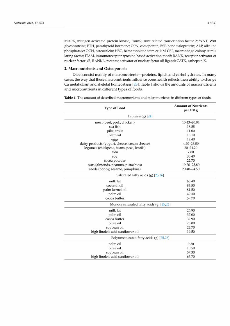
Diets consist mainly of macronutrients—proteins, lipids and carbohydrates. In manycases, the way that these macronutrients influence bone health reflects their ability to changeCa metabolism and skeletal homeostasis [23]. Table 1 shows the amounts of macronutrientsand micronutrients in different types of foods.
Table 1. The amount of described macronutrients and micronutrients in different types of foods.
Type of Food Amount of Nutrientsper 100 g
Proteins (g) [24]
meat (beef, pork, chicken) 15.43–20.04sea fish 18.88
pike, trout 11.00oatmeal 13.10
eggs 12.40dairy products (yogurt, cheese, cream cheese) 4.40–26.00
legumes (chickpeas, beans, peas, lentils) 20–24.20tofu 7.80soy 35.40
cocoa powder 22.70nuts (almonds, peanuts, pistachios) 19.70–25.80seeds (poppy, sesame, pumpkins) 20.40–24.50
Saturated fatty acids (g) [25,26]
milk fat 63.40coconut oil 86.50
palm kernel oil 81.50palm oil 49.30
cocoa butter 59.70
Monounsaturated fatty acids (g) [25,26]
milk fat 25.90palm oil 37.00
cocoa butter 32.90olive oil 73.00
soybean oil 22.70high linoleic acid sunflower oil 19.50
Polyunsaturated fatty acids (g) [25,26]
palm oil 9.30olive oil 10.50
soybean oil 57.30high linoleic acid sunflower oil 65.70

Nutrients 2022, 14, 523 5 of 30
Table 1. Cont.
Type of Food Amount of Nutrientsper 100 g
Carbohydrates (g) [24]
oatmeal 68.10wheat flour 73.10
wheat white bread 50.80legumes (chickpeas, beans, peas, lentils) 58.00–60.50
fruits (blackcurrant, grapes, bananas) 17.20–21.80vegetables (potatoes, sweet corn, garlic) 18.80–25.00
nuts (peanuts, pistachios, chestnuts) 18.20–53.00curry spice 61.80black tea 55.70
bitter chocolate 117.12
Calcium (mg) [25,26]
cow milk (natural) 119.14hard cheese 981.71–1218.00soft cheese 732.86
eggs 57.06marjoram 1388.00
poppy seeds (natural) 1513.71salmon (Atlantic) 20.00
almonds 229.71green-leaf vegetables (head cabbage, curly kale) 47.50–163.57
legume-based dishes 30.91tofu 162.43
seafood-based dishes 38.05mineral water rich in calcium (250 mL) 100.00
Phosphorus (mg) [25,26]
meat (chicken, duck, turkey, goat) 188.57–234.86cow milk 92.86
hard cheese 786.86soft cheese 345.57
cereals and cereal-like grains 271.00seeds (linseed, pumpkin, sesame, sunflower, poppy) 603.00–861.71
nuts (cashew, peanuts, walnuts) 519.57–369.71legume-based dishes 132.37
Magnesium (mg) [25,26]
spinach 61.99legume-based dishes 41.20
nuts (walnuts, hazelnuts, peanuts, almonds, cashew) 150.71–262.14seeds (linseed, pumpkin, sesame, sunflower) 272.00–358.83
grain-based dishes 100.00bitter chocolate 164.29
Vitamin D (µg) [27,28]
D2/D3 in soybean oil 700.00D2/D3 in sunflower oil 11.20–14.50
D2 in dry mushroom powder 4420.00eggs 3.20
fishes (mackerel, salmon, sardines, tuna) 3.20–8.00cod trace

Nutrients 2022, 14, 523 6 of 30
Table 1. Cont.
Type of Food Amount of Nutrientsper 100 g
Vitamin C (mg) [29–31]
citrus fruits (lemon, orange, grapefruit) products 30.00–53.00broccoli 34.80–93.10
tomato products 12.00peppers (red peppers, chili peppers) 190.00–245.00
green leafy vegetables (spinach, cabbage, kale, cauliflower) 30.00–48.00potatoes 25.00papaya 61.00
kiwifruit 93.00red current 80.00
strawberry and its products 54.00–60.00
Vitamin K (µg) [25,26]
dark green leafy vegetables (spinach, curly kale) 362.50–817.00fruits (kiwifruit, blackcurrants, prunes, rose hip) 25.00–92.00
chickpea 264.00liver (beef, chicken, veal) 75.00–89.00
parsley 488.75
2.1. Proteins
Proteins are complex molecules that have a variety of functions in the body. They canbe beneficial as well as harmful to bone health depending on the amount of protein ingested(low-protein diet vs. high-protein diet) and the source of protein (plant vs. animal) [32].Dietary protein is a key nutrient for skeletal health because it influences bone in severalways: (a) it forms a large component of organic bone matrix, (b) regulates serum levelsof insulin-like growth factor 1 (IGF-1) and (c) may affect Ca metabolism [23]. In addition,proteins are important components of bone, accounting for approximately 30% of bonemass, and 50% of bone volume. They can also influence bone metabolism since it dependson dietary protein intake [33].
The current recommended dietary allowance (RDA) for protein is 0.8 g/kg bodyweight (bw)/day for adults (1.5 g/kg bw/day for children, 1.0 g/kg bw/day for ado-lescents and older people) [34,35]. According to Wallace and Frankenfeld [36], proteinsupplementation above the current RDA may play a positive role in preventing osteoporoticfractures and bone loss. Observational studies clearly demonstrate that higher dietary pro-tein consumption is associated with higher BMD in middle-aged and older adults [37–39]and may have a protective impact against vertebral and femoral bone loss [40]. Similarly,elevated protein intake (approximately 0.8–1.3 g/kg bw/day) had no detrimental effect onbone quality in healthy adults as well [41].
Typical proteins of animal origin are present in meat, fish, poultry, eggs and dairyproducts. They are often called “complete” proteins for their sufficient amount of essentialamino acids [42]. Vegetable proteins can be obtained from plant sources, such as legumes,tofu, soy, tempeh, seitan, nuts and seeds. They may have poorer nutritional quality dueto their variable amino acid profile, e.g., lower proportion of lysine, cysteine or methion-ine [43,44]. Various in vitro studies suggest that amino acids can affect bone health by avariety of mechanisms. Osteoblast growth and differentiation is partially supported via astimulation of insulin secretion by alanine, lysine, arginine, leucine and glutamine [45–47].Lysine and arginine have shown a positive outcome for nitric oxide (NO) production andtype I collagen synthesis [48] with the potential to be used in osteoporosis prevention.
Different types of studies attempted to find preferred sources of protein for nutritionalinterventions, but without clear conclusions. On the one hand, Iguacel et al. [49] determinedlower BMD in the femoral neck and lumbar spine of vegetarians and vegans compared toomnivores, and on the other hand, no significant differences between animal and plant
Nutrients 2022, 14, 523 7 of 30
protein consumption were observed in relation to BMD, bone mineral content (BMC), boneturnover markers and hip fractures [50,51]. In any case, it is clear that the impact of proteinson bone health may vary according to Ca intake. Higher protein intake increased BMD andprotected against the risk of fragility fractures in adults with sufficient Ca intake [51].
Insufficient dietary protein intake can lead to muscle wasting, resulting in uninten-tional loss of body weight due to accelerated muscle protein degradation and reducedprotein synthesis. It is a clinically significant complication of many chronic diseases, in-cluding osteoporosis [52]. Lean muscle mass affects not only overall BMD but also keycross-sectional bone parameters related to bone strength. Therefore, bone loss and fragilityfractures in older individuals are often preceded by loss of muscle mass and strength [53].Several epidemiological studies have supported the close association between low musclemass and osteoporosis. Verschueren et al. [54] reported that middle-aged and elderly menwith sarcopenia (a disease characterized by progressive and general loss of muscle masswith either low muscle strength or physical performance) had significantly lower BMDand were more likely to develop osteoporosis compared with those without sarcopenia.Another study involving both men and women over 65 years showed that low musclemass was significantly associated with a higher risk of osteoporosis, even after adjustingfor potential risk factors [55].
In recent decades, attention has also focused on bioactive peptides, specific fragmentsarising from proteins during gastrointestinal digestion by enzymatic proteolysis [56,57].Bioactive peptides are able to directly influence bone regulation by activating signalingpathways and modifying osteoblast functions [58,59]. Administration of collagen pep-tides in postmenopausal women with reduced BMD was consistent with BMD improve-ment [60]. Some dairy components, for example, milk casein-derived peptide, also possessosteoprotective activity by reducing RANKL, IL-6 and TNF-α expression [61]. Seafoodbioactive peptides, which can act as stimulators and inhibitors of bone formation and re-sorption, respectively, are considered another source for the prevention of osteoporosis dueto immunomodulatory, antioxidant, antihypertensive, osteoprotective and antimicrobialproperties [56,62].
2.2. Lipids
Lipids are essential macronutrients that have key functions in the body, such asstructural units of cell membranes, energy storage, precursors of metabolic compoundsinvolved in inflammatory and immune responses [63]. They are also important for theabsorption of fat-soluble vitamins (A, D, E, K). For this reason, the WHO recommends thatfats account for 20–35% of total energy intake [64].
According to the number of chemical double bonds, fatty acids can be divided intosaturated fatty acids (SFAs), monounsaturated fatty acids (MUFAs) and polyunsaturatedfatty acids (PUFAs) [64,65]. Common SFAs are myristic, palmitic and stearic acids, whichaffect cholesterol metabolism and are found in foods rich in animal fats (e.g., meat, dairyproducts) or in vegetable fats (e.g., palm oil, coconut oil) [66]. SFAs have been widelyconsidered to be harmful to health, especially in cardiovascular disorders. The mostcommon MUFA is oleic acid, present mainly in olive oil and meat (e.g., beef, pork) [67].MUFA-rich diet may have beneficial health effects, especially in the presence of coronaryheart disease and type 2 diabetes mellitus [68,69]. The results by Schwingshackl andHoffmann [70] indicate an overall risk reduction of all-cause mortality (11%), cardiovascularmortality (12%), cardiovascular events (9%) and stroke (17%) due to olive oil consumption.However, MUFAs of mixed animal and plant sources did not have any significant effecton aforementioned parameters. These data provide evidence that the source and originof MUFA in a specific diet should be considered in order to assess the potential benefitsof this type of fatty acid. Among PUFAs, α-linolenic acid (n-3) and linoleic acid (n-6) arethe most important. The human body cannot synthesize them and therefore they must beobtained from foods. The main sources of these acids are vegetable oils, such as sunfloweroil, rapeseed oil and soybean oil. Functionally important fatty acids, such as arachidonic

Nutrients 2022, 14, 523 8 of 30
acid (n-6), eicosapentaenoic acid (n-3) and docosahexaenoic acid (n-3), can be synthesizedfrom linoleic acid and α-linolenic acid, although not very efficiently [64]. Some PUFAs canhave a beneficial role as biological mediators associated with cardiovascular diseases [71].
Dietary lipids can also influence bone health. Increased lipid intake may result indecreased BMD and an elevated risk of fractures [72,73]. A negative relationship betweenSFAs consumption and femoral neck BMD has been determined in men over 50 years ofage [74]. In postmenopausal women, higher SFAs intake was associated with elevatedhip fracture risk [75]. Garcia-Martinez et al. [76] revealed that intake of MUFAs, whichare derived from olive oil in the Mediterranean-style diet, is associated with increasedBMD at the distal end of the radius in both sexes. According to Roncero-Martin et al. [77],dietary olive oil administration was positively associated with lumbar spine BMD in adultwomen (23–81 years). The beneficial effects of olive oil on BMD have been attributed tothe high contents of vitamin E and phenolic compounds. Although total PUFA intakeshave been positively associated with bone health, complex interactions between individualfatty acid and bone are gaining attention based on studies published from the Framinghamcohorts. In summary, no significant correlations were recorded with consumption ofindividual PUFA and BMD in both sexes [78]; however, women who simultaneouslyreceived eicosapentaenoic and docosahexaenoic acids had enhanced femoral neck BMDand elevated intake of arachidonic acid. This interaction was also determined in men wherethe individuals in the highest quartile of arachidonic acid intakes lost more hip BMD thanthose with the lowest intakes, but only among individuals with low eicosapentaenoic anddocosahexaenoic acids consumption [79]. Therefore, the protective effects of an arachidonicacid-rich diet may depend on adequate intake of eicosapentaenoic and docosahexaenoicacids. According to Tartibian et al. [80], n-3 fatty acids supplementation in combinationwith aerobic exercise increased BMD in postmenopausal women. Dietary α-linolenic acidhad a protective role against hip fractures in older adults [81]. Men in the highest incomequartile had an 80% lower risk of hip fracture compared to those in the lowest incomequartile. Both men and women with the highest plasma concentrations of α-linolenic acidshowed a 51% lower risk of fractures. Some seafood and fish oil supplements are highin PUFA (especially the n-3 group of fatty acids), so it is recommended that they shouldbe included in the diet in middle age to ensure better BMD. Recent findings show thathigher intake of fish is associated with a higher BMD or lower risk of fragility fracturesin women [82]. The supplementation with 4% highly purified concentrated fish oil waseffective in maintaining BMD during aging [83].
There are many mechanisms by which lipids can exert their effects. One of them ishyperinsulinemia, which may be consistent with hypercalciuria, high urinary Mg levels anda negative balance of Ca and Mg [21]. Other mechanisms include reduced Ca absorptionand elevated retinol intake, which can cause an increased bone resorption. High fat dietalso decreases bone formation and mineral apposition, enhances sclerostin expressionand damages osteocyte canaliculi network [84]. An increased intake of lipids can alsobe associated with a diet low in other important nutrients, which can also affect bonehealth [85].
The interaction between obesity and osteoporosis is not fully understood yet. Theassociation between obesity and fracture risk may be skeletal site- and sex-specific, butresults among studies are inconsistent [86]. While several researches report increased BMDin obese patients, it appears that altered bone quality may be a major determinant of frac-ture risk in this population [87]. This is due to a combination of several factors includingincreased marrow adipogenesis at the expense of osteoblastogenesis, pro-inflammatorycytokine activity, excessive leptin secretion and reduced adiponectin [88]. Although weightreduction is recommended to reduce obesity-related comorbidities, it may also inducebone loss and increases risk of fragility fractures, especially those of the ankle, upper legand humerus [89]. Management strategies to attenuate bone loss during weight reductioninclude physical exercise and dietary interventions with Ca, vitamin D and protein in-

Nutrients 2022, 14, 523 9 of 30
take [90]. Consumption of phytoestrogens and functional foods (e.g., dried plum, flaxseeds,garlic) can also be beneficial in this context [91].
2.3. Carbohydrates
Dietary carbohydrates are macronutrients with a range of physical and physiologicalproperties and health benefits. They are found in fruits, grains, vegetables and milkproducts. These compounds are present in foods as low molecular weight mono- anddisaccharides, intermediate molecular weight oligosaccharides and high molecular weightpolysaccharides [92]. Dietary fibers, a complex group of carbohydrates and lignin, alsohave a significant effect on health. Fiber is not hydrolyzed by human enzymes and istherefore not digested or absorbed in the human body. However, while insoluble fiber(such as cellulose and hemicellulose) passes intact through the digestive tract, soluble fibercan be fermented by gut bacteria [93].
Carbohydrates supply energy to host cells and the intestinal microbiome. Fermentablecarbohydrates (especially mono- and disaccharides) and fiber can play an important rolein osteoporosis prevention [22]. According to Cohen et al. [94], high carbohydrate intakewas consistent with a reduced BMD at the distal end of the radius; however, the corre-lation was significant only for related monosaccharides and disaccharides. On the otherhand, Kato et al. [72] revealed a positive association between total carbohydrate intake andosteoporotic fractures in postmenopausal women.
In diets high in refined (processed) sugar, the monosaccharide glucose and disaccha-ride sucrose are among those that have been thoroughly investigated. Current evidencesuggests that they could influence bone growth and strength. Lower tibial and femoral bonestrength was diagnosed in sucrose-fed rats [95]. Glucose at high concentrations adverselyaffected osteoblast proliferation and differentiation in vitro [96]. In addition, excessiveurinary Ca loss was recorded in young adults administered an oral glucose solution [97],which points to the fact that glucose affects Ca metabolism [23]. High-fructose diet alsoreduced Ca ion transport in animals [98].
Carbonated and sugar-sweetened beverages have been a major source of carbohy-drates for humans in recent decades. Consumption of these beverages is often associatedwith decreased BMD [99]. Some animal studies suggest that changes in bone quality maybe due to a concomitant reduction in the consumption of milk and other nutrient-richfluids [99,100]. In the study by Vartanian et al. [101], consumption of carbonated beverageswas negatively related to Ca intake. Tsanzi et al. [99] compared the impact of differentsugar-sweetened beverages on bone health of growing rats. According to their results,glucose intake had a more detrimental effect on BMD, BMC and Ca retention versusfructose administration.
It should be noted that not all carbohydrates have a harmful effect on bone quality.Most studies examining the relationship between carbohydrate consumption and BMDhave focused on fiber intake, because fiber can prevent a decrease in BMD [21]. It is knownthat water-soluble fiber administration caused elevated Ca retention in the bone [102].Many fruits and vegetables contain indigestible carbohydrates, such as inulin-type fructans.A large increase in Ca absorption was observed in young adults (58% increase) as well asin postmenopausal women (42% increase) after inulin supplementation [103]. Moreover,Abrams et al. [104] determined an enhanced BMD and BMC in young adolescent after1 year of inulin administration.
Carbohydrates among all macronutrients have the greatest impact on postprandialblood glucose levels and therefore monitoring of carbohydrate intake is considered animportant strategy in the management of diabetes mellitus [105]. Patients with diabetesmellitus are diagnosed with an increased risk of bone fragility and fractures, similar toosteoporosis. It follows that bone integrity can also be negatively affected by hyper-glycemia and diabetic bone disease is considered a significant secondary complicationof diabetes [106,107]. It is therefore surprising that bee products have the ability to al-leviate diabetic complications, including the improvement of damaged bone structure.

Nutrients 2022, 14, 523 10 of 30
Martiniakova et al. [108] reported a protective effect of bee bread against hyperglycemiaand diabetic bone disease in Zucker diabetic fatty rats. In addition, honey can also improveglycemic control and reduce diabetic complications [109] due to a wide range of proteins,bioactive peptides, fatty acids, organic acids, phenolic acids, prebiotics, probiotics, fiber,minerals, vitamins, flavonoids and carotenoids.
3. Micronutrients and Osteoporosis
Micronutrients include minerals and vitamins that are important for healthy devel-opment of the skeletal system, disease prevention and well-being. With the exception ofvitamin D, they are not produced in the body and must therefore be taken by food [110].The most important and studied micronutrients in the prevention and treatment of osteo-porosis are Ca and vitamin D. However, other minerals and vitamins (e.g., P, Mg, zinc,selenium, copper, vitamins C, K, A, B) are also involved in bone formation. In our review,from the group of minerals, Ca, P and Mg are characterized, and also vitamins D, C andK are described in more detail. The amount of these micronutrients in different types offoods is shown in Table 1.
3.1. Minerals3.1.1. Calcium
Calcium is the most important nutrient not only for bone health, but it is also es-sential for neuromuscular activity, heart rate regulation, immune function and other keyphysiological processes. In the human body, more than 99% of Ca is stored in the bones,specifically in the form of hydroxyapatite crystals [18,23]. Adequate Ca intake is essentialfor normal skeletal growth and development, as well as for bone mineralization. Con-versely, insufficient Ca supplementation is associated with hormonal disorders, leading toage-related bone loss and increased risk of osteoporosis [22,111]. Numerous studies havereported that adequate Ca consumption at different ages is consistent with raised BMDand reduced risk of fractures [22,112,113]. However, individual Ca supplementation toeliminate fracture risk is recommended only in individuals at high risk of either insufficientCa intake, Ca absorption, or both, in order to achieve the expected benefits to the skeleton,taking into account its potential negative effects, such as an increased risk of kidney stonesand myocardial infarction [114–116]. This fact was supported by the meta-analysis ofTai et al. [117] who found that Ca supplements alone only slightly enhanced BMD (by0.7–1.8%) in clinical trials.
Milk and other dairy products are the main source of Ca in the diet, but significantamounts are also found in foods such as salmon, almonds, leafy green vegetables, legumes,tofu, seafood and Ca-fortified foods, especially orange juice [21,118]. Another source of Cain the diet can be mineral waters enriched with Ca [119]. Heaney’s [120] study revealedthat high-Ca mineral waters have the same or slightly better absorbency as milk Ca andshould provide useful amounts of bioavailable Ca. The recommended Ca intake variesdepending on the age of the individual [121] and acquires values of 1200 mg/day for youngadults, 1000 mg/day for women aged 25 to 50 years and 1500 mg/day for postmenopausalwomen [23,118].
Selected foods with low levels of oxalic acid (e.g., bananas, blueberries, apples, broccoli,cabbage, white rice, eggs, meat, fish, yogurt, cheese, milk, fruit juice) and phytic acid (foodprocessed by several pretreatment methods, such as fermentation, soaking, germination andenzymatic treatment) can contribute to enhanced Ca incorporation into the skeleton. Oxalicacid and phytic acid interfere with the absorption of Ca and the food source containingthem is considered a weak source of Ca [21]. An excessive sodium (Na) consumption mayalso increase urinary excretion of Ca, as Ca and Na compete for reabsorption in the renaltubules [23]. On the contrary, P and vitamin D are effective in increasing Ca intake. TheCa:P ratio is important for proper bone formation. It is common practice to have a Ca:Pmolar ratio of 1–2:1 [122]. The benefits of simultaneous Ca and vitamin D supplementationin preventing bone loss, reducing bone turnover and non-vertebral fractures are obvious

Nutrients 2022, 14, 523 11 of 30
in postmenopausal women [112]. Block et al. [123] demonstrated that a high protein diet(HPD; 45 g protein per meal) causes an increase in urinary Ca and a decrease in the renalreabsorption of Ca. In contrast, sulfur amino acid supplement to a low protein diet (LPD;15 g protein per meal), in an amount equivalent to those in the HPD, had no effect onCa excretion or reabsorption within 4 h after meal ingestion. In other study including24-h urine collections, addition of sulfur amino acids to the LPD (50 g protein daily), withamounts similar to that present in an HPD (150 g protein daily), caused an increase inurinary Ca of only 43% compared to HPD [124].
Calcium supplements are often available in the form of salts, with calcium carbonateand calcium citrate being the most popular. Other common forms of Ca include lactateand gluconate [125]. Calcium sources for these supplements include calcium carbonateores, animal skeletons, seashells and crustaceans. However, natural calcium carbonateores may contain harmful elements, such as heavy metals. Animal bones may carry arisk of prion transfer [126]. Therefore, other resources including egg shells or marineresources, have gained attention due to their high safety and biological activity in recentyears. Swiatkiewicz et al. [127] reported that Ca obtained from egg shells had a higherbioavailability compared to commercially available calcium carbonate. Similar results havebeen obtained in the study of Brennan et al. [128] using calcium-rich marine multimineralcomplex which significantly preserved trabecular bone microarchitecture and slowed theonset of bone loss in comparison with calcium carbonate. Omelka et al. [129] demonstratedthe beneficial effects of co-administration of egg shell Ca with vitamins D3 and K2, as wellas egg shell Ca with vitamin D3 only on the inhibition of bone loss in ovariectomized (OVX)rats. These two combinations significantly improved both biochemical and densitometricparameters consistent with osteoporosis.
3.1.2. Phosphorus
Phosphorus is an essential micronutrient with various physiological roles. It is acomponent of nucleic acids, high-energy compounds (e.g., ATP, ADP, GTP, GDP), phos-pholipids and biological membranes, which plays an important role in energy metabolism,intracellular cell signaling and acid-base balance [130,131]. Phosphorus is the second basiccomponent (after Ca) of bone tissue. The human body contains 550–770 g of P, of whichalmost 85% is stored in teeth and bones in the form of phosphoproteins and hydroxyapatitecrystals [132]. Phosphorus deficiency causes sluggish growth and rickets in children andosteomalacia in adults. Lack of P in the diet is very rare in humans, due to its naturaloccurrence in large amounts of food and also the body’s high ability to absorb it. Inhealthy adults, the current RDA for P is 700 mg/day and 1250 mg/day during adolescentgrowth [130]. The impact of high P consumption on bone health is unequivocal. Somestudies report that elevated P intake is detrimental to bone health in people whose dietaryCa:P ratio is extremely low [133,134]. On the contrary, there is a strong evidence that highP administration has no harmful effect on Ca balance in individuals with adequate Caand P supplementation [135,136]. According to Lee and Cho [137], increased intake of Pwas associated with an improvement of 4.2% for BMC, 2.1% for BMD and reduced risk ofosteoporosis by 45% in adult individuals whose Ca and P supplementation was withinnormal limits. Several animal studies have recorded that high dietary P levels, especiallyon a low Ca diet, decreased BMD through excessive parathyroid hormone (PTH) andosteopontin (OPN, bone matrix protein) excretion [131,138,139].
Phosphorus can be found in foods in naturally occurring forms, such as meat, dairyproducts and cereals, seeds, nuts, legumes, as well as in inorganic phosphate additives,which can be used for various purposes in food processing [130]. Some studies have shownthat elevated P consumption from inorganic phosphate additives, such as cola, had detri-mental effects on bone metabolism in adolescents and postmenopausal women [140,141].On the contrary, Omelka et al. [142] did not observe any impact of long-term cola intake onthe microstructure of cortical and trabecular bone tissues of adult mice using microcom-puted tomography, probably due to a balanced diet and adequate physical activity. Cola as

Nutrients 2022, 14, 523 12 of 30
one of the most frequently consumed beverages today, supplies an amount of phosphoricacid that is easily absorbed. Moreover, cola can displace milk in the diet, so it can contributeto lower Ca and simultaneously higher inorganic P intake [143]. Based on current evidence,it would be desirable to limit the intake of phosphate additives.
3.1.3. Magnesium
Magnesium is an essential micronutrient with a wide range of metabolic, regulatoryand structural functions. It is the basis for ATP, regulates the activity of about 300 enzymesinvolved in the synthesis of proteins, carbohydrates and nucleic acids, and also maintainsnormal neuromuscular function [3,131]. Moreover, Mg stabilizes cell membranes, therebyreducing their permeability. Mg also antagonizes Ca and potassium (K) when it is absorbedin the small intestine, and chronically low levels of Ca and K may be related to the underly-ing Mg deficiency [144]. Bones store about 60% of total body Mg, approximately 30% ispresent in muscles, 9% in soft tissues, and 1% of Mg is found in extracellular fluids [145].One third of skeletal Mg is located in the cortical bone, on the surface of hydroxyapatitecrystals and in the areas around them [131]. Mg is important for bone development, as itstimulates bone formation and is also essential for bone mineralization [146].
Magnesium deficiency can have a detrimental effect on bone health directly (by en-hancing osteoclast and reducing osteoblast activity, decreasing bone stiffness) and indirectly(by interfering with vitamin D and PTH, supporting inflammation and consequent boneloss) [3,116]. In animal studies, Mg deficiency was associated with easily broken bones,visible trabecular damages (microcracks), reduced cortical bone thickness and lower bonemechanical properties [147,148]. Orchard et al. [149] reported a decreased whole body andhip BMD in postmenopausal women with lower daily Mg intake. However, no relation-ship between low Mg supplementation and increased risk of fractures was determined intheir study.
The effect of elevated Mg levels on bone quality remains controversial. Accordingto Nieves [150], increased Mg intake was associated with more frequent wrist fracturesin postmenopausal women, probably due to raising physical activity, as reported by theauthors. Similarly, Orchard et al. [149] mention that the risk of wrist fractures increased withhigher Mg administration. However, they also determined an elevated whole body andlumbar BMD (about 2% and 3%, respectively) in postmenopausal women who consumed>422.5 mg Mg/day compared to those consuming <206.5 mg Mg/day. The study byHoutkooper et al. [151] revealed a positive relationship between Mg intake and total BMDin premenopausal women simultaneously receiving Ca supplements. In postmenopausalwomen, an identical relationship between Mg administration and hip BMD (but not forBMD of radius) was noted [152]. In any case, further studies should be performed to clearlyindicate that Mg supplementation may be beneficial in improving BMD.
The main sources of Mg are green vegetables (such as spinach), legumes, nuts, seeds,whole grains and almonds [22]. The current RDA of Mg is 320 mg/day for women and420 mg/day for men [153]. Several factors, such as age, physical activity and smoking, canaffect the concentration of Mg in the bones. In elderly people, serum Mg levels fall to 60–80%of those identified in children [154]. Insufficient physical activity was associated withdecreased Mg levels in femoral neck samples [155]. Significantly reduced concentrations ofMg were found in the femoral head and trabecular bone of smokers in comparison withnon-smokers [156,157]. In this sense, it is positive that seafood consumption can increaseMg level in the skeletal system [158]. Excessive intake of alcohol and coffee, inappropriatediet, stress and various diseases (e.g., diabetes, heart failure, hypertension, postmenopausalosteoporosis) can also adversely influence the content of Mg in the body.
3.2. Vitamins3.2.1. Vitamin D
Vitamin D (calciferol) is a fat-soluble vitamin that can be ingested as a food or as adietary supplement (ergocalciferol—vitamin D2 or cholecalciferol—vitamin D3) or can be

Nutrients 2022, 14, 523 13 of 30
produced in the skin after sun exposure [144]. All aforementioned forms of vitamin D mustthen undergo two hydroxylation reactions (in the liver and kidneys) to being producing thebiologically active form known as calcitriol—1,25 dihydroxyvitamin D (1,25(OH)2D) [159].A total of 80–90% of vitamin D is reported to be achieved by dermal synthesis after sunexposure [160].
Vitamin D can directly and indirectly influence bone health [119]. It has the ability toregulate intestinal Ca absorption, bone and renal Ca resorption as well as PTH synthesis.Vitamin D also plays an important role in maintaining the optimal serum Ca and P levelsand in skeletal mineralization [22,144]. In addition, vitamin D supplementation has aprofound effect on bone and muscle strength [116], thereby reducing the risk of falls andsubsequent fractures.
Foods that are high in vitamin D include egg yolk, fatty seafood, cod liver oil andbreakfast cereals [21]. In accordance with the general recommendations, the optimal level of1,25(OH)2D should be at least ≥20 ng/mL [161]. In individuals at higher risk of fractures,an elevated level of 1,25(OH)2D ≥30 ng/mL is recommended to maintain successful anti-osteoporotic treatment [162]. Although adequate dietary vitamin D intake is a key factor inpreventing postmenopausal bone loss, it is difficult to obtain sufficient amounts only fromone’s diet [118]. Due to a limited number of vitamin D-containing foods, its supplementsare often needed to ensure appropriate intake [119].
Although many studies have reported a decrease in vertebral and non-vertebralfractures due to sufficient vitamin D supplementation in elderly patients [163–165], themeta-analyses by Boonen et al. [166], Chapuy et al. [167] and Bolland et al. [168] revealedthat it did not alter the risk of osteoporosis. In the study of Burt et al. [169], high doseof vitamin D was consistent with reduced BMD in healthy adults without affecting bonestrength. According to Alwan et al. [170], vitamin D was found to be positively relatedto trabecular bone score (TBS, an indicator of inner bone structural integrity) in healthyadults with adequate vitamin D intake (≥30 ng/mL, [170]). These findings explain datahighlighting the benefits of vitamin D in preventing major vertebral and non-vertebralfractures in individuals with bone metabolic disorders such as osteoporosis or vitaminD deficiency [171]. Therefore, vitamin D supplementation, alone or in combination withCa, appeared to be essential to enhance the positive effects of any specific therapy insuch patients. In this context, simultaneous vitamin D and Ca intake in the treatment ofosteoporosis is more effective than individual administrations [129,172,173].
Vitamin D deficiency can increase the risk of autoimmune diseases as well as non-skeletal chronic diseases and can also have a significant effect on the immune system, inflam-mation and muscle function [23]. In addition, low vitamin D levels are associated with an el-evated risk of hip fractures in the elderly [119]. The study by LeBoff et al. [174] reported that50% of postmenopausal women with a hip fracture had symptoms of vitamin D deficiency.Some anti-obesity drugs have been found to reduce the absorption of vitamin D [175]. Highlevels of vitamin A can also reduce the bioavailability of vitamin D by 30% [176].
Vitamin D deficiency is a well-known common feature in obese individuals [177],suggesting that adipose tissue may play a role in low serum vitamin D levels. It hasbeen suggested that fat-soluble vitamin D could be sequestered in body fat stores, lead-ing to its lower bioavailability in the obese state [178]. Therefore, high doses of vitaminD are needed to normalize serum vitamin D levels in obese individuals. According toMigliaccio et al. [179], vitamin D supplementation has some beneficial effects in the treat-ment of obesity and related comorbidities.
The safety of vitamin D therapy is mainly related to either dosage, type of supplemen-tation, or in combination. Vitamin D3 boluses at doses >100,000 IU should not be usedas they may cause an increased incidence of fractures and falls [180]. In any case, bodilyself-regulation of vitamin D activation, effectively maintained using non-hydroxylatedforms, minimizes the risk of toxicity [181]. Boluses and vitamin D hydroxy-analogs are notcommonly recommended and should only be used in selected patients as both may causehypercalcemia [162].

Nutrients 2022, 14, 523 14 of 30
3.2.2. Vitamin C
Vitamin C (ascorbic acid) is a water-soluble essential vitamin required for manyphysiological processes including biosynthesis of collagen, L-carnitine, hydroxyproline,hydroxylysine, several hormones (e.g., noradrenaline/adrenaline, peptide hormones), genetranscription, regulation of translation and elimination of tyrosine [112,182,183]. VitaminC serves as an essential antioxidant, thus it can be used in the prevention of diseasesassociated with oxidative stress [184]. For this reason, it also plays an important role inimmune responses [144]. Taking into account the bone, it is an essential cofactor not onlyfor collagen production but also for osteoblast synthesis and differentiation and also hasthe ability to suppress osteoclast differentiation [185].
Vitamin C deficiency can lead to scurvy, which is manifested by osteolysis, osteonecrosis,decreased BMD, bone pain, impaired wound healing and pathological fractures [186,187].Scurvy can eventually manifest as osteoporosis [188]. According to Dosedel et al. [183]and Fain [188], deficiency of vitamin C is associated with improper collagen synthesis,inappropriate osteoid formation and increased bone resorption. Several epidemiologicalstudies have shown a negative relationship between vitamin C supplementation andfragility fracture risk (especially at the femoral neck and total hip) [144,186], demonstratingits therapeutic potential in the treatment of osteoporosis. Vitamin C intake is known to beconsistent with increased gene expression for osteoblast differentiation, which stimulatesosteoblast activity and accelerates bone formation [186]. In addition, ascorbic acid has apositive effect on alkaline phosphatase (ALP) activity. A strong correlation between vitaminC and BMD has also been established [79,112,189]. Significantly increased BMD of vertebraeand BMD of the spine and hip after vitamin C supplementation were determined in OVXrats and postmenopausal women, respectively [190,191]. This positive relationship was notconfirmed in postmenopausal women in the Ahmadieh and Arabi study [192], suggestingthe need for further research. Chuin et al. [193] revealed that antioxidant vitamins C and Emay provide some protection against bone loss to the same extent as resistance exercise inelderly women.
The main sources of vitamin C are citrus fruits and juices, broccoli, tomato products,peppers, green leafy vegetables, potatoes, papaya, kiwi, strawberries and fortified breakfastcereals [112,182]. The recommended RDA for vitamin C is set at 75 mg/day for adultwomen and 90 mg/day for adult men [194]. However, the RDA of smokers increases by35 mg/day due to elevated oxidative stress and metabolic turnover of vitamin C [195,196].The harmful effect of ascorbic acid overdose on bone health remains unknown. Onlymild symptoms such as osmotic diarrhea and related gastrointestinal disturbances arediagnosed because elevated vitamin C levels are usually excreted in the urine [197]. Inthis regard, there are concerns about possible urinary stone formation, since vitamin Cincreases oxalate levels in urine dose-dependently. Recent studies revealed that the riskof urinary stone formation seems to be very low after intake, even of high vitamin Cdoses [183]. In general, long-term raised urinary oxalate concentrations are required forstone formation, but elevated basal urinary oxalate levels can only be found in individualsat risk. According to Taylor et al. [198], oral intake of vitamin C in doses higher than 1 gincreased the risk of stone formation by 41%. For this reason, the doses above 1 g shouldnot be routinely recommended.
3.2.3. Vitamin K
Vitamin K is a fat-soluble vitamin that occurs in two forms—as vitamin K1 (phyllo-quinone) and vitamin K2 (menaquinone). The first mentioned (K1) is the principal dietaryform and can be achieved by consuming green vegetables, such as spinach, kale, broccoli,cauliflower, cabbage or supplements. The latter (K2) is produced by bacteria in the gut,but can also be found in fermented soy and dairy products (e.g., cheese), meat, fish, eggs,beef and pork liver. The current RDA is set at 90 µg/day for women and 120 µg/day formen [112,144]. Low vitamin K intake may be consistent with skeletal fragility. In the study

Nutrients 2022, 14, 523 15 of 30
by Feskanich et al. [199], such low administration increased the relative risk of hip fracturein both premenopausal and postmenopausal women.
Vitamin K regulates functions of osteocalcin, the most abundant non-collagenousprotein within bone matrix, primarily produced by osteoblasts during their differentia-tion. After synthesis, osteocalcin undergoes carboxylation at glutamic acid residues byγ-glutamyl carboxylase and this process is facilitated by vitamin K. The carboxylated formof osteocalcin accumulates in bone matrix because of its strong affinity to hydroxyapatite.On the other hand, several studies revealed that uncarboxylated molecules of osteocalcinrepresent the bioactive form of this protein and they possess many metabolic functionsincluding the regulation of glucose and energy metabolism [200,201]. Uncarboxylatedor undercarboxylated osteocalcin promotes β-cell proliferation and insulin synthesis, in-creases the uptake of nutrients including glucose in skeletal muscle. There is also someevidence that osteocalcin affects muscle growth, fertility, brain development and cognitionand anti-tumor immunity [202].
Vitamin K is also an important cofactor in enzymes involved in the synthesis of bloodcoagulation factors. According to Beulens et al. [203], higher vitamin K2 supplementationmay be indirectly consistent with coronary artery calcification and subsequent cardiovascu-lar disorder. On the contrary, increased dietary vitamin K1 intake does not have such aneffect. Azuma and Inoue [204] reported that vitamin K status may also be related to musclephysical performance, and is not related to muscle strength.
Vitamin K2 is recognized as safe and effective in the treatment of age-related boneloss and osteoporosis [116]. It is involved in modulating the RANK/RANKL signalingpathway by inhibiting RANKL and reducing osteoclastogenesis [205]. In addition, vitaminK2 enhances osteoblastogenesis via the steroid and xenobiotic receptor, a nuclear receptorfor osteoblasts, thereby promoting collagen accumulation [206].
The potential benefits of vitamin K2 supplementation for bone loss and BMD (mainlylumbar), especially in patients with osteoporosis, have been confirmed in several stud-ies [207–209]. In non-osteoporotic individuals, no differences in BMD changes were re-ported [208]. Furthermore, its positive impact on fracture risk needs to be further demon-strated. According to Kanellakis et al. [210] and Omelka et al. [129], vitamin K2 in combina-tion with Ca and vitamin D3 has the ability to improve BMD and reduce the risk of fracturesin postmenopausal women and rats after ovariectomy, respectively, thus demonstrating itspotential to enhance Ca and vitamin D3 treatment.
Several studies have shown interactions between vitamin K and postmenopausaltherapies, which include vitamin E, Ca, estrogen and other hormones [22,79].
4. Flavonoid Polyphenols and Osteoporosis
Polyphenols (referred to as natural phytochemicals) are secondary metabolites ofplants abundantly found in vegetables and fruits. They have a wide spectrum of bio-logical activities, e.g., antioxidant, anti-inflammatory, anti-carcinogenic and antibacterialimpacts [211]. Many studies have shown that a diet rich in polyphenols helps to delaythe aging process and reduce the incidence of chronic diseases, such as cardiovasculardisorders, arteriosclerosis, cancer, type 2 diabetes mellitus, cataracts, cognitive impairment,neurological diseases and even osteoporosis [19,107,212–215].
Flavonoids are a group of polyphenols widely distributed among vascular plants andare also omnipresent in the daily diet. Approximately 4000 natural products are knownwithin the flavonoid family [216]. Many studies have reported that dietary flavonoid intakeis closely related to reducing the risk of osteoporosis [217–227]. In our previous review [19],important non-flavonoid polyphenols (resveratrol, curcumin) as well as flavonoid polyphe-nols (genistein, daidzein, icariin, epigallocatechin gallate), which play a crucial role in skele-tal health and prevention of osteoporosis were characterized. Other important flavonoids,such as quercetin, rutin, luteolin, kaempferol and naringin, with anti-osteoporotic impactsare described herein. Chemical structures of aforementioned flavonoid polyphenols are

Nutrients 2022, 14, 523 16 of 30
illustrated in Figure 2. Figure 3 shows their effects on bone-related parameters in vivo aswell as on bone cell parameters in vitro.
Figure 2. Chemical structures of described flavonoid polyphenols.
Figure 3. Effects of described flavonoid polyphenols on bone microstructural and biochemicalparameters in vivo (left) and on bone cell parameters in vitro (right). The increase and decrease ina given parameter is indicated by red arrows pointing up and down, respectively. Q, quercetin; R,rutin; L, luteolin; K, kaempferol; N, naringin.

Nutrients 2022, 14, 523 17 of 30
4.1. Quercetin
Quercetin (3,5,7,3′,4′-pentahydroxyflavone) is a major dietary flavonoid in onion, redleaf lettuce, asparagus, green pepper, tomatoes and other vegetables, fruits and tea, asglycosides [227,228]. It has attracted great attention because of its anti-oxidative, anti-carcinogenic, anti-inflammatory, antibacterial, antiviral, anti-obesity, lipid-reducing andbone-conserving features [225,227]. Taking into account its protective effects against age-related bone loss, the in vivo study by Tsuji et al. [229] showed that dietary quercetin (2.5%for 4 weeks) can increase BMD and improve cortical and trabecular bone microstructurein OVX mice. Enhanced BMD, trabecular bone microarchitecture as well as improvedbone strength have also been determined in OVX rats following quercetin administration(50 mg/kg/day for 8 weeks) [230]. In the study by Abd El-Fattah et al. [231], quercetinsupplementation (50 mg/kg/day for 30 days) was associated with lower levels of ALP,acid phosphatase (ACP) and higher levels of serum Ca and P. Quercetin was also ableto improve trabecular bone microarchitecture in disuse osteoporosis due to hind limbinactivity in mice. In addition, quercetin had a dual effect in promoting bone formationand inhibiting bone resorption, contributing to resistance to disuse-induced bone loss [227].Numerous in vitro studies demonstrated an ability of quercetin to inhibit RANKL-inducedosteoclastogenesis, osteoblast apoptosis and oxidative stress [227,232–234]. On the otherhand, quercetin was able to elevate cell proliferation, bone sialoprotein (BSP), ALP activity,runt-related transcription factor 2 (Runx-2), Ca content when incubated with murine pre-osteoblastic MC3T3-E1 cells [235], rat osteoblast-like ROS 17/2.8 cells [236], osteoblastsderived from rat calvaria [237] and human osteoblast-like MG-63 cells [238]. In addition,quercetin is able to bind to estrogen receptors [239] and affects activity of both osteoblastsand osteoclasts as well as the expression and activity of various inflammatory cytokinesinvolved in bone remodeling [240]. In the study by Wang et al. [241], quercetin enhancedosteogenic differentiation and antioxidant responses of bone mesenchymal stem cells byactivating the AMPK/SIRT1 signaling pathway.
4.2. Rutin
Rutin (quercetin-3-rhamnosyl glucoside) is a flavonoid glycoside found in buckwheat,tobacco and rue and is also present in vegetables, fruits, tea, wine and herbs [242,243]. It isalso known as vitamin P and possesses anti-oxidative, anti-viral, anti-inflammatory, anti-hypertensive, anti-carcinogenic and bone-protective properties [243,244]. Because rutin isa glycosylated form of quercetin, its clinical significance is limited by its low dissolutionrate and oral bioavailability. Ovariectomized rats intragastrically administered with rutin(5 and 10 mg/kg for 3 months) had elevated femoral BMD, enhanced trabecular bonemicroarchitecture and lower levels of pro-inflammatory cytokines (e.g., IL-6, TNF-α andIFN-γ), pointing to a decreased osteoclast activity [221]. Rutin inhibited the expressionof TNF-α and IL-6 also in the studies by Kyung et al. [245] and Middleton et al. [246].Intraperitoneal administration of rutin (50 mg/kg/day for 4 weeks) significantly reducedosteoclast activity via inhibition of IL-1β, TNF-α and IL-6 in OVX mice as well [243]. More-over, rutin significantly improved trabecular bone microarchitecture in the aforementionedstudy. Horcajada-Molteni et al. [247] found that rutin is able to inhibit OVX-induced os-teopenia by increasing osteoblast activity, which is related to a higher level of osteocalcinin OVX rats. Gera et al. [222] used rutin nanosuspension (RUT-NS) as a potential therapyfor osteoporosis. The authors determined elevated ALP activity and enhanced trabecularbone quality in OVX rats receiving RUT-NS (20 mg/kg for 2 months). Their findings fromin vitro experiments with RUT-NS revealed increased cell proliferation, antioxidant activityand osteocalcin production in MG-63 osteoblast cells. In the study by Xiao et al. [248],administration of rutin (10 mg/kg/day for 10 weeks by gastric perfusion) partially re-versed trabecular bone loss in rats after ovariectomy. In addition, rutin promoted bonemarrow mesenchymal stem cell autophagy by inhibiting phosphorylated Akt in osteoporo-sis. Their results also suggest that rutin could regulate FNCD1 (encodes a fibronectin typeIII domain-containing protein) level and autophagy via the Akt/mTOR signaling pathway.

Nutrients 2022, 14, 523 18 of 30
4.3. Luteolin
Luteolin (3,4,5,7-tetrahydroxyflavone) is a flavonoid present in many herbal extracts in-cluding celery, chamomile, green pepper, perilla leaf and seeds. It has a wide range of healthbenefits, including antioxidant, anti-inflammatory and anti-carcinogenic effects [249,250].It effectively reduces the production of pro-inflammatory cytokines (e.g., TNF-α, IL-6) inan activated macrophage-like cell line [251]. An inhibited production of pro-inflammatorymediators by osteoblastic MC3T3-E1 cells has also been noted [252]. Luteolin supplemen-tation (5 and 20 mg/kg/day for 30 days) significantly increased BMD, BMC, trabecularnumber in the femur of OVX mice and also prevented the decrease in bone strength [223].Significantly elevated cortical BMD, cortical BMC, higher ALP levels, reduced osteocalcinand CTX levels were recorded in OVX mice as well [253]. The study by Jing et al. [224]showed that luteolin is able to alleviate glucocorticoid-induced osteoporosis by regulatingthe ERK/Lrp-5/GSK-3β signaling pathway in both in vivo and in vitro conditions. Lu-teolin treatment (25, 50 and 100 mg/kg/day for 2 months) was associated with highertrabecular number, improved trabecular bone microarchitecture, raised femoral BMD andBMC, promoted bone strength, reduced oxidative stress and increased osteoblastic differen-tiation in rats with glucocorticoid-induced osteoporosis. In dexamethasone (DXM)-treatedMC3T3-E1 cells, luteolin administration (0.05, 0.1 and 0.2 µM for 48 h) elevated superoxidedismutase (SOD) activity and intracellular glutathione (GSH) level and decreased oxidativestress. Luteolin also promoted osteoblast differentiation via elevated expression levelsof osteogenic markers as well as activation of the Wnt signaling pathway. In addition,increased ALP activity, promoted mineralization and reduced osteoclast activity via increas-ing OPG/RANKL ratio were demonstrated [224]. According to Kim et al. [223], luteolininhibited osteoclastogenesis from bone marrow mononuclear cells and decreased resorp-tion activity of mature osteoclasts via its inhibitory effects on RANKL-induced osteoclastformation [253].
4.4. Kaempferol
Kaempferol (3,5,7-trihydroxy-2-(4-hydroxyphenyl)-4H-1-benzopyran-4-one) is aflavonoid discovered in many vegetables, fruits and herbs, including spinach, kale, broccoli,tomatoes, grapes, tea and Ginkgo biloba leaves [254]. Kaempferol possesses various healthbenefits, which include cardioprotective, neuroprotective, anti-allergic, anti-carcinogenic,anti-microbial, anti-obesity, anti-oxidative, anti-inflammatory and anti-osteoporotic im-pacts [217,255]. Various studies have shown that kaempferol promotes bone formationand induces bone cell differentiation to alleviate osteoporosis [217,218]. In the study byTrivedi et al. [256], kaempferol supplementation (5 mg/kg/bw for 10 weeks) was associatedwith raised femoral BMD, compressive energy in vertebrae and bone turnover inhibitionaccompanied by decreased serum ALP levels in OVX rats. Moreover, kaempferol increasedmineralized nodules in rat primary osteoblasts and inhibited bone marrow adipogenesis.Increased femoral BMD has also been observed in OVX rats after kaempferol treatment(5 mg/kg/bw for 8 weeks) in the study by Nowak et al. [257]. These authors determinedimproved trabecular bone microarchitecture, decreased levels of bone turnover markers (os-teocalcin, CTX) as well as reduced RANKL level. The results of Liu et al. [258] correspondto the aforementioned studies, as they again point to an elevated BMD and relative bonevolume in OVX rats following kaempferol administration (5 mg/kg/day for 12 weeks). Inaddition, kaempferol promoted the differentiation of bone marrow mesenchymal stem cellsby raising the expression of CTCL12, indicating its ability to alleviate osteoporosis. Severalin vitro studies have shown anti-osteoclastogenic impacts of kaempferol that may be re-lated to downregulation of osteoclastogenic factors including RANKL, Fos proto-oncogene(c-Fos), nuclear factor of activated T-cells cytoplasmic 1 (NFATc1) and tumor necrosisfactor receptor-associated factor 6 (TRAF6) in kaempferol-treated cells [219,259]. More-over, kaempferol can also modulate bone metabolism through estrogen receptor (ER) [260].Studies by Tang et al. [261] and Yang et al. [262] confirmed that kaempferol activatedERβ-mediated ERE-reporter transcription in MG-63 osteoblasts and osteoblast-like UMR

Nutrients 2022, 14, 523 19 of 30
cells, respectively. Stimulation of estrogen signaling is associated with activation of theWnt signaling pathway, thereby achieving the potential for bone-protective effects [218].
4.5. Naringin
Naringin (naringenin 7-O-neohesperidose) is a flavanone glycoside commonly foundin citrus fruits, grapes, cherries, tomatoes and oregano. It has a strong hot taste of grapefruitjuice [263,264]. Naringin possesses a number of biological and pharmacological charac-teristics, such as antioxidant, anti-carcinogenic, anti-inflammatory, anti-ulcer, anti-apopticand anti-osteoporotic effects [265]. Naringin supplementation elevated femoral bone massby increasing both trabecular and cortical bone quality in healthy mice [220]. In the studyby Pang et al. [266], enhanced femoral, tibial and lumbal bone quality as well as reducedurinary Ca excretion and higher bone strength were determined in OVX mice after naringintreatment (200 and 400 mg/kg/day for 6 weeks). Wang et al. [267] reported raised bonestrength in OVX mice even at a lower dose of naringin (5 mg/kg for 6 weeks), while theauthors noted improved ALP, RUNX2 and collagen I expression in vivo as well. Signifi-cantly increased BMD and enhanced trabecular bone microarchitecture were observed inOVX mice following naringin administration (300 mg/kg for 2 months) [268]. Naringinintake was consistent with identical findings also in OVX rats [269]. Various in vitro studiesindicate that naringin significantly elevated proliferation of osteoprogenitor cells, includ-ing murine pre-osteoblasts (MC3T3-E1) as well as cells with an osteoblastic phenotype,such as human and murine primary fetal osteoblasts [226,270]. Naringin administrationraised in vitro expression of bone morphogenetic proteins (BMPs) and activation of Wnt/β-catenin pathway [264]. Moreover, naringin exerts estrogen-like effect and significantlyelevates ALP activity in rat UMR-106 cells [266,271]. Naringin was also able to inhibitosteoclastogenesis by modifying RANK/RANKL interactions and inducing apoptosis inosteoclasts [264].
5. Conclusions
Adequate nutritional status is crucial for skeletal health. A balanced diet that meetsdaily caloric needs and contains the required daily intake of Ca and vitamin D is a keyfactor in achieving maximum peak bone mass as well as reducing the rate of bone loss in theelderly. The amount of other nutrients in food (including both macro- and micronutrients)can also affect bone health. It should be noted that there is no accidental relationshipbetween different nutrients and osteoporosis prevention and treatment with the exceptionof Ca and vitamin D. The lack of these two nutrients causes a higher risk of fragilityfractures. The impacts of Ca and vitamin D on osteoporotic bone quality cannot be assessedseparately from other components of the diet such as P, Mg and vitamins C and K, whichare involved in bone metabolism. Therefore, these nutrients appear to be promising for theprevention and treatment of osteoporosis as well; however, further experiments and dataare required. There are also a large number of flavonoid polyphenols, which are consideredvital elements in reducing the risk of osteoporosis. Diets mainly rich in fruits, vegetables,seafood and fish oil supplements and specific dairy products contain all these nutritionalsubstances and are considered healthy for bones. Thus, in addition to pharmacotherapy,this healthy diet can have an anti-osteoporotic impact (it may slow the degeneration of boneand muscle tissue and thereby reduce the risk of falls and fractures) together with regularphysical activity. However, the effect of diet becomes more important with increasing ageand, in addition, may depend on an individual’s genotype, gene-diet interactions or thecomposition and function of the gut microbiota.
Based on the information provided above, the following dietary recommendationscan be made to help prevent osteoporosis:
- Foods with a high energy density, such as foods rich in PUFAs, fruits and vegetables,high in fibers and high-quality animal or plant-based proteins, should be selected as amatter of priority to ensure sufficient vitamins and minerals.

Nutrients 2022, 14, 523 20 of 30
- Supplements, such as calcium carbonate or calcium citrate, may be used to improveskeletal health if there are dietary deficiencies.
- Vitamin D deficiency can be corrected by either extending time spent outdoors, takingsupplements, or in combination.
- Foods and beverages with a poor nutrient density, such as foods made from simplecarbohydrates, carbonated and sugar-sweetened beverages or products high in Na orSFAs should be either reduced or excluded.
Author Contributions: Conceptualization, M.M. and R.O.; methodology, M.M. and R.O.; formalanalysis, V.K.; writing—original draft preparation, M.M., M.B., V.M., J.B. and R.O.; writing—reviewand editing, M.M. and R.O.; visualization, M.B., J.B. and R.O.; supervision, M.M. and R.O.; projectadministration, M.M. All authors have read and agreed to the published version of the manuscript.
Funding: This research was funded by the Ministry of Education, Science, Research and Sport of theSlovak Republic, grant number VEGA 1/0444/20.
Institutional Review Board Statement: Not applicable.
Informed Consent Statement: Not applicable.
Data Availability Statement: Data sharing not applicable.
Conflicts of Interest: The authors declare no conflict of interest.
References1. Kanis, J.A.; Cooper, C.; Rizzoli, R.; Reginster, J.-Y. Scientific Advisory Board of the European Society for Clinical and Economic
Aspects of Osteoporosis (ESCEO) and the Committees of Scientific Advisors and National Societies of the International Osteo-porosis Foundation (IOF) European Guidance for the Diagnosis and Management of Osteoporosis in Postmenopausal Women.Osteoporos. Int. 2019, 30, 3–44. [CrossRef] [PubMed]
2. Rachner, T.D.; Khosla, S.; Hofbauer, L.C. New Horizons in Osteoporosis. Lancet 2011, 377, 1276–1287. [CrossRef]3. Castiglioni, S.; Cazzaniga, A.; Albisetti, W.; Maier, J.A.M. Magnesium and Osteoporosis: Current State of Knowledge and Future
Research Directions. Nutrients 2013, 5, 3022–3033. [CrossRef] [PubMed]4. Ström, O.; Borgström, F.; Kanis, J.A.; Compston, J.; Cooper, C.; McCloskey, E.V.; Jönsson, B. Osteoporosis: Burden, Health Care
Provision and Opportunities in the EU: A Report Prepared in Collaboration with the International Osteoporosis Foundation (IOF)and the European Federation of Pharmaceutical Industry Associations (EFPIA). Arch. Osteoporos. 2011, 6, 59–155. [CrossRef][PubMed]
5. Bonjour, J.-P.; Guéguen, L.; Palacios, C.; Shearer, M.J.; Weaver, C.M. Minerals and Vitamins in Bone Health: The Potential Value ofDietary Enhancement. Br. J. Nutr. 2009, 101, 1581–1596. [CrossRef]
6. Warensjö Lemming, E.; Byberg, L. Is a Healthy Diet Also Suitable for the Prevention of Fragility Fractures? Nutrients 2020, 12, 2642.[CrossRef]
7. Prentice, A. The Relative Contribution of Diet and Genotype to Bone Development. Proc. Nutr. Soc. 2001, 60, 45–52. [CrossRef]8. Ilesanmi-Oyelere, B.L.; Kruger, M.C. Nutrient and Dietary Patterns in Relation to the Pathogenesis of Postmenopausal
Osteoporosis-A Literature Review. Life 2020, 10, 220. [CrossRef]9. Compston, J.E.; McClung, M.R.; Leslie, W.D. Osteoporosis. Lancet 2019, 393, 364–376. [CrossRef]10. Das, U.N. Catechins and Osteoporosis. Nutrition 2013, 29, 697–699. [CrossRef]11. Goltzman, D. The Aging Skeleton. In Human Cell Transformation: Advances in Cell Models for the Study of Cancer and Aging; Advances
in Experimental Medicine and Biology; Rhim, J.S., Dritschilo, A., Kremer, R., Eds.; Springer International Publishing: Cham,Switzerland, 2019; pp. 153–160. ISBN 978-3-030-22254-3.
12. Rosen, C.J.; Bouxsein, M.L. Mechanisms of Disease: Is Osteoporosis the Obesity of Bone? Nat. Clin. Pract. Rheumatol. 2006, 2,35–43. [CrossRef] [PubMed]
13. Corrado, A.; Cici, D.; Rotondo, C.; Maruotti, N.; Cantatore, F.P. Molecular Basis of Bone Aging. Int. J. Mol. Sci. 2020, 21, 3679.[CrossRef] [PubMed]
14. Chandra, A.; Rajawat, J. Skeletal Aging and Osteoporosis: Mechanisms and Therapeutics. Int. J. Mol. Sci. 2021, 22, 3553.[CrossRef] [PubMed]
15. Cannata-Andía, J.B.; Carrillo-López, N.; Messina, O.D.; Hamdy, N.A.T.; Panizo, S.; Ferrari, S.L.; On behalf of the InternationalOsteoporosis Foundation (IOF). Working Group on Bone and Cardiovascular Diseases Pathophysiology of Vascular Calcificationand Bone Loss: Linked Disorders of Ageing? Nutrients 2021, 13, 3835. [CrossRef]
16. Kearns, A.E.; Khosla, S.; Kostenuik, P.J. Receptor Activator of Nuclear Factor KappaB Ligand and Osteoprotegerin Regulation ofBone Remodeling in Health and Disease. Endocr. Rev. 2008, 29, 155–192. [CrossRef]

Nutrients 2022, 14, 523 21 of 30
17. Eghbali-Fatourechi, G.; Khosla, S.; Sanyal, A.; Boyle, W.J.; Lacey, D.L.; Riggs, B.L. Role of RANK Ligand in Mediating IncreasedBone Resorption in Early Postmenopausal Women. J. Clin. Investig. 2003, 111, 1221–1230. [CrossRef]
18. Hejazi, J.; Davoodi, A.; Khosravi, M.; Sedaghat, M.; Abedi, V.; Hosseinverdi, S.; Ehrampoush, E.; Homayounfar, R.; Shojaie, L.Nutrition and Osteoporosis Prevention and Treatment. Biomed. Res. Ther. 2020, 7, 3709–3720. [CrossRef]
19. Martiniakova, M.; Babikova, M.; Omelka, R. Pharmacological Agents and Natural Compounds: Available Treatments forOsteoporosis. J. Physiol. Pharmacol. 2020, 71. [CrossRef]
20. Kim, B.; Cho, Y.J.; Lim, W. Osteoporosis Therapies and Their Mechanisms of Action (Review). Exp. Ther. Med. 2021, 22, 1379.[CrossRef]
21. Karpouzos, A.; Diamantis, E.; Farmaki, P.; Savvanis, S.; Troupis, T. Nutritional Aspects of Bone Health and Fracture Healing.J. Osteoporos. 2017, 2017, e4218472. [CrossRef]
22. Ratajczak, A.E.; Rychter, A.M.; Zawada, A.; Dobrowolska, A.; Krela-Kazmierczak, I. Nutrients in the Prevention of Osteoporosisin Patients with Inflammatory Bowel Diseases. Nutrients 2020, 12, 1702. [CrossRef]
23. Lorincz, C.; Manske, S.L.; Zernicke, R. Bone Health: Part 1, Nutrition. Sports Health 2009, 1, 253–260. [CrossRef] [PubMed]24. EuroFIR—European Food Information Resource. Available online: https://www.eurofir.org/ (accessed on 21 December 2021).25. Food Composition Data|EFSA. Available online: https://www.efsa.europa.eu/en/microstrategy/food-composition-data
(accessed on 21 December 2021).26. EFSA Panel on Dietetic Products, Nutrition, and Allergies (NDA). Scientific Opinion on Dietary Reference Values for Fats,
Including Saturated Fatty Acids, Polyunsaturated Fatty Acids, Monounsaturated Fatty Acids, Trans Fatty Acids, and Cholesterol.EFSA J. 2010, 8, 1461.
27. Lavelli, V.; D’Incecco, P.; Pellegrino, L. Vitamin D Incorporation in Foods: Formulation Strategies, Stability, and Bioaccessibility asAffected by the Food Matrix. Foods 2021, 10, 1989. [CrossRef] [PubMed]
28. Spiro, A.; Buttriss, J.L. Vitamin D: An Overview of Vitamin D Status and Intake in Europe. Nutr. Bull. 2014, 39, 322–350. [CrossRef]29. Favell, D.J. A Comparison of the Vitamin C Content of Fresh and Frozen Vegetables. Food Chem. 1998, 62, 59–64. [CrossRef]30. Szeto, Y.T.; Tomlinson, B.; Benzie, I.F.F. Total Antioxidant and Ascorbic Acid Content of Fresh Fruits and Vegetables: Implications
for Dietary Planning and Food Preservation. Br. J. Nutr. 2002, 87, 55–59. [CrossRef]31. Giannakourou, M.C.; Taoukis, P.S. Effect of Alternative Preservation Steps and Storage on Vitamin C Stability in Fruit and
Vegetable Products: Critical Review and Kinetic Modelling Approaches. Foods 2021, 10, 2630. [CrossRef]32. Heaney, R.P.; Layman, D.K. Amount and Type of Protein Influences Bone Health. Am. J. Clin. Nutr. 2008, 87, 1567S–1570S.
[CrossRef] [PubMed]33. Heaney, R.P. Protein and Calcium: Antagonists or Synergists? Am. J. Clin. Nutr. 2002, 75, 609–610. [CrossRef]34. Medicine, I. Dietary Reference Intakes for Energy, Carbohydrate, Fiber, Fat, Fatty Acids, Cholesterol, Protein, and Amino Acids; The
National Academies Press: Washington, DC, USA, 2005; ISBN 978-0-309-08525-0.35. Richter, M.; Baerlocher, K.; Bauer, J.M.; Elmadfa, I.; Heseker, H.; Leschik-Bonnet, E.; Stangl, G.; Volkert, D.; Stehle, P. Revised
Reference Values for the Intake of Protein. Ann. Nutr. Metab. 2019, 74, 242–250. [CrossRef] [PubMed]36. Wallace, T.C.; Frankenfeld, C.L. Dietary Protein Intake above the Current RDA and Bone Health: A Systematic Review and
Meta-Analysis. J. Am. Coll. Nutr. 2017, 36, 481–496. [CrossRef] [PubMed]37. Promislow, J.H.E.; Goodman-Gruen, D.; Slymen, D.J.; Barrett-Connor, E. Protein Consumption and Bone Mineral Density in the
Elderly: The Rancho Bernardo Study. Am. J. Epidemiol. 2002, 155, 636–644. [CrossRef] [PubMed]38. Coin, A.; Perissinotto, E.; Enzi, G.; Zamboni, M.; Inelmen, E.M.; Frigo, A.C.; Manzato, E.; Busetto, L.; Buja, A.; Sergi, G. Predictors
of Low Bone Mineral Density in the Elderly: The Role of Dietary Intake, Nutritional Status and Sarcopenia. Eur. J. Clin. Nutr.2008, 62, 802–809. [CrossRef] [PubMed]
39. Kerstetter, J.E.; Looker, A.C.; Insogna, K.L. Low Dietary Protein and Low Bone Density. Calcif. Tissue Int. 2000, 66, 313. [CrossRef]40. Bonjour, J.-P. The Dietary Protein, IGF-I, Skeletal Health Axis. Horm. Mol. Biol. Clin. Investig. 2016, 28, 39–53. [CrossRef]41. Darling, A.L.; Millward, D.J.; Lanham-New, S.A. Dietary Protein and Bone Health: Towards a Synthesised View. Proc. Nutr. Soc.
2021, 80, 165–172. [CrossRef]42. Deane, C.S.; Bass, J.J.; Crossland, H.; Phillips, B.E.; Atherton, P.J. Animal, Plant, Collagen and Blended Dietary Proteins: Effects on
Musculoskeletal Outcomes. Nutrients 2020, 12, 2670. [CrossRef]43. Conigrave, A.D.; Brown, E.M.; Rizzoli, R. Dietary Protein and Bone Health: Roles of Amino Acid-Sensing Receptors in the
Control of Calcium Metabolism and Bone Homeostasis. Annu. Rev. Nutr. 2008, 28, 131–155. [CrossRef]44. Itkonen, S.T.; Päivärinta, E.; Pellinen, T.; Viitakangas, H.; Risteli, J.; Erkkola, M.; Lamberg-Allardt, C.; Pajari, A.-M. Partial
Replacement of Animal Proteins with Plant Proteins for 12 Weeks Accelerates Bone Turnover Among Healthy Adults: ARandomized Clinical Trial. J. Nutr. 2021, 151, 11–19. [CrossRef]
45. Liu, Z.; Jeppesen, P.B.; Gregersen, S.; Chen, X.; Hermansen, K. Dose- and Glucose-Dependent Effects of Amino Acids on InsulinSecretion from Isolated Mouse Islets and Clonal INS-1E Beta-Cells. Rev. Diabet. Stud. 2008, 5, 232–244. [CrossRef] [PubMed]
46. Yang, J.; Zhang, X.; Wang, W.; Liu, J. Insulin Stimulates Osteoblast Proliferation and Differentiation through ERK and PI3K inMG-63 Cells. Cell Biochem. Funct. 2010, 28, 334–341. [CrossRef] [PubMed]
47. Jennings, A.; MacGregor, A.; Spector, T.; Cassidy, A. Amino Acid Intakes Are Associated With Bone Mineral Density andPrevalence of Low Bone Mass in Women: Evidence From Discordant Monozygotic Twins. J. Bone Miner. Res. 2016, 31, 326–335.[CrossRef] [PubMed]
Nutrients 2022, 14, 523 22 of 30
48. Fini, M.; Torricelli, P.; Giavaresi, G.; Carpi, A.; Nicolini, A.; Giardino, R. Effect of L-Lysine and L-Arginine on Primary OsteoblastCultures from Normal and Osteopenic Rats. Biomed. Pharmacother. 2001, 55, 213–220. [CrossRef] [PubMed]
49. Iguacel, I.; Miguel-Berges, M.L.; Gómez-Bruton, A.; Moreno, L.A.; Julián, C. Veganism, Vegetarianism, Bone Mineral Density, andFracture Risk: A Systematic Review and Meta-Analysis. Nutr. Rev. 2019, 77, 1–18. [CrossRef] [PubMed]
50. Shams-White, M.M.; Chung, M.; Fu, Z.; Insogna, K.L.; Karlsen, M.C.; LeBoff, M.S.; Shapses, S.A.; Sackey, J.; Shi, J.;Wallace, T.C.; et al. Animal versus Plant Protein and Adult Bone Health: A Systematic Review and Meta-Analysis from theNational Osteoporosis Foundation. PLoS ONE 2018, 13, e0192459. [CrossRef]
51. Sahni, S.; Cupples, L.A.; Mclean, R.R.; Tucker, K.L.; Broe, K.E.; Kiel, D.P.; Hannan, M.T. Protective Effect of High Protein andCalcium Intake on the Risk of Hip Fracture in the Framingham Offspring Cohort. J. Bone Miner. Res. 2010, 25, 2770–2776.[CrossRef]
52. Castaneda, C. Muscle Wasting and Protein Metabolism1. J. Anim. Sci. 2002, 80, E98–E105. [CrossRef]53. Bettis, T.; Kim, B.-J.; Hamrick, M.W. Impact of Muscle Atrophy on Bone Metabolism and Bone Strength: Implications for
Muscle-Bone Crosstalk with Aging and Disuse. Osteoporos. Int. 2018, 29, 1713–1720. [CrossRef]54. Verschueren, S.; Gielen, E.; O’Neill, T.W.; Pye, S.R.; Adams, J.E.; Ward, K.A.; Wu, F.C.; Szulc, P.; Laurent, M.; Claessens, F.; et al.
Sarcopenia and Its Relationship with Bone Mineral Density in Middle-Aged and Elderly European Men. Osteoporos. Int. 2013, 24,87–98. [CrossRef] [PubMed]
55. Kim, S.; Won, C.W.; Kim, B.S.; Choi, H.R.; Moon, M.Y. The Association between the Low Muscle Mass and Osteoporosis in ElderlyKorean People. J. Korean Med. Sci. 2014, 29, 995–1000. [CrossRef] [PubMed]
56. Chen, Y.; Chen, J.; Chen, J.; Yu, H.; Zheng, Y.; Zhao, J.; Zhu, J. Recent Advances in Seafood Bioactive Peptides and Their Potentialfor Managing Osteoporosis. Crit. Rev. Food Sci. Nutr. 2020, 1–17. [CrossRef] [PubMed]
57. Bhandari, D.; Rafiq, S.; Gat, Y.; Gat, P.; Waghmare, R.; Kumar, V. A Review on Bioactive Peptides: Physiological Functions,Bioavailability and Safety. Int. J. Pept. Res. Ther. 2020, 26, 139–150. [CrossRef]
58. Harnedy, P.A.; FitzGerald, R.J. Bioactive Peptides from Marine Processing Waste and Shellfish: A Review. J. Funct. Foods 2012, 4,6–24. [CrossRef]
59. Kim, S.-K.; Mendis, E. Bioactive Compounds from Marine Processing Byproducts—A Review. Food Res. Int. 2006, 39, 383–393.[CrossRef]
60. Zdzieblik, D.; Oesser, S.; König, D. Specific Bioactive Collagen Peptides in Osteopenia and Osteoporosis: Long-Term Observationin Postmenopausal Women. J. Bone Metab. 2021, 28, 207–213. [CrossRef] [PubMed]
61. Reddi, S.; Mada, S.; Kumar, N.; Kumar, R.; Ahmad, N.; Karvande, A.; Kapila, S.; Kapila, R.; Trivedi, R. Antiosteopenic Effect ofBuffalo Milk Casein-Derived Peptide (NAVPITPTL) in Ovariectomized Rats. Int. J. Pept. Res. Ther. 2019, 25, 1147–1158. [CrossRef]
62. Cicero, A.F.G.; Fogacci, F.; Colletti, A. Potential Role of Bioactive Peptides in Prevention and Treatment of Chronic Diseases: ANarrative Review. Br. J. Pharmacol. 2017, 174, 1378–1394. [CrossRef]
63. Arab, L. Biomarkers of Fat and Fatty Acid Intake. J. Nutr. 2003, 133 (Suppl. 3), 925S–932S. [CrossRef]64. Bajželj, B.; Laguzzi, F.; Röös, E. The Role of Fats in the Transition to Sustainable Diets. Lancet Planet. Health 2021, 5, e644–e653.
[CrossRef]65. Loef, M.; Schoones, J.W.; Kloppenburg, M.; Ioan-Facsinay, A. Fatty Acids and Osteoarthritis: Different Types, Different Effects.
Jt. Bone Spine 2019, 86, 451–458. [CrossRef] [PubMed]66. Grundy, S.M. Cholesterol: Factors Determining Blood Levels. In Encyclopedia of Human Nutrition, 3rd ed.; Caballero, B., Ed.;
Academic Press: Waltham, MA, USA, 2013; pp. 335–340. ISBN 978-0-12-384885-7.67. Degirolamo, C.; Rudel, L.L. Dietary Monounsaturated Fatty Acids Appear Not to Provide Cardioprotection. Curr. Atheroscler.
Rep. 2010, 12, 391–396. [CrossRef]68. Kuna, A.; Achinna, P. Mono Unsaturated Fatty Acids for CVD and Diabetes: A Healthy Choice. Int. J. Nutr. Pharmacol. Neurol.
Dis. 2013, 3, 236. [CrossRef]69. Abdullah, M.M.H.; Jew, S.; Jones, P.J.H. Health Benefits and Evaluation of Healthcare Cost Savings If Oils Rich in Monounsaturated
Fatty Acids Were Substituted for Conventional Dietary Oils in the United States. Nutr. Rev. 2017, 75, 163–174. [CrossRef] [PubMed]70. Schwingshackl, L.; Hoffmann, G. Monounsaturated Fatty Acids, Olive Oil and Health Status: A Systematic Review and Meta-
Analysis of Cohort Studies. Lipids Health Dis. 2014, 13, 154. [CrossRef]71. Del Gobbo, L.C.; Imamura, F.; Aslibekyan, S.; Marklund, M.; Virtanen, J.K.; Wennberg, M.; Yakoob, M.Y.; Chiuve, S.E.;
Dela Cruz, L.; Frazier-Wood, A.C.; et al. ω-3 Polyunsaturated Fatty Acid Biomarkers and Coronary Heart Disease: PoolingProject of 19 Cohort Studies. JAMA Intern. Med. 2016, 176, 1155–1166. [CrossRef]
72. Kato, I.; Toniolo, P.; Zeleniuch-Jacquotte, A.; Shore, R.E.; Koenig, K.L.; Akhmedkhanov, A.; Riboli, E. Diet, Smoking andAnthropometric Indices and Postmenopausal Bone Fractures: A Prospective Study. Int. J. Epidemiol. 2000, 29, 85–92. [CrossRef]
73. Romero Márquez, J.M.; Varela López, A.; Navarro Hortal, M.D.; Badillo Carrasco, A.; Quiles Morales, J.L. Molecular Interactionsbetween Dietary Lipids and Bone Tissue during Aging. Int. J. Mol. Sci. 2021, 22, 6473. [CrossRef]
74. Corwin, R.L.; Hartman, T.J.; Maczuga, S.A.; Graubard, B.I. Dietary Saturated Fat Intake Is Inversely Associated with Bone Densityin Humans: Analysis of NHANES III. J. Nutr. 2006, 136, 159–165. [CrossRef]
75. Orchard, T.S.; Cauley, J.A.; Frank, G.C.; Neuhouser, M.L.; Robinson, J.G.; Snetselaar, L.; Tylavsky, F.; Wactawski-Wende, J.;Young, A.M.; Lu, B.; et al. Fatty Acid Consumption and Risk of Fracture in the Women’s Health Initiative1234. Am. J. Clin. Nutr.2010, 92, 1452–1460. [CrossRef]

Nutrients 2022, 14, 523 23 of 30
76. García-Martínez, O.; Rivas, A.; Ramos-Torrecillas, J.; De Luna-Bertos, E.; Ruiz, C. The Effect of Olive Oil on OsteoporosisPrevention. Int. J. Food Sci. Nutr. 2014, 65, 834–840. [CrossRef] [PubMed]
77. Roncero-Martín, R.; Aliaga Vera, I.; Moreno-Corral, L.J.; Moran, J.M.; Lavado-Garcia, J.M.; Pedrera-Zamorano, J.D.;Pedrera-Canal, M. Olive Oil Consumption and Bone Microarchitecture in Spanish Women. Nutrients 2018, 10, 968. [CrossRef]
78. Farina, E.K.; Kiel, D.P.; Roubenoff, R.; Schaefer, E.J.; Cupples, L.A.; Tucker, K.L. Protective Effects of Fish Intake and InteractiveEffects of Long-Chain Polyunsaturated Fatty Acid Intakes on Hip Bone Mineral Density in Older Adults: The FraminghamOsteoporosis Study. Am. J. Clin. Nutr. 2011, 93, 1142–1151. [CrossRef] [PubMed]
79. Sahni, S.; Mangano, K.M.; McLean, R.R.; Hannan, M.T.; Kiel, D.P. Dietary Approaches for Bone Health: Lessons from theFramingham Osteoporosis Study. Curr. Osteoporos. Rep. 2015, 13, 245–255. [CrossRef] [PubMed]
80. Tartibian, B.; Hajizadeh Maleki, B.; Kanaley, J.; Sadeghi, K. Long-Term Aerobic Exercise and Omega-3 Supplementation ModulateOsteoporosis through Inflammatory Mechanisms in Post-Menopausal Women: A Randomized, Repeated Measures Study. Nutr.Metab. 2011, 8, 71. [CrossRef]
81. Farina, E.K.; Kiel, D.P.; Roubenoff, R.; Schaefer, E.J.; Cupples, L.A.; Tucker, K.L. Dietary Intakes of Arachidonic Acid andα-Linolenic Acid Are Associated with Reduced Risk of Hip Fracture in Older Adults. J. Nutr. 2011, 141, 1146–1153. [CrossRef]
82. Longo, A.B.; Ward, W.E. PUFAs, Bone Mineral Density, and Fragility Fracture: Findings from Human Studies. Adv. Nutr. 2016, 7,299–312. [CrossRef]
83. Abou-Saleh, H.; Ouhtit, A.; Halade, G.V.; Rahman, M.M. Bone Benefits of Fish Oil Supplementation Depend on Its EPA and DHAContent. Nutrients 2019, 11, 2701. [CrossRef]
84. Kim, S.; Henneicke, H.; Caavanagh, L.L.; Macfarlane, E.; Thai, L.J.; Foong, D.; Gasparini, S.J.; Fong-Yee, C.; Swarbrick, M.M.;Seibel, M.J.; et al. Osteoblastic Glucocorticoid Signaling Exacerbates High-Fat-Diet- Induced Bone Loss and Obesity. Bone Res.2021, 9, 40. [CrossRef]
85. Macdonald, H.M.; New, S.A.; Golden, M.H.; Grubb, D.A.; Reid, D.M. Food Groups Affecting Perimenopausal and EarlyPostmenopausal Bone Loss in Scottish Women. Nutr. Asp. Osteoporos. 2001, 1, 399–408.
86. Turcotte, A.-F.; O’Connor, S.; Morin, S.N.; Gibbs, J.C.; Willie, B.M.; Jean, S.; Gagnon, C. Association between Obesity andRisk of Fracture, Bone Mineral Density and Bone Quality in Adults: A Systematic Review and Meta-Analysis. PLoS ONE2021, 16, e0252487. [CrossRef] [PubMed]
87. Walsh, J.S.; Vilaca, T. Obesity, Type 2 Diabetes and Bone in Adults. Calcif. Tissue Int. 2017, 100, 528–535. [CrossRef] [PubMed]88. Cao, J.J. Effects of Obesity on Bone Metabolism. J. Orthop. Surg. Res. 2011, 6, 30. [CrossRef] [PubMed]89. Rinonapoli, G.; Pace, V.; Ruggiero, C.; Ceccarini, P.; Bisaccia, M.; Meccariello, L.; Caraffa, A. Obesity and Bone: A Complex
Relationship. Int. J. Mol. Sci. 2021, 22, 13662. [CrossRef]90. Ben-Porat, T.; Elazary, R.; Sherf-Dagan, S.; Goldenshluger, A.; Brodie, R.; Mintz, Y.; Weiss, R. Bone Health Following Bariatric
Surgery: Implications for Management Strategies to Attenuate Bone Loss. Adv. Nutr. 2018, 9, 114–127. [CrossRef]91. Shapses, S.A.; Sukumar, D. Bone Metabolism in Obesity and Weight Loss. Annu. Rev. Nutr. 2012, 32, 287–309. [CrossRef]92. Kiely, L.J.; Hickey, R.M. Characterization and Analysis of Food-Sourced Carbohydrates. Methods Mol. Biol. 2022, 2370, 67–95.
[CrossRef]93. Soliman, G.A. Dietary Fiber, Atherosclerosis, and Cardiovascular Disease. Nutrients 2019, 11, 1155. [CrossRef]94. Cohen, T.R.; Hazell, T.J.; Vanstone, C.A.; Plourde, H.; Rodd, C.J.; Weiler, H.A. A Family-Centered Lifestyle Intervention to
Improve Body Composition and Bone Mass in Overweight and Obese Children 6 through 8 Years: A Randomized ControlledTrial Study Protocol. BMC Public Health 2013, 13, 383. [CrossRef]
95. Tjäderhane, L.; Larmas, M. A High Sucrose Diet Decreases the Mechanical Strength of Bones in Growing Rats. J. Nutr. 1998, 128,1807–1810. [CrossRef]
96. Terada, M.; Inaba, M.; Yano, Y.; Hasuma, T.; Nishizawa, Y.; Morii, H.; Otani, S. Growth-Inhibitory Effect of a High GlucoseConcentration on Osteoblast-like Cells. Bone 1998, 22, 17–23. [CrossRef]
97. Ericsson, Y.; Angmar-Månsson, B.; Flores, M. Urinary Mineral Ion Loss after Sugar Ingestion. Bone Miner. 1990, 9, 233–237.[CrossRef]
98. Douard, V.; Sabbagh, Y.; Lee, J.; Patel, C.; Kemp, F.W.; Bogden, J.D.; Lin, S.; Ferraris, R.P. Excessive Fructose Intake Causes1,25-(OH)(2)D(3)-Dependent Inhibition of Intestinal and Renal Calcium Transport in Growing Rats. Am. J. Physiol. Endocrinol.Metab. 2013, 304, 1303–1313. [CrossRef] [PubMed]
99. Tsanzi, E.; Light, H.R.; Tou, J.C. The Effect of Feeding Different Sugar-Sweetened Beverages to Growing Female Sprague-DawleyRats on Bone Mass and Strength. Bone 2008, 42, 960–968. [CrossRef]
100. Ogur, R.; Uysal, B.; Ogur, T.; Yaman, H.; Oztas, E.; Ozdemir, A.; Hasde, M. Evaluation of the Effect of Cola Drinks on BoneMineral Density and Associated Factors. Basic Clin. Pharmacol. Toxicol. 2007, 100, 334–338. [CrossRef]
101. Vartanian, L.R.; Schwartz, M.B.; Brownell, K.D. Effects of Soft Drink Consumption on Nutrition and Health: A Systematic Reviewand Meta-Analysis. Am. J. Public Health 2007, 97, 667–675. [CrossRef]
102. Jakeman, S.A.; Henry, C.N.; Martin, B.R.; McCabe, G.P.; McCabe, L.D.; Jackson, G.S.; Peacock, M.; Weaver, C.M. Soluble CornFiber Increases Bone Calcium Retention in Postmenopausal Women in a Dose-Dependent Manner: A Randomized CrossoverTrial. Am. J. Clin. Nutr. 2016, 104, 837–843. [CrossRef]
103. Kim, Y.-Y.; Jang, K.-H.; Lee, E.-Y.; Cho, Y.; Kang, S.-A.; Ha, W.-K.; Choue, R. The Effect of Chicory Fructan Fiber on CalciumAbsorption and Bone Metabolism in Korean Postmenopausal Women. Nutr. Sci. 2004, 7, 151–157.

Nutrients 2022, 14, 523 24 of 30
104. Abrams, S.A.; Griffin, I.J.; Hawthorne, K.M.; Liang, L.; Gunn, S.K.; Darlington, G.; Ellis, K.J. A Combination of Prebiotic Short-and Long-Chain Inulin-Type Fructans Enhances Calcium Absorption and Bone Mineralization in Young Adolescents. Am. J. Clin.Nutr. 2005, 82, 471–476. [CrossRef]
105. McArdle, P.D.; Mellor, D.; Rilstone, S.; Taplin, J. The Role of Carbohydrate in Diabetes Management. Pract. Diabetes 2016, 33,237–242. [CrossRef]
106. Jackuliak, P.; Payer, J. Osteoporosis, Fractures, and Diabetes. Int. J. Endocrinol. 2014, 2014, e820615. [CrossRef] [PubMed]107. Blahova, J.; Martiniakova, M.; Babikova, M.; Kovacova, V.; Mondockova, V.; Omelka, R. Pharmaceutical Drugs and Natural
Therapeutic Products for the Treatment of Type 2 Diabetes Mellitus. Pharmaceuticals 2021, 14, 806. [CrossRef] [PubMed]108. Martiniakova, M.; Blahova, J.; Kovacova, V.; Babikova, M.; Mondockova, V.; Kalafova, A.; Capcarova, M.; Omelka, R. Bee Bread
Can Alleviate Lipid Abnormalities and Impaired Bone Morphology in Obese Zucker Diabetic Rats. Molecules 2021, 26, 2616.[CrossRef] [PubMed]
109. Kadirvelu, A.; Gurtu, S. Potential Benefits of Honey in Type 2 Diabetes Mellitus: A Review. Public Health 2013, 5, 18.110. Kraemer, K.; Badham, J.; Christian, P.; Hyun Rah, J. Sight and Life—Micronutrients, Macro Impact by Sight and Life. Available
online: https://issuu.com/sight_and_life/docs/micronutriens_macro_impact (accessed on 21 December 2021).111. Cashman, K.D. Calcium Intake, Calcium Bioavailability and Bone Health. Br. J. Nutr. 2002, 87 (Suppl 2), S169–S177. [CrossRef]112. Nieves, J.W. Osteoporosis: The Role of Micronutrients. Am. J. Clin. Nutr. 2005, 81, 1232S–1239S. [CrossRef]113. Winzenberg, T.; Shaw, K.; Fryer, J.; Jones, G. Effects of Calcium Supplementation on Bone Density in Healthy Children: Meta-
Analysis of Randomised Controlled Trials. BMJ 2006, 333, 775. [CrossRef]114. Cano, A.; Chedraui, P.; Goulis, D.G.; Lopes, P.; Mishra, G.; Mueck, A.; Senturk, L.M.; Simoncini, T.; Stevenson, J.C.; Stute, P.; et al.
Calcium in the Prevention of Postmenopausal Osteoporosis: EMAS Clinical Guide. Maturitas 2018, 107, 7–12. [CrossRef]115. Celotti, F.; Bignamini, A. Dietary Calcium and Mineral/Vitamin Supplementation: A Controversial Problem. J. Int. Med. Res.
1999, 27, 1–14. [CrossRef]116. Capozzi, A.; Scambia, G.; Lello, S. Calcium, Vitamin D, Vitamin K2, and Magnesium Supplementation and Skeletal Health.
Maturitas 2020, 140, 55–63. [CrossRef]117. Tai, V.; Leung, W.; Grey, A.; Reid, I.R.; Bolland, M.J. Calcium Intake and Bone Mineral Density: Systematic Review and
Meta-Analysis. BMJ 2015, 351, h4183. [CrossRef] [PubMed]118. Price, C.T.; Langford, J.R.; Liporace, F.A. Essential Nutrients for Bone Health and a Review of Their Availability in the Average
North American Diet. Open Orthop. J. 2012, 6, 143–149. [CrossRef] [PubMed]119. Sunyecz, J.A. The Use of Calcium and Vitamin D in the Management of Osteoporosis. Ther. Clin. Risk Manag. 2008, 4, 827–836.
[CrossRef]120. Heaney, R.P. Calcium Intake and Disease Prevention. Arq. Bras. Endocrinol. Metabol. 2006, 50, 685–693. [CrossRef] [PubMed]121. Cashman, K.D. Diet, Nutrition, and Bone Health. J. Nutr. 2007, 137, 2507S–2512S. [CrossRef]122. Pastore, S.M.; Gomes, P.C.; Rostagno, H.S.; Albino, L.F.T.; Calderano, A.A.; Vellasco, C.R.; da Viana, G.S.; de Almeida, R.L.
Calcium Levels and Calcium: Available Phosphorus Ratios in Diets for White Egg Layers from 42 to 58 Weeks of Age. R. Bras.Zootec. 2012, 41, 2424–2432. [CrossRef]
123. Block, G.D.; Wood, R.J.; Allen, L.H. A Comparison of the Effects of Feeding Sulfur Amino Acids and Protein on Urine Calcium inMan. Am. J. Clin. Nutr. 1980, 33, 2128–2136. [CrossRef]
124. Zemel, M.B.; Schuette, S.A.; Hegsted, M.; Linkswiler, H.M. Role of the Sulfur-Containing Amino Acids in Protein-InducedHypercalciuria in Men. J. Nutr. 1981, 111, 545–552. [CrossRef]
125. Straub, D.A. Calcium Supplementation in Clinical Practice: A Review of Forms, Doses, and Indications. Nutr. Clin. Pract. 2007,22, 286–296. [CrossRef]
126. Xu, Y.; Ye, J.; Zhou, D.; Su, L. Research Progress on Applications of Calcium Derived from Marine Organisms. Sci. Rep.2020, 10, 18425. [CrossRef]
127. Swiatkiewicz, S.; Arczewska-Włosek, A.; Krawczyk, J.; Puchała, M.; Józefiak, D. Effects on Performance and Eggshell Quality ofParticle Size of Calcium Sources in Laying Hens’ Diets with Different Ca Concentrations. Arch. Anim. Breed. 2015, 58, 301–307.[CrossRef]
128. Brennan, O.; Sweeney, J.; O’Meara, B.; Widaa, A.; Bonnier, F.J.; Byrne, H.J.; O’Gorman, D.M. A Natural, Calcium-Rich MarineMulti-Mineral Complex Preserves Bone Structure, Composition and Strength in an Ovariectomised Rat Model of Osteoporosis.Calcif. Tissue Int. 2017, 101, 445–455. [CrossRef] [PubMed]
129. Omelka, R.; Martiniakova, M.; Svik, K.; Slovak, L.; Payer, J.; Oppenbergerova, I.; Kovacova, V.; Babikova, M.; Soltesova-Prnova, M.The Effects of Eggshell Calcium (Biomin H®) and Its Combinations with Alfacalcidol (1α-Hydroxyvitamin D3) and Menaquinone-7 (Vitamin K2) on Ovariectomy-Induced Bone Loss in a Rat Model of Osteoporosis. J. Anim. Physiol. Anim. Nutr. 2021, 105,336–344. [CrossRef] [PubMed]
130. Vorland, C.J.; Stremke, E.R.; Moorthi, R.N.; Hill Gallant, K.M. Effects of Excessive Dietary Phosphorus Intake on Bone Health.Curr. Osteoporos. Rep. 2017, 15, 473–482. [CrossRef] [PubMed]
131. Ciosek, Z.; Kot, K.; Kosik-Bogacka, D.; Łanocha-Arendarczyk, N.; Rotter, I. The Effects of Calcium, Magnesium, Phosphorus,Fluoride, and Lead on Bone Tissue. Biomolecules 2021, 11, 506. [CrossRef]
132. Butusov, M.; Jernelov, A. Phosphorus. An Element That Could Have Been Called Lucifer; Springer: New York, NY, USA, 2013;Volume 9, ISBN 978-1-4614-6802-8.

Nutrients 2022, 14, 523 25 of 30
133. Kemi, V.E.; Kärkkäinen, M.U.M.; Lamberg-Allardt, C.J.E. High Phosphorus Intakes Acutely and Negatively Affect Ca and BoneMetabolism in a Dose-Dependent Manner in Healthy Young Females. Br. J. Nutr. 2006, 96, 545–552.
134. Kemi, V.E.; Rita, H.J.; Kärkkäinen, M.U.M.; Viljakainen, H.T.; Laaksonen, M.M.; Outila, T.A.; Lamberg-Allardt, C.J.E. HabitualHigh Phosphorus Intakes and Foods with Phosphate Additives Negatively Affect Serum Parathyroid Hormone Concentration: ACross-Sectional Study on Healthy Premenopausal Women. Public Health Nutr. 2009, 12, 1885–1892. [CrossRef]
135. Heaney, R.P.; Recker, R.R.; Watson, P.; Lappe, J.M. Phosphate and Carbonate Salts of Calcium Support Robust Bone Building inOsteoporosis. Am. J. Clin. Nutr. 2010, 92, 101–105. [CrossRef]
136. Rafferty, K.; Heaney, R.P. Nutrient Effects on the Calcium Economy: Emphasizing the Potassium Controversy. J. Nutr. 2008, 138,166S–171S. [CrossRef]
137. Lee, A.W.; Cho, S.S. Association between Phosphorus Intake and Bone Health in the NHANES Population. Nutr. J. 2015, 14, 28.[CrossRef]
138. Soetan, K.O.; Olaiya, C.O.; Oyewole, O.E. The Importance of Mineral Elements for Humans, Domestic Animals and Plants—AReview. Afr. J. Food Sci. 2010, 4, 200–222. [CrossRef]
139. Koyama, Y.; Rittling, S.R.; Tsuji, K.; Hino, K.; Salincarnboriboon, R.; Yano, T.; Taketani, Y.; Nifuji, A.; Denhardt, D.T.; Noda, M.Osteopontin Deficiency Suppresses High Phosphate Load-Induced Bone Loss via Specific Modulation of Osteoclasts. Endocrinology2006, 147, 3040–3049. [CrossRef] [PubMed]
140. Tucker, K.L.; Morita, K.; Qiao, N.; Hannan, M.T.; Cupples, L.A.; Kiel, D.P. Colas, but Not Other Carbonated Beverages, AreAssociated with Low Bone Mineral Density in Older Women: The Framingham Osteoporosis Study. Am. J. Clin. Nutr. 2006, 84,936–942. [CrossRef] [PubMed]
141. Wyshak, G. Teenaged Girls, Carbonated Beverage Consumption, and Bone Fractures. Arch. Pediatr. Adolesc. Med. 2000, 154,610–613. [CrossRef]
142. Omelka, R.; Meliskova, V.; Conka, J.; Kovacova, V.; Sranko, P.; Celec, P.; Martiniakova, M. No Effect of Long-Term Cola Intake onQuantitative Characteristics of Femoral Bone in Mice. In Proceedings of the Osteoporosis International: WCO-IOF-ESCEO WorldCongress on Osteoporosis, Osteoarthritis and Musculoskeletal Diseases, Florence, Italy, 23–26 March 2017; pp. S588–S589.
143. Kristensen, M.; Jensen, M.; Kudsk, J.; Henriksen, M.; Mølgaard, C. Short-Term Effects on Bone Turnover of Replacing Milk withCola Beverages: A 10-Day Interventional Study in Young Men. Osteoporos. Int. 2005, 16, 1803–1808. [CrossRef]
144. Dennehy, C.; Tsourounis, C. A Review of Select Vitamins and Minerals Used by Postmenopausal Women. Maturitas 2010, 66,370–380. [CrossRef]
145. Saris, N.E.; Mervaala, E.; Karppanen, H.; Khawaja, J.A.; Lewenstam, A. Magnesium. An Update on Physiological, Clinical andAnalytical Aspects. Clin. Chim. Acta 2000, 294, 1–26. [CrossRef]
146. Leidi, M.; Dellera, F.; Mariotti, M.; Maier, J.A.M. High Magnesium Inhibits Human Osteoblast Differentiation in Vitro. Magnes.Res. 2011, 24, 115–121. [CrossRef] [PubMed]
147. Boskey, A.L.; Rimnac, C.M.; Bansal, M.; Federman, M.; Lian, J.; Boyan, B.D. Effect of Short-Term Hypomagnesemia on theChemical and Mechanical Properties of Rat Bone. J. Orthop. Res. 1992, 10, 774–783. [CrossRef]
148. Belluci, M.M.; Giro, G.; Del Barrio, R.A.L.; Pereira, R.M.R.; Marcantonio, E.; Orrico, S.R.P. Effects of Magnesium Intake Deficiencyon Bone Metabolism and Bone Tissue around Osseointegrated Implants. Clin. Oral Implants Res. 2011, 22, 716–721. [CrossRef]
149. Orchard, T.S.; Larson, J.C.; Alghothani, N.; Bout-Tabaku, S.; Cauley, J.A.; Chen, Z.; LaCroix, A.Z.; Wactawski-Wende, J.; Jackson,R.D. Magnesium Intake, Bone Mineral Density, and Fractures: Results from the Women’s Health Initiative Observational Study.Am. J. Clin. Nutr. 2014, 99, 926–933. [CrossRef] [PubMed]
150. Nieves, J.W. Skeletal Effects of Nutrients and Nutraceuticals, beyond Calcium and Vitamin D. Osteoporos. Int. 2013, 24, 771–786.[CrossRef] [PubMed]
151. Houtkooper, L.B.; Ritenbaugh, C.; Aickin, M.; Lohman, T.G.; Going, S.B.; Weber, J.L.; Greaves, K.A.; Boyden, T.W.; Pamenter, R.W.;Hall, M.C. Nutrients, Body Composition and Exercise Are Related to Change in Bone Mineral Density in Premenopausal Women.J. Nutr. 1995, 125, 1229–1237. [CrossRef] [PubMed]
152. Tucker, K.L.; Hannan, M.T.; Chen, H.; Cupples, L.A.; Wilson, P.W.; Kiel, D.P. Potassium, Magnesium, and Fruit and VegetableIntakes Are Associated with Greater Bone Mineral Density in Elderly Men and Women. Am. J. Clin. Nutr. 1999, 69, 727–736.[CrossRef] [PubMed]
153. Rude, R.K.; Singer, F.R.; Gruber, H.E. Skeletal and Hormonal Effects of Magnesium Deficiency. J. Am. Coll. Nutr. 2009, 28, 131–141.[CrossRef] [PubMed]
154. Bancerz, B.; Dus-Zuchowska, M.; Cichy, W.; Matusiewicz, H. Effect of Magnesium on Human Health. Gastroenterol. Rev. 2013, 7,359–366. [CrossRef]
155. Dabrowski, M.; Zioła-Frankowska, A.; Kubaszewski, Ł.; Rogala, P.; Frankowski, M. Urban and Rural Area Differences in theInteraction between Oxidative Process Elements in Human Femoral Bone. Environ. Sci. Pollut. Res. 2018, 25, 30475–30487.[CrossRef]
156. Jurkiewicz, A.; Wiechuła, D.; Loska, K. Original Article/Artykuł Oryginalny. J. Orthop. Trauma Surg. Rel. Res. 2008, 2, 17–24.157. Zioła-Frankowska, A.; Kubaszewski, Ł.; Dabrowski, M.; Kowalski, A.; Rogala, P.; Strzyzewski, W.; Łabedz, W.; Uklejewski, R.;
Novotny, K.; Kanicky, V.; et al. The Content of the 14 Metals in Cancellous and Cortical Bone of the Hip Joint Affected byOsteoarthritis. BioMed Res. Int. 2015, 2015, e815648. [CrossRef]

Nutrients 2022, 14, 523 26 of 30
158. Kuo, H.W.; Kuo, S.M.; Chou, C.H.; Lee, T.C. Determination of 14 Elements in Taiwanese Bones. Sci. Total Environ. 2000, 255, 45–54.[CrossRef]
159. Ghishan, F.K.; Kiela, P.R. Vitamins and Minerals in IBD. Gastroenterol. Clin. N. Am. 2017, 46, 797–808. [CrossRef] [PubMed]160. Muñoz-Garach, A.; García-Fontana, B.; Muñoz-Torres, M. Nutrients and Dietary Patterns Related to Osteoporosis. Nutrients 2020,
12, 1986. [CrossRef] [PubMed]161. Ross, A.C.; Manson, J.E.; Abrams, S.A.; Aloia, J.F.; Brannon, P.M.; Clinton, S.K.; Durazo-Arvizu, R.A.; Gallagher, J.C.; Gallo, R.L.;
Jones, G.; et al. The 2011 Report on Dietary Reference Intakes for Calcium and Vitamin D from the Institute of Medicine: WhatClinicians Need to Know. J. Clin. Endocrinol. Metab. 2011, 96, 53–58. [CrossRef] [PubMed]
162. Holick, M.F.; Binkley, N.C.; Bischoff-Ferrari, H.A.; Gordon, C.M.; Hanley, D.A.; Heaney, R.P.; Murad, M.H.; Weaver, C.M.Endocrine Society Evaluation, Treatment, and Prevention of Vitamin D Deficiency: An Endocrine Society Clinical PracticeGuideline. J. Clin. Endocrinol. Metab. 2011, 96, 1911–1930. [CrossRef] [PubMed]
163. Papadimitropoulos, E.; Wells, G.; Shea, B.; Gillespie, W.; Weaver, B.; Zytaruk, N.; Cranney, A.; Adachi, J.; Tugwell, P.; Josse, R.; et al.Meta-Analyses of Therapies for Postmenopausal Osteoporosis. VIII: Meta-Analysis of the Efficacy of Vitamin D Treatment inPreventing Osteoporosis in Postmenopausal Women. Endocr. Rev. 2002, 23, 560–569. [CrossRef] [PubMed]
164. Bischoff-Ferrari, H.A.; Dawson-Hughes, B.; Willett, W.C.; Staehelin, H.B.; Bazemore, M.G.; Zee, R.Y.; Wong, J.B. Effect of VitaminD on Falls: A Meta-Analysis. JAMA 2004, 291, 1999–2006. [CrossRef] [PubMed]
165. Warensjö, E.; Byberg, L.; Melhus, H.; Gedeborg, R.; Mallmin, H.; Wolk, A.; Michaëlsson, K. Dietary Calcium Intake and Risk ofFracture and Osteoporosis: Prospective Longitudinal Cohort Study. BMJ 2011, 342, d1473. [CrossRef] [PubMed]
166. Boonen, S.; Lips, P.; Bouillon, R.; Bischoff-Ferrari, H.A.; Vanderschueren, D.; Haentjens, P. Need for Additional Calcium to Reducethe Risk of Hip Fracture with Vitamin d Supplementation: Evidence from a Comparative Metaanalysis of Randomized ControlledTrials. J. Clin. Endocrinol. Metab. 2007, 92, 1415–1423. [CrossRef]
167. Chapuy, M.C.; Pamphile, R.; Paris, E.; Kempf, C.; Schlichting, M.; Arnaud, S.; Garnero, P.; Meunier, P.J. Combined Calcium andVitamin D3 Supplementation in Elderly Women: Confirmation of Reversal of Secondary Hyperparathyroidism and Hip FractureRisk: The Decalyos II Study. Osteoporos. Int. 2002, 13, 257–264. [CrossRef]
168. Bolland, M.J.; Grey, A.; Avenell, A. Effects of Vitamin D Supplementation on Musculoskeletal Health: A Systematic Review,Meta-Analysis, and Trial Sequential Analysis. Lancet Diabetes Endocrinol. 2018, 6, 847–858. [CrossRef]
169. Burt, L.A.; Billington, E.O.; Rose, M.S.; Raymond, D.A.; Hanley, D.A.; Boyd, S.K. Effect of High-Dose Vitamin D Supplementationon Volumetric Bone Density and Bone Strength: A Randomized Clinical Trial. JAMA 2019, 322, 736–745. [CrossRef] [PubMed]
170. Alwan, A.; Rizkallah, M.; Maalouf, G.; Matta, J.; Frenn, F.; Berro, A.-J.; Barakat, A.; Bachour, F.; Sebaaly, A.; Howayek, M.; et al.Positive Correlations Between Free Vitamin D and Bone Variables in a Group of Young Lebanese Men. J. Clin. Densitom. 2018, 21,459–461. [CrossRef] [PubMed]
171. Tang, B.M.P.; Eslick, G.D.; Nowson, C.; Smith, C.; Bensoussan, A. Use of Calcium or Calcium in Combination with Vitamin DSupplementation to Prevent Fractures and Bone Loss in People Aged 50 Years and Older: A Meta-Analysis. Lancet 2007, 370,657–666. [CrossRef]
172. Cranney, A.; Horsley, T.; O’Donnell, S.; Weiler, H.; Puil, L.; Ooi, D.; Atkinson, S.; Ward, L.; Moher, D.; Hanley, D.; et al. Effectivenessand Safety of Vitamin D in Relation to Bone Health. Evid. Rep. Technol. Assess. (Full Rep.) 2007, 158, 1–235.
173. Chung, M.; Balk, E.M.; Brendel, M.; Ip, S.; Lau, J.; Lee, J.; Lichtenstein, A.; Patel, K.; Raman, G.; Tatsioni, A.; et al. Vitamin D andCalcium: A Systematic Review of Health Outcomes. Evid. Rep. Technol. Assess. (Full Rep.) 2009, 183, 1–420.
174. LeBoff, M.S.; Kohlmeier, L.; Hurwitz, S.; Franklin, J.; Wright, J.; Glowacki, J. Occult Vitamin D Deficiency in Postmenopausal USWomen with Acute Hip Fracture. JAMA 1999, 281, 1505–1511. [CrossRef]
175. Tak, Y.J.; Lee, S.Y. Anti-Obesity Drugs: Long-Term Efficacy and Safety: An Updated Review. World J. Men’s Health 2021, 39,208–221. [CrossRef]
176. Maurya, V.K.; Aggarwal, M. Factors Influencing the Absorption of Vitamin D in GIT: An Overview. J. Food Sci. Technol. 2017, 54,3753–3765. [CrossRef]
177. Pereira-Santos, M.; Costa, P.R.F.; Assis, A.M.O.; Santos, C.A.S.T.; Santos, D.B. Obesity and Vitamin D Deficiency: A SystematicReview and Meta-Analysis. Obes. Rev. 2015, 16, 341–349. [CrossRef]
178. Need, A.G.; Morris, H.A.; Horowitz, M.; Nordin, C. Effects of Skin Thickness, Age, Body Fat, and Sunlight on Serum25-Hydroxyvitamin D. Am. J. Clin. Nutr. 1993, 58, 882–885. [CrossRef]
179. Migliaccio, S.; Di Nisio, A.; Mele, C.; Scappaticcio, L.; Savastano, S.; Colao, A. Obesity Programs of nutrition, Education, Researchand Assessment (OPERA) Group Obesity and Hypovitaminosis D: Causality or Casualty? Int. J. Obes. Suppl. 2019, 9, 20–31.[CrossRef]
180. Sanders, K.M.; Stuart, A.L.; Williamson, E.J.; Simpson, J.A.; Kotowicz, M.A.; Young, D.; Nicholson, G.C. Annual High-Dose OralVitamin D and Falls and Fractures in Older Women: A Randomized Controlled Trial. JAMA 2010, 303, 1815–1822. [CrossRef][PubMed]
181. Pludowski, P.; Holick, M.F.; Grant, W.B.; Konstantynowicz, J.; Mascarenhas, M.R.; Haq, A.; Povoroznyuk, V.; Balatska, N.;Barbosa, A.P.; Karonova, T.; et al. Vitamin D Supplementation Guidelines. J. Steroid Biochem. Mol. Biol. 2018, 175, 125–135.[CrossRef] [PubMed]
182. Devaki, S.J.; Raveendran, R.L. Vitamin C: Sources, Functions, Sensing and Analysis; IntechOpen: London, UK, 2017; ISBN 978-953-51-3422-0.

Nutrients 2022, 14, 523 27 of 30
183. Dosedel, M.; Jirkovský, E.; Macáková, K.; Krcmová, L.K.; Javorská, L.; Pourová, J.; Mercolini, L.; Remião, F.; Nováková, L.;Mladenka, P.; et al. Vitamin C-Sources, Physiological Role, Kinetics, Deficiency, Use, Toxicity, and Determination. Nutrients 2021,13, 615. [CrossRef] [PubMed]
184. Pehlivan, F.E. Vitamin C: An Antioxidant Agent; IntechOpen: London, UK, 2017; ISBN 978-953-51-3422-0.185. Finck, H.; Hart, A.R.; Jennings, A.; Welch, A.A. Is There a Role for Vitamin C in Preventing Osteoporosis and Fractures? A Review
of the Potential Underlying Mechanisms and Current Epidemiological Evidence. Nutr. Res. Rev. 2014, 27, 268–283. [CrossRef][PubMed]
186. Aghajanian, P.; Hall, S.; Wongworawat, M.D.; Mohan, S. The Roles and Mechanisms of Actions of Vitamin C in Bone: NewDevelopments. J. Bone Miner. Res. 2015, 30, 1945–1955. [CrossRef] [PubMed]
187. Brzezinska, O.; Łukasik, Z.; Makowska, J.; Walczak, K. Role of Vitamin C in Osteoporosis Development and Treatment—ALiterature Review. Nutrients 2020, 12, 2394. [CrossRef]
188. Fain, O. Musculoskeletal Manifestations of Scurvy. Jt. Bone Spine 2005, 72, 124–128. [CrossRef]189. Simon, J.A.; Hudes, E.S. Relation of Ascorbic Acid to Bone Mineral Density and Self-Reported Fractures among US Adults. Am. J.
Epidemiol. 2001, 154, 427–433. [CrossRef]190. Arslan, A.; Orkun, S.; Aydin, G.; Keles, I.; Tosun, A.; Arslan, M.; Caglayan, O. Effects of Ovariectomy and Ascorbic Acid
Supplement on Oxidative Stress Parameters and Bone Mineral Density in Rats. Libyan J. Med. 2011, 6. [CrossRef]191. Kim, Y.A.; Kim, K.M.; Lim, S.; Choi, S.H.; Moon, J.H.; Kim, J.H.; Kim, S.W.; Jang, H.C.; Shin, C.S. Favorable Effect of Dietary
Vitamin C on Bone Mineral Density in Postmenopausal Women (KNHANES IV, 2009): Discrepancies Regarding Skeletal Sites,Age, and Vitamin D Status. Osteoporos. Int. 2015, 26, 2329–2337. [CrossRef] [PubMed]
192. Ahmadieh, H.; Arabi, A. Vitamins and Bone Health: Beyond Calcium and Vitamin D. Nutr. Rev. 2011, 69, 584–598. [CrossRef][PubMed]
193. Chuin, A.; Labonté, M.; Tessier, D.; Khalil, A.; Bobeuf, F.; Doyon, C.Y.; Rieth, N.; Dionne, I.J. Effect of Antioxidants Combined toResistance Training on BMD in Elderly Women: A Pilot Study. Osteoporos. Int. 2009, 20, 1253–1258. [CrossRef] [PubMed]
194. Institute of Medicine (US). Panel on Dietary Antioxidants and Related Compounds In Dietary Reference Intakes for Vitamin C, Vitamin E,Selenium, and Carotenoids; National Academies Press (US): Washington, DC, USA, 2000; ISBN 978-0-309-06949-6.
195. Carr, A.C.; Frei, B. Toward a New Recommended Dietary Allowance for Vitamin C Based on Antioxidant and Health Effects inHumans. Am. J. Clin. Nutr. 1999, 69, 1086–1107. [CrossRef]
196. Jacob, R.A.; Sotoudeh, G. Vitamin C Function and Status in Chronic Disease. Nutr. Clin. Care 2002, 5, 66–74. [CrossRef]197. Carr, A.C.; Lykkesfeldt, J. Discrepancies in Global Vitamin C Recommendations: A Review of RDA Criteria and Underlying
Health Perspectives. Crit. Rev. Food Sci. Nutr. 2021, 61, 742–755. [CrossRef]198. Taylor, E.N.; Stampfer, M.J.; Curhan, G.C. Dietary Factors and the Risk of Incident Kidney Stones in Men: New Insights after
14 Years of Follow-Up. J. Am. Soc. Nephrol. 2004, 15, 3225–3232. [CrossRef]199. Feskanich, D.; Weber, P.; Willett, W.C.; Rockett, H.; Booth, S.L.; Colditz, G.A. Vitamin K Intake and Hip Fractures in Women: A
Prospective Study. Am. J. Clin. Nutr. 1999, 69, 74–79. [CrossRef]200. Lacombe, J.; Al Rifai, O.; Loter, L.; Moran, T.; Turcotte, A.-F.; Grenier-Larouche, T.; Tchernof, A.; Biertho, L.; Carpentier, A.C.;
Prud’homme, D.; et al. Measurement of Bioactive Osteocalcin in Humans Using a Novel Immunoassay Reveals Association withGlucose Metabolism and β-Cell Function. Am. J. Physiol. Endocrinol. Metab. 2020, 318, E381–E391. [CrossRef]
201. Wei, J.; Karsenty, G. An Overview of the Metabolic Functions of Osteocalcin. Rev. Endocr. Metab. Disord. 2015, 16, 93–98.[CrossRef]
202. Lin, X.; Brennan-Speranza, T.C.; Levinger, I.; Yeap, B.B. Undercarboxylated Osteocalcin: Experimental and Human Evidence for aRole in Glucose Homeostasis and Muscle Regulation of Insulin Sensitivity. Nutrients 2018, 10, 847. [CrossRef] [PubMed]
203. Beulens, J.W.J.; Bots, M.L.; Atsma, F.; Bartelink, M.-L.E.L.; Prokop, M.; Geleijnse, J.M.; Witteman, J.C.M.; Grobbee, D.E.; van derSchouw, Y.T. High Dietary Menaquinone Intake Is Associated with Reduced Coronary Calcification. Atherosclerosis 2009, 203,489–493. [CrossRef] [PubMed]
204. Azuma, K.; Inoue, S. Multiple Modes of Vitamin K Actions in Aging-Related Musculoskeletal Disorders. Int. J. Mol. Sci.2019, 20, 2844. [CrossRef] [PubMed]
205. Wu, W.-J.; Kim, M.S.; Ahn, B.-Y. The Inhibitory Effect of Vitamin K on RANKL-Induced Osteoclast Differentiation and BoneResorption. Food Funct. 2015, 6, 3351–3358. [CrossRef] [PubMed]
206. Azuma, K.; Ouchi, Y.; Inoue, S. Vitamin K: Novel Molecular Mechanisms of Action and Its Roles in Osteoporosis. Geriatr. Gerontol.Int. 2014, 14, 1–7. [CrossRef]
207. Cockayne, S.; Adamson, J.; Lanham-New, S.; Shearer, M.J.; Gilbody, S.; Torgerson, D.J. Vitamin K and the Prevention of Fractures:Systematic Review and Meta-Analysis of Randomized Controlled Trials. Arch. Intern. Med. 2006, 166, 1256–1261. [CrossRef]
208. Huang, Z.-B.; Wan, S.-L.; Lu, Y.-J.; Ning, L.; Liu, C.; Fan, S.-W. Does Vitamin K2 Play a Role in the Prevention and Treatment ofOsteoporosis for Postmenopausal Women: A Meta-Analysis of Randomized Controlled Trials. Osteoporos. Int. 2015, 26, 1175–1186.[CrossRef]
209. Su, S.; He, N.; Men, P.; Song, C.; Zhai, S. The Efficacy and Safety of Menatetrenone in the Management of Osteoporosis: ASystematic Review and Meta-Analysis of Randomized Controlled Trials. Osteoporos. Int. 2019, 30, 1175–1186. [CrossRef]

Nutrients 2022, 14, 523 28 of 30
210. Kanellakis, S.; Moschonis, G.; Tenta, R.; Schaafsma, A.; van den Heuvel, E.G.H.M.; Papaioannou, N.; Lyritis, G.; Manios, Y.Changes in Parameters of Bone Metabolism in Postmenopausal Women Following a 12-Month Intervention Period UsingDairy Products Enriched with Calcium, Vitamin D, and Phylloquinone (Vitamin K(1)) or Menaquinone-7 (Vitamin K (2)): ThePostmenopausal Health Study II. Calcif. Tissue Int. 2012, 90, 251–262. [CrossRef]
211. Šikuten, I.; Štambuk, P.; Andabaka, Ž.; Tomaz, I.; Markovic, Z.; Stupic, D.; Maletic, E.; Kontic, J.K.; Preiner, D. Grapevine as a RichSource of Polyphenolic Compounds. Molecules 2020, 25, 5604. [CrossRef]
212. Pandey, K.B.; Rizvi, S.I. Plant Polyphenols as Dietary Antioxidants in Human Health and Disease. Oxid. Med. Cell. Longev. 2009,2, 270–278. [CrossRef] [PubMed]
213. Arts, I.C.W.; Hollman, P.C.H. Polyphenols and Disease Risk in Epidemiologic Studies. Am. J. Clin. Nutr. 2005, 81, 317S–325S.[CrossRef] [PubMed]
214. Pojer, E.; Mattivi, F.; Johnson, D.; Stockley, C.S. The Case for Anthocyanin Consumption to Promote Human Health: A Review.Compr. Rev. Food Sci. Food Saf. 2013, 12, 483–508. [CrossRef] [PubMed]
215. Minatel, I.O.; Borges, C.V.; Ferreira, M.I.; Gomez, H.A.G.; Lima, C.-Y.O.C. Phenolic Compounds: Functional Properties, Impact ofProcessing and Bioavailability; IntechOpen: London, UK, 2017; ISBN 978-953-51-2960-8.
216. Taleb-Contini, S.H.; Salvador, M.J.; Balanco, J.M.F.; Albuquerque, S.; de Oliveira, D.C.R. Antiprotozoal Effect of Crude Extractsand Flavonoids Isolated from Chromolaena Hirsuta (Asteraceae). Phytother. Res. 2004, 18, 250–254. [CrossRef]
217. Wong, S.K.; Chin, K.-Y.; Ima-Nirwana, S. The Osteoprotective Effects Of Kaempferol: The Evidence From In Vivo And In VitroStudies. Drug Des. Dev. Ther. 2019, 13, 3497–3514. [CrossRef]
218. Sharma, A.R.; Nam, J.-S. Kaempferol Stimulates WNT/β-Catenin Signaling Pathway to Induce Differentiation of Osteoblasts.J. Nutr. Biochem. 2019, 74, 108228. [CrossRef]
219. Pang, J.L.; Ricupero, D.A.; Huang, S.; Fatma, N.; Singh, D.P.; Romero, J.R.; Chattopadhyay, N. Differential Activity of Kaempferoland Quercetin in Attenuating Tumor Necrosis Factor Receptor Family Signaling in Bone Cells. Biochem. Pharmacol. 2006, 71,818–826. [CrossRef]
220. Hirata, M.; Matsumoto, C.; Takita, M.; Miyaura, C.; Inada, M. Naringin Suppresses Osteoclast Formation and Enhances BoneMass in Mice. J. Health Sci. 2009, 55, 463–467. [CrossRef]
221. Wang, Q.-L.; Huo, X.-C.; Wang, J.-H.; Wang, D.-P.; Zhu, Q.-L.; Liu, B.; Xu, L.-L. Rutin Prevents the Ovariectomy-InducedOsteoporosis in Rats. Eur. Rev. Med. Pharmacol. Sci. 2017, 21, 1911–1917.
222. Gera, S.; Pooladanda, V.; Godugu, C.; Swamy Challa, V.; Wankar, J.; Dodoala, S.; Sampathi, S. Rutin Nanosuspension for PotentialManagement of Osteoporosis: Effect of Particle Size Reduction on Oral Bioavailability, in Vitro and in Vivo Activity. Pharm. Dev.Technol. 2020, 25, 971–988. [CrossRef]
223. Kim, T.-H.; Jung, J.W.; Ha, B.B.G.; Hong, J.M.; Park, E.K.; Kim, H.-J.; Kim, S.-Y. The Effects of Luteolin on Osteoclast Differentiation,Function in Vitro and Ovariectomy-Induced Bone Loss. J. Nutr. Biochem. 2011, 22, 8–15. [CrossRef] [PubMed]
224. Jing, Z.; Wang, C.; Yang, Q.; Wei, X.; Jin, Y.; Meng, Q.; Liu, Q.; Liu, Z.; Ma, X.; Liu, K.; et al. Luteolin Attenuates Glucocorticoid-Induced Osteoporosis by Regulating ERK/Lrp-5/GSK-3β Signaling Pathway in Vivo and in Vitro. J. Cell. Physiol. 2019, 234,4472–4490. [CrossRef] [PubMed]
225. Vakili, S.; Zal, F.; Mostafavi-pour, Z.; Savardashtaki, A.; Koohpeyma, F. Quercetin and Vitamin E Alleviate Ovariectomy-InducedOsteoporosis by Modulating Autophagy and Apoptosis in Rat Bone Cells. J. Cell. Physiol. 2021, 236, 3495–3509. [CrossRef][PubMed]
226. Li, L.; Zeng, Z.; Cai, G. Comparison of Neoeriocitrin and Naringin on Proliferation and Osteogenic Differentiation in MC3T3-E1.Phytomedicine 2011, 18, 985–989. [CrossRef] [PubMed]
227. Niu, Y.; Yang, Y.; Xiao, X.; Sun, Y.; Zhou, Y.; Zhang, Y.; Dong, D.; Li, C.; Wu, X.; Li, Y.; et al. Quercetin Prevents Bone Lossin Hindlimb Suspension Mice via Stanniocalcin 1-Mediated Inhibition of Osteoclastogenesis. Acta Pharmacol. Sin. 2020, 41,1476–1486. [CrossRef]
228. Nishimuro, H.; Ohnishi, H.; Sato, M.; Ohnishi-Kameyama, M.; Matsunaga, I.; Naito, S.; Ippoushi, K.; Oike, H.; Nagata, T.;Akasaka, H.; et al. Estimated Daily Intake and Seasonal Food Sources of Quercetin in Japan. Nutrients 2015, 7, 2345–2358.[CrossRef]
229. Tsuji, M.; Yamamoto, H.; Sato, T.; Mizuha, Y.; Kawai, Y.; Taketani, Y.; Kato, S.; Terao, J.; Inakuma, T.; Takeda, E. Dietary QuercetinInhibits Bone Loss without Effect on the Uterus in Ovariectomized Mice. J. Bone Miner. Metab. 2009, 27, 673–681. [CrossRef]
230. Yuan, Z.; Min, J.; Zhao, Y.; Cheng, Q.; Wang, K.; Lin, S.; Luo, J.; Liu, H. Quercetin Rescued TNF-Alpha-Induced Impairments inBone Marrow-Derived Mesenchymal Stem Cell Osteogenesis and Improved Osteoporosis in Rats. Am. J. Transl. Res. 2018, 10,4313–4321.
231. Abd El-Fattah, A.I.; Fathy, M.M.; Ali, Z.Y.; El-Garawany, A.E.-R.A.; Mohamed, E.K. Enhanced Therapeutic Benefit of Quercetin-Loaded Phytosome Nanoparticles in Ovariectomized Rats. Chem. Biol. Interact. 2017, 271, 30–38. [CrossRef]
232. Ge, Y.; Feng, K.; Liu, X.; Zhu, Z.; Chen, H.; Chang, Y.; Sun, Z.; Wang, H.; Zhang, J.; Yu, D.; et al. Quercetin Inhibits MacrophagePolarization through the P-38α/β Signalling Pathway and Regulates OPG/RANKL Balance in a Mouse Skull Model. J. Cell. Mol.Med. 2020, 24, 3203–3216. [CrossRef]
233. Yamaguchi, M.; Weitzmann, M.N. Quercetin, a Potent Suppressor of NF-KB and Smad Activation in Osteoblasts. Int. J. Mol. Med.2011, 28, 521–525. [CrossRef] [PubMed]

Nutrients 2022, 14, 523 29 of 30
234. Woo, J.-T.; Nakagawa, H.; Notoya, M.; Yonezawa, T.; Udagawa, N.; Lee, I.-S.; Ohnishi, M.; Hagiwara, H.; Nagai, K. QuercetinSuppresses Bone Resorption by Inhibiting the Differentiation and Activation of Osteoclasts. Biol. Pharm. Bull. 2004, 27, 504–509.[CrossRef] [PubMed]
235. Tripathi, G.; Raja, N.; Yun, H.S. Effect of Direct Loading of Phytoestrogens into the Calcium Phosphate Scaffold on OsteoporoticBone Tissue Regeneration. J. Mater. Chem. B 2015, 3, 8694–8703. [CrossRef]
236. Kim, D.-S.; Takai, H.; Arai, M.; Araki, S.; Mezawa, M.; Kawai, Y.; Murota, K.; Terao, J.; Ogata, Y. Effects of Quercetin and Quercetin3-Glucuronide on the Expression of Bone Sialoprotein Gene. J. Cell. Biochem. 2007, 101, 790–800. [CrossRef] [PubMed]
237. Siddiqui, J.A.; Swarnkar, G.; Sharan, K.; Chakravarti, B.; Gautam, A.K.; Rawat, P.; Kumar, M.; Gupta, V.; Manickavasagam, L.;Dwivedi, A.K.; et al. A Naturally Occurring Rare Analog of Quercetin Promotes Peak Bone Mass Achievement and ExertsAnabolic Effect on Osteoporotic Bone. Osteoporos. Int. 2011, 22, 3013–3027. [CrossRef]
238. Prouillet, C.; Mazière, J.-C.; Mazière, C.; Wattel, A.; Brazier, M.; Kamel, S. Stimulatory Effect of Naturally Occurring FlavonolsQuercetin and Kaempferol on Alkaline Phosphatase Activity in MG-63 Human Osteoblasts through ERK and Estrogen ReceptorPathway. Biochem. Pharmacol. 2004, 67, 1307–1313. [CrossRef]
239. Ross, J.A.; Kasum, C.M. Dietary Flavonoids: Bioavailability, Metabolic Effects, and Safety. Annu. Rev. Nutr. 2002, 22, 19–34.[CrossRef]
240. Wang, T.; Liu, Q.; Tjhioe, W.; Zhao, J.; Lu, A.; Zhang, G.; Tan, R.X.; Zhou, M.; Xu, J.; Feng, H.T. Therapeutic Potential and Outlookof Alternative Medicine for Osteoporosis. Curr. Drug Targets 2017, 18, 1051–1068. [CrossRef]
241. Wang, N.; Wang, L.; Yang, J.; Wang, Z.; Cheng, L. Quercetin Promotes Osteogenic Differentiation and Antioxidant Responsesof Mouse Bone Mesenchymal Stem Cells through Activation of the AMPK/SIRT1 Signaling Pathway. Phytother. Res. 2021, 35,2639–2650. [CrossRef]
242. Sharma, S.; Ali, A.; Ali, J.; Sahni, J.K.; Baboota, S. Rutin: Therapeutic Potential and Recent Advances in Drug Delivery. ExpertOpin. Investig. Drugs 2013, 22, 1063–1079. [CrossRef]
243. Lee, H.-H.; Jang, J.-W.; Lee, J.-K.; Park, C.-K. Rutin Improves Bone Histomorphometric Values by Reduction of OsteoclasticActivity in Osteoporosis Mouse Model Induced by Bilateral Ovariectomy. J. Korean Neurosurg. Soc. 2020, 63, 433–443. [CrossRef][PubMed]
244. Kaptoge, S.; Beck, T.J.; Reeve, J.; Stone, K.L.; Hillier, T.A.; Cauley, J.A.; Cummings, S.R. Prediction of Incident Hip Fracture Risk byFemur Geometry Variables Measured by Hip Structural Analysis in the Study of Osteoporotic Fractures. J. Bone Miner. Res. 2008,23, 1892–1904. [CrossRef] [PubMed]
245. Kyung, T.-W.; Lee, J.-E.; Shin, H.-H.; Choi, H.-S. Rutin Inhibits Osteoclast Formation by Decreasing Reactive Oxygen Species andTNF-α by Inhibiting Activation of NF-KB. Exp. Mol. Med. 2008, 40, 52–58. [CrossRef] [PubMed]
246. Middleton, E.T.; Steel, S.A.; Aye, M.; Doherty, S.M. The Effect of Prior Bisphosphonate Therapy on the Subsequent BMD and BoneTurnover Response to Strontium Ranelate. J. Bone Miner. Res. 2010, 25, 455–462. [CrossRef]
247. Horcajada-Molteni, M.N.; Crespy, V.; Coxam, V.; Davicco, M.J.; Rémésy, C.; Barlet, J.P. Rutin Inhibits Ovariectomy-InducedOsteopenia in Rats. J. Bone Miner. Res. 2000, 15, 2251–2258. [CrossRef]
248. Xiao, Y.; Wei, R.; Yuan, Z.; Lan, X.; Kuang, J.; Hu, D.; Song, Y.; Luo, J. Rutin Suppresses FNDC1 Expression in Bone MarrowMesenchymal Stem Cells to Inhibit Postmenopausal Osteoporosis. Am. J. Transl. Res. 2019, 11, 6680–6690. [PubMed]
249. Kotanidou, A.; Xagorari, A.; Bagli, E.; Kitsanta, P.; Fotsis, T.; Papapetropoulos, A.; Roussos, C. Luteolin ReducesLipopolysaccharide-Induced Lethal Toxicity and Expression of Proinflammatory Molecules in Mice. Am. J. Respir. Crit.Care Med. 2002, 165, 818–823. [CrossRef] [PubMed]
250. Fatokun, A.A.; Tome, M.; Smith, R.A.; Darlington, L.G.; Stone, T.W. Protection by the Flavonoids Quercetin and Luteolin againstPeroxide- or Menadione-Induced Oxidative Stress in MC3T3-E1 Osteoblast Cells. Nat. Prod. Res. 2015, 29, 1127–1132. [CrossRef][PubMed]
251. Xagorari, A.; Papapetropoulos, A.; Mauromatis, A.; Economou, M.; Fotsis, T.; Roussos, C. Luteolin Inhibits an Endotoxin-Stimulated Phosphorylation Cascade and Proinflammatory Cytokine Production in Macrophages. J. Pharmacol. Exp. Ther. 2001,296, 181–187.
252. Choi, E.-M. Modulatory Effects of Luteolin on Osteoblastic Function and Inflammatory Mediators in Osteoblastic MC3T3-E1Cells. Cell Biol. Int. 2007, 31, 870–877. [CrossRef]
253. Kim, S.-Y.; Jung, J.-W.; Kim, T.-H.; Hong, J.M.; Kim, H.-J.; Park, E.K. Effect of Luteolin on Bone Resorption, Bone Loss andMicroarchitecture in Ovariectomized Mice. In Proceedings of the 55th Annual Meeting of the Orthopaedic Research Society, LasVegas, NV, USA, 22-25 February 2009; Volume 1.
254. Calderón-Montaño, J.M.; Burgos-Morón, E.; Pérez-Guerrero, C.; López-Lázaro, M. A Review on the Dietary Flavonoid Kaempferol.Mini Rev. Med. Chem. 2011, 11, 298–344. [CrossRef] [PubMed]
255. Imran, M.; Rauf, A.; Shah, Z.A.; Saeed, F.; Imran, A.; Arshad, M.U.; Ahmad, B.; Bawazeer, S.; Atif, M.; Peters, D.G.; et al.Chemo-Preventive and Therapeutic Effect of the Dietary Flavonoid Kaempferol: A Comprehensive Review. Phytother. Res. 2019,33, 263–275. [CrossRef] [PubMed]
256. Trivedi, R.; Kumar, S.; Kumar, A.; Siddiqui, J.A.; Swarnkar, G.; Gupta, V.; Kendurker, A.; Dwivedi, A.K.; Romero, J.R.;Chattopadhyay, N. Kaempferol Has Osteogenic Effect in Ovariectomized Adult Sprague-Dawley Rats. Mol. Cell. Endocrinol.2008, 289, 85–93. [CrossRef] [PubMed]

Nutrients 2022, 14, 523 30 of 30
257. Nowak, B.; Matuszewska, A.; Nikodem, A.; Filipiak, J.; Landwójtowicz, M.; Sadanowicz, E.; Jedrzejuk, D.; Rzeszutko, M.;Zduniak, K.; Piasecki, T.; et al. Oral Administration of Kaempferol Inhibits Bone Loss in Rat Model of Ovariectomy-InducedOsteopenia. Pharmacol. Rep. 2017, 69, 1113–1119. [CrossRef] [PubMed]
258. Liu, H.; Yi, X.; Tu, S.; Cheng, C.; Luo, J. Kaempferol Promotes BMSC Osteogenic Differentiation and Improves Osteoporosis byDownregulating MiR-10a-3p and Upregulating CXCL12. Mol. Cell. Endocrinol. 2021, 520, 111074. [CrossRef] [PubMed]
259. Kim, C.-J.; Shin, S.-H.; Kim, B.-J.; Kim, C.-H.; Kim, J.-H.; Kang, H.-M.; Park, B.-S.; Kim, I.-R. The Effects of Kaempferol-InhibitedAutophagy on Osteoclast Formation. Int. J. Mol. Sci. 2018, 19, 125. [CrossRef] [PubMed]
260. Jia, M.; Nie, Y.; Cao, D.-P.; Xue, Y.-Y.; Wang, J.-S.; Zhao, L.; Rahman, K.; Zhang, Q.-Y.; Qin, L.-P. Potential Antiosteoporotic Agentsfrom Plants: A Comprehensive Review. Evid. Based Complement. Altern. Med. 2012, 2012, e364604. [CrossRef] [PubMed]
261. Tang, X.; Zhu, X.; Liu, S.; Nicholson, R.C.; Ni, X. Phytoestrogens Induce Differential Estrogen Receptor β-Mediated Responses inTransfected MG-63 Cells. Endocrine 2008, 34, 29–35. [CrossRef]
262. Yang, L.; Chen, Q.; Wang, F.; Zhang, G. Antiosteoporotic Compounds from Seeds of Cuscuta Chinensis. J. Ethnopharmacol. 2011,135, 553–560. [CrossRef]
263. Alam, M.A.; Subhan, N.; Rahman, M.M.; Uddin, S.J.; Reza, H.M.; Sarker, S.D. Effect of Citrus Flavonoids, Naringin andNaringenin, on Metabolic Syndrome and Their Mechanisms of Action. Adv. Nutr. 2014, 5, 404–417. [CrossRef]
264. Yu, K.E.; Alder, K.D.; Morris, M.T.; Munger, A.M.; Lee, I.; Cahill, S.V.; Kwon, H.-K.; Back, J.; Lee, F.Y. Re-Appraising the Potentialof Naringin for Natural, Novel Orthopedic Biotherapies. Ther. Adv. Musculoskelet. 2020, 12, 1759720X20966135. [CrossRef][PubMed]
265. Wang, D.-M.; Yang, Y.-J.; Zhang, L.; Zhang, X.; Guan, F.-F.; Zhang, L.-F. Naringin Enhances CaMKII Activity and ImprovesLong-Term Memory in a Mouse Model of Alzheimer’s Disease. Int. J. Mol. Sci. 2013, 14, 5576–5586. [CrossRef] [PubMed]
266. Pang, W.-Y.; Wang, X.-L.; Mok, S.-K.; Lai, W.-P.; Chow, H.-K.; Leung, P.-C.; Yao, X.-S.; Wong, M.-S. Naringin Improves BoneProperties in Ovariectomized Mice and Exerts Oestrogen-like Activities in Rat Osteoblast-like (UMR-106) Cells. Br. J. Pharmacol.2010, 159, 1693–1703. [CrossRef] [PubMed]
267. Wang, D.; Ma, W.; Wang, F.; Dong, J.; Wang, D.; Sun, B.; Wang, B. Stimulation of Wnt/β-Catenin Signaling to Improve BoneDevelopment by Naring.gin via Interacting with AMPK and Akt. Cell. Physiol. Biochem. 2015, 36, 1563–1576. [CrossRef]
268. Li, N.; Jiang, Y.; Wooley, P.H.; Xu, Z.; Yang, S.-Y. Naringin Promotes Osteoblast Differentiation and Effectively ReversesOvariectomy-Associated Osteoporosis. J. Orthop. Sci. 2013, 18, 478–485. [CrossRef]
269. Zhu, Z.; Xie, W.; Li, Y.; Zhu, Z.; Zhang, W. Effect of Naringin Treatment on Postmenopausal Osteoporosis in Ovariectomized Rats:A Meta-Analysis and Systematic Review. Evid. Based Complement. Altern. Med. 2021, 2021, e6016874. [CrossRef]
270. Wu, J.-B.; Fong, Y.-C.; Tsai, H.-Y.; Chen, Y.-F.; Tsuzuki, M.; Tang, C.-H. Naringin-Induced Bone Morphogenetic Protein-2 Expressionvia PI3K, Akt, c-Fos/c-Jun and AP-1 Pathway in Osteoblasts. Eur. J. Pharmacol. 2008, 588, 333–341. [CrossRef]
271. Wong, R.W.K.; Rabie, A.B.M. Effect of Naringin on Bone Cells. J. Orthop. Res. 2006, 24, 2045–2050. [CrossRef]
Copyright © 2022 FDOKUMEN