The RCSB Protein Data Bank: redesigned web site and web services
10
The RCSB Protein Data Bank: redesigned web site and web services Peter W. Rose 1, *, Bojan Beran 1 , Chunxiao Bi 1 , Wolfgang F. Bluhm 1 , Dimitris Dimitropoulos 1 , David S. Goodsell 2 , Andreas Prlic ´ 1 , Martha Quesada 3 , Gregory B. Quinn 1 , John D. Westbrook 3 , Jasmine Young 3 , Benjamin Yukich 1 , Christine Zardecki 3 , Helen M. Berman 3 and Philip E. Bourne 1,4, * 1 San Diego Supercomputer Center, University of California San Diego, 9500 Gilman Drive, Mailcode 0743, La Jolla, CA 92093-0743, 2 Department of Molecular Biology, The Scripps Research Institute, 10550 North Torrey Pines Road, La Jolla, CA 92093, 3 Department of Chemistry and Chemical Biology, Rutgers, The State University of New Jersey, 610 Taylor Road, Piscataway, NJ 08854-8087 and 4 Skaggs School of Pharmacy and Pharmaceutical Sciences, University of California San Diego, 9500 Gilman Drive, Mailcode 0743, La Jolla, CA 92093-0743, USA Received September 15, 2010; Revised October 7, 2010; Accepted October 10, 2010 ABSTRACT The RCSB Protein Data Bank (RCSB PDB) web site (http://www.pdb.org) has been redesigned to increase usability and to cater to a larger and more diverse user base. This article describes key enhancements and new features that fall into the following categories: (i) query and analysis tools for chemical structure searching, query refinement, tabulation and export of query results; (ii) web site customization and new structure alerts; (iii) pair-wise and representative protein structure alignments; (iv) visualization of large assemblies; (v) integration of structural data with the open access literature and binding affinity data; and (vi) web services and web widgets to facilitate integration of PDB data and tools with other re- sources. These improvements enable a range of new possibilities to analyze and understand struc- ture data. The next generation of the RCSB PDB web site, as described here, provides a rich resource for research and education. INTRODUCTION The RCSB Protein Data Bank (RCSB PDB) (http://www .pdb.org) (1) is a member of the Worldwide Protein Data Bank (http://www.wwpdb.org) (2). The wwPDB partners RCSB PDB (USA), PDBe (Europe, http://pdbe.org) (3), PDBj (Japan, http://www.pdbj.org) and BMRB (USA, http://www.bmrb.wisc.edu) act as data deposition, pro- cessing and distribution centers for PDB data. The PDB archive is the single world-wide repository of experimen- tally determined structures of proteins, nucleic acids, and complex biomolecular assemblies that is curated and annotated following standards set by the wwPDB (4). Each wwPDB partner offers unique views, query, analysis and visualization tools, and web services for the PDB archive on their respective web sites and databases. The RCSB PDB web site has undergone significant changes to improve usability, provide new query and analysis features, integrate additional external resources and enable user customization of the resource. In the 5 years since our last major report (5), the user base has increased from 120 000 unique users (based on number of unique IP addresses) per month to 180 000 unique users per month. At the same time, the archive has doubled from around 34 000 entries at the end of 2005 to almost 68 000 structures as of September 2010. RCSB PDB web site development has required a scalable infra- structure to accommodate the rapid growth of the archive, increased size and complexity of the data, and an expand- ing and broadening user base. The RCSB PDB web site caters to a wide variety of ‘customers’ from education (K-12, undergraduate, graduate), to academic and indus- trial researchers, to programmers and web developers. The redesigned web site supports the disparate requirements of this diverse user base. Here, we describe major new or expanded features from those reported 5 years ago (5), including new query and *To whom correspondence should be addressed. Tel: +858 822 5497; Fax: +858 822 0873; Email: [email protected] Correspondence may also be addressed to Philip E. Bourne. Tel: +858 354 8301; Fax +858 822 0873; Email: [email protected] D392–D401 Nucleic Acids Research, 2011, Vol. 39, Database issue Published online 29 October 2010 doi:10.1093/nar/gkq1021 ß The Author(s) 2010. Published by Oxford University Press. This is an Open Access article distributed under the terms of the Creative Commons Attribution Non-Commercial License (http://creativecommons.org/licenses/ by-nc/2.5), which permits unrestricted non-commercial use, distribution, and reproduction in any medium, provided the original work is properly cited.
-
Upload
independent -
Category
Documents
-
view
4 -
download
0
Transcript of The RCSB Protein Data Bank: redesigned web site and web services

The RCSB Protein Data Bank: redesignedweb site and web servicesPeter W. Rose1,*, Bojan Beran1, Chunxiao Bi1, Wolfgang F. Bluhm1,
Dimitris Dimitropoulos1, David S. Goodsell2, Andreas Prlic1, Martha Quesada3,
Gregory B. Quinn1, John D. Westbrook3, Jasmine Young3, Benjamin Yukich1,
Christine Zardecki3, Helen M. Berman3 and Philip E. Bourne1,4,*
1San Diego Supercomputer Center, University of California San Diego, 9500 Gilman Drive, Mailcode 0743,La Jolla, CA 92093-0743, 2Department of Molecular Biology, The Scripps Research Institute, 10550North Torrey Pines Road, La Jolla, CA 92093, 3Department of Chemistry and Chemical Biology, Rutgers,The State University of New Jersey, 610 Taylor Road, Piscataway, NJ 08854-8087 and 4Skaggs Schoolof Pharmacy and Pharmaceutical Sciences, University of California San Diego, 9500 Gilman Drive,Mailcode 0743, La Jolla, CA 92093-0743, USA
Received September 15, 2010; Revised October 7, 2010; Accepted October 10, 2010
ABSTRACT
The RCSB Protein Data Bank (RCSB PDB) website (http://www.pdb.org) has been redesignedto increase usability and to cater to a larger andmore diverse user base. This article describes keyenhancements and new features that fall into thefollowing categories: (i) query and analysis toolsfor chemical structure searching, query refinement,tabulation and export of query results; (ii) website customization and new structure alerts; (iii)pair-wise and representative protein structurealignments; (iv) visualization of large assemblies;(v) integration of structural data with the openaccess literature and binding affinity data; and(vi) web services and web widgets to facilitateintegration of PDB data and tools with other re-sources. These improvements enable a range ofnew possibilities to analyze and understand struc-ture data. The next generation of the RCSB PDB website, as described here, provides a rich resource forresearch and education.
INTRODUCTION
The RCSB Protein Data Bank (RCSB PDB) (http://www.pdb.org) (1) is a member of the Worldwide Protein DataBank (http://www.wwpdb.org) (2). The wwPDB partnersRCSB PDB (USA), PDBe (Europe, http://pdbe.org) (3),
PDBj (Japan, http://www.pdbj.org) and BMRB (USA,http://www.bmrb.wisc.edu) act as data deposition, pro-cessing and distribution centers for PDB data. The PDBarchive is the single world-wide repository of experimen-tally determined structures of proteins, nucleic acids, andcomplex biomolecular assemblies that is curated andannotated following standards set by the wwPDB (4).Each wwPDB partner offers unique views, query,analysis and visualization tools, and web services for thePDB archive on their respective web sites and databases.The RCSB PDB web site has undergone significantchanges to improve usability, provide new query andanalysis features, integrate additional external resourcesand enable user customization of the resource. In the 5years since our last major report (5), the user base hasincreased from �120 000 unique users (based on numberof unique IP addresses) per month to �180 000 uniqueusers per month. At the same time, the archive hasdoubled from around 34 000 entries at the end of 2005to almost 68 000 structures as of September 2010. RCSBPDB web site development has required a scalable infra-structure to accommodate the rapid growth of the archive,increased size and complexity of the data, and an expand-ing and broadening user base. The RCSB PDB web sitecaters to a wide variety of ‘customers’ from education(K-12, undergraduate, graduate), to academic and indus-trial researchers, to programmers and web developers. Theredesigned web site supports the disparate requirements ofthis diverse user base.
Here, we describe major new or expanded features fromthose reported 5 years ago (5), including new query and
*To whom correspondence should be addressed. Tel: +858 822 5497; Fax: +858 822 0873; Email: [email protected] may also be addressed to Philip E. Bourne. Tel: +858 354 8301; Fax +858 822 0873; Email: [email protected]
D392–D401 Nucleic Acids Research, 2011, Vol. 39, Database issue Published online 29 October 2010doi:10.1093/nar/gkq1021
� The Author(s) 2010. Published by Oxford University Press.This is an Open Access article distributed under the terms of the Creative Commons Attribution Non-Commercial License (http://creativecommons.org/licenses/by-nc/2.5), which permits unrestricted non-commercial use, distribution, and reproduction in any medium, provided the original work is properly cited.
analysis tools, options to customize the web site, structuralcomparison of representative protein chains in thePDB, integration with literature from PubMed Central(http://www.ncbi.nlm.nih.gov/pmc) and binding affinitydata from BindingDB (http://www.bindingdb.org). Forweb developers, we describe new RESTful web servicesand web widgets which enable the integration of RCSBPDB services and data into other web resources.
QUERY AND ANALYSIS
Chemical components search
About 70% of PDB structures contain ligands such assmall molecules, ions, non-aqueous solvents and standardand modified amino acids and nucleotides, collectivelyreferred to as chemical components. The ChemicalComponent Dictionary (http://www.wwpdb.org/ccd.html) contains the unique set of all components in thePDB (�11 000 entries). A rich user interface provides thefollowing search options.
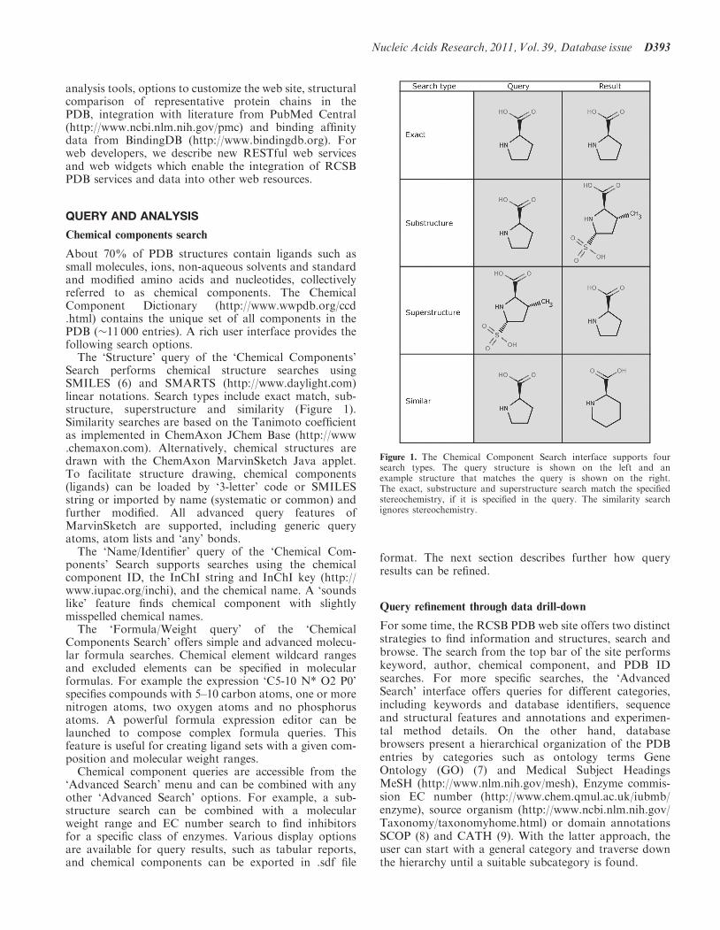
The ‘Structure’ query of the ‘Chemical Components’Search performs chemical structure searches usingSMILES (6) and SMARTS (http://www.daylight.com)linear notations. Search types include exact match, sub-structure, superstructure and similarity (Figure 1).Similarity searches are based on the Tanimoto coefficientas implemented in ChemAxon JChem Base (http://www.chemaxon.com). Alternatively, chemical structures aredrawn with the ChemAxon MarvinSketch Java applet.To facilitate structure drawing, chemical components(ligands) can be loaded by ‘3-letter’ code or SMILESstring or imported by name (systematic or common) andfurther modified. All advanced query features ofMarvinSketch are supported, including generic queryatoms, atom lists and ‘any’ bonds.
The ‘Name/Identifier’ query of the ‘Chemical Com-ponents’ Search supports searches using the chemicalcomponent ID, the InChI string and InChI key (http://www.iupac.org/inchi), and the chemical name. A ‘soundslike’ feature finds chemical component with slightlymisspelled chemical names.
The ‘Formula/Weight query’ of the ‘ChemicalComponents Search’ offers simple and advanced molecu-lar formula searches. Chemical element wildcard rangesand excluded elements can be specified in molecularformulas. For example the expression ‘C5-10 N* O2 P0’specifies compounds with 5–10 carbon atoms, one or morenitrogen atoms, two oxygen atoms and no phosphorusatoms. A powerful formula expression editor can belaunched to compose complex formula queries. Thisfeature is useful for creating ligand sets with a given com-position and molecular weight ranges.
Chemical component queries are accessible from the‘Advanced Search’ menu and can be combined with anyother ‘Advanced Search’ options. For example, a sub-structure search can be combined with a molecularweight range and EC number search to find inhibitorsfor a specific class of enzymes. Various display optionsare available for query results, such as tabular reports,and chemical components can be exported in .sdf file
format. The next section describes further how queryresults can be refined.
Query refinement through data drill-down
For some time, the RCSB PDB web site offers two distinctstrategies to find information and structures, search andbrowse. The search from the top bar of the site performskeyword, author, chemical component, and PDB IDsearches. For more specific searches, the ‘AdvancedSearch’ interface offers queries for different categories,including keywords and database identifiers, sequenceand structural features and annotations and experimen-tal method details. On the other hand, databasebrowsers present a hierarchical organization of the PDBentries by categories such as ontology terms GeneOntology (GO) (7) and Medical Subject HeadingsMeSH (http://www.nlm.nih.gov/mesh), Enzyme commis-sion EC number (http://www.chem.qmul.ac.uk/iubmb/enzyme), source organism (http://www.ncbi.nlm.nih.gov/Taxonomy/taxonomyhome.html) or domain annotationsSCOP (8) and CATH (9). With the latter approach, theuser can start with a general category and traverse downthe hierarchy until a suitable subcategory is found.
Figure 1. The Chemical Component Search interface supports foursearch types. The query structure is shown on the left and anexample structure that matches the query is shown on the right.The exact, substructure and superstructure search match the specifiedstereochemistry, if it is specified in the query. The similarity searchignores stereochemistry.
Nucleic Acids Research, 2011, Vol. 39, Database issue D393
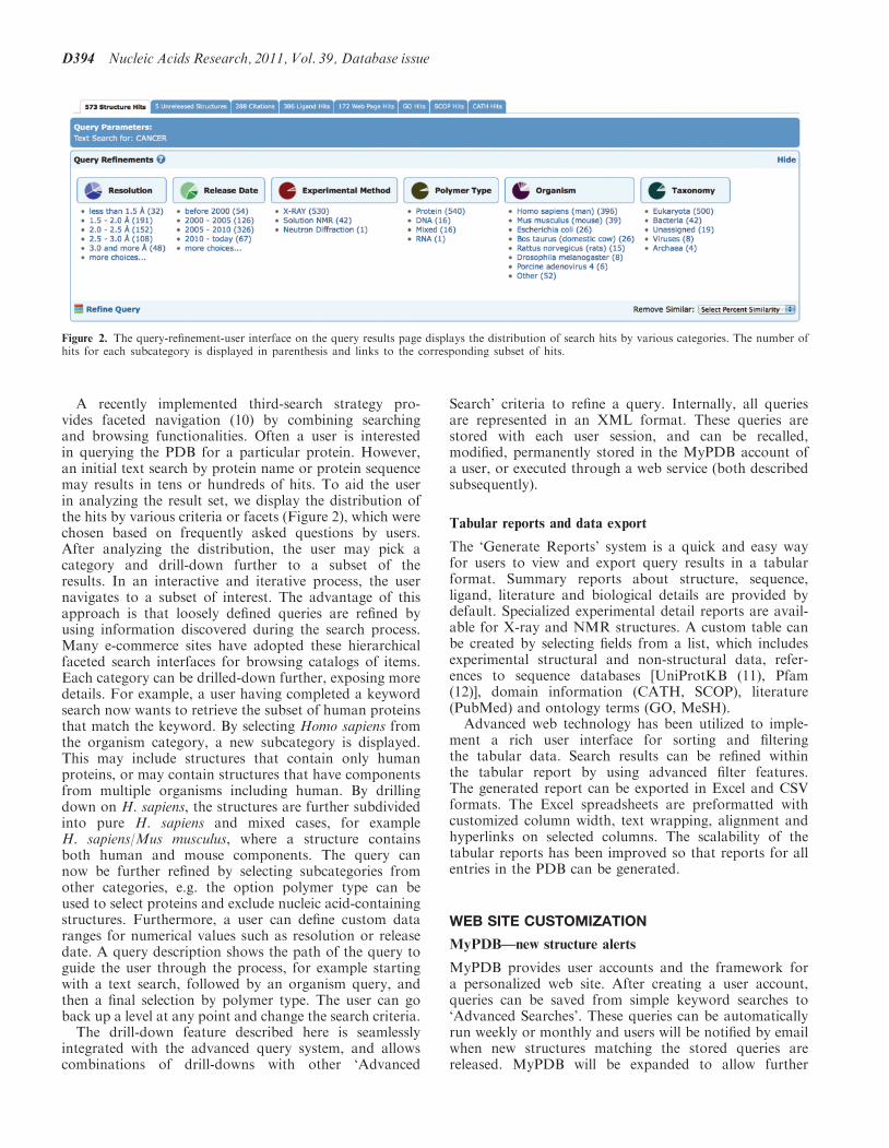
A recently implemented third-search strategy pro-vides faceted navigation (10) by combining searchingand browsing functionalities. Often a user is interestedin querying the PDB for a particular protein. However,an initial text search by protein name or protein sequencemay results in tens or hundreds of hits. To aid the userin analyzing the result set, we display the distribution ofthe hits by various criteria or facets (Figure 2), which werechosen based on frequently asked questions by users.After analyzing the distribution, the user may pick acategory and drill-down further to a subset of theresults. In an interactive and iterative process, the usernavigates to a subset of interest. The advantage of thisapproach is that loosely defined queries are refined byusing information discovered during the search process.Many e-commerce sites have adopted these hierarchicalfaceted search interfaces for browsing catalogs of items.Each category can be drilled-down further, exposing moredetails. For example, a user having completed a keywordsearch now wants to retrieve the subset of human proteinsthat match the keyword. By selecting Homo sapiens fromthe organism category, a new subcategory is displayed.This may include structures that contain only humanproteins, or may contain structures that have componentsfrom multiple organisms including human. By drillingdown on H. sapiens, the structures are further subdividedinto pure H. sapiens and mixed cases, for exampleH. sapiens/Mus musculus, where a structure containsboth human and mouse components. The query cannow be further refined by selecting subcategories fromother categories, e.g. the option polymer type can beused to select proteins and exclude nucleic acid-containingstructures. Furthermore, a user can define custom dataranges for numerical values such as resolution or releasedate. A query description shows the path of the query toguide the user through the process, for example startingwith a text search, followed by an organism query, andthen a final selection by polymer type. The user can goback up a level at any point and change the search criteria.The drill-down feature described here is seamlessly
integrated with the advanced query system, and allowscombinations of drill-downs with other ‘Advanced
Search’ criteria to refine a query. Internally, all queriesare represented in an XML format. These queries arestored with each user session, and can be recalled,modified, permanently stored in the MyPDB account ofa user, or executed through a web service (both describedsubsequently).
Tabular reports and data export
The ‘Generate Reports’ system is a quick and easy wayfor users to view and export query results in a tabularformat. Summary reports about structure, sequence,ligand, literature and biological details are provided bydefault. Specialized experimental detail reports are avail-able for X-ray and NMR structures. A custom table canbe created by selecting fields from a list, which includesexperimental structural and non-structural data, refer-ences to sequence databases [UniProtKB (11), Pfam(12)], domain information (CATH, SCOP), literature(PubMed) and ontology terms (GO, MeSH).
Advanced web technology has been utilized to imple-ment a rich user interface for sorting and filteringthe tabular data. Search results can be refined withinthe tabular report by using advanced filter features.The generated report can be exported in Excel and CSVformats. The Excel spreadsheets are preformatted withcustomized column width, text wrapping, alignment andhyperlinks on selected columns. The scalability of thetabular reports has been improved so that reports for allentries in the PDB can be generated.
WEB SITE CUSTOMIZATION
MyPDB—new structure alerts
MyPDB provides user accounts and the framework fora personalized web site. After creating a user account,queries can be saved from simple keyword searches to‘Advanced Searches’. These queries can be automaticallyrun weekly or monthly and users will be notified by emailwhen new structures matching the stored queries arereleased. MyPDB will be expanded to allow further
Figure 2. The query-refinement-user interface on the query results page displays the distribution of search hits by various categories. The number ofhits for each subcategory is displayed in parenthesis and links to the corresponding subset of hits.
D394 Nucleic Acids Research, 2011, Vol. 39, Database issue

customization of the site, including storage of personal-ized structure annotations.
Site layout customization
Since the RCSB PDB’s users may only be interested invery specific topics, we have added options to customizethe layout of frequently used pages. Throughout the siteare web widgets, which are boxes containing specific in-formation that can be repositioned, hidden or shown on apage. The left-hand menu is comprised of these widgets sousers can move the preferred boxes to the top, and hideoptions that are not as important to the user. The contentof the home page can be customized by choosing from alist of main and side panels. For example, a bioinformaticsscientist may prefer to have the sequence search box onthe home page, whereas a structural biologist may wantthe deposition widget to appear at the top.
Query results are presented in a concise layout, withinformation about polymer and ligand details, and fullabstracts hidden by default. The user can expand thedisplay as needed. The ‘Structure Summary’ page is thesingle-most-used page on the web site and provides infor-mation about a single-PDB entry. Since users have differ-ent interests, the information displayed on this page can beset and arranged in the most meaningful way for a givenuser. Layout changes are currently stored in a cookie onthe web browser. When the user returns to the site fromthe same computer and browser, the customizations areretained.
STRUCTURE COMPARISON AND VISUALIZATION
Pair-wise structural alignments
The RCSB PDB’s Comparison Tool calculates pair-wise3D structure comparisons, as well as sequence alignments.This tool utilizes new Java implementations of the CE (13)and FATCAT (14) structure-alignment algorithms andprovides references to external structure alignmentservers. A new structure alignment service allows serverside calculation of 3D alignments. The tool is comple-mented by a Java Web Start application that allowscustom calculations and provides a novel user interfacefor the visualization of sequence-3D relationships inthe alignment (Figure 3) (15).
Representative structural alignments
The RCSB PDB uses new tools to provide pre-calculatedstructural alignments for representative protein chains inthe PDB. Representatives are chosen to make the com-parisons computationally tractable. The comparisonconsists of two steps: In the first step, protein sequencesare clustered at 40% sequence identity using BLASTClust(http://www.ncbi.nlm.nih.gov/Web/Newsltr/Spring04/blastlab.html). Sequences within a cluster are first rankedby resolution and then by deposition date. Thetop-ranking sequence of each cluster is taken as a repre-sentative for all cluster members under the simplifyingassumption that at 40% sequence identity structuralfeatures are conserved. In the second step, all pairs of
representative protein chains are structurally alignedwith the jFATCAT rigid method and stored in adatabase. The alignments are updated weekly with newincoming protein structures.Novel domain architectures as well as unexpected
structural similarities can be found by analyzing thesestructural alignments. As an example, the structural align-ment results for green fluorescent protein [GFP; PDB ID:2WUR (16)] are displayed in Figure 3. One of thetop-ranking results is nidogen-1. Of GFP (Aequoreavictoria) residues, 94% align with the nidogen-1 G2fragment (M. musculus) with a RMSD of 3 A based onthe Ca positions, but with surprisingly low-sequenceidentity of �9%. Indeed, the structural similarity hasbeen recognized by Hopf (17). Nidogen-1, also known asentactin, is a component of the basement membrane (18)and does not contain a chromophore. PSI-BLASTsearches do not recognize the relationship between thetwo proteins, however, the strong structural conservationof the 11-stranded b-barrel and the internal helices suggestthat the nidogen-1 G2 fragment and GFP share a commonancestor (17). This example demonstrates the utility ofhaving pre-calculated structural alignments available.
Visualization of biological assemblies
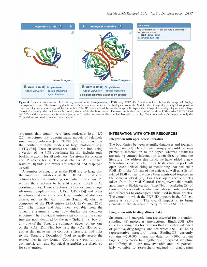
The deposited coordinate set may not always represent thebiologically relevant assembly(s). For example, for struc-tures determined by X-ray crystallography, the asymmet-ric unit is the smallest portion of a crystal structure towhich symmetry operations can be applied in order togenerate the complete unit cell. On the ‘StructureSummary’ page, we display both the asymmetric unitand the biological assembly(s) (Figure 4), with the latterbeing the default view. The biological assembly is eitherspecified by the structure author or assigned by PISA (19)or PQS (20) software and manually checked by PDB an-notators. The biological assembly is generated from theasymmetric unit by applying the symmetry transformationspecified in the PDB entry.
Visualization of large assemblies and split entries
Advances in structure determination methods have ledto an increase in the size and complexity of biologicalmacromolecules in the PDB. Display of very largeassemblies comprising >1 million atoms, or thousandsof protein chains, poses a challenge to currently available3D-visualization software, as well as pushing the memorylimits of standard personal computers. Despite theincreasing speed of the internet, even the download ofthese large structures files (>100 Mb) becomes an issuefor interactive visualization. We have developed methodsto display any large assembly in the PDB on a modernlaptop or desktop computer.Several enhancements to the Jmol (http://www.jmol
.org) viewer page have been made to display very largestructures. Previously, some structures with very large co-ordinate files could not be displayed because they wouldhave required more memory than was available to theJmol applet. We are now able to display structures thatcontain a large number of chains [e.g. 1GAV (21)],
Nucleic Acids Research, 2011, Vol. 39, Database issue D395
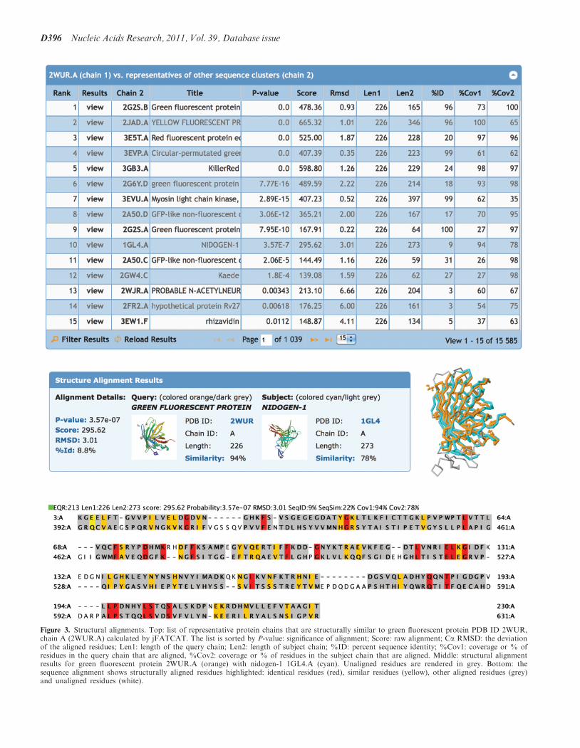
Figure 3. Structural alignments. Top: list of representative protein chains that are structurally similar to green fluorescent protein PDB ID 2WUR,chain A (2WUR.A) calculated by jFATCAT. The list is sorted by P-value: significance of alignment; Score: raw alignment; Ca RMSD: the deviationof the aligned residues; Len1: length of the query chain; Len2: length of subject chain; %ID: percent sequence identity; %Cov1: coverage or % ofresidues in the query chain that are aligned, %Cov2: coverage or % of residues in the subject chain that are aligned. Middle: structural alignmentresults for green fluorescent protein 2WUR.A (orange) with nidogen-1 1GL4.A (cyan). Unaligned residues are rendered in grey. Bottom: thesequence alignment shows structurally aligned residues highlighted: identical residues (red), similar residues (yellow), other aligned residues (grey)and unaligned residues (white).
D396 Nucleic Acids Research, 2011, Vol. 39, Database issue
structures that contain very large molecules [e.g. 1JJ2(22)], structures that contain many models of relativelysmall macromolecules [e.g. 2HYN (23)] and structuresthat contain multiple models of large molecules [e.g.1HTQ (24)]. These structures are loaded into Jmol usinga version of the PDB coordinate file that includes onlybackbone atoms for all polymers (Ca atoms for proteinsand P atoms for nucleic acid chains). All modifiedresidues, ligands and water are retained and displayedas well.
A number of structures in the PDB are so large thatthe historical limitations of the PDB file format (fivecolumns for atom numbering, one column for chain ID)require the structures to be split across multiple PDBcoordinate files. These structures include extremely largeribosome complexes [e.g. 1GIX, 1GIY (25)] and otherstructures that contain a very large number of atoms orchains, such as the vault protein (Figure 4), which iscomposed of the PDB entries 2ZUO, 2ZV4 and 2ZV5(26). The images and Jmol view available from the‘Structure Summary’ page now display the completestructure. The individual entries that comprise the struc-ture are now identified by the new ‘Split Entry’ box onany one of the ‘Structure Summary’ pages for any oneof the PDB IDs. This box lists the PDB IDs of allentries that make up the composite structure, and linksto the ‘Structure Download’ Tool to easily access therelated files in any format. Composite views for bothasymmetric unit and biological assemblies are displayedfor split entries.
INTEGRATION WITH OTHER RESOURCES
Integration with open access literature
The boundaries between scientific databases and journalsare blurring (27). Data are increasingly accessible as sup-plemental information to the paper, whereas databasesare adding curated information taken directly from theliterature. To address this trend, we have added a new‘Literature View’ which, for each structure, reports allopen access articles citing or mentioning that particularPDB ID in the full text of the article, as well as a list ofrelated PDB entries that have been mentioned together inthe same article(s) (28). For these open access articlestaken from PubMed Central (http://www.ncbi.nlm.nih.gov/pmc), a BioLit version (http://biolit.ucsd.edu; 29) ofthose articles is available which includes semantic markupand references to ontological terms used in those articles.The context in which the PDB ID appears in the full-textarticle is also given. The overall impact is to bringelements of the literature directly to the RCSB PDB.
Integration with binding affinity data
Structural and energetic data are essential for the under-standing of molecular interactions. BindingDB (30)collects binding data for proteins that are either validatedor putative drug-targets, and for which the PDB holdsrepresentative structural data. BindingDB currentlycontains �500 000 interaction data for �3000 proteintargets (http://www.bindingdb.org). Integrated structureand affinity data are now available and are particu-larly valuable to researchers engaged in drug-design
Figure 4. Structure visualization. Left: the asymmetric unit of streptavidin in PDB entry 1STP. The 3D viewers listed below the image will displaythe asymmetric unit. The arrow toggles between the asymmetric unit and the biological assembly. Middle: the biological assembly of streptavidinbased on oligomeric state assigned by the author. The 3D viewers listed below the image will display the biological assembly. Right: a very largebiological assembly, the rat liver vault protein, visualized in the Jmol viewer. The structure is the composite of the three PDB entries 2ZUO, 2ZV4and 2ZV5 with symmetry transformation (�x, y, �z) applied to generate the complete biological assembly. To accommodate the large size, only theCa positions are used to render the structure.
Nucleic Acids Research, 2011, Vol. 39, Database issue D397

projects and to scientists calibrating, benchmarking andvalidating computational methods for predicting bindingaffinities.Binding affinity data including the binding constants
IC50, EC50, Ki and thermodynamic data Kd, �G�,�H�, -T�S� are exchanged between BindingDB and theRCSB PDB. These data are listed on the ‘StructureSummary’ pages of the corresponding protein–ligandcomplexes, with links back to BindingDB pages thatcontain detailed information about the experiment. An‘Advanced Search’ is available to find structures withassociated binding affinities. On the ‘Ligand Summary’page links to related entries in BindingDB are provided.Conversely, BindingDB maintains links to the RCSB PDBweb site for individual protein–ligand complexes, ligandsand uses RCSB PDB web services to identify sequencesand chemical structures in the PDB that are similar tothose in BindingDB, thus providing a structural contextto binding-affinity data. The bi-directional links betweenBindingDB and RCSB PDB enable the correlation ofbinding-affinity data with appropriate structural dataand vice versa.
Web services
RCSB PDB web services provide programmatic access toquery tools and PDB data via the Hypertext TransferProtocol (HTTP). We support both Simple ObjectAccess Protocol (SOAP) and Representational StateTransfer (REST) web services. SOAP services have beensupported for several years; more recently light-weightRESTful services were added. Future work on webservices will only use the RESTful protocol, due to theirsimplicity. SOAP services will not be developed anyfurther due to their complexity. Here we describe twotypes of RESTful services (Table 1): (i) ‘Fetch services’return experimental data based on PDB IDs, entity IDs(PDB IDs + Chain IDs), or Chemical Component(Ligand) IDs passed to the server; (ii) ‘Search services’perform a query on the PDB database and return resultsin an XML format.
RESTful services are easy to use; for example the fol-lowing URL:
http://www.pdb.org/pdb/rest/describeMol?structureId=4hhb
specifies a fetch service that returns a description in XMLformat for the polymer entities in PDB entry 4HHB:
<structureId id="4HHB">
<polymerDescriptions>
<polymer entityNr="1" length="141"
type="polypeptide(L)">
<chain id="A"/>
<chain id="C"/>
<polymerDescription description="HEMOGLOBIN
(DEOXY) (ALPHA CHAIN)"/>
</polymer>
<polymer entityNr="2" length="146"
type="polypeptide(L)">
<chain id="B"/>
<chain id="D"/>
<polymerDescription description="HEMOGLOBIN
(DEOXY) (BETA CHAIN)"/>
</polymer>
</polymerDescriptions>
</structureId>
This information can be easily parsed with an XML parserand used by an application or web site.
Web widgets
Third-party web sites often display PDB-related data.Traditionally, another resource would download or linkto information from the RCSB PDB and then display thatinformation on their web site. This approach is not veryscalable and requires a constant update by those sites asnew entries are added to the PDB. Recognizing thisproblem, we have created web widgets, small bits ofcode which provide access to RCSB PDB functionality(31). The widgets are self-contained JavaScript files thatwe maintain, thereby addressing both scalability and datacurrency issues; data will be automatically synchronizedwith the most recent updates. Widgets are customizable;for example, the size and color can be set to match a thirdparty’s web site’s color scheme. An example of extensiveuse of RCSB PDB widgets can be found at The OpenProtein Structure Annotation Network (TOPSAN;http://www.topsan.org), which uses the Molecule of theMonth widget on their home page, the Tag LibraryWidget on each of their web pages and the ComparisonTool widget to find structures in the PDB related bysequence or structural similarity. The TransporterClassification Database (TCDB; http://www.tcdb.org)(32) uses the Image Widget to display images of transportproteins.
Currently, the RCSB PDB offers the following webwidgets.
Image library. A widget that embeds a structure imagebased on a PDB ID. The image size and type of
Table 1. RESTful web services supported by the RCSB PDB
Fetch services PDB entry: description, unique chains (entities),release status.
Chemical Components (ligands): description,occurrence in PDB entries.
Sequence and structure clusters and representativestructures.
Sequence and domain annotations: GO, UniProt,SCOP, CATH, PFAM, InterPro, DP, PDP,secondary structure.
Search services Search chemical components by SMILES: exactmatch, substructure, superstructure, similarityand SMARTS match.
Advanced search: any search supported by theRCSB PDB site.
Detailed descriptions and examples are available at: http://www.pdb.org/pdb/software/rest.do.
D398 Nucleic Acids Research, 2011, Vol. 39, Database issue

assembly (asymmetric unit or biological assembly) can becustomized.
Tag library. A rich mark-up widget that tags PDB IDsand keywords on a web site and automatically providesenhanced functionality that links back to the RCSB PDBweb site. Four types of tags are supported by this widget:author tags are used to mark-up author names. Forexample, structural biologists can use this functionalityto provide always up to date links to their publishedPDB structures on their own web pages. Simple PDB IDtags mark up a section of text or a PDB ID and providetool tips that display a PDB structure image and link tothe ‘Structure Summary’ page. The Menu tag creates amenu to display or download information about a singlePDB entry. The Keyword tag marks up a word or phraseof text and links to a query results page.
Comparison tool. A widget that performs the pair-wisesequence and structure alignments between two proteinchains described above.
Molecule of the Month (MoM). A widget that embeds aMoM image and links to the full article. A short
paragraph can be optionally displayed. The MoMs areeducational articles about important molecules in thePDB. The MoM widget is an ideal way for educationalweb sites to display the most recent MoM articles. Figure5 shows examples of these widgets. A detailed descriptionhow to use these widgets is available at http://www.pdb.org/pdb/static.do?p=widgets/widgetShowcase.jsp.
SUMMARY
The RCSB PDB web site continues to take advantage ofnew scientific understanding and new technological devel-opments. The powerful new Chemical Structure Searchinterface supports simple molecular weight, formula, sub-structure or similarity searches and complex SMARTSqueries. Faceted navigation significantly improvesbrowsing query results with hierarchical navigation andthe ability to refine a query iteratively based on new in-formation gained during the search. Both the ChemicalStructure Search and the new faceted search interfaceare tightly integrated with the ‘Advanced Search’ systemto provide multiple paths for query refinement. The results
Figure 5. Several widgets are available for embedding PDB resources into third-party web sites. Top left: the comparison tool that may be used toalign sequences or structures, shown here in use for a structural alignment of green fluorescent protein and nidogen-1. Bottom left: a PDB ID in asample document (excerpt from http://www.pdb.org/pdb/static.do?p=education_discussion/molecule_of_the_month/pdb109_1.html) is marked upwith the menu tag that displays a menu to view and download information from the RCSB PDB site. Also shown is an example of the imagetag for PDB ID 2OM3. Right: the Molecule of the Month widget that presents the first paragraph of the current Molecule of the Month, and linksto the full article.
Nucleic Acids Research, 2011, Vol. 39, Database issue D399
of the queries can be tabulated, sorted, filtered andexported to enable large-scale data analysis.New sequence and structure analysis tools have been
implemented. New Java versions of the FATCAT andCE algorithms for structural superposition have beenmade available for pair-wise alignments. In addition,all representative protein chains in the PDB have beenstructurally aligned. These pre-calculated alignmentsare updated weekly. Novel structures or new folds canbe readily identified, as well as unexpected similaritiesbetween proteins of low-sequence identity. This informa-tion may be used to find evolutionary relationships ordeduce previously unknown functions of proteins.To deal with the visualization of large and complex
assemblies, special methods have been created to enabletheir visualization on standard hardware. File formatchanges in the future should accommodate the increasingsize of molecular assemblies.PDB entries are now tightly integrated with the
open-access literature that uses or cites the entries. The‘Literature View’ of the RCSB PDB provides new waysto search and analyze the data. Data exchange andbi-directional links with BindingDB enable correlation ofstructure with binding affinity data. For web developers,we provide web widgets to integrate RCSB PDB tools anddata into their web sites, and a RESTful service applica-tion user interface (API) enables programmatic access toRCSB PDB queries and data retrieval.Not all new features and enhancement could be
described here. The ‘What’s New’ page (http://www.pdb.org/pdb/static.do?p=general_information/whats_new.jsp) lists detailed summaries of the latest and past im-provements. The addition of these new features andimprovements represents a new generation of the RCSBPDB web site. It allows more complex analysis to be per-formed and provides systematic comparisons across allof the PDB with the goal to further scientific discoveryand education.
ACKNOWLEDGEMENTS
We are grateful to ChemAxon (http://www.chemaxon.com) for providing Marvin Sketch, JChem Base andStandardizer for the chemical-structure search. MichaelGilson and Tiquing Liu at UCSD worked with us onthe integration with BindingDB. We thank Sean VanTyne and the San Diego User Experience SpecialInterest Group (http://www.uxsig.org) for an in-depth us-ability review of the RCSB web site that led to many im-provements. In addition, we appreciate all users whoprovided feedback. Finally, we thank other RCSB PDBstaff past and present for suggestions, critical review andtesting of new features.
FUNDING
National Science Foundation (NSF DBI 0829586);National Institute of General Medical Sciences(NIGMS); Office of Science, Department of Energy(DOE); National Library of Medicine (NLM); National
Cancer Institute (NCI); National Institute of NeurologicalDisorders and Stroke (NINDS); National Institute ofDiabetes & Digestive & Kidney Diseases (NIDDK).Computational resources for structural alignments areprovided in part by the Open Science Grid (http://www.opensciencegrid.org) funded by the National ScienceFoundation; and the Office of Science, Departmentof Energy (DOE) (NSF 0753335). The RCSB PDBis managed by two members of the RCSB: Rutgers andUCSD. Funding for open access charge: National ScienceFoundation.
Conflict of interest statement. None declared.
REFERENCES
1. Berman,H.M., Westbrook,J., Feng,Z., Gilliland,G., Bhat,T.N.,Weissig,H., Shindyalov,I.N. and Bourne,P.E. (2000) The ProteinData Bank. Nucleic Acids Res., 28, 235–242.
2. Berman,H., Henrick,K. and Nakamura,H. (2003) Announcing theworldwide Protein Data Bank. Nat. Struct. Biol., 10, 980.
3. Velankar,S., Best,C., Beuth,B., Boutselakis,C.H., Cobley,N.,Sousa Da Silva,A.W., Dimitropoulos,D., Golovin,A.,Hirshberg,M., John,M. et al. (2010) PDBe: Protein Data Bankin Europe. Nucleic Acids Res., 38, D308–D317.
4. Dutta,S., Burkhardt,K., Young,J., Swaminathan,G.J.,Matsuura,T., Henrick,K., Nakamura,H. and Berman,H.M. (2009)Data deposition and annotation at the Worldwide Protein DataBank. Mol. Biotechnol., 42, 1–13.
5. Deshpande,N., Addess,K.J., Bluhm,W.F., Merino-Ott,J.C.,Townsend-Merino,W., Zhang,Q., Knezevich,C., Xie,L., Chen,L.,Feng,Z. et al. (2005) The RCSB Protein Data Bank: a redesignedquery system and relational database based on the mmCIFschema. Nucleic Acids Res., 33, D233–D237.
6. Weininger,D. (1988) SMILES, a chemical language andinformation system. 1. Introduction to methodology and encodingrules. J. Chem. Inf. Comput. Sci., 28, 31–36.
7. The Gene Ontology Consortium. (2000) Gene Ontology: tool forthe unification of biology. Nat. Genet., 25, 25–29.
8. Andreeva,A., Howorth,D., Chandonia,J.-M., Brenner,S.E.,Hubbard,T.J.P., Chothia,C. and Murzin,A.G. (2008) Datagrowth and its impact on the SCOP database: new developments.Nucleic Acids Res., 36, D419–D425.
9. Cuff,A., Redfern,O.C., Greene,L., Sillitoe,I., Lewis,T., Dibley,M.,Reid,A., Pearl,F., Dallman,T., Todd,A. et al. (2009) The CATHhierarchy revisited-structural divergence in domain superfamiliesand the continuity of fold space. Structure, 17, 1051–1062.
10. Hearst,M.A. (2009) Search User Interfaces. Cambridge UniversityPress, New York.
11. The UniProt Consortium. (2010) The Universal Protein Resource(UniProt) in 2010. Nucleic Acids Res., 38, D142–D148.
12. Finn,R.D., Mistry,J., Tate,J., Coggill,P., Heger,A., Pollington,J.E.,Gavin,O.L., Gunasekaran,P., Ceric,G., Forslund,K. et al. (2010)The Pfam protein families database. Nucleic Acids Res., 38,D211–D222.
13. Shindyalov,I.N. and Bourne,P.E. (1998) Protein structurealignment by incremental combinatorial extension (CE) of theoptimal path. Protein Eng., 11, 739–747.
14. Ye,Y. and Godzik,A. (2003) Flexible structure alignment bychaining aligned fragment pairs allowing twists. Bioinformatics, 19(Suppl. 2), II246–II255.
15. Prlic,A., Bliven,S., Rose,P.W., Bluhm,W.F., Bizon,C., Godzik,A.and Bourne,P.E. (2010) Precalculated protein structure alignmentsat the RCSB PDB website. Bioinformatics, doi:10.1093/bioinformatics/btq572.
16. Shinobu,A., Palm,G.J., Schierbeek,A.J. and Agmon,N. (2010)Visualizing proton antenna in a high-resolution green fluorescentprotein structure. J. Am. Chem. Soc., 132, 11093–11102.
17. Hopf,M., Gohring,W., Ries,A., Timpl,R. and Hohenester,E.(2001) Crystal structure and mutational analysis of a
D400 Nucleic Acids Research, 2011, Vol. 39, Database issue
perlecan-binding fragment of nidogen-1. Nat. Struct. Biol., 8,634–640.
18. Chung,A.E. and Durkin,M.E. (1990) Entactin: structure andfunction. Am. J. Respir. Cell Mol. Biol., 3, 275–282.
19. Krissinel,E. and Henrick,K. (2007) Inference of macromolecularassemblies from crystalline state. J. Mol. Bio., 372, 774–797.
20. Henrick,K. and Thornton,J.M. (1998) PQS: a protein quaternarystructure file server. Trends Biochem. Sci., 23, 358–361.
21. Tars,K., Bundule,M., Fridborg,K. and Liljas,L. (1997) The crystalstructure of bacteriophage GA and a comparison ofbacteriophages belonging to the major groups of Escherichia colileviviruses. J. Mol. Biol., 271, 759–773.
22. Klein,D.J., Schmeing,T.M., Moore,P.B. and Steitz,T.A. (2001)The kink-turn: a new RNA secondary structure motif. EMBO J.,20, 4214–4221.
23. Potluri,S., Yan,A.K., Chou,J.J., Donald,B.R. andBailey-Kellogg,C. (2006) Structure determination of symmetrichomo-oligomers by a complete search of symmetry configurationspace, using NMR restraints and van der Waals packing.Proteins, 65, 203–219.
24. Gill,H.S., Pfluegl,G.M. and Eisenberg,D. (2002) Multicopycrystallographic refinement of a relaxed glutamine synthetase fromMycobacterium tuberculosis highlights flexible loops in theenzymatic mechanism and its regulation. Biochemistry, 41,9863–9872.
25. Yusupov,M.M., Yusupova,G.Z., Baucom,A., Lieberman,K.,Earnest,T.N., Cate,J.H.D. and Noller,H.F. (2001) Crystal structureof the ribosome at 5.5 A resolution. Science, 292, 883–896.
26. Tanaka,H., Kato,K., Yamashita,E., Sumizawa,T., Zhou,Y.,Yao,M., Iwasaki,K., Yoshimura,M. and Tsukihara,T. (2009)The structure of rat liver vault at 3.5 angstrom resolution.Science, 323, 384–388.
27. Bourne,P.E. (2005) Will a biological database be different froma biological journal? PLoS Comput. Biol., 1, 179–181.
28. Prlic,A., Martinez,M.A., Dimitropoulos,D., Beran,B.,Yukich,B.T., Rose,P.W., Bourne,P.E. and Fink,J.L. (2010)Integration of open access literature into the RCSB Protein DataBank using BioLit. BMC Bioinformatics, 11, 220, doi:10.1186/1471-2105-11-220.
29. Fink,J.L., Kushch,S., Williams,P.R. and Bourne,P.E. (2008)BioLit: integrating biological literature with databases. NucleicAcids Res., 36, W385–W389.
30. Liu,T., Lin,Y., Wen,X., Jorissen,R.N. and Gilson,M.K. (2007)BindingDB: a web-accessible database of experimentallydetermined protein-ligand binding affinities. Nucleic Acids Res.,35, D198–D201.
31. Bourne,P.E., Beran,B., Bi,C., Bluhm,W., Dunbrack,R., Prlic,A.,Quinn,G., Rose,P., Shah,R., Tao,W. et al. (2010) Will widgetsand semantic tagging change computational biology? PLoSComput. Biol., 6, e1000673, doi:10.1371/journal.pcbi.1000673.
32. Saier,M.H. Jr, Yen,M.R., Noto,K., Tamang,D.G. and Elkan,C.(2009) The Transporter Classification Database: recent advances.Nucleic Acids Res., 37, D274–D278.
Nucleic Acids Research, 2011, Vol. 39, Database issue D401