The Pressing Issue of Micro- and Nanoplastic Contamination
25
Citation: Ferrante, M.C.; Monnolo, A.; Del Piano, F.; Mattace Raso, G.; Meli, R. The Pressing Issue of Micro- and Nanoplastic Contamination: Profiling the Reproductive Alterations Mediated by Oxidative Stress. Antioxidants 2022, 11, 193. https://doi.org/10.3390/ antiox11020193 Academic Editors: Alica Pizent and Eva Tvrdá Received: 18 November 2021 Accepted: 17 January 2022 Published: 19 January 2022 Publisher’s Note: MDPI stays neutral with regard to jurisdictional claims in published maps and institutional affil- iations. Copyright: © 2022 by the authors. Licensee MDPI, Basel, Switzerland. This article is an open access article distributed under the terms and conditions of the Creative Commons Attribution (CC BY) license (https:// creativecommons.org/licenses/by/ 4.0/). antioxidants Review The Pressing Issue of Micro- and Nanoplastic Contamination: Profiling the Reproductive Alterations Mediated by Oxidative Stress Maria Carmela Ferrante 1, * , Anna Monnolo 1, *, Filomena Del Piano 1 , Giuseppina Mattace Raso 2 and Rosaria Meli 2 1 Department of Veterinary Medicine and Animal Productions, University of Naples Federico II, Via F. Delpino 1, 80137 Naples, Italy; fi[email protected] 2 Department of Pharmacy, University of Naples Federico II, Via D. Montesano 49, 80131 Naples, Italy; [email protected] (G.M.R.); [email protected] (R.M.) * Correspondence: [email protected] (M.C.F.); [email protected] (A.M.) Abstract: Micro- and nanoplastics (MPs/NPs) are among the most widely distributed pollutants in the environment. It has been suggested that exposure to MPs/NPs can trigger toxicity pathways among which inflammation and oxidative stress (OS) play a pivotal role. Once absorbed, MPs/NPs may act locally or access the bloodstream and, following the translocation process, reach several organs and tissues, including the gonads. Notably, MPs/NPs can bioaccumulate in human and murine placenta, opening new scenarios for toxicological evaluations. We review recent studies on the effects of MPs/NPs on the reproductive health in aquatic and terrestrial organisms of both sexes, focusing on the role of OS and the antioxidant defence system failure as the main underlying mechanisms. Alterations in gametogenesis, embryonic and offspring development, and survival have been shown in most studies and often related to a broken redox balance. All these detrimental effects are inversely related to particle size, whereas they are closely linked to shape, plastic polymer type, superficial functionalization, concentration, and time of exposure. To date, the studies provide insights into the health impacts, but no conclusions can be drawn for reproduction toxicity. The main implication of the few studies on antioxidant substances reveals their potential role in mitigating MP-induced toxic effects. Keywords: microplastics; nanoplastics; reproduction; oxidative damage; aquatic organisms; terres- trial organisms; antioxidant substances 1. Growing Concerns on Effects of Micro/Nanoplastics Toxicity During the last twenty years, increasing attention has been paid to plastic environ- mental pollution, which affects terrestrial and aquatic ecosystems and air [1], representing a threat to living organisms. This pollution is expected to grow in the near future [2]. Several studies have proposed classifications of plastics depending on their size. Even if there is not yet a general consensus, many researchers agree to define microplastics (MPs) as those in the range 100 nm–5 mm in diameter and nanoplastics (NPs) as those in the range 1–100 nm [3]. Based on their shape, they are mainly listed as pellets, fibres, fragments, spheroids, and granules. MPs largely include the following three polymer types: polyethylene (PE), polypropylene (PP), and polystyrene (PS) [3]. MPs may be divided into primary and secondary ones based on their origin [4]. Primary MPs are small, persistent, chemically inert, raw particles employed in several applications (i.e., personal care products, drug vectors, and synthetic fabrics). Deriving from anthropic activities, they are released directly into the environment through wastewater, sewage systems, and industrial discharge [5,6]. Secondary MPs originate from the fragmentation over time of plastic debris due to biodegradation processes, abrasion by the wind, or UV rays [4]. Both Antioxidants 2022, 11, 193. https://doi.org/10.3390/antiox11020193 https://www.mdpi.com/journal/antioxidants
-
Upload
khangminh22 -
Category
Documents
-
view
0 -
download
0
Transcript of The Pressing Issue of Micro- and Nanoplastic Contamination

�����������������
Citation: Ferrante, M.C.; Monnolo,
A.; Del Piano, F.; Mattace Raso, G.;
Meli, R. The Pressing Issue of Micro-
and Nanoplastic Contamination:
Profiling the Reproductive
Alterations Mediated by Oxidative
Stress. Antioxidants 2022, 11, 193.
https://doi.org/10.3390/
antiox11020193
Academic Editors: Alica Pizent and
Eva Tvrdá
Received: 18 November 2021
Accepted: 17 January 2022
Published: 19 January 2022
Publisher’s Note: MDPI stays neutral
with regard to jurisdictional claims in
published maps and institutional affil-
iations.
Copyright: © 2022 by the authors.
Licensee MDPI, Basel, Switzerland.
This article is an open access article
distributed under the terms and
conditions of the Creative Commons
Attribution (CC BY) license (https://
creativecommons.org/licenses/by/
4.0/).
antioxidants
Review
The Pressing Issue of Micro- and Nanoplastic Contamination:Profiling the Reproductive Alterations Mediated byOxidative StressMaria Carmela Ferrante 1,* , Anna Monnolo 1,*, Filomena Del Piano 1, Giuseppina Mattace Raso 2
and Rosaria Meli 2
1 Department of Veterinary Medicine and Animal Productions, University of Naples Federico II,Via F. Delpino 1, 80137 Naples, Italy; [email protected]
2 Department of Pharmacy, University of Naples Federico II, Via D. Montesano 49, 80131 Naples, Italy;[email protected] (G.M.R.); [email protected] (R.M.)
* Correspondence: [email protected] (M.C.F.); [email protected] (A.M.)
Abstract: Micro- and nanoplastics (MPs/NPs) are among the most widely distributed pollutants inthe environment. It has been suggested that exposure to MPs/NPs can trigger toxicity pathwaysamong which inflammation and oxidative stress (OS) play a pivotal role. Once absorbed, MPs/NPsmay act locally or access the bloodstream and, following the translocation process, reach severalorgans and tissues, including the gonads. Notably, MPs/NPs can bioaccumulate in human andmurine placenta, opening new scenarios for toxicological evaluations. We review recent studieson the effects of MPs/NPs on the reproductive health in aquatic and terrestrial organisms of bothsexes, focusing on the role of OS and the antioxidant defence system failure as the main underlyingmechanisms. Alterations in gametogenesis, embryonic and offspring development, and survivalhave been shown in most studies and often related to a broken redox balance. All these detrimentaleffects are inversely related to particle size, whereas they are closely linked to shape, plastic polymertype, superficial functionalization, concentration, and time of exposure. To date, the studies provideinsights into the health impacts, but no conclusions can be drawn for reproduction toxicity. The mainimplication of the few studies on antioxidant substances reveals their potential role in mitigatingMP-induced toxic effects.
Keywords: microplastics; nanoplastics; reproduction; oxidative damage; aquatic organisms; terres-trial organisms; antioxidant substances
1. Growing Concerns on Effects of Micro/Nanoplastics Toxicity
During the last twenty years, increasing attention has been paid to plastic environ-mental pollution, which affects terrestrial and aquatic ecosystems and air [1], representinga threat to living organisms. This pollution is expected to grow in the near future [2].
Several studies have proposed classifications of plastics depending on their size. Evenif there is not yet a general consensus, many researchers agree to define microplastics(MPs) as those in the range 100 nm–5 mm in diameter and nanoplastics (NPs) as thosein the range 1–100 nm [3]. Based on their shape, they are mainly listed as pellets, fibres,fragments, spheroids, and granules. MPs largely include the following three polymertypes: polyethylene (PE), polypropylene (PP), and polystyrene (PS) [3]. MPs may bedivided into primary and secondary ones based on their origin [4]. Primary MPs are small,persistent, chemically inert, raw particles employed in several applications (i.e., personalcare products, drug vectors, and synthetic fabrics). Deriving from anthropic activities,they are released directly into the environment through wastewater, sewage systems, andindustrial discharge [5,6]. Secondary MPs originate from the fragmentation over time ofplastic debris due to biodegradation processes, abrasion by the wind, or UV rays [4]. Both
Antioxidants 2022, 11, 193. https://doi.org/10.3390/antiox11020193 https://www.mdpi.com/journal/antioxidants

Antioxidants 2022, 11, 193 2 of 25
primary and secondary MPs tend to fragment in NPs. MPs/NPs toxicity is closely relatedto their bioaccumulation and size [7,8]. Several studies reported that entanglement by [9]and ingestion of anthropogenically induced plastic debris, or their capturing in gills [10],are the primary causes of MP damage in aquatic species. The trophic transfer amongtrophic levels, from producers to top predators [11], can lead to the biomagnification ofMPs/NPs toxicity. Humans, as with other terrestrial mammals, are exposed to MPs/NPsprimarily by ingestion of contaminated food (i.e., sea salt, sea products, beer, honey, andpotable water) [12–16]. In the last years, chronic inhalation has been considered mainlyfor industrial and agricultural workers exposed to high concentrations of MPs/NPs (seereferences in [17,18]). Human exposure through indoor air also has been reported [19,20].Moreover, NPs could cross the dermal barrier in mammals [21], even though this route ofcontact may be less significant than the oral or inhalation one. Besides the skin and mouth,hair has also proved to be a significant passive receptor [22].
MPs in the size range 0.1–150 µm, once absorbed, act locally or translocate from thegut to lymphatic and blood circulatory systems, reaching mammalian organs and tissuesfar from the site of exposure (see [3]). Therefore MPs/NPs may bioaccumulate in severalorgans, including female and male gonads [23–25].
MPs/NPs toxicity is influenced by plastic type, particle size, superficial functionaliza-tion, environmental conditions, species, experimental model used, time of exposure, routeof exposure, and concentration/dose difference [7,17,21,26,27]. MPs can be fragmentedand/or biotransformed into smaller particles (nanometre size); these modifications mayoccur in both abiotic matrices and living beings. In addition to size, surface chemistry andcharge, the cellular uptake of NPs can modulate their interaction with biological molecules(i.e., phospholipids, carbohydrates, and proteins), modifying the NPs’ behaviour [28,29].
MPs/NPs toxicity is due to oxidative stress (OS), inflammation, cytotoxicity, immuno-toxicity, and metabolic disorders, among the others [21]. Most studies indicate OS as themajor mechanism responsible for MP/NP-induced damage. However, all the mechanismsare strictly interconnected with each other and the induction of one pathway can trigger orsustain the others.
The main MPs/NPs cytotoxic mechanisms are membrane injury and OS, and otherones are genotoxicity and immune response induction [30]. NPs uptake leads to a loss ofcell membrane integrity, consequent pore formation, and increased intracellular reactiveoxygen species (ROS) production from the mitochondria [31]. Increased ROS generation isin turn responsible of mitochondrial dysfunction and release of pro-apoptotic factors andpro-inflammatory cytokines, among others, which result in cell death [26]. Moreover, NPsimpair membrane structure and function, modifying the lipid bilayer with an increasedthickness and altered molecules flow [32].
MPs cytotoxic damage is also related to an increased expression of pro-inflammatorygenes potentially responsible for pulmonary, dermal, and immune damages [33–35]. In-flammation induced by MPs causes an alteration in gut microbiota composition, increasingthe serum cytokine levels, accompanied by the impairment of the innate and adaptiveimmunity [36]. The dysfunction of the immune response induced by NPs is related to theinduction of the apoptosis process triggered by ROS production, the reduced phagocytosis,the increased lysozyme activity, and the modulation of transcription of genes implicatedin the OS, inflammatory, and apoptotic pathways (i.e., NF-kB, Bcl-2 and Hsp90) [37–39].Indeed, several lines of evidence show a strict relationship between MPs/NPs toxicityand OS, identifying alterations in redox balance and an impaired response of the antioxi-dant defence system at the onset of the immune and nervous system damage, as well ascarcinogenicity (see, for instance, [40]).
The detrimental effect of MPs/NPs on several organs and functions, including thereproductive ones, can be aggravated by the co-exposure to other toxic chemical xenobioticsadsorbed onto their surface. In fact, MPs/NPs can act as a vector for other pollutants,allowing their biomagnification (see [41] and references therein). In particular, the mix ofMPs/NPs with additives (bisphenols or phthalates) and/or persistent organic pollutants

Antioxidants 2022, 11, 193 3 of 25
(POPs) produces a higher toxicity than that due to MPs/NPs alone [42,43]. Pollutantsadsorption may be increased by biofilms that are microorganisms colonizing MPs [44].
Co-exposure of clams to MPs and antibiotics (oxytetracycline and florfenicol) resultedin increased bioaccumulation of these drugs, revealing an enhanced toxicological potentialand antibiotic resistance risk for human consumption of fishery products [45].
Despite the absence of evidence in humans, animal studies (particularly on marinespecies) indicate that MPs/NPs affect several reproductive parameters and functions inliving beings. Very recently, preliminary evaluations of the effects of MPs exposure onmammalian reproduction have revealed the alterations in spermatogenesis/sperm qualityand ovary in exposed animals and the indirect damages on the embryo and offspringoccurring via gestational exposure [23,46,47].
Here, we review the recent literature data to better define the effects of MPs/NPson reproduction in aquatic and terrestrial organisms, focusing on the role of OS as theunderlying cross-sectional mechanism of toxicity. Studies regarding the co-exposure ofchemical contaminants and MPs/NPs were also considered. Antioxidant substances with arole in the reduction of MP-induced damages were examined since such substances aremolecules that hinder OS, preventing ROS overproduction.
In this review, we analysed the journal articles reported on PUBMED® that com-prises citations for biomedical literature from the MEDLINE database, life science journals,and online books. Moreover, we considered papers reported on Google Scholar, Web ofScience, and Scopus (until October 2021), using as keywords: microplastics or nanoplas-tics, reproduction, oxidative stress, and antioxidant substances. We tried to identify theknowledge gaps in this new research field and provide recommendations to optimizefurther investigation.
2. Oxidative Stress and Inflammation in Micro/Nanoplastics Toxicity
MP/NPs-induced toxicity has been mainly investigated in aquatic organisms andonly few experimental studies have been carried out on terrestrial species. All thesestudies deal with different biological structures—from cells (i.e., head-kidney leucocytesand fibroblasts [48,49] to tissues (e.g., [50,51]) and to the whole organism (e.g., [52,53])—through the evaluation of biochemical parameters [54]. However, most data are focused onthe effects of PS particles, overlooking those of the other plastic types (i.e., PE and PP).
Recently, MP toxicity mechanisms were reviewed in terms of ecotoxicity and humanhealth risk assessment [55]. It was suggested that the key event was ROS (i.e., hydrogenperoxide, singlet oxygen, superoxide anion, ozone, hydroxyl radicals, and nitric oxide)overproduction. ROS cause damage to cell components, including lipids, proteins, andDNA, and the adverse outcomes are a decrease in growth rates, reproduction failure, andincreased mortality. Antioxidant enzymes counteract reactive species overproduction;however, when it overcomes the antioxidant power, oxidative damage may occur [56].MPs/NPs can differently impact the antioxidant system. It is commonly assumed thatsmaller MPs are associated with elevated OS [57–59], even if contrasting results are alsoreported in the literature [60,61]. This variability can be attributed to (i) the type andsize of the MPs used in the different experimental models; (ii) the organs, tissues or celltypes examined; and (iii) the concentrations/doses used, which are typically higher thanthe environmentally relevant ones. Probably, low MPs/NPs contamination could not beenough to trigger a response, but its extent is expected to increase in the near future [2].
Recently, the effects of short- and long-term exposure (3 and 13 days) to PS-MPs/NPsof different sizes (65 nm, 100 nm and 1µm) and concentrations (1 and 10 mg L−1) on growthand OS-related parameters were assessed for the dinoflagellate Karenia mikimotoi [62].PS-NPs induced a major growth inhibition, probably caused by physical blockage andmembrane damage due to the aggregation of NPs to microalgae. The smaller particlescrossed the membranes through pores allowing the leakage of cytoplasm, generating agreater cytotoxic effect. Moreover, PS-NPs caused the most severe effects on catalase

Antioxidants 2022, 11, 193 4 of 25
(CAT) and superoxide dismutase (SOD) activity and malondialdehyde (MDA) content andROS production.
A significant augmentation of lipid peroxidation (LPO), estimated as thiobarbituricacid reactive substances, was observed in the brain, muscle, and gills of wild sea bass (Dicen-trarchus labrax), Atlantic horse mackerel (Trachurus trachurus), and Atlantic chub mackerel(Scomber colias) contaminated with MPs with respect to non-contaminated fishes [63]. TheLPO increase was also observed by other authors in different species [64–66].
Many studies analyse the effect of MPs/NPs exposure on gene expression and theactivity of glutathione (GSH) peptide and antioxidant enzymes, mainly CAT, SOD, glu-tathione peroxidase (GPx), peroxidase (POD), glutathione reductase (GR), and glutathioneS-transferase (GST), a biotransformation enzyme [67]. A significant increase in GST enzy-matic activity was described in the liver of striped red mullet [68]. In in vivo experimentalmodels, several authors observed MP/NP-induced oxidative damage in various animalspecies. PS increased ROS production in marine copepod (Tigriopus japonicus) exposedto 50 nm and 10 µm microbeads, modifying the expression of genes related to OS (e.g.,Gr, Gst, CuZnSod, and MnSod) [57]. Interestingly, the treatment with the antioxidant N-acetyl-L-cysteine (NAC) at a 0.5 mM concentration significantly decreased ROS production.Murano et al. [69] demonstrated an increase in ROS and reactive nitrogen species (RNS) incoelomocytes of sea urchins exposed to 10 and 45 µm PS. An increased total oxidant statusand alteration in total antioxidant capacity and LPO were found in the digestive glands ofmussels exposed for 96 h to nano-PS particles [70].
Qiao et al. [51] evidenced inflammation and OS in the gut of zebrafish exposed to PS(50 and 500 µg L−1 for 21 days) with increased levels of CAT, SOD, GSH, bowel permeability,and dysbiosis. Notably, chronic inflammation is strongly related to OS, gut dysbiosis, andmetabolic disturbances [71,72]. In the liver of the same fish species, exposed to the sameplastic particles (20–2000 µg L−1 for 7 days), tissue inflammation was observed, dose-dependently, and an increase in SOD and CAT enzymes. These alterations were related toan impairment in lipid and energy metabolism [60]. PS also reduced the CAT activity, GSHand ascorbate levels, and increased some mediators linked to OS, evidencing the alterationin the antioxidant mechanisms in zebrafish larvae [61]. Testing the same type of particles,Yu et al. [73] observed in the crab a reduced expression of genes involved in the mitogen-activated protein kinase (MAPK) signalling pathway (ERK, AKT, and MEK), leading toOS and inflammation, affecting cell survival. Other authors examined the ROS contentand the modification of the MAPK-HIF-1/NF-kB pathway in addition to the antioxidantgene expression and enzyme activity in microcrustacean Daphnia pulex exposed to nano-PSparticles [74]. The low concentrations of NPs (0.1 and 0.5 mg L−1) induced low levels ofROS, subsequent activation of the MAPK-HIF-1/NF-kB pathway, and increased antioxidantgene expression and enzyme activities. The highest NPs concentration (2 mg L−1) inducedthe overproduction of ROS, resulting in OS that inhibited growth and reproduction viacellular damage. Tang et al. [75] observed an impairment in the detoxification processesmediated by c-Jun N-terminal kinase (JNK) and extracellular signal-regulated kinase (ERK)signalling pathway activation.
Polyvinyl chloride (PVC) reduced, in a time-dependent manner, the SOD, GPx, andCAT activities in the liver of catfish and augmented LPO in a concentration-dependentmanner [65]. Hamed et al. [64] evidenced an increase in total peroxides, as well as LPO,and SOD and CAT activity in Nile tilapia sub-chronically exposed to MPs (1–100 mg L−1 for15 days). Interestingly, after a recovery period of 15 days, these alterations were reversedfor the fishes exposed to the lowest MP concentration.
Wang et al. [76] evidenced in the digestive glands of mussels exposed to PS (for 14 daysand two different water pH conditions) a slight OS; this effect was reversed after a recoveryperiod of 7 days under normal conditions without stressors.
Regarding terrestrial fauna, PE and PS (0–20% d.w.) significantly increased the activityof detoxifying enzymes (POD and CAT) but reduced that of SOD and GST in earthworms(Eisenia fetida), using MP amounts that generally do not occur in natural conditions [77];

Antioxidants 2022, 11, 193 5 of 25
similar results and conclusions were reported by Rodriguez-Seijo et al. [78]. Conversely,Sun et al. [79] did not evidence a significant modification of the antioxidant defencesystem and MDA content in earthworms exposed both at low and high MP concentrations(300 and 3000 mg kg−1) in soil; similar conclusions were reached by De Felice et al. [80]for the giant snail (Achatina reticulata) receiving a diet containing PE-terephthalate. On theother hand, the exposure to MPs modified the oxidative response in the liver, gut, andkidney of mice with increased GPx and SOD and decreased CAT. Substantial alterationswere also observed in metabolites related to OS (pyruvate, lysine, and threonine) [52].
In vitro experimental conditions, it has been observed that ultrafine PS particles(78 nm−1 µm) may enter in pig’s lung and human red blood cells by diffusion or adhesiveinteractions. PS did not bind the cell membrane but directly accessed to the intracellularenvironment [81]. Cerebral and epithelial human cells as well as human colon adenocarci-noma Caco-2 cells exposed to the same type of plastic particles (10 mg mL−1 for 24 h [82]and 0.1 µm and 5 µm for 12 h [83,84], respectively) boosted ROS accumulation. NH2-labeledPS (5–40 µg mL−1) enhanced ROS production also in macrophages and lung epithelial cellstreated for 6 h and this effect was reduced by a pre-treatment of 1 h with 5 mM NAC [85].ROS generation was also increased in blood and immune system cells by PP particles (about25 µm at a concentration of 1000 µg mL−1) [33], as well as in human fibroblasts exposed to100 nm PS (5 µg mL−1) [49]; this effect was reduced when the cells were co-exposed to anantioxidant saffron (Crocus sativus L.) extract. Dong et al. [34] observed not only increasedROS production but also an amplified expression of heme oxygenase-1 in human lungepithelial cells exposed for 20 min or 24 h to PS (1000 µg cm2) [86]. As well known, hemeoxygenase-1 is a ubiquitous protein, activated by most of OS inducers. Moreover, renal pri-mary leucocytes from sea bream head exposed to PVC and PE (100 mg mL−1 for 1 and 24 h)showed the upregulation of nrf2 transcription, a gene involved in the response pathway tooxidative stimulus [48]; similar evidence was reported for rainbow trout liver [87].
OS has also been considered among the underlying mechanisms responsible forrespiratory system injury provoked by inhaled MPs/NPs in humans (ROS production, celldamage, release of inflammatory mediators) [17,88,89]. Very recently, the bioremediationof OS and hematobiochemical alterations by active components from herbal plants (i.e.,lycopene, citric acid, and chlorella) have been investigated in African catfish exposed to MPs(500 mg kg−1 diet) [90]. Moreover, Shengchen et al. [91] evidenced the role of NAC in thereduction of ROS overproduction and the imbalance between myogenic differentiation andadipogenic differentiation in mouse myoblasts treated with PS (1–10 µm and 50–100 µm).
Summarized evidence about aquatic, terrestrial fauna, and in vitro models suggeststhat OS may be considered as one of the key mechanisms determining the toxicity ofMPs/NPs. [21,40]. These pollutants induce an inflammatory response leading to ROS andRNS overproduction [56], and often accompanied by a failure of the antioxidant defencesystem. However, in a few papers, ROS overproduction and an alteration in antioxidantcapacity were not observed. A reversal of the effect has been even evidenced after arecovery period. A mitigation effect of antioxidant substances has been observed in in vitroand in vivo studies on aquatic fishes.
3. Effects of Micro/Nanoplastics on Reproduction
An updated summary of the studies that focus on MPs/NPs reproductive toxicity isprovided in Table 1.
It has been proved that MPs/NPs (70 nm–45 µm) after absorption and translocationfrom the gut reach various organs, including the gonads (see [7,11,24,25,69,92,93] andreferences therein). However, because of the blood–gonad barrier, access of larger MPscan be blocked and only the smaller NPs can accumulate [7]. NPs (240 nm) can alsoaccumulate in several reproductive tissues, for instance, in human placenta [94], alteringthe integrity of the reproductive organs and causing their dysfunction (i.e., defects in theovary, spermatogenesis, and sperm quality) [23–25,95,96]. The presence of PS particles,inversely related to MPs size, was observed in the gonads of sea urchins [69]. Sea urchins

Antioxidants 2022, 11, 193 6 of 25
were placed in experimental glass tanks (1 specimen per litre) and exposed for 72 h to PS(10 and 45 µm, 10 particles mL−1). After extraction by fresh tissue, the PS amount in severalorgans and gonads was measured by optical microscopy and was inversely related to MPssize [69].
Among the consequences of exposure to plastic debris, reproductive toxicity must beconsidered [97,98]. In fact, recent data show that MPs/NPs toxicity mechanisms, such asmembrane damage, inflammation and immune response impairment [99], genotoxicity,and OS [100], underly the alterations in reproduction in the exposed species. MPs/NPsreduce fecundity up to infertility in male and female organisms [101–103] and induce asmaller survival rate of progeny [104], causing impaired reproductive performance.
Most data in the literature on MP/NP-induced reproductive toxicity have been ob-tained from aquatic species at different trophic levels, mainly focusing on zooplankton.The prolonged exposure of the pelagic copepod Calanus helgolandicus to PS beads (20 µm,75 MPs mL−1) causes the production of smaller eggs with reduced hatching success [101].Similarly, it was shown a decreased fecundity in the copepod Tigriopus japonicus exposedto PS (0.5 and 6 µm, 0.125, 1.25, 12.5, and 25 µg mL−1) as evidenced by the lessenednumber of nauplius/female [102]. In the same species exposed to PE and polyamide (PA)(12.5 mg L−1), it was observed a longer development time and increased interval timebetween egg sacs [105].
The exposure of the waterflea Ceriodaphnia dubia to PE beads (62.5–2000 µg L−1) and fi-bres (31.25–1000 µg L−1) for 8 days determines a reproductive injury with a dose-dependentdecrease in both the number and body size of neonates, mainly for the fibres [106]. More-over, a 40% mortality rate of adults was observed for both types of MPs.
Martins and Guilhermino [107], in a transgenerational study (F0–F3), showed that21-day exposure of Daphnia magna to MPs (100 µg L−1) determines adverse impact ongrowth and reproduction, increasing parental death (up to the extinction of MPs-exposedF1 generation). Moreover, a reduction in the reproduction and population growth ratewas observed. The authors showed a slight recovery of these alterations, which neededseveral generations to occur. Behind neonatal malformations, decreased progeny numberand body size were also evidenced by Besseling et al. [97] after Daphnia magna exposure tonano-PS (0.22–150 mg L−1 for 21 days). Altered reproduction was evidenced in the samesmall planktonic crustacean after the same time of exposure to 1 and 5 µm MPs (0.012 and12 mg L−1), in addition to an increased time of first brood emission (49%) and usually fewerreleased clutches (71%) at 12 mg L−1 MPs. Other authors observed that the total numberof progenies was reduced while the development of immobile juveniles was increased byMPs [104].
In the marine worm Arenicola marina, an MP-induced reduction in lipid reserves andavailable energy was related to reproduction disorders, and suppressed feeding activ-ity [108]. Concentrations of coarse PP fibres (1 mm length), similar to those measured in theenvironment, induced a negative impact on reproduction in sand crabs (Emerita analoga)after about 10 weeks of exposure [109]. In fact, the decreased retention of egg clutchesin later embryonic stages and the augmented number of these stages were observed asdepending on the amount of the absorbed fibres.
Two months of exposure of the oyster Pinctada margaritifera to 6–10 µm PS (0.25, 2.5,and 25 µg L−1) was correlated to energy deficit and impaired male gametogenesis, asevidenced by histological analyses [110]. Previously, other authors observed a decreasein oocyte number and diameter, sperm speed, and larval development in adult Pacificoysters (Crassostrea gigas) exposed to PS (23 µg L−1) for the same exposure time [103].Moreover, nano-PS decreased the oyster fertilization efficacy and early life-stages (i.e.,embryogenesis and larval growth), with several alterations up to the developmental arrest.The severity of these modifications depended on the size and structure functionalization,with NH2 particles (50 nm) exhibiting the major toxicity on gametes and embryos [111].More recently, the same authors evidenced that acute exposure of oyster spermatozoa to

Antioxidants 2022, 11, 193 7 of 25
different concentrations of PS beads induced a high spermiotoxicity, characterized by adecrease in the motility and velocity of spermatozoa [112].
Chisada et al. [113] studied the Japanese aquatic vertebrate medaka (Oryzias latipes),showing a decrease in egg number and hatching rate after 3 months of exposure to PEmicrobeads (10–63 µm, 65 and 650 µg L−1). Notably, the growth rate was also reduced atthe highest concentration [113]. Similar data were obtained after feeding medaka with aPS-contaminated diet (10 µm, 0.5–2 mg g−1), evidencing fewer egg production during thedevelopment from the immature stage to adult deposition age [114].
Conversely, Assas et al. [115] did not notice modifications in egg production, growth,and survival of medaka after 3 weeks of exposure to a lower PS concentration (2 µm,44 µg L−1). In the annelid Lumbriculus variegatus exposed to PE (concentrations between0.51 and 20 g kg−1 dry sediment), no effect was observed on reproduction and biomass [116].Moreover, no significant alterations in the reproductive activity were evidenced in thefreshwater cnidarian, Hydra attenuate [117]. Qiang et al. [118] exposed the zebrafish to micro-PS (1 µm, 10–1000 µg L−1 for 21 days) without observing variations in the testosteroneand 17-β-estradiol levels. The authors also reported higher mRNA expression of somesteroidogenic genes in testis than in ovary, but only at the highest concentration employed,without significant effects on reproduction and progeny development. In a similar study,zebrafish exposure via feeding to nano-PS particles (42 nm, 1 mg g−1 bw for 1 week) causedtheir transfer in the yolk sac, probably through the adsorption onto vitellogenin. However,no effect on offspring survival rate and development was evidenced [119]. Based on thisevidence, MPs/NPs can represent a potential hazard for aquatic biota and mainly forspecies conservation.
Regarding MPs/NPs reproductive toxicity in terrestrial species, the literature dataare limited and mainly focused on experimental models by using rodents; to the best ofour knowledge, very few papers involve invertebrate species. MPs/NPs present in soilcould be ingested by organisms, such as worms, potentially impairing their reproduction.Indeed, the exposure of Caenorhabditis elegans to MPs (1–100 mg L−1) caused a reductionin offspring, independently from MP type, with a bell-shaped trend [120]. Analogously,Lahive et al. [121] showed a dose-dependent damage in reproductive efficiency, charac-terized by fewer juveniles per adult when the worm Enchytraeus crypticus was exposedto 13–18 µm PA (20, 50, 90 and 120 g nylon kg−1 dry soil); however, no effect on survivalwas observed. Recently, a comparison between the transgenerational reproductive toxicityinduced by NPs and NPs-NH2 in Caenorhabditis elegans was shown. The parental exposureto NPs-NH2 determined a stronger transgenerational toxicity on reproductive capacityand gonad development than when exposed to NPs. This effect was mainly due to theinduction of germline apoptosis [122].
NPs from PE-MPs breakdown have been experimentally investigated in soil ecosys-tem using the earthworm Eisenia andrei [28]. The authors described the impact of NPson coelomocyte viability and damage on male reproductive organs, while negligible ef-fects were evidenced on female reproductive organs. They reported the impairment ofspermatogenesis and the arrangement of sperm bundles in the seminal vesicles. Whenspringtail Folsomia candida was chronically exposed to PE (<500 µm), their reproductivefunction was severely reduced, up to 70% at the highest concentration tested (1% PE w/win dry soil) [123].
Reported results indicate that MPs/NPs, mainly introduced through diet, can accu-mulate in male gonads, affecting reproduction in aquatic and terrestrial species, with atransgenerational impact (e.g., in Daphnia magna and Caenorhabditis elegans). MPs/NPs exerttheir adverse effect mainly on gametogenesis, embryos, and progeny. Among the mainmechanisms involved, we can include the altered metabolism of sex hormones, inflam-matory process, OS, and disruption in energy balance. Reproductive and developmentaltoxicity has been investigated in males, but less frequently in female organisms; thus, rarelyanalysing the MPs/NPs female gonadal content and without evidence of particle pres-

Antioxidants 2022, 11, 193 8 of 25
ence. In females, MPs/NPs toxicity also may be related to the unbalance in sex hormonesynthesis [100].
Very recently, adverse effects on the reproductive system were highlighted in malemice exposed for 35 days to 5 µm PS (approximatively 0.65, 6.5 and 65 µg day−1/mousein drinking water), with a reduced spermatids/spermatozoa quantity and relevant spermmalformations at the highest doses [124]. Increased expression of NF-kB transcription andinflammatory cytokines interleukin (IL)-1β and IL-6 was evidenced and related to spermalterations [124]. Similar results were obtained by Jin et al. [25] in mice exposed daily for28 days to PS (0.5, 4, 10 µm) by gavage using a thousand-time higher dose than that usedby Hou et al. [124]. The authors also evidenced a decreased testosterone level and damagedblood–testis barrier.
It was also reported that the daily exposure of male mice (Mus musculus, CD-1) toPE (0.4–5 µm, 100 mg kg−1 bw, for 30 days) by oral gavage resulted in a decreased spermnumber and an increased level of the biomarker acid phosphatase (ACP) [24]. This en-zyme is mainly located in Sertoli cells and provides structural and nutritional support inspermatogenesis. Likewise, alterations in sex hormones and a reduction in sperm qualitystandards (i.e., spermatozoa concentration, motility, morphology, and DNA integrity) weredetected in Wistar rats following oral nano-PS exposure (1, 3, 6 and 10 mg kg−1 bw day−1,for 35 days) [95]. The presence of small PS particles (few µm and nm) was detected inBALB/c mice [25] and Wistar and Fisher 344 rat testes by using biofluorescence imagingsystems [95,125], and PE accumulation was also detected in mice (Mus musculus, CD-1) bygas chromatography–mass spectrometry [24]. The daily administration of PE (roughly 3.75,15, or 60 mg kg−1 bw by gavage) to ICR mice for 3 months determined a marked enlarge-ment of the Fallopian tubes in the parent mice and a strong injury on reproduction [126]. Atthe highest MP dose, a significant reduction in the number of live births per dam and bodyweight of pups was evidenced; an alteration of the sex ratio (male/female) of the progenywas also shown. All these studies on murine models have utilized high MP concentrations,which are rarely detected in environmental matrices [109,118].
Recently, pregnant rats were exposed to labelled nano-PS beads (20 nm) via intra-tracheal instillation (at gestational day 19) [127]. The authors observed that maternalpulmonary exposure to PS results in the translocation of PS particles to placental and foetaltissues making the fetoplacental unit vulnerable to deriving adverse effects.
The occurrence of MPs/NPs in human gonads and their possible effects on repro-duction have not been so far identified. However, the presence of plasticizers, includingphthalates in human semen, was found, and negatively correlated to sperm quality andconcentration [128,129]. Pigmented MPs have been found in placental tissues (maternaland foetal portion and chorioamniotic membrane) obtained from women with normalpregnancies [94], but the consequences on foetuses have been overlooked. PS (up to240 nm) is known to cross the human placenta, as demonstrated by an ex vivo perfusionmodel [130]. Conversely, Hesler et al. [131] did not evidence placental translocation, usingin vitro models. However, the same authors observed a slight embryotoxic effect and non-genotoxic damage. Nano- and micro-PS internalization in placenta supports the hypothesisof MPs/NPs accumulation and the following impairment of the placental barrier function.Despite being limited, these data open a new scenario to understand the reproductivetoxicity induced by these pollutants and the risk for human health.
Controversial data on toxic reproductive effects can be explained by different MPs/NPsconcentrations, size, or the surface functionalization used in the several experimental pro-cedures. NPs, due to their smaller size, could possibly induce effects at different biologiclevels when compared to MPs. Conversely, Hesler et al. [131] observed an increased toxicitywhen murine fibroblasts and embryonic stem cells cultures were treated with micro-PSparticles compared to nano-PS particles.
Both MPs and NPs determine gamete abnormalities, but NPs can also alter membranefluidity, hampering contact between gametes. Particle size can differently affect embryodevelopment. MPs cannot penetrate the embryo, but by covering the surface of the chorion,
Antioxidants 2022, 11, 193 9 of 25
they prevent oxygen uptake, with serious consequences for embryo health and hatching.On the other hand, NPs, being able to enter the embryo, accumulate in the yolk sac, alteringnutrient absorption (see [93] and references therein). Even if evidence collected on terrestrialmammals and human placenta accumulation are still preliminary, they appear intriguingand open a new scenario to understand the reproductive toxicity and evaluate the risk forhuman health induced by these pollutants.
Effects of the Co-Exposure to Other Pollutants on Reproductive Toxicity by Micro/Nanoplastics
MPs can sensitize living organisms to environmental factors that may impact fertilityduring the lifespan, intensifying the toxic effect of environmental pollutants or endocrinedisruptors on reproduction. To date, the role of the combined exposure to MPs/NPs andother chemical pollutants on reproductive toxicity has been rarely investigated. Chenget al. [132] reported an increase of annetocin mRNA expression in earthworms (Eiseniafetida) exposed to MPs and atrazine with respect to each pollutant alone. Annetocin isconsidered a regulating gene involved in the reproduction process of soil earthworms.
In African catfish, the exposure for 96 h to low-density PE particles loaded withphenanthrene (Phe) (50, 500 µg L−1 MP and 10, 100 µg L−1 Phe) determined an inverseU-shaped pattern of GnRH and ftz-f1 mRNA, with a significant downregulation effect forGnRH (at 10 µg L−1 Phe combined with 500 µg L−1 MP) with respect to virgin MP in thebrain [133]. GnRH and ftz-f1 are genes involved in the biosynthesis and balance of steroidhormones; therefore, their altered transcription may impact the reproductive process.
Alterations in different reproductive parameters were reported by Pacheco et al. [104]in Daphnia magna sub-chronically exposed to a mixture of gold nanoparticles (0.2, 2 mg L−1)and MPs (0.02, 0.2 mg L−1). The combined exposures determined a further detrimentaleffect on the decreased total number of offspring (equivalent to the sum of mobile juveniles,immobile juveniles, and aborted eggs) and augmented the total number of aborted eggscaused by gold nanoparticles alone.
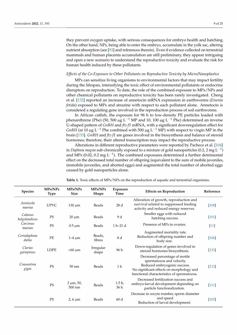
Table 1. Toxic effects of MPs/NPs on the reproduction of aquatic and terrestrial organisms.
Species MPs/NPsType
MPs/NPsSize
MPs/NPsShape
ExposureTime Effects on Reproduction Reference
Arenicolamarina UPVC 130 µm Beads 28 d
Alteration of growth, reproduction andsurvival related to suppressed feedingactivity and reduced energy reserves.
[108]
Calanushelgolandicus PS 20 µm Beads 9 d
Smaller eggs with reducedhatching success. [101]
Carcinusmaenas PS 0.5 µm Beads 1 h–21 d Presence of MPs in ovaries. [11]
Ceriodaphniadubia PE 1–4 µm Beads,
fibres 8 d
Augmented mortality rate.Reduction of offspring number and
body size.[106]
Clariasgariepinus LDPE <60 µm Irregular
shape 96 hDown-regulation of genes involved in
steroid hormones biosynthesis. [133]
Crassostreagigas PS 50 nm Beads 1 h
Decreased percentage of motilespermatozoa and velocity.
Reduced embryogenic success.No significant effects on morphology andfunctional characteristics of spermatozoa.
[112]
PS 2 µm, 50,500 nm Beads 1.5 h,
36 h
Decreased fertilization success andembryo-larval development depending on
particle functionalization.[111]
PS 2, 6 µm Beads 60 d
Decrease in oocyte number, sperm diameterand speed.
Reduction of larval development.[103]

Antioxidants 2022, 11, 193 10 of 25
Table 1. Cont.
Species MPs/NPsType
MPs/NPsSize
MPs/NPsShape
ExposureTime Effects on Reproduction Reference
Danio rerio PS 1 µm Beads 21 d
Higher expression of steroidogenic genes intestis but not in ovaries.
No variation of testosterone and17-β-estradiol levels.
No significant effects onprogeny development.
[118]
PS 70 nm Beads 30 d Accumulation of MPs in gonads. [7]
Daphniamagna MPs 1–5 µm Beads 21 d
Parental death up to the extinction ofF1 generation.
Reduced fecundity and populationgrowth rate.
Slight transgenerational recovery after thedepuration period.
[107]
MPs 1–5 µm Beads 21 d
Increased time of first brood emission.Increased number of immobile juveniles.
Decreased clutches and numberof progenies.
Worsened effects with the co-exposure togold nanoparticles and MPs.
[104]
PS 70 nm Beads 21 d
Impairment of population growth.Reduction of progeny.
Decrease in newborn number and body size.Increase of progeny malformations.
[107]
Emeritaanaloga PP 1 mm Fibres 71 d
Decrease in retention time of egg clutches.Augmented number of later
embryonic stages.[109]
Hemicentrotuspulcherrimus wild MPs 27–4742 µm
Fibres,fragments,
sheets,beads
u Presence of MPs in gonads. [92]
Hydraattenuate PE <400 µm Irregular
shape30 m,60 m
No significant impairment of reproduction. [117]
Mytilus edulis PS 0.5 µm Beads 1 h–21 d Presence of MPs in ovaries. [11]Oryzias
javanicusOryzias latipes
PS 2 µm Beads 21 dNo alteration in growth, survival and
egg production. [115]
Oryzias latipes PS 10 µm Beads 70 d Reduction in egg production. [114]
PE 10–63 µm Beads 90 dFewer egg number, hatching rate and
growth rate. [113]
Paracentrotuslividus PS 10, 45 µm Beads 72 h Presence of MPs in gonads. [69]
Pinctadamargaritifera PS 6, 10 µm Beads 60 d
Impaired gametogenesis.Histological alterations in the gonads. [110]
Tigriopusjaponicus
PEPA
10–30 µm5–20 µm
Irregularshape
24 h,14 d
Prolongation in development time and ininterval time between egg sacs. [105]
PS 0.5, 6 µm Beads 96 h
Impaired fecundity evidenced by thereduction in number of nauplius
per female.[102]
Caenorhabditiselegans PS 35 nm Beads 4 d
Transgenerational effects on reproductivefunction, gonadal development and
germline apoptosis, depending on particlefunctionalization.
[122]
LDPEPLA/PBAT
57 µm41 µm
Irregularshape 6 d Reduction in offspring. [120]

Antioxidants 2022, 11, 193 11 of 25
Table 1. Cont.
Species MPs/NPsType
MPs/NPsSize
MPs/NPsShape
ExposureTime Effects on Reproduction Reference
Eisenia andrei PE 180–212 µm250–300 µm Beads 21 d
Impaired spermatogenesis and histologicalalterations in male gonads.
Negligible effects on oogenesis andfemale gonads.
[28]
Enchytraeuscrypticus PA
13–18 µm63–90 µm90–150 µm
Irregularshape 20 h Reduction of juveniles per adult. [121]
Folsomiacandida PE <500 µm Beads 28 d
Decreased reproductive function withreduction of juvenile number. [123]
BALB/c mice PS 0.5, 4, 10 µm u 28 d
Presence of PS into testicular cells.Decreased sperm quality and increased
abnormality rate.Reduced testosterone levels.
Testicular inflammation and damagedblood-testis barrier.
[25]
ICR mice PS 5 µm u 35 d
Decreased number ofspermatids/spermatozoa with altered
sperm quality.Increased testicular inflammation and
apoptosis rate.
[124]
PE 40–48 µm u 90 d
Enlargement of Fallopian tubes in dams.Fewer live births per dam and altered sex
ratio of progeny.Reduced body weight of pups.
[126]
SpragueDawley rats PS 20 nm Beads 24 h
PS particles translocation to placental andfoetal tissues 24 h after maternal exposure. [127]
Wistar rats PS 25, 50 nm Beads 35 d
Presence of PS in testis.Histological alterations of testicular tissue.
Alteration of sex hormones levels.Impaired spermatogenesis and increased
DNA damage.
[95]
Humanplacenta MPs u
Beads,irregular
shapeu
Presence of MPs fragments in humanplacental tissues. [94]
PS 0.5 µm, 50nm Beads 24 h
Internalization of PS particles inplacental cells. [131]
PS 50, 80, 240,500 nm Beads 3 h
Crossing of the placental barrier by PSparticles in a size-dependent manner. [130]
d: days; h: hours; LDPE: low-density polyethylene; m: minutes; MPs: microplastics; NPs: nanoplastics; PA:polyamide; PE: polyethylene; PLA/PBAT: polylactide/poly(butylene adipate-co-terephthalate); PP: polypropy-lene; PS: polystyrene; PVC: polyvinyl chloride; u: unknown; UPVC: unplasticised polyvinylchloride.
4. Role of Oxidative Stress on Micro/Nanoplastic-Induced Reproduction Alterations
The impairment of MPs/NPs on reproduction can be caused by the redox unbalancesince OS is fully recognised as a key factor in ovarian and testicular dysfunctions anddevelopment of infertility [134,135]. The main toxic effects of MPs/NPs on reproductionvia OS are summarised in Table 2.
4.1. Reproductive Toxicity of Micro/Nanoplastics Mediated by Oxidative Stress inAquatic Organisms
To the best of our knowledge, the above toxicity mechanism underlying reproductivealterations was observed only in aquatic species experimentally exposed to MPs/NPs,

Antioxidants 2022, 11, 193 12 of 25
and not in wild animals. In these experimental models, OS was caused by the increasedROS/RNS production [136] and the reduction in antioxidant defences [100].
Firstly, the marine copepod Paracyclopina nana acutely exposed to PS (0.05, 0.5 and6 µm, 20 µg mL−1) showed an increase in both ROS production and antioxidant enzymeactivities (SOD, GR, GPx and GST) [58]. These effects were inversely related to PS size. ROS-dependent activation of the MAPK pathway and augmented expression of inflammatorymediators, i.e., p-ERK, p-p38 and Nrf2, resulted in the defence response to OS. The latterwas related to the impairment of reproductive function with fewer newborn nauplii [58].In comparable experimental conditions, in the monogonont rotifer Brachionus koreanus,similar results were reported of ROS overproduction, as well as an increase in antioxidantenzyme activity and p-p38 and p-JNK [59]. These effects were related to a reduction infecundity and lifespan, as well as a prolongation of reproduction time. Interestingly, inthese studies, ROS scavenger NAC (0.5 mM) reduced the ROS production. Xue et al. [137]exposed the freshwater rotifer Brachionus calyciflorus to PE (0.5 × 10³–1.25 × 10⁴ particlesmL−1, 10-22 µm) and observed an oxidative unbalance proved by abnormal SOD andGPx activities. The authors showed a compromised reproductive capability (alterations ofsurvival, lifespan, rates and time of reproduction, and population growth), linking theseeffects directly to OS and indirectly to a reduced energy availability necessary to counteractthe oxidative process. Moreover, impaired fecundity was revealed in Daphnia pulex exposedto PS (75 nm, 0–2 mg L−1, for 21 days), showing a prolonged time to first eggs and to firstclutch, and a reduction in number of clutches, newborns per clutch, and total number ofneonates per female [138]. The authors observed an altered gene expression of antioxidantenzymes (SOD, CAT, GST, and GPx), with a non-linear trend, and an augmented expressionof HSP70 and HSP90 genes. As known, heat shock proteins are considered biomarkersof stress induced by chemical pollutants and contribute to maintaining the protein com-position unchanged [139]. Very recently, the effect of the same spherical PS nanoparticleson reproduction of Daphnia pulex was studied, by examining the proteome together withmolecular and biochemical data involved in adverse outcome occurrence, evidencing ROSproduction and thus OS [140,141]. Both subacute and chronic NPs exposure negativelyaffected cumulative offspring production. Modifications of specific immune and metabolicsignalling pathways and cellular metabolism were observed, resulting in growth inhibitionand decreased reproduction. Previously, these authors reported that NPs at typical environ-mental concentrations (1µg L−1) have potent long-term toxic effects on Daphnia pulex [142].Chronic exposure to NPs modulated the response of antioxidant defences, vitellogenin syn-thesis, development, and energy production in the F0–F1 generations. Conversely, in theF2 generation, NPs did not affect the survival but altered the growth rate and reproduction,showing inhibitory effects on antioxidant responses.
Lee et al. [66] showed that 96 h treatment of the marine mysid (Neomysis awatschensis)with PS (5× 10⁵ particles mL−1, 1 and 10 µm) determined an increase in the MDA levels andantioxidant enzymes (SOD, CAT, GR, GSH, and GPx) activity in organisms exposed bothto 7 and 30 days after hatching. The results did not follow a clear linear trend, influencedby particle size, exposure time, and mysid age. Moreover, the protracted exposure causeda decrease in the 20-hydroxyecdysone levels (a steroid-like hormone strictly involved inarthropod moulting and growth, as well as ovarian and sexual development) and totalnewborns per female, more evident at the highest concentration used. Recently, Qiang andCheng [136] exposed zebrafish sub-chronically to 1 µm PS, and observed increased ROSgeneration in both the ovary and testis (at 100 and 1000 µg L−1 concentrations), as well as anaugmented spermatocytes apoptosis rate (at 1000 µg L−1). Moreover, an apoptosis increase(i.e., p-53, Bax, and caspase-7-8-9) and thickness reduction in the basement membrane wereobserved in male gonads. In the same species, a long-term exposure (35 days) to PS-MPscaused an alteration in the metabolic mechanisms and gene regulation patterns induced byROS generation and OS [143].
A broken redox balance was also found in marine medaka Oryzias melastigma afterexposure to environmentally relevant concentrations of PS (2–200 µg L−1, 10 µm, for
Antioxidants 2022, 11, 193 13 of 25
60 days) [100]. The results showed an increase in MDA levels and a decrease in antioxi-dant enzyme activity (SOD, CAT, GST, GPx, and GSH) in the ovary and testis, related tohistological changes (i.e., impaired seminiferous lobules architecture, reduction in spermfluid in male, reduced number of mature spawning follicles, and augmented presence ofearly vitellogenic oocytes in female). Moreover, the authors evidenced a reduction in plas-matic 17-β-estradiol and testosterone in females related to alteration of the hypothalamic–pituitary–gonadal axis. The latter effect determined the gonadosomatic index reduction,as well as the belated gonadal development. The reproductive impairment was alsodemonstrated by the onset of transgenerational consequences (i.e., decrease in the fertilityand hatching rate of progeny). Thereafter, the same authors adopted a whole life-cycleexposure of medaka to determine the impact of PS on the hatching of embryos, growthand reproduction of the F0 generation, and embryonic and larval development of the F1offspring [144]. Interestingly, 150 days of exposure caused gonad damage and a decreasedegg production and fertilization rate. Transcriptome analysis evidenced modifications insteroid hormone biosynthesis and cytochrome P450 pathways in the testes of male fishafter 20 µg L−1 MPs exposure.
Differently from the previous evidence, another transgenerational study revealed ascarce involvement of an antioxidant defence system in specimens of Danio rerio orallyexposed to nano-PS (1 mg g−1 bw, for 1 week). No alteration in parental fertility andoffspring development was observed, except a reduction in GR in the F0 male gonads, aswell as in the F1 larvae arising from exposed parents [119].
The reported studies show that MPs/NPs determine ROS overproduction, and wherethe related OS induction activate the signalling pathways involved in the production ofinflammatory mediators, causing immune and endocrine alterations. These effects resultin decreased fecundity, impaired reproductive capability, and compromised growth andsurvival of newborns, observed both in zooplankton and aquatic species at higher trophiclevels. The mitigation effect of antioxidant substances has been very poorly investigated.
4.2. Reproductive Toxicity of Micro/Nanoplastics Mediated by Oxidative Stress inTerrestrial Organisms
To date, few papers have analysed reproductive disorders related to MP/NP-inducedOS in terrestrial organisms. Lei et al. [145] observed a reduced number of embryos andclutch size in nematode Caenorhabditis elegans acutely exposed to 5 mg m−2 PA, PE, PP,PVC (70 µm) and PS (0.1, 1, and 5 µm). The authors also showed increased expressionof GST-4 enzyme in the gut, suggesting the involvement of OS. Other authors reportedthe enhancement of ROS production in the same species acutely exposed to 50 nm PS(86.8 mg L−1) [146] or 1 µm PS (1, 10 and 100 µg L−1) [147]; in both cases, a reduced numberof newborns was evidenced. Moreover, the OS involvement was further assessed by theincreased expression of OS-related genes (i.e., gst-4 and sod) and lipofuscin levels, whoseaccumulation is an indicator of cellular toxicity in response to oxidative damage [147].
The exposure of Caenorhabditis elegans to PS-NPs (20 and 100 nm) determined a severetransgenerational toxicity more evident for the smaller sizes (detected at the F1–F6 genera-tions) [148], and the toxicity on reproduction and locomotion behaviour was associated toincreased ROS production.
Very few and recent studies have been performed on rodent experimental modelsshowing effects comparable to those observed in aquatic species. The dose-dependentincrease in ROS and MDA content and the decrease in GSH activity were evidenced in micetestis after chronic oral exposure to PS (5–5.9 µm, 0.01–1 mg day−1) [149]. In testis, a redoxunbalance resulted in a not always linear activation of the JNK and p38 MAPK pathwaysand subsequent release of pro-inflammatory cytokines (TNF-α, IL-1β and IL-6) and pro-apoptotic caspase-3. The same authors also evidenced a reduction in sperm cell numberand quality, in testosterone levels, and in succinate dehydrogenase (SDH) and lactatedehydrogenase (LDH), enzymes involved in sperm generation and energy metabolism.Interestingly enough, the above effects were mitigated by the co-administration of the
Antioxidants 2022, 11, 193 14 of 25
antioxidant NAC (100 mg kg−1 bw day−1 intraperitoneally), further supporting the keyrole of OS in the onset of reproductive damage [149].
Deng et al. [24] showed the presence of MPs in the Sertoli cells of mice exposed to PE(0.4–5 µm, 100 mg kg−1 bw for 30 days) and an increase in the SOD and MDA content intestis. The authors evidenced a reduction in SDH and the augmentation of ACP and LDH,compromised spermatogenesis, as well as reduced number and viability of sperm cells andtestis weight. Similarly, Ijaz et al. [150] reported reduced sperm count, motility, and viabilityin rats treated with PS (2–2000 µg L−1). The effects, more evident at higher concentrations,were accompanied by a decreased expression of antioxidant enzymes together with anincrease in LPO and ROS. Moreover, the plasma levels of a few steroidogenic enzymes andthe key hormones of male reproductive health were reduced. The authors pointed at OS toexplain the effects reported above and the histological damages in the testis.
Li et al. [151] studied the effect of PS (0.015–1.5 mg day−1 for 90 days) on the blood–testis barrier impairment in Wistar rats, leading to the damage of the seminiferous tubules,apoptosis of spermatogenic cells, and decreased sperm motility and concentration. PSinduced OS (increasing MDA and decreasing antioxidant enzymes) and activated the p38MAPK pathway, depleting Nrf2. Analogously, higher doses of PS (4 and 10 µm, 20 and40 mg kg−1 bw) induced an alteration in the blood–testis barrier and, consequently, sper-matogenesis dysfunction in BALB/c mice, evidenced by cytoskeleton disorganization [152].This effect strictly depended on the increased ROS production that causes the imbalance inmTOR1 and mTOR2, which plays a role in regulating the actin cytoskeleton.
The effects of 0.5 µm PS on ovary were also evaluated by An et al. [23] treating femalerats with 0.015–1.5 mg day−1 MP in drinking water for 3 months. The authors evidencedthe presence of PS particles in the ovary and the induction of a fibrotic process limited by apre-treatment with 3 mmol NAC for 4 h.
Moreover, granulosa cells (GCs) from untreated rats were incubated with 1–25 µg mL−1
PS for 24 h; increased ROS and MDA content was observed, as well as decreased SOD, CAT,and GPx activities, evidencing the pivotal role of OS in the mechanism of toxicity of MPs.The increase in apoptotic rate of GCs was related to a Bax increase and Bcl-2 reduction,evidencing the activation of the Wnt/β-catenin pathway in the ovary. Lastly, the authorsnoticed the alterations in follicle morphology and number in the ovary, and the decrease inthe anti-Müllerian hormone (AMH) [23].
Accordingly, Hou et al. [96], in the same experimental conditions, observed overlap-ping results regarding the antioxidant enzymes, MDA, and AMH level in ovary tissue.Moreover, the authors hypothesized that OS may trigger the inflammation-associated factorNLRP3 inflammasome/caspase-1 signalling pathway, leading to pyroptosis and apoptosisof GCs, suggesting the involvement of PS in female infertility. The main OS-mediatedreproductive alterations caused by MPs/NPs are shown in Figure 1.
In terrestrial species, similarly to aquatic species, the studies reviewed show thatMPs/NPs affect reproduction, determining ROS generation, MDA production, and theincreased expression of OS-related genes. The latter effect is responsible for the release ofpro-inflammatory cytokines and pro-apoptotic mediators that cause impaired spermatoge-nesis, reduced sperm, and GCs viability. The apoptosis of sperm cells is also a consequenceof the blood–testis barrier damage. The limited number of studies on mammalian speciesdoes not allow us to well define the passage of the MPs/NPs in reproductive organs andthe following alterations. The beneficial role of antioxidant substances in reducing ROSoverproduction remain to be elucidated.
4.3. Reproductive Toxicity of Micro/Nanoplastics in Combination with Other Chemical PollutantsMediated by Oxidative Stress
To the best of our knowledge, only very few papers report data on reproductivetoxicity mediated by OS and which are related to joint exposure to MPs/NPs and otherxenobiotics. The four-week exposure to MPs and pesticide atrazine in earthworm (Eisenia

Antioxidants 2022, 11, 193 15 of 25
fetida) determined ROS accumulation and MDA production and a decrease in SOD, CAT,and GST activity [132].
Figure 1. Oxidative stress-mediated effects of MPs/NPs on reproduction.
Deng et al. [24] showed that phthalates esters (PAEs) may strengthen MP-inducedreproductive impairment in mice and hypothesized that the effect is related to OS. Indeed,the exposure to MPs combined with a PAE mixture determined a significant decrease insperm number and vitality compared to virgin MPs. Testicular weight was also reducedbut only at the highest PAE concentration (50 µg L−1). The combined treatment inducedsignificant increase in the ACP and LDH levels in the testes and decreased SDH. Themixture with the highest PAE concentration induced an enhancement of the oxidativeunbalance with augmented SOD activity and MDA content. Finally, the transcriptomicanalysis performed on the left-side testis after the exposure to MPs plus the high di-2-ethylhexylphthalate concentration or the PAE mixture aggravated the toxicity of the MPs.It was observed that the altered expression of genes was involved not only in endocrineand immune system functioning but also in lipid and energy metabolism. The authorsspeculated that the latter reported alterations may play a role in reproductive stress.
In the rotifer Brachionus koreanus, nano-PS pre-exposure followed by 2,2’,4,4’-tetrabromodiphenyl ether or triclosan challenge induced a reduction in the reproductive outputcompared to single POP exposure [153]. This alteration seems to be associated to the membranedysfunction provoked by OS, causing impairment of membrane fluidity and permeability.
In the same species, Yoon et al. [154] examined how multiple exposure to tributyltin(TBT)—an organotin compound—MPs, and a dietary regimen affects reproduction andcan influence offspring. Surprisingly, the results showed that environmentally relevantMP concentrations and feed can attenuate the TBT toxic effects, evidencing the majorefficacy of the diet. However, 24 h of MPs and/or TBT exposure in F0 can modulate theF1 life cycle, changing the population growth. The co-exposure to PS and TBT induceda slight increase in ROS production and alteration in SOD and CAT activities, without alinear trend at different nutritional schemes. MP co-exposure did not exacerbate the effectof decreased fecundity induced by TBT, even if a slight mitigation was observed for thelowest concentration of the microalgae Tetraselmis suecica in the diet. Similarly, the ingestionof irregular PS-MPs (exposure level of 6.4–100,000 particles mL−1) by the freshwatergastropod Lymnaea stagnalis did not induce significant effects on survival, reproduction,

Antioxidants 2022, 11, 193 16 of 25
energy reserves, and OS, and did not worsen the reproductive toxicity of copper [155].The lack of adverse effects may be explained by stress resilience or the adaptation ofthe freshwater gastropod to the contaminants, which occurs not only at environmentallyrelevant concentrations but also at higher concentrations.
Table 2. Toxic effects of MPs/NPs on reproduction in aquatic and terrestrial species via OS andoxidative unbalance.
Species MPs/NPsType
MPs/NPsSize
MPs/NPsShape
ExposureTime Effects on Reproduction Oxidative
Unbalance Reference
Brachionuscalyciflorus PE 10–22 µm Beads
Until thedeath ofmaternalrotifers
Reduction of reproductiveparameters, such as: survival,
lifespan, rates and time ofreproduction and population
growth.
↓ SOD↑ GPx
No alteration ofMDA levels
[137]
Brachionuskoreanus PS 0.05 µm Beads 24 h
Mitigated effects on reproductivetoxicity of TBT following the
co-exposure to MPs.
↑ ROSImpairment of SODand CAT activities
[154]
PS 0.05, 0.5,6 µm Beads 24 h
Reduced number of rotifers andfewer newborns following the
co-exposure to MPs and POPs.↑ ROS↑MDA [153]
PS 0.05, 0.5,6 µm Beads 24 h, 12 d
Reduced fecundity and lifespan.Augmented reproduction time.
↑ ROS↑ SOD, ↑ GR, ↑ GPx
and ↑ GST[59]
Danio rerio PS 1 µm Beads 21 d
Increased apoptosis rate in testisbut not in ovary.
Impairment of basementmembrane.
↑ ROS in ovary andtestis [136]
PS 42 nm Beads 7 d
Transfer in the yolk sac withouteffects on survival and
development of offspring.↓ GR in male gonads
and in F1 larvae [119]
Daphniapulex PS ~71 nm Beads 21 d
Lower production of cumulativeoffspring. ↑ GSH and ↑ GST [141]
PS ~75 nm Beads 21 d
Reduction of growth rate, totalclutches and offspring per female
in F2 generation.No significant effect on F0 and F1
generations.
↑ H2O2, ↑ CAT and↑GST
Affected expressionof genes related to
oxidative stress (i.e.,CAT, GSTD, MnSO
and CuZn SOD)
[142]
PS 75 nm Beads 21 d
Decreased number of clutchesand newborns.
Increased time to first eggs and tofirst clutch.
Altered geneexpression of SOD,CAT, GST and GPx
[138]
Eiseniafetida LDPE 550–100 µm Irregular
shape 28 d
Increased gene expression ofannetocin.
Worsened effects following theco-exposure to MPs and atrazine.
↑ ROS, ↑MDA and ↑8-OHdG
↓ SOD, ↓ CAT and ↓GST
[132]
Lymnaeastagnalis PS <63 µm Irregular
shape28 d No effects on the egg numbers
and survival rate.No effect on
oxidative balance [155]
Neomysisawatschen-
sisPS 1, 10 µm Beads 96 h
Reduction of 20-hydroxyecdysonelevels.
Fewer newborns per female.No alteration of survival rate of
newborns.
↑MDA↑ SOD, ↑ CAT, ↑ GR,↑ GSH and ↑ GPx
[66]
Oryziasmelastigma PS 2 µm Beads 150 d
Histological alteration in testisand ovaries.
Reduced eggs production andsuppressed fertilization rates.
Impairment of steroid hormonebiosynthesis.
Accelerated sexual maturity infemale.
Premature hatching and alteredgrowth in F1 generation.
↑MDA in ovary andfertilized eggs
↓ SOD, ↓ CAT, ↓ GPx,and ↓ GST in testis,ovary and fertilized
eggs
[144]

Antioxidants 2022, 11, 193 17 of 25
Table 2. Cont.
Species MPs/NPsType
MPs/NPsSize
MPs/NPsShape
ExposureTime Effects on Reproduction Oxidative
Unbalance Reference
PS 10 µm Beads 60 d
Histological alteration in maleand female gonads.
Reduced 17-β-estradiol andtestosterone plasmatic levels in
female related to gonadosomaticindex reduction and delayed
gonadal development.Decreased fertility and hatching
rate of progeny.
↑MDA↓ SOD, ↓ CAT, ↓ GST,↓ GPx and ↓ GSH in
ovary and testis
[100]
Paracyclopinanana PS 0.05, 0.5,
6 µm Beads 24 h Fewer newborn nauplii.↑ ROS
↑ SOD, ↑ GR, ↑ GPxand ↑ GST
[58]
Caenorhabditiselegans PS 1 µm Beads 72 h Fewer newborns.
↑ ROS↑ gst-4 and ↑ sodgenes expression↑ Lipofuscin levels
[147]
PS 50 nm Beads 24 h Fewer newborns per worm. ↑ ROS [146]
PA, PE, PP,PVC and
PS
70 µm0.1, 1,5 µm
Irregularshapeand
beads
2 d Fewer embryos and smallestclutch size. ↑ GST-4 [145]
BALB/cmale mice PS 5–5.9 µm Beads 42 d
Reduced sperm quality andquantity.
Decreased testosterone levels.Reduction in SDH and LDH
activities.
↑ ROS↑MDA↓ GSH
[149]
Musmusculus,male mice
PE 0.4–5 µm Beads 30 d
Presence of PE in testis.Impaired spermatogenesis
evidenced by alteration in ACP,LDH and SDH levels.
Reduced sperm quality.Worsened effects and reduced
testicular weight with theco-exposure to PAEs and MPs.
↑ SOD↑MDA [24]
FemaleWistar rats PS 0.5 µm Beads 90 d
Presence of PS particles in GCs.Increased apoptosis of GCs.Increased fibrosis in ovary
mediated by Wnt/β-Cateninsignalling pathway.
Alteration of follicle morphologyand quantity.
Reduced AMH levels.
↑ ROS and ↑MDA↓ SOD, ↓ CAT and ↓
GPx[23]
PS 0.5 µm Beads 90 d
Presence of PS particles in GCs.Fewer growing follicles.
Reduced AMH level.Pyroptosis and apoptosis of GCsmediated by NLRP3/Caspase-1
signalling pathway.
↑MDA↓ CAT, ↓ SOD and ↓
GPx[96]
Male Wistarrats PS 0.5 µm Beads 90 d
Disruption of blood-testis barrier.Histological alteration in testis.
Apoptosis of spermatogenic cells.Decreased sperm motility,concentration and quality.
↑MDA↓ CAT, ↓ SOD and ↓
GPx[151]
8-OHdG: 8-hydroxydeoxyguanosine; ACP: acid phosphatase; AMH: anti-Müllerian hormone; CAT: catalase; d:days; GCs: granulosa cells; GPx: glutathione peroxidase; GR: glutathione reductase; GSH: glutathione; GST:glutathione S-transferase; h: hours; LDH: lactate dehydrogenase; MDA: malondialdehyde; MPs: microplastics;NPs: nanoplastics; PA: polyamide; PAEs: phthalates esters; PE: polyethylene; POPs: persistent organic pollutants;PP: polypropylene; PS: polystyrene; PVC: polyvinyl chloride; ROS: reactive oxygen species; SDH: succinatedehydrogenase; SOD: superoxide dismutase; TBT: tributyltin.
Some authors report that reproductive toxicity via OS worsens with the co-exposureof MPs/NPs and other chemical pollutants. Some others find instead that MPs/NPsmay even mitigate the effects of chemical pollutants, thus making it difficult to drawclear conclusions.

Antioxidants 2022, 11, 193 18 of 25
5. Conclusions and Recommendations for Future Directions of Research onReproductive Alterations by Micro/Nanoplastics
According to the studies reviewed, OS is the leitmotif underlying the toxic effectsdetermined by MPs/NPs on reproduction. The alteration of the antioxidant defencemechanisms and the increase in ROS production causing the OS may trigger gene instability,impair immune response, and induce reproductive abnormalities.
Concerns arise relative to the experimental conditions adopted and the achievedresults. We noticed a mismatch between the concentrations, type, and size of MPs/NPs usedin experimental conditions and those detected in environmental abiotic and biotic matrices.In aquatic models, the MPs/NPs concentrations employed in the water are much higherthan those found in the environment, in freshwater, and even in heavily populated andindustrialized coastal areas. At low concentrations/doses, MPs/NPs have negligible toxiceffects and may even mitigate the negative impact of other contaminants in the case of co-exposure [156]. Following the hormesis principle, a biphasic concentration/dose–responserelationship may be observed. Organisms’ exposure to low and constant concentrations(below the toxicological threshold) of a stressor may induce adaptive mechanisms. Thus,over time, if the detrimental conditions are sustained, a positive outcome is observed inthe physiological function. A high enough stressor concentration (above the toxicologicalthreshold) may instead result in negative effects [156,157], though an adaptive responsehas been observed in some species (i.e., freshwater gastropods).
In aquatic ecosystem, MPs/NPs mainly consist of fibres, followed by fragments and,to a smaller extent, by other shapes such as beads, films, and foams. However, most studiesfocused on spherical particles and on PS as the most frequent polymer type.
The literature data suggested that MP/NP-induced toxicity may increase when ex-perimental procedures are based on small dimension particles with a positive charge, inaddition to high concentrations. Further, the prevailing particle size used in reproductivestudies is in the range 1–100 µm, and hence smaller than those frequently detected in theenvironment (see [158,159] and references therein). The majority of examined studies reportthe effects of virgin MPs/NPs manufactured by chemical companies and rarely those ofweathered plastics.
Many studies are carried out on marine species and rarely on mammals, terrestrial,and/or wild species. The use of wild species for the evaluation of MPs/NPs toxicity inreproductive systems may nevertheless present several challenges. Wild organisms areexposed to different types of external stressors in the environment, making it extremelydifficult to address the reported alterations to a specific stressor. Moreover, other factorscan deeply impact the stressor’s toxicity: age, sex, background history, and environment.
Besides PS and the other few polymers usually examined, further studies are neededto investigate other MP/NP types, testing mixtures of MPs/NPs in the appropriate propor-tions, mirroring natural environmental conditions. It is worth noting that PE is the mostcommon polymer detected in environmental samples. Pristine and aged plastics must beconsidered, adopting long-term exposure conditions, to better evaluate the chronic toxiceffects that reflect the continued exposure of living beings.
All these discrepancies (between experimental and natural conditions) make it difficultto assess the toxic effects of MPs/NPs, and they do not allow to draw any firm conclusionsabout the reproduction toxicity of MPs/NPs. It has been evident that exposure to MPs/NPscan trigger toxicity pathways, among which inflammation and OS play a pivotal role;OS-related signalling pathways involved in immune and endocrine alterations have beenobserved. Indeed, ROS overproduction, MDA generation, altered expression of antioxidantenzymes genes, and OS-related genes have been reported in both aquatic and terrestrialspecies, and are related to reproductive damage. This was frequently determined byimpaired spermatogenesis and reduced sperm quality, altered ovarian follicles morphologyand quantity, and reduced reproductive parameters, among which are the survival anddevelopment of offspring. Moreover, reduced levels of hormonal and enzymatic regulators

Antioxidants 2022, 11, 193 19 of 25
(i.e., testosterone, AMH, LDH, and SDH) have been observed, along with histologicalalterations in male and female gonads.
Very few studies have investigated the role of antioxidant substances on MP/NP-induced toxicity and reproductive impairment. They focused on NAC, evidencing itscapability to counteract oxidative damage through a reduction in ROS production and, inturn, the mitigation of several effects related to OS.
Notably, the lack of (i) adequate standard sampling protocols, (ii) identification ofappropriate species for MPs/NPs monitoring, (iii) development of standardized analyticalmethods for their detection in tissues, including those of the reproductive system, and (iv)uniformity in reporting units for both monitoring and toxicity studies, must be filled. Theharmonized laboratory procedures for MPs/NPs detection would allow the comparison ofavailable data on the occurrence of plastics in living organisms and the environment, aswell as the risk assessment for human health. The presence of MPs in foetal and maternalplacenta and in chorioamniotic membranes provides new light on the issue concerninghuman exposure to MPs. It is a matter of great concern, based on the crucial role of theplacenta in supporting foetal development and, at the same time, providing an interfacebetween the internal and the external environment. However, if the presence of MPsin human placenta is harmful to pregnancy, it must be investigated to determine theinvolvement of the immune system and inflammation. In fact, the results collected todate do not allow any extrapolation to mammal and human health but only lead us tohypothesize about the negative effects on male and female reproductive functions, whichneed to be elucidated.
Author Contributions: Conceptualization, M.C.F., R.M., and A.M.; formal analysis, M.C.F., A.M., F.D.P.and R.M.; investigation, M.C.F., A.M., F.D.P., G.M.R., and R.M.; data curation, M.C.F., A.M., F.D.P.,G.M.R., and R.M.; writing—original draft preparation, M.C.F., A.M., F.D.P., and R.M.; writing—reviewand editing, M.C.F., A.M., F.D.P., G.M.R., and R.M.; visualization, A.M., F.D.P., and G.M.R.; supervision,M.C.F., and R.M. All authors have read and agreed to the published version of the manuscript.
Funding: This research was funded by Italian Ministry of Health, IZS ME 06/19, CUP numberC75J19000170001.
Conflicts of Interest: The authors declare that there is no conflict of interest.
References1. Petersen, F.; Hubbart, J.A. The occurrence and transport of microplastics: The state of the science. Sci. Total Environ. 2021, 758,
143936. [CrossRef]2. Satti, S.M.; Shah, A.A. Polyester-based biodegradable plastics: An approach towards sustainable development. Lett. Appl. Microbiol.
2020, 70, 413–430. [CrossRef]3. EFSA. Presence of microplastics and nanoplastics in food, with particular focus on seafood. EFSA J. 2016, 14, e04501. [CrossRef]4. Andrady, A.L. The plastic in microplastics: A review. Mar. Pollut. Bull. 2017, 119, 12–22. [CrossRef]5. Deng, H.; Wei, R.; Luo, W.; Hu, L.; Li, B.; Di, Y.N.; Shi, H. Microplastic pollution in water and sediment in a textile industrial area.
Environ. Pollut. 2020, 258, 113658. [CrossRef] [PubMed]6. Bitter, H.; Lackner, S. First quantification of semi-crystalline microplastics in industrial wastewaters. Chemosphere 2020, 258,
127388. [CrossRef] [PubMed]7. Sarasamma, S.; Audira, G.; Siregar, P.; Malhotra, N.; Lai, Y.H.; Liang, S.T.; Chen, J.R.; Chen, K.H.; Hsiao, C.D. Nanoplastics cause
neurobehavioral impairments, reproductive and oxidative damages, and biomarker responses in zebrafish: Throwing up alarmsof wide spread health risk of exposure. Int. J. Mol. Sci. 2020, 21, 1410. [CrossRef] [PubMed]
8. Prokic, M.D.; Radovanovic, T.B.; Gavric, J.P.; Faggio, C. Ecotoxicological effects of microplastics: Examination of biomarkers,current state and future perspectives. Trends Anal. Chem. 2019, 111, 37–46. [CrossRef]
9. Gall, S.C.; Thompson, R.C. The impact of debris on marine life. Mar. Pollut. Bull. 2015, 92, 170–179. [CrossRef]10. Koongolla, J.B.; Lin, L.; Pan, Y.F.; Yang, C.P.; Sun, D.R.; Liu, S.; Xu, X.R.; Maharana, D.; Huang, J.S.; Li, H.X. Occurrence of
microplastics in gastrointestinal tracts and gills of fish from Beibu Gulf, South China Sea. Environ. Pollut. 2020, 258, 113734.[CrossRef] [PubMed]
11. Farrell, P.; Nelson, K. Trophic level transfer of microplastic: Mytilus edulis (L.) to Carcinus maenas (L.). Environ. Pollut. 2013, 177,1–3. [CrossRef]
12. Mercogliano, R.; Avio, C.G.; Regoli, F.; Anastasio, A.; Colavita, G.; Santonicola, S. Occurrence of microplastics in commercialseafood under the perspective of the human food chain. A review. J. Agric. Food Chem. 2020, 68, 5296–5301. [CrossRef]

Antioxidants 2022, 11, 193 20 of 25
13. Kim, J.S.; Lee, H.J.; Kim, S.K.; Kim, H.J. Global pattern of microplastics (MPs) in commercial food-grade salts: Sea salt as anindicator of seawater MP pollution. Environ. Sci. Technol. 2018, 52, 12819–12828. [CrossRef]
14. Koelmans, A.A.; Mohamed Nor, N.H.; Hermsen, E.; Kooi, M.; Mintenig, S.M.; De France, J. Microplastics in freshwaters anddrinking water: Critical review and assessment of data quality. Water Res. 2019, 155, 410–422. [CrossRef]
15. Smith, M.; Love, D.C.; Rochman, C.M.; Neff, R.A. Microplastics in seafood and the implications for human health.Curr. Environ. Health Rep. 2018, 5, 375–386. [CrossRef]
16. Bai, C.L.; Liu, L.Y.; Hu, Y.B.; Zeng, E.Y.; Guo, Y. Microplastics: A review of analytical methods, occurrence and characteristics infood, and potential toxicities to biota. Sci. Total Environ. 2021, 806 Pt 1, 150263. [CrossRef]
17. Prata, J.C. Airborne microplastics: Consequences to human health? Environ. Pollut. 2018, 234, 115–126. [CrossRef] [PubMed]18. Enyoh, C.E.; Shafea, L.; Verla, A.W.; Verla, E.N.; Qingyue, W.; Chowdhury, T.; Paredes, M. Microplastics exposure routes and
toxicity studies to ecosystems: An overview. Environ. Anal. Health Toxicol. 2020, 35, e2020004. [CrossRef] [PubMed]19. Chen, E.Y.; Lin, K.T.; Jung, C.C.; Chang, C.L.; Chen, C.Y. Characteristics and influencing factors of airborne microplastics in nail
salons. Sci. Total Environ. 2022, 806 Pt 4, 151472. [CrossRef]20. Liao, Z.; Ji, X.; Ma, Y.; Lv, B.; Huang, W.; Zhu, X.; Fang, M.; Wang, Q.; Wang, X.; Dahlgren, R.; et al. Airborne microplastics in
indoor and outdoor environments of a coastal city in Eastern China. J. Hazard. Mater. 2021, 417, 126007. [CrossRef] [PubMed]21. Prata, J.C.; da Costa, J.P.; Lopes, I.; Duarte, A.C.; Rocha-Santos, T. Environmental exposure to microplastics: An overview on
possible human health effects. Sci. Total Environ. 2020, 702, 134455. [CrossRef] [PubMed]22. Abbasi, S.; Turner, A. Human exposure to microplastics: A study in Iran. J. Hazard. Mater. 2021, 403, 123799. [CrossRef]23. An, R.; Wang, X.; Yang, L.; Zhang, J.; Wang, N.; Xu, F.; Hou, Y.; Zhang, H.; Zhang, L. Polystyrene microplastics cause granulosa
cells apoptosis and fibrosis in ovary through oxidative stress in rats. Toxicology 2021, 449, 152665. [CrossRef] [PubMed]24. Deng, Y.; Yan, Z.; Shen, R.; Huang, Y.; Ren, H.; Zhang, Y. Enhanced reproductive toxicities induced by phthalates contaminated
microplastics in male mice (Mus musculus). J. Hazard. Mater. 2021, 406, 124644. [CrossRef]25. Jin, H.; Ma, T.; Sha, X.; Liu, Z.; Zhou, Y.; Meng, X.; Chen, Y.; Han, X.; Ding, J. Polystyrene microplastics induced male reproductive
toxicity in mice. J. Hazard. Mater. 2021, 401, 123430. [CrossRef]26. Banerjee, A.; Shelver, W.L. Micro- and nanoplastic induced cellular toxicity in mammals: A review. Sci. Total Environ. 2021, 755 Pt
2, 142518. [CrossRef]27. Zarus, G.M.; Muianga, C.; Hunter, C.M.; Pappas, R.S. A review of data for quantifying human exposures to micro and nanoplastics
and potential health risks. Sci. Total Environ. 2021, 756, 144010. [CrossRef] [PubMed]28. Kwak, J.I.; An, Y.J. Microplastic digestion generates fragmented nanoplastics in soils and damages earthworm spermatogenesis
and coelomocyte viability. J. Hazard. Mater. 2021, 402, 124034. [CrossRef] [PubMed]29. Lehner, R.; Weder, C.; Petri-Fink, A.; Rothen-Rutishauser, B. Emergence of Nanoplastic in the Environment and Possible Impact
on Human Health. Environ. Sci. Technol. 2019, 53, 1748–1765. [CrossRef] [PubMed]30. Bhattacharjee, S.; Ershov, D.; Islam, M.A.; Kämpfer, A.M.; Maslowska, K.A.; van der Gucht, J.; Alink, G.M.; Marcelis, A.T.M.;
Zuilhof, H.; Rietjens, I.M.C.M. Role of membrane disturbance and oxidative stress in the mode of action underlying the toxicityof differently charged polystyrene nanoparticles. RSC Adv. 2014, 4, 19321–19330. [CrossRef]
31. Wang, F.; Salvati, A.; Boya, P. Lysosome-dependent cell death and deregulated autophagy induced by amine-modified polystyrenenanoparticles. Open Biol. 2018, 8, 170271. [CrossRef]
32. Holloczki, O.; Gehrke, S. Can nanoplastics alter cell membranes? ChemPhysChem 2020, 21, 9–12. [CrossRef]33. Hwang, J.; Choi, D.; Han, S.; Choi, J.; Hong, J. An assessment of the toxicity of polypropylene microplastics in human derived
cells. Sci. Total Environ. 2019, 684, 657–669. [CrossRef]34. Dong, C.D.; Chen, C.W.; Chen, Y.C.; Chen, H.H.; Lee, J.S.; Lin, C.H. Polystyrene microplastic particles: In vitro pulmonary toxicity
assessment. J. Hazard. Mater. 2020, 385, 121575. [CrossRef]35. Xu, M.; Halimu, G.; Zhang, Q.; Song, Y.; Fu, X.; Li, Y.; Li, Y.; Zhang, H. Internalization and toxicity: A preliminary study of effects
of nanoplastic particles on human lung epithelial cell. Sci. Total Environ. 2019, 694, 133794. [CrossRef] [PubMed]36. Li, B.; Ding, Y.; Cheng, X.; Sheng, D.; Xu, Z.; Rong, Q.; Wu, Y.; Zhao, H.; Ji, X.; Zhang, Y. Polyethylene microplastics affect the
distribution of gut microbiota and inflammation development in mice. Chemosphere 2020, 244, 125492. [CrossRef]37. Auguste, M.; Lasa, A.; Balbi, T.; Pallavicini, A.; Vezzulli, L.; Canesi, L. Impact of nanoplastics on hemolymph immune parameters
and microbiota composition in Mytilus galloprovincialis. Mar. Environ. Res. 2020, 159, 105017. [CrossRef] [PubMed]38. Auguste, M.; Balbi, T.; Ciacci, C.; Canonico, B.; Papa, S.; Borello, A.; Vezzulli, L.; Canesi, L. Shift in immune parameters after
repeated exposure to nanoplastics in the marine bivalve Mytilus. Front. Immunol. 2020, 11, 426. [CrossRef]39. Bergami, E.; Krupinski Emerenciano, A.; González-Aravena, M.; Cárdenas, C.A.; Hernández, P.; Silva, J.R.M.C.; Corsi, I.
Polystyrene nanoparticles affect the innate immune system of the Antarctic sea urchin Sterechinus neumayeri. Polar Biol. 2019, 42,743–757. [CrossRef]
40. Rahman, A.; Sarkar, A.; Yadav, O.P.; Achari, G.; Slobodnik, J. Potential human health risks due to environmental exposure tonano- and microplastics and knowledge gaps: A scoping review. Sci. Total Environ. 2021, 757, 143872. [CrossRef] [PubMed]
41. Fred-Ahmadu, O.H.; Bhagwat, G.; Oluyoye, I.; Benson, N.U.; Ayejuyo, O.O.; Palanisami, T. Interaction of chemical contaminantswith microplastics: Principles and perspectives. Sci. Total Environ. 2020, 706, 135978. [CrossRef]
42. Bhagat, J.; Nishimura, N.; Shimada, Y. Toxicological interactions of microplastics/nanoplastics and environmental contaminants:Current knowledge and future perspectives. J. Hazard. Mater. 2021, 405, 123913. [CrossRef]

Antioxidants 2022, 11, 193 21 of 25
43. Capò, X.; Company, J.J.; Alomar, C.; Compa, M.; Sureda, A.; Grau, A.; Hansjosten, B.; Lopez-Vazquez, J.; Quintana, J.B.; Rodil,R.; et al. Long-term exposure to virgin and seawater exposed microplastic enriched-diet causes liver oxidative stress andinflammation in gilthead seabream Sparus aurata, Linnaeus 1758. Sci. Total Environ. 2021, 767, 144976. [CrossRef] [PubMed]
44. Wang, J.; Guo, X.; Xue, J. Biofilm-Developed Microplastics as Vectors of Pollutants in Aquatic Environments. Environ. Sci. Technol.2021, 55, 12780–12790. [CrossRef]
45. Zhou, W.; Tang, Y.; Du, X.; Han, Y.; Shi, W.; Sun, S.; Zhang, W.; Zheng, H.; Liu, G. Fine polystyrene microplastics render immuneresponses more vulnerable to two veterinary antibiotics in a bivalve species. Mar. Pollut. Bull. 2021, 164, 111995. [CrossRef][PubMed]
46. D’Angelo, S.; Meccariello, R. Microplastics: A threat for male fertility. Int. J. Environ. Res. Public Health 2021, 18, 2392. [CrossRef][PubMed]
47. Luo, T.; Zhang, Y.; Wang, C.; Wang, X.; Zhou, J.; Shen, M.; Zhao, Y.; Fu, Z.; Jin, Y. Maternal exposure to different sizes ofpolystyrene microplastics during gestation causes metabolic disorders in their offspring. Environ. Pollut. 2019, 255, 113122.[CrossRef]
48. Espinosa, C.; Garcia Beltran, J.M.; Esteban, M.A.; Cuesta, A. In vitro effects of virgin microplastics on fish head-kidney leucocyteactivities. Environ. Pollut. 2018, 235, 30–38. [CrossRef]
49. Poma, A.; Vecchiotti, G.; Colafarina, S.; Zarivi, O.; Aloisi, M.; Arrizza, L.; Chichiricco, G.; Di Carlo, P. In vitro genotoxicity ofpolystyrene nanoparticles on the human fibroblast Hs27 cell line. Nanomaterials 2019, 9, 1299. [CrossRef]
50. Jin, Y.; Lu, L.; Tu, W.; Luo, T.; Fu, Z. Impacts of polystyrene microplastic on the gut barrier, microbiota and metabolism of mice.Sci. Total Environ. 2019, 649, 308–317. [CrossRef] [PubMed]
51. Qiao, R.; Sheng, C.; Lu, Y.; Zhang, Y.; Ren, H.; Lemos, B. Microplastics induce intestinal inflammation, oxidative stress, anddisorders of metabolome and microbiome in zebrafish. Sci. Total Environ. 2019, 662, 246–253. [CrossRef]
52. Deng, Y.; Zhang, Y.; Lemos, B.; Ren, H. Tissue accumulation of microplastics in mice and biomarker responses suggest widespreadhealth risks of exposure. Sci. Rep. 2017, 7, 46687. [CrossRef] [PubMed]
53. Shi, W.; Han, Y.; Sun, S.; Tang, Y.; Zhou, W.; Du, X.; Liu, G. Immunotoxicities of microplastics and sertraline, alone and incombination, to a bivalve species: Size-dependent interaction and potential toxication mechanism. J. Hazard. Mater. 2020, 396,122603. [CrossRef]
54. Banaee, M.; Gholamhosseini, A.; Sureda, A.; Soltanian, S.; Fereidouni, M.S.; Ibrahim, A.T.A. Effects of microplastic exposureon the blood biochemical parameters in the pond turtle (Emys orbicularis). Environ. Sci. Pollut. Res. Int. 2021, 28, 9221–9234.[CrossRef]
55. Jeong, J.; Choi, J. Adverse outcome pathways potentially related to hazard identification of microplastics based on toxicitymechanisms. Chemosphere 2019, 231, 249–255. [CrossRef]
56. Hu, M.; Palic, D. Micro- and nano-plastics activation of oxidative and inflammatory adverse outcome pathways. Redox Biol. 2020,37, 101620. [CrossRef] [PubMed]
57. Choi, J.S.; Hong, S.H.; Park, J.W. Evaluation of microplastic toxicity in accordance with different sizes and exposure times in themarine copepod Tigriopus japonicus. Mar. Environ. Res. 2019, 153, 104838. [CrossRef] [PubMed]
58. Jeong, C.B.; Kang, H.M.; Lee, M.C.; Kim, D.H.; Han, J.; Hwang, D.S.; Souissi, S.; Lee, S.J.; Shin, K.H.; Park, H.G.; et al. Adverseeffects of microplastics and oxidative stress-induced MAPK/Nrf2 pathway-mediated defense mechanisms in the marine copepodParacyclopina nana. Sci. Rep. 2017, 7, 41323. [CrossRef]
59. Jeong, C.B.; Won, E.J.; Kang, H.M.; Lee, M.C.; Hwang, D.S.; Hwang, U.K.; Zhou, B.; Souissi, S.; Lee, S.J.; Lee, J.S. MicroplasticSize-Dependent Toxicity, Oxidative Stress Induction, and p-JNK and p-p38 Activation in the Monogonont Rotifer (Brachionuskoreanus). Environ. Sci. Technol. 2016, 50, 8849–8857. [CrossRef]
60. Lu, Y.; Zhang, Y.; Deng, Y.; Jiang, W.; Zhao, Y.; Geng, J.; Ding, L.; Ren, H. Uptake and Accumulation of Polystyrene Microplasticsin Zebrafish (Danio rerio) and Toxic Effects in Liver. Environ. Sci. Technol. 2016, 50, 4054–4060. [CrossRef]
61. Wan, Z.; Wang, C.; Zhou, J.; Shen, M.; Wang, X.; Fu, Z.; Jin, Y. Effects of polystyrene microplastics on the composition of themicrobiome and metabolism in larval zebrafish. Chemosphere 2019, 217, 646–658. [CrossRef] [PubMed]
62. Zhao, T.; Tan, L.; Zhu, X.; Huang, W.; Wang, J. Size-dependent oxidative stress effect of nano/micro-scaled polystyrene on Kareniamikimotoi. Mar. Pollut. Bull. 2020, 154, 111074. [CrossRef] [PubMed]
63. Barboza, L.G.A.; Lopes, C.; Oliveira, P.; Bessa, F.; Otero, V.; Henriques, B.; Raimundo, J.; Caetano, M.; Vale, C.; Guilhermino, L.Microplastics in wild fish from North East Atlantic Ocean and its potential for causing neurotoxic effects, lipid oxidative damage,and human health risks associated with ingestion exposure. Sci. Total Environ. 2020, 717, 134625. [CrossRef]
64. Hamed, M.; Soliman, H.A.M.; Osman, A.G.M.; Sayed, A.E.H. Antioxidants and molecular damage in Nile Tilapia (Oreochromisniloticus) after exposure to microplastics. Environ. Sci. Pollut. Res. Int. 2020, 27, 14581–14588. [CrossRef] [PubMed]
65. Iheanacho, S.C.; Odo, G.E. Dietary exposure to polyvinyl chloride microparticles induced oxidative stress and hepatic damage inClarias gariepinus (Burchell, 1822). Environ. Sci. Pollut. Res. 2020, 27, 21159–21173. [CrossRef] [PubMed]
66. Lee, D.H.; Lee, S.; Rhee, J.S. Consistent exposure to microplastics induces age-specific physiological and biochemical changes in amarine mysid. Mar. Pollut Bull. 2021, 162, 111850. [CrossRef]
67. Trestrail, C.; Nugegoda, D.; Shimeta, J. Invertebrate responses to microplastic ingestion: Reviewing the role of the antioxidantsystem. Sci. Total Environ. 2020, 734, 138559. [CrossRef] [PubMed]
Antioxidants 2022, 11, 193 22 of 25
68. Alomar, C.; Sureda, A.; Capo, X.; Guijarro, B.; Tejada, S.; Deudero, S. Microplastic ingestion by Mullus surmuletus Linnaeus, 1758fish and its potential for causing oxidative stress. Environ. Res. 2017, 159, 135–142. [CrossRef]
69. Murano, C.; Agnisola, C.; Caramiello, D.; Castellano, I.; Casotti, R.; Corsi, I.; Palumbo, A. How sea urchins face microplastics:Uptake, tissue distribution and immune system response. Environ. Pollut 2020, 264, 114685. [CrossRef]
70. Brandts, I.; Teles, M.; Goncalves, A.P.; Barreto, A.; Franco-Martinez, L.; Tvarijonaviciute, A.; Martins, M.A.; Soares, A.; Tort, L.;Oliveira, M. Effects of nanoplastics on Mytilus galloprovincialis after individual and combined exposure with carbamazepine. Sci.Total Environ. 2018, 643, 775–784. [CrossRef] [PubMed]
71. Furukawa, S.; Fujita, T.; Shimabukuro, M.; Iwaki, M.; Yamada, Y.; Nakajima, Y.; Nakayama, O.; Makishima, M.; Matsuda, M.;Shimomura, I. Increased oxidative stress in obesity and its impact on metabolic syndrome. J. Clin. Investig. 2004, 114, 1752–1761.[CrossRef]
72. Goyette, P.; Labbe, C.; Trinh, T.T.; Xavier, R.J.; Rioux, J.D. Molecular pathogenesis of inflammatory bowel disease: Genotypes,phenotypes and personalized medicine. Ann. Med. 2007, 39, 177–199. [CrossRef]
73. Yu, P.; Liu, Z.; Wu, D.; Chen, M.; Lv, W.; Zhao, Y. Accumulation of polystyrene microplastics in juvenile Eriocheir sinensis andoxidative stress effects in the liver. Aquat. Toxicol. 2018, 200, 28–36. [CrossRef]
74. Liu, Z.; Huang, Y.; Jiao, Y.; Chen, Q.; Wu, D.; Yu, P.; Li, Y.; Cai, M.; Zhao, Y. Polystyrene nanoplastic induces ROS productionand affects the MAPK-HIF-1/NFkB-mediated antioxidant system in Daphnia pulex. Aquat. Toxicol. 2020, 220, 105420. [CrossRef][PubMed]
75. Tang, J.; Ni, X.; Zhou, Z.; Wang, L.; Lin, S. Acute microplastic exposure raises stress response and suppresses detoxification andimmune capacities in the scleractinian coral Pocillopora damicornis. Environ. Pollut. 2018, 243 Pt A, 66–74. [CrossRef]
76. Wang, X.; Huang, W.; Wei, S.; Shang, Y.; Gu, H.; Wu, F.; Lan, Z.; Hu, M.; Shi, H.; Wang, Y. Microplastics impair digestiveperformance but show little effects on antioxidant activity in mussels under low pH conditions. Environ. Pollut. 2020, 258, 113691.[CrossRef]
77. Wang, J.; Coffin, S.; Sun, C.; Schlenk, D.; Gan, J. Negligible effects of microplastics on animal fitness and HOC bioaccumulation inearthworm Eisenia fetida in soil. Environ. Pollut. 2019, 249, 776–784. [CrossRef]
78. Rodriguez-Seijo, A.; da Costa, J.P.; Rocha-Santos, T.; Duarte, A.C.; Pereira, R. Oxidative stress, energy metabolism and molecularresponses of earthworms (Eisenia fetida) exposed to low-density polyethylene microplastics. Environ. Sci. Pollut. Res. Int. 2018, 25,33599–33610. [CrossRef] [PubMed]
79. Sun, W.; Meng, Z.; Li, R.; Zhang, R.; Jia, M.; Yan, S.; Tian, S.; Zhou, Z.; Zhu, W. Joint effects of microplastic and dufulin onbioaccumulation, oxidative stress and metabolic profile of the earthworm (Eisenia fetida). Chemosphere 2021, 263, 128171. [CrossRef][PubMed]
80. De Felice, B.; Ambrosini, R.; Bacchetta, R.; Ortenzi, M.A.; Parolini, M. Dietary exposure to polyethylene terephthalate microplastics(PET-MPs) induces faster growth but not oxidative stress in the giant snail Achatina reticulata. Chemosphere 2020, 270, 129430.[CrossRef]
81. Geiser, M.; Rothen-Rutishauser, B.; Kapp, N.; Schurch, S.; Kreyling, W.; Schulz, H.; Semmler, M.; Im Hof, V.; Heyder, J.; Gehr, P.Ultrafine particles cross cellular membranes by nonphagocytic mechanisms in lungs and in cultured cells. Environ. Health Perspect.2005, 113, 1555–1560. [CrossRef]
82. Schirinzi, G.F.; Perez-Pomeda, I.; Sanchis, J.; Rossini, C.; Farre, M.; Barcelo, D. Cytotoxic effects of commonly used nanomaterialsand microplastics on cerebral and epithelial human cells. Environ. Res. 2017, 159, 579–587. [CrossRef] [PubMed]
83. Wang, Q.; Bai, J.; Ning, B.; Fan, L.; Sun, T.; Fang, Y.; Wu, J.; Li, S.; Duan, C.; Zhang, Y.; et al. Effects of bisphenol A and nanoscaleand microscale polystyrene plastic exposure on particle uptake and toxicity in human Caco-2 cells. Chemosphere 2020, 254, 126788.[CrossRef] [PubMed]
84. Wu, B.; Wu, X.; Liu, S.; Wang, Z.; Chen, L. Size-dependent effects of polystyrene microplastics on cytotoxicity and efflux pumpinhibition in human Caco-2 cells. Chemosphere 2019, 221, 333–341. [CrossRef]
85. Chiu, H.W.; Xia, T.; Lee, Y.H.; Chen, C.W.; Tsai, J.C.; Wang, Y.J. Cationic polystyrene nanospheres induce autophagic cell deaththrough the induction of endoplasmic reticulum stress. Nanoscale 2015, 7, 736–746. [CrossRef]
86. Choi, A.; Alam, J. Heme oxygenase-1: Function, regulation, and implication of a novel stress-inducible protein in oxidant-inducedlung injury. Am. J. Respir. Cell. Mol. Biol. 1996, 15, 9–19. [CrossRef]
87. Asmonaite, G.; Larsson, K.; Undeland, I.; Sturve, J.; Carney Almroth, B. Size matters: Ingestion of relatively large microplasticscontaminated with environmental pollutants posed little risk for fish health and fillet quality. Environ. Sci. Technol. 2018, 52,14381–14391. [CrossRef] [PubMed]
88. Xia, T.; Kovochich, M.; Brant, J.; Hotze, M.; Sempf, J.; Oberley, T.; Sioutas, C.; Yeh, J.I.; Wiesner, M.R.; Nel, A.E. Comparison ofthe abilities of ambient and manufactured nanoparticles to induce cellular toxicity according to an oxidative stress paradigm.Nano Lett. 2006, 6, 1794–1807. [CrossRef]
89. Donaldson, K.; Brown, D.; Clouter, A.; Duffin, R.; MacNee, W.; Renwick, L.; Tran, L.; Stone, V. The pulmonary toxicology ofultrafine particles. J. Aerosol Med. 2002, 15, 213–220. [CrossRef] [PubMed]
90. Sayed, A.E.H.; Hamed, M.; Badrey, A.E.A.; Soliman, H.A.M. Bioremediation of hemotoxic and oxidative stress induced bypolyethylene microplastic in Clarias gariepinus using lycopene, citric acid, and chlorella. Comp. Biochem. Physiol. C Toxicol. Pharm.2021, 250, 109189. [CrossRef]

Antioxidants 2022, 11, 193 23 of 25
91. Shengchen, W.; Jing, L.; Yujie, Y.; Yue, W.; Shiwen, X. Polystyrene microplastics-induced ROS overproduction disrupts the skeletalmuscle regeneration by converting myoblasts into adipocytes. J. Hazard. Mater. 2021, 417, 125962. [CrossRef] [PubMed]
92. Feng, Z.; Wang, R.; Zhang, T.; Wang, J.; Huang, W.; Li, J.; Xu, J.; Gao, G. Microplastics in specific tissues of wild sea urchins alongthe coastal areas of northern China. Sci. Total Environ. 2020, 728, 138660. [CrossRef] [PubMed]
93. Yin, K.; Wang, Y.; Zhao, H.; Wang, D.; Guo, M.; Mu, M.; Liu, Y.; Nie, X.; Li, B.; Li, J.; et al. A comparative review of microplasticsand nanoplastics: Toxicity hazards on digestive, reproductive and nervous system. Sci. Total Environ. 2021, 774, 145758. [CrossRef]
94. Ragusa, A.; Svelato, A.; Santacroce, C.; Catalano, P.; Notarstefano, V.; Carnevali, O.; Papa, F.; Rongioletti, M.C.A.; Baiocco, F.;Draghi, S.; et al. Plasticenta: First evidence of microplastics in human placenta. Environ. Int. 2021, 146, 106274. [CrossRef]
95. Amereh, F.; Babaei, M.; Eslami, A.; Fazelipour, S.; Rafiee, M. The emerging risk of exposure to nano(micro)plastics on endocrinedisturbance and reproductive toxicity: From a hypothetical scenario to a global public health challenge. Environ. Pollut. 2020, 261,114158. [CrossRef] [PubMed]
96. Hou, J.; Lei, Z.; Cui, L.; Hou, Y.; Yang, L.; An, R.; Wang, Q.; Li, S.; Zhang, H.; Zhang, L. Polystyrene microplastics lead topyroptosis and apoptosis of ovarian granulosa cells via NLRP3/Caspase-1 signaling pathway in rats. Ecotoxicol. Environ. Saf.2021, 212, 112012. [CrossRef]
97. Besseling, E.; Wang, B.; Lurling, M.; Koelmans, A.A. Nanoplastic affects growth of S. obliquus and reproduction of D. magna.Environ. Sci. Technol. 2014, 48, 12336–12343. [CrossRef] [PubMed]
98. De Sà, L.C.; Oliveira, M.; Ribeiro, F.; Rocha, T.L.; Futter, M.N. Studies of the effects of microplastics on aquatic organisms: Whatdo we know and where should we focus our efforts in the future? Sci. Total Environ. 2018, 645, 1029–1039. [CrossRef]
99. Choi, D.; Bang, J.; Kim, T.; Oh, Y.; Hwang, Y.; Hong, J. In vitro chemical and physical toxicities of polystyrene microfragments inhuman-derived cells. J. Hazard. Mater. 2020, 400, 123308. [CrossRef]
100. Wang, J.; Li, Y.; Lu, L.; Zheng, M.; Zhang, X.; Tian, H.; Wang, W.; Ru, S. Polystyrene microplastics cause tissue damages,sex-specific reproductive disruption and transgenerational effects in marine medaka (Oryzias melastigma). Environ. Pollut. 2019,254 Pt B, 113024. [CrossRef]
101. Cole, M.; Lindeque, P.; Fileman, E.; Halsband, C.; Galloway, T.S. The impact of polystyrene microplastics on feeding, function andfecundity in the marine copepod Calanus helgolandicus. Environ. Sci. Technol. 2015, 49, 1130–1137. [CrossRef] [PubMed]
102. Lee, K.W.; Shim, W.J.; Kwon, O.Y.; Kang, J.H. Size-dependent effects of micro polystyrene particles in the marine copepodTigriopus japonicus. Environ. Sci. Technol. 2013, 47, 11278–11283. [CrossRef] [PubMed]
103. Sussarellu, R.; Suquet, M.; Thomas, Y.; Lambert, C.; Fabioux, C.; Pernet, M.E.; Le Goic, N.; Quillien, V.; Mingant, C.; Epelboin, Y.;et al. Oyster reproduction is affected by exposure to polystyrene microplastics. Proc. Natl. Acad. Sci. USA 2016, 113, 2430–2435.[CrossRef]
104. Pacheco, A.; Martins, A.; Guilhermino, L. Toxicological interactions induced by chronic exposure to gold nanoparticles andmicroplastics mixtures in Daphnia magna. Sci. Total Environ. 2018, 628–629, 474–483. [CrossRef] [PubMed]
105. Yu, J.; Tian, J.Y.; Xu, R.; Zhang, Z.Y.; Yang, G.P.; Wang, X.D.; Lai, J.G.; Chen, R. Effects of microplastics exposure on ingestion,fecundity, development, and dimethylsulfide production in Tigriopus japonicus (Harpacticoida, copepod). Environ. Pollut. 2020,267, 115429. [CrossRef] [PubMed]
106. Ziajahromi, S.; Kumar, A.; Neale, P.A.; Leusch, F.D.L. Impact of microplastic beads and fibers on waterflea (Ceriodaphnia dubia)survival, growth, and reproduction: Implications of single and mixture exposures. Environ. Sci. Technol. 2017, 51, 13397–13406.[CrossRef]
107. Martins, A.; Guilhermino, L. Transgenerational effects and recovery of microplastics exposure in model populations of thefreshwater cladoceran Daphnia magna Straus. Sci. Total Environ. 2018, 631–632, 421–428. [CrossRef] [PubMed]
108. Wright, S.L.; Rowe, D.; Thompson, R.C.; Galloway, T.S. Microplastic ingestion decreases energy reserves in marine worms.Curr. Biol. 2013, 23, R1031–R1033. [CrossRef] [PubMed]
109. Horn, D.A.; Granek, E.F.; Steele, C.L. Effects of environmentally relevant concentrations of microplastic fibers on Pacific molecrab (Emerita analoga) mortality and reproduction. Limnol. Oceanogr. Lett. 2019, 5, 74–83. [CrossRef]
110. Gardon, T.; Reisser, C.; Soyez, C.; Quillien, V.; Le Moullac, G. Microplastics affect energy balance and gametogenesis in the pearloyster Pinctada margaritifera. Environ. Sci. Technol. 2018, 52, 5277–5286. [CrossRef]
111. Tallec, K.; Huvet, A.; Di Poi, C.; Gonzalez-Fernandez, C.; Lambert, C.; Petton, B.; Le Goic, N.; Berchel, M.; Soudant, P.; Paul-Pont,I. Nanoplastics impaired oyster free living stages, gametes and embryos. Environ. Pollut. 2018, 242 Pt B, 1226–1235. [CrossRef]
112. Tallec, K.; Paul-Pont, I.; Boulais, M.; Le Goïc, N.; González-Fernández, C.; Le Grand, F.; Bideau, A.; Quéré, C.; Cassone, A.-L.;Lambert, C.; et al. Nanopolystyrene beads affect motility and reproductive success of oyster spermatozoa (Crassostrea gigas).Nanotoxicology 2020, 14, 1039–1057. [CrossRef]
113. Chisada, S.; Yoshida, M.; Karita, K. Ingestion of polyethylene microbeads affects the growth and reproduction of medaka, Oryziaslatipes. Environ. Pollut. 2019, 254 Pt B, 113094. [CrossRef]
114. Zhu, M.; Chernick, M.; Rittschof, D.; Hinton, D.E. Chronic dietary exposure to polystyrene microplastics in maturing Japanesemedaka (Oryzias latipes). Aquat. Toxicol. 2020, 220, 105396. [CrossRef] [PubMed]
115. Assas, M.; Qiu, X.; Chen, K.; Ogawa, H.; Xu, H.; Shimasaki, Y.; Oshima, Y. Bioaccumulation and reproductive effects of fluorescentmicroplastics in medaka fish. Mar. Pollut. Bull. 2020, 158, 111446. [CrossRef] [PubMed]

Antioxidants 2022, 11, 193 24 of 25
116. Silva, C.J.; Patricio Silva, A.L.; Campos, D.; Soares, A.M.; Pestana, J.L.; Gravato, C. Lumbriculus variegatus (oligochaeta) exposed topolyethylene microplastics: Biochemical, physiological and reproductive responses. Ecotoxicol. Environ. Saf. 2021, 207, 111375.[CrossRef]
117. Murphy, F.; Quinn, B. The effects of microplastic on freshwater Hydra attenuata feeding, morphology & reproduction.Environ. Pollut. 2018, 234, 487–494. [CrossRef] [PubMed]
118. Qiang, L.; Lo, L.S.H.; Gao, Y.; Cheng, J. Parental exposure to polystyrene microplastics at environmentally relevant concentrationshas negligible transgenerational effects on zebrafish (Danio rerio). Ecotoxicol. Environ. Saf. 2020, 206, 111382. [CrossRef] [PubMed]
119. Pitt, J.A.; Trevisan, R.; Massarsky, A.; Kozal, J.S.; Levin, E.D.; Di Giulio, R.T. Maternal transfer of nanoplastics to offspring inzebrafish (Danio rerio): A case study with nanopolystyrene. Sci. Total Environ. 2018, 643, 324–334. [CrossRef]
120. Schöpfer, L.; Menzel, R.; Schnepf, U.; Ruess, L.; Marhan, S.; Brümmer, F.; Pagel, H.; Kandeler, E. Microplastics effects onreproduction and body length of the soil-dwelling nematode Caenorhabditis elegans. Front. Environ. Sci. 2020, 8, 41. [CrossRef]
121. Lahive, E.; Walton, A.; Horton, A.A.; Spurgeon, D.J.; Svendsen, C. Microplastic particles reduce reproduction in the terrestrialworm Enchytraeus crypticus in a soil exposure. Environ. Pollut. 2019, 255 Pt 2, 113174. [CrossRef] [PubMed]
122. Sun, L.; Liao, K.; Wang, D. Comparison of transgenerational reproductive toxicity induced by pristine and amino modifiednanoplastics in Caenorhabditis elegans. Sci. Total Environ. 2021, 768, 144362. [CrossRef]
123. Ju, H.; Zhu, D.; Qiao, M. Effects of polyethylene microplastics on the gut microbial community, reproduction and avoidancebehaviors of the soil springtail, Folsomia candida. Environ. Pollut. 2019, 247, 890–897. [CrossRef]
124. Hou, B.; Wang, F.; Liu, T.; Wang, Z. Reproductive toxicity of polystyrene microplastics: In vivo experimental study on testiculartoxicity in mice. J. Hazard. Mater. 2021, 405, 124028. [CrossRef]
125. Walczak, A.P.; Hendriksen, P.J.; Woutersen, R.A.; van der Zande, M.; Undas, A.K.; Helsdingen, R.; van den Berg, H.H.; Rietjens,I.M.; Bouwmeester, H. Bioavailability and biodistribution of differently charged polystyrene nanoparticles upon oral exposure inrats. J. Nanopart. Res. 2015, 17, 231. [CrossRef] [PubMed]
126. Park, E.J.; Han, J.S.; Park, E.J.; Seong, E.; Lee, G.H.; Kim, D.W.; Son, H.Y.; Han, H.Y.; Lee, B.S. Repeated-oral dose toxicity ofpolyethylene microplastics and the possible implications on reproduction and development of the next generation. Toxicol. Lett.2020, 324, 75–85. [CrossRef] [PubMed]
127. Fournier, S.B.; D’Errico, J.N.; Adler, D.S.; Kollontzi, S.; Goedken, M.J.; Fabris, L.; Yurkow, E.J.; Stapleton, P.A. Nanopolystyrenetranslocation and fetal deposition after acute lung exposure during late-stage pregnancy. Part. Fibre Toxicol. 2020, 17, 55. [CrossRef]
128. Chang, W.H.; Wu, M.H.; Pan, H.A.; Guo, P.L.; Lee, C.C. Semen quality and insulin-like factor 3: Associations with urinary andseminal levels of phthalate metabolites in adult males. Chemosphere 2017, 173, 594–602. [CrossRef]
129. Pant, N.; Shukla, M.; Kumar Patel, D.; Shukla, Y.; Mathur, N.; Kumar Gupta, Y.; Saxena, D.K. Correlation of phthalate exposureswith semen quality. Toxicol. Appl. Pharmacol. 2008, 231, 112–116. [CrossRef]
130. Wick, P.; Malek, A.; Manser, P.; Meili, D.; Maeder-Althaus, X.; Diener, L.; Diener, P.A.; Zisch, A.; Krug, H.F.; von Mandach, U.Barrier capacity of human placenta for nanosized materials. Environ. Health Perspect. 2010, 118, 432–436. [CrossRef]
131. Hesler, M.; Aengenheister, L.; Ellinger, B.; Drexel, R.; Straskraba, S.; Jost, C.; Wagner, S.; Meier, F.; von Briesen, H.; Buchel, C.;et al. Multi-endpoint toxicological assessment of polystyrene nano- and microparticles in different biological models in vitro.Toxicol. Vitr. 2019, 61, 104610. [CrossRef]
132. Cheng, Y.; Zhu, L.; Song, W.; Jiang, C.; Li, B.; Du, Z.; Wang, J.; Wang, J.; Li, D.; Zhang, K. Combined effects of mulch film-derivedmicroplastics and atrazine on oxidative stress and gene expression in earthworm (Eisenia fetida). Sci. Total Environ. 2020, 746,141280. [CrossRef] [PubMed]
133. Karami, A.; Romano, N.; Galloway, T.; Hamzah, H. Virgin microplastics cause toxicity and modulate the impacts of phenanthreneon biomarker responses in African catfish (Clarias gariepinus). Environ. Res. 2016, 151, 58–70. [CrossRef]
134. Barati, E.; Nikzad, H.; Karimian, M. Oxidative stress and male infertility: Current knowledge of pathophysiology and role ofantioxidant therapy in disease management. Cell. Mol. Life Sci. 2020, 77, 93–113. [CrossRef] [PubMed]
135. Ciani, F.; Cocchia, N.; d’Angelo, D.; Tafuri, S. Influence of ROS on ovarian functions. New Discov. Embryol. 2015, 3, 1–34.136. Qiang, L.; Cheng, J. Exposure to polystyrene microplastics impairs gonads of zebrafish (Danio rerio). Chemosphere 2021, 263,
128161. [CrossRef]137. Xue, Y.H.; Sun, Z.X.; Feng, L.S.; Jin, T.; Xing, J.C.; Wen, X.L. Algal density affects the influences of polyethylene microplastics on
the freshwater rotifer Brachionus calyciflorus. Chemosphere 2020, 270, 128613. [CrossRef]138. Liu, Z.; Yu, P.; Cai, M.; Wu, D.; Zhang, M.; Huang, Y.; Zhao, Y. Polystyrene nanoplastic exposure induces immobilization,
reproduction, and stress defense in the freshwater cladoceran Daphnia pulex. Chemosphere 2019, 215, 74–81. [CrossRef]139. Rhee, J.S.; Raisuddin, S.; Lee, K.W.; Seo, J.S.; Ki, J.S.; Kim, I.C.; Park, H.G.; Lee, J.S. Heat shock protein (Hsp) gene responses of the
intertidal copepod Tigriopus japonicus to environmental toxicants. Comp. Biochem. Physiol. C Toxicol. Pharmacol. 2009, 149, 104–112.[CrossRef] [PubMed]
140. Liu, Z.; Li, Y.; Pérez, E.; Jiang, Q.; Chen, Q.; Jiao, Y.; Huang, Y.; Yang, Y.; Zhao, Y. Polystyrene nanoplastic induces oxidative stress,immune defense, and glycometabolism change in Daphnia pulex: Application of transcriptome profiling in risk assessment ofnanoplastics. J. Hazard. Mater. 2021, 402, 123778. [CrossRef]
141. Liu, Z.; Li, Y.; Sepulveda, M.S.; Jiang, Q.; Jiao, Y.; Chen, Q.; Huang, Y.; Tian, J.; Zhao, Y. Development of an adverse outcomepathway for nanoplastic toxicity in Daphnia pulex using proteomics. Sci. Total Environ. 2021, 766, 144249. [CrossRef] [PubMed]
Antioxidants 2022, 11, 193 25 of 25
142. Liu, Z.; Cai, M.; Wu, D.; Yu, P.; Jiao, Y.; Jiang, Q.; Zhao, Y. Effects of nanoplastics at predicted environmental concentration onDaphnia pulex after exposure through multiple generations. Environ. Pollut. 2020, 256, 113506. [CrossRef]
143. Umamaheswari, S.; Priyadarshinee, S.; Bhattacharjee, M.; Kadirvelu, K.; Ramesh, M. Exposure to polystyrene microplasticsinduced gene modulated biological responses in zebrafish (Danio rerio). Chemosphere 2021, 281, 128592. [CrossRef] [PubMed]
144. Wang, J.; Zheng, M.; Lu, L.; Li, X.; Zhang, Z.; Ru, S. Adaptation of life-history traits and trade-offs in marine medaka (Oryziasmelastigma) after whole life-cycle exposure to polystyrene microplastics. J. Hazard. Mater. 2021, 414, 125537. [CrossRef]
145. Lei, L.; Wu, S.; Lu, S.; Liu, M.; Song, Y.; Fu, Z.; Shi, H.; Raley-Susman, K.M.; He, D. Microplastic particles cause intestinal damageand other adverse effects in zebrafish Danio rerio and nematode Caenorhabditis elegans. Sci. Total Environ. 2018, 619–620, 1–8.[CrossRef]
146. Kim, H.M.; Lee, D.K.; Long, N.P.; Kwon, S.W.; Park, J.H. Uptake of nanopolystyrene particles induces distinct metabolic profilesand toxic effects in Caenorhabditis elegans. Environ. Pollut. 2019, 246, 578–586. [CrossRef]
147. Yu, Y.; Chen, H.; Hua, X.; Dang, Y.; Han, Y.; Yu, Z.; Chen, X.; Ding, P.; Li, H. Polystyrene microplastics (PS-MPs) toxicity inducedoxidative stress and intestinal injury in nematode Caenorhabditis elegans. Sci. Total Environ. 2020, 726, 138679. [CrossRef] [PubMed]
148. Liu, H.; Tian, L.; Wang, S.; Wang, D. Size-dependent transgenerational toxicity induced by nanoplastics in nematode Caenorhabditiselegans. Sci. Total Environ. 2021, 790, 148217. [CrossRef]
149. Xie, X.; Deng, T.; Duan, J.; Xie, J.; Yuan, J.; Chen, M. Exposure to polystyrene microplastics causes reproductive toxicity throughoxidative stress and activation of the p38 MAPK signaling pathway. Ecotoxicol. Environ. Saf. 2020, 190, 110133. [CrossRef]
150. Ijaz, M.U.; Shahzadi, S.; Samad, A.; Ehsan, N.; Ahmed, H.; Tahir, A.; Rehman, H.; Anwar, H. Dose-Dependent Effect of PolystyreneMicroplastics on the Testicular Tissues of the Male Sprague Dawley Rats. Dose Response 2021, 19, 1–11. [CrossRef] [PubMed]
151. Li, S.; Wang, Q.; Yu, H.; Yang, L.; Sun, Y.; Xu, N.; Wang, N.; Lei, Z.; Hou, J.; Jin, Y.; et al. Polystyrene microplastics induceblood-testis barrier disruption regulated by MAPK-Nrf2 signaling pathway in rats. Res. Sq. 2021, 28, 47921–47931. [CrossRef]
152. Wei, Y.; Zhou, Y.; Long, C.; Wu, H.; Hong, Y.; Fu, Y.; Wang, J.; Wu, Y.; Shen, L.; Wei, G. Polystyrene microplastics disrupt theblood-testis barrier integrity through ROS-Mediated imbalance of mTORC1 and mTORC2. Environ. Pollut. 2021, 289, 117904.[CrossRef] [PubMed]
153. Jeong, C.B.; Kang, H.M.; Lee, Y.H.; Kim, M.S.; Lee, J.S.; Seo, J.S.; Wang, M.; Lee, J.S. Nanoplastic ingestion enhances toxicityof persistent organic pollutants (POPs) in the monogonont rotifer Brachionus koreanus via multixenobiotic resistance (MXR)disruption. Environ. Sci. Technol. 2018, 52, 11411–11418. [CrossRef] [PubMed]
154. Yoon, D.S.; Lee, Y.; Park, J.C.; Lee, M.C.; Lee, J.S. Alleviation of tributyltin-induced toxicity by diet and microplastics in the marinerotifer Brachionus koreanus. J. Hazard. Mater. 2021, 402, 123739. [CrossRef]
155. Weber, A.; von Randow, M.; Voigt, A.L.; von der Au, M.; Fischer, E.; Meermann, B.; Wagner, M. Ingestion and toxicity ofmicroplastics in the freshwater gastropod Lymnaea stagnalis: No microplastic-induced effects alone or in combination withcopper. Chemosphere 2021, 263, 128040. [CrossRef] [PubMed]
156. Agathokleous, E.; Iavicoli, I.; Barcelo, D.; Calabrese, E.J. Micro/nanoplastics effects on organisms: A review focusing on ‘dose’.J. Hazard. Mater. 2021, 417, 126084. [CrossRef] [PubMed]
157. Sun, T.; Zhan, J.; Li, F.; Ji, C.; Wu, H. Effect of microplastics on aquatic biota: A hormetic perspective. Environ. Pollut. 2021, 285,117206. [CrossRef] [PubMed]
158. Burns, E.E.; Boxall, A.B.A. Microplastics in the aquatic environment: Evidence for or against adverse impacts and majorknowledge gaps. Environ. Toxicol. Chem. 2018, 37, 2776–2796. [CrossRef]
159. Wu, P.; Huang, J.; Zheng, Y.; Yang, Y.; Zhang, Y.; He, F.; Chen, H.; Quan, G.; Yan, J.; Li, T.; et al. Environmental occurrences, fate,and impacts of microplastics. Ecotoxicol. Environ. Saf. 2019, 184, 109612. [CrossRef] [PubMed]