Microbial contamination and tissue procurement location
14
HAL Id: hal-02929322 https://hal.umontpellier.fr/hal-02929322 Submitted on 17 Feb 2021 HAL is a multi-disciplinary open access archive for the deposit and dissemination of sci- entific research documents, whether they are pub- lished or not. The documents may come from teaching and research institutions in France or abroad, or from public or private research centers. L’archive ouverte pluridisciplinaire HAL, est destinée au dépôt et à la diffusion de documents scientifiques de niveau recherche, publiés ou non, émanant des établissements d’enseignement et de recherche français ou étrangers, des laboratoires publics ou privés. Distributed under a Creative Commons Attribution| 4.0 International License Microbial contamination and tissue procurement location: A conventional operating room is not mandatory. An observational study Benjamin Louart, Claire Charles, Tri-Long Nguyen, Nicolas Builles, Claire Roger, Jean-Yves Lefrant, Florence Vachiery-Lahaye, John de Vos, Guilhem Couderc, Laurent Muller To cite this version: Benjamin Louart, Claire Charles, Tri-Long Nguyen, Nicolas Builles, Claire Roger, et al.. Microbial contamination and tissue procurement location: A conventional operating room is not mandatory. An observational study. PLoS ONE, Public Library of Science, 2019, 14 (1), pp.e0210140. 10.1371/jour- nal.pone.0210140. hal-02929322
-
Upload
khangminh22 -
Category
Documents
-
view
1 -
download
0
Transcript of Microbial contamination and tissue procurement location
HAL Id: hal-02929322https://hal.umontpellier.fr/hal-02929322
Submitted on 17 Feb 2021
HAL is a multi-disciplinary open accessarchive for the deposit and dissemination of sci-entific research documents, whether they are pub-lished or not. The documents may come fromteaching and research institutions in France orabroad, or from public or private research centers.
L’archive ouverte pluridisciplinaire HAL, estdestinée au dépôt et à la diffusion de documentsscientifiques de niveau recherche, publiés ou non,émanant des établissements d’enseignement et derecherche français ou étrangers, des laboratoirespublics ou privés.
Distributed under a Creative Commons Attribution| 4.0 International License
Microbial contamination and tissue procurementlocation: A conventional operating room is not
mandatory. An observational studyBenjamin Louart, Claire Charles, Tri-Long Nguyen, Nicolas Builles, Claire
Roger, Jean-Yves Lefrant, Florence Vachiery-Lahaye, John de Vos, GuilhemCouderc, Laurent Muller
To cite this version:Benjamin Louart, Claire Charles, Tri-Long Nguyen, Nicolas Builles, Claire Roger, et al.. Microbialcontamination and tissue procurement location: A conventional operating room is not mandatory. Anobservational study. PLoS ONE, Public Library of Science, 2019, 14 (1), pp.e0210140. �10.1371/jour-nal.pone.0210140�. �hal-02929322�

RESEARCH ARTICLE
Microbial contamination and tissue
procurement location: A conventional
operating room is not mandatory. An
observational study
Benjamin LouartID1*, Claire Charles1, Tri-Long Nguyen2, Nicolas Builles3, Claire Roger1,
Jean-Yves Lefrant1, Florence Vachiery-Lahaye4, John De Vos3, Guilhem Couderc3,
Laurent Muller1
1 Department of Anesthesiology Intensive Care, Pain and Emergency medicine, Nımes University Hospital,
Montpellier University, Nımes, France, 2 Department of Clinical Pharmacy, Niımes University Hospital,
Niımes, France, 3 Banque de Tissue, Centre des Collections Biologiques Hospitalières de Montpellier,
Centre Hospitalier Universitaire de Montpellier, Montpellier, France, 4 Coordination Hospitalière des
Prelèvements d’Organes et de Tissus, Centre Hospitalier Universitaire de Montpellier, Montpellier, France
Abstract
Background
Standard operating rooms (SOR) are assumed to be the best place to prevent microbial
contamination when performing tissue procurement. However, mobilizing an operating
room is time and cost consuming if no organ retrieval is performed. In such case, non-oper-
ating dedicated rooms (NODR) are usually recommended by European guidelines for tissue
harvesting. Performing the tissue retrieval in the Intensive care unit (ICU) when possible
might be considered as it allows a faster and simpler procedure.
Objective
Our primary objective was to study the relationship between the risk of microbial contamina-
tion and the location (ICU, SOR or NODR) of the tissue retrieval in heart-beating and non-
heart-beating deceased donors.
Materials and method
We retrospectively reviewed all deceased donors’ files of the local tissue banks of Montpel-
lier and Marseille from January 2007 to December 2014. The primary endpoint was the
microbial contamination of the grafts. We built a multivariate regression model and used a
GEE (generalized estimating equations) allowing us to take into account the clustered struc-
ture of our data.
Results
2535 cases were analyzed involving 1027 donors. The retrieval took place for 1189 in a
SOR, for 996 in a hospital mortuary (NODR) and for 350 in an ICU. 285 (11%) microbial
PLOS ONE | https://doi.org/10.1371/journal.pone.0210140 January 8, 2019 1 / 13
a1111111111
a1111111111
a1111111111
a1111111111
a1111111111
OPEN ACCESS
Citation: Louart B, Charles C, Nguyen T-L, Builles
N, Roger C, Lefrant J-Y, et al. (2019) Microbial
contamination and tissue procurement location: A
conventional operating room is not mandatory. An
observational study. PLoS ONE 14(1): e0210140.
https://doi.org/10.1371/journal.pone.0210140
Editor: Lucio Careddu, Policlinico S. Orsola-
Malpighi, ITALY
Received: July 5, 2017
Accepted: December 18, 2018
Published: January 8, 2019
Copyright: © 2019 Louart et al. This is an open
access article distributed under the terms of the
Creative Commons Attribution License, which
permits unrestricted use, distribution, and
reproduction in any medium, provided the original
author and source are credited.
Data Availability Statement: There are neither
ethical nor legal restrictions on sharing the data
set, as it is completely anonymous without any
potentially identifying or sensitive patient
information. It has been approved (declaration
number 2085172 v 0) by the CNIL (Commission
Nationale de l’Informatique et des Libertes), a
French independent administrative authority
responsible for ensuring that the use of personal
data respects the principles of confidentiality. The
dataset is available on the public repository EASY
(https://easy.dans.knaw.nl/ui/home) with open

contaminations were revealed. The multivariate analysis found that the location in a hospital
mortuary was associated with a lower risk of contamination (OR 0.43, 95% CI [0.2–0.91], p
= 0.03). A procurement performed in the ICU was not associated with a significant increased
risk (OR 0.62, 95% CI [0.26–1.48], p = 0.4).
Conclusion
According to our results, performing tissue procurement in dedicated non-sterile rooms
could decrease the rate of allograft tissue contamination. This study also suggests that in
daily clinical practice, transferring patients from ICU to SOR for tissue procurement could be
avoided as it does not lead to less microbial contamination.
Introduction
Tissue graft is a life enhancing and occasionally a life saving therapy[1]. It remains the ultimate
treatment of various diseases and transplantation of tissues can range from life-saving treat-
ments (e.g. of catastrophic burns) to quality-of-life improvements. More than 1.5 millions tis-
sue grafts are performed annually in USA[2], and approximately 38 000 per year in France[3].
The continuous improvement and actualization of international guidelines have led to increase
the quality and safety of tissue graft over the last 20 years[4]. The majority (80%) of tissue allo-
grafts are bone femoral heads, obtained from living donors during hip surgery[3]. Hip surgery
is a highly sterile surgical procedure, limiting the risk of microbial contamination. About 20%
tissue grafts are performed from deceased donors (heart-beating and non-heart-beating
deceased donors), by descending order: corneal, skin, artery, cardiac valves and full-length
skeletal bones grafts [1, 3]. In deceased donors, because of the natural exposure of skin and
cornea to bacteria, tissue retrieval procedures are at high risk of microbiological contamina-
tion[1, 2]. This risk has been widely reported. The main microorganisms isolated on tissue
samples obtained from deceased donors (heart beating or non-heart beating) are Staphylococ-cus spp, Streptococci, Propionibacterium, Clostridium, Escherichia coli and Fungi, mainly Can-dida species[5–9]. Several risks factors of microbiological contamination related to donors
have been identified: duration between death and retrieval, donor age, cause of death. How-
ever, the respective importance of such factors remains debated [6, 10–13].
The EDQM guidelines (European Directorate for the Quality of Medicines and Healthcare)
recommend that tissue collection should be performed as soon as possible after circulatory
arrest, ideally in the 24 following hours if body refrigeration is achieved in the 6 hours follow-
ing death[4]. Theoretically, standard operating room (SOR) represents the best place to per-
form tissue collection in terms of infectious risk. Indeed, operating theaters are clean
controlled and air quality monitored area with a classification ISO7 thanks to the International
organization for standardization standards and to French guidelines [14, 15]. However, mobi-
lizing an operating room is time and cost consuming if no organ retrieval is performed. The
third EDQM guidelines [4] describe 4 categories of facilities for procurement: operating the-
ater or equivalent (corresponding to SOR), dedicated procurement area with controlled clean-
ing and with or without routine air-quality monitoring (corresponding to Non-operating
dedicated room, NODR) and finally non-dedicated area with only local cleaning. The choice
of the location may depend on the contamination risk assessment. Factors are provided to
help decision, but no clear rule is given and this risk assessment is subjective and could lead to
different appreciations. Thanks to these guidelines, for skin and corneal grafts, SOR is not
Microbial contamination and tissue procurement location
PLOS ONE | https://doi.org/10.1371/journal.pone.0210140 January 8, 2019 2 / 13
access via this link: https://doi.org/10.17026/dans-
z7z-w4nq.
Funding: The authors received no specific funding
for this work.
Competing interests: The authors have declared
that no competing interests exist.
necessary, while for cardiovascular tissues SOR is recommended but not mandatory[4].
Because a significant proportion of patients die in Intensive care unit (ICU), the possibility of
performing tissue retrieval in ICU just after death might be considered as it would allow a
faster procedure. Moreover, an ICU room has theoretically the same characteristics as a
NODR. Interestingly, the influence of the type of room dedicated to tissue retrieval in terms of
tissue microbiological contamination has never been studied. Therefore, it appears licit to
compare for deceased donors the rate of microbiological contamination according to the loca-
tion of the tissue collection.
Materials and methods
Study design
After Institutional Review Board approval (N˚15/06-12), all deceased (brain death and circula-
tory death) donors’ files of the local tissue banks of Montpellier and Marseille from January
2007 to December 2014 were retrospectively reviewed. All data we worked on from donors are
anonymously recorded in tissue establishments.
Ethical issues
In France, the legal consent system for expressing consent to donation is an opting-out system,
as in the majority of European countries (4), in which consent to donation is presumed where
no objection to donation has been registered by an individual during their lifetime or is
known to the donor’s family[16]. The procurement staff discusses with donor’s relatives or
legal representative to give information about the tissue procurement process and decide if
donation was in accordance with the person’s wishes, values and beliefs and whether the
deceased had expressed an objection to donation during their lifetime. For vulnerable deceased
people, the French law allows tissue procurement provided that the legal representative gives
his authorization[16]. Vulnerable people included children and people under guardianship
because of mental illness. The dataset we worked on was provided by tissue banks and
completely anonymous. The consent issue was addressed prior to this work.
Study objectives
The primary endpoint was the microbial contamination of the grafts (Fig 1). Our primary
objective was to study the relationship between the risk of microbial contamination and the
location (ICU room, SOR or NODR) of the tissue retrieval in heart-beating and non-heart-
beating deceased donors. The secondary objective was to assess the contamination risk associ-
ated with other covariates (type of tissue, donor age, donor gender, duration of procurement
process, cause and determination of the death). We also aimed to describe the microorganisms
involved in the tissue grafts contamination.
Techniques, data variables and definitions
All tissue retrieval procedures were performed according to EDQM guidelines, by trained per-
sonnel, operating under aseptic conditions after hand disinfection and wearing sterile clothes,
sterile gloves and surgical facemasks[4]. The surgical techniques, conservation solutions, stor-
age temperature and storage duration were the same for each hospital and for each tissue and
are summarized in appendix in S1 Appendix. The microbiological protocols used to assess tis-
sue contaminations are reported in appendix in S2 Appendix.
The contamination checking process routinely implemented at the tissue banks is summa-
rized in Fig 1 and was used as the primary endpoint of the present study. Tissues processing,
Microbial contamination and tissue procurement location
PLOS ONE | https://doi.org/10.1371/journal.pone.0210140 January 8, 2019 3 / 13

which require more stringent clean and air control than procurement, was separately per-
formed by tissue establishments in an area class A of the GMP classification[17] and a back-
ground environment of Grade B to D as recommended (4). All tissues were considered for
analysis except from bone tissues, as they are necessary procured in operating theatres, due to
sterility requirements. Other data collected were: donor gender (male or female), donor age,
location of the procurement process (ICU, SOR or NODR), death determination (brain death
or circulatory death), cause of the death (gathered in 3 categories: cerebrovascular diseases,
trauma and other), time between death and the start of the tissue removal, duration of the pro-
curement process, failure to pass the quality and safety process, microbial agents involved in
contamination. Reasons for failing the safety and quality process were: microbial contamina-
tion, abnormal cells count for corneal tissues, positive viral serologic tests and other reasons.
Cerebrovascular diseases included stroke and cerebral hemorrhage. Among other causes of
death we found anoxic cerebral injuries following cardiac arrest. The start of the procurement
was defined as the start of the surgical removing of the tissue and the end as the storing of the
graft in the preservation solution. The delay between these two times defined the duration of
Fig 1. Summary of the tissue allograft microbiology contamination checking process. All positive results for the 2nd
or 3rd tests defined tissue sample contaminations and were used as the primary endpoint of the present study.
https://doi.org/10.1371/journal.pone.0210140.g001
Microbial contamination and tissue procurement location
PLOS ONE | https://doi.org/10.1371/journal.pone.0210140 January 8, 2019 4 / 13

the procurement. Although the present study consisted in a retrospective analysis, all data
were prospectively collected, as it was a part of the usual tissue banking process.
Statistical analysis
Descriptive statistics were reported using mean and standard deviation for continuous vari-
ables. For categorical variables, frequencies and proportions were given. To study the influence
of the procurement location on microbial contamination we used a multivariate regression
model involving 8 covariates (3 continuous and 5 categorical variables): cause of death, loca-
tion of the procurement process, donor age, donor gender, death determination (brain or cir-
culatory death), type of tissue, time between death and the start of the procurement, duration
of the procurement process. Because several observations could be related to the same donor,
we needed a model allowing us to account for correlation between grafts provided by the same
donor. For this purpose, we chose a GEE (generalized estimating equations) model with an
exchangeable correlation structure. This method allows parameters of generalized linear
model to be estimated in clustered data[18] and returns a marginal model that fits in with our
purpose (i.e. estimating average response in overall population)[19]. We managed continuous
covariates by assuming a linear relation with the outcome. In order to consider nonlinearity
issues, we also planned to check if estimates given by a model handling continuous covariates
with a restricted cubic splines approach[20] were not significantly different. 95% confidence
intervals (CI) for odds ratio (OR) were given in the univariate and the multivariate analysis
based on the robust standard error provided by the Huber-White standard error[21] and
assuming the normal distribution of the regression coefficients. For the multivariate analysis
we also calculated the 95% CI with a non-parametric bootstrap approach consisting in resam-
pling 1000 times on the cluster level[22] with no replacement. To test the significance of OR
we performed Wald tests. A P value of 0.05 was considered to indicate statistical significance.
Because it was a clustered study with few observations per cluster, we aimed at collecting a
large amount of donors (> 1000) to ensure accuracy of coefficients estimates[23]. To achieve
this target we queried the tissue bank database from 2007 to 2014 allowing us to collect at least
2000 observations. All statistical analysis were performed using R software (version 3.3.2)[24].
Results
Studied population characteristics
From January 2007 to December 2014, 2800 observations were collected of which 1 was
removed because the outcome was missing, 80 because related to bone tissues and 184 because
of too much missing values for covariates. Finally, 2535 observations were analyzed from 1027
donors consisting in 658 skin grafts, 1525 corneal grafts, 46 heart valves grafts and 306 blood
vessels grafts. Characteristics of the population are reported in the Table 1. 1394 (55%) pro-
curements followed a circulatory death whereas 1141 (45%) followed a brain death. The
retrieval took place for 1189 (47%) in a SOR, for 996 (39%) in a hospital mortuary (NODR)
and for 350 (14%) in an ICU.
Grafts characteristics
Over the 2535 available grafts, 285 (11%) microbial contaminations were revealed. It con-
cerned respectively 131 (20%), 114 (7%), 4 (9%) and 36 (12%) from skin, corneal, heart valves
and arterial grafts. 57 different species of micro organisms were involved, 52 bacteria account-
ing for 230 contaminations, and 9 fungi responsible for 41 contaminations (Tables 2 and 3). In
15 cases no organism was specified. Over the 230 bacterial contaminations, 128 (56%) were
Microbial contamination and tissue procurement location
PLOS ONE | https://doi.org/10.1371/journal.pone.0210140 January 8, 2019 5 / 13

due to Staphylococcus species. Candida accounted for 21 of the 41 fungi contaminations
(52%). 355 grafts failed to pass the safety and quality process, 244 because of microbial
Table 1. Population characteristics.
Variable Overall
n = 2535
Contamination
n = 285
No contamination
n = 2250
Location
Operating room 1189 (47%) 128 (11%) 1061 (89%)
Hospital mortuary 996 (39%) 87 (9%) 909 (91%)
ICU 350 (14%) 70 (20%) 280 (80%)
Death
Circulatory death 1394 (55%) 167 (12%) 1227 (88%)
Brain death 1141 (45%) 118 (10%) 1023 (90%)
Gender
Female 858 (34%) 73 (9%) 785 (91%)
Male 1677 (66%) 212 (13%) 1465 (87%)
Cause of death
Other 994 (39%) 117 (12%) 877 (88%)
CVD 678 (27%) 59 (9%) 619 (91%)
Trauma 863 (34%) 109 (13%) 754 (87%)
Type of tissue
Corneas 1525 (60%) 114 (7%) 1411 (93%)
Skin 658 (26%) 131 (20%) 527 (80%)
Blood vessels 306 (12%) 36 (12%) 270 (88%)
Heart valves 46 (2%) 4 (9%) 42 (91%)
Age (years) 57 ± 17 55 ± 18 57 ± 17
Time since death (h) 11.7 ± 7.5 11.9 ± 9.6 11.7 ± 7.2
Duration (min) 39 ± 30 47 ± 31 38 ± 29
For categorical variables, results are given as number of patients (percentage), for continuous variables as mean ± SD. ICU = Intensive Care Unit, SD = standard
deviation, CVD = Cerebrovascular disease, h = hours, min = minutes.
https://doi.org/10.1371/journal.pone.0210140.t001
Table 2. Number of microbial contaminations for each kind of tissue and microorganisms involved.
Corneas Skin Heart valves Blood vessels Total
TOTAL 114 (100%) 131 (100%) 4 (100%) 36 (100%) 285 (100%)
Micro organism
Gram-positive bacteria
Staphyloccus aureusNon aureus staphylococciStreptococciEnterococci
30 (26.3%)
0
24 (21%)
0
1 (0.9%)
92 (70.2%)
5 (3.8%)
72 (55%)
0
8 (6.1%)
2 (50%)
0
2 (50%)
0
0
28 (77.8%)
0
25 (69.4%)
1 (2.8%)
0
152 (53.3%)
5 (1.7%)
123 (43.2%)
1 (0.3%)
9 (3.2%)
Gram-negative bacteria
EnterobacterPseudomonas aeruginosaEscherichia coli
43 (37.7%)
1 (0.9%)
6 (5.3%)
7 (6.1%)
24 (18.3%)
11(8.4%)
3(2.3%)
2(1.5%)
0
0
0
0
7 (19.4%)
1(2.8%)
0
1(2.8%)
74 (26%)
13 (4.6%)
9 (3.2%)
10 (3.5%)
Anaerobes 1 (0.9%) 3(2.3%) 0 3(8.3%) 7 (2.5%)
Fungi
Candida26 (22.8%)
16 (14%)
14 (10.7%)
5 (3.8%)
0
0
1 (2.8%)
0
41 (14.4%)
21 (7.4%)
Mycobacteria 3 (2.6%) 0 0 0 3 (1%)
Unspecified 12 (10.5%) 1 (0.8%) 2 (50%) 0 15 (5.3%)
https://doi.org/10.1371/journal.pone.0210140.t002
Microbial contamination and tissue procurement location
PLOS ONE | https://doi.org/10.1371/journal.pone.0210140 January 8, 2019 6 / 13
contamination, 42 for cell count, 30 because of positive viral serologic test and 39 for unspeci-
fied reasons. Over the 285 contaminations only 244 didn’t pass the process. First, for some
contaminations, especially due du coagulase-negative staphylococci, transplantation is not
necessarily a contraindication. Second, a first testing could show a contamination whereas a
second testing after an antibiotic course could be negative leading to accept the tissue for
transplantation.
Multivariate analysis
The multivariate analysis, reported in Table 4, found 4 variables significantly associated with
an increased risk of contamination: age expressed in year (Odd Ratio (OR) 1.02, 95% CI
[1.001–1.03], p = 0.03), skin tissues (OR 5.42, 95% CI [3.09–9.52], p =<0.001), blood vessels
tissues (OR 3.86, 95% CI [2.01–7.43], p<0.001) and time between death and start of the pro-
curement process expressed in hour (OR 1.04, 95% CI [1.01–1.07], p = 0.01). Two variables
were associated with a lower risk of infection: hospital mortuary rather than SOR (OR 0.43,
95% CI [0.2–0.91], p = 0.03) and brain death rather than circulatory arrest (OR 0.16, 95% CI
[0.06–0.4], p<0.001). The multivariate analysis was unable to show an increased contamina-
tion risk when the procurement was performed in the ICU (OR 0.62, 95% CI [0.26–1.48],
p = 0.4). Using a restricted cubic splines to handle nonlinearity of continuous predictors, we
found no difference in estimated coefficients comparing with a linear approach[20]. Fig 2 dis-
plays the number of grafts procured in the 3 different locations, the respective number of con-
taminations and corresponding OR. Fig 3 shows the percentage of contamination associated
with each kind of tissue and the corresponding OR.
Discussion
Theoretically, SOR with trained personnel and air-quality control represent the best place for
procurement. The present study challenges this common idea showing that mortuary rooms
(NODR) were associated with a lower risk of tissue microbial contamination as compared to
SOR (Fig 2). Moreover, skin and blood vessels tissues were at higher risk of contamination, as
compared with cornea or cardiac valves (Table 3, Fig 3).
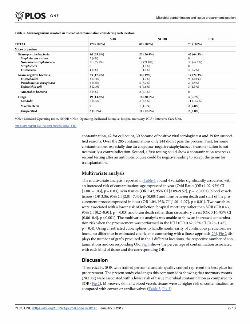
Table 3. Microorganisms involved in microbial contamination considering each location.
SOR NODR ICU
TOTAL 128 (100%) 87 (100%) 70 (100%)
Micro organism
Gram-positive bacteria:
Staphyloccus aureusNon aureus staphylococciStreptococciEnterococci
84 (65.6%)
5 (4%)
71 (55.5%)
0
4 (3%)
23 (26.4%)
0
19 (21.8%)
1 (1.1%)
1 (1.1%)
45 (64.3%)
0
33 (47.1%)
0
4 (5.7%)
Gram-negative bacteria:
EnterobacterPseudomonas aeruginosaEscherichia coli
23 (17.2%)
3 (2.3%)
2 (1.6%)
3 (2.3%)
34 (39%)
1 (1.1%)
5 (5.7%)
4 (4.6%)
17 (24.3%)
9 (12.8%)
2 (2.8%)
3 (4.3%)
Anaerobes bacteria 5 (4%) 2 (2.3%) 0
Fungi:
Candida19 (14.8%)
7 (5.5%)
18 (20.7%)
3 (3.4%)
4 (5.7%)
11 (15.7%)
Mycobacteria 0 1 (1.1%) 2 (2.8%)
Unspecified 2 (1.6%) 11 (12.6%) 2 (2.8%)
SOR = Standard Operating room, NODR = Non-Operating Dedicated Room i.e. hospital mortuary, ICU = Intensive Care Unit.
https://doi.org/10.1371/journal.pone.0210140.t003
Microbial contamination and tissue procurement location
PLOS ONE | https://doi.org/10.1371/journal.pone.0210140 January 8, 2019 7 / 13

Table 4. Univariate and multivariate analyses.
Univariate analysis Multivariate analysis
Variable OR [95% CI] P value Adjusted OR
[95% CI]
Bootstrap
95% CI
P value
Location Operating room 1 1
Hospital mortuary 0.9 [0.6–1.4] 0.8 0.4 [0.2–0.9] [0.2–0.9] 0.03
Intensive care unit 2.7 [1.5–4.7] <0.001 0.6 [0.3–1.5] [0.2–1.5] 0.4
Death Circulatory arrest 1 1
Brain death 0.7 [0.5–1.02] 0.06 0.2[0.1–0.4] [0.1–0.4] <0.001
Age (year) 1 [0.98–1.06] 0.5 1.02 [1.001–1.03] [1–1.03] 0.03
Gender Male 1 1
Female 1.1 [0.8–1.6] 0.5 1.2 [0.8–1.8] [0.8–1.7] 0.4
Cause of death CVD 1 1
Other 1.6 [1.01–2.5] 0.046 1.4 [0.8–2.4] [0.8–2.3] 0.2
Trauma 1.4 [0.9–2.3] 0.2 1.6 [0.9–2.8] [0.9–2.8] 0.1
Tissue Corneas 1 1
Skin 3.6 [2.4–5.3] <0.001 5.4 [3.1–9.5] [3–10] <0.001
Blood vessels 2 [1.3–3] <0.001 3.9 [2–7.4] [2–8] <0.001
Heart valves 1.2 [0.3–4.5] 0.8 1.7 [0.4–8] [0.3–9] 0.5
Time since death (hours) 0.99 [0.96–1.03] 0.9 1.04 [1.01–1.07] [1.02–1.1] 0.01
Duration (minutes) 1.01 [1–1.02] <0.001 1 [0.99–1.01] [0.99–1.01] 0.7
For categorical covariates, the first level is the reference with a corresponding OR of 1. OR were calculated with a GEE model assuming an exchangeable correlation
structure and involving only one explanatory variable for the univariate analysis and the whole set of variables for the multivariate analysis. P values result of a Wald test.
Bootstrap CI were built by resampling 1000 times on the cluster level (donor) with replacement and calculated as follow: estimated OR of the initial multivariate
model ± SD estimated from the 1000 bootstrap samples. OR = Odds ratio, CI = Confidence interval, GEE = Generalized Estimating Equations, CVD = Cerebrovascular
disease.
https://doi.org/10.1371/journal.pone.0210140.t004
Fig 2. Microbial contamination according to the location of procurement. For each location, number of
contaminated and uncontaminated grafts (percentages) with corresponding adjusted odds ratio (OR) are given.
https://doi.org/10.1371/journal.pone.0210140.g002
Microbial contamination and tissue procurement location
PLOS ONE | https://doi.org/10.1371/journal.pone.0210140 January 8, 2019 8 / 13

The influence of the site of tissue procurement has been poorly studied. To the best of our
knowledge, this is the first report describing tissue recovery in ICU rooms. In USA, tissue
recovery can be done in SOR, NODR, autopsy room or funerals home[25]. In a study analyz-
ing all tissue samples from 1031 donors recovered in 7 tissue banks, SOR was identified as a
protective factor for tissue contamination. In this study, positive cultures were 5 to 11% lower
for the SOR versus the others locations, making it a small factor affecting the contamination
risk. Authors suggested that surgical technique, site preparation, site disinfection and surgical
isolation techniques play a larger role in preventing contamination than the recovery environ-
ment[25]. In this study, recoveries after autopsy, length of time taken for recovery, recovery
teams with less than 3 members and skin recovery were important factors for tissue contami-
nation[25]. For European guidelines SOR is not mandatory, even if for cardiovascular tissues
it is recommended[4]. These guidelines mention that the environment for tissue retrieval
should have the following characteristics:
• Adequate size regarding floor space and work-tops that will be used,
• Appropriately located to ensure cleanliness and privacy,
• Sufficient and suitable lighting,
• Good state of repair,
• Free of pests,
• Sufficiently clean or cleanable not to contribute to cells or tissues contamination.
These characteristics suggest that a room not monitored for air quality is sufficient for tissue
recovery. This can be applied to SOR, NODR or ICU rooms and explains why tissue procure-
ment is done in these 3 settings. They also recommend assessing contamination risk to choose
the kind of facility used for retrieval, but no reproducible and clear rule is provided. Our
Fig 3. Microbial contamination according to the type of tissue. For each tissue, number of contaminated and
uncontaminated grafts (percentages) with corresponding adjusted odds ratio (OR) are given.
https://doi.org/10.1371/journal.pone.0210140.g003
Microbial contamination and tissue procurement location
PLOS ONE | https://doi.org/10.1371/journal.pone.0210140 January 8, 2019 9 / 13

results suggest that non-operating dedicated rooms (NODR) are at lower risk of contamina-
tion as compared to SOR. Several explanations could be advanced. First, in the present study,
tissue recovery performed in SOR was exclusively done in brain-dead donors after multiple
organ recovery. In such case, the time from cardiac arrest to tissue recovery cannot be stan-
dardized. In case of rapid procedure (only kidneys recovery), tissue retrieval is performed less
than 6 hours after cardiac arrest. In case of multiple organ recovery: heart, lungs, liver, pan-
creas, kidneys, the time from cardiac arrest to tissue recovery can be longer than 6 hours. Euro-
pean guidelines emphasized the fact that tissue recovery should be done as earlier as possible,
in the 24 hours following cardiac death, if a body cooling can be done within the 6 hours fol-
lowing death[4]. In case of multiple organ retrieval, this time interval cannot be reached.
Second, prolonged body cooling has been proposed as a protective factor for microbial con-
tamination[6, 25]. In case of recovery in NODR, body cooling was systematically performed for 8
to 12 hours. This prolonged cooling can decrease bacterial growth and therefore bacterial inocu-
lum at the time of recovery. However, our results do not allow confirming this hypothesis.
Third, cornea samples were mainly recovered in NODR, while skin and cardiac tissues
retrieval were mainly performed in SOR or ICU. As report in Fig 3, skin and vascular tissues
are associated with a greater risk of contamination. A multivariate analysis could imperfectly
adjust for measured confounders. Thus, a selection bias due to a greater proportion of cornea
recovery in NODR could explain the difference of contamination observed in the present
report.
Fourth, the temperature in the hospital mortuary is lower than in the operating room.
Because hypothermia in the operating room is commonly described as responsible for signifi-
cant morbidity, a not too low temperature is advocated. In France, standards mandate a mean
temperature between 19˚C and 26˚C[26]. Such recommendations are found worldwide[27].
On the contrary, for hospital mortuary, a temperature below 17˚C is recommended in France
[28]. It could be expected that a higher temperature in the operating room could lead to an
increased risk of tissue infection.
Finally, because it was an observational study, issues due to unknown confounders were
not handled. Especially, residual antibiotics concentrations can play a role in tissue contamina-
tion[29, 30]. Tissue banks use a wide variety of homemade antibiotics cocktails in order to
limit microbial tissue allograft contamination and to maximize the safety of allografts[31].
Buzzi et al recently demonstrated that antibiotic residues present in cardiac tissue allografts
and processing liquids after decontamination may mask microbial contamination during
microbiological analysis performed with standard tissue bank methods, leading to false nega-
tives[32]. Some studies suggest that the use of specific devices (RESEP) allowing antibiotics
residues removal from liquid samples can help to limit false negative[32]. For tissue procure-
ment procedure performed in SOR or ICU, it can be hypothesized that donors have previously
received broad-spectrum systemic antibiotics for infection. These antibiotics can increase the
risk of residual antibiotics concentrations and lead to false negative allograft testing performed
just before graft.
Study limitations
The retrospective design of the study cannot allow a definitive conclusion. Further prospective,
comparative studies are needed to elucidate this issue. More, some contamination factors
couldn’t be studied to further understand the contamination process in procurement. Espe-
cially, bacterial environment evaluation and air quality characteristics were not available.
Because we studied more than 1000 patients and more than 2500 allografts, we think that our
study plea for comparative studies.
Microbial contamination and tissue procurement location
PLOS ONE | https://doi.org/10.1371/journal.pone.0210140 January 8, 2019 10 / 13
Conclusion
According to our results, performing tissue procurement in non-operating dedicated rooms,
when no organ retrieval is needed, could decrease the rate of allograft tissue contamination.
This study also suggests that in daily clinical practice, transferring patients from ICU to SOR
for tissue procurement could be avoided as it does not lead to less microbial contamination.
ICU seems to be a valuable alternative for tissue retrieval.
Supporting information
S1 Appendix. Procurement procedures.
(DOCX)
S2 Appendix. Microbiological protocols used to assess tissue contaminations.
(DOCX)
Author Contributions
Conceptualization: Benjamin Louart, Claire Charles, Tri-Long Nguyen, Claire Roger, Laurent
Muller.
Data curation: Claire Charles, Florence Vachiery-Lahaye, John De Vos, Guilhem Couderc.
Formal analysis: Benjamin Louart.
Methodology: Benjamin Louart, Tri-Long Nguyen, Laurent Muller.
Supervision: Claire Charles, Claire Roger, Jean-Yves Lefrant, Laurent Muller.
Validation: Benjamin Louart, Nicolas Builles, Claire Roger, Jean-Yves Lefrant, Florence
Vachiery-Lahaye, John De Vos, Guilhem Couderc, Laurent Muller.
Visualization: Nicolas Builles, Claire Roger, Jean-Yves Lefrant, Florence Vachiery-Lahaye,
John De Vos, Guilhem Couderc, Laurent Muller.
Writing – original draft: Claire Charles, Laurent Muller.
Writing – review & editing: Benjamin Louart, Laurent Muller.
References
1. Gaum L, Reynolds I, Jones MN, Clarkson AJ, Gillan HL, Kaye SB. Tissue and corneal donation and
transplantation in the UK. British journal of anaesthesia. 2012; 108 Suppl 1:i43–7. Epub 2012/01/04.
https://doi.org/10.1093/bja/aer398 PMID: 22194430.
2. Mallick TK, Mosquera A, Zinderman CE, St Martin L, Wise RP. Reported infections after human tissue
transplantation before and after new Food and Drug Administration (FDA) regulations, United States,
2001 through June, 2010. Cell and tissue banking. 2012; 13(2):259–67. Epub 2011/04/12. https://doi.
org/10.1007/s10561-011-9253-5 PMID: 21479712.
3. Rapport annuel de l’agence de biomedecine. www.biomedecine.fr. 2015.
4. (EDQM). ECootEDftQoMaH. Guide to the quality and safety of tissue and cells for human application.
www.edqm.eu. 2015.
5. Germain M, Strong DM, Dowling G, Mohr J, Duong A, Garibaldi A, et al. Disinfection of human cardiac
valve allografts in tissue banking: systematic review report. Cell and tissue banking. 2016; 17(4):593–
601. Epub 2016/08/16. https://doi.org/10.1007/s10561-016-9570-9 PMID: 27522194; PubMed Central
PMCID: PMC5116039.
6. Pianigiani E, Ierardi F, Cuciti C, Brignali S, Oggioni M, Fimiani M. Processing efficacy in relation to
microbial contamination of skin allografts from 723 donors. Burns: journal of the International Society for
Burn Injuries. 2010; 36(3):347–51. Epub 2009/07/21. https://doi.org/10.1016/j.burns.2009.04.020
PMID: 19616385.
Microbial contamination and tissue procurement location
PLOS ONE | https://doi.org/10.1371/journal.pone.0210140 January 8, 2019 11 / 13
7. Khouani M, Debellemaniere G, Malugani C, Gauthier AS, Pouthier F, Delbosc B, et al. Evaluation of
microbial contamination of corneal transplants: one-year report from a French regional eye bank. Cor-
nea. 2014; 33(9):899–904. Epub 2014/07/24. https://doi.org/10.1097/ICO.0000000000000178 PMID:
25055144.
8. Ireland L, Spelman D. Bacterial contamination of tissue allografts—experiences of the donor tissue
bank of Victoria. Cell and tissue banking. 2005; 6(3):181–9. Epub 2005/09/10. https://doi.org/10.1007/
s10561-005-7365-5 PMID: 16151958.
9. Builles N, Perraud M, Reverdy ME, Burillon C, Crova P, Brun F, et al. Reducing contamination when
removing and storing corneas: a multidisciplinary, transversal, and environmental approach. Cornea.
2006; 25(2):185–92. Epub 2005/12/24. PMID: 16371779.
10. Borderie VM, Laroche L. Microbiologic study of organ-cultured donor corneas. Transplantation. 1998;
66(1):120–3. Epub 1998/07/29. PMID: 9679833.
11. Borderie VM, Scheer S, Touzeau O, Vedie F, Carvajal-Gonzalez S, Laroche L. Donor organ cultured
corneal tissue selection before penetrating keratoplasty. The British journal of ophthalmology. 1998; 82
(4):382–8. Epub 1998/06/26. PubMed Central PMCID: PMC1722561. PMID: 9640185
12. Rehany U, Balut G, Lefler E, Rumelt S. The prevalence and risk factors for donor corneal button con-
tamination and its association with ocular infection after transplantation. Cornea. 2004; 23(7):649–54.
Epub 2004/09/28. PMID: 15448488.
13. Robert PY, Camezind P, Drouet M, Ploy MC, Adenis JP. Internal and external contamination of donor
corneas before in situ excision: bacterial risk factors in 93 donors. Graefe’s archive for clinical and
experimental ophthalmology = Albrecht von Graefes Archiv fur klinische und experimentelle Ophthal-
mologie. 2002; 240(4):265–70. Epub 2002/05/01. https://doi.org/10.1007/s004170100322 PMID:
11981639.
14. ISO. Technical Committee 209 (ISO/TC 209). Cleanrooms and associated controlled environments–
Part 1: Classification of air cleanliness by particle concentration. 2015.
15. SF2H. Societe Francaise d’Hyg!ène Hospitalière. Qualite de l’air au bloc operatoire et autres secteurs
interventionnels. Recommandations. ISSN1249-0075. Hygienes2015. p. 1–62.
16. Code de la sante publique. Art L3212-1; Art L3212-2. Available from https://www.legifrance.gouv.fr/.
17. EudraLex. EU guidelines for good manufacturing practices for medicinal products for human and veteri-
nary use (GMP). Annex 1: Manufacturing of sterile medicinal products. Brussels, November 2008.
18. Zeger SL, Liang KY. Longitudinal data analysis for discrete and continuous outcomes. Biometrics.
1986; 42(1):121–30. Epub 1986/03/01. PMID: 3719049.
19. Gardiner JC, Luo Z, Roman LA. Fixed effects, random effects and GEE: what are the differences? Sta-
tistics in medicine. 2009; 28(2):221–39. Epub 2008/11/18. https://doi.org/10.1002/sim.3478 PMID:
19012297.
20. Harrell FE Jr., Lee KL, Pollock BG. Regression models in clinical studies: determining relationships
between predictors and response. Journal of the National Cancer Institute. 1988; 80(15):1198–202.
Epub 1988/10/05. PMID: 3047407.
21. White H. A Heteroskedasticity-Consistent Covariance Matrix Estimator and a Direct Test for Heteroske-
dasticity. Econometrica. 1980; 48:817–38.
22. Bouwmeester W, Moons KG, Kappen TH, van Klei WA, Twisk JW, Eijkemans MJ, et al. Internal valida-
tion of risk models in clustered data: a comparison of bootstrap schemes. American journal of epidemi-
ology. 2013; 177(11):1209–17. Epub 2013/05/11. https://doi.org/10.1093/aje/kws396 PMID: 23660796.
23. Moineddin R, Matheson FI, Glazier RH. A simulation study of sample size for multilevel logistic regres-
sion models. BMC medical research methodology. 2007; 7:34. Epub 2007/07/20. https://doi.org/10.
1186/1471-2288-7-34 PMID: 17634107; PubMed Central PMCID: PMC1955447.
24. R CT. A language and environment for statistical computing. R Foundation for Statistical Computing,
Vienna, Austria. URL https://www.R-project.org/. 2016.
25. Forsell JH, Liesman J. Analysis of potential causes of positive microbiological cultures in tissue donors.
Cell and tissue banking. 2000; 1(2):111–5. Epub 2004/07/17. https://doi.org/10.1023/
A:1010106214542 PMID: 15256955.
26. Association Francaise de Normalisation (AFNOR). Etablissements de sante. Zones à environnement
maıtrise. Exigences relatives à la maıtrise de la contamination aeroportee. Norme francaise NF S90–
35. 2013.
27. Balaras CA, Dascalaki E, Gaglia A. HVAC and indoor thermal conditions in hospital operating rooms.
Energy and Buildings. 2007; 39(4):454–70. http://dx.doi.org/10.1016/j.enbuild.2006.09.004.
28. Ministère de l’Emploi et de la Solidarite. Arrête relatif aux prescriptions techniques applicables aux
chambres mortuaires des etablissements de sante. Journal Officiel de la Republique Francaise n˚114.
Article 5. NOR: MESH0121712A., (2001).
Microbial contamination and tissue procurement location
PLOS ONE | https://doi.org/10.1371/journal.pone.0210140 January 8, 2019 12 / 13

29. Soo A, Healy DG, El-Bashier H, Shaw S, Wood AE. Quality control in homograft valve processing:
when to screen for microbiological contamination? Cell and tissue banking. 2011; 12(3):185–90. Epub
2010/05/22. https://doi.org/10.1007/s10561-010-9180-x PMID: 20490931.
30. Gatto C, Giurgola L, D’Amato Tothova J. A suitable and efficient procedure for the removal of decon-
taminating antibiotics from tissue allografts. Cell and tissue banking. 2013; 14(1):107–15. Epub 2012/
03/13. https://doi.org/10.1007/s10561-012-9305-5 PMID: 22407218.
31. Pitt TL, Tidey K, Roy A, Ancliff S, Lomas R, McDonald CP. Activity of four antimicrobial cocktails for tis-
sue allograft decontamination against bacteria and Candida spp. of known susceptibility at different
temperatures. Cell and tissue banking. 2014; 15(1):119–25. Epub 2013/06/15. https://doi.org/10.1007/
s10561-013-9382-0 PMID: 23765096.
32. Buzzi M, Guarino A, Gatto C, Manara S, Dainese L, Polvani G, et al. Residual antibiotics in decontami-
nated human cardiovascular tissues intended for transplantation and risk of falsely negative microbio-
logical analyses. PloS one. 2014; 9(11):e112679. Epub 2014/11/15. https://doi.org/10.1371/journal.
pone.0112679 PMID: 25397402; PubMed Central PMCID: PMC4232473.
Microbial contamination and tissue procurement location
PLOS ONE | https://doi.org/10.1371/journal.pone.0210140 January 8, 2019 13 / 13