The presence of the pilus locus is a clonal property among pneumococcal invasive isolates
11
BioMed Central Page 1 of 11 (page number not for citation purposes) BMC Microbiology Open Access Research article The presence of the pilus locus is a clonal property among pneumococcal invasive isolates Sandra I Aguiar, Isa Serrano, Francisco R Pinto, José Melo-Cristino and Mario Ramirez* Address: Instituto de Microbiologia, Instituto de Medicina Molecular, Faculdade de Medicina Universidade de Lisboa, Lisboa, Portugal Email: Sandra I Aguiar - [email protected]; Isa Serrano - [email protected]; Francisco R Pinto - [email protected]; José Melo- Cristino - [email protected]; Mario Ramirez* - [email protected] * Corresponding author Abstract Background: Pili were recently recognized in Streptococcus pneumoniae and implicated in the virulence of this bacterium, which led to the proposal of using these antigens in a future pneumococcal vaccine. However, pili were found to be encoded by the rlrA islet that was not universally distributed in the species. We examined the distribution of the pilus islet, using the presence of the rlrA gene as a marker for the locus, among a collection of invasive isolates recovered in Portugal and analyzed its association with capsular serotypes, clusters defined by the pulsed-field gel electrophoretic profiles (PFGE) and multilocus sequence types. Results: Only a minority of the isolates were positive for the presence of the rlrA gene (27%). There was a high correspondence between the serotype and the presence or absence of rlrA (Wallace coefficient, W = 0.778). In particular, there was an association between the presence of rlrA and the vaccine serotypes 4, 6B, 9V and 14 whereas the gene was significantly absent from other serotypes, namely 1, 7F, 8, 12B and 23F, a group that included a vaccine serotype (23F) and serotype 1 associated with enhanced invasiveness. Even within serotypes, there was variation in the presence of the pilus islet between PFGE clones and a higher Wallace coefficient (W = 0.939) indicates that carriage of the islet is a clonal property of pneumococci. Analysis of rlrA negative isolates revealed heterogeneity in the genomic region downstream of the rfl gene, the region where the islet is found in other isolates, compatible with recent loss of the islet in some lineages. Conclusion: The pilus islet is present in a minority of pneumococcal isolates recovered from human invasive infections and is therefore not an essential virulence factor in these infections. Carriage of the pilus islet is a clonal property of pneumococci that may vary between isolates expressing the same serotype and loss and acquisition of the islet may be ongoing. Background Non-flagellar polymeric cell-surface organelles designated as pili were initially identified in Gram-negative bacteria. In the last decade, pilus-like surface structures have been described in Gram-positive bacteria like Corynebacterium spp., Actinomyces spp. and several streptococcal species [1], but only recently were pili identified in the major patho- genic species of the genus: Streptococcus pyogenes, Lancefield Published: 28 February 2008 BMC Microbiology 2008, 8:41 doi:10.1186/1471-2180-8-41 Received: 31 October 2007 Accepted: 28 February 2008 This article is available from: http://www.biomedcentral.com/1471-2180/8/41 © 2008 Aguiar et al; licensee BioMed Central Ltd. This is an Open Access article distributed under the terms of the Creative Commons Attribution License (http://creativecommons.org/licenses/by/2.0 ), which permits unrestricted use, distribution, and reproduction in any medium, provided the original work is properly cited.
-
Upload
independent -
Category
Documents
-
view
5 -
download
0
Transcript of The presence of the pilus locus is a clonal property among pneumococcal invasive isolates
BioMed CentralBMC Microbiology
ss
Open AcceResearch articleThe presence of the pilus locus is a clonal property among pneumococcal invasive isolatesSandra I Aguiar, Isa Serrano, Francisco R Pinto, José Melo-Cristino and Mario Ramirez*Address: Instituto de Microbiologia, Instituto de Medicina Molecular, Faculdade de Medicina Universidade de Lisboa, Lisboa, Portugal
Email: Sandra I Aguiar - [email protected]; Isa Serrano - [email protected]; Francisco R Pinto - [email protected]; José Melo-Cristino - [email protected]; Mario Ramirez* - [email protected]
* Corresponding author
AbstractBackground: Pili were recently recognized in Streptococcus pneumoniae and implicated in thevirulence of this bacterium, which led to the proposal of using these antigens in a futurepneumococcal vaccine. However, pili were found to be encoded by the rlrA islet that was notuniversally distributed in the species. We examined the distribution of the pilus islet, using thepresence of the rlrA gene as a marker for the locus, among a collection of invasive isolatesrecovered in Portugal and analyzed its association with capsular serotypes, clusters defined by thepulsed-field gel electrophoretic profiles (PFGE) and multilocus sequence types.
Results: Only a minority of the isolates were positive for the presence of the rlrA gene (27%).There was a high correspondence between the serotype and the presence or absence of rlrA(Wallace coefficient, W = 0.778). In particular, there was an association between the presence ofrlrA and the vaccine serotypes 4, 6B, 9V and 14 whereas the gene was significantly absent from otherserotypes, namely 1, 7F, 8, 12B and 23F, a group that included a vaccine serotype (23F) andserotype 1 associated with enhanced invasiveness. Even within serotypes, there was variation in thepresence of the pilus islet between PFGE clones and a higher Wallace coefficient (W = 0.939)indicates that carriage of the islet is a clonal property of pneumococci. Analysis of rlrA negativeisolates revealed heterogeneity in the genomic region downstream of the rfl gene, the region wherethe islet is found in other isolates, compatible with recent loss of the islet in some lineages.
Conclusion: The pilus islet is present in a minority of pneumococcal isolates recovered fromhuman invasive infections and is therefore not an essential virulence factor in these infections.Carriage of the pilus islet is a clonal property of pneumococci that may vary between isolatesexpressing the same serotype and loss and acquisition of the islet may be ongoing.
BackgroundNon-flagellar polymeric cell-surface organelles designatedas pili were initially identified in Gram-negative bacteria.In the last decade, pilus-like surface structures have been
described in Gram-positive bacteria like Corynebacteriumspp., Actinomyces spp. and several streptococcal species [1],but only recently were pili identified in the major patho-genic species of the genus: Streptococcus pyogenes, Lancefield
Published: 28 February 2008
BMC Microbiology 2008, 8:41 doi:10.1186/1471-2180-8-41
Received: 31 October 2007Accepted: 28 February 2008
This article is available from: http://www.biomedcentral.com/1471-2180/8/41
© 2008 Aguiar et al; licensee BioMed Central Ltd. This is an Open Access article distributed under the terms of the Creative Commons Attribution License (http://creativecommons.org/licenses/by/2.0), which permits unrestricted use, distribution, and reproduction in any medium, provided the original work is properly cited.
Page 1 of 11(page number not for citation purposes)
BMC Microbiology 2008, 8:41 http://www.biomedcentral.com/1471-2180/8/41
group A [2]; Streptococcus agalactiae, Lancefield group B [3]and Streptococcus pneumoniae (pneumococcus) [4].
In S. pneumoniae pili are encoded by the rlrA pathogenicityislet, a 14.2 kb region composed of 7 genes encoding aputative transcriptional regulator (RlrA), 3 LPXTG surfaceproteins with weak homology to microbial surface com-ponents recognizing adhesive matrix molecules –MSCRAMMs (RrgA, RrgB and RrgC) and 3 sortases (SrtB,SrtC and SrtD) [4-6]. Pneumococcal pili have the appear-ance of flexible rods ranging from 0.3 μm to 3 μm that areformed by a transpeptidase reaction involving sortase-mediated covalent cross-linking of the proteins contain-ing the LPXTG-like motif and several lines of evidencesuggest a one-to-one sortase-to-surface-protein corre-spondence for the proteins encoded by the islet [7].
The rlrA gene was identified in the original signaturetagged mutagenesis study as an essential virulence gene inmurine models of infection [5]. Later studies confirmedthat the RlrA protein acted as a transcription factor recog-nizing several promoters within the rlrA islet and showedit to be essential for wild-type levels of expression of thepili structural genes and associated sortases [6]. The prod-uct of the mgrA gene, located outside the rlrA islet, wasshown to act as a transcriptional repressor of the isletgenes, including rlrA, being responsible for the silencingof the locus in the absence of RlrA [8].
The presence of the rlrA pathogenicity islet was shown toinfluence pneumococcal capacity to adhere to humanlung epithelial cells [4,8]. Mouse models of pneumococ-cal pneumonia and bacteremia have also suggested a roleof pili in virulence and host inflammatory responses [4,5].More recently, immunization of mice with pilus structuralantigens was shown to induce protection against lethalchallenge by piliated strains [9]. Moreover, these studiesindicate that vaccination with the pilus subunits offers thesame protection as vaccination with heat killed bacteria,raising the possibility of using pilus antigens in a multiva-lent pneumococcal vaccine [9].
In spite of these favorable results, early genomic studiesindicated that the rlrA islet was not present in all pneumo-coccal isolates, suggesting that it could have been acquiredby horizontal gene transfer [10] and raising a cautionarynote concerning the use of pilus antigens in any futurevaccine. A survey for the distribution of the sortase genesidentified the srtA gene in all tested bacteria, but the sor-tases associated with the rlrA islet were present in only17% of the isolates tested [11]. A recent study, specificallydesigned to evaluate the distribution of the rlrA isletamong pneumococcal isolates associated with coloniza-tion and infections in Native Americans, found that only21% of the isolates were positive for rrgC, a gene encoding
a pilus structural protein, and that there was an associa-tion between the presence of this gene and the serotypesincluded in the seven-valent conjugate vaccine [12]. How-ever, the same study could not show a difference in thepresence of rrgC between the isolates recovered from thenasopharynx and those recovered from sterile sites. Incontrast, the mgrA gene encoding the transcriptionalrepressor of the rlrA islet seems to be universally distrib-uted within S. pneumoniae [8].
In view of these findings, a better understanding of thedistribution of the pilus islet in the invasive pneumococ-cal population will help in the evaluation of its proposedrole as a virulence factor and as a potential vaccine candi-date. In the present study, we determined the prevalenceof the rlrA gene in a recent collection of invasive isolatesfrom both children and adults with the aim of identifyingassociations between the presence of the pilus islet andserotype, antimicrobial resistance or clonal types definedby pulsed-field gel electrophoresis (PFGE) and multilocussequence typing (MLST).
ResultsDistribution of the rlrA islet among invasive pneumococciOnly a small portion (27%) of our invasive isolates wasrlrA positive and a total of 355 (73%) isolates were foundto lack the pilus islet by Southern blot hybridization andPCR amplification of the rlrA gene (Additional file 1:Southern hybridization of a representative set of isolateswith the rlrA gene probe.). A group of 39 isolates repre-sentative of the diversity found among the rlrA positiveisolates (n = 128) was subject to long-range PCR. All iso-lates yielded fragments larger than 14 kb compatible withthe presence of the entire pilus locus (data not shown),confirming that the presence of the rlrA gene is a goodmarker for the presence of the entire pilus islet.
Overall rlrA positive isolates were found in 11 serotypes ofthe 41 serotypes in our collection (Additional file 2: Char-acteristics of the clones where the pilus locus was identi-fied.). Isolates expressing serotypes 1, 3, 4, 6A, 6B, 9V, 13,14, 19A, 19F, 35B were shown to carry the rlrA gene aswell as non-typable isolates, but 83% of the rlrA positiveisolates expressed vaccine serotypes (4, 6B, 9V, 14, and19F). The proportion of isolates carrying the pilus isletamong isolates of the same serotype was variable (Figure1), but overall serotype was a good predictor of the pres-ence or absence of the pilus islet, as indicated by the highWallace coefficient (W = 0.778).
Although there were differences in the overall proportionof isolates carrying the pilus islet between the isolatesrecovered from children < 6 yrs. old (37%) and fromadults (24%) (p = 0.026), we could not shown differencesbetween the two age groups when we considered the pro-
Page 2 of 11(page number not for citation purposes)
BMC Microbiology 2008, 8:41 http://www.biomedcentral.com/1471-2180/8/41
portion of rlrA positive isolates stratified by serotype usingthe Mantel-Haenszel test (p = 0.200), indicating that theoverall disparity is due to a different distribution of sero-types in the two age groups.
In order to identify which serotypes were associated withthe presence of the rlrA islet, odds ratios were calculated.An OR > 1 implies that the serotype is associated with thepresence of rlrA, while an OR < 1 indicates that the sero-type is significantly depleted of rlrA positive isolates.According to this analysis, serotypes 4, 6B, 9V and 14 werehighly associated with the presence of the pilus islet (p <0.01 for all serotypes). On the other hand, the rlrA islet
was significantly absent from serotypes 1, 7F, 8, 12B and23F (Table 1).
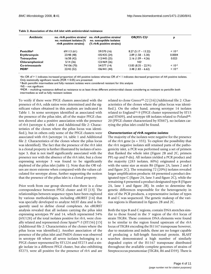
Association between the presence of the pilus islet and resistance to antimicrobialsA clear correlation between the presence of rlrA and anti-microbial resistance was noted. We found that most iso-lates carrying the pilus islet were resistant to at least oneantimicrobial – 54% (69/128) were non-susceptible topenicillin, 18% (23/128) were resistant to erythromycinand 17% (22/128) were multi-resistant. This associationwas further confirmed since the OR calculated for resist-ance to individual antibiotics and the presence of the pilusislet were mostly > 1 and significant (Table 2).
Presence of the pilus islet is a clonal propertyCharacterization of the genetic lineages of the pneumo-coccal collection included in the present study had beenperformed previously [13]. Analysis by genetic lineage ofthe isolates demonstrated that the presence of the rlrA isletwas not only associated with serotype but also with PFGEcluster within each serotype, defined as described previ-ously [13] (Additional file 2 – Characteristics of the cloneswhere the pilus locus was identified – and Additional file3 – Characteristics of the bacterial clones not associatedwith the pilus locus). The Wallace coefficient relating thePFGE clusters with the presence or absence of rlrA (W =0.939) was higher than for serotype (W = 0.778) indicat-ing that the pathogenicity islet is clonally distributed. Thevalue of the Wallace coefficient indicates that only one outof 20 pairs of isolates grouped in the same PFGE clusterwill differ in the presence of the rlrA islet in their genome.
Frequency of the rlrA islet among serotypes in which at least one rlrA positive isolate was foundFigure 1Frequency of the rlrA islet among serotypes in which at least one rlrA positive isolate was found. Black bars indicate the number of isolates positive for the presence of the rlrA islet. White bars indicate the number of isolates neg-ative for the presence of the rlrA islet.
0
10
20
30
40
50
60
70
1 3 4 6A 6B 9V 13 14 19A 19F 35B NT
Serotype
no
. iso
late
s
rlrA negativerlrA positive
Table 1: Association of the rlrA islet with serotypes and PFGE clusters
Serotype OR (95% CI)a p STs in PFGE clus-terb
OR (95% CI) p
1 0.22 (0.07 – 0.53) 0.0011 [ST306 + ST228] 0.079 (0.01 – 0.59) 0.00073 NSc [ST1220 + ST260] +
[ST1230 + ST180]0.06 (0.01 – 0.44) < 10-4
4 12.74 (5.05 – 32.12) < 10-4 ST1221 80.26 (4.72 – 1363.4) < 10-4
ST247 30 (3.8 – 236.84) < 10-4
6B 7.44 (2.29 – 24.16) 0.0005 ST1224 + ST273 47.33 (2.7 – 830.37) < 10-4
7F 0 (0 – 0.61) 0.0052 ST191 0 (0.0 – 0.70) 0.00898 0 (0 – 0.37) 0.0002 ST53 0 (0.0 – 0.57) 0.0057
9V 14.36 (4.76 – 43.33) < 10-4 [ST156 + ST557 + ST644 + ST1225]
16.76 (4.80 – 58.54) < 10-4
12B 0 (0 – 0.70) 0.0089 NSc NSc
14 16.13 (8.50 – 30.63) < 10-4 [ST156 + ST557 + ST790]
212.4 (28.89 – 1561.8) < 10-4
23F 0 (0 – 0.44) 0.0006 ST338 + ST1371 0 (0.0 – 0.57) 0.0057
aAn OR of > 1 indicates increased proportion of rlrA positive isolates whereas OR of < 1 indicates decreased proportion of rlrA positive isolates. Only statistically significant results (FDR < 0.05) are presented.bSTs in PFGE clusters with a Dice similarity coefficient of > 80%. Brackets indicate STs that belong to the same lineage, as defined by eBURST analysis with the complete S. pneumoniae database available at spneumoniae.mlst.net. Only statistically significant results (FDR < 0.05) are presented.cNS – not significant
Page 3 of 11(page number not for citation purposes)
BMC Microbiology 2008, 8:41 http://www.biomedcentral.com/1471-2180/8/41
To verify if there were PFGE clusters associated with thepresence of rlrA, odds ratios were determined and the sig-nificant values obtained in this analysis are indicated inTable 1. In some serotypes identified as associated withthe presence of the pilus islet, all of the major PFGE clus-ters showed also a positive association with the presenceof rlrA (serotype 4, table 1 and Additional file 2: Charac-teristics of the clones where the pilus locus was identi-fied.), but in others only some of the PFGE clusters wereassociated with rlrA (serotype 14, table 1 and Additionalfile 2: Characteristics of the clones where the pilus locuswas identified). The fact that the presence of the rlrA isletis a clonal property is further illustrated by isolates of sero-type 3, that is not itself significantly associated with thepresence nor with the absence of the rlrA islet, but a cloneexpressing serotype 3 was found to be significantlydepleted of the pilus islet (table 1). Note that the OR val-ues are more extreme when compared with the values cal-culated for serotype alone, further supporting the notionthat the presence of the pilus islet is a clonal property.
Prior work from our group showed that there is a closecorrespondence between PFGE cluster and ST [13]. Therelationships between sequence types have been exploredby various methodologies, including eBURST [14] thatwas specially developed to analyze MLST data and is fre-quently used to define clonal complexes. An eBURSTanalysis revealed that all isolates carrying the pilus isletexpressing serotypes 9V and 14, which represented 54%(69/128) of the total isolates positive for rlrA, were clon-ally related and represented clone Spain9V-3 (ST156) [15](Additional file 2: Characteristics of the clones where thepilus locus was identified.). Another association of thepresence of the pilus islet and PMEN clones was observedin serotype 6B where all seven isolates belonging to aPFGE cluster represented by ST1224 and ST273 and a sin-gle isolate in a different PFGE cluster, but also exhibitingST273, were all positive for the presence of rlrA and are
related to clone Greece6B-22 [16] (Additional file 2: Char-acteristics of the clones where the pilus locus was identi-fied.). On the other hand, among serotype 14 isolatesrelated to England14-9 (PFGE cluster represented by ST15and ST409), and serotype 6B isolates related to Poland6B-20 (PFGE cluster characterized by ST887), no isolates car-rying the pilus islet could be found.
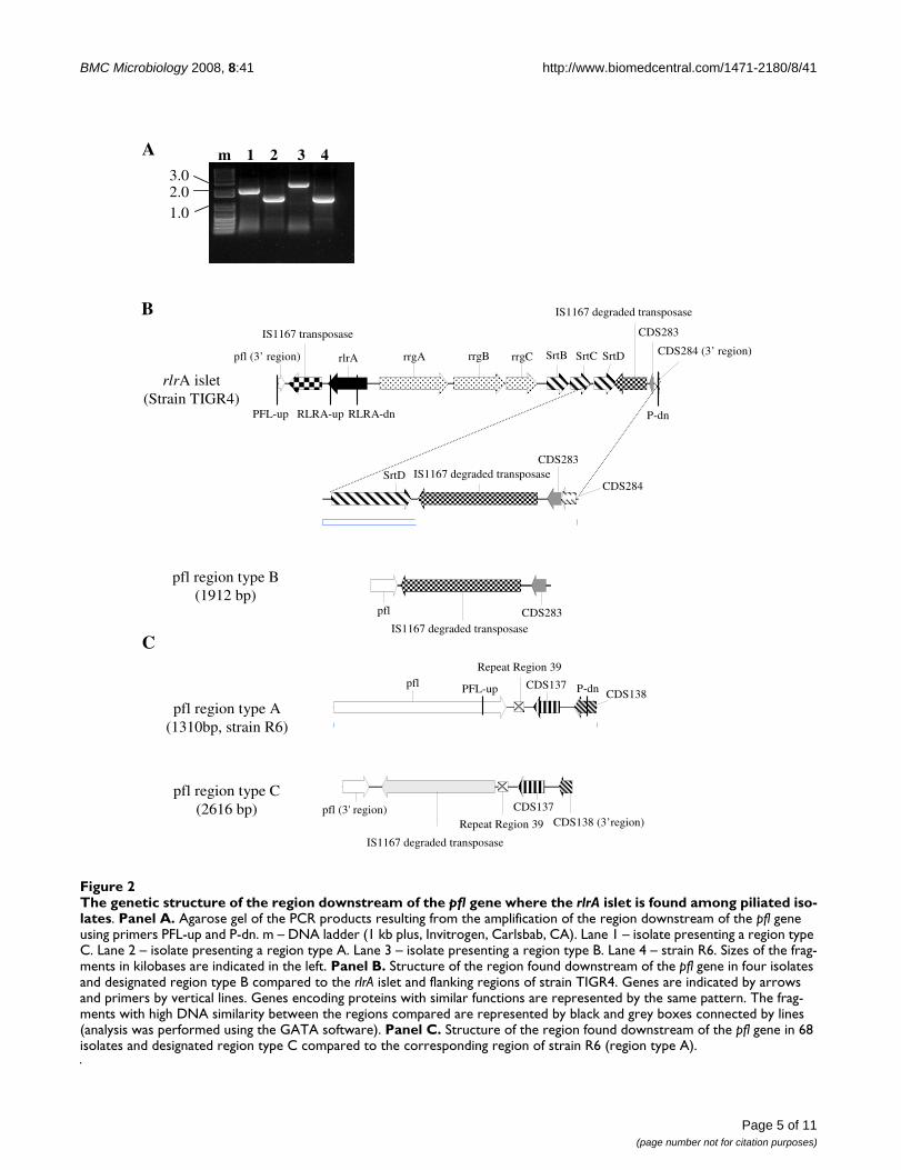
Characterization of rlrA negative isolatesThe majority of the isolates were negative for the presenceof the rlrA gene (n = 355). To explore the possibility thatthe rlrA negative isolates still retained parts of the patho-genicity islet, a PCR was performed using a set of primersthat flanked the whole islet (Figure 2B and 2C, primersPFL-up and P-dn). All isolates yielded a PCR product andthe majority (283 isolates, 80%) originated a productwith the same size as strain R6 (Figure 2A, lanes 2 and 4and figure 2C). The remaining 72 (20%) isolates revealedlarger amplification products: 68 presented a product des-ignated type C (figure 2A, lane 3 and figure 2C), while theremaining 4 presented a product designated type B (Figure2A, lane 1 and figure 2B). In order to determine thegenetic differences responsible for the heterogeneity insize of the PCR products, a representative isolate of typesB and C was sequenced. The genetic makeup of the vari-ous regions is illustrated in figures 2B and 2C.
Both the type B and C regions, contain DNA stretches sim-ilar to those found in the 3' region of the rlrA locus ofstrain TIGR4. These common DNA elements were foundto be similar to the region found upstream of the rlrAlocus of TIGR4 encoding the IS1167 transposase however,due to mutations and indels, these are no longer capableof producing a full-length functional protein. BLASTsearches demonstrated a high number of similarlydegraded copies of the IS1167 transposase distributedthroughout the available complete genomes of strains ofStreptococcus pneumoniae (TIGR4, R6 and D39). There is
Table 2: Association of the rlrA islet with antimicrobial resistance
Antibiotic no. rlrA positive strains/no. resistant isolates (%
rlrA positive strains)
no. rlrA positive strains/no. susceptible isolates (% rlrA positive strains)
OR(95% CI)a p
Penicillinb 69/113 (61) 59/370 (16) 8.27 (5.17 – 13.22) < 10-4
Erythromycin 23/48 (48) 105/435 (24) 2.89 (1.58 – 5.30) 0.0004Tetracycline 16/38 (42) 112/445 (25) 2.16 (1.09 – 4.26) 0.023Chloramphenicol 5/14 (36) 123/469 (26) NSc
Co-trimoxazole 74/106 (70) 54/377 (14) 13.83 (8.35 – 22.91) < 10-4
MDRd 22/42 (52) 106/441 (44) 3.48 (1.83 – 6.62) < 10-4
aAn OR of > 1 indicates increased proportion of rlrA positive isolates whereas OR of < 1 indicates decreased proportion of rlrA positive isolates. Only statistically significant results (FDR < 0.05) are presented.b Both penicillin intermediate and fully resistant isolates were considered resistant for this analysiscNS – not significantdMDR – multidrug resistance defined as resistance to at least three different antimicrobial classes considering as resistant to penicillin both intermediate as well as fully resistant isolates
Page 4 of 11(page number not for citation purposes)
BMC Microbiology 2008, 8:41 http://www.biomedcentral.com/1471-2180/8/41
Page 5 of 11(page number not for citation purposes)
The genetic structure of the region downstream of the pfl gene where the rlrA islet is found among piliated isolatesFigure 2The genetic structure of the region downstream of the pfl gene where the rlrA islet is found among piliated iso-lates. Panel A. Agarose gel of the PCR products resulting from the amplification of the region downstream of the pfl gene using primers PFL-up and P-dn. m – DNA ladder (1 kb plus, Invitrogen, Carlsbab, CA). Lane 1 – isolate presenting a region type C. Lane 2 – isolate presenting a region type A. Lane 3 – isolate presenting a region type B. Lane 4 – strain R6. Sizes of the frag-ments in kilobases are indicated in the left. Panel B. Structure of the region found downstream of the pfl gene in four isolates and designated region type B compared to the rlrA islet and flanking regions of strain TIGR4. Genes are indicated by arrows and primers by vertical lines. Genes encoding proteins with similar functions are represented by the same pattern. The frag-ments with high DNA similarity between the regions compared are represented by black and grey boxes connected by lines (analysis was performed using the GATA software). Panel C. Structure of the region found downstream of the pfl gene in 68 isolates and designated region type C compared to the corresponding region of strain R6 (region type A).
pfl region type A(1310bp, strain R6)
pfl CDS137CDS138PFL-up P-dn
Repeat Region 39
pfl region type C(2616 bp) pfl (3' region)
IS1167 degraded transposase
CDS137
Repeat Region 39 CDS138 (3’region)
C
rlrA islet(Strain TIGR4)
pfl region type B(1912 bp)
CDS283pfl
IS1167 degraded transposase
SrtDCDS283
CDS284IS1167 degraded transposase
IS1167 transposase
rlrA rrgA rrgB rrgC SrtB SrtC SrtD
CDS283
P-dnPFL-up RLRA-dnRLRA-up
IS1167 degraded transposase
CDS284 (3’ region)pfl (3’ region)
B
A m 1 2 3 4
2.01.0
3.0
BMC Microbiology 2008, 8:41 http://www.biomedcentral.com/1471-2180/8/41
a high identity between the IS1167 degraded transposasesequence found downstream of the rlrA locus in TIGR4and the one found in the type B region (97% identity inthe 1277 nt aligned, with only two single nucleotidegaps). The similarity extends to other coding and non-coding regions found between srtD and spr0470 of TIGR4(Figure 2B). This observation is compatible with thehypothesis that region type B was generated by the recentloss of the rlrA islet. Further supporting this notion is theobservation that this region was found exclusively amongthe few isolates not carrying the pilus islet among largePFGE clusters of rlrA positive isolates (Additional file 2:Characteristics of the clones where the pilus locus wasidentified.).
The DNA sequence corresponding to the degraded copy ofthe IS1167 transposase in region type C differed moreextensively from the two discussed above, namely by a 50nt insertion and a 16 nt deletion. A comparison of thetype C region with the corresponding sequence of strainR6 revealed extensive similarity, including the presence ofORF CDS137 and a downstream repeat region found instrain R6 but absent from the pilus locus in strain TIGR4(Figure 2C). The type C region is therefore closely relatedto the one found in strain R6 with the exception of thepresence of a degraded copy of the IS1167 transposase,suggesting that the region downstream of the pfl gene mayconstitute a hotspot for recombination events. The type Cregion was found associated with specific PFGE clustersexpressing a limited number of serotypes (Additional file2 – Characteristics of the clones where the pilus locus wasidentified – and Additional file 3 – Characteristics of thebacterial clones not associated with the pilus locus.), butthis was not an exclusive association since isolates pre-senting regions type A and C were frequently grouped inthe same PFGE cluster. For instance, the largest PFGE clus-ter in serotype 3 contains almost equal numbers of iso-lates presenting each of the regions.
DiscussionThe long-term efficacy of the seven-valent conjugate vac-cine is being challenged by the emergence of infectionscaused by replacement serotypes not targeted by the vac-cine. The difficulty in producing higher valency vaccinesand the realization that any polysaccharide based vaccinemay be ultimately limited in its efficacy, due to the largenumber of pneumococcal capsular types, prompted thesearch for other vaccine targets. Recently, the three struc-tural proteins constituting pneumococcal pili were shownto protect mice against invasive infections and were pro-posed as potential vaccine candidates [9].
The data presented here argues that the efficacy of such avaccine would be limited by the small proportion of iso-lates carrying the pilus islet (27%) and therefore poten-
tially expressing pili, among those causing invasiveinfections in humans. The strong association of the rlrAislet with four of the seven serotypes included in the con-jugate vaccine would further limit the advantages of apilus component vaccine over the one currently availa-ble. Recently, Basset et al. explored the distribution ofthe rlrA islet, using rrgC as a marker for the presence ofthe pilus locus [12]. The population analyzed by Bassetet al. differed markedly from the one analyzed here byincluding a large fraction of isolates associated with col-onization, a different distribution of serotypes and, mostimportantly, the genetic makeup of the pneumococcalpopulation was strikingly different with only 15 com-mon STs out of the 158 STs identified overall in the twocollections. In spite of these differences, a surprisinglysimilar proportion of isolates carrying the pilus islet wasfound among invasive isolates by Basset et al. (24%, n =123). Taken together the results of the two studies sug-gest that the pilus does not represent an essential viru-lence factor for invasive disease in humans, in contrast toprevious indications from mouse models [4,5]. This sug-gestion receives further support from the observationthat the same fraction of isolates associated with asymp-tomatic carriage were positive for the pilus islet [12].
The collection characterized here was previously ana-lyzed for the phase variant expressed by the isolates [17],another phenotype implicated in enhanced invasive-ness. A proportion of opaque variants similar to the onepositive for the rlrA gene was found (26%), promptingus to evaluate the association between opacity and thepilus islet. However, no association could be shownbetween the opacity phenotype of a given isolate andcarriage of the pilus islet. In fact, two serotypes found tobe associated with predominantly opaque variants, sero-type 1 and 4 [17], were found to be significantly depletedof isolates positive for rlrA and strongly associated withthe presence of the islet, respectively.
A prior study emphasized the higher prevalence of thepilus islet among the serotypes included in the currentlyavailable conjugate vaccine versus all other serotypes[12]. Here we expand these findings by identifying whichserotypes are particularly enriched or depleted in thepresence of the pilus locus. Specifically, among the sevenserotypes included in the conjugate vaccine, althoughmost serotypes are indeed associated with the presenceof the pilus locus (4, 6B, 9V and 14), serotype 23F wassignificantly depleted of the presence of this locus, indi-cating that an association with the presence of the piluslocus is not a property of all vaccine serotypes, whileserotypes 18C and 19F did not reach statistical signifi-cance. Interestingly, while vaccine-serotype 14 was asso-ciated with the presence of the pilus islet in the presentstudy, the islet was found in less than 10% of the isolates
Page 6 of 11(page number not for citation purposes)
BMC Microbiology 2008, 8:41 http://www.biomedcentral.com/1471-2180/8/41
expressing this serotype among a collection of NorthAmerican isolates [12]. This discrepancy is possibly dueto differences in the genetic structure of this serotype inthe two collections, as discussed below, and highlightsthe pitfalls of establishing an association between thepresence of the pilus locus and serotype without consid-eration for the clonal structure of the bacterial popula-tion expressing a particular serotype. It is alsonoteworthy that serotype 1, previously identified as hav-ing enhanced invasive disease potential [18], was signif-icantly depleted of isolates carrying the pilus islet (table1) in contrast to the proposed role of pili as virulencepotentiators.
We could also show an association between the presenceof the pilus islet and resistance to several antimicrobials,including penicillin (table 2). Such an association wasexpected since the serotypes where the pilus islet wasfound also concentrate the majority of resistant isolates.Recently, pili were suggested to play an important role inthe dissemination of penicillin non-susceptible isolatesin Sweden [19]. The clonal lineage implicated in Swedenwas represented by ST156 that was also found to be sig-nificantly associated with the presence of the pilus isletin our collection, independently of the serotypeexpressed by the isolates (serotype 9V or 14, table 1).
An analysis of the genetic backgrounds of the isolatesshowed that the best predictor of the presence of thepilus islet was the PFGE cluster, independently of theserotype, indicating that this is a clonal property. Partic-ularly interesting, is the fact that different lineagesexpressing the same serotype can be associated with thepresence or absence of the locus, which may confoundan analysis of association of the pilus islet with serotypeonly disregarding the clonal structure of the population.For instance, among the isolates expressing serotype 14,the dominant lineage characterized by ST156 and relatedSTs was associated with the presence of the pilus locus ina serotype independent analysis (table 1). However, inthe second most prevalent lineage expressing serotype14, characterized by ST15 and related STs, the locus isconspicuously absent (Additional file 2: Characteristicsof the clones where the pilus locus was identified.). A dif-ferent clonal structure could thus explain the low preva-lence (< 10%) of the pilus locus in serotype 14 in thereport of Basset et al. [12] that is in sharp contrast withthe high prevalence of the locus in this serotype found inour collection (78%, Additional file 2: Characteristics ofthe clones where the pilus locus was identified.). Impor-tantly, we also show that some PFGE clusters are signifi-cantly depleted in isolates carrying the pilus locus and,since these were the dominant clones among their sero-types, a significant depletion at the serotype level wasalso evident in most cases (table 1). The only exception
was serotype 3 where, in spite of the presence of a dom-inant clone significantly depleted of the pilus locus, thepresence of a minor cluster represented by ST458 consti-tuted exclusively by isolates positive for the pilus isletprevented the association at the serotype level to reachsignificance. The existence of clones expressing the sameserotype associated or depleted in the pilus islet raisesthe possibility that in other geographic areas with a dif-ferent clonal structure, the association between particu-lar serotypes and the presence or absence of the piluslocus may be different from the one reported here.
A prior study had suggested a strong association betweenthe PMEN clones and the presence of the pilus islet [12].In the present collection, the Spain9V-3 clone was foundto be strongly associated with the presence of the pilusislet similarly to what was found previously [12,19], butother PMEN clones found in our collection were devoidof isolates positive for rlrA such as England14-9 andPoland6B-20, so that carriage of the pilus locus is not auniversal property of widely disseminated lineages.
Basset et al. discuss the limitations of using the detectionof rrgC by PCR as a marker for the presence of the pilusislet [12]. One of the concerns is that this may have beena too broad criterion in that strains that carry the rrgCgene may lack other genes essential for pilus expression.Although our study suffers from the same limitation, thelong-range PCR performed on a subset of the isolatespositive for the rlrA gene yielded products whose sizeswere compatible with the presence of the entire locus, inagreement with previous studies that indicated that therlrA islet is either present or absent in its entirety [12,19].However, the primary concern of Basset et al. was thattheirs could have been a too stringent criterion, sincestrains not containing the rrgC gene or negative by PCRdue to variability in this structural gene may still expresspili. Our choice of using the presence of rlrA to definepiliated isolates was designed to avoid these problems.RlrA is a regulatory protein and it is therefore expected tobe more conserved than a structural protein and the pres-ence of this protein was shown to be essential for wild-type levels of expression of the pilus structural genes andassociated sortases [6], indicating that isolates lackingthis gene would also be impaired in pilus expression.Moreover, the PCR performed using primers targetingconserved DNA regions flanking the rlrA locus conclu-sively showed that all strains lacking the rlrA gene alsolacked all the sortases and structural genes necessary forpilus biosynthesis. Therefore, the 27% of invasive iso-lates containing the rlrA gene constitute the fractiongenetically equipped to express pili, although only theavailability of specific antibodies targeting the piluscould determine if this genetic potential was fullyrealized.
Page 7 of 11(page number not for citation purposes)
BMC Microbiology 2008, 8:41 http://www.biomedcentral.com/1471-2180/8/41
The study of the rlrA negative isolates revealed differentgenetic organizations of the region where the pilus isletis usually found, suggesting that this region may be ahotspot for DNA insertions and deletions. Transposonsand IS elements have been shown to be a frequent causeof duplications and deletions in bacterial chromosomesthrough homologous recombination [20]. Significantly,the sequence of the IS1167 transposase retained in the 3'region of the pilus locus is severely altered when compar-ing to the one in the 5' region (fig. 2) which may hinderhomologous recombination and stabilize the presenceof the pilus locus. Notwithstanding, the distribution andgenetic arrangement of region type B, with the presenceof CDS283 that was exclusively associated with the piluslocus, suggests that it may have resulted from the recentloss of the pilus islet in some lineages, possibly throughintra-chromosomal homologous recombinationbetween the conserved 3' regions of the flanking IS1167transposase encoding segments or through an error inexcision mediated by a functional IS1167 transposase,such as the one putatively encoded in the 5' region of thelocus. A recombination event would be expected toretain most of the 3' copy of the IS1167 transposase andthe downstream region as was indeed verified (Fig. 2).The DNA sequence of the type C region, that differs fromthe more common type A region found in strain R6 bythe presence of a degraded IS1167 transposase encodingsegment, offers limited clues as to its origin. However,the presence of an IS1167 sequence could further pro-mote the acquisition of the pilus islet by providing aregion of sequence similarity that could facilitate homol-ogous recombination.
Carriage of the pilus islet was shown to be a clonal prop-erty and its association with a widely disseminated pneu-mococcal clone (Spain9V-3), may indicate that pili couldfacilitate dissemination or colonization of the host, assuggested recently [19]. The pneumococcal populationincluded in our study was obtained from a period priorto the introduction of the heptavalent vaccine [13,21].Since the majority of the isolates positive for the piluslocus expressed vaccine serotypes, a decrease of piliatedstrains due to vaccine introduction, which was alreadyobserved in the USA [12], might be expected. However,some serotypes that are emerging due to vaccine pres-sure, such as 19A [22,23], include clones that carried therlrA islet and the diversity of the region associated withthe pilus locus suggests that loss and acquisition of theislet may be ongoing. Since serotype prevalence ischanging as a consequence of vaccine introduction[22,23] the acquisition of the pilus locus by the emergingclones or the increase in incidence of piliated clones mayshed light on the role of this surface structure in humaninfections.
ConclusionThe low prevalence of the rlrA islet in our collection ofinvasive isolates indicates that the pilus does not representan essential virulence factor in human infections and thatits potential use in a vaccine would offer limited benefits.Carriage of the pilus islet was shown to be a clonal prop-erty that may vary between isolates expressing the sameserotype. The variable nature of the region downstream ofthe pfl gene in isolates lacking the pilus islet suggests thatacquisition and loss of the islet may be ongoing.
MethodsBacterial isolatesA collection of 465 invasive pneumococcal isolates recov-ered during the years of 1999 to 2002 in Portugal werepreviously described [21]. Additionally, 18 invasive iso-lates, recovered during the same period, were added to ourcollection and further characterized (total n = 483).Briefly, 87 isolates were recovered from children < 6 yrs.and 381 from adults (≥ 18 yrs). Overall, serotypes 14, 1,3, 4, 8, 9V, 23F, 7F, 19A and 12B were the most prevalentby decreasing rank order. The isolates were characterizedby a combination of macrorestriction profiling using theSmaI endonuclease and pulsed-field gel electrophoresis(PFGE) and multilocus sequence typing (MLST) [13]. Theisolates were analyzed separately by serotype and PFGE-based clusters were defined as isolates with ≥ 80% related-ness on an UPGMA dendrogram constructed using theDice coefficient. MLST analysis was performed for at leastone isolate in each major PFGE cluster (n = 127) andrevealed 66 different sequence types (ST) correspondingto 47 different lineages by eBURST analysis [13]. Impor-tantly, a comparison of the clones found in this collectionwith those found in the United States by Gertz et al. [24]showed that only 11 lineages, as defined by MLST, werecommon out of a total in excess of 150 STs identified inthe two studies. Similarly, a comparison with the study ofBasset et al. [12] revealed that only 15 out of the 158 STsidentified overall in the two studies were common.
The reference strains TIGR4 [10] and R6 [25] were used aspositive and negative controls for the presence of the pilusislet.
Detection of the rlrA isletIdentification of isolates carrying the rlrA islet was per-formed by southern blot hybridization. Genomic DNAfrom S. pneumoniae was prepared in agarose plugs asdescribed previously, digested with SmaI and separated byPFGE in a CHEF-DR II apparatus (Bio-Rad Laboratories,USA) [13]. Following electrophoresis, the DNA was trans-ferred to nylon membranes (Hybond-N+, Amersham Bio-sciences, Bucks, UK) with a VacuGene system (PharmaciaBiotech AB, Uppsala, Sweden) and subjected to Southernhybridization under stringent conditions. The probe was
Page 8 of 11(page number not for citation purposes)
BMC Microbiology 2008, 8:41 http://www.biomedcentral.com/1471-2180/8/41
constructed using primers RLRA-up (5'-TCT GAT AGATGA GAC GCT GTT G-3') and RLRA-dn (5'-CTC CGC TTCTTT CTA CTA CAA G-3'), which allowed the amplificationof an 1177 bp internal fragment of the rlrA gene usingDNA from strain TIGR4 as template (Figure 2B). Labelingand hybridization were performed using ECL directnucleic acid labeling and detection system (GE Health-care, Buckinghamshire, UK), according to the instructionsof the manufacturer. The molecular size of the hybridiza-tion signals and the corresponding SmaI fragments weredetermined.
For some isolates hybridization yielded ambiguousresults, possibly due to problems during blotting of theDNA onto the nylon membranes or degradation of themembranes during storage. In these cases, the rlrA genewas detected by PCR, using RLRA-up and RLRA-dn prim-ers. Template DNA was prepared by diluting 9 μl of anovernight culture in 441 μl of water and boiling of thismixture for 2 min. The PCR reactions were performed in a50 μl volume containing 20 μl of template solution, 1×reaction buffer (Biotools, Madrid, Spain), 1.25U Tth DNApolymerase (Biotools, Madrid, Spain), 10 mM dNTPs(Fermentas, Vilnius, Lithuania) and 20 pmol of each ofthe primers. The PCR program consisted of 30 cycles of95°C for 30 s, 55°C for 30 s, and 72°C for 30 s, followedby 10 min incubation at 72°C.
Characterization of the chromosomal region where the rlrA islet is usually found in non-piliated strainsIn order to confirm that the rlrA negative strains did notcarry the pilus pathogenicity islet or parts of it, a set ofprimers that flanked the rlrA pathogenicity islet weredeveloped – PFL-up (5'-ATC TCA TTG ACT ACA CAA GTATCA CCT C-3') and P-dn (5'-CAA GAG CAT ACT CCA ACTCAT AAA TAT GTG-3') – with the aim of amplifying theentire pilus islet (Figure 2B and 2C). All strains that werenegative for the presence of the rlrA gene were subject toPCR with primers PFL-up and P-dn, as well as TIGR4 (pil-iated control), and R6 (non-piliated control). If the pilusislet or parts of it were absent, the PCR product expectedusing these primers would be similar to that obtainedwhen using R6 DNA as template (1310 bp). The PCR reac-tions were performed in a 50 μl volume containing 20 μlof template solution, 1× reaction buffer (Biotools,Madrid, Spain), 1.25U Tth DNA polymerase (Biotools,Madrid, Spain), 10 mM dNTPs (Fermentas, Vilnius,Lithuania) and 20 pmol of each of the primers. The PCRprogram consisted of 30 cycles of 95°C for 30 s, 60°C for30 s, and 72°C for 3 min, followed by 10 min incubationat 72°C. Whenever no PCR product was obtained usingthese conditions, and in a subset of the isolates positivefor the rlrA gene representing the diversity found withinthe collection, the reaction was performed in conditionsallowing the amplification of fragments larger than 10 kb.
In this case DNA was extracted using CTAB [26] and thePCR reaction was prepared using the Expand Long Tem-plate PCR System (Roche, Mannheim, Germany) accord-ing to the manufacturers instructions. The PCR programconsisted of incubation at 94°C for 2 min followed by 10cycles of 94°C for 10 s, 58°C for 30 s, and 68°C for 13min, and 20 cycles of 94°C for 15 s, 58°C for 30 s, and68°C for 13 min with an incremental increase of 20 s percycle, followed by 7 min incubation at 68°C.
Strains representative of the various sizes of PCR productsobtained were sequenced by primer walking. Thesequences were compared using BLAST against the availa-ble sequence databases at the NCBI to identify similarsequences and to each other using the MEGA [27] andGATA [28] software programs.
Statistical analysisWallace coefficients (W) were used to compare partitions.This coefficient indicates the probability that two isolatessharing the same characteristic by a given typing methodwill also be grouped together by a different typing method[29]. In this study, Wallace coefficients were used to esti-mate the power of a given typing method in predicting thepresence or absence of the pilus locus.
Statistical associations between the presence of the rlrAislet and serotype, PFGE cluster or antimicrobial resist-ance profile were characterized by odds ratios (OR) with95% Wald confidence intervals (CI) [30]. For null OR,95%CI were computed through the Fisher method [31].OR significance or statistical differences between propor-tions were tested with the chi-square statistic. The result-ing p-values were corrected for multiple testing bycontrolling the False Discovery Rate (FDR) under or equalto 0.05 through the linear procedure of Benjimini andHochberg [32]. Difference in proportion of rlrA islet pres-ence in isolates recovered in different age groups, stratify-ing for serotype was evaluated with the Mantel-Haenszeltest [33]. The p-values indicated in the text were consid-ered significant if lower than 0.05.
Wallace coefficient analysis was done in a web application[34] and the remaining statistical tests were executed inthe R statistical language using functions implemented inthe epitools package [35].
Nucleotide sequence accession numbersThe sequences of isolates representing the various sizes ofthe PCR products obtained in rlrA negative isolates weredeposited in GenBank with accession numbers EU126839and EU126840.
Page 9 of 11(page number not for citation purposes)
BMC Microbiology 2008, 8:41 http://www.biomedcentral.com/1471-2180/8/41
Authors' contributionsSIA and IS carried out molecular studies. FRP performedthe statistical analysis. SIA and FRP drafted the manu-script. JMC participated in the design of the study andhelped to draft the manuscript. MR conceived the study,and participated in its design and coordination andhelped to draft the manuscript. All authors read andapproved the final manuscript.
Additional material
AcknowledgementsSIA, IS and FRP were supported by grants SFRH/BD/27518/2006, SFRH/BD/14158/2003 and SFRH/BPD/21746/2005, respectively, from Fundação para a Ciência e Tecnologia, Portugal. Partial support for this work was provided by PREVIS (LSHM-CT-2003-503413 from the European Community), Fundação para a Ciência e Tecnologia (POCTI/ESP/47914) and Fundação Calouste Gulbenkian.
References1. Telford JL, Barocchi MA, Margarit I, Rappuoli R, Grandi G: Pili in
gram-positive pathogens. Nat Rev Microbiol 2006, 4:509-519.2. Mora M, Bensi G, Capo S, Falugi F, Zingaretti C, Manetti AG, Maggi T,
Taddei AR, Grandi G, Telford JL: Group A Streptococcus pro-duce pilus-like structures containing protective antigens andLancefield T antigens. Proc Natl Acad Sci U S A 2005,102:15641-15646.
3. Lauer P, Rinaudo CD, Soriani M, Margarit I, Maione D, Rosini R, Tad-dei AR, Mora M, Rappuoli R, Grandi G, Telford JL: Genome analysisreveals pili in Group B Streptococcus. Science 2005, 309:105.
4. Barocchi MA, Ries J, Zogaj X, Hemsley C, Albiger B, Kanth A, Dahl-berg S, Fernebro J, Moschioni M, Masignani V, Hultenby K, Taddei AR,Beiter K, Wartha F, von Euler A, Covacci A, Holden DW, NormarkS, Rappuoli R, Henriques-Normark B: A pneumococcal pilus influ-ences virulence and host inflammatory responses. Proc NatlAcad Sci U S A 2006, 103:2857-2862.
5. Hava DL, Camilli A: Large-scale identification of serotype 4Streptococcus pneumoniae virulence factors. Mol Microbiol2002, 45:1389-1406.
6. Hava DL, Hemsley CJ, Camilli A: Transcriptional regulation inthe Streptococcus pneumoniae rlrA pathogenicity islet byRlrA. J Bacteriol 2003, 185:413-421.
7. LeMieux J, Hava DL, Basset A, Camilli A: RrgA and RrgB are com-ponents of a multisubunit pilus encoded by the Streptococ-cus pneumoniae rlrA pathogenicity islet. Infect Immun 2006,74:2453-2456.
8. Hemsley C, Joyce E, Hava DL, Kawale A, Camilli A: MgrA, an ortho-logue of Mga, Acts as a transcriptional repressor of the geneswithin the rlrA pathogenicity islet in Streptococcus pneumo-niae. J Bacteriol 2003, 185:6640-6647.
9. Gianfaldoni C, Censini S, Hilleringmann M, Moschioni M, Facciotti C,Pansegrau W, Masignani V, Covacci A, Rappuoli R, Barocchi MA, Rug-giero P: Streptococcus pneumoniae pilus subunits protectmice against lethal challenge. Infect Immun 2007, 75:1059-1062.
10. Tettelin H, Nelson KE, Paulsen IT, Eisen JA, Read TD, Peterson S, Hei-delberg J, DeBoy RT, Haft DH, Dodson RJ, Durkin AS, Gwinn M,Kolonay JF, Nelson WC, Peterson JD, Umayam LA, White O, Salz-berg SL, Lewis MR, Radune D, Holtzapple E, Khouri H, Wolf AM,Utterback TR, Hansen CL, McDonald LA, Feldblyum TV, Angiuoli S,Dickinson T, Hickey EK, Holt IE, Loftus BJ, Yang F, Smith HO, VenterJC, Dougherty BA, Morrison DA, Hollingshead SK, Fraser CM: Com-plete genome sequence of a virulent isolate of Streptococcuspneumoniae. Science 2001, 293:498-506.
11. Paterson GK, Mitchell TJ: The role of Streptococcus pneumo-niae sortase A in colonisation and pathogenesis. MicrobesInfect 2006, 8:145-153.
12. Basset A, Trzcinski K, Hermos C, O'Brien KL, Reid R, Santosham M,McAdam AJ, Lipsitch M, Malley R: Association of the pneumococ-cal pilus with certain capsular serotypes but not withincreased virulence. J Clin Microbiol 2007, 45:1684-1689.
13. Serrano I, Melo-Cristino J, Carriço JA, Ramirez M: Characteriza-tion of the genetic lineages responsible for pneumococcalinvasive disease in Portugal. J Clin Microbiol 2005, 43:1706-1715.
14. Spratt BG, Hanage WP, Li B, Aanensen DM, Feil EJ: Displaying therelatedness among isolates of bacterial species -- theeBURST approach. FEMS Microbiol Lett 2004, 241:129-134.
15. McGee L, McDougal L, Zhou J, Spratt BG, Tenover FC, George R,Hakenbeck R, Hryniewicz W, Lefevre JC, Tomasz A, Klugman KP:Nomenclature of major antimicrobial-resistant clones ofStreptococcus pneumoniae defined by the pneumococcalmolecular epidemiology network. J Clin Microbiol 2001,39:2565-2571.
16. The PMEN clone list [http://www.sph.emory.edu/PMEN/pmen_clone_collection.html]
17. Serrano I, Melo-Cristino J, Ramirez M: Heterogeneity of pneumo-coccal phase variants in invasive human infections. BMCMicrobiol 2006, 6:67.
18. Brueggemann AB, Griffiths DT, Meats E, Peto T, Crook DW, SprattBG: Clonal relationships between invasive and carriageStreptococcus pneumoniae and serotype- and clone-specificdifferences in invasive disease potential. J Infect Dis 2003,187:1424-1432.
19. Sjöström K, Blomberg C, Fernebro J, Dagerhamn J, Morfeldt E, Baroc-chi MA, Browall S, Moschioni M, Andersson M, Henriques F, AlbigerB, Rappuoli R, Normark S, Henriques-Normark B: Clonal successof piliated penicillin nonsusceptible pneumococci. Proc NatlAcad Sci U S A 2007, 104:12907-12912.
20. Weinstock GM, Lupski JR: Chromosomal rearrangements. InBacterial genomes - physical structure and analysis Edited by: Bruijn FJ,Lupski JR and Weinstock GM. Dordrecht, The Netherlands, KluwerAcademic Publishers; 1999:112-118.
21. Serrano I, Ramirez M, The Portuguese Surveillance Group for theStudy of Respiratory Pathogens, Melo-Cristino J: Invasive Strepto-coccus pneumoniae from Portugal: implications for vaccina-tion and antimicrobial therapy. Clin Microbiol Infect 2004,10:652-656.
22. Whitney CG, Farley MM, Hadler J, Harrison LH, Bennett NM, LynfieldR, Reingold A, Cieslak PR, Pilishvili T, Jackson D, Facklam RR, Jor-gensen JH, Schuchat A: Decline in invasive pneumococcal dis-ease after the introduction of protein-polysaccharideconjugate vaccine. N Engl J Med 2003, 348:1737-1746.
Additional file 1Southern hybridization of a representative set of isolates with the rlrA gene probe. m, lambda PFGE ladder marker (New England Biolabs, Bev-erly, MA). Lane 1, strain R6. Lanes 2–4, 6 and 8, isolates lacking the pilus islet. Lanes 5 and 9–14, isolates positive for the presence of the rlrA gene. Lane 7, isolate with a weak hybridization signal where the presence of the islet was confirmed by PCR. In all isolates with a negative hybridi-zation result the absence of the pilus islet was confirmed by PCR (see text).Click here for file[http://www.biomedcentral.com/content/supplementary/1471-2180-8-41-S1.pdf]
Additional file 2Characteristics of the clones where the pilus locus was identified.Click here for file[http://www.biomedcentral.com/content/supplementary/1471-2180-8-41-S2.pdf]
Additional file 3Characteristics of the bacterial clones not associated with the pilus locus.Click here for file[http://www.biomedcentral.com/content/supplementary/1471-2180-8-41-S3.pdf]
Page 10 of 11(page number not for citation purposes)
BMC Microbiology 2008, 8:41 http://www.biomedcentral.com/1471-2180/8/41
Publish with BioMed Central and every scientist can read your work free of charge
"BioMed Central will be the most significant development for disseminating the results of biomedical research in our lifetime."
Sir Paul Nurse, Cancer Research UK
Your research papers will be:
available free of charge to the entire biomedical community
peer reviewed and published immediately upon acceptance
cited in PubMed and archived on PubMed Central
yours — you keep the copyright
Submit your manuscript here:http://www.biomedcentral.com/info/publishing_adv.asp
BioMedcentral
23. Aguiar SI, Serrano I, Pinto FR, Melo-Cristino J, Ramirez M, the Portu-guese Surveillance Group for the Study of Respiratory Pathogens:Changes in pneumococcal invasive disease with non-univer-sal vaccination coverage of the seven-valent conjugate vac-cine. :Submitted.
24. Gertz RE Jr., McEllistrem MC, Boxrud DJ, Li Z, Sakota V, ThompsonTA, Facklam RR, Besser JM, Harrison LH, Whitney CG, Beall B:Clonal distribution of invasive pneumococcal isolates fromchildren and selected adults in the United States prior to 7-valent conjugate vaccine introduction. J Clin Microbiol 2003,41:4194-4216.
25. Hoskins J, Alborn WE Jr., Arnold J, Blaszczak LC, Burgett S, DeHoffBS, Estrem ST, Fritz L, Fu DJ, Fuller W, Geringer C, Gilmour R, GlassJS, Khoja H, Kraft AR, Lagace RE, LeBlanc DJ, Lee LN, Lefkowitz EJ,Lu J, Matsushima P, McAhren SM, McHenney M, McLeaster K, MundyCW, Nicas TI, Norris FH, O'Gara M, Peery RB, Robertson GT,Rockey P, Sun PM, Winkler ME, Yang Y, Young-Bellido M, Zhao G,Zook CA, Baltz RH, Jaskunas SR, Rosteck PR Jr., Skatrud PL, Glass JI:Genome of the bacterium Streptococcus pneumoniae strainR6. J Bacteriol 2001, 183:5709-5717.
26. Ausubel FM, Brent R, Kingston RE, Moore DD, Seidman JG, Smith JA:Current protocols in molecular biology. In Current ProtocolsEdited by: Chanda VB. New York, Published by Greene Pub. Associ-ates and Wiley-Interscience : J. Wiley; 1999.
27. Tamura K, Dudley J, Nei M, Kumar S: MEGA4: Molecular Evolu-tionary Genetics Analysis (MEGA) Software Version 4.0. MolBiol Evol 2007, 24:1596-1599.
28. Nix DA, Eisen MB: GATA: a graphic alignment tool for com-parative sequence analysis. BMC Bioinformatics 2005, 6:9.
29. Carriço JA, Silva-Costa C, Melo-Cristino J, Pinto FR, de Lencastre H,Almeida JS, Ramirez M: Illustration of a common framework forrelating multiple typing methods by application to mac-rolide-resistant Streptococcus pyogenes. J Clin Microbiol 2006,44:2524-2532.
30. Altman DG: Practical statistics for medical research. BocaRaton, Fla., Chapman & Hall/CRC; 1999:xii, 611 p..
31. Fisher RA: Confidence limits for a cross-product ratio. Austral-ian Journal of Statistics 1962, 4:41.
32. Benjamini Y, Hochberg Y: Controlling the false discovery rate -a practical and powerful approch to multiple testing. J R StatSoc Ser B Statistical Methodology 1995, 57:289-300.
33. Agresti A: Categorical data analysis. In Wiley series in probabilityand mathematical statistics Applied probability and statistics New York,Wiley; 1990:xv, 558 p..
34. Comparing partitions website [http://www.comparingpartitions.info]
35. Aragon T: EpiTools: R package for Epidemiologic Data andGraphics. 0.4-8 edition. 2007 [http://www.epitools.net].
Publish with BioMed Central and every scientist can read your work free of charge
"BioMed Central will be the most significant development for disseminating the results of biomedical research in our lifetime."
Sir Paul Nurse, Cancer Research UK
Your research papers will be:
available free of charge to the entire biomedical community
peer reviewed and published immediately upon acceptance
cited in PubMed and archived on PubMed Central
yours — you keep the copyright
Submit your manuscript here:http://www.biomedcentral.com/info/publishing_adv.asp
BioMedcentral
Page 11 of 11(page number not for citation purposes)