The Potential Effect of Insulin on AChE and Its Interactions ...
11
pharmaceuticals Article The Potential Effect of Insulin on AChE and Its Interactions with Rivastigmine In Vitro Tahereh Jamshidnejad-Tosaramandani 1,2,3 , Soheila Kashanian 1,4,5, * , Mahsa Babaei 2 , Mohamed H. Al-Sabri 3 and Helgi B. Schiöth 3,6 Citation: Jamshidnejad- Tosaramandani, T.; Kashanian, S.; Babaei, M.; Al-Sabri, M.H.; Schiöth, H.B. The Potential Effect of Insulin on AChE and Its Interactions with Rivastigmine In Vitro. Pharmaceuticals 2021, 14, 1136. https://doi.org/ 10.3390/ph14111136 Academic Editor: Réjean Couture Received: 7 October 2021 Accepted: 5 November 2021 Published: 9 November 2021 Publisher’s Note: MDPI stays neutral with regard to jurisdictional claims in published maps and institutional affil- iations. Copyright: © 2021 by the authors. Licensee MDPI, Basel, Switzerland. This article is an open access article distributed under the terms and conditions of the Creative Commons Attribution (CC BY) license (https:// creativecommons.org/licenses/by/ 4.0/). 1 Nanobiotechnology Department, Faculty of Innovative Science and Technology, Razi University, Kermanshah 6714414971, Iran; [email protected] 2 Department of Biology, Faculty of Science, Razi University, Kermanshah 6714414971, Iran; [email protected] 3 Department of Neuroscience, Functional Pharmacology, University of Uppsala, BMC, Husargatan 3, Box 593, 751 24 Uppsala, Sweden; [email protected] (M.H.A.-S.); [email protected] (H.B.S.) 4 Nano Drug Delivery Research Center, Health Technology Institute, Kermanshah University of Medical Science, Kermanshah 6734667149, Iran 5 Faculty of Chemistry, Sensor and Biosensor Research Center (SBRC), Razi University, Kermanshah 6714414971, Iran 6 Institute for Translational Medicine and Biotechnology, I.M. Sechenov First Moscow State Medical University, Trubetskay Str. 8, bldg 2, 119991 Moscow, Russia * Correspondence: [email protected]; Tel./Fax: +98-833-4274559 Abstract: There is no definite cure for Alzheimer’s disease (AD) due to its multifactorial origin. Drugs that inhibit acetylcholinesterase (AChE), such as rivastigmine, are promising symptomatic treatments for AD. Emerging evidence suggests that insulin therapy can hinder several aspects of AD pathology. Insulin has been shown to modify the activity of AChE, but it is still unknown how insulin and AChE interact. Combination therapy, which targets several features of the disease based on existing medications, can provide a worthy therapy option for AD management. However, to date, no studies have examined the potential interaction of insulin with AChE and/or rivastigmine in vitro. In the present study, we employed the Response Surface Methodology (RSM) as an in vitro assessment to investigate the effect of insulin on both AChE activity and rivastigmine inhibitory action using a common spectrophotometric assay for cholinesterase activity, Ellman’s method. Our results showed that insulin, even at high concentrations, has an insignificant effect on both the activity of AChE and rivastigmine’s inhibitory action. The variance of our data is near zero, which means that the dispersion is negligible. However, to improve our understanding of the possible interaction of insulin and rivastigmine, or its target AChE, more in silico modelling and in vivo studies are needed. Keywords: Alzheimer disease; insulin; rivastigmine; acetylcholinesterase inhibition; combina- tional therapy 1. Introduction AD is a progressive and irreversible neurodegenerative disease with around 50 million cases worldwide, and 10 million new cases every year [1]. To date, there is no well-defined therapy option for AD [2]. AD is rapidly becoming a pressing public health and economic concern, with the global number of patients predicted to be more than 130 million by 2050 [3]. The cause of the common form of AD is still unknown, but a few forms of the disease are heritable, caused by mutations in specific genes [4]. It has been shown that the accumulation of extracellular insoluble β amyloid peptides (Aβ), intracellular hyper- phosphorylated tau protein neurofibrillary tangles (NFTs), the progressive degeneration of the cholinergic neurons, oxidative stress, and metabolic malfunctions in neural cells are among the most important cellular mechanisms in AD [5–12]. AD is usually characterized by a decline in cognitive functions that affects patients’ daily activities [13]. AD is fatal, Pharmaceuticals 2021, 14, 1136. https://doi.org/10.3390/ph14111136 https://www.mdpi.com/journal/pharmaceuticals
-
Upload
khangminh22 -
Category
Documents
-
view
4 -
download
0
Transcript of The Potential Effect of Insulin on AChE and Its Interactions ...

pharmaceuticals
Article
The Potential Effect of Insulin on AChE and Its Interactionswith Rivastigmine In Vitro
Tahereh Jamshidnejad-Tosaramandani 1,2,3, Soheila Kashanian 1,4,5,* , Mahsa Babaei 2 ,Mohamed H. Al-Sabri 3 and Helgi B. Schiöth 3,6
�����������������
Citation: Jamshidnejad-
Tosaramandani, T.; Kashanian, S.;
Babaei, M.; Al-Sabri, M.H.; Schiöth,
H.B. The Potential Effect of Insulin on
AChE and Its Interactions with
Rivastigmine In Vitro. Pharmaceuticals
2021, 14, 1136. https://doi.org/
10.3390/ph14111136
Academic Editor: Réjean Couture
Received: 7 October 2021
Accepted: 5 November 2021
Published: 9 November 2021
Publisher’s Note: MDPI stays neutral
with regard to jurisdictional claims in
published maps and institutional affil-
iations.
Copyright: © 2021 by the authors.
Licensee MDPI, Basel, Switzerland.
This article is an open access article
distributed under the terms and
conditions of the Creative Commons
Attribution (CC BY) license (https://
creativecommons.org/licenses/by/
4.0/).
1 Nanobiotechnology Department, Faculty of Innovative Science and Technology, Razi University,Kermanshah 6714414971, Iran; [email protected]
2 Department of Biology, Faculty of Science, Razi University, Kermanshah 6714414971, Iran;[email protected]
3 Department of Neuroscience, Functional Pharmacology, University of Uppsala, BMC, Husargatan 3, Box 593,751 24 Uppsala, Sweden; [email protected] (M.H.A.-S.); [email protected] (H.B.S.)
4 Nano Drug Delivery Research Center, Health Technology Institute, Kermanshah University of MedicalScience, Kermanshah 6734667149, Iran
5 Faculty of Chemistry, Sensor and Biosensor Research Center (SBRC), Razi University,Kermanshah 6714414971, Iran
6 Institute for Translational Medicine and Biotechnology, I.M. Sechenov First Moscow State Medical University,Trubetskay Str. 8, bldg 2, 119991 Moscow, Russia
* Correspondence: [email protected]; Tel./Fax: +98-833-4274559
Abstract: There is no definite cure for Alzheimer’s disease (AD) due to its multifactorial origin.Drugs that inhibit acetylcholinesterase (AChE), such as rivastigmine, are promising symptomatictreatments for AD. Emerging evidence suggests that insulin therapy can hinder several aspects ofAD pathology. Insulin has been shown to modify the activity of AChE, but it is still unknown howinsulin and AChE interact. Combination therapy, which targets several features of the disease basedon existing medications, can provide a worthy therapy option for AD management. However, todate, no studies have examined the potential interaction of insulin with AChE and/or rivastigminein vitro. In the present study, we employed the Response Surface Methodology (RSM) as an in vitroassessment to investigate the effect of insulin on both AChE activity and rivastigmine inhibitoryaction using a common spectrophotometric assay for cholinesterase activity, Ellman’s method. Ourresults showed that insulin, even at high concentrations, has an insignificant effect on both the activityof AChE and rivastigmine’s inhibitory action. The variance of our data is near zero, which means thatthe dispersion is negligible. However, to improve our understanding of the possible interaction ofinsulin and rivastigmine, or its target AChE, more in silico modelling and in vivo studies are needed.
Keywords: Alzheimer disease; insulin; rivastigmine; acetylcholinesterase inhibition; combina-tional therapy
1. Introduction
AD is a progressive and irreversible neurodegenerative disease with around 50 millioncases worldwide, and 10 million new cases every year [1]. To date, there is no well-definedtherapy option for AD [2]. AD is rapidly becoming a pressing public health and economicconcern, with the global number of patients predicted to be more than 130 million by2050 [3]. The cause of the common form of AD is still unknown, but a few forms of thedisease are heritable, caused by mutations in specific genes [4]. It has been shown thatthe accumulation of extracellular insoluble β amyloid peptides (Aβ), intracellular hyper-phosphorylated tau protein neurofibrillary tangles (NFTs), the progressive degeneration ofthe cholinergic neurons, oxidative stress, and metabolic malfunctions in neural cells areamong the most important cellular mechanisms in AD [5–12]. AD is usually characterizedby a decline in cognitive functions that affects patients’ daily activities [13]. AD is fatal,
Pharmaceuticals 2021, 14, 1136. https://doi.org/10.3390/ph14111136 https://www.mdpi.com/journal/pharmaceuticals
Pharmaceuticals 2021, 14, 1136 2 of 11
and just a few symptomatic treatments are available due to the obscure origin of thedisease [14,15].
Due to its multifactorial origin, current therapeutic approaches have failed to addressthe root cause of AD and are therefore employed for symptomatic treatment [16–18]. Theonly available therapeutics for AD in the market works on the cholinergic pathway or theN-Methyl-D-Aspartate (NMDA) receptor [19]. However, combination therapy using theexisting medications to form multifunctional agents as advanced drug delivery systemsor prodrugs that can target several features of the disease can provide huge progressin AD treatment [20–24]. For example, combinations of metal chelation, neuroprotectiveantioxidants, Aβ anti-aggregations, cholinergic modification, and anti-inflammatory agentsinto a single drug delivery system yield significant enhancement in AD management [25–30].
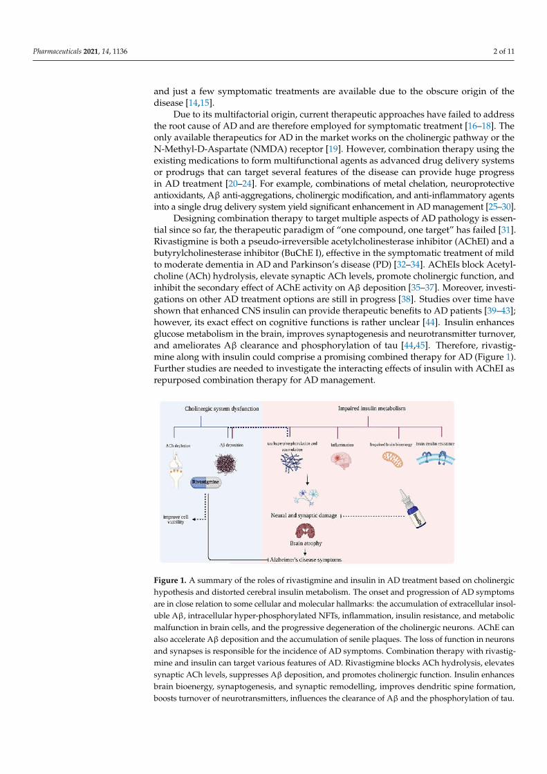
Designing combination therapy to target multiple aspects of AD pathology is essen-tial since so far, the therapeutic paradigm of “one compound, one target” has failed [31].Rivastigmine is both a pseudo-irreversible acetylcholinesterase inhibitor (AChEI) and abutyrylcholinesterase inhibitor (BuChE I), effective in the symptomatic treatment of mildto moderate dementia in AD and Parkinson’s disease (PD) [32–34]. AChEIs block Acetyl-choline (ACh) hydrolysis, elevate synaptic ACh levels, promote cholinergic function, andinhibit the secondary effect of AChE activity on Aβ deposition [35–37]. Moreover, investi-gations on other AD treatment options are still in progress [38]. Studies over time haveshown that enhanced CNS insulin can provide therapeutic benefits to AD patients [39–43];however, its exact effect on cognitive functions is rather unclear [44]. Insulin enhancesglucose metabolism in the brain, improves synaptogenesis and neurotransmitter turnover,and ameliorates Aβ clearance and phosphorylation of tau [44,45]. Therefore, rivastig-mine along with insulin could comprise a promising combined therapy for AD (Figure 1).Further studies are needed to investigate the interacting effects of insulin with AChEI asrepurposed combination therapy for AD management.
Figure 1. A summary of the roles of rivastigmine and insulin in AD treatment based on cholinergichypothesis and distorted cerebral insulin metabolism. The onset and progression of AD symptomsare in close relation to some cellular and molecular hallmarks: the accumulation of extracellular insol-uble Aβ, intracellular hyper-phosphorylated NFTs, inflammation, insulin resistance, and metabolicmalfunction in brain cells, and the progressive degeneration of the cholinergic neurons. AChE canalso accelerate Aβ deposition and the accumulation of senile plaques. The loss of function in neuronsand synapses is responsible for the incidence of AD symptoms. Combination therapy with rivastig-mine and insulin can target various features of AD. Rivastigmine blocks ACh hydrolysis, elevatessynaptic ACh levels, suppresses Aβ deposition, and promotes cholinergic function. Insulin enhancesbrain bioenergy, synaptogenesis, and synaptic remodelling, improves dendritic spine formation,boosts turnover of neurotransmitters, influences the clearance of Aβ and the phosphorylation of tau.
Pharmaceuticals 2021, 14, 1136 3 of 11
Studies have reported that insulin can affect the enzyme activity of AChE, but the re-sults are inconclusive. For example, a study by Agrawal et al. showed an anticholinesteraseeffect of insulin and melatonin in the brain of amnesic mice [46]. Additionally, a more recentstudy reported that insulin treatment is associated with a reduction of AChE activity inthe hippocampus and the frontal cortex regions of the rat brains [47]. S. Lakhman showeda significant rise in AChE activity from the isolated regions of diabetic rat brains. Thesame study demonstrated that acute hyperglycemia elevates AChE activity, and insulinadministration reversed this effect [48]. On the other hand, several studies have reportedthat insulin can enhance AChE activity [49–51].
Insulin, which is a peptide hormone, could interact directly with AChE since in vitrostudies have reported non-specific interactions of different peptides with AChE or itsinhibitors [52–54]. For example, Shamsi et al. has reported an interaction between rivastig-mine and human transferrin, a protein with a molecular weight of around 80 kDa, througha formation of a drug-protein static complex [54]. In other in vitro studies, different kindsof proteins such as a cysteine protease glycoprotein (ZCPG), aflatoxin B (AFB), anchovyprotein hydrolysate (APH), and fasciculin 2 have been reported to interact and inhibitAChE [55–58]. Interestingly, hemp seed protein hydrolysates can inhibit AChE activity,and the amino acid composition of the hydrolysates remarkably affects the enzyme’s in-hibitory potential due to its alternative interactions with the peripheral anionic site (PAS)of the AChE [59]. Additionally, Z. Yu et al. showed both in vitro and in silico studiesthat the Salmo salar roe protein-derived peptide, named WIR, exhibited potent inhibitionagainst AChE. The same study indicated that WIR can bind to both PAS and the catalyticanionic site (CAS) of AChE by hydrogen bond and pi–alkyl interactions [60]. Similarly,insulin, which is a charged protein with a molecular weight of 5.81 kDa, might have suchinteractions with AChE or its inhibitors such as rivastigmine.
Overall, elucidating protein-drug interactions between insulin and rivastigmine orits target AChE is essential in the pharmacological profiling of repurposed combinationtherapy. To date, no in silico nor in vitro studies have examined the possible interactionsof insulin with rivastigmine, or with its target AChE. Hence, the current study aimed toconduct in vitro assessments of the effect of insulin on AChE activity or rivastigmine’sinhibitory action.
2. Results2.1. Determination of Kinetics for ATCh and Rivastigmine to Study Insulin Effects onAChE Activity
To measure the effect of insulin on AChE activity and rivastigmine’s inhibitory ac-tion, we first inquired into the optimized substrate (ATCh) and inhibitor (rivastigmine)concentrations needed to design basic enzymatic tests in the absence of insulin. We startedby measuring AChE activity and rivastigmine inhibitory action through a spectrophoto-metric analysis based on Ellman’s method [61], and calculated all the parameters based onMichaelis–Menten kinetics. Initially, substrate (ATCh) concentration was increased to deter-mine the concentration at which AChE was the most active. In the case of AChE, maximumenzyme activity was reached between 100 µM and 1 mM of ATCh (Figure S1). Accord-ingly, the determined 500 µM concentration of ATCh was chosen for the determinationof rivastigmine IC50. Our result showed that an increase in rivastigmine concentrationscaused a reduction in enzymatic reaction rate up to 1200 µM, above which there was nochange (Figure S2). The rivastigmine inhibition type for AChE was pseudo-irreversible.
Next, we asked whether different concentrations of insulin could affect AChE activity.The 0–50 µM concentrations of insulin were added to the basic enzymatic reaction with thedetermined ATCh concentration (500 µM), which was subsequently evaluated according toEllman’s method [61] and Michaelis–Menten kinetics. No significant effect was observed,even at high concentrations of insulin. As can be seen, the variance of our data is near zero(0.00001), which means that the dispersion of our data is negligible and therefore the effectof insulin on the enzyme activity is insignificant (Figure S3). Since the enzyme functionis highly related to an active site and substrate structures, insulin alone most probably

Pharmaceuticals 2021, 14, 1136 4 of 11
cannot easily change the structure-function relationship, which explains our result. Thus,the modulatory effect of insulin on the cholinergic system indicated in the literature mightbe explained by insulin-induced hypoglycemia in different brain regions, which causesa significant decrease in AChE activity [62], and by the insulin-enhancing effect on AChsynthesis through stimulating the expression of choline acetyltransferase [63]. Since thedirect influence of insulin on AChE activity is unlikely based on our result, the precisemechanisms of insulin interaction with AChE remain to be determined by further studies.
2.2. RSM Studies on the Effect of Different ATCh, Rivastigmine, and Insulin Concentrations onAChE Activity
An RSM was carried out to obtain more information about the integrated effects ofdifferent rivastigmine and insulin concentrations on AChE activity. To examine the effect ofinsulin on AChE activity and rivastigmine inhibitory action under a close-to-real synapticcondition, different ATCh concentrations were considered in the study design. All the testswere conducted via spectrophotometer according to Ellman’s method [61].
2.2.1. Integrated Effect of Rivastigmine and ATCh
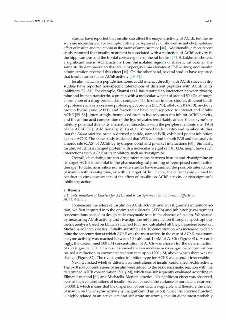
First, in order to investigate the integrated effect of rivastigmine and ATCh, with aconstant concentration of insulin, we conducted enzymatic reaction tests at insulin concen-trations of 0 and 50 µM based on Ellman’s method (Figure 2a,b). In both diagrams, the redand black curves are related to the ATCh concentrations of 1000 and 0 µM, respectively.As expected, enzyme activity was notably reduced by the increase in the concentrationof rivastigmine, an effective AChEI. As in Figure 2a,b, there was no AChE activity in theabsence of ATCh, the substrate. However, in both figures and for the ATCh concentrationof 1000 µM (red curves), the rivastigmine inhibitory effect was sustained in the presence orabsence of insulin, at all measuring points, indicating that neither AChE activity nor therivastigmine inhibitory effect were influenced by the presence of insulin. According to thep-value (>0.0001), these responses are significant. Figure 2c also shows the integrated effectof rivastigmine and ATCh at an insulin concentration of 25 µM in a 3D plot.
Figure 2. RSM plots of the integrated effects of rivastigmine and ATCh (a) at concentrations of0 and (b) 50 µM of insulin (in both diagrams A and B, the red and black curves represent ATChconcentrations of 1000 and 0 µM, respectively). (c) A 3D response surface plot illustrating theintegrated effect of rivastigmine and ATCh at an insulin concentration of 25 µM, all based on thestandard Ellman’s method (p-value > 0.0001).
2.2.2. Integrated Effect of Insulin and ATCh
Second, the integrated effect of insulin and ATCh, with a constant concentration ofrivastigmine, was evaluated by the standard Ellman’s method. Figure 3a,b shows theintegrated effect of insulin and ATCh at concentrations of 0 and 4000 µM of rivastigmine,respectively. In both diagrams, the red and black curves are related to ATCh concentrationsof 1000 and 0 µM, respectively. In Figure 3a,b, in the absence of the substrate (black curves),there was no AChE activity. Nevertheless, at the concentration of 1000 µM for ATCh(red curves), AChE activity decreased only by 2% and 3% in the absence and presence
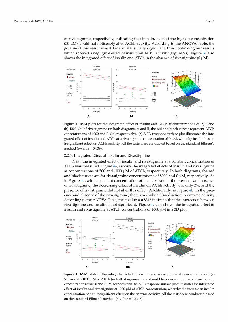
Pharmaceuticals 2021, 14, 1136 5 of 11
of rivastigmine, respectively, indicating that insulin, even at the highest concentration(50 µM), could not noticeably alter AChE activity. According to the ANOVA Table, thep-value of this result was 0.039 and statistically significant, thus confirming our resultswhich showed a negligible effect of insulin on AChE activity (Figure S3). Figure 3c alsoshows the integrated effect of insulin and ATCh in the absence of rivastigmine (0 µM).
Figure 3. RSM plots for the integrated effect of insulin and ATCh at concentrations of (a) 0 and(b) 4000 µM of rivastigmine (in both diagrams A and B, the red and black curves represent ATChconcentrations of 1000 and 0 µM, respectively). (c) A 3D response surface plot illustrates the inte-grated effect of insulin and ATCh at a rivastigmine concentration of 0 µM, whereby insulin has aninsignificant effect on AChE activity. All the tests were conducted based on the standard Ellman’smethod (p-value = 0.039).
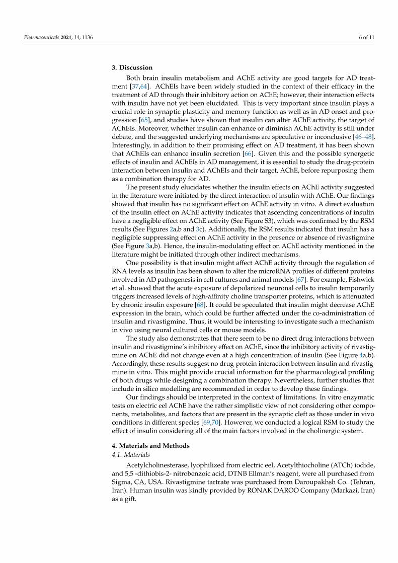
2.2.3. Integrated Effect of Insulin and Rivastigmine
Next, the integrated effect of insulin and rivastigmine at a constant concentration ofATCh was measured. Figure 4a,b shows the integrated effects of insulin and rivastigmineat concentrations of 500 and 1000 µM of ATCh, respectively. In both diagrams, the redand black curves are for rivastigmine concentrations of 8000 and 0 µM, respectively. Asin Figure 4a, with a constant concentration of the substrate in the presence and absenceof rivastigmine, the decreasing effect of insulin on AChE activity was only 2%, and thepresence of rivastigmine did not alter this effect. Additionally, in Figure 4b, in the pres-ence and absence of the rivastigmine, there was only a 3%reduction in enzyme activity.According to the ANOVA Table, the p-value = 0.8346 indicates that the interaction betweenrivastigmine and insulin is not significant. Figure 4c also shows the integrated effect ofinsulin and rivastigmine at ATCh concentrations of 1000 µM in a 3D plot.
Figure 4. RSM plots of the integrated effect of insulin and rivastigmine at concentrations of (a)500 and (b) 1000 µM of ATCh (in both diagrams, the red and black curves represent rivastigmineconcentrations of 8000 and 0 µM, respectively). (c) A 3D response surface plot illustrates the integratedeffect of insulin and rivastigmine at 1000 µM of ATCh concentration, whereby the increase in insulinconcentration has an insignificant effect on the enzyme activity. All the tests were conducted basedon the standard Ellman’s method (p-value = 0.8346).
Pharmaceuticals 2021, 14, 1136 6 of 11
3. Discussion
Both brain insulin metabolism and AChE activity are good targets for AD treat-ment [37,64]. AChEIs have been widely studied in the context of their efficacy in thetreatment of AD through their inhibitory action on AChE; however, their interaction effectswith insulin have not yet been elucidated. This is very important since insulin plays acrucial role in synaptic plasticity and memory function as well as in AD onset and pro-gression [65], and studies have shown that insulin can alter AChE activity, the target ofAChEIs. Moreover, whether insulin can enhance or diminish AChE activity is still underdebate, and the suggested underlying mechanisms are speculative or inconclusive [46–48].Interestingly, in addition to their promising effect on AD treatment, it has been shownthat AChEIs can enhance insulin secretion [66]. Given this and the possible synergeticeffects of insulin and AChEIs in AD management, it is essential to study the drug-proteininteraction between insulin and AChEIs and their target, AChE, before repurposing themas a combination therapy for AD.
The present study elucidates whether the insulin effects on AChE activity suggestedin the literature were initiated by the direct interaction of insulin with AChE. Our findingsshowed that insulin has no significant effect on AChE activity in vitro. A direct evaluationof the insulin effect on AChE activity indicates that ascending concentrations of insulinhave a negligible effect on AChE activity (See Figure S3), which was confirmed by the RSMresults (See Figures 2a,b and 3c). Additionally, the RSM results indicated that insulin has anegligible suppressing effect on AChE activity in the presence or absence of rivastigmine(See Figure 3a,b). Hence, the insulin-modulating effect on AChE activity mentioned in theliterature might be initiated through other indirect mechanisms.
One possibility is that insulin might affect AChE activity through the regulation ofRNA levels as insulin has been shown to alter the microRNA profiles of different proteinsinvolved in AD pathogenesis in cell cultures and animal models [67]. For example, Fishwicket al. showed that the acute exposure of depolarized neuronal cells to insulin temporarilytriggers increased levels of high-affinity choline transporter proteins, which is attenuatedby chronic insulin exposure [68]. It could be speculated that insulin might decrease AChEexpression in the brain, which could be further affected under the co-administration ofinsulin and rivastigmine. Thus, it would be interesting to investigate such a mechanismin vivo using neural cultured cells or mouse models.
The study also demonstrates that there seem to be no direct drug interactions betweeninsulin and rivastigmine’s inhibitory effect on AChE, since the inhibitory activity of rivastig-mine on AChE did not change even at a high concentration of insulin (See Figure 4a,b).Accordingly, these results suggest no drug-protein interaction between insulin and rivastig-mine in vitro. This might provide crucial information for the pharmacological profilingof both drugs while designing a combination therapy. Nevertheless, further studies thatinclude in silico modelling are recommended in order to develop these findings.
Our findings should be interpreted in the context of limitations. In vitro enzymatictests on electric eel AChE have the rather simplistic view of not considering other compo-nents, metabolites, and factors that are present in the synaptic cleft as those under in vivoconditions in different species [69,70]. However, we conducted a logical RSM to study theeffect of insulin considering all of the main factors involved in the cholinergic system.
4. Materials and Methods4.1. Materials
Acetylcholinesterase, lyophilized from electric eel, Acetylthiocholine (ATCh) iodide,and 5,5 -dithiobis-2- nitrobenzoic acid, DTNB Ellman’s reagent, were all purchased fromSigma, CA, USA. Rivastigmine tartrate was purchased from Daroupakhsh Co. (Tehran,Iran). Human insulin was kindly provided by RONAK DAROO Company (Markazi, Iran)as a gift.
Pharmaceuticals 2021, 14, 1136 7 of 11
4.2. Methods4.2.1. Determination of Kinetics for ATCh and Rivastigmine to Study Insulin Effect onAChE Activity
To find the kinetic model for AChE inhibition, the enzymatic reaction rate of AChE wasdetermined by Ellman’s method [61]. It is based on the fact that thiocholine reacts imme-diately, quantitatively, and irreversibly to Ellman’s reagent, 5,5′-dithiobis-(2-nitrobenzoicacid) (DTNB), forming a yellow product (5-mercapto-2-nitrobenzoic acid). We measuredthe absorbance (A) versus time (t) at pH 7.4 in phosphate-buffered saline (PBS) and at roomtemperature, with a constant wavelength of 412 nm using a UV-VIS spectrophotometer(Carry, Australia). The kinetic studies were conducted using a quartz cuvette as a reactor.The cuvette was filled with chosen volumes of PBS, a determined excess amount of DTNB,ascending volumes of substrate solutions (ATCh) for Km evaluation, and ascending vol-umes of the inhibitor (rivastigmine) for a half-maximal inhibitory concentration (IC50),which was determined based on Michaelis–Menten kinetics. The reaction was started bythe fast (<1 s) pipetting of the chosen volume for the AChE solution with the vigorouslymixed reaction mixture. The values of A were continuously measured vs. t over 1 min, andsubsequently saved in a lab PC. Then, we evaluate the influence of the different concentra-tions of insulin (0–50 µM) on AChE activity with the same method for investigating thepossible alternation of AChE activity.
4.2.2. RSM Studies on the Effect of Different Atch, Rivastigmine, and InsulinConcentrations on AChE Activity
RSM studies [71] on different ATCh, rivastigmine, and insulin concentrations wereconducted to simulate the comprehensive effect of insulin under close-to-real conditionsin a synaptic cleft. The effect of three quantitative variables was evaluated based on RSMby Central Composite Design (CCD) using the design expert (DoE) software 10 State Ease(Minneapolis, MN). AChE activity was the corresponding response of three variables:A = (insulin), B = (ATCh), and C = (rivastigmine). For each factor, five different levels andthe following points were selected for 20 runs of the experiments, using CCD (Table S1).
The quadratic model was chosen as the best model to define the factor responses(Table S2). Analysis of variance (ANOVA) was used to evaluate the significance of thequadratic regression model. Moreover, the model terms were assessed using the p-valuewith a 95% confidence level. The coefficient parameters were assessed by response surfaceregression analysis using the software DoE. Additionally, it was applied to obtain theresiduals, three-dimensional (3D) surface, and two-dimensional (2D) contour plots of theresponse models (Table S3).
From Table S3, the quadratic model developed from the RSM was statistically signifi-cant for AChE activity (Y). The low value of P > F (less than 0.05) indicated the randomnessof the results and the significant effect of the model terms on the response. The “Lack ofFit p-value” of 0.2 implied that the Lack of Fit was not significant. The quadratic modelwas used to explain the mathematical relationship between the independent variables andthe dependent response. The mathematical expression for the relationship between AChEinhibition, as a corresponding response, and the three variables of insulin (A), ATCh (B),and rivastigmine (C) should be calculated according to the following Equation (1):
R = A + B + C + AB + AC + BC + A2 + B2 + C2 (1)
Statistical analysis showed that the coefficients AC, A2, and B2 were statistically notsignificant. Therefore, A2 and B2 were omitted from the model. However, since the studypurpose was to evaluate the simultaneous effect of insulin (variable A) and rivastigmine(variable C) on the enzyme activity, AC was not omitted from the model, although it hadthe p-value > 0.1 (Table S2). The final model was obtained in both coded and non-codedforms (Tables S4 and S5). The F value shown in Table S2 indicates that ATCh (F value= 311.88) had the greatest effect on the enzyme activity, followed by rivastigmine andinsulin. It can be explained by the fact that ATCh is the enzyme substrate; thus, the enzyme

Pharmaceuticals 2021, 14, 1136 8 of 11
activity is highly dependent on this factor. Furthermore, the interaction between ATCh andrivastigmine showed a high F value of 235.23. Following the same logic, since rivastigmineis an effective AChE inhibitor, the effect of this factor was expected to be considerable. Theinsulin F-value was 5.59 and the p-value was 0.039, which was less than 0.05 and thus stillconsidered significant. The new equation was modified to:
R = R0 + A + B + C + AB + AC + BC + C2 (2)
The high value of the coefficient of determination (R2 = 0.9838) is a measure of thegoodness-of-fit to the model. This indicates a high degree of correlation between the pre-dicted response and the experimental responses. The adjusted R2 = 0.9743 also confirmedthe high correlation between the observed and theoretical values. From Figure S4, it wasalso confirmed that the experimental (actual) values were very close to the predicted ones.
The Adeq Precision value indicates the signal-to-noise ratio, and the optimal valueis greater than 4. As can be seen, the Adeq precision value for the proposed model was37.15, which indicates an excellent signal-to-noise ratio, signifying that the proposed modelcan be used to predict points in the design-covered area. At last, the ANOVA of eachresponse was evaluated, and the effects of the independent variables were expressed in 3Dresponse plots.
5. Conclusions
Our study demonstrates that insulin has no direct effect on either AChE activity orthe rivastigmine inhibitory action in vitro. This may rule out possible direct interactionsbetween insulin and AChE, suggesting other indirect mechanisms for the cross-talk be-tween insulin and AChE. Most importantly, given the potential benefits of insulin in themanagement of AD, concurrent use of insulin and rivastigmine as a single drug deliverysystem, perhaps as a prodrug or as an adjunct treatment, could be considered since theinhibitory action of rivastigmine on AChE is preserved even at considerable concentrationsof insulin in vitro. Nevertheless, more in vivo studies using neural cells or sophisticatedanimal models are needed to elucidate possible drug interactions and potential synergeticeffects of insulin and rivastigmine as a combination therapy for AD management.
Supplementary Materials: The following are available online at https://www.mdpi.com/article/10.3390/ph14111136/s1, Table S1: CCD matrix for the inhibition of AChE activity as the correspondingresponse of three variables (A = (insulin), B = (ATCh), and C = (rivastigmine)), Table S2: ANOVA dataof the quadratic model, Table S3: Quadratic model as the best model to define the factor responses,Table S4: Coded form of the final model, Table S5. Non-coded form of the final model (Actual),Figure S1: Km determination for ATCh, Figure S2: AChE activity evaluation in presence of ATCh(500 µM) and ascending volumes of rivastigmine for the determination of the IC50, Figure S3: Theeffect of different concentrations of insulin vs. AChE activity, Figure S4: Predicted versus actual datafor the quadratic model (R2 ≤ 0.2).
Author Contributions: Conceptualization, S.K., T.J.-T., and M.H.A.-S.; methodology, T.J.-T.; soft-ware, M.B.; validation, S.K. and H.B.S.; formal analysis, M.B.; investigation, T.J.-T.; resources, S.K.;data curation, M.B.; writing—original draft preparation, T.J.-T. and M.H.A.-S.; writing—review andediting, M.H.A.-S. and H.B.S.; visualization, T.J.-T. and M.B.; supervision, S.K.; project administra-tion, S.K.; funding acquisition, S.K. All authors have read and agreed to the published version ofthe manuscript.
Funding: H.B.S is supported by The Swedish Research Council and The Novo Nordisk Foundation.
Institutional Review Board Statement: Not applicable.
Informed Consent Statement: Not applicable.
Data Availability Statement: Data is contained in the article and Supplementary Materials.
Pharmaceuticals 2021, 14, 1136 9 of 11
Acknowledgments: We want to extend our special thanks and gratitude to Razi University for thefinancial and technical support provided to this work, and to Uppsala University for their valuableassistance and guidelines.
Conflicts of Interest: The authors declare no conflict of interest.
References1. Calabrò, M.; Rinaldi, C.; Santoro, G.; Crisafulli, C. The biological pathways of Alzheimer disease: A review. AIMS Neurosci. 2021,
8, 86. [CrossRef] [PubMed]2. Uddin, M.S.; Al Mamun, A.; Kabir, M.T.; Ashraf, G.M.; Bin-Jumah, M.N.; Abdel-Daim, M.M. Multi-target drug candidates for
multifactorial Alzheimer’s disease: AChE and NMDAR as molecular targets. Mol. Neurobiol. 2021, 58, 281–303. [CrossRef][PubMed]
3. Khoury, R.; Patel, K.; Gold, J.; Hinds, S.; Grossberg, G.T. Recent progress in the pharmacotherapy of Alzheimer’s disease. DrugsAging 2017, 34, 811–820. [CrossRef] [PubMed]
4. Sanabria-Castro, A.; Alvarado-Echeverría, I.; Monge-Bonilla, C. Molecular Pathogenesis of Alzheimer’s Disease: An Update.Ann. Neurosci. 2017, 24, 46–54. [CrossRef]
5. Kent, S.A.; Spires-Jones, T.L.; Durrant, C.S. The physiological roles of tau and Aβ: Implications for Alzheimer’s disease pathologyand therapeutics. Acta Neuropathol. 2020, 140, 417–447. [CrossRef]
6. Selkoe, D.J.; Hardy, J. The amyloid hypothesis of Alzheimer’s disease at 25 years. EMBO Mol. Med. 2016, 8, 595–608. [CrossRef]7. Polanco, J.C.; Li, C.; Bodea, L.-G.; Martinez-Marmol, R.; Meunier, F.A.; Götz, J. Amyloid-β and tau complexity—Towards
improved biomarkers and targeted therapies. Nat. Rev. Neurol. 2018, 14, 22–39. [CrossRef]8. Bhatt, S.; Puli, L.; Patil, C.R. Role of reactive oxygen species in the progression of Alzheimer’s disease. Drug Discov. Today 2020,
26, 794–803. [CrossRef]9. Francis, P.T.; Palmer, A.M.; Snape, M.; Wilcock, G.K. The cholinergic hypothesis of Alzheimer’s disease: A review of progress. J.
Neurol. Neurosurg. Psychiatry 1999, 66, 137–147. [CrossRef]10. Auld, D.S.; Kornecook, T.J.; Bastianetto, S.; Quirion, R. Alzheimer’s disease and the basal forebrain cholinergic system: Relations
to β-amyloid peptides, cognition, and treatment strategies. Prog. Neurobiol. 2002, 68, 209–245. [CrossRef]11. Hara, Y.; McKeehan, N.; Fillit, H.M. Translating the biology of aging into novel therapeutics for Alzheimer disease. Neurology
2019, 92, 84–93. [CrossRef]12. Guo, T.; Zhang, D.; Zeng, Y.; Huang, T.Y.; Xu, H.; Zhao, Y. Molecular and cellular mechanisms underlying the pathogenesis of
Alzheimer’s disease. Mol. Neurodegener. 2020, 15, 20. [CrossRef]13. Andreeva, T.V.; Lukiw, W.J.; Rogaev, E.I. Biological Basis for Amyloidogenesis in Alzheimer’s Disease. Biochemistry 2017, 82,
122–139. [CrossRef]14. Haake, A.; Nguyen, K.; Friedman, L.; Chakkamparambil, B.; Grossberg, G.T. An update on the utility and safety of cholinesterase
inhibitors for the treatment of Alzheimer’s disease. Expert Opin. Drug Saf. 2020, 19, 147–157. [CrossRef]15. Alzheimer’s Association. Alzheimer’s Association Report (2016 Alzheimer’s disease facts and figures). Alzheimer’s Dement. 2016,
12, 459–509. [CrossRef]16. Gong, C.-X.; Liu, F.; Iqbal, K. Multifactorial hypothesis and multi-targets for Alzheimer’s disease. J. Alzheimer’s Dis. 2018, 64,
S107–S117. [CrossRef]17. Fish, P.V.; Steadman, D.; Bayle, E.D.; Whiting, P. New approaches for the treatment of Alzheimer’s disease. Bioorg. Med. Chem.
Lett. 2019, 29, 125–133. [CrossRef]18. Breijyeh, Z.; Karaman, R. Comprehensive Review on Alzheimer’s Disease: Causes and Treatment. Molecules 2020, 25, 5789.
[CrossRef]19. Marucci, G.; Buccioni, M.; Ben, D.D.; Lambertucci, C.; Volpini, R.; Amenta, F. Efficacy of acetylcholinesterase inhibitors in
Alzheimer’s disease. Neuropharmacology 2021, 190, 108352. [CrossRef]20. Kabir, M.T.; Uddin, M.S.; Mamun, A.A.; Jeandet, P.; Aleya, L.; Mansouri, R.A.; Ashraf, G.M.; Mathew, B.; Bin-Jumah, M.N.;
Abdel-Daim, M.M. Combination Drug Therapy for the Management of Alzheimer’s Disease. Int. J. Mol. Sci. 2020, 21, 3272.[CrossRef]
21. Xiao, G.; Li, Y.; Qiang, X.; Xu, R.; Zheng, Y.; Cao, Z.; Luo, L.; Yang, X.; Sang, Z.; Su, F. Design, synthesis and biological evaluationof 4′-aminochalcone-rivastigmine hybrids as multifunctional agents for the treatment of Alzheimer’s disease. Bioorg. Med. Chem.2017, 25, 1030–1041. [CrossRef] [PubMed]
22. Luc, M.; Wozniak, M.; Helemejko, M.; Rymaszewska, J. Tackling Alzheimer’s disease: Hypothetical synergism between anti-inflammatory and anti-diabetic agents. Life Sci 2019, 231, 116483. [CrossRef] [PubMed]
23. Liu, Z.; Zhang, B.; Xia, S.; Fang, L.; Gou, S. ROS-responsive and multifunctional anti-Alzheimer prodrugs: Tacrine-ibuprofenhybrids via a phenyl boronate linker. Eur. J. Med. Chem. 2021, 212, 112997. [CrossRef] [PubMed]
24. Kim, M.; Park, M.H.; Nam, G.; Lee, M.; Kang, J.; Song, I.-S.; Choi, M.-K.; Jin, H.K.; Bae, J.-S.; Lim, M.H. A Glycosylated Prodrug toAttenuate Neuroinflammation and Improve Cognitive Deficits in Alzheimer’s Disease Transgenic Mice. Mol. Pharm. 2021, 18,101–112. [CrossRef]
25. Dhala, I.; Khan, T.; Prabhu, A. Chimeric Conjugates for Alzheimer’s Disease. Exon Publ. 2020, Ch10, 165–180.
Pharmaceuticals 2021, 14, 1136 10 of 11
26. Waseem, R.; Shamsi, A.; Mohammad, T.; Alhumaydhi, F.A.; Kazim, S.N.; Hassan, M.I.; Ahmad, F.; Islam, A. Multispectroscopicand Molecular Docking Insight into Elucidating the Interaction of Irisin with Rivastigmine Tartrate: A Combinational TherapyApproach to Fight Alzheimer’s Disease. ACS Omega 2021, 6, 7910–7921. [CrossRef]
27. Abdel, M.E.A.E.-F.; Khalil, W.F.; Mohamed, S.M. Effect of Curcumin, Exelon and their Combination on Brain in Alzheimer’sDisease-Induced Rats. J. Adv. Med. Med. Res. 2021, 33, 65–78. [CrossRef]
28. Zhao, J.; Yin, F.; Ji, L.; Wang, C.; Shi, C.; Liu, X.; Yang, H.; Wang, X.; Kong, L. Development of a Tau-Targeted Drug DeliverySystem Using a Multifunctional Nanoscale Metal–Organic Framework for Alzheimer’s Disease Therapy. ACS Appl. Mater.Interfaces 2020, 12, 44447–44458. [CrossRef]
29. Pardridge, W.M. Treatment of Alzheimer’s Disease and Blood–Brain Barrier Drug Delivery. Pharmaceuticals 2020, 13, 394.[CrossRef]
30. Stavrakov, G.; Philipova, I.; Lukarski, A.; Atanasova, M.; Zheleva, D.; Zhivkova, Z.D.; Ivanov, S.; Atanasova, T.; Konstantinov, S.;Doytchinova, I. Galantamine-Curcumin Hybrids as Dual-Site Binding Acetylcholinesterase Inhibitors. Molecules 2020, 25, 3341.[CrossRef]
31. Cummings, J.L.; Tong, G.; Ballard, C. Treatment Combinations for Alzheimer’s Disease: Current and Future PharmacotherapyOptions. J. Alzheimer’s Dis. 2019, 67, 779–794. [CrossRef]
32. Pakala, R.S.; Brown, K.N.; Preuss, C.V. Cholinergic Medications. In StatPearls; StatPearls Publishing: Treasure Island, FL,USA, 2020.
33. Patel, P.H.; Gupta, V. Rivastigmine. In StatPearls; StatPearls Publishing: Treasure Island, FL, USA, 2020.34. Ray, B.; Maloney, B.; Sambamurti, K.; Karnati, H.K.; Nelson, P.T.; Greig, N.H.; Lahiri, D.K. Rivastigmine modifies the α-secretase
pathway and potentially early Alzheimer’s disease. Transl. Psychiatry 2020, 10, 47. [CrossRef]35. Campos, E.O.; Alvarez, A.; Inestrosa, N.C. Brain acetylcholinesterase promotes amyloid-β-peptide aggregation but does not
hydrolyze amyloid precursor protein peptides. Neurochem. Res. 1998, 23, 135–140. [CrossRef]36. Inestrosa Cantín, N. Acetylcholinesterase Accelerates Assembly of Amyloid-B-Peptides Into Alzheimer’s Fibrils: Possible Role of
the Peripheral Site of the Enzyme. Neuron 1996, 16, 881–891. [CrossRef]37. Nguyen, K.; Hoffman, H.; Chakkamparambil, B.; Grossberg, G.T. Evaluation of rivastigmine in Alzheimer’s disease. Neurodegener.
Dis. Manag. 2021, 11, 35–48. [CrossRef]38. Henstridge, C.M.; Hyman, B.T.; Spires-Jones, T.L. Beyond the neuron–cellular interactions early in Alzheimer disease pathogenesis.
Nat. Rev. Neurosci. 2019, 20, 94–108. [CrossRef]39. Freiherr, J.; Hallschmid, M.; Frey, W.H.; Brünner, Y.F.; Chapman, C.D.; Hölscher, C.; Craft, S.; De Felice, F.G.; Benedict, C.
Intranasal insulin as a treatment for Alzheimer’s disease: A review of basic research and clinical evidence. CNS Drugs 2013, 27,505–514. [CrossRef]
40. Benedict, C.; Hallschmid, M.; Hatke, A.; Schultes, B.; Fehm, H.L.; Born, J.; Kern, W. Intranasal insulin improves memory inhumans. Psychoneuroendocrinology 2004, 29, 1326–1334. [CrossRef]
41. De la Monte, S.M. Intranasal insulin therapy for cognitive impairment and neurodegeneration: Current state of the art. ExpertOpin. Drug Deliv. 2013, 10, 1699–1709. [CrossRef]
42. Akhtar, A.; Bishnoi, M.; Sah, S.P. Sodium orthovanadate improves learning and memory in intracerebroventricular-streptozotocinrat model of Alzheimer’s disease through modulation of brain insulin resistance induced tau pathology. Brain Res. Bull. 2020, 164,83–97. [CrossRef]
43. Berlanga-Acosta, J.; Guillén-Nieto, G.; Rodríguez-Rodríguez, N.; Bringas-Vega, M.L.; García-del-Barco-Herrera, D.; Berlanga-Saez,J.O.; García-Ojalvo, A.; Valdés-Sosa, M.J.; Valdés-Sosa, P.A. Insulin Resistance at the Crossroad of Alzheimer Disease Pathology:A Review. Front. Endocrinol. 2020, 11, 560375. [CrossRef] [PubMed]
44. Kellar, D.; Craft, S. Brain insulin resistance in Alzheimer’s disease and related disorders: Mechanisms and therapeutic approaches.Lancet Neurol. 2020, 19, 758–766. [CrossRef]
45. Tauber, S.C.; Djukic, M.; Gossner, J.; Eiffert, H.; Brück, W.; Nau, R. Sepsis-associated encephalopathy and septic encephalitis: Anupdate. Expert Rev. Anti-Infect. Ther. 2020, 6, 215–230. [CrossRef] [PubMed]
46. Agrawal, R.; Tyagi, E.; Shukla, R.; Nath, C. Effect of insulin and melatonin on acetylcholinesterase activity in the brain of amnesicmice. Behav. Brain Res. 2008, 189, 381–386. [CrossRef]
47. Nampoothiri, M.; Kumar, N.; Venkata Ramalingayya, G.; Gopalan Kutty, N.; Krishnadas, N.; Mallikarjuna Rao, C. Effect of insulinon spatial memory in aluminum chloride-induced dementia in rats. NeuroReport 2017, 28, 540–544. [CrossRef]
48. Lakhman, S.; Kaur, G. Effect of alloxan-induced diabetes on acetylcholinesterase activity from discrete areas of rat brain.Neurochem. Int. 1994, 24, 159–163. [CrossRef]
49. Catalan, R.; Martinez, A.; Mata, F.; Aragones, M. Effect of insulin on acetylcholinesterase activity. Biochem. Biophys. Res. Commun.1981, 101, 1216–1220. [CrossRef]
50. Zhang, B.; Yang, L.; Yu, L.; Lin, B.; Hou, Y.; Wu, J.; Huang, Q.; Han, Y.; Guo, L.; Ouyang, Q.; et al. Acetylcholinesterase isassociated with apoptosis in β cells and contributes to insulin-dependent diabetes mellitus pathogenesis. Acta Biochim. Biophys.Sin. 2012, 44, 207–216. [CrossRef]
51. Rizvi, S.I.; Zaid, M.A. Insulin-Like Effect Of (–) Epicatechin On Erythrocyte Membrane Acetylcholinesterase Activity In Type 2Diabetes Mellitus. Clin. Exp. Pharmacol. Physiol. 2001, 28, 776–778. [CrossRef]
Pharmaceuticals 2021, 14, 1136 11 of 11
52. Prasasty, V.; Radifar, M.; Istyastono, E. Natural Peptides in Drug Discovery Targeting Acetylcholinesterase. Molecules 2018,23, 2344. [CrossRef]
53. Torrent, J.; Vilchez-Acosta, A.; Munoz-Torrero, D.; Trovaslet, M.; Nachon, F.; Chatonnet, A.; Grznarova, K.; Acquatella-Tran VanBa, I.; Le Goffic, R.; Herzog, L.; et al. Interaction of prion protein with acetylcholinesterase: Potential pathobiological implicationsin prion diseases. Acta Neuropathol. Commun. 2015, 3, 18. [CrossRef]
54. Shamsi, A.; Mohammad, T.; Khan, M.S.; Shahwan, M.; Husain, F.M.; Rehman, M.T.; Hassan, M.I.; Ahmad, F.; Islam, A.Unraveling Binding Mechanism of Alzheimer’s Drug Rivastigmine Tartrate with Human Transferrin: Molecular Docking andMulti-Spectroscopic Approach towards Neurodegenerative Diseases. Biomolecules 2019, 9, 495. [CrossRef]
55. Jamir, K.; Ganguly, R.; Seshagirirao, K. ZCPG, a cysteine protease from Zingiber montanum rhizome exhibits enhanced anti-inflammatory and acetylcholinesterase inhibition potential. Int. J. Biol. Macromol. 2020, 163, 2429–2438. [CrossRef]
56. Waqar, M.; Batool, S. In silico analysis of binding of neurotoxic venom ligands with acetylcholinesterase for therapeutic use intreatment of Alzheimer’s disease. J. Theor. Biol. 2015, 372, 107–117. [CrossRef]
57. Arduini, F.; Errico, I.; Amine, A.; Micheli, L.; Palleschi, G.; Moscone, D. Enzymatic Spectrophotometric Method for Aflatoxin BDetection Based on Acetylcholinesterase Inhibition. Anal. Chem. 2007, 79, 3409–3415. [CrossRef]
58. Zhao, T.; Su, G.; Wang, S.; Zhang, Q.; Zhang, J.; Zheng, L.; Sun, B.; Zhao, M. Neuroprotective Effects of AcetylcholinesteraseInhibitory Peptides from Anchovy (Coilia mystus) against Glutamate-Induced Toxicity in PC12 Cells. J. Agric. Food Chem. 2017,65, 11192–11201. [CrossRef]
59. Malomo, S.A.; Aluko, R.E. In vitro acetylcholinesterase-inhibitory properties of enzymatic hemp seed protein hydrolysates. J. Am.Oil Chem. Soc. 2016, 93, 411–420. [CrossRef]
60. Yu, Z.; Ji, H.; Shen, J.; Kan, R.; Zhao, W.; Li, J.; Ding, L.; Liu, J. Identification and molecular docking study of fish roe-derivedpeptides as potent BACE 1, AChE, and BChE inhibitors. Food Funct. 2020, 11, 6643–6651. [CrossRef]
61. Ellman, G.L.; Courtney, K.D.; Andres, V., Jr.; Featherstone, R.M. A new and rapid colorimetric determination of acetyl-cholinesterase activity. Biochem. Pharmacol. 1961, 7, 88–95. [CrossRef]
62. Kaur, G.; Arora, S.K. Acetylcholinesterase and Na+, K+-ATPase activities in different regions of rat brain during insulin-inducedhypoglycemia. Mol. Chem. Neuropathol. 1994, 21, 83–93. [CrossRef]
63. Baruah, P.; Das, A.; Paul, D.; Chakrabarty, S.; Aguan, K.; Mitra, S. Sulfonylurea Class of Antidiabetic Drugs Inhibit Acetyl-cholinesterase Activity: Unexplored Auxiliary Pharmacological Benefit toward Alzheimer’s Disease. ACS Pharmacol. Transl. Sci.2021, 4, 193–205. [CrossRef]
64. Dubey, S.K.; Lakshmi, K.; Krishna, K.V.; Agrawal, M.; Singhvi, G.; Saha, R.N.; Saraf, S.; Saraf, S.; Shukla, R.; Alexander, A. Insulinmediated novel therapies for the treatment of Alzheimer’s disease. Life Sci. 2020, 249, 117540. [CrossRef]
65. Mejido, D.C.; Andrade, J.; Vieira, M.N.; Ferreira, S.T.; De Felice, F.G. Insulin and leptin as potential cognitive enhancers inmetabolic disorders and Alzheimer’s disease. Neuropharmacology 2020, 171, 108115. [CrossRef]
66. Doyle, M.E.; Egan, J.M. Pharmacological agents that directly modulate insulin secretion. Pharmacol. Rev. 2003, 55, 105–131.[CrossRef]
67. Samadian, M.; Gholipour, M.; Hajiesmaeili, M.; Taheri, M.; Ghafouri-Fard, S. The Eminent Role of microRNAs in the Pathogenesisof Alzheimer’s Disease. Front. Aging Neurosci. 2021, 13, 107. [CrossRef]
68. Fishwick, K.J.; Rylett, R.J. Insulin regulates the activity of the high-affinity choline transporter CHT. PLoS ONE 2015, 10, e0132934.[CrossRef]
69. Cao, Y.; Herrero-Nogareda, L.; Cedergreen, N. A comparative study of acetylcholinesterase and general-esterase activity assaysusing different substrates, in vitro and in vivo exposures and model organisms. Ecotoxicol. Environ. Saf. 2020, 189, 109954.[CrossRef]
70. Kasteel, E.E.J.; Nijmeijer, S.M.; Darney, K.; Lautz, L.S.; Dorne, J.L.C.M.; Kramer, N.I.; Westerink, R.H.S. Acetylcholinesteraseinhibition in electric eel and human donor blood: An in vitro approach to investigate interspecies differences and humanvariability in toxicodynamics. Arch. Toxicol. 2020, 94, 4055–4065. [CrossRef]
71. Bezerra, M.A.; Santelli, R.E.; Oliveira, E.P.; Villar, L.S.; Escaleira, L.A. Response surface methodology (RSM) as a tool foroptimization in analytical chemistry. Talanta 2008, 76, 965–977. [CrossRef]