Genital lesions in cows naturally infected with trypanosomes ...
Upload
independentCategory
view
1download
0
REPRODUCTIONRESEARCH
The PATE gene is expressed in the accessory tissues ofthe human male genital tract and encodes a secretedsperm-associated protein
Angel A Soler-Garcıa, Rangan Maitra, Vasantha Kumar, Tomoko Ise, Satoshi Nagata,Richard Beers, Tapan K Bera and Ira Pastan
Laboratory of Molecular Biology, Center for Cancer Research, National Cancer Institute, National Institutesof Health, Bethesda, Maryland 20892-4264, USA
Correspondence should be addressed to I Pastan; Email: [email protected]
Angel A Soler-Garcıa is now at Center for Genetic Medicine Research, Division of Nephrology, Children’s Research Institute,
Children’s National Medical Center, 3.5 Floor, Room R165, 111 Michigan Ave., NW, Washington DC 20010, USA
Rangan Maitra is now at Paradigm Genetics, Inc., 108 T W Alexander Drive, Research Triangle Park, North Carolina 27709,
USA
Abstract
The PATE gene is expressed in prostate and testis. To determine if PATE is expressed in other accessory tissues of the male
genital tract, RT-PCR of the epididymis and seminal vesicle was performed. PATE mRNA was highly expressed in the epididy-
mis and seminal vesicle. In situ hybridization of the testis showed PATE mRNA is strongly expressed in the spermatogonia.
The PATE gene encodes a 14-kDa protein with a predicted signal sequence and a cleavage site between residues G21 and S22.
To determine if PATE is a secreted protein, 293T cells were transfected with a pcDNA-PATE-myc-His plasmid and protein
immunoprecipitated with anti-myc monoclonal antibody. Western blot analysis showed the presence of PATE-myc-His protein
was in the medium and the cell lysate. Confocal microscopy demonstrated that PATE-myc-His protein is found in the endo-
plasmic reticulum. The polyclonal antibody SOL-1 was generated by immunization of rabbits with recombinant PATE protein
expressed and purified from Escherichia coli. Western blots were performed on extracts of prostate, testis, seminal vesicle and
ejaculated spermatozoa, but PATE protein was only detected in the spermatozoa. Immunostaining of sperm smears revealed
that PATE is located in a band-like pattern in the sperm head. Our data indicate that PATE is made by various sexual accessory
tissues and secreted into the semen where it becomes associated with sperm, suggesting that PATE is a novel sperm-associated
protein with a possible role in mammalian sperm maturation.
Reproduction (2005) 129 515–524
Introduction
Mammalian sperm are terminally differentiated cells.Their differentiation from round spermatids to highlypolarized and fully motile cells is characterized by exten-sive biochemical, physiological and morphological events(Toshimori 1998, Ducheux et al. 2003). These events startin the testis in the process called spermiogenesis. For thesperm, extensive post-gonadal differentiation to maturityis required for successful fertilization. This post-gonaddifferentiation occurs in the male accessory glands suchas the epididymis. These accessory glands contribute tothe sperm maturation process by secreting factors into theseminal plasma, a complex biological fluid formed fromthe mix of various secretions in the male genital tract (Luoet al. 2001).
The epididymis provides a luminal environment thatpromotes both maturation and survival of the spermatozoa
(Hinton & Palladino 1995, Hinton et al. 1996). In thisluminal environment there are factors involved in gluta-thione conjugation and metabolism (Robaire & Viger1995, Hinton et al. 1996), inhibitors of complement-mediated lysis (Griswold et al. 1986, Collard & Griswold1987, Sylvester et al. 1991) and protease inhibitors (Corn-wall et al. 1992, Kirchhoff et al. 1998). The sperm becomefunctionally mature while traveling through the epididy-mis duct gaining fertilization ability and zone recognition(Orgebin-Crist 1969, Bedford 1975). In hamsters, it hasbeen proposed that the epididymis possesses a sperm sort-ing mechanism which discriminates viable from non-viable sperm (NagDas et al. 2000).
Seminal vesicle secretions constitute the major portionof the seminal plasma contributing factors that affectsperm motility. The contribution of the seminal vesiclesecretions to sperm motility has been investigated using
q 2005 Society for Reproduction and Fertility DOI: 10.1530/rep.1.00576
ISSN 1470–1626 (paper) 1741–7899 (online) Online version via www.reproduction-online.org
semen of several different mammals such as boar (Iwa-moto et al. 1992, Jeng et al. 1993, Nichol et al. 1997),bull (Al-Somai et al. 1994), mouse (Peitz 1988) andhuman (Robert & Gagnon 1996). In mice, the removal ofthe seminal vesicle greatly reduces mouse sperm fertility(Pang et al. 1979, Peitz & Olds-Clarke 1986). Because theprocess by which mammalian sperm develops andmatures to fully functional cells is not completely under-stood, the identification and characterization of new fac-tors related to the process is important. In the followingstudy we report the identification and characterization ofPATE, a gene expressed in prostate and testis as a novelsperm-associated protein that may be involved in spermmaturation.
Materials and Methods
Reverse transcription-PCR (RT-PCR) analysis
Total RNA from epididymis and seminal vesicle wasextracted using the Absolutely RNA RT-PCR Miniprep kitfollowing instructions of the manufacturer (Stratagene, LaJolla, CA, USA). Total RNA from prostate and testis wasobtained by BD Biosciences Clontech (Palo Alto, CA,USA). Single-stranded cDNA from RNA samples of eachtissue was synthesized from 5mg total RNA using the First-strand cDNA Synthesis kit (Amersham Biosciences;Piscataway, NJ, USA) following instructions from the manu-facturer. PATE PCR amplification was performed using1.5ml of the first-strand reaction and PATE specific primersPATE-12 sense (50-ACAAGTCCCTCTTGCTGGAACTC-30)and PATE-363 antisense (50-AAGGTCTTCATTGCACAGGT-CATG-30) using the Hot Star Taq Master mix (Qiagen, Ala-meda, CA, USA). The thermocycling protocol was initialdenaturation at 95 8C for 15 min, 35 cycles of denaturationat 94 8C for 1 min, annealing at 60 8C for 1 min, andelongation at 72 8C for 1 min. This set of primers generatesa 351 bp fragment. For RNA control, PCR was performedusing commercially available actin primers that generate a640 bp PCR fragment. The PCR products were analyzed on1.0% 0.5 £ TAE (20mM Tris, 10 M acetate, 1 mM EDTA;pH 8.0) agarose gel followed by ethidium bromide staining.
In situ hybridization
In situ hybridization of PATE mRNA on testis tissues wasperformed as described earlier (Kumar & Collins 1994,Olsson et al. 2001). Biotinylated probes were preparedusing cDNAs encoding PATE (1500 bp), the small nuclearRNA U6 (250 bp) and the B cell differentiation antigenCD22 (2068 bp) cloned in the pBluescript II SK(þ ) plas-mid, using the BioNick Labeling System kit (Life Technol-ogies, Gaithersburg, MD, USA) according to themanufacturer’s instructions. Slides were hybridized usingthe In situ Hybridization and Detection System (Life Tech-nologies) according to the manufacturer’s instructions. Theslides were counterstained using 0.2% Light Green stain,rinsed through a series of alcohol grades, and mounted
in Cytoseal (Stephens Scientific, Riverdale, NJ, USA).Microscopic evaluation was performed using a NikonEclipse 800 microscope.
Cell culture and transfection
293T cells (American Type Culture Collection, Manassas,VA, USA) were grown in a 5% CO2/95% air humidifiedincubator at 37 8C and in D-MEM (Quality Biological Inc.,Gaithersburg, MD, USA) supplemented with 10% FBS,2 mM L-glutamine, 100 units/ml penicillin, and 100mg/mlstreptomycin. Plasmid DNA transfection of 293T cells wasperformed with Lipofectamine 2000 (Invitrogen, Carlsbad,CA, USA) according to the manufacturer’s instructions.Assays were performed 48 h after transfection.
Detection of secreted PATE
293T cells were transfected with pcDNA3-PATE-myc-Hisplasmid expressing PATE protein with a myc-His epitopetag at the carboxy terminus using Lipofectamine 2000 fol-lowing the manufacturer’s protocol (Invitrogen). Culturemedia and cells were collected by aspiration and trypsini-zation respectively. PATE-myc-His and actin proteins wereimmunoprecipitated with an anti-myc monoclonal anti-body (mAb) and an actin polyclonal antibody (pAb) (SantaCruz Biotechnology, Inc., Santa Cruz, CA, USA) from the5 ml culture medium and 1 mg total cell lysate respect-ively, following standard protocols (Harlow & Lane 1999).One-fifth of the immunoprecipitated protein was loadedin 4–20% PAGE gel (BioRad, Hercules, CA, USA) andtransferred to PVDF membranes. Membranes were immu-noblotted with anti-myc or anti-actin mAbs (1:1000dilution) followed by rat anti-mouse IgG1-horseradish per-oxidase (HRP)-conjugated mAb (1:1000 dilution). Immu-noblotted membranes were detected using an ECLdetection kit (Amersham Biosciences).
Subcellular localization of PATE
For PATE subcellular localization, 293T cells were trans-fected with pcDNA PATE-myc-His. After 48 h, cells werewashed 3 times with PBS, fixed in 1 ml 3.7% formal-dehyde in PBS for 10 min at 25 8C and washed 3 times for10 min with PBS. Cells were permeabilized with 1 ml0.1% Triton X-100 in PBS for 5 min at 25 8C, washed andblocked with 5mg/ml normal goat globulin (NGG) inPBS þ 0.1% saponin for 20 min at 25 8C. Blocked cellswere incubated with mouse anti-myc mAb (MolecularProbes, Inc., Eugene OR, USA), 5mg/ml in PBS-NGG-saponin for 90 min at 25 8C or overnight at 4 8C. Coverslipswere washed 3 times, incubated with goat anti-mouseAlexa Fluor 594 antibody (5mg/ml) in PBS-NGG-saponinfor 60 min at 25 8C and washed 3 times with PBS (Molecu-lar Probes, Inc.). To visualize the nucleus, cells werestained with DAPI (5mM in PBS; Molecular Probes, Inc.)for 5 min at 25 8C and washed twice with PBS for 5 min.To visualize the endoplasmic reticulum, cells were
516 A A Soler-Garcıa and others
Reproduction (2005) 129 515–524 www.reproduction-online.org
incubated with 2mM 3,30dihexyloxacarbocyanide iodide(DiOC6(3)) for 5 min (Molecular Probes, Inc.) (Sabnis et al.1997). Slides were analyzed in a Zeiss LSM 510 confocalmicroscope (Carl Zeiss, Inc., Thornwood, NY, USA).
Purification of recombinant PATE (rPATE) protein
Plasmid pRB302 was constructed using a 321 bp PCR pro-duct, generated by primers PATE2C: 50-GAC TGA CTT CATATG GGA TCA CTT TCA ATG AGA AAT-30 and PATE3N:50-TTC CAT TGG AAT TCT AAA GGT CTT CAT TGC ACAG-30 and CR2.1-PATE plasmid (Bera et al. 2002) as a tem-plate. The PCR product was subsequently subcloned into apET-based expression vector pULI7 (Brinkmann et al.1991) using NdeI and EcoRI restriction sites. PlasmidpRB302 expresses the PATE protein lacking the signalsequence. Escherichia coli BL21 (aDE3) (Studier & Moffatt1986) was transformed with plasmid pRB302. Protein pro-duction was induced by isopropyl-beta D-thiogalactoside(IPTG), and PATE accumulated in inclusion bodies. Purifi-cation of the protein was performed as described beforefor immunotoxin purification (Pastan et al. 2003). Briefly,inclusion bodies were solubilized in guanidine hydrochlo-ride solution and reduced with dithioerythritol. Solubilizedprotein was refolded by dilution in a refolding buffer con-taining arginine and glutathione to prevent aggregationand to facilitate redox shuffling respectively. Refoldedprotein was dialyzed and purified by ion exchangechromatography. Protein concentration was determined byBio-Rad DC protein assay (Bio-Rad).
Generation of anti-PATE rabbit antibody (SOL-1 pAb)
The generation of polyclonal anti-PATE rabbit antibody(named SOL-1 pAb) was performed by immunization oftwo rabbits with purified rPATE protein by Spring ValleyLaboratories, Inc. (Woodbine, MD, USA) following stan-dard protocols. Two rabbits were primary immunized with100mg rPATE protein in complete Freund’s adjuvant. Afterprimary immunization they were boosted at 21, 42 and74 days post-immunization with 100mg rPATE protein inincomplete Freund’s adjuvant. Rabbits were bled and thedevelopment of anti-PATE rabbit antibodies was monitoredby Western blot against rPATE protein.
Human sperm collection and tissue samples
Human semen ejaculates were obtained from normalmale donors by masturbation after 3 days of abstinencefrom ejaculation (samples were designated exempt by theOffice of Human Subjects Research, NIH). Ejaculateswere allowed to liquefy at 25 8C. Sperms were collectedas described by Gupta et al. (1990). Liquified semen wascentrifuged at 400 £ g for 15 min, washed 3 times in Tris–buffered saline (TBS; 10 mM Tris, 145 mM NaCl, pH 7.4)and stored at 270 8C. Epididymis and seminal vesicle tis-sues were obtained from the cooperative Human TissueNetwork, Southern Division (Philadelphia, PA, USA).
Preparation of cell extracts and Western blot analysis
Transfected and untransfected 293T cells, seminal vesicle,epididymis and sperm lysates were prepared using NP-40lysis buffer containing protease inhibitors by incubationon ice for 1 h (Harlow & Lane 1999). Frozen tissues weremacerated with a cold mortar and pestle and then resus-pended in NP-40 lysis buffer (Boehringer). Prostate, testis,brain and spermatic cord total cell lysates were obtainedfrom Clontech. Thirty-five micrograms protein extracts(5mg for sperm fractions) were run on a 4–20% Tris–gly-cine gel (Bio-Rad) and transferred to a 0.2mm poly (vinyli-dene difluoride) membrane (Millipore, Billerica, MA,USA) in transfer buffer (25 mM Tris/192 mM glycine/20%(vol/vol) methanol, pH 8.3) at 4 8C overnight. Filters wereimmunoblotted with anti-PATE SOL-1 pAb (1:1500) fol-lowed by goat anti-rabbit HRP conjugated antibodies(1:5000; BioSource, Camarillo, CA, USA). Signals weredetected using an ECL chemiluminescence Western blot-ting kit according to the manufacturer’s instructions(Amersham Biosciences).
Association of PATE with sperm
SOL-1 pAbs raised in rabbit were used to detect the pre-sence of PATE on sperm cells. The secondary fluorescentantibody used was goat anti-rabbit IgG Alexa Fluor 594(Molecular Probes, Inc.). Fifty microliter aliquots of spermsuspension in PBS were smeared onto Superfrost Plusmicro slides (VWR, West Chester, PA, USA). The slideswere allowed to air dry in a chemical hood for 10 min.The slides were fixed and stained as described above withthe following changes. Sperm smears were fixed with for-maldehyde and washed with PBS for 5 min. Slides wereblocked and incubated with 100ml SOL-1 pAb (1:250) at4 8C overnight in a humid chamber. After primary anti-body, slides were washed with PBS and secondary anti-body was applied in 100ml (1:250 dilution of 1 mg/ml)and incubated for 1 h at 25 8C in a humid chamber in thedark. After secondary antibody, slides were washed andAnti-Fade was applied according to the manufacturer’sinstructions (Molecular Probes, Inc.). A coverslip wasplaced on the sperm smear and slides were placed in aslide box and left to dry overnight at 4 8C wrapped withfoil. Slides were analyzed in a Zeiss LSM 510 confocalmicroscope (Carl Zeiss, Inc.).
Results
PATE mRNA is expressed in the accessory glands of themale genital tract
The PATE gene was identified by a functional genomicapproach (Bera et al. 2002). Of many tissues examined,PATE mRNA expression was found to be limited to theprostate and testis by multiple tissue dot-blot, PCR oncDNAs from 24 different human tissues and Northern blotanalyses (Bera et al. 2002). This limited tissue expression
PATE is a secreted sperm-associated protein 517
www.reproduction-online.org Reproduction (2005) 129 515–524
suggests that PATE expression is restricted to the male gen-ital tract. To determine if PATE is expressed in the acces-sory glands of the male genital tract, RT-PCR wasperformed using total RNA from prostate, testis, epididy-mis and seminal vesicle. As previously described, PATEmRNA was detected in the prostate and testis. PATEmRNA was also detected in the epididymis and the semi-nal vesicle (Fig. 1A). As expected, no PCR products wereobtained in the water control. These data indicate that theRT-PCR products are specific.
In the normal prostate, PATE mRNA was restricted tothe epithelial cells (Bera et al. 2002). To determine thecell types in the testis that express PATE mRNA, in situhybridization was used with biotin-labeled PATE cDNA asa probe as described in Materials and Methods. As shownin Fig. 1B, PATE mRNA is expressed in spermatogonia.CD22 and U6 probes were used as negative and positivecontrols respectively. As expected, in situ hybridizationwith CD22 showed no signal and the U6 probe gave astrong signal confirming the specificity of the in situhybridization (Fig. 1B). These data suggest that PATE ismade in spermatogonia of the testis.
The PATE gene encodes a secreted protein
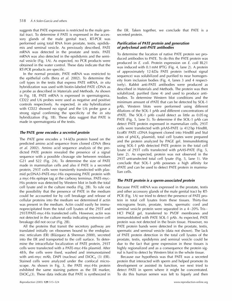
The PATE gene encodes a 14-kDa protein based on thepredicted amino acid sequence from cloned cDNA (Beraet al. 2002). Amino acid sequence analysis of the pre-dicted PATE protein revealed the presence of a signalsequence with a possible cleavage site between residuesG21 and S22 (Fig. 2A). To determine the size of PATEmade in mammalian cells and also if PATE is a secretedprotein, 293T cells were transiently transfected with plas-mid pcDNA3-PATE-myc-His expressing PATE protein witha myc-His epitope tag at the carboxy terminus. PATE-myc-His protein was detected by Western blot in both the totalcell lysate and in the culture media (Fig. 2B). To rule outthe possibility that the presence of PATE in the mediumcould be accounted for by cell breakage and release ofcellular proteins into the medium we determined if actinwas present in the medium. Actin could easily be immu-noprecipitated from the total cell lysates of the 293T and293T/PATE-myc-His transfected cells. However, actin wasnot detected in the culture media indicating extensive cellbreakage did not occur (Fig. 2B).
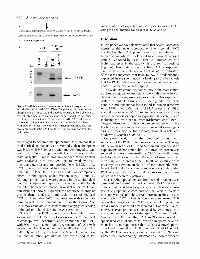
All the proteins that transit the secretory pathway aretranslated initially on ribosomes bound to the endoplas-mic reticulum (ER) (Blazquez & Shennan 2000), secretedinto the ER and transported to the cell surface. To deter-mine the intracellular localization of PATE protein, 293Tcells were transfected with a PATE-myc-His plasmid. After48 h, the cells were fixed, washed and immunostainedwith anti-myc mAb, DAPI (nucleus) and DiOC6 (3) (ER).Stained cells were analyzed under the confocal micro-scope. As shown in Fig. 3, the PATE myc-His proteinexhibited the same staining pattern as the ER marker,DiOC6(3). These data indicate that PATE is synthesized in
the ER. Taken together, we conclude that PATE is asecreted protein.
Purification of PATE protein and generationof polyclonal anti-PATE antibodies
To determine the location of native PATE protein we pro-duced antibodies to PATE. To do this the PATE protein wasproduced in E. coli. Protein expression on E. coli BL21was induced with 0.1 mM IPTG (Fig. 4, lane 2). A proteinof approximately 12-kDa PATE protein (without signalsequence) was solubilized and purified to near homogen-eity from inclusion bodies (Fig. 4, lanes 3 and 4 respect-ively). Rabbit anti-PATE antibodies were produced asdescribed in Materials and Methods. The protein was thensolubilized, purified (lane 4) and used to produce anti-bodies. To determine Western blot conditions and theminimum amount of rPATE that can be detected by SOL-1pAb, Western blots were performed using differentdilutions of the SOL-1 pAb and different concentrations ofrPATE. The SOL-1 pAb could detect as little as 0.05 ngPATE (Fig. 5, lane 5). To determine if the SOL-1 pAb candetect PATE protein expressed in mammalian cells, 293Tcells were transfected with pAAS-PATE (a 412 bp HindIII,EcoRV PATE cDNA fragment cloned into HindIII and StuIsites of pNLX2 plasmid), total cell lysates were preparedand the protein analyzed by PAGE gel. Immunoblottingusing SOL-1 pAb detected PATE protein in the total celllysate of 293T cells transfected with pAAS-PATE (Fig. 5,lane 2). As expected, protein was not observed in the293T untransfected total cell lysate (Fig. 5, lane 1). Weconclude that SOL-1 pAb posseses a high affinity forrPATE and can be used to detect PATE protein in mamma-lian cells.
The PATE protein is a sperm-associated protein
Because PATE mRNA was expressed in the prostate, testisand other accessory glands of the male genital tract by RT-PCR (Fig. 1A) we tried to detect the presence of PATE pro-tein in total cell lysates from these tissues. Thirty-fivemicrograms brain, prostate, testis, spermatic cord andseminal vesicle protein were separated in a 4–20% Tris–HCl PAGE gel, transferred to PVDF membranes andimmunoblotted with PATE SOL-1 pAb. As expected, PATEprotein was not detected in the brain tissue. However, noPATE protein bands were detected in the prostate, testis,spermatic and seminal vesicle (data not shown). The lackof PATE protein detection in the total cell lysates of theprostate, testis, epididymis and seminal vesicle could bedue to the fact that gene expression in these tissues ishighly regionalized and as a consequence the protein sig-nal is hard to detect by Western blot in the whole tissue.
Because our hypothesis was that PATE was a secretedprotein that interacted with sperm and helped promote itsdevelopment or assisted its function, we next tried todetect PATE in sperm where it might be concentrated.To do this human semen was left to liquefy and then
518 A A Soler-Garcıa and others
Reproduction (2005) 129 515–524 www.reproduction-online.org

Figure 1 PATE mRNA expression in accessory glands of the male genital tract. (A) RT-PCR analysis of PATE mRNA expression was performedwith specific PATE primers (top) or actin primers (bottom) with cDNAs derived from each representative tissue. RT-PCRs performed without tem-plates are indicated as dH2O. PCR products were separated on a 1% agarose gel and visualized by ethidium bromide staining. (B) In situ localiz-ation of PATE mRNA in the testis. Testis tissue sections were stained with hematoxylin/eosin (H/E) to show general morphology or probed withplasmid Bluescript containing CD22 (negative control), U6 (positive control) or PATE cDNA.
PATE is a secreted sperm-associated protein 519
www.reproduction-online.org Reproduction (2005) 129 515–524
centrifuged to separate the sperm from the seminal fluidas described in Materials and Methods. Then the spermwas lysed with NP-40 lysis buffer and centrifuged to sep-arate the soluble (supernatant) from the non-solublematerial (pellet). Five micrograms of each sperm fractionwere analyzed in 4–20% PAGE gel followed by PVDFmembrane transfer and immunoblotting with SOL-1 pAb.PATE protein was detected in the sperm supernatant frac-tion (Fig. 5, lane 3). The 12-kDa PATE was completelyabsent in the sperm pellet fraction (Fig. 5, lane 4).Although protein bands were detected in the seminal fluidfraction of ejaculated spermatozoa none of the bandsexhibited the expected molecular weight of the PATE pro-tein (data not shown). However, the reactivity of proteinsgreater than 12-kDa (the molecular mass of secretedPATE) indicates that PATE may associate with other pro-teins present in the seminal fluid or in the sperm. AlsoPATE may associate with itself forming aggregates leadingto the ladder of band sizes in the Western blot.
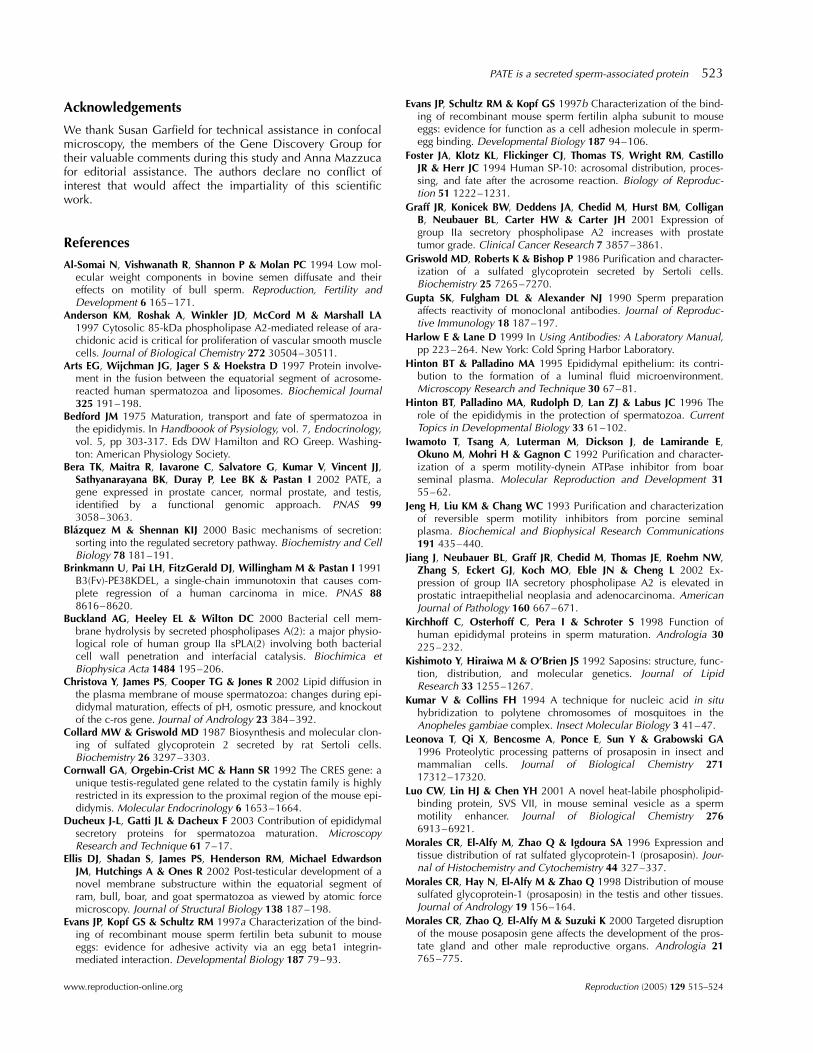
To confirm that PATE protein is associated with humansperm and to determine its location on sperm, confocalmicroscopy was performed after immunostaining PATEwith SOL-1 pAb (1:250 dilution). Association of PATE withsperm could be observed and was localized to a band-likepattern lying in the sperm head (Fig. 6E and H). As a nega-tive control, rabbit pre-immune sera were used at the
same dilution. As expected, no PATE protein was detectedusing the pre-immune rabbit sera (Fig. 6A and D).
Discussion
In this paper we have demonstrated that several accessorytissues of the male reproductive system contain PATEmRNA, but that PATE protein can only be detected onhuman sperm where it is located in an unusual bandingpattern. We found by RT-PCR that PATE mRNA was alsohighly expressed in the epididymis and seminal vesicles(Fig. 1A). This finding confirms that PATE is expressedexclusively in the male genital tract. In situ hybridizationof the testis indicated that PATE mRNA is predominantlyexpressed in the spermatogonia leading to the hypothesisthat the PATE protein may be involved in the developmentand/or is associated with the sperm.
The wide expression of PATE mRNA in the male genitaltract may suggest an important role of this gene in celldevelopment. Prosaposin is an example of this expressionpattern in multiple tissues of the male genital tract. Thisgene is a multifunctional locus found in human (Leonovaet al. 1996), mouse (Sun et al. 1994, Morales et al. 1998)and rat (Morales et al. 1996) and encodes four glyco-protein activators or saponins expressed in several tissuesincluding the male genital tract (Kishimoto et al. 1992).Targeted disruption of this widely expressed gene in miceleads to a decrease in testis size with reduced spermiogen-esis and involution of the prostate, seminal vesicle andepididymis (Morales et al. 2000).
Computer analysis of the predicted amino acidsequence of the PATE protein revealed a putative cleavagesite between residues G21 and S22. Immunoprecipitationexperiments demonstrated that PATE-myc-His protein wassecreted to the culture media of 293T transiently trans-fected cells as shown in the Western blot using anti-mycmAb (Fig. 2B). Moreover, the subcellular localization ofPATE-myc-His protein in the ER of the transiently trans-fected 293T cells by confocal microscopy confirms thatPATE is a secreted protein that is processed and trans-ported in the secretory pathway.
SOL-1 pAb, a polyclonal antibody raised in rabbits, wasgenerated and therefore used to detect PATE protein incommercially and laboratory-made protein lysates of pros-tate, testis, spermatic cord and seminal vesicle. Westernblot analysis did not show PATE protein in these tissueseven though PATE mRNA is abundantly expressed. Thisobservation suggests that PATE as a secreted protein israpidly made, processed and secreted out of these tissues.However, PATE protein was detected by Western blot inthe supernatant fraction of the sperm. The latter findingtogether with the fact that PATE mRNA was present inspecialized cells of the testis involved in sperm develop-ment led us to hypothesize that PATE is a novel sperm-associated protein (Fig. 1B). Furthermore, BLASTP analysisof the PATE amino acid sequence against the NationalCenter for Biotechnology Information’s ‘non-redundant’
Figure 2 PATE is a secreted protein. (A) Amino acid sequenceencoded by the cloned PATE cDNA. The putative cleavage site andphospholipase A2 motif are indicated by arrows and bold charactersrespectively. Underlined is a histidine residue thought to be criticalfor phospholipase activity. (B) Secretion of PATE. 293T cells weretransfected with pcDNA3-PATE-myc-His. Forty-eight hours laterPATE-myc-His or actin proteins were immunoprecipitated with anti-myc mAb or anti-actin pAb from the culture medium and from thecell lysates.
520 A A Soler-Garcıa and others
Reproduction (2005) 129 515–524 www.reproduction-online.org
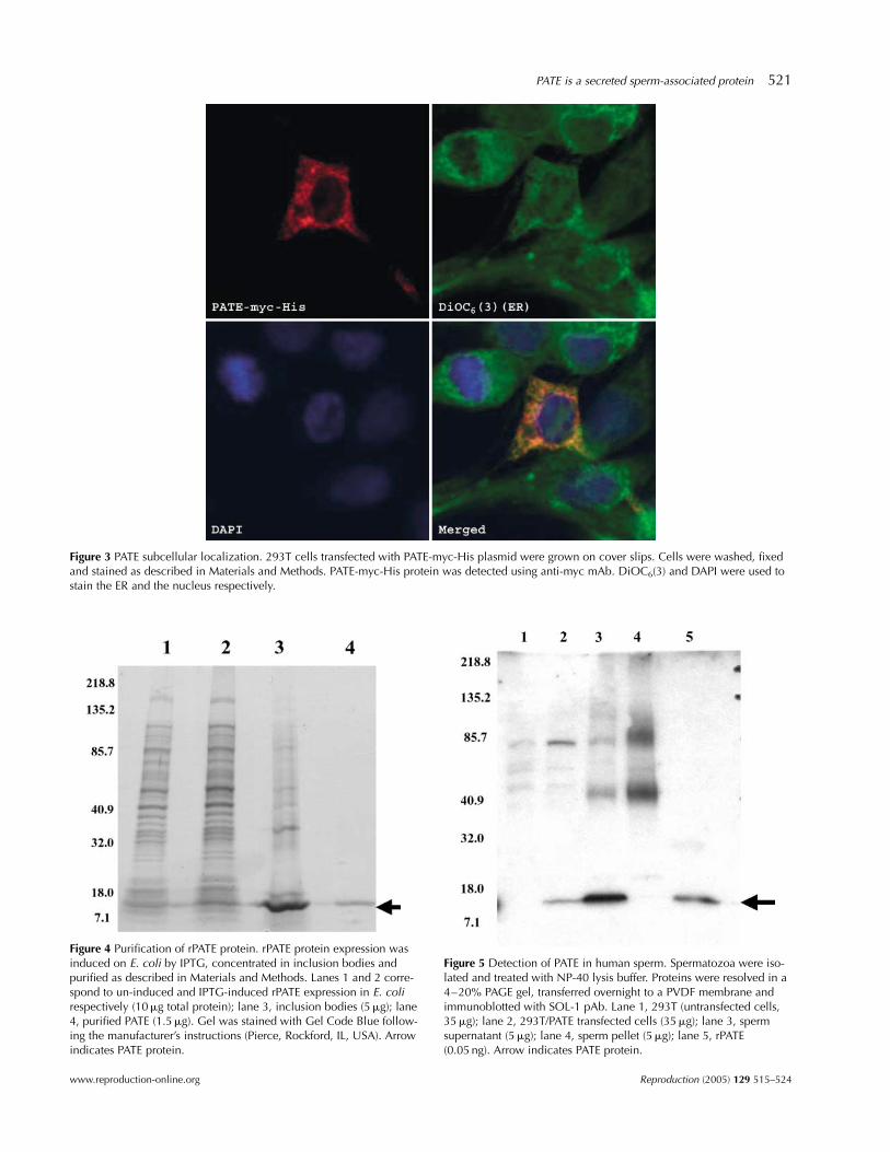
Figure 3 PATE subcellular localization. 293T cells transfected with PATE-myc-His plasmid were grown on cover slips. Cells were washed, fixedand stained as described in Materials and Methods. PATE-myc-His protein was detected using anti-myc mAb. DiOC6(3) and DAPI were used tostain the ER and the nucleus respectively.
Figure 4 Purification of rPATE protein. rPATE protein expression wasinduced on E. coli by IPTG, concentrated in inclusion bodies andpurified as described in Materials and Methods. Lanes 1 and 2 corre-spond to un-induced and IPTG-induced rPATE expression in E. colirespectively (10mg total protein); lane 3, inclusion bodies (5mg); lane4, purified PATE (1.5mg). Gel was stained with Gel Code Blue follow-ing the manufacturer’s instructions (Pierce, Rockford, IL, USA). Arrowindicates PATE protein.
Figure 5 Detection of PATE in human sperm. Spermatozoa were iso-lated and treated with NP-40 lysis buffer. Proteins were resolved in a4–20% PAGE gel, transferred overnight to a PVDF membrane andimmunoblotted with SOL-1 pAb. Lane 1, 293T (untransfected cells,35mg); lane 2, 293T/PATE transfected cells (35mg); lane 3, spermsupernatant (5mg); lane 4, sperm pellet (5mg); lane 5, rPATE(0.05 ng). Arrow indicates PATE protein.
PATE is a secreted sperm-associated protein 521
www.reproduction-online.org Reproduction (2005) 129 515–524

database (http://www.ncbi.nlm.nih.gov/blast/) resulted insimilarities to the acrosomal vesicle protein SP-10. Thealigned part of the SP-10 protein belongs to a snake toxinfamily of proteins according to the sequence-based pro-tein classification database, Pfam. This protein may play arole in sperm-zona binding and penetration (Foster et al.1994).
Confocal microscopy of immunostained sperm smearsdemonstrated that PATE protein is indeed associated withthe human sperm. PATE protein was localized to a band-like pattern in the sperm head (Fig. 6E and H). Othersperm-associated proteins that exhibit this band-like pat-tern are cystatin-related epididymal spermatogenic andequatorial sperm proteins (Wassler et al. 2002, Wolko-wicz et al. 2003). In the case of these two proteins thecharacteristic band-like pattern is located between theprincipal and the equatorial segments of the sperm head.The equatorial segment is formed in the late spermatidstage and it is maintained until it is incorporated into theoocyte (Toshimori 1998). A role for the sperm equatorialsegment during fertilization has been proposed by Elliset al. (2002). After the sperm penetrates the egg the equa-torial segment is believed to initiate sperm-egg adhesion.This adhesion step is mediated by the protein fertilin(Evans et al. 1997a, b). The fusion capacity of the equator-ial segment has been demonstrated experimentally byfusion with liposomes (Arts et al. 1997). This finding
suggests that PATE protein probably plays a role in mam-malian fertilization (Wassarman et al. 2001).
The predicted amino acid sequence of the PATE proteinexhibits a phospholipase A2 motif (Fig. 2A) and fold-rec-ognition programs group PATE protein as a member of asuperfamily of proteins that includes the snake venomtoxin, neurotoxins and cardiotoxins (Bera et al. 2002). Thepresence of the phospholipase motif and the low molecu-lar weight of the protein suggest that PATE could exhibitphospholipase activity. This is important because phos-pholipases have been shown to influence a wide range ofcellular activities such as inflammation (Murakami et al.1997), proliferation (Anderson et al. 1997), apoptosis(Zhang et al. 1999, Taketo & Sonoshita 2002), carcinogen-esis (Graff et al. 2001, Jiang et al. 2002), and protectionagainst microbial infection (Buckland et al. 2000). Thephospholipase activity can be an important property ofthe PATE protein given that the membranes of the mam-malian spermatozoa undergo extensive plasma membraneremodeling during maturation in the epididymal duct(Christova et al. 2002). Riffo and Parraga (1997) demon-strated through in vitro experiments that antibodies againstphospholipase A2 inibit sperm-egg fusion.
In conclusion, PATE is a highly expressed gene in themale genital tract that encodes a novel secreted sperm-associated protein that may play crucial roles duringsperm development, maturation and fertilization.
Figure 6 Localization of PATE associated with human sperm. Human spermatozoa were collected by centrifugation. Sperm smears wereprepared in Superfrost Plus slides. Cells were washed, fixed and stained as described in Materials and Methods. PATE protein was detectedusing PATE SOL-1 pAb (1:250 dilution). Pre-immune rabbit serum was used as a negative control. (A) Pre-immune; (B and F) DAPI stain; (C andG) differential interference contrast microscopy (DIC); (E) rabbit anti-PATE (SOL-1); (D and H) merged images of A, B and E, F respectively.Magnification £ 630.
522 A A Soler-Garcıa and others
Reproduction (2005) 129 515–524 www.reproduction-online.org
Acknowledgements
We thank Susan Garfield for technical assistance in confocalmicroscopy, the members of the Gene Discovery Group fortheir valuable comments during this study and Anna Mazzucafor editorial assistance. The authors declare no conflict ofinterest that would affect the impartiality of this scientificwork.
References
Al-Somai N, Vishwanath R, Shannon P & Molan PC 1994 Low mol-ecular weight components in bovine semen diffusate and theireffects on motility of bull sperm. Reproduction, Fertility andDevelopment 6 165–171.
Anderson KM, Roshak A, Winkler JD, McCord M & Marshall LA1997 Cytosolic 85-kDa phospholipase A2-mediated release of ara-chidonic acid is critical for proliferation of vascular smooth musclecells. Journal of Biological Chemistry 272 30504–30511.
Arts EG, Wijchman JG, Jager S & Hoekstra D 1997 Protein involve-ment in the fusion between the equatorial segment of acrosome-reacted human spermatozoa and liposomes. Biochemical Journal325 191–198.
Bedford JM 1975 Maturation, transport and fate of spermatozoa inthe epididymis. In Handboook of Psysiology, vol. 7, Endocrinology,vol. 5, pp 303-317. Eds DW Hamilton and RO Greep. Washing-ton: American Physiology Society.
Bera TK, Maitra R, Iavarone C, Salvatore G, Kumar V, Vincent JJ,Sathyanarayana BK, Duray P, Lee BK & Pastan I 2002 PATE, agene expressed in prostate cancer, normal prostate, and testis,identified by a functional genomic approach. PNAS 993058–3063.
Blazquez M & Shennan KIJ 2000 Basic mechanisms of secretion:sorting into the regulated secretory pathway. Biochemistry and CellBiology 78 181–191.
Brinkmann U, Pai LH, FitzGerald DJ, Willingham M & Pastan I 1991B3(Fv)-PE38KDEL, a single-chain immunotoxin that causes com-plete regression of a human carcinoma in mice. PNAS 888616–8620.
Buckland AG, Heeley EL & Wilton DC 2000 Bacterial cell mem-brane hydrolysis by secreted phospholipases A(2): a major physio-logical role of human group IIa sPLA(2) involving both bacterialcell wall penetration and interfacial catalysis. Biochimica etBiophysica Acta 1484 195–206.
Christova Y, James PS, Cooper TG & Jones R 2002 Lipid diffusion inthe plasma membrane of mouse spermatozoa: changes during epi-didymal maturation, effects of pH, osmotic pressure, and knockoutof the c-ros gene. Journal of Andrology 23 384–392.
Collard MW & Griswold MD 1987 Biosynthesis and molecular clon-ing of sulfated glycoprotein 2 secreted by rat Sertoli cells.Biochemistry 26 3297–3303.
Cornwall GA, Orgebin-Crist MC & Hann SR 1992 The CRES gene: aunique testis-regulated gene related to the cystatin family is highlyrestricted in its expression to the proximal region of the mouse epi-didymis. Molecular Endocrinology 6 1653–1664.
Ducheux J-L, Gatti JL & Dacheux F 2003 Contribution of epididymalsecretory proteins for spermatozoa maturation. MicroscopyResearch and Technique 61 7–17.
Ellis DJ, Shadan S, James PS, Henderson RM, Michael EdwardsonJM, Hutchings A & Ones R 2002 Post-testicular development of anovel membrane substructure within the equatorial segment ofram, bull, boar, and goat spermatozoa as viewed by atomic forcemicroscopy. Journal of Structural Biology 138 187–198.
Evans JP, Kopf GS & Schultz RM 1997a Characterization of the bind-ing of recombinant mouse sperm fertilin beta subunit to mouseeggs: evidence for adhesive activity via an egg beta1 integrin-mediated interaction. Developmental Biology 187 79–93.
Evans JP, Schultz RM & Kopf GS 1997b Characterization of the bind-ing of recombinant mouse sperm fertilin alpha subunit to mouseeggs: evidence for function as a cell adhesion molecule in sperm-egg binding. Developmental Biology 187 94–106.
Foster JA, Klotz KL, Flickinger CJ, Thomas TS, Wright RM, CastilloJR & Herr JC 1994 Human SP-10: acrosomal distribution, proces-sing, and fate after the acrosome reaction. Biology of Reproduc-tion 51 1222–1231.
Graff JR, Konicek BW, Deddens JA, Chedid M, Hurst BM, ColliganB, Neubauer BL, Carter HW & Carter JH 2001 Expression ofgroup IIa secretory phospholipase A2 increases with prostatetumor grade. Clinical Cancer Research 7 3857–3861.
Griswold MD, Roberts K & Bishop P 1986 Purification and character-ization of a sulfated glycoprotein secreted by Sertoli cells.Biochemistry 25 7265–7270.
Gupta SK, Fulgham DL & Alexander NJ 1990 Sperm preparationaffects reactivity of monoclonal antibodies. Journal of Reproduc-tive Immunology 18 187–197.
Harlow E & Lane D 1999 In Using Antibodies: A Laboratory Manual,pp 223–264. New York: Cold Spring Harbor Laboratory.
Hinton BT & Palladino MA 1995 Epididymal epithelium: its contri-bution to the formation of a luminal fluid microenvironment.Microscopy Research and Technique 30 67–81.
Hinton BT, Palladino MA, Rudolph D, Lan ZJ & Labus JC 1996 Therole of the epididymis in the protection of spermatozoa. CurrentTopics in Developmental Biology 33 61–102.
Iwamoto T, Tsang A, Luterman M, Dickson J, de Lamirande E,Okuno M, Mohri H & Gagnon C 1992 Purification and character-ization of a sperm motility-dynein ATPase inhibitor from boarseminal plasma. Molecular Reproduction and Development 3155–62.
Jeng H, Liu KM & Chang WC 1993 Purification and characterizationof reversible sperm motility inhibitors from porcine seminalplasma. Biochemical and Biophysical Research Communications191 435–440.
Jiang J, Neubauer BL, Graff JR, Chedid M, Thomas JE, Roehm NW,Zhang S, Eckert GJ, Koch MO, Eble JN & Cheng L 2002 Ex-pression of group IIA secretory phospholipase A2 is elevated inprostatic intraepithelial neoplasia and adenocarcinoma. AmericanJournal of Pathology 160 667–671.
Kirchhoff C, Osterhoff C, Pera I & Schroter S 1998 Function ofhuman epididymal proteins in sperm maturation. Andrologia 30225–232.
Kishimoto Y, Hiraiwa M & O’Brien JS 1992 Saposins: structure, func-tion, distribution, and molecular genetics. Journal of LipidResearch 33 1255–1267.
Kumar V & Collins FH 1994 A technique for nucleic acid in situhybridization to polytene chromosomes of mosquitoes in theAnopheles gambiae complex. Insect Molecular Biology 3 41–47.
Leonova T, Qi X, Bencosme A, Ponce E, Sun Y & Grabowski GA1996 Proteolytic processing patterns of prosaposin in insect andmammalian cells. Journal of Biological Chemistry 27117312–17320.
Luo CW, Lin HJ & Chen YH 2001 A novel heat-labile phospholipid-binding protein, SVS VII, in mouse seminal vesicle as a spermmotility enhancer. Journal of Biological Chemistry 2766913–6921.
Morales CR, El-Alfy M, Zhao Q & Igdoura SA 1996 Expression andtissue distribution of rat sulfated glycoprotein-1 (prosaposin). Jour-nal of Histochemistry and Cytochemistry 44 327–337.
Morales CR, Hay N, El-Alfy M & Zhao Q 1998 Distribution of mousesulfated glycoprotein-1 (prosaposin) in the testis and other tissues.Journal of Andrology 19 156–164.
Morales CR, Zhao Q, El-Alfy M & Suzuki K 2000 Targeted disruptionof the mouse posaposin gene affects the development of the pros-tate gland and other male reproductive organs. Andrologia 21765–775.
PATE is a secreted sperm-associated protein 523
www.reproduction-online.org Reproduction (2005) 129 515–524
Murakami M, Nakatani Y, Atsumi G, Inoue K & Kudo I 1997 Regu-latory functions of phospholipase A2. Critical Reviews in Immu-nology 17 225–283.
NagDas SK, Winfrey VP & Olson GE 2000 Identification of a hamsterepididymal region-specific secretory glycoprotein that binds nonvi-able spermatozoa. Biology of Reproduction 63 1428–1436.
Nichol R, Hunter RH, de Lamirande E, Gagnon C & Cooke GM1997 Motility of spermatozoa in hydrosalpingeal and follicularfluid of pigs. Journal of Reproduction and Fertility 110 79–86.
Olsson P, Bera TK, Essand M, Kumar V, Duray P, Vincent J, Lee B &Pastan I 2001 GDEP a new gene differentially expressed in normalprostate and prostate cancer. Prostate 48 231–241.
Orgebin-Crist MC 1969 Studies on the function of the epididymis.Biology of Reproduction 1 (Suppl 1) 155–175.
Pang SF, Chow PH & Wong TM 1979 The role of the seminal ves-icles, coagulating glands and prostate glands on the fertility andfecundity of mice. Journal of Reproduction and Fertility 56129–132.
Pastan I, Beers R & Bera TK 2003 Recombinant immunotoxins in thetreatment of cancer. Methods in Molecular Biology 248 503–518.
Peitz B 1988 Effects of seminal vesicle fluid components on spermmotility in the house mouse. Journal of Reproduction and Fertility83 169–176.
Peitz B & Olds-Clarke P 1986 Effects of seminal vesicle removal onfertility and uterine sperm motility in the house mouse. Biology ofReproduction 35 608–617.
Riffo MS & Parrga M 1997 Role of phospholipase A2 in mammaliansperm-egg fusion: development of hamster oolemma fusibility bylysophosphatidylcholine. Journal of Experimental Zoology 27981–88.
Robaire B & Viger RS 1995 Regulation of epididymal epithelial cellfunctions. Biology of Reproduction 52 226–236.
Robert M & Gagnon C 1996 Purification and characterization ofthe active precursor of a human sperm motility inhibitor secretedby the seminal vesicles: identity with semenogelin. Biology ofReproduction 55 813–821.
Sabnis RW, Deligeorgiev TG, Jachak MN & Dalvi TS 1997 DiOC6(3):a useful dye for staining the endoplasmic reticulum. Biotechnicand Histochemistry 72 253–258.
Studier FW & Moffatt BA 1986 Use of bacteriophage T7 RNA poly-merase to direct selective high-level expression of cloned genes.Journal of Molecular Biology 189 113–130.
Sun Y, Witte DP & Grabowski GA 1994 Developmental and tissue-specific expression of prosaposin mRNA in murine tissues. Amer-ican Journal of Pathology 145 1390–1398.
Sylvester SR, Morales C, Oko R & Griswold MD 1991 Localization ofsulfated glycoprotein-2 (clusterin) on spermatozoa and in the repro-ductive tract of the male rat. Biology of Reproduction 45 195–207.
Taketo MM & Sonoshita M 2002 Phospholipase A2 and apoptosis.Biochimica et Biophysica Acta 1585 72–76.
Toshimori K 1998 Maturation of mammalian spermatozoa: modifi-cations of the acrosome and plasma membrane leading to fertiliza-tion. Cell Tissue Research 293 177–187.
Wassarman PM, Jovine L & Litscher ES 2001 A profile of fertilizationin mammals. Nature Cell Biology 3 59–64.
Wassler M, Syntin P, Sutton-Walsh HG, Hsia N, Hardy DM &Cornwall GA 2002 Identification and characterization of cystatin-related epididymal spermatogenic protein in human spermatozoa:localization in the equatorial segment. Biology of Reproduction 67795–803.
Wolkowicz MJ, Shetty J, Westbrook A, Klotz K, Jayes F, Mandal A,Flickinger CJ & Herr JC 2003 Equatorial segment protein defines adiscrete acrosomal subcompartment persisting throughout acroso-mal biogenesis. Biology of Reproduction 69 735–745.
Zhang Y, Lemasters J & Herman B 1999 Secretory group IIA phos-pholipase A(2) generates anti-apoptotic survival signals in kidneyfibroblasts. Journal of Biological Chemistry 274 27726–27733.
Received 18 November 2004First decision 21 December 2004Revised manuscript received 4 January 2005Accepted 14 January 2005
524 A A Soler-Garcıa and others
Reproduction (2005) 129 515–524 www.reproduction-online.org
Copyright © 2022 FDOKUMEN