Dynallax: Solid State Dynamic Parallax Barrier Autostereoscopic VR Display
Upload
khangminh22Category
view
1download
0
on June 6, 2016http://rstb.royalsocietypublishing.org/Downloaded from
rstb.royalsocietypublishing.org
ReviewCite this article: Kim HR, Angelaki DE,
DeAngelis GC. 2016 The neural basis of depth
perception from motion parallax. Phil.
Trans. R. Soc. B 371: 20150256.
http://dx.doi.org/10.1098/rstb.2015.0256
Accepted: 6 January 2016
One contribution of 15 to a theme issue
‘Vision in our three-dimensional world’.
Subject Areas:neuroscience
Keywords:depth, motion parallax, neural computation
Author for correspondence:Gregory C. DeAngelis
e-mail: [email protected]
†Present address: Department of Molecular and
Cell Biology, Harvard University, Cambridge,
MA 02138, USA.
& 2016 The Author(s) Published by the Royal Society. All rights reserved.
The neural basis of depth perception frommotion parallax
HyunGoo R. Kim1,†, Dora E. Angelaki2,3 and Gregory C. DeAngelis1
1Department of Brain and Cognitive Sciences, Center for Visual Science, University of Rochester, NY 14627, USA2Department of Neuroscience, Baylor College of Medicine, Houston, TX 77030, USA3Department of Electrical and Computer Engineering, Rice University, Houston, TX 77005, USA
HRK, 0000-0002-9106-4960; GCD, 0000-0002-1635-1273
In addition to depth cues afforded by binocular vision, the brain processes
relative motion signals to perceive depth. When an observer translates rela-
tive to their visual environment, the relative motion of objects at different
distances (motion parallax) provides a powerful cue to three-dimensional
scene structure. Although perception of depth based on motion parallax
has been studied extensively in humans, relatively little is known regarding
the neural basis of this visual capability. We review recent advances in elu-
cidating the neural mechanisms for representing depth-sign (near versus far)
from motion parallax. We examine a potential neural substrate in the middle
temporal visual area for depth perception based on motion parallax, and we
explore the nature of the signals that provide critical inputs for disambiguating
depth-sign.
This article is part of the themed issue ‘Vision in our three-dimensional
world’.
1. IntroductionHumans and animals frequently move within their environments. Many beha-
viours that are essential for survival (e.g. foraging, fighting and fleeing), as well
as behaviours for social interaction and entertainment (e.g. shaking hands or
playing tennis), involve interacting with objects in three-dimensional space
while we are moving in the world. Accurate perception of depth during self-
motion is critical for success in many such tasks: for example, a lion will
decide whether to chase a deer based on the distance between them, and a
tennis player will stop running if the ball is not likely to be within reach.
The brain makes use of a variety of cues to estimate depth. These include pic-
torial depth cues that are present in a single static image of a scene, such as
occlusion, relative size, perspective, shading, texture gradients and blur [1,2].
Although such pictorial cues are valuable in interpreting three-dimensional
scene structure, they generally do not provide precise quantitative information
about depth. Additional powerful depth cues arise when a scene is viewed
from multiple vantage points (figure 1). Binocular disparity cues arise because
the two eyes are separated horizontally, and provide information about depth
[3,4]. Additionally, when an observer translates through the environment,
motion parallax cues also provide a powerful source of depth information.
Motion parallax refers to the difference in image motion between objects at
different depths [1]. Although some literature considers motion parallax
induced by object motion in a scene (e.g. [5]), we focus here on motion parallax
that is generated by translation of an observer relative to the scene (i.e. observer-
induced motion parallax). It is worth noting that motion parallax and binocular
disparity cues both provide quantitative information about depth because they
arise from similar geometry (figure 1; see also [1]). For example, when an obser-
ver with the right eye closed translates to the right through one inter-ocular
distance (figure 1a), the retinal displacement of an object over time matches
the binocular disparity that would occur in the absence of self-motion
(figure 1b).

motion parallax binocular disparity
timeL eye R eye
L R
(a) (b)
Figure 1. Similarity between motion parallax and binocular disparity asdepth cues. (a) Motion parallax. If the head translates rightward, theimage of a far object (open symbol, top) moves on the retina. If the eyemoves through one inter-ocular distance, the position change on theretina due to motion parallax is equivalent to the object’s binocular disparity(as shown in panel b). Hence depth from motion parallax is often expressedin units of equivalent disparity. (b) Binocular disparity. Points falling alongthe geometric horopter, or Vieth-Muller circle (curved line), have zero binocu-lar disparity. Here, the far object projects to disparate points in the retinalimage for the two eyes (bottom). The binocular disparity shown here isequal to the change in position of the monocular image in a.
rstb.royalsocietypublishing.orgPhil.Trans.R.Soc.B
371:20150256
2
on June 6, 2016http://rstb.royalsocietypublishing.org/Downloaded from
For humans and non-human primates, depth perception
based on binocular disparity cues generally outperforms
that based on motion parallax, both in precision [6,7] and
accuracy [8,9]. For many species with laterally placed eyes,
such as birds and rodents, the visual fields of the two eyes
have much less binocular overlap than for primates. For
these species, motion parallax cues to depth may be more
important in many natural behaviours. For example, Ellard
et al. [10] trained Mongolian gerbils to jump across a gap
having variable distance. They observed that the frequency
of head bobbing increased with the distance to be jumped.
Since head bobbing generates motion parallax, these findings
suggest that gerbils rely on motion parallax cues to estimate
distance. Analogous results have been observed in other
small animals [11,12].
Although it is clear that motion parallax cues make
important contributions to three-dimensional vision, the
neural basis of depth perception from motion parallax has
been poorly understood, especially in comparison with the
neural basis of stereopsis (see [13–15] for reviews). Here,
we review important recent advances toward establishing a
neural account of depth from motion parallax.
2. Necessary factors for perception of depth frommotion parallax
Is motion parallax a sufficient cue for depth perception?
Although motion parallax had been appreciated by Helm-
holtz [16], it was not thought by some to be a valid cue to
depth [17]. Many years passed before it was convincingly
demonstrated that motion parallax is a sufficient cue for
depth perception ([18], see [19] for a historical treatment).
This was a large challenge because, in natural scenes,
motion parallax cues generally co-occur with other depth
cues such as binocular disparity and pictorial cues. Thus, it
was necessary to isolate motion parallax cues and also to
link them with observer movement. Building upon the suc-
cesses of using random-dot stimuli to study stereopsis [3],
Rogers & Graham developed random-dot kinemato-
grams that simulated corrugated surfaces in depth while
eliminating other depth cues [18,20]. In addition, they
designed a customized display apparatus that synchronized
the motion of random dots with the observer’s head move-
ments. With these advances, subjects reliably perceived
three-dimensional structure in random-dot kinematograms
viewed monocularly [18].
Critically, without extra-retinal inputs related to head or
eye movements, the perceived depth-sign (i.e. near versus
far, relative to the plane of fixation) of motion parallax stimuli
can be ambiguous [18,21,22]. In Rogers & Graham’s [18] pio-
neering study, observers translated their head side to side
while maintaining visual fixation on a world-fixed target by
counter-rotating their eyes. Thus, in principle, signals related
to either head translation or eye rotation might have provided
the necessary extra-retinal inputs for perception of depth-
sign from motion parallax. It was later demonstrated that
eye movement signals, in fact, provide a sufficient input to
disambiguate depth-sign [23–25]. In one manipulation,
Nawrot [24] moved the fixation target with the head such
that no overt eye movements were required. Interestingly,
subjects perceived inverted depth-sign, consistent with the
action of a smooth eye movement command signal that
serves to cancel the translational vestibulo-ocular reflex
(tVOR). This clearly dissociates perception of depth-sign
from head translation. It was possible that retinal slip due
to imperfect eye movements might be used to infer depth-
sign, however, Naji & Freeman [25] demonstrated that this
was not the case. These and subsequent studies have demon-
strated clearly that smooth eye movement command signals
provide an input that can disambiguate depth-sign based
on motion parallax in humans (e.g. [8,26,27]).
It is worth noting that some studies have shown that
depth perception from motion parallax depends on the
relationship between retinal motion velocity and head vel-
ocity [28–31] or viewing distance [9]. When subjects fixate
a world-fixed target during head translation, eye velocity is
systematically related to both head velocity and viewing dis-
tance. Thus, these findings are compatible with the idea that
smooth eye movements provide a critical input [26,27].
3. Neural correlates of depth perception frommotion parallax
Establishing neural correlates of depth perception based on
motion parallax has also posed major challenges, and sub-
stantial progress has been made in animal models only
within the past decade or so. Schiller and colleagues took
an important step by training macaque monkeys to discrimi-
nate among surface patches that differed in depth defined by
motion parallax. In these experiments, a random-dot plane
rotated about a vertical axis, and one or more patches of
the plane protruded in depth toward the observer [5,32,33].
Animals performed better as the differences in depth
among the patches increased, consistent with the animals
perceiving depth from motion parallax. In addition, Schiller
(a) (b)
(c)
+1°
2
posi
tion
(cm
)ve
loci
ty (
cms–1
)
0
–2
6
0
–60 1
time (s)2
0°
–1°
(d)
Figure 2. Schematic of motion parallax and stimulus design used by Nadleret al. [35] to establish neural correlates of depth from motion parallax. (a) Asthe head moves to the right, the image of a near object moves leftward,while the image of a far object moves rightward. (b) The opposite occursduring leftward head movement. Without pictorial depth cues, an extra-reti-nal signal is needed to determine depth-sign. (c) Random-dot stimuli werescaled so that size and density were identical across simulated depths. Threedepths—far (þ18), near (218) and zero—are illustrated. (d ) Animalswere passively translated laterally using a motion platform. Movementfollowed one cycle of a modified sinusoid in the frontoparallel plane, andanimals were trained to maintain fixation on a world-fixed target. Thickblack and grey curves represent the head movement trajectory for twopossible starting phases. Thin curves represent average eye position andvelocity traces for a single session, in equivalent stimulus units. (Reproducedfrom Nadler et al. [35].)
60 motion parallaxretinal motionhead onlyeye only
40
20
0
30
20
10
0
–2 –1 0 1 2
–2near farsimulated depth (°)
–1 0 1 2
firi
ng r
ate
(spi
kes
s–1)
firi
ng r
ate
(spi
kes
s–1)
(a)
(b)
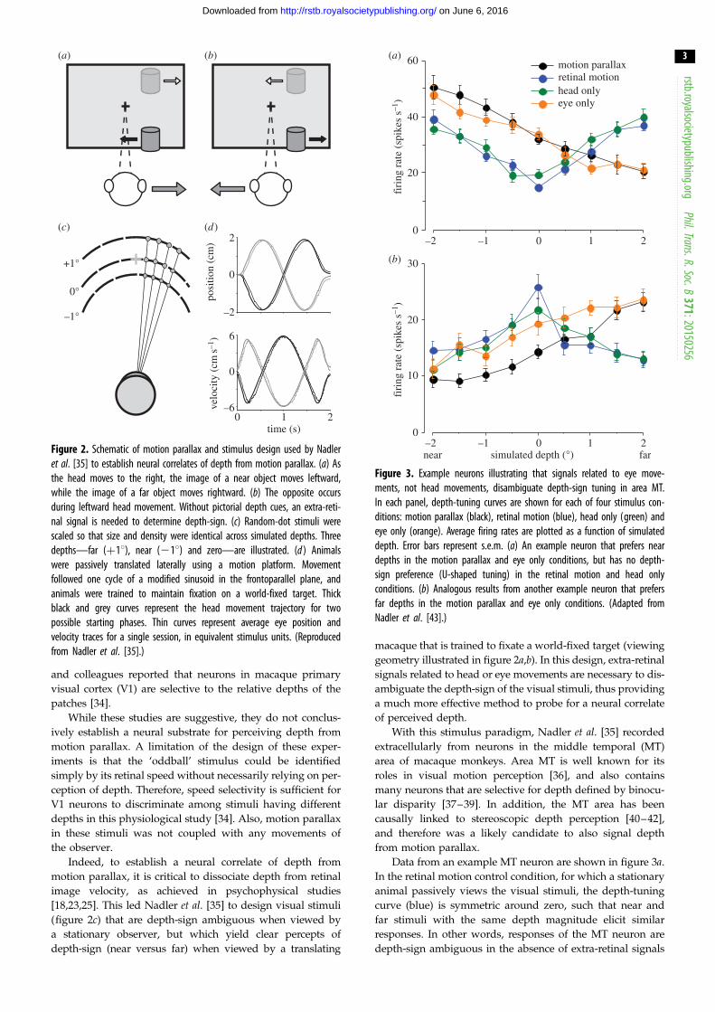
Figure 3. Example neurons illustrating that signals related to eye move-ments, not head movements, disambiguate depth-sign tuning in area MT.In each panel, depth-tuning curves are shown for each of four stimulus con-ditions: motion parallax (black), retinal motion (blue), head only (green) andeye only (orange). Average firing rates are plotted as a function of simulateddepth. Error bars represent s.e.m. (a) An example neuron that prefers neardepths in the motion parallax and eye only conditions, but has no depth-sign preference (U-shaped tuning) in the retinal motion and head onlyconditions. (b) Analogous results from another example neuron that prefersfar depths in the motion parallax and eye only conditions. (Adapted fromNadler et al. [43].)
rstb.royalsocietypublishing.orgPhil.Trans.R.Soc.B
371:20150256
3
on June 6, 2016http://rstb.royalsocietypublishing.org/Downloaded from
and colleagues reported that neurons in macaque primary
visual cortex (V1) are selective to the relative depths of the
patches [34].
While these studies are suggestive, they do not conclus-
ively establish a neural substrate for perceiving depth from
motion parallax. A limitation of the design of these exper-
iments is that the ‘oddball’ stimulus could be identified
simply by its retinal speed without necessarily relying on per-
ception of depth. Therefore, speed selectivity is sufficient for
V1 neurons to discriminate among stimuli having different
depths in this physiological study [34]. Also, motion parallax
in these stimuli was not coupled with any movements of
the observer.
Indeed, to establish a neural correlate of depth from
motion parallax, it is critical to dissociate depth from retinal
image velocity, as achieved in psychophysical studies
[18,23,25]. This led Nadler et al. [35] to design visual stimuli
(figure 2c) that are depth-sign ambiguous when viewed by
a stationary observer, but which yield clear percepts of
depth-sign (near versus far) when viewed by a translating
macaque that is trained to fixate a world-fixed target (viewing
geometry illustrated in figure 2a,b). In this design, extra-retinal
signals related to head or eye movements are necessary to dis-
ambiguate the depth-sign of the visual stimuli, thus providing
a much more effective method to probe for a neural correlate
of perceived depth.
With this stimulus paradigm, Nadler et al. [35] recorded
extracellularly from neurons in the middle temporal (MT)
area of macaque monkeys. Area MT is well known for its
roles in visual motion perception [36], and also contains
many neurons that are selective for depth defined by binocu-
lar disparity [37–39]. In addition, the MT area has been
causally linked to stereoscopic depth perception [40–42],
and therefore was a likely candidate to also signal depth
from motion parallax.
Data from an example MT neuron are shown in figure 3a.
In the retinal motion control condition, for which a stationary
animal passively views the visual stimuli, the depth-tuning
curve (blue) is symmetric around zero, such that near and
far stimuli with the same depth magnitude elicit similar
responses. In other words, responses of the MT neuron are
depth-sign ambiguous in the absence of extra-retinal signals

rstb.royalsocietypublishing.orgPhil.Trans.R.Soc.B
371:20150256
4
on June 6, 2016http://rstb.royalsocietypublishing.org/Downloaded from
related to head or eye movements. Responses increase with
depth magnitude for neurons that prefer moderate to fast
speeds (figure 3a), or decrease with depth magnitude for
neurons that prefer slow speeds (figure 3b). Strikingly, when
the animal was physically translated by a motion platform
and counter-rotated his eyes to maintain fixation (motion
parallax condition), the example neuron of figure 3a (black
curve) becomes selective for depth-sign, with a clear prefer-
ence for near depths. By contrast, other neurons prefer far
depths in the motion parallax condition (e.g. figure 3b,
black). Because the retinal image motion over the MT receptive
field is the same in these two stimulus conditions (assuming
accurate pursuit tracking, [35]), the difference in response
between the retinal motion and motion parallax conditions
must reflect the action of extra-retinal signals related to head
translation or eye rotation. Note that the data shown by the
green and orange curves in figure 3 will be described in a
subsequent section.
Well over one-half of neurons in area MT showed signifi-
cant selectivity for depth-sign in these initial experiments
[35], thus establishing area MT as a potential neural substrate
for computing depth from motion parallax. It is currently
unknown whether similar selectivity appears in earlier
stages of visual processing, such as V1, V2 and V3, that
provide substantial inputs to MT [44,45].
4. Linking middle temporal activity to depthperception based on motion parallax
Although the findings of Nadler et al. [35] established that MT
neurons carry information about depth-sign based on motion
parallax, the experiments were performed in animals that
were simply required to track a world-fixed target. To further
understand the functional links between MT responses and
depth perception from motion parallax, it is necessary to exam-
ine neural activity in animals that are trained to report their
depth percepts. Kim et al. [6] trained animals to judge whether
a random-dot stimulus, in which depth was only defined by
motion parallax, appeared to be nearer or farther than the fix-
ation target. The experimental approach was largely similar to
that used by Nadler et al. [35], except that the proportion of
dots that appeared at a near or far depth was variable and
this ‘depth coherence’ parameter was used to titrate the diffi-
culty of the task. Overall, Kim et al. [6] found that the
sensitivity of individual MT neurons was lower than that of
the animal, although the most sensitive MT neurons rivaled
behavioural performance. This suggests that behavioural per-
formance could be accounted for by pooling the responses of
relatively small populations of MT neurons.
In addition, Kim et al. [6] examined whether the activity
of MT neurons was predictive of perceptual decisions
regarding depth by computing choice probabilities [46,47].
Choice probability quantifies the trial-by-trial correlation
between responses of a single neuron and the animal’s
choices, and generally reflects both correlated noise among
neurons as well as the weights that are applied to neural
responses in decoding [48]. The finding of significant choice
probabilities is consistent with the possibility that neurons
provide evidence used to perform the task, but it could also
reflect top-down signals related to featural attention or
decision-making (see [47,49] for more extensive discussions
of the interpretation of choice probabilities). In the depth-
discrimination task based on motion parallax, Kim et al. [6]
found that responses of many MT neurons were weakly pre-
dictive of the animals’ choices. The average choice probability
for MT neurons was comparable with that found previously
in studies of coarse motion discrimination [46,50], but smaller
than that measured in a coarse depth-discrimination task
based on binocular disparity cues [51]. The latter difference
may stem from MT neurons having lower sensitivity
to depth variations based on motion parallax cues than to
depth variations based on binocular disparity cues [6].
Together, these findings from behaving animals support
the hypothesis that area MT provides important sensory infor-
mation to inform perception of depth based on motion parallax
cues. However, a causal test of area MT’s role in perceiving
depth from motion parallax has not yet been performed, and
such a test (e.g. based on electrical microstimulation or revers-
ible inactivation, [40,42]) will be important to establish that MT
is needed to perceive depth from motion parallax.
5. Sources of extra-retinal signals for depth-signcoding in area middle temporal
As described above, human psychophysical studies have
demonstrated that eye movement signals, not head trans-
lation signals, disambiguate the sign of depth cued by
motion parallax [24,25]. Thus, it was of considerable interest
to establish whether the selectivity of MT neurons for
depth-sign would show the same dependencies.
This issue was addressed by Nadler et al. [43], who
extended the previous experimental approach [35] in two
important ways. First, they introduced a head only condition
in which the fixation target moved with the animal, such that
no eye movements were necessary to maintain visual fixation
(unlike in the scheme of figure 2a,b). In this head only con-
dition, vestibular and other signals related to head/body
translation were identical, but command signals to move
the eyes were no longer needed. Second, they introduced
an eye only condition in which the animal remained station-
ary but the virtual environment (including the fixation target)
translated in front of the animal. This eliminated vestibular
and other signals related to head/body translation, but
required tracking eye movements. Findings from these con-
ditions were compared with those obtained using the
original design [35], in which both head and eye movements
were involved (motion parallax condition). Importantly,
visual stimuli over the receptive fields of MT neurons were
the same across all these conditions.
The results reported by Nadler et al. [43] were remarkably
clear. In the head only condition, MT neurons showed little or
no depth-sign selectivity, and there was no correlation with
results from the motion parallax condition (figure 4, open
symbols). In striking contrast, results from the eye only con-
dition were nearly identical to those from the motion
parallax condition (figure 4, filled symbols). This indicates
that the depth-sign selectivity of MT neurons depends on
extra-retinal signals related to eye movements but not those
related to head/body translation.
Whereas Nawrot [24] found reversed perception of
depth-sign in humans tested with stimuli similar to our
head only condition, we did not find reversed depth-sign
tuning in MT responses during the head only condition.
However, we were not able to maintain neural recordings

+1n = 79
0
–1–1 0 +1
–1
0
+1
motion parallax DSDI
head
onl
y D
SDI
eye
only
DSD
I
Figure 4. Eye movement signals, not vestibular translation signals, producedepth-sign tuning in MT neurons. This scatter plot compares depth-sign dis-crimination index (DSDI) values for the eye only condition (filled symbols)and the head only condition (open symbols) to DSDI values obtained inthe motion parallax condition (79 neurons from two monkeys; M1: circles,M2: triangles). DSDI is a measure of depth-sign preference, such that negativevalues indicate neurons that prefer near depths and positive values indicateneurons that prefer far depths. Only DSDIs obtained in the eye only conditionare correlated with those obtained in the motion parallax condition. (Adaptedfrom Nadler et al. [43].)
D
d
F
f
translation
q(t)
a(t)
Figure 5. Schematic of variables involved in the motion-pursuit law. Theobserver’s eye translates rightward while maintaining fixation on the pointF, which is located at a viewing distance given by f. A point of interest,D, has a depth, d, relative to the plane of fixation. u(t) denotes thetime-varying angle subtended by the point D relative to the fovea. a(t) rep-resents the time-varying orientation of the eye relative to the scene.(Reproduced from Nawrot & Stroyan [27].)
rstb.royalsocietypublishing.orgPhil.Trans.R.Soc.B
371:20150256
5
on June 6, 2016http://rstb.royalsocietypublishing.org/Downloaded from
during head movements large enough to be likely to elicit a
robust tVOR (see [43] for additional discussion). Thus, our
findings are not in conflict with those of Nawrot [24].
The mechanisms by which eye movement command sig-
nals modulate responses of MT neurons to generate depth-
sign selectivity require further study. However, preliminary
findings suggest that eye movement signals powerfully
modulate the response gain of MT neurons during specific
directions of eye movements [52]. By analogy to the role of
eye movement gain fields in performing coordinate trans-
formations [53,54], a population of such gain-modulated
MT neurons may be able to represent depth from motion
parallax [52]. It should be noted that gain modulations of
MT responses by pursuit eye movements have been reported
previously [55], and it is possible that these effects could be
related to the same mechanisms that generate depth-sign
selectivity from motion parallax.
6. Role of visual cues in disambiguating depth-sign from motion parallax
Why should computation of depth from motion parallax,
both behaviourally and in MT neurons, depend on eye
movement signals and not signals (e.g. vestibular inputs)
related to head translation? Important insights were provided
by the theoretical work of Nawrot & Stroyan [27]. They
demonstrated that depth from motion parallax can be com-
puted from the following simple relationship, called the
motion-pursuit law:
df¼ du
da
1
1� du=da
� �� du
da,
where f is the fixation distance, d is the depth of an object rela-
tive to the fixation plane, da (which is shorthand for da/dt)
denotes the change of eye orientation relative to the scene and
du (short for du/dt) represents the retinal image motion of
the point of interest (figure 5). Note that the approximation
on the right holds when eye rotation is large relative to retinal
motion. Critically, the motion-pursuit law requires knowl-
edge of eye rotation relative to the scene, but does not
involve head translation. Thus, this theoretical relationship
helps explain why both perceptual and neural responses to
motion parallax are disambiguated by eye movements but
not head movements. Moreover, Nawrot et al. [8] have
shown that psychophysical depth magnitude estimates are
well described by a modified form of the motion-pursuit law.
The critical variable in the motion-pursuit law, da, rep-
resents eye rotation relative to the scene. Thus, it is sensible
that pursuit eye movement command signals, which control
eye rotation relative to the head, would contribute to a
neural representation of da. However, any information
related to da could be used, in principle, to compute depth
from motion parallax. Could visual motion signals also be
used to infer da? Optic flow is determined by translation
and rotation of the eye relative to the scene, and theoretical
work has shown that it is possible to decompose optic flow
into rotation and translation components of observer
motion [56,57]. Of specific interest here, whenever the eye
rotates relative to the scene, the resulting images (under

motionparallax
dynamicperspective
retinalmotion
retinal motiondynamic perspectivemotion parallax
–2 –1 0simulated depth (°)
1 2
(a)
resp
onse
(sp
ikes
s–1
)
150
100
50
0
(b)
RF
RF
+
+
+
Figure 6. Stimulus conditions used in the dynamic perspective experiment of Kim et al. [58] and results from an example neuron. (a) Frontal views for eachexperimental condition. In the motion parallax condition, animals experience full-body translation and make counteractive eye movements to maintain fixationon a world-fixed target (yellow cross). In the retinal motion condition, the animal’s head and eyes are stationary, but visual stimuli replicate the imagemotion experienced in the motion parallax condition. The dynamic perspective condition is the same as the retinal motion condition except that a three-dimensionalcloud of background dots was added to the display. Background dots near the receptive field (RF) were masked. (b) For each stimulus condition, depth-tuning curvesare shown for an example MT neuron. Tuning in the retinal motion condition is symmetrical around zero depth (blue, DSDI ¼ – 0.09), whereas tuning curves showa clear preference for near depths in the motion parallax (black, DSDI ¼ – 0.80) and dynamic perspective (magenta, DSDI ¼ – 0.67) conditions. Error bars rep-resent s.e.m. (Adapted from Kim et al. [58].)
rstb.royalsocietypublishing.orgPhil.Trans.R.Soc.B
371:20150256
6
on June 6, 2016http://rstb.royalsocietypublishing.org/Downloaded from
planar projection) contain systematic perspective distortions
that appear as a component of ‘rocking’ motion in the
image ([58], see supplementary movies 1–3). Therefore,
under viewing conditions similar to those depicted in
figure 2a,b, these ‘dynamic perspective’ cues could potentially
be used to estimate the da term in the motion-pursuit law.
Early evidence that the visual system might process
dynamic perspective cues to compute depth from motion
parallax came from the work of Rogers & Rogers [59]. Obser-
vers viewed ambiguous depth corrugation stimuli while the
visual display was rotated about a vertical axis through the
centre of the display. This rotation of the display generates
dynamic perspective cues similar to those resulting from
eye rotations. Interestingly, Rogers & Rogers [59] found that
this rotation of the entire display could disambiguate
depth-sign in motion parallax displays. Eye movements
were not monitored or enforced in these experiments, so it
is difficult to be sure that the disambiguation resulted from
dynamic perspective cues instead of systematic eye move-
ments related to rotation of the display, but these results
suggested a role for visual perspective cues in specifying da.
Recently, Kim et al. [58] have provided a clear visual dem-
onstration that dynamic perspective cues can disambiguate
depth-sign (see their supplemental videos). Moreover, they
have shown that MT neurons can unambiguously signal
depth-sign based on motion parallax when dynamic perspec-
tive cues are present. Building upon the retinal motion
stimulus condition used by Nadler et al. [35], they introduced
a large stationary random-dot field around the small stimulus
patch that was presented over the receptive field of an MT
neuron. There was no head/body translation or eye rotation
in this dynamic perspective condition, but rocking motion
of the background dots simulated an eye rotation
(figure 6a). Remarkably, many MT neurons showed robust
depth-sign tuning in the dynamic perspective condition
(e.g. figure 6b, magenta), even though the visual stimulus
within the receptive field was identical to that of the retinal
motion control condition for which MT neurons show no
depth-sign selectivity (figure 6b, blue). Moreover, for many
neurons, the depth-sign selectivity induced by dynamic per-
spective cues was similar to that induced by eye movements
in the standard motion parallax condition (figure 6b, black).
It is worth noting that the visual stimuli for the retinal
motion control condition also contain dynamic perspective
cues [58]. However, when the stimulus is spatially restricted,
these dynamic perspective cues are not sufficient to disambig-
uate depth-sign. This is consistent with recent psychophysical
experiments showing that dynamic perspective cues in a
small stimulus are not sufficient to disambiguate depth-sign
[60]. Thus, dynamic perspective cues are mainly useful in
large rich scenes, and thus it makes sense for the brain to
make use of both dynamic perspective cues and eye move-
ment signals to compute the critical variable (da) needed in
the motion-pursuit law.
These findings show that large-field patterns of image
motion can be used to infer eye rotations relative to the
scene, and that these signals can be used in useful neural
computations instead of efference copies of eye movement
command signals. In this light, it is worth noting that there
is also evidence that dynamic perspective cues are used to

40
30
20
10
0
45
30
15
0–2 –1 0 1 2
–2 –1 0 1 2
simulated depth (°)
motion parallaxbinocular disparityretinal motioncombined
both sigMP only sig
BD only signeither sig
1.0
0.5
0
–0.5
–1.0–1.0 –0.5 0 0.5 1.0
DSDI, motion parallax
DSD
I, b
inoc
ular
dis
pari
tyfi
ring
rat
e (s
pike
s s–1
)fi
ring
rat
e (s
pike
s s–1
)(a)
(b)
(c)
Figure 7. (Caption opposite.)
Figure 7. (Opposite.) Comparison of depth-tuning from motion parallax anddisparity. (a) Depth-tuning curves are shown, for an example MT neuron, forthe retinal motion (blue), motion parallax (black), binocular disparity (green)and combined (orange) conditions. In the binocular disparity condition, depthwas cued solely by disparity, without motion parallax. The combined conditionwas similar to the motion parallax condition except that dots were presenteddichoptically. Each tuning curve plots mean firing rate (averaged across the dur-ation of the trial as well as the two starting phases of motion) against simulateddepth of the stimulus, and error bars denote s.e.m. This neuron has consistentdepth-sign preferences for disparity and motion parallax. (b) Another exampleneuron, with opposite depth-sign preferences for disparity and motion parallax.Format as in a. (c) The depth-sign discrimination index (DSDI) for the binoculardisparity (BD) condition (ordinate) is plotted against the DSDI value for themotion parallax (MP) condition (abscissa) (N ¼ 134). Filled black symbolsindicate DSDI values that are significantly different from zero ( p , 0.05, per-mutation test) in both the motion parallax and binocular disparity conditions.Filled red symbols indicate DSDI values that are significantly different fromzero in the motion parallax condition, but not in the binocular disparity con-dition. Open black circles indicate DSDI values that are significantly differentfrom zero in the binocular disparity condition, but not in the motion parallaxcondition. Finally, open red circles indicate DSDI values that are not significantlydifferent from zero in either the binocular disparity or motion parallaxconditions. (Adapted from Nadler et al. [70].)
rstb.royalsocietypublishing.orgPhil.Trans.R.Soc.B
371:20150256
7
on June 6, 2016http://rstb.royalsocietypublishing.org/Downloaded from
compensate for eye rotations in perception of heading [61]
and neural coding of heading [62].
7. Integration of binocular disparity and motionparallax cues to depth
Under natural conditions, a variety of depth cues are typi-
cally available to an observer; thus, it is of interest to ask
how motion parallax and binocular disparity cues to depth
are integrated by the brain. A number of psychophysical
studies in humans [63–65] and monkeys [32] have provided
evidence that depth perception involves integration of dis-
parity and motion parallax cues, but until recently very
little was known about neural integration of these cues. In
humans, Ban et al. [66] used fMRI pattern analysis techniques
to identify brain regions that appear to integrate disparity
and motion parallax cues to depth. In animals, a few studies
have examined how neurons signal three-dimensional sur-
face orientation based on combinations of motion and
disparity gradients [67] or perspective gradients and
disparity gradients [68,69].
Recently, Nadler et al. [70] measured the depth-sign
selectivity of macaque MT neurons based on both binocular
disparity and motion parallax cues. One might expect
neurons to prefer the same depth-sign (near or far) for each
cue. Indeed, this was the case for many MT neurons, such
as the example neuron illustrated in figure 7a. We refer to
such neurons as ‘congruent’ cells. Surprisingly, however,
other MT neurons prefer opposite depth-signs defined by
disparity and motion parallax cues, and we refer to these as
‘opposite’ cells. Figure 7b shows an opposite cell that prefers
near depths from motion parallax and far depths from
binocular disparity. Overall, approximately 40% of MT
neurons were found to prefer opposite depth-signs based
on binocular disparity and motion parallax cues (data
points in the top-left and bottom-right quandrants of
figure 7c), whereas the remaining 60% have matched depth-
sign preferences (top-right and bottom-left quadrants of
figure 7c). We shall return to a potential functional role of
opposite cells below.
Nadler et al. [70] also measured responses of MT neurons
to stimuli involving congruent combinations of binocular dis-
parity and motion parallax cues (combined condition). In
general, responses in the combined condition reflected a mix-
ture of selectivity for both depth cues. For the example
congruent cell of figure 7a, depth-tuning in the combined
condition (orange) appears to be dominated by binocular
disparity tuning (green), but tuning strength is slightly
enhanced in the combined condition, reflecting the influence

far
near
STOP
Figure 8. Schematic of the local discrepancy between disparity and motioncues that can arise for a moving object in the world. In this illustration, anobserver maintains visual fixation on the traffic light while moving their headto the right. In this case, all stationary objects in the scene have an imagevelocity that is determined by their three-dimensional location in the scene(as specified by binocular disparity, for example) and the movement of theobserver’s head. Stationary near objects move leftward in the image, whereasstationary far objects move rightward in the image. However, a moving object(the car) creates local retinal image motion that is not consistent with thatexpected from the binocular disparity of the object and the movement of theobserver. This local discrepancy between disparity and retinal image motionmight be sensed by the relative activity of congruent and opposite cells inarea MT. (Adapted from Nadler et al. [70].)
rstb.royalsocietypublishing.orgPhil.Trans.R.Soc.B
371:20150256
8
on June 6, 2016http://rstb.royalsocietypublishing.org/Downloaded from
of motion parallax inputs. For other neurons, such as the
opposite cell illustrated in figure 7b, depth-tuning in the com-
bined condition (orange) appears to be dominated by motion
parallax tuning (black), and selectivity in the combined con-
dition is slightly reduced by the incongruent disparity
selectivity. These findings suggest that congruent cells
might contribute to perceptual cue integration of depth
cues, whereas opposite cells would not, but the study of
Nadler et al. [70] was not designed to test these issues
directly. Additional studies, in which both psychophysical
and neuronal performance is tested with both congruent
and conflicting combinations of disparity and motion paral-
lax cues, will be needed to evaluate whether activity of MT
neurons can account for perceptual cue integration and cue
weighting (as explored previously for multisensory neurons
involved in heading perception, [71–73]).
Thus far, we have focused on perception of depth of objects
that are stationary in the world. However, it is often important
to detect objects that move in the world. When an observer
translates through the environment, all objects in a scene
(except those on which the eye fixates) will move in the retinal
image, which greatly complicates the task of detecting objects
that move in the world. Conflicts between binocular disparity
and motion parallax cues may play important roles in solving
this problem, as illustrated in figure 8. As the observer trans-
lates to the right, objects that are nearer than the plane of
fixation move to the left, whereas farther objects move to the
right. The speed of image motion depends on the distance of
an object from the fixation target. Thus, if the depth of an
object is known (e.g. from binocular disparity cues), then the
expected image velocity could be computed under the assump-
tion that the object is stationary in the scene. For an object that
moves in the world (such as the car in figure 8), the image
velocity will deviate from this prediction. Thus, for an indepen-
dently moving object, binocular disparity and motion parallax
cues to depth would be in conflict, and it is possible that the
brain could detect a moving object by identifying this cue con-
flict (without having to perform calculations that depend on
self-motion velocity, viewing distance, etc.). Does the brain
have neural mechanisms that can detect this cue conflict to
identify objects that are moving in the world?
Human psychophysical studies suggest that the visual
system is sensitive to conflict between disparity and motion
parallax cues. Early evidence came from Rogers & Collett
[74], who examined how humans perceive conflict between
disparity and motion parallax cues in random-dot stimuli.
Interestingly, such conflicts led subjects to perceive rotation
of the stimulus relative to the scene. More recently, Rushton
et al. [75] showed that human observers can efficiently
detect an independently moving object among stationary dis-
tractors when there is an appropriate conflict between motion
parallax and binocular disparity cues. This suggested that the
brain may contain specialized mechanisms for detecting
objects that move in the world during observer motion.
Nadler et al. [70] have proposed that opposite cells in area
MT, which have opposite depth-sign preferences for motion par-
allax and binocular disparity cues, may play an important role in
detecting moving objects during self-motion. Specifically, oppo-
site cells may have greater activity than congruent cells when
stimulated by an object that moves in the world, because the reti-
nal image motion for such an object is not consistent with its
binocular disparity. A preliminary report by Kim et al. [76] pro-
vides further support for this idea. Animals were trained to
perform a task similar to that used by Rushton et al. [75], in
which they detect an object that moves in the world relative to
other objects that are stationary in the scene. Interestingly, in a
subset of conditions in which all objects had identical retinal
motion, MT neurons that responded more strongly to objects
that move in the world were found to predict trial-by-trial vari-
ations in the animals’ choices [76]. This supports the notion that
the brain may use a mixture of congruent and opposite cells to
detect moving objects during observer translation.
8. Summary and future directionsOur understanding of how the brain computes depth from
motion parallax has evolved substantially over the past
decade, and this review has summarized some of the major
advances. These advances provide an excellent example
of how psychophysical, theoretical and neurophysiological
studies can inspire each other to achieve a deeper understanding
of an advanced perceptual function. At the same time, our
understanding of this problem is still quite limited and there
are a number of fruitful directions for additional research.
Thus far, depth-sign selectivity has only been observed in
area MT, and we do not know whether the computations
required to generate depth-sign selectivity are initiated in
MT or whether MT inherits this selectivity from its inputs.
Additional experiments in areas that provide direct input to
MT, including areas V1, V2 and V3, will be valuable for estab-
lishing the circuitry underlying computations of depth from

rstb.royalsocietypublishing.orgPhil.Trans.R.Soc.B
371:20150256
9
on June 6, 2016http://rstb.royalsocietypublishing.org/Downloaded from
motion parallax. Along these same lines, the neural origins of
the pursuit command signals and dynamic perspective signals
that disambiguate depth-sign in MT neurons, as well as the
detailed mechanisms of their operation, remain unclear.
The critical variable (da) in the motion-pursuit law [27] is
a change of eye orientation relative to the scene. In the exper-
iments conducted so far, eye orientation relative to the head
has been varied. More generally, as we move and fixate on
a target in the world, eye rotation relative to the scene may
be determined by some combination of eye rotation relative
to the head, head rotation relative to the body and body
rotation relative to the scene. Thus, it is possible that multiple
extra-retinal signals are used to disambiguate depth. Future
experiments should examine whether perception of depth
from motion parallax depends on a combination of these
rotation variables or just eye rotations relative to the head.
Corresponding neural studies could examine whether neur-
ons in area MT (or elsewhere) also receive additional
signals (e.g. vestibular rotation inputs) that can disambiguate
depth-sign. In addition, it will be important to examine
whether and how the brain combines different sources of sig-
nals related to da. For example, are extra-retinal inputs
related to eye, head and body rotations combined to provide
a more general representation of da that is used to perceive
depth? In this regard, it is worth noting that dynamic perspec-
tive cues provide direct information about eye rotation relative
to the scene. Thus, a representation of da based on multiple
extra-retinal signals might be integrated with dynamic per-
spective cues to provide a unified, multimodal da signal
that will be robust across many environmental conditions.
Finally, depth from motion parallax has generally been
studied during lateral translation of observers. Although this
simplifies the geometry and has a number of advantages for
experimental design, our movements through the environ-
ment often have substantial (if not predominant) fore–aft
components. Fore–aft translations are studied extensively in
the domain of heading perception [77–79]. Psychophysical
studies have shown that the human visual system can ‘parse
out’ objects from optic flow patterns [80,81], but we know
little about how humans or animals estimate depth of objects
under these conditions. Thus, there may be an opportunity
to integrate domains of research related to motion parallax
and heading perception.
Together, these observations suggest that studying the
neural computations that mediate perception of depth from
motion parallax will continue to provide exciting opportu-
nities to understand how visual and extra-retinal signals are
combined in specific ways to generate specialized neural
representations that underlie behaviour.
Authors’ contributions. H.R.K. conceptualized the review, wrote firstdraft, edited drafts and approved final version; D.E.A. edited draftsand approved final version; G.C.D. conceptualized the review,edited drafts and approved final version.
Competing interests. We have no competing interests
Funding. H.R.K. and G.C.D. were supported by a grant from theNational Eye Institute (EY013644). This work was supported byNIH grant no. EY013644 (to G.C.D.) and by an NEI CORE grantno. (EY001319).
References
1. Howard IP, Rogers BJ. 1995 Binocular vision andstereopsis. New York, NY: Oxford University Press.
2. Howard IP, Rogers BJ. 2002 Seeing in depth. Volume2: depth perception. Toronto, Canada: I. Porteus.
3. Julesz B. 1964 Binocular depth perception withoutfamiliarity cues. Science 145, 356 – 362. (doi:10.1126/science.145.3630.356)
4. Julesz B. 1971 Foundations of cylclopean perception.Chicago, IL: University of Chicago Press.
5. Cao A, Schiller PH. 2002 Behavioral assessment ofmotion parallax and stereopsis as depth cues inrhesus monkeys. Vision Res. 42, 1953 – 1961.(doi:10.1016/S0042-6989(02)00117-7)
6. Kim HR, Angelaki DE, DeAngelis GC. 2015 Afunctional link between MT neurons and depthperception based on motion parallax. J. Neurosci.35, 2766 – 2777. (doi:10.1523/JNEUROSCI.3134-14.2015)
7. McKee SP, Taylor DG. 2010 The precision ofbinocular and monocular depth judgmentsin natural settings. J. Vis. 10, 5. (doi:10.1167/10.10.5)
8. Nawrot M, Ratzlaff M, Leonard Z, Stroyan K. 2014Modeling depth from motion parallax with themotion/pursuit ratio. Front. Psychol. 5, 1103.(doi:10.3389/fpsyg.2014.01103)
9. Ono ME, Rivest J, Ono H. 1986 Depth perception asa function of motion parallax and absolute-distance
information. J. Exp. Psychol. Hum. Percept. Perform.12, 331 – 337. (doi:10.1037/0096-1523.12.3.331)
10. Ellard CG, Goodale MA, Timney B. 1984 Distanceestimation in the Mongolian gerbil: the role ofdynamic depth cues. Behav. Brain Res. 14, 29 – 39.(doi:10.1016/0166-4328(84)90017-2)
11. Goulet M, Campan R, Lambin M. 1981 Thevisual perception of relative distances in thewood-cricket, Nemobius sylvestris. Physiol. Entomol.6, 357 – 367. (doi:10.1111/j.1365-3032.1981.tb00651.x)
12. Sobel EC. 1990 Depth perception by motion parallaxand paradoxical parallax in the locust.Naturwissenschaften 77, 241 – 243. (doi:10.1007/BF01138494)
13. Cumming BG, DeAngelis GC. 2001 The physiology ofstereopsis. Annu. Rev. Neurosci. 24, 203 – 238.(doi:10.1146/annurev.neuro.24.1.203).
14. Parker AJ. 2007 Binocular depth perception and thecerebral cortex. Nat. Rev. Neurosci. 8, 379 – 391.(doi:10.1038/nrn2131)
15. Roe AW, Parker AJ, Born RT, DeAngelis GC. 2007Disparity channels in early vision. J. Neurosci. 27,11 820 – 11 831. (doi:10.1523/JNEUROSCI.4164-07.2007)
16. Helmholtz H. 1925 Helmholtz’s treatise onphysiological optics. New York, NY: Optical Society ofAmerica.
17. Gibson EJ, Gibson JJ, Smith OW, Flock H. 1959 Motionparallax as a determinant of perceived depth. J. Exp.Psychol. 58, 40 – 51. (doi:10.1037/h0043883)
18. Rogers B, Graham M. 1979 Motion parallax as anindependent cue for depth perception. Perception 8,125 – 134. (doi:10.1068/p080125)
19. Ono H, Wade NJ. 2005 Depth and motion inhistorical descriptions of motion parallax. Perception34, 1263 – 1273. (doi:10.1068/p5232)
20. Rogers B. 2009 Motion parallax as an independentcue for depth perception: a retrospective. Perception38, 907 – 911. (doi:10.1068/pmkrog)
21. Hayashibe K. 1991 Reversals of visual depth causedby motion parallax. Perception 20, 17 – 28. (doi:10.1068/p200017)
22. Farber JM, McConkie AB. 1979 Optical motions asinformation for unsigned depth. J. Exp. Psychol.Hum. Percept. Perform. 5, 494 – 500. (doi:10.1037/0096-1523.5.3.494)
23. Nawrot M. 2003 Depth from motion parallax scaleswith eye movement gain. J. Vis. 3, 841 – 851.(doi:10:1167/3.11.17)
24. Nawrot M. 2003 Eye movements provide the extra-retinal signal required for the perception of depthfrom motion parallax. Vision Res. 43, 1553 – 1562.(doi:10.1016/S0042-6989(03)00144-5)
25. Naji JJ, Freeman TC. 2004 Perceiving depthorder during pursuit eye movement. Vision

rstb.royalsocietypublishing.orgPhil.Trans.R.Soc.B
371:20150256
10
on June 6, 2016http://rstb.royalsocietypublishing.org/Downloaded from
Res. 44, 3025 – 3034. (doi:10.1016/j.visres.2004.07.007)
26. Nawrot M, Joyce L. 2006 The pursuit theory ofmotion parallax. Vision Res. 46, 4709 – 4725.(doi:10.1016/j.visres.2006.07.006).
27. Nawrot M, Stroyan K. 2009 The motion/pursuit lawfor visual depth perception from motion parallax.Vision Res. 49, 1969 – 1978. (doi:10.1016/j.visres.2009.05.008)
28. Ono H, Ujike H. 1994 Apparent depth with motionaftereffect and head movement. Perception 23,1241 – 1248. (doi:10.1068/p231241)
29. Ono H, Ujike H. 2005 Motion parallax driven by headmovements: conditions for visual stability, perceiveddepth, and perceived concomitant motion.Perception 34, 477 – 490. (doi:10.1068/p5221)
30. Ujike H, Ono H. 2001 Depth thresholds of motionparallax as a function of head movement velocity.Vision Res. 41, 2835 – 2843. (doi:10.1016/S0042-6989(01)00164-X)
31. Yoonessi A, Baker Jr CL. 2011 Contribution ofmotion parallax to segmentation and depthperception. J. Vis. 11, 13. (doi:10.1167/11.9.13)
32. Schiller PH, Slocum WM, Jao B, Weiner VS. 2011The integration of disparity, shading and motionparallax cues for depth perception in humans andmonkeys. Brain Res. 1377, 67 – 77. (doi:10.1016/j.brainres.2011.01.003)
33. Zhang Y, Schiller PH. 2008 The effect of overallstimulus velocity on motion parallax. Vis. Neurosci.25, 3 – 15. (doi:10.1017/S0952523808080012)
34. Cao A, Schiller PH. 2003 Neural responses to relativespeed in the primary visual cortex of rhesusmonkey. Vis. Neurosci. 20, 77 – 84. (doi:10.1017/S0952523803201085)
35. Nadler JW, Angelaki DE, DeAngelis GC. 2008 Aneural representation of depth from motion parallaxin macaque visual cortex. Nature 452, 642 – 645.(doi:10.1038/nature06814)
36. Born RT, Bradley DC. 2005 Structure and function ofvisual area MT. Annu. Rev. Neurosci. 28, 157 – 189.(doi:10.1146/annurev.neuro.26.041002.131052)
37. DeAngelis GC, Newsome WT. 1999 Organization ofdisparity-selective neurons in macaque area MT.J. Neurosci. 19, 1398 – 1415.
38. DeAngelis GC, Uka T. 2003 Coding of horizontaldisparity and velocity by MT neurons in the alertmacaque. J. Neurophysiol. 89, 1094 – 1111. (doi:10.1152/jn.00717.2002)
39. Maunsell JH, Van Essen DC. 1983 Functionalproperties of neurons in middle temporal visualarea of the macaque monkey. II. Binocularinteractions and sensitivity to binocular disparity.J. Neurophysiol. 49, 1148 – 1167.
40. Chowdhury SA, DeAngelis GC. 2008 Finediscrimination training alters the causal contributionof macaque area MT to depth perception. Neuron 60,367– 377. (doi:10.1016/j.neuron.2008.08.023)
41. DeAngelis GC, Cumming BG, Newsome WT. 1998Cortical area MT and the perception of stereoscopicdepth. Nature 394, 677 – 680. (doi:10.1038/29299)
42. Uka T, DeAngelis GC. 2006 Linking neuralrepresentation to function in stereoscopic depth
perception: roles of the middle temporal area incoarse versus fine disparity discrimination.J. Neurosci. 26, 6791 – 6802. (doi:10.1523/JNEUROSCI.5435-05.2006)
43. Nadler JW, Nawrot M, Angelaki DE, DeAngelis GC.2009 MT neurons combine visual motion with asmooth eye movement signal to code depth-signfrom motion parallax. Neuron 63, 523 – 532.(doi:10.1016/j.neuron.2009.07.029)
44. Ponce CR, Hunter JN, Pack CC, Lomber SG, Born RT.2011 Contributions of indirect pathways to visualresponse properties in macaque middle temporalarea MT. J. Neurosci. 31, 3894 – 3903. (doi:10.1523/JNEUROSCI.5362-10.2011)
45. Ponce CR, Lomber SG, Born RT. 2008 Integratingmotion and depth via parallel pathways. Nat.Neurosci. 11, 216 – 223. (doi:10.1038/nn2039)
46. Britten KH, Newsome WT, Shadlen MN, Celebrini S,Movshon JA. 1996 A relationship betweenbehavioral choice and the visual responses ofneurons in macaque MT. Vis. Neurosci. 13, 87 – 100.(doi:10.1017/S095252380000715X)
47. Nienborg H, Cohen MR, Cumming BG. 2012Decision-related activity in sensory neurons:correlations among neurons and with behavior.Annu. Rev. Neurosci. 35, 463 – 483. (doi:10.1146/annurev-neuro-062111-150403)
48. Haefner RM, Gerwinn S, Macke JH, Bethge M. 2013Inferring decoding strategies from choiceprobabilities in the presence of correlated variability.Nat. Neurosci. 16, 235 – 242. (doi:10.1038/nn.3309)
49. Pitkow X, Liu S, Angelaki DE, DeAngelis GC, PougetA. 2015 How can single sensory neurons predictbehavior? Neuron 87, 411 – 423. (doi:10.1016/j.neuron.2015.06.033)
50. Cohen MR, Newsome WT. 2009 Estimates of thecontribution of single neurons to perception dependon timescale and noise correlation. J. Neurosci. 29,6635 – 6648. (doi:10.1523/JNEUROSCI.5179-08.2009)
51. Uka T, DeAngelis GC. 2004 Contribution of area MT tostereoscopic depth perception: choice-relatedresponse modulations reflect task strategy. Neuron42, 297 – 310. (doi:10.1016/S0896-6273(04)00186-2)
52. Kim HR, Angelaki DE, DeAngelis GC. 2013 Gainmodulation by eye movements as a mechanism forrepresenting depth from motion parallax in areaMT. Society for Neuroscience Abstract 360.05.
53. Pouget A, Snyder LH. 2000 Computational approachesto sensorimotor transformations. Nat. Neurosci.3(Suppl.), 1192– 1198. (doi:10.1038/81469)
54. Zipser D, Andersen RA. 1988 A back-propagationprogrammed network that simulates responseproperties of a subset of posterior parietal neurons.Nature 331, 679 – 684. (doi:10.1038/331679a0)
55. Chukoskie L, Movshon JA. 2009 Modulation of visualsignals in macaque MT and MST neurons duringpursuit eye movement. J. Neurophysiol. 102,3225 – 3233. (doi:10.1152/jn.90692.2008)
56. Rieger JH, Lawton DT. 1985 Processing differentialimage motion. J. Opt. Soc. Am. A 2, 354 – 360.(doi:10.1364/JOSAA.2.000354)
57. Longuet-Higgins HC, Prazdny K. 1980 Theinterpretation of a moving retinal image.
Proc. R. Soc. Lond. B 208, 385 – 397. (doi:10.1098/rspb.1980.0057)
58. Kim HR, Angelaki DE, DeAngelis GC. 2015 A novelrole for visual perspective cues in the neuralcomputation of depth. Nat. Neurosci. 18, 129 – 137.(doi:10.1038/nn.3889)
59. Rogers S, Rogers BJ. 1992 Visual and nonvisualinformation disambiguate surfaces specified bymotion parallax. Percept. Psychophys. 52, 446 – 452.(doi:10.3758/BF03206704)
60. George JM, Johnson JI, Nawrot M. 2013 In pursuitof perspective: does vertical perspectivedisambiguate depth from motion parallax?Perception 42, 631 – 641. (doi:10.1068/p7250)
61. Grigo A, Lappe M. 1999 Dynamical use of differentsources of information in heading judgments fromretinal flow. J. Opt. Soc. Am. A 16, 2079 – 2091.(doi:10.1364/JOSAA.16.002079)
62. Sunkara A, DeAngelis GC, Angelaki DE. 2015 Role ofvisual and non-visual cues in constructing arotation-invariant representation of heading inparietal cortex. Elife 4, 865. (doi:10.7554/eLife.04693)
63. Bradshaw MF, Rogers BJ. 1996 The interaction ofbinocular disparity and motion parallax in thecomputation of depth. Vision Res. 36, 3457 – 3468.(doi:10.1016/0042-6989(96)00072-7)
64. Domini F, Caudek C, Tassinari H. 2006 Stereo andmotion information are not independentlyprocessed by the visual system. Vision Res. 46,1707 – 1723. (doi:10.1016/j.visres.2005.11.018).
65. Nawrot M, Blake R. 1989 Neural integration ofinformation specifying structure from stereopsis andmotion. Science 244, 716 – 718. (doi:10.1126/science.2717948)
66. Ban H, Preston TJ, Meeson A, Welchman AE. 2012The integration of motion and disparity cues todepth in dorsal visual cortex. Nat. Neurosci. 15,636 – 643. (doi:10.1038/nn.3046)
67. Sanada TM, Nguyenkim JD, DeAngelis GC. 2012Representation of 3-D surface orientation by velocityand disparity gradient cues in area MT.J. Neurophysiol. 107, 2109 – 2122. (doi:10.1152/jn.00578.2011)
68. Liu Y, Vogels R, Orban GA. 2004 Convergence ofdepth from texture and depth from disparity inmacaque inferior temporal cortex. J. Neurosci. 24,3795 – 3800. (doi:10.1523/JNEUROSCI.0150-04.2004)
69. Tsutsui K, Jiang M, Yara K, Sakata H, Taira M. 2001Integration of perspective and disparity cues insurface-orientation-selective neurons of area CIP. J.Neurosci. 86, 2856 – 2867.
70. Nadler JW, Barbash D, Kim HR, Shimpi S, AngelakiDE, DeAngelis GC. 2013 Joint representation ofdepth from motion parallax and binocular disparitycues in macaque area MT. J. Neurosci. 33, 14 061 –14 074. (doi:10.1523/JNEUROSCI.0251-13.2013)
71. Chen A, DeAngelis GC, Angelaki DE. 2013 Functionalspecializations of the ventral intraparietal area formultisensory heading discrimination. J. Neurosci. 33,3567 – 3581. (doi:10.1523/JNEUROSCI.4522-12.2013)
72. Fetsch CR, Pouget A, DeAngelis GC, Angelaki DE.2012 Neural correlates of reliability-based cue

rstb.royalsocietypublishing.orgPhil.T
11
on June 6, 2016http://rstb.royalsocietypublishing.org/Downloaded from
weighting during multisensory integration. Nat.Neurosci. 15, 146 – 154. (doi:10.1038/nn.2983)
73. Gu Y, Angelaki DE, DeAngelis GC. 2008 Neuralcorrelates of multisensory cue integration inmacaque MSTd. Nat. Neurosci. 11, 1201 – 1210.(doi:10.1038/nn.2191)
74. Rogers BJ, Collett TS. 1989 The appearance ofsurfaces specified by motion parallax and binoculardisparity. Q. J. Exp. Psychol. A 41, 697 – 717.(doi:10.1080/14640748908402390)
75. Rushton SK, Bradshaw MF, Warren PA. 2007The pop out of scene-relative object movementagainst retinal motion due to self-movement.
Cognition 105, 237 – 245. (doi:10.1016/j.cognition.2006.09.004)
76. Kim HR, Angelaki DE, DeAngelis GC. 2014Detecting moving objects based on cueconflict between disparity and motion parallax:behavior and physiology. Society for NeuroscienceAbstract 726.17.
77. Britten KH. 2008 Mechanisms of self-motionperception. Annu. Rev. Neurosci. 31, 389 – 410.(doi:10.1146/annurev.neuro.29.051605.112953)
78. Duffy CJ. 2000 Optic flow analysis for self-movementperception. Int. Rev. Neurobiol. 44, 199 – 218. (doi:10.1016/S0074-7742(08)60743-6)
79. Fetsch CR, DeAngelis GC, Angelaki DE. 2013Bridging the gap between theories of sensory cueintegration and the physiology of multisensoryneurons. Nat. Rev. Neurosci. 14, 429 – 442. (doi:10.1038/nrn3503)
80. Rushton SK, Warren PA. 2005 Moving observers,relative retinal motion and the detection of objectmovement. Curr. Biol. 15, R542 – R543. (doi:10.1016/j.cub.2005.07.020)
81. Warren PA, Rushton SK. 2009 Optic flow processingfor the assessment of object movement during egomovement. Curr. Biol. 19, 1555 – 1560. (doi:10.1016/j.cub.2009.07.057)
ra ns. R.Soc.B371:20150256
Copyright © 2022 FDOKUMEN