Understanding Conflict in Shakespeare's Plays and Chinese ...

IntroductionIt has become generally accepted that myofibroblasts,specialized contractile fibroblasts displaying smooth-muscle-cell-like features, are responsible for granulation tissuecontraction during wound healing and pathologicalcontractures (Serini and Gabbiani, 1999; Powell et al.,1999). Myofibroblasts are defined morphologically andimmunohistochemically by the expression and organization ofcytoskeletal proteins (Sappino et al., 1990; Schmitt-Graff et al.,1994). α-Smooth-muscle actin (α-SMA), the actin isoformmost prominent in vascular smooth-muscle cells (Darby et al.,1990; Skalli et al., 1986), is considered the most reliablemarker of myofibroblastic differentiation (Tomasek et al.,2002).
Actin is a major component of the cytoskeleton in alleukaryotic cells and is encoded by a gene family producinghighly conserved proteins, including six different isoforms invertebrates: α-skeletal, α-cardiac, α-smooth-muscle, γ-smoothmuscle, β-cytoplasmic and γ-cytoplasmic actin. Iso-actins areexpressed in a tissue-specific fashion that is conserved acrossspecies (Herman, 1993; Khaitlina, 2001; Rubenstein, 1990).Displaying only small differences in their overall aminoacid sequences, actin isoforms have unique N-termini(Vandekerckhove and Weber, 1978) and we have therefore
hypothesized that this domain probably represents a candidatefor functional specialization mediated through specificbinding. Consistent with this hypothesis, previous studiesshowed that the monoclonal antibody anti-αsm-1, which isdirected against α-SMA (Skalli et al., 1986) and recognizes itsN-terminal tetrapeptide Ac-EEED as epitope (Chaponnier etal., 1995), induces α-SMA polymerization in vitro (Chaponnieret al., 1995). Microinjection of the peptide Ac-EEED intocultured fibroblasts leads to the disappearance of α-SMA-specific immunodetection (Chaponnier et al., 1995), whereasmicroinjection of equivalent peptide sequences of otheractin isoforms has no effect, suggesting that the peptideinterferes with the function of this specific isoform. Moreover,when fused to the Antennapedia third helix sequence (pAntp-Pro50) to allow cell penetration (Derossi et al., 1994), the Ac-EEED fusion peptide (SMA-FP) inhibits contractility ofmyofibroblasts in vitro and granulation tissue contraction invivo (Hinz et al., 2002).
To further understand how the N-terminus of α-SMA exertsthese activities, we created α-SMA tagged with enhancedgreen fluorescent protein (EGFP), which allows us to followthe effects of SMA-FP on α-SMA-organization in living cells.We provide evidence that SMA-FP blocks α-SMA-dynamicsand its incorporation in stress fibers. One consequence of this
1395
We have previously shown that the N-terminal sequenceAcEEED of α-smooth-muscle actin causes the loss of α-smooth-muscle actin from stress fibers and a decrease incell contractility when introduced in myofibroblasts as acell-penetrating fusion peptide. Here, we have investigatedthe function of this sequence on stress fiber organization inliving cells, using enhanced green fluorescent protein(EGFP)-tagged α-smooth-muscle actin. The fusion peptideprovokes the gradual disappearance of EGFP fluorescenceof α-smooth-muscle actin from stress fibers and theformation of hitherto unknown rod-like structures. Inaddition to α-smooth-muscle actin, these structures containcytoplasmic actins, gelsolin and cofilin but not other majoractin-binding proteins. These rod-like structures are alsovisible in wild-type fibroblasts during normal cell
spreading, suggesting that they represent a physiologicalstep in the organization of α-smooth-muscle actin instress fibers. Fluorescence-recovery-after-photobleachingexperiments suggest that the fusion peptide reduces thedynamics of α-smooth-muscle actin and its incorporationin stress fibers. Here, we propose a new mechanism of howα-smooth-muscle actin is incorporated in stress fibersinvolving the sequence Ac-EEED.
Supplementary material available online athttp://jcs.biologists.org/cgi/content/full/118/7/1395/DC1
Key words: Actin isoforms, GFP fusion protein, Actin dynamism,Cytoskeleton, Actin-binding protein
Summary
The N-terminal Ac-EEED sequence plays a role inα-smooth-muscle actin incorporation into stressfibersSophie Clément1,*, Boris Hinz2,*, Vera Dugina3, Giulio Gabbiani1 and Christine Chaponnier1,‡
1Dept of Pathology and Immunology, CMU, Geneva, Switzerland2Laboratory of Cell Biophysics, Ecole polytechnique Fédérale de Lausanne (EPFL), 1015 Lausanne, Switzerland3Moscow State University, 119899 Moscow, Russia*Both authors contributed equally to this work‡Author for correspondence (e-mail: [email protected])
Accepted 21 December 2004Journal of Cell Science 118, 1395-1404 Published by The Company of Biologists 2005doi:10.1242/jcs.01732
Research Article
Jour
nal o
f Cel
l Sci
ence
1396
perturbed incorporation of α-SMA in stress fibers appears tobe a dramatic reduction of α-SMA-dependent contractility.
Materials and Methodsα-SMA-EGFP fusion constructα-SMA cDNA was amplified by PCR using the sequence-specificprimers 5′-CTCGAGCTCGAAGCTGCTCCAGCTATGTGTGAA-3′(sense) and 5′- GCGACCGGTGGCTTTCCAAGTCGGTTCATCTC-3′ (antisense) that contain a Sac1 and an Age1 restriction site,respectively. A plasmid encoding α-SMA tagged with VSVG(Mounier et al., 1997) was used as template for amplification. PCRcycling conditions (30 cycles) consisted of 1 minute denaturation at94°C, 1 minute annealing at 55°C, and 1 minute extension at 72°C.The resultant α-SMA sequence was inserted into pEGFP-N1(Clontech, Palo Alto, CA). The sequence of the generated plasmid waschecked with T7 and Sp6 primers.
Cell culture and transfectionRat embryonic fibroblasts (REF-52 cells) were grown in Dulbecco’sModified Eagle’s Medium (DMEM; Gibco-BRL, Life TechnologiesAG, Basel, Switzerland) supplemented with 100 U/ml penicillin, 100µg/ml streptomycin, 2 mM L-glutamine and 10% fetal calf serum(FCS) (Hyclone, Logan, UT) at 37°C, 5% CO2. Cells were transfectedwith the α-SMA-EGFP construct using FuGENE 6 transfectionreagent (Roche Diagnostics AG, Rotkreuz, Switzerland) according tothe manufacturer’s instructions, and, after 48 hours, were selection-pressured for two passages with 2 mg/ml G418 (Calbiochem-Novabiochem Corporation, San Diego, CA). Cells were then collectedand sorted into three populations of EGFP-positive cells (high,medium and low expressors) using a FACStar+ flow cytometer(Becton Dickinson, BD Biosciences, Basel, Switzerland). The
population of medium-expressor cells was used for cloning by FACS-sorting single cells into the wells of 96-well plates. Fifteen clonesexhibited normal cell morphology and significant incorporation of α-SMA-EGFP into stress fibers. Experiments were generally performedwith one clone (SM02), and all results were confirmed with two otherclones. Cells were then split and grown either in plastic dishes inDMEM-FCS (10%) for cell expansion or in 1 ml glass chambers(produced by gluing a silicone ring, diameter 25 mm, onto a 22�22mm coverslip) in DMEM-FCS (2%) to test expression andorganization of α-SMA-EGFP (Hinz et al., 2003). Transfected or non-transfected cells were treated with either 5 µg/ml SMA-FP or α-skeletal actin (α-SKA)-FP as a control, produced as previouslydescribed (Hinz et al., 2002).
Spreading experimentsFor spreading experiments, cells transfected with α-SMA-EGFP werecultured in the presence of 2% FCS for 4 days, trypsinized and seededin culture dishes with F12 medium (Gibco). Cells were fixed after 4,6, 8 and 12 hours and were stained with anti-α-SMA and anti-β-cytoplasmic actin antibodies (see below). At each time point, cellsdisplaying rod-like structures (RLSs) or stress fibers were countedunder the microscope (n=200 for each condition). In a secondexperiment, SMA-FP (5 µg/ml) was added 2 hours after spreading fora further 2 hours.
Electrophoretic and immunoblot analysisFor immunoblotting, cells were thoroughly scraped from culturedishes in sample buffer [62.5 mM Tris-HCl, pH 6.8, 2% sodiumdodecyl sulfate (SDS), 10% glycerol, 50 mM DTT, 0.01%bromophenol blue]. Total cell lysates were run on 10% SDS-minigels(Bio-Rad Laboratories AG, Glattbrugg, Switzerland) (Laemmli,1970) and electroblotted to nitrocellulose according to Towbin et al.
Journal of Cell Science 118 (7)
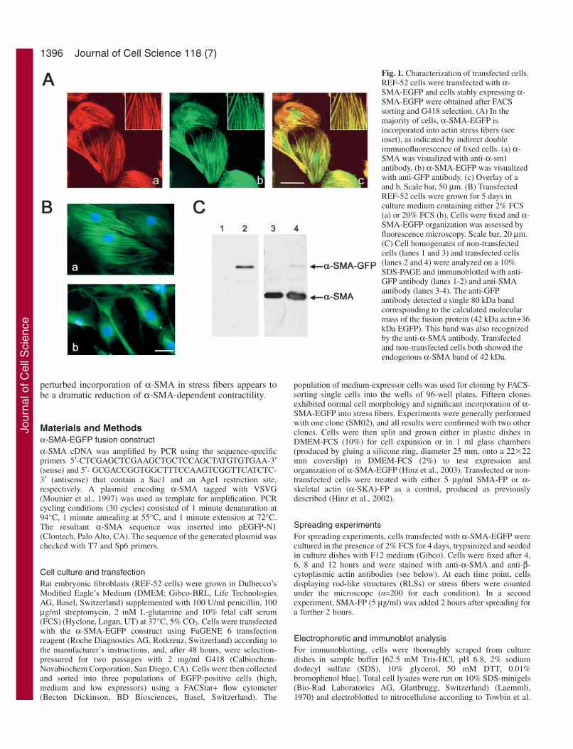
Fig. 1. Characterization of transfected cells.REF-52 cells were transfected with α-SMA-EGFP and cells stably expressing α-SMA-EGFP were obtained after FACSsorting and G418 selection. (A) In themajority of cells, α-SMA-EGFP isincorporated into actin stress fibers (seeinset), as indicated by indirect doubleimmunofluorescence of fixed cells. (a) α-SMA was visualized with anti-α-sm1antibody, (b) α-SMA-EGFP was visualizedwith anti-GFP antibody. (c) Overlay of aand b. Scale bar, 50 µm. (B) TransfectedREF-52 cells were grown for 5 days inculture medium containing either 2% FCS(a) or 20% FCS (b). Cells were fixed and α-SMA-EGFP organization was assessed byfluorescence microscopy. Scale bar, 20 µm.(C) Cell homogenates of non-transfectedcells (lanes 1 and 3) and transfected cells(lanes 2 and 4) were analyzed on a 10%SDS-PAGE and immunoblotted with anti-GFP antibody (lanes 1-2) and anti-SMAantibody (lanes 3-4). The anti-GFPantibody detected a single 80 kDa bandcorresponding to the calculated molecularmass of the fusion protein (42 kDa actin+36kDa EGFP). This band was also recognizedby the anti-α-SMA antibody. Transfectedand non-transfected cells both showed theendogenous α-SMA band of 42 kDa.
Jour
nal o
f Cel
l Sci
ence
1397Ac-EEED peptide and stress fiber formation
(Towbin et al., 1979). Nitrocellulose membranes were incubated withanti-αsm-1 (Skalli et al., 1986) and anti-GFP (Molecular Probes Inc,Eugene, OR) diluted in Tris-buffered saline (TBS) solution containing3% BSA and 0.1% Triton X-100 for 2 hours at room temperature.After three washes with TBS, a second incubation was performed withperoxidase-conjugated affinity-purified goat anti-rabbit IgG (JacksonImmunoresearch Laboratories, West Grove, PA) at a dilution of1:10,000 in TBS containing 0.1% BSA and 0.1% Triton X-100.Peroxidase activity was developed using the ECL western blottingsystem (Amersham, Rahn AG, Zürich, Switzerland), according to themanufacturer’s instructions followed by scanning of the blots (ArcusII; Agfa, Mortsel, Belgium).
Immunofluorescence microscopyFor immunofluorescence staining, cells were fixed in 1%paraformaldehyde in phosphate-buffered saline solution (PBS) for
10 minutes at RT, followed by three washes with PBS and 3 minutestreatment with methanol at –20°C. Subsequently, cells were stainedwith a large panel of primary antibodies (Table 1). Samples werethen incubated with either Alexa Fluor® 568-conjugated anti-mouseor anti-rabbit Ig (Molecular Probes, diluted 1:500 in PBS), for30 minutes at RT. F-actin was probed with Alexa Fluor® 568phalloidin (Molecular Probes) on cells fixed only with 1%paraformaldehyde. After washing in PBS, cells were mountedin polyvinyl alcohol (PVA) (Lennette, 1978): 50 mM Tris-phosphate pH 9.0, 0.1% chlorobutanol, 20% polyvinyl alcohol,0.5‰ phenol red, 20% glycerol). Images were taken with either aZeiss Axiophot microscope (Carl Zeiss, Oberkochen, Germany;using an 63� oil immersion objective) equipped with a high-sensibility color camera (Axiocam, Zeiss) or a confocal microscope(LSM510, Zeiss) with similar optics at 3� zoom, and printedwith a digital Fujifilm Pictography 4000 printer (Fujifilm, Tokyo,Japan).
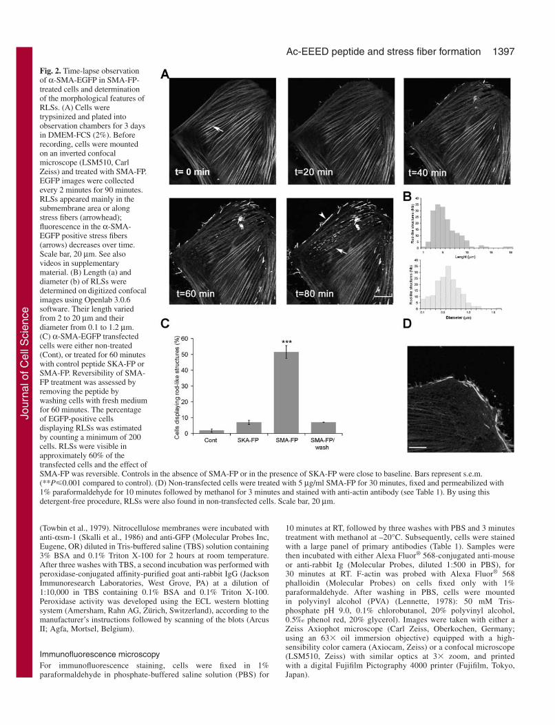
Fig. 2. Time-lapse observationof α-SMA-EGFP in SMA-FP-treated cells and determinationof the morphological features ofRLSs. (A) Cells weretrypsinized and plated intoobservation chambers for 3 daysin DMEM-FCS (2%). Beforerecording, cells were mountedon an inverted confocalmicroscope (LSM510, CarlZeiss) and treated with SMA-FP.EGFP images were collectedevery 2 minutes for 90 minutes.RLSs appeared mainly in thesubmembrane area or alongstress fibers (arrowhead);fluorescence in the α-SMA-EGFP positive stress fibers(arrows) decreases over time.Scale bar, 20 µm. See alsovideos in supplementarymaterial. (B) Length (a) anddiameter (b) of RLSs weredetermined on digitized confocalimages using Openlab 3.0.6software. Their length variedfrom 2 to 20 µm and theirdiameter from 0.1 to 1.2 µm.(C) α-SMA-EGFP transfectedcells were either non-treated(Cont), or treated for 60 minuteswith control peptide SKA-FP orSMA-FP. Reversibility of SMA-FP treatment was assessed byremoving the peptide bywashing cells with fresh mediumfor 60 minutes. The percentageof EGFP-positive cellsdisplaying RLSs was estimatedby counting a minimum of 200cells. RLSs were visible inapproximately 60% of thetransfected cells and the effect ofSMA-FP was reversible. Controls in the absence of SMA-FP or in the presence of SKA-FP were close to baseline. Bars represent s.e.m.(**P�0.001 compared to control). (D) Non-transfected cells were treated with 5 µg/ml SMA-FP for 30 minutes, fixed and permeabilized with1% paraformaldehyde for 10 minutes followed by methanol for 3 minutes and stained with anti-actin antibody (see Table 1). By using thisdetergent-free procedure, RLSs were also found in non-transfected cells. Scale bar, 20 µm.
Jour
nal o
f Cel
l Sci
ence
1398
Live time-lapse observations and fluorescence recovery afterphotobleaching (FRAP) analysesFor video-microscopy, cells were grown in observation chambers (seeabove) for 3 days in DMEM-FCS (2%). Prior to recording, mediumwas changed to serum-free F12 and cells were mounted on LSM510confocal microscope equipped with a Plan-Neofluar 63� 1.3 Ph3objective and maintained at 37°C with a heating stage and CO2-incubation chamber. GFP images were acquired every 2 minutes at488 nm excitation and 515-545 nm emission over a period of time of90 minutes from cells treated with either SMA-FP or SKA-FP as acontrol. For spreading experiments, SM02 cells were seeded inobservation chambers in F12 medium supplemented with 2% FCS andallowed to spread for 2 hours. Cell-spreading was imaged on anAxiovert 200M equipped with an array laser confocal spinning wheel(Nipkow disc; Visitech, Sunderland, UK) using a 63�, 1.4 NA oil-immersion objective (Carl Zeiss). Images were acquired every 10minutes for 15 hours by using a cooled, 16-bit CCD camera(CoolSnap HQ; Roper Scientific, Trenton, NJ) operated byMetamorph 6.1 software (Universal Imaging, West Chester, PA).
In FRAP experiments, bar and square-shaped regions or completeRLSs were bleached with 100% transmission (λ=488 nm) with 10iterations each lasting 1 second, and recovery was monitored at 1minute intervals in control conditions or on cells treated for 15minutes with FP before photobleaching. To quantify FRAP, length andgrayscale pixel-values were measured on digitized confocal imagesusing the Openlab 3.0.6 software (Improvision, Basel, Switzerland).The average rate of movement was determined by calculating distanceversus time. Mean values were calculated from at least ten cells fromthree independent experiments per condition.
Statistical analysisQuantitative results are presented as mean values±s.e.m. Differencesbetween mean values were calculated by using the Student’s t test and
were considered to be statistically significant at values ofP�0.001(***).
ResultsCharacterization of α-SMA-EGFPAfter FACS sorting and G418 selection, we obtained 15clones that stably expressed α-SMA-EGFP at differentexpression levels in stress fibers. Clones showed normal cellmorphology and proliferation rates compared with wild-typeREF-52 cells. Moreover, expression of exogenous α-SMA-EGFP did not interfere with endogenous α-SMA expressionwhen induced by TGFβ and low serum levels (data notshown). For this study we used one clone (SM02) that, inapproximately 80% of the cells, resulted in cells whichhad α-SMA-EGFP organized in stress fibers, colocalizingperfectly with endogenous α-SMA (Fig. 1A). Likeendogenous α-SMA, incorporation of α-SMA-EGFP in stressfibers was induced by 2% FCS in the culture medium (Fig.1B, a) and by TGFβ (data not shown). Thus, the organizationof α-SMA-EGFP was regulated by TGFβ, as previouslydescribed for endogenous α-SMA (Moustakas andStournaras, 1999) (data not shown) and expression of α-SMA-EGFP did not appear to interfere with cell function.
To ensure that the expression of α-SMA-EGFP did notalter the dynamic properties of α-SMA, we cultured SM02cells in different concentrations of FCS, ranging from 2%to 20% FCS. In 20% FCS, cells did not exhibit α-SMA-EGFP-positive stress fibers (Fig. 1B, b) as previouslyobserved for endogenous α-SMA in REF-52 cells (Hinz etal., 2003). Expression of α-SMA-EGFP was also tested
Journal of Cell Science 118 (7)
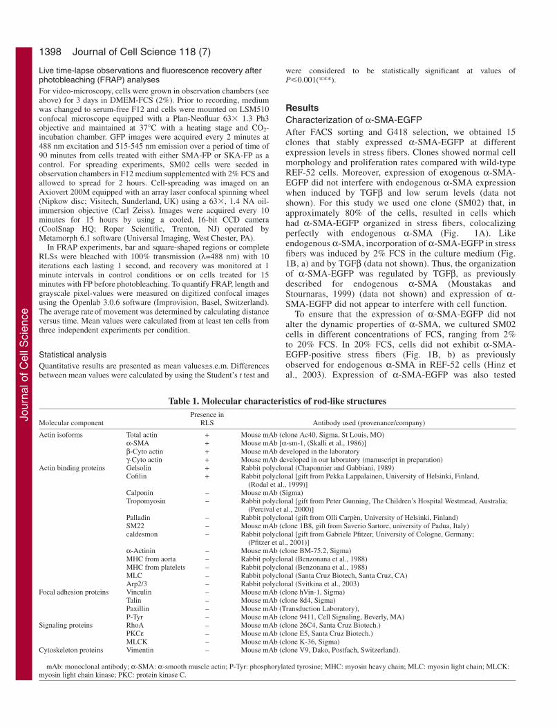
Table 1. Molecular characteristics of rod-like structuresPresence in
Molecular component RLS Antibody used (provenance/company)
Actin isoforms Total actin + Mouse mAb (clone Ac40, Sigma, St Louis, MO) α-SMA + Mouse mAb [α-sm-1, (Skalli et al., 1986)]β-Cyto actin + Mouse mAb developed in the laboratoryγ-Cyto actin + Mouse mAb developed in our laboratory (manuscript in preparation)
Actin binding proteins Gelsolin + Rabbit polyclonal (Chaponnier and Gabbiani, 1989)Cofilin + Rabbit polyclonal [gift from Pekka Lappalainen, University of Helsinki, Finland,
(Rodal et al., 1999)]Calponin – Mouse mAb (Sigma)Tropomyosin – Rabbit polyclonal [gift from Peter Gunning, The Children’s Hospital Westmead, Australia;
(Percival et al., 2000)]Palladin – Rabbit polyclonal (gift from Olli Carpèn, University of Helsinki, Finland)SM22 – Mouse mAb (clone 1B8, gift from Saverio Sartore, university of Padua, Italy)caldesmon – Rabbit polyclonal [gift from Gabriele Pfitzer, University of Cologne, Germany;
(Pfitzer et al., 2001)]α-Actinin – Mouse mAb (clone BM-75.2, Sigma)MHC from aorta – Rabbit polyclonal (Benzonana et al., 1988)MHC from platelets – Rabbit polyclonal (Benzonana et al., 1988)MLC – Rabbit polyclonal (Santa Cruz Biotech, Santa Cruz, CA)Arp2/3 – Rabbit polyclonal (Svitkina et al., 2003)
Focal adhesion proteins Vinculin – Mouse mAb (clone hVin-1, Sigma)Talin – Mouse mAb (clone 8d4, Sigma)Paxillin – Mouse mAb (Transduction Laboratory), P-Tyr – Mouse mAb (clone 9411, Cell Signaling, Beverly, MA)
Signaling proteins RhoA – Mouse mAb (clone 26C4, Santa Cruz Biotech.)PKCε – Mouse mAb (clone E5, Santa Cruz Biotech.)MLCK – Mouse mAb (clone K-36, Sigma)
Cytoskeleton proteins Vimentin – Mouse mAb (clone V9, Dako, Postfach, Switzerland).
mAb: monoclonal antibody; α-SMA: α-smooth muscle actin; P-Tyr: phosphorylated tyrosine; MHC: myosin heavy chain; MLC: myosin light chain; MLCK:myosin light chain kinase; PKC: protein kinase C.
Jour
nal o
f Cel
l Sci
ence

1399Ac-EEED peptide and stress fiber formation
biochemically. Western blot analyses with anti-GFP antibodyshowed a single 80 kDa band that corresponded to thepredicted molecular mass of the α-SMA-EGFP protein [actin(42 kDa) + EGFP (36 kDa)] (Fig. 1C, lane 2). This bandwas also recognized by the anti-αsm-1 antibody, indicatingthat the antigenic properties of the Ac-EEED N-terminuswere not altered by tagging the C-terminus with EGFP (Fig.1C, lane 4). Both transfected and non-transfected cellsshowed the 42-kDa band of endogenous α-SMA (Fig. 1C,lanes 3 and 4).
Time-lapse observation of α-SMA-EGFP in cells treatedwith SMA-FPAs a first step in understanding the function of Ac-EEED, westudied the effect of SMA-FP on the organization of α-SMA-EGFP in living cells. As shown in Fig. 2A, 5 µg/ml SMA-FPinduced two phenomena visible 30 minutes after the beginningof the treatment: (1) the gradual decrease of fluorescence-intensity of α-SMA-EGFP in stress fibers and, (2) the time-correlated appearance of RLSs, mainly located at the cellperiphery and close to stress fibers. Both effects were observedin approximately 60% of the transfected cells (Fig. 2C).Morphological features of RLSs were determined on digitizedconfocal images. The length of RLSs varied widely from 2-20µm, fitting a Gaussian curve with a peak at 4 µm (average:5.03±0.21, Fig. 2B). Their diameter ranged between 0.1 and1.2 µm (average: 0.54±0.02). Controls, performed either in theabsence of SMA-FP or the presence of SKA-FP, showed RLSsonly in a minority of the cell population (7% at most), furtherindicating the specificity of the effect caused by SMA-FP (Fig.2C). Removal of SMA-FP reversed its effects: after 1 hour ofwashing, RLSs completely disappeared and stress fibersbecame again highly fluorescent (Fig. 2C).
Interestingly, RLSs dissappeared afterpermeabilizing cells with classic methods (suchas 0.1% Triton-X100 for 1 minute followingfixation with 1% paraformaldehyde); thissusceptibility of RLSs to detergent may accountfor the fact that previously they have not beenobserved in non-transfected cells. We thusdeveloped an alternative protocol for fixing cells,using 1% paraformaldehyde for 10 minutesfollowed by permeabilization with methanol(–20°C) for 3 minutes, allowing routinedemonstration of these structures. Using this newmethod, we were able to obtain very clearantibody staining and RLS preservation andrevealed that RLSs were also formed in44.6±4.9% of non-transfected cells when treatedwith SMA-FP for 30 minutes (Fig. 2D), thusexcluding the possibility of an artifact owing toEGFP. Similarly, SMA-FP provoked theformation of RLSs in myofibroblasts of variousorigins, such as rat lung fibroblasts andDupuytren fibrobasts treated with TGFβ (data notshown).
Fig. 3. Presence of actin isoforms in the RLSs.(A) Overlay images of EGFP fluorescence (green) and(a) staining of β-cytoplasmic actin (red) or (b) γ-cytoplasmic actin (red) in non-treated α-SMA-EGFP-transfected cells are shown. Scale bar, 10 µm. (B) Cellswere treated for 30 minutes with SMA-FP, fixed with1% paraformaldehyde for 10 minutes followed bymethanol incubation for 3 minutes and stained witheither (b) anti β-cytoplasmic actin antibody, (e) γ-cytoplasmic actin antibody or (h) Alexa 568-phalloidin.(k) The distribution of rhodamine-labeled SMA-FP inα-SMA-EGFP-transfected cells was also investigated.α-SMA-EGFP patterns are shown in images a, d, g,and j and colocalization was visualized by confocalmicroscopy. Overlay images are shown in c, f, i and l.Bar, 10 µm.
Jour
nal o
f Cel
l Sci
ence

1400
Characterization of the RLSsWe determined the molecular composition of RLSs byimmunostaining, to investigate which proteins might beassociated with the RLSs and potentially interfere with the N-terminus of α-SMA. A large panel of primary antibodies wasused in confocal microscopy, as listed in Table 1. All otheractin isoforms expressed in REF-52 cells were partially presentin RLSs, as demonstrated by the colocalization of EGFP withboth β-cytoplasmic actin (Fig. 3B, a-c) and γ-cytoplasmic actin(Fig. 3B, d-f). The rhodamine-phalloidin staining of thesestructures (shown in Fig. 3B, g-i) indicated that they representpolymerized actin. In addition, after 30 minutes of treatment,SMA-FP was localized in all structures where α-SMA-EGFPwas present (stress fibers as well as RLSs; Fig. 3B, j-l). Amongall actin-binding proteins tested, only cofilin and gelsolinlocalized in RLSs together with α-SMA. In non-treated SM02cells (Fig. 4A) and wild-type REF-52 cells (data not shown),gelsolin (Fig. 4A, a) and cofilin (Fig. 4A, b) both colocalizedwith α-SMA in stress fibers, appeared diffusely in the cytosoland, in the case of cofilin, in membrane ruffles (Fig. 4A, b,
arrow). Following SMA-FP treatment, gelsolin(Fig. 4B, a-c) and cofilin (Fig. 4B, d-f) werepresent in RLSs. No other actin-binding proteinsexamined (e.g. α-actinin, Fig. 4B, g-i, Table 1),were found in RLS (green RLSs in the overlayimage i).
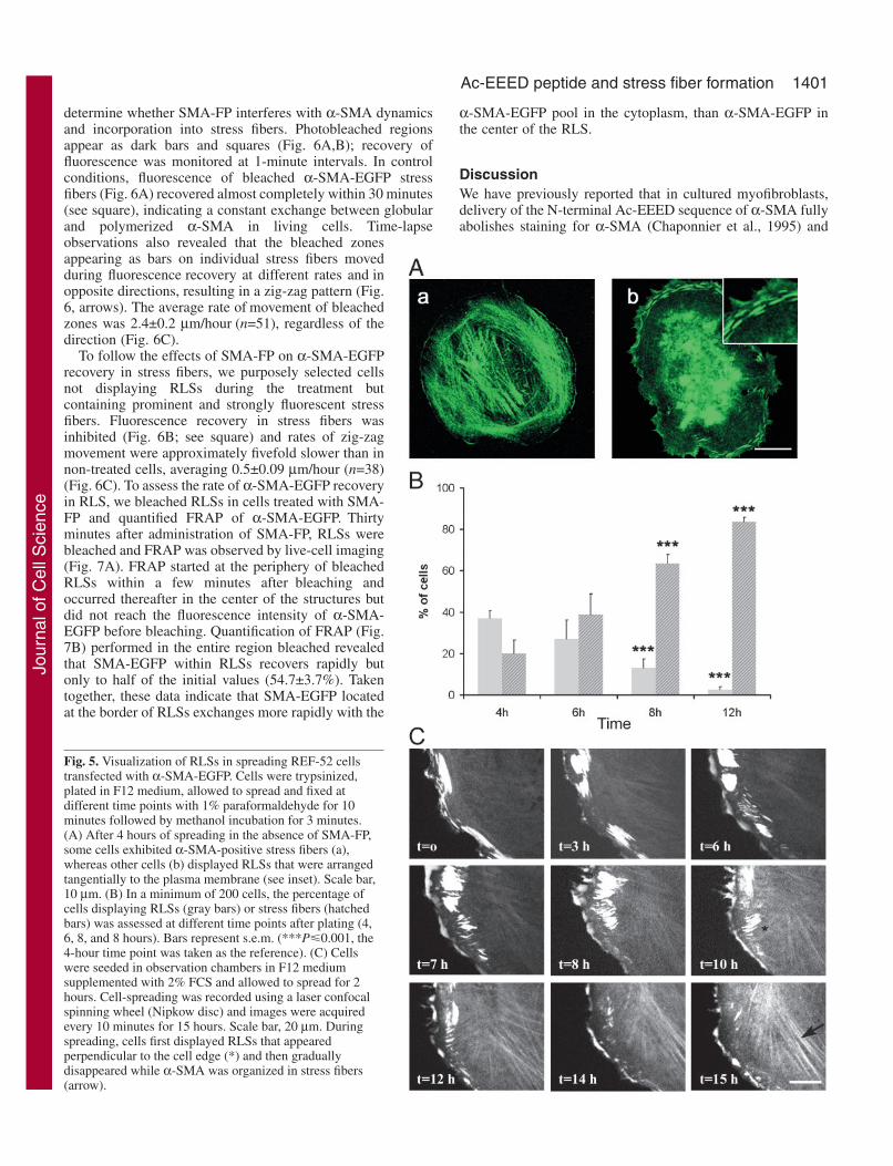
Spreading-experimentsTo investigate whether RLSs occur during α-SMA re-organization in physiologicalconditions, we studied transfected REF-52 cellsas they spread on planar culture surfaces, aprocess during which stress fibers are formed denovo. Four hours after seeding, α-SMAexhibited essentially three types of organization:(1) predominantly diffuse (not shown); (2)present in stress fibers (Fig. 5A, a) and, (3)arranged in RLSs, longitudinal to the cellmembrane (Fig. 5A, b), showing the samemolecular characteristics as described above(data not shown). The proportion of cellsshowing organization of α-SMA either in stressfibers or in RLSs varied, depending on howmuch time passed after seeding the cells (Fig.5B). In particular, the percentage of cellsdisplaying RLSs decreased from 37±3.7% at 4hours after plating to 2.5±1.3% at 12 hours afterplating. By contrast, a comparable increase in
the number of cells displaying stress fibers was observedduring the same interval. Continuous time-lapse observation ofindividual spreading cells (Fig. 5C) recurrently showed thatRLSs first displayed a tangential orientation to the cell edge(t=0-3 hours), followed by reorientation perpendicularly to thecell edge (t=6-10 hours). Then, RLSs gradually becamesmaller and disappeared almost completely (t=12-15 hours)when α-SMA started to become organized in stress fibers.Importantly, during spreading, formation of α-SMA-positivestress fibers was severely impaired when, 2 hours afterspreading, cells were treated with SMA-FP for 2 hours:59.3±3.2% of the cells now contained tangential RLSs andonly 5.1±1.0% of cells displayed α-SMA organized in stressfibers. This suggests that RLSs represent a step in theorganization of α-SMA preceding their incorporation in stressfibers.
FRAP experimentsWe used FRAP analysis of α-SMA-EGFP stress fibers to
Journal of Cell Science 118 (7)
Fig. 4. Presence of actin-binding proteins in RLS. (A) Gelsolin (a) and cofilin (b)organization in α-SMA-EGFP transfected cells in control conditions. Overlayimages with α-SMA-EGFP show that gelsolin (a, red) completely colocalized withα-SMA-EGFP (green) in stress fibers, whereas cofilin (b, red) was additionallyfound at the periphery of the cell (arrow). (B) Cells were treated for 30 minutes withSMA-FP, fixed with 1% paraformaldehyde for 10 minutes followed by methanol for3 minutes and stained with a large panel of anti actin-binding protein antibodies (seetable 1). EGFP fluorescence (green) and immunofluorescence staining (red) withanti-gelsolin (b) and anti-cofilin (e) showed that they were both present in RLS(yellow RLS in the overlay images c and f), whereas many other actin bindingproteins (α-actinin, h shown as example) were not found (green RLS in the overlayimage i). Bar, 10 µm.
Jour
nal o
f Cel
l Sci
ence
1401Ac-EEED peptide and stress fiber formation
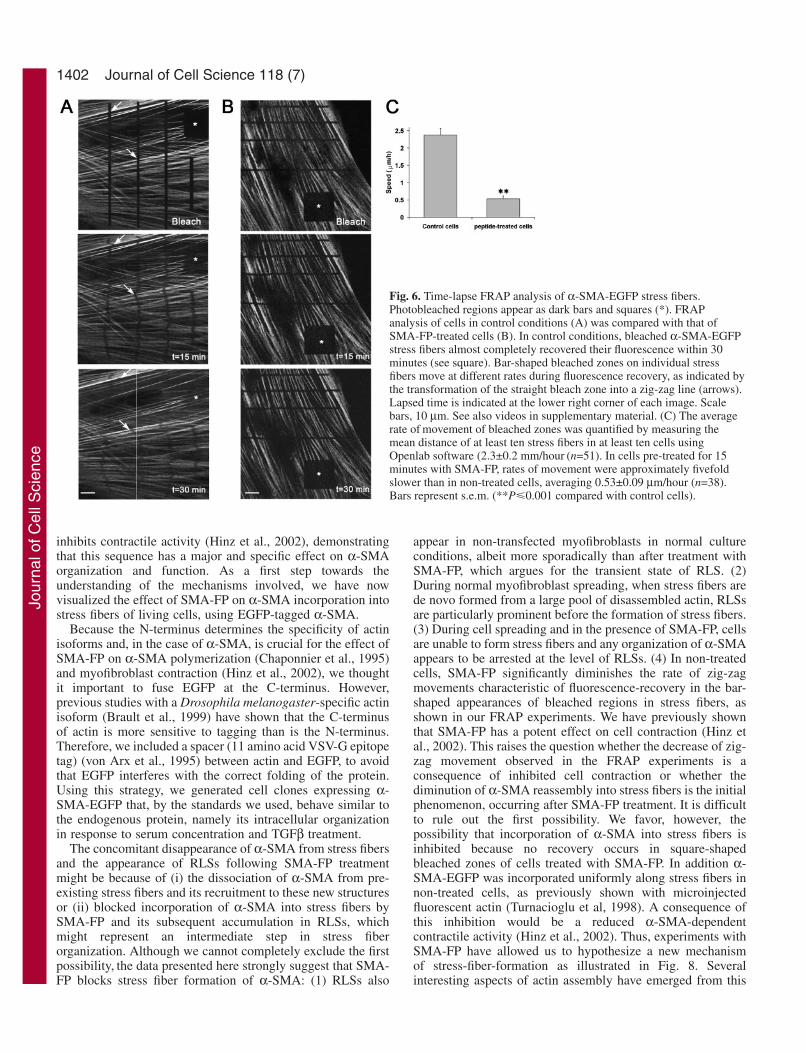
determine whether SMA-FP interferes with α-SMA dynamicsand incorporation into stress fibers. Photobleached regionsappear as dark bars and squares (Fig. 6A,B); recovery offluorescence was monitored at 1-minute intervals. In controlconditions, fluorescence of bleached α-SMA-EGFP stressfibers (Fig. 6A) recovered almost completely within 30 minutes(see square), indicating a constant exchange between globularand polymerized α-SMA in living cells. Time-lapseobservations also revealed that the bleached zonesappearing as bars on individual stress fibers movedduring fluorescence recovery at different rates and inopposite directions, resulting in a zig-zag pattern (Fig.6, arrows). The average rate of movement of bleachedzones was 2.4±0.2 µm/hour (n=51), regardless of thedirection (Fig. 6C).
To follow the effects of SMA-FP on α-SMA-EGFPrecovery in stress fibers, we purposely selected cellsnot displaying RLSs during the treatment butcontaining prominent and strongly fluorescent stressfibers. Fluorescence recovery in stress fibers wasinhibited (Fig. 6B; see square) and rates of zig-zagmovement were approximately fivefold slower than innon-treated cells, averaging 0.5±0.09 µm/hour (n=38)(Fig. 6C). To assess the rate of α-SMA-EGFP recoveryin RLS, we bleached RLSs in cells treated with SMA-FP and quantified FRAP of α-SMA-EGFP. Thirtyminutes after administration of SMA-FP, RLSs werebleached and FRAP was observed by live-cell imaging(Fig. 7A). FRAP started at the periphery of bleachedRLSs within a few minutes after bleaching andoccurred thereafter in the center of the structures butdid not reach the fluorescence intensity of α-SMA-EGFP before bleaching. Quantification of FRAP (Fig.7B) performed in the entire region bleached revealedthat SMA-EGFP within RLSs recovers rapidly butonly to half of the initial values (54.7±3.7%). Takentogether, these data indicate that SMA-EGFP locatedat the border of RLSs exchanges more rapidly with the
α-SMA-EGFP pool in the cytoplasm, than α-SMA-EGFP inthe center of the RLS.
DiscussionWe have previously reported that in cultured myofibroblasts,delivery of the N-terminal Ac-EEED sequence of α-SMA fullyabolishes staining for α-SMA (Chaponnier et al., 1995) and
Fig. 5. Visualization of RLSs in spreading REF-52 cellstransfected with α-SMA-EGFP. Cells were trypsinized,plated in F12 medium, allowed to spread and fixed atdifferent time points with 1% paraformaldehyde for 10minutes followed by methanol incubation for 3 minutes.(A) After 4 hours of spreading in the absence of SMA-FP,some cells exhibited α-SMA-positive stress fibers (a),whereas other cells (b) displayed RLSs that were arrangedtangentially to the plasma membrane (see inset). Scale bar,10 µm. (B) In a minimum of 200 cells, the percentage ofcells displaying RLSs (gray bars) or stress fibers (hatchedbars) was assessed at different time points after plating (4,6, 8, and 8 hours). Bars represent s.e.m. (***P�0.001, the4-hour time point was taken as the reference). (C) Cellswere seeded in observation chambers in F12 mediumsupplemented with 2% FCS and allowed to spread for 2hours. Cell-spreading was recorded using a laser confocalspinning wheel (Nipkow disc) and images were acquiredevery 10 minutes for 15 hours. Scale bar, 20 µm. Duringspreading, cells first displayed RLSs that appearedperpendicular to the cell edge (*) and then graduallydisappeared while α-SMA was organized in stress fibers(arrow).
Jour
nal o
f Cel
l Sci
ence
1402
inhibits contractile activity (Hinz et al., 2002), demonstratingthat this sequence has a major and specific effect on α-SMAorganization and function. As a first step towards theunderstanding of the mechanisms involved, we have nowvisualized the effect of SMA-FP on α-SMA incorporation intostress fibers of living cells, using EGFP-tagged α-SMA.
Because the N-terminus determines the specificity of actinisoforms and, in the case of α-SMA, is crucial for the effect ofSMA-FP on α-SMA polymerization (Chaponnier et al., 1995)and myofibroblast contraction (Hinz et al., 2002), we thoughtit important to fuse EGFP at the C-terminus. However,previous studies with a Drosophila melanogaster-specific actinisoform (Brault et al., 1999) have shown that the C-terminusof actin is more sensitive to tagging than is the N-terminus.Therefore, we included a spacer (11 amino acid VSV-G epitopetag) (von Arx et al., 1995) between actin and EGFP, to avoidthat EGFP interferes with the correct folding of the protein.Using this strategy, we generated cell clones expressing α-SMA-EGFP that, by the standards we used, behave similar tothe endogenous protein, namely its intracellular organizationin response to serum concentration and TGFβ treatment.
The concomitant disappearance of α-SMA from stress fibersand the appearance of RLSs following SMA-FP treatmentmight be because of (i) the dissociation of α-SMA from pre-existing stress fibers and its recruitment to these new structuresor (ii) blocked incorporation of α-SMA into stress fibers bySMA-FP and its subsequent accumulation in RLSs, whichmight represent an intermediate step in stress fiberorganization. Although we cannot completely exclude the firstpossibility, the data presented here strongly suggest that SMA-FP blocks stress fiber formation of α-SMA: (1) RLSs also
appear in non-transfected myofibroblasts in normal cultureconditions, albeit more sporadically than after treatment withSMA-FP, which argues for the transient state of RLS. (2)During normal myofibroblast spreading, when stress fibers arede novo formed from a large pool of disassembled actin, RLSsare particularly prominent before the formation of stress fibers.(3) During cell spreading and in the presence of SMA-FP, cellsare unable to form stress fibers and any organization of α-SMAappears to be arrested at the level of RLSs. (4) In non-treatedcells, SMA-FP significantly diminishes the rate of zig-zagmovements characteristic of fluorescence-recovery in the bar-shaped appearances of bleached regions in stress fibers, asshown in our FRAP experiments. We have previously shownthat SMA-FP has a potent effect on cell contraction (Hinz etal., 2002). This raises the question whether the decrease of zig-zag movement observed in the FRAP experiments is aconsequence of inhibited cell contraction or whether thediminution of α-SMA reassembly into stress fibers is the initialphenomenon, occurring after SMA-FP treatment. It is difficultto rule out the first possibility. We favor, however, thepossibility that incorporation of α-SMA into stress fibers isinhibited because no recovery occurs in square-shapedbleached zones of cells treated with SMA-FP. In addition α-SMA-EGFP was incorporated uniformly along stress fibers innon-treated cells, as previously shown with microinjectedfluorescent actin (Turnacioglu et al, 1998). A consequence ofthis inhibition would be a reduced α-SMA-dependentcontractile activity (Hinz et al., 2002). Thus, experiments withSMA-FP have allowed us to hypothesize a new mechanismof stress-fiber-formation as illustrated in Fig. 8. Severalinteresting aspects of actin assembly have emerged from this
Journal of Cell Science 118 (7)
Fig. 6. Time-lapse FRAP analysis of α-SMA-EGFP stress fibers.Photobleached regions appear as dark bars and squares (*). FRAPanalysis of cells in control conditions (A) was compared with that ofSMA-FP-treated cells (B). In control conditions, bleached α-SMA-EGFPstress fibers almost completely recovered their fluorescence within 30minutes (see square). Bar-shaped bleached zones on individual stressfibers move at different rates during fluorescence recovery, as indicated bythe transformation of the straight bleach zone into a zig-zag line (arrows).Lapsed time is indicated at the lower right corner of each image. Scalebars, 10 µm. See also videos in supplementary material. (C) The averagerate of movement of bleached zones was quantified by measuring themean distance of at least ten stress fibers in at least ten cells usingOpenlab software (2.3±0.2 mm/hour (n=51). In cells pre-treated for 15minutes with SMA-FP, rates of movement were approximately fivefoldslower than in non-treated cells, averaging 0.53±0.09 µm/hour (n=38).Bars represent s.e.m. (**P�0.001 compared with control cells).
Jour
nal o
f Cel
l Sci
ence

1403Ac-EEED peptide and stress fiber formation
study. The RLS represents a hithertounknown structure, which appears tofunction as a reservoir for pre-polymerized α-SMA during stress-fiber-formation. Its visualization wasfacilitated by blocking incorporationof α-SMA into stress fibers by SMA-FP. However, RLSs may also beinterpreted as a stress-relatedresponse, where they would thenrepresent a temporary rescuemechanism for the cell. Althoughboth explanations are not mutuallyexclusive, the disappearance of RLSsin favor of stress-fiber-formationduring normal cell spreading suggeststhat, in one given population, all cellsthat form stress fibers will experiencethis phase. This strongly argues thatRLSs are an intermediate α-SMA-organization preceding stress fiberformation, rather than being part ofan erroneous, occasionally occurringpathway.
The fact that RLSs are lost when using classic cell-permeabilization procedures with detergent, might explain whythey have not been described previously. The effect ofdetergents and the sub-cellular localization of RLSs, suggestthat RLSs are somehow connected with the plasma membrane.This is in agreement with the notion that actin polymerizationis initiated at the plasma membrane throughphosphatidylinositol (4,5)-bisphosphate (for a review, see Yinand Janmey, 2003). Additional important clues regarding theorigin of RLSs are provided by their molecular composition.None of the architectural stress fiber proteins (e.g. α-actinin,myosin-heavy chain, tropomyosin) became dissociated fromstress fibers following treatment with SMA-FP. Despite theresemblance of RLSs to focal adhesions in some cells, none ofthe focal adhesion proteins we examined were found in RLSs.It has recently been postulated that thrombin activation leadsto the formation of a protein kinase Cε (PKCε)-RhoA-αSMAternary complex, leading to stress fiber formation of α-SMA(Bogatkevich et al., 2003). Neither PKCε nor Rho-A, however,were found in RLS. In fact, of the tested actin-binding proteins,only cofilin and gelsolin were present in RLSs. Gelsolin
belongs to a family of proteins that are essential formicrofilament remodeling (Burtnick et al., 1997; Kwiatkowskiet al., 1986; Yin, 1987). In vitro, gelsolin interacts with G-actinand F-actin, promotes nucleation and both severs and capsactin filaments. Cofilin belongs to another family of actin-binding proteins also severing actin filaments, thereforeincreasing polymerization dynamics (Bamburg, 1999). Thepresence of these two proteins is consistent with the hypothesisthat SMA-FP interferes with actin-dynamics. In addition,although both cytoplasmic actins do not disappear from stressfibers as previously observed (Hinz et al, 2002), a smallproportion of them accompanied α-SMA in RLSs.
In conclusion, we propose a model that identifies theimportance of the N-terminal AcEEED sequence for α-SMAorganization and function. We suggest that RLSs represent anintermediate step in the process of α-SMA incorporation intostress fibers and possibly provide an α-SMA reservoir whenthis incorporation process is disturbed. The presence ofgelsolin and cofilin in RLS suggests their active participationin this process. Because SMA-FP blocks α-SMA at this step,we propose that the peptide interferes with a putative partner
Fig. 7. FRAP analysis of SMA-EGFPturnover in RLSs. (A) Transfected cellswere treated for 30 minutes with SMA-FP.α-SMA-EGFP RLSs were photobleachedand images were taken at indicated timepoints. Bar, 5 µm. (B) Fluorescenceintensities were measured at differenttimes using Openlab software and relatedto the intensity measured in the first videoframe. Fluorescence before bleaching wasnormalized to 100%. Each data pointrepresents the mean intensity offluorescence of 33 RLSs analyzed in eightdifferent cells. Bars represent s.e.m.
Fig. 8. Schematic representation ofthe proposed mechanism of SMA-FPaction and stress-fiber-formation. (a)α-SMA incorporation in pre-existingcytoplasmic-actin-containing stressfibers represented as light red dashedlines. α-SMA is first recruited inRLSs, a pre-requisite step before itsincorporation in stress fibers (seearrows). (b) α-SMA incorporationinto stress fibers (dark red lines)confers higher cell-contractility(illustrated by the arrows on the topof the cell). When such contractilecells are treated with SMA-FP, α-SMA incorporation in stress fibers is inhibited (see zoomed-in part of the cell c), resulting in α-SMAaccumulation in RLSs, its disappearance from stress fibers, and subsequently ‘relaxation’ of the cell.
Jour
nal o
f Cel
l Sci
ence
1404
of α-SMA allowing its incorporation in stress fibers. Whetherthis process is limited to incorporation of α-SMA or it is ageneral phenomenon that occurs with all actin isoformsremains to be determined.
We wish to thank P. Lappalainen, O. Carpèn, (University ofHelsinki, Finland), P. Gunning (The Children’s Hospital Westmead,Australia), S. Sartore (University of Padua, Italy) and G. Pfitzer(University of Cologne, Germany) for providing the antibodiesdirected against cofilin, palladin, tropomyosin, SM22 and caldesmon,respectively. We are grateful to R. B. Low for his valuable advicethroughout the project and for critical reading of the manuscript. Wegratefully acknowledge G. Celetta, A. Hiltbrunner-Maurer and S.Kampf for technical help, J.-C. Rumbeli and E. Denkinger forphotographic work, and Serge Arnaudeau (Bioimaging Core Facility,Faculty of Medicine, Geneva) for his help and advice in using theNipkow system. This work was supported by the Swiss NationalScience Foundation, grant numbers PMPDA-102408 to S.C.,3100A0-102150/1 to B.H. and 31-68313.02 to C.C.
ReferencesBamburg, J. R. (1999). Proteins of the ADF/cofilin family: essential
regulators of actin dynamics. Annu. Rev. Cell. Dev. Biol. 15, 185-230.Benzonana, G., Skalli, O. and Gabbiani, G. (1988). Correlation between the
distribution of smooth muscle or non muscle myosins and alpha-smoothmuscle actin in normal and pathological soft tissues. Cell. Motil.Cytoskeleton 11, 260-274.
Bogatkevich, G. S., Tourkina, E., Abrams, C. S., Harley, R. A., Silver, R.M. and Ludwicka-Bradley, A. (2003). Contractile activity and smoothmuscle alpha-actin organization in thrombin-induced human lungmyofibroblasts. Am. J. Physiol. Lung Cell Mol. Physiol. 285, L334-343.
Brault, V., Sauder, U., Reedy, M. C., Aebi, U. and Schoenenberger, C. A.(1999). Differential epitope tagging of actin in transformed Drosophilaproduces distinct effects on myofibril assembly and function of the indirectflight muscle. Mol. Biol. Cell 10, 135-149.
Burtnick, L. D., Koepf, E. K., Grimes, J., Jones, E. Y., Stuart, D. J.,McLaughlin, P. J. and Robinson, R. C. (1997). The crystal structure ofplasma gelsolin: implications for actin severing, capping, and nucleation.Cell 90, 661-670.
Chaponnier, C. and Gabbiani, G. (1989). Gelsolin modulation in epithelialand stromal cells of mammary carcinoma. Am. J. Path. 134, 597-603.
Chaponnier, C., Goethals, M., Janmey, P. A., Gabbiani, F., Gabbiani, G.and Vandekerckhove, J. (1995). The specific NH2-terminal sequence Ac-EEED of alpha-smooth muscle actin plays a role in polymerization in vitroand in vivo. J. Cell Biol. 130, 887-895.
Darby, I., Skalli, O. and Gabbiani, G. (1990). α-smooth muscle actin istransiently expressed by myofibroblasts during experimental wound healing.Lab. Invest. 63, 21-29.
Derossi, D., Joliot, A. H., Chassaing, G. and Prochiantz, A. (1994). Thethird helix of the Antennapedia homeodomain translocates throughbiological membranes. J. Biol. Chem. 269, 10444-10450.
Herman, I. M. (1993). Actin isoforms. Curr. Opin. Cell Biol. 5, 48-55.Hinz, B., Gabbiani, G. and Chaponnier, C. (2002). The NH2-terminal
peptide of α-smooth muscle actin inhibits force generation by themyofibroblast in vitro and in vivo. J. Cell Biol. 157, 657-663.
Hinz, B., Dugina, V., Ballestrem, C., Wehrle-Haller, B. and Chaponnier,C. (2003). Alpha-smooth muscle actin is crucial for focal adhesionmaturation in myofibroblasts. Mol. Biol. Cell 14, 2508-2519.
Khaitlina, S. Y. (2001). Functional specificity of actin isoforms. Int. Rev.Cytol. 202, 35-98.
Kreis, T. E., Winterhalter, K. H. and Birchmeier, W. (1979). In vivodistribution and turnover of fluorescently labeled actin microinjected intohuman fibroblasts. Proc. Natl. Acad. Sci. USA 76, 3814-3818.
Kwiatkowski, D. J., Stossel, T. P., Orkin, S. H., Mole, J. E., Colten, H. R.and Yin, H. L. (1986). Plasma and cytoplasmic gelsolins are encoded by asingle gene and contain a duplicated actin-binding domain. Nature 323, 455-458.
Laemmli, U. K. (1970). Cleavage of structural proteins during the assemblyof the head of bacteriophage T4. Nature 227, 680-685.
Lennette, D. A. (1978). An improved mounting medium forimmunofluorescence microscopy [letter]. Am. J. Clin. Pathol. 69, 647-648.
Mounier, N., Perriard, J.-C., Gabbiani, G. and Chaponnier, C. (1997).Transfected muscle and non-muscle actins are differentially sortedby cultured smooth muscle and non-muscle cells. J. Cell Sci. 110, 839-846.
Moustakas, A. and Stournaras, C. (1999). Regulation of actinorganisation by TGF-beta in H-ras-transformed fibroblasts. J. Cell Sci.112, 1169-1179.
Percival, J. M., Thomas, G., Cock, T. A., Gardiner, E. M., Jeffrey, P. L.,Lin, J. J., Weinberger, R. P. and Gunning, P. (2000). Sorting oftropomyosin isoforms in synchronised NIH 3T3 fibroblasts: evidence fordistinct microfilament populations. Cell Motil. Cytoskeleton 47, 189-208.
Pfitzer, G., Sonntag-Bensch, D. and Brkic-Koric, D. (2001).Thiophosphorylation-induced Ca(2+) sensitization of guinea-pig ileumcontractility is not mediated by Rho-associated kinase. J. Physiol. 533, 651-664.
Powell, D. W., Mifflin, R. C., Valentich, J. D., Crowe, S. E., Saada, J. I.and West, A. B. (1999). Myofibroblasts. I. Paracrine cells important inhealth and disease. Am. J. Physiol. 277, C1-9.
Rodal, A. A., Tetreault, J. W., Lappalainen, P., Drubin, D. G. and Amberg,D. C. (1999). Aip1p interacts with cofilin to disassemble actin filaments. J.Cell. Biol. 145, 1251-1264.
Rubenstein, P. A. (1990). The functional importance of multiple actinisoforms. BioEssays 12, 309-315.
Sappino, A. P., Schürch, W. and Gabbiani, G. (1990). Differentiationrepertoire of fibroblastic cells: expression of cytoskeletal proteins as markersof phenotypic modulations. Lab. Invest. 63, 144-161.
Schmitt-Graff, A., Desmouliere, A. and Gabbiani, G. (1994). Heterogeneityof myofibroblast phenotypic features: an example of fibroblastic cellplasticity. Virchows Arch. 425, 3-24.
Serini, G. and Gabbiani, G. (1999). Mechanisms of myofibroblast activityand phenotypic modulation. Exp. Cell Res. 250, 273-283.
Skalli, O., Ropraz, P., Trzeciak, A., Benzonana, G., Gillessen, D. andGabbiani, G. (1986). A monoclonal antibody against α-smooth muscleactin: a new probe for smooth muscle differentiation. J. Cell Biol. 103, 2787-2796.
Svitkina, T. M., Bulanova, E. A., Chaga, O. Y., Vignjevic, D. M., Kojima,S., Vasiliev, J. M. and Borisy, G. G. (2003). Mechanism of filopodiainitiation by reorganization of a dendritic network. J. Cell Biol. 160, 409-421.
Tomasek, J. J., Gabbiani, G., Hinz, B., Chaponnier, C. and Brown, R. A.(2002). Myofibroblasts and mechano-regulation of connective tissueremodelling. Nat. Rev. Mol. Cell Biol. 3, 349-363.
Towbin, H., Staehelin, T. and Gordon, J. (1979). Electrophoretic transfer ofproteins from polyacrylamide gels to nitrocellulose sheets: procedure andsome applications. Proc. Natl. Acad. Sci. USA 76, 4350-4354.
Turnacioglu, K. K., Sanger, J. W. and Sanger, J. M. (1998). Sites ofmonomeric actin incorporation in living PTK2 and REF-52 cells. Cell Motil.Cytoskel. 40, 59-70.
Vandekerckhove, J. and Weber, K. (1978). Actin amino-acid sequences.Comparison of actins from calf thymus, bovine brain, and SV40-transformed mouse 3T3 cells with rabbit skeletal actin. Eur. J. Biochem. 90,451-462.
von Arx, P., Bantle, S., Soldati, T. and Perriard, J. C. (1995). Dominantnegative effect of cytoplasmic actin isoproteins on cardiomyocytecytoarchitecture and function. J. Cell Biol. 131, 1759-1773.
Yin, H. L. (1987). Gelsolin: calcium- and polyphosphoinositide-regulatedactin-modulating protein. BioEssays 7, 176-179.
Yin, H. L. and Janmey, P. A. (2003). Phosphoinositide regulation of the actincytoskeleton. Annu. Rev. Physiol. 65, 761-789.
Journal of Cell Science 118 (7)
Jour
nal o
f Cel
l Sci
ence
Copyright © 2022 FDOKUMEN