The mycotoxins beauvericin and T-2 induce cell death and alteration to the ascorbate metabolism in...
8
The mycotoxins beauvericin and T-2 induce cell death and alteration to the ascorbate metabolism in tomato protoplasts Costantino Paciolla a, * , Nunzio Dipierro a , Giuseppina Mule ` b , Antonio Logrieco b , Silvio Dipierro a a Department of Plant Biology and Pathology, University of Bari, 70125 Bari, Italy b Institute of Sciences of Food Production, National Research Council, 70125 Bari, Italy Accepted 23 July 2004 Abstract The phytotoxicity of the mycotoxins from Fusarium spp. beauvericin and T-2 toward tomato protoplasts was evaluated. Both toxins caused protoplast death, but the phytotoxicity of beauvericin was much more severe in terms of reduction in protoplast viability. The possible involvement of the ascorbate system in the response induced by the two mycotoxins was also studied. Tomato protoplast death was preceded by an imbalance of the ascorbate system, which was diversely affected by the two mycotoxins. Beauvericin caused a decrease in the ascorbate level, whereas T-2 induced a significant increase in dehydroascorbate. The changes in the activity of ascorbate peroxidase, the most efficient scavenger of hydrogen peroxide in plant cells present in all cell compartments, were also analysed. Ascorbate peroxidase activity decreased after 3 h of treatment with beauvericin. The activity of dehydroascorbate reductase, the enzyme involved in ascorbate regeneration from its oxidized form, also increased within the first hour of treatment. T-2 treatment did not induce significant changes in either ascorbate peroxidase or dehydroascorbate reductase activities. The possible role of the ascorbate system in tomato protoplast death induced by treatment with beauvericin and T-2 mycotoxins is discussed. q 2004 Elsevier Ltd. All rights reserved. Keywords: Ascorbate; Cell death; Dehydroascorbate; Licopersicon esculentum; Mycotoxins; Phytotoxicity; Protoplast viability 1. Introduction Fungal pathogens produce a large number of secondary metabolites which are toxic for plants and often contribute to disease. T-2 toxin (T-2) and beauvericin (BEA) are mycotoxins produced by several species of the Fusarium genus, a common contaminant of cereals [1,2]. BEA is a cyclic hexadepsipeptide with insecticidal [3], antibiotic [4], apoptotic [5], and cholesterol acyltransferase-inhibitory properties [6]. The toxic effects of BEA have been studied by biological assays both in various species of inverte- brates [4,7] and, in vitro, in mammalian cell lines [8], where it is effective at micromolar concentrations. Although recent studies have shown that BEA is produced by various phytopathogenic Fusarium species [2,9], suggesting that the toxin may play an important role in the etiology of plant diseases, limited information exists on the toxigenicity of BEA against plant systems. Preliminary studies reported that BEA was highly toxic to melon protoplasts compared with fusaric acid and fumonisin B 1 [10]. Differently from BEA, the toxicity of T-2 toward humans, animals, and plants systems has been well studied. In particular, T-2 is a potent eukaryotic protein synthesis inhibitor [1] and, at least in animals, induces the production of free radicals which are responsible for lipid peroxidation and alteration of the structure of cell membranes [11,12]. T-2 is found as a natural contaminant of various plants [13] and shows high phytotoxicity [14]. Among 16 different metabolites sharing trichothecene structure, the toxicity of which has been studied on plant systems, T-2 showed the highest relative toxicity [15]. 0885-5765/$ - see front matter q 2004 Elsevier Ltd. All rights reserved. doi:10.1016/j.pmpp.2004.07.006 Physiological and Molecular Plant Pathology 65 (2004) 49–56 www.elsevier.com/locate/pmpp Abbreviations: APX, ascorbate peroxidase; ASC, ascorbate; BEA, beauvericin; DHA, dehydroascorbate; DHAR, dehydroascorbate reductase; ROS, reactive oxygen species. * Corresponding author. Present Address: Dipartimento di Biologia e Patologia Vegetale, Sezione Biologia Vegetale, via E. Orabona, 4, 70125 Bari, Italy. Tel.: C39 080 544 3470; fax: C39 080 544 2162. E-mail address: [email protected] (C. Paciolla).
Transcript of The mycotoxins beauvericin and T-2 induce cell death and alteration to the ascorbate metabolism in...

The mycotoxins beauvericin and T-2 induce cell death and alteration
to the ascorbate metabolism in tomato protoplasts
Costantino Paciollaa,*, Nunzio Dipierroa, Giuseppina Muleb, Antonio Logriecob, Silvio Dipierroa
aDepartment of Plant Biology and Pathology, University of Bari, 70125 Bari, ItalybInstitute of Sciences of Food Production, National Research Council, 70125 Bari, Italy
Accepted 23 July 2004
Abstract
The phytotoxicity of the mycotoxins from Fusarium spp. beauvericin and T-2 toward tomato protoplasts was evaluated. Both toxins
caused protoplast death, but the phytotoxicity of beauvericin was much more severe in terms of reduction in protoplast viability. The possible
involvement of the ascorbate system in the response induced by the two mycotoxins was also studied. Tomato protoplast death was preceded
by an imbalance of the ascorbate system, which was diversely affected by the two mycotoxins. Beauvericin caused a decrease in the ascorbate
level, whereas T-2 induced a significant increase in dehydroascorbate. The changes in the activity of ascorbate peroxidase, the most efficient
scavenger of hydrogen peroxide in plant cells present in all cell compartments, were also analysed. Ascorbate peroxidase activity decreased
after 3 h of treatment with beauvericin. The activity of dehydroascorbate reductase, the enzyme involved in ascorbate regeneration from its
oxidized form, also increased within the first hour of treatment. T-2 treatment did not induce significant changes in either ascorbate
peroxidase or dehydroascorbate reductase activities. The possible role of the ascorbate system in tomato protoplast death induced by
treatment with beauvericin and T-2 mycotoxins is discussed.
q 2004 Elsevier Ltd. All rights reserved.
Keywords: Ascorbate; Cell death; Dehydroascorbate; Licopersicon esculentum; Mycotoxins; Phytotoxicity; Protoplast viability
1. Introduction
Fungal pathogens produce a large number of secondary
metabolites which are toxic for plants and often contribute
to disease. T-2 toxin (T-2) and beauvericin (BEA) are
mycotoxins produced by several species of the Fusarium
genus, a common contaminant of cereals [1,2]. BEA is a
cyclic hexadepsipeptide with insecticidal [3], antibiotic [4],
apoptotic [5], and cholesterol acyltransferase-inhibitory
properties [6]. The toxic effects of BEA have been studied
by biological assays both in various species of inverte-
brates [4,7] and, in vitro, in mammalian cell lines [8],
0885-5765/$ - see front matter q 2004 Elsevier Ltd. All rights reserved.
doi:10.1016/j.pmpp.2004.07.006
Abbreviations: APX, ascorbate peroxidase; ASC, ascorbate; BEA,
beauvericin; DHA, dehydroascorbate; DHAR, dehydroascorbate reductase;
ROS, reactive oxygen species.
* Corresponding author. Present Address: Dipartimento di Biologia e
Patologia Vegetale, Sezione Biologia Vegetale, via E. Orabona, 4, 70125
Bari, Italy. Tel.: C39 080 544 3470; fax: C39 080 544 2162.
E-mail address: [email protected] (C. Paciolla).
where it is effective at micromolar concentrations.
Although recent studies have shown that BEA is produced
by various phytopathogenic Fusarium species [2,9],
suggesting that the toxin may play an important role in
the etiology of plant diseases, limited information exists on
the toxigenicity of BEA against plant systems. Preliminary
studies reported that BEA was highly toxic to melon
protoplasts compared with fusaric acid and fumonisin
B1 [10].
Differently from BEA, the toxicity of T-2 toward
humans, animals, and plants systems has been well studied.
In particular, T-2 is a potent eukaryotic protein synthesis
inhibitor [1] and, at least in animals, induces the production
of free radicals which are responsible for lipid peroxidation
and alteration of the structure of cell membranes [11,12].
T-2 is found as a natural contaminant of various plants [13]
and shows high phytotoxicity [14]. Among 16 different
metabolites sharing trichothecene structure, the toxicity of
which has been studied on plant systems, T-2 showed the
highest relative toxicity [15].
Physiological and Molecular Plant Pathology 65 (2004) 49–56
www.elsevier.com/locate/pmpp
C. Paciolla et al. / Physiological and Molecular Plant Pathology 65 (2004) 49–5650
In plants there are many potential sources of reactive
oxygen species (ROS) [16]. It is well known that toxic ROS
levels cause lipid peroxidation so damaging nucleic acids
and proteins to the point of inducing cellular dysfunction
and/or death. In a variety of plant–pathogen interactions
microbial phytotoxins induce the production of ROS [17]
and it has been suggested that the fate of the infection
mainly depends on the capability of the plant to modulate
such a ROS increase opportunely [18].
In plant cells ROS detoxification is carried out by a
network of reactions involving enzymes and metabolites
with redox properties. Ascorbic acid (ASC) and its related
enzymes play a key role in the detoxification of ROS [19].
ASC can either directly react by reducing oxygen free
radicals or be involved, as a substrate of ascorbate
peroxidase (APX; EC 1.11.1.11), in the scavenging of
hydrogen peroxide [19]. In higher plants APX is one of the
main detoxifying enzymes. It has been studied in many
species of Angiosperms and Gymnosperms [20] and is
present in almost all cellular compartments [21]. Among the
different APX isoenzymes, the cytosolic ones seem to be the
most involved in the responses against stress. Their
expression/activity is increased under several abiotic
stresses, even when the major alterations concern the
photosynthetic apparatus [22], whereas their suppression
seems to be a critical point for the success of programmed
cell death in plants, and in the hypersensive reaction [16,23].
Dehydroascorbate reductase (DHAR; EC 1.8.5.1), the
enzyme that reconverts dehydroascorbate (DHA), the final
oxidized form of ASC, is also involved in the responses
against stress [16] and, similarly to APX, is present in all the
organelles in which ASC is utilised [24].
The aim of the study described here was to investigate
the effects of BEA and T-2 on tomato plants and the
involvement of ASC metabolism, in particular the level and
redox state of the ASC/DHA redox pair and the activities of
the cytosolic APX and DHAR, in the metabolic pertur-
bations induced by the two mycotoxins. When studying the
incorporation of complex organic compounds, intact tissues
may constitute an unsuitable system owing to the inter-
ference of cuticle and cell cohesion that hamper solute
transport thus inducing non-homogeneous incorporation
[25]. In this respect, protoplasts represent a more suitable
system and can be used as a model system to evaluate the
phytotoxicity of mycotoxins [10]. For this reason, the effects
of the mycotoxin were studied on freshly isolated tomato
leaf protoplasts.
2. Materials and methods
2.1. Chemicals
T-2 and BEA toxins were purchased from Sigma (Sigma-
Aldrich Chemie, Steinheim, Germany). Since both com-
pounds are poorly soluble in water, a 1.28 mM stock
solution in methanol of each was prepared. The stock
solution was further diluted to the desired concentration.
2.2. Plant growth and protoplast isolation
Seeds of Lycopersicon esculentum L. cv. Marmande
were germinated at 23G1 8C and 55–60% relative humidity
under white fluorescent light (24 mmol mK2 sK1) with a
light/dark photoperiod of 14/10 h.
The youngest fully expanded leaves of 28–30-day-old
plants were harvested and utilized for protoplast isolation.
Prior to the isolation of protoplasts plants were kept in the
dark for 24 h. Tomato leaf segments of 2–3 g deprived of the
lower epidermis were incubated at room temperature for
30 min in 20 ml of 25 mM Tris–Mes pH 5.5 containing
0.6 M sorbitol, 0.5% BSA and 0.5% CaCl2 (pre-plasmolysis
buffer). The incubation medium was then replaced by 20 ml
of the same medium supplemented with 1% (w/v) Caylase
(Cayla, Toulouse, France), 0.02% (w/v) Pectolyase and
0.2% (w/v) Pectinase (Sigma-Aldrich Chemie, Steinheim,
Germany). The digestion proceeded for 4–5 h, at room
temperature in the dark. Protoplasts were released by gentle
stirring, filtered through nylon cloth (75 mm mesh), and
centrifuged at 50g for 6 min at 4 8C. The pellet was washed
four times with pre-plasmolysis medium adjusted at pH 7.4
and re-suspended to the final concentration of 1!105
protoplast mlK1. After isolation, protoplast viability was
85G2% (GSE). About 0.2 ml aliquots of the protoplast
suspension were utilized for incubation with BEA and T-2 at
the concentrations of 50 and 100 mM and for different times.
Control protoplast suspensions contained the same amount
of MeOH as the treated sample. Protoplast viability was
estimated by the method of Kanai and Edwards [26]. Briefly,
samples of the incubation with toxins, were taken at
intervals and the protoplast suspensions were mixed with
an equal volume of 0.25% (w/v) Evans blue. After 10 min
protoplasts were examined under a light microscope. Evan’s
blue dye enhances the differences between live and dead
protoplasts since it is excluded by intact protoplasts, while
the dye is strongly absorbed by broken protoplasts. The
exclusion of dye by intact protoplasts is taken as an estimate
of protoplast viability.
Five separate experiments were performed to estimate
the levels of toxicity of BEA and T-2. In each experiment
replicate samples (nZ4) were taken, so that a total of 1000–
1200 protoplasts was analyzed in each assay. Results are
presented as percent viability.
2.3. Measurement of H2O2
Intracellular H2O2 production was measured using
dihydrorhodamine 123 (DHR123) (Sigma-Aldrich Chemie,
Steinheim, Germany) as a probe [27]. The protoplasts were
stained for 5 min with 20 mM DHR123 and then viewed
under a fluorescence microscope (DMSL, Leica) with an

C. Paciolla et al. / Physiological and Molecular Plant Pathology 65 (2004) 49–56 51
excitation filter of 450–490 nm and a barrier filter of
510 nm.
To determine the amount of H2O2 released in the
medium, aliquots of incubated protoplasts were pelletted
by centrifugation and the H2O2 concentration was measured
in the supernatant according to Bellincampi et al. [28].
2.4. Analysis of ascorbate and glutathione pools
Aliquots of incubated protoplasts were pelletted and
homogenized in a suitable medium using a conical pestle
(motor cordless Kontes). For determinations of ASC, DHA,
reduced glutathione (GSH) and oxidized glutathione
(GSSG), samples were homogenized in two volumes of
cold 5% (w/v) metaphosphoric acid. The homogenate was
centrifuged for 15 min at 20,000g. The ASC, DHA, GSH
and GSSG contents of the supernatant were measured
according to Zhang and Kirkham [29].
2.5. Enzyme assays
For the determination of enzymatic activities samples
were homogenized in 50 mM Tris–HCl, pH 7.8 containing
0.3 mM mannitol, 1 mM EDTA, and 0.05% (w/v)
cysteine. For GLDH analysis 0.1% (v/v) Triton X-100
was also added. The homogenate was centrifuged for
20 min at 25,000g. The supernatant was desalted by
dialysis against 50 mM Tris–HCl, pH 7.8, and used for
spectrophotometric analysis. All procedures were carried
out at 4 8C.
Ascorbate peroxidase (L-ascorbate: hydrogen peroxide
oxidoreductase, EC 1.11.1.11) activity was determined
following the decrease in absorbance at 265 nm in a
reaction mixture containing 50 mM ascorbate, 90 mM
hydrogen peroxide, 50 mM phosphate buffer, pH 6.5, and
50 mg protein [30]. The oxidation of ASC non-dependent on
H2O2 addition was subtracted. Since ASC was absent in the
extraction buffer, the measured activity should only have
been due to cytosolic ascorbate peroxidase [21].
DHA-reductase (glutathione: dehydroascorbate
reductase, EC 1.8.5.1) activity was determined following
the increase in absorbance at 265 nm in a reaction mixture
containing 1 mM DHA, 1 mM GSH, 100 mM phosphate
buffer, pH 6.3, and 50 mg protein [31]. The rate of non-
enzymatic DHA reduction was subtracted. Both ascorbate
peroxidase and DHA-reductase activities were estimated by
assuming a molar absorbance coefficient for ASC of
14,000 MK1 cmK1 at 265 nm.
Catalase (hydrogen peroxide: hydrogen peroxide oxido-
reductase, EC 1.11.1.6) activity was determined according to
Beaumont et al. [32] with minor modifications, by following
the H2O2 dismutation at 240 nm in a reaction mixture
containing 0.1 M phosphate buffer, pH 7.0, 18 mM H2O2
and 50 mg protein (extinction coefficient 23.5 mMK1 cmK1).
GLDH (L-galactono-1,4-lactone: ferricytochrome-c
oxidoreductase, EC 1.3.2.3) activity was determined
according to Oba et al. [33] with minor modifications,
following the reduction of cytochrome c at 550 nm. The
reaction medium contained 50 mM Tris–HCl buffer, pH 8.0,
60 mM cytochrome c, 1 mM sodium azide, 1 mM L-galac-
tono-1,4-lactone (GL), 1 mM FAD and 50 mg protein and
utilising an extinction coefficient of 27 mMK1 cmK1.
Sodium azide was added to inhibit cytochrome oxidase
activity thus avoiding underestimation of GLDH. The
variation in absorbance not dependent on GL was
subtracted.
The protein content was determined according to
Bradford [34], using bovine serum albumin as a standard.
2.6. Lipid peroxidation
The level of lipid peroxidation in the protoplasts was
measured in terms of malondialdehyde (MDA) content
determined by the thiobarbituric acid reaction as described
by Zhang and Kirkham [29].
2.7. Statistics
Statistical analysis of the differences between mean
values of control and treated protoplasts was performed
using the Student’s t test. Differences at P!0.05 were
considered significant.
3. Results
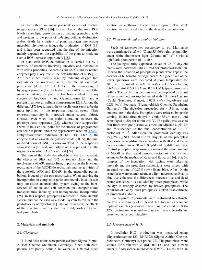
3.1. Time course of protoplast killing
The effects of BEA and T-2 at either 50 or 100 mM on
tomato protoplast viability are shown in Fig. 1. These toxin
concentrations were chosen for two reasons: (a) similar
concentrations are produced by various Fusarium species on
plant matrices [1,2] and (b) similar concentrations proved to
be toxic toward other bioassays [10,14].
Compared to the control, the protoplast viability was
87% after the first hour of incubation with 50 mM BEA, 70%
after 2 h and 50% after 3 h. After 6 h protoplast viability
was only 9% of that of the control (Fig. 1A).
The treatment with 50 mM T-2 only resulted in a
significant reduction in protoplast viability after 6 h
(Fig. 1A). A dose–response relationship was shown
by increasing the concentration of both toxins to 100 mM
(Fig. 1B). At this concentration, the reduction of protoplast
viability following treatment with BEA was already
statistically significant after 30 min, and that induced by
T-2 after 3 h.
Viable protoplasts, both in the control and after different
treatments, appeared spherical shaped with well-defined
chloroplasts. No alterations of protoplast morphology
occurred during the first 30 min of treatment with BEA or
during the first hour of treatment with T-2 (data not shown).
The toxic action of the two mycotoxins was shown by
Fig. 1. Effects of BEA or T-2 toxins on protoplast viability. The protoplasts were treated with 50 (A) or 100 mM (B) toxin. Values represent the mean of five
experiments GSE. * and ** indicate values significantly different from the control by Student’s t test with P!0.05 and 0.01, respectively.
C. Paciolla et al. / Physiological and Molecular Plant Pathology 65 (2004) 49–5652
the appearance of altered morphology of protoplasts, which
resulted in protoplast death. Dead protoplasts showed both
cytoplasm and chloroplast disruption with increasing
damage to the plasma membrane until complete lysis
(data not shown).
3.2. H2O2 production in treated protoplasts
To ascertain whether BEA and T-2 induced ROS
generation, the H2O2 production in control and treated
protoplasts was monitored. The production of extracellular
H2O2 induced by 50 mM BEA and T-2 was investigated at
different times of treatment (Fig. 2). The level of H2O2
increased both in BEA and T-2 with respect to the control
Fig. 2. H2O2 release in the incubation medium by tomato protoplasts treated
with 50 mM BEA or T-2. At the indicated times, 1 ml of protoplasts
suspension was taken for the determination of H2O2 concentration. Values
represent the mean of four experiments GSE.
(after 4 h by about 66 and 40% in BEA and T-2,
respectively). Both mycotoxins induced a similar increase
in H2O2 inside the cells, which was revealed by an increase
in fluorescence of rhodamine of the treated cells (data not
shown).
In addition to H2O2 determination, the oxidative damage
in T-2 treated protoplasts, was monitored by measuring lipid
peroxidation. As reported in Table 1, after 1 h of T-2
treatment a notable increase in lipid peroxidation occurred.
3.3. Determination of ascorbate and dehydroascorbate
While no significant difference in ASC and DHA contents
occurred in control protoplasts during the time of the
analysis, the treatment with 50 mM BEA resulted in a rapid
and sharp decrease in the ASC content in the protoplasts
(Fig. 3A). After 1 h of treatment the ASC content of BEA-
treated protoplasts was 66% lower than that of the control and
further decreases occurred with time (15 and 9% residual
content after 2 and 3 h, respectively). Conversely, BEA did
not induce a parallel increase in DHA content (Fig. 3B), the
difference of which from that of the control remained
insignificant throughout treatment. Consequently, the total
Table 1
Effect of 50 mM T-2 mycotoxin on lipid peroxidation
Time (h) Lipid peroxidation (nmol MDA) (1!106 protoplasts)K1
Control T-2
0.5 38.3G3.1 42.6G5.0
1 43.2G4.2 83.5G7.1**
3 50.4G6.1 157.1G11.3**
6 62.4G6.3 142.3G13.1**
Values represent the mean of four experiments GSE. ** indicates values
significantly different from the control by Student’s t test with P!0.01.
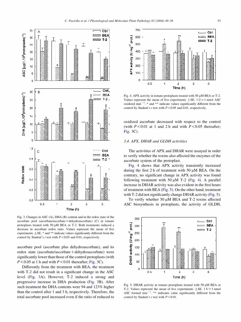
Fig. 3. Changes in ASC (A), DHA (B) content and in the redox state of the
ascorbate pool (ascorbate/ascorbateCdehydroascorbate) (C) in tomato
protoplasts treated with 50 mM BEA or T-2. Both treatments induced a
decrease in ascorbate redox ratio. Values represent the mean of five
experiments GSE. * and ** indicate values significantly different from the
control by Student’s t test with P!0.05 and 0.01, respectively.
Fig. 4. APX activity in tomato protoplasts treated with 50 mM BEA or T-2.
Values represent the mean of five experiments GSE. 1 UZ1 nmol ASC
oxidized minK1. * and ** indicate values significantly different from the
control by Student’s t test with P!0.05 and 0.01, respectively.
Fig. 5. DHAR activity in tomato protoplasts treated with 50 mM BEA or
T-2. Values represent the mean of five experiments GSE. 1 UZ1 nmol
ASC formed minK1. ** indicates value significantly different from the
control by Student’s t test with P!0.01.
C. Paciolla et al. / Physiological and Molecular Plant Pathology 65 (2004) 49–56 53
ascorbate pool (ascorbate plus dehydroascorbate), and its
redox state (ascorbate/ascorbateCdehydroascorbate) were
significantly lower than those of the control protoplasts (with
P!0.05 at 1 h and with P!0.01 thereafter; Fig. 3C).
Differently from the treatment with BEA, the treatment
with T-2 did not result in a significant change in the ASC
level (Fig. 3A). However, T-2 induced a strong and
progressive increase in DHA production (Fig. 3B). After
such treatment the DHA contents were 94 and 123% higher
than the control after 1 and 3 h, respectively. Therefore, the
total ascorbate pool increased even if the ratio of reduced to
oxidized ascorbate decreased with respect to the control
(with P!0.01 at 1 and 2 h and with P!0.05 thereafter;
Fig. 3C).
3.4. APX, DHAR and GLDH activities
The activities of APX and DHAR were assayed in order
to verify whether the toxins also affected the enzymes of the
ascorbate system of the protoplast.
Fig. 4 shows that APX activity transiently increased
during the first 2 h of treatment with 50 mM BEA. On the
contrary, no significant change in APX activity was found
following treatment with 50 mM T-2 (Fig. 4). A parallel
increase in DHAR activity was also evident in the first hours
of treatment with BEA (Fig. 5). On the other hand, treatment
with T-2 did not significantly change DHAR activity (Fig. 5).
To verify whether 50 mM BEA and T-2 toxins affected
ASC biosynthesis in protoplasts, the activity of GLDH,

Table 2
GLDH activity in tomato protoplasts treated with 50 mM BEA or T-2
Time (h) Galactono-1,4-lactone dehydrogenase (nmol cyto-
chrome c reduced minK1 mgK1 prot.)
Control BEA T-2
0.5 73G6 65G5 79G7
1 65G5 36G3** 90G9*
3 57G6 30G3** 85G9*
6 51G5 13G2** 75G7**
Values represent the mean of five experiments GSE. * and ** indicate
values significantly different from the control by Student’s t test with
P!0.05 and 0.01, respectively.
C. Paciolla et al. / Physiological and Molecular Plant Pathology 65 (2004) 49–5654
the enzyme which catalyzes the final step of ASC
biosynthesis, was analyzed. Table 2 shows that the GLDH
activity in T-2 treated protoplasts was significantly higher
than that of the control protoplasts after 1 h, and remained
constant thereafter. On the contrary, the treatment with BEA
induced a progressive decrease in GLDH activity (Table 2).
3.5. Other antioxidants
To evaluate whether alternative antioxidant compounds
were affected by toxin treatments, the levels of reduced and
oxidized glutathione as well as catalase activity were
analyzed. No significant difference in GSH and GSSG
contents (data not shown) were found either with BEA or
with T-2 treatment with respect to the control. No significant
change in catalase activity was observed in protoplasts
treated with T-2, while BEA treatment resulted in a sharp
decrease in catalase activity (Table 3).
4. Discussion
The data reported in this study indicate that both toxins
induce premature protoplast death but the phytotoxic action
of BEA is higher than that of T-2, as shown by the more
remarkable and rapid reduction of protoplast viability
occurring under BEA-treatment (Fig. 1A and B). The cell
death caused by 50 mM BEA on tomato protoplasts was
almost complete within 6 h of treatment, while that of T-2 at
the same time and concentration was much lower. The
phytotoxicity of BEA has been reported to be effective at
Table 3
Catalase activity in tomato protoplasts treated with 50 mM BEA or T-2
Time (h) Catalase (nmol H2O2 dismutated min K1 mgK1 prot.)
Control BEA T-2
0.5 95G8 84G7 89G8
1 89G9 43G3** 79G7
3 80G7 10G1** 74G6
6 51G5 8G1** 61G6
Values represent the mean of five experiments GSE. ** indicates values
significantly different from the control by Student’s t test with P!0.01.
micromolar concentrations in other plant and animal
systems [8,10]. It is known that BEA acts on biological
membranes as an efficient ionophor for monovalent and
divalent cations [35,36]. Indeed, in the presence of this
mycotoxin, cells undergo an upsetting of the cellular cations
(e.g. KC and NaC, CaCC) some of which play important
roles in metabolism regulation [37].
Even if many phytotoxins induce ROS generation, which
in turn causes an oxidative stress in the phytotoxin treated
cells [17], there is no clear evidence suggesting that BEA
and T-2 activate ROS generating systems. We show that
BEA and T-2 toxin produce increased levels of H2O2
(Fig. 2). Thus, our results suggest the involvement of an
oxidative stress in the damage caused by T-2 and BEA.
Moreover, the increase in DHA and in lipid peroxidation
observed under T-2 treatments clearly indicates that the
treated protoplasts undergo oxidative damage (Fig. 3B and
Table 1). The defence response activated in tomato
protoplasts by the presence of T-2, should be an increase
in ASC biosynthesis, since a rise in the total pool of vitamin
C (ASCCDHA) and an increase in GLDH activity occurs
(Fig. 3A and B and Table 2), whereas the activity of APX
and DHAR are not affected at all (Figs. 4 and 5). The
increase in DHA content within cells could have negative
metabolic effects contributing to the damage induced by this
mycotoxin. Indeed it has been demonstrated that a rise in the
DHA content induces the oxidation of several thiol-
containing proteins and hence inhibits many enzymes
[38]. Interestingly, one of the effects of T-2 is to bind
proteic –SH groups, a process that inactivates certain thiol-
containing enzymes [39,40]. Thus the increase in DHA
generation could strengthen the T-2 damaging effect on –SH
containing proteins.
In the case of BEA toxicity, the presence of oxidative
stress is also supported by an increase in APX activity that
is, however, only transient (Fig. 4). The more severe
damage induced by this toxin on tomato protoplasts is also
reflected by its effect on the ASC system. The strong
depletion in ASC does not seem to be due to a net increase in
its oxidation, since no rise in DHA was observed (Fig. 3B).
The higher ASC oxidation due to the increase in APX
activity is probably compensated by the parallel increase in
DHAR. Due to the fact that BEA affects cell permeability,
we can speculate that a certain amount of ASC is lost from
the protoplast treated with this phytotoxin. The release of
ASC from the cytosol to the apoplast could have some
interesting repercussions for the penetration of phytopatho-
gens in plant tissues. Indeed, it has been reported that in the
cell wall, the presence of ASC causes the production of OH,
the most powerful radical generated in biological systems
[41]. This reactive species causes the loosening of the plant
cell wall by non-enzymatic scission of the matrix poly-
saccharides [42]. Therefore, the release of ASC from the
cytosol to the apoplast induced by BEA could facilitate
pathogen penetration by increasing cell wall plasticity.
Alternatively, we can also speculate that the ionophoric
C. Paciolla et al. / Physiological and Molecular Plant Pathology 65 (2004) 49–56 55
action of BEA [43] interferes with ASC biosynthesis by
altering the mitochondrial membranes. In fact, GLDH, the
last enzyme of ASC biosynthesis is located in the inner
mitochondrial membrane and, moreover, its activity
requires a functional mitochondrial electron flow [44].
As regards the GSH system, in contrast to ASC, the
preliminary data of this study seem to indicate that the
involvement of this system in the defence response of
protoplasts versus phytotoxicity of BEA and T-2 mycotox-
ins is not important. However, further investigations are
necessary to support this thesis.
As far as the ROS-scavenging enzyme catalase is
concerned, the decrease in activity induced by BEA
treatment contributes to the maintaining of high H2O2
concentrations and to protoplast death. In this respect T-2 is
less effective since it does not induce any change in catalase
activity compared to control.
In conclusion, our data suggest that both BEA and T-2
cause protoplast damage, inducing their death, although
differently affecting plant cell metabolism. In both cases the
ascorbate system is altered, thus confirming the relevance of
this metabolite in the perception and signal transduction of
unfavourable environmental conditions [45,46]. Our results
also confirm that the protoplast can be a useful tool for
studying mycotoxin phytotoxicity and for ascertaining
whether and how a single metabolite affects cellular
metabolism in plants.
Acknowledgements
The authors thank Mrs L. Bianco (DPBP, University of
Bari) for her valuable technical assistance.
References
[1] Marasas WFO, Nelson PE, Toussoun TA. Toxigenic Fusarium
species: identity and mycotoxicology. Philadelphia, PA: Pennsylvania
State University Press; 1984.
[2] Logrieco A, Moretti A, Castella G, Kosteck M, Golinski P, Ritieni A,
et al. Beauvericin production by Fusarium species. Appl Environ
Microbiol 1998;64:3084–8.
[3] Vey A, Quiot J-M, Vago C. Mise en evidence et etude de l’action dune
mycotoxine, la beauvericine, dur des cellules d’insectes cultivees in
vitro. Comp Rend Acad Sci Paris 1973;276:2489–92.
[4] Hamil RL, Higgens CE, Boaz HE, Gorman M. The structure of
beauvericin, a new depsipeptide antibiotic toxic to Artemia salina.
Tetrahedron Lett 1969;49:4255–8.
[5] Ojcius DM, Zychlinsky A, Zheng LM, Young D-EJ. Ionophore-
induced apoptosis: role of DNA fragmentation and calcium fluxes.
Exp Cell Res 1991;197:43–9.
[6] Tomoda H, Huang X-H, Cao J, Nishida H, Nagao R, Okuda S, et al.
Inhibition of acyl-CoA: cholesterol acyltransferase activity by
cyclodepsipeptide antibiotics. J Antibiot (Tokyo) 1992;45:1626–32.
[7] Gupta S, Kranoff SB, Underwood NL, Renwick JAA, Roberts DW.
Isolation of beauveuricin as an insect toxin from Fusarium semitectum
and Fusarium moniliforme var. subglutinans. Mycopathologia 1991;
115:185–9.
[8] Que FG, Gores GJ, La Russo NF. Development and initial application
of an in vitro model of apoptosis in rodent cholangiocytes. Am
J Physiol 1997;2772:G106–G15.
[9] Moretti A, Belisario A, Tafuri A, Ritieni A, Corazza L, Logrieco A.
Production of beauvericin by different races of Fusarium oxysporum
F. sp. melonis, the Fusarium wilt agent of muskmelon. Eur J Plant
Pathol 2002;108:661–6.
[10] Sagakuchi M, Moretti A, Endo E, Matsuda Y, Toyoda H, Ouchi S. An
approach to the use of plant sensitivity for simple detection of
mycotoxin. Proceedings of First Asian Conference of Plant Pathology,
Kuala Lumpur, Malaysia; August 2000. p. 262–79.
[11] Karppanen E, Rizzo A, Saari L, Berg S, Bostrom H. Investigation on
trichothecene stimulated lipid peroxidation and toxic effects of
trichothecenes in animals. Acta Veter Scand 1989;30:391–9.
[12] Rizzo AF, Atroshi F, Ahotupa M, Sankari S, Elovaara E. Protective
effect of antioxidants against free-radical mediated lipid peroxidation
induced by DON or T-2 toxin. J Vet Med Assoc 1994;41:81–90.
[13] Jelinek CF, Pohland AE, Wood G. Worldwide occurrence of
mycotoxins in food and feeds—an update. J Assoc Off Anal Chem
1989;72:223–30.
[14] Marasas WFO, Smalley EB, Bamburg JR, Strong FM. Phytotoxicity
of T-2 toxin produced by Fusarium tricinctum. Phytopathology 1971;
61:1488–91.
[15] Madhyastha MS, Marquardt RR, Abramson D. Structure–activity
relationships and interactions among trichothecene mycotoxins as
assessed by yeast bioassay. Toxicon 1994;32:1147–52.
[16] Mittler R. Oxidative stress, antioxidants and stress tolerance. Trends
Plant Sci 2002;7:405–10.
[17] Heiser I, Oßwald W, Elstner EF. The formation of reactive oxygen
species by fungal and bacterial phytotoxins. Plant Physiol Biochem
1998;36:703–13.
[18] De Gara L, de Pinto MC, Tommasi F. The antioxidant systems vis-a-
vis reactive oxygen species during plant–pathogen interaction. Plant
Physiol Biochem 2003;41:863–70.
[19] Noctor G, Foyer CH. Ascorbate and glutathione: keeping active
oxygen under control. Annu Rev Plant Physiol Plant Mol Biol 1998;
49:249–79.
[20] Paciolla C, De Gara L, De Tullio MC, Arrigoni O. Distribution of
cytosolic ascorbate peroxidase in angiosperms. Giornale Botanico
Italiano 1996;130:729–37.
[21] Shigeoka S, Ishikawa T, Tamoi M, Miyagawa Y, Takeda T, Yabuta Y,
et al. Regulation and function of ascorbate peroxidase isoenzymes.
J Exp Bot 2002;53:1305–19.
[22] Karpinski S, Escobar C, Karpinska B, Creissen G, Mullineaux PM.
Photosynthetic electron transport regulates the expression of cytosolic
ascorbate peroxidase genes in Arabidopsis during excess light stress.
Plant Cell 1997;9:627–40.
[23] de Pinto MC, Tommasi F, De Gara L. Changes in the antioxidant
systems as part of the signaling pathway responsible for the
programmed cell death activated by nitric oxide and reactive oxygen
species in tobacco Bright-Yellow 2 cells. Plant Physiol 2002;130:
698–708.
[24] Asada K. The water–water cycle in chloroplasts: scavenging of active
oxygens and dissipation of excess photons. Annu Rev Plant Physiol
Plant Mol Biol 1999;50:601–39.
[25] March G, Tremolieres A. Protoplast growth and photoregulation. In:
Pilet PE, editor. The physiological properties of plant protoplast. New
York: Springer; 1985. p. 258–66.
[26] Kanai R, Edwards GE. Purification of enzymatically isolated
mesophyll protoplasts from C3, C4 and Crassulacean acid metabolism
plants using an aqueous dextran-polythylene glycol two-phase
system. Plant Physiol 1973;52:484–90.
[27] Royall JA, Ischiropoulos H. Evaluation of 2 0,7 0-dichlorofluorescein
and dihydrorhodamine 123 as fluorescent probes for intracellular
H2O2 in cultured endothelial cells. Arch Biochem Biophys 1993;302:
348–55.
C. Paciolla et al. / Physiological and Molecular Plant Pathology 65 (2004) 49–5656
[28] Bellincampi D, Dipierro N, Salvi G, Cervone F, De Lorenzo G.
Extracellular H2O2 induced by oligogalacturonides is not involved in
the inhibition of the auxine-regulated rolB gene expression in tobacco
leaf explants. Plant Physiol 2000;122:1379–85.
[29] Zhang J, Kirkham B. Antioxidant responses to drought in sunflower
and sorghum seedlings. New Phytologist 1996;132:361–73.
[30] De Gara L, Paciolla C, De Tullio M, Motto M, Arrigoni O. Ascorbate-
dependent hydrogen peroxide detoxification and ascorbate regener-
ation during germination of a high productive maize hybrid: evidence
of an improved detoxification mechanism against reactive oxygen
species. Physiol Plant 2000;109:7–13.
[31] Dipierro S, Borraccino G. Dehydroascorbate reductase from potato
tubers. Phytochemistry 1991;30:427–9.
[32] Beaumont F, Jouve HM, Gagnon J, Gaillard J, Pelmont J. Purification
and properties of a catalase from potato tubers (Solanum tuberosum).
Plant Sci 1990;72:19–26.
[33] Oba K, Seiko I, Nishikawa M, Mizuno H, Yamamoto T. Purification
and properties of L-Galactono-g-lactone dehydrogenase, a key
enzyme for ascorbic acid biosynthesis, from sweet potato roots.
J Biochem 1995;117:120–4.
[34] Bradford MM. A rapid and sensitive method for the quantitation of
microgram quantities of protein utilizing the principle of protein-dye
binding. Anal Biochem 1976;72:248–54.
[35] Dorschner E, Lardy H. Specificity of ion transport induced by
beaveuricin. Antimicrob Agents Chemother 1968;8:11–14.
[36] Hamilton JA, Steinrauf LK, Braden B. Beauvericin and divalent
cations: crystal structure of the barium complex. Biochem Biophys
Res Commun 1975;64:151–6.
[37] Lemmens-Gruber R, Rachoy B, Steininger E, Kouri K, Saleh P,
Krska R, et al. The effect of the Fusarium metabolite beauvericin on
electromechanical and physiological properties in isolated smooth and
heart muscle preparations of guinea pigs. Mycopathologia 2000;149:
5–12.
[38] Paciolla C, De Tullio MC, Chiappetta A, Innocenti AM, Bitonti MB,
Liso R, et al. Short- and long-term effects of dehydroascorbate in
Lupinus albus and Allium cepa roots. Plant Cell Physiol 2001;42:
857–63.
[39] Ueno Y, Matsumoto H. Inactivation of some thiol enzymes by
trichothecene mycotoxins from Fusarium species. Chem Pharma Bull
1975;23:2439–42.
[40] Ueno Y. Trichothecenes as environmental toxicant. Toxicology 1984;
5:1–15.
[41] Fry SC. Oxidative scission of plant cell wall polysaccharides by
ascorbate-induced hydroxyl radicals. Biochem J 1998;332:507–15.
[42] Schopfer P. Hydroxyl radical-induced cell-wall loosening in vitro and
in vivo: implications for the control of elongation growth. Plant J
2001;28:679–88.
[43] Kouri K, Lemmens M, Lemmens-Gruber R. Beauvericin-induced
channels in ventricular myocytes and liposomes. Biochim Biophys
Acta Biomembr 2003;1609:203–10.
[44] Millar H, Mittova V, Kiddle G, Heazlewood JL, Bartoli CG,
Theodoulou FL, et al. Control of ascorbate synthesis by respiration
and its implications for stress responses. Plant Physiol 2003;33:443–7.
[45] de Pinto MC, Tommasi F, De Gara L. Enzymes of the ascorbate
biosynthesis and ascorbate–glutathione cycle in cultured cells of
tobacco Bright Yellow 2. Plant Cell Physiol 2000;38:541–50.
[46] Foyer CH, Noctor G. Redox sensing and signalling associated with
reactive oxygen in chloroplasts, peroxisomes and mitochondria.
Physiol Plant 2003;119:355–64.