The melatonergic system in mood and anxiety disorders and the role of agomelatine: implications for...
26
Int. J. Mol. Sci. 2013, 14, 12458-12483; doi:10.3390/ijms140612458 International Journal of Molecular Sciences ISSN 1422-0067 www.mdpi.com/journal/ijms Review The Melatonergic System in Mood and Anxiety Disorders and the Role of Agomelatine: Implications for Clinical Practice Domenico De Berardis 1,2, *, Stefano Marini 1,2 , Michele Fornaro 3 , Venkataramanujam Srinivasan 4 , Felice Iasevoli 5 , Carmine Tomasetti 5 , Alessandro Valchera 6 , Giampaolo Perna 7,8,9 , Maria-Antonia Quera-Salva 10 , Giovanni Martinotti 2 and Massimo di Giannantonio 2 1 National Health Service, Department of Mental Health, Psychiatric Service of Diagnosis and Treatment, Hospital “G. Mazzini”, ASL 4 Teramo, Italy; E-Mail: [email protected] 2 Department of Neuroscience and Imaging, Chair of Psychiatry, University “G. D’Annunzio”, Chieti 66013, Italy; E-Mails: [email protected] (G.M.); [email protected] (M. G.) 3 Department of “Scienze della Formazione”, University of Catania, Catania 95121, Italy; E-Mail: [email protected] 4 Sri Sathya Sai Medical Educational and Research Foundation, Medical Sciences Research Study Center, Prasanthi Nilayam, 40-Kovai Thirunagar Coimbatore, Tamilnadu 641014, India; E-Mail: [email protected] 5 Laboratory of Molecular Psychiatry and Psychopharmacotherapeutics, Section of Psychiatry, Department of Neuroscience, University School of Medicine “Federico II”, Naples 80131, Italy; E-Mails: [email protected] (F.I.); [email protected] (C.T.) 6 Hermanas Hospitalarias, FoRiPsi, Villa S. Giuseppe Hospital, Ascoli Piceno 63100, Italy; E-Mail: [email protected] 7 Hermanas Hospitalarias, FoRiPsi, Department of Clinical Neurosciences, Villa San Benedetto Menni, Albese con Cassano, Como 22032, Italy; E-Mail: [email protected] 8 Department of Psychiatry and Behavioral Sciences, Leonard Miller School of Medicine, University of Miami, 33124 Miami, USA 9 Department of Psychiatry and Neuropsychology, University of Maastricht, 6200 MD Maastricht, The Netherlands 10 AP-HP Sleep Unit, Department of Physiology, Raymond Poincaré Hospital, Garches 92380, France; E-Mail: [email protected] * Author to whom correspondence should be addressed; E-Mail: [email protected]; Tel.: +39-0861429708; Fax: +39-0861429706. OPEN ACCESS
-
Upload
humanitas-research -
Category
Documents
-
view
1 -
download
0
Transcript of The melatonergic system in mood and anxiety disorders and the role of agomelatine: implications for...
Int. J. Mol. Sci. 2013, 14, 12458-12483; doi:10.3390/ijms140612458
International Journal of
Molecular Sciences ISSN 1422-0067
www.mdpi.com/journal/ijms
Review
The Melatonergic System in Mood and Anxiety Disorders and the Role of Agomelatine: Implications for Clinical Practice
Domenico De Berardis 1,2,*, Stefano Marini 1,2, Michele Fornaro 3,
Venkataramanujam Srinivasan 4, Felice Iasevoli 5, Carmine Tomasetti 5, Alessandro Valchera 6,
Giampaolo Perna 7,8,9, Maria-Antonia Quera-Salva 10, Giovanni Martinotti 2 and
Massimo di Giannantonio 2
1 National Health Service, Department of Mental Health, Psychiatric Service of Diagnosis and Treatment,
Hospital “G. Mazzini”, ASL 4 Teramo, Italy; E-Mail: [email protected] 2 Department of Neuroscience and Imaging, Chair of Psychiatry, University “G. D’Annunzio”,
Chieti 66013, Italy; E-Mails: [email protected] (G.M.);
[email protected] (M. G.) 3 Department of “Scienze della Formazione”, University of Catania, Catania 95121, Italy;
E-Mail: [email protected] 4 Sri Sathya Sai Medical Educational and Research Foundation, Medical Sciences Research Study
Center, Prasanthi Nilayam, 40-Kovai Thirunagar Coimbatore, Tamilnadu 641014, India;
E-Mail: [email protected] 5 Laboratory of Molecular Psychiatry and Psychopharmacotherapeutics, Section of Psychiatry,
Department of Neuroscience, University School of Medicine “Federico II”, Naples 80131, Italy;
E-Mails: [email protected] (F.I.); [email protected] (C.T.) 6 Hermanas Hospitalarias, FoRiPsi, Villa S. Giuseppe Hospital, Ascoli Piceno 63100, Italy;
E-Mail: [email protected] 7 Hermanas Hospitalarias, FoRiPsi, Department of Clinical Neurosciences,
Villa San Benedetto Menni, Albese con Cassano, Como 22032, Italy; E-Mail: [email protected] 8 Department of Psychiatry and Behavioral Sciences, Leonard Miller School of Medicine,
University of Miami, 33124 Miami, USA 9 Department of Psychiatry and Neuropsychology, University of Maastricht, 6200 MD Maastricht,
The Netherlands 10 AP-HP Sleep Unit, Department of Physiology, Raymond Poincaré Hospital, Garches 92380,
France; E-Mail: [email protected]
* Author to whom correspondence should be addressed; E-Mail: [email protected];
Tel.: +39-0861429708; Fax: +39-0861429706.
OPEN ACCESS
Int. J. Mol. Sci. 2013, 14 12459
Received: 17 March 2013; in revised form: 22 May 2013 / Accepted: 22 May 2013 /
Published: 13 June 2013
Abstract: Melatonin exerts its actions through membrane MT1/MT2 melatonin receptors,
which belong to the super family of G-protein-coupled receptors consisting of the typical
seven transmembrane domains. MT1 and MT2 receptors are expressed in various tissues of
the body either as single ones or together. A growing literature suggests that the
melatonergic system may be involved in the pathophysiology of mood and anxiety
disorders. In fact, some core symptoms of depression show disturbance of the circadian
rhythm in their clinical expression, such as diurnal mood and other symptomatic variation,
or are closely linked to circadian system functioning, such as sleep-wake cycle alterations.
In addition, alterations have been described in the circadian rhythms of several biological
markers in depressed patients. Therefore, there is interest in developing antidepressants
that have a chronobiotic effect (i.e., treatment of circadian rhythm disorders). As melatonin
produces chronobiotic effects, efforts have been aimed at developing agomelatine, an
antidepressant with melatonin agonist activity. The present paper reviews the role of the
melatonergic system in the pathophysiology of mood and anxiety disorders and the clinical
characteristics of agomelatine. Implications of agomelatine in “real world” clinical practice
will be also discussed.
Keywords: melatonin; melatonergic receptors; serotonin; dopamine; noradrenaline;
agomelatine; major depression; anxiety
1. Anatomy and Physiology of the Brain Melatonergic System
The neurohormone melatonin (N-acetyl-5-methoxytryptamine) is prominently, albeit not exclusively,
synthesized in the pineal gland and is secreted in a phasic manner, since its circulating levels vary in a
daily cycle. Melatonin derives from the precursor tryptophan (taken up from circulating blood) by
subsequent steps, implicating tryptophan transformation in serotonin, N-acetylserotonin and, finally, in
melatonin [1]. Transformation of serotonin in melatonin is regulated by the light-dark cycle, since
enzymatic activity of N-acetyltransferase (the rate limiting biosynthetic enzyme) is low during daytime
or under exposure to light stimuli and higher during dark phases.
Once synthesized, melatonin is released both in the cerebrospinal fluid and in the capillary, which
distributes the hormone in multiple body tissues [2]. Melatonin exerts pleiotropic actions on several
body compartments and organs; however, these actions are beyond the scope of this review, and the
reader is referred to other reviews for more information [3]. Melatonin is regarded as a “chronobiotic”
hormone, since it regulates several chronobiological actions and is, in particular, responsible for
circadian phase shifting. However, under conditions of total darkness (especially in the Arctic or
Antarctic winter, where there is effectively no sunlight for several months), melatonin may still exhibit
a diurnal rhythm, albeit that the rhythms become desynchronized. The melatonin-mediated effects are,
Int. J. Mol. Sci. 2013, 14 12460
in turn, under the control of a group of hypothalamic nuclei, mostly the suprachiasmatic nucleus
(SCN), which is considered an endogenous circadian pacemaker [4,5]. In seasonal breeders,
moreover, melatonin effects are also mediated by the pars tuberalis (PT), which is implicated in
photoperiodically-regulated reproduction and the premammillary hypothalamus.
Among the more relevant biological functions exerted by melatonin are the control of the
sleep-wake cycle, the modulation of the immune system (including anti-inflammatory properties) and of
energy metabolism [6]. Melatonin interacts with at least two receptor types, named MT1 and
MT2 [7–11], although adjunctive binding sites have also been described [12,13].
Melatonin receptors belong to the class of G-protein-coupled receptors and are primarily expressed
in the CNS; however, they are also widely distributed in other body tissues, together and separately.
Within the CNS, the MT1 receptor is prominently expressed in the SCN, the hippocampus, the retina,
the caudate putamen, the nucleus accumbens, the substantia nigra and the ventrotegmental area [7,14].
Notably, most of these areas belong to the central dopaminergic pathways, suggesting a tight
correlation between the melatonergic and monoaminergic systems, at least the dopaminergic one. MT1
receptors are also found in several other hypothalamic nuclei and brain areas, such as the
paraventricular nucleus, the periventricular nucleus, the supraoptic nucleus, the diagonal band of
Broca, the Meynert nucleus, the tuberomammillary nucleus and the mammillary bodies [14]. The MT2
receptor has been mostly found in the hippocampus, the SCN and the retina [7]. Outside the CNS,
MT1 receptors are thoroughly distributed in several tissues, while expression of MT2 receptors is more
restricted [7]. Expression of both MT1 and MT2 receptors has also been reported in neurons and glial
cells of the cortex, thalamus and cerebellar cortex. Moreover, both receptors are expressed in the
pineal gland [15], which is consistent with reported melatonin autocrine and paracrine actions.
Intriguingly, expression of melatonin receptors in both central and peripheral tissues is affected on a
circadian basis, since mRNA expression of the MT1 receptor has been found to be increased in rodents
during daytime [16]. Binding to the MT1 receptor was also found to be increased during the daytime
and by light exposure during the nighttime, while MT1 mRNA expression was reduced by this latter
procedure [15]. Despite an increase in mRNA expression during the daytime, however, surface
expression of the melatonergic receptor in SCN neurons has been reported to be very low during the
day and high at night, paralleling melatonin’s trough and peak [17]. These findings suggest that the
levels of circulating melatonin elicit a feedback regulation on the surface receptor amount, causing a
downregulation of receptors with blood peaks. At the same time, surface receptors are more expressed
when melatonin levels are expected to be higher and vice versa. This group of feedback regulations
allows the system to preserve its homeostasis and to provide fine-regulation with external light-dark
stimuli. However, regulation of melatonin receptor expression is under multiple other biological
factors, e.g., estradiol levels, which cooperate to modulate melatonergic signaling in its different steps.
MT1 receptor signaling occurs mainly through interaction with inhibitory G-proteins and
subsequent reduction of intracellular cAMP levels, a decrease in protein kinase A activity and reduced
phosphorylation of the transcription factor, CREB [18]. However, in an artificial system, i.e., cultured
cells, melatonin has also been demonstrated to increase cAMP levels via interaction with the MT1
receptor [19], although the actual relevance of these in vivo observations remains questionable.
MT2 receptors couple to multiple and diverse transduction pathways, depending on the biological
system taken into consideration and ranging from inhibition of cAMP synthesis to the increase of
Int. J. Mol. Sci. 2013, 14 12461
protein kinase C activity or from inhibition of guanylyl cyclase signaling to increase cGMP
levels [20–22].
At the subcellular level, melatonin modulates the activity of a number of ion channels and affects
intracellular ion levels [23,24]. The hypothalamic SCN and the hippocampus are two major sites of
melatonin action in the CNS. The SCN activity is inhibited by melatonin via MT1 receptors [25],
mostly during the daytime, when the SCN neuronal activity is higher. However, melatonergic
inhibition of SCN activity is blunted by melatonin itself through the regulation of surface receptor
expression [26] by their desensitization. Desensitization occurs after exposure to melatonin by a
beta-arrestin-1-dependent mechanism [27], requiring a phosphorylation on the MT1 and MT2 receptor
C-terminal [28]. Melatonergic receptor desensitization is mainly associated to receptor downregulation.
Prevention of melatonergic receptor desensitization has been obtained by depolymerizing microtubules
and the blockade of receptor internalization [29]. However, in SCN cells, exposure to physiological
concentrations of melatonin caused the reversible desensitization and downregulation of MT2
receptors and the desensitization of MT1 receptors, however, without their downregulation [30].
Therefore, multiple molecular mechanisms may concur to diminish melatonin-mediated signaling.
In the hippocampus, melatonin has been reported to increase the firing rate of CA1 neurons through
activation of MT2 receptors [30]. In hippocampal slices, melatonin disrupted long-term potentiation in
a dose-dependent fashion [30]. Both effects were prevented by application of a MT2 receptor
antagonist and were lacking in MT2 gene knock-out mice, but not in MT1 defective mice [31].
Melatonergic signaling via the MT2 receptor in hippocampus has been described to substantially affect
cognitive behavior in preclinical paradigms. Knock-out mice for the MT2 gene were found to perform
worse than wild-type littermates at the elevated plus-maze behavioral task, a procedure investigating
learning and memory abilities [32], possibly implying an impairment in hippocampal-mediated
synaptic plasticity. It has also been demonstrated that agomelatine, a potent melatonin receptor agonist
drug that strongly binds to and stimulates the activity of melatonin MT1 and MT2 receptors, showed
cognitive enhancing properties, at least in preclinical studies [33,34].
The physiology of melatonergic signaling has been elucidated only in part. In mammals, melatonin
exerts a main action on phase shifting, which reflects a feedback loop between the pineal gland and the
endogenous circadian pacemaker, i.e., the SCN [3]. Within this feedback loop, melatonin controls the
amplitude and phase of circadian oscillation. Namely, the hormone exerts phase shifting via MT2
receptors and affects neuronal firing by MT1 receptors [35]. In functional agreement with these actions,
melatonin also favors sleep initiation by a number of mechanisms, including MT1 receptor-mediated
effects on the hypothalamic sleep switch [36] and modulation of discrete thalamic projections to the
cortex that are implicated in sleep stage transitions [37]. Moreover, melatonin also takes part in
sedating and anxiolytic effects.
Recently, Ochoa-Sanchez et al. [38] demonstrated that the melatonin MT2 receptor was involved in
the regulation of non-REM (NREM) slow wave sleep (SWS), and the MT2 selective agonist,
UCM765, increased NREM by activating the reticular thalamus neurons, where the MT2 receptors are
located. The melatonin MT2 receptor also mediates anxiety function [39]. On the other hand,
it has been demonstrated that the melatonin MT1 receptor is mostly involved in the control of
REM sleep [40].
Int. J. Mol. Sci. 2013, 14 12462
2. Interactions between Melatonergic System and Monoaminergic Systems
The main role of the pineal gland is to produce melatonin in response to the absence of light stimuli,
which may, in turn, activate a glutamate-mediated response from retinal receptors to SCN GABAergic
neurons, thereby generating an environment-to-endocrine input translation that is at the basis of
circadian rhythms in humans [41]. Located in the middle of the brain, although externally to the
blood-brain barrier, the pineal gland represents a powerful triage organ, where neurotransmission
signals from the SCN are converted to endocrine secretion, which, in turn, may regulate other
monoaminergic neurotransmitter systems, such as dopamine, norepinephrine and serotonin. For
instance, recent reports demonstrated that chronic melatonin treatment in animal models of aging may
help normalize levels of all monoamines, such as dopamine, serotonin and norepinephrine, thus
contrasting age-related impairment in catecholamines neurotransmission [42].
Here, we review the complex interconnections between catecholaminergic systems and melatonin
neuroendocrine secretion.
2.1. Serotonin Is the Main Controller of Circadian Clocks
Melatonin secretion is obviously tightly dependent on the availability of serotonin in pinealocytes.
Since serotonin is the precursor of melatonin, this neurotransmitter is, indeed, the principal actor of the
light/dark circadian regulation of melatonin secretion [43]. The SCN contains a hyper-dense
serotonergic terminal plex [44]. Early studies demonstrated that serotonin administration or
serotonergic agonist agents may phase-shift the circadian SCN pacemaker [45]. Moreover, lesions in
the raphe nuclei may cause a decrease in circadian amplitude or a phase change [43]. Several studies
demonstrated multiple mechanisms of modulation by serotonin receptors on the circadian clock.
Serotonin, indeed, may postsynaptically increase potassium currents in a subset of SCN neurons, in
order to alter circadian phases [46]. Moreover, SCN-evoked currents may be presynaptically inhibited
by serotonin, through a direct reduction in excitatory stimuli originating from the retinal tract [42].
However, more recent reports demonstrated a more complex regulation of SCN rhythms by serotonin.
Indeed, serotonin agonists are able to phase-shift circadian clocks only when serotonin release has
been previously decreased or in in vitro environments devoid of serotonin concentrations. When
serotonergic agents are pre-applied to SCN neurons, a new application of serotonin is less able to
phase shift the circadian clock, thereby demonstrating that the SCN is affected by serotonin only,
depending on the pre-existent serotonergic signaling [47]. Substantially, serotonin may act as a
synchronizer with the same effects as light on SCN neurons [48]. Indeed, synchronization of circadian
clocks by both light and serotonin coincide with modulation of specific clock genes, Per1 and
Per2 [49–51]. However, synchronizing effects of serotonin on circadian rhythms have been
demonstrated to occur only at daytime. When administered at nighttime, serotonin may only modulate
light-induced phase-shifts in clock genes, but is not able to phase-shift clocks by itself [52]. Recent
studies demonstrated that the reasons for this “paradox” reside in the light/dark-dependent differential
expression of serotonergic-specific receptors in the SCN [53].
Emerging evidence supports the view that melatonin may also regulate serotonin secretion in a
retrograde circadian feedback loop. Serotonin, indeed, is also secreted by raphe nuclei with a clear
Int. J. Mol. Sci. 2013, 14 12463
downward trend during the night and an upward increase in the daytime [54]. Melatonin, on the other
hand, may inhibit raphe neuron firing, possibly through acting on MT1 receptors [55].
2.2. Norepinephrine Controls Limiting Steps of Enzymatic Melatonin Production
Several studies demonstrated that melatonin secretion is not tightly dependent upon light, since in
complete darkness, melatonin is produced by SCN-triggered stimulus. Circadian rhythms are
synchronized by light-dark cycles.
The limiting enzymatic step of melatonin production in pinealocytes is the N-acetylation of
serotonin by aryl-alkylamine-N-acetyl-transferase (AANAT), which has been demonstrated to increase
its functioning levels at night [56]. In fact, it is possible to assume that AANAT functioning is the real
circadian clock at the pineal gland level.
Several studies have demonstrated that the nocturnal increase in melatonin secretion is directly
related to norepinephrine nightly release by SCN to pineal gland. Norepinephrine, indeed, may
stimulate alpha- and beta-adrenergic pineal receptors, which, in turn, trigger a cAMP-dependent
increase in PKA intracellular activity that induces the CREB-mediated transcription of AANAT
protein [57]. This cascade provides a rapid increase in AANAT activity, which results in melatonin
formation in a 2 h time range after norepinephrine stimulus. However, other norepinephrine-related
mechanisms have been implicated in melatonin secretion control. Indeed MAPK activation by
adrenergic receptors has also been involved in AANAT pinealocytes responses [58]. Moreover,
adrenergic signaling has been demonstrated to modulate histone deacetylation and phosphorylation by
means of a MAPK-dependent mechanism, which suggests a further control on AANAT functioning
(for a review, see [59]).
Emerging evidence suggests that norepinephrine control of melatonin secretion may be under a
dynamic presynaptic control. Indeed, early studies demonstrated that, besides SCN innervation, the
pineal gland receives projections from sphenopalatine, otic and trigeminal ganglia [60], which have
been reported to secrete substance P [61]. Recent works demonstrated that pinealocytes are enriched
with substance P receptors [62] and that substance P may inhibit norepinephrine-induced AANAT
activation and melatonin secretion, though it does not impair basal levels of AANAT functioning [63].
As a further suggestion of presynaptic control of norepinephrine-mediated melatonin activity, Koch
and coworkers recently demonstrated that phytocannabinoids (e.g., tetrahydrocannabinol) application
to rat pineal gland cultures may reduce stimulation of melatonin secretion by norepinephrine [64].
Further studies by the same group recognized endocannabinoid receptors and metabolizing
enzymes in pinealocytes, thereby demonstrating a possible endocannabinoid-dependent control of
norepinephrine-stimulated melatonin secretion [65].
Conversely, some hormones, such as insulin, may enhance norepinephrine-mediated melatonin
synthesis and AANAT activity [66].
2.3. Melatonin-Dopamine Reciprocal Interactions: Molecular Bases for Neuropsychiatric
Disorder Pathophysiology
Emerging evidence supports the bi-univocal relation between melatonergic and dopaminergic
transmission. Dopamine, indeed, is present in sympathetic nerves projecting to the pineal gland, not
Int. J. Mol. Sci. 2013, 14 12464
only as a precursor of norepinephrine, but also as a neurotransmitter, which has been demonstrated to
have a crucial role in melatonin secretion control. Indeed, pinealocytes express high levels of
dopamine D4 receptors (D4R), whose expression has the singular ability of being dynamically
regulated in the pineal gland based on night/day circadian retinal synchronization [67]. Recent
evidence demonstrated that D4R expression is directly controlled by norepinephrine action at
alpha1- and beta1-adrenergic receptors, since beta-adrenergic agonists may increase D4Rs on the
pinealocyte cell surface [68]. Notably, D4R expression in pinealocytes seems to be controlled by a
“double gate” mechanism, which requires the thyroid hormone-mediated activation of II iodothyronine
deiodinase (Dio2), which transforms T4 into T3 active hormone, which acts simultaneously on the
adrenergic-mediated cAMP formation at the basis of D4R translation [67].
Therefore, dopamine seems to exert a complex modulatory control on melatonin synthesis, highly
dependent on light/dark cycles. Recent evidence demonstrated an entangled mechanism by which
dopamine may regulate norepinephrine-dependent melatonin secretion. D4R, indeed, may form
heteromeric complexes with both alpha1- and beta1-adrenergic receptors. These heteromers, whose
formation is controlled by light–dark circadian rhythms, may enable D4R to modulate the
adrenergic-mediated activation of MAPK and Akt cascades, which, in turn, promote melatonin
synthesis and secretion [69].
On the other hand, melatonin has been demonstrated to control dopamine signaling in selected
regions of the forebrain. Indeed, melatonin may inhibit dopamine release in hypothalamic areas
(tuberoinfundibular region), as well as in ventral hippocampus [70]. The decrease in dopamine
concentrations in these areas occurs simultaneously with a concurrent increasing in tyrosine
hydroxylase activity [71]. Melatonin seems also able to reduce cortical glutamate-mediated stimulation
of striatal responses [72]. These effects may be directly dependent on the reduction in dopamine
striatal release by nigrostriatal fibers induced by melatonin. Moreover, it has been demonstrated that
melatonin may directly reduce NMDA glutamate receptor functions in striatal neurons, thereby
reducing NMDA-mediated long-term synaptic responses in this brain region [73]. Other studies
demonstrated that melatonin may directly act on dopaminergic receptor functions in the brain. Indeed,
melatonin has been reported to increase dopamine D2 receptors (D2R) affinity in rat striatum [74]. In
contrast, melatonin may dose-dependently counteract the dopamine D1 receptors (D1R)-mediated
cAMP enhancement in neurons [75].
Melatonergic receptors (MT1, MT2) are widely distributed in the brain, above all, in hippocampus,
cortex, hypothalamus and cerebellum [76–78]. Moreover, melatonin receptors seem to have special
relations with dopaminergic systems. Indeed, MT1 receptors are localized in ventral tegmental
area and striatum (above all, nucleus accumbens shell). Surprisingly, these receptors have
light/dark-dependent expression, with high levels during the night and low levels during the day [14].
This specific connection between melatonin receptors and dopaminergic sites may have an essential
role in the pathophysiology of behavioral disorders that depends upon dopaminergic dysfunctions.
Indeed, it has been demonstrated that cocaine reward sensitization yields a critical dependence on
daylight, with pineal gland having a crucial role in this diurnal reward [79]. Moreover, pharmacological
doses of melatonin may block the cocaine-induced diurnal behavioral sensitization [80]. Inversely,
cocaine protracted administration may reduce MT1 receptor expression in striatum [81].
Int. J. Mol. Sci. 2013, 14 12465
Last, melatonin has been demonstrated to have protective effects in animal models of dopaminergic
dysfunctions, such as 6-hydroxydopamine-induced parkinsonisms [82] or rotenone-induced motor
disturbances [83].
Recently, a significant association of the MT1 receptor haplotype ATG has been reported in
antipsychotic-treated schizophrenic patients that do not develop tardive dyskinesia after prolonged
treatments [84].
3. Circadian Disturbances in Depression
The complex relationships between the endogenous circadian pacemaker and the development of
depressive symptoms are far from being elucidated [85]. The worsening of diurnal mood variation
(DMV) with the early morning is a classic symptom of the melancholic features of major depressive
disorder (MDD) and is one of the time-linked symptoms that has promoted speculation about the role
of the circadian system in its pathogenesis [86]. MDD seems to be related to a disruption in the central
circadian clock function and not to an alteration in a specific rhythm [85].
In addition, the type of rhythm abnormality seems to be highly variable in depressed patients,
including phase advance or phase delay of rhythms and increase or decrease in the rhythm
amplitude [87]. There is substantial evidence that circadian rhythms are more attenuated in MDD than
euthymic states, with decreased circadian amplitudes in core body temperature, motor activity,
thyroid-stimulating hormone, norepinephrine (NE) and cortisol, as was found in several studies [88].
These decreased amplitudes might result from the weakened output of the endogenous oscillator and
are one of the most relevant chronobiological abnormalities in depression that may be corrected by
antidepressant drugs [87,88]. A phase advance of the rhythm of cortisol, adrenocorticotropin, prolactin
and growth hormone has also been noted in depressed patients [85].
4. Chronobiotic Properties of Agomelatine
Agomelatine (Valdoxan®/Thymanax®) (S20098, N-[2-(7-methoxynaphth-1-yl)ethyl]acetamide) was
first reported in the literature in 1992, among a series of synthetic naphthalene melatonin analogs.
Various animal models of abrupt shifts and disorganization of the light-dark cycle, of free-running
conditions, as well as of delayed sleep-phase syndrome have shown that agomelatine accelerates the
resynchronization of circadian rhythms of locomotor activity and relevant biological parameters
(i.e., body temperature, secretions of hormones) [33]. The resynchronizing activity of agomelatine has
been mainly shown in both nocturnal (rats, mice, hamsters) and diurnal (Arvicanthis mordax) animals.
The ability of agomelatine to synchronize rest-activity rhythms in free-running animals requires the
integrity of the SCN [34].
The accelerating effect of agomelatine was particularly notable if treatment was started three weeks
prior to the induced phase shift [89,90]. Agomelatine treatment did not cause any major change in
corticosterone or adrenocorticotropic hormone concentrations, vasopressin, corticotropin-releasing
hormone and mineralocorticoid receptor mRNAs levels, which suggests that the mechanism of
agomelatine action is not related to hypothalamic-pituitary adrenocortical axis changes. This study
showed that agomelatine displays some characteristics of antidepressant drug action in the transgenic
mouse model, effects that could be partially related to its chronobiotic properties [85].
Int. J. Mol. Sci. 2013, 14 12466
5. Pharmacodynamics and Pharmacokinetics of Agomelatine
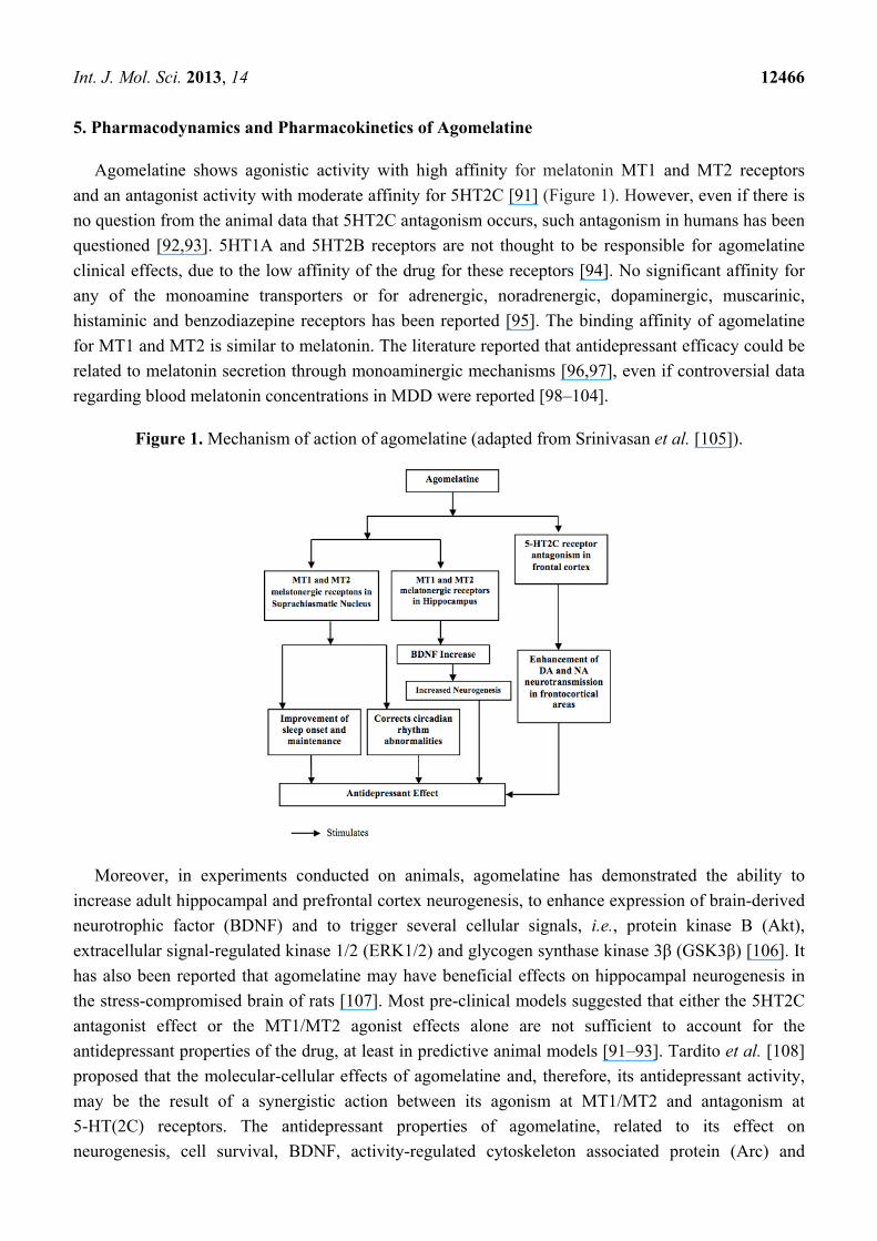
Agomelatine shows agonistic activity with high affinity for melatonin MT1 and MT2 receptors
and an antagonist activity with moderate affinity for 5HT2C [91] (Figure 1). However, even if there is
no question from the animal data that 5HT2C antagonism occurs, such antagonism in humans has been
questioned [92,93]. 5HT1A and 5HT2B receptors are not thought to be responsible for agomelatine
clinical effects, due to the low affinity of the drug for these receptors [94]. No significant affinity for
any of the monoamine transporters or for adrenergic, noradrenergic, dopaminergic, muscarinic,
histaminic and benzodiazepine receptors has been reported [95]. The binding affinity of agomelatine
for MT1 and MT2 is similar to melatonin. The literature reported that antidepressant efficacy could be
related to melatonin secretion through monoaminergic mechanisms [96,97], even if controversial data
regarding blood melatonin concentrations in MDD were reported [98–104].
Figure 1. Mechanism of action of agomelatine (adapted from Srinivasan et al. [105]).
Moreover, in experiments conducted on animals, agomelatine has demonstrated the ability to
increase adult hippocampal and prefrontal cortex neurogenesis, to enhance expression of brain-derived
neurotrophic factor (BDNF) and to trigger several cellular signals, i.e., protein kinase B (Akt),
extracellular signal-regulated kinase 1/2 (ERK1/2) and glycogen synthase kinase 3β (GSK3β) [106]. It
has also been reported that agomelatine may have beneficial effects on hippocampal neurogenesis in
the stress-compromised brain of rats [107]. Most pre-clinical models suggested that either the 5HT2C
antagonist effect or the MT1/MT2 agonist effects alone are not sufficient to account for the
antidepressant properties of the drug, at least in predictive animal models [91–93]. Tardito et al. [108]
proposed that the molecular-cellular effects of agomelatine and, therefore, its antidepressant activity,
may be the result of a synergistic action between its agonism at MT1/MT2 and antagonism at
5-HT(2C) receptors. The antidepressant properties of agomelatine, related to its effect on
neurogenesis, cell survival, BDNF, activity-regulated cytoskeleton associated protein (Arc) and
Int. J. Mol. Sci. 2013, 14 12467
stress-induced glutamate release, are due to this synergistic action. Intriguingly, agomelatine is the
only one able to resynchronize these effectors at distinct levels, circuital and intracellular [102].
After oral administration, agomelatine is rapidly (Tmax ranging from 0.5 to 4 h) and well absorbed
(80%), but its bioavailability is relatively low (<5% at the therapeutic oral dose) due to its high
first-pass metabolism [109,110], which may be of concern, especially in elderly patients or in subjects
with liver disorders. In humans, agomelatine has a moderate volume of distribution of approximately
35 L, a plasma protein binding of 90%–94% (albumin and alpha L-acid glycoprotein) and a short
plasma half-life (1–2 h) [111]. At therapeutic levels, agomelatine blood concentration increases
proportionally with dose; at higher doses, a saturation of the first-pass effect may occur. The
bioavailability is 2-fold higher for women compared to men [97]. About 90% of agomelatine is
metabolized by cytochrome P450 (CYP) 1A2 (hydroxylation) and about 10% by CYP 450 2C9
(demethylation) isoforms. At higher serum concentrations, also CYP 450 2C19 is involved in
metabolism. Metabolites are conjugated with glucuronic acid and then sulfonated. About 80% of the
drug is eliminated through urinary excretion of the metabolites (61%–81% of the dose in humans),
whereas a small amount of the metabolites undergoes fecal excretion [112].
6. Agomelatine in the Treatment of Major Depressive Disorder
Major depressive disorder is one of the most disabling and common psychiatric disorders. Recent
data estimate a lifetime prevalence of MDD at 16.6% and the one-year prevalence at 6.7% [113–116].
MDD is a leading cause of premature death and ongoing disability [117,118]. Psychopharmacological
treatments include a number of antidepressant drugs; however, over 60% of treated patients respond
unsatisfactorily, and almost 20% of patients become refractory to the treatments [119–121]. Patients
who respond satisfactorily to the treatments benefit from reduced suicide rates, increased participation
in the workforce, reduced secondary alcohol or other substance misuse and decreased risk of
cardiovascular disease [122,123]. In clinical studies, patients with a reduction of 50% or more on the
Hamilton Depression Rating Scale (HAMD) total score at endpoint are considered responders to
treatment; remission, which represents complete or near complete symptom resolution, including
resolution of functional impairment, is commonly defined as an HAM-D total score of ≤7 [118].
6.1. Materials and Methods of Literature Review
Searches of the Medline database from 1988 through August 2012 and the PsycInfo/Embase
database from 1988 through January 2013 were carried out with the help of a professional librarian
(M.C.), restricted to the English language. The search term “agomelatine” was combined with
“depression”, “major depression”, “major depressive disorder”, “mood disorders”, “placebo”,
“efficacy” and “adverse effects” to identify relevant original research and review articles. All citations
were screened, and the full texts of peer-reviewed journal articles considered relevant to the purposes
of the review were obtained. Furthermore, articles in press were included, if relevant. Bibliographies
were scanned to locate additional relevant publications, even those older than 1988.
Int. J. Mol. Sci. 2013, 14 12468
6.2. Acute Phase Trials with Agomelatine versus Placebo
There are eight acute phase trial studies (five published and three unpublished) comparing
agomelatine versus placebo (see Table 1). From published studies, three trials showed that agomelatine
was more effective than placebo on the total HAMD score [124–126]; one trial reported that only
50 mg of agomelatine provided a statistically significant improvement in the HAMD score from first
baseline visit through week 8, whereas 25 mg of agomelatine did not [101]; one trial reported that
25 mg of agomelatine was more efficacious based on the HAMD total score compared to placebo
throughout the treatment period, whereas 50 mg of agomelatine did not [102].
From unpublished studies, one unpublished trial reported no significant differences in HAMD and
Clinical Global Impression scale (CGI) scores in agomelatine versus placebo compared to fluoxetine
versus placebo groups. (CL3-022) [97]; two studies (CL3-023 and CL3-024) were failed trials.
6.3. Antidepressant Efficacy in Active Comparator Trials
Agomelatine treatment efficacy, based on HAMD, CGI and the Montgomery-Asberg Depression
Scale (MADRS), have been rated by several studies. Treatment with agomelatine systematically
showed, at least, comparable efficacy with other antidepressants. Numerous studies have been
conducted comparing agomelatine and venlafaxine. It is interesting that although antidepressant
efficacy on HAMD was similar, CGI improvement was significantly higher and statistically significant
for agomelatine compared to venlafaxine [127,128]. Based on MADRS scores at the end point for
response and remission rates, antidepressant efficacy was similar in the two treatment groups [129].
After six weeks of treatment, the HAMD final score, as well as CGI was significantly better for
agomelatine than for sertraline [130]. Over eight weeks, the mean decrease in HAMD total score was
significantly greater with agomelatine than fluoxetine [131]. Based on HAMD scores, agomelatine was
reported to be statistically non-inferior to escitalopram at six weeks [132].
One study compared the efficacy of agomelatine and sertraline in the treatment of depression and
anxiety in depressed patients with type 2 diabetes mellitus [133]. Agomelatine was effective in the
treatment of depression and anxiety, as well as in the improvement of health-related behaviors, in
depressed patients with non-optimally controlled type 2 DM.
6.4. Anhedonia in Major Depressive Disorder
Anhedonia is defined as a loss of interest and lack of reactivity to pleasurable stimuli. It is
considered a core symptom of MDD, a predictor of poor outcome [134], a common residual symptom
after treatment [135] and associated with dysfunctions within the brain reward system [136,137]. In the
first study where agomelatine was reported to be effective in the treatment of anhedonia,
Di Giannantonio et al. found a significant improvement in the Snaith Hamilton Rating Scale
(SHAPS) [138]. Moreover, after eight weeks of treatment, agomelatine showed a more relevant
reduction compared to venlafaxine in SHAPS score [124].
Int. J. Mol. Sci. 2013, 14 12469
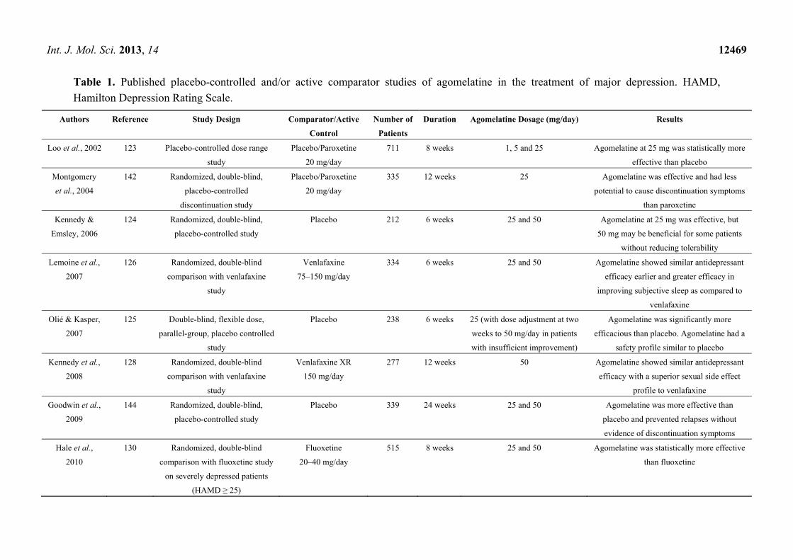
Table 1. Published placebo-controlled and/or active comparator studies of agomelatine in the treatment of major depression. HAMD,
Hamilton Depression Rating Scale.
Authors Reference Study Design Comparator/Active
Control
Number of
Patients
Duration Agomelatine Dosage (mg/day) Results
Loo et al., 2002 123 Placebo-controlled dose range
study
Placebo/Paroxetine
20 mg/day
711 8 weeks 1, 5 and 25 Agomelatine at 25 mg was statistically more
effective than placebo
Montgomery
et al., 2004
142 Randomized, double-blind,
placebo-controlled
discontinuation study
Placebo/Paroxetine
20 mg/day
335 12 weeks 25 Agomelatine was effective and had less
potential to cause discontinuation symptoms
than paroxetine
Kennedy &
Emsley, 2006
124 Randomized, double-blind,
placebo-controlled study
Placebo 212 6 weeks 25 and 50 Agomelatine at 25 mg was effective, but
50 mg may be beneficial for some patients
without reducing tolerability
Lemoine et al.,
2007
126 Randomized, double-blind
comparison with venlafaxine
study
Venlafaxine
75–150 mg/day
334 6 weeks 25 and 50 Agomelatine showed similar antidepressant
efficacy earlier and greater efficacy in
improving subjective sleep as compared to
venlafaxine
Olié & Kasper,
2007
125 Double-blind, flexible dose,
parallel-group, placebo controlled
study
Placebo 238 6 weeks 25 (with dose adjustment at two
weeks to 50 mg/day in patients
with insufficient improvement)
Agomelatine was significantly more
efficacious than placebo. Agomelatine had a
safety profile similar to placebo
Kennedy et al.,
2008
128 Randomized, double-blind
comparison with venlafaxine
study
Venlafaxine XR
150 mg/day
277 12 weeks 50 Agomelatine showed similar antidepressant
efficacy with a superior sexual side effect
profile to venlafaxine
Goodwin et al.,
2009
144 Randomized, double-blind,
placebo-controlled study
Placebo 339 24 weeks 25 and 50 Agomelatine was more effective than
placebo and prevented relapses without
evidence of discontinuation symptoms
Hale et al.,
2010
130 Randomized, double-blind
comparison with fluoxetine study
on severely depressed patients
(HAMD ≥ 25)
Fluoxetine
20–40 mg/day
515 8 weeks 25 and 50 Agomelatine was statistically more effective
than fluoxetine
Int. J. Mol. Sci. 2013, 14 12470
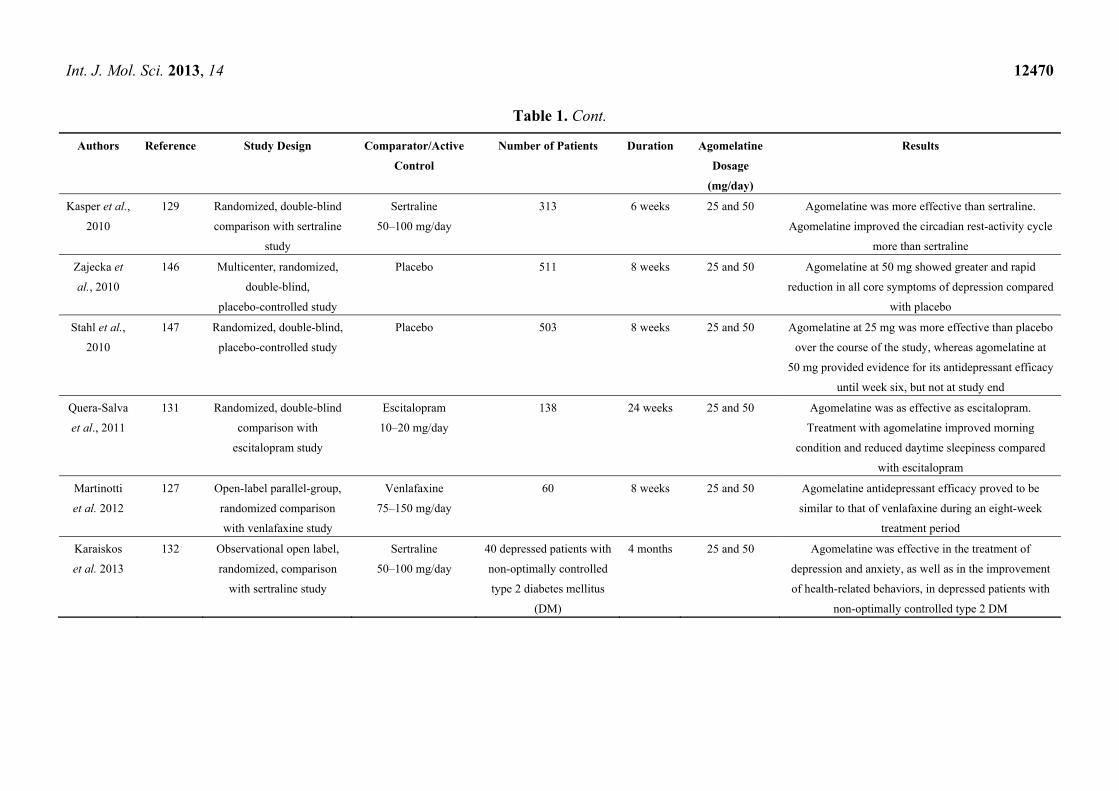
Table 1. Cont.
Authors Reference Study Design Comparator/Active
Control
Number of Patients Duration Agomelatine
Dosage
(mg/day)
Results
Kasper et al.,
2010
129 Randomized, double-blind
comparison with sertraline
study
Sertraline
50–100 mg/day
313 6 weeks 25 and 50 Agomelatine was more effective than sertraline.
Agomelatine improved the circadian rest-activity cycle
more than sertraline
Zajecka et
al., 2010
146 Multicenter, randomized,
double-blind,
placebo-controlled study
Placebo 511 8 weeks 25 and 50 Agomelatine at 50 mg showed greater and rapid
reduction in all core symptoms of depression compared
with placebo
Stahl et al.,
2010
147 Randomized, double-blind,
placebo-controlled study
Placebo 503 8 weeks 25 and 50 Agomelatine at 25 mg was more effective than placebo
over the course of the study, whereas agomelatine at
50 mg provided evidence for its antidepressant efficacy
until week six, but not at study end
Quera-Salva
et al., 2011
131 Randomized, double-blind
comparison with
escitalopram study
Escitalopram
10–20 mg/day
138 24 weeks 25 and 50 Agomelatine was as effective as escitalopram.
Treatment with agomelatine improved morning
condition and reduced daytime sleepiness compared
with escitalopram
Martinotti
et al. 2012
127 Open-label parallel-group,
randomized comparison
with venlafaxine study
Venlafaxine
75–150 mg/day
60 8 weeks 25 and 50 Agomelatine antidepressant efficacy proved to be
similar to that of venlafaxine during an eight-week
treatment period
Karaiskos
et al. 2013
132 Observational open label,
randomized, comparison
with sertraline study
Sertraline
50–100 mg/day
40 depressed patients with
non-optimally controlled
type 2 diabetes mellitus
(DM)
4 months 25 and 50 Agomelatine was effective in the treatment of
depression and anxiety, as well as in the improvement
of health-related behaviors, in depressed patients with
non-optimally controlled type 2 DM
Int. J. Mol. Sci. 2013, 14 12471
6.5. Sleep in Major Depressive Disorder
Sleep and daytime functioning are important aspects of major depressive disorder. Agomelatine
showed an important difference in getting to sleep and quality of sleep in comparison with venlafaxine
and sertraline [105]. Agomelatine was superior to venlafaxine, but no statistically significant
difference was found when compared to sertraline in ease of awakening and integrity of behavior after
awakening [117,120].
Two open-label studies evaluated agomelatine efficacy on sleep parameters in patients with
MDD [139,140]. No change in rapid eye movement (REM) latency, amount of REM or REM density
was observed. Agomelatine improved sleep continuity and quality, and it increased sleep efficiency,
time awake after sleep onset and the total amount of SWS [128]. Agomelatine treatment improved
very early NREM and REM sleep [129].
Recently, agomelatine has been evaluated on nighttime sleep and daytime condition compared to
escitalopram [122]. Agomelatine was associated with reduction in sleep latency, preserved the number
of sleep cycles and reduced daytime sleepiness. In a recent open-label study, 80% of patients with MDD
receiving a flexible dose of agomelatine showed significant improvements at all visits in Leeds Sleep
Evaluation Questionnaire (LSEQ) [127].
6.6. Sexual Function
An important and non-negligible side effect of antidepressants is represented by sexual dysfunction.
Kennedy et al. [118] found that treatment related sexual dysfunctions were significantly lower in the
agomelatine group, whereas venlafaxine was associated with greater deterioration in the domains of
desire and orgasm of the Sex Effects Questionnaire. In a randomized, placebo-controlled, eight-week
study involving healthy male volunteers, agomelatine was shown to have better sexual acceptability
than paroxetine. In fact, the Psychotropic Related Sexual Dysfunction Questionnaire (PRSEXDQ),
reported better scores for agomelatine, similar to placebo, compared to paroxetine [141].
6.7. Anxiety Symptoms within Depression
Anxiety symptoms are common in patients with MDD. Some trial studies evaluating agomelatine
treatment efficacy in depressed patients reported Hamilton Anxiety (HAMA) scale scores. Final
HAMA scores were similar when agomelatine was compared to paroxetine, fluoxetine and
venlafaxine [114,117,118,121]. Agomelatine was superior in reducing HAMA scores compared to
sertraline [120].
6.8. Discontinuation Symptoms
Discontinuation symptoms in MDD have been evaluated in only one randomized, double-blind,
placebo controlled study, with paroxetine as the active comparator [142]. No significant differences
were present in the Discontinuation-Emergent Signs and Symptoms scale (DESS) between patients
who stopped or continued agomelatine treatment. On the other hand, DESS scores were higher in
patients discontinuing paroxetine, who reported insomnia, dreaming, dizziness, muscle ache, nausea,
diarrhea, rhinorrhea and chills. These data suggest that agomelatine is not associated with
Int. J. Mol. Sci. 2013, 14 12472
discontinuation symptoms. In an active comparator trial, discontinuation rates were fewer for agomelatine
than venlafaxine, sertraline and fluoxetine [119–121], but similar to paroxetine [114].
6.9. Responders, Remitters and Relapse Prevention
Remission is the final goal of antidepressant treatment. Six studies reported response and remission
rates (three after six months of treatment and three in the acute phase). At six months, the efficacy of
agomelatine was superior to venlafaxine in CGI scores [117], but no significant differences in the
proportion of responders and remitters were found [117,119]. The responders’ proportion was superior
for agomelatine compared to sertraline by HAMD, but no differences were found in the proportion of
responders by CGI or remitters by HAMD or CGI [120]. Compared to placebo, in acute phase trials,
response rates were significantly higher for agomelatine [114–116]. Only one study reported higher
levels of remission rates for agomelatine after an eight-week treatment period [114]. Sparshatt et al.
conducted a multicenter naturalistic evaluation of the use of agomelatine over a two-year period, in
order to provide a picture of its clinical value in the treatment of depression [143]. Agomelatine was
largely used in difficult-to-treat or refractory patients. After 12 weeks of treatment, a substantial
number of patients improved by at least one point of the CGI (severity) scale.
Two trial studies (one unpublished and one published) investigated long-term antidepressant effect
of agomelatine treatment compared to placebo, regarding relapse prevention. The incidence of relapse
over six months was significantly lower with agomelatine [144]. No significant differences in relapse
rates were shown in the unpublished study (CL3-021) [97].
6.10. Serum Transaminases
Servier Laboratories reported that agomelatine may cause a dose-related elevated liver function test
(LFT), specifically serum transaminases >3-times the upper limit of normal [145]. The European
Medicines Agency requires the monitoring of liver function during treatment at all doses [97].
Two studies reported notable aminotransferase elevations in 2.4% and 4.5% of patients in treatment
with agomelatine at 50 mg, but not with agomelatine at 25 mg or with placebo [146,147]. These LFT
increases were isolated, mainly within the first month of treatment, and no clinical signs of liver
damage were found. A higher proportion of patients with LFT elevations had a history of cholecystitis,
gallbladder disorder or hepatic steatosis. For these reasons, agomelatine is contraindicated in patients
with hepatic impairment. Consequently, it is a condition of treatment that LFTs should be performed
for all patients at the initiation of treatment, then periodically after around six, 12 and 24 weeks and,
thereafter, when clinically indicated [148]. If an increase in serum transaminases occurs, blood liver
function analyses must to be repeated within 48 h, and it is necessary to discontinue the therapy if such
an increase is three-times the upper limit of the normal range. The liver function test must be evaluated
until serum transaminases return to the normal range.
6.11. Limitations of Agomelatine Trials in Major Depressive Disorder
Despite the majority of positive study results regarding agomelatine in the treatment of MDD,
limitations of the reviewed studies should be considered. For example, inclusion and exclusion criteria

Int. J. Mol. Sci. 2013, 14 12473
employed in the trials may have somewhat favored individuals who would respond to treatment. In
fact, patients with a recent history of suicidality, electroconvulsive therapy, psychotic features or
recent substance abuse were excluded in almost all studies. Moreover, most of the studies with an
active comparator arm employed relatively low dosages of venlafaxine, paroxetine, sertraline and
fluoxetine, which may have improved the relative efficacy of agomelatine [149]. Four trials with an
active comparator arm did not have a placebo group [117,118,140,141]. These studies reported high
rates of response, but the lack of placebo groups makes it difficult to place the high rates of response in
a proper context. In addition to high response rates, two studies reported that the differences in
antidepressant efficacy were not statistically significant when comparing agomelatine with venlafaxine
or paroxetine [117,119]. In both of the studies, antidepressant efficacy was only a secondary outcome,
and no clear description of statistical power was provided. Thus, the similar rates of antidepressant
efficacy between agomelatine and the comparator agents may have been affected by the relatively low
dose of venlafaxine (75–150 mg/day) and paroxetine (20 mg/day) and by an inadequate power when
comparing antidepressant efficacy between these agents.
However, despite these shortcomings, the placebo-controlled trials reported improvements in
depression rating scale scores (i.e., 2–3 points) that were similar to responses reported to the Food and
Drug Administration involving a number of agents approved for the treatment of MDD [139]. In 2006,
agomelatine was denied marketing authorization in Europe, due to a reported lack of efficacy [148].
Since that time, additional studies demonstrating agomelatine’s efficacy have been published, and in
November 2008, the committee for medicinal products for human use of the European Medicines
Agency provided marketing authorization for treating MDD episodes in adults with agomelatine [120].
7. Conclusions
MDD is one of the most disabling and common psychiatric disorders. It should be noted that some
unpublished studies reported no significant differences in HAMD and CGI scores between agomelatine vs.
placebo compared to fluoxetine/paroxetine vs. placebo groups, but no significant differences in relapse
rates were also shown when agomelatine was compared to placebo. On the other hand, published data
reported agomelatine efficacy, compared to placebo and based on HAMD, in the treatment of major
depressive disorder. Howland et al. [150,151] have suggested that a publication bias may be present,
as the favorable studies have generally been published and unfavorable studies have generally
not been published. When compared to other antidepressants (venlafaxine, sertraline, fluoxetine and
escitalopram), agomelatine showed, at least, comparable efficacy.
The efficacy of agomelatine on the dimension of anhedonia may be of particular importance in the
treatment of MDD with anhedonic features. In fact, on the basis of SHAPS scores, agomelatine was
reported to be effective in the treatment of anhedonia. In particular, agomelatine showed a more
relevant reduction in SHAPS scores when compared to venlafaxine after eight weeks of treatment.
Some studies reported that agomelatine was similar to sertraline and superior to venlafaxine and
escitalopram in the improvement of sleep parameters in patients with MDD. Lower deterioration in the
domains of desire and orgasm of the Sex Effects Questionnaire were reported when agomelatine was
compared to venlafaxine. In healthy male volunteers, agomelatine was shown to have better sexual
acceptability than paroxetine.
Int. J. Mol. Sci. 2013, 14 12474
Discontinuation rates for any cause were fewer for agomelatine than venlafaxine, sertraline and
fluoxetine. Moreover, data suggest that agomelatine is not associated with discontinuation symptoms,
even if this potential side effect warrants further investigation, especially regarding long-term risks.
About responder and remission rates, data are contrasting, even if agomelatine was largely used in
difficult-to-treat or refractory patients. The incidence of relapse over six months was significantly
lower with agomelatine.
References
1. Wurtman, R.J.; Larin, F.; Axelrod, J.; Shein, H.M.; Rosasco, K. Formation of melatonin and
5-hydroxyindole acetic acid from 14C-tryptophan by rat pineal glands in organ culture. Nature
1968, 217, 953–954.
2. Cardinali, D.P.; Pévet, P. Basic aspects of melatonin action. Sleep. Med. Rev. 1998, 2, 175–190.
3. Hardeland, R.; Cardinali, D.P.; Srinivasan, V.; Spence, D.W.; Brown, G.M.; Pandi-Perumal, S.R.
Melatonin—A pleiotropic, orchestrating regulator molecule. Prog. Neurobiol. 2011, 93, 350–384.
4. Pévet, P. Melatonin. Dialogues Clin. Neurosci. 2002, 4, 57–72.
5. Stehle, J.H.; von Gall, C.; Korf, H.W. Melatonin: A clock-output, a clock-input. J. Neuroendocrinol.
2003, 15, 383–389.
6. Hardeland, R. Melatonin metabolism in the central nervous system. Curr. Neuropharmacol.
2010, 8, 168–181.
7. Dubocovich, M.L.; Markowska, M. Functional MT1 and MT2 melatonin receptors in mammals.
Endocrine 2005, 27, 101–110.
8. Nosjean, O.; Ferro, M.; Coge, F.; Beauverger, P.; Henlin, J.M.; Lefoulon, F.; Fauchere, J.L.;
Delagrange, P.; Canet, E.; Boutin, J.A. Identification of the melatonin-binding site MT3 as the
quinone reductase 2. J. Biol. Chem. 2000, 275, 31311–31317.
9. Ferry, G.; Hecht, S.; Berger, S.; Moulharat, N.; Coge, F.; Guillaumet, G.; Leclerc, V.; Yous, S.;
Delagrange, P.; Boutin, J.A. Old and new inhibitors of quinone reductase 2. Chem. Biol. Interact.
2010, 186, 103–109.
10. Macias, M.; Escames, G.; Leon, J.; Coto, A.; Sbihi, Y.; Osuna, A.; Acuna-Castroviejo, D.
Calreticulin-melatonin. An unexpected relationship. Eur. J. Biochem. 2003, 270, 832–840.
11. Benitez-King, G. Melatonin as a cytoskeletal modulator: Implications for cell physiology and
disease. J. Pineal Res. 2006, 40, 1–9.
12. Carrillo-Vico, A.; Guerrero, J.M.; Lardone, P.J.; Reiter, R.J. A review of the multiple actions of
melatonin on the immune system. Endocrine 2005, 27, 189–200.
13. Hardeland, R. Melatonin: Signaling mechanisms of a pleiotropic agent. Biofactors 2009, 35,
183–192.
14. Wu, Y.H.; Zhou, J.N.; Balesar, R.; Unmehopa, U.; Bao, A.; Jockers, R.; van Heerikhuize, J.;
Swaab, D.F. Distribution of MT1 melatonin receptor immunoreactivity in the human
hypothalamus and pituitary gland: Colocalization of MT1 with vasopressin, oxytocin, and
corticotropin-releasing hormone. J. Comp. Neurol. 2006, 499, 897–910.
Int. J. Mol. Sci. 2013, 14 12475
15. Brunner, P.; Sozer-Topcular, N.; Jockers, R.; Ravid, R.; Angeloni, D.; Fraschini, F.; Eckert, A.;
Muller-Spahn, F.; Savaskan, E. Pineal and cortical melatonin receptors MT1 and MT2 are
decreased in Alzheimer’s disease. Eur. J. Histochem. 2006, 50, 311–316.
16. Masson-Pevet, M.; Gauer, F.; Schuster, C.; Guerrero, H.Y. Photic regulation of mt(1) melatonin
receptors and 2-iodomelatonin binding in the rat and Siberian hamster. Biol. Signals Recept.
2000, 9, 188–196.
17. Masana, M.I.; Benloucif, S.; Dubocovich, M.L. Circadian rhythm of mt1 melatonin receptor
expression in the suprachiasmatic nucleus of the C3H/HeN mouse. J. Pineal Res. 2000, 28,
185–192.
18. Brydon, L.; Roka, F.; Petit, L.; de Coppet, P.; Tissot, M.; Barrett, P.; Morgan, P.J.; Nanoff, C.;
Strosberg, A.D.; Jockers, R. Dual signaling of human Mel1a melatonin receptors via G(i2),
G(i3), and G(q/11) proteins. Mol. Endocrinol. 1999, 13, 2025–2038.
19. Schuster, C.; Williams, L.M.; Morris, A.; Morgan, P.J.; Barrett, P. The human MT1 melatonin
receptor stimulates cAMP production in the human neuroblastoma cell line SH-SY5Y cells via a
calcium-calmodulin signal transduction pathway. J. Neuroendocrinol. 2005, 17, 170–178.
20. Jones, M.P.; Melan, M.A.; Witt-Enderby, P.A. Melatonin decreases cell proliferation and
transformation in a melatonin receptor-dependent manner. Cancer Lett. 2000, 151, 133–143.
21. Rimler, A.; Jockers, R.; Lupowitz, Z.; Zisapel, N. Gi and RGS proteins provide biochemical
control of androgen receptor nuclear exclusion. J. Mol. Neurosci. 2007, 31, 1–12.
22. Boutin, J.A.; Audinot, V.; Ferry, G.; Delagrange, P. Molecular tools to study melatonin pathways
and actions. Trends Pharmacol. Sci. 2005, 26, 412–419.
23. Roka, F.; Brydon, L.; Waldhoer, M.; Strosberg, A.D.; Freissmuth, M.; Jockers, R.; Nanoff, C.
Tight association of the human Mel(1a)-melatonin receptor and G(i): Precoupling and
constitutive activity. Mol. Pharmacol. 1999, 56, 1014–1024.
24. Brown, T.M.; Piggins, H.D. Electrophysiology of the suprachiasmatic circadian clock.
Prog. Neurobiol. 2007, 82, 229–255.
25. Liu, C.; Weaver, D.R.; Jin, X.; Shearman, L.P.; Pieschl, R.L.; Gribkoff, V.K.; Reppert, S.M.
Molecular dissection of two distinct actions of melatonin on the suprachiasmatic circadian clock.
Neuron 1997, 19, 91–102.
26. Gerdin, M.J.; Masana, M.I.; Rivera-Bermudez, M.A.; Hudson, R.L.; Earnest, D.J.;
Gillette, M.U.; Dubocovich, M.L. Melatonin desensitizes endogenous MT2 melatonin receptors
in the rat suprachiasmatic nucleus: Relevance for defining the periods of sensitivity of the
mammalian circadian clock to melatonin. FASEB J. 2004, 18, 1646–1656.
27. Roy, D.; Angelini, N.L.; Fujieda, H.; Brown, G.M.; Belsham, D.D. Cyclical regulation of GnRH
gene expression in GT1–7 GnRH-secreting neurons by melatonin. Endocrinology 2001, 142,
4711–4720.
28. Witt-Enderby, P.A.; Jarzynka, M.J.; Krawitt, B.J.; Melan, M.A. Knock-down of RGS4
and β-tubulin in CHO cells expressing the human MT1 melatonin receptor prevents
melatonin-induced receptor desensitization. Life Sci. 2004, 75, 2703–2715.
29. Jarzynka, M.J.; Passey, D.K.; Ignatius, P.F.; Melan, M.A.; Radio, N.M.; Jockers, R.;
Rasenick, M.M.; Brydon, L.; Witt-Enderby, P.A. Modulation of melatonin receptors and
G-protein function by microtubules. J. Pineal Res. 2006, 41, 324–336.
Int. J. Mol. Sci. 2013, 14 12476
30. Musshoff, U.; Riewenherm, D.; Berger, E.; Fauteck, J.D.; Speckmann, E.J. Melatonin receptors
in rat hippocampus: Molecular and functional investigations. Hippocampus 2002, 12, 165–173.
31. Wang, L.M.; Suthana, N.A.; Chaudhury, D.; Weaver, D.R.; Colwell, C.S. Melatonin inhibits
hippocampal long-term potentiation. Eur. J. Neurosci. 2005, 22, 2231–2237.
32. Larson, J.; Jessen, R.E.; Uz, T.; Arslan, A.D.; Kurtuncu, M.; Imbesi, M.; Manev, H. Impaired
hippocampal long-term potentiation in melatonin MT2 receptor-deficient mice. Neurosci. Lett.
2006, 393, 23–26.
33. Conboy, L.; Tanrikut, C.; Zoladz, P.R.; Campbell, A.M.; Park, C.R.; Gabriel, C.; Mocaër, E.;
Sandi, C.; Diamond, D.M. The antidepressant agomelatine blocks the adverse effects of stress on
memory and enables spatial learning to rapidly increase neural cell adhesion molecule (NCAM)
expression in the hippocampus of rats. Int. J. Neuropsychopharmacol. 2009, 12, 329–341.
34. Bertaina-Anglade, V.; Drieu-La-Rochelle, C.; Mocaër, E.; Seguin, L. Memory facilitating effects
of agomelatine in the novel object recognition memory paradigm in the rat. Pharmacol. Biochem.
Behav. 2011, 98, 511–517.
35. Dubocovich, M.L.; Rivera-Bermudez, M.A.; Gerdin, M.J.; Masana, M.I. Molecular
pharmacology, regulation and function of mammalian melatonin receptors. Front. Biosci. 2003,
8, d1093–d1108.
36. Fuller, P.M.; Gooley, J.J.; Saper, C.B. Neurobiology of the sleep-wake cycle: Sleep architecture,
circadian regulation, and regulatory feedback. J. Biol. Rhythms. 2006, 21, 482–493.
37. Jan, J.E.; Reiter, R.J.; Wasdell, M.B.; Bax, M. The role of the thalamus in sleep, pineal melatonin
production, and circadian rhythm sleep disorders. J. Pineal Res. 2009, 46, 1–7.
38. Ochoa Sanchez, R.; Comai, S.; Lacoste, B.; Bambico, F.R.; Lopez-Dominguez, S.; Rivara S.;
Mor, M.; Bedini, A.L.; Spadoni, G.; Fraschini, F.; et al. Promotion of non-rapid eye movement
sleep and activation of reticular thalamic neurons by a novel MT2 melatonin receptor ligand.
J. Neurosci. 2011, 14, 18439–18452.
39. Ochoa Sanchez, R.; Comai, S.; Rainer, Q.; Rivara, S.; Mor, M.; Bedini, A.L.; Spadoni, G.;
Fraschini, F.; Tarzia, G.; Gobbi, G. Anxiolytic effects of the melatonin MT2 receptor partial
agonist UCM765: Comparison with melatonin and diazepam. Prog. Neuropsychopharmacol.
Biol. Psychiatr. 2012, 39, 318–325.
40. Comai, S.; Ochoa-Sanchez, R.; Gobbi, G. Sleep-wake characterization of double MT1 and
MT2 receptor knockout mice and comparison with MT1 and MT2 receptor knockout mice.
Behav. Brain Res. 2013, 243, 231–238.
41. Reiter, R.J. Pineal melatonin: Cell biology of its synthesis and of its physiological interactions.
Endocr. Rev. 1991, 12, 151–180.
42. Esteban, S.; Garau, C.; Aparicio, S.; Moranta, D.; Barcelo, P.; Fiol, M.A.; Rial, R. Chronic
melatonin treatment and its precursor L-tryptophan improve the monoaminergic neurotransmission
and related behavior in the aged rat brain. J. Pineal Res. 2010, 48, 170–177.
43. Jiang, Z.G.; Teshima, K.; Yang, Y.; Yoshioka, T.; Allen, C.N. Pre- and postsynaptic actions of
serotonin on rat suprachiasmatic nucleus neurons. Brain Res. 2000, 866, 247–256.
44. Morin, L.P. Serotonin and the regulation of mammalian circadian rhythmicity. Ann. Med. 1999,
31, 12–33.
Int. J. Mol. Sci. 2013, 14 12477
45. Edgar, D.M.; Miller, J.D.; Prosser, R.A.; Dean, R.R.; Dement, W.C. Serotonin and the
mammalian circadian system: II. Phase-shifting rat behavioral rhythms with serotonergic
agonists. J. Biol. Rhythms 1993, 8, 17–31.
46. Prosser, R.A.; Heller, H.C.; Miller, J.D. Serotonergic phase advances of the mammalian
circadian clock involve protein kinase A and K+ channel opening. Brain Res. 1994, 644, 67–73.
47. Prosser, R.A.; Lee, H.M.; Wehner, A. Serotonergic pre-treatments block in vitro serotonergic
phase shifts of the mouse suprachiasmatic nucleus circadian clock. Neuroscience 2006, 142,
547–555.
48. Meyer-Bernstein, E.L.; Morin, L.P. Differential serotonergic innervation of the suprachiasmatic
nucleus and the intergeniculate leaflet and its role in circadian rhythm modulation. J. Neurosci.
1996, 16, 2097–2111.
49. Yan, L.; Takekida, S.; Shigeyoshi, Y.; Okamura, H. Per1 and Per2 gene expression in the rat
suprachiasmatic nucleus: Circadian profile and the compartment-specific response to light.
Neuroscience 1999, 94, 141–150.
50. Caldelas, I.; Challet, E.; Saboureau, M.; Pevet, P. Light and melatonin inhibit in vivo
serotonergic phase advances without altering serotonergic-induced decrease of per expression in
the hamster suprachiasmatic nucleus. J. Mol. Neurosci. 2005, 25, 53–63.
51. Mendoza, J.; Clesse, D.; Pevet, P.; Challet, E. Serotonergic potentiation of dark pulse-induced
phase-shifting effects at midday in hamsters. J. Neurochem. 2008, 106, 1404–1414.
52. Gannon, R.L.; Millan, M.J. Evaluation of serotonin, noradrenaline and dopamine reuptake
inhibitors on light-induced phase advances in hamster circadian activity rhythms.
Psychopharmacology 2007, 195, 325–332.
53. Cuesta, M.; Clesse, D.; Pevet, P.; Challet, E. New light on the serotonergic paradox in the rat
circadian system. J. Neurochem. 2009, 110, 231–243.
54. Ciarleglio, C.M.; Resuehr, H.E.; McMahon, D.G. Interactions of the serotonin and circadian
systems: Nature and nurture in rhythms and blues. Neuroscience 2011, 197, 8–16.
55. Dominguez-Lopez, S.; Mahar, I.; Bambico, F.R.; Labonte, B.; Ochoa-Sanchez, R.; Leyton, M.;
Gobbi, G. Short-term effects of melatonin and pinealectomy on serotonergic neuronal activity
across the light-dark cycle. J. Psychopharmacol. 2012, 26, 830–844.
56. Klein, D.C.; Coon, S.L.; Roseboom, P.H.; Weller, J.L.; Bernard, M.; Gastel, J.A.; Zatz, M.;
Iuvone, P.M.; Rodriguez, I.R.; Begay, V.; et al. The melatonin rhythm-generating enzyme:
Molecular regulation of serotonin N-acetyltransferase in the pineal gland. Recent. Prog. Horm. Res.
1997, 52, 307–357; discussion 357–308.
57. Maronde, E.; Saade, A.; Ackermann, K.; Goubran-Botros, H.; Pagan, C.; Bux, R.; Bourgeron, T.;
Dehghani, F.; Stehle, J.H. Dynamics in enzymatic protein complexes offer a novel principle for
the regulation of melatonin synthesis in the human pineal gland. J. Pineal Res. 2011, 51,
145–155.
58. Ho, A.K.; Mackova, M.; Price, L.; Chik, C.L. Diurnal variation in p42/44 mitogen-activated
protein kinase in the rat pineal gland. Mol. Cell. Endocrinol. 2003, 208, 23–30.
59. Ho, A.K.; Chik, C.L. Modulation of Aanat gene transcription in the rat pineal gland.
J. Neurochem. 2010, 112, 321–331.
Int. J. Mol. Sci. 2013, 14 12478
60. Moller, M.; Liu, W. Innervation of the rat pineal gland by nerve fibres originating in the
sphenopalatine, otic and trigeminal ganglia. A retrograde in vivo neuronal tracing study.
Reprod. Nutr. Dev. 1999, 39, 345–353.
61. Moller, M.; Phansuwan-Pujito, P.; Govitrapong, P.; Schmidt, P. Indications for a central
innervation of the bovine pineal gland with substance P-immunoreactive nerve fibers. Brain Res.
1993, 611, 347–351.
62. Mukda, S.; Chetsawang, B.; Govitrapong, P.; Schmidt, P.T.; Hay-Schmidt, A.; Moller, M.
Tachykinins and tachykinin-receptors in the rat pineal gland. Eur. J. Neurosci. 2005, 21,
2743–2751.
63. Mukda, S.; Moller, M.; Ebadi, M.; Govitrapong, P. The modulatory effect of substance P on rat
pineal norepinephrine release and melatonin secretion. Neurosci. Lett. 2009, 461, 258–261.
64. Koch, M.; Dehghani, F.; Habazettl, I.; Schomerus, C.; Korf, H.W. Cannabinoids attenuate
norepinephrine-induced melatonin biosynthesis in the rat pineal gland by reducing arylalkylamine
N-acetyltransferase activity without involvement of cannabinoid receptors. J. Neurochem. 2006,
98, 267–278.
65. Koch, M.; Habazettl, I.; Dehghani, F.; Korf, H.W. The rat pineal gland comprises an
endocannabinoid system. J. Pineal Res. 2008, 45, 351–360.
66. Garcia, R.A.; Afeche, S.C.; Scialfa, J.H.; do Amaral, F.G.; dos Santos, S.H.; Lima, F.B.;
Young, M.E.; Cipolla-Neto, J. Insulin modulates norepinephrine-mediated melatonin synthesis in
cultured rat pineal gland. Life Sci. 2008, 82, 108–114.
67. Bailey, M.J.; Coon, S.L.; Carter, D.A.; Humphries, A.; Kim, J.S.; Shi, Q.; Gaildrat, P.; Morin, F.;
Ganguly, S.; Hogenesch, J.B.; et al. Night/day changes in pineal expression of >600 genes:
Central role of adrenergic/cAMP signaling. J. Biol. Chem. 2009, 284, 7606–7622.
68. Kim, J.S.; Bailey, M.J.; Weller, J.L.; Sugden, D.; Rath, M.F.; Moller, M.; Klein, D.C. Thyroid
hormone and adrenergic signaling interact to control pineal expression of the dopamine receptor
D4 gene (Drd4). Mol. Cell. Endocrinol. 2010, 314, 128–135.
69. Gonzalez, S.; Moreno-Delgado, D.; Moreno, E.; Perez-Capote, K.; Franco, R.; Mallol, J.;
Cortes, A.; Casado, V.; Lluis, C.; Ortiz, J.; et al. Circadian-related heteromerization of adrenergic
and dopamine D(4) receptors modulates melatonin synthesis and release in the pineal gland.
PLoS Biol. 2012, 10, e1001347.
70. Zisapel, N.; Egozi, Y.; Laudon, M. Inhibition of dopamine release by melatonin: Regional
distribution in the rat brain. Brain Res. 1982, 246, 161–163.
71. Alexiuk, N.A.; Uddin, M.; Vriend, J. Melatonin increases the in situ activity of tyrosine
hydroxylase in the mediobasal hypothalamus of male Syrian hamsters. Life Sci. 1996, 59, 687–694.
72. Escames, G.; Acuna Castroviejo, D.; Vives, F. Melatonin-dopamine interaction in the striatal
projection area of sensorimotor cortex in the rat. Neuroreport 1996, 7, 597–600.
73. Di Chiara, G.; Morelli, M.; Consolo, S. Modulatory functions of neurotransmitters in the
striatum: ACh/dopamine/NMDA interactions. Trends Neurosci. 1994, 17, 228–233.
74. Hamdi, A. Melatonin administration increases the affinity of D2 dopamine receptors in the rat
striatum. Life Sci. 1998, 63, 2115–2120.
75. Iuvone, P.M.; Gan, J. Functional interaction of melatonin receptors and D1 dopamine receptors
in cultured chick retinal neurons. J. Neurosci. 1995, 15, 2179–2185.
Int. J. Mol. Sci. 2013, 14 12479
76. Al-Ghoul, W.M.; Herman, M.D.; Dubocovich, M.L. Melatonin receptor subtype expression in
human cerebellum. Neuroreport 1998, 9, 4063–4068.
77. Poirel, V.J.; Cailotto, C.; Streicher, D.; Pevet, P.; Masson-Pevet, M.; Gauer, F. MT1 melatonin
receptor mRNA tissular localization by PCR amplification. Neuro Endocrinol. Lett. 2003, 24, 33–38.
78. Mazzucchelli, C.; Pannacci, M.; Nonno, R.; Lucini, V.; Fraschini, F.; Stankov, B.M. The
melatonin receptor in the human brain: Cloning experiments and distribution studies. Brain Res.
Mol. Brain Res. 1996, 39, 117–126.
79. Kurtuncu, M.; Arslan, A.D.; Akhisaroglu, M.; Manev, H.; Uz, T. Involvement of the pineal gland
in diurnal cocaine reward in mice. Eur J. Pharmacol. 2004, 489, 203–205.
80. Sircar, R. Effect of melatonin on cocaine-induced behavioral sensitization. Brain Res. 2000, 857,
295–299.
81. Imbesi, M.; Uz, T.; Yildiz, S.; Arslan, A.D.; Manev, H. Drug- and region-specific effects of
protracted antidepressant and cocaine treatment on the content of melatonin MT(1) and MT(2)
receptor mRNA in the mouse brain. Int. J. Neuroprot. Neuroregener. 2006, 2, 185–189.
82. Sharma, R.; McMillan, C.R.; Niles, L.P. Neural stem cell transplantation and melatonin treatment in
a 6-hydroxydopamine model of Parkinson’s disease. J. Pineal Res. 2007, 43, 245–254.
83. Lin, C.H.; Huang, J.Y.; Ching, C.H.; Chuang, J.I. Melatonin reduces the neuronal loss,
downregulation of dopamine transporter, and upregulation of D2 receptor in rotenone-induced
parkinsonian rats. J. Pineal Res. 2008, 44, 205–213.
84. Lai, I.C.; Chen, M.L.; Wang, Y.C.; Chen, J.Y.; Liao, D.L.; Bai, Y.M.; Lin, C.C.; Chen, T.T.;
Liou, Y.J. Analysis of genetic variations in the human melatonin receptor (MTNR1A, MTNR1B)
genes and antipsychotics-induced tardive dyskinesia in schizophrenia. World J. Biol. Psychiatr.
2011, 12, 143–148.
85. Courtet, P.; Olié, E. Circadian dimension and severity of depression. Eur. Neuropsychopharmacol.
2012, 22, S476–S481.
86. Wirz-Justice, A. Diurnal variation of depressive symptoms. Dialogues Clin. Neurosci. 2008, 10,
337–343.
87. Dallaspezia, S.; Benedetti, F. Chronobiological therapy for mood disorders. Expert Rev.
Neurother. 2011, 11, 961–970.
88. Coogan, A.N.; Thome, J. Chronotherapeutics and psychiatry: Setting the clock to relieve the
symptoms. World J. Biol. Psychiatr. 2011, 12, 40–43.
89. Barden, N.; Shink, E.; Labbé, M.; Vacher, R.; Rochford, J.; Mocaër, E. Antidepressant action of
agomelatine (S 20098) in a transgenic mouse model. Prog. Neuropsychopharmacol. Biol.
Psychiatr. 2005, 29, 908–916.
90. Leproult, R.; Van Onderbergen, A.; L’hermite-Balériaux, M.; van Cauter, E.; Copinschi, G.
Phase-shifts of 24 h rhythms of hormonal release and body temperature following early evening
administration of the melatonin agonist agomelatine in healthy older men. Clin. Endocrinol.
2005, 63, 298–304.
91. Audinot, V.; Mailliet, F.; Lahaye-Brasseur, C.; Bonnaud, A.; Le Gall, A.; Amossé, C.; Dromaint, S.;
Rodriguez, M.; Nagel, N.; Galizzi, J.P.; et al. New selective ligands of human cloned melatonin
MT1 and MT2 receptors. Naunyn Schmiedebergs Arch. Pharmacol. 2003, 6, 553–561.
Int. J. Mol. Sci. 2013, 14 12480
92. Sharpley, A.L.; Rawlings, N.B.; Brain, S.; McTavish, S.F.; Cowen, P.J. Does agomelatine block
5-HT2C receptors in humans? Psychopharmacology 2011, 213, 653–655.
93. Norman, T.R. The effect of agomelatine on 5HT(2C) receptors in humans: A clinically relevant
mechanism? Psychopharmacology 2012, 221, 177–178.
94. Millan, M.J.; Gobert, A.; Lejeune, F.; Dekeyne, A.; Newman-Tancredi, A.; Pasteau, V.;
Rivet, J.M.; Cussac, D. The novel melatonin agonist agomelatine (S20098) is an antagonist at
5-hydroxytryptamine2C receptors, blockade of which enhances the activity of frontocortical
dopaminergic and adrenergic pathways. J. Pharmacol. Exp. Ther. 2003, 306, 954–964.
95. Dubocovich, M.L. Drug evaluation: Agomelatine targets a range of major depressive disorder
symptoms. Curr. Opin. Investig. Drugs 2006, 7, 670–680.
96. Palazidou, E.; Papadopoulos, A.; Ratcliff, H.; Dawling, S.; Checkley, S.A. NE uptake inhibition
increases melatonin secretion, a measure of noradrenergic neurotransmission, in depressed
patients. Psychol. Med. 1992, 22, 309–315.
97. Mitchell, H.A.; Weinshenker, D. Good night and good luck: Norepinephrine in sleep
pharmacology. Biochem. Pharmacol. 2009, 79, 801–809.
98. Kripke, D.F.; Youngstedt, S.D.; Rex, K.M.; Klauber, M.R.; Elliott, J.A. Melatonin excretion with
affect disorders over age 60. Psychiatr. Res. 2003, 118, 47–54.
99. Carvalho, L.A.; Gorenstein, C.; Moreno, R.A.; Markus, R.P. Melatonin levels in drug-free
patients with major depression from the southern hemisphere. Psychoneuroendocrinology 2006,
31, 761–768.
100. Rubin, R.T.; Heist, E.K.; McGeoy, S.S.; Hanada, K.; Lesser, I.M. Neuroendocrine aspects of
primary endogenous depression. XI. Serum melatonin measures in patients and matched control
subjects. Arch. Gen. Psychiatr. 1992, 49, 558–567.
101. Shafii, M.; MacMillan, D.R.; Key, M.P.; Derrick, A.M.; Kaufman, N.; Nahinsky, I.D. Nocturnal
serum melatonin profile in major depression in children and adolescents. Arch. Gen. Psychiatr.
1996, 53, 1009–1013.
102. Sekula, L.K.; Lucke, J.F.; Heist, E.K.; Czambel, R.K.; Rubin, R.T. Neuroendocrine aspects of
primary endogenous depression. XV: Mathematical modeling of nocturnal melatonin secretion in
major depressives and normal controls. Psychiatr. Res. 1997, 69, 143–153.
103. Crasson, M.; Kjiri, S.; Colin, A.; Kjiri, K.; L’Hermite-Baleriaux, M.; Ansseau, M.;
Legros, J.J. Serum melatonin and urinary 6- sulfatoxymelatonin in major depression.
Psychoneuroendocrinology 2004, 29, 1–12.
104. Wetterberg, L. Clinical importance of melatonin. Prog. Brain Res. 1979, 52, 539–547.
105. Srinivasan, V.; de Berardis, D.; Shillcutt, S.D.; Brzezinski, A. Role of melatonin in mood disorders
and the antidepressant effects of agomelatine. Expert Opin. Investig. Drugs 2012, 21, 1503–1522.
106. Pompili, M.; Serafini, G.; Innamorati, M.; Venturini, P.; Fusar-Poli, P.; Sher. L.; Amore M.;
Girardi, P. Agomelatine, a novel intriguing antidepressant option enhancing neuroplasticity: A
critical review. World J. Biol. Psychiatr. 2013, in press.
107. Dagytė, G.; Crescente, I.; Postema, F.; Seguin, L.; Gabriel, C.; Mocaër, E.; Boer, J.A.;
Koolhaas, J.M. Agomelatine reverses the decrease in hippocampal cell survival induced by
chronic mild stress. Behav. Brain Res. 2011, 218, 121–128.
Int. J. Mol. Sci. 2013, 14 12481
108. Tardito, D.; Molteni, R.; Popoli, M.; Racagni, G. Synergistic mechanisms involved in the
antidepressant effects of agomelatine. Eur. Neuropsychopharmacol. 2012, 22, 482–486.
109. San, L.; Arranz, B. Agomelatine: A novel mechanism of antidepressant action involving the
melatonergic and the serotonergic system. Eur. Psychiatr. 2008, 23, 396–402.
110. European Medicines Agency. Evaluation of Medicines for Human Use CHMP Assessment
Report for Valdoxan. Available online: http://www.emea.europa.eu/humandocs/PDFs/EPAR/
valdoxan/H-915-en6.pdf (accessed on 26 December 2009).
111. Servier Laboratories Ltd. Valdoxan (agomelatine) Summary of Product Characteristics, 2009.
Available online: http://emc. medicines.org.uk/medicine/21830/SPC/Valdoxan/ (accessed on 29
January 2010).
112. Dolder, C.R.; Nelson, M.; Snider, M. Agomelatine treatment of major depressive disorder.
Ann. Pharmacother. 2008, 42, 1822–1831.
113. Sadock, B.J.; Sadock, V.A. Kaplan Sadock’s Synopsis of Psychiatry: Behavioral Sciences;
Lippincott Williams & Wilkins: Philadelphia, PA, USA, 2005.
114. Kessler, R.C.; Berglund, P.; Demler, O.; Jin, R.; Merikangas, K.R.; Walters, E.E. Lifetime
prevalence and age-of-onset distributions of DSM-IV disorders in the National Comorbidity
Survey Replication. Arch. Gen. Psychiatr. 2005, 62, 593–602.
115. Kessler, R.C.; Chiu, W.T.; Demler, O.; Merikangas, K.R.; Walters, E.E. Prevalence, severity,
and comorbidity of 12-month DSM-IV disorders in the National Comorbidity Survey
Replication. Arch. Gen. Psychiatr. 2005, 62, 617–627.
116. De Berardis, D.; Serroni, N.; Campanella, D.; Carano, A.; Gambi, F.; Valchera, A.; Conti, C.;
Sepede, G.; Scali, M.; Fulcheri, M.; et al. Alexithymia and its relationships with C-reactive
protein and serum lipid levels among drug naïve adult outpatients with major depression.
Prog. Neuropsychopharmacol. Biol. Psychiatr. 2008, 32, 1982–1986.
117. Murray, C.J.; Lopez, A.D. Alternative projections of mortality and disability by cause
1990–2020: Global burden of disease study. Lancet 1997, 349, 1498–1504.
118. Lopez, A.D.; Mathers, C.D.; Ezzati, M.; Jamison, D.T.; Murray, C.J. Global and regional burden
of disease and risk factors, 2001: Systematic analysis of population health data. Lancet 2006,
367, 1747–1757.
119. Fava, M. Diagnosis and definition of treatment-resistant depression. Biol. Psychiatr. 2003, 15,
649–659.
120. Little, A. Treatment-resistant depression. Am. Fam. Physician 2009, 80, 167–172.
121. Parker, G.; Brotchie, H. Do the old psychostimulant drugs have a role in managing
treatment-resistant depression? Acta Psychiatr. Scand. 2009, 121, 308–314.
122. Hickie, I. Is depression overdiagnosed? No. BMJ 2007, 335, 329.
123. Hall, W.D.; Mant, A.; Mitchell, P.B.; Rendle, V.A.; Hickie, I.B.; McManus, P. Association
between antidepressant prescribing and suicide in Australia, 1991−2000: Trend analysis. BMJ
2003, 326, 1008.
124. Loo, H.; Hale, A.; D’Haenen, H. Determination of the dose of agomelatine, a melatoninergic
agonist and selective 5-HT(2C) antagonist, in the treatment of major depressive disorder: A
placebo-controlled dose range study. Int. Clin. Psychopharmacol. 2002, 17, 239–247.

Int. J. Mol. Sci. 2013, 14 12482
125. Kennedy, S.H.; Emsley, R. Placebo-controlled trial of agomelatine in the treatment of major
depressive disorder. Eur. Neuropsychopharmacol. 2006, 16, 93–100.
126. Olie, J.P.; Kasper, S. Efficacy of agomelatine, a MT1/MT2 receptor agonist with 5-HT2C
antagonistic properties, in major depressive disorder. Int. J. Neuropsychopharmacol. 2007, 10,
661–673.
127. Lemoine, P.; Guilleminault, C.; Alvarez, E. Improvement in subjective sleep in major depressive
disorder with a novel antidepressant, agomelatine: Randomized, double-blind comparison with
venlafaxine. J. Clin. Psychiatr. 2007, 68, 1723–1732.
128. Martinotti, G.; Sepede, G.; Gambi, F.; di Iorio, G.; de Berardis, D.; di Nicola, M.; Onofrj, M.;
Janiri, L.; di Giannantonio, M. Agomelatine versus venlafaxine XR in the treatment of anhedonia
in major depressive disorder: A pilot study. J. Clin. Psychopharmacol. 2012, 32, 487–491.
129. Kennedy, S.H.; Rizvi, S.; Fulton, K.; Rasmussen, J. A double-blind comparison of sexual
functioning, antidepressant efficacy, and tolerability between agomelatine and venlafaxine XR.
J. Clin. Psychopharmacol. 2008, 28, 329–333.
130. Kasper, S.; Hajak, G.; Wulff, K.; Hoogendijk, W.J.; Montejo, A.L.; Smeraldi, E.; Rybakowski, J.K.;
Quera-Salva, M.A.; Wirz-Justice, A.M.; Picarel-Blanchot, F.; et al. Efficacy of the novel
antidepressant agomelatine on the circadian rest-activity cycle and depressive and anxiety
symptoms in patients with major depressive disorder: A randomized, double-blind comparison
with sertraline. J. Clin. Psychiatr. 2010, 71, 109–120.
131. Hale, A.; Corral, R.M.; Mencacci, C.; Ruiz, J.S.; Severo, C.A.; Gentil, V. Superior antidepressant
efficacy results of agomelatine versus fluoxetine in severe MDD patients: A randomized,
double-blind study. Int. Clin. Psychopharmacol. 2010, 25, 305–314.
132. Quera-Salva, M.A.; Hajak, G.; Philip, P.; Montplaisir, J.; Keufer-Le Gall, S.; Laredo, J.;
Guilleminault, C. Comparison of agomelatine and escitalopram on nighttime sleep and daytime
condition and efficacy in major depressive disorder patients. Int. Clin. Psychopharmacol. 2011,
26, 252–262.
133. Karaiskos, D.; Tzavellas, E.; Ilias, I.; Liappas, I.; Paparrigopoulos, T. Agomelatine and sertraline
for the treatment of depression in type 2 diabetes mellitus. Int. J. Clin. Pract. 2013, 67, 257–260.
134. Spijker, J.; Bijl, R.V.; de Graaf, R.; Nolen, W.A. Determinants of poor 1-year outcome of
DSM-III-R major depression in the general population: Results of the Netherlands Mental Health
Survey and Incidence Study (NEMESIS). Acta Psychiatr. Scand. 2001, 103, 122–130.
135. Taylor, D.J.; Walters, H.M.; Vittengl, J.R.; Krebaum, S.; Jarret, R.B. Which depressive
symptoms remain after response to cognitive therapy of depression and predict relapse and
recurrence? J. Affect. Disord. 2010, 123, 181–187.
136. Keedwell, P.A.; Andrew, C.; Williams, S.C.; Brammer, M.J.; Phillips, M.L. The neural correlates
of anhedonia in major depressive disorder. Biol. Psychiatr. 2005, 58, 843–853.
137. Di Giannantonio, M.; Martinotti, G. Anhedonia and major depression: The role of agomelatine.
Eur. Neuropsychopharmacol. 2012, 22, 505–510.
138. Di Giannantonio, M.; di Iorio, G.; Guglielmo, R.; de Berardis, D.; Conti, C.M.; Acciavatti, T.;
Cornelio, M.; Martinotti, G. Major depressive disorder, anhedonia and agomelatine: An open-label
study. J. Biol. Regul. Homeost. Agents 2011, 25, 109–114.
Int. J. Mol. Sci. 2013, 14 12483
139. Quera Salva, M.A.; Vanier, B.; Laredo, J.; Hartley, S.; Chapotot, F.; Moulin, C.; Lofaso, F.;
Guilleminault, C. Major depressive disorder, sleep EEG and agomelatine: An open-label study.
Int. J. Neuropsychopharmacol. 2007, 10, 691–696.
140. Lopes, M.C.; Quera-Salva, M.A.; Guilleminault, C. Non-REM sleep instability in patients with
major depressive disorder: Subjective improvement and improvement of non-REM sleep
instability with treatment (agomelatine). Sleep Med. 2007, 9, 33–41.
141. Montejo, A.; Prieto, N.; Terleira, A.; Matias, J.; Alonso, S.; Paniagua, G.; Naval, S.; Parra, D.G.;
Gabriel, C.; Mocaër, E.; et al. Better sexual acceptability of agomelatine (25 and 50 mg)
compared with paroxetine (20 mg) in healthy male volunteers: An 8-week, placebo-controlled
study using the PRSEXDQ-SALSEX scale. J. Psychopharmacol. 2010, 24, 111–120.
142. Montgomery, S.A.; Kennedy, S.H.; Burrows, G.D.; Lejoyeux, M.; Hindmarch, I. Absence of
discontinuation symptoms with agomelatine and occurrence of discontinuation symptoms with
paroxetine: A randomized, double-blind, placebo-controlled discontinuation study. Int. Clin.
Psychopharmacol. 2004, 19, 271–280.
143. Sparshatt, A.; McAllister Williams, R.H.; Baldwin, D.S.; Haddad, P.M.; Bazire, S.; Weston, E.;
Taylor, P.; Taylor, D. A naturalistic evaluation and audit database of agomelatine: Clinical
outcome at 12 weeks. Acta Psychiatr. Scand. 2012, in press.
144. Goodwin, G.; Emsley, R.; Rembry, S.; Rouillon, F. Agomelatine Study Group. Agomelatine
prevents relapse in patients with major depressive disorder, without evidence of a discontinuation
syndrome. J. Clin. Psychiatr. 2009, 70, 1128–1237.
145. Servier Laboratories. Valdoxan. Available online: http://www.valdoxan.com/index.php/
valdoxan-package-leaflet-information-for the-user (accessed on 3 May 2011).
146. Zajecka, J.; Schatzberg, A.; Stahl, S.; Shah, A.; Caputo, A.; Post, A. Efficacy and safety of
agomelatine in the treatment of major depressive disorder: A multicenter, randomized, double-blind,
placebo-controlled trial. J. Clin. Psychopharmacol. 2010, 30, 135–144.
147. Stahl, S.M.; Fava, M.; Trivedi, M.H.; Caputo, A.; Shah, A.; Post, A. Agomelatine in the
treatmentof major depressive disorder: An 8-week, multicenter, randomized, placebo-controlled
trial. J. Clin. Psychiatr. 2010, 71, 616–626.
148. European Medicines Agency. CHMP Assessment Report for Valdoxan. Document Reference
EMEA/655251/2008. European Medicines Agency: London, UK, 2008.
149. De Berardis, D.; di Iorio, G.; Acciavatti, T.; Conti, C.; Serroni, N.; Olivieri, L.; Cavuto, M.;
Martinotti, G.; Janiri, L.; Moschetta, F.S.; et al. The emerging role of melatonin agonists in the
treatment of major depression: Focus on agomelatine. CNS Neurol. Disord. Drug Targets 2011,
10,119–132.
150. Howland, R.H. A benefit-risk assessment of agomelatine in the treatment of major depression.
Drug Saf. 2011, 34, 709–731.
151. Howland, R.H. Publication bias and outcome reporting bias: Agomelatine as a case example.
J. Psychosoc. Nurs. Ment. Health Serv. 2011, 49, 11–14.
© 2013 by the authors; licensee MDPI, Basel, Switzerland. This article is an open access article
distributed under the terms and conditions of the Creative Commons Attribution license
(http://creativecommons.org/licenses/by/3.0/).