The lower cretaceous lizard genus chometokadmon from Italy
11
The Lower Cretaceous lizard genus Chometokadmon from Italy Susan E. Evans a, * , Pasquale Raia b , Carmela Barbera b a Department of Anatomy and Developmental Biology, University College London, Gower Street, London WC1E 6BT, UK b Dipartimento di Paleontologia, Universita ` di Napoli, Largo S. Marcellino 10, Napoli, Italy Received 3 October 2005; accepted in revised form 21 November 2005 Available online 19 June 2006 Abstract The Lower Cretaceous (Albian) locality of Pietraroia, Italy, has yielded a rich and diverse assemblage of fossil vertebrates, including at least one genus of rhynchocephalian (Derasmosaurus) and three named lizards (Chometokadmon, Costasaurus and Eichstaettisaurus). The type and only specimen of Chometokadmon is well-preserved but has never been comprehensively described or assessed. It was mistakenly classified as a sphenodontian for many years, but detailed reanalysis has shown that Chometokadmon is a squamate. The genus has a relatively unspecialised postcranial skeleton, but the skull is distinctive in having an elongated parietal, expanded squamosal, recurved teeth, and cranial osteoderms. A combination of cranial and postcranial characters (including separable cranial osteoderms, an elongate supratemporal, tooth and pubic morphol- ogy) supports a relationship with Anguimorpha, a hypothesis corroborated by cladistic analysis. Ó 2006 Elsevier Ltd. All rights reserved. Keywords: Squamata; Lizards; Anguimorphs; Italy; Cretaceous; Pietraroia 1. Introduction The Lower Cretaceous (early Albian; Bravi and Garassino, 1998) locality of Pietroroia is in the Apennine Mountains of southern Italy, roughly 75 km northeast of Naples. Excava- tions have been ongoing, albeit intermittently, for more than 150 years (Costa, 1864, 1866; D’Erasmo, 1915). During this time, the site has yielded a rich assemblage of plants, inverte- brates (echinoderms, crustaceans, molluscs) and vertebrates, including fish, amphibians, small reptiles and dinosaurs, most notably the juvenile theropod dinosaur Scipionyx samni- ticus (Leonardi and Teruzzi, 1993; Dal Sasso and Signore, 1998; Bausch and Bravi, 1999; Bravi, 1999). The lepidosaur fauna includes at least one rhynchocephalian, Derasmosaurus (Barbera and Macuglia, 1988) and three named lizards, Cho- metokadmon (Costa, 1864), Costasaurus (Estes, 1983) and Eichstaettisaurus (Evans et al., 2004). Another partial skeleton may represent a second, unnamed, rhynchocephalian (Evans et al., 2004), and a fourth lizard taxon awaits description. Chometokadmon fitzingeri was described by Costa (1864) on the basis of a single specimen (MPN 539) that he identified as a lizard. In an appendix to the same paper, Costa figured and briefly described another small skeleton (MPN 541) from the same locality. He interpreted this as a second lizard, distinct from Chometokadmon, and subsequently named it Lacerta brevicauda (Costa, 1866). D’Erasmo (1915) synony- mized the two specimens under Chometokadmon and referred a third specimen (now lost) to the same genus. Based on the acrodont dentition of Costa’s second specimen (MPN 541), D’Erasmo attributed Chometokadmon to Rhynchocephalia, despite noting dental inconsistencies in the holotype. Most subsequent reviewers (e.g., von Huene, 1956; Cocude-Michel, 1963; Kuhn, 1969) followed D’Erasmo, although Romer (1956) listed Chometokadmon as an indeterminate lizard. The question was resolved by Barbera and Macuglia (1988); Costa was essentially correct. The holotype specimen of Cho- metokadmon is a squamate, but MPN 541 is a rhynchocepha- lian, and is now the holotype of the genus Derasmosaurus (Barbera and Macuglia, 1988). * Corresponding author. E-mail address: [email protected] (S.E. Evans). 0195-6671/$ - see front matter Ó 2006 Elsevier Ltd. All rights reserved. doi:10.1016/j.cretres.2006.03.004 Cretaceous Research 27 (2006) 673e683 www.elsevier.com/locate/CretRes
-
Upload
independent -
Category
Documents
-
view
1 -
download
0
Transcript of The lower cretaceous lizard genus chometokadmon from Italy
Cretaceous Research 27 (2006) 673e683www.elsevier.com/locate/CretRes
The Lower Cretaceous lizard genus Chometokadmon from Italy
Susan E. Evans a,*, Pasquale Raia b, Carmela Barbera b
a Department of Anatomy and Developmental Biology, University College London, Gower Street, London WC1E 6BT, UKb Dipartimento di Paleontologia, Universita di Napoli, Largo S. Marcellino 10, Napoli, Italy
Received 3 October 2005; accepted in revised form 21 November 2005
Available online 19 June 2006
Abstract
The Lower Cretaceous (Albian) locality of Pietraroia, Italy, has yielded a rich and diverse assemblage of fossil vertebrates, including at leastone genus of rhynchocephalian (Derasmosaurus) and three named lizards (Chometokadmon, Costasaurus and Eichstaettisaurus). The type andonly specimen of Chometokadmon is well-preserved but has never been comprehensively described or assessed. It was mistakenly classified asa sphenodontian for many years, but detailed reanalysis has shown that Chometokadmon is a squamate. The genus has a relatively unspecialisedpostcranial skeleton, but the skull is distinctive in having an elongated parietal, expanded squamosal, recurved teeth, and cranial osteoderms. Acombination of cranial and postcranial characters (including separable cranial osteoderms, an elongate supratemporal, tooth and pubic morphol-ogy) supports a relationship with Anguimorpha, a hypothesis corroborated by cladistic analysis.� 2006 Elsevier Ltd. All rights reserved.
Keywords: Squamata; Lizards; Anguimorphs; Italy; Cretaceous; Pietraroia
1. Introduction
The Lower Cretaceous (early Albian; Bravi and Garassino,1998) locality of Pietroroia is in the Apennine Mountains ofsouthern Italy, roughly 75 km northeast of Naples. Excava-tions have been ongoing, albeit intermittently, for more than150 years (Costa, 1864, 1866; D’Erasmo, 1915). During thistime, the site has yielded a rich assemblage of plants, inverte-brates (echinoderms, crustaceans, molluscs) and vertebrates,including fish, amphibians, small reptiles and dinosaurs,most notably the juvenile theropod dinosaur Scipionyx samni-ticus (Leonardi and Teruzzi, 1993; Dal Sasso and Signore,1998; Bausch and Bravi, 1999; Bravi, 1999). The lepidosaurfauna includes at least one rhynchocephalian, Derasmosaurus(Barbera and Macuglia, 1988) and three named lizards, Cho-metokadmon (Costa, 1864), Costasaurus (Estes, 1983) andEichstaettisaurus (Evans et al., 2004). Another partial skeleton
* Corresponding author.
E-mail address: [email protected] (S.E. Evans).
0195-6671/$ - see front matter � 2006 Elsevier Ltd. All rights reserved.
doi:10.1016/j.cretres.2006.03.004
may represent a second, unnamed, rhynchocephalian (Evanset al., 2004), and a fourth lizard taxon awaits description.
Chometokadmon fitzingeri was described by Costa (1864)on the basis of a single specimen (MPN 539) that he identifiedas a lizard. In an appendix to the same paper, Costa figuredand briefly described another small skeleton (MPN 541)from the same locality. He interpreted this as a second lizard,distinct from Chometokadmon, and subsequently named itLacerta brevicauda (Costa, 1866). D’Erasmo (1915) synony-mized the two specimens under Chometokadmon and referreda third specimen (now lost) to the same genus. Based on theacrodont dentition of Costa’s second specimen (MPN 541),D’Erasmo attributed Chometokadmon to Rhynchocephalia,despite noting dental inconsistencies in the holotype. Mostsubsequent reviewers (e.g., von Huene, 1956; Cocude-Michel,1963; Kuhn, 1969) followed D’Erasmo, although Romer(1956) listed Chometokadmon as an indeterminate lizard.The question was resolved by Barbera and Macuglia (1988);Costa was essentially correct. The holotype specimen of Cho-metokadmon is a squamate, but MPN 541 is a rhynchocepha-lian, and is now the holotype of the genus Derasmosaurus(Barbera and Macuglia, 1988).

674 S.E. Evans et al. / Cretaceous Research 27 (2006) 673e683
Barbera and Macuglia (1988) gave a preliminary descriptionof Chometokadmon and tentatively referred it to Scincidae.In the intervening period, however, there has been considerableprogress in our knowledge of Jurassic and Early Cretaceoussquamates and squamate phylogeny (for a review, see Evans,2003), prompting a detailed reanalysis of the specimen. Rela-tively little articulated lizard material exists for the Jurassicand Early Cretaceous, particularly in Euramerica. Furthermore,Pietraroia provides the only European squamate assemblagecontemporaneous with mid-Cretaceous (AlbianeCenomanian)lizard assemblages from North America (Nydam, 2000; Ny-dam and Cifelli, 2002) and Asia (Nessov, 1985, 1988, 1997;Alifanov, 1993).
Institutional abbreviations. BMNH, The Natural HistoryMuseum, London, UK; MPN, Museo di Paleontologia, Napoli,Italy.
2. Geology and material
At the locality of ‘‘Civita di Pietraroja’’ (Mt. Matese, south-ern Italy), two distinct plattenkalk horizons are exposed. Thelower horizon is relatively unfossiliferous (Bausch and Bravi,1999). Above it is a thick sequence of lagoonal limestones,overlain by a second plattenkalk horizon that is the sourceof the major finds from Pietraroia (early Albian; Bravi andGarassino, 1998). The thickness of this second plattenkalk in-creases to the southwest reaching a maximum (ca. 15 m) at theoriginal ‘‘la Cavere’’ outcrop. Bausch and Bravi (1999) havereconstructed a shallow lagoonal environment, close to landand frequently isolated from the open sea, but subject to tidalinfluence and occasional storms. The water would thereforehave varied in the level of salinity, and this is reflected inthe rock layers that show varying marine or terrestrial influ-ence. The low lying landmass with which the deposit was as-sociated was one of a chain of islands running for perhaps100e200 km, rather like the Antilles or Bermuda island chaintoday.
3. Systematic palaeontology
Lepidosauria: Haeckel, 1866Squamata: Oppel, 1811Anguimorpha: Furbringer, 1900Genus Chometokadmon Costa, 1864
Type and only species. Chometokadmon fitzingeri Costa,1864.
Holotype. MPN 539, articulated skeleton and partialcounterpart.
Locality and horizon. La Cavere outcrop, Pietraroia, MountMatese, Italy. Upper Plattenkalk horizon. IGM (Italian Mili-tary Geographic Institute) map sheet 162, III SW-CusanoMutri, N4577431, E2482228. Lower Cretaceous, Albian.
Diagnosis (emended from Kuhn, 1969). Lizard showing thefollowing combination of derived characters: premaxilla withlong nasal process; large maxilla with sharp, recurved teeth;maxilla meets frontal posterodorsally to separate prefrontaland nasal; maxilla with tapering in-turned premaxillary pro-cess and posterior process not extending beyond midpoint oforbit; large, ovoid, posteriorly extended external nares; nar-row, paired nasals; jugal reaching anterior margin of orbitbut separated from prefrontal by lacrimal; maxilla excludedfrom orbital rim; frontals paired with subparallel orbital mar-gins; nasals and frontals sculptured with low tubercles; smallcranial osteoderms associated with orbital and postorbital re-gions, not attached to skull bones; parietal elongate withlong narrow body and long divergent posterior processes; pa-rietal foramen small, anterior to midpoint of bone; supratem-poral elongate reaching anterior to level of postparietalnotch; squamosal broad with small dorsal process and curvedposterior head; anterior margin of supraoccipital overlaps pos-terior edge of parietal; paroccipital processes long, crested andtapering; vertebrae procoelous, with broad, low neural spines,wide horizontal zygapophyses, but no zygospheneezygantrumsystem; anterior caudals with long transverse processes, caudalautotomy septum present (bisecting transverse process); iliumwith short narrow blade and small anterior process; pubisdirected anteriorly with short symphysis; femur straight, footlonger than femur; fused astragalocalcaneum, wide but proxi-modistally short, with small groove between tibial and fibularfacets; only distal tarsals 3 and 4 retained; fifth metatarsalshort and hamate with lateral flange; other metatarsals elon-gate; phalanges becoming increasingly gracile distally.
4. Description
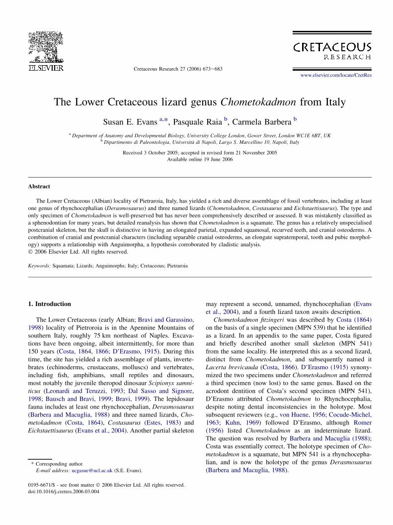
The type and only specimen (MPN 539) is preserved indorsal view and is fully articulated (Fig. 1), but both sidesof the skull are damaged behind the orbit, and parts of theforelimbs are either obscured by the body (right) or still inthe matrix (left). The tail had been autotomised during lifeand had regenerated to almost its full length. The feet are dam-aged but are shown in impression on a partial counterpart(mounted in plaster adjacent to the main block).
4.1. Skull
The premaxilla appears to be single, but it is not well pre-served and the width of the alveolar region can only be esti-mated. The dorsal process is narrow and elongate, meetingthe tips of the nasals and separating them only for a short dis-tance. A small bifurcate process lies in contact with the tip ofthe right maxilla, and probably represents the right edge of thepremaxillary alveolar margin. (Figs. 1, 2A, B)
The maxilla is well preserved on the right side of the skull,but less so on the left. It is a large bone with a long, deep facialprocess that had a nearly straight dorsal suture with the nasaland an acute posterior margin that met the prefrontal, lacrimal,and jugal, but was excluded from the anterior and ventralorbital margins. The posterodorsal angle of the bone met the
675S.E. Evans et al. / Cretaceous Research 27 (2006) 673e683
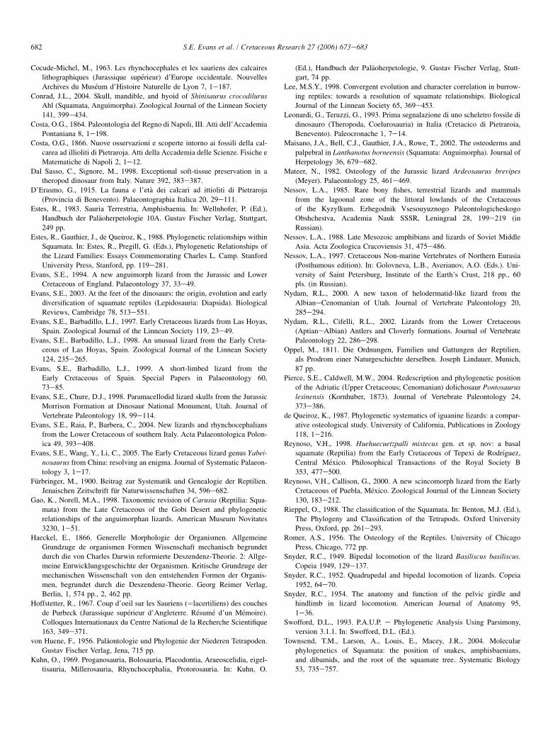
Fig. 1. Chometokadmon fitzingeri (MPN 539). A, entire skeleton in dorsal view, with partial counterpart of feet and tail as inset; scale bar represents 10 mm.
B, enlargement of skull in dorsal view; scale bar represents 5 mm.

676 S.E. Evans et al. / Cretaceous Research 27 (2006) 673e683
anterolateral process of the frontal. The facial process wasprobably medially inclined rather than vertical in life, so thatthe snout was somewhat depressed. The anterior premaxillaryprocess is tapering and curved, whereas the posterior processis relatively short, not extending beyond the midpoint of theorbital margin. On the right side, the alveolar margin is seento bear 12 teeth with gaps between them. These teeth aresharp, narrow and posteriorly recurved.
The nasals are narrow bones with roughly parallel medialand lateral margins and an extended anteromedial premaxil-lary process. The anterior margins are deeply excavated bythe external nares while laterally each bone has a long straightsuture with the maxilla. The nasal does not meet the prefron-tal. The right bone has been rotated slightly about its long axisso that its medial margin faces slightly dorsally. It is a simplesurface and the two nasals met in a butt joint. Posteriorly, thenasal tapers into an apex that fits into a notch in the anteriormargin of the frontal.
The frontals are also paired with a simple median suture.The left bone is almost intact but the posterior half of the rightbone is shattered. The dorsal surface is weakly sculptured ina tuberculate pattern. The frontals are longer than wide,slightly wider posteriorly than anteriorly, and only slightlynarrowed between the orbits. The anterior margin is deeplynotched for the nasal, with a short median process and a longerlateral process that met the posterior tip of the maxilla. Ante-rolaterally, the bone has a shallow notch for the orbital processof the prefrontal but nothing of the ventral surface is visible.The posterior margin appears straight but part of the bone ismissing so that the contact surface for the parietal is not pre-served (reconstructed in Fig. 2B).
The unpaired parietal is probably the most distinctive boneof the entire skull. The parietal plate is slightly shorter than theoverall length of the frontals, but with the long divergent post-parietal processes, the parietal is much longer. The dorsal sur-face is flat, with no trace of a dorsal crest and no sculpture.The lateral margins are sloping and relatively deep, providingthe surface of attachment for the adductor muscles. The boneis narrowest between the large ovoid supratemporal fenestrae,widening anteriorly and posteriorly, but not strongly so. Asmall parietal foramen persists roughly one third of the wayfrom the anterior margin of the bone. The postparietal pro-cesses are strongly divergent, enclosing a posterior angle ofaround 110 �. The medial and lateral surfaces of the processesare both strongly oblique, creating a narrow dorsal crest be-tween them. Posteriorly, these sloping surfaces accommodatedepaxial neck muscles. Centrally, the posterior margin isslightly recessed and is overlapped by the supraoccipital, sug-gesting that metakinesis was at best limited.
Along the antorbital margin of the maxilla, there are threebones. The most dorsal of these is the prefrontal. It has a nar-row orbital portion that runs parallel to the frontal, notchingthe lateral margin of that bone, and a more expanded ventralpart that contributes to the antorbital skull wall. Below the pre-frontal is a narrow lacrimal and this, in turn, meets the jugalventrally. The jugal is preserved on the right side as a long nar-row bar running along the dorsal surface of the maxilla,
notching it slightly, before contacting the lacrimal. Nothingof the posterior part of the jugal is visible.
A crack divides the anterior and posterior parts of the skull,disrupting the postorbital region. A short region of the skullroof is missing. The problem is exacerbated by a scatter of os-teoderms that obscure some of the underlying morphology(see below). The posterolateral corner of the left frontal isflanked by a small oblique bar of bone and a similar structurecontacts the anterolateral edge of the parietal on the right side.These bony fragments are here tentatively interpreted (Fig. 2)as anterior and posterior parts of a bone straddling the lateraledge of the frontoparietal suture, but with the central part lostin the cracked region. The only bone with these relations ina squamate is the postfrontal or postorbitofrontal, if thepostfrontal is fused to the more ventrolaterally placedpostorbital.
Distinctive squamosals are preserved on both sides of theskull. They have blade-like anterior processes and longcurved terminal portions offset from the anterior blade. Atthe junction between the two parts there is a slight dorsal ex-pansion. Neither squamosal shows an obvious postorbital orpostorbitofrontal facet, and both are rotated so that the ante-rior blade is directed anteroventrally. This suggests, at least,that the articulation between the squamosal and postorbitalbar was not strong. The anterior process of the squamosalwould not have reached beyond the midpoint of the uppertemporal fenestra. On the left side, the squamosal contactsan anteroposterior series of bone fragments. Some of thesemust pertain to the lower jaw but more dorsal fragmentsmay belong to an upper temporal bar, placed well lateral tothe parietal.
On both sides, long slender supratemporals lie medial to thesquamosals and overlap the lateral margins of the parietals.Both streptostylic quadrates are in situ but are rather obscuredby overlying bones.
The braincase is well preserved but only partially visible.The supraoccipital is fully exposed behind the parietal, despitethe length of the parietal plate. It has a strong dorsal crest and,somewhat unusually, its anterior margin is drawn into shortprocesses that overlap the posterior margin of the parietal (asimilar condition was recently reported in the Late Cretaceousmosasauroid, Pontosaurus; Pierce and Caldwell, 2004). Bilat-erally, the supraoccipital is sutured to a large otooccipital.Each otooccipital extends posterolaterally into a long slenderparoccipital process that meets the squamosal and supratem-poral but apparently not the parietal. The foramen magnumis also visible with the basioccipital forming its ventral mar-gin. The prootics are largely obscured by the parietal, but theirlateral margins are exposed below the parietal plate. They ex-tend well forward suggesting the presence of strong alaryprocesses.
The palate is not exposed and since the specimen is embed-ded in a block of plaster, it is not possible to prepare the ven-tral surface. However, a slender element running back lateralto the left prootic is presumably the posterior ramus of thepterygoid. A small columnar epipterygoid runs perpendicularto it to reach the anterior part of the prootic.
677S.E. Evans et al. / Cretaceous Research 27 (2006) 673e683
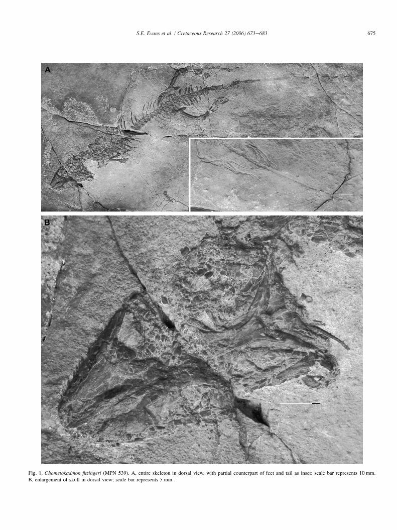
Fig. 2. The skull of Chometokadmon fitzingeri (MPN 539). A, dorsal view as preserved. B, reconstruction of skull and osteoderms. Scale bar represents 5 mm.
Abbreviations: E, epipterygoid; Fr, frontal; Hy, hyoid; J, jugal; L, lacrimal; Mx, maxilla; N, nasal; O, osteoderms; Oto, oto-occipital; P, parietal; Pm, premaxilla;
Poc, paroccipital process; Pof, postfrontal or postorbitofrontal; Prf, prefrontal; Pro, prootic; Pt, pterygoid; Q, quadrate; Soc, supraoccipital; Sq, squamosal; St,
supratemporal; ?, fragments of lower jaw and/or upper temporal bar.
The exposed right side of the skull is damaged posterolat-erally due to the break in the block, and several of the skullbones have fragmented. However, the right orbital and tempo-ral regions show a tessellate pattern of bone fragments that aretoo many to have come from broken skull bones, and, in theorbit at least, are too orderly in their arrangement. The skulltherefore seems to have been covered, at least partially, bya mosaic of small osteoderms that were not fused to the under-lying skull bones. Small patches of these are preserved in situon the left maxilla, on the front of the parietal, and over theright squamosal and quadrate (Fig. 2A, B). Although a fewof these osteoderms have been displaced inwards, they seemto map the boundaries of the eye opening (Fig. 3A). Thereis no trace of the scleral ossicles, but these presumably col-lapsed inward with the eyeball. The osteoderms do not extendpostcranially.
4.2. Postcranial skeleton
4.2.1. Vertebral columnThe cervical column is disrupted behind the skull and
again at the level of the twelfth presacral vertebra. The at-lanto-axial region has been dislocated from the braincaseand moved towards the right side, where it lies adjacent to
the right quadrate. Small rectangular elements in this regionmay be parts of the atlantal arch. Immediately adjacent tothese is an irregular bony element here interpreted as theaxis in left lateral view. It has a low, anteriorly overhangingneural spine with a prominent anterior zygapophysis. Theneural arch pedicels are notably shorter but the centrum is ex-posed and is seen to have a posterior condyle, establishingthat the vertebrae were procoelous. At least one additionalvertebra lies between the axis and the main vertebral seriesas the column returns into dorsal view. The pectoral girdleis mostly obscured and this makes it difficult to judge thelevel of the first rib long enough to reach the sternum. Presac-ral vertebrae (PS) 5e7 bear short sturdy ribs that may havesupported the girdle. PS 8 has a longer rib, and that on PS9 was probably, but not certainly, the first dorsal. This wouldgive a conservative cervical count. In all, there seem to havebeen ca. 25 presacrals (thus cervical:dorsal as 7:18, 8:17 or9:16). The presacral vertebrae are fully exposed and are ofgeneralised form (Fig. 3B). The neural arches are relativelyshort, with large, widely spaced zygapophyses but no acces-sory zygosphenes. There is little development of a neuralspine.
The sacrum consists of two vertebrae with strong, sacralribs. Following these are five anterior caudals with long

678 S.E. Evans et al. / Cretaceous Research 27 (2006) 673e683
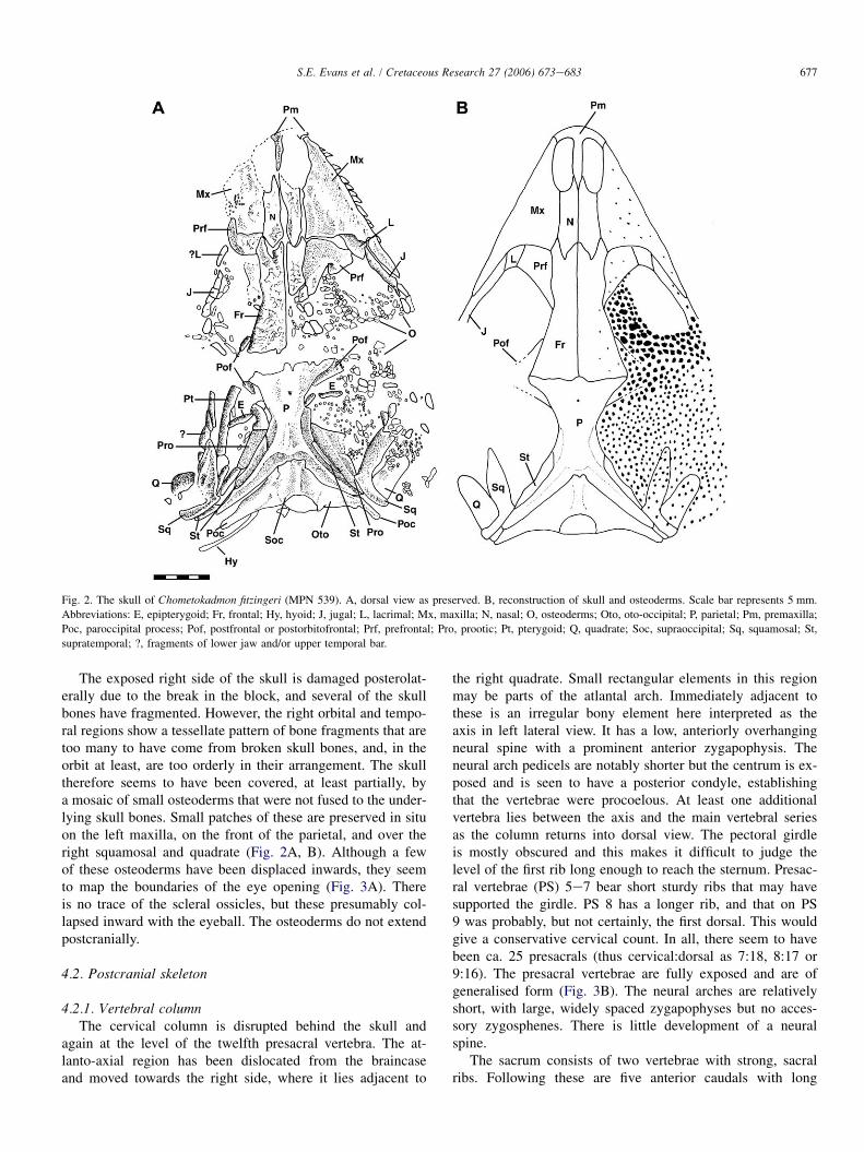
Fig. 3. Chometokadmon fitzingeri (MPN 539). A, enlargement of right orbital region to show detail of the osteoderms. B, mid-dorsal vertebrae. C, enlargement of
pelvic region and sacrum. D, hindlimbs and tail. Scale bars represents 1 mm in A and C, 5 mm in B, and 10 mm in D. Abbreviations: a.t, autotomised tail re-
placement; lFr, left frontal; Il, ilium; Mx, maxilla; Os, cranial osteoderms; Pu, pubis; rFr, right frontal.
transverse processes (length greater than the width of the neu-ral arch), and then a further six with processes that decrease insize. On the third of these, an autotomy septum passesthrough the transverse process. The tail apparently fracturedin life through the fourteenth caudal and then regenerated toa length roughly equal to that of the trunk. An impressionof the soft tissue replacement is preserved on the block(Fig. 3D, a.t).
4.2.2. Pectoral girdle and forelimbThe pectoral girdle is largely obscured by the vertebrae
and ribs, except for some of the cartilaginous epicoracoidparts on the right. Lying below presacral vertebrae 4e5,and overlain by the short ribs of the neck, is a pair of bonyrods connected in a roughly V-shape. This is probably partof an emarginated coracoid plate. There is a similar rod con-tralaterally. The left humerus is almost complete (ca. 18 mmin length), with a narrow proximal head and a broader distal
head bearing a strong entepicondyle and a weak ectepicon-dyle. There is no obvious ectepicondylar foramen. The prox-imal end of the ulna is in articulation, with a fully ossifiedolecranon and no trace of the epiphysial suture, suggestingthe animal was mature (but see below). The rest of the fore-arm and hand extends into the block but further preparationhas not been possible.
4.2.3. Pelvic girdle and hind limbThe pelvis is better preserved than the pectoral girdle
(Fig. 3C, D). Somewhat surprisingly, despite the strong ossifi-cation in other parts of the body, the components of the pelvisare not conjoined. This is a feature usually found either inimmature squamates or aquatic ones, but this is problematicgiven the fusion of the epiphyses. Both ilia are exposed inmedial view (since they have fallen outwards). Each hasa long, rather narrow blade. This is almost horizontal and thesacral rib facets are placed in a relatively proximal position.
679S.E. Evans et al. / Cretaceous Research 27 (2006) 673e683
Each bone has a small anterior tuberosity of a kind frequentlyfound in living terrestrial lizards that show some tendency toraise the trunk on the hind limbs, either in standing (varanids)or bipedal running (many iguanians, some teiids and varanids:Snyder, 1949, 1952, 1954; Brian Ruth, pers. comm. July 2004).Each pubis is elongate, with curved medial and lateral borders,and a short, anteriorly placed, symphysial region. The pectinealprocess is not obvious and was either reduced or dorsally placedand obscured. The ischia are obscured by the sacrum.
The femur is essentially straight (ca. 20 mm) and feature-less. The tibia and fibula are shorter (ca. 16.5 mm), with a ro-bust tibia and a markedly more slender fibula. The ankle ispreserved on the left side. It shows a fully co-ossified astra-galocalcaneum that is proximodistally short but quite wide.The tibial facet is narrow and covers part of the medial mar-gin; the fibular facet is separated from the tibial facet by a nar-row groove, but is raised above the surface of the bone andextends further onto the dorsal surface. The lateral borderof the astragalocalcaneum curves distally, expanding intoa small lateral wing close to the position of metatarsal 5.The distal border is concave medially where it receives a largedistal tarsal (dt) 4, and straighter laterally where it extends to-wards the lateral wing. There are only two distal tarsals, thelarge dt4 and a small, trapezoid dt3. The proximal surface ofdt4 appears to have two condylar surfaces but these mergedistally into a single body. Dt4 meets the fourth and fifthmetatarsals. With the exception of the fifth metatarsal (seebelow), the metatarsals are long and straight with strongproximal heads. They were undoubtedly the strongest partof the foot and make the greatest contribution to its length.The fifth metatarsal is much shorter, proximally displaced,and has both a plantar tubercle and a distolateral flange.The phalanges are relatively short but quite slender and thepedal phalangeal formula is 2:3:4:5:4. The unguals are slen-der. In total, the foot (ca. 32.5 mm along digit 4) is nearlyas long as the femur and tibia combined (ca. 36.5 mm),with a robust proximal part and a rather more delicate distalregion.
5. Phylogenetic position of Chometokadmon
The genus Chometokadmon has remained in obscurity formore than a century, yet the type specimen is one of the rela-tively few articulated lizard fossils known from the Early Cre-taceous of Euramerica (with, for example, Las Hoyas, Spain,Evans and Barbadillo, 1997, 1998, 1999; Tepexi de Rodriguez,Mexico, Reynoso, 1998, Reynoso and Callison, 2000; Pietrar-oia, Evans et al., 2004).
Chometokadmon is clearly a squamate (Costa, 1864) andnot a rhynchocephalian (D’Erasmo, 1915), as demonstratedby the dentition; the structure of the quadrate and its suspen-sion; the procoelous vertebrae; the emarginated coracoid; theelongated anterior ramus of the pubis; and the derived tarsalmorphology. Today, most squamate systematists recognisefour major clades: Iguania, Gekkota, Scincomorpha, and
Anguimorpha, with Serpentes and Amphisbaenia(þDibamidae) variously placed (e.g., Estes et al., 1988; Riep-pel, 1988; Evans and Barbadillo, 1998; Lee, 1998). Mostworkers unite all squamates except iguanians into the cladeScleroglossa (for a detailed discussion of different hypothesesof relationship, see Evans, 2003, but see also Townsend et al.,2004, for an alternative view). Osteologically, Scleroglossaare characterised, amongst other features, by a bifurcate me-dial ramus of the postfrontal/postorbitofrontal (Estes et al.,1988). In addition, with the exception of the marine iguanaAmblyrhynchus (de Queiroz, 1987), no iguanian is known tohave either cranial or postcranial osteoderms, and this featurehas not been reported in any of the JurassiceCretaceous taxaplaced on the squamate stem in recent analyses (e.g., Evansand Barbadillo, 1998, 1999; Evans and Chure, 1998; Rey-noso, 1998). The Jurassic Ardeosaurus (Mateer, 1982) andthe Early Cretaceous Yabeinosaurus (Evans et al., 2005),for example, have cranial ornament but there is no evidencethat this results from the attachment of osteoderms. WithinScleroglossa, osteoderms are rare in Gekkota (but not un-known, e.g., Gekkonia, SEE, pers. obs.), absent in teiids, xan-tusiids and gymnophthalmids, and variable in Varanus (Esteset al., 1988). They are highly developed in anguids, scincids,cordylids, and the extinct Paramacellodidae (scincoid rela-tives; Evans and Chure, 1998), and are also present in helo-dermatids, Lanthanotus (Maisano et al., 2002), Xenosaurusand Shinisaurus (Bever et al., 2005). Indeed, the presenceof dorsal body osteoderms has been listed as a synapomorphyof Anguimorpha (e.g., Estes et al., 1988; Gao and Norell,1998). However, although cranial osteoderms occur in all ofthese groups, body osteoderms may be reduced. Thus Xeno-saurus and Shinisaurus have cranial osteoderms but thereare relatively few on the body (Bever et al., 2005); inLanthanotus, osteoderms are thin and scattered within theskin (Maisano et al., 2002), and in Varanus, they are reducedor absent.
In their preliminary study, Barbera and Macuglia (1988)suggested that Chometokadmon might be a scincoid, butthe absence of dorsal and ventral body osteoderms arguesagainst this, as does the attachment of the jaw adductormusculature to the lateral rather than ventral surfaces ofthe parietal. The combination of slender, recurved, andwell-separated teeth; the small, loosely attached cranial os-teoderms; reduced or absent body osteoderms; paired frontalswith subparallel margins; a slight retraction of the nares;a long, anteriorly extended supratemporal; a relatively elon-gated pubis; and the non-fusion of the pelvic componentsare all features variably found in non-anguid anguimorphs(Estes et al., 1988; Lee, 1998). Conflicting characters includethe slight dorsal process/thickening of the squamosal (butalso in the anguimorph Xenosaurus), the relatively short pre-sacral series (fewer than 26; also in Xenosaurus and Shini-saurus, Estes et al., 1988), and the retention of autotomysepta in the tail. Unfortunately, many key characters of thebraincase, the palate, and the jaws (Estes et al., 1988; Gaoand Norell, 1998; Lee, 1998) remain unknown forChometokadmon.
680 S.E. Evans et al. / Cretaceous Research 27 (2006) 673e683
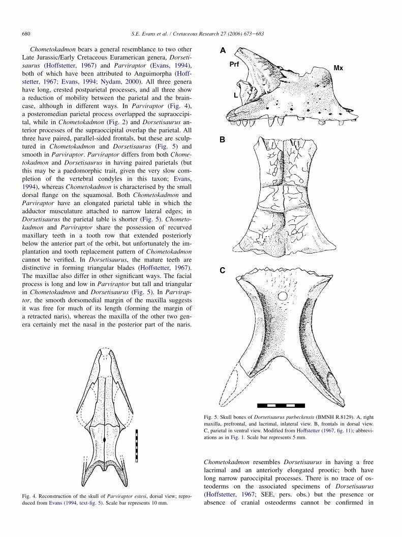
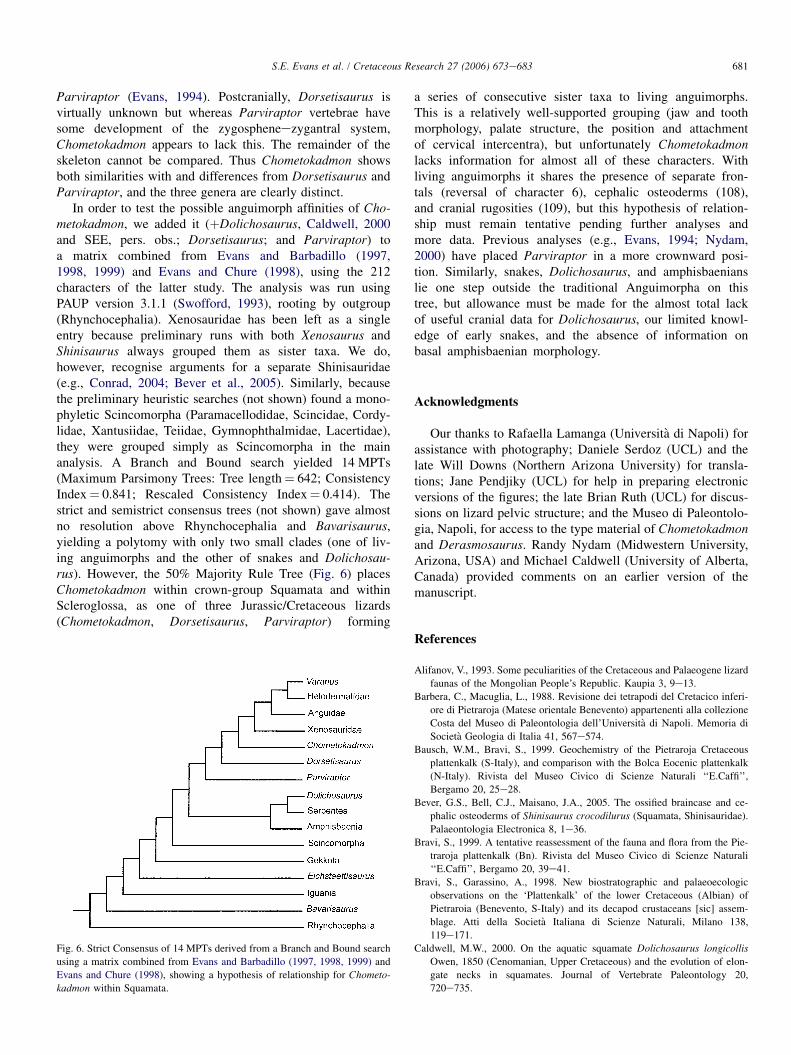
Chometokadmon bears a general resemblance to two otherLate Jurassic/Early Cretaceous Euramerican genera, Dorseti-saurus (Hoffstetter, 1967) and Parviraptor (Evans, 1994),both of which have been attributed to Anguimorpha (Hoff-stetter, 1967; Evans, 1994; Nydam, 2000). All three generahave long, crested postparietal processes, and all three showa reduction of mobility between the parietal and the brain-case, although in different ways. In Parviraptor (Fig. 4),a posteromedian parietal process overlapped the supraoccipi-tal, while in Chometokadmon (Fig. 2) and Dorsetisaurus an-terior processes of the supraoccipital overlap the parietal. Allthree have paired, parallel-sided frontals, but these are sculp-tured in Chometokadmon and Dorsetisaurus (Fig. 5) andsmooth in Parviraptor. Parviraptor differs from both Chome-tokadmon and Dorsetisaurus in having paired parietals (butthis may be a paedomorphic trait, given the very slow com-pletion of the vertebral condyles in this taxon; Evans,1994), whereas Chometokadmon is characterised by the smalldorsal flange on the squamosal. Both Chometokadmon andParviraptor have an elongated parietal table in which theadductor musculature attached to narrow lateral edges; inDorsetisaurus the parietal table is shorter (Fig. 5). Chometo-kadmon and Parviraptor share the possession of recurvedmaxillary teeth in a tooth row that extended posteriorlybelow the anterior part of the orbit, but unfortunately the im-plantation and tooth replacement pattern of Chometokadmoncannot be verified. In Dorsetisaurus, the mature teeth aredistinctive in forming triangular blades (Hoffstetter, 1967).The maxillae also differ in other significant ways. The facialprocess is long and low in Parviraptor but tall and triangularin Chometokadmon and Dorsetisaurus (Fig. 5). In Parvirap-tor, the smooth dorsomedial margin of the maxilla suggestsit was free for much of its length (forming the margin ofa retracted naris), whereas the maxilla of the other two gen-era certainly met the nasal in the posterior part of the naris.
Fig. 4. Reconstruction of the skull of Parviraptor estesi, dorsal view; repro-
duced from Evans (1994, text-fig. 5). Scale bar represents 10 mm.
Chometokadmon resembles Dorsetisaurus in having a freelacrimal and an anteriorly elongated prootic; both havelong narrow paroccipital processes. There is no trace of os-teoderms on the associated specimens of Dorsetisaurus(Hoffstetter, 1967; SEE, pers. obs.) but the presence orabsence of cranial osteoderms cannot be confirmed in
Fig. 5. Skull bones of Dorsetisaurus purbeckensis (BMNH R.8129). A, right
maxilla, prefrontal, and lacrimal, inlateral view. B, frontals in dorsal view.
C, parietal in ventral view. Modified from Hoffstetter (1967, fig. 11); abbrevi-
ations as in Fig. 1. Scale bar represents 5 mm.
681S.E. Evans et al. / Cretaceous Research 27 (2006) 673e683
Parviraptor (Evans, 1994). Postcranially, Dorsetisaurus isvirtually unknown but whereas Parviraptor vertebrae havesome development of the zygospheneezygantral system,Chometokadmon appears to lack this. The remainder of theskeleton cannot be compared. Thus Chometokadmon showsboth similarities with and differences from Dorsetisaurus andParviraptor, and the three genera are clearly distinct.
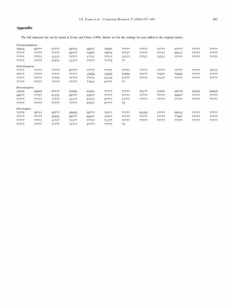
In order to test the possible anguimorph affinities of Cho-metokadmon, we added it (þDolichosaurus, Caldwell, 2000and SEE, pers. obs.; Dorsetisaurus; and Parviraptor) toa matrix combined from Evans and Barbadillo (1997,1998, 1999) and Evans and Chure (1998), using the 212characters of the latter study. The analysis was run usingPAUP version 3.1.1 (Swofford, 1993), rooting by outgroup(Rhynchocephalia). Xenosauridae has been left as a singleentry because preliminary runs with both Xenosaurus andShinisaurus always grouped them as sister taxa. We do,however, recognise arguments for a separate Shinisauridae(e.g., Conrad, 2004; Bever et al., 2005). Similarly, becausethe preliminary heuristic searches (not shown) found a mono-phyletic Scincomorpha (Paramacellodidae, Scincidae, Cordy-lidae, Xantusiidae, Teiidae, Gymnophthalmidae, Lacertidae),they were grouped simply as Scincomorpha in the mainanalysis. A Branch and Bound search yielded 14 MPTs(Maximum Parsimony Trees: Tree length¼ 642; ConsistencyIndex¼ 0.841; Rescaled Consistency Index¼ 0.414). Thestrict and semistrict consensus trees (not shown) gave almostno resolution above Rhynchocephalia and Bavarisaurus,yielding a polytomy with only two small clades (one of liv-ing anguimorphs and the other of snakes and Dolichosau-rus). However, the 50% Majority Rule Tree (Fig. 6) placesChometokadmon within crown-group Squamata and withinScleroglossa, as one of three Jurassic/Cretaceous lizards(Chometokadmon, Dorsetisaurus, Parviraptor) forming
Fig. 6. Strict Consensus of 14 MPTs derived from a Branch and Bound search
using a matrix combined from Evans and Barbadillo (1997, 1998, 1999) and
Evans and Chure (1998), showing a hypothesis of relationship for Chometo-
kadmon within Squamata.
a series of consecutive sister taxa to living anguimorphs.This is a relatively well-supported grouping (jaw and toothmorphology, palate structure, the position and attachmentof cervical intercentra), but unfortunately Chometokadmonlacks information for almost all of these characters. Withliving anguimorphs it shares the presence of separate fron-tals (reversal of character 6), cephalic osteoderms (108),and cranial rugosities (109), but this hypothesis of relation-ship must remain tentative pending further analyses andmore data. Previous analyses (e.g., Evans, 1994; Nydam,2000) have placed Parviraptor in a more crownward posi-tion. Similarly, snakes, Dolichosaurus, and amphisbaenianslie one step outside the traditional Anguimorpha on thistree, but allowance must be made for the almost total lackof useful cranial data for Dolichosaurus, our limited knowl-edge of early snakes, and the absence of information onbasal amphisbaenian morphology.
Acknowledgments
Our thanks to Rafaella Lamanga (Universita di Napoli) forassistance with photography; Daniele Serdoz (UCL) and thelate Will Downs (Northern Arizona University) for transla-tions; Jane Pendjiky (UCL) for help in preparing electronicversions of the figures; the late Brian Ruth (UCL) for discus-sions on lizard pelvic structure; and the Museo di Paleontolo-gia, Napoli, for access to the type material of Chometokadmonand Derasmosaurus. Randy Nydam (Midwestern University,Arizona, USA) and Michael Caldwell (University of Alberta,Canada) provided comments on an earlier version of themanuscript.
References
Alifanov, V., 1993. Some peculiarities of the Cretaceous and Palaeogene lizard
faunas of the Mongolian People’s Republic. Kaupia 3, 9e13.
Barbera, C., Macuglia, L., 1988. Revisione dei tetrapodi del Cretacico inferi-
ore di Pietraroja (Matese orientale Benevento) appartenenti alla collezione
Costa del Museo di Paleontologia dell’Universita di Napoli. Memoria di
Societa Geologia di Italia 41, 567e574.
Bausch, W.M., Bravi, S., 1999. Geochemistry of the Pietraroja Cretaceous
plattenkalk (S-Italy), and comparison with the Bolca Eocenic plattenkalk
(N-Italy). Rivista del Museo Civico di Scienze Naturali ‘‘E.Caffi’’,
Bergamo 20, 25e28.
Bever, G.S., Bell, C.J., Maisano, J.A., 2005. The ossified braincase and ce-
phalic osteoderms of Shinisaurus crocodilurus (Squamata, Shinisauridae).
Palaeontologia Electronica 8, 1e36.
Bravi, S., 1999. A tentative reassessment of the fauna and flora from the Pie-
traroja plattenkalk (Bn). Rivista del Museo Civico di Scienze Naturali
‘‘E.Caffi’’, Bergamo 20, 39e41.
Bravi, S., Garassino, A., 1998. New biostratographic and palaeoecologic
observations on the ‘Plattenkalk’ of the lower Cretaceous (Albian) of
Pietraroia (Benevento, S-Italy) and its decapod crustaceans [sic] assem-
blage. Atti della Societa Italiana di Scienze Naturali, Milano 138,
119e171.
Caldwell, M.W., 2000. On the aquatic squamate Dolichosaurus longicollis
Owen, 1850 (Cenomanian, Upper Cretaceous) and the evolution of elon-
gate necks in squamates. Journal of Vertebrate Paleontology 20,
720e735.
682 S.E. Evans et al. / Cretaceous Research 27 (2006) 673e683
Cocude-Michel, M., 1963. Les rhynchocephales et les sauriens des calcaires
lithographiques (Jurassique superieur) d’Europe occidentale. Nouvelles
Archives du Museum d’Histoire Naturelle de Lyon 7, 1e187.
Conrad, J.L., 2004. Skull, mandible, and hyoid of Shinisaurus crocodilurusAhl (Squamata, Anguimorpha). Zoological Journal of the Linnean Society
141, 399e434.
Costa, O.G., 1864. Paleontologia del Regno di Napoli, III. Atti dell’Accademia
Pontaniana 8, 1e198.
Costa, O.G., 1866. Nuove osservazioni e scoperte intorno ai fossili della cal-
carea ad illioliti di Pietraroja. Atti della Accademia delle Scienze. Fisiche e
Matematiche di Napoli 2, 1e12.
Dal Sasso, C., Signore, M., 1998. Exceptional soft-tissue preservation in a
theropod dinosaur from Italy. Nature 392, 383e387.
D’Erasmo, G., 1915. La fauna e l’eta dei calcari ad ittioliti di Pietraroja
(Provincia di Benevento). Palaeontographia Italica 20, 29e111.
Estes, R., 1983. Sauria Terrestria, Amphisbaenia. In: Wellnhofer, P. (Ed.),
Handbuch der Palaoherpetologie 10A. Gustav Fischer Verlag, Stuttgart,
249 pp.
Estes, R., Gauthier, J., de Queiroz, K., 1988. Phylogenetic relationships within
Squamata. In: Estes, R., Pregill, G. (Eds.), Phylogenetic Relationships of
the Lizard Families: Essays Commemorating Charles L. Camp. Stanford
University Press, Stanford, pp. 119e281.
Evans, S.E., 1994. A new anguimorph lizard from the Jurassic and Lower
Cretaceous of England. Palaeontology 37, 33e49.
Evans, S.E., 2003. At the feet of the dinosaurs: the origin, evolution and early
diversification of squamate reptiles (Lepidosauria: Diapsida). Biological
Reviews, Cambridge 78, 513e551.
Evans, S.E., Barbadillo, L.J., 1997. Early Cretaceous lizards from Las Hoyas,
Spain. Zoological Journal of the Linnean Society 119, 23e49.
Evans, S.E., Barbadillo, L.J., 1998. An unusual lizard from the Early Creta-
ceous of Las Hoyas, Spain. Zoological Journal of the Linnean Society
124, 235e265.
Evans, S.E., Barbadillo, L.J., 1999. A short-limbed lizard from the
Early Cretaceous of Spain. Special Papers in Palaeontology 60,
73e85.
Evans, S.E., Chure, D.J., 1998. Paramacellodid lizard skulls from the Jurassic
Morrison Formation at Dinosaur National Monument, Utah. Journal of
Vertebrate Paleontology 18, 99e114.
Evans, S.E., Raia, P., Barbera, C., 2004. New lizards and rhynchocephalians
from the Lower Cretaceous of southern Italy. Acta Palaeontologica Polon-
ica 49, 393e408.
Evans, S.E., Wang, Y., Li, C., 2005. The Early Cretaceous lizard genus Yabei-
nosaurus from China: resolving an enigma. Journal of Systematic Palaeon-
tology 3, 1e17.
Furbringer, M., 1900. Beitrag zur Systematik und Genealogie der Reptilien.
Jenaischen Zeitschrift fur Naturwissenschaften 34, 596e682.
Gao, K., Norell, M.A., 1998. Taxonomic revision of Carusia (Reptilia: Squa-
mata) from the Late Cretaceous of the Gobi Desert and phylogenetic
relationships of the anguimorphan lizards. American Museum Novitates
3230, 1e51.
Haeckel, E., 1866. Generelle Morphologie der Organismen. Allgemeine
Grundzuge de organismen Formen Wissenschaft mechanisch begrundet
durch die von Charles Darwin reformierte Deszendenz-Theorie. 2: Allge-
meine Entwicklungsgeschichte der Organismen. Kritische Grundzuge der
mechanischen Wissenschaft von den entstehenden Formen der Organis-
men, begrundet durch die Deszendenz-Theorie. Georg Reimer Verlag,
Berlin, 1, 574 pp., 2, 462 pp.
Hoffstetter, R., 1967. Coup d’oeil sur les Sauriens (¼lacertiliens) des couches
de Purbeck (Jurassique superieur d’Angleterre. Resume d’un Memoire).
Colloques Internationaux du Centre National de la Recherche Scientifique
163, 349e371.
von Huene, F., 1956. Palaontologie und Phylogenie der Niederen Tetrapoden.
Gustav Fischer Verlag, Jena, 715 pp.
Kuhn, O., 1969. Proganosauria, Bolosauria, Placodontia, Araeoscelidia, eigel-
tisauria, Millerosauria, Rhynchocephalia, Protorosauria. In: Kuhn, O.
(Ed.), Handbuch der Palaoherpetologie, 9. Gustav Fischer Verlag, Stutt-
gart, 74 pp.
Lee, M.S.Y., 1998. Convergent evolution and character correlation in burrow-
ing reptiles: towards a resolution of squamate relationships. Biological
Journal of the Linnean Society 65, 369e453.
Leonardi, G., Teruzzi, G., 1993. Prima segnalazione di uno scheletro fossile di
dinosauro (Theropoda, Coelurosauria) in Italia (Cretacico di Pietraroia,
Benevento). Paleocronache 1, 7e14.
Maisano, J.A., Bell, C.J., Gauthier, J.A., Rowe, T., 2002. The osteoderms and
palpebral in Lanthanotus borneensis (Squamata: Anguimorpha). Journal of
Herpetology 36, 679e682.
Mateer, N., 1982. Osteology of the Jurassic lizard Ardeosaurus brevipes(Meyer). Palaeontology 25, 461e469.
Nessov, L.A., 1985. Rare bony fishes, terrestrial lizards and mammals
from the lagoonal zone of the littoral lowlands of the Cretaceous
of the Kyzylkum. Ezhegodnik Vsesouyuznogo Paleontologicheskogo
Obshchestva, Academia Nauk SSSR, Leningrad 28, 199e219 (in
Russian).
Nessov, L.A., 1988. Late Mesozoic amphibians and lizards of Soviet Middle
Asia. Acta Zoologica Cracoviensis 31, 475e486.
Nessov, L.A., 1997. Cretaceous Non-marine Vertebrates of Northern Eurasia
(Posthumous edition). In: Golovneva, L.B., Averianov, A.O. (Eds.). Uni-
versity of Saint Petersburg, Institute of the Earth’s Crust, 218 pp., 60
pls. (in Russian).
Nydam, R.L., 2000. A new taxon of helodermatid-like lizard from the
AlbianeCenomanian of Utah. Journal of Vertebrate Paleontology 20,
285e294.
Nydam, R.L., Cifelli, R.L., 2002. Lizards from the Lower Cretaceous
(AptianeAlbian) Antlers and Cloverly formations. Journal of Vertebrate
Paleontology 22, 286e298.
Oppel, M., 1811. Die Ordnungen, Familien und Gattungen der Reptilien,
als Prodrom einer Naturgeschichte derselben. Joseph Lindauer, Munich,
87 pp.
Pierce, S.E., Caldwell, M.W., 2004. Redescription and phylogenetic position
of the Adriatic (Upper Cretaceous; Cenomanian) dolichosaur Pontosaurus
lesinensis (Kornhuber, 1873). Journal of Vertebrate Paleontology 24,
373e386.
de Queiroz, K., 1987. Phylogenetic systematics of iguanine lizards: a compar-
ative osteological study. University of California, Publications in Zoology
118, 1e216.
Reynoso, V.H., 1998. Huehuecuetzpalli mixtecus gen. et sp. nov: a basal
squamate (Reptilia) from the Early Cretaceous of Tepexi de Rodrıguez,
Central Mexico. Philosophical Transactions of the Royal Society B
353, 477e500.
Reynoso, V.H., Callison, G., 2000. A new scincomorph lizard from the Early
Cretaceous of Puebla, Mexico. Zoological Journal of the Linnean Society
130, 183e212.
Rieppel, O., 1988. The classification of the Squamata. In: Benton, M.J. (Ed.),
The Phylogeny and Classification of the Tetrapods. Oxford University
Press, Oxford, pp. 261e293.
Romer, A.S., 1956. The Osteology of the Reptiles. University of Chicago
Press, Chicago, 772 pp.
Snyder, R.C., 1949. Bipedal locomotion of the lizard Basiliscus basiliscus.
Copeia 1949, 129e137.
Snyder, R.C., 1952. Quadrupedal and bipedal locomotion of lizards. Copeia
1952, 64e70.
Snyder, R.C., 1954. The anatomy and function of the pelvic girdle and
hindlimb in lizard locomotion. American Journal of Anatomy 95,
1e36.
Swofford, D.L., 1993. P.A.U.P. e Phylogenetic Analysis Using Parsimony,
version 3.1.1. In: Swofford, D.L. (Ed.).
Townsend, T.M., Larson, A., Louis, E., Macey, J.R., 2004. Molecular
phylogenetics of Squamata: the position of snakes, amphisbaenians,
and dibamids, and the root of the squamate tree. Systematic Biology
53, 735e757.
683S.E. Evans et al. / Cretaceous Research 27 (2006) 673e683
Appendix
The full character list can be found in Evans and Chure (1998). Below we list the codings for taxa added to the original matrix.
Chometokadmon?0010 00??? 0???? 00??2 000?1 ?000? ????? ????? ?1??? 0???? ????? ?????
????? ????? ????? 00??? ?200? ?00?0 ???1? ????? ???1? 0011? ????? ?????
????? ????1 1111? ????? 1??1? ???1? 111?? ???1? ?121? ????? ????? ?????
????? ????? 1?0?1 111?? ????? ????2 ??
Dolichosaurus
????? ????? ????? 0???? ????? ????? ????? ????? ????? ????? ????? ?2??1
?0??? ????? ????? ????? 12024 10230 2?000 ?0??? ??3?? ?0000 ????? ?????
????? ????? ???0? ????? ????1 21110 1???? ????? ??1?? ????? ????? ?????
????? ????? ????? ????? ??01? 0???? ??
Dorsetisaurus100?0 00000 0???? 01000 01001 ????? ????? ?01?? ?100? 00??0 00010 00010
000?? ???1? 01121 00??? 020?? ????? ????? ????? ????? 0000? ????? ?????
????? ????? 1???? 111?? 2?1?1 2???? 1???? ????? ????? ????? ????? ?????
????? ????? ????? ????? 0?01? 0???? ?2
Parviraptor
?1??0 00?11 00??? 00002 00??? ?01?? ??1?? 00100 1???? 00012 ????? ?????
????? ????? 02221 00??? 0201? 1?0?? ????? ????? ????? ??00? ????? ?????
????? ????1 11?1? ?11?? ???1? ?11?? ????? ????? ????? ????? ????? ?????
????? ????? 1???? ?1??? 0???? ????? ?2