Effects of Photosystem-II-Interfering Herbicides Atrazine and Bentazon on the Soybean Transcriptome
Upload
independentCategory
view
0download
0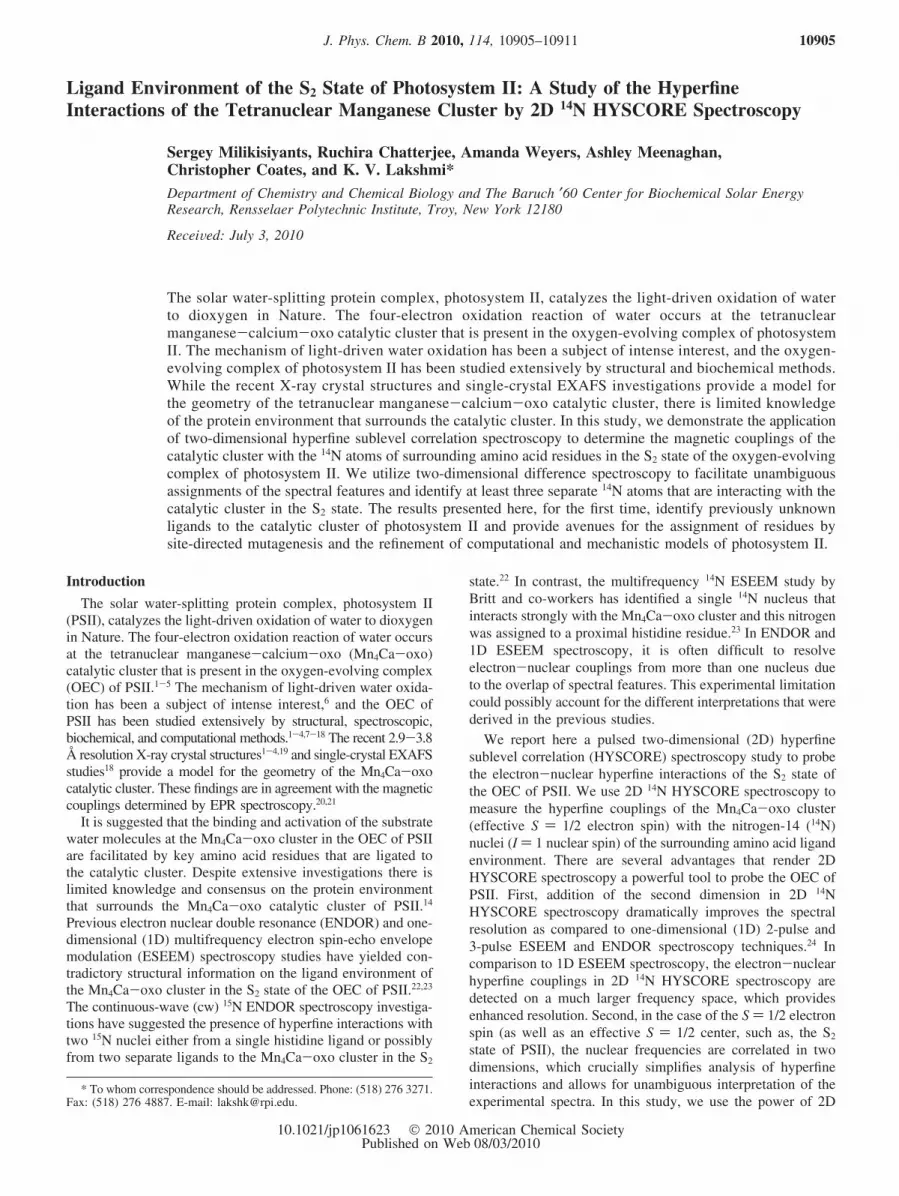
Ligand Environment of the S2 State of Photosystem II: A Study of the HyperfineInteractions of the Tetranuclear Manganese Cluster by 2D 14N HYSCORE Spectroscopy
Sergey Milikisiyants, Ruchira Chatterjee, Amanda Weyers, Ashley Meenaghan,Christopher Coates, and K. V. Lakshmi*Department of Chemistry and Chemical Biology and The Baruch ′60 Center for Biochemical Solar EnergyResearch, Rensselaer Polytechnic Institute, Troy, New York 12180
ReceiVed: July 3, 2010
The solar water-splitting protein complex, photosystem II, catalyzes the light-driven oxidation of waterto dioxygen in Nature. The four-electron oxidation reaction of water occurs at the tetranuclearmanganese-calcium-oxo catalytic cluster that is present in the oxygen-evolving complex of photosystemII. The mechanism of light-driven water oxidation has been a subject of intense interest, and the oxygen-evolving complex of photosystem II has been studied extensively by structural and biochemical methods.While the recent X-ray crystal structures and single-crystal EXAFS investigations provide a model forthe geometry of the tetranuclear manganese-calcium-oxo catalytic cluster, there is limited knowledgeof the protein environment that surrounds the catalytic cluster. In this study, we demonstrate the applicationof two-dimensional hyperfine sublevel correlation spectroscopy to determine the magnetic couplings of thecatalytic cluster with the 14N atoms of surrounding amino acid residues in the S2 state of the oxygen-evolvingcomplex of photosystem II. We utilize two-dimensional difference spectroscopy to facilitate unambiguousassignments of the spectral features and identify at least three separate 14N atoms that are interacting with thecatalytic cluster in the S2 state. The results presented here, for the first time, identify previously unknownligands to the catalytic cluster of photosystem II and provide avenues for the assignment of residues bysite-directed mutagenesis and the refinement of computational and mechanistic models of photosystem II.
Introduction
The solar water-splitting protein complex, photosystem II(PSII), catalyzes the light-driven oxidation of water to dioxygenin Nature. The four-electron oxidation reaction of water occursat the tetranuclear manganese-calcium-oxo (Mn4Ca-oxo)catalytic cluster that is present in the oxygen-evolving complex(OEC) of PSII.1-5 The mechanism of light-driven water oxida-tion has been a subject of intense interest,6 and the OEC ofPSII has been studied extensively by structural, spectroscopic,biochemical, and computational methods.1-4,7-18 The recent 2.9-3.8Å resolution X-ray crystal structures1-4,19 and single-crystal EXAFSstudies18 provide a model for the geometry of the Mn4Ca-oxocatalytic cluster. These findings are in agreement with the magneticcouplings determined by EPR spectroscopy.20,21
It is suggested that the binding and activation of the substratewater molecules at the Mn4Ca-oxo cluster in the OEC of PSIIare facilitated by key amino acid residues that are ligated tothe catalytic cluster. Despite extensive investigations there islimited knowledge and consensus on the protein environmentthat surrounds the Mn4Ca-oxo catalytic cluster of PSII.14
Previous electron nuclear double resonance (ENDOR) and one-dimensional (1D) multifrequency electron spin-echo envelopemodulation (ESEEM) spectroscopy studies have yielded con-tradictory structural information on the ligand environment ofthe Mn4Ca-oxo cluster in the S2 state of the OEC of PSII.22,23
The continuous-wave (cw) 15N ENDOR spectroscopy investiga-tions have suggested the presence of hyperfine interactions withtwo 15N nuclei either from a single histidine ligand or possiblyfrom two separate ligands to the Mn4Ca-oxo cluster in the S2
state.22 In contrast, the multifrequency 14N ESEEM study byBritt and co-workers has identified a single 14N nucleus thatinteracts strongly with the Mn4Ca-oxo cluster and this nitrogenwas assigned to a proximal histidine residue.23 In ENDOR and1D ESEEM spectroscopy, it is often difficult to resolveelectron-nuclear couplings from more than one nucleus dueto the overlap of spectral features. This experimental limitationcould possibly account for the different interpretations that werederived in the previous studies.
We report here a pulsed two-dimensional (2D) hyperfinesublevel correlation (HYSCORE) spectroscopy study to probethe electron-nuclear hyperfine interactions of the S2 state ofthe OEC of PSII. We use 2D 14N HYSCORE spectroscopy tomeasure the hyperfine couplings of the Mn4Ca-oxo cluster(effective S ) 1/2 electron spin) with the nitrogen-14 (14N)nuclei (I ) 1 nuclear spin) of the surrounding amino acid ligandenvironment. There are several advantages that render 2DHYSCORE spectroscopy a powerful tool to probe the OEC ofPSII. First, addition of the second dimension in 2D 14NHYSCORE spectroscopy dramatically improves the spectralresolution as compared to one-dimensional (1D) 2-pulse and3-pulse ESEEM and ENDOR spectroscopy techniques.24 Incomparison to 1D ESEEM spectroscopy, the electron-nuclearhyperfine couplings in 2D 14N HYSCORE spectroscopy aredetected on a much larger frequency space, which providesenhanced resolution. Second, in the case of the S ) 1/2 electronspin (as well as an effective S ) 1/2 center, such as, the S2
state of PSII), the nuclear frequencies are correlated in twodimensions, which crucially simplifies analysis of hyperfineinteractions and allows for unambiguous interpretation of theexperimental spectra. In this study, we use the power of 2D
* To whom correspondence should be addressed. Phone: (518) 276 3271.Fax: (518) 276 4887. E-mail: [email protected].
J. Phys. Chem. B 2010, 114, 10905–10911 10905
10.1021/jp1061623 2010 American Chemical SocietyPublished on Web 08/03/2010
14N HYSCORE difference spectroscopy to identify at least threeseparate 14N nuclei that are interacting with the Mn4Ca-oxocluster in the S2 state of the OEC of PSII. The results presentedhere directly address the lack of experimental data on the ligandenvironment of the Mn4Ca-oxo cluster of PSII and identifypreviously unknown ligands to the cluster.
Materials and Methods
Preparation of Photosystem II Complexes and Trappingof the S State Intermediates. The PsbB hexa-histidine-tagged(His-tagged) mutant Synechocystis PCC 6803 cells were grownat 24 °C in BG-11 medium.25 The PSII complexes were isolatedfrom the cells by minor modifications of previously publishedprocedures.26 The PSII samples were monitored by SDS-polyacrylamide gel electrophoresis, and the densitometry scansshowed very minor PSI contamination (<3%). The O2-evolutionrates were monitored by a Clark electrode and were typically4800 µmol of O2 (mg of Chl)-1 h-1.27 The PSII complexes usedfor the EPR measurements of the S2 and S1 states of the OECwere resuspended in buffer containing 50 mM 2-(N-morpholi-no)ethanesulfonic acid-sodium hydroxide (MES-NaOH), 20 mMcalcium chloride (CaCl2), 5 mM magnesium chloride (MgCl2),0.03% (w/v) dodecyl-�-D-maltoside (�-DM), and 30% glycerol(w/v) at pH 6.0. The PSII samples were preincubated with 200µM potassium ferricyanide prior to illumination. The S2 statewas cryo-trapped by 45 s illumination at 200 K followed byrapid freezing (5-10 s) at 77 K, in the dark. The S1 state wasaccumulated by dark adapting the PSII sample at 0 °C for 30min.
Pulsed EPR Spectroscopy. EPR spectra were recorded ona custom-built continuous-wave (cw)/pulsed X-band BrukerElexsys 580 spectrometer. The pulsed EPR measurements wereconducted with a dielectric flex-line probe ER 4118-MD5(Bruker BioSpin Corp., Billerica, MA) and a dynamic continu-ous-flow cryostat CF935 (Oxford Instruments, Oxfordshire,U.K.). The operating microwave frequency was set to 9.71 GHzto best match the broad band of the strongly overcoupled pulsedresonator. All of the pulsed EPR spectra were acquired at 5 K.
For the echo-detected field sweep spectra, the primary electronspin echo was generated using the pulse sequence (π/2-τ-πecho). The echo was integrated over a 40 ns time window, whichwas centered at the maximum of the echo signal. The length ofthe π/2 and π pulse was 12 and 24 ns, respectively. Theinterpulse separation, τ, was 144 ns, and the delay in the pulsesequence is defined as the difference in the starting point of thepulses. The spectra were recorded over the range of 300 mTcentered at 350 mT.
For the 2D HYSCORE spectra, the echo amplitude wasmeasured using the pulse sequence (π/2-τ-π/2-t1-π-t2-π/2echo) with an 8 and 16 ns length for the π/2 and π pulse,respectively, and an 8 ns detector gate (that is centered at themaximum of the echo signal). The delays in the pulse sequenceare defined as the difference in the starting point of the pulses.The echo intensity was measured as a function of t1 and t2, wheret1 and t2 were incremented in steps of 16 ns from an initial valueof 40 and 32 ns, respectively. There were 256 steps used foreach dimension. The 8 ns time difference between the initialvalue of t1 and t2 was set to take into account the difference inlength between the π/2 and π pulse. This provided symmetricspectra in both dimensions. The unwanted echoes and antiechoeswere eliminated by applying a 16-step phase-cycling procedure.A third-order polynomial baseline was subtracted from theresulting time-domain spectra. The corrected spectra were zerofilled to obtain a [2048 × 2048] matrix and Fourier transformedusing a fast Fourier transformation (FFT) algorithm. Thefrequency domain spectra were plotted as the amplitude(absolute value) of the 2D frequency components.
Results and Discussion
In the echo-detected EPR spectra shown in Figure 1A and1B, we observe the characteristic ‘multiline’ EPR spectrum ofthe S2 state (S ) 1/2) (shown as a red trace) and the dark stablebackground EPR spectrum of the S1 state (shown as a blue trace)of PSII.7,28-30 Upon comparison of the line shape of the EPRspectra of the S2 and S1 state, we observe that in addition to themultiline EPR signal that arises from the 55Mn electron-nuclearhyperfine interactions of the Mn4Ca-oxo cluster in the S2 statethere are at least two additional paramagnetic centers thatcontribute to the observed spectrum: (i) a very narrow signal atg ≈ 2 from the redox-active tyrosyl radical, YD
• (this signal ismarked by an asterisk (*) in Figure 1A), and (ii) the heme Fe(III)center of cytochrome b559 (Cyt b559) that displays a rhombicspectrum due to the presence of g anisotropy (gX ) 1.5, gY )2.3, and gZ ) 3.0).31 In the present study, we use S2-minus-S1
state difference spectroscopy to eliminate the dark-stable spectralcontribution from the heme Fe(III) center of Cyt b559. Thederivative of the S2-minus-S1 state EPR difference spectrum isshown in Figure 1B.
On the basis of the S2-minus-S1 EPR difference spectrum inFigure 1B, we perform 2D 14N HYSCORE spectroscopy at threedifferent magnetic field positions (B) of 355 (g′ ) 1.95), 380(g′ ) 1.82), and 337.5 mT (g′ ) 2.055), respectively, wherethe effective g value (g′) is calculated as g′ ) hVmw/�eB usingthe microwave frequency (Vmw) and Bohr magneton (�e). We
Figure 1. (A) Echo-detected EPR spectrum of intact PSII with OEC trapped in the S2 (red line) and S1 (blue line) state. (B) Numerical derivativeof the S2-minus-S1 difference EPR spectrum that is obtained from the data shown in A. (*) is the region of the spectrum containing the EPR signalfrom the YD
• radical; (**) is the region of the spectrum containing unavoidable resonator artifacts.
10906 J. Phys. Chem. B, Vol. 114, No. 33, 2010 Milikisiyants et al.
also incorporate three different values for the interpulse delay,τ, of 132, 140, and 176 ns in the 2D 14N HYSCOREmeasurements. We select the magnetic field position(s) for theacquisition of the 2D 14N HYSCORE spectrum to precludeoverlap with the residual signal from the dark-stable YD
• radicalof PSII, and we optimize the interpulse delay(s) in the pulsesequence to avoid blind spots in the spectrum.
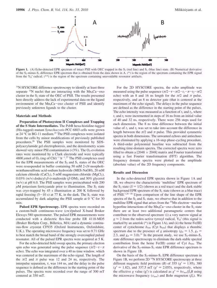
Figure 2A and 2B displays the 2D 14N HYSCORE spectraof the S2 and S1 state of PSII, respectively, acquired at themagnetic field position of g′ ) 1.95. On the basis of the EPRspectra shown in Figure 1A, we expect that the 2D 14NHYSCORE spectrum of the S2 state at the magnetic fieldposition of g′ ) 1.95 (Figure 2A) could exhibit hyperfinecouplings due to the Mn4Ca-oxo cluster in the S2 state and theheme Fe(III) center of Cyt b559. To identify the spectralcontributions from the heme Fe(III) center of Cyt b559, weexamine the 2D 14N HYSCORE spectrum of intact PSII withthe OEC in the dark-stable S1 state (Figure 2B). As can be seenin Figure 2B, there is a small spectral contribution from theheme Fe(III) center of Cyt b559 in the S1 state of the OEC ofPSII. Therefore, we isolate the spectral features that arise fromthe S2 state of PSII with the observation of the S2-minus-S1 2D14N HYSCORE difference spectrum (Figure 2C).
In the S2-minus-S1 2D 14N HYSCORE difference spectrumin Figure 2C, we observe the 14N hyperfine couplings of theMn4Ca-oxo cluster in the S2 state of the OEC of PSII. Inaddition, we also observe weak 14N hyperfine couplings thatcould be due to the QA
- center in the S2 state of PSII. Whenthe S1 state is illuminated at cryogenic temperature (200 K), itundergoes a one-electron oxidation and advances to the S2 state.The electron is transferred to the primary quinone, QA, whichis reduced to the semiquinone anion, QA
- (also referred to asthe ‘iron-QA
- couple’ as the QA- semiquinone is magnetically
coupled to the nonheme Fe(II) center of PSII).5 Thus, in additionto the presence of 14N hyperfine couplings from the S2 state,there exists the possibility of 14N hyperfine contributions fromQA
- (or iron-QA- couple) in the S2-minus-S1 2D 14N HY-
SCORE difference spectrum. The spectral features that wetentatively assign to QA
- (labeled as Fe2+-QA- in Figure 2C)
are similar to previously published hyperfine couplings of theQA
- center of PSII.32,33
We also acquire the 2D 14N HYSCORE spectrum of the S2
and S1 state of PSII at the magnetic field position of g′ ) 1.82and 2.055. We observe that the unwanted spectral features fromweakly coupled 14N nuclei from QA
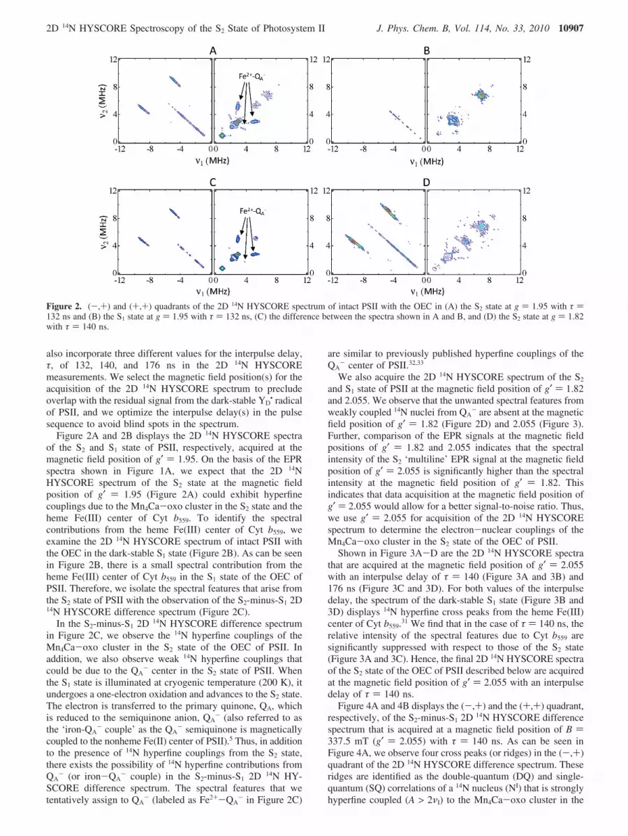
- are absent at the magneticfield position of g′ ) 1.82 (Figure 2D) and 2.055 (Figure 3).Further, comparison of the EPR signals at the magnetic fieldpositions of g′ ) 1.82 and 2.055 indicates that the spectralintensity of the S2 ‘multiline’ EPR signal at the magnetic fieldposition of g′ ) 2.055 is significantly higher than the spectralintensity at the magnetic field position of g′ ) 1.82. Thisindicates that data acquisition at the magnetic field position ofg′ ) 2.055 would allow for a better signal-to-noise ratio. Thus,we use g′ ) 2.055 for acquisition of the 2D 14N HYSCOREspectrum to determine the electron-nuclear couplings of theMn4Ca-oxo cluster in the S2 state of the OEC of PSII.
Shown in Figure 3A-D are the 2D 14N HYSCORE spectrathat are acquired at the magnetic field position of g′ ) 2.055with an interpulse delay of τ ) 140 (Figure 3A and 3B) and176 ns (Figure 3C and 3D). For both values of the interpulsedelay, the spectrum of the dark-stable S1 state (Figure 3B and3D) displays 14N hyperfine cross peaks from the heme Fe(III)center of Cyt b559.31 We find that in the case of τ ) 140 ns, therelative intensity of the spectral features due to Cyt b559 aresignificantly suppressed with respect to those of the S2 state(Figure 3A and 3C). Hence, the final 2D 14N HYSCORE spectraof the S2 state of the OEC of PSII described below are acquiredat the magnetic field position of g′ ) 2.055 with an interpulsedelay of τ ) 140 ns.
Figure 4A and 4B displays the (-,+) and the (+,+) quadrant,respectively, of the S2-minus-S1 2D 14N HYSCORE differencespectrum that is acquired at a magnetic field position of B )337.5 mT (g′ ) 2.055) with τ ) 140 ns. As can be seen inFigure 4A, we observe four cross peaks (or ridges) in the (-,+)quadrant of the 2D 14N HYSCORE difference spectrum. Theseridges are identified as the double-quantum (DQ) and single-quantum (SQ) correlations of a 14N nucleus (NI) that is stronglyhyperfine coupled (A > 2νI) to the Mn4Ca-oxo cluster in the
Figure 2. (-,+) and (+,+) quadrants of the 2D 14N HYSCORE spectrum of intact PSII with the OEC in (A) the S2 state at g ) 1.95 with τ )132 ns and (B) the S1 state at g ) 1.95 with τ ) 132 ns, (C) the difference between the spectra shown in A and B, and (D) the S2 state at g ) 1.82with τ ) 140 ns.
2D 14N HYSCORE Spectroscopy of the S2 State of Photosystem II J. Phys. Chem. B, Vol. 114, No. 33, 2010 10907
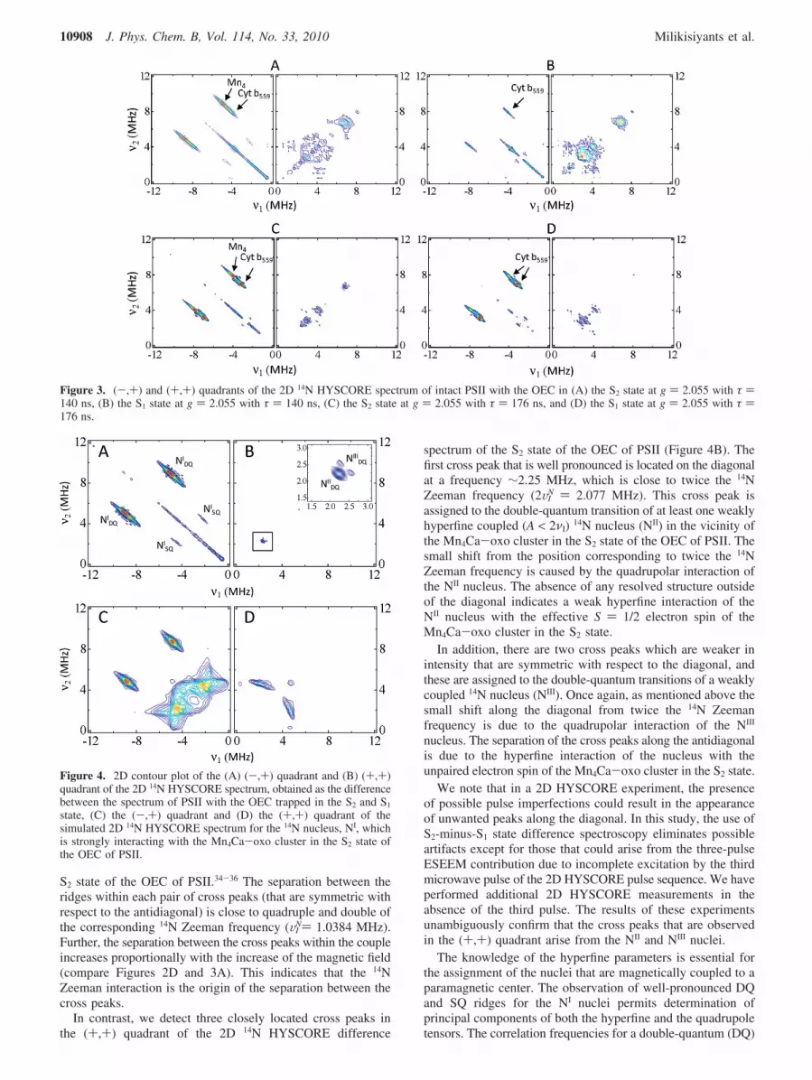
S2 state of the OEC of PSII.34-36 The separation between theridges within each pair of cross peaks (that are symmetric withrespect to the antidiagonal) is close to quadruple and double ofthe corresponding 14N Zeeman frequency (VI
N) 1.0384 MHz).Further, the separation between the cross peaks within the coupleincreases proportionally with the increase of the magnetic field(compare Figures 2D and 3A). This indicates that the 14NZeeman interaction is the origin of the separation between thecross peaks.
In contrast, we detect three closely located cross peaks inthe (+,+) quadrant of the 2D 14N HYSCORE difference
spectrum of the S2 state of the OEC of PSII (Figure 4B). Thefirst cross peak that is well pronounced is located on the diagonalat a frequency ∼2.25 MHz, which is close to twice the 14NZeeman frequency (2VI
N ) 2.077 MHz). This cross peak isassigned to the double-quantum transition of at least one weaklyhyperfine coupled (A < 2νI) 14N nucleus (NII) in the vicinity ofthe Mn4Ca-oxo cluster in the S2 state of the OEC of PSII. Thesmall shift from the position corresponding to twice the 14NZeeman frequency is caused by the quadrupolar interaction ofthe NII nucleus. The absence of any resolved structure outsideof the diagonal indicates a weak hyperfine interaction of theNII nucleus with the effective S ) 1/2 electron spin of theMn4Ca-oxo cluster in the S2 state.
In addition, there are two cross peaks which are weaker inintensity that are symmetric with respect to the diagonal, andthese are assigned to the double-quantum transitions of a weaklycoupled 14N nucleus (NIII). Once again, as mentioned above thesmall shift along the diagonal from twice the 14N Zeemanfrequency is due to the quadrupolar interaction of the NIII
nucleus. The separation of the cross peaks along the antidiagonalis due to the hyperfine interaction of the nucleus with theunpaired electron spin of the Mn4Ca-oxo cluster in the S2 state.
We note that in a 2D HYSCORE experiment, the presenceof possible pulse imperfections could result in the appearanceof unwanted peaks along the diagonal. In this study, the use ofS2-minus-S1 state difference spectroscopy eliminates possibleartifacts except for those that could arise from the three-pulseESEEM contribution due to incomplete excitation by the thirdmicrowave pulse of the 2D HYSCORE pulse sequence. We haveperformed additional 2D HYSCORE measurements in theabsence of the third pulse. The results of these experimentsunambiguously confirm that the cross peaks that are observedin the (+,+) quadrant arise from the NII and NIII nuclei.
The knowledge of the hyperfine parameters is essential forthe assignment of the nuclei that are magnetically coupled to aparamagnetic center. The observation of well-pronounced DQand SQ ridges for the NI nuclei permits determination ofprincipal components of both the hyperfine and the quadrupoletensors. The correlation frequencies for a double-quantum (DQ)
Figure 3. (-,+) and (+,+) quadrants of the 2D 14N HYSCORE spectrum of intact PSII with the OEC in (A) the S2 state at g ) 2.055 with τ )140 ns, (B) the S1 state at g ) 2.055 with τ ) 140 ns, (C) the S2 state at g ) 2.055 with τ ) 176 ns, and (D) the S1 state at g ) 2.055 with τ )176 ns.
Figure 4. 2D contour plot of the (A) (-,+) quadrant and (B) (+,+)quadrant of the 2D 14N HYSCORE spectrum, obtained as the differencebetween the spectrum of PSII with the OEC trapped in the S2 and S1
state, (C) the (-,+) quadrant and (D) the (+,+) quadrant of thesimulated 2D 14N HYSCORE spectrum for the 14N nucleus, NI, whichis strongly interacting with the Mn4Ca-oxo cluster in the S2 state ofthe OEC of PSII.
10908 J. Phys. Chem. B, Vol. 114, No. 33, 2010 Milikisiyants et al.
transition in the case of S ) 1/2 and I ) 1 spins are given byfollowing expressions34
where VI is the Zeeman frequency for the given magnetic field(VI
N) 1.0384 MHz for B ) 337.5 mT), A1,2,3 are the principalcomponents of the hyperfine tensor, l1,2,3 are the direction cosinesof the magnetic field, K ) e2qQ/4p, Q is the quadrupole momentinteracting with the electric field gradient q, and η is theasymmetry parameter. The signs of eqs 1 and 2 are equal forthe case of weakly interacting nuclei and the cross peak appearsin the (+,+) and (-,-) quadrants of the 2D HYSCOREspectrum. In the case of strongly interacting nuclei, the signsof υR and υ� are opposite and the cross peak appears in the(-,+) and (+,-) quadrants.
Since in powder samples all of the orientations are excitedby the microwave pulses at X-band EPR frequency, the crosspeaks form extended ridges on the 2D frequency domain. Theridges are confined between two points which are determinedby the orientation of the magnetic field along the canonicaldirections corresponding to the smallest (A1) and to the largest(A3) hyperfine components. The coordinates of these two pointsfor the upper ridge are easily obtained from the eqs 1 and 2
For the lower ridge, the order of the coordinates for each pointis reversed, i.e., [υR;υ�] point of the upper ridge corresponds tothe [υ�;υR] point of the lower ridge.
In the present study, for the strongly interacting 14N nucleus,NI, the coordinates of two extreme positions of the upper DQridge, [-6.17 MHz; 10.22 MHz] and [-3.65 MHz; 7.60 MHz],provide an accurate determination of the components of thehyperfine tensor, A1 ) 5.34 MHz, A3 ) 8.0 MHz, and the valueof K2(3 + η2) ) 0.705 MHz2. To obtain the values for A2 andη we perform numerical simulations using the ‘saffron’ functionof the EasySpin (version 3.1.0) software.37 For the spectralsimulations, A1, A3, and K2(3 + η2) are fixed at the values
determined above. As an approximation, we assume that theprincipal axis frames of the hyperfine and quadrupole tensorsare collinear. The best fit of the experimental and simulatedspectra is obtained with A2 ) 6.7 MHz and η ) 0.9 (thesimulations of the (+,-) and (+,+) quadrants of the 2D 14NHYSCORE spectrum are shown in Figure 4C and 4D).However, a range of values A2 ) 6.2-7.2 MHz and η ) 0.6 -1 are in agreement with the simulations of the experimental2D 14N HYSCORE spectra. Please note that the simulations ofthe (+,-) and (+,+) quadrants of the 2D 14N HYSCOREspectrum that are shown in Figure 4C and 4D contain additionalfeatures that are not observed in the experimental 2D 14NHYSCORE spectra in Figure 4A and 4B. This is because theadditional peaks that are observed in the simulated spectrumare an order of magnitude weaker in intensity and are compa-rable to the overall noise level of the experiment. Hence, theweak features that are present in the simulation are not observedin the experimental spectrum.
For the second 14N nucleus, NII, one DQ cross peak isobserved corresponding to the correlation frequencies υR ≈ υ�
≈ 2�(υI2 + K2(3 + η2)) ) 2.25 MHz, providing the value of
K2(3 + η2) ) 0.22 MHz2. The homogeneous width of this crosspeak is determined by the decay time of the correspondingmodulations in the time domain spectra. The width of the crosspeak also increases along the antidiagonal direction due to thehyperfine interaction. Since it is difficult to separate the twocontributions, we provide a conservative estimate of the upperlimit for the hyperfine components |A1,2,3| e 0.3 MHz for the14N nucleus, NII that is interacting with the Mn4Ca-oxo clusterin the S2 state of the OEC of PSII.
For the third 14N nucleus, NIII, the position of the upper DQridge is limited by the extreme positions with coordinates [2.63MHz; 2.16 MHz] and [2.43 MHz; 2.36 MHz]. Application ofeqs 3 and 4 to these coordinates yield the following values: A1
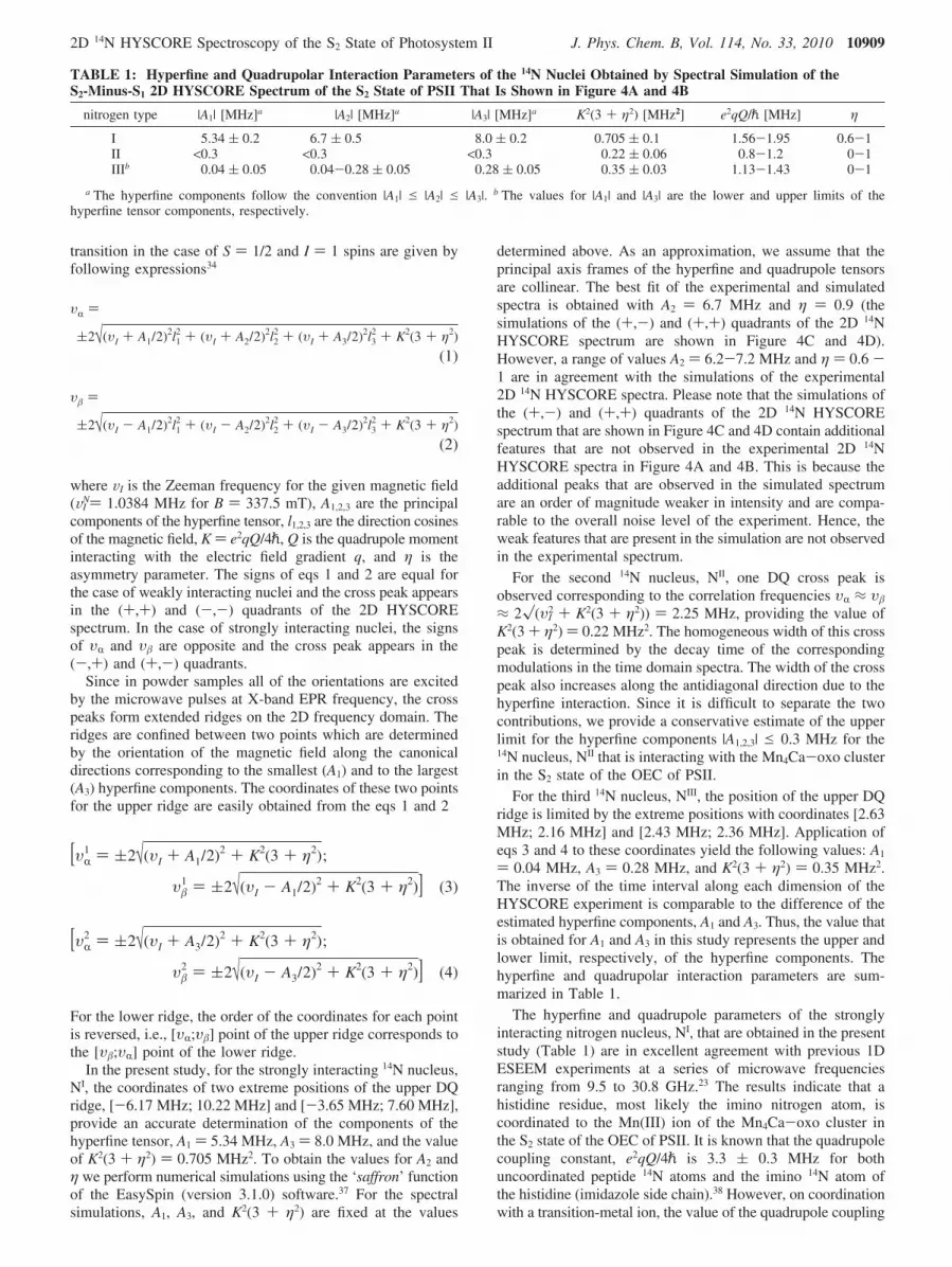
) 0.04 MHz, A3 ) 0.28 MHz, and K2(3 + η2) ) 0.35 MHz2.The inverse of the time interval along each dimension of theHYSCORE experiment is comparable to the difference of theestimated hyperfine components, A1 and A3. Thus, the value thatis obtained for A1 and A3 in this study represents the upper andlower limit, respectively, of the hyperfine components. Thehyperfine and quadrupolar interaction parameters are sum-marized in Table 1.
The hyperfine and quadrupole parameters of the stronglyinteracting nitrogen nucleus, NI, that are obtained in the presentstudy (Table 1) are in excellent agreement with previous 1DESEEM experiments at a series of microwave frequenciesranging from 9.5 to 30.8 GHz.23 The results indicate that ahistidine residue, most likely the imino nitrogen atom, iscoordinated to the Mn(III) ion of the Mn4Ca-oxo cluster inthe S2 state of the OEC of PSII. It is known that the quadrupolecoupling constant, e2qQ/4p is 3.3 ( 0.3 MHz for bothuncoordinated peptide 14N atoms and the imino 14N atom ofthe histidine (imidazole side chain).38 However, on coordinationwith a transition-metal ion, the value of the quadrupole coupling
TABLE 1: Hyperfine and Quadrupolar Interaction Parameters of the 14N Nuclei Obtained by Spectral Simulation of theS2-Minus-S1 2D HYSCORE Spectrum of the S2 State of PSII That Is Shown in Figure 4A and 4B
nitrogen type |A1| [MHz]a |A2| [MHz]a |A3| [MHz]a K2(3 + η2) [MHz2] e2qQ/p [MHz] η
I 5.34 ( 0.2 6.7 ( 0.5 8.0 ( 0.2 0.705 ( 0.1 1.56-1.95 0.6-1II <0.3 <0.3 <0.3 0.22 ( 0.06 0.8-1.2 0-1IIIb 0.04 ( 0.05 0.04-0.28 ( 0.05 0.28 ( 0.05 0.35 ( 0.03 1.13-1.43 0-1
a The hyperfine components follow the convention |A1| e |A2| e |A3|. b The values for |A1| and |A3| are the lower and upper limits of thehyperfine tensor components, respectively.
υR )
(2√(υI + A1/2)2l12 + (υI + A2/2)2l2
2 + (υI + A3/2)2l32 + K2(3 + η2)
(1)
υ� )
(2√(υI - A1/2)2l12 + (υI - A2/2)2l2
2 + (υI - A3/2)2l32 + K2(3 + η2)
(2)
[υR1 ) (2√(υI + A1/2)2 + K2(3 + η2);
υ�1 ) (2√(υI - A1/2)2 + K2(3 + η2)] (3)
[υR2 ) (2√(υI + A3/2)2 + K2(3 + η2);
υ�2 ) (2√(υI - A3/2)2 + K2(3 + η2)] (4)
2D 14N HYSCORE Spectroscopy of the S2 State of Photosystem II J. Phys. Chem. B, Vol. 114, No. 33, 2010 10909
constant is reduced to ∼2 MHz for an imino nitrogen atom ofthe histidine39 while it slightly increases for a peptide nitrogenatom.40 The quadrupole constant for the 14N nucleus, NI, that isobserved in this study is too small to be attributed to a peptidenitrogen atom. However, it is excellent agreement with the rangeof values that have been reported for the imino nitrogen atomof a histidine interacting with a metal cluster.39 Thus, we assignthe hyperfine-coupled NI nucleus as an imino nitrogen atom ofa histidine residue that is coordinated to the Mn4Ca-oxo clusterin the S2 state of the OEC of PSII. This assignment is inagreement with previous suggestions that a histidine residue isdirectly coordinated to the Mn4Ca-oxo cluster in the OEC.41
The hyperfine tensor of the 14N nucleus, NI, that is observedin this study is not in agreement with previous cw 15N ENDORspectroscopy studies that indicated that the Mn4Ca-oxo clusterinteracts with two nitrogen nuclei with hyperfine couplingconstants of 3.7 and 0.7 MHz.22 Taking into account thedifference of the gyromagnetic ratio of the 15N and 14N isotopes,the larger hyperfine coupling constant of 3.7 MHz obtained inthe cw ENDOR study would correspond to a hyperfine couplingconstant of 2.64 MHz in this study. This value is much lessthan any of the three principal hyperfine components that areobtained for the 14N nucleus, NI (Table 1).
By exploiting the power of the higher resolution of 2DHYSCORE spectroscopy, for the first time, we demonstrate thepresence of two additional 14N nuclei, NII and NIII, that areweakly hyperfine coupled to the Mn4Ca-oxo cluster in the S2
state of the OEC of PSII. These interactions have previouslybeen elusive using 1D ESEEM and ENDOR spectroscopytechniques. The value for the quadrupolar interaction of NII istoo small to be attributed to either an imino or an amino nitrogenatom of a histidine residue39 or a peptide nitrogen atom.42 Thequadrupolar interaction of the NII nucleus is in agreementwith that previously reported for the terminal nitrogen atomof amino acids.38 Although the X-ray crystal structures andthe computational models of photosystem II suggest thepresence of nitrogen atoms in the vicinity of the Mn4Ca-oxocluster,1-4,16,17,43-46 these structures do not display a terminalnitrogen atom of an amino acid side chain as a potentialligand to the cluster. This could be due to the limitedresolution of currently available X-ray crystal structures ofphotosystem II. Thus, at the present time the unambiguousassignment of the NII nitrogen remains a topic for furtherinvestigations.
The value of the quadrupolar interaction of the NIII nucleuscan most reliably be attributed to the presence of the aminonitrogen atom that likely belongs to the same histidine residueas the directly coordinated imino nitrogen (NI) that is discussedabove. Previous studies of transition-metal-imidazole com-plexes have demonstrated that the hyperfine constant of the distalnitrogen atom is typically more than an order of magnitude lessthan that of the directly coordinated proximal nitrogen atom ofthe imidazole group.47 The value that is obtained for thehyperfine components of both the NIII and the NI nitrogen atomscorrespond to this finding and provide additional support forthe presence of a histidine ligand that is coordinated to theMn4Ca-oxo cluster in the S2 state of the OEC of PSII. Theresults presented here, for the first time, identify three 14N atomsthat are magnetically coupled to the catalytic cluster of photo-system II and provide a template for the nitrogen ligation ofthe Mn4Ca-oxo cluster in the S2 state of the OEC of PSII. Thisstudy presents opportunities for the assignment of residues bysite-directed mutagenesis and for the refinement of computa-tional and mechanistic models of PSII.
Acknowledgment. This research was supported by the SolarEnergy Utilization Program, Office of Basic Energy Sciences,United States Department of Energy (DE-FG02-0ER06-15).
Note Added after ASAP Publication. This paper waspublished on the Web on August 3, 2010. Two referencecitations were misnumbered in the Results and Discussionsection. The corrected version was reposted on August 19, 2010.
References and Notes
(1) Zouni, A.; Witt, H. T.; Kern, J.; Fromme, P.; Krauss, N.; Saenger,W.; Orth, P. Nature 2001, 409, 739.
(2) Kamiya, N.; Shen, J. R. Proc. Natl. Acad. Sci. U.S.A. 2003, 100,98.
(3) Ferreira, K. N.; Iverson, T. M.; Maghlaoui, K.; Barber, J.; Iwata,S. Science 2004, 303, 1831.
(4) Loll, B.; Kern, J.; Saenger, W.; Zouni, A.; Biesiadka, J. Nature2005, 438, 1040.
(5) Debus, R. J. Biochim. Biophys. Acta 1992, 1102, 269.(6) McEvoy, J. P.; Brudvig, G. W. Phys. Chem. Chem. Phys. 2004, 6,
4754.(7) Brudvig, G. W.; Thorp, H. H.; Pecoraro, V. L. AdV. Chem. Ser.
1995, 246, 249.(8) Peloquin, J. M.; Britt, R. D. Biochim. Biophys. Acta, Bioenerg. 2001,
1503, 96.(9) Messinger, J.; Nugent, J. H. A.; Evans, M. C. W. Biochemistry
1997, 36, 11055.(10) Ahrling, K. A.; Peterson, S.; Styring, S. Biochemistry 1998, 37,
8115.(11) Kulik, L. V.; Epel, B.; Lubitz, W.; Messinger, J. J. Am. Chem.
Soc. 2005, 127, 2392.(12) Yachandra, V. K.; Sauer, K.; Klein, M. P. Chem. ReV. 1996, 96,
2927.(13) Chu, H. A.; Hillier, W.; Law, N. A.; Babcock, G. T. Biochim.
Biophys. Acta, Bioenerg. 2001, 1503, 69.(14) Debus, R. J. Coord. Chem. ReV. 2008, 252, 244.(15) Yano, J.; Pushkar, Y.; Glatzel, P.; Lewis, A.; Sauer, K.; Messinger,
J.; Bergmann, U.; Yachandra, V. J. Am. Chem. Soc. 2005, 127, 14974.(16) Sproviero, E. M.; Gascon, J. A.; McEvoy, J. P.; Brudvig, G. W.;
Batista, V. S. Curr. Opin. Struct. Biol. 2007, 17, 173.(17) Sproviero, E. M.; Gascon, J. A.; McEvoy, J. P.; Brudvig, G. W.;
Batista, V. S. Coord. Chem. ReV. 2008, 252, 395.(18) Yano, J.; Kern, J.; Sauer, K.; Latimer, M. J.; Pushkar, Y.; Biesiadka,
J.; Loll, B.; Saenger, W.; Messinger, J.; Zouni, A.; Yachandra, V. K. Science2006, 314, 821.
(19) Guskov, A.; Kern, J.; Gabdulkhakov, A.; Broser, M.; Zouni, A.;Saenger, W. Nat. Struct. Mol. Biol. 2009, 16, 334.
(20) Peloquin, J. M.; Campbell, K. A.; Randall, D. W.; Evanchik, M. A.;Pecoraro, V. L.; Armstrong, W. H.; Britt, R. D. J. Am. Chem. Soc. 2000,122, 10926.
(21) Randall, D. W.; Sturgeon, B. E.; Ball, J. A.; Lorigan, G. A.; Chan,M. K.; Klein, M. P.; Armstrong, W. H.; Britt, R. D. J. Am. Chem. Soc.1995, 117, 11780.
(22) Tang, X. S.; Sivaraja, M.; Dismukes, G. C. J. Am. Chem. Soc. 1993,115, 2382.
(23) Yeagle, G. J.; Gilchrist, M. L.; McCarrick, R. M.; Britt, R. D. Inorg.Chem. 2008, 47, 1803.
(24) Schweiger, A.; Jeschke, G. Principles of Pulsed Electron Para-magnetic Resonance; Oxford University Press: New York, 2001.
(25) Rippka, R.; Deruelles, J.; Waterbury, J. B.; Herdman, M.; Stanier,R. Y. J. Gen. Microbiol. 1979, 111, 1.
(26) Lakshmi, K. V.; Reifler, M. J.; Chisholm, D. A.; Wang, J. Y.; Diner,B. A.; Brudvig, G. W. Photosynth. Res. 2002, 72, 175.
(27) Beck, W. F.; Depaula, J. C.; Brudvig, G. W. Biochemistry 1985,24, 3035.
(28) Brudvig, G. W. ACS Symp. Ser. 1988, 372, 221.(29) Depaula, J. C.; Beck, W. F.; Brudvig, G. W. J. Am. Chem. Soc.
1986, 108, 4002.(30) Zheng, M.; Dismukes, G. C. Inorg. Chem. 1996, 35, 3307.(31) Garcia-Rubio, I.; Martinez, J. I.; Picorel, R.; Yruela, I. L.; Alonso,
P. J. J. Am. Chem. Soc. 2003, 125, 15846.(32) Deligiannakis, Y.; Hanley, J.; Rutherford, A. W. J. Am. Chem. Soc.
1999, 121, 7653.(33) Deligiannakis, Y.; Rutherford, A. W. J. Inorg. Biochem. 2000, 79,
339.(34) Dikanov, S. A.; Xun, L. Y.; Karpiel, A. B.; Tyryshkin, A. M.;
Bowman, M. K. J. Am. Chem. Soc. 1996, 118, 8408.(35) Astashkin, A. V.; Dikanov, S. A.; Tsvetkov, Y. D. J. Struct. Chem.
1984, 25, 45.
10910 J. Phys. Chem. B, Vol. 114, No. 33, 2010 Milikisiyants et al.

(36) Flanagan, H. L.; Singel, D. J. J. Chem. Phys. 1987, 87, 5606.(37) Stoll, S.; Schweiger, A. J. Magn. Reson. 2006, 178, 42.(38) Edmonds, D. T. Phys. Rep., Phys. Lett. 1977, 29, 234.(39) Ashby, C. I. H.; Cheng, C. P.; Brown, T. L. J. Am. Chem. Soc.
1978, 100, 6057.(40) Ashby, C. I. H.; Paton, W. F.; Brown, T. L. J. Am. Chem. Soc.
1980, 102, 2990.(41) Tang, X. S.; Diner, B. A.; Larsen, B. S.; Gilchrist, M. L.; Lorigan,
G. A.; Britt, R. D. Proc. Natl. Acad. Sci. U.S.A. 1994, 91, 704.(42) Blinc, R.; Mali, M.; Osredkar, R.; Seliger, J.; Ehrenber., L. Chem.
Phys. Lett. 1974, 28, 158.
(43) Sproviero, E. M.; McEvoy, J. P.; Gascon, J. A.; Brudvig, G. W.;Batista, V. S. Photosynth. Res. 2008, 97, 91.
(44) Sproviero, E. M.; Gascon, J. A.; McEvoy, J. P.; Brudvig, G. W.;Batista, V. S. J. Am. Chem. Soc. 2008, 130, 3428.
(45) Sproviero, E. M.; Shinopoulos, K.; Gascon, J. A.; McEvoy, J. P.; Brudvig,G. W.; Batista, V. S. Philos. Trans. R. Soc., Ser. B: Biol. Sci. 2008, 363, 1149.
(46) Batista, V.; Sproviero, E.; Gascon, J.; McEvoy, J. P.; Brudvig, G.Photosyn. Res. 2007, 91, PS42.
(47) Deligiannakis, Y.; Louloudi, M.; Hadjiliadis, N. Coord. Chem. ReV.2000, 204, 1.
JP1061623
2D 14N HYSCORE Spectroscopy of the S2 State of Photosystem II J. Phys. Chem. B, Vol. 114, No. 33, 2010 10911
Copyright © 2022 FDOKUMEN