Synthesis, characterization and crystal structure of a new unsymmetric tetranuclear copper-carbonate...
7
Synthesis, characterization and crystal structure of catena-bis(1,10-phenanthroline)-l-chloranilatocadmium(II): a zig-zag polymer based on eight-coordinated square antiprismatic units Adonis Michaelides, Christos D. Papadimitriou, John C. Plakatouras * , Stavroula Skoulika, Panagiotis G. Veltsistas * Department of Chemistry, University of Ioannina, 451 10 Ioannina, Greece Received 29 June 2004; accepted 22 September 2004 Available online 14 October 2004 Abstract [Cd(phen) 2 (CA)] n (I, where phen is 1,10-phenanthroline and CA 2 is the dianion of 3,6-dichloro-2,5-dihydroxy-1,4-benzoqui- none) has been synthesized and its crystal structure has been determined by single-crystal X-ray diffraction at room temperature. The crystal of I is built from infinite chains of chloranilate-bridged cadmium(II) leading to a zig-zag structure, with the phenanthr- oline ligands being stacked between the chains. These interactions assist the formation of a closely packed 3D structure. The coor- dination environment of cadmium(II) – very scarce in its chemistry – is square antiprismatic. The polymer was also characterized with spectroscopic (IR, Raman, DRS) and thermogravimetric (TG, DTA) techniques. Ó 2004 Elsevier Ltd. All rights reserved. Keywords: Cadmium(II); Chloranilate anions; Phenanthroline; Square antiprism; Crystal structures; Chain polymers 1. Introduction The synthesis of coordination polymers is one of the most essential elements in advancing materials chemistry. In general, it remains more of an art than a science, since most new compounds have been synthesized through self- assembly routes and not from controlled synthesis. Re- search on that area is directed towards the utilization of ligands that maintain their structure in solution and pos- sess more than one well-known coordination sites [1]. Building metal complex – based polymers is an important step to create not only a system to perform optical, elec- tronic and magnetic functions, but also an intercalation system for ion- or molecule-exchange, absorption and catalytic properties (see for example [2,3]). Substituted dihydroxy benzoquinones, and especially chloranilic acid (CA), hold an important place in the fam- ily of bridging ligands [4–15]. This is due to their two allyl systems connected by C–C single bonds, with four par- tially negatively charged oxygen atoms [16] (Scheme 1). Thus, the chelating effect in addition to the bridging capa- bility of the chloranilate dianion allows the formation of robust frameworks. In addition, the polyoxocarbon mol- ecules have frontier orbitals with energies comparable to those of the transition metal ions and the nature of the re- duced and oxidized species depends on the metal ions and the coordination environment [12]. The delocalized p sys- tem associated with its planar geometry can provide [9,13] interesting and possibly useful electronic communi- cation between suitable metal centers. 0277-5387/$ - see front matter Ó 2004 Elsevier Ltd. All rights reserved. doi:10.1016/j.poly.2004.09.013 * Corresponding authors. Tel.: +30 26510 98417; fax: +30 26510 44831. E-mail address: [email protected] (J.C. Plakatouras). www.elsevier.com/locate/poly Polyhedron 23 (2004) 2587–2593
Transcript of Synthesis, characterization and crystal structure of a new unsymmetric tetranuclear copper-carbonate...
www.elsevier.com/locate/poly
Polyhedron 23 (2004) 2587–2593
Synthesis, characterization and crystal structure ofcatena-bis(1,10-phenanthroline)-l-chloranilatocadmium(II):
a zig-zag polymer based on eight-coordinatedsquare antiprismatic units
Adonis Michaelides, Christos D. Papadimitriou, John C. Plakatouras *,Stavroula Skoulika, Panagiotis G. Veltsistas *
Department of Chemistry, University of Ioannina, 451 10 Ioannina, Greece
Received 29 June 2004; accepted 22 September 2004
Available online 14 October 2004
Abstract
[Cd(phen)2(CA)]n (I, where phen is 1,10-phenanthroline and CA2� is the dianion of 3,6-dichloro-2,5-dihydroxy-1,4-benzoqui-
none) has been synthesized and its crystal structure has been determined by single-crystal X-ray diffraction at room temperature.
The crystal of I is built from infinite chains of chloranilate-bridged cadmium(II) leading to a zig-zag structure, with the phenanthr-
oline ligands being stacked between the chains. These interactions assist the formation of a closely packed 3D structure. The coor-
dination environment of cadmium(II) – very scarce in its chemistry – is square antiprismatic. The polymer was also characterized
with spectroscopic (IR, Raman, DRS) and thermogravimetric (TG, DTA) techniques.
� 2004 Elsevier Ltd. All rights reserved.
Keywords: Cadmium(II); Chloranilate anions; Phenanthroline; Square antiprism; Crystal structures; Chain polymers
1. Introduction
The synthesis of coordination polymers is one of the
most essential elements in advancingmaterials chemistry.
In general, it remains more of an art than a science, since
most new compounds have been synthesized through self-
assembly routes and not from controlled synthesis. Re-
search on that area is directed towards the utilization of
ligands that maintain their structure in solution and pos-sess more than one well-known coordination sites [1].
Buildingmetal complex – based polymers is an important
step to create not only a system to perform optical, elec-
tronic and magnetic functions, but also an intercalation
0277-5387/$ - see front matter � 2004 Elsevier Ltd. All rights reserved.
doi:10.1016/j.poly.2004.09.013
* Corresponding authors. Tel.: +30 26510 98417; fax: +30 26510
44831.
E-mail address: [email protected] (J.C. Plakatouras).
system for ion- or molecule-exchange, absorption andcatalytic properties (see for example [2,3]).
Substituted dihydroxy benzoquinones, and especially
chloranilic acid (CA), hold an important place in the fam-
ily of bridging ligands [4–15]. This is due to their two allyl
systems connected by C–C single bonds, with four par-
tially negatively charged oxygen atoms [16] (Scheme 1).
Thus, the chelating effect in addition to the bridging capa-
bility of the chloranilate dianion allows the formation ofrobust frameworks. In addition, the polyoxocarbon mol-
ecules have frontier orbitals with energies comparable to
those of the transition metal ions and the nature of the re-
duced and oxidized species depends on themetal ions and
the coordination environment [12]. The delocalized p sys-
tem associated with its planar geometry can provide
[9,13] interesting and possibly useful electronic communi-
cation between suitable metal centers.

O
O O
O
Cl
Cl
Cl
Cl
O-
-O O
O
(a) (b)
Scheme 1.
2588 A. Michaelides et al. / Polyhedron 23 (2004) 2587–2593
Our continuous interest in chloranilate chemistry is
stimulated by both its analytical applications and coor-
dination properties. Chloranilic acid can be used for the
determination of Mg(II) with FAAS [17], for the con-
struction of electrode membranes [18], and the kinetic
determinations of formaldehyde [19] and Mn(II) [20].
Our involvement in the coordination chemistry of the
chloranilates have produced several monomeric [21–23]and polymeric [11,24] coordination compounds with
interesting structural features. For example, in the case
of hydrated sodium chloranilate [11], the ligand can
bind four sodium cations using all of its oxygen atoms
and one of the organic chlorine atoms, while the number
of sodium cations bound to CA is reduced to three when
1,10-phenanthroline is used to block sodium coordina-
tion sites.In the present paper, we present the synthesis, the crys-
tal structure and the spectroscopic and thermal character-
ization of the chain compound [Cd(phen)2(CA)]n. The
structure is based on rare square antiprismatic CdN4O4
polyhedra connected through bis-chelating chloranilato
bridges. Stacking interactions provide a 3D supramolec-
ular architecture.
2. Experimental
2.1. Materials
All manipulations were carried out under aerobic
conditions. Cd(CH3COO)2 Æ 2H2O was purchased from
Merck, sodium silicate solution, KBr, BaSO4 and organicreagents were purchased from Aldrich. Solvents were
of analytical grade and purchased from Lab-Scan
Chemical Co.
2.2. Instrumentation
C, H and N analyses were conducted by the Univer-
sity of Ioannina, Greece, Microanalytical Service. IR(4000–370 cm�1) and far-IR (600–30 cm�1) spectra were
recorded on a Perkin–Elmer Spectrum GX FT IR Sys-
tem with samples prepared as KBr and polyethylene pel-
lets, respectively. The Raman spectrum was recorded on
a SPEX Model 1403 0.85 m double spectrometer,
equipped with a Spectra Physics Model 2020 argon ion
laser; a spinning cell sample holder was used to avoid
thermal degradation of the compound. The excitation
was provided by the 514.5 nm line with a laser power
of 100 mW, at a resolution of 2 cm�1 and signal record-
ing was done using photon counting with an integrationtime of 1 s. UV/Vis diffuse reflectance spectra were re-
corded on a Shimadzu UV-2401 PC spectrophotometer
with a Shimadzu integration sphere. Thermal studies
were carried out on a Shimadzu DTG-60 simultaneous
DTA-TG apparatus, under N2 or forced air flow (50
cm3 min�1) with a heating rate of 5 �C min�1. Sample
sizes were about 5 mg. X-ray powder diffraction patterns
were recorded on a Siemens (now Bruker) D8 AD-VANCE diffractometer.
2.3. Preparation of the compound
Cd(CH3COO)2 Æ 2H2O (0.50 g, 1.88 mmol) was dis-
solved in H2O (20 cm3). To this, a solution of 1,10-phen-
anthroline (0.676 g, 3.75 mmol) in ethanol (10 cm3) was
added. A white solid precipitated readily and dissolvedafter the addition of 2 cm3 of glacial acetic acid. Sepa-
rately, chloranilic acid (0.393 g, 1.88 mmol) was dis-
solved in water and the pH was adjusted to 7 with an
aqueous solution (ca. 0.5 g in 20 cm3 H2O) of
tris(hydroxymethyl)amino methane. The resulting solu-
tion was added to the metal solution to give a dark pur-
ple mixture. The mixture was refluxed for 2 h. The dark
purple solid which separated upon cooling was filtered,washed with ethanol (3 · 10 cm3) and diethylether
(3 · 10 cm3) and dried under vacuum over silica gel.
Yield: 0.97 g (76% based on cadmium).
Slow reaction of an aqueous cadmium acetate solu-
tion (0.1 M) with an aqueous solution containing so-
dium chloranilate (0.05 M) and 1,10-phenanthroline
(0.05 M) through a sodium silicate gel (formed after
titration of 2 M HNO3 with a ca. 2 M sodium silicateup to pH � 5.3) in a U tube led to large purple crystals
suitable for X-ray structural studies.
Anal. Calc. for C30H16CdCl2N4O4: C, 53.00; H, 2.38;
N, 8.24. Found: C, 52.71; H, 2.63; N, 8.39%. Selected IR
(KBr pellet) and far-IR (polyethylene pellet) data
(cm�1): 3077w, 3065w, 1624w, 1513vs, 1424ms, 1374m,
1275m, 1207w, 1099w, 1047w, 988w, 841ms, 770w,
728m, 634w, 589w, 573m, 434m, 417m, 377w, 272m,230s, 215msh, 178w, 156s, 145s, 127s. Selected Raman
data (cm�1): 3076w, 1615w, 1521m, 1460s, 1427m,
1346s, 1305s, 1051m, 725s, 586s, 566w, 423m, 312w,
123m. Electronic spectrum (DRS, BaSO4 solution)
(nm): 520, 325.
2.4. X-ray crystallography
A suitable single crystal of I was selected under a
polarizing microscope, cleaned from the gel residues
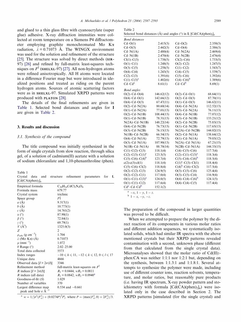
Table 2
Selected bond distances (A) and angles (�) in I, [Cd(CA)(phen)2]n
Bond distances
Cd–O(1) 2.415(3) Cd–O(2) 2.350(3)
Cd–O(3) 2.442(3) Cd–O(4) 2.386(3)
Cd–N(1A) 2.480(4) Cd–N(2A) 2.469(4)
Cd–N(1B) 2.470(4) Cd–N(2B) 2.476(4)
Cl(1)–C(3) 1.738(5) Cl(2)–C(6) 1.733(5)
O(1)–C(1) 1.248(5) O(2)–C(2) 1.247(5)
O(3)–C(5) 1.258(5) C(1)–C(2) 1.543(7)
O(4)–C(4) 1.243(5) C(4)–C(5) 1.539(7)
C(2)–C(3) 1.391(6) C(5)–C(6) 1.392(6)
C(1)–C(3)a 1.402(6) C(4)–C(6)b 1.389(6)
Cd–Cda 8.61(1) Cd–Cdb 8.69(1)
Bond angles
O(2)–Cd–O(4) 146.62(12) O(2)–Cd–O(1) 68.64(11)
O(4)–Cd–O(1) 142.66(12) O(2)–Cd–O(3) 87.70(11)
O(4)–Cd–O(3) 67.47(11) O(1)–Cd–O(3) 146.62(11)
O(2)–Cd–N(2A) 80.60(14) O(4)–Cd–N(2A) 112.52(13)
O(1)–Cd–N(2A) 77.01(13) O(3)–Cd–N(2A) 76.11(13)
O(2)–Cd–N(1B) 108.44(13) O(4)–Cd–N(1B) 77.07(12)
A. Michaelides et al. / Polyhedron 23 (2004) 2587–2593 2589
and glued to a thin glass fibre with cyanoacrylate (super
glue) adhesive. X-ray diffraction intensities were col-
lected at room temperature on a Siemens P4 diffractom-
eter employing graphite monochromated Mo Karadiation, k = 0.71073 A. The WINGX environment
was used for the solution and refinement of the structure[25]. The structure was solved by direct methods (SIRSIR-
97) [26] and refined by full-matrix least-squares tech-
niques on F2 (SHELXLSHELXL-97) [27]. All non-hydrogen atoms
were refined anisotropically. All H atoms were located
in a difference Fourier map but were introduced in ide-
alized positions and treated as riding on the parent
hydrogen atoms. Sources of atomic scattering factors
were as in SHELXLSHELXL-97. Simulated XRPD patterns wereproduced with PLATONPLATON [28].
The details of the final refinements are given in
Table 1. Selected bond distances and angles for I
are given in Table 2.
O(1)–Cd–N(1B) 78.31(13) O(3)–Cd–N(1B) 133.21(12)
N(2A)–Cd–N(1B) 148.22(14) O(2)–Cd–N(2B) 75.83(13)
O(4)–Cd–N(2B) 76.73(13) O(1)–Cd–N(2B) 117.65(13)
O(3)–Cd–N(2B) 76.15(13) N(2A)–Cd–N(2B) 144.02(15)
N(1B)–Cd–N(2B) 66.54(15) O(2)–Cd–N(1A) 138.64(12)
O(4)–Cd–N(1A) 72.79(13) O(1)–Cd–N(1A) 79.03(12)
O(3)–Cd–N(1A) 107.90(13) N(2A)–Cd–N(1A) 67.21(15)
N(1B)–Cd–N(1A) 88.70(14) N(2B)–Cd–N(1A) 144.33(15)
C(1)–C(2)–C(3) 118.1(4) C(4)–C(5)–C(6) 117.7(4)
C(2)–C(3)–C(1)a 123.5(5) C(2)–C(1)–C(3)a 118.3(4)
C(5)–C(6)–C(4)b 123.7(4) C(5)–C(4)–C(6)b 118.5(4)
c(2)-c(3)-cl(1) 118.1(4) C(1)a–C(3)–Cl(1) 118.4(4)
3. Results and discussion
3.1. Synthesis of the compound
The title compound was initially synthesized in the
form of single crystals from slow reaction, through silica
gel, of a solution of cadmium(II) acetate with a solution
of sodium chloranilate and 1,10-phenanthroline (phen).
Table 1
Crystal data and structure refinement parameters for I,
[Cd(CA)(phen)2]n
Empirical formula C30H16CdCl2N4O4
Formula mass 679.77
Crystal system triclinic
Space group P�1a (A) 9.317(1)
b (A) 10.773(1)
c (A) 14.761(2)
a (�) 87.90(1)
b (�) 72.84(1)
c (�) 69.79(1)
V (A3) 1325.0(3)
Z 2
qcalc (g cm�3) 1.704
k (Mo Ka) (A) 0.71073
l (mm�1) 1.072
h Range (�) 2.02–25.00
Total data collected 5573
Index ranges �10 6 h 6 11, �12 6 k 6 12, 0 6 l 6 17
Unique data 4666
Observed data [I > 2r (I)] 3346
Refinement method full-matrix least-squares on F2
R indices [I > 2r(I)] R1 = 0.0444, wR2 = 0.0811
R indices (all data) R1 = 0.0842, wR2 = 0.0940a
Goodness-of-fit (S) 1.029
Number of variables 370
Largest difference map
peak and hole e A�3
0.534 and �0.661
a w ¼ 1=½r2ðF 2oÞ þ ð0:0274P Þ2P �; where P ¼ ðmaxðF 2
o; 0Þ þ 2F 2cÞ=3.
C(5)–C(6)–Cl(2) 118.8(4) C(4)b–C(6)–Cl(2) 117.5(4)
O(2)–C(2)–C(3) 124.9(5) O(3)–C(5)–C(6) 125.4(4)
O(2)–C(2)–C(1) 117.0(4) O(3)–C(5)–C(4) 116.9(4)
O(1)–C(1)–C(3)a 124.0(5) O(4)–C(4)–C(6)b 124.1(5)
O(1)–C(1)–C(2) 117.6(4) O(4)–C(4)–C(5) 117.4(4)
Cda–Cd–Cdb 152.1(2)
a �x, 1 � y, 1 � z.b 1 � x, �y, �z.
The preparation of the compound in larger quantities
was proved to be difficult.
When we attempted to prepare the polymer by the di-
rect reaction of its components in various molar ratios
and different addition sequences, we systematically iso-
lated solids, which had similar IR spectra with the above
mentioned crystals but their XRPD patterns revealed
contamination with a second, unknown phase (differentfrom that calculated from the single crystal data).
Microanalyses showed that the molar ratio of Cd(II):-
phen:CA was neither 1:1:1 nor 1:2:1 but, depending on
the synthesis, between 1:1.3:1 and 1:1.8:1. Several at-
tempts to synthesize the polymer were made, including
use of different counter ions, reaction solvents, tempera-
ture, and molar ratios, but reasonably pure products
(i.e. having IR spectrum, X-ray powder pattern and sto-ichiometry with formula [Cd(CA)(phen)2]n) were iso-
lated only in the case described in Section 2. The
XRPD patterns [simulated (for the single crystal) and

2590 A. Michaelides et al. / Polyhedron 23 (2004) 2587–2593
experimental (for the separately prepared bulk sample)]
are presented in Fig. 1.
We believe that this behavior can be explained in
terms of the kinetic and thermodynamic stability of
the two products, [Cd(CA)(phen)]n and [Cd(CA)-
(phen)2]n, probably involved in the reaction mixture.Bearing in mind that chloranilate will act as bis-chelat-
ing bridging ligand, when in lower concentration than
the metal, the 1D polymer [Cd(CA)(phen)]n would be
the initial product, according to Eq. (I). This product
must be kinetically stable.
nCd2þ þ nCA2� þ nphen ! ½CdðCAÞðphenÞ�n ðIÞThe next, most obvious, step in a successive additionscenario is the addition of the second phen molecule
to the coordination sphere of cadmium, which should
begin almost simultaneously, and lead to the formation
of the thermodynamically stable product as shown in
the following equation.
Fig. 1. Experimental (b) and simulated (a) powder XRD patterns for I.
Fig. 2. (a) ORTEP [29] plot of the asymmetric unit of [Cd(CA)(phen)2]n (I) i
clarity. (b) The coordination polyhedron about Cd, defined as square antipr
½CdðCAÞðphenÞ�n þ nphen ! ½CdðCAÞðphenÞ2�n ðIIÞThough speculative, the mechanism above is sup-
ported by two experimental observations: (a) the micro-
analyses of the insoluble products suggested
formulations [Cd(CA)(phen)x], where 1 < x < 2 and (b)
only after extended heating of the reaction mixture
was the desired product isolated.
3.2. Description of the structure
An ORTEP drawing of the asymmetric unit of
[Cd(CA)(phen)2]n is presented in Fig. 2(a). Selected
bond distances (A) and angles (�) with their standard
deviations are presented in Table 2.
The crystal structure of the polymer is built from zig-
zag chains (type I chain structure), extended along the
[�1,1,1] direction, accompanied by by stacking interac-
tions (Fig. 3).The asymmetric unit consists of one cadmium atom,
two 1,10-phenanthroline molecules and halves of two
different chloranilato ligands (Fig. 2(a)). The coordina-
tion sphere of the metal is formed by four nitrogen
atoms belonging to two chelating phen ligands, and four
oxygen atoms belonging to two different bis-chelating
bridging chloranilato ligands.
There are several examples of eight-coordinate cad-mium complexes in the literature. They can be separated
in two different groups. Those with multidentate ligands
(see for example [30–32]) and those with bidentate
chelating ligands with small bite angles (see for example
[33–35]). To the best of our knowledge the only exam-
ples of eight-coordinated Cd(II) complexes with four
five-membered chelate rings belong to the family of
the oxalate polymers [36–39].The geometry about Cd(II) can be described as dis-
torted square antiprismatic. As indicated by Drew [40],
the values of u angles, which assess the planarity of
the two trapezoids formed in an eight-coordination
ncluding the labelling scheme. Hydrogen atoms have been omitted for
ism.

Fig. 3. A part of the 2D sheet (a) formed by stacking interactions of phenanthroline (phen A) molecules. The second phenanthroline (B) forms
stacking interactions with the corresponding phen of a neighboring sheet. By this interaction a second sheet intersecting the previous is formed (b). As
a result a 3D structure is obtained.
A. Michaelides et al. / Polyhedron 23 (2004) 2587–2593 2591
environment, are a good test for the determination of
the shape of the coordination polyhedron. The u angles,
calculated by using the hard sphere model (HSM) shape
characteristics, are 0.0� and 24.5� for the ideal dodecahe-dron and the ideal square antiprism, respectively. In our
case, the u values are 26.78� and 29.53�, which indicatesthat the coordination polyhedron is best described as a
distorted square antiprism. The coordination polyhe-
dron is presented in Fig. 2(b). The centroids of the CA
moieties are located on inversion centres.
The Cd–N bond distances span the range 2.470(4)–
2.481(4) A and they can be considered long, related to
other Cd–phen complexes. A survey of the Cambridge
Structural Database shows that there are ca. 40 crystalstructures of Cd–phen complexes. None of them has
eight-coordinated cadmium, probably due to steric ef-
fects. Phen is too large and two of them cannot be easily
accommodated in an eight-coordination environment
unless other stabilizing interactions (p–p stacking for
example) are involved, as well. The coordination num-
ber itself can explain the long length of the Cd–N bonds.
The two CA ligands, in the coordination sphere ofCd, are bonded in a slightly different fashion. There
are four different Cd–O bond distances in the range
2.350(3)–2.443(3) A. These distances are well in the
range observed in the literature [41,42]. This asymmetry,
in addition to the square antiprismatic geometry of
course, leads to the formation of the zig-zag chain. As
far as the C–O distances are concerned, both of the chlo-
ranilates chelate Cd symmetrically (C(1)–O(1), C(2)–O(2), 1.246(5) A, C(4)–O(4), 1.244(5) A, C(5)–O(3),
1.256(5) A). The differences are very small compared
with the associated su�s. The C–O bond characteristics
are similar to those reported for the free chloranilate
dianion [43]. The similarity between the C–O bonds is
reflected to the C–C bond lengths, connecting the two
parts of the chloranilato ligand, as shown in Scheme
1(b). In both chloranilates the distances of the C–C
bonds connecting the negatively charged sites are longerthan the rest C–C distances (mean values; 1.542, 1.394
A, respectively). These differences are systematic and
suggest extended conjugation between halves of the
CA ligands.
Both phen moieties are involved in stacking interac-
tions (Fig. 3) which lead to a 3D supramolecular archi-
tecture. Phen A (Fig. 3(a)) utilizes the central ring for
p–p interaction with the same ring of a parallel chain.The distance between the least square planes is 3.55 A,
and the offset of the centroids is 0.50 A. That zipper-like
interaction leads to the formation of 2D sheets, which
interact with each other through the stacking of the B
phen molecule (Fig. 3(b)). Two rings, the central and
one external, are involved in this interaction. The dis-
tance of the least square planes is 3.44 A, and the offset
of the two ring centroids is 1.36 A.
3.3. Thermal stability and spectroscopic characterization
The polymer is stable up to 315 �C, when heated in
nitrogen atmosphere. A rather complicated decomposi-
tion process begins after that temperature, which in-
volves at least four different exothermic steps as shown
by the DTA curve. The decomposition proceeds up to800 �C, where no residue remains. This is probably
due to the reduction of Cd(II), from the organic frag-
ments produced by the complex decomposition, and

2592 A. Michaelides et al. / Polyhedron 23 (2004) 2587–2593
its subsequent sublimation. When a sample is heated un-
der forced air, the decomposition begins at 275 �C. Thedecomposition is rather fast, the final plateau is reached
at 345 �C and corresponds good to CdO (found 17.5%,
calculated 18.9%).
The bands present below 1600 cm�1 in the vibra-tional spectra of the polymer (IR: 1514, 1374; Raman:
1521, 1346 cm�1) are characteristic of symmetry close
to D2h for the CA2� ligand, since any other bands
above 1600 cm�1 are very weak. This is in agreement
with its structure. The bands at 434 and 417 cm�1 in
the far-IR spectrum are assignable to the Cd–O
stretches while the Cd–N stretching vibrations appear
at 230 and 215 cm�1.The electronic spectrum of the complex is dominated
by two broad bands centered at 520 and 325 nm. These
bands are most likely associated with p–p* transitions
and their position is in agreement with delocalization
of the negative charge over the allyl system. The band
at higher energy probably overlaps with phen absorp-
tion [44].
4. Concluding remarks
In this work, we have synthesized and characterized a
new zig-zag polymer using as bridging ligand the CA2�
dianion. In our case CA2� is planar and the charge is
delocalized over the two allyl systems presented in
Scheme 1(b). Phen forms pairwise offset face-to-facestacking motifs that lead to a 3D supramolecular archi-
tecture. We have also described physicochemical and
spectroscopic characteristics.
Synthetic attempts to prepare and characterize cad-
mium(II) chloranilate adducts with Lewis bases other
than 1,10-phenanthroline are in progress.
5. Supplementary material
Crystallographic data (excluding structure factors)
for the structure reported in this paper have been depos-
ited with the Cambridge Crystallographic Data Centre
as supplementary publication No. CCDC 235812. Cop-
ies of the data can be obtained free of charge on appli-
cation to CCDC, 12 Union Road, Cambridge CB21EZ, UK (fax: +44 1223 336 033; e-mail: deposit@ccdc.
cam.ac.uk, or on the web: http://www.ccdc.cam.ac.uk).
Acknowledgments
The authors thank the Horizontal Laboratory Net-
work of the University of Ioannina for the X-ray (pow-der and single crystal) measurements. One of the referees
is also acknowledged for his helpful comments.
References
[1] O.M. Yaghi, M. O�Keefe, N.W. Ockwig, H.K. Chae, M.
Eddaoudi, J.U. Kim, Nature 423 (2003) 705, and references cited
therein.
[2] P. Delhaes, M. Drillon (Eds.), Organic and Inorganic Low
Dimensional Crystalline Materials, Reidel, Dordrecht, 1987.
[3] D.W. Bruce, D. O�Hare (Eds.), Inorganic Materials, Wiley, New
York, 1992.
[4] C.G. Pierpont, L.C. Francesconi, D.N. Hendrickson, Inorg.
Chem. 16 (1977) 2367.
[5] P.E. Riley, S.F. Haddad, K.N. Raymond, Inorg. Chem. 22 (1983)
3090.
[6] M.A. Calvo, A.M. Manotti Lanfredi, L.A. Oro, M.T. Pinillos,
C. Tejel, A. Tiripicchio, F. Ugozzoli, Inorg. Chem. 32 (1993)
1147.
[7] S. Kawata, S. Kitagawa, M. Kondo, I. Furuchi, M. Munakata,
Angew. Chem., Int. Ed. Engl. 33 (1994) 1759.
[8] S. Kawata, S. Kitagawa, H. Kumagai, C. Kudo, H. Kamesaki,
T. Ishiyama, R. Suzuki, M. Kondo, M. Katada, Inorg. Chem.
35 (1996) 4449.
[9] M.D. Ward, Inorg. Chem. 35 (1996) 1712.
[10] L.-M. Zheng, H.W. Schmalle, R. Huber, S. Decurtins, Polyhedron
15 (1996) 4399.
[11] C. Papadimitriou, P. Veltsistas, J. Marek, J. Novosad, A.M.Z.
Slawin, J.D. Woollins, Inorg. Chem. Commun. 1 (1998) 418.
[12] S. Kawata, S. Kitagawa, H. Kumagai, T. Ishiyama, K. Honda,
H. Tobita, K. Adachi, M. Katada, Chem. Mater. 10 (1998) 3902.
[13] B.F. Abrahams, K.D. Lu, B. Moubaraki, K.S. Murray, R.
Robson, J. Chem. Soc., Dalton Trans. (2000) 1793.
[14] S. Kitagawa, S. Kawata, Coord. Chem. Rev. 224 (2002) 11.
[15] M. Kawahara, Md.K. Kabir, K. Yamada, K. Adachi, H.
Kumagai, Y. Narumi, K. Kindo, S. Kitagawa, S. Kawata, Inorg.
Chem. 43 (2004) 92.
[16] S. Kulpe, J. Prakt. Chem. 316 (1974) 353.
[17] D.L. Giokas, E.K. Paleologos, P.G. Veltsistas, M.I. Karayannis,
Talanta 56 (2002) 415.
[18] P.G. Veltsistas, M.I. Karayannis, Analyst 112 (1987) 1579.
[19] P.G. Veltsistas, M.I. Karayannis, Quim. Anal. 15 (1996) 334.
[20] P.G. Veltsistas, M.I. Karayannis, Analyst 115 (1990) 741.
[21] H. Burzlaff, J. Lange, R. Spengler, M.I. Karayannis, P.G.
Veltsistas, Acta Crystallogr., Sect. C 51 (1995) 190.
[22] A. Bram, G. Bruederl, H. Burzlaff, J. Lange, W. Rothammel, R.
Spengler, M.I. Karayannis, P.G. Veltsistas, Acta Crystallogr.,
Sect. C 50 (1994) 178.
[23] R. Spengler, J. Lange, H. Zimmerman, H. Burzlaff, P.G. Veltsistas,
M.I. Karayannis, Acta Crystallogr., Sect. B 51 (1995) 174.
[24] P. Veltsistas, C. Papadimitriou, Y. Arabatzis, A.M.Z. Slawin,
J.D. Woolins, Phosphorus Sulfur 125 (1997) 407.
[25] L.J. Farrugia, J. Appl. Crystallogr. 32 (1999) 837.
[26] A. Altomare, M.C. Burla, M. Camalli, G.L. Cascarano, C.
Giacovazzo, A. Guagliardi, A.G.G. Moliterni, G. Polidori, R.
Spagna, SIRSIR-97, J. Appl. Crystallogr. 32 (1999) 115.
[27] G.M. Sheldrick, SHELXSHELX97: Programs for Crystal StructureAnalysis
(Release 97-2), Institut fur Anorganische Chemie der Universitat,
Tammanstrasse 4, D-3400 Gottingen, Germany, 1998.
[28] (a) A.L. Spek, Acta Crystallogr., Sect. A 34 (1990) 46;
(b) A.L. Spek, J. Appl. Crystallogr. 36 (2003) 7.
[29] L.J. Farrugia, ORTEP3 for Windows, J. Appl. Crystallogr. 30
(1997) 565.
[30] C.B. Smith, A.K.W. Stephens, K.S. Wallwork, S.F. Lincoln,
M.R. Taylor, K.P. Wainright, Inorg. Chem. 41 (2002) 1093.
[31] A. Jantti, M. Wagner, R. Suontamo, E. Kolehmainen, K.
Rissannen, Eur. J. Inorg. Chem. (1998) 1555.
[32] H. Zhang, X. Wang, H. Zhu, W. Xiao, B.K. Teo, J. Am. Chem.
Soc. 119 (1999) 5463.

A. Michaelides et al. / Polyhedron 23 (2004) 2587–2593 2593
[33] N.W. Alcock, E.H. Curzon, P. Moore, C. Pierpoint, J. Chem.
Soc., Dalton Trans. (1984) 605.
[34] F. Hueso-Urena, S.B. Jimenez-Pulido, M.N. Moreno-Carretero,
M. Quiros-Olozabal, J.M. Salas-Peregrin, Inorg. Chim. Acta 277
(1998) 103.
[35] Z.-F. Chen, R.-G. Xiong, B.F. Abrahams, X.-Z. You, C.-M.
Che, J. Chem. Soc., Dalton Trans. (2001) 2453.
[36] P.A. Prasad, S. Neeraj, S. Natarajan, C.N.R. Rao, Chem.
Commun. (2000) 1251.
[37] E. Jeanneau, N. Audebrand, J.-P. Auffredic, D. Louer, J. Mater.
Chem. 11 (2001) 2545.
[38] E. Jeanneau,N.Audebrand,D.Louer, Chem.Mater. 14 (2002) 1187.
[39] R. Vaidhyanathan, S. Natarajan, C.N.R. Rao, J. Solid State
Chem. 162 (2001) 150.
[40] M.G.B. Drew, Coord. Chem. Rev. 24 (1977) 179.
[41] A. Tadjarodi, A. Taeb, S.W. Ng, Main Group Met. Chem. 24
(2001) 805.
[42] D. Sun, R. Cao, Y. Liang, Q. Shi, W. Su, M. Hong, J. Chem.Soc.,
Dalton Trans. (2001) 2335.
[43] E.K. Andersen, Acta Crystallogr. 22 (1967) 191.
[44] J. Chambers, in: S. Patai (Ed.), Chemistry of Quinoid Com-
pounds: Part I, Wiley, New York, 1974.