Decoding complex multicomponent chromatograms by fourier analysis

Ginovyan et al. Annals of Clinical Microbiology and Antimicrobials (2015) 14:28 DOI 10.1186/s12941-015-0087-z
RESEARCH Open Access
The large scale antibacterial, antifungal andanti-phage efficiency of Petamcin-A: newmulticomponent preparation for skindiseases treatmentMikayel Ginovyan1, Andranik Keryan1, Inga Bazukyan1, Petros Ghazaryan2 and Armen Trchounian1*
Abstract
Background: Human and animal skin diseases of bacterial, fungal and viral nature and their complications arewidespread and globally cause a serious trouble. Their prevalence is increasing mainly due to drug resistance.Consequently, demand has increased for new effective antimicrobial drugs, which also should be less toxic,possess a wider spectrum of action and be economically more beneficial. The goal was to investigate antibacterial,antifungal and anti-phage activity of Petamcin-A-a new multicomponent preparation. It contains acetic acid andhexamethylenetetramine as main active antimicrobial components, as well as phosphatidylcholine, tocopherylacetate and glycerol as excipients.
Methods: Bacteriostatic activity and minimal inhibitory concentrations of the preparation against various test-organismswere determined by agar well diffusion assay. Antifungal activity was tested by agar dilution assay. To explore anti-phageactivity double agar overlay plaque assay was used. Nystatin, chlorhexidine and acetic acid were used as control agentsfor comparative analysis. Statistical analysis was done with GraphPad Prism 5.03 or R 3.1.0 software.
Results: The results showed a higher activity of Petamcin-A against all bacterial and fungal test strains comparedwith its components or control agents. The preparation was more effective against tested gram-positive bacteria thangram-negative ones. Petamcin-A expressed bactericidal activity against almost all test strains. In addition, the preparationdemonstrated high activity against T4 phage of Escherichia coli C-T4 completely inhibiting its growth. 5-fold dilutedPetamcin-A also exhibited considerable activity reducing phage concentration by 2.6 Log10.
Conclusions: Petamcin-A has a high antimicrobial activity against all tested strains of bacteria, yeasts and moulds.The preparation also exhibited high anti-phage activity. Moreover, taking into account that Petamcin-A has noobservable toxicity on skin and its components are not expensive, it can be advantageous for management ofvarious skin medical conditions.
Keywords: Acetic acid, Petamcin-A, Antimicrobial activity, Anti-phage activity, Biologically active compounds,Skin infectious disease
* Correspondence: [email protected] of Microbiology, Plants and Microbes Biotechnology, Faculty ofBiology, Yerevan State University, Yerevan, ArmeniaFull list of author information is available at the end of the article
© 2015 Ginovyan et al.; licensee BioMed Central. This is an Open Access article distributed under the terms of the CreativeCommons Attribution License (http://creativecommons.org/licenses/by/4.0), which permits unrestricted use, distribution, andreproduction in any medium, provided the original work is properly credited. The Creative Commons Public DomainDedication waiver (http://creativecommons.org/publicdomain/zero/1.0/) applies to the data made available in this article,unless otherwise stated.

Ginovyan et al. Annals of Clinical Microbiology and Antimicrobials (2015) 14:28 Page 2 of 7
IntroductionThe prevalence of bacterial, fungal and viral skin infec-tious diseases and their complications are continuously in-creasing throughout the world. Because of the limitednumber of therapeutic methods this problem is one of themost urgent challenges in biology and medicine [1–3].The frequency of these diseases is particularly higher indeveloping countries, mainly due to lower level of theirhealthcare systems as well as inability of many patients toreceive expensive treatment for a long period of time [4].Treatment difficulties of these diseases mainly are a conse-quence of acquired resistance development by microor-ganisms towards commonly used drugs [5–8]. Therefore,it is not a coincidence, that 21th century is considered aspost-antibiotic era in order to highlight importance of theproblem [9]. Another serious challenge of the field is theacquisition of pathogenicity by microorganisms previouslyconsidered as non-pathogenic, which could happen due tovarious mutations, increasing number of immune-compromised patients, etc. [10]. Hence, the developmentof new, more efficient drugs is of utmost importance. Onthe other hand, it is essential to create economically morebeneficial antimicrobials affordable for a population livingin poor regions. Another criterion of new antimicrobials isa wide spectrum of action, considering the fact that manyskin infections are caused by various microbial associa-tions [11].Both topical and systemic antimicrobial agents could
be used for treatment of skin infections depending onvarious conditions (infection stage, size of infected area,type of pathogens, depth of infections, etc.). Both ofthem have their advantages and disadvantages [12, 13].Particularly, topical antimicrobials help to avoid systemictoxicity and side effects as well as to decrease the possibil-ity of resistance acquisition. Moreover, they allow to applyhigh concentration of antimicrobials directly on the zoneof infection. The main disadvantage of topical agents is adifficulty to supply active agents to affected area, especiallyduring deep skin infections [12, 13].The object of this study was the multi-component liquid
preparation Petamcin-A, which was developed and pat-ented as topical antifungal agent [14]. Preparation containsacetic acid, hexamethylenetetramine, phosphatidylcholine,tocopheryl acetate and glycerol [14]. It has been success-fully used during recent years for treatment of skin fungaldiseases. This preparation does not have any noticeable tox-icity and does not express any side effects [14, 15]. Accord-ing to the preliminary data, under the influence of thispreparation, statistically significant normalization of cyto-toxic and membranolytic lysophosphatides and phospholip-ase A2 activity are observed at experimental animals [15].This testifies about Petamin-A’s membrane stabilizing prop-erties. The preparation also expresses antioxidant activitywhich is partially connected with its phosphatidylcholine
and tocopheryl acetate components [14]. As it was shown,the preparation promotes fast purification of the leukocyticand necrotic mass of the wound, suppresses the activity ofinflammatory reactions, stimulates cellular proliferation,and promotes the growth of connective tissue and regener-ation of wound epithelium [15].Two components of Petamcin-A-acetic acid and hexa-
methylenetetramine are responsible for its antimicrobialactivity. Apart from antifungal activity, these compoundsalso possess antibacterial and antiviral activity and separ-ately have been used for some applications [16, 17].The main purpose of this study was to investigate
antibacterial, antifungal and anti-phage properties ofPetamcin-A. The obtained results could serve as a basisto develop its production for skin diseases treatment.
Materials and MethodsMicroorganisms, growth conditions and antimicrobials usedFollowing microorganisms were used as test strains:bacteria Escherichia coli VKPM-M17 (Russian NationalCollection of Industrial Microorganisms at the Instituteof Genetics and Selection of Industrial Microorganisms,Russia) (Laboratory control strain), Pseudomonas aeru-ginosa GRP3 (Soil and Water Research Institute, Iran)(Laboratory control strain), Bacillus subtilis WT-A1, iso-lated from metal polluted soils of Kajaran, Armenia, andBacillus licheniformis WT (Department of Microbiology,Plants and Microbes Biotechnology, Yerevan State Uni-versity (YSU), Armenia) (Laboratory control strains),Micrococcus luteus WT (Department of Microbiology,Plants and Microbes Biotechnology, YSU, Armenia)(Laboratory control strain), Salmonella typhimuriumMDC 1754 (Microbial Depository Center, Armbiotechnol-ogy Scientific and Production Center, Armenia) (Labora-tory control strain), Staphylococcus aureus MDC 5233(Microbial Depository Center, Armbiotechnology Scientificand Production Center, Armenia) (Laboratory controlstrain), Lactobacillus delbrueckii subsp. lactis INRA-2010-4.2 (Department of Microbiology, Plants and MicrobesBiotechnology, YSU, Armenia) (Probiotic strain), Lacto-bacillus delbrueckii subsp. bulgaricus INRA-2010-5.2(Department of Microbiology, Plants and Microbes Bio-technology, YSU, Armenia) (Probiotic strain), Escherichiacoli C-T4 (Eliava Institute of Bacteriophage, Microbiologyand Virology, Georgia), yeasts Candida albicans WT-174isolated from infected vaginal microbiota of hospitalizedpatients (Department of Botany and Mycology, YSU,Armenia) (Clinical strain), Candida guilliermondii HP-17(Department of Botany and Mycology, YSU Armenia)(Laboratory control strain), Debaryomyces hansenii WT(French National Institute for Agricultural Research(INRA), France) (Laboratory control strain), mould Mucorplumbeus WT and Geotrichum candidum WT (INRA,France) (Food pathogens), Penicillium aurantioviolaceum

Ginovyan et al. Annals of Clinical Microbiology and Antimicrobials (2015) 14:28 Page 3 of 7
WT, isolated from spoiled food, Trichoderma viride WT,isolated from spoiled food, and Aspergillus flavus WTisolated from spoiled food (Department of Botany andMycology, YSU, Armenia) (Food pathogens).LB medium was used for cultivation of bacteria (peptone
10 g l−1, yeast extract 5 g l−1, sodium chloride 10 g l−1,sucrose 5 g l−1, MgSO4 0.5 g l−1), except Lactobacillusdelbrueckii subsp. lactis and Lactobacillus delbrueckiisubsp. bulgaricus which were cultivated in MRS (HiMedia,India). Beer wort agar (BWA) (Sigma-Aldrich, USA) andSabouraud (glucose 40 g l−1, peptone 10 g l−1, yeast extract5 g l−1) media were used for cultivation of fungi. To obtainsolid medium, agar (9 g l−1) was added to nutrient mediamentioned above.12 % v/v acetic acid (one of the active compounds of
Petamcin-A) as well as 30 μg ml−1 concentration ofnystatin (“Borisov Plant of Medical Preparations” JSC,Borisov, Belarus) dissolved in 5 % DMSO and 0.2 % w/vchlorhexidine gluconate (“Arsanit” LLC, Yervan, Armenia)were used as control agents for comparative analysis ofthe Petamcin-A activity.
Determination of antibacterial and anti-yeast activityThe bacteriostatic activity of Petamcin-A was tested bymodified agar well diffusion assay [18]. 100 μl of overnightbacterial suspensions of indicator strains (adjusted to 106
colony-forming unit (CFU) ml−1) were poured into Petridishes. Subsequently, 25 ml medium was added on top ofthem at 50 °C and gently shaken. After medium solidifica-tion wells with a diameter of 8 mm were cut in it. 100 μlof compounds were added into wells. Plates were incu-bated at 37 °C except for D. hansenii, which was incubatedat 27 °C. After 24 h zones of growth inhibition around thewells were measured.For determination of minimal inhibitory concentrations
(MIC) different concentrations of the preparation weretested by the method described above. The preparationwas diluted in sterile distilled water. The maximal dilutionwhich caused well defined growth inhibition zone of atleast 2 mm was considered as MIC.Bactericidal activity was determined by modified broth
macrodilution method [19]. Approximately 1 mm3 partsof the medium from growth inhibition zones were cut offand subcultured in a liquid medium without antimicro-bials. Tubes with media were incubated for 24 h. Thegrowth of bacteria was estimated by measuring the opticaldensity (OD) of culture media at 595 nm wavelength lightusing a spectrophotometer (GENESYS 10S UV–VIS,Thermo Scientific, USA). The absence of growth indicatedabout bactericidal activity of the preparation.
Determination of anti-mould activityAnti-mould activity of the preparation was explored byagar dilution assay [18]. BWA plates were inoculated with
test strains and kept at 27 °C for 10 day for completespore formation. Then spores were separated from myce-lia by washing with saline solution and poured into tubes.The concentration of spores was calculated with hemocyt-ometer. The preparations were poured into small (5 ml)Petri dishes in appropriate concentrations, then 2 ml ofBWA medium at 50 °C was added and gently shaken. Aftersolidification 104 CFU ml−1 fungal spores were added. Petridishes were incubated for 10 day at 27 °C. The absence ofgrowth indicated about anti-mould activity. Different con-centrations of preparations were used in order to deter-mine the MICs.
Determination of anti-phage activityAnti-phage activity was determined by double agar overlayplaque assay [18]. Firstly, phage suspension was prepared.The concentration of plaque-forming units (PFU) wasdetermined by the same method. In experimental group30 μl of phage suspension at 1011 PFU ml−1 concentrationand 30 μl of tested preparations were poured in a tube,while in the control group 30 μl saline solution was usedinstead of the preparations. The mixtures were incubatedfor 90 min at 37 °C. Then phage mixtures were diluted upto 10−8 in LB broth by serial ten-fold dilutions. E. coliC-T4 was pre-cultivated on slant agar then washed withLB broth and transferred into sterile tubes. The concen-tration of cells was determined measuring OD at 595 nmwavelength light. Appropriate dilution of phage (in vol-ume of 1 ml) was poured into a tube. E. coli C-T4 (100 μl)was added and then 6 ml of LB containing 0.7 % agar at50 °C was added on top. The mixture was shaken andpoured in Petri dishes containing 20 ml 1.8 % LB agar.The plates were swirled, left to dry for 10 min at roomtemperature and incubated for 24 h at 37 °C. Viable phagesform plaques on the seeded plates which could be enumer-ated. The number of viable phage particles in stock solu-tion was determined by multiplying plaque numbers withdilution factor. The efficiency of the preparations wasdetermined by comparing the amount of viable phageparticles from experimental and control groups.
Data processingAll experiments were independently repeated three times.Obtained data were processed; standard deviations andstandard errors were calculated using GraphPad Prism5.03 (GraphPad Software, Inc.; USA) software. P-valueswere determined by Student’s t-test with R 3.1.0 (The Rfoundation of statistical computing, Vienna, Austria,2014) software.
ResultsAntimicrobial activity of Petamcin-A against differenttest strains was investigated. The obtained data showedthat Petamcin-A has a high antibacterial activity against
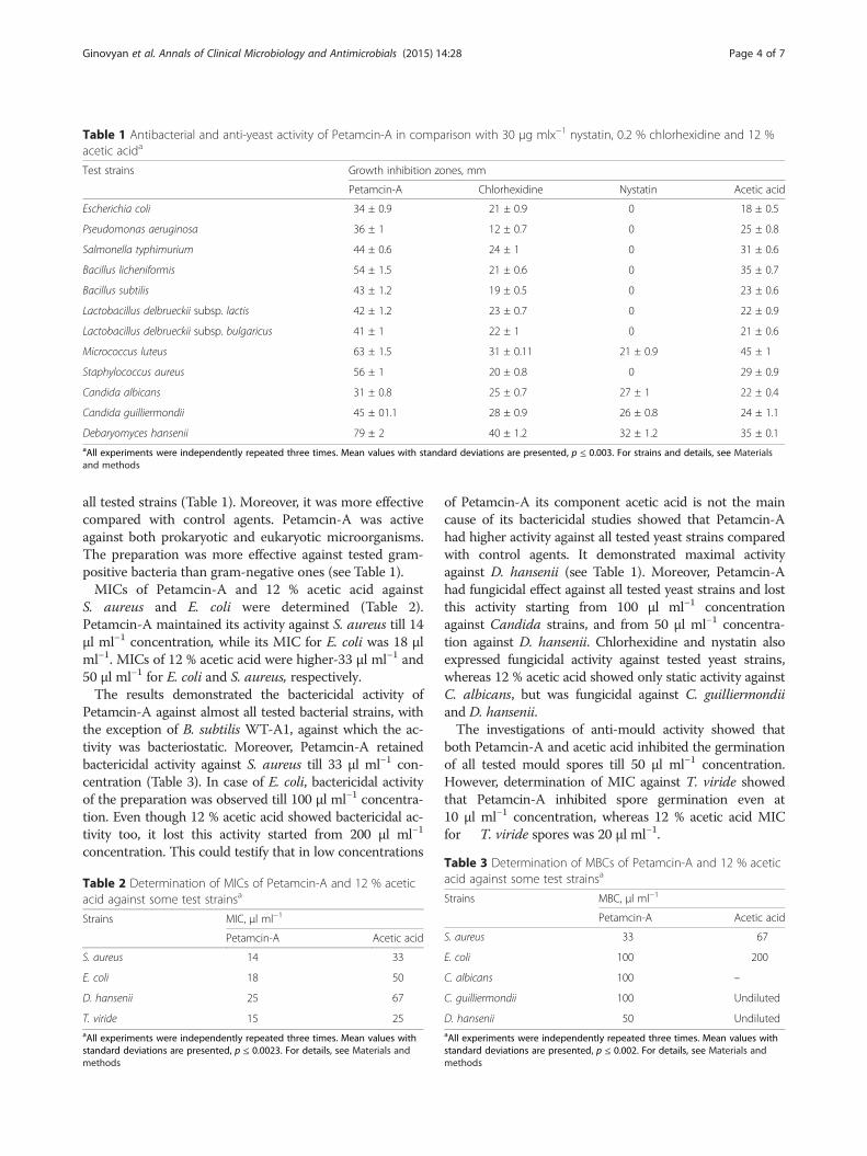
Table 1 Antibacterial and anti-yeast activity of Petamcin-A in comparison with 30 μg mlx−1 nystatin, 0.2 % chlorhexidine and 12 %acetic acida
Test strains Growth inhibition zones, mm
Petamcin-A Chlorhexidine Nystatin Acetic acid
Escherichia coli 34 ± 0.9 21 ± 0.9 0 18 ± 0.5
Pseudomonas aeruginosa 36 ± 1 12 ± 0.7 0 25 ± 0.8
Salmonella typhimurium 44 ± 0.6 24 ± 1 0 31 ± 0.6
Bacillus licheniformis 54 ± 1.5 21 ± 0.6 0 35 ± 0.7
Bacillus subtilis 43 ± 1.2 19 ± 0.5 0 23 ± 0.6
Lactobacillus delbrueckii subsp. lactis 42 ± 1.2 23 ± 0.7 0 22 ± 0.9
Lactobacillus delbrueckii subsp. bulgaricus 41 ± 1 22 ± 1 0 21 ± 0.6
Micrococcus luteus 63 ± 1.5 31 ± 0.11 21 ± 0.9 45 ± 1
Staphylococcus aureus 56 ± 1 20 ± 0.8 0 29 ± 0.9
Candida albicans 31 ± 0.8 25 ± 0.7 27 ± 1 22 ± 0.4
Candida guilliermondii 45 ± 01.1 28 ± 0.9 26 ± 0.8 24 ± 1.1
Debaryomyces hansenii 79 ± 2 40 ± 1.2 32 ± 1.2 35 ± 0.1aAll experiments were independently repeated three times. Mean values with standard deviations are presented, p ≤ 0.003. For strains and details, see Materialsand methods
Ginovyan et al. Annals of Clinical Microbiology and Antimicrobials (2015) 14:28 Page 4 of 7
all tested strains (Table 1). Moreover, it was more effectivecompared with control agents. Petamcin-A was activeagainst both prokaryotic and eukaryotic microorganisms.The preparation was more effective against tested gram-positive bacteria than gram-negative ones (see Table 1).MICs of Petamcin-A and 12 % acetic acid against
S. aureus and E. coli were determined (Table 2).Petamcin-A maintained its activity against S. aureus till 14μl ml−1 concentration, while its MIC for E. coli was 18 μlml−1. MICs of 12 % acetic acid were higher-33 μl ml−1 and50 μl ml−1 for E. coli and S. aureus, respectively.The results demonstrated the bactericidal activity of
Petamcin-A against almost all tested bacterial strains, withthe exception of B. subtilis WT-A1, against which the ac-tivity was bacteriostatic. Moreover, Petamcin-A retainedbactericidal activity against S. aureus till 33 μl ml−1 con-centration (Table 3). In case of E. coli, bactericidal activityof the preparation was observed till 100 μl ml−1 concentra-tion. Even though 12 % acetic acid showed bactericidal ac-tivity too, it lost this activity started from 200 μl ml−1
concentration. This could testify that in low concentrations
Table 2 Determination of MICs of Petamcin-A and 12 % aceticacid against some test strainsa
Strains MIC, μl ml−1
Petamcin-A Acetic acid
S. aureus 14 33
E. coli 18 50
D. hansenii 25 67
T. viride 15 25aAll experiments were independently repeated three times. Mean values withstandard deviations are presented, p ≤ 0.0023. For details, see Materials andmethods
of Petamcin-A its component acetic acid is not the maincause of its bactericidal studies showed that Petamcin-Ahad higher activity against all tested yeast strains comparedwith control agents. It demonstrated maximal activityagainst D. hansenii (see Table 1). Moreover, Petamcin-Ahad fungicidal effect against all tested yeast strains and lostthis activity starting from 100 μl ml−1 concentrationagainst Candida strains, and from 50 μl ml−1 concentra-tion against D. hansenii. Chlorhexidine and nystatin alsoexpressed fungicidal activity against tested yeast strains,whereas 12 % acetic acid showed only static activity againstC. albicans, but was fungicidal against C. guilliermondiiand D. hansenii.The investigations of anti-mould activity showed that
both Petamcin-A and acetic acid inhibited the germinationof all tested mould spores till 50 μl ml−1 concentration.However, determination of MIC against T. viride showedthat Petamcin-A inhibited spore germination even at10 μl ml−1 concentration, whereas 12 % acetic acid MICfor T. viride spores was 20 μl ml−1.
Table 3 Determination of MBCs of Petamcin-A and 12 % aceticacid against some test strainsa
Strains MBC, μl ml−1
Petamcin-A Acetic acid
S. aureus 33 67
E. coli 100 200
C. albicans 100 –
C. guilliermondii 100 Undiluted
D. hansenii 50 UndilutedaAll experiments were independently repeated three times. Mean values withstandard deviations are presented, p ≤ 0.002. For details, see Materials andmethods

Ginovyan et al. Annals of Clinical Microbiology and Antimicrobials (2015) 14:28 Page 5 of 7
In addition, Petamcin-A demonstrated high activityagainst T4 phage of E. coli C-T4 completely inhibiting itsgrowth. 12 % acetic acid also expressed anti-phage activity,but with less efficiency (Fig. 1). It caused only 2.4 Log10reduction of phage units. 200 μl ml−1 concentration ofPetamcin-A also demonstrated considerable activityreducing phage numbers by 2.6 Log10, while the use of100 μl ml−1 concentration of 12 % acetic acid resultedin only 1.4 Log10 reduction of E. coli C-T4.
DiscussionPetamcin-A is patented as antifungal preparation [14]for the treatment of various fungal skin infections. Itsactive antimicrobial components are acetic acid andhexamethylentetramine which separately have been usedin various medical applications, including the treatmentof skin infections for a long time [16, 17, 20]. However,there is no data about their activity when appliedtogether. Thus, it was very interesting to study the anti-bacterial and antiviral activity of Petamcin-A as a complexpreparation.Obtained data showed high efficiency of Petamcin-A
against all tested strains, including bacteria, yeast, mouldsand phages. Studies of antibacterial activity of the prepar-ation illustrated that it was more effective than its compo-nent acetic acid and control agents. Moreover, its activity
0
2
4
6
8
10
12
control AC PA
log1
0 P
FU
ml-1
200 µl ml-1 undiluted
Fig. 1 Anti-phage activity of Petamcin-A (PA) and 12 % acetic acid(AC) and their 200 μl ml−1 concentrations against T4 phage of E. coliC-T4 after 90 min exposure time. Saline solution was used as a negativecontrol. Concentrations of PFU of T4 phage of E. coli C-T4 wereillustrated with Log10 values. Mean values with standard deviationsare presented, p ≤ 0.005. For details, see Materials and methods
was higher against gram-positive bacteria in comparisonwith gram-negative ones. Such a difference was previouslydescribed for acetic acid [21]. At the same time there areno data indicating such differences for another active com-ponent of Petamcin-A–formaldehyde, which is released bydecomposition of hexamethylenetetramine in even slightlyacidic environment [22–24]. This allows speculating thatthis effect of Petamcin-A may be due to acetic acid.The results allow to make some assumptions concerning
Petamcin-A’s components activities. Particularly, it wasdetermined, that in case of gram-negative bacteria growthinhibition zones by Petamcin-A were bigger than that by12 % acetic acid (see Table 1). This difference was maximal(16 mm) for E. coli. Conversely, this difference in case ofgram-positive bacteria was minimal (18 mm) for M. luteus,while it reached maximal value (27 mm) for S. aureus.Accordingly, it could be speculated that formaldehyde wasmore effective against gram-negative bacteria comparedwith gram-positive ones. Such a phenomenon was notdescribed previously.It is important to mention that Petamcin-A was particu-
larly active against one of the most common causativeagents of skin infections-S. aureus [25].Petamcin-A killed all tested bacterial strains, except
B. subtilis WT-A1. Bactericidal activity of the prepar-ation is due to both acetic acid and formaldehyde. Manyresearchers reported about bactericidal activity of aceticacid and formaldehyde [13, 16, 17, 21, 26]. This meansthat both these components are responsible bactericidalactivity of Petamcin-A. Two endospore forming bacteriawere used in the study. Petamcin-A activity againstB. licheniformis WT was bactericidal, whereas it wasbacteriostatic against B. subtilis WT-A1. This may leadto speculations that there was a difference in the activityof Petamcin-A against sporulating and non-sporulatingbacterial strains. However, B. subtilis WT-A1 strain wasisolated from heavy metal polluted soils. Some studiesstate that genes responsible for heavy metal toleranceand antibiotic resistance are located in the same plasmidmeaning there is a correlation between these two traits[27, 28]. Consequently, it can be assumed that B. subtilisWT-A1 strains have resistance against antimicrobials.Thus, the reason of Petamcin-A’s only bacteriostatic ac-tivity may be due to enhanced resistance of a particularstrain, instead of its endospore forming ability. There is noevidence about acetic acid activity against bacterial spores,but many authors state about sporocidal activity offormaldehyde [21, 29, 30]. This affirms that Petamcin-Apossesses activity against spores.Petamcin-A showed high antifungal activity against
tested three yeast and five mould strains. MIC determin-ation of Petamcin-A and 12 % acetic acid against T. viridedemonstrated that both active components of the pre-paration have their contribution in antifungal activity.

Ginovyan et al. Annals of Clinical Microbiology and Antimicrobials (2015) 14:28 Page 6 of 7
Literature data confirms these results. For instance, highantifungal activity of acetic acid even at low concentrationsagainst Aspergilus flavus, Aspergilus luchuensis, Mucor spp.,Penicillium oxalicum etc. was shown [31]. High fungicidalactivity of formaldehyde was also reported [29].The investigation of anti-phage activity showed that
there was a considerable difference between activity ofPetamcin-A and its component acetic acid. This suggeststhat antiviral activity of Petamcin-A is only partially dueto acetic acid and, therefore, formaldehyde also havenoticeable implementation in this activity. According toliterature data acetic acid has been reported to be lesseffective against viruses compared with formaldehydewhich has confirmed our results [26, 29]. It was shownthat 6 % acetic acid showed only 0.32 Log10 reduction ofPoliovirus after 5 min exposure time [17].The mechanisms of Petamcin-A’s antimicrobial activity
can be derived based on activities of its two components-acetic acid and formaldehyde. On the other hand, it ispossible that together they could gain some new advanta-geous properties. Consequently, some further investiga-tions are needed to understand the mode of Petamcin-Aaction as a complex preparation.It is known, that mechanisms that underlie the activity
of formaldehyde are due to its reactions with proteinsand nucleic acids [29, 32]. Particularly, it alkylates aminoand sulfhydryl groups of proteins, as well as nitrogenatoms of the purine rings of nucleic acids [32]. There-fore, this component is active against almost all types ofmicroorganisms.The modes of action of another active component of
Petamcin-A-acetic acid against microbes have not beenunderstood completely yet. But it is assumed that mainlyits undissociated forms are responsible for antimicrobialactivity [33, 34]. They could passively cross the cell walland dissociate at neutral pH within the cells. This bringsto a decrease of internal pH and cause further inhibitoryeffects on microbes. For example, interior acidic pHcauses denaturation of acid-sensitive proteins and DNA,consumption of energy during regulation of internal pH,etc. It has also been proposed that acetic acid brings tointerruption of the action of bacterial proton pumpsresponsible for ion regulation [33, 34].The main part of this work was done using laboratory
control strains. It would be interesting to investigatePetamcin-A’s activity against more clinical isolates andhuman pathogenic strains. This will be the next step ofthe work which could help to broaden its potentialapplications.
Concluding remarksPetamcin-A has a high antimicrobial activity against alltested bacterial, yeast and mould strains. The preparationalso exhibited high anti-phage activity. Furthermore, taking
into account that Petamcin-A has not shown observabletoxicity on skin and its components are not expensive, itcan be advantageous for management of various skinmedical conditions.As far as Petamcin-A expressed antimicrobial activity
against various groups of microorganisms, it also mayhave perspective to be used as a disinfectant but furtherinvestigations are needed.
Competing interestsThe authors declare that they have no competing interests.
Authors’ contributionsMG, IB, AT proposed and designed the study; MG, AK carried out theexperimental part of the manuscript. MG, AK, IB analyzed the generated data.IB, PG, AT helped to draft the manuscript and in critical revision. All authorsread and approved the final manuscript.
Author details1Department of Microbiology, Plants and Microbes Biotechnology, Faculty ofBiology, Yerevan State University, Yerevan, Armenia. 2Department ofPharmaceutical Chemistry, Faculty of Chemistry, Yerevan State University,Yerevan, Armenia.
Received: 28 December 2014 Accepted: 15 May 2015
References1. Godin B, Touitou E, Rubinstein E, Athamna A, Athamna M. A new approach
for treatment of deep skin infections by an ethosomal antibioticpreparation- an in vivo study. J Antimicrob Chemother. 2005;55:989–94.
2. Hernandez PO, Lema S, Tyring SK, Mendoza N. Ceftaroline in complicatedskin and skin-structure infections. Infect Drug Resist. 2012;5:23–35.
3. Hafez MM, Maghrabi IA, Zaki NM. Toward an alternative therapeuticapproach for skin infections: antagonistic activity of lactobacilli againstantibiotic-resistant Staphylococcus aureus and Pseudomonas aeruginosa.Probiotics Antimicrob Proteins. 2013;5:216–26.
4. World Health Organization. Epidemiology and management of commonskin diseases in children in developing countries. Geneva: WHO; 2005.
5. Spellberg B, Talbot GH, Boucher HW, Bradley JS, Gilbert D, Scheld WM, et al.Antimicrobial agents for complicated skin and skin-structure infections:justification of noninferiority margins in the absence of placebo-controlledtrials. Clin Infect Dis. 2009;49:383–91.
6. Bobek LA, Situ H. MUC7 20-Mer: investigation of antimicrobial activity,secondary structure, and possible mechanism of antifungal action.Antimicrob Agents Chemother. 2003;47:643–52.
7. Torres HA, Hachem RY, Chemaly RF, Kontoyiannis DP, Raad II. Posaconazole:a broad-spectrum triazole antifungal. Lancet Infect Dis. 2005;5:775–85.
8. Coogan MM, Fidel PL, Komesu MC, Maeda N, Samaranayake LP. (B1)Candida and mycotic infections. Adv Dent Res. 2006;19:130–8.
9. World Health Organization. Antimicrobial resistance: global report onsurveillance. Geneva, Switzerland: WHO Press; 2014.
10. Burns T, Breathnach S, Cox N, Griffiths C. Rook’s textbook of dermatology.8th ed. Wiley-Blackwell: Hoboken, New Jersey, USA; 2010.
11. Lee SY, Kuti JL, Nicolau DP. Antimicrobial management of complicated skinand skin structure infections in the era of emerging resistance. Surg Infect(Larchmt). 2005;6:283–95.
12. Hedrick J. Acute bacterial skin infections in pediatric medicine: currentissues in presentation and treatment. Paediatr Drugs. 2003;5:35–46.
13. Lipsky BA, Hoey C. Topical antimicrobial therapy for treating chronicwounds. Clin Infect Dis. 2009;49:1541–9.
14. Ghazaryan PA, Ghazaryan AP, Paronikyan RG. Treatment of fungal skininfections with “Petamcin-A”. Armenia, PAN 96168 by 08.08: Patentcertificate for invention, issued by the Intellectual Property Agency; 1997.p. N 315.
15. Vardapetyan AR, Ghazaryan AP, Ghazaryan PA. Effect of Petamcin-A on theregeneration of the epithelium. Veterinariya. 2007;2:8–50 (in Russian).

Ginovyan et al. Annals of Clinical Microbiology and Antimicrobials (2015) 14:28 Page 7 of 7
16. Ryssel H, Kloeters O, Germann G, Schafer T, Wiedemann G, Oehlbauer M.The antimicrobial effect of acetic acid-an alternative to common localantiseptics? Burns. 2009;35:695–700.
17. Cortesia C, Vilchèze C, Bernut A, Contreras W, Gómez K, de Waard J, et al.Acetic acid, the active component of vinegar, is an effective tuberculocidaldisinfectant. mBio. 2014;5(2):e00013–4.
18. Netrusov AI, Yegorova MA, Zakharchuk LM. Practical microbiology manuals.Moscow: Academia; 2005 (in Russian).
19. Barry AL, Craig WA, Nadler H, Reller BL, Sanders CC, Swenson JM. Methodsfor determining bactericidal activity of antimicrobial agents. Approvedguideline M26-A. Wayne, Pa: National Committee for Clinical LaboratoryStandards; 1999.
20. Nagoba BS, Deshmukh SR, Wadher BJ, Patil SB. Acetic acid treatment ofpseudomonal postoperative wound infection. J Hosp Infect. 1997;36:243–4.
21. Quinn PJ, Markey BK, Leonard FC, FitzPatrick ES, Fanning S, Hartigan P.Veterinary microbiology and microbial disease. Wiley-Blackwell: Hoboken,New Jersey, USA; 2011.
22. Greenwood D, Slack RCB. The antibacterial activity of hexamine(methenamine), hexamine hippurate and hexamine mandelate. Infection.1981;9(5):223–7.
23. Grayson ML, Crowe SM, McCarthy JS, Mills J, Mouton JW, Norrby SR, et al.Kucers’ the use of antibiotics. Sixth edition: a clinical review of antibacterial,antifungal and antiviral drugs. Boca Raton, Florida, USA: CRC Press; 2010.
24. Musher DM, Griffith DP. Generation of formaldehyde from methenamine:effect of pH and concentration, and antibacterial effect. Antimicrob AgentsChemother. 1974;6:708–11.
25. Dryden MS. Complicated skin and soft tissue infection. J AntimicrobChemother. 2010;65:35–44.
26. Rutala WA, Barbee SL, Aguiar NC, Sobsey MD, Weber DJ. Antimicrobialactivity of home disinfectants and natural products against potential humanpathogens. Infect Control Hosp Epidemiol. 2000;21:33–8.
27. Silver S, Misra TK. Plasmid-mediated heavy metal resistances. Annu RevMicrobiol. 1988;42(1):717–43.
28. Calomiris JJ, Armstrong JL, Seidler RJ. Association of metal tolerance withmultiple antibiotic resistance of bacteria isolated from drinking water.Appl Environ Microbiol. 1984;47(6):1238–42.
29. Rutala WA, Weber DJ. Healthcare infection control practices advisorycommittee. CDC, Atlanta, GA: Guideline for disinfection and sterilization inhealth care facilities; 2008.
30. McDonnell G, Russell AD. Antiseptics and disinfectants: activity, action, andresistance. Clin Microbiol Rev. 1999;12:147–79.
31. Pundir RK, Jain P. Screening for antifungal activity of commercially availablechemical food preservatives. Int J Pharm Sci Rev Res. 2010;5:25–7.
32. Maris P. Modes of action of disinfectants. Rev Sci Tech. 1995;14:47–55.33. Ricke SC. Perspectives on the use of organic acids and short chain fatty
acids as antimicrobials. Poult Sci. 2003;82:632–9.34. Bradley EM, Williams JB, Schilling MW, Coggins PC, Crist C, Yoder S, et al.
Effects of sodium lactate and acetic acid derivatives on the quality andsensory characteristics of hot-boned pork sausage patties. Meat Sci.2011;88:145–50.
Submit your next manuscript to BioMed Centraland take full advantage of:
• Convenient online submission
• Thorough peer review
• No space constraints or color figure charges
• Immediate publication on acceptance
• Inclusion in PubMed, CAS, Scopus and Google Scholar
• Research which is freely available for redistribution
Submit your manuscript at www.biomedcentral.com/submit
Copyright © 2022 FDOKUMEN