The Kv4.2 Potassium Channel Subunit Is Required for Pain Plasticity
12
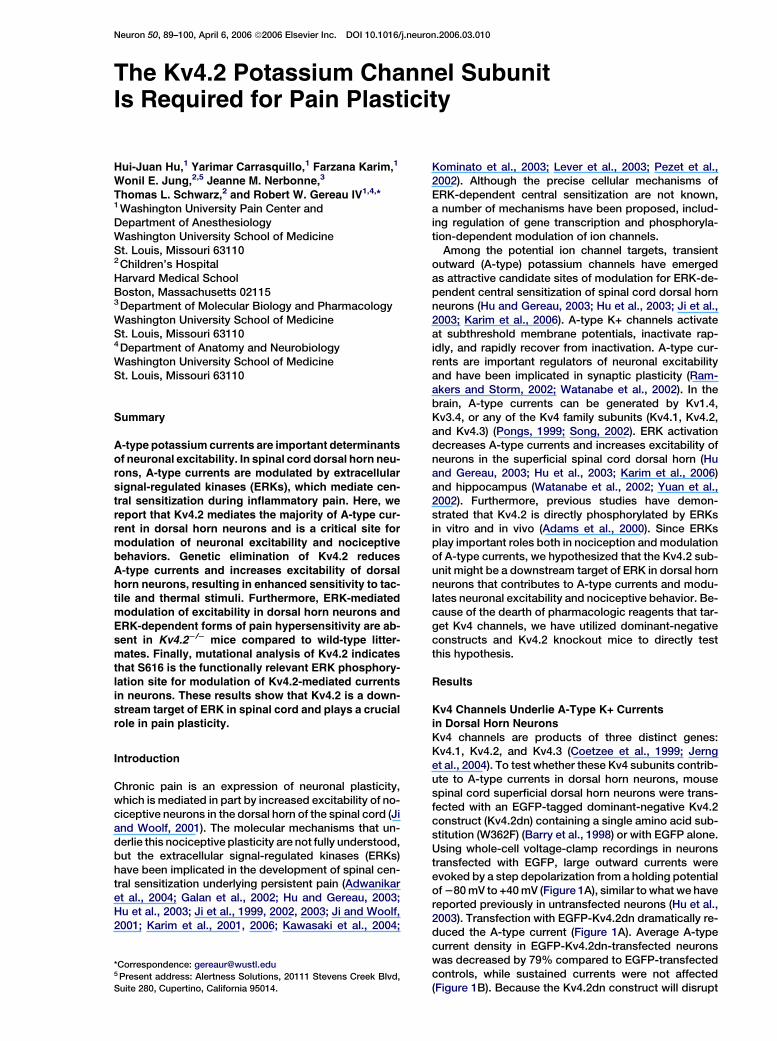
Neuron 50, 89–100, April 6, 2006 ª2006 Elsevier Inc. DOI 10.1016/j.neuron.2006.03.010 The Kv4.2 Potassium Channel Subunit Is Required for Pain Plasticity Hui-Juan Hu, 1 Yarimar Carrasquillo, 1 Farzana Karim, 1 Wonil E. Jung, 2,5 Jeanne M. Nerbonne, 3 Thomas L. Schwarz, 2 and Robert W. Gereau IV 1,4, * 1 Washington University Pain Center and Department of Anesthesiology Washington University School of Medicine St. Louis, Missouri 63110 2 Children’s Hospital Harvard Medical School Boston, Massachusetts 02115 3 Department of Molecular Biology and Pharmacology Washington University School of Medicine St. Louis, Missouri 63110 4 Department of Anatomy and Neurobiology Washington University School of Medicine St. Louis, Missouri 63110 Summary A-type potassium currents are important determinants of neuronal excitability. In spinal cord dorsal horn neu- rons, A-type currents are modulated by extracellular signal-regulated kinases (ERKs), which mediate cen- tral sensitization during inflammatory pain. Here, we report that Kv4.2 mediates the majority of A-type cur- rent in dorsal horn neurons and is a critical site for modulation of neuronal excitability and nociceptive behaviors. Genetic elimination of Kv4.2 reduces A-type currents and increases excitability of dorsal horn neurons, resulting in enhanced sensitivity to tac- tile and thermal stimuli. Furthermore, ERK-mediated modulation of excitability in dorsal horn neurons and ERK-dependent forms of pain hypersensitivity are ab- sent in Kv4.2 2/2 mice compared to wild-type litter- mates. Finally, mutational analysis of Kv4.2 indicates that S616 is the functionally relevant ERK phosphory- lation site for modulation of Kv4.2-mediated currents in neurons. These results show that Kv4.2 is a down- stream target of ERK in spinal cord and plays a crucial role in pain plasticity. Introduction Chronic pain is an expression of neuronal plasticity, which is mediated in part by increased excitability of no- ciceptive neurons in the dorsal horn of the spinal cord (Ji and Woolf, 2001). The molecular mechanisms that un- derlie this nociceptive plasticity are not fully understood, but the extracellular signal-regulated kinases (ERKs) have been implicated in the development of spinal cen- tral sensitization underlying persistent pain (Adwanikar et al., 2004; Galan et al., 2002; Hu and Gereau, 2003; Hu et al., 2003; Ji et al., 1999, 2002, 2003; Ji and Woolf, 2001; Karim et al., 2001, 2006; Kawasaki et al., 2004; Kominato et al., 2003; Lever et al., 2003; Pezet et al., 2002). Although the precise cellular mechanisms of ERK-dependent central sensitization are not known, a number of mechanisms have been proposed, includ- ing regulation of gene transcription and phosphoryla- tion-dependent modulation of ion channels. Among the potential ion channel targets, transient outward (A-type) potassium channels have emerged as attractive candidate sites of modulation for ERK-de- pendent central sensitization of spinal cord dorsal horn neurons (Hu and Gereau, 2003; Hu et al., 2003; Ji et al., 2003; Karim et al., 2006). A-type K+ channels activate at subthreshold membrane potentials, inactivate rap- idly, and rapidly recover from inactivation. A-type cur- rents are important regulators of neuronal excitability and have been implicated in synaptic plasticity (Ram- akers and Storm, 2002; Watanabe et al., 2002). In the brain, A-type currents can be generated by Kv1.4, Kv3.4, or any of the Kv4 family subunits (Kv4.1, Kv4.2, and Kv4.3) (Pongs, 1999; Song, 2002). ERK activation decreases A-type currents and increases excitability of neurons in the superficial spinal cord dorsal horn (Hu and Gereau, 2003; Hu et al., 2003; Karim et al., 2006) and hippocampus (Watanabe et al., 2002; Yuan et al., 2002). Furthermore, previous studies have demon- strated that Kv4.2 is directly phosphorylated by ERKs in vitro and in vivo (Adams et al., 2000). Since ERKs play important roles both in nociception and modulation of A-type currents, we hypothesized that the Kv4.2 sub- unit might be a downstream target of ERK in dorsal horn neurons that contributes to A-type currents and modu- lates neuronal excitability and nociceptive behavior. Be- cause of the dearth of pharmacologic reagents that tar- get Kv4 channels, we have utilized dominant-negative constructs and Kv4.2 knockout mice to directly test this hypothesis. Results Kv4 Channels Underlie A-Type K+ Currents in Dorsal Horn Neurons Kv4 channels are products of three distinct genes: Kv4.1, Kv4.2, and Kv4.3 (Coetzee et al., 1999; Jerng et al., 2004). To test whether these Kv4 subunits contrib- ute to A-type currents in dorsal horn neurons, mouse spinal cord superficial dorsal horn neurons were trans- fected with an EGFP-tagged dominant-negative Kv4.2 construct (Kv4.2dn) containing a single amino acid sub- stitution (W362F) (Barry et al., 1998) or with EGFP alone. Using whole-cell voltage-clamp recordings in neurons transfected with EGFP, large outward currents were evoked by a step depolarization from a holding potential of 280 mV to +40 mV (Figure 1A), similar to what we have reported previously in untransfected neurons (Hu et al., 2003). Transfection with EGFP-Kv4.2dn dramatically re- duced the A-type current (Figure 1A). Average A-type current density in EGFP-Kv4.2dn-transfected neurons was decreased by 79% compared to EGFP-transfected controls, while sustained currents were not affected (Figure 1B). Because the Kv4.2dn construct will disrupt *Correspondence: [email protected] 5 Present address: Alertness Solutions, 20111 Stevens Creek Blvd, Suite 280, Cupertino, California 95014.
-
Upload
independent -
Category
Documents
-
view
0 -
download
0
Transcript of The Kv4.2 Potassium Channel Subunit Is Required for Pain Plasticity
Neuron 50, 89–100, April 6, 2006 ª2006 Elsevier Inc. DOI 10.1016/j.neuron.2006.03.010
The Kv4.2 Potassium Channel SubunitIs Required for Pain Plasticity
Hui-Juan Hu,1 Yarimar Carrasquillo,1 Farzana Karim,1
Wonil E. Jung,2,5 Jeanne M. Nerbonne,3
Thomas L. Schwarz,2 and Robert W. Gereau IV1,4,*1Washington University Pain Center andDepartment of AnesthesiologyWashington University School of MedicineSt. Louis, Missouri 631102Children’s HospitalHarvard Medical SchoolBoston, Massachusetts 021153Department of Molecular Biology and PharmacologyWashington University School of MedicineSt. Louis, Missouri 631104Department of Anatomy and NeurobiologyWashington University School of MedicineSt. Louis, Missouri 63110
Summary
A-type potassium currents are important determinants
of neuronal excitability. In spinal cord dorsal horn neu-rons, A-type currents are modulated by extracellular
signal-regulated kinases (ERKs), which mediate cen-tral sensitization during inflammatory pain. Here, we
report that Kv4.2 mediates the majority of A-type cur-rent in dorsal horn neurons and is a critical site for
modulation of neuronal excitability and nociceptivebehaviors. Genetic elimination of Kv4.2 reduces
A-type currents and increases excitability of dorsalhorn neurons, resulting in enhanced sensitivity to tac-
tile and thermal stimuli. Furthermore, ERK-mediatedmodulation of excitability in dorsal horn neurons and
ERK-dependent forms of pain hypersensitivity are ab-sent in Kv4.22/2 mice compared to wild-type litter-
mates. Finally, mutational analysis of Kv4.2 indicatesthat S616 is the functionally relevant ERK phosphory-
lation site for modulation of Kv4.2-mediated currentsin neurons. These results show that Kv4.2 is a down-
stream target of ERK in spinal cord and plays a crucialrole in pain plasticity.
Introduction
Chronic pain is an expression of neuronal plasticity,which is mediated in part by increased excitability of no-ciceptive neurons in the dorsal horn of the spinal cord (Jiand Woolf, 2001). The molecular mechanisms that un-derlie this nociceptive plasticity are not fully understood,but the extracellular signal-regulated kinases (ERKs)have been implicated in the development of spinal cen-tral sensitization underlying persistent pain (Adwanikaret al., 2004; Galan et al., 2002; Hu and Gereau, 2003;Hu et al., 2003; Ji et al., 1999, 2002, 2003; Ji and Woolf,2001; Karim et al., 2001, 2006; Kawasaki et al., 2004;
*Correspondence: [email protected] Present address: Alertness Solutions, 20111 Stevens Creek Blvd,
Suite 280, Cupertino, California 95014.
Kominato et al., 2003; Lever et al., 2003; Pezet et al.,2002). Although the precise cellular mechanisms ofERK-dependent central sensitization are not known,a number of mechanisms have been proposed, includ-ing regulation of gene transcription and phosphoryla-tion-dependent modulation of ion channels.
Among the potential ion channel targets, transientoutward (A-type) potassium channels have emergedas attractive candidate sites of modulation for ERK-de-pendent central sensitization of spinal cord dorsal hornneurons (Hu and Gereau, 2003; Hu et al., 2003; Ji et al.,2003; Karim et al., 2006). A-type K+ channels activateat subthreshold membrane potentials, inactivate rap-idly, and rapidly recover from inactivation. A-type cur-rents are important regulators of neuronal excitabilityand have been implicated in synaptic plasticity (Ram-akers and Storm, 2002; Watanabe et al., 2002). In thebrain, A-type currents can be generated by Kv1.4,Kv3.4, or any of the Kv4 family subunits (Kv4.1, Kv4.2,and Kv4.3) (Pongs, 1999; Song, 2002). ERK activationdecreases A-type currents and increases excitability ofneurons in the superficial spinal cord dorsal horn (Huand Gereau, 2003; Hu et al., 2003; Karim et al., 2006)and hippocampus (Watanabe et al., 2002; Yuan et al.,2002). Furthermore, previous studies have demon-strated that Kv4.2 is directly phosphorylated by ERKsin vitro and in vivo (Adams et al., 2000). Since ERKsplay important roles both in nociception and modulationof A-type currents, we hypothesized that the Kv4.2 sub-unit might be a downstream target of ERK in dorsal hornneurons that contributes to A-type currents and modu-lates neuronal excitability and nociceptive behavior. Be-cause of the dearth of pharmacologic reagents that tar-get Kv4 channels, we have utilized dominant-negativeconstructs and Kv4.2 knockout mice to directly testthis hypothesis.
Results
Kv4 Channels Underlie A-Type K+ Currentsin Dorsal Horn Neurons
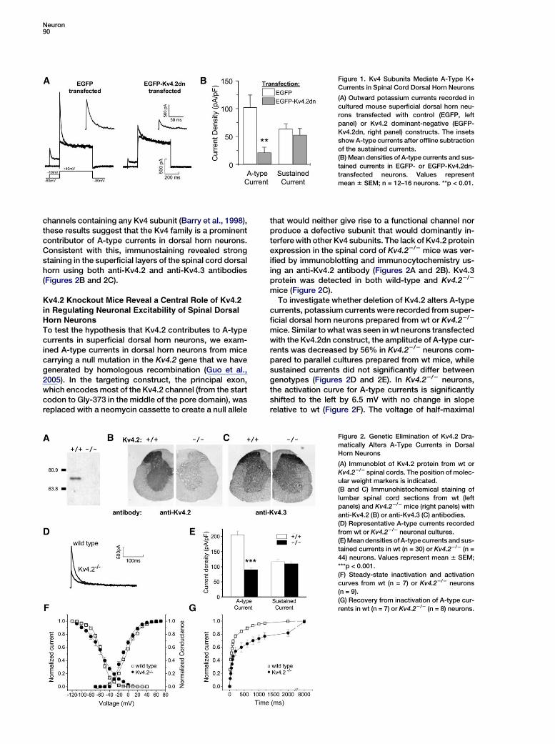
Kv4 channels are products of three distinct genes:Kv4.1, Kv4.2, and Kv4.3 (Coetzee et al., 1999; Jernget al., 2004). To test whether these Kv4 subunits contrib-ute to A-type currents in dorsal horn neurons, mousespinal cord superficial dorsal horn neurons were trans-fected with an EGFP-tagged dominant-negative Kv4.2construct (Kv4.2dn) containing a single amino acid sub-stitution (W362F) (Barry et al., 1998) or with EGFP alone.Using whole-cell voltage-clamp recordings in neuronstransfected with EGFP, large outward currents wereevoked by a step depolarization from a holding potentialof 280 mV to +40 mV (Figure 1A), similar to what we havereported previously in untransfected neurons (Hu et al.,2003). Transfection with EGFP-Kv4.2dn dramatically re-duced the A-type current (Figure 1A). Average A-typecurrent density in EGFP-Kv4.2dn-transfected neuronswas decreased by 79% compared to EGFP-transfectedcontrols, while sustained currents were not affected(Figure 1B). Because the Kv4.2dn construct will disrupt
Neuron90
Figure 1. Kv4 Subunits Mediate A-Type K+
Currents in Spinal Cord Dorsal Horn Neurons
(A) Outward potassium currents recorded in
cultured mouse superficial dorsal horn neu-
rons transfected with control (EGFP, left
panel) or Kv4.2 dominant-negative (EGFP-
Kv4.2dn, right panel) constructs. The insets
show A-type currents after offline subtraction
of the sustained currents.
(B) Mean densities of A-type currents and sus-
tained currents in EGFP- or EGFP-Kv4.2dn-
transfected neurons. Values represent
mean 6 SEM; n = 12–16 neurons. **p < 0.01.
channels containing any Kv4 subunit (Barry et al., 1998),these results suggest that the Kv4 family is a prominentcontributor of A-type currents in dorsal horn neurons.Consistent with this, immunostaining revealed strongstaining in the superficial layers of the spinal cord dorsalhorn using both anti-Kv4.2 and anti-Kv4.3 antibodies(Figures 2B and 2C).
Kv4.2 Knockout Mice Reveal a Central Role of Kv4.2
in Regulating Neuronal Excitability of Spinal DorsalHorn Neurons
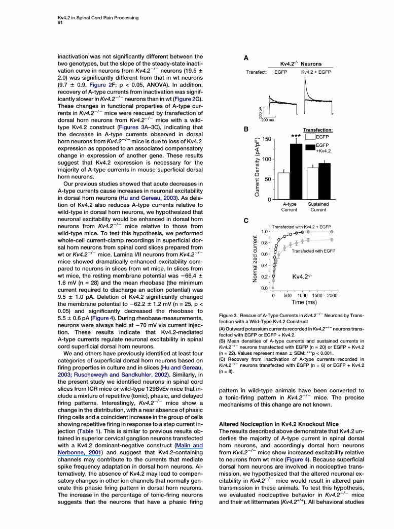
To test the hypothesis that Kv4.2 contributes to A-typecurrents in superficial dorsal horn neurons, we exam-ined A-type currents in dorsal horn neurons from micecarrying a null mutation in the Kv4.2 gene that we havegenerated by homologous recombination (Guo et al.,2005). In the targeting construct, the principal exon,which encodes most of the Kv4.2 channel (from the startcodon to Gly-373 in the middle of the pore domain), wasreplaced with a neomycin cassette to create a null allele
that would neither give rise to a functional channel norproduce a defective subunit that would dominantly in-terfere with other Kv4 subunits. The lack of Kv4.2 proteinexpression in the spinal cord of Kv4.22/2 mice was ver-ified by immunoblotting and immunocytochemistry us-ing an anti-Kv4.2 antibody (Figures 2A and 2B). Kv4.3protein was detected in both wild-type and Kv4.22/2
mice (Figure 2C).To investigate whether deletion of Kv4.2 alters A-type
currents, potassium currents were recorded from super-ficial dorsal horn neurons prepared from wt or Kv4.22/2
mice. Similar to what was seen in wt neurons transfectedwith the Kv4.2dn construct, the amplitude of A-type cur-rents was decreased by 56% in Kv4.22/2 neurons com-pared to parallel cultures prepared from wt mice, whilesustained currents did not significantly differ betweengenotypes (Figures 2D and 2E). In Kv4.22/2 neurons,the activation curve for A-type currents is significantlyshifted to the left by 6.5 mV with no change in sloperelative to wt (Figure 2F). The voltage of half-maximal
Figure 2. Genetic Elimination of Kv4.2 Dra-
matically Alters A-Type Currents in Dorsal
Horn Neurons
(A) Immunoblot of Kv4.2 protein from wt or
Kv4.22/2 spinal cords. The position of molec-
ular weight markers is indicated.
(B and C) Immunohistochemical staining of
lumbar spinal cord sections from wt (left
panels) and Kv4.22/2 mice (right panels) with
anti-Kv4.2 (B) or anti-Kv4.3 (C) antibodies.
(D) Representative A-type currents recorded
from wt or Kv4.22/2 neuronal cultures.
(E) Mean densities of A-type currents and sus-
tained currents in wt (n = 30) or Kv4.22/2 (n =
44) neurons. Values represent mean 6 SEM;
***p < 0.001.
(F) Steady-state inactivation and activation
curves from wt (n = 7) or Kv4.22/2 neurons
(n = 9).
(G) Recovery from inactivation of A-type cur-
rents in wt (n = 7) or Kv4.22/2 (n = 8) neurons.
Kv4.2 in Spinal Cord Pain Processing91
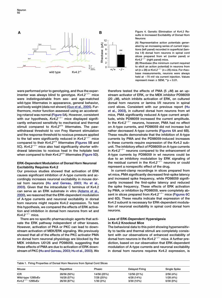
inactivation was not significantly different between thetwo genotypes, but the slope of the steady-state inacti-vation curve in neurons from Kv4.22/2 neurons (19.5 62.0) was significantly different from that in wt neurons(9.7 6 0.9, Figure 2F; p < 0.05, ANOVA). In addition,recovery of A-type currents from inactivation was signif-icantly slower in Kv4.22/2 neurons than in wt (Figure 2G).These changes in functional properties of A-type cur-rents in Kv4.22/2 mice were rescued by transfection ofdorsal horn neurons from Kv4.22/2 mice with a wild-type Kv4.2 construct (Figures 3A–3C), indicating thatthe decrease in A-type currents observed in dorsalhorn neurons from Kv4.22/2 mice is due to loss of Kv4.2expression as opposed to an associated compensatorychange in expression of another gene. These resultssuggest that Kv4.2 expression is necessary for themajority of A-type currents in mouse superficial dorsalhorn neurons.
Our previous studies showed that acute decreases inA-type currents cause increases in neuronal excitabilityin dorsal horn neurons (Hu and Gereau, 2003). As dele-tion of Kv4.2 also reduces A-type currents relative towild-type in dorsal horn neurons, we hypothesized thatneuronal excitability would be enhanced in dorsal hornneurons from Kv4.22/2 mice relative to those fromwild-type mice. To test this hypothesis, we performedwhole-cell current-clamp recordings in superficial dor-sal horn neurons from spinal cord slices prepared fromwt or Kv4.22/2 mice. Lamina I/II neurons from Kv4.22/2
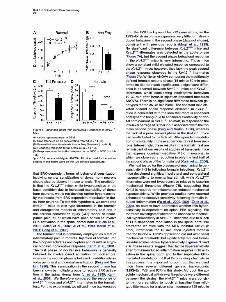
mice showed dramatically enhanced excitability com-pared to neurons in slices from wt mice. In slices fromwt mice, the resting membrane potential was 266.4 61.6 mV (n = 28) and the mean rheobase (the minimumcurrent required to discharge an action potential) was9.5 6 1.0 pA. Deletion of Kv4.2 significantly changedthe membrane potential to 262.2 6 1.2 mV (n = 25, p <0.05) and significantly decreased the rheobase to5.5 6 0.6 pA (Figure 4). During rheobase measurements,neurons were always held at 270 mV via current injec-tion. These results indicate that Kv4.2-mediatedA-type currents regulate neuronal excitability in spinalcord superficial dorsal horn neurons.
We and others have previously identified at least fourcategories of superficial dorsal horn neurons based onfiring properties in culture and in slices (Hu and Gereau,2003; Ruscheweyh and Sandkuhler, 2002). Similarly, inthe present study we identified neurons in spinal cordslices from ICR mice or wild-type 129SvEv mice that in-clude a mixture of repetitive (tonic), phasic, and delayedfiring patterns. Interestingly, Kv4.22/2 mice show achange in the distribution, with a near absence of phasicfiring cells and a coincident increase in the group of cellsshowing repetitive firing in response to a step current in-jection (Table 1). This is similar to previous results ob-tained in superior cervical ganglion neurons transfectedwith a Kv4.2 dominant-negative construct (Malin andNerbonne, 2001) and suggest that Kv4.2-containingchannels may contribute to the currents that mediatespike frequency adaptation in dorsal horn neurons. Al-ternatively, the absence of Kv4.2 may lead to compen-satory changes in other ion channels that normally gen-erate this phasic firing pattern in dorsal horn neurons.The increase in the percentage of tonic-firing neuronssuggests that the neurons that have a phasic firing
pattern in wild-type animals have been converted toa tonic-firing pattern in Kv4.22/2 mice. The precisemechanisms of this change are not known.
Altered Nociception in Kv4.2 Knockout Mice
The results described above demonstrate that Kv4.2 un-derlies the majority of A-type current in spinal dorsalhorn neurons, and accordingly dorsal horn neuronsfrom Kv4.22/2 mice show increased excitability relativeto neurons from wt mice (Figure 4). Because superficialdorsal horn neurons are involved in nociceptive trans-mission, we hypothesized that the altered neuronal ex-citability in Kv4.22/2 mice would result in altered paintransmission in these animals. To test this hypothesis,we evaluated nociceptive behavior in Kv4.22/2 miceand their wt littermates (Kv4.2+/+). All behavioral studies
Figure 3. Rescue of A-Type Currents in Kv4.22/2 Neurons by Trans-
fection with a Wild-Type Kv4.2 Construct
(A) Outward potassium currents recorded in Kv4.22/2 neurons trans-
fected with EGFP or EGFP + Kv4.2.
(B) Mean densities of A-type currents and sustained currents in
Kv4.22/2 neurons transfected with EGFP (n = 20) or EGFP + Kv4.2
(n = 22). Values represent mean 6 SEM; ***p < 0.001.
(C) Recovery from inactivation of A-type currents recorded in
Kv4.22/2 neurons transfected with EGFP (n = 6) or EGFP + Kv4.2
(n = 8).
Neuron92
Figure 4. Genetic Elimination of Kv4.2 Re-
sults in Increased Excitability of Dorsal Horn
Neurons
(A) Representative action potentials gener-
ated by an increasing series of current injec-
tions (left panel) recorded in superficial (lam-
ina I-II) dorsal horn neurons in spinal cord
slices prepared from wt (center panel) or
Kv4.22/2 (right panel) mice.
(B) Rheobase (the minimum current required
to elicit an action potential) in neurons from
wt (n = 28) or Kv4.22/2 (n = 25) mice. For rheo-
base measurements, neurons were always
held at 270 mV via current injection. Values
represent mean 6 SEM; **p < 0.01.
were performed prior to genotyping, and thus the exper-imenter was always blind to genotype. Kv4.22/2 micewere indistinguishable from sex- and age-matchedwild-type littermates in appearance, general behavior,and body weight (data not shown) (Guo et al., 2005). Fur-thermore, motor function assessed using an accelerat-ing rotarod was normal (Figure 5A). However, consistentwith our hypothesis, Kv4.22/2 mice displayed signifi-cantly enhanced sensitivity to mechanical and thermalstimuli compared to Kv4.2+/+ littermates. The paw-withdrawal threshold to von Frey filament stimulationand the response threshold to noxious pressure appliedto the tail were significantly reduced in Kv4.22/2 micecompared to their Kv4.2+/+ littermates (Figures 5B and5C). Kv4.22/2 mice also had significantly shorter with-drawal latencies to noxious heat in the hotplate testwhen compared to their Kv4.2+/+ littermates (Figure 5D).
ERK-Dependent Modulation of Dorsal Horn Neuronal
Excitability Requires Kv4.2Our previous studies showed that activation of ERKcauses significant inhibition of A-type currents and ac-cordingly increases neuronal excitability in mouse dor-sal horn neurons (Hu and Gereau, 2003; Hu et al.,2003). Given that the intracellular C terminus of Kv4.2can serve as an ERK substrate in vitro (Adams et al.,2000), we reasoned that the ERK-dependent modulationof A-type currents and neuronal excitability in dorsalhorn neurons might require Kv4.2 expression. To testthis hypothesis, we compared the effects of ERK activa-tion and inhibition in dorsal horn neurons from wt andKv4.22/2 mice.
There are no specific pharmacologic agents that acti-vate the ERK pathway independent of other kinases.However, activation of PKA or PKC can lead to down-stream activation of MEK/ERK signaling. We previouslyshowed that all of the effects of the PKC activator PMAon dorsal horn neuronal physiology are blocked by theMEK inhibitors U0126 and PD98059, suggesting thatthese effects of PMA are due to activation of ERK down-stream of PKC (Hu and Gereau, 2003; Hu et al., 2003). We
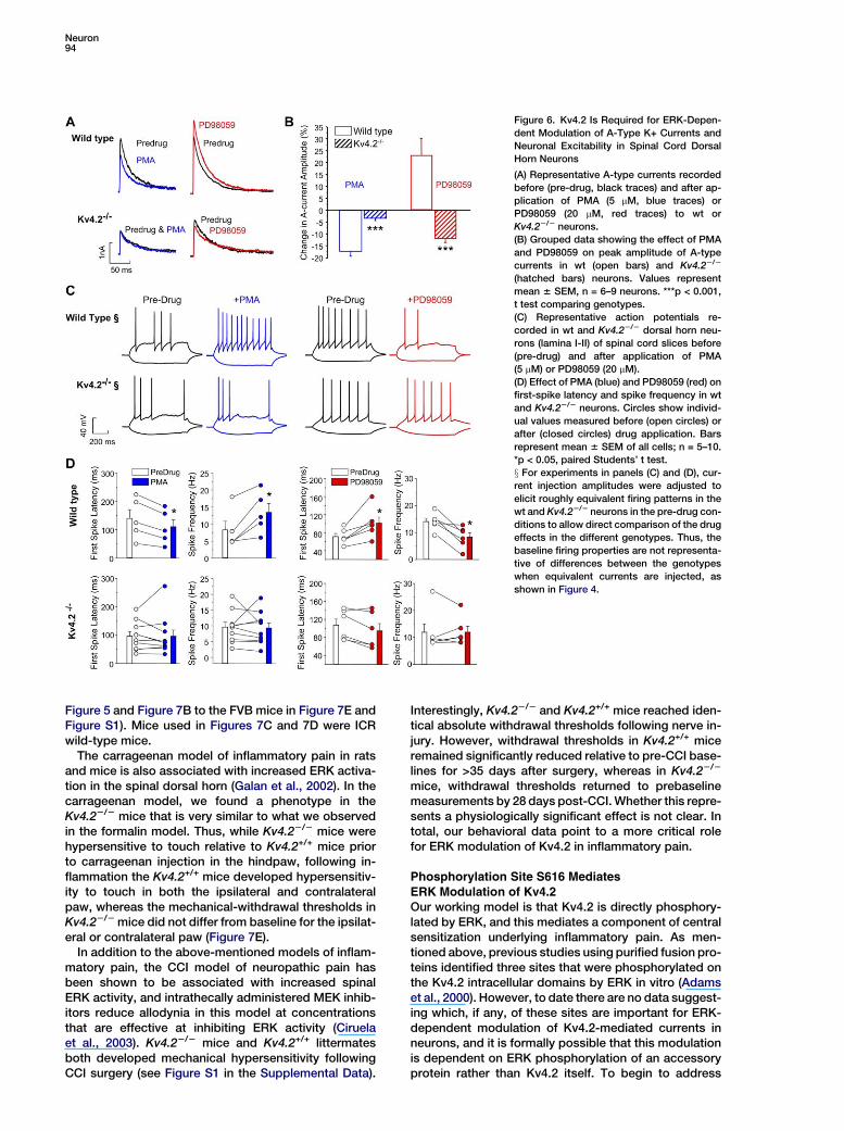
therefore tested the effects of PMA (5 mM) as an up-stream activator of ERK, or the MEK inhibitor PD98059(20 mM), which inhibits activation of ERK, on cultureddorsal horn neurons or lamina I/II neurons in spinalcord slices. Consistent with our previous report (Huet al., 2003), in cultured dorsal horn neurons from wtmice, PMA significantly reduced A-type current ampli-tude, while PD98059 increased the current amplitude.In the Kv4.22/2 neurons, however, PMA had no effecton A-type currents, and PD98059 did not increase butrather decreased A-type currents (Figures 6A and 6B).These results demonstrate that the inhibition of A-typecurrents by PMA and the PD98059-induced increasesin these currents require expression of the Kv4.2 sub-unit. The inhibitory effect of PD98059 on A-type currentsin Kv4.22/2 neurons compared to the enhancement ofA-type currents by PD98059 in wt neurons could bedue to an inhibitory modulation by ERK signaling ofthe residual current in the Kv4.22/2 neurons or couldrepresent a nonspecific effect of the drug.
In current-clamp recordings in slices prepared fromwt mice, PMA significantly decreased first-spike latencyand increased spike frequency, while PD98059 signifi-cantly increased the first-spike latency and reducedthe spike frequency. These effects of ERK activationby PMA, or inhibition by PD98059, were completely ab-sent in slices prepared from Kv4.22/2 mice (Figures 6Cand 6D). These results indicate that expression of theKv4.2 subunit is necessary for ERK-dependent modula-tion of neuronal excitability in spinal cord dorsal hornneurons.
Loss of ERK-Dependent Hyperalgesia
in Kv4.2 Knockout MiceThe behavioral data to this point showing hypersensitiv-ity to tactile and thermal stimuli are completely consis-tent with our observations of enhanced excitability ofdorsal horn neurons in the Kv4.22/2 mice. A further pre-diction, based on our observation that ERK-dependentmodulation of A-type currents and neuronal excitabilityin dorsal horn neurons requires Kv4.2 expression, is
Table 1. Firing Properties of Dorsal Horn Neurons from Spinal Cord Slices
Mouse Repetitive Phasic Delayed Firing Single Spike
ICR 28/56 (50%) 14/56 (25%) 12/56 (21%) 2/56 (4%)
Wild-type 129SvEv 16/28 (57%) 7/28 (25%) 4/28 (14%) 1/28 (4%)
Kv4.22/2 129SvEv 26/30 (87%) 1/30 (3%) 3/30 (10%) 0/30 (0%)
Kv4.2 in Spinal Cord Pain Processing93
that ERK-dependent forms of behavioral sensitizationinvolving central sensitization of dorsal horn neuronsshould also be absent in these animals. The predictionis that the Kv4.22/2 mice, while hypersensitive in thebasal condition due to increased excitability of dorsalhorn neurons, would not develop further hypersensitiv-ity that results from ERK-dependent modulation in dor-sal horn neurons. To test this hypothesis, we comparedKv4.22/2 mice to wild-type littermates in the formalinand carrageenan models of inflammatory pain and inthe chronic constriction injury (CCI) model of neuro-pathic pain, all of which have been shown to involveERK activation in the spinal dorsal horn (Ciruela et al.,2003; Galan et al., 2002; Ji et al., 1999; Karim et al.,2001; Song et al., 2005).
The formalin test is commonly employed as a test ofinflammatory pain in rodents. Injection of formalin intothe hindpaw activates nociceptors and results in a typi-cal biphasic nociceptive response (Karim et al., 2001).The first phase of nocifensive behaviors is generallybelieved to involve direct activation of nociceptors,whereas the second phase is believed to additionally in-volve peripheral and central sensitization (Puig and Sor-kin, 1996). This sensitization in the second phase hasbeen shown by multiple groups to require ERK activa-tion in the spinal dorsal horn (Ji et al., 1999; Karimet al., 2001). We therefore compared the response ofKv4.22/2 mice and Kv4.2+/+ littermates in the formalintest. For this experiment, we utilized mice backcrossed
Figure 5. Enhanced Basal Pain Behavioral Responses in Kv4.22/2
Mice
All values represent mean 6 SEM.
(A) Drop latencies on an accelerating rotarod (n = 14–16).
(B) Paw-withdrawal thresholds to von Frey filaments (n = 8–11).
(C) Response threshold to tail pressure (n = 14–16).
(D) Response latencies in the hot-plate test at 52ºC or 56ºC (n = 14–
16).
*p < 0.05, versus wild-type, ANOVA. All mice used for behavioral
studies in this figure were on the 129 genetic background.
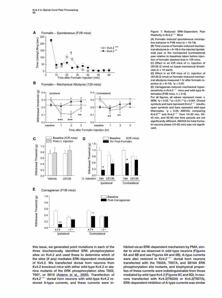
onto the FVB background for >12 generations, as the129SvEv strain of mice expressed very little formalin-in-duced behaviors in the second phase (data not shown),consistent with previous reports (Mogil et al., 1999).No significant difference between Kv4.22/2 mice andKv4.2+/+ littermates was detected in the acute phase(Figure 7A), but the second phase behavioral responsein the Kv4.22/2 mice is very interesting. These miceshow a constant mild elevated response compared tothe Kv4.2+/+ mice; however, they lack the peak secondphase response observed in the Kv4.2+/+ littermates(Figure 7A). While an ANOVA comparing the traditionallydefined formalin second phase (15 min to 60 min post-formalin) did not reach significance, a significant differ-ence is observed between Kv4.22/2 mice and Kv4.2+/+
littermates when considering nociceptive behaviors10–30 min after formalin injection (repeated-measuresANOVA). There is no significant difference between ge-notypes for the 35–45 min block. The constant mild ele-vated second phase response observed in Kv4.22/2
mice is consistent with the idea that there is enhancedpostsynaptic firing (due to enhanced excitability of dor-sal horn neurons in Kv4.22/2 animals) in response to thelow-level barrage of C fiber input associated with the for-malin second phase (Puig and Sorkin, 1996), whereasthe lack of a peak second phase in the Kv4.22/2 micecan be attributed to the lack of ERK-dependent modula-tion of excitability in these superficial dorsal horn neu-rons. Interestingly, these results in the formalin test arereminiscent of our results of studies of transgenic micethat express dominant-negative MEK in neurons, inwhich we observed a reduction in only the first half ofthe second phase of the formalin test (Karim et al., 2006).
We next tested for the presence of mechanical hyper-sensitivity 1–3 hr following formalin injections. Kv4.2+/+
mice developed significant ipsilateral and contralateralhypersensitivity to mechanical stimuli, while Kv4.22/2
littermates were not hypersensitive relative to baselinemechanical thresholds (Figure 7B), suggesting thatKv4.2 is required for inflammation-induced mechanicalhypersensitivity. While previous studies have reportedenhanced nociceptive sensitivity following formalin-in-duced inflammation (Fu et al., 2000, 2001; Zeitz et al.,2004), no studies have addressed whether this hyper-sensitivity is dependent on spinal ERK signaling. Wetherefore investigated whether the absence of mechan-ical hypersensitivity in Kv4.22/2 mice was due to a lackof ERK-dependent modulation in the spinal cord. Wepretreated wt mice with the MEK inhibitor U0126 (2nmol, intrathecal) for 15 min, then injected formalininto the hindpaw. U0126 application did not alter basalmechanical thresholds, but significantly reduced forma-lin-induced mechanical hypersensitivity (Figures 7C and7D). These results suggest that tactile hypersensitivityafter formalin-induced inflammation requires ERK acti-vation in the spinal cord, and further implicates ERK-mediated modulation of Kv4.2-containing channels inthis process. It is important to point out that we usedmice from several different genetic backgrounds(129SvEv, FVB, and ICR) in this study. Although the ab-solute mechanical withdrawal thresholds were differentbetween the strains, the Kv4.22/2 mice were consis-tently more sensitive to touch at baseline than wild-type littermates for a given strain (compare 129 mice in
Neuron94
Figure 6. Kv4.2 Is Required for ERK-Depen-
dent Modulation of A-Type K+ Currents and
Neuronal Excitability in Spinal Cord Dorsal
Horn Neurons
(A) Representative A-type currents recorded
before (pre-drug, black traces) and after ap-
plication of PMA (5 mM, blue traces) or
PD98059 (20 mM, red traces) to wt or
Kv4.22/2 neurons.
(B) Grouped data showing the effect of PMA
and PD98059 on peak amplitude of A-type
currents in wt (open bars) and Kv4.22/2
(hatched bars) neurons. Values represent
mean 6 SEM, n = 6–9 neurons. ***p < 0.001,
t test comparing genotypes.
(C) Representative action potentials re-
corded in wt and Kv4.22/2 dorsal horn neu-
rons (lamina I-II) of spinal cord slices before
(pre-drug) and after application of PMA
(5 mM) or PD98059 (20 mM).
(D) Effect of PMA (blue) and PD98059 (red) on
first-spike latency and spike frequency in wt
and Kv4.22/2 neurons. Circles show individ-
ual values measured before (open circles) or
after (closed circles) drug application. Bars
represent mean 6 SEM of all cells; n = 5–10.
*p < 0.05, paired Students’ t test.
x For experiments in panels (C) and (D), cur-
rent injection amplitudes were adjusted to
elicit roughly equivalent firing patterns in the
wt and Kv4.22/2 neurons in the pre-drug con-
ditions to allow direct comparison of the drug
effects in the different genotypes. Thus, the
baseline firing properties are not representa-
tive of differences between the genotypes
when equivalent currents are injected, as
shown in Figure 4.
Figure 5 and Figure 7B to the FVB mice in Figure 7E andFigure S1). Mice used in Figures 7C and 7D were ICRwild-type mice.
The carrageenan model of inflammatory pain in ratsand mice is also associated with increased ERK activa-tion in the spinal dorsal horn (Galan et al., 2002). In thecarrageenan model, we found a phenotype in theKv4.22/2 mice that is very similar to what we observedin the formalin model. Thus, while Kv4.22/2 mice werehypersensitive to touch relative to Kv4.2+/+ mice priorto carrageenan injection in the hindpaw, following in-flammation the Kv4.2+/+ mice developed hypersensitiv-ity to touch in both the ipsilateral and contralateralpaw, whereas the mechanical-withdrawal thresholds inKv4.22/2 mice did not differ from baseline for the ipsilat-eral or contralateral paw (Figure 7E).
In addition to the above-mentioned models of inflam-matory pain, the CCI model of neuropathic pain hasbeen shown to be associated with increased spinalERK activity, and intrathecally administered MEK inhib-itors reduce allodynia in this model at concentrationsthat are effective at inhibiting ERK activity (Ciruelaet al., 2003). Kv4.22/2 mice and Kv4.2+/+ littermatesboth developed mechanical hypersensitivity followingCCI surgery (see Figure S1 in the Supplemental Data).
Interestingly, Kv4.22/2 and Kv4.2+/+ mice reached iden-tical absolute withdrawal thresholds following nerve in-jury. However, withdrawal thresholds in Kv4.2+/+ miceremained significantly reduced relative to pre-CCI base-lines for >35 days after surgery, whereas in Kv4.22/2
mice, withdrawal thresholds returned to prebaselinemeasurements by 28 days post-CCI. Whether this repre-sents a physiologically significant effect is not clear. Intotal, our behavioral data point to a more critical rolefor ERK modulation of Kv4.2 in inflammatory pain.
Phosphorylation Site S616 Mediates
ERK Modulation of Kv4.2Our working model is that Kv4.2 is directly phosphory-lated by ERK, and this mediates a component of centralsensitization underlying inflammatory pain. As men-tioned above, previous studies using purified fusion pro-teins identified three sites that were phosphorylated onthe Kv4.2 intracellular domains by ERK in vitro (Adamset al., 2000). However, to date there are no data suggest-ing which, if any, of these sites are important for ERK-dependent modulation of Kv4.2-mediated currents inneurons, and it is formally possible that this modulationis dependent on ERK phosphorylation of an accessoryprotein rather than Kv4.2 itself. To begin to address
Kv4.2 in Spinal Cord Pain Processing95
Figure 7. Reduced ERK-Dependent Pain
Plasticity in Kv4.22/2 Mice
(A) Formalin induced spontaneous nocicep-
tive behavior in FVB mice (n = 16–18).
(B) Time course of formalin-induced mechan-
ical allodynia (n = 8–18) in the injected (ipsilat-
eral) paw or the noninjected (contralateral)
paw relative to baselines taken before injec-
tion of formalin (dashed line) in 129 mice.
(C) Effect in wt ICR mice of i.t. injection of
U0126 (2 nmol) on basal mechanical thresh-
olds (n = 10 each).
(D) Effect in wt ICR mice of i.t. injection of
U0126 (2 nmol) on formalin-induced mechan-
ical allodynia measured 1 hr after formalin in-
jection (n = 9–10). *p < 0.05.
(E) Carrageenan-induced mechanical hyper-
sensitivity in Kv4.22/2 mice and wild-type lit-
termates (FVB mice, n = 5–6).
For all figures, all values represent mean 6
SEM. *p < 0.05, **p < 0.01,***p < 0.001. Closed
symbols and bars represent Kv4.22/2 results,
open symbols and bars represent wild-type
littermates. yp < 0.05, ANOVA, comparing
Kv4.2+/+ and Kv4.22/2 from 10–30 min, 35–
45 min, and 50–60 min time periods are not
significantly different. ANOVA for total forma-
lin second phase (10–60 min) was not signifi-
cant.
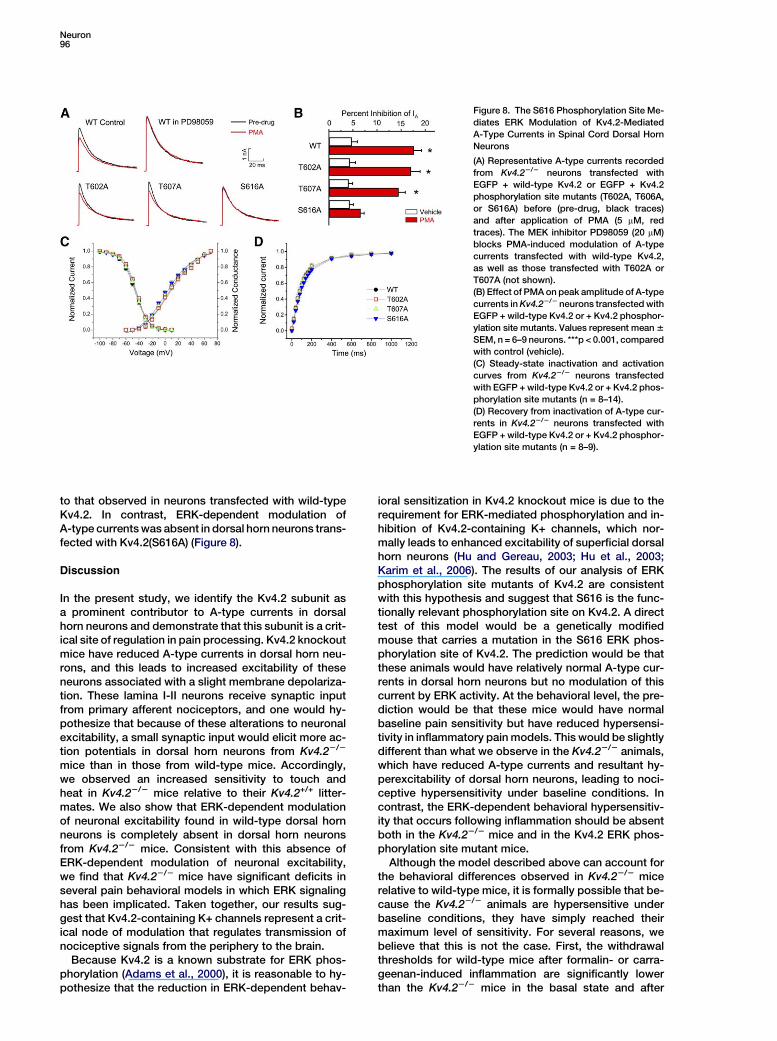
this issue, we generated point mutations in each of thethree biochemically identified ERK phosphorylationsites on Kv4.2 and used these to determine which ofthe sites (if any) mediates ERK-dependent modulationof Kv4.2. We transfected dorsal horn neurons fromKv4.2 knockout mice with either wild-type Kv4.2 or ala-nine mutants of the ERK phosphorylation sites T602,T607, or S616 (Adams et al., 2000). Transfection ofKv4.22/2 dorsal horn neurons with wild-type Kv4.2 re-stored A-type currents, and these currents were in-
hibited via an ERK-dependent mechanism by PMA, sim-ilar to what we observed in wild-type neurons (Figures8A and 8B and see Figures 6A and 6B). A-type currentswere also restored in Kv4.22/2 dorsal horn neuronstransfected with the T602A, T607A, and S616A ERKphosphorylation site mutants, and biophysical proper-ties of these currents were indistinguishable from thosemediated by wild-type Kv4.2 (Figures 8C and 8D). In neu-rons transfected with Kv4.2(T602A) or Kv4.2(T607A),ERK-dependent inhibition of A-type currents was similar
Neuron96
Figure 8. The S616 Phosphorylation Site Me-
diates ERK Modulation of Kv4.2-Mediated
A-Type Currents in Spinal Cord Dorsal Horn
Neurons
(A) Representative A-type currents recorded
from Kv4.22/2 neurons transfected with
EGFP + wild-type Kv4.2 or EGFP + Kv4.2
phosphorylation site mutants (T602A, T606A,
or S616A) before (pre-drug, black traces)
and after application of PMA (5 mM, red
traces). The MEK inhibitor PD98059 (20 mM)
blocks PMA-induced modulation of A-type
currents transfected with wild-type Kv4.2,
as well as those transfected with T602A or
T607A (not shown).
(B) Effect of PMA on peak amplitude of A-type
currents in Kv4.22/2 neurons transfected with
EGFP + wild-type Kv4.2 or + Kv4.2 phosphor-
ylation site mutants. Values represent mean 6
SEM, n = 6–9 neurons. ***p < 0.001, compared
with control (vehicle).
(C) Steady-state inactivation and activation
curves from Kv4.22/2 neurons transfected
with EGFP + wild-type Kv4.2 or + Kv4.2 phos-
phorylation site mutants (n = 8–14).
(D) Recovery from inactivation of A-type cur-
rents in Kv4.22/2 neurons transfected with
EGFP + wild-type Kv4.2 or + Kv4.2 phosphor-
ylation site mutants (n = 8–9).
to that observed in neurons transfected with wild-typeKv4.2. In contrast, ERK-dependent modulation ofA-type currents was absent in dorsal horn neurons trans-fected with Kv4.2(S616A) (Figure 8).
Discussion
In the present study, we identify the Kv4.2 subunit asa prominent contributor to A-type currents in dorsalhorn neurons and demonstrate that this subunit is a crit-ical site of regulation in pain processing. Kv4.2 knockoutmice have reduced A-type currents in dorsal horn neu-rons, and this leads to increased excitability of theseneurons associated with a slight membrane depolariza-tion. These lamina I-II neurons receive synaptic inputfrom primary afferent nociceptors, and one would hy-pothesize that because of these alterations to neuronalexcitability, a small synaptic input would elicit more ac-tion potentials in dorsal horn neurons from Kv4.22/2
mice than in those from wild-type mice. Accordingly,we observed an increased sensitivity to touch andheat in Kv4.22/2 mice relative to their Kv4.2+/+ litter-mates. We also show that ERK-dependent modulationof neuronal excitability found in wild-type dorsal hornneurons is completely absent in dorsal horn neuronsfrom Kv4.22/2 mice. Consistent with this absence ofERK-dependent modulation of neuronal excitability,we find that Kv4.22/2 mice have significant deficits inseveral pain behavioral models in which ERK signalinghas been implicated. Taken together, our results sug-gest that Kv4.2-containing K+ channels represent a crit-ical node of modulation that regulates transmission ofnociceptive signals from the periphery to the brain.
Because Kv4.2 is a known substrate for ERK phos-phorylation (Adams et al., 2000), it is reasonable to hy-pothesize that the reduction in ERK-dependent behav-
ioral sensitization in Kv4.2 knockout mice is due to therequirement for ERK-mediated phosphorylation and in-hibition of Kv4.2-containing K+ channels, which nor-mally leads to enhanced excitability of superficial dorsalhorn neurons (Hu and Gereau, 2003; Hu et al., 2003;Karim et al., 2006). The results of our analysis of ERKphosphorylation site mutants of Kv4.2 are consistentwith this hypothesis and suggest that S616 is the func-tionally relevant phosphorylation site on Kv4.2. A directtest of this model would be a genetically modifiedmouse that carries a mutation in the S616 ERK phos-phorylation site of Kv4.2. The prediction would be thatthese animals would have relatively normal A-type cur-rents in dorsal horn neurons but no modulation of thiscurrent by ERK activity. At the behavioral level, the pre-diction would be that these mice would have normalbaseline pain sensitivity but have reduced hypersensi-tivity in inflammatory pain models. This would be slightlydifferent than what we observe in the Kv4.22/2 animals,which have reduced A-type currents and resultant hy-perexcitability of dorsal horn neurons, leading to noci-ceptive hypersensitivity under baseline conditions. Incontrast, the ERK-dependent behavioral hypersensitiv-ity that occurs following inflammation should be absentboth in the Kv4.22/2 mice and in the Kv4.2 ERK phos-phorylation site mutant mice.
Although the model described above can account forthe behavioral differences observed in Kv4.22/2 micerelative to wild-type mice, it is formally possible that be-cause the Kv4.22/2 animals are hypersensitive underbaseline conditions, they have simply reached theirmaximum level of sensitivity. For several reasons, webelieve that this is not the case. First, the withdrawalthresholds for wild-type mice after formalin- or carra-geenan-induced inflammation are significantly lowerthan the Kv4.22/2 mice in the basal state and after
Kv4.2 in Spinal Cord Pain Processing97
inflammation (Figures 7B and 7E). Second, the Kv4.22/2
mice are clearly able to be made more hypersensitive totouch, as there is significant hypersensitivity in theKv4.22/2 mice following CCI relative to presurgery base-lines (Figure S1). These data are consistent with the hy-pothesis that the lack of hypersensitivity in the Kv4.22/2
mice relative to Kv4.2+/+ littermates is due to reducedplasticity in the Kv4.22/2 mice and not due to a behav-ioral ‘‘floor effect.’’
The finding that Kv4.22/2 mice still have some hyper-sensitivity in the CCI model of persistent neuropathicpain is interesting given the diversity of actions of ERKsignaling in neurons (Ji et al., 2003). It is possible thatERK phosphorylation and modulation of Kv4.2-contain-ing K+ channels mediates a component of central sensi-tization but that transcription-dependent changes un-derlie other long-term components of ERK-dependentcentral sensitization that are not reduced in the Kv4.2knockouts (Ji and Rupp, 1997; Ji et al., 2002; Woolfand Costigan, 1999). Our data are consistent with thehypothesis that Kv4.2 modulation plays a more sig-nificant role in inflammatory pain than in longer-termneuropathic pain conditions, where ERK-dependenttranscriptional regulation may be more important. TheKv4.22/2 animals and potential phosphorylation sitemutant animals may provide useful tools for future stud-ies examining the relative importance of acute phos-phorylation and modulation of K+ channels and tran-scription/translation-dependent changes in mediatingvarious forms of long-term pain hypersensitivity.
Although our data are consistent with a critical role ofERK modulation of Kv4.2-containing potassium chan-nels in superficial spinal dorsal horn neurons in mediat-ing behavioral sensitization following inflammation, it iscertainly true that Kv4.2 is expressed in many areas ofthe nervous system. Thus, it is possible that Kv4.2 playsimportant roles in other parts of the pain neuraxis anda component of the alterations in behavior observed inthe Kv4.2 knockouts could be mediated by altered phys-iology in these areas in addition to the spinal cord. Wewere particularly curious to see whether Kv4.2 mightbe impacting the physiology of nociceptive primaryafferent neurons, but we were unable to detect anyKv4.2 expression in the mouse dorsal root ganglion byimmunostaining (data not shown). In addition, the super-ficial laminae of the spinal cord contain both excitatoryand inhibitory neurons, and excitatory neurons can beprojection neurons or interneurons. Because we donot know the transmitter phenotype of all of the neuronsfrom which we have recorded, the specific cellular cir-cuitry impacted by these changes remains to be deter-mined.
Our results reveal a link between Kv4.2 and ERK sig-naling in the spinal cord and support a model in whichKv4.2-containing potassium channels in spinal super-ficial dorsal horn neurons are modulated by ERKs to in-duce a component of central sensitization. Our data fur-ther implicate direct phosphorylation of Kv4.2 at S616 inthis process. ERK-dependent phosphorylation of Kv4.2and modulation of A-type K+ currents appears to under-lie plasticity in hippocampal pyramidal neurons asso-ciated with long-term potentiation (LTP) (Kim et al.,2005; Schrader et al., 2005; Watanabe et al., 2002). Therehas been considerable interest in the parallels between
LTP and central sensitization (Basbaum, 1996; Ji et al.,2003; Karim et al., 2001; Sandkuhler and Liu, 1998; Willis,2002); the present study supports the idea that LTP andcentral sensitization may share common mechanisms,as our studies and previous work have demonstratedthe importance of ERK signaling and Kv4.2 in both ofthese processes (Ji et al., 2003). Whether these similar-ities translate to similarities in the forms of synaptic plas-ticity observed in dorsal horn neurons to mediate centralsensitization is worthy of extensive study.
The results presented here advance our understand-ing of the mechanisms underlying pain processing andare especially relevant to our understanding of inflam-mation-induced pain hypersensitivity. Our findings sug-gest that manipulation of the ERK-Kv4.2 signaling path-way could be useful for novel therapies for the treatmentof pain.
Experimental Procedures
Cell Culture and Spinal Cord Preparation
Primary cultures of spinal cord superficial dorsal horn neurons were
prepared from 4- to 8-day-old CD1 mice and 129SvEv wild-type or
Kv4.22/2 mice as previously described (Hugel and Schlichter,
2000). Lumbar spinal cord slices (300–350 mm) were prepared from
6- to 10-day-old CD1 or 129SvEv mice as previously described (Ed-
wards et al., 1989) and maintained in artificial cerebrospinal fluid
(ACSF) containing (in mM) 118 NaCl, 3 KCl, 24 NaHCO3, 2 MgCl2,
1.25 NaH2PO4, 1 CaCl2, and 12 glucose at room temperature under
continuous oxygenation for 1–4 hr.
Transfection of Dorsal Horn Neurons
The EGFP-Kv4.2dn construct was generously provided by Dr. Paul
Pfaffinger (Baylor College of Medicine). Plasmid DNA was isolated
using the Qiagen plasmid maxi protocol. Spinal cord dorsal horn
neurons were cultured for 24 hr and then transfected with plasmid
DNA constructs (0.45–0.9 mg/well) overnight using LipofectAMINE
Plus or 2000 reagent according to the manufacturer’s protocol (In-
vitrogen Carlsbad, CA). Patch-clamp recordings were performed
beginning w16 hr following transfection. Point mutations in ERK
phosphorylation sites were generated as described previously (Ger-
eau and Heinemann, 1998).
Electrophysiological Recording
Whole-cell recordings were performed using standard procedures
at room temperature using either an AXOPATCH 200B amplifier
and CLAMPEX 8.0 software (Axon Instruments, Union City, CA) or
an EPC-10 amplifier and Pulse v8.62 software (HEKA Elektronik,
Lambrecht, Germany) as previously described (Hu and Gereau,
2003; Hu et al., 2003). Electrode resistances were 3–6 MU with series
resistances around 6–15 MU and were compensated by R60%. For
voltage-clamp recordings in cultured neurons, the bath solution was
Hank’s solution (HBSS) (in mM: 137 NaCl, 5.4 KCl, 0.4 KH2PO4,
1 CaCl2, 0.5 MgCl2, 0.4 MgSO4, 4.2 NaHCO3, 0.3 Na2HPO4, 5.6 glu-
cose) containing 500 nM TTX and 2 mM CoCl2 to block voltage-
gated Na+ currents, Ca2+ currents, and Ca2+-activated K+ currents.
The electrode solution contained (in mM) 140 KCl, 1 MgCl2,
0.5 CaCl2, 5 EGTA, 10 HEPES, 3 Na2ATP, and 0.3 Na2GTP (pH 7.4).
The membrane voltage was held at 280 mV, and transient po-
tassium currents (IA) were isolated by a two-step voltage protocol
as previously described (Hu et al., 2003). To determine the voltage-
dependent activation, voltage steps of 500 ms were applied at 5 s in-
tervals in +10 mV increments from 270 mV to a maximum of +70 mV.
To determine the voltage-dependent inactivation, conditioning pre-
pulses ranging from –120 mV to +40 mV were applied at 5 s intervals
in +10 mV increments for 150 ms followed by a step to +40 mV for
500 ms. To determine time-dependent recovery from inactivation,
conditioning pulses (40 mV) were applied for 500 ms followed by
steps to +40 mV for 500 ms in 20 or 200 ms incremental duration.
For current-clamp recording in slice, the bath solution was ACSF
bubbled with 95% O2-5% CO2. Intracellular solution contained
Neuron98
(in mM) 140 KMeSO4, 2 MgCl2, 1 EGTA, 10 HEPES, 3 Na2ATP, and 0.3
Na2GTP (pH 7.4). Action potentials were generated by current injec-
tion from a holding potential of 270 mV for rheobase or 277 mV for
drug application.
Generation of Kv4.2 Knockout Mice
Kv4.2-deficient mice were generated as we have previously de-
scribed (Guo et al., 2005). Briefly, the targeting vector was con-
structed in the pPNT vector using 129Sv genomic DNA from a phage
isolate. For the 30 arm, a 7.1 kb AflII genomic fragment, downstream
of the first coding exon, was inserted into a SrfI site that previously
been introduced between the KpnI and EcoRI sites of the vector. For
the 50 arm, a 3.1 kb SpeI-ScaI genomic fragment upstream of the
predicted start codon was inserted between the NotI and the XhoI
sites. Thus, the coding region of the first exon of Kv4.2 was replaced
with the neor gene. ES cells (gift of Dr. Andras Nagy, University of
Toronto) were electroporated with the targeting vector and selected
with G418 and gancyclovir. Clones were injected into blastocysts to
generate chimeric mice, which were crossed to 129/SvEv mice to
produce Kv4.2+/2 mice. Wild-type and Kv4.22/2 littermate mice gen-
erated from heterozygous intercrosses were used for behavior tests.
Western Blot Analysis
The lumbar section of the spinal cord was dissected from adult
129SvEv wild-type or Kv4.22/2 mice and homogenized using
homogenization buffer (HB) (Tris 20 mM, pH 7.5, EDTA 1 mM,
Na4P2O7 1 mM, aprotinin 25 mg/ml, leupeptin 25 mg/ml, Na3VO4
0.2 mg/ml, and PMSF 0.4 mM). Membrane proteins were separated
and electrophoresed in 10% SDS polyacrylamide gels. The blots
were blocked with 5% milk and probed with anti-Kv4.2 primary
antibody (1:500, Chemicon). The blots were incubated with HRP-
conjugated secondary antibody (1:20,000, Cell Signaling) and devel-
oped with the SuperSignal West Femto reagent (Pierce).
Immunohistochemistry
Thirty-micron sections of paraformaldehyde-fixed lumbar spinal
cord from adult 129SvEv wild-type or Kv4.22/2 mice were stained
with rabbit anti-Kv4.2 (1:800, Alomone) or rabbit anti-Kv4.3 primary
antibody (1:200, Chemicon). Sections were then incubated in bioti-
nylated secondary antibody (1:200, Vector) and treated in extrAvidin
peroxidase (1:1000, Sigma). Detection was performed using a diami-
nobenzidine (DAB) substrate kit (Vector).
Behavioral Studies
All behavioral tests were performed blind using 7- to 9-week-old
mice. All the experiments were done in accordance with the guide-
lines of the National Institutes of Health and The International Asso-
ciation for the study of Pain and were approved by the Animal Care
and Use Committee of Washington University School of Medicine.
Wild-type and Kv4.22/2 littermate mice of both sexes were tested
except where mentioned.
Motor Function
Mice were tested for motor function using the accelerating rotarod
(4–40 rpm) (UGO Basile, Varese, Italy). The time spent on the rotarod
was recorded.
Basal Sensitivity to Mechanical and Thermal Stimulation
Mechanical sensitivity was measured in male mice using von Frey fil-
aments (North Coast Medical, Inc., San Jose, CA) as previously de-
scribed (Yang and Gereau, 2003). The smallest monofilament that
evoked paw-withdrawal responses on three out of five trials was
taken as the mechanical threshold. The tail pressure test was con-
ducted using the Basile Analgesy-Meter, and the response (strug-
gle/vocalization) threshold was measured as previously described
(Nassar et al., 2004). Hot plate latencies were measured as the
time taken for a mouse to lick or shake its hindpaw at hot plate tem-
peratures of either 52ºC or 56ºC.
Formalin Model
The formalin test was performed by injection of 5% formalin subcu-
taneously into the plantar surface of the right hindpaw. The total time
spent in spontaneous nociceptive behavior (licking and lifting of the
injected paw) was recorded in 5 min intervals for 1 hr as previously
described (Karim et al., 2001). Mechanical allodynia was measured
in male mice from 1–3 hr after injection of formalin using von Frey
filaments. For experiments analyzing the effect of U0126 on forma-
lin-induced allodynia, mechanical thresholds were measured 1 hr af-
ter formalin injection. Biochemical studies were performed to test
the optimal dose and timing for U0126 intrathecal injections and
revealed that intrathecal injection of 2 nmol of U0126 was able to in-
hibit ERK activation for up to 2 hr after intrathecal injection (data not
shown). Based on this finding, U0126 was injected 15 min prior to
formalin injection, and allodynia was measured 1 hr after formalin
injection.
Carrageenan Model
Two percent carrageenan was injected subcutaneously into the
plantar surface of the right hindpaw. Mechanical hypersensitivity
was measured in male mice 2 hr after injection of carrageenan, using
von Frey filaments as described above.
Chronic Constrictive Injury Model
CCI of the sciatic nerve was induced as previously described (Ben-
nett and Xie, 1988) in male FVB mice. Briefly, under pentobarbital so-
dium anesthesia, the left sciatic nerve was exposed at mid-thigh,
and two ligatures were loosely ligated proximal to the sciatic’s
trifurcation at w1.0 mm intervals with 6-0 chromic gut sutures. Me-
chanical hypersensitivity was measured on 1, 4, 7, 10, 14, 21, 28, and
35 days post-CCI, using von Frey filaments as described above.
Drug Application
Stock solutions of phorbol 12-myristate 13-acetate (PMA), PD
098059 (Sigma-Aldrich, St. Louis, MO), and U-0126 (Calbiochem,
La Jolla, CA) were made in DMSO and diluted to final concentrations
in HBSS for bath applications or in PBS for intrathecal injections.
Data Analysis
Offline evaluation was done using clampfit 8.0 (Axon Instrument) or
Pulse v8.62 (HEKA) and Origin (Microcal Software Inc., Northamp-
ton, MA). Data are expressed as original traces and/or as mean 6
SEM. The voltage dependence of activation and inactivation of the
IA was fitted with the Boltzmann function as previously described
(Hu et al., 2003). Behavioral experiments were statistically analyzed
by ANOVA (or repeated-measures ANOVA when appropriate as indi-
cated) followed by the appropriate post hoc tests. Paired or two-
sample Student’s t test was used when comparisons were restricted
to two means. Error probabilities of p < 0.05 were considered statis-
tically significant.
Supplemental Data
The Supplemental Data for this article can be found online at http://
www.neuron.org/cgi/content/full/50/1/89/DC1/.
Acknowledgments
The authors wish to thank P. Pfaffinger for generously providing the
Kv4.2dn construct; and C. Qiu, S. Shalin, and B. McGill for help with
mouse colony maintenance, immunostaining, and dorsal horn neu-
ronal transfections, respectively. This work was supported by grants
from the NIH (R.W.G., Y.C., J.M.N., and T.L.S.), Arthritis Foundation
(F.K.), and the Paralyzed Veterans of America Spinal Cord Research
Foundation (R.W.G.).
Received: July 8, 2005
Revised: August 10, 2005
Accepted: March 3, 2006
Published: April 5, 2006
References
Adams, J.P., Anderson, A.E., Varga, A.W., Dineley, K.T., Cook, R.G.,
Pfaffinger, P.J., and Sweatt, J.D. (2000). The A-type potassium chan-
nel kv4.2 is a substrate for the mitogen-activated protein kinase
ERK. J. Neurochem. 75, 2277–2287.
Adwanikar, H., Karim, F., and Gereau, R.W. (2004). Inflammation per-
sistently enhances nocifensive behaviors mediated by spinal group I
mGluRs through sustained ERK activation. Pain 111, 125–135.

Kv4.2 in Spinal Cord Pain Processing99
Barry, D.M., Xu, H., Schuessler, R.B., and Nerbonne, J.M. (1998).
Functional knockout of the transient outward current, long-QT
syndrome, and cardiac remodeling in mice expressing a domi-
nant-negative Kv4 alpha subunit. Circ. Res. 83, 560–567.
Basbaum, A.I. (1996). Memories of Pain. Sci. Med. (Phila.) (Novem-
ber/December): 22–31.
Bennett, G.J., and Xie, Y.K. (1988). A peripheral mononeuropathy in
rat that produces disorders of pain sensation like those seen in man.
Pain 33, 87–107.
Ciruela, A., Dixon, A.K., Bramwell, S., Gonzalez, M.I., Pinnock, R.D.,
and Lee, K. (2003). Identification of MEK1 as a novel target for the
treatment of neuropathic pain. Br. J. Pharmacol. 138, 751–756.
Coetzee, W.A., Amarillo, Y., Chiu, J., Chow, A., Lau, D., McCormack,
T., Moreno, H., Nadal, M.S., Ozaita, A., Pountney, D., et al. (1999).
Molecular diversity of K+ channels. Ann. N Y Acad. Sci. 868, 233–
285.
Edwards, F.A., Konnerth, A., Sakmann, B., and Takahashi, T. (1989).
A thin slice preparation for patch clamp recordings from neurones of
the mammalian central nervous system. Pflugers Arch. 414, 600–
612.
Fu, K.Y., Light, A.R., and Maixner, W. (2000). Relationship between
nociceptor activity, peripheral edema, spinal microglial activation
and long-term hyperalgesia induced by formalin. Neuroscience
101, 1127–1135.
Fu, K.Y., Light, A.R., and Maixner, W. (2001). Long-lasting inflamma-
tion and long-term hyperalgesia after subcutaneous formalin injec-
tion into the rat hindpaw. J. Pain 2, 2–11.
Galan, A., Lopez-Garcia, J.A., Cervero, F., and Laird, J.M. (2002).
Activation of spinal extracellular signaling-regulated kinase-1 and -2
by intraplantar carrageenan in rodents. Neurosci. Lett. 322, 37–40.
Gereau, R.W., and Heinemann, S.F. (1998). Role of protein kinase C
phosphorylation in rapid desensitization of metabotropic glutamate
receptor 5. Neuron 20, 143–151.
Guo, W., Jung, W.E., Marionneau, C., Aimond, F., Xu, H., Yamada,
K.A., Schwarz, T.L., Demolombe, S., and Nerbonne, J.M. (2005). Tar-
geted deletion of Kv4.2 eliminates Ito,f and results in electrical and
molecular remodeling, with no evidence of ventricular hypertrophy
or myocardial dysfunction. Circ. Res. 97, 1342–1350.
Hu, H.J., and Gereau, R.W. (2003). ERK integrates PKA and PKC sig-
naling in superficial dorsal horn neurons. II. Modulation of neuronal
excitability. J. Neurophysiol. 90, 1680–1688.
Hu, H.J., Glauner, K.S., and Gereau, R.W. (2003). ERK integrates
PKA and PKC signaling in superficial dorsal horn neurons. I. Modu-
lation of A-type K+ currents. J. Neurophysiol. 90, 1671–1679.
Hugel, S., and Schlichter, R. (2000). Presynaptic P2X receptors facil-
itate inhibitory GABAergic transmission between cultured rat spinal
cord dorsal horn neurons. J. Neurosci. 20, 2121–2130.
Jerng, H.H., Pfaffinger, P.J., and Covarrubias, M. (2004). Molecular
physiology and modulation of somatodendritic A-type potassium
channels. Mol. Cell Neurosci. 27, 343–369.
Ji, R., and Rupp, F. (1997). Phosphorylation of transcription factor
CREB in rat spinal cord after formalin-induced hyperalgesia: rela-
tionship to c-fos induction. J. Neurosci. 17, 1776–1785.
Ji, R.R., and Woolf, C.J. (2001). Neuronal plasticity and signal trans-
duction in nociceptive neurons: implications for the initiation and
maintenance of pathological pain. Neurobiol. Dis. 8, 1–10.
Ji, R.-R., Baba, H., Brenner, G.J., and Woolf, C.J. (1999). Nocicep-
tive-specific activation of ERK in spinal neurons contributes to
pain hypersensitivity. Nat. Neurosci. 2, 1114–1119.
Ji, R.R., Befort, K., Brenner, G.J., and Woolf, C.J. (2002). ERK MAP
kinase activation in superficial spinal cord neurons induces prody-
norphin and NK-1 upregulation and contributes to persistent inflam-
matory pain hypersensitivity. J. Neurosci. 22, 478–485.
Ji, R.R., Kohno, T., Moore, K.A., and Woolf, C.J. (2003). Central sen-
sitization and LTP: do pain and memory share similar mechanisms?
Trends Neurosci. 26, 696–705.
Karim, F., Wang, C.-C., and Gereau, R.W. (2001). Metabotropic glu-
tamate receptor subtypes 1 and 5 are activators of extracellular sig-
nal-regulated kinase signaling required for inflammatory pain in
mice. J. Neurosci. 21, 3771–3779.
Karim, F., Hu, H.J., Adwanikar, H., Kaplan, D.R., and Gereau, R.W.
(2006). Impaired inflammatory pain and thermal hyperalgesia in
mice expressing neuron-specific dominant negative mitogen acti-
vated protein kinase kinase (MEK). Mol Pain 2, 2.
Kawasaki, Y., Kohno, T., Zhuang, Z.Y., Brenner, G.J., Wang, H., Van
Der Meer, C., Befort, K., Woolf, C.J., and Ji, R.R. (2004). Ionotropic
and metabotropic receptors, protein kinase A, protein kinase C,
and Src contribute to C-fiber-induced ERK activation and cAMP re-
sponse element-binding protein phosphorylation in dorsal horn neu-
rons, leading to central sensitization. J. Neurosci. 24, 8310–8321.
Kim, J., Wei, D.S., and Hoffman, D.A. (2005). Kv4 potassium channel
subunits control action potential repolarization and frequency-
dependent broadening in rat hippocampal CA1 pyramidal neurones.
J. Physiol. 569, 41–57.
Kominato, Y., Tachibana, T., Dai, Y., Tsujino, H., Maruo, S., and No-
guchi, K. (2003). Changes in phosphorylation of ERK and Fos ex-
pression in dorsal horn neurons following noxious stimulation in
a rat model of neuritis of the nerve root. Brain Res. 967, 89–97.
Lever, I.J., Pezet, S., McMahon, S.B., and Malcangio, M. (2003). The
signaling components of sensory fiber transmission involved in the
activation of ERK MAP kinase in the mouse dorsal horn. Mol. Cell
Neurosci. 24, 259–270.
Malin, S.A., and Nerbonne, J.M. (2001). Molecular heterogeneity of
the voltage-gated fast transient outward K+ current, I(Af), in mam-
malian neurons. J. Neurosci. 21, 8004–8014.
Mogil, J.S., Wilson, S.G., Bon, K., Lee, S.E., Chung, K., Raber, P.,
Pieper, J.O., Hain, H.S., Belknap, J.K., Hubert, L., et al. (1999). Heri-
tability of nociception I: responses of 11 inbred mouse strains on 12
measures of nociception. Pain 80, 67–82.
Nassar, M.A., Stirling, L.C., Forlani, G., Baker, M.D., Matthews, E.A.,
Dickenson, A.H., and Wood, J.N. (2004). Nociceptor-specific gene
deletion reveals a major role for Nav1.7 (PN1) in acute and inflamma-
tory pain. Proc. Natl. Acad. Sci. USA 101, 12706–12711.
Pezet, S., Malcangio, M., Lever, I.J., Perkinton, M.S., Thompson,
S.W., Williams, R.J., and McMahon, S.B. (2002). Noxious stimulation
induces Trk receptor and downstream ERK phosphorylation in spi-
nal dorsal horn. Mol. Cell Neurosci. 21, 684–695.
Pongs, O. (1999). Voltage-gated potassium channels: from hyperex-
citability to excitement. FEBS Lett. 452, 31–35.
Puig, S., and Sorkin, L.S. (1996). Formalin-evoked activity in identi-
fied primary afferent fibers: systemic lidocaine suppresses phase-
2 activity. Pain 64, 345–355.
Ramakers, G.M., and Storm, J.F. (2002). A postsynaptic transient
K(+) current modulated by arachidonic acid regulates synaptic inte-
gration and threshold for LTP induction in hippocampal pyramidal
cells. Proc. Natl. Acad. Sci. USA 99, 10144–10149.
Ruscheweyh, R., and Sandkuhler, J. (2002). Lamina-specific mem-
brane and discharge properties of rat spinal dorsal horn neurones
in vitro. J. Physiol. 541, 231–244.
Sandkuhler, J., and Liu, X. (1998). Induction of long-term potentia-
tion at spinal synapses by noxious stimulation or nerve injury. Eur.
J. Neurosci. 10, 2476–2480.
Schrader, L.A., Birnbaum, S.G., Nadin, B.M., Ren, Y., Bui, D., Ander-
son, A.E., and Sweatt, J.D. (2005). ERK/MAPK regulates the Kv4.2
potassium channel by direct phosphorylation of the pore-forming
subunit. Am. J. Physiol. Cell Physiol. 290, C852–C861.
Song, W.J. (2002). Genes responsible for native depolarization-
activated K+ currents in neurons. Neurosci. Res. 42, 7–14.
Song, X.-s., Cao, J.-l., Xu, Y.-b., He, J.-h., Zhang, L.-c., and Zeng,
Y.-m. (2005). Activation of ERK/CREB pathway in spinal cord con-
tributes to chronic constrictive injury-induced neuropathic pain in
rats. Acta Pharmacol. Sin. 26, 789–798.
Watanabe, S., Hoffman, D.A., Migliore, M., and Johnston, D. (2002).
Dendritic K+ channels contribute to spike-timing dependent long-
term potentiation in hippocampal pyramidal neurons. Proc. Natl.
Acad. Sci. USA 99, 8366–8371.
Willis, W.D. (2002). Long-term potentiation in spinothalamic neu-
rons. Brain Res. Brain Res. Rev. 40, 202–214.
Neuron100
Woolf, C.J., and Costigan, M. (1999). Transcriptional and posttrans-
lational plasticity and the generation of inflammatory pain. Proc.
Natl. Acad. Sci. USA 96, 7723–7730.
Yang, D., and Gereau, R.W. (2003). Peripheral group II metabotropic
glutamate receptors mediate endogenous anti-allodynia in inflam-
mation. Pain 106, 411–417.
Yuan, L.L., Adams, J.P., Swank, M., Sweatt, J.D., and Johnston, D.
(2002). Protein kinase modulation of dendritic K+ channels in hippo-
campus involves a mitogen-activated protein kinase pathway.
J. Neurosci. 22, 4860–4868.
Zeitz, K.P., Giese, K.P., Silva, A.J., and Basbaum, A.I. (2004). The
contribution of autophosphorylated alpha-calcium-calmodulin ki-
nase II to injury-induced persistent pain. Neuroscience 128, 889–
898.