The Islamia University of Bahawalpur - Pakistan Research ...
204
i Lipid-Polymer Hybrid Nanoparticles for Potential Delivery of Chemotherapeutic Agents: Formulation and Characterization A thesis submitted in partial fulfillment of the requirement for the degree of Doctor of Philosophy (Pharmaceutics) By Muhammad Muzamil Khan M.Phil. Pharmaceutics Session: 2016-2019 Department of Pharmacy Faculty of Pharmacy and Alternative Medicines, The Islamia University of Bahawalpur
-
Upload
khangminh22 -
Category
Documents
-
view
1 -
download
0
Transcript of The Islamia University of Bahawalpur - Pakistan Research ...

i
Lipid-Polymer Hybrid Nanoparticles for Potential
Delivery of Chemotherapeutic Agents: Formulation
and Characterization
A thesis submitted
in partial fulfillment of the requirement for the degree
of
Doctor of Philosophy
(Pharmaceutics)
By
Muhammad Muzamil Khan M.Phil. Pharmaceutics
Session: 2016-2019
Department of Pharmacy
Faculty of Pharmacy and Alternative Medicines,
The Islamia University of Bahawalpur

ii

iii

iv

v

vi
Dedication
This thesis work is dedicated to my
Mother
Nasreen Akhtar (Late)
Who has supported me since the start of my studies and
helped me with her love, care and affection

vii
Acknowledgment
FIRST of all thanks to ALMIGHTY ALLAH (subhana wa taala) for giving me
power, courage and knowledge to conduct this study.
I had experienced difficult situations during my research but when I was in any
difficulty ALLAH was always there to help me. It was due to ALLAH’ blessing that
I have been able to overcome all the problem during my studies.
The sayings of our Holy Prophet Muhammad ( ) were also the continuous
source of guidance for me.
I have no words to pay my gratitude to my Research Supervisor, Dr. Muhammad
Asadullah Madni, Assistant Professor, Department of Pharmacy, Faculty of
Pharmacy and Alternative Medicine, The Islamia University of Bahawalpur (IUB),
for investing so much time and effort to teach me how to make scientifically sound
research, for always pushing me to do things. He has inculcated me the true spirit of
pharmaceutical research and it was due to his restlessness efforts that I have
completed my thesis. My research work would have been incomplete without the
guidance and support of Prof. Dr. Vladimir Torchilin, Head, Center of
Pharmaceutical Biotechnology and Nanomedicines (CPBN), Northeastern University,
Boston, USA I truly appreciate all the time and suggestions given by him. His kind
behavior and way of teaching novel ideas really helped me to complete my tasks in
due time.
I am exceedingly grateful to Prof. Dr. Naveed Akhtar (Dean), Faculty of Pharmacy
and Alternative Medicine, IUB, for providing me academic opportunities and
arranging intellectual inputs and necessary facilities to carry out my studies and
research work fruitfully.
I am thankful to my father Ghulam Muhammad Khan, for his continuous support and
encouragement and to my sister for their love and support, and my brother. I am
thankful to my wife for her support during my research stay at USA.
―Friend in need is a friend indeed‖ - my heartfelt thanks towards my dearest
colleagues Jiayi pan (PhD Candidate, CPBN), Nina Filipczak (Post-doc fellow,
CPBN), Dr. Mubashar Rehman (PhD), Dr. Nayab Tahir (PhD), Livia P Mendes (Post

viii
doc fellow, CPBN), Hassan Shah (PhD Scholar), Safi Ullah (M.Phil. Pharmaceutics),
Danish Saeed (M.Phil. Pharmaceutics), Farzana Parveen (PhD Scholar), Nadia Rai
(PhD Scholar), Abdul Raheem (PhD Scholar), Nasrulla Jan (PhD Scholar),
Muhammad Ahmad Mehmood (PhD Scholar), Abdul Jabbar (PhD Scholar),
Muhammad Shahzad Khan (PhD Scholar), Muhammad Umair Akram (M.Phil
Scholar), Assadullah Jan (M.Phil Scholar) who all shared their research and analytical
knowledge as well as helped me whenever I need them during my Lab work and stay
at University
I would like to express my sound gratitude to my departmental colleagues Dr. Attiq
Ur Rehman, Dr. Mudassir Shafiq, Khurram Mumtaz Khan, Shahzad Ali for their help
during my PhD.
In the end I want to express my sincere gratitude to everyone who in their own way,
helped me throughout the achievement of this destination.
Muhammad Muzamil Khan

ix
Table of Contents
Part I
Title Page i
Bismillah ii
Student Certificate iii
Supervisor Certificate iv
Approval Certificate v
Dedication vi
Acknowledgment vii
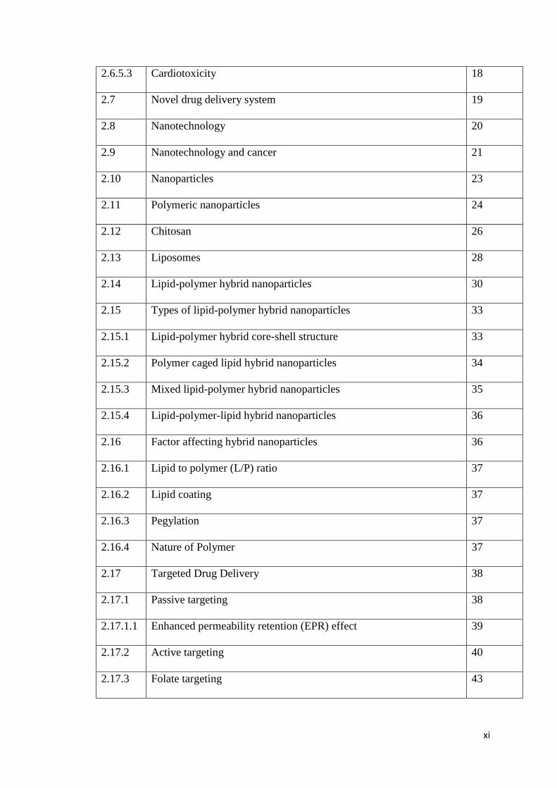
Table of contents ix
List of Figure xvii
List of Tables xxi
List of Abbreviations xxii
Appendences Xxiv
Abstract Xxv
Part II
Chapter 1. Introduction
1.1 Introduction 1
1.2 Main objectives of study 8
Chapter 2. Literature Review
2.1 Cancer 11
2.2 Ovarian cancer 12

x
2.3 Classification of ovarian cancer 12
2.3.1 Epithelial tumors 12
2.3.2 Stromal cell tumors 13
2.3.3 Germ cell tumors 13
2.4 Risk factors of ovarian cancer 14
2.4.1 Hormonal 14
2.4.2 Pregnancy and infertility 14
2.4.3 Lactation 14
2.4.4 Use of oral contraceptives 14
2.5 Treatment of ovarian cancer 15
2.5.1 Surgery 15
2.5.2 Chemotherapy 15
2.6 Cisplatin 15
2.6.1 Chemical structure 16
2.6.2 Mechanism of action 16
2.6.3 Pharmacokinetic properties 16
2.6.4 Therapeutic uses 17
2.6.4.1 Cisplatin and ovarian cancer 17
2.6.4.2 Cisplatin and lung cancer 17
2.6.4.3 Cisplatin and breast cancer 17
2.6.4.4 Cisplatin and brain cancer 17
2.6.5 Side effects of cisplatin 18
2.6.5.1 Hepatoxicity 18
2.6.5.2 Nephrotoxicity 18
xi
2.6.5.3 Cardiotoxicity 18
2.7 Novel drug delivery system 19
2.8 Nanotechnology 20
2.9 Nanotechnology and cancer 21
2.10 Nanoparticles 23
2.11 Polymeric nanoparticles 24
2.12 Chitosan 26
2.13 Liposomes 28
2.14 Lipid-polymer hybrid nanoparticles 30
2.15 Types of lipid-polymer hybrid nanoparticles 33
2.15.1 Lipid-polymer hybrid core-shell structure 33
2.15.2 Polymer caged lipid hybrid nanoparticles 34
2.15.3 Mixed lipid-polymer hybrid nanoparticles 35
2.15.4 Lipid-polymer-lipid hybrid nanoparticles 36
2.16 Factor affecting hybrid nanoparticles 36
2.16.1 Lipid to polymer (L/P) ratio 37
2.16.2 Lipid coating 37
2.16.3 Pegylation 37
2.16.4 Nature of Polymer 37
2.17 Targeted Drug Delivery 38
2.17.1 Passive targeting 38
2.17.1.1 Enhanced permeability retention (EPR) effect 39
2.17.2 Active targeting 40
2.17.3 Folate targeting 43

xii
Chapter 3. Synthesis and physicochemical characterization of LPHNPs
3.1 Background 48
3.2 Materials and Methods 50
3.2.1 Materials 50
3.2.2 Method of preparation 50
3.2.3 Size and Zeta Potential of hybrid nanoparticles 51
3.2.4 Determination of drug loading and entrapment efficiency 51
3.2.5 Morphology of hybrid nanoparticles 52
3.2.6 Fourier transform infrared spectroscopy (FTIR) 52
3.2.7 Powdered X-Ray diffraction analysis (X-RD) 52
3.2.8 Differential scanning calorimeter (DSC) 52
3.2.9 Thermo-gravimetric analysis (TGA) 52
3.2.10 In vitro drug release studies 53
3.3 Results 54
3.3.1 Size and surface charge 54
3.3.2 Morphology of nanoparticles 56
3.3.3 Entrapment efficiency and drug loading 56
3.3.4 FTIR spectroscopy 57
3.3.5 XRD Studies 58
3.3.6 Differential scanning calorimetery (DSC) 59
3.3.7 Thermogravimetric analysis (TGA) 61
3.3.8 In vitro dissolution studies 63
3.5 Discussion 66
3.6 Conclusion 68
xiii
Chapter 4. Biological characterization of LPHNPs
4.1 Background 70
4.2 Materials and Methods 71
4.2.1 Materials 71
4.2.2 Cell viability 71
4.2.3 Florescence microscopy 71
4.2.4 Cellular uptake 72
4.2.5 In vivo toxicity 72
4.2.6 In vivo pharmacokinetics 72
4.3 Results 73
4.3.1 Cell viability studies 73
4.3.2 Cellular uptake studies 76
4.3.3 Cellular association 77
4.3.4 Toxicological studies in rats 79
4.3.4.1 Biochemical and blood analysis 80
4.3.4.2 Histopathological examination 82
4.3.5 In vivo pharmacokinetics 84
4.4 Discussion 86
4.5 Conclusion 88
Chapter 5. Synthesis and Physicochemical characterization of Folate LPHNPs
5.1 Background 90
5.2 Materials and Methods 93
5.2.1 Materials 93
5.2.2 Preparation of folate-chitosan conjugates 93

xiv
5.2.3 Preparation of folate-chitosan conjugated lipid hybrid nanoparticles 95
5.2.4 Purity of folate-chitosan conjugate 98
5.2.5 Nuclear magnetic resonance 98
5.2.6 Size and zeta potential of hybrid nanoparticles 98
5.2.7 Determination of drug loading and entrapment efficiency 98
5.2.8 Morphology of hybrid nanoparticles 99
5.2.9 In vitro drug release studies 99
5.3 Results 100
5.3.1 Purity of the conjugate 100
5.3.2. Nuclear magnetic resonance spectroscopy 101
5.3.3 Size and zeta potential of hybrid nanoparticles 103
5.3.4 Drug entrapment and loading efficiency 104
5.3.5 Morphology of folate LPHNPs 105
5.3.6 In vitro drug release profile 106
5.4 Discussion 108
5.5 Conclusion 109
Chapter 6. Biological characterization of LPHNPs
6.1 Background 111
6.2 Materials and Methods 113
6.2.1 Materials 113
6.2.2 Cell viability studies 113
6.2.3 Florescence microscopy 113
6.2.4 Cellular uptake 114
6.2.5 Cell apoptosis 114

xv
6.2.6 Cell cycle 114
6.2.7 Preparation of 3D spheroids 115
6.2.8 Cell viability towards 3D spheroids 115
6.2.9 Cell uptake studies towards 3D spheroids 115
6.2.10 Florescence microscopy images of 3D spheroids 115
6.3 Results 117
6.3.1 Cytotoxicity studies 117
6.3.2 Cell uptake studies 125
6.3.3 Florescence microscopy 126
6.3.4 Cell cycle studies 127
6.3.5 Cell apoptosis studies 128
6.3.6 Cell Cytotoxicity towards cancer cell in 3D spheroids 134
6.3.7 Cell uptake studies towards cancer cell in 3D spheroids 137
6.3.8 Florescence microscopic images of cell uptake studies towards
cancer cell in 3D spheroids
138
6.4 Discussion 142
6.5 Conclusion 144
Chapter 7. Conclusions and Recommendation
7.1 Future prospects 148
Chapter 8. References

xvi
List of Figures
Figure 2.1: Chemical structure of Cisplatin 16
Figure 2.2: Areas of nanotechnology applications. 21
Figure 2.3: Structure of polymeric nanoparticles. 25
Figure 2.4: Chemical structure of chitosan 27
Figure 2.5: Structure of liposomes with PEG coating. 29
Figure 2.6: Core- shell lipid-polymer hybrid nanoparticles. 34
Figure 2.7: Lipid-polymer cage hybrid nanoparticle. 35
Figure 2.8: Mixed lipid-polymer hybrid system. 35
Figure 2.9: Lipid-polymer-lipid hybrid nanoparticles. 36
Figure 2.10: Advantage of targeted drug delivery system 38
Figure 2.11: Antibody targeted drug delivery system. 41
Figure 2.12: Folate receptor internalization mechanism. 44
Figure 3.1: Schematic diagram of lipid-chitosan hybrid nanoparticles. 51
Figure 3.2: Transmission electron microscopy images of lipid-chitosan
hybrid nanoparticles.
56
Figure 3.3: FTIR Spectra of individual components and lipid-polymer
hybrid formulation.
58
Figure 3.4: X-ray diffraction analysis of components and LPHNPs. 59
Figure 3.5: Differential Scanning calorimetry graph of LPHNPs and
components.
60
Figure 3.6: Thermo gravimetric analysis of cisplatin loaded lipid-chitosan
hybrid nanoparticles.
62
Figure 3.7: In vitro drug release profile of LPHNPs with various ratios of
lipid to polymer.
64
Figure 4.1: Cytotoxicity studies of cisplatin LPHNPs compared with
cisplatin solution and blank LPHNPs on A2780 cell lines after
24 hours of treatment.
74
Figure 4.2: Cytotoxicity studies of cisplatin LPHNPs compared with
cisplatin solution and blank LPHNPs on A2780 cell lines after
24 hours of treatment.
75

xvii
Figure 4.3: Cellular uptake of LPHNPs by A2780 cell lines. 76
Figure 4.4: Uptake of chitosan-lipid hybrid nanoparticles loaded with Rh-PE
by A2780 cells.
78
Figure 4.5: Representative histopathological images of rat vital organs (A)
Control (B) Blank LPHNPs (C) Cisplatin loaded LPHNPs.
83
Figure 4.6: Concentration versus time profile curve of cisplatin LPHNPs
and cisplatin solution. (Mean± SD n=6)
85
Figure 5.1: Schematic chemistry of folate-chitosan conjugate. 94
Figure 5.2: Schematic diagram of folate targeted lipid-chitosan hybrid
nanoparticles.
95
Figure 5.3: Schematic chemistry of formation of folate lipid-chitosan hybrid
nanoparticles.
96
Figure 5.4: Mechanism of internalization of folate targeted lipid-chitosan
hybrid nanoparticles via endocytosis.
97
Figure 5.5: Thin layer chromatography of folic acid and folate-chitosan
conjugate.
100
Figure 5.6: Figure 5.6. 1H-NMR spectra of folate-chitosan conjugate,
chitosan and folic acid.
102
Figure 5.7: Transmission electron microscopy image of folate targeted lipid-
chitosan hybrid nanoparticles.
106
Figure 5.8: In vitro release profile of folate lipid-chitosan hybrid
nanoparticles and cisplatin drug solution
107
Figure 6.1: Cell viability study on A2780 cell lines after 24 hours of
incubation comparison of blank folate LPHNPS, folate LPHNPS
and LPHNPs.
119
Figure 6.2: Cell viability study on A2780 cell lines after 48 hours of
incubation comparison of blank folate LPHNPS, folate LPHNPS
and LPHNPs.
120
Figure 6.3: Cell viability study on SKOV3 cell lines after 24 hours of
incubation comparison of blank folate LPHNPS, folate LPHNPS
and LPHNPs.
121
Figure 6.4: Cell viability study on SKOV3 cell lines after 48 hours of
incubation comparison of blank folate LPHNPS, folate LPHNPS
122

xviii
and LPHNPs.
Figure 6.5: Cell viability study on MCF-7 cell lines after 24 hours of
incubation comparison of blank folate LPHNPS, folate LPHNPS
and LPHNPs.
123
Figure 6.6: Cell viability study on MCF-7 cell lines after 48 hours of
incubation comparison of blank folate LPHNPS, folate LPHNPS
and LPHNPs.
124
Figure 6.7: Cell uptake studies using flow cytometry. Comparison of
LPHNPs and Folate LPHNPs loaded with rhodamie-123.
125
Figure 6.8: Fluorescence microscopy images of SKOV3 cells treated with
folate LPHNPs and folate LPHNPs loaded with Rh-123 and Rh-
PE.
126
Figure 6.9: Cell cycle studies after 24 hours of treatment. 127
Figure 6.10: Cell cycle studies after 48 hours of treatment. 128
Figure 6.11: Cell apoptosis after 24 and 48 hours of treatment comparison of
cisplatin solution, LPHNPs and folate LPHNPs.
129
Figure 6.12: Control group cell apoptosis showing early and late apoptosis. 130
Figure 6.13: Cell apoptosis of cisplatin drug solution showing early and late
apoptosis.
131
Figure 6.14: Cell apoptosis of lipid- chitosan hybrid nanoparticle loaded with
cisplatin.
132
Figure 6.15: Cell apoptosis of folate targeted lipid-chitosan hybrid
nanoparticles.
133
Figure 6.16: Cytotoxicity studies on 3D spheroids after 24 hours of treatment
comparison of folate LPHNPs, LPHNPs and cisplatin drug
solution.1
135
Figure 6.17: Cytotoxicity studies on 3D spheroids after 24 hours of treatment
comparison of folate LPHNPs, LPHNPs and cisplatin drug
solution.
136
Figure 6.18:
Cell uptake studies using flow cytometry comparison of folate
LPHNPs, LPHNPs.
137
Figure 6.19: Cell uptake towards 3D spheroids using fluorescence
microscope comparison of folate LPHNPs with LPHNPs.
138

xix
Figure 6.20:
Z-stack images of 3D spheroids control group. 139
Figure 6.21: Z-stack image of lipid-chitosan hybrid nanoparticle group of 3D
spheroids.
140
Figure 6.22: Z-stack image of folate targeted lipid-chitosan hybrid
nanoparticle group of 3D spheroids.
141

xx
List of Tables
Table 2.1: Summary of clinical development of folate targeted ovarian
cancer drugs.
45
Table 3.1: Composition of cisplatin loaded lecithin-chitosan hybrid
nanoparticles
55
Table 3.2: Effect of lipid to polymer ratio on particle size and surface
charge
55
Table 3.3: Effect of Lipid to Polymer ratio on entrapment efficiency
and Drug Loading
57
Table 3.1: Kinetic Modeling of Drug Release profile of Cisplatin
loaded Lecithin-Chitosan Hybrid Nanoparticles
65
Table 4.1: Body weight and food, water intake of rats of different group 79
Table 4.2: Comparative hematological parameters in rats. 81
Table 4.3: Comparative liver and renal function parameters in rats. 82
Table 5.2: Composition of cisplatin loaded lecithin-chitosan hybrid
nanoparticles
103
Table 5.3: Effect of lipid to polymer ratio on particle size and surface
charge.
104
Table 5.4: Effect of Lipid to polymer ratio on entrapment efficiency
and drug loading
105

xxi
List of abbreviations
Term Description
CTB Cell titer blue
CTG Cell titer glo
CBC Complete blood count
DMEM Dulbecco’s Modified Eagle’s Medium
DMSO Dimethyl sulfoxide
DLS Dynamic light scattering
DSC Differential scanning calorimetry
DDS Drug delivery system(s)
DL Drug loading
EDC 1-ethyl-3-(3- dimethylaminopropyl) carbodiimide hydrochloride
EE Encapsulation efficiency
FBS Fetal bovine serum
FTIR Fourier transform infrared spectroscopy
Folate LPHNPs Folate lipid-polymer hybrid nanoparticles
LPHNPs Lipid-polymer hybrid nanoparticles
LFT Liver function test
MRT Mean residence time
PBS Phosphate buffer saline
PI Propidium iodide
PDI Polydispersity index

xxii
RPMI Roswell Park Memorial Institute medium
Rh-123 Rhodamine-123
Rh-PE Rhodamine phosphatidylehtanolamine
RFT Renal function test
TLC Thin layer chromatography
TEM Transmission electron microscope
UV Ultraviolet
X-RD X-ray diffraction

xxiii
List of Publications
1. Lipid-chitosan hybrid nanoparticles for controlled delivery of cisplatin
(Drug Delivery) (IF: 3.89) …171
Submitted
1. Lipid-chitosan hybrid nanoparticles for co-delivery of cisplatin and curcumin
with enhanced therapeutic efficacy
2. Folate targeted lipid-chitosan hybrid nanoparticles for enhanced therapeutic
efficacy

xxiv
Abstract
Nanotechnology has emerged as a hope to deliver drugs at targeted site to obtain
maximum therapeutic benefits. Lipid-polymer hybrid nanoparticles have provided the
platform to deliver drugs in a controlled manner with enhanced stability and
biocompatibility. LPHNPs have the dual advantages of polymeric and liposomal drug
delivery system and can encapsulate both hydrophilic and hydrophobic drugs. The
objective of this dissertation was to develop the lipid-chitosan hybrid nanoparticles
for potential delivery of chemotherapeutic agents at tumor site to achieve maximum
therapeutic benefits and decrease side effects associated with chemotherapeutic
agents.
Lipid-chitosan hybrid nanoparticles were prepared by using chitosan as a polymer and
LIPOID S75 as a lipid using modified ionic gelation method. The prepared
nanoparticles have size in range of 200-300nm with PDI values less than 0.3 and
surface charge showed good stability in suspension form. The transmission electron
microscopy images showed spherical nanoparticles having lipoplex like structure. All
prepared nanoparticles showed high entrapment efficiency (>80%) and good drug
loading. The FTIR analysis confirmed the compatibility among the excipients and X-
RD analysis showed no sharp peaks of cisplatin in formulation and cisplatin is
converted into amorphous form inside lipid-chitosan hybrid nanoparticles system.
Thermal studies using differential scanning calorimetry and thermogravimetric
analysis confirmed the excellent stability of prepared hybrid nanoparticles. The in
vitro release showed that controlled release of drug over prolong period of time.
Kinetic modeling showed that the release pattern follows super case II transport
mechanism. The physicochemical evaluation confirmed the excellent stability and
controlled release profile.
The therapeutic efficacy of cisplatin loaded lipid-chitosan hybrid nanoparticles was
evaluated by using A2780 ovarian cell lines. The results confirmed the enhanced
cytotoxicity of cisplatin loaded lipid-chitosan hybrid nanoparticles as compared to
cisplatin solution. Cellular interaction and cell uptake showed 8 times greater uptake
as compared to control. Further in vivo pharmacokinetic studies confirmed enhanced
mean residence time of LPHNPS inside the biological system. Toxicology studies
confirmed the safety profile of lipid-chitosan hybrid nanoparticles.

xxv
The folic acid was conjugated with chitosan for folate targeting to achieve maximum
therapeutic benefits at the tumor site. The TLC analysis confirmed the purity of
conjugate and absence of free folic acid while nuclear magnetic resonance
spectroscopy confirmed the successful conjugation of folic acid with chitosan. The
folate-chitosan conjugate was then used to prepare nanoparticle by ionic gelation
method with anionic lipids. The prepared nanoparticles have particle size in range of
200nm and low polydispersity index and surface charge of greater than +20. The
folate LPHNPs showed greater than 75% encapsulation with excellent drug loading.
The prepared nanoparticles are spherical in shape with lipoplex like structure having
folate on the outer side of nanoparticles. In vitro release profile shows sustained
release of cisplatin over a period of 48 hours.
The therapeutic efficacy of folate lipid-chitosan hybrid nanoparticles was evaluated
on ovarian and breast cell lines. The A2780 and SKOV3 were used as ovarian cell
lines and treated with folate targeted LPHNPs and untargeted LPHNPs and cisplatin
solution. The results confirmed the enhanced cytotoxic effect of folate LPHNPs as
compared to untargeted LPHNPs and cisplatin solution. Similar enhanced cytotoxic
effect of folate LPHNPs was observed on MCF-7 breast cancer cell lines. The cell
uptake studies showed two times more uptake of folate LPHNPs are compared to
untargeted LPHNPs that is due to folate receptor mediated endocytosis and leads to
enhanced therapeutic efficacy. The therapeutic efficacy of folate LPHNPs was further
evaluated on 3D spheroids in vivo model to check the response of nanoparticles in in
vivo environment. The cell viability studies on 3D spheroids confirmed the enhanced
cytotoxic effect of folate LPHNPs as compared to untargeted and much more
significant cytotoxic effect as compared to cisplatin solution. The fluorescence
microscopy images, and flow cytometry analysis confirmed the enhanced cellular
uptake of folate LPHNPs in 3D spheroids that leads to enhanced therapeutic efficacy.
In conclusion, lipid-chitosan hybrid nanoparticles are suitable platform for controlled
delivery of chemotherapeutic agents. Folate targeted LPHNPs with added advantage
of lipid coating is suitable for active targeting with enhanced therapeutic efficacy and
minimum side effects.

0
CHAPTER 1
Introduction

1
1.1. Introduction
Nanotechnology has multiple applications including agriculture, space , forensic and
medical therapeutics (Lai et al., 2006). The term nanotechnology was first coined by
an American scientist Richar Fenman in 1959, that result in conceptual building of
nanomaterial (Lyshevski et al., 2007). Nanotechnology has played wonders in the
area of food technology in identification of bacteria and monitoring quality of food.
Biosensors carbon nanotubes are being used in packaging of food to improve its
mechanical properties (Kang et al., 2007). Nanoparticles have also found applications
in cosmetics development of moisturizers with prolong skin contact, sun block and
anti-aging. Liposomes provide sustained skin contact and delivery of vitamins and
regeneration of epidermis with improved efficiency (Thong et al., 2007).
Pharmaceutical nanotechnology has provided a breakthrough in the delivery of drugs.
Nanotechnology is helpful in targeting drugs to the tissue and cellular level, crossing
the endothelial and blood brain barrier and co-delivery in combinatorial therapy
(Farokhzad and Langer, 2009). The emergence of nanotechnology has a great
influence on different research industries and particularly pharmaceutical research
industry. Nanotechnology tools enable drug to be delivered at a specific site and being
utilize for drug development and biological screening at nanoscale (Kharb et al.,
2006). Nanotechnology has significantly increased the bioavailability of drugs and
also play role in diagnosis of disease by interacting at cellular level and detoxification
of drugs from body is another application of nanotechnology that has been
successfully tested in rats. Nanotechnology can deliver the drug into the cell
cytoplasm, thus reducing side effect and enhance therapeutic efficacy. Nanoparticle
can serve as a diagnostic sensor and also can protect the drug for prolong time and
release upon specific signals (Nikalje, 2015). Delivery of genes such as siRNA and
DNA has proved to be fundamental in treatment of ailments, but their oral and
intravenous delivery is limited due to enzymatic cleavage, uptake by RES, and kidney
filtration, however nanotechnology provided the way for the delivery of siRNA and
DNA (Anderson et al., 2004). Pharmaceutical nanotechnology plays a vital role in the
formulation of new chemical entities with poor solubility and permeability and helps
in achieving desired pharmacokinetic properties. Size of nanoparticle play crucial role

2
in achieving the desired properties (Devalapally et al., 2007). Pharmaceutical
nanotechnology has also played the role to revolutionize the herbal medicines.
Cancer nanotechnology is an emerging field, and bringing wonders in the diagnosis
and treatment of cancer (Bharali and Mousa, 2010). Traditional drugs delivery system
in cancer treatment has several disadvantages, such as non-specific delivery and toxic
effects on normal cells. Cancer nanotechnology has several advantages and in
combination with biodegradable polymer, idea of polymer-drug conjugate in
nanotechnology was first introduced by Ringdorf in 1975 (Ringsdorf, 1975).
Nanotechnology is also overcoming the challenges of tumor diagnosis by using
quantum dots and high quality imaging (Ehdaie, 2007). The main concern in cancer
treatment is to achieve desired therapeutic concentration of drug at the tumor site and
to avoid the side effects on the normal body tissues, thus nanotechnology has served
as a tool for the targeted delivery by conjugating with various targeting moieties
including aptamers and antibodies (Misra et al., 2010).
First ever nanotechnology system discovered was of lipid vesicles in 1960 and later
on it was called as Liposomes (Bangham et al., 1965). Specific delivery of liposomes
at targeted site was achieved in 1980 (Leserman et al., 1980). Liposomes are formed
by incorporating phospholipids in aqueous phase, forming bilayer structure. They can
incorporate both hydrophilic and hydrophobic drugs (Israelachvili et al., 1980). First
generation of nanotherapeutics was non-targeted but still provide advantage over
conventional drug delivery system (DDS) such as increase in half life and enhanced
solubility and enabling sustained release (Riehemann et al., 2009). Liposomes have
superior biocompatible properties and have ability to load the drug as well as RNA,
DNA. Liposome has long circulating properties and ability to specifically target the
cells by attaching to various targeting moieties. Liposomes has the ability to deliver
their load into the cytoplasm and also utilized as a vehicle in photodynamic therapy
(Torchilin, 2005). Liposomes has provided breakthrough in gene therapy by
successful delivery of nucleic acid with increase transfection efficiency. Cationic
lipids are particularly used for the nucleic acid delivery. High biocompatible and
biodegradable properties also make liposome ideal system for delivery of drug to
brain (Samad et al., 2007). Lipids can adopt different structure including gel, sol and
micelle formation. Liposomes prepared from the saturated lipids have the better
stability as compared to unsaturated lipids. Liposome surface can be modified by

3
using anionic, cationic and neutral lipids according to the target of action. Liposomes
can permeate through the leaky microvasculature and reside for longer time, enabling
targeted delivery at the tumor site (Maurer et al., 2001).
The major drawbacks of liposomes are rapid clearance from the body and are
captured by RES, mainly in Liver. Liposomes have biomimetic properties but are
cleared by macrophages, thus suffer the problem of shorter half-life. Liposomes have
low encapsulation efficiency for hydrophilic drugs because of limited aqueous phase
and volume of hydration is greater on the outer side. Sterilization of liposome is
challenging because of their composition, phospholipids can undergo phase transition
after exposure to the surrounding high temperature, which may result in loss of
product. The lipid component of phospholipid may also undergo oxidation and result
in degradation (Pattni et al., 2015). The rapid elimination of liposomes is due to
opsonization by the plasma components, uptake by reticuloendothelial system in
spleen (Hua and Wu, 2013).
Polymeric nanoparticles (PNPs) are biodegradable, stable and can effectively deliver
the drug at the target site with controlled manner. PNPs have excellent entrapment
efficiency because drug is confined inside the polymer matrix that provide stability
and controlled release (Soppimath et al., 2001). PNPs can respond to both internal and
external stimuli, thus delivering the drug to targeted tissue and ensuring maximum
therapeutic efficacy and minimizing side effects (Cheng et al., 2013). Polymers used
for nanoparticle-based drug delivery are biocompatible, they deliver drug at the
targeted site, then degraded and released out of the body. PNPs can increase the
solubility of poorly soluble drugs by attaching solubilizing moieties. The drug having
short half-life can be effectively encapsulated inside polymeric nanoparticles, thus
tremendously increasing the stability and circulating half-life. Drug release from the
polymeric nanoparticles can be controlled by modulating the characteristics of
polymers (Parveen and Sahoo, 2008). PNPs can be used for targeted delivery to tumor
tissue by exploiting the anatomical differences between normal and tumor tissue.
Tumor’s vasculature is large is size, more permeable and leakier as compared to tight
endothelium of healthy tissue. Increased production of vasodilation mediators also
facilitates extravasation. Thus polymeric nanoparticles can reside at tumor side and
increased therapeutic effects (Maeda et al., 2000).

4
Despite of several advantages of polymeric nanoparticles, they also suffer from some
drawbacks. Polymeric nanoparticles are not an ideal system for delivery of protein
and peptides, instability of protein could result during degradation of the polymer.
Polymer can influence the conformational changes of the protein. Burst release of
protein also occur from the polymeric nanoparticles that can lead to unwanted effects
(Mohammadi-Samani and Taghipour, 2015). Polymeric nanoparticle sometimes
results in aggregate formation that can affect the release of drug and overall
physicochemical properties of the drug delivery system (Feng, 2004). Most of the
polymers used for polymeric nanoparticles have hydrophobic surface, that might be
recognized as a foreign body that result in rapid clearance from the body (Hu et al.,
2011).
Lipid-polymer hybrid nanoparticles (LPHNPs) combine the advantages of both
liposomal and polymeric drug delivery system. These types of nanoparticles are
typically consisting of a hydrophobic core and a lipid covering and a hydrophilic
polymer sheath to provide long circulating characteristics. They can incorporate both
hydrophilic and hydrophobic drugs (Zhang and Zhang, 2010). Polymeric
nanoparticles may suffer from the drawback of membrane permeability, combination
of lipid with polymer result in additional biomimetic properties and enhanced
therapeutic efficacy (Rajendiran et al., 2016). Lipids are usually attached to the
polymer layer via ionic interaction or hydrophobic interaction. Ionic interaction
results due to oppositely charged polymer and lipid. LPHNPS have much higher
transfection efficiency as compared to liposomal based and polymer based gene
delivery (Bose et al., 2016). Lipid-polymer hybrid nanoparticles alter the
pharmacokinetic properties of the drug and a suitable vehicle for controlled and
sustained drug delivery. Because of both hydrophilic and hydrophobic portions,
hybrid nanoparticles can deliver multiple agents at the same time. Co-loading of
siRNA and drug is possible, and two drugs can also be delivered at the same time
with excellent loading efficiency. LPHNPs can also co-deliver diagnostic and
therapeutic agents at the same time. Surface modification of hybrid nanoparticles can
also serve as a suitable vehicle for the oral delivery of chemotherapeutic agents
(Zhang et al., 2017). The hybrid nanoparticles usually exhibit core-shell structure;
polymeric core can efficiently deliver drugs with small molecular weight and
diagnostic agents, while lipid core imparts stability, biomimetic and biocompatible

5
properties resulting in enhanced therapeutic efficacy. PEG coating over the lipid layer
further enhance the long circulating properties of these nanoparticles (Wakaskar,
2018). Some studies suggest that LPHNPS have built-in MDR reversal properties,
They have the ability to down regulate the P-gp and increase the toxicity of the
chemotherapeutic agents (Wong et al., 2006).
Chitosan is a naturally occurring polysaccharide and produce from the chitin, which is
naturally occurring structural element of crab and lobsters. Chitosan also occurs in
some fungi. Structure of Chitosan is like cellulose, but it has additional hydroxyl
group, free amino group, due to which it exhibits different functional properties.
Chitosan can respond to different environmental stimuli, such as pH. Chitosan has
great applications in pharmaceutical field due to its intrinsic favorable properties such
as biocompatibility, susceptible to hydrolysis by enzyme and ability to bind with
some organic compounds. Chitosan has stimuli responsive drug delivery properties; it
can be easily protonated in acidic environment and can form complex with negatively
charged polymers, lipids and genes. Chitosan also has mucoadhesive properties and
can lead to significantly enhanced bioavailability. Chitosan has the advantage for
controlled drug delivery, which result in increased efficacy and reduced side effects
(Elgadir et al., 2015). Most of the conventional drug delivery system result in
immediate release of drug that cause fluctuation in the plasma level of drug and
results in unwanted side effect. The drug incorporated in suitable polymer result in
controlled diffusion and slow release. Chitosan is one of suitable polymer for
controlled release formulations (Kumar, 2000). Chitosan has unique cationic
characteristics and gel forming ability, it can form complex with oppositely charged
molecules for the controlled delivery of drug, protein and peptides (Shu and Zhu,
2000). Chitosan has also been investigated for oral controlled drug delivery in
stomach due to its gel forming ability at lower pH and increased gastrointestinal
retention time. Chitosan also has mucoadhesive and antacid properties, which reduce
the drug irritation in the stomach. Chitosan is suitable for both oral and intravenous
controlled drug delivery (Chandy and Sharma, 1992).
Lipids are used in drug delivery system. Most commonly used lipids in drug delivery
are phosphatidylcholine (PC) and phosphatidylethanolamine (PE), phosphatidylserine
(Ps). These are naturally occurring phospholipids and obtained from plants and
animals and are constituent of biological membrane. Lipids used in drug delivery

6
mostly contain phosphatidylcholine and very little amount of PE, because PE can
disrupt bilayer structure under physiological pH (Yingchoncharoen et al., 2016).
Lipids have gain importance for the delivery of drugs having poor water solubility.
Lipids based drug delivery system also enhances the bioavailability of the drugs. The
absorption of drug from the lipid formulation depends on the degree of dispersion and
emulsification of lipid. In general lipid based formulation accelerate the absorption by
increasing dissolution and formation of solubilized phase (Kalepu et al., 2013).
Incorporating the poorly absorbed drug with phosphatidylcholine has been shown to
increase the bioavailability. Intestinal permeability of some drugs have been shown to
increase 20 time by incorporating into phosphatidylcholine containing lipids
(Fagerholm et al., 1998). The lipid used in the formulation is LIPOID S75 that
contain 75% phosphatidylcholine and is suitable for better bioavailability and
controlled drug delivery (Hafner et al., 2009).
Folate receptors were first recognized as a tumor marker when monoclonal antibodies
raised against ovarian tumor were found to recognize the alpha form of folate
receptor. Antibodies that produce against alpha folate receptor found its application as
diagnostic agent in gynecological tumor. After these initial finding the overexpression
of folate receptor was found in more than 90% of ovarian tumors. Because of their
high affinity to folate linked drug they have exploited as an agent for targeted drug
delivery to tumor (Lu and Low, 2003). Folic acid is now used as a targeting agent for
the targeted delivery to the tumor because of its intrinsic small size, penetration
ability, stability, solubility in organic solvents and ability to conjugate with various
therapeutic agents (Cho et al., 1997). Targeting folate receptor is a promising
approach by conjugating folic acid with the polymer that result in significantly
increased uptake of drug by the tumor cells (Saul et al., 2003). Folate membrane
receptors have emerged as a target for drug delivery, in fact, folate metabolism is
primary in replication of DNA and drugs that inhibit folate metabolism can be
targeted. Folate receptors are overly expressed in tumors, which are not in normal
cells. Folate is a vitamin, which is water soluble and carbon donor in the synthesis of
purines, which lead to synthesis of DNA. Folate is required by the normal cells for
multiplication and it enters the cell via anion exchange pathway. Folate is not
overexpressed in the healthy cells, while rapidly dividing tumor cell overexpress the
folate receptor and present as a target for drug delivery (Marchetti et al., 2014).

7
Cisplatin has been used as a first line agent in the treatment of ovarian cancer since
decades. But it exerts side effect on major organs particularly kidney. Several
researches have been made to reduce the side effects and increase the therapeutic
efficacy of the cisplatin by formulating in various nanoparticles and liposomes.
Controlled drug release from the nanoparticles could be an approach to reduce the
side effects associated with the cisplatin (Cheng et al., 2011). Cisplatin inhibit the
tumor growth by inhibiting the DNA mediated functions. Cisplatin inhibit the cell
cycle that result in abnormal mitosis and leads to apoptosis of cells. Cisplatin is
known to cause the inhibition of cell cycle at G2/M phase of cell cycle. Cisplatin form
the bifunctional adduct with the DNA and inhibit the tumor growth (Eastman, 1999).
Here we formulated the novel chitosan lipid hybrid nanoparticles for controlled drug
delivery of cisplatin to reduce the side effect and increase the therapeutic efficacy.
Then chitosan was conjugated with the folic acid and folate targeting was done with
the LPHNPs that added the advantage of lipid layer with folate targeting.

8
1.2. Main objective of study
1. To formulate LPHNPs consisting of chitosan as a polymer and LIPOID S -75
as a lipid by ionic-gelation method and perform physicochemical
characterization.
2. To develop the controlled release formulation of cisplatin loaded in chitosan
LPHNPs.
3. To evaluate the therapeutic efficacy and cell uptake of cisplatin loaded
LPHNPs on ovarian cell line A2780.
4. To evaluate bioavailability, bioavailability studies will be performed in animal
species.
5. To evaluate the in vivo toxicity studies with higher doses of cisplatin and
observe the effect on biochemical parameters and tissue level.
6. To formulate chitosan- folate conjugate for folate targeting and prepare folate
LPHNPS from conjugate and perform physicochemical characterization.
7. To evaluate the effect of folate targeting on ovarian cell line A2780 and
SKOV3 and breast cancer cell lines MCF-7.
8. To perform qualitative cell uptake studies of folate targeted LPHNPs using
fluorescence microscopy and perform quantitative cell uptake of folate
targeted LPHNPS and compare that with untargeted LPHNPS using flow
cytometry
9. To evaluate the cell cycle inhibition mechanism of cisplatin and cell apoptosis
using ovarian cell line.
10. To formulate the three-dimensional (3D) tumor cell culture model to mimic
the in vivo microenvironment of cultured tumor cell

9
11. To study the cell cytotoxicity and cell uptake of folate LPHNPs on 3 D
spheroids to establish their efficacy in in vivo model.

10
Bjs Waltham
Literature Review
CHAPTER 2

11
2.1. Cancer
Cancer is characterized by abnormal cell growth that invades into other body parts
and symptoms include lump, loss of appetite and abnormal bleeding but the
symptoms varies in different types of cancer (Heim and Mitelman, 2015). Cancer cell
differ from normal body cells in some characteristics such as loss of differentiation
and reduced sensitivity to drug and increased invasiveness (Klein, 1987). Cancer
results from the changes in the genome in which oncogene results in increase in their
function and dominance and decrease in function of suppressor gene. Studies reveal
that tumorigenesis is a multistep process that leads to the conversion of normal cell
into malignant cell (Hanahan and Weinberg, 2000). Some studies suggest that
external environmental factors play key role in the development of type of cancer as
compared to genetic factors (Blackadar, 2016).
Cancer results from a series of mutations in genes and these mutations results in
change in cellular function. Chemical compounds also play their role in type of
genetic mutation and cancer (Aizawa et al., 2016). During proliferation the cell
replicates its genome and then divide into daughter cells, the process of proliferation
required energy. In the tumor cell the proliferation is uncontrolled, and cells lost its
check on the process of proliferation. In order to meet the energy demand of
uncontrolled proliferation the cells may adopt different pathway for energy and
metabolism (Garber, 2006). Some studies suggest that alteration in energy metabolism
process may be among root cause of cancer. Warburg proposed that cancer cell use
glycolysis instead of oxidative phosphorylation for ATP production even in the
presence of large amount of oxygen (Warburg, 1956). Increased metabolic demand is
an inherent challenge for the tumor cell and tumor cell modify the metabolism at
cellular level to support the proliferation and adopt the strategies to survive during the
period of metabolic stress (Deberardinis et al., 2008). The major metabolic change in
the proliferating cells is the start of aerobic glycolysis. Glucose is one of primary
nutrient for the proliferating cells. In the proliferating tumors, the pyruvate is
converted into lactate which is then secreted externally and cell recovers NAD+,
which is important to maintain glycolysis and is used for ATP production and
proliferation of tumor cell (Jones and Thompson, 2009).

12
2.2. Ovarian Cancer
Ovarian cancer is one of the leading causes of death. It can be broadly divided into
three different categories, most prevalent one is epithelial cell carcinoma, others are
stromal and germ cell cancer (Agarwal and Kaye, 2003). Ovarian cancer accounts for
0.23 million new cases and 0.15 million deaths annually. The highest rate are
prevalent in the central Europe while around 21000 new cases and 14000 death are
expected to occur in USA annually from ovarian cancer, while in china the rate is low
as compared to population and incidence is 4 in 100,000 peoples (Atlanta, 2015).
The risk of developing ovarian cancer in women in one in 75, while the risk of death
is one in 100. The diagnosis of ovarian cancer is difficult, and is usually diagnosed at
late stage with poor prognosis.
2.3. Classification of ovarian cancer
The exact etiology and cellular origin of ovarian cancer is not well understood in most
cases the tumor seems to be originated from the gynecological tissue and then spread
to the ovary. Broadly ovarian tumors are divided into three types epithelial, stromal
and germ cell origin but more than 90% of ovarian tumors arise from the epithelial
cells (Sankaranarayanan and Ferlay, 2006).
2.3.1. Epithelial tumors
These are most prevalent form of ovarian tumors. These usually originate from the
surface epithelium of the ovary. The epithelial tumor could be benign, atypical
proliferating and malignant. Epithelial ovarian tumors accounts for 61% of the total
ovarian tumors and 90% of the malignant form of ovarian cancer (Scully et al., 1999).
The epithelial cell tumors may include serous, mucinous, endometrial, clear cell
tumor and transitional cell tumor.
Serous tumors originate from the cells that are similar to the cell that lines the internal
fallopian tubes. Benign serous tumors usually do not spread to the other parts and are
characterize by the single chamber that is filled by the colored fluid. Internal linings
of serous tumors are sometimes flat but sometimes show the papillary projections.
Benign serous tumors account for the 2/3rd
of serous tumors and these usually occur in
both ovaries and surgical resection is curative. Borderline serous tumors have more

13
papillary projections. They usually do not invade other body tissues but are
sometimes invasive and accounts for 15% of all serous tumors. Treatment is usually
surgical removal and recurrence could occur and 5 year survival rate is 75%. Mostly
malignant serous tumors contain cystic chambers and have some solid areas. They
also contain papillae projections on the outer surface of tumor (Scully et al., 1998).
Mucinous tumors originate from the cells like endocervical epithelium and intestinal
epithelium. These are usually filled with mucoid tumor and account for 1/4th
of
benign epithelial cell tumors. Endometroid tumors originate from the cells that are
like the endometrium. They may present with the aberrant growth of internal lining of
uterus. Transitional tumors usually arise from the urothelium (internal lining of
urinary bladder). The benign transitional tumors are usually asymptomatic and
clinically non-significant while malignant may contain internal papillary projections
(Kurman, 2013).
2.3.2. Stromal cell tumors
These are believed to originate from the theca cells and granulosa cells. These usually
accounts for 8% of total ovarian tumors. Granulosa cell tumors are rare form of sex
cord tumors and derived from the germinal cells that line the follicles.
Thecoma are solid tumors which are formed by a form of stromal cells that resemble
theca cells. These usually develop in postmenopausal age and are unilateral. Fibromas
are solid tumors that arise from the spindled stromal cells (Chen et al., 2003).
2.3.3. Germ cell tumors
These tumors are derived from the primordial germ cell. These usually account for
6% of malignant ovarian tumors. The prevalence is high in Asia and adolescents.
These include dysgerminomas, yolk sac and embryonal tumors.
Dysgerminomas are solid tumors and are usually unilateral. The incidence of
dysgerminomas is high is early adulthood. These are usually presented with high level
of lactic dehydrogenase in serum. Yolk sac tumors are a form of germ cell tumors
their shape is like primitive yolk sac. These are also called as endometrial sinus
tumors. These are solid and presented with cyst. These are mostly malignant and
invade the surrounding tissues (Chen et al., 2003).

14
2.4. Risk Factors of ovarian cancer
2.4.1. Hormonal
The incessant ovulatory hypothesis suggests that increase in no. of ovulatory cycles
results in higher rate of cell division and increased chances of mutation (Tung et al.,
2005). It relates to the age at first menarche and age at menopause. The higher no of
ovulation cycles results in higher exposure to the gonadotropin, FSH and luteinizing
hormone (Cramer and Welch, 1983).
2.4.2. Pregnancy and infertility
Pregnancies cause stoppage of ovulation cycle and suppression of release of
gonadotropin and thus reduce the chances of ovarian cancer. The studies suggest that
increased number of pregnancy reduce the risk of ovarian tumors by 30-60%. Studies
also showed the increased risk of ovarian tumors in females with first pregnancy
(Tavani et al., 1993).
2.4.3. Lactation
Lactation decreases the secretion of gonadotropin and cause anovulation in initial
months after delivery. Both hypothesis suggest that lactation reduce the risk of
ovarian tumor (Schottenfeld and Fraumeni Jr, 2006).
2.4.4. Use of oral contraceptives
The studies suggest that use of inverse relation of ovarian cancer with use of oral
contraceptives. There is 20% reduction in the risk with 5 year usage of oral
contraceptives (La Vecchia, 2006).

15
2.5 Treatment of ovarian cancer
Treatment of ovarian cancer includes surgery to reduce the tumor size, chemotherapy,
radiations and immunotherapy.
2.5.1. Surgery
The purpose of surgery before chemotherapy is to reduce the tumor size as much as
possible and establish staging and help in diagnosis (Prat and Oncology, 2014).
Recent trend is optimum debulking and omentectomy. Studies from various
retrospective data suggest that maximum removal of tumor during surgery is
associated with the longer survival (Du Bois et al., 2009). Interval debulking is
performed during the process of chemotherapy (Trimbos, 2003).
2.5.2. Chemotherapy
Platinum based regime has been considered as a standard of treatment for ovarian
cancer (Mcguire et al., 1996). Cisplatin is considered as a first line agent for all forms
of ovarian cancer. Studies have shown enhanced efficacy with the addition of
paclitaxel. The response rate is assessed by measuring the serum CA 125 level (Rustin
et al., 2006).
2.6. Cisplatin
Cisplatin was first discovered by Rosenberg as anti-cancer agent (Rosenberg et al.,
1969). In 1971 Cisplatin was approved for clinical trials and in 1978 it got approval
for the treatment of ovarian and testicular cancer (Wong and Giandomenico, 1999).
Metals are important for cellular process, some have redox activity, some are
associated with pathological condition and some have anticancer activity (Frezza et
al., 2010). Cisplatin in one of the most commonly used anti-cancer agent in the
treatment of various malignancies including head and neck, lung, ovarian and
testicular carcinoma (Mattheolabakis et al., 2009). After the discovery of cisplatin
various platinum based drugs were tested but only carboplatin and oxalipatin has
drawn some clinical attention (Desoize and Madoulet, 2002). Cisplatin is also
associated with severe side effects particularly on the renal function. Effective
hydration is necessary to decrease side effects on renal parameters and decrease
dosing (Loehrer and Einhorn, 1984).
16
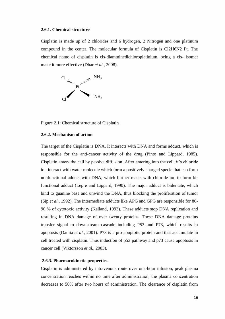
2.6.1. Chemical structure
Cisplatin is made up of 2 chlorides and 6 hydrogen, 2 Nitrogen and one platinum
compound in the center. The molecular formula of Cisplatin is Cl2H6N2 Pt. The
chemical name of cisplatin is cis-diamminedichloroplatinium, being a cis- isomer
make it more effective (Dhar et al., 2008).
Figure 2.1: Chemical structure of Cisplatin
2.6.2. Mechanism of action
The target of the Cisplatin is DNA, It interacts with DNA and forms adduct, which is
responsible for the anti-cancer activity of the drug (Pinto and Lippard, 1985).
Cisplatin enters the cell by passive diffusion. After entering into the cell, it’s chloride
ion interact with water molecule which form a positively charged specie that can form
nonfunctional adduct with DNA, which further reacts with chloride ion to form bi-
functional adduct (Lepre and Lippard, 1990). The major adduct is bidentate, which
bind to guanine base and unwind the DNA, thus blocking the proliferation of tumor
(Sip et al., 1992). The intermediate adducts like APG and GPG are responsible for 80-
90 % of cytotoxic activity (Kelland, 1993). These adducts stop DNA replication and
resulting in DNA damage of over twenty proteins. These DNA damage proteins
transfer signal to downstream cascade including P53 and P73, which results in
apoptosis (Damia et al., 2001). P73 is a pro-apoptotic protein and that accumulate in
cell treated with cisplatin. Thus induction of p53 pathway and p73 cause apoptosis in
cancer cell (Viktorsson et al., 2003).
2.6.3. Pharmacokinetic properties
Cisplatin is administered by intravenous route over one-hour infusion, peak plasma
concentration reaches within no time after administration, the plasma concentration
decreases to 50% after two hours of administration. The clearance of cisplatin from
Pt
Cl
Cl
NH3
NH3

17
the body is triphasic, distribution is achieved immediately as distribution half-life in
thirteen minutes, and elimination half-life in forty-five minutes, while terminal half-
life is almost five days. Major route of excretion is renal as 90% of drug is excreted
via this route. 25% of drug is eliminated within 24 hours of administration (Sturgeon,
2004). Ultra-filterable platinum, which is non-protein bound drug, is responsible for
major pharmacological action of drug. After administration of 110mg/m2 of Cisplatin
the blood plasma level of almost 6 µg/ml reaches immediately after administration
and declines to 2 µg/ml after two hours of administration (Go and Adjei, 1999).
2.6.4. Therapeutic uses
Cisplatin is used in variety of solid tumors including testicular, ovarian and bladder. It
is most commonly used in combination with other chemotherapeutic agents, as the
combination therapy is more effective and has better results.
2.6.4.1. Cisplatin and ovarian cancer
Ovarian cancer is gynecological malignancy with high mortality rate due to late
diagnosis. Most commonly used regime in ovarian cancer is combination of paclitaxel
and cisplatin. Similarly cisplatin in combination with gemcitabine is also used in
ovarian cancer (Agarwal and Kaye, 2003).
2.6.4.2. Cisplatin and lung cancer
At present Lung cancer is a fatal malignancy, Cisplatin is a key drug used in small
cell lung cancer. Non-small cell lung cancer is also treated with cisplatin based
adjuvant therapy (Youlden et al., 2008).
2.6.4.3. Cisplatin and breast cancer
Breast cancer is one of the most prevalent forms of cancer in women throughout the
world. Chemotherapeutic agents are effective in the treatment of breast cancer and
reducing the tumor size before surgery. Cisplatin is important anti-cancer drug used in
the treatment of breast cancer (Dhar et al., 2011).
2.6.4.4. Cisplatin and brain cancer
Brain cancer is one of the most fatal diseases. Cisplatin is used effectively in
childhood brain tumors. It improves the survival rate (Khan et al., 1982).

18
2.6.5. Side effects of cisplatin
Cisplatin interacts with DNA and cause the apoptosis of cell , this interaction with
DNA of cell is major cause of side effects of cisplatin (Yousef et al., 2009). The
major side effects of cisplatin are hepatotoxicity, nephrotoxicity and cardiac toxicity.
2.6.5.1. Hepatotoxicity
Cisplatin induce the oxidative stress resulting in reduced level of glutathione and
major side effects occur due to this , such as toxic effects on liver (Yilmaz et al.,
2004). Transaminase are released in blood circulation immediately after the cellular
damage and are biomarker for the toxicity and cellular damage, increased in level of
liver enzymes is indication of disturbance in liver function (İşeri et al., 2007).
Cisplatin induced hepatotoxicity has been observed to be greater in patients having
overexpression of CYP P450-2E1 enzyme (Caro and Cederbaum, 2004). Side effect
on liver include degeneration of hepatocytes and tissue necrosis (Kart et al., 2010).
The hepatotoxicity induced by cisplatin may be reduced by using Selenium ad vitamin
E (Liao et al., 2008).
2.6.5.2. Nephrotoxicity
The major route of excretion of cisplatin is via urine hence it accumulates in the
proximal tubules of kidney and its concentration is five times greater in proximal
tubules of kidney as compared to serum concentration. The increased level of
accumulation of cisplatin in the kidney contribute to renal damage (Ali and Al
Moundhri, 2006). Cisplatin is excreted via glomerular filtration and tubular secretion
and its accumulation cause side effects on renal function (Yao et al., 2007).
Concentration of cisplatin increase in kidney as compared to blood particularly in
renal parenchymal cell (Ishida et al., 2002).
2.6.5.3. Cardiotoxicity
Cisplatin may cause lipid peroxidation in cardiac tissues and cardiac myocytes may
release creatinine kinase and lactic dehydrogenase. Degeneration and necrosis of
cardiac muscles may occur (Al-Majed et al., 2006).

19
2.7. Novel drug delivery system
Since the start of life on earth, human has been trying to develop drug delivery system
from chewing leaves and plant roots and inhaling smoke of plant for treatment of
disease to the development of tablets and capsules for the delivery of drugs. In the
start of 20th
century efforts have been made to move from traditional and uncontrolled
drug delivery system to controlled drug delivery system. Invention of new drug
molecules having larger molecular weight and poor solubility lead to the development
of capsulated drug delivery system to enable drug to deliver at the site and show its
pharmacological action. Sustained drug delivery systems lead to the decrease in the
frequency of administration and ease to the patients. Controlled drug delivery system
control both rate of release of drug and site of release of drugs for maximum
therapeutic output (Barbe et al., 2004).
The chemotherapy currently being used is nonspecific and exerts serious side effects
to the normal tissues. To overcome this problem various efforts has been made to
achieve site specific drug delivery by using novel technologies. The liposomal drug
delivery system has proved to be very effective for the targeted delivery of
chemotherapeutic agent but is suffers problem of stability and storage (Juliano and
Stamp, 1975). The leakage of liposomes is covered by the PEG coating that enhance
circulation time. DOXIL® a liposomal formulation containing doxorubicin is
approved for cancer treatment in 1995(Lasic and Martin, 1995).
Hydrogels are biodegradable and safe drug delivery systems for targeted delivery to
bone tumors. Hydrogels can be used for thermoresponsive drug delivery as it is liquid
at room temperature and convert to gel at body temperature. Hydrogels can also be
used for pH sensitive drug delivery(Ta et al., 2009). Niosomes are also one of the
novel system for delivery of drugs and they resemble with liposomes except they have
non-ionic surfactant. Different types of surfactant are used which have the ability to
entrap both water loving and hydrophobic drugs. The presence of cholesterol increase
the rigidity of the system and their presence improve the permeability and fluidity
(Rajera et al., 2011).

20
Microspheres have excellent carrier capacity and having small size and suitable for
drug delivery, they have disadvantage of decrease residence time at the site of
absorption. This disadvantage can be overcome by formulating novel bio-adhesive
microspheres. Bio-adhesive microspheres can stick to the mucosal tissues such as in
GI tract, nasal and other surfaces. This offers highly controlled and localized drug
delivery system (Vasir et al., 2003).
Hollow microspheres are used for site specific drug delivery to the GI tract, because
normal drug suffer from the problem of decrease GI transit time, this can be overcome
by formulating floating microspheres, which provide prolong retention in GI tract and
reliable release of drug (Kawashima et al., 1992).
2.8. Nanotechnology
Nanotechnology is a multidisciplinary field paving its role in academia and research.
Due to its emerging role, the US government has established the National
Nanotechnology Institute (NNI) that support the research and commercialization in
the field of nanotechnology (Science and . 2005). Broadly nanotechnology is defined
as, the understanding application of materials in range of 1-100nm. Systematic
reviews suggest that almost 90% of newly discovered molecules have poor
pharmacokinetic properties (Brayden, 2003).
Nanotechnology enables the delivery of therapeutic molecule to the targeted tissue or
organ. Nanotechnology also enable the future ways for delivery of new therapeutic
molecules as ― nanomedicines‖ that helps the targeted and controlled delivery and
reduce the side effect associated with that drug molecule (Science and . 2005). The
emergence of nanotechnology has played a pivotal role in revolution of drug delivery.
The nanotechnology helps to increase the bioavailability of drugs with poor solubility,
passage of drug across tight epithelium barriers such as blood brain barrier, delivery
of large molecular weight drugs and protein at desired site of action. Nanotechnology
enable the co-delivery of drug and imaging agents at the site of action that enable the
visualization of desired site (Liong et al., 2008).
Nanomaterial provides an opportunity to enhance efficacy by modifying size, shape
and surface properties. The shape of nanoparticle affects bio distribution, drug loading
and extravasation at site of action. Squashable nanoparticles have long circulating

21
characteristics because of their ability to avoid from phagocytic cells (Anselmo and
Mitragotri, 2017). Nanomaterials made up of inorganic silica allow higher drug
loading and multistage delivery of therapeutic agent. A combination approach involve
the lipid coating over the porous material that gives higher drug loading and long
circulating characteristics (Ashley et al., 2011).
2.9. Nanotechnology and cancer
Nanotechnology is no stranger to oncology. The liposomes were first ever approved
nanomedicines for the treatment of cancer. Cancer nanotechnology is broad area of
research with application of nanocarrier in drug delivery, imaging and diagnosis.
Nanocarrier are conjugated with the functional groups and used for targeting of
tumors (Ferrari, 2005). Researchers have developed nanocarriers that are covalently
linked with the biological peptide and proteins. These also have diagnostic
applications in cancer such as nanoparticles made up of iron oxide are used as a
contrasting agent for prostate cancer. Personalized nano-oncology enables the
quantification to which extent the drug has reached the desired site of action. The
treatment of transtuzumab, decision is based on the measure of extent of
overexpression of HER2 receptors in individual patients (Evans and Relling, 2004).
Figure 2.2: Areas of nanotechnology applications
The early diagnosis and detection of neoplastic lesion has always remained an elusive
goal. To identify the invading tumor, cell imaging technologies does not provide the

22
enough resolution for early detection. Recent trends in imaging technology require
contrasting agent that is conjugated to a specific targeting agent. Nanoparticle
technologies are helping in early detection by co-delivery of targeting moiety and
contrasting agent that enable the anatomical explanation of lesion (Sullivan and
Ferrari, 2004). Nanocarrier probes loaded with molecular targeting agents provide
information about relative distribution of tumor marker in tumor microenvironment
(Li et al., 2004). Nanoparticles loaded with iron oxide are conjugated with Annexin-V
that detect the phosphatidylserine present in apoptotic cell, which is then used for
MRI identification of jurket T cell (Schellenberger et al., 2002). Nanotechnology has
provided new horizons form the treatment of cancer. The optimum success of
nanoparticle depends on the stability of nanoparticle during circulating in blood
stream and their ability to cross the tight physiological barrier and reach at the
targeted site of action (Grodzinski et al., 2019).
Nanoparticles have therapeutic and diagnostic applications. Magnetic nanoparticles
are well known for diagnostic, drug delivery and thermal properties. Recent
theranostics involve delivery of drugs and diagnostic agents. Biomedical technologies
require methodology to manipulate the living cells that are magnetically responsive
and can be controlled by applying external magnetic field. Cell surface engineering
provide the platform to tailor the magnetic nanoparticles on the surface of cell (Setua
et al., 2018). Recently developed iron oxide based magnetic nanoparticles consist of a
magnetic core coated with the biocompatible material embedded inside is sustained
release drug and a biomarker for diagnostic application (Shevtsov and Multhoff,
2016). Liposomes also serve as a theranostics agent to deliver the drug and diagnostic
agents by encapsulating one agent in the hydrophilic domain of liposomes and other
agent in the hydrophobic chain embedded within the liposomal bilayer. Liposomes
also have the superiority of high biocompatibility and can be conjugated with the
targeted moiety and can be used for passive and active targeting of tumor to serve the
diagnostic and therapeutic purpose at the same time (Yue and Dai, 2018).

23
2.10. Nanoparticles
Nanotechnology is a scientific technique undergoing explosive development and
being used in the delivery of drugs, protein and peptides. Nanotechnology has opened
new therapeutic application for poorly soluble drugs to be delivered orally.
Nanoparticles can easily cross the minute membrane barrier including blood-brain
barrier (Emerich and Thanos, 2003). One of the latest novel drug delivery
technologies is the use of nanoparticles for drug delivery. Polymeric biodegradable
nanoparticles are effective biodegradable delivery system. Various polymers are being
used to achieve the targeted drug delivery. Nanoparticles has the advantage over
liposomes due to better stability and efficient controlled release profile (Soppimath et
al., 2001). Particulate drug delivery system is capable of targeting tumors with
relative better efficacy and safety profile. Nanoparticles are novel technology in the
field of particulate based drug delivery system (Paciotti et al., 2004). Nanoparticles
have the ability to penetrate deep into the tissue and controlled release of drug. They
have advantage over microparticles due to better encapsulation efficiency and
bioavailability (Nagpal et al., 2010).
Nanoparticles have shown large number of advantages over other delivery systems
Nanoparticles have the advantage of their size, which is large enough to avoid
leakage into capillaries and small enough to be captured by macrophages. The
size gap between endothelial cell is 100-600nm, so nanoparticles can reach to
the tumor site for the site specific delivery of drug (Cho et al., 2008).
Polymeric nanoparticles can be tailed to achieve disease-specific delivery and
controlled release of drug. They can be concentrated at the tumor site and may
work as a depot of local delivery for solid tumors by using a suitable carrier.
They can be used for sustained drug delivery by protecting against the
enzymatic degradation (Singh and Lillard, 2009).
Better stability profile of nanoparticles enable oral administration feasible and
sustained release of drug can be achieved after oral administration. Bio
adhesive properties of polymeric nanoparticles enable increased absorption of
drug and better bioavailability. Nanoparticles can be delivered via different
routes from topical to inhalation (Gelperina et al., 2005).

24
Nanoparticles based drug delivery system improves the solubility of
hydrophobic drugs, increase the half-life during circulation by reducing
immune attack, enable stimuli responsive release of drug and decrease
frequency of administration. Moreover more than one drug can be
administered simultaneously this increasing the patient compliance (Zhang et
al., 2008).
Nanoparticles have gain special applications in the delivery of
chemotherapeutic agents. Conventional delivery of anti-cancer drugs also
causes severe harmful effects on normal cells. Nanoparticles have enhanced
permeability and retention effect and may concentrate at the tumor site. Hence
higher intratumoral drug concentration is achieved and less distribution of
drug toward normal tissues sites. This reduces the dose related side effects of
drugs. secondly poorly soluble anti-cancer drugs can also be delivered at the
targeted site easily (Wang et al., 2012).
Nanoparticles have disadvantage of decreased loading capacity that is not greater
than 5% of total weight of nanoparticles. They also suffer from the problem of
burst release after approaching the site of action (Couvreur, 2013).
2.11. Polymeric nanoparticles
Over the decades, polymeric nanoparticles are promising for delivering drugs to the
target site and cover wide range of diseases. The efficiency at the target site of disease
is due to their flexible nature, nano-sized, composition, molecular weight, and stability
of the polymeric drug carrier. The effectiveness of the polymeric drug carrier covers
all the limitation that is the major issues with the conventional dosage form. Polymeric
nanoparticles enhanced the solubility of the drug, leading to enhanced bioavailability
in the systemic circulation and sustained the release of drug at target site. For the
delivery of nanoparticles, various types of natural and synthetic polymers are
employed for drug delivery to the active target site (Naahidi et al., 2013).
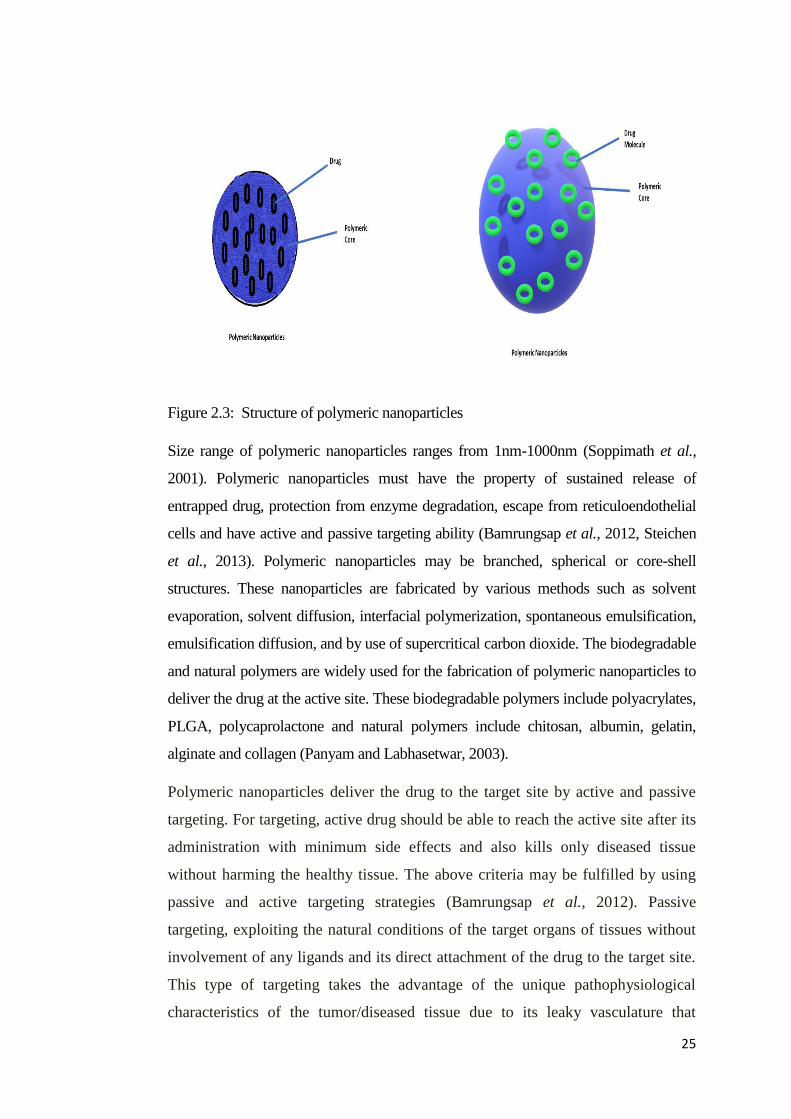
25
Figure 2.3: Structure of polymeric nanoparticles
Size range of polymeric nanoparticles ranges from 1nm-1000nm (Soppimath et al.,
2001). Polymeric nanoparticles must have the property of sustained release of
entrapped drug, protection from enzyme degradation, escape from reticuloendothelial
cells and have active and passive targeting ability (Bamrungsap et al., 2012, Steichen
et al., 2013). Polymeric nanoparticles may be branched, spherical or core-shell
structures. These nanoparticles are fabricated by various methods such as solvent
evaporation, solvent diffusion, interfacial polymerization, spontaneous emulsification,
emulsification diffusion, and by use of supercritical carbon dioxide. The biodegradable
and natural polymers are widely used for the fabrication of polymeric nanoparticles to
deliver the drug at the active site. These biodegradable polymers include polyacrylates,
PLGA, polycaprolactone and natural polymers include chitosan, albumin, gelatin,
alginate and collagen (Panyam and Labhasetwar, 2003).
Polymeric nanoparticles deliver the drug to the target site by active and passive
targeting. For targeting, active drug should be able to reach the active site after its
administration with minimum side effects and also kills only diseased tissue
without harming the healthy tissue. The above criteria may be fulfilled by using
passive and active targeting strategies (Bamrungsap et al., 2012). Passive
targeting, exploiting the natural conditions of the target organs of tissues without
involvement of any ligands and its direct attachment of the drug to the target site.
This type of targeting takes the advantage of the unique pathophysiological
characteristics of the tumor/diseased tissue due to its leaky vasculature that

26
enables the nano-drug carrier(s) that leads to the accumulation at the site. The
leaky vasculature allows the migration of molecules having size range up to 400
nm in diameter refers to the enhanced permeability retention (EPR) effect in the
tumor region (Kubik et al., 2005).Active targeting is the attachment of affinity
ligand(s) such as antibodies, biological macromolecules (peptides), vitamins,
aptamers, small molecules such as small interfering RNS (siRNA) and DNA for
co-delivery action. These ligands only bind to specific receptors or epitope on the
cell surface for the delivery of drug to the site of action. The novel nanocarriers
will recognize and then binds to the target cells through the mechanism of ligand-
receptor complex (Sudimack and Lee, 2000).
Polymeric nanoparticles have been considered to be the effective drug delivery system
in the field of therapeutics. This efficacy is due to maximum therapeutic benefit and
minimum side-effects. For this, polymeric nanoparticles have been used as a potential
drug delivery carrier system for loading of drug for sustained/controlled release. The
carrier system used in polymeric nanoparticles may be matrix or core system
depending upon method of preparation. Firstly, the drug used for the carrier system is
dissolved, then loaded, and entrapped to the carrier system (Soppimath et al., 2001).
Besides the promising characteristics of polymeric nanoparticles, their applications
have been limited due to several factors such as control size range and loading of
hydrophilic drugs. The control of size in the polymeric network is quite difficult due to
use of high molecular weight polymers. Mostly drugs for parenteral delivery should be
in size range of less than 400nm because if the size range is more than 400nm than
these will be engulfed by the RES system (Acharya and Sahoo, 2011). Polymeric
nanoparticles have low loading capacity of hydrophilic drugs due to the limited
portioning of more hydrophilic drugs into the lipophilic polymer coat (Yoo and Park,
2001).
2.12. Chitosan
Chitin is highly abundant naturally occurring polysaccharide and is found in the
shells of crabs and other insects. Chitin has poor solubility in water and has poor
biodegradability that limits its vast applications. The degradation product of chitin
are chitosan and chitosan oligosaccharide, which are highly biodegradable and

27
compatible with biological fluids and have vast application in food and
pharmaceutical industry (Du et al., 2009).
Figure 2.4: Chemical structure of chitosan
Chitosan is obtained by N-deacetylation of chitin. It is insoluble in water and is
soluble in acidic solution. It is a moderate cationic polymer and have amino and
carboxylic group in repeated subunits that can be utilized in conjugation chemistry
(Agrawal et al., 2010). Chitosan is believe to act as a scavenger for free radicals
and provide antioxidant effect (Zou et al., 2013). It is readily uptake by the
intestinal cells and biocompatible properties make it a suitable antioxidant. It can
also increase the activity of antioxidant enzymes. It also reduce the lipid
peroxidation (Xia et al., 2011).
Chitosan is also believed to have antimicrobial effects, which also depends on the
molecular weight of chitosan, degree of deacetylation and pH. The Minimum
inhibitory concentration(MIC) of chitosan is found to be 0.1% and also depends
on the type of bacterial growth in culture (No et al., 2002). The investigation of
mechanism of antibacterial effect has shown that amino group of glucosamine
ring of chitosan interact with carboxylic group of bacterial cell wall and form a
complex and inhibit their growth (Kim et al., 2003).

28
The structural integrity, availability in different molecular weight ranges,
conjugation chemistry, and biological compatibility make chitosan an ideal
polymer for drug delivery applications.
2.13. Liposomes
The word liposome is derived from two Greek word ―Lipos‖ and ―Soma‖ which
means ―fat body‖. They can be unilamellar or multilamellar with phospholipid as
a major structural component. They were first discovered by Alec D Bangham in
1965 (Bangham, 1993). The composition of liposomes is similar to the cell
membrane. The major structural component of biological membrane is
phospholipid, which is composed of hydrophilic head and a hydrophobic tail
(Papahadjopoulos et al., 1978).
Liposomes are bilayer membrane structure made up of phospholipids. Liposomes
have attracted a lot of attention due to its high biocompatibility and able to
delivery various therapeutic agents including drugs and genes. Liposomes are first
nanomaterials to be approved for clinical usage in 1995 (Torchilin, 2005). The
phospholipid membrane is a cell like boundary suitable for cellular investigation
and has the ability to mimic the biological system (Hua and Wu, 2013). Since the
liposomes have provided a breakthrough in the field of nanotechnology various
efforts have been done to improve the drug delivery via liposomes by attaching
targeting agents and preparing stimuli responsive liposomes (Lemière et al.,
2015). Some of the recent approaches include increasing the circulating life of
liposomes by attaching suitable amphiphiles. Triggered release approach include
enabling the release of drug in response to pH, temperature or radiation (Bibi et
al., 2012). Liposomes are suitable for gene delivery and offer protection to the
DNA, RNA. They can form complexes with cationic and anionic molecules and
are suitable for targeting the specific site (Alavi et al., 2017).

29
Figure 2.5: Structure of liposomes with PEG coating
Traditional liposomes suffer from the problem of rapid clearance from the body
and have stability issues. It happens because membrane of traditional liposomes is
affected by the proteins circulating in the blood, which cause opsonization and
rapid clearance from the body. In order to compensate these problems, efforts
have been done to modify the surface charge of liposomes and enabling long
circulating characteristics. Stealth liposomes were prepared by modifying the
surface with polyethylene glycol (PEG) that helps liposomes in protection from
circulating protein and increase the circulation life of liposomes (Van Slooten et
al., 2001). Stealth liposomes provide sustained delivery of drug and can also be
used in localized depot formulation. The first example is enhanced efficacy of
cytosine arabinose in treatment of lymphoma and first subcutaneous formulation
of liposomes was to deliver vasopressin as sustained release (Allen et al., 1992).
The surface modification includes attaching the lipid with hydrophilic polymer
such as polyethylene glycol. The covering of PEG prevents the adhesion of
plasma protein to the surface of liposomes that otherwise would have cleared by
mononuclear phagocytic system (MPS). The coating of PEG enables the
liposomes to oppose the uptake by the MPS thus incorporating long circulating
characteristics to the liposomes (Gabizon et al., 1994). Stealth liposomes can be
prepared by various chain lengths of PEG. PEG is biocompatible and have good

30
solubility and in non-ionic in nature. Some studies suggest that size of liposomes
should be less than 300nm in order to have protected stealth properties (Nag and
Awasthi, 2013). Stealth liposomes with PEG coating are also suitable candidate
for active targeting of therapeutic drugs. Active liposomes are used for targeting
folate and transferrin receptors, which are overexpressed in many solid tumors
(Torchilin, 2007). Although PEG is considered as a gold standard for increasing
the circulating life of liposomes and for steric protection most recent include
addition of biodegradable polymer and preparing lipid-polymer conjugating and
incorporating the long circulating characteristics of polymers with lipid (Metselaar
et al., 2003). The clinical trials of stealth liposomes loaded with doxorubicin in
human has produces significant results as compared to doxorubicin drug. The
results indicate that drug remain encapsulated for 7 days after injection. The
presence of metabolites of drug at tumor site indicates the release of drug at tumor
site. There was 10 times greater absorption of drug at tumor site as compared to
control drug solution. These results showed stealth liposomes have achieved
substantial improvement in the treatment of tumor (Daraee et al., 2016).
Due to the stability issues, liposomes are required to be stored at controlled
temperature. At freezing temperature, they lead to crystal formation and breakage
of phospholipid bilayer structure (Perche and Torchilin, 2013). Liposomes with
ultra-small size have less interaction with ligand and receptor. Liposomes with
positive charge have greater electrostatic interaction with anionic charged cell
membrane. Higher ligand density could lead to aggregation of small particles
(Albanese et al., 2012). Liposomes suffer from disadvantages like leakage of
encapsulated drug. Lipid in liposomes may undergo oxidation and hydrolysis that
leads to the unwanted release of therapeutic agent. They have short half-life and
difficulty in solubility of hydrophilic drugs (Daraee et al., 2016).
2.14. Lipid-polymer hybrid nanoparticles (LPHNPs)
Nanotechnology has provided the innovation in the delivery of drug, after many
pre-clinical studies liposomal drug delivery system and polymeric drug delivery
system has proven to be useful. The latest technology is the combining the
beneficial effects of both liposomal and polymeric drug delivery system and
achieving lipid-polymer hybrid nanoparticles. They have the advantage of

31
sustained and targeted delivery. They have better stability as lipid coat provide
diffusional barrier from external stimuli (Kong et al., 2013).
Very few studies have been reported on the development of lipid-chitosan
nanoparticles for the delivery of drugs through oral, topic and parenteral
administration. The reported studies are summarized as:
Nayab,et al (2019) prepared hybrid nanoparticles for controlled delivery of
doxorubicin hydrochloride and doxorubicin base. The prepared nanoparticles
showed enhanced encapsulation and higher internalization and greater cytotoxicity
as compared to doxorubicin solution (Tahir et al., 2019).
Q tan, Weidong Liu et al, (2011) developed the lecithin-chitosan nanoparticle
system for the topical delivery of the drug. They prepared quercitin loaded
nanoparticles and these nanoparticles showed enhanced permeation ability and
greater accumulation in epidermis. Interaction between nanoparticles and skin
surface enhance the skin permeation and nanoparticles are ideal for the topical
delivery of drugs (Tan et al., 2011).
Anita Hafner et al, (2009) prepared the lecithin-chitosan nanoparticles for the
trans-mucosal delivery of drugs. The prepared nanoparticles were of 120 to
340nm. The permeability of melatonin was studies on Caco2 cell lines for in-vitro
evaluation. Different types of lecithin and chitosan ratios were used in the study.
The permeability was better by using Lipoid S45 and Lecithin/Chitosan ratio of
20:1. The results showed that lecithin-chitosan nanoparticles are successful for
trans-mucousal drug delivery and does not induce any damage to plasma
membrane (Hafner et al., 2009).
YS Chhonker et al, (2015) prepared the hybrid nanoparticles for ocular drug
delivery of amphotericin. The Lecithin- Chitosan nanoparticles were prepared by
ionic gelation method with lecithin to chitosan ratio (10:1). The nanoparticles
were of 162 to 230nm size and entrapment efficiency was around 75%. The
prepared nanoparticles were more effective as compared to marketed formulations
and showed enhanced muco-adhesive properties. In-vivo studies showed two fold
enhanced bioavailability and increase in corneal residence time (Chhonker et al.,
2015).

32
Anita Hafner et al, (2011) formulated lecithin-chitosan nanoparticles loaded with
melatonin. The ratio of lecithin-chitosan was 20:1 and have size of 160-328nm.
The prepared nanoparticle suspension was then lyophilized to retain the physical
and chemical stability for longer period of time, which is not possible in nano-
suspension. trehalose was used as excipient for lyophylization of nanoparticles.
The freeze-dried nanoparticles have amorphous nature as indicated by DSC. The
nanoparticles have glass-transition temperature above room temperature and were
capable of retaining their characteristics and appearance for seven months at 4 °C
(Hafner et al., 2011).
Ipek Ozcan et al, (2013) prepared the hybrid lecithin-chitosan nanoparticles for
the topical preparation of betamethasone valerate. The prepared nanoparticle were
compared with the commercially available formulation and results showed that
Lecithin-chitosan nanoparticles showed 1.58 time increase permeation in
epidermis. The prepared formulations also showed skin bleaching and anti-
inflammatory effects (Özcan et al., 2013).
Ana Grenha et al, (2008) formulated the lipid-polymer nanoparticles for the
pulmonary delivery of drugs using mannitol as excipient. These nanoparticles
were then encapsulated in the microspheres. The physicochemical properties of
lipid-polymer nanoparticles were dependent on phospholipid concentration, which
provide controlled release of insulin. They have deep inhalation properties and
release the drug after contacting with the lung surface (Grenha et al., 2008).
Manuel J. Santander-Ortega et al, (2010) prepared the nanoparticles consisting of
lipid core which is surrounded by polymer chitosan for the delivery of DNA and
Hydrophobic drugs. Different formulation were prepared by varying the
concentration of polymer and lipid to measure the effects on physicochemical
properties. The nanoparticles were prepared by solvent-displacement technique.
The prepared nanoparticles have better colloidal stability and inert to
physiological fluid, thus presenting an ideal system for delivery of DNA
(Santander-Ortega et al., 2010).
Marthyna P. Souza et al, (2104) prepared the lipid-chitosan nanoparticles by ionic
gelation method. They have encapsulated the quercitin with excellent loading
capacity of 95%. The possess average size of 168nm and have better stability

33
profile. The loaded quercitin showed much better antioxidant properties as
compared to the normal formulation. Hence Lecithin-chitosan nanoparticles can
be used in preparation of functional food material (Souza et al., 2014).
Renu Chadha et al, (2012) formulated the artsunate loaded lipid-chitosan
nanoparticles. Artesunate has poor bioavailability and low aqueous solubility. The
nanoparticle provided controlled and sustained release of artesunate. The
nanoparticles were prepared by ionic gelation method and prepared particles have
size below 300nm. The drug loading capacity was 90%. The prepared
nanoparticles have shown increased in-vivo anti-malarial activity (Chadha et al.,
2012).
2.15. Types of lipid-polymer hybrid nanoparticles
LPHNPs have the advantage that they provide the sustained release of drug due to
the presence of polymeric core and drug can also be released in a controlled
manner. They also provide enhanced biocompatibility and stability due to the
presence of lipid layer (Chan et al., 2009). The polymer plays its role in release
behavior of drug and structural integrity of LPHNPs. The lipid provide
biomimetic properties and efficacy of system is enhanced as compared to both of
them alone (Zhang et al., 2008). Depends on the nature of lipid and nature of
polymer lipid-polymer HNPs may adopt one of the following arrangements of
lipid and polymer:
1. Polymer inside as a core and lipid as a shell
2. Polymer-caged lipids
3. Lipid and polymer mixed with each other
4. Lipid based then polymer core and lipid coating on the outer side
2.15.1. Lipid-polymer hybrid core-shell structure
In these cases, the polymer used is usually hydrophobic and dissolved in organic
solvent that is embedded inside and serve as a core of the system, while lipid
provide the outer layer and serve as a shell with hydrophobic end of the lipid is
inside and hydrophilic end on the outer surface of lipid-polymer hybrid
nanoparticles. Such type of system is effectively used in drug resistant tumor for
the delivery of therapeutic agent and a moiety to reduce the drug resistance (Zeng

34
et al., 2017). Such type of system is formed by intermixing of lipid and polymer.
The core polymer is surrounded by the layer of cationic or anionic lipids.
Hydrophobic drug can be incorporated inside the polymer core. Efforts have been
done to incorporate ionic drug by interaction of drug molecule with the polymer.
The lipid layer provide stability to the system (Wong et al., 2006).
Figure 2.6: Core- shell lipid-polymer hybrid nanoparticles
2.15.2. Polymer caged lipid hybrid nanoparticles
The type of hybrid nanoparticles has an outer polymer layer to achieve the desire
structure integrity and functionality. It depends on the nature of polymer such as
polyacrylic acid can be coated on the outer surface of lipid. In this cases method of
preparation differ as they involve preparation of liposomes followed by coating of
polymer with functional group that can be linked with different moiety to impart
specific properties such pH responsive behavior and to protect liposomes from
different pH environment (Lee et al., 2007).

35
Figure 2.7: Lipid-polymer cage hybrid nanoparticle
2.15.3. Mixed lipid-polymer hybrid nanoparticles
Such types of hybrid nanoparticles are formed by the interaction of lipid with
polymer. The lipid and polymer are entangled with each other via positive and
negative charge throughout the hybrid system (Gao et al., 2008). Sometimes these
are presented as a vesicle like structure but mostly the polymer and lipid and mixed
and uniformly present through the hybrid system. They possess lipoplex like
structure.
Figure 2.8: Mixed lipid-polymer hybrid system

36
2.15.4. Lipid-polymer-lipid hybrid nanoparticles
They usually consist of a first coat of lipid, which is then followed by the polymeric
core and then a coating of lipid on the outer surface (Shi et al., 2011). The shape is
usually depending on the method of preparation such hybrid nanoparticles are usually
formed by double emulsion method. The outer lipid layer is conjugated with Peg,
which provide enhance circulation time and prevent them to be engulfed by
macrophages system and provide more stability to the system (Pautot et al., 2003).
This system is suitable for co-delivery of different drug and drug and siRNA.
Figure 2.9: Lipid-polymer-lipid hybrid nanoparticles
2.16. Factor affecting hybrid nanoparticles
Lipid-polymer hybrid NP is formulated to a certain particle size via different methods
of preparation. The size and shape of nanoparticles is affected by the lipid to polymer
ratio, pegylation, lipid coat, type of polymer.

37
2.16.1. Lipid to polymer (L/P) ratio
Lipid to polymer ratio is prime factor to control the size and shape of particle. To
obtain the appropriate size of nanoparticles and to provide a proper lipid shell over the
core of polymer the ratio of lipid to polymer is of crucial importance. In case of
PLGA-Lecithin nanoparticles, the optimum ratio of lipid to polymer is 15% (w/w) to
obtain the desire size and shape of hybrid nanoparticles increase in lipid concentration
result in liposomes as a separate formulation (Chan et al., 2009). In a different study
the nanoparticle with size of 65nm were prepared with 10% L/p ratio using PEG coat
and PLA as a polymer and lipid shell. PEG prevents the stearic hindrance in absence
of PEG a higher ratio L/P ratio will be required (Zheng et al., 2010).
2.16.2. Lipid coating
The role of lipid coating is of prime importance. On one hand it act as barrier and
increase the encapsulation efficiency by preventing the leakage of drug one the other
hand it provide the controlled release behavior, which is different form polymeric
nanoparticles where polymer is only controlling factor in release of drug the lipid
coating provide additional controlled behavior (Chan et al., 2009).
2.16.3. Pegylation
The ratio of lipid to PEG is very important to provide colloidal stability. In PLGA
nanoparticle the lipid shell is not sufficient to confer the stability and aggregation of
particles occurs. PEG coating provides the stearic stability to the hybrid nanoparticles
at physiological pH. The optimum size of nanoparticles is obtained with lipid-PEG
ratio of 25%. It is important to note that the ratio of PEG does not affect the
encapsulation and release characteristics (Hadinoto et al., 2013).
2.16.4. Nature of Polymer
Polymer being the prime component of hybrid nanoparticles plays a crucial role in the
size, shape and release behavior of hybrid nanoparticles. The density and charge of
the polymer effect on zeta potential of hybrid nanoparticles and is important for the
stability of the hybrid nanoparticles. High density polymer may have high chances for
aggregation. Anionic polymer have more affinity towards cationic lipid like DOTAP

38
and cationic polymers formed the stable conjugation with anionic lipid (Thevenot et
al., 2008).
2.17. Targeted Drug Delivery
Targeted drug delivery system enables the drug to be delivered at the targeted site for
enhanced therapeutic output. Targeted drug delivery could be achieved by passive or
active targeting.
Figure 2.10: Advantage of targeted drug delivery system
For successful targeting the drug has to reach at its target site and retain and evade at
target side and then release drug (Bae and Park, 2011).
2.17.1. Passive Targeting
Passive targeting is mostly achieved in inflammatory diseases like tumor and
inflammatory tissue. Angiogenesis increase the leakiness of tumor vasculature and
cytokines causes enhanced permeability. Angiogenesis represents as irregular
diameter vessels (Oeffinger and Wheatley, 2004). Increased level of bradykinin cause
vasodilation and leads of extravasation and retention of molecules in tumor
(Matsumura et al., 1988). Most of the solid tumors have vascular pore size in range of
400-800nm, while organization of tumor may differ based on type of tumor (Hobbs et
al., 1998).
39
The size of the nanoparticle should be in this range in order to permeate the leaky
vasculature, in contrast the vasculature of normal cells is impermeable to cells greater
than 4nm (Firth, 2002). The nano size vasculature window provides the opportunity
for drug accumulation at the targeted site and reduces the drug distribution to the
healthy tissue. Recent researched have also shown the role of hyperthermia to further
increase the vascular permeability (Meyer et al., 2001). To ensure passive targeting
nanoparticles must circulate for longer times to have maximum possibilities to pass
through the target site. Nanocarrier usually have short circulating life and are
eliminated by the phagocytic system of the body and pegylation increase the
circulating life (Moghimi et al., 2001).
2.17.1.1. Enhanced permeability and retention (EPR) effect
The main concept of passive tumor targeting is based on the EPR effect, this idea was
first proposed by Dr.Maeda (Maeda et al., 1985). The enhance permeability at the
infection site and tumors is related to different factors including secretion of
bradykinin. Bradykinin cause the increase in vascular permeability, some studies
related to finding the root cause of ascites have revealed that bradykinin is also
responsible for extravasation of ascitic fluid (Maeda et al., 2003). Enhanced
permeability also occur in normal infectious tissue but the retention is not enhanced
because of proper functioning of lymphatic system and inflammation usually ends in
couple of days, which in malignant tissue there is enhanced permeation and retention
occur for longer period because of lack of lymphatic drainage system (Maeda et al.,
2001). So, it was recognized that EPR effect could be utilized for targeted drug
delivery to tumor site. EPR effect involves that increased extravasation of large
molecules like albumin from tumor vessels and their retention in tumor tissue for
longer period, which is not observed in healthy tissue (Matsumura and Maeda, 1986).
It was found that nitric oxide also plays its role in enhanced permeability and some
pro-inflammatory mediators such as VEGF (vascular endothelial growth factor) and
prostaglandins. VEGF plays their role to increase the vascular permeability and in
angiogenesis. The role of nitric oxide in tumor growth is also established
(Papapetropoulos et al., 1997). There is also anatomical difference in vasculature of
tumor and normal tissue. Scanning electron images have revealed that the
endothelium in tumor tissue could be 4.7µm in diameter (Hashizume et al., 2000).

40
The EPR effect can be summarized in following points:
1. Increased in leakage from tumor vessel particularly under high blood pressure
condition as vessels lack smooth muscles.
2. Irregular architecture of vessels and large fenestrations.
3. Irregular blood flow in vessels.
4. Poor lymphatic drainage that leads to accumulation of lipid nanoparticles and
macromolecules in tumor interstitium.
5. Decreased venous return that cause increased retention of macromolecules.
6. Extensive angiogenesis at tumor site.
2.17.2. Active targeting
In addition to the leaky vasculature the tumors also express some receptors or
epitopes that can be used as a targeting agent for active targeting of tumor. Therefore,
nanomedicines can be conjugated with these receptor targeting agents and used for the
active targeting (Farokhzad et al., 2004). Active targeting and enhanced cellular
uptake is important for medicines that are not actively taken up by the cells and
required endocytosis or facilitated diffusion to access the active site of cell (Forssen
and Willis, 1998).
Active targeting also increase the bio distribution of medicine specifically within the
tumor (Drummond et al., 1999). Different techniques are being used to couple the
targeting ligand with the nanocarrier. Mostly employed techniques are covalent and
non-covalent conjugation. Covalent conjugation involve the formation of disulfide
bonds, linkage between two amine group and linking of amine group with carboxylic
and aldehyde group (Nobs et al., 2004). Active targeting via use of nanocarrier is
superior to the drug-ligand conjugate. The use of nanocarrier enables the delivery of
higher drug concentration and delivery of payload at the targeted site. Secondly, drug-
ligand conjugate may modify the activity of the therapeutic molecule. Thirdly,
multiple therapeutic agents can be encapsulating inside the nanocarrier (Sethi et al.,
2003).
Different types of targeting agents are used for active targeting such as antibody
targeting, folate targeting, transferrin targeting, aptamer targeting based on the type of
tumor and conjugation chemistry of nanocarrier.

41
Monoclonal antibodies are target specific reagent used for the therapeutic and
diagnostic purpose. These were first agent used for active targeting based on the
surface antigen present on the tumor (Steinhauser et al., 2006). These antibodies can
be conjugated with the nanocarrier to deliver the payload at the targeted site. It is of
prime importance that the functionality of antibody and drug should be preserved till
the desired site of action. Antibody targeted drug delivery increase the bioavailability
of drug at tumor site and provide controlled release profile with enhanced therapeutic
output (Carter et al., 2016). The first ever antibody to target cancer cell was
developed in 1975 but the potential role of monoclonal antibody in cancer therapy
was established after 20 years (Béduneau et al., 2007). Monoclonal antibodies utilize
different types of receptors for targeting such as HER2 receptors, EGFR receptor, and
transferrin receptors.
Figure 2.11: Antibody targeted drug delivery system
Some types of breast cancer overexpress the human epidermal growth factor 2
receptors on the surface particularly it is overexpressed in 25% of invasive breast
tumors. This serves as a targeting agent for monoclonal antibody such as
transtuzumab. HER2 receptors not only overexpressed but also have extracellular
accessibility and well defined mechanism to internalize the antibody after
conjugation, thus suitable to serve targeting receptors for breast cancer (Wartlick et
al., 2004). Transtuzumab serve as active targeting agent specifically for HER2

42
positive breast cancer and its ability to do so have been proven on various HER2
positive breast cell lines in multiple studies (Ruan et al., 2012).
EGFR receptors are significantly overexpressed in various epithelial tumors.
Cetuximab is a monoclonal antibody that has high affinity for EGFR receptors and
bind competitively to these receptors and used for active targeting of tumors with
overexpression of EGFR receptors (Harding and Burtness, 2005). Different studies
have proved the efficacy of cetuximab conjugated with drug loaded nanoparticles to
specifically target the EGFR positive colorectal cancer cell lines and pancreatic
cancer cell lines (Glazer et al., 2010).
Transferrin receptors is ubiquitously expressed in healthy cells and many fold
overexpressed in malignant cells (Daniels et al., 2012). These are also expressed in
endothelial cells of brain. Loperamide is a drug that does not cross the BBB but if it is
loaded in nanoparticles conjugated with transferrin targeting monoclonal antibody
such as OX26 it can be delivered into brain (Ulbrich et al., 2009).
Transferrin is a glycoprotein that function to transport iron into the cell via transferrin
receptor mediated endocytosis. There is 100 fold high expression of transferrin
receptors in drug resistant malignant tumor, this make transferrin promising agent for
targeted drug delivery (Danhier et al., 2010). Different studies have shown liposomes
conjugated with transferrin loaded with docetaxel efficiently deliver their payload at
neoplastic cell expressing high level of transferrin receptors (Kobayashi et al., 2007).
Aptamers are three-dimensional structure made of single stranded RNA or DNA
nucleotide. They have ability to bind specific target site in biological system. They
have high sensitivity for their target and are specific in their actions (Ni et al., 2011).
Aptamer are composed of nucleotide sequence and can easily be conjugated with
cationic nanoparticles. Nanoparticle loaded with docetaxel and conjugated with A10
RNA has shown higher in-vitro cytotoxicity on cell lines as compared to non-
conjugated (Farokhzad et al., 2006).

43
2.17.3. Folate Targeting
Folate is a vitamin B9 and is present in many nutritional sources including vegetables.
It is required for the normal functions of cells. It acts as a mediator in the transfer of
carbon atom and plays an important role in synthesis of purines and pyrimidines that
leads to DNA and RNA synthesis. It is also involved in the methylation of
phospholipids and proteins. The deficiency of folate could lead to faulty methylation
and abnormal DNA synthesis (Choi and Mason, 2000). Folate receptors are widely
known tumor marker with strong affinity for its substrate folic acid. Folic acid is also
important for synthesis of nucleotide basis. Folate receptors also have strong affinity
for folic acid-drug conjugate and folic acid-polymer conjugate. The mechanism
involve for the internalization is endocytosis (Danhier et al., 2010).
Folate is required in rapid proliferating cells for DNA synthesis and methylation. It is
a vitamin and natural supplement for the body (Locasale, 2013). Dysregulation of
folate metabolism is related to improper embryonic development, heart and brain
defects. The mechanism of transport of folate across the cell membrane is through
reduced folate carrier which assist in transport of dietary folate (Matherly and
Goldman, 2003). The other mechanism involve in the transport of folic acid is
through proton coupled folate transporters. Folate can also be transported via four
different types of folate receptor namely (α, β, γ , δ) having molecular weight in range
of 38 to 45 (Ledermann et al., 2015).
Folate alpha receptors are overexpressed in majority of solid tumors, on contrary in
normal tissues folate alpha receptors are confined to low level in the apical surface
(Toffoli et al., 1997). Folate alpha receptors are particularly overexpressed in tumors
of epithelial origin, ovarian, breast and kidney tumors. The highest concentration
being found in tumors of endometrium, ovary and kidney, while breast, pancreatic
and colorectal tumors also have high expression of folate- α receptors. Folate
receptors are overexpressed in 81% of epithelial ovarian tumors and the extent of
expression in correlated to the grading of tumor. High grade serous tumors have
increased expression of folate receptors. Folate receptors are also used as marker for
tumor aggressiveness and is associated with poor disease survival (Siu et al., 2012).

44
Figure 2.12: Folate receptor internalization mechanism
Folate α receptors are also overexpressed in breast cancer, further studies reveal that
high level of expression is found in ER/PR negative breast cancer patients
(O’shannessy et al., 2012). Physiological hormones such as estrogen, progesterone
have been associated with down regulation of folate alpha receptors and there has
been an inverse relation with expression of folate receptors and estrogen/progesterone
hormone. Triple negative breast cancer that represents 15% of all breast cancer has
been associated with high expression of folate receptors (Anders and Carey, 2009).
Folate cycle is involved in key metabolic reactions for proliferating cells. The folate
taken as a vitamin supplement is reduced and transported inside the cells via folate
carriers. After reaching inside the cells folate play key role in the synthesis of DNA
base pairs purines and pyrimidines (Zwicke et al., 2012).
In the malignant tumors, the folate receptors lose their polarized location and spread
over cell surface of tumor (Kane et al., 1988). Folate receptors are overexpressed in
majority of solid tumors and in 45% of squamous cell tumors and in 40% of lymph
node metastasis. Folate receptors are further divided into alpha and beta forms. The
α-folate receptors are believe to be overexpressed in 40% of tumors , while β-folate
receptors are found in malignant tumors of hematopoietic origin (Low and Kularatne,
2009). Folate receptors have particularly more attraction because these are expressed
at the surface of the receptors. Studies have shown that oral cancer cells incubated

45
with folate conjugated liposomes have shown 31 times more fluorescence compared
to the conventional liposomes (Yang et al., 2014). In non-malignant tissue, the folate
receptors are present on the luminal surface of epithelial cells are inaccessible to
circulation, while in the malignant cells, these cover the surface of the tumor and are
approachable to circulation. Folate receptors also have ability to bind with the folic
acid and can be penetrated inside the tumors via endocytosis. Folic acid can be
conjugated with variety of therapeutic agents to deliver drug in a targeted manner
(Sega and Low, 2008). Recent studies show that folate receptor is present on the
surface of tumor even after the chemotherapy, as it is expressed in a specific manner
at the surface of tumor, thus providing the opportunity to serve as a therapeutic and
diagnostic tool (Salazar and Ratnam, 2007). Currently, different therapeutic agents
utilizing folate targeting are under clinical development. Two therapies that target
folate receptor are under phase-III clinical trial are farletuzumab (Konner et al., 2010)
and vintafolide (Naumann et al., 2013).
Table 2.1: Summary of clinical development of folate targeted ovarian cancer drugs
Trial phase Patient
group
Treatment arm Primary end
point
Result
I Platinum
resistant
ovarian
cancer
Farletizumab Safety of drug
and
tolerability in
patients
Dose can be
increased to
40 mg/m2
III Platinum
resistant
epithelial
tumor of
ovary and
fallopian
tube
Farletizumab+
Paclitaxel
Versus
Farletizumab
Progression
free survival
and overall
survival
No study
results
available
III Platinum
sensitive
ovarian
cancer
Farletizumab in
low dose+
Carboplatin+
Taxane
Vs
High dose
Farletizumab+
carboplatin+taxane
VS
Carboplatin+taxane
Progression
free survival
Some patients
have shown
difference in
progression
free survival

46
I Metastatic
solid tumor
patients
Vintafolide (Low
dose)+ Platinum
drug+ taxane
Vs
Vintafolide (Low
dose)+ Platinum
drug+ taxane
Dose limiting
toxicity
Ongoing
study
I Recurrent
solid tumors
Vintafolide bolus
dose
Vs
Vintafolide IV
infusion over one
hour
Maximum
tolerable dose
2.5 mg as
MTD in both
IV bolus and
IV infusion
group.
Constipation
was dose
limiting
toxicity.
II Platinum
resistant
folate
receptor
positive
ovarian
cancer
Vintafolide To observe
clinical
benefit after 6
cycle of
chemotherapy
without
progression of
disease
5% partial
response.
37% stable
disease
43% disease
control rate
(Morris et al.,
2014).
Farletezumab (A monoclonal antibody) bind selectively to the folate alpha receptors
as a tumor marker and cause immune dependent cytotoxicity. It has high affinity for
the folate alpha receptors and non-selective to healthy cells (Ebel et al., 2007).
Vintafolide is a combination of conjugated folate and vinca alkaloid. This is
selectively taken up by the folate receptors present on the surface of tumors and does
not bind to non-malignant cells, hence providing targeted drug delivery to tumor cells
and avoiding side effect on healthy tissues (Reddy et al., 2007).
Therefore, folate targeting is recent concept and is under clinical trials with different
combinations. Here, we have provided a novel combination of folate conjugation with
lipid covering to provide excellent bioavailability and enhanced therapeutic efficacy.

47
Lipid-Chitosan Hybrid
Nanoparticles for Controlled
Release of Cisplatin
Synthesis and Physicochemical
Characterization
CHAPTER 3

48
3.1. Background
Chemotherapy is an established way of treatment of various forms of cancer. The
important task is to deliver chemotherapeutic agent to the targeted cells and avoiding
side effects on the normal cells (Dayan et al., 2012). Chemotherapy is associated with
various side effects, some studies have shown that 15% of patients suffer from
cognitive function impairment after treatment with conventional chemotherapy,
sometimes called as ―Chemofog‖ (Mehnert et al., 2007). The poor solubility of most
of chemotherapeutic agents and dose related adverse effects instigate the development
of new drug delivery techniques. The use of nanoparticle technology has served as a
tool to avoid the serious side effect on the normal body tissues and delivery the drug
in a controlled manner (Zheng et al., 2015).
The concept of nanocarrier drug delivery was first generated by Sir Paul Ehrlich in
1905 and he termed it as a ―magic bullet‖ which is now termed as ―magic guns‖.
These nanocarrier are effective for enhanced drug loading and site-specific delivery of
drugs (Kumar et al., 2016). The new trend is to use biocompatible, biodegradable
polymers, which have ability to deliver the drugs in controlled and sustained manner
(Tang and Singh, 2009). Two established system of nanocarrier drug delivery system
are polymeric nanoparticles and liposomal drug delivery system (Mieszawska et al.,
2013).
Liposomes are biocompatible and they have enhanced permeability to the lipid based
biological membrane as compared to polymer nanoparticles (Torchilin, 2005).
However they have drawback of poor stability and leakage of drug could occur, they
required storage at controlled temperature (Crist et al., 2001). Liposomes also have
drawback of rapid clearance from the body because of their uptake by phagocytic
cells and pegylation is usually done to enhance their circulation life and to protect
them from phagocytosis (Aad et al., 2012). The methoxy chain in polyethylene glycol
play its role in preventing the activation of defense mechanism of body (Salvador-
Morales et al., 2009).
Polymeric nanoparticles have enhanced encapsulation efficiency and prolong
retention time and suitable vehicle to improve the bioavailability of drugs (Mathew et
al., 2017). They are superior in respect of controlled drug release and targeted

49
delivery of drug. Different kind of targeted moieties can be attached to polymeric
nanoparticles and targeting depends on the size , shape and nature of polymer being
used (Brannon-Peppas and Blanchette, 2012). Polymeric nanoparticles generally have
better cellular internalization, further it depends on the charge of the nanoparticles.
Cationic charge is usually favorable because it can interact with cell membrane and
enhance the internalization of nanoparticles (Chaudhary et al., 2018, Feng, 2004).
Despite of several advantages they have few drawbacks such as not suitable for
delivery of protein and peptides and particle aggregation could occur and release of
drug in acidic environment is aberrant from polymeric nanoparticles (Mohammadi-
Samani and Taghipour, 2015).
Recent technology to overcome the drawbacks of liposomal drug delivery system is to
combine them with polymeric nanoparticles to have structural advantages of the
polymer and to deliver the drug in efficient way at the targeted site of action (Prabhu
et al., 2015). LPHNPs have combined advantage of liposomal and polymeric drug
delivery system. LPHNPs can deliver the drug in controlled and sustained manner
because of polymeric core and enhanced biocompatibility due to liposomal drug
delivery (Chaudhary et al., 2018).
Chitosan is a biodegradable polymer with high degree of acetylation and it is being
used in sponges and sutures for the treatment of wounds. Chitosan undergoes
enzymatic hydrolysis and products are nontoxic and easily excreted form the body. It
is biocompatible and found applications in controlled release of drugs. The rate of
release is also affected by the degree of cross linking and molecular weight of drug
being used. Because of cationic charge it form complexes with negatively charged
lipids via ionic gelation and also suitable for delivery of siRNA and DNA (Stolnik et
al., 1995).
Cisplatin is a potent chemotherapeutic agent that crosslink with DNA and cause
damage to DNA and stop the proliferation of malignant cell but its maximum
therapeutic output is limited due to its severe side effects on renal and hepatic profile
(Rosenberg, 1985). Cisplatin also have less solubility in both hydrohphillic and
hydrophobic environment that limit the development of nanoparticles with excellent
drug loading. Cisplatin has low molecular weight and is rapidly cleared by the body.

50
We have produced chitosan-lipid hybrid nanoparticles for controlled delivery of
cisplatin and enhanced therapeutic output (Khan et al., 2019).
3.2. Materials and Methods
3.2.1. Materials
Low molecular weight chitosan was purchased from Sigma Aldrich. Lipid was kindly
provided by LIPOID®. Zeta Sizer (Malvern, UK). Cisplatin was received as a gift
sample from Pharmedic(PVT) Ltd. Ethanol and acetic acid were purchased from
Thermo Fisher Scientific.
3.2.2 Method of preparation
The nanoparticles were prepared by ionic gelation method as described by F. Sonvico,
et al (Sonvico et al., 2006). Briefly, 5 mg of chitosan was dissolved in 46 ml of acetic
acid solution by overnight stirring and cisplatin was dissolved in that solution. 100mg
of lipid was dissolved in 4ml of ethanolic solution and then ethanolic solution was
added drop wise in the chitosan solution and allowed to stir for 30 min at 10,000 rpm.
The lipid-chitosan hybrid nanoparticles were formed via ionic interaction of chitosan
and lipid. The nanoparticles were prepared with different concentration of lipid and
polymer to further characterize them. The lipid to chitosan ratio was varied from 5:1
to 60:1 in different formulation to optimize the formulation for further
characterization.

51
Figure 3.1: Schematic diagram of lipid-chitosan hybrid nanoparticles
3.2.3. Size and Zeta potential of hybrid nanoparticles
The prepared LPHNPs were analyzed for size, surface charge, polydispersity index
using Zeta Sizer (Malvern, UK). The 50 µl sample was used in a cuvette for the
determination of size and it was diluted with 950 PBS for the measurement of zeta
potential. The readings were performed at 25°C in triplicate.
3.2.4. Determination of drug loading and entrapment efficiency
The entrapment efficiency was determined by the indirect method, by measuring the
amount of unbound drug in the supernatant liquid after the completion of
centrifugation. The drug contents were determined by using UV Spectrophotometer
(Spectrum Scientific). The absorbance of standard and samples were measured at
210nm. The drug was dissolved in PBS at pH 7.4. Suitable dilutions were made from
the supernatant liquid to measure the drug contents. The experiment was performed in
triplicate. The entrapment efficiency and drug loading were determined by the given
formula.
Encapsulation efficiency = amount of entrapped drug / theoretical amount of drug
×100
Percentage drug loading will be calculated by following equation;
Drug loading = Mass of drug in nanoparticles / mass of nanoparticles × 100

52
3.2.5. Morphology of hybrid nanoparticles
Morphology of the cisplatin loaded hybrid nanoparticles was determined by using the
transmission electron microscope (TEM) (Jeol, USA). The prepared nanoparticles
were applied directly on the grid. The excess of sample was removed from the grid
and suitable images were taken at different magnifications.
3.2.6. Fourier transform infrared spectroscopy (FTIR)
The FTIR analysis was performed to evaluate any possible interaction between the
components of the formulation and to ascertain stability within these components by
using Thermo scientific Nicolet iS5 FTIR Spectrometer. Chitosan-lipid hybrid
nanoparticle formulations as well as the individual components lipid, polymer and
cisplatin were analyzed. Graphs were compared among the formulations to detect any
measurable change in the peaks.
3.2.7. Powered X-Ray diffraction analysis (X-RD)
X-RD analysis was performed by using the Xpert Pro Super, PANalytical ((JDX
3532, JEOL, Japan). The sample slides were prepared for the chitosan-polymer hybrid
nanoparticles formulation and the individual components including the lipid, chitosan,
cisplatin and physical mixture. The diffraction was recorded at ambient temperature
with 2θ range of 10-80.
3.2.8. Differential scanning calorimeter (DSC)
DSC analysis was carried out to evaluate the physical form of lipid-chitosan hybrid
nanoparticles. Indium was used for calibration of heat flow and temperature. The
Aluminum pan was used as a reference while other pan was used to analyze the
samples. The samples were heated from 25-450°C at a rate of 11°C/min.
3.2.9. Thermo-gravimetric analysis (TGA)
Thermo gravimetric analysis was performed to measure the change of mass over a
range of temperature. 38 mg of LPHNPs were used and individual components of
LPHNPs such as cisplatin, chitosan, and lipid were put on the gravimetric analyzer
and the temperature was run from 25- 450°C. The loss weight during run time was
recorded for each second.

53
3.2.10. In vitro drug release studies
The drug release study was conducted by using dialysis bag method. The dialysis
membrane of MWCO 10-12 KDa was used. Six different LPHNP formulations were
packed in different dialysis bags and the release was compared with cisplatin solution.
Phosphate buffer saline (pH 7.4) was used as a dissolution medium and temperature
was maintained at 37°C±0.5 with stirring speed at 100 rpm. Samples were withdrawn
at pre-determined time interval and similar amount of fresh medium was added to
maintain the final volume throughout the experiment. Kinetic modeling was used zero
order, first order, higuchi and korsmeyerpeppas models were applied.

54
3.3. Results
3.3.1. Size and surface charge
Size and PDI are important parameters for the nanoparticles, as they effect on the
loading and release of drugs from the formulation. Nanoparticles should have
appropriate size and low polydispersity index (Souza et al., 2014). The particle size of
all 6 formulations varies between 180 nm to 250 nm, which is suitable for passive
targeting of tumor through EPR effect (Cho et al., 2008). The surface charge of 5:1
ratio of lecithin-chitosan ratio to 30:1 ratio remains positive due to electrostatic
interaction of chitosan and lecithin but as the concentration of lipid is increased
further the zeta potential shift from positive to negative value and the formulation of
ratio 60:1 showed poor stability and as the ratio of lecithin is increased further the
formulation showed poor stability (Hafner et al., 2011).
The lecithin to chitosan ratio 60:1 showed that the aggregation of particles occurs
soon after the formation of nano-suspension that occurs due to imbalance of
electrostatic charges and increase in overall negative charge due to higher
concentration of lipid. The ratio of 10:1 and 20:1 showed lower PDI of 0.1 and 0.2
respectively and zeta potential values also showed better stability in these two
formulations. This might be due to proper interaction of negative and positive charges
in appropriate concentration. This already been described in literature (Sonvico et al.,
2006). The particle size and zeta potential of all formulation is shown in Table 3.2.
These results indicate that LPHNPs with lipid to chitosan ratio of 20:1 is most suitable
having particle size of 181nm and low polydispersity index and suitable for further
analysis of morphology of nanoparticles.
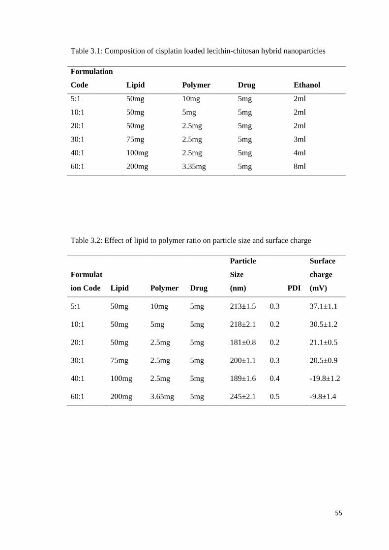
55
Table 3.1: Composition of cisplatin loaded lecithin-chitosan hybrid nanoparticles
Formulation
Code Lipid Polymer Drug Ethanol
5:1 50mg 10mg 5mg 2ml
10:1 50mg 5mg 5mg 2ml
20:1 50mg 2.5mg 5mg 2ml
30:1 75mg 2.5mg 5mg 3ml
40:1 100mg 2.5mg 5mg 4ml
60:1 200mg 3.35mg 5mg 8ml
Table 3.2: Effect of lipid to polymer ratio on particle size and surface charge
Formulat
ion Code Lipid Polymer Drug
Particle
Size
(nm) PDI
Surface
charge
(mV)
5:1 50mg 10mg 5mg 213±1.5 0.3 37.1±1.1
10:1 50mg 5mg 5mg 218±2.1 0.2 30.5±1.2
20:1 50mg 2.5mg 5mg 181±0.8 0.2 21.1±0.5
30:1 75mg 2.5mg 5mg 200±1.1 0.3 20.5±0.9
40:1 100mg 2.5mg 5mg 189±1.6 0.4 -19.8±1.2
60:1 200mg 3.65mg 5mg 245±2.1 0.5 -9.8±1.4

56
3.3.2. Morphology of nanoparticles
Morphology of the lecithin-chitosan hybrid nanoparticles was studies by using TEM.
The images showed the spherical shaped nanoparticles of around 200nm with the core
shell morphology of the lipid and the polymer present inside the hybrid particles. The
size of the particle was in accordance with the size determined by DLS. The lipid and
chitosan are entangled with each other through ionic interactions. The core of polymer
and lipid shell is entangled with each other to form a spherical lipoplex like structure.
The images of hybrid nanoparticles are shown in figure 3.2.
Figure 3.2: Transmission electron microscopy images of lipid-chitosan hybrid
nanoparticles
3.3.3. Entrapment efficiency and Drug loading
The entrapment of drug in lipid-chitosan hybrid nanoparticles was measured by
indirect method i.e. by calculating the amount of un-entrapped drug in the
nanoparticles. Lipid-polymer nanoparticles with ratio of 20:1 showed the highest
encapsulation efficiency (89.54%), while 60:1 showed the lowest encapsulation
efficiency of the nanoparticles. The low entrapment in 60:1 Formulation is attributed
to the greater concentration of lipid as compared to chitosan and hence imbalance of
positive and negative charges and weaker electrostatic interaction (Chuah et al.,
2009). The formulation 30:1 showed the highest drug loading that may be due to
presence of greater number of particles with multilamellar structure (Gerelli et al.,
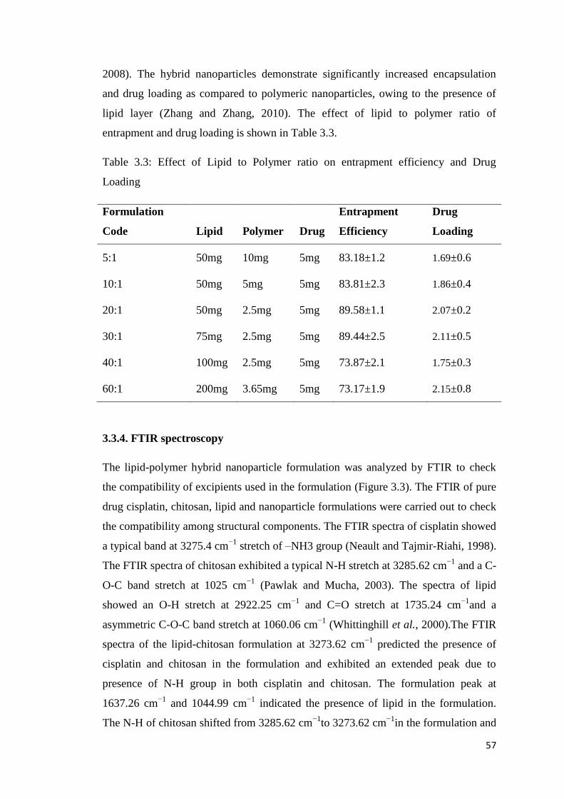
57
2008). The hybrid nanoparticles demonstrate significantly increased encapsulation
and drug loading as compared to polymeric nanoparticles, owing to the presence of
lipid layer (Zhang and Zhang, 2010). The effect of lipid to polymer ratio of
entrapment and drug loading is shown in Table 3.3.
Table 3.3: Effect of Lipid to Polymer ratio on entrapment efficiency and Drug
Loading
Formulation
Code Lipid Polymer Drug
Entrapment
Efficiency
Drug
Loading
5:1 50mg 10mg 5mg 83.18±1.2 1.69±0.6
10:1 50mg 5mg 5mg 83.81±2.3 1.86±0.4
20:1 50mg 2.5mg 5mg 89.58±1.1 2.07±0.2
30:1 75mg 2.5mg 5mg 89.44±2.5 2.11±0.5
40:1 100mg 2.5mg 5mg 73.87±2.1 1.75±0.3
60:1 200mg 3.65mg 5mg 73.17±1.9 2.15±0.8
3.3.4. FTIR spectroscopy
The lipid-polymer hybrid nanoparticle formulation was analyzed by FTIR to check
the compatibility of excipients used in the formulation (Figure 3.3). The FTIR of pure
drug cisplatin, chitosan, lipid and nanoparticle formulations were carried out to check
the compatibility among structural components. The FTIR spectra of cisplatin showed
a typical band at 3275.4 cm−1
stretch of –NH3 group (Neault and Tajmir-Riahi, 1998).
The FTIR spectra of chitosan exhibited a typical N-H stretch at 3285.62 cm−1
and a C-
O-C band stretch at 1025 cm−1
(Pawlak and Mucha, 2003). The spectra of lipid
showed an O-H stretch at 2922.25 cm−1
and C=O stretch at 1735.24 cm−1
and a
asymmetric C-O-C band stretch at 1060.06 cm−1
(Whittinghill et al., 2000).The FTIR
spectra of the lipid-chitosan formulation at 3273.62 cm−1
predicted the presence of
cisplatin and chitosan in the formulation and exhibited an extended peak due to
presence of N-H group in both cisplatin and chitosan. The formulation peak at
1637.26 cm−1
and 1044.99 cm−1
indicated the presence of lipid in the formulation.
The N-H of chitosan shifted from 3285.62 cm−1
to 3273.62 cm−1
in the formulation and

58
the C=O stretch shifted from 1735.24 cm−1
to 1637.26 cm−1
in the formulation and the
C-O-C band shifted from 1060.06 cm−1
to 1044.99 cm−1
. The absence of any new
peak in the formulation indicated that there was no chemical change during
entrapment of drug in the lipid-polymer hybrid nanoparticles and that the different
structural components are compatible with each other.
Figure 3.3: FTIR Spectra of individual components and lipid-polymer hybrid
nanoparticle formulation
3.3.5. XRD Studies
The promising sharp peaks of cisplatin were identified at 15,28, 46 and 70 2θvalues,
which indicated that cisplatin is of crystalline nature (Jayasuriya and Darr, 2013).
The broader peak between 15 and 25 depicted the amorphous nature of lipid (2 θ
scattering) (Lavanya et al., 2016). The characteristic peak of chitosan at 20 (2 θ value)
illustrated its crystalline nature (Pang and Zhitomirsky, 2005). Similarly, the sharp
peaks at 15 and 28 appeared in the physical mixture also portrayed crystalline nature
of cisplatin, However, reduction in intensity and diffused sharp peaks of cisplatin and
chitosan were observed in the lipid-chitosan formulation, illustrating encapsulation of
cisplatin the hybrid nanoparticles. These results were also in good agreement with the

59
findings of DSC and FTIR that confirmed the absence of any significant interaction
among all formulations components.
Figure 3.4: X-ray diffraction analysis of components and LPHNPs
3.3.6. Differential Scanning Calorimetry (DSC)
The DSC analysis was performed to check the crystalline/amorphous nature of drug
and components in lipid-chitosan hybrid nanoparticles. The cisplatin loaded LPHNPs
exhibit a typical endothermic peak at 102°C, this is due to the evaporation of water
associated with the drug. The cisplatin pure drug showed a dehydration peak at
around 100°C due to water associated with it and then an endothermic peak at 270°C,
which related to the melting point of the drug (Dixit et al., 2015). However there is no
sharp endothermic peak of cisplatin in the nanoparticle formulation, which showed
that the drug has loose its crystalline behavior in the formulation, as also reported in
previous studies (Dixit et al., 2015). The curve of chitosan typically showed the broad
endothermic peak in temperature range of 70-150°C, this corresponds to the loss of
water of crystallization and melting point of the chitosan (Cervera et al., 2011). The
thermal decomposition of chitosan starts at 300°, which is an exothermic process and
its peak is seen at around 320°C (Drebushchak et al., 2006). This behavior of chitosan
is also seen in the formulation with endothermic peak starting at 70°C and broader
endothermic peak in the formulation is due to presence of chitosan and lipid and
cisplatin converted to amorphous form. Therefore, no physical interaction between the
components was seen.

60
Figure 3.5: Differential Scanning calorimetry graph of LPHNPs and components
3.3.7. Thermogravimetric analysis (TGA)
TGA was performed to measure the percentage of weight loss over time at certain
range of temperature. TGA of cisplatin, chitosan and lipid and formulation was

61
performed. The results showed that the weight loss of cisplatin occur at 270 °C, which
corresponds to the melting point of drug (Prodana et al., 2014). Chitosan showed
slight weight loss at 70°C and then at around 290°C there is gradual weight loss
(Javaid et al., 2018). Weight loss in lipid mixture also starts at 250°C. The melting
points of all three components differ from each other and in formulation there is
combined peak of weight loss in the same range, which also confirms absence of any
physical interaction between components and thermal stability of the formulation.
Figure 3.6: Thermo gravimetric analysis of cisplatin loaded lipid-chitosan hybrid
nanoparticles

62

63
3.3.8. In vitro Dissolution Studies
Drug release studies were performed in phosphate buffer saline pH 7.4 at 37°C and
100rpm using the dialysis membrane method (Jeong et al., 2008). None of the
formulation showed burst release, studies on release pattern of polymer-lipid hybrid
system also showed absence of immediate burst release and hence drug can be
released in controlled manner (Kim et al., 2008).
The delayed release in 40:1 and 60:1 formulation might be due to excessive lipid layer
over the polymer, which retard diffusion of drug (Kim et al., 2008). After that there is
a sustained release over 24 hours from the lipid-polymer complex (Mandal et al.,
2016). Kinetic modeling was applied to the cisplatin release data including zero order,
first order, higuchi and korsmeyerpeppas and the results showed that best fit model
for the formulation is korsmeyerpeppas model which is usually followed in lipid-
polymer hybrid system (Tran et al., 2015). The controlled release of drug is attributed
to lipid layer and minor contribution of polymer and change is concentration of lipid
effect the rate of release of drug from formulations (Chan et al., 2009). The value of n
suggest that the drug follow super case II transport mechanism in most of the
formulation, the release mechanism if different in 40:1 and 60:1 due to higher
concentration of lipid and imbalance of positive and negative charges (Sonawane et
al., 2016). The drug release pattern of different formulation is shown in Figure 3.7
and kinetic modeling is shown in Table 3.4.

64
Figure 3.7: In vitro drug release profile of LPHNPs with various ratios of lipid to
polymer.

65
Table 3.5: Kinetic Modeling of Drug Release profile of Cisplatin loaded Lecithin-
Chitosan Hybrid Nanoparticles
Formulation Zero Order
R2
First
Order
R2
Higuchi
Model
R2
Korsmeyer
Pepas
Model
R2
n
5:1 0.6626
0.7324
0.5855
0.8930
1.750
10:1 0.6845
0.8700
0.8333
0.8457
1.591
20:1 0.7054
0.8401
0.7536
0.9069
1.067
30:1 0.7538
0.7686
0.6899
0.8846
1.187
40:1 0.1375
0.8731
0.5040
0.9478
0.350
60:1 -0.0727
0.8412
0.3954
0.9686
0.322
Drug 0.6643 0.7248
0.6600
0.9887
0.253

66
3.5. Discussion
Lipid polymer hybrid nanoparticles have the advantage of both polymeric and
liposomal drug delivery systems and have the ability to entrap both hydrophilic and
hydrophobic drugs. The chitosan-lipid hybrid nanoparticles have been formulated for
controlled delivery of cisplatin and to achieve maximum therapeutic benefit. The
formed LPHNPs were evaluated by both physicochemical tests.
Physicochemical test include size, entrapment, morphology, compatibility among the
excipients and thermal stability and in vitro release profile. Size and polydispersity
index have effects on the loading and release of drug from the nanoparticle
formulation (Souza et al., 2014). LPHNPs were formulated with various ratio of
chitosan to lipid, at a ratio of 20:1 they showed mono-dispersity and a better stability
profile because of an interaction of negative and positive charges in appropriate
concentration (Sonvico et al., 2006). The comparative low entrapment in the 60:1
formulation is attributed to the greater concentration of lipid as compared to chitosan
and hence an imbalance of positive and negative charge and a weaker electrostatic
interaction (Chuah et al., 2009). The 30:1 formulation showed the highest drug
loading. This may be due to the presence of a multiple layered structure and strong
interaction of positive and negative charges (Gerelli et al., 2008).
The images exhibited a core shell morphology with some lipid covering which
prevented diffusion of drug and water penetration into the system the shell of
chitosan-lipid provides long circulating characteristics (Mandal et al., 2013). The
absence of any new peak in the formulation indicated that there was no chemical
change during entrapment of drug in the lipid-polymer hybrid nanoparticles and that
the different structural components are compatible with each other. The release
pattern of the polymer-lipid hybrid system also showed an absence of immediate burst
release and suggested that drug can be released in a controlled manner.
The lipid-polymer hybrid nanoparticles with drug distributed inside the polymer
showed a controlled release profile. The release of drug from the polymer matrix
depends on diffusion. Further lipid layering prolongs the drug release (Li et al., 2008).
The value of release component (n) suggests that the drug follows a super case II
transport mechanism in most of the formulations. The release mechanism is different
67
in 40:1 and 60:1 due to a higher concentration of lipid and imbalance of positive and
negative charges (Sonawane et al., 2016). The results indicated that best fit model for
the formulation is a korsmeyerpeppas model which is usually followed in lipid-
polymer hybrid systems (Tran et al., 2015).
Cisplatin loaded lipid-chitosan hybrid nanoparticles were successfully fabricated. The
best formulation with a suitable combination of lipid and chitosan showed mono-
dispersity, small size and a controlled release profile. Further release of drug was
affected by an increase in the lipid concentration. The rate of release of drug was
attributable to both polymer and lipid. The release of drug is controlled by the
polymer matrix and further by a lipid layer that prevents leakage of drugs. There was
an absence of burst release of cisplatin due to its entrapment in the inner polymer
layer and outer lipid covering.

68
3.6. Conclusion
The physicochemical tests concluded that cisplatin loaded lipid-polymer hybrid
nanoparticles with ratio of lipid to chitosan (20:1) have core shell morphology with
high drug encapsulation efficiency and loading capacity. The components are
compatible with each other and cisplatin is converted from crystalline to amorphous
form in LPHNPs. The LPHNPs showed excellent thermal stability and a controlled
drug release profile. The lipid-chitosan hybrid nanoparticles with ratio of (20:1) will
be used for further biological characterization.
69
Lipid-Chitosan Hybrid
Nanoparticles for Controlled
Release of Cisplatin
Biological characterization
CHPATER 4

70
4.1. Background
In previous studies, we have prepared cisplatin loaded folate-chitosan hybrid
nanoparticles and were characterize for size, potential, shape, drug release and
thermal studies. The optimized formulation is now required to be tested on the ovarian
cell lines to establish the therapeutic efficacy of prepared hybrid nanoparticles and
biocompatibility of blank hybrid nanoparticles.
Cell culture is the growth of cells in artificial environment and is recognized for
studying the physiology of cells and effect of drug on particular cell lines. Every cell
type grow in specific medium that contain all necessary nutrient required for the
growth of that cell lines (Antoni et al., 2015). Cells grow as a monolayer attach to the
surface of flask and some cell lines grow in the suspension form and growth of
particular cell line depends on its cycle of exponential growth. Cell lines are used for
variety of studies to establish the efficacy of drug and to check the uptake of
nanoparticle system inside the specific cell line (Ravi et al., 2015).
The lipid-chitosan hybrid nanoparticles are hybrid system made up of chitosan as a
polymer, which is biocompatible and have structural integrity. The lipid is component
of biological membrane and enhances the biocompatible properties. Previous studies
of lipid-polymer nanoparticles show the biocompatible nature of nanoparticles
(Tezgel et al., 2018).
Ovarian cell line A2780 is extensively used to evaluate the efficacy of drug delivery
system. A2780 are of epithelial origin and grow as a monolayer. Cisplatin is first line
agent in the treatment of ovarian cancer. Cisplatin loaded lipid-chitosan hybrid
nanoparticles were evaluated by using A2780 cell lines (Wojewódzka et al., 2019).
OECD has issued guidelines for the conduct of acute oral toxicity studies to establish
the safety of polymer and lipids being used in large quantity. These studies enable the
evaluation of target organ and clinical symptoms and any hematological changes
observed during the high dose of drug delivery system (Jonsson et al., 2013).

71
4.2. Material and Methods
4.2.1. Material
The Cell titer blue for cytotoxicity studies was purchased from Promega®. A2780
ovarian cell lines were purchased from Sigma Aldrich. RPMI media for growth of
A2780 cell lines and fetal bovine serum and antibacterial and antimycotic
solutions were purchased from Cell Gro (VA, USA). Rabbits and Rats were
obtained from animal house of The Islamia University of Bahawalpur.
4.2.2. Cell viability
Cell cytotoxicity studies were carried out using A2780 ovarian cancer cell lines.
Cells (5000) were seeded in each well of 96-well plate. After overnight
incubation, cells were treated with blank LPHNPs, cisplatin-loaded LPHNPs and
cisplatin solution at a cisplatin concentration range of 1.25 to 50 µg/ml to check
the effect at different concentrations (Wang et al., 2014). Formulations were
washed out after 4 hours treatment and replaced with fresh RMPI media. The
effect on cytotoxicity was observed at 20 and 44 hours after treatment using a Cell
TiterBlue®
assay following manufacturer instructions.
4.2.3. Fluorescence microscopy
Microscopic slides were carefully placed in each well of 12 well plates and cells
(100,000) were seeded a 12-well plate. After overnight incubation cells were
treated with the cisplatin loaded LPHNP formulation labeled with Rh-PE and a
control blank formulation for 4hours. After four hours, cells were washed with
phosphate buffer saline (pH 7.4) and fixed with same solution containing 4%
paraformaldehyde (PFA) for 30 min at room temperature. Cells were then washed
with PBS (pH 7.4) three times and stained with 10 µg/ml Hoechst 33342 in PBS
pH 7.4 for 15 min. Cells were washed again with PBS, pH 7.4, and mounted on
microscope slides with mounting buffer for analysis by keyence fluorescence
microscope.
4.2.4. Cellular uptake
The uptake of cisplatin loaded LPHNP was evaluated using flow cytometry
(Beckton Dickinson FACS Calibur™, NJ, USA). Cells (500,000) were seeded in

72
each well of 6-well plate. After 24-hour incubation, cells were treated with Rh-
123 containing formulations (0.5mol %) for 4h. After completion of incubation
period cells were detached using trypsin and washed with PBS, pH 7.4, three
times and centrifuged at 2500 rpm for 5 min and then re-suspended in 300µl of
PBS. This solution was analyzed by flow cytometry and a total of 10,000 gated
live cell events were collected.
4.2.5. In vivo toxicity
Acute toxicity studies were conducted following approval from the research ethics
committee of the faculty of pharmacy and alternative medicines Islamia
University Bahawalpur. Female white Albino rats were selected for acute toxicity
studies according to guidelines of Organization of Economic Cooperation and
development (Oecd, 2000). Rats were divided into three groups. Female rats are
more sensitive hence selected for the toxicity studies. One group served as a
control and another was treated with lipid-polymer hybrid nanoparticles as a blank
formulation at the dose of 2000 mg/kg to check the toxicity of the components.
The third group was treated with cisplatin loaded lecithin-chitosan hybrid
nanoparticles at a dose of 30mg/kg. The signs of toxicity were evaluated after 14
days of treatment.
4.2.6. In vivo pharmacokinetic
Twelve healthy rabbits were selected for the pharmacokinetic studies (average
weight, 2.4 ± 0.4 kg). Rabbits were divided into two groups. One group was
administered 4mg/kg of the cisplatin drug solution as a reference while the other
group received 4mg/kg of the cisplatin loaded lecithin-chitosan hybrid
nanoparticles. Rabbits were kept on fasting overnight before the start of the study.
Blood samples were collected at defined time intervals over a period of 24 hours
and centrifuged immediately to separate plasma. Plasma was stored at -20°C. The
samples were treated to separate protein and 20 µl samples were used to determine
drug concentration using HPLC (Huo et al., 2005). The mixture of acetonitrile:
Water: Methanol (30:30:40) was used as a mobile phase and detector was adjusted
at 210nm for flow rate was 1.5ml/min

73
4.3. Results
4.3.1. Cell viability studies
The cytotoxicity studies were carried out using cell titer blue by measuring
fluorescence from cells using a plate reader. The reading from plate reader indicated
that blank chitosan-lipid hybrid nanoparticles have no cytotoxic effect on the cells and
are safe. The readings were taken after 24 hours and 48 hours of incubation period.
The results after 24 hours period showed that there is not much significant difference
in the cytotoxic effect of cisplatin drug solution and cisplatin loaded lipid-chitosan
hybrid nanoparticles. This is due to slow release of drug from the LPHNPs. The
cisplatin solution showed more cytotoxic effect only at one concentration of
12.5µg/ml as compared to cisplatin LPHNPs and at remaining all concentration range
there was not any significant difference in the cytotoxic effect of cisplatin loaded
LPHNPs on A2780 ovarian cell lines. This is due to easy access of cisplatin solution,
while in LPHNPs the drug in embedded inside lipid and polymer layers.
The reading from second 96 well plate was measured after 48 hours and the results
showed that the cisplatin loaded lipid-chitosan hybrid nanoparticles have significantly
higher cytotoxic effect as compared to cisplatin solution particularly at concentration
of 6.2µg/ml, while at concentration of 3.1µg/ml and 1.6 µg/ml there is also significant
higher toxicity in cisplatin LPHNPs as compared to cisplatin. The significant higher
cytotoxic effect after 48 hours showed that cisplatin loaded LPHNPs release the drug
in a controlled manner and significant higher effect are shown after 48 hours of
incubation period.
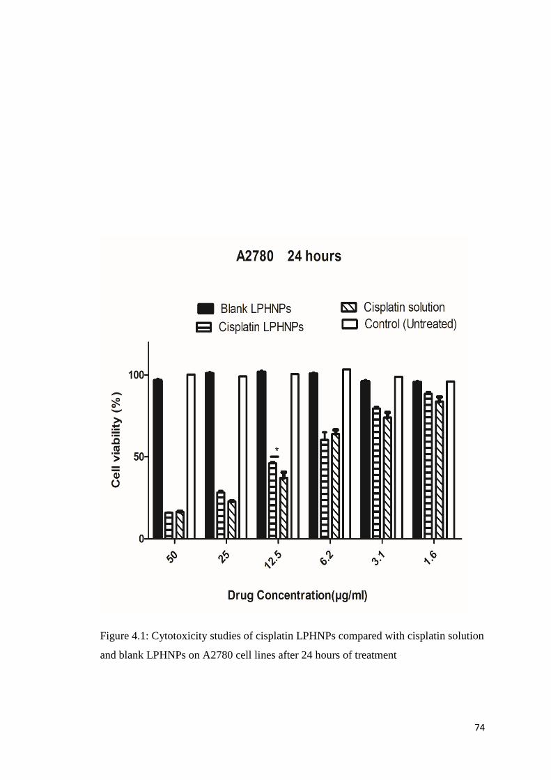
74
Figure 4.1: Cytotoxicity studies of cisplatin LPHNPs compared with cisplatin solution
and blank LPHNPs on A2780 cell lines after 24 hours of treatment

75
Figure 4.2: Cytotoxicity studies of cisplatin LPHNPs compared with cisplatin solution
and blank LPHNPs on A2780 cell lines after 24 hours of treatment
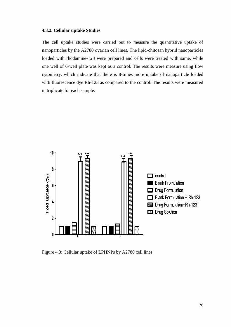
76
4.3.2. Cellular uptake Studies
The cell uptake studies were carried out to measure the quantitative uptake of
nanoparticles by the A2780 ovarian cell lines. The lipid-chitosan hybrid nanoparticles
loaded with rhodamine-123 were prepared and cells were treated with same, while
one well of 6-well plate was kept as a control. The results were measure using flow
cytometry, which indicate that there is 8-times more uptake of nanoparticle loaded
with fluorescence dye Rh-123 as compared to the control. The results were measured
in triplicate for each sample.
Figure 4.3: Cellular uptake of LPHNPs by A2780 cell lines

77
4.3.3. Cellular association
The cell association of the nanoparticles was also observed by staining with the
fluorescent dye and observing under Keyence fluorescence microscope. Hoechst is
used for staining nucleic acid and it produces blue color after interaction with DNA of
the cell. Rhodamine PE produces the red color fluorescence in the cells. It can be
predictive from the images that nanoparticle showed internalization in A2780 ovarian
cancer cells lipid present in the LPHNP may facilitate the internalization by
interacting with the lipid layer at the surface of cell membrane (Guo et al., 2015). The
cells exhibit association with LPHNPs and produce fluorescence.

78
Figure 4.4: Uptake of chitosan-lipid hybrid nanoparticles loaded with Rh-PE by
A2780 cells

79
4.3.4. Toxicological studies in rats
In vivo toxicity studies were conducted in rats (Co-Operation and Development,
2002). The study was conducted in accordance with the protocol of animal ethics
committee of The Islamia University of Bahawalpur. The weight of rats was noted at
the start of the study period, after 7 days of administration of cisplatin and at the end
of the 14 days study period. There was no significant difference in the weight of the
rats in that time period (Kim et al., 2008).
Table 4.1: Body weight and food, water intake of rats of different groups
Parameters Control Group Blank
Formulation
Drug Loaded
Formulation
Body weight (Kg)
Pre-treatment 1.40±1.21 2.20±1.8 2.10±1.6
Day 7 1.40±1.32 2.14±1.9 1.98±1.6
Day 14 1.34±1.40 1.70±1.2 1.60±1.2
Water In-take
(ml)
Pre-treatment 180 ± 1.57 201 ± 2.03 220 ± 2.03
Day 7 194 ± 2.01 210 ± 1.98 230 ± 1.98
Day 14 201 ± 1.95 215 ± 2.15 198± 2.15
Food Intake (gm.)
Pre-treatment 65.2 ± 1.50 72.30 ± 1.50 74.30 ± 1.50
Day 7 68.3 ± 1.43 71.20 ± 1.93 69.20 ± 1.93
Day 14 63.4 ± 1.24 68.50 ± 1.82 72.50 ± 1.82
Change in
Behavior
Nil Nil Nil
Death Nil Nil Nil

80
4.3.4.1. Biochemical and blood analysis
Biochemical blood analysis was carried out before and after treatment. Liver function
parameters and renal function parameters were also observed (Table 4.2). The blood
was collected after the completion of study periods in vials containing EDTA as an
anticoagulant for the complete blood count measurement while for renal function test
the blood was collected in other vial. After collection of blood samples, the samples
were tested in a laboratory to check the level of different hematological parameters.
The hematological study was carried out to evaluate the effect of lipid-polymer hybrid
system and to evaluate the cellular response to nanomaterial (Medina et al., 2007).
The effect of blank LPHNPS and cisplatin loaded LPHNPs was measured on blood
parameters and liver function and renal function parameter to check the compatibility
of the drug delivery system and to measure whether blank LPHNPs effect any
parameters or not. The results confirmed the safety profile of blank LPHNPs, which is
in accordance with out in vitro results on A2780 cell lines.
The effect of cisplatin loaded hybrid nanoparticles was also observed at a dose of
30mg/kg cisplatin. The dose of cisplatin was kept high to measure any noticeable
changes at higher doses. There was no substantial difference in hematological
parameters. However, slight change in the liver alkaline phosphatase level was
observed. These data are in accordance with previous studies (Dhar et al., 2011).

81
Table 4.2: Comparative hematological parameters in rats
Parameters Control Drug free LPHNP Cisplatin-LPHNP
Hemoglobin (g/dl) 14.2±0.3 13.7±0.20 12.9±0.31
Neutrophils (%) 24±3.0 23 ± 2.0 28±2.1
Lymphocytes (%) 58 ± 4.1 66 ± 1.0 61 ± 3.5
Monocytes (%) 5 ± 1 7 ± 2 8 ± 2
Eosinophils (%) 2 ± 1 2 ± 1 2 ± 1
Total leukocytes
(count/mm3)
5800 ± 400 4600 ± 200 2600 ± 300*
Platelet
(count/mm3)
410,000 ± 2000 464000 ± 3000 448000 ± 3000

82
Table 4.3: Comparative liver and renal function parameters in rats
Liver function test
Parameters
Control
Drug free LPHNP
Cisplatin-LPHNP
Bilirubin (IU/L) 0.4 ± 0.2 0.4 ± 0.1 0.3 ± 0.1
ALT(IU/L) 50 ± 2 66 ± 3 71 ± 2*
Alkaline Phosphatase
(IU/L)
78 ± 7 125 ± 10 310 ± 30*
Renal Function
Urea (mg/dl) 55 ± 3 60 ± 4 64 ± 6
Creatinine (mg/dl) 0.7 ± 0.1 0.7 ± 0.2 0.8 ± 0.2
All results indicate mean ± SD, n=6. *P < 0.05 (compared to control).
4.3.4.2. Histopathological examination
For histopathological examination rats were sacrificed after 14 days of treatment, and
the vital organs were separated including heart, lungs, stomach, liver and kidneys and
preserved in 10% formalin solution. Slides were prepared from these organs and
observed under optical microscopy for changes at the cellular level (Figure 4.5).
There were no changes in vital organs of blank LPHNPs group. However, rats
administered with the cisplatin containing formulation showed a slight change in
hepatic cells but, there were no signs of disruption or deformation of the other vital
organs. This is in accordance with previous studies with cisplatin (Mattheolabakis et
al., 2009).

83
Figure 4.5: Representative histopathological images of rat vital organs (A) Control
(B) Blank LPHNPs (C) Cisplatin loaded LPHNPs

84
4.3.5. In vivo pharmacokinetics
To determine the pharmacokinetic behavior of drug inside biological system, in vivo
studies were carried out on rabbits. The rabbits were divided into two groups and
were injected with same amount of drug as a cisplatin solution and cisplatin loaded
lipid-chitosan hybrid nanoparticles. The plasma samples were run in HPLC using
acetonitrile: Water: Methanol (30:30:40) as a mobile phase. The concentration versus
time relationship graph was drawn to evaluate the pharmacokinetic parameters
(Figure 4.6).
These studies suggest that combination of polymer and phospholipid can strongly
influence the pharmacokinetic parameters and could serve as a vehicle for controlled
drug delivery (Feng et al., 2011). The time to reach maximum concentration was
1±0.05 hours in the cisplatin solution group and 6 ±0.15 hours in the cisplatin loaded
lipid-chitosan hybrid formulation group. Maximum serum concentration was
observed at 1.01µg/ml compared to 4.07 µg/ml in the drug solution group. A lower
peak concentration is considered effective for prolonged exposure and reduced side
effects of chemotherapeutic drugs (Cheng et al., 2015). There was no burst release of
drug that is useful in case of anticancer drugs because immediate exposure to high
concentration of chemotherapeutic drug cause harm to normal body cells.
The half-life of the cisplatin loaded lipid-chitosan formulation was 14.0±0.4 hours
which was much greater than 1.25±0.04 hours for the drug solution group, which
indicates a prolonged release of drug in controlled manner. Mean residence time
(MRT) was 20.8±0.3 compared to 6.0±0.5 in cisplatin drug solution, indicating that
the lipid-polymer hybrid formulation provides a controlled release of cisplatin. A
similar increase in MRT was observed for a cisplatin formulation by Nakano, et
al(Nakano et al., 1997). The AUC with the lipid-chitosan formulation was 9.83±0.3
µg h/ml as compared to drug solution at 18.4±0.5 µg h/ml. Similarly, the lipid-
polymer formulation group showed a 4.6 fold increase in volume of distribution of
cisplatin as compared to the cisplatin solution Kai, et al also showed a 4.2 fold
increase in volume of distribution of cisplatin (Kai et al., 2015). Overall the
pharmacokinetic parameters of cisplatin were vastly improved with the lipid-polymer
hybrid nanoparticle system.

85
Figure 4.6: Concentration versus time profile curve of cisplatin LPHNPs and cisplatin
solution (Mean± SD n=6)

86
4.4. Discussion
Nanotechnology is progressing leaps and bounds in present era. Nanotechnology has
wide range of applications in the material sciences and drug delivery it deals with the
synthesis of materials at molecular level and delivery of drug at cellular level (Pal et
al., 2011). The lipid polymer hybrid nanoparticles are novel drug delivery system
with advantages of both polymeric and liposomal drug delivery system. Polymer
provided the structural advantages while out lipid confers the stability and serve as a
barrier and enhance the biocompatibility of the system. It is suitable for the controlled
delivery of drug (Wakaskar, 2018).
Therapeutic efficacy of cisplatin loaded LPHNP was evaluated on A2780 ovarian cell
lines. The LPHNPs cause more than 70% of cell death. The cytotoxic effect of
cisplatin is time and concentration dependent. Optimum activity after 48hours is in
agreement with the previous studies (Zhang et al., 2017). It is also important that after
48 hours, the cisplatin loaded lipid-polymer hybrid nanoparticles showed a 20-30%
increased cytotoxicity at same concentration as compared to 24hours and up to 50%
of cell death at the lowest concentration. These results are in agreement with the
controlled release formulation of cisplatin observed by Readon, et al (Reardon et al.,
2017). The controlled release of formulation is also evident from the in vitro
dissolution and in vivo pharmacokinetic studies.
Cell uptake studies showed significant increase uptake of the cisplatin loaded LPHNP
formulation and cellular internalization was observed using fluorescence microscope.
Cisplatin is rapidly excreted due its low molecular weight and long retention in the
blood is important to retain efficacy for longer period. The efforts have been made to
increase the retention and ensure controlled delivery of cisplatin. In vivo
pharmacokinetic studies suggest that combination of polymer and phospholipid can
strongly influence the pharmacokinetic properties and could serve as a vehicle for
controlled delivery (Feng et al., 2011). The cisplatin encapsulated in LPHNPs showed
controlled release profile and can be used as an effective system for delivery of
cisplatin.
Cell viability studies confirmed the cytotoxic effect on the A2780 ovarian cancer cell
line over a 48hours period. Cell uptake studies showed increased cellular uptake of

87
LPHNPs. In vivo pharmacokinetic studies in rabbits showed a controlled release
behavior. Toxicity studies in rats provided safety profile of the LPHNPs. Based on the
characterization and in vitro release profile, the lipid-chitosan hybrid nanoparticles
can provide controlled delivery of cisplatin and act as a useful platform for the
potential delivery of cisplatin to tumors. Further studies in tumor animal models
should be undertaken to examine the effectiveness of treatment of tumors with
LPHNPs.

88
4.5. Conclusion
It was concluded from that chapter that cisplatin loaded lipid-chitosan hybrid
nanoparticles have better cytotoxic effects on ovarian cell lines as compared to
cisplatin drug solution after 48 hours of treatment. The cell uptake studies showed 8-
fold increase in uptake as compared to control. Fluorescence microscope images
prove the cellular association. Toxicity studies prove the safety profile of lipid-
chitosan hybrid nanoparticles. In vivo pharmacokinetic studies prove the controlled
release of cisplatin. This cisplatin loaded lipid-chitosan hybrid nanoparticles can be
effectively used for the controlled release of cisplatin to the tumor cell with enhanced
efficacy.

89
Bjs Waltham
Folate Targeted Chitosan-Lipid
Hybrid Nanoparticles for
Enhanced Therapeutic Efficacy
Synthesis and Physicochemical
Characterization
CHAPTER 5

90
5.1. Background
Chemotherapy is considered as a front line treatment for cancer; however, it is
associated with injurious effects to normal body cells. The side effects associated with
the chemotherapy are major point of concern for the patients as well as for clinician.
Patients are put on additional drug to counteract the side effects of chemotherapy
which sometimes fails to resolve the issue and only add pain to the patients. Most of
the patients undergoing chemotherapy experience with nausea, vomiting,
gastrointestinal disturbances and peripheral neuropathy. It is time to deliver
chemotherapeutic agents to the targeted tumor cell and avoid the harmful effects on
normal body cells (Nurgali et al., 2018).
Nanotechnology is emerging hope to fight with plethora of diseases like cancer. It is
effectively used to deliver the drug and diagnostic agents. The drugs and diagnostic
agents are encapsulated inside the nanoparticles and release mechanism depends on
the nature of polymer used or on the external stimuli. The size of nanoparticles
enable the targeted delivery at tumor site (Kim et al., 2010). Nanoparticles also enable
the drug to be delivered at targeted site by conjugation with active targeting agents
and using the advantage of enhanced permeation and retention (Gu et al., 2007).
Polymeric nanoparticles have structural superiority and stability during shelf life and
ability to delivery drugs in a controlled release manner. They can be used for active
targeting. PEG coating further enhance their long circulating life and immune-
compatibility (Owens Iii and Peppas, 2006). Polymeric nanoparticles generally have
better cellular internalization, further it depends on the charge of the nanoparticles.
Cationic charge is usually favorable because it can interact with cell membrane and
enhance the internalization of nanoparticles (Chaudhary et al., 2018, Feng, 2004).
Despite of several advantages they have few drawbacks such as not suitable for
delivery of protein and peptides and particle aggregation could occur and release of
drug in acidic environment is aberrant from polymeric nanoparticles (Mohammadi-
Samani and Taghipour, 2015).
Liposomal drug delivery system has the advantage of superior biocompatibility as the
phospholipid mimic the biological membrane. Liposomes have short circulating life
and are easily cleared by reticuloendothelial system of body and pegylation is usually
done to enhance their circulating life. However liposomes have disadvantage of

91
structural integrity and leakage of drug could occur during storage (Maurer et al.,
2001).
To overcome the drawbacks of liposomal and polymeric drug delivery system a new
class of lipid-polymer hybrid nanoparticles has been introduced. They have the
structural superiority of polymers and enhanced biocompatibility of liposomes. They
provide controlled release profile with enhanced encapsulation efficiency. They also
have immune-compatibility and serve as a suitable controlled drug delivery system
(Salvador-Morales et al., 2009).
Folate receptors were first recognized as a tumor marker when monoclonal antibodies
raised against an ovarian tumor were found to recognize the alpha form of folate
receptor. Antibodies that produce against alpha folate receptor found its application as
diagnostic agent in gynecological tumor. After these initial finding the overexpression
of folate receptor was found in more than 90% of ovarian tumors. Because of their
high affinity to folate linked drug they have exploited as an agent for targeted drug
delivery to tumor (Lu and Low, 2003). Folic acid is now used as a targeting agent for
the targeted delivery to the tumor because of its intrinsic small size, penetration
ability, stability, solubility in organic solvents and ability to conjugate with various
therapeutic agents (Cho et al., 1997). Targeting folate receptor is a promising
approach by conjugating folic acid with the polymer that result in significant
increased uptake of drug by the tumor cells (Saul et al., 2003). Folate membrane
receptors have emerged as a target for drug delivery, in fact, folate metabolism is
primary in replication of DNA and drugs that inhibit folate metabolism can be
targeted. Folate receptors are overly expressed in tumors, which are not in normal
cells. Folate is a vitamin, which is water soluble and carbon donor in the synthesis of
purines, which lead to synthesis of DNA. Folate is required by the normal cells for
cell division it enter the cell via anion exchange pathway. Folate is not overexpressed
in the healthy cells, while rapidly dividing tumor cell overexpress the folate receptor
and present as a target for drug delivery (Marchetti et al., 2014).
Cisplatin has been used as a first line agent in the treatment of ovarian cancer since
decades. But it exerts side effect on major organs particularly kidney. Several
researches have been made to reduce the side effects and increase the therapeutic
efficacy of the cisplatin by formulating in various nanoparticles and liposomes.

92
Controlled drug release from the nanoparticles could be an approach to reduce the
side effects associated with the cisplatin (Cheng et al., 2011). Cisplatin inhibit the
tumor growth by inhibiting the DNA mediated functions. Cisplatin inhibit the cell
cycle that result in abnormal mitosis and leads to apoptosis of cells. Cisplatin is
known to cause the inhibition of cell cycle at G2/M phase of cell cycle. Cisplatin form
the bi-functional adduct with the DNA and inhibit the tumor growth (Eastman, 1999).
Here we formulated the folate targeted lipid-chitosan hybrid nanoparticles for active
targeting of tumor cell. This ensures site specific delivery of cisplatin and reduces the
side effects on the nearby health cells with enhanced cell penetration and efficacy.

93
5.2. Materials and Methods
5.2.1. Materials
Low molecular weight chitosan, folic acid and 1-ethyl-3-(3- dimethylaminopropyl)
carbodiimide hydrochloride (EDC) were purchased from Sigma Aldrich (USA). Lipid
was kindly provided by LIPOID® (Germany). Cisplatin was received as a gift sample
from Pharmedic (PVT) Ltd. (Pakistan). Ethanol and acetic acid were purchased from
Thermo Fisher Scientific (USA). Transmission electron microscope (JEOL, USA)
was used for imaging.
5.2.2. Preparation of folate-chitosan conjugate
Folic acid was conjugated with chitosan using carbidoiimide chemistry as described
by wang et al, (Wang et al., 2015). Briefly, chitosan was dissolved in 20 ml of acetic
acid solution by overnight stirring. Folic acid and 1-ethyl-3-(3- dimethylaminopropyl)
carbodiimide hydrochloride (EDC) were dissolved in anhydrous dimethyl sulfoxide.
After that the mixture of folic acid and EDC was added drop wise into the chitosan
solution and allowed to stir for 16 hours under dark. The pH of the mixture was
adjusted to 9 by using 1M sodium hydroxide solution.
The mixture was then centrifuged at 2000rpm for 10 minutes and the collected
precipitates were dialyzed against phosphate buffer saline using dialysis bag with
MWCO 10k for 3 days and then the same mixture was again dialyzed for 4 days
against deionized water to remove the unconjugated folic acid and to purify the
conjugated. Then the folic acid-chitosan conjugate was lyophilized overnight to
obtain the folate-chitosan conjugate as yellow colored sticky powder.

94
+
Figure 5.1: Schematic chemistry of folate-chitosan conjugate
EDC Dialysis

95
5.2.3. Preparation of folate-chitosan conjugated lipid hybrid nanoparticles
The folate-chitosan conjugate was then used for the preparation of hybrid
nanoparticles. The folate-chitosan conjugate was dissolved in the acetic acid solution
and lipid was dissolved in ethanol. The ethanolic solution of lipid was then added
drop wise to the acetic acid solution at varying ratio of lipid at 1000 rpm. The hybrid
nanoparticles were then formed by interaction of positively charged chitosan in folate-
chitosan conjugated and negatively charged lipid. The prepared nanoparticles were
then centrifuged and dried by lyophilization for further characterization.
Figure 5.2: Schematic diagram of folate targeted lipid-chitosan hybrid nanoparticles.

96
+
Figure 5.3: Schematic chemistry of formation of folate lipid-chitosan hybrid
nanoparticles

97
Figure 5.4: Mechanism of internalization of folate targeted lipid-chitosan hybrid
nanoparticles via endocytosis.
Folate targeted lipid-chitosan hybrid nanoparticles internalize the cell via
overexpressed folate receptors on the tumor cell and show targeted action via active
targeting.

98
5.2.4. Purity of folate-chitosan conjugate
The purity of folate chitosan conjugate was confirmed by thin layer chromatography.
The silica gel coated on aluminum foil plates were used as a stationary phase, while
methanol was used as a mobile phase. Two drops were placed on the stationary phase.
One spot of free folic acid solution and other of folate chitosan conjugate and mobile
phase was ran through the stationary phase till completion of time to check the
presence of free folic acid in the conjugate.
5.2.5. Nuclear magnetic resonance
Nuclear magnetic resonance was performed to confirm the conjugation of folic acid
and chitosan (500 MHZ Bruker). 1H-NMR was performed using Bruker nuclear
magnetic resonance spectroscopy. Folic acid was dissolved in deuterium dimethyl
sulfoxide solution and chitosan was dissolved in deuterium acetic acid solution and
folate-chitosan conjugate was also dissolved in deuterium acetic acid solution and
prepared solution were filled in NMR tubes and NMR was performed using standard
shim for DMSO and water.
5.2.6. Size and zeta potential of hybrid nanoparticles
The prepared folate LPHNPs were analyzed for size, surface charge, polydispersity
index using Zeta Sizer (Malvern, UK). The 50 µl sample was used in a cuvette for the
determination of size and it was diluted with 950 PBS for the measurement of zeta
potential. The readings were performed at 25°C in triplicate.
5.2.7. Determination of drug loading and entrapment efficiency
The entrapment efficiency was determined by the indirect method, by measuring the
amount of unbound drug in the supernatant liquid after the completion of
centrifugation. The drug contents were determined by using HPLC method. The
mixture of acetonitrile: water: Methanol (30:30:40) was used as a mobile phase and
detector was set at 210nm to detect cisplatin and flow rate was 1.5ml/min. The
absorbance of standard and samples were measured at 210nm. The drug was
dissolved in PBS at pH 7.4. Suitable dilutions were made from the supernatant liquid
to measure the drug contents. The experiment was performed in triplicate. The
entrapment efficiency and drug loading were determined by the given formula.

99
Encapsulation efficiency = amount of entrapped drug / theoretical amount of drug ×
100
Percentage drug loading will be calculated by following equation;
Drug loading = Mass of drug in nanoparticles / mass of nanoparticles × 100
5.2.8. Morphology of hybrid nanoparticles
Morphology of the cisplatin loaded folate LPHNPs was determined by using the
Transmission electron microscope (TEM) (Jeol, USA). The prepared nanoparticles
were applied directly on the grid. The excess of sample was removed from the grid
and suitable images were taken at different magnifications.
5.2.9. In vitro drug release studies
The drug release study was carried out by using dialysis bag method. The dialysis
membrane of MWCO 10-12 KDa was used. Folate LPHNP formulations were packed
in different dialysis bags and the release was compared with cisplatin solution.
Phosphate buffer saline (pH 7.4) was used as a dissolution medium and temperature
was maintained at 37°C±0.5 with stirring speed at 100 rpm. Samples were withdrawn
at pre-determined time interval and similar amount of fresh medium was added to
maintain the final volume throughout the experiment. The samples were run in HPLC
to determine the drug content. The mixture of acetonitrile: water: Methanol (30:30:40)
was used as a mobile phase and detector was set at 210nm to detect cisplatin and flow
rate was 1.6ml/min.

100
5.3. Results
5.3.1 Purity of the conjugate
Purity of the conjugate was determined by this layer chromatography. The free folic
acid and folate-chitosan conjugate were run together on TLC plate and methanol was
used as a mobile phase. After completion of run time the plate was observed under
UV light. The folic acid spot on right side of plate showed that free folic acid
throughout the run area, while no free folic acid was observed in folate-chitosan
conjugate on left side of plate. No free folic acid in region of folate-chitosan spot
confirms the proper conjugation of folate-chitosan and absence of free folic acid.
Figure 5.5: Thin layer chromatography of folic acid and folate-chitosan conjugate

101
5.3.2. Nuclear magnetic resonance spectroscopy
The conjugation of folic acid with chitosan was confirmed by 1H-NMR. The
individual spectra of folic acid was taken using deuterium dimethyl sulfoxide as a
solvent and spectra of chitosan and folate-chitosan conjugate were taken using
deuterium acetic acid as a solvent.
The spectra of chitosan showed typical peaks of glucosamine ring of chitosan at 3.4 to
3.7ppm (these are linked to carbon atom in chitosan ring C1 to C4) and folic acid
showed the spectra at 6.6 to 8.6ppm that correspond to the aromatic ring of folic acid.
The spectra of folic acid-chitosan conjugate clearly depicted peaks at 3.4 to 3.78 ppm
that confirmed the presence of chitosan in the conjugate and peaks at 6.8, 7.8, 8.5ppm
confirmed that presence of folic acid and presence of characteristic peaks of both folic
acid and chitosan in spectra of conjugate confirmed the successful conjugation of folic
acid with chitosan. The characteristic spectra of folic acid, chitosan and folate-
chitosan conjugate is shown in figure 5.6.

102
Figure 5.6: 1H-NMR spectra of folate-chitosan conjugate, chitosan and folic acid

103
5.3.3. Size and zeta potential of hybrid nanoparticles
The size and zeta potential were measured of six different formulations with varying
ratio of folate-chitosan conjugate to lipid by using zetasizer. The hybrid nanoparticles
were prepared from 5:1 ratio to 30:1 of lipid to folate-chitosan conjugate. The hybrid
nanoparticles showed good colloidal stability with zeta potentials values in +20 to
+39.
The size of prepared hybrid nanoparticles with lipid to folate-chitosan ratio of 5:1 to
20:1 was in range of 200nm to 300nm while the hybrid nanoparticles with lipid to
chitosan ratios greater than 20:1 showed large particle size and greater polydispersity
index. This showed that increasing the amount of lipid increase the negative charge
too much, while there is not enough cationic charge available to folate-chitosan
conjugate to interact with negatively charged lipid. Hence the particles formed have
greater particle size and showed polydispersity with particles of different size in same
formulation. Further the hybrid nanoparticles with lipid to folate-chitosan ratio of 30:1
were quite unstable and aggregation of particles occurs soon after the formation of
nano-suspension. The hybrid nanoparticles with lipid to folate-chitosan ratio of 15:1
showed low PDI and particle size in range of 210nm that is suitable for passive and
active targeting.
Table 5.6: Composition of cisplatin loaded lecithin-chitosan hybrid nanoparticles
Formulation
Code
Folate-
chitosan
conjugate
(mg)
Lipid
(mg) Drug Ethanol
5:1 0.312 1.56 500 µg 63µl
10:1 0.156 1.56 500 µg 63µl
15:1 0.156 2.34 500 µg 94µl
20:1 0.156 3.12 500 µg 125µl
25:1 0.156 3.91 500 µg 157µl
30:1 0.156 4.68 500 µg 186µl

104
Table 5.7: Effect of lipid to polymer ratio on particle size and surface charge
Formulat
ion Code
Folate-
chitosan
(mg)
Lipid
(mg) Drug
Particle
Size
(nm) PDI
Surface
charge
(mV)
5:1 0.312 1.56 500 µg 296± 1.36 0.3 40.1±1.2
10:1 0.156 1.56 500 µg 312± 1.12 0.2 37.2±1.1
15:1 0.156 2.34 500 µg 210± 0.61 0.2 30.5±0.7
20:1 0.156 3.12 500 µg 260± 0.91 0.3 37.6±0.9
25:1 0.156 3.91 500 µg 375± 2.12 0.4 27.6±1.4
30:1 0.156 4.68 500 µg 525± 2.85 0.5 21.5±1.8
5.3.4. Drug entrapment and loading efficiency
The entrapment of drug was calculated using indirect method by measuring the
amount of un-entrapped drug. The standard sample was prepared and run in HPLC to
draw a calibration curve. The prepared formulations were centrifuged, and
supernatant was run into HPLC using mobile phase and the amount of un-entrapped
drug was calculated and results showed the all formulations with varying ratios of
lipid to folate-chitosan conjugate have good encapsulation efficiency of more than
80% drug entrapped inside the hybrid nanoparticles and nanoparticles with ratio of
15:1 showed highest encapsulation efficiency of more than 75% and good drug
loading.
The hybrid nanoparticles with lipid to folate-chitosan ratio of 15:1 were selected for
further characterization based on particle size, PDI, zeta potential and encapsulation
efficiency.

105
Table 5.8. Effect of Lipid to polymer ratio on entrapment efficiency and drug loading
Formulation
Code
Folate-
chitosan
(mg)
Lipid
(mg) Drug
Entrapment
Efficiency (%)
Drug
Loading (%)
5:1 0.312 1.56 500 µg 84.7±2.1 1.5±0.7
10:1 0.156 1.56 500 µg 83.5±1.9 2.1±0.6
15:1 0.156 2.34 500 µg 90.6±0.8 3.0±0.3
20:1 0.156 3.12 500 µg 86.5±1.2 2.8±0.5
25:1 0.156 3.91 500 µg 78.5±1.4 1.6±0.4
30:1 0.156 4.68 500 µg 76.3±1.5 1.4±0.5
5.3.5. Morphology of folate LPHNPs
The morphology of folate targeted lipid-polymer hybrid nanoparticles was checked by
using transmission electron microscopy. The TEM images showed the lipoplex like
structure that is formed by ionic interaction of positively charged folate-chitosan
conjugate and negatively charged lipid and covering of folate on the outer side of the
particles. The images showed that the outer surface of particles is black, which is due
to coating of folate on the outer surface. The size of nanoparticles was in range of
200nm which is in accordance with the size determined by differential light scattering
method. The shape of hybrid nanoparticles is shown in figure 5.7.

106
Figure 5.7: Transmission electron microscopy image of folate targeted lipid-chitosan
hybrid nanoparticles
5.3.6. In vitro drug release profile
Drug release studies were conducted using dialysis bag method. The hybrid
nanoparticles with lipid to folate-chitosan ratio of 15:1 were placed in a dialysis bag
of MWCO value 12k and cisplatin drug solution was placed in other bag and both
were placed in phosphate buffer saline pH 7.4 as a dissolution medium. The
temperature of the medium was kept at 37°C and stirring speed of 100rpm. The
samples were taken after defined time intervals. In cisplatin drug solution group, more
than 90% drug was released in 24 hours while the folate lipid-chitosan hybrid
nanoparticles showed sustained release behavior and only 60% of drug was released
after 24 hours. In FLPHNPs formulation group the drug release reach to 90% after 48
hours. The sustained released from the FLPHNPs group is due to entrapment of drug
inside the polymer and a lipid layer on the outer side to prevent the leakage of drug
and ensure sustained release behavior of formulation.
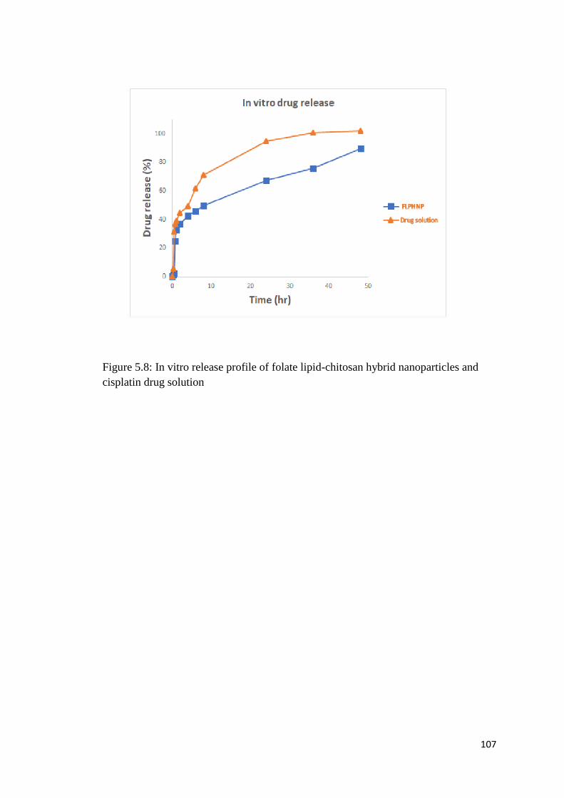
107
Figure 5.8: In vitro release profile of folate lipid-chitosan hybrid nanoparticles and
cisplatin drug solution

108
5.4. Discussion
Folic acid has attracted a lot of attention as a targeting agent due to its high
internalization. It is water soluble compound and has low molecular weight and it is
biocompatible and has the advantage of its conjugation chemistry that enables it to be
conjugated with variety of polymers including chitosan. We have conjugated folic
acid with chitosan using carbidoiimide chemistry (Shia et al., 2009). Folate receptors
are overexpressed in tumor cell and belong to high affinity protein family these are
mainly overexpressed in tumors of epithelial origin (Vllasaliu et al., 2013).
The nuclear magnetic resonance spectroscopy was performed to confirm the
conjugation of folic acid with chitosan. The conjugation of folic acid with chitosan
occurs via carbidoiimide chemistry. First EDC reacts with folic acid and activate the
carboxylic group of folic acid which is then conjugated with amine group of chitosan.
FA-CS conjugate showed the typical spectral peaks at 3.8 3.9 and 4.2ppm that
correspond to the chitosan group and spectral peaks at 6.8 7.7ppm confirmed the
presence of folic acid. The same spectral peaks of FA-CS were also observed in
previous studies (Yang et al., 2014). The coupling of folic acid with chitosan leads to
slight vibration in the peaks as compared to pure folic acid and chitosan (Li et al.,
2011).
The size of prepared nanoparticles and zeta potential are of critical importance in
defining the characteristics of the nanoparticles. The charge on the nanoparticles will
affect the stability in the suspension. The value of surface charge of all prepared
formulation is above +20 that showed better stability of the nanoparticles. The
spherical shape of the prepared nanoparticles is evident from the transmission electron
microscope images. The similar findings of morphology and surface charge have been
observed with folate-chitosan nanoparticles in previous studies (Wang et al., 2015).
In vitro release studies showed sustained release of cisplatin over a period of 48 hours
that is due to entrapment of drug inside the polymer core and lipid layer serve as a
barrier and prevent the leakage of drug from the hybrid nanoparticles. Lipid layer
provides a diffusional barrier and drug is released in a sustained manner and results in
increased therapeutic benefits. Immediate release of large amount of
chemotherapeutic agent could cause harmful effects on normal body tissues, thus,

109
sustained release pattern may results in decreased side effects and provision of
therapeutic dose over a prolong time period (Kim et al., 2008).
5.5. Conclusion
Folic acid has been successfully conjugated with chitosan and hybrid nanoparticles
have been prepared by ionic gelation method using anionic lipid by ionic interaction
of positively charged folate-chitosan conjugate and negatively charged lipid. The
prepared hybrid nanoparticles have suitable size and PDI and cationic charge with
excellent stability in the suspension form. The morphology of hybrid nanoparticles
was confirmed by transmission electron microscopy and images showed spherical
shaped nanoparticles. The entrapment efficiency was more than 75% and drug release
studies provide sustained release for 48 hours. The prepared folate conjugated lipid-
chitosan hybrid nanoparticles could be used for sustained delivery of cisplatin further
cell lines studies needed to be carried out to establish the therapeutic efficacy of
hybrid nanoparticles.

110
Bjs Waltham
Folate Targeted Chitosan-
Lipid Hybrid Nanoparticles
for Enhanced Therapeutic
Efficacy
Biological characterization
CHAPTER 6

111
6.1. Background
The hybrid nanoparticle drug delivery system provides enhanced therapeutic efficacy
and biocompatibility. The biocompatible properties of nanoparticle drug delivery
system are usually assessed by cell culture. Cell cultures are suitable to measure the
efficacy of the drug delivery system. Cell culture has established pivotal role in
measuring the therapeutic efficacy and biocompatibility of drug delivery system since
the development of first ever HeLa cell lines. Cells either attached to the surface of
flask as a monolayer or remain in the suspension form. The doubling time of cells
vary depending on cell type and cell cycle (Ravi et al., 2015).
In recent era, 3D cell cultures have gain importance that mimic the in vivo
environment. They are more realistic and closer to in vivo environment to establish
the efficacy of drug delivery system. To obtain 3D shape that is closer to in vivo
environment different type of scaffold are used and most common type is agarose gel
(Vinci et al., 2012). The major advantage of using 3D culture spheroids over 2D
culture is to minimize the gap between in vitro cell culture and physiology of cell. In
traditional 2D cell culture system most of important parameters such as cell to cell
interaction and cell to matrix interaction cannot be studied. The 3D cell culture
provides more clear mechanism of tumorigenesis and in vivo conditions
(Muthuswamy, 2011). 3D cell cultures are also extensively used for drug screening
and to check the pharmacological effects at initial stage and exclude the toxic and
relatively ineffective substance and it also reduce the unethical animal studies and
thereby the cost associated with the controversial animal studies (Pampaloni et al.,
2009).
3D spheroids made up of cancer cell lines have three layers the upper proliferating
layer and central necrotic layer and inner quiescent layer that mimic the environment
of solid tumors. 3D spheroids have most of features of in vivo solid tumors including
hypoxic environment and cellular interaction (Lee et al., 2007). 3D spheroids have
many peculiar features of in vivo tumors particularly of at initial stage of tumor
growth. They also have necrotic area and proliferative index. The drug resistance in
spheroids is also found to be similar to the in vivo animal studies. These are also
suitable to study the pattern of cell proliferation and cell cycle mechanism. The

112
microenvironment of tumor plays key role in 3D spheroids and are useful to mimic
tumor’s microenvironment (Mazzoleni et al., 2009).
Here, we studies the biocompatibility of blank folate LPHNPs and cytotoxicity of
cisplatin loaded folate LPHNPs, LPHNPs on ovarian cell lines A2780 and SKOV3
and also on MCF-7 breast cell lines and prepared 3D spheroids using breast cell lines
to mimic the in vivo environment and studies the efficacy of cisplatin loaded lipid
chitosan hybrid nanoparticles against 3D spheroids. Cell uptake sstudies were also
carried out on 3D spheroids model to check the folate mediated penetration and
uptake of folate LPHNPs.

113
6.2. Materials and Methods
6.2.1. Materials
Cell Titer Blue and Cell titer Glo (CTG) were purchased from Promega (WI, USA).
The ovarian cell lines SKOV3 and A2780 were purchased from Sigma Aldrich, breast
cancer cell line MCF-7 was purchased from Sigma Aldrich. Roswell Park Memorial
Institute medium (RPMI), DMEM media, fetal bovine serum (FBS), and penicillin-
streptomycin solution were obtained from Cell Gro (VA, USA). Hoechst 33342 was
purchased from Molecular Probes Inc. (Eugene, OR). Paraformaldehyde was from
Electron Microscopy Sciences (Hatfield, PA, USA). Trypan blue solution was
obtained from Hyclone (Logan, UT, USA). Rdodamine-123 and rhodamine-PE were
purchased from Thermo Fischer Scientific (USA).
6.2.2. Cell viability studies
Cell viability studies were conducted on A2780 and SKOV3 ovarian cell lines and
MCF-7 breast cell lines. Cells (5000) were seeded in each well of two 96 well plates.
After overnight incubation the cells were treated with folate LPHNPs, LPHNPs and
cisplatin solution and blank folate LPHNPs to check the cytotoxic effect on cell lines.
The reading was taken after 24 and 48 hours of treatment on different plates using
plate reader. Cell titer blue was used to measure the cell viability. It is based on the
ability of live cells to convert resazurin, which is a redox dye into fluorescent dye
resorufin. The dead cell does not have the metabolic activity and cannot convert the
redox dye and only living cells are counted using plate reader.
6.2.3. Fluorescence microscopy
Cells (100,000) were seeded in a 6 well plate using SKOV3 cell lines. After 24 hours
of incubation periods the cells were treated with folate LPHNPs loaded with cisplatin
and rhodamine-123 that produce green fluorescence and other group of folate
LPHNPs was loaded with cisplatin and rhodamine-PE that produce red fluorescence.
The third group of folate LPHNPs was loaded with both fluorescence dyes
rhodamine-123 and rhodamine-PE. Similar lipid-chitosan hybrid nanoparticles were
also loaded with fluorescent dyes. After four hours, cells were washed with PBS (pH
7.4) and fixed with same solution containing 4% paraformaldehyde (PFA). After

114
fixation cells washed with again using PBS (pH 7.4) three times and stained with 10
µg/ml Hoechst for 15 min. Cells were washed again with PBS, pH 7.4, and mounted
on microscope slides with mounting buffer for analysis by keyence fluorescence
microscope.
6.2.4. Cellular uptake
Flow cytometry was used for quantitative determination of cell uptake. Cells
(400,000) were seeded overnight in each well of 6-well plate. After overnight
incubation period, cells were treated with Rh-123 containing formulation (0.1 mol %)
for 4hours. After four hours of treatment cells were detached using trypsin and
washed with PBS, pH 7.4, three times and centrifuged at 2000 rpm for 5 min and the
re-suspended in 300µl of PBS. The re-suspended cell suspension was then used for
determination of cell uptake by flow cytometry using fluorescence signals.
6.2.5. Cell apoptosis
Cell apoptosis studies were performed using annexin-V FITC apoptosis detection kit.
Cells (100,000) were seeded in each well of 6 well plate and after overnight
incubation period cells were treated with cisplatin loaded folate LPHNPs, LPHNPs
and cisplatin drug solution to compare the cell apoptosis. The cells were collected
after 24 and 48 hours of treatment using trypsin and then centrifuged and washed
using PBS and cell were incubated with propidium iodide (PI) for 15 minutes and
kept on ice and then cell apoptosis was measured using flow cytometry.
6.2.6. Cell cycle
Cell cycle studies were conducted to evaluate the inhibition mechanism. The cells
(700,000) were seeded in each well of 6 well plate. After overnight incubation they
were treated with cisplatin loaded folate LPHNPs, LPHNPs and cisplatin solution.
After 24 and 48 hours of treatment periods the cells were collected and fixed using
10% ethanol. After fixation the cells were centrifuged and washed with PBS and
incubated with propidium iodide for 30 minutes and then reading was taken using
flow cytometry.

115
6.2.7. Preparation of 3 D spheroids
The spheroids were prepared in 96 well plated. 96 well plates were coated with 1.5%
agarose and kept at 4°C. Cells (10,000) were then seeded in each well of 96 well plate
already coated with agarose. The cells were then centrifuged at 15000rpm at 15°C for
20 minutes and then kept in the incubator at 25°C and 5% CO2.The shape of spheroids
was monitored using microscope. The plates were kept in incubator for about 7 days
until they attain a proper 3D spheroids shape.
6.2.8. Cell viability towards 3D spheroids
Cell viability studies were performed on spheroids using 96 well plates. Cells
(10,000) were then seeded in each well of 96 well plate already coated with agarose.
The cells were then centrifuged at 15000rpm at 15°C for 20 minutes and then kept in
the incubator at 25°C and 5% CO2. After 7 days of incubation the spheroids were
ready for treatment. They were treated with cisplatin loaded folate LPHNPs, LPHNPs
and cisplatin solution. After 24 hours and 48 hours of treatment the cells were treated
with cell titer Glo (Promega) using standard protocol and luminescence was measured
using plate reader.
6.2.9. Cell uptake studies toward 3 D spheroids
Cell uptake studies were performed on spheroids using 96 well plate. Cells (10,000)
were then seeded in each well of 96 well plate already coated with agarose. The cells
were then centrifuged at 15000rpm at 15°C for 20 minutes and then kept in the
incubator at 25°C and 5% CO2. After 7 days of incubation the spheroids were ready
for treatment. They were treated with cisplatin loaded folate LPHNPs, LPHNPs and
cisplatin solution. After 24 hours and 48 hours of treatment the spheroids were
collected in eppendorf tubes and washed with PBS and uptake was measured using
flow cytometry.
6.2.10. Fluorescence microscopic images of 3D spheroids
Cells (10,000) were then seeded in each well of 96 well plate already coated with
agarose. The cells were then centrifuged at 15000rpm at 15°C for 20 minutes and then
kept in the incubator at 25°C and 5% CO2. After 7 days of incubation the spheroids
were ready for treatment. They were treated with cisplatin loaded folate LPHNPs,

116
LPHNPs and cisplatin solution. After 24 hours and 48 hours of treatment the
spheroids were collected in eppendorf tubes and washed with PBS and then fixed
containing 4% paraformaldehyde (PFA). After fixation cells washed with again using
PBS (pH 7.4) three times and stained with 10 µg/ml Hoechst for 15 min. Cells were
washed again with PBS, pH 7.4, and placed in 85mm tubes and images of spheroids
were taken using fluorescence microscope and depth of penetration of checked by
using Z-stacking of fluorescence microscope.

117
6.3. Results
6.3.1. Cytotoxicity studies
Cytotoxicity studies were carried out on ovarian cancer cell lines and breast cancer
cell lines. A2780 and SKOV3 were used as ovarian cell lines and MCF-7 was used as
breast cancer cell lines.
Two 96 well plates were seeded with A2780 cell line and cells were treated with
blank folate LPHNPs and cisplatin loaded folate LPHNPS and LPHNPs and cisplatin
solution. Cell viability was measured using cell titer blue after 24 hours for one plate
and after 48 hours for other plate using a plate reader. The results showed that blank
folate LPHNPs does not have any cytotoxic effect and are safe drug delivery system.
The results showed that after 24 hours of incubation folate LPHNPs have more
significant cytotoxicity as compared to cisplatin drug solution and LPHNPs at
concentration 12.5µg/ml and 6.25µg/ml while there in no significant difference at
other concentration. After 48 hours of treatment folate LPHNPs have more significant
cytotoxic effect compared to LPHNPS and cisplatin drug solution at concentration of
(25,12.5,6.25,3.12 µg/ml). This is due to folate receptors mediated endocytosis of
folate LPHNPs and enhanced uptake of folate LPHNPs that result in enhanced
cytotoxic effect.
Two 96 well plates were seeded with SKOV3 cell line and cells were treated with
blank folate LPHNPs and cisplatin loaded folate LPHNPS and LPHNPs. Cell viability
was measured using cell titer blue after 24 hours for one plate and after 48 hours for
other plate using a plate reader. The results showed that blank folate LPHNPs does
not have any cytotoxic effect and are safe drug delivery system. The results showed
that after 24 hours of incubation there is not much significant difference in cytotoxic
effect of folate LPHNPS and LPHNPS. After 48 hours of treatment folate LPHNPs
have more significant cytotoxic effect compared to LPHNPS and cisplatin drug
solution at concentration of (50, 25, 12.5, 6.25, 3.12 and 1.6µg/ml). This is due to
folate receptors mediated endocytosis of folate LPHNPs and enhanced uptake of
folate LPHNPs that result in enhanced cytotoxic effect. There is no significant
difference after 24 hours while much enhanced significant cytotoxic effect after 48
hours was due to sustained release of cisplatin from folate LPHNPs.

118
Two 96 well plates were seeded with MCF-7 cell line and cells were treated with
blank folate LPHNPs and cisplatin loaded folate LPHNPS and LPHNPs. Cell viability
was measured using cell titer blue after 24 hours for one plate and after 48 hours for
other plate using a plate reader. The results showed that blank folate LPHNPs does
not have any cytotoxic effect and are safe drug delivery system. The results showed
that after 24 hours of incubation folate LPHNPs have significant more cytotoxic effect
as compared to cisplatin drug solution at concentration of 12.5µg/ml and 3.1µg/ml but
there is not much significant difference in cytotoxic effect of folate LPHNPS and
LPHNPS. After 48 hours of treatment folate LPHNPs have more significant cytotoxic
effect compared to LPHNPS and cisplatin drug solution at concentration of (12.5 and
3.1µg/ml). This is due to folate receptors mediated endocytosis of folate LPHNPs and
enhanced uptake of folate LPHNPs that result in enhanced cytotoxic effect.

119
Figure 6.1: Cell viability study on A2780 cell lines after 24 hours of incubation
comparison of blank folate LPHNPS, folate LPHNPS and LPHNPs

120
Figure 6.2: Cell viability study on A2780 cell lines after 48 hours of incubation
comparison of blank folate LPHNPS, folate LPHNPS and LPHNPs

121
Figure 6.3: Cell viability study on SKOV3 cell lines after 24 hours of incubation
comparison of blank folate LPHNPS, folate LPHNPS and LPHNPs

122
Figure 6.4: Cell viability study on SKOV3 cell lines after 48 hours of incubation
comparison of blank folate LPHNPS, folate LPHNPS and LPHNPs

123
Figure 6.5: Cell viability study on MCF-7 cell lines after 24 hours of incubation
comparison of blank folate LPHNPS, folate LPHNPS and LPHNPs

124
Figure 6.6: Cell viability study on MCF-7 cell lines after 48 hours of incubation
comparison of blank folate LPHNPS, folate LPHNPS and LPHNPs

125
6.3.2. Cell uptake studies
Cell uptake studies were performed using flow cytometry to measure the quantitative
difference of cell uptake between folate LPHNPs and LPHNPs using rhodamine-123
as a fluorescent dye.
The results showed 12-fold more uptake of LPHNPs loaded with rhodamine-123
compared to control and folate LPHNPs have 24 fold increase in uptake as compared
to control and 12 fold increased in uptake as compared to LPHNPs. The statistical
analysis showed that there is significant difference of uptake of LPHNPs as compared
to control and folate LPHNPs also showed significantly higher uptake as compared to
LPHNPs that is due to folate receptor mediated endocytosis and enhanced uptake by
the cells.
Figure 6.7: Cell uptake studies using flow cytometry. Comparison of LPHNPs and
Folate LPHNPs loaded with rhodamie-123

126
6.3.3. Fluorescence microscopy
Qualitative uptake of hybrid nanoparticles by SKOV3 cells was checked by using
fluorescence microscopy. Folate targeted lipid-chitosan hybrid nanoparticles were
loaded with fluorescent dyes rhodamine-123 and rhodamine-PE. Rhodamine-123
stains the cell green color and rhodamine-PE stains red color. Cells were already
stained by HOECST a blue color dye that stains the nucleic acid during the process of
fixation of cells.
The results showed that SKOV3 cell actively uptake the folate targeted lipid-chitosan
hybrid nanoparticles and showed green and red fluorescence. The uptake of folate
LPHNPs is due to folate receptors mediated endocytosis.
Figure 6.8: Fluorescence microscopy images of SKOV3 cells treated with folate
LPHNPs and folate LPHNPs loaded with Rh-123 and Rh-PE

127
6.3.4. Cell cycle studies
Cell cycle studies were performed using propidium iodide and fixation of cells using
70% ethanol. The cell population was recorded using flow cytometry after 24 and 48
hours of treatment. The results showed that after 24 hours of treatment control group
have 65% of population in G0/G1 phase and 15 % of population is S phase and
remaining population is G2/M phase of cell cycle. After 24 hours of treatment the
population of cells in drug treated group has shifted from G0/G1phase to S phase and
there is significant more shift in population of folate LPHNPs as compared to
LPHNPs and cisplatin solution group. After 48 hours of treatment the cell population
has shifted to G2/M phase and there is significant more population in folate LPHNPs
as compared to cisplatin drug solution of LPHNPs. The results showed that folate
LPHNPs more significantly halt the cell cycle as compared to cisplatin loaded
LPHNPs and cisplatin drug solution.
Figure 6.9: Cell cycle studies after 24 hours of treatment

128
Figure 6.10: Cell cycle studies after 48 hours of treatment
6.3.5. Cell apoptosis studies
Cell apoptosis studies were carried out using propidium iodide and annexin-V to
detect the early and late apoptosis of the cells. The results were compared between
cisplatin drug solution, LPHNPs and folate LPHNPs. The results showed that after 24
hours of treatment there is significant shift of cells toward early and late apoptosis as
compared to control group and folate LPHNPs have significantly higher apoptosis as
compared to LPHNPs and cisplatin drug solution. After 24 hours of treatment
cisplatin solution has 10% of cells in early apoptosis and LPHNPs have 15% of cells
while folate LPHNPs have 26% of cells at early apoptosis and 39% of cells at late
apoptosis as compared to only 18% in late apoptosis of LPHNPs.
After 48 hours of treatment there is an increase in late apoptosis and decrease in early
apoptosis of the cells. Folate LPHNPs showed significantly increased in late apoptosis
as compared to LPHNPs and cisplatin drug solution but cisplatin solution showed
increase in early apoptosis as compared to folate LPHNPs. Late apoptosis of cells is
considered as of critical importance as cells in early apoptosis might have chances to
revert. The result showed significant increase in late apoptosis by folate LPHNPs
where more than 60% cells are in late apoptosis as compared to 40% in cisplatin drug
solution group.
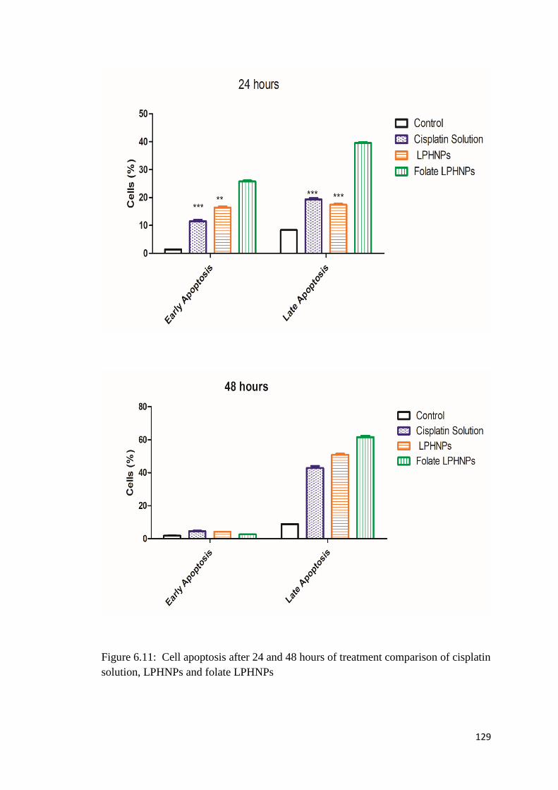
129
Figure 6.11: Cell apoptosis after 24 and 48 hours of treatment comparison of cisplatin
solution, LPHNPs and folate LPHNPs

130
Figure 6.12: Control group cell apoptosis showing early and late apoptosis

131
Figure 6.13: Cell apoptosis of cisplatin drug solution showing early and late
apoptosis

132
Figure 6.14: Cell apoptosis of lipid- chitosan hybrid nanoparticle loaded with
cisplatin

133
Figure 6.15: Cell apoptosis of folate targeted lipid-chitosan hybrid nanoparticles

134
6.3.6. Cell Cytotoxicity toward cancer cell in 3D spheroids
Cell cytotoxicity studies were conducted on 3D spheroids model that mimic the in
vivo environment to evaluate the in vivo efficacy of folate targeted LPHNPs and
compare the cytotoxicity on 3D spheroids with LPHNPs. The spheroids were grown
for 10 days and treated with cisplatin loaded folate LPHNPs, LPHNPs and cisplatin
drug solution and CTG protocol was used to calculate the cell viability that measure
the luminescence by the cell in 3D spheroids.
The results showed that after 24 hours of treatment folate LPHNPs have more
significant cytotoxic effect against tumor cell line as compared to cisplatin drug
solution at concentration of (50, 12.5, 3.1µg/ml) and more cytotoxic effect as
compared to LPHNPs at concentration of 6.25 µg/ml.
After 48 hours of treatment folate LPHNPs have more significant cytotoxicity as
compared to cisplatin drug solution at concentration of (25, 12.5, 6.2 µg/ml) and more
significant cytotoxic effect as compared to LPHNPs at concentration of (25, 12.5, 6.2,
3.1 µg/ml). This showed that folate targeted lipid-chitosan hybrid nanoparticles are
more effective as compared to LPHNPs and cisplatin drug solution and exert greater
cytotoxic effects.

135
Figure 6.16: Cytotoxicity studies on 3D spheroids after 24 hours of treatment
comparison of folate LPHNPs, LPHNPs and cisplatin drug solution

136
Figure 6.17: Cytotoxicity studies on 3D spheroids after 24 hours of treatment
comparison of folate LPHNPs, LPHNPs and cisplatin drug solution

137
6.3.7. Cell uptake studies toward cancer cell in 3D spheroids
Cell uptake studies were conducted in 3D spheroids as in vivo model. Spheroids were
treated with folate LPHNPs and LPHNPs and the uptake was measured after 4 hours
of treatment using flow cytometry. The results showed that LPHNPs are have 2-fold
more uptake in 3D spheroids as compared to control and folate LPHNPs have double
uptake as compared to LPHNPs and 4 times more uptake as compared to control. This
enhanced uptake by spheroids is due to folate mediated endocytosis of folate LPHNPs
that results in enhanced uptake by the cell and results in enhanced efficacy.
Figure 6.18: Cell uptake studies using flow cytometry comparison of folate LPHNPs,
LPHNPs

138
6.3.8. Fluorescence microscope images of cell uptake towards 3D spheroids
Cell uptake by 3D spheroids was also measured qualitatively by taking images of
spheroids using flow cytometry. Rhodamine-123 is a fluorescent dye and was loaded
inside the nanoparticles and used to measure the uptake. It gives green color after
uptake inside the cells.
The results showed that folate targeted lipid-chitosan hybrid nanoparticles produce
more fluorescence as compared to LPHNPs and there is more bright green color as
compared to LPHNPs. The images of spheroids were also taken using z-stacking to
confirm that weather the cells in the lower layer of spheroids have uptake the folate
LPHNPs loaded with rhodamine and results showed that there is greater uptake as
compared to LPHNPs.
Figure 6.19: Cell uptake towards 3D spheroids using fluorescence microscope
comparison of folate LPHNPs with LPHNPs

139
Figure 6.20: Z-stack images of 3D spheroids control group
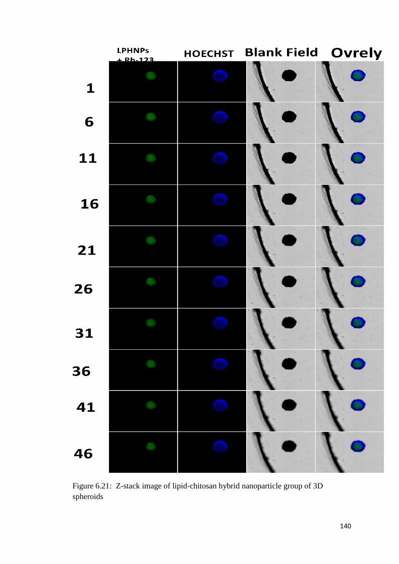
140
Figure 6.21: Z-stack image of lipid-chitosan hybrid nanoparticle group of 3D
spheroids

141
Figure 6.22: Z-stack image of folate targeted lipid-chitosan hybrid nanoparticle
group of 3D spheroids

142
6.4. Discussion
The lipid-chitosan hybrid nanoparticles are made of lipid that are component of
biological membrane and polymer chitosan that is biocompatible and obtained from
the chitin. The folate is conjugate with chitosan which is a vitamin and required by the
body. The studies on different ovarian and breast cell lines proved the
biocompatibility of blank LPHNPs and folate conjugated lipid-chitosan hybrid
nanoparticles. The results prove that there is not any cytotoxic effect of blank folate
LPHNPs on the cell lines and cell count is almost similar to the control group. Several
previous studies also support the biocompatibility of folate-chitosan nanoparticles (Jin
et al., 2016).
The cell viability studies on ovarian cell lines showed that folate LPHNPs have more
cytotoxic effect on A2780 cell lines as compared to LPHNPs and cisplatin drug
solution that is due to enhanced internalization of folate LPHNPs and exhibit greater
cytotoxic effect. The greater cytotoxic effect was observed after 48 hours of treatment
because of controlled release of drug from folate LPHNPs. Previous studies of folate
targeting also showed that folate targeted nanoparticles showed more cytotoxic effect
as compared to without folate conjugation (Mansouri et al., 2006).
The cell uptake studies were conducted to measure the internalization of
nanoparticles. Qualitative uptake was measured using fluorescence microscope and
quantitative uptake was measured fluorescence microscopy. The results showed folate
targeted nanoparticles showed 2 times more cell uptake as compared to untargeted
hybrid nanoparticles. The enhanced uptake was folate targeted LPHNPs as compared
to untargeted LPHNPs is due to folate mediated internalization and uptake via folate
receptors. The enhanced cell uptake of folate targeted nanoparticles is also established
in previous studies (Xu et al., 2013).
The cell cycle studies were conducted using cisplatin loaded folate LPHNPs and
cisplatin loaded LPHNPs and cisplatin solution. The results showed that after 24
hours of treatment there is regular reduction in G0/G1phase and increase in S phase
population of cells and after 48 hours of treatment there is significant reduction in
G0/G1phase and increase in G2/M phase and folate LPHNPs have significant more
increase in G2/M phase population as compared to without folate. Previous studies on

143
folate targeted cisplatin nanoparticles also report increase in G2/M phase population
of cell (Wang et al., 2013).
The studies on 3D spheroids were conducted to mimic the in vivo environment. The
cytotoxicity studies on 3D spheroids showed that folate targeted LPHNPs have more
cytotoxic effect as compared to without folate targeted. There is also enhanced cell
uptake of folate LPHNPs due to folate mediated endocytosis both qualitative and
quantitative uptake studies established the internalization of folate LPHNPs.

144
6.5. Conclusion
It was concluded from the biological characterization that folate lipid-chitosan hybrid
nanoparticles have enhanced cytotoxic effect as compared to LPHNPs on A2780
ovarian cell lines after 24 and 48 hours of treatment. Similar studies were conducted
on SKOV3 ovarian cell lines and enhanced efficacy of folate LPHNPs was
established. MCF-7 cell lines were used to check the cytotoxic effects against breast
cancer cell lines the results also showed greater cytotoxic effects as compared to
LPHNPs and cisplatin drug solution. The cell uptake studies showed greater uptake of
folate LPHNPs as compared to without folate targeting. The enhanced effect on cell
uptake is due to folate receptors mediated endocytosis. The cytotoxicity studies on 3D
spheroids established that folate LPHNPs could showed enhanced cytotoxic effect in
in vivo environment. The results showed that folate targeted lipid-chitosan hybrid
nanoparticles could serve as a system for active targeting of tumor cells.

145
Bjs Waltham
Conclusions and
Recommendations
CHAPTER 7

146
7. Conclusions and recommendations
LPHNPs are drug delivery system for controlled drug delivery. In this dissertation
we used chitosan and lipid for the preparation of hybrid nanoparticles. The
LPHNPs were then loaded with cisplatin for controlled drug delivery to the tumor
site. The chitosan was then conjugated with folic acid for active targeting of
tumor. The efficacy of cisplatin loaded hybrid nanoparticles was then evaluated by
using different ovarian cell lines.
The following conclusion were made from these studies
1. Cisplatin loaded lipid-chitosan hybrid nanoparticles were successfully fabricated
using different lipid-to chitosan ratios. The lipids to chitosan ratios of (10:1 to
60:1) were used and 6 different formulations were prepared. The size, surface
charge, entrapment efficiency and drug loading was measured and it was
concluded that LPHNPs with ratio of 20:1 have excellent size with low PDI and
good entrapment efficiency and drug loading and these were used for further
analysis.
2. Cisplatin loaded LPHNPs were evaluated for the thermal stability using
differential scanning calorimetry and thermogravimetric analysis and they showed
excellent thermal stability and drug encapsulation within the system. The prepared
hybrid nanoparticles were further evaluated by using FTIR to check the
compatibility among the formulation excipients and it was concluded that
components are compatible as there are no new peaks.
3. The drug release studies were performed and it was concluded that LPHNPs
showed controlled release of drug due to entrapment of drug inside polymer core
and lipid provide diffusional barrier and drug is release in a controlled manner.
The kinetic modeling was applied, and it was concluded that cisplatin loaded
LPHNPs follow korsmeyerpeppas model and super case II transport mechanism.
4. The therapeutic efficacy of cisplatin loaded LPHNPs was evaluated in vitro using
A2780 ovarian cell lines. It was concluded from the results that cisplatin loaded
LPHNPs have more cytotoxic effect as compared to cisplatin drug solution and
uptake studies confirmed the cellular association and internalization of LPHNPs.
5. The cisplatin loaded LPHNPs system was further evaluated on rats to confirm the
safety profile of LPHNPs system. Toxicological studies on rats were performed

147
and evaluated for biochemical evaluation and histopathological evaluation using
higher doses of LPHNPs and cisplatin. The results showed safety profile of
LPHNPs and there are not any significant changes in hematological parameters
and histopathological evaluation. The pharmacokinetic studies were performed on
rabbits to check the in vivo profile of cisplatin and confirmed higher mean
residence time of LPHNPs compared to cisplatin solution group.
6. The chitosan was then successfully conjugated with folic acid for active targeting.
The TLC analysis confirms the purity of conjugate and nuclear magnetic
resonance spectroscopy confirmed the conjugation of folic acid with chitosan.
7. The folate LPHNPs were successfully fabricated by the ionic gelation method.
The folate LPHNPs containing lipid to folate-chitosan ratio of (15:1) showed
better size and stability with good entrapment efficiency and drug loading.
8. The morphology confirmed the spherical shaped nanoparticles with folate
covering on the outer surface. The drug release studies showed that sustained
release of cisplatin over the periods of 48 hours compared to cisplatin solution.
9. The therapeutic efficacy of folate LPHNPs was evaluated on ovarian cell lines and
breast cell lines. A2780 and SKOV3 were used as ovarian cell lines and it was
concluded from the results that after 48 hours of treatment folate LPHNPs have
more significant cytotoxic effect against ovarian cell lines compared to LPHNPs
and cisplatin drug solution. The studies on MCF-7 breast cell lines also showed
the significantly enhanced cytotoxic effect of folate LPHNPs as compared to
LPHNPs and cisplatin solution.
10. To evaluate the in vivo efficacy of folate LPHNPs we prepared 3D spheroids that
mimic the in vivo environment and performed the cell cytotoxicity and cell uptake
studies on 3D spheroids. The results of 3D spheroids further establish the
enhanced cytotoxic effect of folate LPHNPs as compared to LPHNPs without
folate targeting and cisplatin solution. It was concluded the folate targeted
LPHNPs are more effective to provide targeted and controlled drug delivery at
tumor site.

148
7.1 . Future prospects
This study provides the promising results for the fabrication and optimization of
lipid-chitosan hybrid nanoparticles for the delivery of chemotherapeutic agents.
This study also provides a new drug delivery system for folate targeting. Folate-
chitosan conjugate was used to prepared nanoparticles using anionic lipid which is
new system of folate targeting having advantage of lipid over traditional folate
targeting.
1. Different synthetic and natural polymers can be evaluated with combination of
cationic and anionic lipids to prepared LPHNPs for controlled delivery of
different chemotherapeutic agents.
2. These lipid-chitosan hybrid nanoparticles can be evaluated for co-delivery of
siRNA and drug to combat with drug resistance and provide optimum therapeutic
effectiveness.
3. LPHNPs can be evaluated for the sustained and controlled delivery of different
antidiabetic, antihypertensive and other drugs.
4. Folate targeted LPHNPs can also be used for co-delivery of siRNA and
chemotherapeutic agents because of their cationic charge and ability to interact
with negatively charged siRNA and DNA.
5. 3D spheroids establish the estimated in vivo efficacy of folate LPHNPs further
studies on animal tumor models and pre-clinical studies can be conducted to
establish folate LPHNPs as an effective drug delivery system compared to
traditional folate targeting and moving towards FDA approval.

149
Bjs Waltham
References
CHAPTER 8
150
References
Aad, G., Abajyan, T., Abbott, B., Abdallah, J., Khalek, S. A., Abdelalim, A. A.,
Abdinov, O., Aben, R., Abi, B. and Abolins, M. (2012). Observation of a new
particle in the search for the standard model higgs boson with the atlas
detector at the lhc. Physics Letters B. 716: 1-29.
Acharya, S. and Sahoo, S. K. (2011). Plga nanoparticles containing various anticancer
agents and tumour delivery by epr effect. Advanced drug delivery reviews. 63:
170-183.
Agarwal, R. and Kaye, S. B. (2003). Ovarian cancer: Strategies for overcoming
resistance to chemotherapy. Nature Reviews Cancer. 3: 502.
Agarwal, R. and Kaye, S. B. (2003). Ovarian cancer: Strategies for overcoming
resistance to chemotherapy. Nature Reviews Cancer. 3: 502-516.
Agrawal, P., Strijkers, G. J. and Nicolay, K. (2010). Chitosan-based systems for
molecular imaging. Advanced drug delivery reviews. 62: 42-58.
Aizawa, K., Liu, C., Tang, S., Veeramachaneni, S., Hu, K. Q., Smith, D. E. and
Wang, X. D. (2016). Tobacco carcinogen induces both lung cancer and non‐
alcoholic steatohepatitis and hepatocellular carcinomas in ferrets which can be
attenuated by lycopene supplementation. International journal of cancer. 139:
1171-1181.
Al-Majed, A. A., Sayed-Ahmed, M. M., Al-Yahya, A. A., Aleisa, A. M., Al-Rejaie, S.
S. and Al-Shabanah, O. A. (2006). Propionyl-l-carnitine prevents the
progression of cisplatin-induced cardiomyopathy in a carnitine-depleted rat
model. Pharmacological research. 53: 278-286.
Alavi, M., Karimi, N. and Safaei, M. (2017). Application of various types of
liposomes in drug delivery systems. Advanced pharmaceutical bulletin. 7: 3.
Albanese, A., Tang, P. S. and Chan, W. C. (2012). The effect of nanoparticle size,
shape, and surface chemistry on biological systems. Annual review of
biomedical engineering. 14: 1-16.
Ali, B. H. and Al Moundhri, M. S. (2006). Agents ameliorating or augmenting the
nephrotoxicity of cisplatin and other platinum compounds: A review of some
recent research. Food and Chemical Toxicology. 44: 1173-1183.

151
Allen, T. M., Mehra, T., Hansen, C. and Chin, Y. C. (1992). Stealth liposomes: An
improved sustained release system for 1-β-d-arabinofuranosylcytosine. Cancer
research. 52: 2431-2439.
Anders, C. K. and Carey, L. A. (2009). Biology, metastatic patterns, and treatment of
patients with triple-negative breast cancer. Clinical breast cancer. 9: S73-S81.
Anderson, D. G., Peng, W., Akinc, A., Hossain, N., Kohn, A., Padera, R., Langer, R.
and Sawicki, J. A. (2004). A polymer library approach to suicide gene therapy
for cancer. Proceedings of the National Academy of Sciences. 101: 16028-
16033.
Anselmo, A. C. and Mitragotri, S. (2017). Impact of particle elasticity on particle-
based drug delivery systems. Advanced drug delivery reviews. 108: 51-67.
Antoni, D., Burckel, H., Josset, E. and Noel, G. (2015). Three-dimensional cell
culture: A breakthrough in vivo. International journal of molecular sciences.
16: 5517-5527.
Ashley, C. E., Carnes, E. C., Phillips, G. K., Padilla, D., Durfee, P. N., Brown, P. A.,
Hanna, T. N., Liu, J., Phillips, B. and Carter, M. B. (2011). The targeted
delivery of multicomponent cargos to cancer cells by nanoporous particle-
supported lipid bilayers. Nature materials. 10: 389.
Atlanta, G. (2015). Cancer facts and figures.
Bae, Y. H. and Park, K. (2011). Targeted drug delivery to tumors: Myths, reality and
possibility. Journal of controlled release. 153: 198.
Bamrungsap, S., Zhao, Z., Chen, T., Wang, L., Li, C., Fu, T. and Tan, W. (2012).
Nanotechnology in therapeutics: A focus on nanoparticles as a drug delivery
system. Nanomedicine. 7: 1253-1271.
Bangham, A. (1993). Liposomes: The babraham connection. Chemistry and physics of
lipids. 64: 275-285.
Bangham, A., Standish, M. M. and Watkins, J. C. (1965). Diffusion of univalent ions
across the lamellae of swollen phospholipids. Journal of molecular biology.
13: 238-IN27.
Barbe, C., Bartlett, J., Kong, L., Finnie, K., Lin, H. Q., Larkin, M., Calleja, S., Bush,
A. and Calleja, G. (2004). Silica particles: A novel drug‐delivery system.
Advanced materials. 16: 1959-1966.
Béduneau, A., Saulnier, P. and Benoit, J.-P. (2007). Active targeting of brain tumors
using nanocarriers. Biomaterials. 28: 4947-4967.
152
Bharali, D. J. and Mousa, S. A. (2010). Emerging nanomedicines for early cancer
detection and improved treatment: Current perspective and future promise.
Pharmacology & therapeutics. 128: 324-335.
Bibi, S., Lattmann, E., Mohammed, A. R. and Perrie, Y. (2012). Trigger release
liposome systems: Local and remote controlled delivery? Journal of
microencapsulation. 29: 262-276.
Blackadar, C. B. (2016). Historical review of the causes of cancer. World journal of
clinical oncology. 7: 54.
Bose, R. J., Lee, S.-H. and Park, H. (2016). Lipid-based surface engineering of plga
nanoparticles for drug and gene delivery applications. Biomaterials research.
20: 34.
Brannon-Peppas, L. and Blanchette, J. O. (2012). Nanoparticle and targeted systems
for cancer therapy. Advanced drug delivery reviews. 64: 206-212.
Brayden, D. J. (2003). Controlled release technologies for drug delivery. Drug
discovery today. 21: 976-978.
Caro, A. A. and Cederbaum, A. I. (2004). Oxidative stress, toxicology, and
pharmacology of cyp2e1. Annu. Rev. Pharmacol. Toxicol. 44: 27-42.
Carter, T., Mulholland, P. and Chester, K. (2016). Antibody-targeted nanoparticles for
cancer treatment. Immunotherapy. 8: 941-958.
Cervera, M. F., Heinämäki, J., De La Paz, N., López, O., Maunu, S. L., Virtanen, T.,
Hatanpää, T., Antikainen, O., Nogueira, A. and Fundora, J. (2011). Effects of
spray drying on physicochemical properties of chitosan acid salts. AAPS
PharmSciTech. 12: 637-649.
Chadha, R., Gupta, S. and Pathak, N. (2012). Artesunate-loaded chitosan/lecithin
nanoparticles: Preparation, characterization, and in vivo studies. Drug
development and industrial pharmacy. 38: 1538-1546.
Chan, J. M., Zhang, L., Yuet, K. P., Liao, G., Rhee, J.-W., Langer, R. and Farokhzad,
O. C. (2009). Plga–lecithin–peg core–shell nanoparticles for controlled drug
delivery. Biomaterials. 30: 1627-1634.
Chandy, T. and Sharma, C. P. (1992). Chitosan beads and granules for oral sustained
delivery of nifedipine: In vitro studies. Biomaterials. 13: 949-952.
Chaudhary, Z., Ahmed, N., . Ur. Rehman, A. and Khan, G. M. (2018). Lipid polymer
hybrid carrier systems for cancer targeting: A review. International Journal of
Polymeric Materials and Polymeric Biomaterials. 67: 86-100.

153
Chen, V. W., Ruiz, B., Killeen, J. L., Coté, T. R., Wu, X. C., Correa, C. N. and Howe,
H. L. (2003). Pathology and classification of ovarian tumors. Cancer:
Interdisciplinary International Journal of the American Cancer Society. 97:
2631-2642.
Cheng, L., Jin, C., Lv, W., Ding, Q. and Han, X. (2011). Developing a highly stable
plga-mpeg nanoparticle loaded with cisplatin for chemotherapy of ovarian
cancer. PloS one. 6: e25433.
Cheng, Q., Shi, H., Huang, H., Cao, Z., Wang, J. and Liu, Y. (2015). Oral delivery of
a platinum anticancer drug using lipid assisted polymeric nanoparticles.
Chemical Communications. 51: 17536-17539.
Cheng, R., Meng, F., Deng, C., Klok, H.-A. and Zhong, Z. (2013). Dual and multi-
stimuli responsive polymeric nanoparticles for programmed site-specific drug
delivery. Biomaterials. 34: 3647-3657.
Chhonker, Y. S., Prasad, Y. D., Chandasana, H., Vishvkarma, A., Mitra, K., Shukla,
P. K. and Bhatta, R. S. (2015). Amphotericin-b entrapped lecithin/chitosan
nanoparticles for prolonged ocular application. International journal of
biological macromolecules. 72: 1451-1458.
Cho, B. K., Roy, E. J., Patrick, T. A. and Kranz, D. M. (1997). Single-chain fv/folate
conjugates mediate efficient lysis of folate-receptor-positive tumor cells.
Bioconjugate chemistry. 8: 338-346.
Cho, K., Wang, X., Nie, S. and Shin, D. M. (2008). Therapeutic nanoparticles for
drug delivery in cancer. Clinical cancer research. 14: 1310-1316.
Choi, S.-W. and Mason, J. B. (2000). Folate and carcinogenesis: An integrated
scheme. The Journal of nutrition. 130: 129-132.
Chuah, A. M., Kuroiwa, T., Ichikawa, S., Kobayashi, I. and Nakajima, M. (2009).
Formation of biocompatible nanoparticles via the self‐assembly of chitosan
and modified lecithin. Journal of food science. 74.
Co-Operation, O. F. E. and Development (2002). Test no. 423: Acute oral toxicity-
acute toxic class method, OECD Publishing. pp
Couvreur, P. (2013). Nanoparticles in drug delivery: Past, present and future.
Advanced drug delivery reviews. 65: 21-23.
Cramer, D. W. and Welch, W. R. (1983). Determinants of ovarian cancer risk. Ii.
Inferences regarding pathogenesis. Journal of the National Cancer institute.
71: 717-721.

154
Crist, W. M., Anderson, J. R., Meza, J. L., Fryer, C., Raney, R. B., Ruymann, F. B.,
Breneman, J., Qualman, S. J., Wiener, E. and Wharam, M. (2001). Intergroup
rhabdomyosarcoma study-iv: Results for patients with nonmetastatic disease.
Journal of Clinical Oncology. 19: 3091-3102.
Damia, G., Filiberti, L., Vikhanskaya, F., Carrassa, L., Taya, Y., Dincalci, M. and
Broggini, M. (2001). Cisplatinum and taxol induce different patterns of p53
phosphorylation. Neoplasia. 3: 10-16.
Danhier, F., Feron, O. and Préat, V. (2010). To exploit the tumor microenvironment:
Passive and active tumor targeting of nanocarriers for anti-cancer drug
delivery. Journal of controlled release. 148: 135-146.
Daniels, T. R., Bernabeu, E., Rodríguez, J. A., Patel, S., Kozman, M., Chiappetta, D.
A., Holler, E., Ljubimova, J. Y., Helguera, G. and Penichet, M. L. (2012). The
transferrin receptor and the targeted delivery of therapeutic agents against
cancer. Biochimica et Biophysica Acta (BBA)-General Subjects. 1820: 291-
317.
Daraee, H., Etemadi, A., Kouhi, M., Alimirzalu, S. and Akbarzadeh, A. (2016).
Application of liposomes in medicine and drug delivery. Artificial cells,
nanomedicine, and biotechnology. 44: 381-391.
Dayan, A., Koca, D., Akman, T., Oztop, I., Ellidokuz, H. and Yilmaz, U. (2012). The
factors that have an impact on the development of brain metastasis in the
patients with breast cancer. Journal of cancer research and therapeutics. 8:
542.
Deberardinis, R. J., Lum, J. J., Hatzivassiliou, G. and Thompson, C. B. (2008). The
biology of cancer: Metabolic reprogramming fuels cell growth and
proliferation. Cell metabolism. 7: 11-20.
Desoize, B. and Madoulet, C. (2002). Particular aspects of platinum compounds used
at present in cancer treatment. Critical reviews in oncology/hematology. 42:
317-325.
Devalapally, H., Chakilam, A. and Amiji, M. M. (2007). Role of nanotechnology in
pharmaceutical product development. Journal of Pharmaceutical Sciences. 96:
2547-2565.
Dhar, S., Gu, F. X., Langer, R., Farokhzad, O. C. and Lippard, S. J. (2008). Targeted
delivery of cisplatin to prostate cancer cells by aptamer functionalized pt (iv)

155
prodrug-plga–peg nanoparticles. Proceedings of the National Academy of
Sciences. 105: 17356-17361.
Dhar, S., Kolishetti, N., Lippard, S. J. and Farokhzad, O. C. (2011). Targeted delivery
of a cisplatin prodrug for safer and more effective prostate cancer therapy in
vivo. Proceedings of the National Academy of Sciences. 108: 1850-1855.
Dixit, N., Vaibhav, K., Pandey, R. S., Jain, U. K., Katare, O. P., Katyal, A. and
Madan, J. (2015). Improved cisplatin delivery in cervical cancer cells by
utilizing folate-grafted non-aggregated gelatin nanoparticles. Biomedicine &
Pharmacotherapy. 69: 1-10.
Dixit, N., Vaibhav, K., Pandey, R. S., Jain, U. K., Katare, O. P., Katyal, A. and
Madan, J. (2015). Improved cisplatin delivery in cervical cancer cells by
utilizing folate-grafted non-aggregated gelatin nanoparticles. Biomed
Pharmacother. 69: 1-10.
Drebushchak, V., Shakhtshneider, T. P., Apenina, S. A., Medvedeva, A. S. and
Boldyrev, V. (2006). Thermoanalytical investigation of drug–excipient
interaction. Journal of thermal analysis and calorimetry. 86: 303-309.
Drummond, D. C., Meyer, O., Hong, K., Kirpotin, D. B. and Papahadjopoulos, D.
(1999). Optimizing liposomes for delivery of chemotherapeutic agents to solid
tumors. Pharmacological reviews. 51: 691-744.
Du Bois, A., Reuss, A., Pujade‐Lauraine, E., Harter, P., Ray‐Coquard, I. and Pfisterer,
J. (2009). Role of surgical outcome as prognostic factor in advanced epithelial
ovarian cancer: A combined exploratory analysis of 3 prospectively
randomized phase 3 multicenter trials: By the arbeitsgemeinschaft
gynaekologische onkologie studiengruppe ovarialkarzinom (ago‐ovar) and the
groupe d'investigateurs nationaux pour les etudes des cancers de l'ovaire
(gineco). Cancer. 115: 1234-1244.
Du, Y.-Z., Wang, L., Yuan, H., Wei, X.-H. and Hu, F.-Q. (2009). Preparation and
characteristics of linoleic acid-grafted chitosan oligosaccharide micelles as a
carrier for doxorubicin. Colloids and Surfaces B: Biointerfaces. 69: 257-263.
Eastman, A. (1999). The mechanism of action of cisplatin: From adducts to apoptosis.
Cisplatin. Chemistry and biochemistry of a leading anticancer drug. 111-134.
Ebel, W., Routhier, E. L., Foley, B., Jacob, S., Mcdonough, J. M., Patel, R. K.,
Turchin, H. A., Chao, Q., Kline, J. B. and Old, L. J. (2007). Preclinical
evaluation of morab-003, a humanized monoclonal antibody antagonizing

156
folate receptor-alpha. Cancer immunity: a journal of the Academy of Cancer
Immunology. 7.
Ehdaie, B. (2007). Application of nanotechnology in cancer research: Review of
progress in the national cancer institute's alliance for nanotechnology.
International journal of biological sciences. 3: 108.
Elgadir, M. A., Uddin, M. S., Ferdosh, S., Adam, A., Chowdhury, A. J. K. and Sarker,
M. Z. I. (2015). Impact of chitosan composites and chitosan nanoparticle
composites on various drug delivery systems: A review. Journal of food and
drug analysis. 23: 619-629.
Emerich, D. F. and Thanos, C. G. (2003). Nanotechnology and medicine. Expert
Opinion on Biological Therapy. 3: 655-663.
Evans, W. E. and Relling, M. V. (2004). Moving towards individualized medicine
with pharmacogenomics. Nature. 429: 464.
Fagerholm, U., Sjöström, B., Sroka‐Markovic, J., Wijk, A., Svensson, M. and
Lennernäs, H. (1998). The effect of a drug‐delivery system consisting of
soybean phosphatidyl choline and medium‐chain monoacylglycerol on the
intestinal permeability of hexarelin in the rat. Journal of pharmacy and
pharmacology. 50: 467-473.
Farokhzad, O. C., Cheng, J., Teply, B. A., Sherifi, I., Jon, S., Kantoff, P. W., Richie,
J. P. and Langer, R. (2006). Targeted nanoparticle-aptamer bioconjugates for
cancer chemotherapy in vivo. Proceedings of the National Academy of
Sciences. 103: 6315-6320.
Farokhzad, O. C., Jon, S., Khademhosseini, A., Tran, T.-N. T., Lavan, D. A. and
Langer, R. (2004). Nanoparticle-aptamer bioconjugates: A new approach for
targeting prostate cancer cells. Cancer research. 64: 7668-7672.
Farokhzad, O. C. and Langer, R. (2009). Impact of nanotechnology on drug delivery.
ACS nano. 3: 16-20.
Feng, S.-S. (2004). Nanoparticles of biodegradable polymers for new-concept
chemotherapy. Expert review of medical devices. 1: 115-125.
Feng, S.-S., Zhao, L. and Tang, J. (2011). Nanomedicine for oral chemotherapy.
Nanomedicine. 6: 407-410.
Ferrari, M. (2005). Cancer nanotechnology: Opportunities and challenges. Nature
reviews cancer. 5: 161.

157
Firth, J. (2002). Endothelial barriers: From hypothetical pores to membrane proteins.
Journal of anatomy. 200: 523-534.
Forssen, E. and Willis, M. (1998). Ligand-targeted liposomes. Advanced drug
delivery reviews. 29: 249-271.
Frezza, M., Hindo, S., Chen, D., Davenport, A., Schmitt, S., Tomco, D. and Ping Dou,
Q. (2010). Novel metals and metal complexes as platforms for cancer therapy.
Current pharmaceutical design. 16: 1813-1825.
Gabizon, A., Catane, R., Uziely, B., Kaufman, B., Safra, T., Cohen, R., Martin, F.,
Huang, A. and Barenholz, Y. (1994). Prolonged circulation time and enhanced
accumulation in malignant exudates of doxorubicin encapsulated in
polyethylene-glycol coated liposomes. Cancer research. 54: 987-992.
Gao, H., Schwarz, J. and Weisspapir, M. 2008. Hybrid lipid-polymer nanoparticulate
delivery composition. Google Patents.
Garber, K. 2006. Energy deregulation: Licensing tumors to grow. American
Association for the Advancement of Science.
Gelperina, S., Kisich, K., Iseman, M. D. and Heifets, L. (2005). The potential
advantages of nanoparticle drug delivery systems in chemotherapy of
tuberculosis. American journal of respiratory and critical care medicine. 172:
1487-1490.
Gerelli, Y., Barbieri, S., Di Bari, M., Deriu, A., Cantu, L., Brocca, P., Sonvico, F.,
Colombo, P., May, R. and Motta, S. (2008). Structure of self-organized
multilayer nanoparticles for drug delivery. Langmuir. 24: 11378-11384.
Glazer, E. S., Massey, K. L., Zhu, C. and Curley, S. A. (2010). Pancreatic carcinoma
cells are susceptible to noninvasive radio frequency fields after treatment with
targeted gold nanoparticles. Surgery. 148: 319-324.
Go, R. S. and Adjei, A. A. (1999). Review of the comparative pharmacology and
clinical activity of cisplatin and carboplatin. Journal of Clinical Oncology. 17:
409-409.
Grenha, A., Remuñán-López, C., Carvalho, E. L. and Seijo, B. (2008). Microspheres
containing lipid/chitosan nanoparticles complexes for pulmonary delivery of
therapeutic proteins. European Journal of Pharmaceutics and
Biopharmaceutics. 69: 83-93.
Grodzinski, P., Kircher, M., Goldberg, M. and Gabizon, A. 2019. Integrating
nanotechnology into cancer care. ACS Publications.
158
Gu, F. X., Karnik, R., Wang, A. Z., Alexis, F., Levy-Nissenbaum, E., Hong, S.,
Langer, R. S. and Farokhzad, O. C. (2007). Targeted nanoparticles for cancer
therapy. Nano today. 2: 14-21.
Guo, Y., Wang, L., Lv, P. and Zhang, P. (2015). Transferrin-conjugated doxorubicin-
loaded lipid-coated nanoparticles for the targeting and therapy of lung cancer.
Oncology letters. 9: 1065-1072.
Hadinoto, K., Sundaresan, A. and Cheow, W. S. (2013). Lipid–polymer hybrid
nanoparticles as a new generation therapeutic delivery platform: A review.
European Journal of Pharmaceutics and Biopharmaceutics. 85: 427-443.
Hafner, A., Dürrigl, M., Pepić, I. and Filipović-Grčić, J. (2011). Short-and long-term
stability of lyophilised melatonin-loaded lecithin/chitosan nanoparticles.
Chemical and pharmaceutical bulletin. 59: 1117-1123.
Hafner, A., Lovrić, J., Pepić, I. and Filipović-Grčić, J. (2011). Lecithin/chitosan
nanoparticles for transdermal delivery of melatonin. Journal of
microencapsulation. 28: 807-815.
Hafner, A., Lovrić, J., Voinovich, D. and Filipović-Grčić, J. (2009). Melatonin-loaded
lecithin/chitosan nanoparticles: Physicochemical characterisation and
permeability through caco-2 cell monolayers. International journal of
pharmaceutics. 381: 205-213.
Hanahan, D. and Weinberg, R. A. (2000). The hallmarks of cancer. Cell. 100: 57-70.
Harding, J. and Burtness, B. (2005). An epidermal growth factor receptor chimeric
human-murine monoclonal antibody. Drugs Today (Barc). 41: 107-127.
Hashizume, H., Baluk, P., Morikawa, S., Mclean, J. W., Thurston, G., Roberge, S.,
Jain, R. K. and Mcdonald, D. M. (2000). Openings between defective
endothelial cells explain tumor vessel leakiness. The American journal of
pathology. 156: 1363-1380.
Heim, S. and Mitelman, F. (2015). Cancer cytogenetics: Chromosomal and molecular
genetic aberrations of tumor cells, John Wiley & Sons. pp
Hobbs, S. K., Monsky, W. L., Yuan, F., Roberts, W. G., Griffith, L., Torchilin, V. P.
and Jain, R. K. (1998). Regulation of transport pathways in tumor vessels:
Role of tumor type and microenvironment. Proceedings of the National
Academy of Sciences. 95: 4607-4612.
Hu, C.-M. J., Zhang, L., Aryal, S., Cheung, C., Fang, R. H. and Zhang, L. (2011).
Erythrocyte membrane-camouflaged polymeric nanoparticles as a biomimetic

159
delivery platform. Proceedings of the National Academy of Sciences. 108:
10980-10985.
Hua, S. and Wu, S. Y. (2013). The use of lipid-based nanocarriers for targeted pain
therapies. Frontiers in pharmacology. 4: 143.
Huo, D., Deng, S., Li, L. and Ji, J. (2005). Studies on the poly (lactic-co-glycolic) acid
microspheres of cisplatin for lung-targeting. International journal of
pharmaceutics. 289: 63-67.
İşeri, S., Ercan, F., Gedik, N., Yüksel, M. and Alican, I. (2007). Simvastatin
attenuates cisplatin-induced kidney and liver damage in rats. Toxicology. 230:
256-264.
Ishida, S., Lee, J., Thiele, D. J. and Herskowitz, I. (2002). Uptake of the anticancer
drug cisplatin mediated by the copper transporter ctr1 in yeast and mammals.
Proceedings of the National Academy of Sciences. 99: 14298-14302.
Israelachvili, J., Marčelja, S. and Horn, R. G. (1980). Physical principles of
membrane organization. Quarterly reviews of biophysics. 13: 121-200.
Javaid, M. A., Khera, R. A., Zia, K. M., Saito, K., Bhatti, I. A. and Asghar, M. (2018).
Synthesis and characterization of chitosan modified polyurethane bio-
nanocomposites with biomedical potential. International Journal of Biological
Macromolecules.
Jayasuriya, A. C. and Darr, A. J. (2013). Controlled release of cisplatin and cancer
cell apoptosis with cisplatin encapsulated poly (lactic-co-glycolic acid)
nanoparticles. Journal of Biomedical Science and Engineering. 6: 586.
Jeong, Y. I., Kim, S. T., Jin, S. G., Ryu, H. H., Jin, Y. H., Jung, T. Y., Kim, I. Y. and
Jung, S. (2008). Cisplatin‐incorporated hyaluronic acid nanoparticles based on
ion‐complex formation. Journal of pharmaceutical sciences. 97: 1268-1276.
Jin, H., Pi, J., Yang, F., Jiang, J., Wang, X., Bai, H., Shao, M., Huang, L., Zhu, H. and
Yang, P. (2016). Folate-chitosan nanoparticles loaded with ursolic acid confer
anti-breast cancer activities in vitro and in vivo. Scientific reports. 6: 30782.
Jones, R. G. and Thompson, C. B. (2009). Tumor suppressors and cell metabolism: A
recipe for cancer growth. Genes & development. 23: 537-548.
Jonsson, M., Jestoi, M., Nathanail, A. V., Kokkonen, U.-M., Anttila, M., Koivisto, P.,
Karhunen, P. and Peltonen, K. (2013). Application of oecd guideline 423 in
assessing the acute oral toxicity of moniliformin. Food and chemical
toxicology. 53: 27-32.

160
Juliano, R. and Stamp, D. (1975). The effect of particle size and charge on the
clearance rates of liposomes and liposome encapsulated drugs. Biochemical
and biophysical research communications. 63: 651-658.
Kai, M. P., Keeler, A. W., Perry, J. L., Reuter, K. G., Luft, J. C., O'neal, S. K.,
Zamboni, W. C. and Desimone, J. M. (2015). Evaluation of drug loading,
pharmacokinetic behavior, and toxicity of a cisplatin-containing hydrogel
nanoparticle. Journal of controlled release. 204: 70-77.
Kalepu, S., Manthina, M. and Padavala, V. (2013). Oral lipid-based drug delivery
systems–an overview. Acta Pharmaceutica Sinica B. 3: 361-372.
Kane, M., Elwood, P., Portillo, R., Antony, A., Najfeld, V., Finley, A., Waxman, S.
and Kolhouse, J. (1988). Influence on immunoreactive folate-binding proteins
of extracellular folate concentration in cultured human cells. The Journal of
clinical investigation. 81: 1398-1406.
Kang, S., Pinault, M., Pfefferle, L. D. and Elimelech, M. (2007). Single-walled carbon
nanotubes exhibit strong antimicrobial activity. Langmuir. 23: 8670-8673.
Kart, A., Cigremis, Y., Karaman, M. and Ozen, H. (2010). Caffeic acid phenethyl
ester (cape) ameliorates cisplatin-induced hepatotoxicity in rabbit.
Experimental and Toxicologic Pathology. 62: 45-52.
Kawashima, Y., Niwa, T., Takeuchi, H., Hino, T. and Itoh, Y. (1992). Hollow
microspheres for use as a floating controlled drug delivery system in the
stomach. Journal of pharmaceutical sciences. 81: 135-140.
Kelland, L. R. (1993). New platinum antitumor complexes. Critical reviews in
oncology/hematology. 15: 191-219.
Khan, A., D'souza, B., Wharam, M., Champion, L., Sinks, L., Woo, S., Mccullough,
D. and Leventhal, B. (1982). Cisplatin therapy in recurrent childhood brain
tumors. Cancer treatment reports. 66: 2013-2020.
Khan, M. M., Madni, A., Torchilin, V., Filipczak, N., Pan, J., Tahir, N. and Shah, H.
(2019). Lipid-chitosan hybrid nanoparticles for controlled delivery of
cisplatin. Drug delivery. 26: 765-772.
Kharb, V., Bhatia, M., Dureja, H. and Kaushik, D. (2006). Nanoparticle technology
for the delivery of poorly water-soluble drugs.
Kim, B. Y., Rutka, J. T. and Chan, W. C. (2010). Nanomedicine. New England
Journal of Medicine. 363: 2434-2443.

161
Kim, J.-H., Kim, Y.-S., Park, K., Lee, S., Nam, H. Y., Min, K. H., Jo, H. G., Park, J.
H., Choi, K. and Jeong, S. Y. (2008). Antitumor efficacy of cisplatin-loaded
glycol chitosan nanoparticles in tumor-bearing mice. Journal of controlled
release. 127: 41-49.
Kim, J. Y., Lee, J. K., Lee, T. S. and Park, W. H. (2003). Synthesis of
chitooligosaccharide derivative with quaternary ammonium group and its
antimicrobial activity against streptococcus mutans. International journal of
biological macromolecules. 32: 23-27.
Kim, Y. S., Kim, J. S., Cho, H. S., Rha, D. S., Kim, J. M., Park, J. D., Choi, B. S.,
Lim, R., Chang, H. K. and Chung, Y. H. (2008). Twenty-eight-day oral
toxicity, genotoxicity, and gender-related tissue distribution of silver
nanoparticles in sprague-dawley rats. Inhalation toxicology. 20: 575-583.
Klein, G. (1987). The approaching era of the tumor suppressor genes. Science. 238:
1539-1545.
Kobayashi, T., Ishida, T., Okada, Y., Ise, S., Harashima, H. and Kiwada, H. (2007).
Effect of transferrin receptor-targeted liposomal doxorubicin in p-
glycoprotein-mediated drug resistant tumor cells. International journal of
pharmaceutics. 329: 94-102.
Kong, S. D., Sartor, M., Hu, C.-M. J., Zhang, W., Zhang, L. and Jin, S. (2013).
Magnetic field activated lipid–polymer hybrid nanoparticles for stimuli-
responsive drug release. Acta biomaterialia. 9: 5447-5452.
Konner, J. A., Bell-Mcguinn, K. M., Sabbatini, P., Hensley, M. L., Tew, W. P.,
Pandit-Taskar, N., Vander Els, N., Phillips, M. D., Schweizer, C. and Weil, S.
C. (2010). Farletuzumab, a humanized monoclonal antibody against folate
receptor α, in epithelial ovarian cancer: A phase i study. Clinical Cancer
Research. 16: 5288-5295.
Kubik, T., Bogunia-Kubik, K. and Sugisaka, M. (2005). Nanotechnology on duty in
medical applications. Current pharmaceutical biotechnology. 6: 17-33.
Kumar, M. N. R. (2000). A review of chitin and chitosan applications. Reactive and
functional polymers. 46: 1-27.
Kumar, P., Raza, K., Kaushik, L., Malik, R., Arora, S. and Prakash Katare, O. (2016).
Role of colloidal drug delivery carriers in taxane-mediated chemotherapy: A
review. Current pharmaceutical design. 22: 5127-5143.

162
Kurman, R. J. (2013). Blaustein's pathology of the female genital tract, Springer
Science & Business Media. pp
La Vecchia, C. (2006). Oral contraceptives and ovarian cancer: An update, 1998–
2004. European journal of cancer prevention. 15: 117-124.
Lai, F., Wissing, S. A., Müller, R. H. and Fadda, A. M. (2006). Artemisia arborescens
l essential oil-loaded solid lipid nanoparticles for potential agricultural
application: Preparation and characterization. Aaps Pharmscitech. 7: E10.
Lasic, D. D. and Martin, F. J. (1995). Stealth liposomes, CRC press. pp
Lavanya, N., Muzib, Y. I., Aukunuru, J. and Balekari, U. (2016). Preparation and
evaluation of a novel oral delivery system for low molecular weight heparin.
Int J Pharm Investig. 6: 148-57.
Ledermann, J., Canevari, S. and Thigpen, T. (2015). Targeting the folate receptor:
Diagnostic and therapeutic approaches to personalize cancer treatments.
Annals of Oncology. 26: 2034-2043.
Lee, G. Y., Kenny, P. A., Lee, E. H. and Bissell, M. J. (2007). Three-dimensional
culture models of normal and malignant breast epithelial cells. Nature
methods. 4: 359.
Lee, S.-M., Chen, H., Dettmer, C. M., O'halloran, T. V. and Nguyen, S. T. (2007).
Polymer-caged lipsomes: A ph-responsive delivery system with high stability.
Journal of the American Chemical Society. 129: 15096-15097.
Lemière, J., Carvalho, K. and Sykes, C. (2015). Cell-sized liposomes that mimic cell
motility and the cell cortex. Methods in cell biology. Elsevier. pp 271-285.
Lepre, C. and Lippard, S. (1990). Interaction of platinum antitumor compounds with
DNA. Nucleic acids and molecular biology 4. Springer. pp 9-38.
Leserman, L. D., Barbet, J., Kourilsky, F. and Weinstein, J. N. (1980). Targeting to
cells of fluorescent liposomes covalently coupled with monoclonal antibody or
protein a. Nature. 288: 602.
Li, K. C., Pandit, S. D., Guccione, S. and Bednarski, M. D. (2004). Molecular
imaging applications in nanomedicine. Biomedical Microdevices. 6: 113-116.
Li, P., Wang, Y., Zeng, F., Chen, L., Peng, Z. and Kong, L. X. (2011). Synthesis and
characterization of folate conjugated chitosan and cellular uptake of its
nanoparticles in ht-29 cells. Carbohydrate research. 346: 801-806.
Li, Y., Wong, H. L., Shuhendler, A. J., Rauth, A. M. and Wu, X. Y. (2008).
Molecular interactions, internal structure and drug release kinetics of

163
rationally developed polymer–lipid hybrid nanoparticles. Journal of controlled
release. 128: 60-70.
Liao, Y., Lu, X., Lu, C., Li, G., Jin, Y. and Tang, H. (2008). Selection of agents for
prevention of cisplatin-induced hepatotoxicity. Pharmacological research. 57:
125-131.
Liong, M., Lu, J., Kovochich, M., Xia, T., Ruehm, S. G., Nel, A. E., Tamanoi, F. and
Zink, J. I. (2008). Multifunctional inorganic nanoparticles for imaging,
targeting, and drug delivery. ACS nano. 2: 889-896.
Locasale, J. W. (2013). Serine, glycine and one-carbon units: Cancer metabolism in
full circle. Nature reviews cancer. 13: 572.
Loehrer, P. J. and Einhorn, L. H. (1984). Cisplatin. Annals of internal medicine. 100:
704-713.
Low, P. S. and Kularatne, S. A. (2009). Folate-targeted therapeutic and imaging
agents for cancer. Current opinion in chemical biology. 13: 256-262.
Lu, Y. and Low, P. S. (2003). Immunotherapy of folate receptor-expressing tumors:
Review of recent advances and future prospects. Journal of controlled release.
91: 17-29.
Lyshevski, S. E., Iafrate, G. J., Brenner, D. and Goddard Iii, W. A. (2007). There's
plenty of room at the bottom: An invitation to enter a new field of physics.
Handbook of nanoscience, engineering, and technology, second edition. CRC
Press. pp 27-36.
Maeda, H., Fang, J., Inutsuka, T. and Kitamoto, Y. (2003). Vascular permeability
enhancement in solid tumor: Various factors, mechanisms involved and its
implications. International immunopharmacology. 3: 319-328.
Maeda, H., Sawa, T. and Konno, T. (2001). Mechanism of tumor-targeted delivery of
macromolecular drugs, including the epr effect in solid tumor and clinical
overview of the prototype polymeric drug smancs. Journal of controlled
release. 74: 47-61.
Maeda, H., Ueda, M., Morinaga, T. and Matsumoto, T. (1985). Conjugation of poly
(styrene-co-maleic acid) derivatives to the antitumor protein neocarzinostatin:
Pronounced improvements in pharmacological properties. Journal of
medicinal chemistry. 28: 455-461.

164
Maeda, H., Wu, J., Sawa, T., Matsumura, Y. and Hori, K. (2000). Tumor vascular
permeability and the epr effect in macromolecular therapeutics: A review.
Journal of controlled release. 65: 271-284.
Mandal, B., Bhattacharjee, H., Mittal, N., Sah, H., Balabathula, P., Thoma, L. A. and
Wood, G. C. (2013). Core–shell-type lipid–polymer hybrid nanoparticles as a
drug delivery platform. Nanomedicine: Nanotechnology, Biology and
Medicine. 9: 474-491.
Mandal, B., Mittal, N. K., Balabathula, P., Thoma, L. A. and Wood, G. C. (2016).
Development and in vitro evaluation of core–shell type lipid–polymer hybrid
nanoparticles for the delivery of erlotinib in non-small cell lung cancer.
European Journal of Pharmaceutical Sciences. 81: 162-171.
Mansouri, S., Cuie, Y., Winnik, F., Shi, Q., Lavigne, P., Benderdour, M., Beaumont,
E. and Fernandes, J. C. (2006). Characterization of folate-chitosan-DNA
nanoparticles for gene therapy. Biomaterials. 27: 2060-2065.
Marchetti, C., Palaia, I., Giorgini, M., De Medici, C., Iadarola, R., Vertechy, L.,
Domenici, L., Di Donato, V., Tomao, F. and Muzii, L. (2014). Targeted drug
delivery via folate receptors in recurrent ovarian cancer: A review.
OncoTargets and therapy. 7: 1223.
Matherly, L. and Goldman, I. 2003. Membrane transport of folates, in vitamins and
hormones (litwack g ed). Academic Press, San Diego, CA.
Mathew, A., Cho, K.-H., Uthaman, S., Cho, C.-S. and Park, I.-K. (2017). Stimuli-
regulated smart polymeric systems for gene therapy. Polymers. 9: 152.
Matsumura, Y., Kimura, M., Yamamoto, T. and Maeda, H. (1988). Involvement of
the kinin‐generating cascade in enhanced vascular permeability in tumor
tissue. Japanese journal of cancer research. 79: 1327-1334.
Matsumura, Y. and Maeda, H. (1986). A new concept for macromolecular
therapeutics in cancer chemotherapy: Mechanism of tumoritropic
accumulation of proteins and the antitumor agent smancs. Cancer research.
46: 6387-6392.
Mattheolabakis, G., Taoufik, E., Haralambous, S., Roberts, M. L. and Avgoustakis, K.
(2009). In vivo investigation of tolerance and antitumor activity of cisplatin-
loaded plga-mpeg nanoparticles. European Journal of Pharmaceutics and
Biopharmaceutics. 71: 190-195.

165
Maurer, N., Fenske, D. B. and Cullis, P. R. (2001). Developments in liposomal drug
delivery systems. Expert opinion on biological therapy. 1: 923-947.
Mazzoleni, G., Di Lorenzo, D. and Steimberg, N. (2009). Modelling tissues in 3d:
The next future of pharmaco-toxicology and food research? Genes &
nutrition. 4: 13.
Mcguire, W. P., Hoskins, W. J., Brady, M. F., Kucera, P. R., Partridge, E. E., Look,
K. Y., Clarke-Pearson, D. L. and Davidson, M. (1996). Cyclophosphamide
and cisplatin compared with paclitaxel and cisplatin in patients with stage iii
and stage iv ovarian cancer. New England Journal of Medicine. 334: 1-6.
Medina, C., Santos‐Martinez, M., Radomski, A., Corrigan, O. and Radomski, M.
(2007). Nanoparticles: Pharmacological and toxicological significance. British
journal of pharmacology. 150: 552-558.
Mehnert, A., Scherwath, A., Schirmer, L., Schleimer, B., Petersen, C., Schulz-
Kindermann, F., Zander, A. R. and Koch, U. (2007). The association between
neuropsychological impairment, self-perceived cognitive deficits, fatigue and
health related quality of life in breast cancer survivors following standard
adjuvant versus high-dose chemotherapy. Patient Education and Counseling.
66: 108-118.
Metselaar, J. M., Bruin, P., De Boer, L. W., De Vringer, T., Snel, C., Oussoren, C.,
Wauben, M. H., Crommelin, D. J., Storm, G. and Hennink, W. E. (2003). A
novel family of l-amino acid-based biodegradable polymer− lipid conjugates
for the development of long-circulating liposomes with effective drug-
targeting capacity. Bioconjugate chemistry. 14: 1156-1164.
Meyer, D. E., Shin, B., Kong, G., Dewhirst, M. and Chilkoti, A. (2001). Drug
targeting using thermally responsive polymers and local hyperthermia.
Journal of controlled release. 74: 213-224.
Mieszawska, A. J., Kim, Y., Gianella, A., Van Rooy, I., Priem, B., Labarre, M. P.,
Ozcan, C., Cormode, D. P., Petrov, A. and Langer, R. (2013). Synthesis of
polymer–lipid nanoparticles for image-guided delivery of dual modality
therapy. Bioconjugate chemistry. 24: 1429-1434.
Misra, R., Acharya, S. and Sahoo, S. K. (2010). Cancer nanotechnology: Application
of nanotechnology in cancer therapy. Drug discovery today. 15: 842-850.

166
Moghimi, S. M., Hunter, A. C. and Murray, J. C. (2001). Long-circulating and target-
specific nanoparticles: Theory to practice. Pharmacological reviews. 53: 283-
318.
Mohammadi-Samani, S. and Taghipour, B. (2015). Plga micro and nanoparticles in
delivery of peptides and proteins; problems and approaches. Pharmaceutical
development and technology. 20: 385-393.
Morris, R., Joyrich, R., Naumann, R., Shah, N., Maurer, A., Strauss, H., Uszler, J.,
Symanowski, J., Ellis, P. and Harb, W. (2014). Phase ii study of treatment of
advanced ovarian cancer with folate-receptor-targeted therapeutic (vintafolide)
and companion spect-based imaging agent (99mtc-etarfolatide). Annals of
Oncology. 25: 852-858.
Muthuswamy, S. K. 2011. 3d culture reveals a signaling network. BioMed Central.
Naahidi, S., Jafari, M., Edalat, F., Raymond, K., Khademhosseini, A. and Chen, P.
(2013). Biocompatibility of engineered nanoparticles for drug delivery.
Journal of Controlled Release. 166: 182-194.
Nag, O. and Awasthi, V. (2013). Surface engineering of liposomes for stealth
behavior. Pharmaceutics. 5: 542-569.
Nagpal, K., Singh, S. K. and Mishra, D. N. (2010). Chitosan nanoparticles: A
promising system in novel drug delivery. Chemical and Pharmaceutical
Bulletin. 58: 1423-1430.
Nakano, K., Ike, O., Wada, H., Hitomi, S., Amano, Y., Ogita, I., Nakai, N. and
Takada, K. (1997). Oral sustained‐release cisplatin preparation for rats and
mice. Journal of pharmacy and pharmacology. 49: 485-490.
Naumann, R. W., Coleman, R. L., Burger, R. A., Sausville, E. A., Kutarska, E.,
Ghamande, S. A., Gabrail, N. Y., Depasquale, S. E., Nowara, E. and Gilbert,
L. (2013). Precedent: A randomized phase ii trial comparing vintafolide
(ec145) and pegylated liposomal doxorubicin (pld) in combination versus pld
alone in patients with platinum-resistant ovarian cancer. J Clin Oncol. 31:
4400-4406.
Neault, J. and Tajmir-Riahi, H. (1998). Interaction of cisplatin with human serum
albumin. Drug binding mode and protein secondary structure. Biochimica et
Biophysica Acta (BBA)-Protein Structure and Molecular Enzymology. 1384:
153-159.

167
Ni, X., Castanares, M., Mukherjee, A. and Lupold, S. E. (2011). Nucleic acid
aptamers: Clinical applications and promising new horizons. Current
medicinal chemistry. 18: 4206-4214.
Nikalje, A. P. (2015). Nanotechnology and its applications in medicine. Med chem. 5:
081-089.
No, H. K., Park, N. Y., Lee, S. H. and Meyers, S. P. (2002). Antibacterial activity of
chitosans and chitosan oligomers with different molecular weights.
International journal of food microbiology. 74: 65-72.
Nobs, L., Buchegger, F., Gurny, R. and Allémann, E. (2004). Current methods for
attaching targeting ligands to liposomes and nanoparticles. Journal of
Pharmaceutical Sciences. 93: 1980-1992.
Nurgali, K., Jagoe, R. T. and Abalo, R. (2018). Adverse effects of cancer
chemotherapy: Anything new to improve tolerance and reduce sequelae?
Frontiers in pharmacology. 9: 245.
O’shannessy, D. J., Somers, E. B., Maltzman, J., Smale, R. and Fu, Y.-S. (2012).
Folate receptor alpha (fra) expression in breast cancer: Identification of a new
molecular subtype and association with triple negative disease. Springerplus.
1: 22.
Oecd (2000). Guidance document on the recognition, assessment, and use of clinical
signs as humane endpoints for experimental animals used in safety evaluation.
(ENV/JM/MONO (2000) 7).
Oeffinger, B. E. and Wheatley, M. A. (2004). Development and characterization of a
nano-scale contrast agent. Ultrasonics. 42: 343-347.
Owens Iii, D. E. and Peppas, N. A. (2006). Opsonization, biodistribution, and
pharmacokinetics of polymeric nanoparticles. International journal of
pharmaceutics. 307: 93-102.
Özcan, İ., Azizoğlu, E., Şenyiğit, T., Özyazıcı, M. and Özer, Ö. (2013). Enhanced
dermal delivery of diflucortolone valerate using lecithin/chitosan
nanoparticles: In-vitro and in-vivo evaluations. International journal of
nanomedicine. 8: 461.
Paciotti, G. F., Myer, L., Weinreich, D., Goia, D., Pavel, N., Mclaughlin, R. E. and
Tamarkin, L. (2004). Colloidal gold: A novel nanoparticle vector for tumor
directed drug delivery. Drug delivery. 11: 169-183.

168
Pal, S. L., Jana, U., Manna, P. K., Mohanta, G. P. and Manavalan, R. (2011).
Nanoparticle: An overview of preparation and characterization. Journal of
applied pharmaceutical science. 1: 228-234.
Pampaloni, F., Stelzer, E. H. and Masotti, A. (2009). Three-dimensional tissue models
for drug discovery and toxicology. Recent patents on biotechnology. 3: 103-
117.
Pang, X. and Zhitomirsky, I. (2005). Electrodeposition of composite hydroxyapatite–
chitosan films. Materials Chemistry and Physics. 94: 245-251.
Panyam, J. and Labhasetwar, V. (2003). Biodegradable nanoparticles for drug and
gene delivery to cells and tissue. Advanced drug delivery reviews. 55: 329-
347.
Papahadjopoulos, D., Portis, A. and Pangborn, W. (1978). Calcium‐induced lipid
phase transitions and membrane fusion. Annals of the New York Academy of
Sciences. 308: 50-66.
Papapetropoulos, A., García-Cardeña, G., Madri, J. A. and Sessa, W. C. (1997). Nitric
oxide production contributes to the angiogenic properties of vascular
endothelial growth factor in human endothelial cells. The Journal of clinical
investigation. 100: 3131-3139.
Parveen, S. and Sahoo, S. K. (2008). Polymeric nanoparticles for cancer therapy.
Journal of drug targeting. 16: 108-123.
Pattni, B. S., Chupin, V. V. and Torchilin, V. P. (2015). New developments in
liposomal drug delivery. Chemical reviews. 115: 10938-10966.
Pautot, S., Frisken, B. J., Cheng, J.-X., Xie, X. S. and Weitz, D. (2003). Spontaneous
formation of lipid structures at oil/water/lipid interfaces. Langmuir. 19: 10281-
10287.
Pawlak, A. and Mucha, M. (2003). Thermogravimetric and ftir studies of chitosan
blends. Thermochimica acta. 396: 153-166.
Perche, F. and Torchilin, V. P. (2013). Recent trends in multifunctional liposomal
nanocarriers for enhanced tumor targeting. Journal of drug delivery. 2013.
Pinto, A. L. and Lippard, S. J. (1985). Binding of the antitumor drug cis-
diamminedichloroplatinum (ii)(cisplatin) to DNA. Biochimica et Biophysica
Acta (BBA)-Reviews on Cancer. 780: 167-180.
169
Prabhu, R. H., Patravale, V. B. and Joshi, M. D. (2015). Polymeric nanoparticles for
targeted treatment in oncology: Current insights. International journal of
nanomedicine. 10: 1001.
Prat, J. and Oncology, F. C. O. G. (2014). Staging classification for cancer of the
ovary, fallopian tube, and peritoneum. International Journal of Gynecology &
Obstetrics. 124: 1-5.
Prodana, M., Voiculet, A., Garea, S., Radu, M., Iovu, H., Demetrescu, I. and
Dinischiotu, A. (2014). Synthesis, characterization and controlled toxicity of a
novel hybrid material based on cisplatin and docetaxel. Open Chemistry. 12:
1008-1015.
Rajendiran, J., Byoung-Ju, K., Lee, S. and Park, H. Surface modification of polymeric
nanoparticles with human adipose derived stem cell membranes admscs.
Front. Bioeng. Biotechnol. Conference Abstract: 10th World Biomaterials
Congress, 2016.
Rajera, R., Nagpal, K., Singh, S. K. and Mishra, D. N. (2011). Niosomes: A
controlled and novel drug delivery system. Biological and Pharmaceutical
Bulletin. 34: 945-953.
Ravi, M., Paramesh, V., Kaviya, S., Anuradha, E. and Solomon, F. P. (2015). 3d cell
culture systems: Advantages and applications. Journal of cellular physiology.
230: 16-26.
Reardon, P. J., Parhizkar, M., Harker, A. H., Browning, R. J., Vassileva, V., Stride,
E., Pedley, R. B., Edirisinghe, M. and Knowles, J. C. (2017).
Electrohydrodynamic fabrication of core–shell plga nanoparticles with
controlled release of cisplatin for enhanced cancer treatment. International
journal of nanomedicine. 12: 3913.
Reddy, J. A., Dorton, R., Westrick, E., Dawson, A., Smith, T., Xu, L.-C., Vetzel, M.,
Kleindl, P., Vlahov, I. R. and Leamon, C. P. (2007). Preclinical evaluation of
ec145, a folate-vinca alkaloid conjugate. Cancer research. 67: 4434-4442.
Riehemann, K., Schneider, S. W., Luger, T. A., Godin, B., Ferrari, M. and Fuchs, H.
(2009). Nanomedicine—challenge and perspectives. Angewandte Chemie
International Edition. 48: 872-897.
Ringsdorf, H. Structure and properties of pharmacologically active polymers. Journal
of Polymer Science: Polymer Symposia, 1975. Wiley Online Library, 135-
153.

170
Rosenberg, B. (1985). Charles f. Kettring prize. Fundamental studies with cisplatin.
Cancer. 55: 2303-2316.
Rosenberg, B., Vancamp, L., Trosko, J. E. and Mansour, V. H. (1969). Platinum
compounds: A new class of potent antitumour agents. Nature. 222: 385-386.
Ruan, J., Song, H., Qian, Q., Li, C., Wang, K., Bao, C. and Cui, D. (2012). Her2
monoclonal antibody conjugated rnase-a-associated cdte quantum dots for
targeted imaging and therapy of gastric cancer. Biomaterials. 33: 7093-7102.
Rustin, G. J., Timmers, P., Nelstrop, A., Shreeves, G., Bentzen, S. M., Baron, B.,
Piccart, M. J., Bertelsen, K., Stuart, G. and Cassidy, J. (2006). Comparison of
ca-125 and standard definitions of progression of ovarian cancer in the
intergroup trial of cisplatin and paclitaxel versus cisplatin and
cyclophosphamide. Journal of Clinical Oncology. 24: 45-51.
Salazar, M. D. A. and Ratnam, M. (2007). The folate receptor: What does it promise
in tissue-targeted therapeutics? Cancer and Metastasis Reviews. 26: 141-152.
Salvador-Morales, C., Zhang, L., Langer, R. and Farokhzad, O. C. (2009).
Immunocompatibility properties of lipid–polymer hybrid nanoparticles with
heterogeneous surface functional groups. Biomaterials. 30: 2231-2240.
Samad, A., Sultana, Y. and Aqil, M. (2007). Liposomal drug delivery systems: An
update review. Current drug delivery. 4: 297-305.
Sankaranarayanan, R. and Ferlay, J. (2006). Worldwide burden of gynaecological
cancer: The size of the problem. Best practice & research Clinical obstetrics
& gynaecology. 20: 207-225.
Santander-Ortega, M. J., Lozano-López, M. V., Bastos-González, D., Peula-García, J.
M. and Ortega-Vinuesa, J. L. (2010). Novel core-shell lipid-chitosan and lipid-
poloxamer nanocapsules: Stability by hydration forces. Colloid and Polymer
Science. 288: 159-172.
Saul, J. M., Annapragada, A., Natarajan, J. V. and Bellamkonda, R. V. (2003).
Controlled targeting of liposomal doxorubicin via the folate receptor in vitro.
Journal of controlled release. 92: 49-67.
Schellenberger, E. A., Bogdanov Jr, A., Högemann, D., Tait, J., Weissleder, R. and
Josephson, L. (2002). Annexin v–clio: A nanoparticle for detecting apoptosis
by mri. Molecular Imaging. 1: 15353500200202103.
Schottenfeld, D. and Fraumeni Jr, J. F. (2006). Cancer epidemiology and prevention,
Oxford University Press. pp

171
Science, N. and ., T. C. 2005. National nanotechnology initiative, research and
development leading to a revolution in technology and industry. Office of
Science and Technology Policy Washington, DC.
Scully, R., Young, R. and Clement, P. (1998). Atlas of tumor pathology, third series,
fascicle 23: Tumors of the ovary, maldeveloped gonads, fallopian tube, and
broad ligament. Washington, DC: Armed Forces Institute of Pathology.
Scully, R. E., Scully, R., Sobin, L. and Serov, S. (1999). Histological typing of
ovarian tumours, Springer Science & Business Media. pp
Sega, E. I. and Low, P. S. (2008). Tumor detection using folate receptor-targeted
imaging agents. Cancer and Metastasis Reviews. 27: 655.
Sethi, V., Onyuksel, H. and Rubinstein, I. A novel therapy for rheumatoid arthritis
using a-helix vip. FASEB 2003 conference proceedings, San Diego, CA,
2003. 660.
Setua, S., Jaggi, M., Yallapu, M. M., Chauhan, S. C., Danilushkina, A., Lee, H., Choi,
I. S., Fakhrullin, R., Degli Esposti, L. and Tampieri, A. (2018). Targeted and
theranostic applications for nanotechnologies in medicine. Nanotechnologies
in preventive and regenerative medicine. Elsevier. pp 399-511.
Shevtsov, M. and Multhoff, G. (2016). Recent developments of magnetic
nanoparticles for theranostics of brain tumor. Current drug metabolism. 17:
737-744.
Shi, J., Xiao, Z., Votruba, A. R., Vilos, C. and Farokhzad, O. C. (2011). Differentially
charged hollow core/shell lipid–polymer–lipid hybrid nanoparticles for small
interfering rna delivery. Angewandte Chemie International Edition. 50: 7027-
7031.
Shia, J., Low, P., Qin, J., Hedvat, C., Murphy, L., Klimstra, D. and Soslow, R.
Immunohistochemical expression of folate receptor-alpha in ovarian epithelial
neoplasms bears clinical and pathological significance. LABORATORY
INVESTIGATION, 2009. NATURE PUBLISHING GROUP 75 VARICK
ST, 9TH FLR, NEW YORK, NY 10013-1917 USA, 237A-237A.
Shu, X. and Zhu, K. (2000). A novel approach to prepare tripolyphosphate/chitosan
complex beads for controlled release drug delivery. International journal of
pharmaceutics. 201: 51-58.
Singh, R. and Lillard, J. W. (2009). Nanoparticle-based targeted drug delivery.
Experimental and molecular pathology. 86: 215-223.

172
Sip, M., Schwartz, A., Vovelle, F., Ptak, M. and Leng, M. (1992). Distortions induced
in DNA by cis-platinum interstrand adducts. Biochemistry. 31: 2508-2513.
Siu, M. K., Kong, D. S., Chan, H. Y., Wong, E. S., Ip, P. P., Jiang, L., Ngan, H. Y.,
Le, X.-F. and Cheung, A. N. (2012). Paradoxical impact of two folate
receptors, frα and rfc, in ovarian cancer: Effect on cell proliferation, invasion
and clinical outcome. PloS one. 7: e47201.
Sonawane, S. J., Kalhapure, R. S., Rambharose, S., Mocktar, C., Vepuri, S. B.,
Soliman, M. and Govender, T. (2016). Ultra-small lipid-dendrimer hybrid
nanoparticles as a promising strategy for antibiotic delivery: In vitro and in
silico studies. International journal of pharmaceutics. 504: 1-10.
Sonvico, F., Cagnani, A., Rossi, A., Motta, S., Di Bari, M., Cavatorta, F., Alonso, M.,
Deriu, A. and Colombo, P. (2006). Formation of self-organized nanoparticles
by lecithin/chitosan ionic interaction. International journal of pharmaceutics.
324: 67-73.
Soppimath, K. S., Aminabhavi, T. M., Kulkarni, A. R. and Rudzinski, W. E. (2001).
Biodegradable polymeric nanoparticles as drug delivery devices. Journal of
controlled release. 70: 1-20.
Souza, M. P., Vaz, A. F., Correia, M. T., Cerqueira, M. A., Vicente, A. A. and
Carneiro-Da-Cunha, M. G. (2014). Quercetin-loaded lecithin/chitosan
nanoparticles for functional food applications. Food and bioprocess
technology. 7: 1149-1159.
Steichen, S. D., Caldorera-Moore, M. and Peppas, N. A. (2013). A review of current
nanoparticle and targeting moieties for the delivery of cancer therapeutics.
European journal of pharmaceutical sciences. 48: 416-427.
Steinhauser, I., Spänkuch, B., Strebhardt, K. and Langer, K. (2006). Trastuzumab-
modified nanoparticles: Optimisation of preparation and uptake in cancer
cells. Biomaterials. 27: 4975-4983.
Stolnik, S., Illum, L. and Davis, S. (1995). Long circulating microparticulate drug
carriers. Advanced drug delivery reviews. 16: 195-214.
Sturgeon, J. (2004). Clinical uses of cisplatin. Ototoxicity. 50.
Sudimack, J. and Lee, R. J. (2000). Targeted drug delivery via the folate receptor.
Advanced drug delivery reviews. 41: 147-162.
Sullivan, D. C. and Ferrari, M. (2004). Nanotechnology and tumor imaging: Seizing
an opportunity. Molecular Imaging. 3: 15353500200404139.

173
Ta, H. T., Dass, C. R., Larson, I., Choong, P. F. and Dunstan, D. E. (2009). A
chitosan–dipotassium orthophosphate hydrogel for the delivery of doxorubicin
in the treatment of osteosarcoma. Biomaterials. 30: 3605-3613.
Tahir, N., Madni, A., Correia, A., Rehman, M., Balasubramanian, V., Khan, M. M.
and Santos, H. A. (2019). Lipid-polymer hybrid nanoparticles for controlled
delivery of hydrophilic and lipophilic doxorubicin for breast cancer therapy.
International journal of nanomedicine. 14: 4961.
Tan, Q., Liu, W., Guo, C. and Zhai, G. (2011). Preparation and evaluation of
quercetin-loaded lecithin-chitosan nanoparticles for topical delivery.
International journal of nanomedicine. 6: 1621.
Tang, Y. and Singh, J. (2009). Biodegradable and biocompatible thermosensitive
polymer based injectable implant for controlled release of protein.
International journal of pharmaceutics. 365: 34-43.
Tavani, A., Negri, E., Franceschi, S., Parazzini, F. and La Vecchia, C. (1993). Risk
factors for epithelial ovarian cancer in women under age 45. European journal
of cancer. 29: 1297-1301.
Tezgel, Ö., Szarpak-Jankowska, A., Arnould, A., Auzély-Velty, R. and Texier, I.
(2018). Chitosan-lipid nanoparticles (cs-lnps): Application to siRNA delivery.
Journal of colloid and interface science. 510: 45-56.
Thevenot, J., Troutier, A.-L., Putaux, J.-L., Delair, T. and Ladaviere, C. (2008). Effect
of the polymer nature on the structural organization of lipid/polymer particle
assemblies. The Journal of Physical Chemistry B. 112: 13812-13822.
Thong, H.-Y., Zhai, H. and Maibach, H. I. (2007). Percutaneous penetration
enhancers: An overview. Skin pharmacology and physiology. 20: 272-282.
Toffoli, G., Cernigoi, C., Russo, A., Gallo, A., Bagnoli, M. and Boiocchi, M. (1997).
Overexpression of folate binding protein in ovarian cancers. International
journal of cancer. 74: 193-198.
Torchilin, V. P. (2005). Recent advances with liposomes as pharmaceutical carriers.
Nature reviews Drug discovery. 4: 145.
Torchilin, V. P. (2007). Targeted pharmaceutical nanocarriers for cancer therapy and
imaging. The AAPS journal. 9: E128-E147.
Tran, T. H., Nguyen, T. D., Poudel, B. K., Nguyen, H. T., Kim, J. O., Yong, C. S. and
Nguyen, C. N. (2015). Development and evaluation of artesunate-loaded

174
chitosan-coated lipid nanocapsule as a potential drug delivery system against
breast cancer. AAPS PharmSciTech. 16: 1307-1316.
Trimbos, J. (2003). International collaborative ovarian neoplasm 1; european
organisation for research and treatment of cancer collaborators-adjuvant
chemotherapy un ovarian neoplasm. International collaborative ovarian
neoplasm trial 1 and adjuvant chemotherapy in ovarian neoplasm trial: Two
parallel randomized phase iii trials of adjuvant chemotherapy in patients with
early-stage ovarian carcinoma. J Natl Cancer Inst. 95: 105-112.
Tung, K.-H., Wilkens, L. R., Wu, A. H., Mcduffie, K., Nomura, A. M., Kolonel, L.
N., Terada, K. Y. and Goodman, M. T. (2005). Effect of anovulation factors
on pre-and postmenopausal ovarian cancer risk: Revisiting the incessant
ovulation hypothesis. American journal of epidemiology. 161: 321-329.
Ulbrich, K., Hekmatara, T., Herbert, E. and Kreuter, J. (2009). Transferrin-and
transferrin-receptor-antibody-modified nanoparticles enable drug delivery
across the blood–brain barrier (bbb). European Journal of Pharmaceutics and
Biopharmaceutics. 71: 251-256.
Van Slooten, M., Boerman, O., Romøren, K., Kedar, E., Crommelin, D. and Storm,
G. (2001). Liposomes as sustained release system for human interferon-γ:
Biopharmaceutical aspects. Biochimica et Biophysica Acta (BBA)-Molecular
and Cell Biology of Lipids. 1530: 134-145.
Vasir, J. K., Tambwekar, K. and Garg, S. (2003). Bioadhesive microspheres as a
controlled drug delivery system. International journal of pharmaceutics. 255:
13-32.
Viktorsson, K., Ekedahl, J., Lindebro, M. C., Lewensohn, R., Zhivotovsky, B.,
Linder, S. and Shoshan, M. C. (2003). Defective stress kinase and bak
activation in response to ionizing radiation but not cisplatin in a non-small cell
lung carcinoma cell line. Experimental cell research. 289: 256-264.
Vinci, M., Gowan, S., Boxall, F., Patterson, L., Zimmermann, M., Lomas, C.,
Mendiola, M., Hardisson, D. and Eccles, S. A. (2012). Advances in
establishment and analysis of three-dimensional tumor spheroid-based
functional assays for target validation and drug evaluation. BMC biology. 10:
29.

175
Vllasaliu, D., Casettari, L., Bonacucina, G., Cespi, M., Filippo Palmieri, G. and Illum,
L. (2013). Folic acid conjugated chitosan nanoparticles for tumor targeting of
therapeutic and imaging agents. Pharm Nanotechnol. 1: 184.
Wakaskar, R. R. (2018). General overview of lipid–polymer hybrid nanoparticles,
dendrimers, micelles, liposomes, spongosomes and cubosomes. Journal of
drug targeting. 26: 311-318.
Wang, A. Z., Langer, R. and Farokhzad, O. C. (2012). Nanoparticle delivery of cancer
drugs. Annual review of medicine. 63: 185-198.
Wang, W., Tong, C.-Y., Liu, X.-Y., Li, T., Liu, B. and Xiong, W. (2015). Preparation
and functional characterization of tumor-targeted folic acid-chitosan
conjugated nanoparticles loaded with mitoxantrone. Journal of Central South
University. 22: 3311-3317.
Wang, Y., Liu, P., Qiu, L., Sun, Y., Zhu, M., Gu, L., Di, W. and Duan, Y. (2013).
Toxicity and therapy of cisplatin-loaded egf modified mpeg-plga-pll
nanoparticles for skov3 cancer in mice. Biomaterials. 34: 4068-4077.
Wang, Y., Zhou, J., Qiu, L., Wang, X., Chen, L., Liu, T. and Di, W. (2014).
Cisplatin–alginate conjugate liposomes for targeted delivery to egfr-positive
ovarian cancer cells. Biomaterials. 35: 4297-4309.
Warburg, O. (1956). On the origin of cancer cells. Science. 123: 309-314.
Wartlick, H., Michaelis, K., Balthasar, S., Strebhardt, K., Kreuter, J. and Langer, K.
(2004). Highly specific her2-mediated cellular uptake of antibody-modified
nanoparticles in tumour cells. Journal of drug targeting. 12: 461-471.
Whittinghill, J., Norton, J. and Proctor, A. (2000). Stability determination of soy
lecithin-based emulsions by fourier transform infrared spectroscopy. Journal
of the American Oil Chemists' Society. 77: 37-42.
Wojewódzka, M., Wasilewska, M., Męczyńska-Wielgosz, S., Brzóska, K., Lankoff,
A. and Kruszewski, M. (2019). Comparison of migration capabilities of a2780
human ovarian cancer cell line and 4t1 mouse breast cancer cell line by wound
healing assay using live-cell microscopy. Prof. Jacek Michalik, Ph. D., D. Sc.:
51.
Wong, E. and Giandomenico, C. M. (1999). Current status of platinum-based
antitumor drugs. Chemical reviews. 99: 2451-2466.
Wong, H. L., Rauth, A. M., Bendayan, R., Manias, J. L., Ramaswamy, M., Liu, Z.,
Erhan, S. Z. and Wu, X. Y. (2006). A new polymer–lipid hybrid nanoparticle

176
system increases cytotoxicity of doxorubicin against multidrug-resistant
human breast cancer cells. Pharmaceutical research. 23: 1574-1585.
Xia, W., Liu, P., Zhang, J. and Chen, J. (2011). Biological activities of chitosan and
chitooligosaccharides. Food Hydrocolloids. 25: 170-179.
Xu, S., Xu, Q., Zhou, J., Wang, J., Zhang, N. and Zhang, L. (2013). Preparation and
characterization of folate-chitosan-gemcitabine core–shell nanoparticles for
potential tumor-targeted drug delivery. Journal of nanoscience and
nanotechnology. 13: 129-138.
Yang, C., Gao, S. and Kjems, J. (2014). Folic acid conjugated chitosan for targeted
delivery of siRNA to activated macrophages in vitro and in vivo. Journal of
Materials Chemistry B. 2: 8608-8615.
Yang, G., Yang, T., Zhang, W., Lu, M., Ma, X. and Xiang, G. (2014). In vitro and in
vivo antitumor effects of folate-targeted ursolic acid stealth liposome. Journal
of agricultural and food chemistry. 62: 2207-2215.
Yao, X., Panichpisal, K., Kurtzman, N. and Nugent, K. (2007). Cisplatin
nephrotoxicity: A review. The American journal of the medical sciences. 334:
115-124.
Yilmaz, H. R., Iraz, M., Sogut, S., Ozyurt, H., Yildirim, Z., Akyol, O. and
Gergerlioglu, S. (2004). The effects of erdosteine on the activities of some
metabolic enzymes during cisplatin-induced nephrotoxicity in rats.
Pharmacological research. 50: 287-290.
Yingchoncharoen, P., Kalinowski, D. S. and Richardson, D. R. (2016). Lipid-based
drug delivery systems in cancer therapy: What is available and what is yet to
come. Pharmacological reviews. 68: 701-787.
Yoo, H. S. and Park, T. G. (2001). Biodegradable polymeric micelles composed of
doxorubicin conjugated plga–peg block copolymer. Journal of Controlled
Release. 70: 63-70.
Youlden, D. R., Cramb, S. M. and Baade, P. D. (2008). The international
epidemiology of lung cancer: Geographical distribution and secular trends.
Journal of Thoracic Oncology. 3: 819-831.
Yousef, M., Saad, A. and El-Shennawy, L. (2009). Protective effect of grape seed
proanthocyanidin extract against oxidative stress induced by cisplatin in rats.
Food and Chemical Toxicology. 47: 1176-1183.

177
Yue, X. and Dai, Z. (2018). Liposomal nanotechnology for cancer theranostics.
Current medicinal chemistry. 25: 1397-1408.
Zeng, S.-Q., Chen, Y.-Z., Chen, Y. and Liu, H. (2017). Lipid–polymer hybrid
nanoparticles for synergistic drug delivery to overcome cancer drug resistance.
New Journal of Chemistry. 41: 1518-1525.
Zhang, L., Chan, J. M., Gu, F. X., Rhee, J.-W., Wang, A. Z., Radovic-Moreno, A. F.,
Alexis, F., Langer, R. and Farokhzad, O. C. (2008). Self-assembled lipid−
polymer hybrid nanoparticles: A robust drug delivery platform. ACS nano. 2:
1696-1702.
Zhang, L., Gu, F., Chan, J., Wang, A., Langer, R. and Farokhzad, O. (2008).
Nanoparticles in medicine: Therapeutic applications and developments.
Clinical pharmacology & therapeutics. 83: 761-769.
Zhang, L. and Zhang, L. (2010). Lipid–polymer hybrid nanoparticles: Synthesis,
characterization and applications. Nano Life. 1: 163-173.
Zhang, R. X., Ahmed, T., Li, L. Y., Li, J., Abbasi, A. Z. and Wu, X. Y. (2017).
Design of nanocarriers for nanoscale drug delivery to enhance cancer
treatment using hybrid polymer and lipid building blocks. Nanoscale. 9: 1334-
1355.
Zhang, T., Ma, J., Li, C., Lin, K., Lou, F., Jiang, H., Gao, Y., Yang, Y., Ming, C. and
Ruan, B. (2017). Core‑shell lipid polymer nanoparticles for combined chemo
and gene therapy of childhood head and neck cancers. Oncology reports. 37:
1653-1661.
Zheng, M., Gong, P., Zheng, C., Zhao, P., Luo, Z., Ma, Y. and Cai, L. (2015). Lipid-
polymer nanoparticles for folate-receptor targeting delivery of doxorubicin.
Journal of nanoscience and nanotechnology. 15: 4792-4798.
Zheng, Y., Yu, B., Weecharangsan, W., Piao, L., Darby, M., Mao, Y., Koynova, R.,
Yang, X., Li, H. and Xu, S. (2010). Transferrin-conjugated lipid-coated plga
nanoparticles for targeted delivery of aromatase inhibitor 7α-aptadd to breast
cancer cells. International journal of pharmaceutics. 390: 234-241.
Zou, P., Yang, X., Huang, W.-W., Zhao, H.-T., Wang, J., Xu, R.-B., Hu, X.-L., Shen,
S.-Y. and Qin, D. (2013). Characterization and bioactivity of polysaccharides
obtained from pine cones of pinus koraiensis by graded ethanol precipitation.
Molecules. 18: 9933-9948.

178
Zwicke, G. L., Ali Mansoori, G. and Jeffery, C. J. (2012). Utilizing the folate receptor
for active targeting of cancer nanotherapeutics. Nano reviews. 3: 18496.