The involvement of tumor necrosis factor-related apoptosis-inducing ligand (TRAIL) in...
7
The Involvement of Tumor Necrosis Factor-Related Apoptosis-Inducing Ligand (TRAIL) in Atherosclerosis Yoav Michowitz, MD,* Emil Goldstein, MD,* Arie Roth, MD,* Arnon Afek, MD,* Anastasia Abashidze, BSC,* Yanai Ben Gal, MD,† Gad Keren, MD, FACC,* Jacob George, MD* Tel Aviv, Israel OBJECTIVES Herein, we determined the significance of tumor necrosis factor (TNF)-related apoptosis- inducing ligand (TRAIL) in atherosclerotic vascular disease. BACKGROUND Inflammation is associated with the pathogenesis of atherosclerosis. The TNF-related apoptosis-inducing ligand/APO-2L, a member of the TNF superfamily, has a role in apoptosis induction and is recognized for its immunomodulatory properties. METHODS Stable and vulnerable atherosclerotic human plaques and aortas from atherosclerotic mice were assayed for the presence of TRAIL, and its inducibility was assayed by immunoblot and real-time polymerase chain reaction on peripheral mononuclear cells incubated with oxidized low-density lipoprotein (oxLDL). Enzyme-linked immunosorbent assay was used for the determination of soluble TRAIL levels in atherosclerotic patients. RESULTS Tumor necrosis factor-related apoptosis-inducing ligand is present in stable atherosclerotic lesions, is increased in vulnerable plaques, and is found to colocalize with CD3 cells and oxLDL. The TNF-related apoptosis-inducing ligand messenger ribonucleic acid (mRNA) and protein expression was up-regulated in peripheral blood mononuclear cells after incubation with oxLDL. Serum levels of soluble TRAIL but not TNF-alpha or Fas-ligand were reduced significantly in patients with unstable angina as compared with patients with stable atherosclerotic disease and healthy subjects. A negative correlation was demonstrated between soluble TRAIL and C-reactive protein levels but not with levels of mRNA of TRAIL in peripheral blood mononuclear cells. CONCLUSIONS Tumor necrosis factor-related apoptosis-inducing ligand is expressed in plaque-infiltrating CD3 cells and induced by oxLDL, whereas levels of soluble TRAIL are reduced in patients with acute coronary syndromes and negatively correlate with C-reactive protein levels. These results support a possible role for TRAIL in atherosclerosis. (J Am Coll Cardiol 2005;45: 1018 –24) © 2005 by the American College of Cardiology Foundation In recent years, accumulating data implicate inflammatory processes in the pathogenesis of atherosclerosis and pheno- type transition of the plaque from stable to a vulnerable one (1–3). Atherosclerotic plaques are composed of a lipid core, fibrous cap, and inflammatory infiltrates containing princi- pally T cells and macrophages. Activated T lymphocytes play an important role in the initiation and progression of atherosclerosis (1–3). Several antigens are implicated in T-cell activation, the principal one of which is oxidized low-density lipoprotein (oxLDL) (3,4). Acute coronary syndromes (ACS), including unstable angina and acute myocardial infarction, are caused predom- inantly by the rupture of the fibrous cap overlying a vulnerable coronary atherosclerotic plaque, with subsequent platelet aggregation and thrombus formation. Inflammation appears to play a key factor in these events (5,6). Inflam- matory markers such as C-reactive protein (CRP) and interleukin-6 were shown to correlate with coronary adverse events (1–3), whereas interleukin-10, which exhibits anti- inflammatory properties, may have a protective role in atherosclerosis (7). Tumor necrosis factor (TNF)-related apoptosis-inducing ligand (TRAIL)/APO-2L is a member of the TNF ligand superfamily (8,9). Its primarily recognized biologic activity is induction of apoptosis in cancer cells through its interac- tion with death receptors (DR4 and/or DR5) on trans- formed or infected cells. Although TRAIL is not constitu- tively expressed on the surface of inactivated cells of the immune system, its expression is up-regulated in response to stimulation with cytokines (10). Moreover, TRAIL has been demonstrated to inhibit autoantigen-specific T cells, suggesting it may suppress autoimmune responses (11). Recent studies have reported that TRAIL confers endothelial cell protection from apoptosis and prolifera- tion by the Akt and extracellular signal-regulated kinase pathways (12,13). These results are complemented by observations showing that the addition of TRAIL to primary human endothelial cells increased the phosphor- ylation of endothelial nitric oxide synthase activity, with subsequent nitric oxide synthesis (13). In view of the key role of T cells and endothelial cells in the pathogenesis of atheroma and plaque destabilization, we reasoned that TRAIL could be expressed within atherosclerotic plaques and may be associated with immune-modulating proper- ties that could prove protective. From the *Department of Cardiology and †Department of Cardiothoracic Surgery, Tel Aviv Sourasky Medical Center, Sackler Faculty of Medicine, Tel Aviv University, Tel Aviv, Israel. Drs. Michowitz, Goldstein, Afek, Abashidze, Ben Gal, and George were involved in planning and experimentation. Drs. Michowitz and George wrote the article. Dr. Keren was involved in the planning and writing of the article. Manuscript received October 15, 2004; revised manuscript received November 26, 2004, accepted December 6, 2004. Journal of the American College of Cardiology Vol. 45, No. 7, 2005 © 2005 by the American College of Cardiology Foundation ISSN 0735-1097/05/$30.00 Published by Elsevier Inc. doi:10.1016/j.jacc.2004.12.065
-
Upload
independent -
Category
Documents
-
view
4 -
download
0
Transcript of The involvement of tumor necrosis factor-related apoptosis-inducing ligand (TRAIL) in...

TAYAT
Ipt(fippaTl
aivpamie
TTwt
2
Journal of the American College of Cardiology Vol. 45, No. 7, 2005© 2005 by the American College of Cardiology Foundation ISSN 0735-1097/05/$30.00P
he Involvement of Tumor Necrosis Factor-Relatedpoptosis-Inducing Ligand (TRAIL) in Atherosclerosis
oav Michowitz, MD,* Emil Goldstein, MD,* Arie Roth, MD,* Arnon Afek, MD,*nastasia Abashidze, BSC,* Yanai Ben Gal, MD,† Gad Keren, MD, FACC,* Jacob George, MD*el Aviv, Israel
OBJECTIVES Herein, we determined the significance of tumor necrosis factor (TNF)-related apoptosis-inducing ligand (TRAIL) in atherosclerotic vascular disease.
BACKGROUND Inflammation is associated with the pathogenesis of atherosclerosis. The TNF-relatedapoptosis-inducing ligand/APO-2L, a member of the TNF superfamily, has a role inapoptosis induction and is recognized for its immunomodulatory properties.
METHODS Stable and vulnerable atherosclerotic human plaques and aortas from atherosclerotic micewere assayed for the presence of TRAIL, and its inducibility was assayed by immunoblot andreal-time polymerase chain reaction on peripheral mononuclear cells incubated with oxidizedlow-density lipoprotein (oxLDL). Enzyme-linked immunosorbent assay was used for thedetermination of soluble TRAIL levels in atherosclerotic patients.
RESULTS Tumor necrosis factor-related apoptosis-inducing ligand is present in stable atheroscleroticlesions, is increased in vulnerable plaques, and is found to colocalize with CD3 cells andoxLDL. The TNF-related apoptosis-inducing ligand messenger ribonucleic acid (mRNA)and protein expression was up-regulated in peripheral blood mononuclear cells afterincubation with oxLDL. Serum levels of soluble TRAIL but not TNF-alpha or Fas-ligandwere reduced significantly in patients with unstable angina as compared with patients withstable atherosclerotic disease and healthy subjects. A negative correlation was demonstratedbetween soluble TRAIL and C-reactive protein levels but not with levels of mRNA ofTRAIL in peripheral blood mononuclear cells.
CONCLUSIONS Tumor necrosis factor-related apoptosis-inducing ligand is expressed in plaque-infiltratingCD3 cells and induced by oxLDL, whereas levels of soluble TRAIL are reduced in patientswith acute coronary syndromes and negatively correlate with C-reactive protein levels. Theseresults support a possible role for TRAIL in atherosclerosis. (J Am Coll Cardiol 2005;45:
ublished by Elsevier Inc. doi:10.1016/j.jacc.2004.12.065
1018–24) © 2005 by the American College of Cardiology Foundation
ia
lsitftisbs
etpopysraTa
n recent years, accumulating data implicate inflammatoryrocesses in the pathogenesis of atherosclerosis and pheno-ype transition of the plaque from stable to a vulnerable one1–3). Atherosclerotic plaques are composed of a lipid core,brous cap, and inflammatory infiltrates containing princi-ally T cells and macrophages. Activated T lymphocyteslay an important role in the initiation and progression oftherosclerosis (1–3). Several antigens are implicated in-cell activation, the principal one of which is oxidized
ow-density lipoprotein (oxLDL) (3,4).Acute coronary syndromes (ACS), including unstable
ngina and acute myocardial infarction, are caused predom-nantly by the rupture of the fibrous cap overlying aulnerable coronary atherosclerotic plaque, with subsequentlatelet aggregation and thrombus formation. Inflammationppears to play a key factor in these events (5,6). Inflam-atory markers such as C-reactive protein (CRP) and
nterleukin-6 were shown to correlate with coronary adversevents (1–3), whereas interleukin-10, which exhibits anti-
From the *Department of Cardiology and †Department of Cardiothoracic Surgery,el Aviv Sourasky Medical Center, Sackler Faculty of Medicine, Tel Aviv University,el Aviv, Israel. Drs. Michowitz, Goldstein, Afek, Abashidze, Ben Gal, and Georgeere involved in planning and experimentation. Drs. Michowitz and George wrote
he article. Dr. Keren was involved in the planning and writing of the article.
tManuscript received October 15, 2004; revised manuscript received November 26,
004, accepted December 6, 2004.
nflammatory properties, may have a protective role intherosclerosis (7).
Tumor necrosis factor (TNF)-related apoptosis-inducingigand (TRAIL)/APO-2L is a member of the TNF liganduperfamily (8,9). Its primarily recognized biologic activitys induction of apoptosis in cancer cells through its interac-ion with death receptors (DR4 and/or DR5) on trans-ormed or infected cells. Although TRAIL is not constitu-ively expressed on the surface of inactivated cells of themmune system, its expression is up-regulated in response totimulation with cytokines (10). Moreover, TRAIL haseen demonstrated to inhibit autoantigen-specific T cells,uggesting it may suppress autoimmune responses (11).
Recent studies have reported that TRAIL confersndothelial cell protection from apoptosis and prolifera-ion by the Akt and extracellular signal-regulated kinaseathways (12,13). These results are complemented bybservations showing that the addition of TRAIL torimary human endothelial cells increased the phosphor-lation of endothelial nitric oxide synthase activity, withubsequent nitric oxide synthesis (13). In view of the keyole of T cells and endothelial cells in the pathogenesis oftheroma and plaque destabilization, we reasoned thatRAIL could be expressed within atherosclerotic plaques
nd may be associated with immune-modulating proper-
ies that could prove protective.
M
MlsL
batom(tmAImriaaclspptMdfflcbvabwiau
pBstnIlcw(owwHsBwmPcipdpdsp(dt(lI(lIUcoaDWhaLcwmbaiRcmd
1019JACC Vol. 45, No. 7, 2005 Michowitz et al.April 5, 2005:1018–24 TRAIL and Atherosclerosis
ETHODS
aterials, reagents, and antibodies. Oxidized low-densityipoprotein was prepared as previously described (14). Ly-ophosphatidylcholine (LPC), an active derivative of ox-DL, was purchased from Sigma (St. Louis, Missouri).Antibodies used for immunohistochemistry and Western
lotting consisted of rabbit polyclonal anti-human TRAILntibodies that are reactive with the amino acids 25-281 athe carboxy terminus of TRAIL (Santa Cruz Biotechnol-gy, Santa Cruz, California) and are cross-reactive withurine TRAIL. Anti-CD3 antibodies were from DAKO
Carpinteria, California). Anti-oxLDL antibodies were ei-her polyclonal, prepared as previously described (14), orouse anti-Apo B100 IgG (ICN Pharmaceuticals Inc.,urora, Ohio).
mmunoblotting for detection of TRAIL in human andurine plaques. Human atherosclerotic plaques from ca-
otid endarterectomy samples (n � 8) and sections of leftnternal mammary arteries (n � 7) obtained during coronaryrtery bypass graft operations were kept frozen at �80°Cfter washing them from surrounding the tissue and bloodlots. For comparison of stable and unstable (vulnerable)esions with regard to TRAIL expression, we obtainedamples of plaques removed electively from patients witheripheral vascular disease (stable plaques; n � 5) and fromatients with ACS that underwent rheolytic therapy withhe Angiojet system (n � 4; Possis Medical, Minneapolis,
innesota). This technique allows for a suction force thatisintegrates and removes thrombus and plaque contentrom culprit arteries in patients with ACS. The generateduid was centrifuged, red cells were lysed, and plaqueontent was homogenized and frozen at �80°C untillotting was performed. These plaques are considered to beulnerable because of their association with the thrombusnd the respective clinical picture. On the day of immuno-lotting, samples were defrosted at room temperature,ashed with phosphate-buffered saline, and homogenized
n a lysis buffer. After centrifugation, the supernatant werepplied on a 10% acrylamide sodium dodecyl sulfate gel
Abbreviations and AcronymsACS � acute coronary syndromeCRP � C-reactive proteinhsCRP � high-sensitivity C-reactive proteinLPC � lysophosphatidylcholineoxLDL � oxidized low-density lipoproteinmRNA � messenger ribonucleic acidNCA � normal coronary arteriesPBMCs � peripheral blood mononuclear cellsRT-PCR � reverse-transcription polymerase chain
reactionTNF � tumor necrosis factorTRAIL � tumor necrosis factor-related apoptosis-
inducing ligand
nder reduced conditions, transferred to nitrocellulose pa- f
er, and probed with anti-TRAIL antibodies (Santa Cruziotechnology). The same procedure was applied to athero-
clerotic aortas of 10-month-old ApoE knockout mice andhose of age-matched C57BL/6 mice (used as controlonatherosclerotic aortas).mmunohistochemical study of human atheroscleroticesions. Five-micrometer thick frozen sections of humanarotid plaques were sectioned. After fixation and blockingith nonimmune serum, we added the primary antibodies
anti-TRAIL antibodies, anti-CD3 antibodies, and anti-xLDL antibodies) for 1 h at room temperature. Afterashing, biotinylated affinity-purified secondary antibodiesere added. The slides were then incubated with 0.3%2O2, followed by additional rinses and incubation
treptavidin-peroxidase conjugate (Jackson Laboratories,ar Harbor, Maine) for 30 min. The slides were developedith 3-amino-9 ethylcarbazole substrate (DAKO) for 15in and counterstained with hematoxylin.atients. Three group of subjects were selected. Group 1omprised patients with ACS that were admitted in thentensive coronary care unit (n � 40), group 2 comprisedatients with stable angina pectoris with angiographicallyocumented atherosclerosis (n � 28), and group 3 com-rised subjects with normal coronary arteries (NCA) asetermined by angiography (n � 20). Acute coronaryyndrome in all patients was defined as chest pain accom-anied by definite ischemic electrocardiographic changesST-segment changes and/or T-wave inversions). Myocar-ial infarction was diagnosed if either: 1) elevation ofroponin I (�0.8 ng/ml) or creatine kinase-myocardial band�), or 2) definite (�2 mm) ST-segment elevations in ateast two consecutive leads also was present.solation of peripheral blood mononuclear cellsPBMCs). Peripheral blood mononuclear cells were iso-ated from 30 ml of freshly drawn heparinized blood usingsopaque-Ficoll (Amersham Biosciences, Buckinghamshire,nited Kingdom) gradient centrifugation. To eliminate
ontamination by monocyte/macrophage cells were seededn plastic plates for 4 h and nonadherent cells were collectednd subjected for further assays.
etection of TRAIL in oxLDL/LPC-primed PBMCs byestern blot. Peripheral blood mononuclear cells from
ealthy volunteers were suspended in RPMI 1640 plusntibiotics (50 mg/ml of penicillin and streptomycin),-glutamine, and 10% fetal calf serum. The cells wereultured in six-well plates and incubated for 24 h at 37°Cith oxLDL (50 �g/ml), LPC (50 and 100 �mol/l), oredium. The cells were harvested, washed with phosphate-
uffered saline, and lysed with a lysis buffer. We thennalyzed them by western blot using anti-TRAIL antibod-es (Santa Cruz Biotechnology) as described previously.
eal-time quantitative reverse-transcription polymerasehain reaction (RT-PCR) for the detection of TRAILRNA. Total ribonucleic acid from the PBMCs of ran-
om patients from the three groups (nine from group 1, six
rom group 2, and five from group 3) was isolated using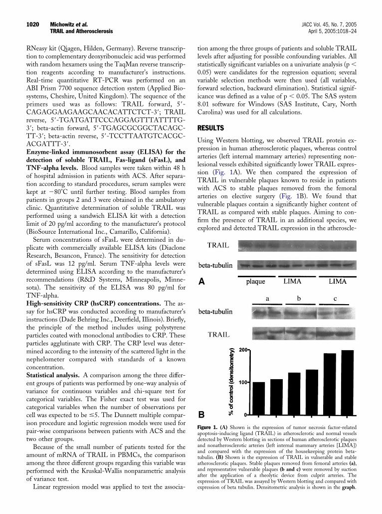
RtwtRAspCr3TAEdTotkpcpl(
pRodrsTHsitppmncSevcccipt
aapo
tls0vfi8C
R
UpalsTwavTfie
Fadaataaa
1020 Michowitz et al. JACC Vol. 45, No. 7, 2005TRAIL and Atherosclerosis April 5, 2005:1018–24
Neasy kit (Qiagen, Hilden, Germany). Reverse transcrip-ion to complementary deoxyribonucleic acid was performedith random hexamers using the TaqMan reverse transcrip-
ion reagents according to manufacturer’s instructions.eal-time quantitative RT-PCR was performed on anBI Prism 7700 sequence detection system (Applied Bio-
ystems, Cheshire, United Kingdom). The sequence of therimers used was as follows: TRAIL forward, 5=-AGAGGAAGAAGCAACACATTCTCT-3=; TRAIL
everse, 5=-TGATGATTCCCAGGAGTTTATTTTG-=; beta-actin forward, 5=-TGAGCGCGGCTACAGC-T-3=; beta-actin reverse, 5=-TCCTTAATGTCACGC-CGATTT-3=.nzyme-linked immunosorbent assay (ELISA) for theetection of soluble TRAIL, Fas-ligand (sFasL), andNF-alpha levels. Blood samples were taken within 48 hf hospital admission in patients with ACS. After separa-ion according to standard procedures, serum samples wereept at �80°C until further testing. Blood samples fromatients in groups 2 and 3 were obtained in the ambulatorylinic. Quantitative determination of soluble TRAIL waserformed using a sandwich ELISA kit with a detection
imit of 20 pg/ml according to the manufacturer’s protocolBioSource International Inc., Camarillo, California).
Serum concentrations of sFasL were determined in du-licate with commercially available ELISA kits (Diacloneesearch, Besancon, France). The sensitivity for detectionf sFasL was 12 pg/ml. Serum TNF-alpha levels wereetermined using ELISA according to the manufacturer’secommendations (R&D Systems, Minneapolis, Minne-ota). The sensitivity of the ELISA was 80 pg/ml forNF-alpha.igh-sensitivity CRP (hsCRP) concentrations. The as-
ay for hsCRP was conducted according to manufacturer’snstructions (Dade Behring Inc., Deerfield, Illinois). Briefly,he principle of the method includes using polystyrenearticles coated with monoclonal antibodies to CRP. Thesearticles agglutinate with CRP. The CRP level was deter-ined according to the intensity of the scattered light in theephelometer compared with standards of a knownoncentration.tatistical analysis. A comparison among the three differ-nt groups of patients was performed by one-way analysis ofariance for continuous variables and chi-square test forategorical variables. The Fisher exact test was used forategorical variables when the number of observations perell was expected to be �5. The Dunnett multiple compar-son procedure and logistic regression models were used forair-wise comparisons between patients with ACS and thewo other groups.
Because of the small number of patients tested for themount of mRNA of TRAIL in PBMCs, the comparisonmong the three different groups regarding this variable waserformed with the Kruskal-Wallis nonparametric analysisf variance test.
Linear regression model was applied to test the associa-ee
ion among the three groups of patients and soluble TRAILevels after adjusting for possible confounding variables. Alltatistically significant variables on a univariate analysis (p �.05) were candidates for the regression equation; severalariable selection methods were then used (all variables,orward selection, backward elimination). Statistical signif-cance was defined as a value of p � 0.05. The SAS system.01 software for Windows (SAS Institute, Cary, Northarolina) was used for all calculations.
ESULTS
sing Western blotting, we observed TRAIL protein ex-ression in human atherosclerotic plaques, whereas controlrteries (left internal mammary arteries) representing non-esional vessels exhibited significantly lower TRAIL expres-ion (Fig. 1A). We then compared the expression ofRAIL in vulnerable plaques known to reside in patientsith ACS to stable plaques removed from the femoral
rteries on elective surgery (Fig. 1B). We found thatulnerable plaques contain a significantly higher content ofRAIL as compared with stable plaques. Aiming to con-rm the presence of TRAIL in an additional species, wexplored and detected TRAIL expression in the atheroscle-
igure 1. (A) Shown is the expression of tumor necrosis factor-relatedpoptosis-inducing ligand (TRAIL) in atherosclerotic and normal vesselsetected by Western blotting in sections of human atherosclerotic plaquesnd nonatherosclerotic arteries (left internal mammary arteries [LIMA])nd compared with the expression of the housekeeping protein beta-ubulin. (B) Shown is the expression of TRAIL in vulnerable and stabletherosclerotic plaques. Stable plaques removed from femoral arteries (a),nd representative vulnerable plaques (b and c) were removed by suctionfter the application of a rheolytic device from culprit arteries. The
xpression of TRAIL was assayed by Western blotting and compared withxpression of beta tubulin. Densitometric analysis is shown in the graph.
rpb
Twes(waw
reor(Ia
itwtAa
saapanlp
lgacw9dmtwsNhwab
Fs( The ud
1021JACC Vol. 45, No. 7, 2005 Michowitz et al.April 5, 2005:1018–24 TRAIL and Atherosclerosis
otic aortas of 10-month-old apo-E–deficient mice as com-ared with control C57BL/6 littermates of similar geneticackground exhibiting no atherosclerosis (data not shown).
Next, we aimed to define the nature of cells expressingRAIL protein and to determine its possible colocalizationith oxLDL. Indeed, TRAIL was expressed in carotid ath-
rosclerotic plaques within CD3-positive cells, and its expres-ion was abundant in areas that stained positive for oxLDLFig. 2). The principal regions in which TRAIL-positive cellsere evident were the interface regions between the lipid core
nd the media and within the shoulder regions of the plaqueshere lymphocytes are predominantly known to reside.Because oxLDL was found to colocalize with TRAIL, we
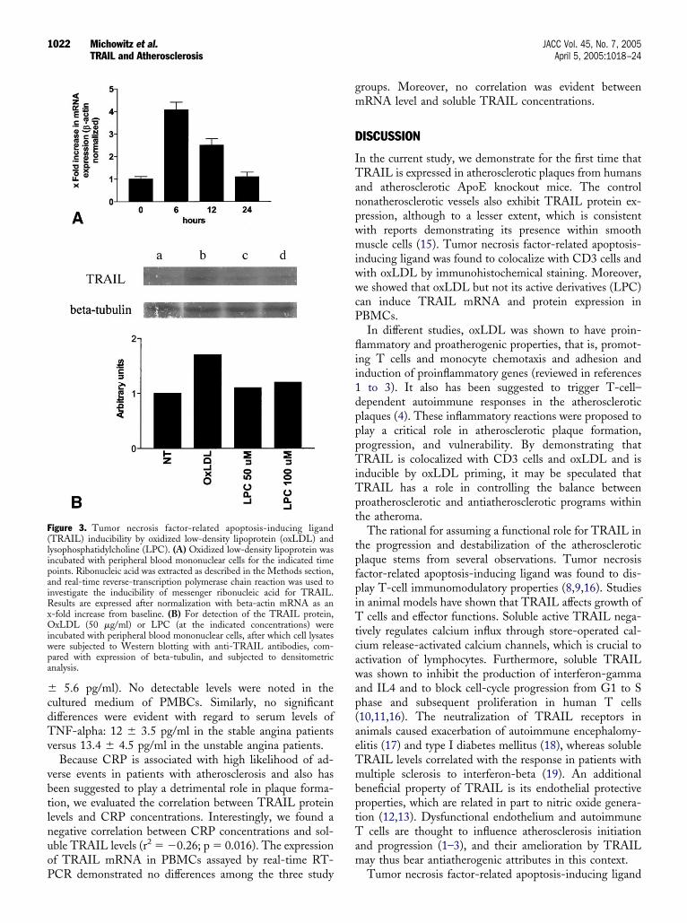
easoned that it could play a causative role by inducing thexpression of the latter. Indeed, incubation of PBMCs withxLDL but not with its active derivative, LPC, up-egulated expression of TRAIL messenger ribonucleic acidmRNA) (Fig. 3A) and protein (Fig. 3B) in these cells.nduction of TRAIL mRNA was evident and peakedlready 6 h after incubation with 50 �g/ml of oxLDL.
After having confirmed the presence of TRAIL and itsnducibility in atherosclerotic plaques, we went on to testhe functional significance of this protein. For this purpose,e assayed serum levels of soluble TRAIL in the sera of
hree groups of patients. Group 1 consisted of patients withCS (n � 40), group 2 consisted of patients with stable
igure 2. The presence and immunolocalization of tumor necrosis factorections from human atherosclerotic plaque where TRAIL (lower right pupper left panel) represent the corresponding primary antibody used.iscriminates the fibrous cap from the lipid core.
therosclerotic disease (n � 28), and group 3 consisted of s
ubjects with angiographically confirmed normal coronaryrteries (n � 20). To exclude a confounding effect oftherosclerotic burden on TRAIL levels, we matched theatients with ACS and those with stable angina for coronaryrtery affliction. This resulted in two groups with a similarumber of afflicted atherosclerotic coronary arteries. Base-
ine clinical characteristics of the three subject groups areresented in Table 1.We found that levels of soluble TRAIL were significantly
ower in patients with ACS as compared with both otherroups of patients (p � 0.0001, compared with stabletherosclerosis patients; mean difference, 344 pg/ml, 95%onfidence interval 191.8 to 496.4, and when comparedith subjects with NCA; mean difference, 398.5 pg/ml,5% confidence interval 224.2 to 567.7) (Fig. 4A). Thisifference remained statistically significant, even after usingultiple linear regression analysis and including all variables
hat were significant on univariate analysis. No differenceas found in soluble TRAIL levels between patients with
table atherosclerotic coronary disease and patients withCA (p � 0.56), although the mean level was slightly
igher in the latter group. In group 1 there were 24 patientsith acute myocardial infarction and 16 with unstable
ngina. No difference in soluble TRAIL levels existedetween these two subgroups (p � 0.51).Levels of sFasL did not differ between patients with
d apoptosis-inducing ligand (TRAIL) in human atheroma. Consecutive, oxidized low-density lipoprotein (oxLDL) (lower left panel), and CD3
pper right panel is a Masson’s trichrome stain of fibrotic tissues and
-relateanel)
table angina (34.2 � 7.3 pg/ml) and unstable angina (38.6
�cdTv
vbtlnuoP
gm
D
ITanpwmiwwcP
flii1dpppTiTpt
tpfpiTtcawap(aeTmbptTam
F(lipaiRxOiwpa
1022 Michowitz et al. JACC Vol. 45, No. 7, 2005TRAIL and Atherosclerosis April 5, 2005:1018–24
5.6 pg/ml). No detectable levels were noted in theultured medium of PMBCs. Similarly, no significantifferences were evident with regard to serum levels ofNF-alpha: 12 � 3.5 pg/ml in the stable angina patients
ersus 13.4 � 4.5 pg/ml in the unstable angina patients.Because CRP is associated with high likelihood of ad-
erse events in patients with atherosclerosis and also haseen suggested to play a detrimental role in plaque forma-ion, we evaluated the correlation between TRAIL proteinevels and CRP concentrations. Interestingly, we found aegative correlation between CRP concentrations and sol-ble TRAIL levels (r2 � �0.26; p � 0.016). The expressionf TRAIL mRNA in PBMCs assayed by real-time RT-
igure 3. Tumor necrosis factor-related apoptosis-inducing ligandTRAIL) inducibility by oxidized low-density lipoprotein (oxLDL) andysophosphatidylcholine (LPC). (A) Oxidized low-density lipoprotein wasncubated with peripheral blood mononuclear cells for the indicated timeoints. Ribonucleic acid was extracted as described in the Methods section,nd real-time reverse-transcription polymerase chain reaction was used tonvestigate the inducibility of messenger ribonucleic acid for TRAIL.esults are expressed after normalization with beta-actin mRNA as an
-fold increase from baseline. (B) For detection of the TRAIL protein,xLDL (50 �g/ml) or LPC (at the indicated concentrations) were
ncubated with peripheral blood mononuclear cells, after which cell lysatesere subjected to Western blotting with anti-TRAIL antibodies, com-ared with expression of beta-tubulin, and subjected to densitometricnalysis.
CR demonstrated no differences among the three study
roups. Moreover, no correlation was evident betweenRNA level and soluble TRAIL concentrations.
ISCUSSION
n the current study, we demonstrate for the first time thatRAIL is expressed in atherosclerotic plaques from humans
nd atherosclerotic ApoE knockout mice. The controlonatherosclerotic vessels also exhibit TRAIL protein ex-ression, although to a lesser extent, which is consistentith reports demonstrating its presence within smoothuscle cells (15). Tumor necrosis factor-related apoptosis-
nducing ligand was found to colocalize with CD3 cells andith oxLDL by immunohistochemical staining. Moreover,e showed that oxLDL but not its active derivatives (LPC)
an induce TRAIL mRNA and protein expression inBMCs.In different studies, oxLDL was shown to have proin-
ammatory and proatherogenic properties, that is, promot-ng T cells and monocyte chemotaxis and adhesion andnduction of proinflammatory genes (reviewed in references
to 3). It also has been suggested to trigger T-cell–ependent autoimmune responses in the atheroscleroticlaques (4). These inflammatory reactions were proposed tolay a critical role in atherosclerotic plaque formation,rogression, and vulnerability. By demonstrating thatRAIL is colocalized with CD3 cells and oxLDL and is
nducible by oxLDL priming, it may be speculated thatRAIL has a role in controlling the balance betweenroatherosclerotic and antiatherosclerotic programs withinhe atheroma.
The rational for assuming a functional role for TRAIL inhe progression and destabilization of the atheroscleroticlaque stems from several observations. Tumor necrosisactor-related apoptosis-inducing ligand was found to dis-lay T-cell immunomodulatory properties (8,9,16). Studiesn animal models have shown that TRAIL affects growth of
cells and effector functions. Soluble active TRAIL nega-ively regulates calcium influx through store-operated cal-ium release-activated calcium channels, which is crucial toctivation of lymphocytes. Furthermore, soluble TRAILas shown to inhibit the production of interferon-gamma
nd IL4 and to block cell-cycle progression from G1 to Shase and subsequent proliferation in human T cells10,11,16). The neutralization of TRAIL receptors innimals caused exacerbation of autoimmune encephalomy-litis (17) and type I diabetes mellitus (18), whereas solubleRAIL levels correlated with the response in patients withultiple sclerosis to interferon-beta (19). An additional
eneficial property of TRAIL is its endothelial protectiveroperties, which are related in part to nitric oxide genera-ion (12,13). Dysfunctional endothelium and autoimmune
cells are thought to influence atherosclerosis initiationnd progression (1–3), and their amelioration by TRAILay thus bear antiatherogenic attributes in this context.
Tumor necrosis factor-related apoptosis-inducing ligand
itetdadisntPstTppswc
am
iglpptWroiutenua
C �sient i
1023JACC Vol. 45, No. 7, 2005 Michowitz et al.April 5, 2005:1018–24 TRAIL and Atherosclerosis
s a transmembrane protein with an extracellular, carboxy-erminal domain. Soluble TRAIL is generated through thenzymatic cleavage of this extracellular domain. We foundhat soluble TRAIL levels in patients with ACS arerastically reduced as compared with patients with stabletherosclerotic disease and individuals with normal coronaryisease. Interestingly, other members of the TNF superfam-
ly that were assayed (TNF-alpha and Fas-Ligand) in theerum of the patients with stable and unstable angina didot differ between the groups, suggesting the results ob-ained for soluble TRAIL were not of a “class effect.”atients with ACS are a subgroup of patients with athero-clerosis that are thought to display vulnerable plaques andhus suffer clinical sequels due to vessel obstruction (5,6).he significantly lower TRAIL levels in this group ofatients suggest a possible protective stabilizing role for thisrotein. Although this observation is speculative, it isomewhat supported by the finding that TRAIL expressionas more robust in plaques with a vulnerable phenotype as
Table 1. Clinical Data of the Three Different
Acute CoronarySyndrome(n � 40)
Men/women 34/6Age (yrs) 61.2 � 14.8LVEF (%) 49.6 � 10.8NYHA-FC 4CAD extent (n � vessels) 1.59 � 0.8Past history
Previous MI, n (%) 14 (35)Previous CABG, n (%) 3 (7.5)Previous PTCA, n (%) 15 (37.5)Previous TIA/CVA, n(%) 1 (2.5)CRF† 6 (15)
Risk factorsHypertension, n (%) 19 (47.5)Diabetes, n (%) 8 (20)SmokingCurrent smoker, n (%) 21 (52.5)Past smoker, n (%) 10 (25)Hyperlipidemia, n (%) 27 (67.5)
MedicationsBeta-blockers, n (%) 18 (45)ACEI, n (%) 10 (25)ARB, n (%) 1 (2.5)Aspirin, n (%) 25 (62.5)Clopidogrel, n (%) 4 (10)Statins, n (%) 20 (50)Calcium blocker, n (%) 4 (10)Nitrates, n (%) 11 (27.5)Diuretics, n (%) 5 (12.5)Hypoglycemics, n (%) 2 (5)
BiochemistrySoluble-TRAIL, pg/ml 238.6 � 199.3CRP 12.2 � 16.8
Data are in number (range) or mean � SD. *Comparison onlpatients. †Defined as baseline creatinine above 1.5 mg/dl. ‡Ca
ACEI � angiotensin-converting enzyme inhibitors; ARBgraft; CAD � coronary artery disease; CRF � chronic renalejection function; MI � myocardial infarction; NYHA-Fpercutaneous transluminal coronary angioplasty; TIA � tran
ompared with stable lesions. This finding may be related to c
consumption effect of TRAIL into the lesions that areore likely to be infiltrated by immune cells.Interestingly, we found that TRAIL mRNA expression
n PBMCs was not different in the patients from the threeroups and that no significant correlation to soluble TRAILevels existed. These findings could suggest that the TRAILrotein may not be reduced in ACS because of diminishedroduction by circulatory T cells but rather consumed intohe plaque, as suggested after the results obtained with the
estern blotting assays. Recently, Nakajima et al. (20)eported that the expression of TRAIL by flow cytometryn peripheral lymphocytes in patients with acute myocardialnfarction was increased when compared with healthy vol-nteers. Unlike our study, TRAIL levels were evaluated inhe PMBCs of patients with established acute ST-segmentlevation myocardial infarction in which cardiomyocyteecrosis was evident, whereas in our study most patients hadnstable angina. Moreover, the control patients were healthynd, therefore, their atherosclerosis burden was not similar and
ps of Patients: Univariate Analysis
table Angina(n � 28)
Normal CoronaryArteries(n � 20) p Value
25/3 15/5 0.4‡4.1 � 10.7 55.6 � 9.2 0.073.5 � 8.6 59.4 � 2.3 0.001.46 � 0.63 1.15 � 0.48
2 � 0.9 0 (0) 0.12*‡
15 (53.5) 0 (0) 0.12*8 (28.5) 0 (0) 0.08*‡
20 (71.4) 0 (0) 0.006*2 (7) 1 (5) ‡4 (14.2) 1 (5) ‡
16 (57.1) 8 (40) 0.512 (42.8) 1 (5) 0.007
12 (42.8) 5 (25) 0.126 (21.4) 4 (20) 0.89
24 (85.7) 8 (40) 0.004
22 (78.5) 3 (15) �0.000114 (50) 5 (25) 0.071 (3.5) 0 (0) 0.7‡
22 (78.5) 9 (45) 0.0577 (25) 0 (0) 0.03‡
22 (78.5) 4 (20) 0.000311 (39.2) 2 (10) 0.00611 (39.2) 0 (0) 0.0078 (28.5) 2 (10) 0.15‡5 (17.8) 0 (0) 0.112‡
2.7 � 349.1 637 � 281.2 �0.00018.2 � 16.7 4.9 � 5.1 0.19
een acute coronary syndrome (ACS) and stable angina pectorisal variables that were analyzed by the Fisher exact test.
giotensin receptor blocker; CABG � coronary artery bypass; CVA � cerebrovascular accident; LVEF � left ventricularNew York Heart Association functional class; PTCA �schemic attack.
Grou
S
651
58
y betwtegoric� an
failure
ould have initially confounded the interpretation of the data.

Fce
aaSlpiCiaiTppvva
RpA
R
1
1
1
1
1
1
1
1
1
1
2
2
2
F((piop
1024 Michowitz et al. JACC Vol. 45, No. 7, 2005TRAIL and Atherosclerosis April 5, 2005:1018–24
or this purpose and to avoid the effect of plaque burden, wehose patients with ACS and stable lesions that had a similarxtent of coronary affliction by atherosclerosis.
C-reactive protein, a nonspecific “acute-phase” protein, isknown marker of acute vascular events (21) and is closely
ssociated with plaque instability and oxidative stress (22).oluble TRAIL levels were found to be negatively corre-
ated with hsCRP concentrations, supporting a possiblerotective role of TRAIL in atherosclerosis and plaquenstability.
onclusions. We demonstrated that TRAIL is expressedn atherosclerotic plaques and is colocalized with CD3 cellsnd oxLDL. We also demonstrated that TRAIL is inducedn PBMCs by oxLDL. In ACS, the levels of solubleRAIL are reduced and correlate negatively with theroinflammatory acute-phase reactant CRP, whereas ex-ression of TRAIL is significantly higher in plaques with aulnerable as compared with a stable phenotype. If furtheralidated, in vivo TRAIL could have a role as a protective
igure 4. Soluble tumor necrosis factor-related apoptosis-inducing ligandsTRAIL) protein concentrations and correlation with C-reactive protein.A) Sera from patients with acute coronary syndrome (ACS), stable anginaectoris, or NCA were assayed for sTRAIL levels using enzyme-linkedmmunosorbent assay as described in the Methods section. (B) Correlationf TRAIL serum levels with concentrations of high-sensitivity C-reactiverotein (hsCRP) (described in the Methods section) is shown.
gent in patients with ACS.
eprint requests and correspondence: Dr. Jacob George, De-artment of Cardiology, Tel Aviv Sourasky Medical Center, Telviv, Israel. E-mail: [email protected].
EFERENCES1. Glass CK, Witztum JL. Atherosclerosis. The road ahead. Cell 2001;104:
503–16.2. Libby P. Inflammation in atherosclerosis. Nature 2002;420:868–74.3. Hansson GK, Libby P, Schonbeck U, et al. Innate and adaptive immunity
in the pathogenesis of atherosclerosis. Circ Res 2002;91:281–91.4. Stemme S, Faber B, Holm J, et al. T lymphocytes from human
atherosclerotic plaques recognize oxidized low-density lipoprotein.Proc Natl Acad Sci USA 1995;92:3893–7.
5. Libby P. Molecular bases of the acute coronary syndromes. Circulation1995;91:2844–50.
6. Libby P. Current concepts of the pathogenesis of the acute coronarysyndromes. Circulation 2001;104:365–72.
7. Smith DA, Irving SD, Sheldon J, et al. Serum levels of the antiin-flammatory cytokine interleukin-10 are decreased in patients withunstable angina. Circulation 2001;104:746–9.
8. Ashkenazi A. Targeting death and decoy receptors of the tumour-necrosis factor superfamily. Nat Rev Cancer 2002;2:420–30.
9. Smyth MJ, Takeda K, Hayakawa Y, et al. Nature’s TRAIL—on apath to cancer immunotherapy. Immunity 2003;18:1–6.
0. Kayagaki N, Yamaguchi N, Nakayama M, et al. Type I interferons(IFNs) regulate tumor necrosis factor-related apoptosis-inducing li-gand (TRAIL) expression on human T cells: a novel mechanism forthe antitumor effects of type I IFNs. J Exp Med 1999;189:1451–60.
1. Lunemann JD, Waiczies S, Ehrlich S, et al. Death ligand TRAILinduces no apoptosis but inhibits activation of human (auto)antigen-specific T cells. J Immunol 2002;168:4881–8.
2. Secchiero P, Gonelli A, Carnevale E, et al. TRAIL promotes the survivaland proliferation of primary human vascular endothelial cells by activatingthe Akt and ERK pathways. Circulation 2003;107:2250–6.
3. Zauli G, Pandolfi A, Gonelli A, et al. Tumor necrosis factor-relatedapoptosis-inducing ligand (TRAIL) sequentially upregulates nitricoxide and prostanoid production in primary human endothelial cells.Circ Res 2003;92:732–40.
4. George J, Afek A, Gilburd B, et al. Hyperimmunization of apo-E-deficient mice with homologous malondialdehyde low-density li-poprotein suppresses early atherogenesis. Atherosclerosis 1998;138:147–52.
5. Gochuico BR, Zhang J, Ma BY, et al. TRAIL expression in vascularsmooth muscle. Am J Physiol 2000;278:L1045–50.
6. Song K, Chen Y, Goke R, et al. Tumor necrosis factor-relatedapoptosis-inducing ligand (TRAIL) is an inhibitor of autoimmuneinflammation and cell cycle progression. J Exp Med 2000;191:1095–104.
7. Hilliard B, Wilmen A, Seidel C, et al. Roles of TNF-relatedapoptosis-inducing ligand in experimental autoimmune encephalomy-elitis. J Immunol 2001;166:1314–9.
8. Mi QS, Ly D, Lamhamedi-Cherradi SE, et al. Blockade of tumornecrosis factor-related apoptosis-inducing ligand exacerbates type 1diabetes in NOD mice. Diabetes 2003;52:1967–75.
9. Wandinger KP, Lunemann JD, Wengert O, et al. TNF-related apoptosisinducing ligand (TRAIL) as a potential response marker for interferon-beta treatment in multiple sclerosis. Lancet 2003;361:2036–43.
0. Nakajima H, Yanase N, Oshima K, et al. Enhanced expression of theapoptosis inducing ligand TRAIL in mononuclear cells after myocar-dial infarction. Jpn Heart J 2003;44:833–44.
1. Blake GJ, Ridker PM. C-reactive protein and other inflammatory riskmarkers in acute coronary syndromes. J Am Coll Cardiol 2003;41:37S–42S.
2. Kobayashi S, Inoue N, Ohashi Y, et al. Interaction of oxidative stressand inflammatory response in coronary plaque instability: importantrole of C-reactive protein. Arterioscler Thromb Vasc Biol 2003;23:
1398–404.