The influence of protective coatings on the magnetic properties of acicular iron nanoparticles
7
INSTITUTE OF PHYSICS PUBLISHING NANOTECHNOLOGY Nanotechnology 17 (2006) 1421–1427 doi:10.1088/0957-4484/17/5/043 The influence of protective coatings on the magnetic properties of acicular iron nanoparticles Ra´ ul Pozas 1 , Manuel Oca ˜ na 1,3 , M Puerto Morales 2 and Carlos J Serna 2 1 Instituto de Ciencia de Materiales de Sevilla (CSIC-UNSE), Americo Vespucio s/n, Isla de La Cartuja, 41092 Sevilla, Spain 2 Instituto de Ciencia de Materiales de Madrid (CSIC), Cantoblanco, 28049 Madrid, Spain E-mail: [email protected] Received 4 November 2005, in final form 13 December 2005 Published 16 February 2006 Online at stacks.iop.org/Nano/17/1421 Abstract Core–shell acicular nanoparticles of ∼90 nm length and ∼5 axial ratio consisting of an iron core coated with an oxidized layer of different composition have been obtained by thermal reduction with hydrogen of coated goethite precursors. Uniform goethite particles were prepared by oxidation in air of FeSO 4 solutions previously precipitated with Na 2 CO 3 . Al or Y oxide layers were deposited on the goethite particle surface by heterocoagulation. The efficiencies of both compounds as protecting agents for preventing particle sintering and corrosion and their effects on the magnetic properties of the final α-Fe nanoparticles have been evaluated. Yttrium oxide coating was found to be more effective than alumina coating, giving rise to iron nanoparticles with larger coercivity and higher corrosion resistance. This behaviour is mainly discussed in terms of the observed differences in location of the two sintering prevention agents during the transformation from iron oxide to metal, which give rise to different particle microstructures. 1. Introduction The growing interest in nanoparticle magnetism arises from a variety of potential applications, ranging from soft to hard magnetic material use and from ultrahigh density information storage to medicine [1]. As the particle size is reduced, the chemical and thermal stability of the magnetic nanoparticles become key issues for most of these applications. In the case of magnetic recording, the most advanced materials consist of acicular α-Fe or α-Fe–Co nanoparticles. They are usually prepared through a multi-step process involving precipitation and further reduction under hydrogen of acicular particles of an iron oxide or oxyhydroxide precursor, whose morphology has to be preserved during the transformation [2–4], for which careful control of every aspect of the synthesis route is required for optimizing the characteristics of the final product and its properties. An 3 Author to whom any correspondence should be addressed. additional difficulty is associated with the production of metal nanoparticles, related to the fact that large surfaces are easily oxidized or otherwise subject to corrosion. That is why most magnetic particles currently used in biomedicine are based on ferrimagnetic iron oxides with low magnetic moments [5], although metallic iron nanoparticles coated with iron oxide would have larger magnetic moments. To minimize corrosion, metallic nanoparticles have to be protected by coating with a surfactant or an inorganic material, resulting in core–coating interactions, which have been shown to influence the electronic, magnetic and spatial arrangement for Co nanoparticles, for example [6, 7]. Nevertheless, the shell is expected to enhance the stability of the core and the resulting core–shell particles to exhibit improved physical and chemical properties compared with the single-component counterparts. Over the last decade there have been immense efforts to fabricate core–shell colloidal materials with tailored properties, not only magnetic, but also structural, optical and surface [8]. 0957-4484/06/051421+07$30.00 © 2006 IOP Publishing Ltd Printed in the UK 1421
-
Upload
independent -
Category
Documents
-
view
0 -
download
0
Transcript of The influence of protective coatings on the magnetic properties of acicular iron nanoparticles
INSTITUTE OF PHYSICS PUBLISHING NANOTECHNOLOGY
Nanotechnology 17 (2006) 1421–1427 doi:10.1088/0957-4484/17/5/043
The influence of protective coatings on themagnetic properties of acicular ironnanoparticlesRaul Pozas1, Manuel Ocana1,3, M Puerto Morales2 andCarlos J Serna2
1 Instituto de Ciencia de Materiales de Sevilla (CSIC-UNSE), Americo Vespucio s/n,Isla de La Cartuja, 41092 Sevilla, Spain2 Instituto de Ciencia de Materiales de Madrid (CSIC), Cantoblanco, 28049 Madrid, Spain
E-mail: [email protected]
Received 4 November 2005, in final form 13 December 2005Published 16 February 2006Online at stacks.iop.org/Nano/17/1421
AbstractCore–shell acicular nanoparticles of ∼90 nm length and ∼5 axial ratioconsisting of an iron core coated with an oxidized layer of differentcomposition have been obtained by thermal reduction with hydrogen ofcoated goethite precursors. Uniform goethite particles were prepared byoxidation in air of FeSO4 solutions previously precipitated with Na2CO3.Al or Y oxide layers were deposited on the goethite particle surface byheterocoagulation. The efficiencies of both compounds as protecting agentsfor preventing particle sintering and corrosion and their effects on themagnetic properties of the final α-Fe nanoparticles have been evaluated.Yttrium oxide coating was found to be more effective than alumina coating,giving rise to iron nanoparticles with larger coercivity and higher corrosionresistance. This behaviour is mainly discussed in terms of the observeddifferences in location of the two sintering prevention agents during thetransformation from iron oxide to metal, which give rise to different particlemicrostructures.
1. Introduction
The growing interest in nanoparticle magnetism arises froma variety of potential applications, ranging from soft to hardmagnetic material use and from ultrahigh density informationstorage to medicine [1]. As the particle size is reduced, thechemical and thermal stability of the magnetic nanoparticlesbecome key issues for most of these applications.
In the case of magnetic recording, the most advancedmaterials consist of acicular α-Fe or α-Fe–Co nanoparticles.They are usually prepared through a multi-step processinvolving precipitation and further reduction under hydrogenof acicular particles of an iron oxide or oxyhydroxideprecursor, whose morphology has to be preserved duringthe transformation [2–4], for which careful control of everyaspect of the synthesis route is required for optimizing thecharacteristics of the final product and its properties. An
3 Author to whom any correspondence should be addressed.
additional difficulty is associated with the production of metalnanoparticles, related to the fact that large surfaces are easilyoxidized or otherwise subject to corrosion. That is why mostmagnetic particles currently used in biomedicine are basedon ferrimagnetic iron oxides with low magnetic moments [5],although metallic iron nanoparticles coated with iron oxidewould have larger magnetic moments.
To minimize corrosion, metallic nanoparticles have to beprotected by coating with a surfactant or an inorganic material,resulting in core–coating interactions, which have been shownto influence the electronic, magnetic and spatial arrangementfor Co nanoparticles, for example [6, 7]. Nevertheless, theshell is expected to enhance the stability of the core andthe resulting core–shell particles to exhibit improved physicaland chemical properties compared with the single-componentcounterparts. Over the last decade there have been immenseefforts to fabricate core–shell colloidal materials with tailoredproperties, not only magnetic, but also structural, optical andsurface [8].
0957-4484/06/051421+07$30.00 © 2006 IOP Publishing Ltd Printed in the UK 1421

R Pozas et al
Figure 1. TEM images obtained for the uncoated goethite (G) and the Fe(Al)5 and Fe(Y)5 metallic samples.
The aim of this paper is the synthesis of stable core–shell iron nanoparticles with acicular morphology by tailoringthe surface properties of acicular precursors. This will beaccomplished by coating the latter with a shell of two differentmaterials, aluminium and yttrium oxide, recognized by severalauthors as good sintering prevention agents in the synthesis ofmetal pigments [9–13], although, as already observed, yttriumoxide seems to be more effective than alumina [11, 13]. Thereason for this behaviour is still unclear; the study of theinfluence of the coating nature on the microstructure, particlemorphology, corrosion resistance and magnetic properties ofacicular iron nanoparticles will be the final goal of this paper.
2. Experimental section
2.1. Precursor preparation
Goethite particles can be obtained by precipitation withsodium carbonate from aqueous solutions of Fe(II) sulfate andsubsequent oxidation at controlled temperature by bubblingair through the dispersion at a constant flow rate. A detailedanalysis of the effects of the experimental parameters on thesize, shape and composition of the resulting particles as wellas their formation mechanism can be found in [14]. Inthis work, we have selected those experimental conditions(0.15 mol dm−3 Fe(II) sulfate, 0.225 mol dm−3 sodiumcarbonate, synthesis temperature 40 ◦C, reaction time 6 hand air flow rate 2 dm3 min−1) that produce uniform aciculargoethite nanoparticles with ∼120 nm mean length and axialratio ∼5 (figure 1). The so-prepared precursor will be hereaftercalled G.
These goethite nanoparticles were further coated with analuminium hydrous oxide or yttrium basic carbonate layer. Thecoating process involved first the dissolution of the desiredamount of Al(III) nitrate (Al(NO3)3·9H2O, Fluka, 99%) orY(III) nitrate (Y(NO3)3·6H2O, Fluka, 99%) in 150 cm3 ofdouble-distilled water and the addition of a 10% wt sodiumhydroxide (NaOH, Fluka, 98%) aqueous solution to reach abasic pH value (12.5). Then, the goethite particles (1 g dm−3)
were homogeneously suspended in the resulting medium usingan ultrasonic bath and, finally, the pH was lowered to 8.5 by theslow addition of a 0.5 mol dm−3 HNO3 (HNO3, Fluka, 65%)aqueous solution into the slurry while stirring. The pH regionswere selected to induce the process of heterocoagulation(electrostatically induced coagulation of particles having
opposite charge density) of the goethite particles and the Aland Y species precipitated [9, 13]. The samples synthesizedby this method are called G(Al)x and G(Y)y, ‘x’ and ‘y’being the added Al/(Fe + Al) and Y/(Fe + Y) atomic ratiosrespectively, expressed as percentages (from 3 to 15%).
All precipitates were centrifuged and washed several timeswith deionized water for purification. Finally, the powderedsolids were collected by filtration and dried at 50 ◦C beforeanalyses.
2.2. Thermal reduction
To obtain the final metal nanoparticles, the goethite samples(∼60 mg) were first dehydrated by heating in air for 4 hyielding haematite, which was then reduced at constanttemperature in a hydrogen (99.9999%) stream (flow rate20 dm3 h−1) for 4 h. Dehydroxylation and reductiontemperatures were optimized in each case to achieve a decreasein internal porosity of the particles while preserving theacicular morphology as well as to complete the reduction.After reduction, the samples were cooled to room temperatureunder the hydrogen atmosphere, and finally, passivated byexposing them to alcohol vapours for 1 h, which weregenerated by bubbling N2 gas (99.999%) at a constantflow rate (40 dm3 h−1) into a flask containing pure ethanol(99.8%). Metal samples prepared after reduction of G, G(Al)x
and G(Y)y powders are called Fe, Fe(Al)x and Fe(Y)y ,respectively.
Finally, the stability against corrosion of the metallicnanoparticles obtained was analysed through the change ofmagnetization saturation (Ms) observed for these sampleswhen they were exposed to an accelerated oxidation process(seven days at 60 ◦C and 90% relative humidity) [3].
2.3. Characterization techniques
Transmission electron microscopy (TEM, Philips 200 CM)was used to examine the morphology of the particles. Theparticle size distribution of the powders was evaluated fromthe electron micrographs by counting around one hundredparticles. From these data, the degree of polydispersity, definedas the standard deviation (SD)/mean size [15], was evaluated.The mean and the SD values associated with the axial ratio(L/W ) were determined from the L/W ratios obtained fromeach particle.
1422

The influence of protective coatings on the magnetic properties of acicular Fe nanoparticles
Phase identification was carried out by means of x-raydiffraction (XRD) in a Siemens D501 apparatus using Cu Kα
radiation and a diffracted beam graphite monochromator. Anestimation of the crystallite size was determined from the fullwidth at half-maximum of the XRD selected reflection byusing the Scherrer equation [16]. Unit cell parameters of thesamples were determined by a least-squares fit of the XRD datausing Si as the internal reference standard.
The quantitative composition of the samples in terms ofthe Al and Y contents was determined using x-ray fluorescence(XRF, Siemens SRS 3000). An energy dispersive x-ray (EDX,Philips DX4) analyser, installed in the TEM microscope, wasalso used to gain information on the particle composition. Thevariation of the aluminium and yttrium concentrations at theparticle outer layers during the transformation from goethiteto haematite, and finally to iron, was obtained from the XPSspectra, recorded in a VG Escalab 220 using the Mg Kα
excitation source. Calibration of the binding energy scale ofthe spectra was done at the C 1s peak of the surface carboncontamination taken at 284.6 eV. Atomic percentages of theelements were calculated from the peak areas after backgroundsubtraction (Shirley background). The areas were referredto the sensitivity factors of the elements as supplied by theinstrument manufacturers.
Particle surface characterization of the goethite sampleswas also carried out by measuring the electrophoretic mobilityof aqueous suspensions of the powders as a function of pHat 25 ◦C using a Malvern Zetamaster apparatus. For this,between 3 and 5 mg of sample were dispersed in 100 cm3
of a 0.01 mol dm−3 KNO3 solution to keep the ionic strengthconstant. HNO3 or KOH was then added to several 10 cm3
aliquots of these suspensions to adjust the pH to within therequired range and the resulting dispersions were finally agedat room temperature for 24 h before analyses.
The 57Fe Mossbauer absorption spectra were recordedwith a maximum velocity of 10 mm s−1 at room temperatureand 4.2 K with a 57Co:Rh source. By fitting the spectraat 4.2 K, the fraction of iron oxide on the surface of thenanoparticles was determined.
Magnetic characterization of the samples was carried outin a vibrating sample magnetometer (MLVSM9 MagLab 9 T,Oxford Instruments). Magnetization curves were recorded atroom temperature. First the sample was saturated in a field of3 T. Then, the saturation magnetization (Ms), the squareness(Mr/Ms, where Mr is the remanent magnetization) and thecoercivity (Hc) were determined for each sample. The Ms
values were evaluated by extrapolating to infinite field theexperimental results obtained in the high field range where themagnetization increase linearly with 1/H .
The activation volume for reversal (Vact), which describesthe volume of the magnetic material that reverses coherently,can be determined following Gaunt [17]:
Vact = kBT/[MsSmax/(χirr)max]
where (χirr)max is the maximum irreversible susceptibility (emucm−3 T−1), Smax is the maximum coefficient of magneticviscosity (emu cm−3), kB is Boltzmann’s constant (1.38 ×10⁻²³ J K−1), T is the temperature (K) and Ms is the saturationmagnetization (emu cm−3).
Table 1. Content in the protecting agent (M = Al or Y) for thecoated particles as determined using XRF and XPS.
M/(Fe + M) atomic ratio
Nominal XRF XPSSample (%) (%) (%)
G — — —G(Al)5 5 5.1 30G(Y)5 5 5.2 32
(χirr)max was obtained from the maximum value of theDC demagnetization remanence differential curve resultingfrom the saturation remanent state by the application andremoval of first a saturating field of 3 T, and then an increasingdemagnetizing field. Smax was obtained from the maximumvalue of the coefficient of magnetic viscosity (S) obtained asa function of the demagnetizing field applied. These S valueswere obtained from the measurement of the time dependenceof the magnetization around the coercive field (between 1000and 2000 Oe). The variation of the magnetization (M) withtime in the presence of a constant demagnetizing field followedthe form
M(t) = M(0) − S ln(t)
where S = (−dM/d[ln(t)]) is the slope of the resultingstraight line.
3. Results and discussion
3.1. Uncoated and coated goethite nanoparticles
Goethite particles were coated with increasing amountsof aluminium or yttrium compounds, from 3 to 15%(M/(M + Fe) atomic ratio; M = Al or Y), finding that, in bothcases, the maximum coating was achieved for an atomic ratioof 10%, since at higher values, a mixture of coated goethite andirregular particles of Al or Y compounds was detected by TEM.The amount of aluminium and yttrium in the coated goethitesamples was similar to that initially added as determined usingXRF (table 1). The presence of Al or Y in each goethite particlewas confirmed by EDX analysis (data not shown) whereasXPS spectroscopy showed that they were located at the particlesurface. Thus, as illustrated for the G(Al)5 and G(Y)5 samples,the M/(Fe + M) atomic ratio obtained from the intensity ofthe Fe 2p (Fe 2p3/2 and Fe 2p1/2) and Al 2p1/2 or Y 2p1/2 XPSpeaks was much higher (∼30%) than the value for the overallsolid (∼5%) (table 1), indicating an important enrichment ofaluminium or yttrium in the particle outer layers, as expectedfor coated particles.
Further information on the homogeneity of the depositedlayers could be obtained in the case of the alumina coatingfrom electrophoretic mobility measurements, as a function ofpH (figure 2). The isoelectric point (IEP) for the Al coatedpowders was higher (9.1) than that observed for uncoatedgoethite (7.6) and very close to that of an aluminium blank,synthesized in the absence of the goethite (9.0), which suggeststhat the goethite particles were completely coated. In the caseof Y coated goethite, no information could be obtained on thecoating homogeneity by this technique, since the IEP for theyttrium blank was very similar (7.9) to that of the uncoated
1423

R Pozas et al
Figure 2. Electrophoretic mobility measured as a function of pH forG, G(Al)5 and an aluminium hydrous oxide sample synthesizedunder the same conditions as the Al coated goethite in the absence oftemplates.
goethite (7.6). Finally, it must be mentioned that the IEPvalue for the aluminium and yttrium blanks suggested that theyconsist of aluminium hydrous oxide containing carbonates [18]and yttrium basic carbonate [19] respectively, which wasconfirmed by IR spectroscopy.
3.2. Formation of metallic nanoparticles
Before thermal reduction, the goethite precursors were heatedin air to dehydrate, leading to haematite (α-Fe2O3) particles,and to decrease the internal porosity of the latter, whichharms the magnetic properties of the final metallic particles,as has been widely reported [13]. Obviously, the heating mustbe restricted to temperatures in which the acicular shape ispreserved. It should be noted that whereas in a previous workwe have observed that for Co-doped goethite, the maximumtemperature fulfilling such a requirement was much higherfor the yttrium protected particles (600 ◦C) than that thosecoated with alumina (400 ◦C) [13], in our case, the maximumtemperature was 500 ◦C, irrespective of the nature of thecoating. After this heat treatment, the hydrated aluminiumoxide and yttrium basic carbonate layers are expected totransform into alumina and yttria, respectively, as suggestedby XRD for the Al and Y blanks.
Haematite samples were then first reduced under H2 gasat the minimum temperature required for completing reduction(400 ◦C) to minimize particle sintering, and finally passivatedto obtain the final metallic nanoparticles. XRD revealed similarpatterns for the two reduced samples, which correspond to abody-centred-cubic structure assigned to α-Fe (figure 3). Thepresence of the iron oxide passivation layer in the sampleswas confirmed by the positions observed for the Fe 2p3/2
(710.3 eV) and Fe 2p1/2 (724.0 eV) XPS peaks which, asillustrated for the Fe(Y)5 sample (figure 4), were consistentwith the presence of oxidized iron [20]. Such an oxide phasecould not be detected by means of XRD due to a low oxidecontent and/or to the fact that it normally appears in the formof nanocrystals about 2 nm in size (with a grain size lowerthan ∼2 nm, diffraction effects are diffuse and close to thebackground noise).
30 40 50 60
2θ
Fe(Y)5
Fe(Al)5
α-Fe(200)
α-Fe(110)
70
Figure 3. X-ray diffraction patterns obtained for the Fe(Y)5 andFe(Al)5 metallic samples resulting from the thermal dehydroxylationand reduction of the goethite precursors.
Figure 4. Fe 2p XPS spectrum for the Fe(Y)5 sample.
3.3. Magnetic properties of metallic samples
The magnetic parameters obtained from the hysteresis loopsrecorded for the metallic samples (figure 5) are collected intable 2. As observed, in the case of the alumina protectedparticles, the values of Hc and Mr/Ms increased from 940 to1050 Oe and from 0.42 to 0.47, respectively, as the aluminiumcontent increased from 3 to 5%. These data can be explainedby the higher Fe crystal size (34 nm) of the Fe(Al)3 sample ascompared with the Fe(Al)5 sample (25 nm), indicating that a3% Al/(Al + Fe) atomic ratio is not sufficient for completelyavoiding sintering during the metallic particle preparation.
1424

The influence of protective coatings on the magnetic properties of acicular Fe nanoparticles
Table 2. Magnetic properties (Ms: saturation magnetization, Hc: coercivity, Mr: remanent magnetization, Mr/Ms: squareness, Vact: activationvolume, Vf: physical particle volume considering ellipsoidal shape), crystal size (DXRD) and iron oxide content (Feox) for the metallicnanoparticles obtained from differently coated precursors (M = Al or Y).
M/(Fe + M) Hc Ms DXRD Feox Vact Vf
Sample (at.%) (Oe) (emu g−1) Mr/Ms (nm) (%)at (×1018 cm3) (×1018 cm3) Vact/Vf
Fe(Al)3 3 940 130 0.42 34 — — — —Fe(Al)5 5 1050 122 0.47 25 35 2.4 6.6 0.36Fe(Al)10 10 1040 108 0.47 25 — — — —
Fe(Y)3 3 1200 132 0.43 36 — — — —Fe(Y)5 5 1300 118 0.48 27 37 3.7 6.6 0.56Fe(Y)10 10 1290 103 0.48 26 — — — —
Figure 5. Magnetization curves obtained at room temperature for theFe(Al)5 and Fe(Y)5 metallic samples.
A further increase of the aluminium content to 10% resultedin a decrease of the Ms value, which is as expected due tothe addition of a higher amount of the diamagnetic aluminaphase. However, the values of Hc and Mr/Ms did not changefor the Fe(Al)10 sample with respect to the Fe(Al)5 sample,in agreement with the lack of crystal size variation observedfor both samples (table 2). Therefore, it is concluded that thealuminium content required to obtain metal particles with thebest magnetic properties is about 5%. A similar behaviourwas observed in the case of yttria protection, i.e. the optimumyttrium content was also about 5%.
On comparing the magnetic properties of the twooptimum samples, Fe(Al)5 and Fe(Y)5 (table 2), similar Ms
(∼120 emu g−1) and Mr/Ms (∼0.5) values were observed.However, the Hc value for the Fe(Y)5 sample was clearlyhigher (1300 Oe) than that for the Fe(Al)5 sample (1050 Oe)confirming the higher efficiency of yttria coating, as previouslyreported for Fe–Co nanoparticles [13]. It should be noted thanin the latter case, the higher efficiency of yttria was ascribedto the lower degree of porosity present in the yttria protectedsample when compared with that protected with alumina,which resulted from the higher dehydration temperature usedfor the former. Such an explanation should be disregardedin our case since the two samples were dehydroxylated atthe same temperature (500 ◦C) and, therefore, they wouldbe expected to show similar microstructures. In agreement,the particle morphology (figure 1) and crystal size (table 2)obtained for the two samples were similar, which indicates thatthese factors are not responsible either for the difference in Hc.
-10 -5 0
95
95
100
100
T = 4.2 K
T = 298 K
Velocity (mm/s)
Tra
nsm
issi
on
5 10
Figure 6. Mossbauer spectra for the Fe(Y)5 sample registered at 4.2and 298 K.
To investigate the possible effect of the coating nature onthe coercivity of the metallic nanoparticles, the composition,structure and magnetic behaviour of the passivation layerspresent in the two samples were analysed by using Mossbauerspectroscopy. The spectrum obtained at 4.2 K for the Fe(Y)5
sample consisted of two sextets (figure 6), one correspondingto the metallic α-Fe core with a hyperfine field of 34 T andanother with a larger hyperfine field around 50 T and relativelybroad lines, characteristic of a thin iron oxide layer with aspinel structure [21, 22]. An accurate measure of the relativeproportion of iron atoms in the iron oxide passivation layerand in the metallic core was obtained from the relative spectralarea of the two sextets, resulting in 37% iron oxide for thissample (table 2). When the Mossbauer spectrum associatedwith this iron oxide was measured at room temperature abroad unresolved line and a broad quadrupolar doublet wereobserved (figure 6), which are typical of iron oxide spinelwith a superparamagnetic behaviour [21–24]. Therefore, thispassivation layer does not contribute to the coercivity of thesample at room temperature [9]. The Mossbauer spectrafor the Fe(Al)5 sample revealed an iron oxide percentagesimilar (35%) to that of the yttria coated sample with a similarmagnetic behaviour. Hence, the nature of the iron oxide spinellayer is not responsible for the different Hc values measuredfor the Fe(Al)5 and Fe(Y)5 samples.
To gain information on the origin of the different Hc
values observed for the iron samples coated with alumina
1425
R Pozas et al
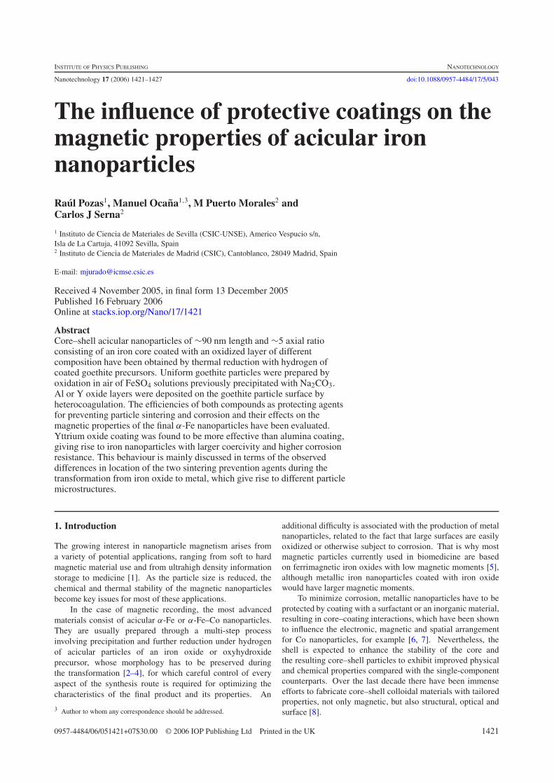
Figure 7. Remanent magnetization (Mr) (down) and magneticviscosity (S) (up) obtained as a function of the demagnetizingexternal field applied (H ) for the Fe(Y)5 and Fe(Al)5 samples.
or yttria, the activation volumes (Vact) were calculated fromthe remanence curves and magnetic viscosity measurements(figure 7) for both samples, as described in the experimentalsection. It must be taken into account that Vact is expectedto be close to the physical volume (Vf) for particles wherereversal magnetization follows a coherent rotation mechanism(higher coercivity values), whereas Vact should be lower thanVf when magnetization reversal is incoherent or heterogeneous(lower coercivity values) [25–27]. As observed in table 2, theactivation volumes for both samples, Fe(Al)5 and Fe(Y)5, weresmaller than the core volumes of the particles estimated fromthe TEM micrographs. Nevertheless, the value of Vact for theFe(Y)5 sample (3.7 × 10⁻¹⁸ cm3) was higher than that forthe Fe(Al)5 sample (2.4 × 10⁻¹⁸ cm3) (table 2). Since thevalues of Vf were similar for the two samples (table 2), it isconcluded that magnetization in the particles protected withyttrium oxide switches by a process somewhat more coherent(Vact/Vf = 0.56) than that undergone by particles protectedwith aluminium oxide (Va/Vf = 0.36; table 2), which would bein agreement with the higher coercivity value of the former. Itshould be noted that switching volumes similar to the physicalsize have only been found in iron oxide particles with a goodstructural order, whereas particles with shape and structureirregularities, and particles modified by addition of dopingions present a smaller activation volume because of the lackof order, which makes the occurrence of an incoherent orheterogeneous reversal more probable [28–30].
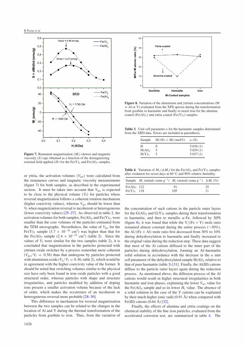
This difference in mechanism for reversal magnetizationbetween the two samples can be related to the changes in thelocation of Al and Y during the thermal transformation of theparticles from goethite to iron. Thus, from the variation of
Figure 8. Variation of the aluminium and yttrium concentrations (M= Al or Y) evaluated from the XPS spectra during the transformationfrom goethite to haematite and finally to metal iron for the aluminacoated (Fe(Al)5) and yttria coated (Fe(Y)5) samples.
Table 3. Unit cell parameter a for the haematite samples determinedfrom the XRD data. Errors are included in parenthesis.
Sample M/(Fe + M) (mol%) a (A)
H 0 5.036 (1)H(Al)5 5 5.029 (1)H(Y)5 5 5.037 (1)
Table 4. Variation of Ms (�Ms) for the Fe(Al)5 and Fe(Y)5 samplesafter oxidation for seven days at 60 ◦C and 90% relative humidity.
Sample Ms (initial) (emu g−1) Ms (stored) (emu g−1) �Ms (%)
Fe(Al)5 122 91 25Fe(Y)5 118 105 11
the concentration of such cations in the particle outer layersfor the G(Al)5 and G(Y)5 samples during their transformationto haematite, and then to metallic α-Fe, followed by XPS(figure 8), it was found that while the Y/(Fe + Y) mole ratioremained almost constant during the entire process (∼30%),the Al/(Fe + Al) mole ratio first decreased from 30% to 10%during dehydroxylation to haematite and finally increased tothe original value during the reduction step. These data suggestthat most of the Al cations diffused to the inner part of theparticles during dehydroxylation, forming an Al–haematitesolid solution in accordance with the decrease in the a unitcell parameter of the dehydroxylated sample H(Al)5 relative tothat of pure haematite (table 3) [31]. Finally, the Al(III) cationsdiffuse to the particle outer layers again during the reductionprocess. As mentioned above, the diffusion process of the Alcations would result in higher structural irregularities in bothhaematite and iron phases, explaining the lower Vact value forthe Fe(Al)5 sample and so its lower Hc value. The absence ofa solid solution in the case of the Y cations can be explainedby their much higher ionic radii (0.91 A) when compared withFe(III) cations (0.64 A) [32].
Finally, the effects of alumina and yttria coatings on thechemical stability of the fine iron particles, evaluated from theaccelerated corrosion test, are summarized in table 4. The
1426
The influence of protective coatings on the magnetic properties of acicular Fe nanoparticles
variation of the Ms value observed after this treatment (�Ms)for the Fe(Al)5 sample was clearly higher (25%) than that forthe Fe(Y)5 sample (11%), showing that the protection of thenanoparticles achieved by coating with yttrium oxide is moreeffective not only for obtaining better magnetic properties, butalso for minimizing the corrosion of the iron nanoparticles.This beneficial effect of yttria in preventing oxidation has beenpreviously recognized for pure iron plates [33].
4. Conclusions
The optimum amount of aluminium or yttrium cations thatmust be added to a goethite precursor by coating to obtain thebest magnetic properties for final iron nanoparticles is about5% (atomic). Nevertheless, the yttrium oxide coating is moreeffective not only for obtaining the largest coercivity, but alsofor minimizing corrosion of the iron metallic nanoparticles.This different behaviour can be attributed to the fact thatmagnetization in particles protected with alumina switchesby a process somewhat less coherent than that undergone byparticles protected with yttria, which is due to the presence ofmore structural irregularities in the former as a consequence ofthe diffusion of Al cations inside the iron oxide phases duringthe different steps of the α-Fe synthesis.
Acknowledgments
This work was supported by the Spanish CICYT under projectsMAT2002-04001-C02 and MAT2003-01479. The fellowshipof RP from the Spanish Ministerio de Ciencia y Tecnologıais gratefully acknowledged. We thank Dr P Bonville forhis assistance in measuring and interpreting the Mossbauerspectra.
References
[1] Kodama R H 1999 J. Magn. Magn. Mater. 200 359[2] Veitch R J, Ilmer A, Lenz W and Ritchter V 1999 J. Magn.
Magn. Mater. 193 279[3] Sugita N, Maekawa M, Ohta Y, Okinawa K and Magai N 1995
IEEE Trans. Magn. 31 2854[4] Onodera S, Kondo H and Kawana T 1996 MRS Bull. 21
(September) 35
[5] Tartaj P, Morales M P, Veintemillas-Verdaguer S,Gonzalez-Carreno T and Serna C J 2003 J. Phys. D: Appl.Phys. 36 182
[6] Hormes J, Modrow H, Bonnemann H and Kumar C S S R 2005J. Appl. Phys. 3 97 102
[7] Skumryev V, Stoyanov S, Zhang Y, Hadjipanayis G,Givord D and Nogues J 2003 Nature 19 850
[8] Caruso F 2001 Adv. Mater. 13 11[9] Nunez N O, Tartaj P, Morales M P, Pozas R, Ocana M and
Serna C J 2003 Chem. Mater. 15 3558[10] Pozas R, Ocana M, Morales M P, Tartaj P, Nunez N O and
Serna C J 2004 Nanotechnology 15 S190[11] Nunez N O, Tartaj P, Morales M P, Gonzalez-Carreno T and
Serna C J 2006 Acta Mater. 54 219[12] Lin C H, Chin T S, Kuo P C, Chem S C and Shih C S 1996
Mater. Chem. Phys. 44 90[13] Nunez N O, Tartaj P, Morales M P, Bonville P and Serna C J
2004 Chem. Mater. 16 3119[14] Pozas R, Ocana M, Morales M P and Serna C J 2002 J. Colloid
Interface Sci. 254 87[15] Hunter R 1987 Foundations of Colloid Science (Oxford:
Clarendon) p 127[16] Azaroff L V 1968 Elements of X-ray Crystallography
(New York: McGraw-Hill) p 549[17] Gaunt P 1986 J. Appl. Phys. 59 4129[18] Yue-Hua G and Yi Z 1997 Mater. Chem. Phys. 47 211[19] Plaza R C, Duran J D G, Quirantes A, Ariza M J and
Delgado A V 1997 J. Colloid Interface Sci. 194 398[20] Tetsukawa T, Inoue M and Kondo H 2004 J. Magn. Magn.
Mater. 269 423[21] Parker F T, Spada F E, Cox T J and Berkowitz A E 1995
J. Appl. Phys. 77 5853[22] Rojas T C, Sanchez-Lopez J C, Greneche J M, Conde A and
Fernandez A 2004 J. Mater. Sci. 39 4877[23] Haneda K and Morıs A H 1978 Surf. Sci. 77 584[24] Gangopadhyay S, Hadjipanayis G C, Shah S I, Sorensen C M,
Klabunde K J, Papaefthymlou V and Kostikas A 1991J. Appl. Phys. 70 5888
[25] Sharrock M P 1999 IEEE Trans. Magn. 35 4414[26] Stachen M, Morales M P, Ocana M and Serna C J 1999 Phys.
Chem. Chem. Phys. 1 4465[27] Morales M P, El-Hilo M and O’Grady K 1995 J. Magn. Magn.
Mater. 140 2211[28] Bottoni G 1993 J. Magn. Magn. Mater. 3 120[29] Bottoni G 1999 J. Magn. Magn. Mater. 196 602[30] Mendoza-Resendez R, Pozas R, Morales M P, Bonville P,
Ocana M and Serna C J 2005 Nanotechnology 16 1[31] Stanjek H and Schwertmann U 1992 Clays Clay Miner. 40 347[32] Shannon R D 1976 Acta Crystallogr. A 32 751[33] Josse-Courty C, Buscail H, Stroosnijder M F, Dufour P and
Larpin J P 1999 Oxid. Met. 52 321
1427