The influence of organic solvents on estimates of genotoxicity and antigenotoxicity in the SOS...
12
The influence of organic solvents on estimates of genotoxicity and antigenotoxicity in the SOS chromotest Nathalia Quintero 1 , Elena E. Stashenko 2 and Jorge Luis Fuentes 1,2 1 Laboratorio de Microbiología y Mutagénesis Ambiental, Escuela de Biología, Facultad de Ciencias, Universidad Industrial de Santander, Bucaramanga, Colombia. 2 Centro de Investigación en Biomoléculas, Centro de Investigación de Excelencia, Universidad Industrial de Santander, Bucaramanga, Colombia. Abstract In this work, the toxicity and genotoxicity of organic solvents (acetone, carbon tetrachloride, dichloromethane, dimethylsulfoxide, ethanol, ether and methanol) were studied using the SOS chromotest. The influence of these sol- vents on the direct genotoxicity induced by the mutagens mitomycin C (MMC) and 4-nitroquinoline-1-oxide (4-NQO) were also investigated. None of the solvents were genotoxic in Escherichia coli PQ37. However, based on the inhibi- tion of protein synthesis assessed by constitutive alkaline phosphatase activity, some solvents (carbon tetrachloride, dimethylsulfoxide, ethanol and ether) were toxic and incompatible with the SOS chromotest. Solvents that were nei- ther toxic nor genotoxic to E. coli (acetone, dichloromethane and methanol) significantly reduced the genotoxicity of MMC and 4-NQO. When these solvents were used to dissolve vitamin E they increased the antigenotoxic activity of this compound, possibly through additive or synergistic effects. The relevance of these results is discussed in rela- tion to antigenotoxic studies. These data indicate the need for careful selection of an appropriate diluent for the SOS chromotest since some solvents can modulate genotoxicity and antigenotoxicity. Key words: antigenotoxicity, genotoxicity, interference, mitomycin C, 4-nitro-quinoline-1-oxide, SOS chromotest, solvents, vitamin E. Received: November 19, 2011; Accepted: February 21, 2012. Introduction Antimutagens are agents that reduce the number of spontaneous and/or induced mutations in cells. These agents are classified based on their mechanism of action (Wattenberg, 1985). A practical classification is one that separates antimutagens into des-mutagens and bio-antimu- tagens (Kada and Shimoi, 1987), with des-mutagens being compounds that inactivate mutagens before they can attack the DNA molecule whereas bio-antimutagens interfere with cellular processes that repair DNA damage, e.g., DNA replication and/or repair mechanisms (Kuroda and Inoue, 1988). Antimutagens may help to prevent mutations in- volved in cancer and aging by regulating cellular mutabil- ity. Consequently, antimutagens can be classified into sev- eral categories and subcategories, depending on the stage in which they interfere with mutagenesis and carcinogenesis and on how they modulate host defense mechanisms; these points of interference allow the rational implementation of chemoprevention strategies for controlling cancer and de- generative diseases (De Flora, 1998; De Flora and Ramel, 1988; De Flora and Ferguson, 2005). The assessment of natural antimutagens has involved mainly the use of short-term bacterial assays because their sensitivity, simplicity and flexibility to different experi- mental settings facilitates the study of antimutagenesis mechanisms (De Flora et al., 1992a,b). Among the in vitro assays used in antimutagen studies, the SOS chromotest (Quillardet et al., 1982) is one of the most widely used. This assay, which is based on a colorimetric assessment of SOS transcriptional gene fusion in Escherichia coli PQ37, al- lows one to estimate primary DNA damage produced by chemical and physical mutagens by measuring the expres- sion of a reporter gene, b-galactosidase (Quillardet and Hofnung, 1993). Compared to other methods, the SOS chromotest allows greater throughput when screening natu- ral compounds (Fuentes et al., 2006a). The use of in vitro assays to study antimutagens/anti- genotoxins can be limited by the toxicity, e.g., cytotoxic and cytostatic effects and inhibition of macromolecule syn- thesis, of the compounds being investigated (Zeiger, 2007). Secondary interactions between the solvent used to dis- solve the test compounds or extracts and the mutagen may result in antagonistic effects in the cells, depending on the compounds or mixtures involved (Donnelly et al., 1998). Genetics and Molecular Biology, 35, 2, 503-514 (2012) Copyright © 2012, Sociedade Brasileira de Genética. Printed in Brazil www.sbg.org.br Send correspondence to Jorge Luis Fuentes. Laboratorio de Micro- biología y Mutagénesis Ambiental, Escuela de Biología, Facultad de Ciencias, Universidad Industrial de Santander, A.A. 678, Bucaramanga, Colombia. E-mail: [email protected], [email protected]. Research Article
-
Upload
independent -
Category
Documents
-
view
3 -
download
0
Transcript of The influence of organic solvents on estimates of genotoxicity and antigenotoxicity in the SOS...
The influence of organic solvents on estimates of genotoxicity andantigenotoxicity in the SOS chromotest
Nathalia Quintero1, Elena E. Stashenko2 and Jorge Luis Fuentes1,2
1Laboratorio de Microbiología y Mutagénesis Ambiental, Escuela de Biología, Facultad de Ciencias,
Universidad Industrial de Santander, Bucaramanga, Colombia.2Centro de Investigación en Biomoléculas, Centro de Investigación de Excelencia,
Universidad Industrial de Santander, Bucaramanga, Colombia.
Abstract
In this work, the toxicity and genotoxicity of organic solvents (acetone, carbon tetrachloride, dichloromethane,dimethylsulfoxide, ethanol, ether and methanol) were studied using the SOS chromotest. The influence of these sol-vents on the direct genotoxicity induced by the mutagens mitomycin C (MMC) and 4-nitroquinoline-1-oxide (4-NQO)were also investigated. None of the solvents were genotoxic in Escherichia coli PQ37. However, based on the inhibi-tion of protein synthesis assessed by constitutive alkaline phosphatase activity, some solvents (carbon tetrachloride,dimethylsulfoxide, ethanol and ether) were toxic and incompatible with the SOS chromotest. Solvents that were nei-ther toxic nor genotoxic to E. coli (acetone, dichloromethane and methanol) significantly reduced the genotoxicity ofMMC and 4-NQO. When these solvents were used to dissolve vitamin E they increased the antigenotoxic activity ofthis compound, possibly through additive or synergistic effects. The relevance of these results is discussed in rela-tion to antigenotoxic studies. These data indicate the need for careful selection of an appropriate diluent for the SOSchromotest since some solvents can modulate genotoxicity and antigenotoxicity.
Key words: antigenotoxicity, genotoxicity, interference, mitomycin C, 4-nitro-quinoline-1-oxide, SOS chromotest, solvents, vitamin E.
Received: November 19, 2011; Accepted: February 21, 2012.
Introduction
Antimutagens are agents that reduce the number of
spontaneous and/or induced mutations in cells. These
agents are classified based on their mechanism of action
(Wattenberg, 1985). A practical classification is one that
separates antimutagens into des-mutagens and bio-antimu-
tagens (Kada and Shimoi, 1987), with des-mutagens being
compounds that inactivate mutagens before they can attack
the DNA molecule whereas bio-antimutagens interfere
with cellular processes that repair DNA damage, e.g., DNA
replication and/or repair mechanisms (Kuroda and Inoue,
1988).
Antimutagens may help to prevent mutations in-
volved in cancer and aging by regulating cellular mutabil-
ity. Consequently, antimutagens can be classified into sev-
eral categories and subcategories, depending on the stage in
which they interfere with mutagenesis and carcinogenesis
and on how they modulate host defense mechanisms; these
points of interference allow the rational implementation of
chemoprevention strategies for controlling cancer and de-
generative diseases (De Flora, 1998; De Flora and Ramel,
1988; De Flora and Ferguson, 2005).
The assessment of natural antimutagens has involved
mainly the use of short-term bacterial assays because their
sensitivity, simplicity and flexibility to different experi-
mental settings facilitates the study of antimutagenesis
mechanisms (De Flora et al., 1992a,b). Among the in vitro
assays used in antimutagen studies, the SOS chromotest
(Quillardet et al., 1982) is one of the most widely used. This
assay, which is based on a colorimetric assessment of SOS
transcriptional gene fusion in Escherichia coli PQ37, al-
lows one to estimate primary DNA damage produced by
chemical and physical mutagens by measuring the expres-
sion of a reporter gene, �-galactosidase (Quillardet and
Hofnung, 1993). Compared to other methods, the SOS
chromotest allows greater throughput when screening natu-
ral compounds (Fuentes et al., 2006a).
The use of in vitro assays to study antimutagens/anti-
genotoxins can be limited by the toxicity, e.g., cytotoxic
and cytostatic effects and inhibition of macromolecule syn-
thesis, of the compounds being investigated (Zeiger, 2007).
Secondary interactions between the solvent used to dis-
solve the test compounds or extracts and the mutagen may
result in antagonistic effects in the cells, depending on the
compounds or mixtures involved (Donnelly et al., 1998).
Genetics and Molecular Biology, 35, 2, 503-514 (2012)
Copyright © 2012, Sociedade Brasileira de Genética. Printed in Brazil
www.sbg.org.br
Send correspondence to Jorge Luis Fuentes. Laboratorio de Micro-biología y Mutagénesis Ambiental, Escuela de Biología, Facultadde Ciencias, Universidad Industrial de Santander, A.A. 678,Bucaramanga, Colombia. E-mail: [email protected],[email protected].
Research Article
For example, dimethylsulfoxide (DMSO, a commonly used
solvent for many test compounds) reduces the mutagenicity
of benzo[a]pyrene, cisplatin derivatives, dimethylnitro-
samine, dimethylcarbamylchlorides and diethyl-carbamyl-
chlorides in the Salmonella/microsome reversion assay
(Yahagi et al., 1977; Hermann et al., 1978; Maron et al.,
1981; Uno and Morita, 1993), attenuates the genotoxicity
of 2-aminoanthracene and 2-nitrofluorene in the micro-
screen prophage-induction assay (DeMarini et al., 1991)
and attenuates cisplatin genotoxicity in the SOS chromotest
(Gebel and Koenig, 1999). Other solvents, such as acetone
and ethanol, also reduce benzo[a]pyrene mutagenicity in
the Salmonella/microsome reversion assay (Maron et al.,
1981) whereas methanol inhibits 2-aminoanthracene and
2-nitrofluorene genotoxicity in the microscreen prophage-
induction assay (DeMarini et al., 1991). Since the inhibi-
tion of toxicity and genotoxicity by solvents can lead to
overestimates of antigenotoxicity it is essential to deter-
mine which solvents are appropriate for screening anti-
genotoxins with the SOS chromotest.
We have previously shown that the antigenotoxicity
of certain compounds is influenced by the solvent used to
dissolve essential oils (Vicuña et al., 2010; López et al.,
2011). We have observed that the use of DMSO leads to
overestimation of the antigenotoxicity of essential oils
when compared with results obtained using distilled water
as the diluent (unpublished data). In the present study, we
used a series of approaches to examine the extent to which
organic solvents can affect estimates of antigenotoxicity in
the SOS chromotest. First, we examined the direct toxicity
and genotoxicity of several solvents (acetone, carbon tetra-
chloride, dichloromethane, DMSO, ether, ethanol, and me-
thanol) in E. coli PQ-37. Second, solvents that were neither
toxic nor genotoxic to E. coli (acetone, dichloromethane
and methanol) were assayed for their ability to interfere
with the genotoxic effect of the directly acting mutagens
mitomycin C (MMC) and 4-nitro-quinoline-1-oxide (4-
NQO). MMC is an alkylating agent that produces adducts
and strand crosslinking in DNA (Tomasz and Palom, 1997)
while 4-NQO produces mainly guanine adducts (Fronza et
al., 1994). Both mutagens are powerful inducers of the SOS
response in E. coli (Quillardet and Hofnung, 1993). Third,
solvent concentrations that did or did not interfere (non-
interfering) with direct genotoxic activities were assayed to
assess whether they increased the antigenotoxicity of �-to-
copherol (vitamin E). Vitamin E was used because this
compound protects against oxidative mutagenesis (Odin,
1997), the primary mechanism by which MMC and 4-NQO
damage DNA.
Materials and Methods
Chemicals
Luria-Bertani (LB) medium, antibiotics (ampicillin,
MMC and tetracycline), vitamin E and 4-NQO were ob-
tained from Sigma-Aldrich Co. (St. Louis, MO, USA). The
substrates for �-galactosidase (o-nitrophenyl-�-d-galacto-
pyranoside or ONPG) and alkaline phosphatase (p-nitro-
phenylphosphate or PNPP) were purchased from Merck
(Darmstadt, Germany). The solvents (acetone, carbon tet-
rachloride, dichloromethane, DMSO, ether, ethanol and
methanol) and the other chemicals were obtained from dif-
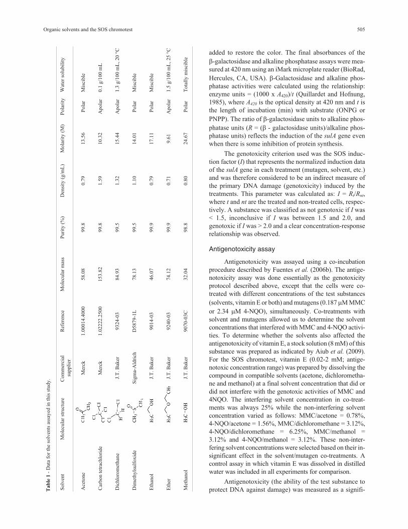
ferent commercial suppliers (Table 1).
Bacterial strain and culture
Escherichia coli strain PQ37 [F- thr leu his-4 pyrD thi
galE galK or galT lac�U169 srl300::Tn10 rpoB rpsL uvrA
rfa trp::Muc+ sfiA::Mud(Ap,lac)ts] which has been recom-
mended for the detection of genotoxic carcinogens (Quil-
lardet et al., 1982) was used in this work. This strain carries
the sulA::lacZ fusion gene as a reporter gene for primary
DNA damage induced during the SOS response. The cells
were grown overnight at 37 °C and shaken at 100 rpm in LB
medium (10 g tryptone/L, 5 g yeast extract/L, 10 g sodium
chloride/L, pH 7.4) supplemented with ampicillin
(50 �g/mL) and tetracycline (17 �g/mL).
Genotoxicity assay
Genotoxicity assays were done using the SOS chro-
motest as described by Quillardet et al. (1982), with small
modifications. Briefly, overnight cultures were grown in
fresh LB medium to an optical density (OD600nm) of 0.4, di-
luted 10-fold in double strength LB medium (20 g trypto-
ne/L, 10 g yeast extract/L, 20 g sodium chloride/L, pH 7.4)
and mixed (v/v) with the test substances of interest (muta-
gens and solvents). A negative control (distilled water) was
always included in each assay. The bacteria were exposed
to different concentration ranges, depending on the test
substance, for 30 min at 8 °C and then cultured for 2 h at
37 °C.
�-Galactosidase and alkaline phosphatase were as-
sayed in 96-well plates (Brand GMBH, Germany). For
�-galactosidase activity, cell membranes were disrupted by
mixing 142 �L of Z buffer (60 mM Na2HPO4, 40 mM
NaH2PO4, 10 mM KCl, 1 mM Mg2SO4, 0.1% SDS and
40 mM �-mercaptoethanol, pH 7.0) with 15 �L of cell cul-
ture for 20 min at room temperature. The reaction was
started by adding 30 �L of ONPG (4 mg/mL in T buffer: 1
M Tris-HCl, pH 8.8). After 40 min, the enzymatic reaction
was stopped by adding 50 �L of 1 M Na2CO3. After five
min, 50 �L of 2 M Tris was added to restore the color. For
alkaline phosphatase activity, cell membranes were dis-
rupted by adding 130 �L of T buffer, 7 �L of chloroform
and 5 �L of 0.1% sodium dodecyl sulfate (SDS) solution to
15 �L of cell culture followed by mixing vigorously for
20 min at room temperature. The enzyme reaction was
started by adding 30 �L of PNPP solution (4 mg/mL in T
buffer). After 40 min, the reaction was stopped by adding
50 �L of 2 M HCl. After 5 min, 50 �L of 2 M Tris was
504 Quintero et al.
added to restore the color. The final absorbances of the
�-galactosidase and alkaline phosphatase assays were mea-
sured at 420 nm using an iMark microplate reader (BioRad,
Hercules, CA, USA). �-Galactosidase and alkaline phos-
phatase activities were calculated using the relationship:
enzyme units = (1000 x A420)/t (Quillardet and Hofnung,
1985), where A420 is the optical density at 420 nm and t is
the length of incubation (min) with substrate (ONPG or
PNPP). The ratio of �-galactosidase units to alkaline phos-
phatase units (R = (� - galactosidase units)/alkaline phos-
phatase units) reflects the induction of the sulA gene even
when there is some inhibition of protein synthesis.
The genotoxicity criterion used was the SOS induc-
tion factor (I) that represents the normalized induction data
of the sulA gene in each treatment (mutagen, solvent, etc.)
and was therefore considered to be an indirect measure of
the primary DNA damage (genotoxicity) induced by the
treatments. This parameter was calculated as: I = Rt/Rnt,
where t and nt are the treated and non-treated cells, respec-
tively. A substance was classified as not genotoxic if I was
< 1.5, inconclusive if I was between 1.5 and 2.0, and
genotoxic if I was > 2.0 and a clear concentration-response
relationship was observed.
Antigenotoxicity assay
Antigenotoxicity was assayed using a co-incubation
procedure described by Fuentes et al. (2006b). The antige-
notoxicity assay was done essentially as the genotoxicity
protocol described above, except that the cells were co-
treated with different concentrations of the test substances
(solvents, vitamin E or both) and mutagens (0.187 �M MMC
or 2.34 �M 4-NQO), simultaneously. Co-treatments with
solvent and mutagens allowed us to determine the solvent
concentrations that interfered with MMC and 4-NQO activi-
ties. To determine whether the solvents also affected the
antigenotoxicity of vitamin E, a stock solution (8 mM) of this
substance was prepared as indicated by Aiub et al. (2009).
For the SOS chromotest, vitamin E (0.02-2 mM; antige-
notoxic concentration range) was prepared by dissolving the
compound in compatible solvents (acetone, dichlorometha-
ne and methanol) at a final solvent concentration that did or
did not interfere with the genotoxic activities of MMC and
4NQO. The interfering solvent concentration in co-treat-
ments was always 25% while the non-interfering solvent
concentration varied as follows: MMC/acetone = 0.78%,
4-NQO/acetone = 1.56%, MMC/dichloromethane = 3.12%,
4-NQO/dichloromethane = 6.25%, MMC/methanol =
3.12% and 4-NQO/methanol = 3.12%. These non-inter-
fering solvent concentrations were selected based on their in-
significant effect in the solvent/mutagen co-treatments. A
control assay in which vitamin E was dissolved in distilled
water was included in all experiments for comparison.
Antigenotoxicity (the ability of the test substance to
protect DNA against damage) was measured as a signifi-
Organic solvents and the SOS chromotest 505
Tab
le1
-D
ata
for
the
solv
ents
assa
yed
inth
isst
udy.
Solv
ent
Mole
cula
rst
ruct
ure
Com
mer
cial
suppli
er
Ref
eren
ceM
ole
cula
rm
ass
Puri
ty(%
)D
ensi
ty(g
/mL
)M
ola
rity
(M)
Pola
rity
Wat
erso
lubil
ity
Ace
tone
Mer
ck1.0
0014.4
000
58.0
899.8
0.7
913.5
6P
ola
rM
isci
ble
Car
bon
tetr
achlo
ride
Mer
ck1.0
2222.2
500
153.8
299.8
1.5
910.3
2A
pola
r0.1
g/1
00
mL
Dic
hlo
rom
ethan
eJ.
T.B
aker
9324-0
384.9
399.5
1.3
215.4
4A
pola
r1.3
g/1
00
mL
,20
°C
Dim
ethyls
ulf
oxid
eS
igm
a-A
ldri
chD
5879-1
L78.1
399.5
1.1
014.0
1P
ola
rM
isci
ble
Eth
anol
J.T
.B
aker
9014-0
346.0
799.9
0.7
917.1
1P
ola
rM
isci
ble
Eth
erJ.
T.B
aker
9240-0
374.1
299.9
0.7
19.6
1A
pola
r1.5
g/1
00
mL
,25
°C
Met
han
ol
J.T
.B
aker
9070-0
3C
32.0
498.8
0.8
024.6
7P
ola
rT
ota
lly
mis
cible

cant reduction in the induction factor (I) in the combined
treatments (substance + mutagens) and expressed as a per-
centage of the genotoxicity inhibition (%GI):
%GII I
I I
CO BASAL
MUT BASAL
� ��
��1 100
where ICO is the SOS induction factor in co-treated cells
(substance + mutagen), IBASAL is the basal SOS induction
factor and IMUT is the SOS induction factor in mutagen-
treated cells. Negative values of %GI were considered as
zero, which meant that this parameter ranged from a mini-
mum of 0% to a maximum of 100%.
Statistical analysis
The �-galactosidase and alkaline phosphatase activities
and the induction factor (I) values were expressed as the
meanSEM. The data normality was tested using the Kolmo-
gorov-Smirnov test and the homogeneity of variances was as-
sessed by analysis of variance (ANOVA). Statistical compari-
sons were done using Student’s t-test. Product-moment
(Pearson) correlation analysis was used to examine the con-
centration-response relationship in the genotoxicity experi-
ments. A value of p < 0.05 indicated significance in all cases.
All data analyses were done using the STATISTICA software
package v. 6 (StatSoft Inc., Tulsa, OK, USA).
Results
Induction kinetics of the sulA gene by standardmutagens
Figure 1 shows the induction kinetics of the sulA gene
in E. coli PQ37 following treatment with mutagens MMC
and 4-NQO. The extent and kinetics of sulA gene induction
by MMC and 4-NQO varied with the mutagen concentra-
tion, with the highest levels of induction occurring at
0.187 �M and 2.34 �M, respectively. At these concentra-
tions, there was no significant reduction in protein synthe-
sis as indicated by the constitutive alkaline phosphatase
activity. These concentrations were therefore used for co-
treatments in antigenotoxicity assays.
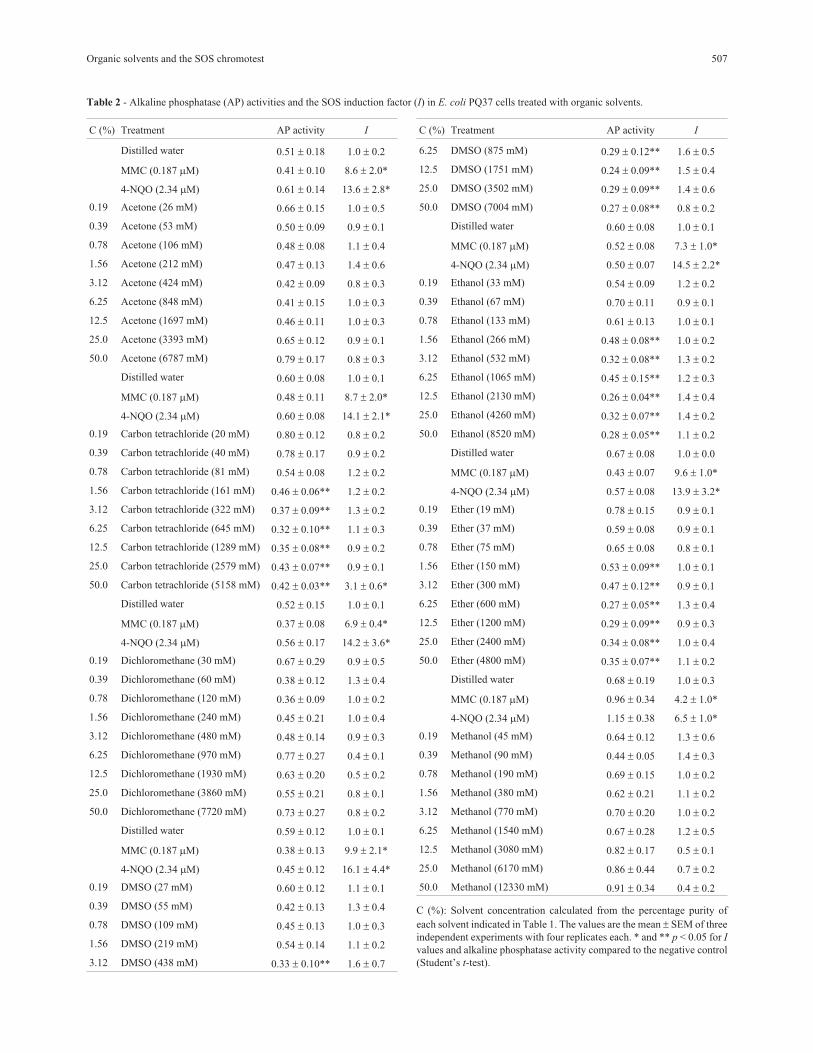
Solvent toxicity and genotoxicity in E. coli PQ37
Solvent toxicity was measured indirectly based on the
inhibition of constitutive alkaline phosphatase activity in E.
coli PQ37 (Table 2). Acetone, dichloromethane and metha-
nol did not significantly reduce protein synthesis over the
concentration range studied. In contrast, carbon tetrachlo-
ride, DMSO, ethanol and ether significantly reduced alka-
line phosphatase activity at concentrations = 161, 438, 266
and 150 mM, respectively. The inhibition of protein syn-
thesis by these solvents was also evidenced by the decrease
in �-galactosidase activity (data not shown).
The genotoxicity of the organic solvents was assayed
before their modulating effect was investigated (Table 2).
Acetone, dichloromethane, DMSO, ethanol, ether and me-
thanol did not significantly increase the I values in E. coli
PQ37, indicating that these compounds do not induce the
SOS response in E. coli. Carbon tetrachloride significantly
increased the I values at the highest concentration
(5158 mM), although correlation analysis revealed no con-
centration-response relationship for this solvent (data not
shown). Only solvents that were neither toxic nor genotoxic
to E. coli PQ37 (acetone, dichloromethane and methanol)
were considered for further assays.
506 Quintero et al.
Figure 1 - Induction kinetics of the sulA gene in response to mutagens MMC (A) and 4-NQO (B) in E. coli PQ37 cells. (■) alkaline phosphatase, (�)
�-galactosidase and (▲) SOS induction factor.
Organic solvents and the SOS chromotest 507
Table 2 - Alkaline phosphatase (AP) activities and the SOS induction factor (I) in E. coli PQ37 cells treated with organic solvents.
C (%) Treatment AP activity I
Distilled water 0.51 0.18 1.0 0.2
MMC (0.187 �M) 0.41 0.10 8.6 2.0*
4-NQO (2.34 �M) 0.61 0.14 13.6 2.8*
0.19 Acetone (26 mM) 0.66 0.15 1.0 0.5
0.39 Acetone (53 mM) 0.50 0.09 0.9 0.1
0.78 Acetone (106 mM) 0.48 0.08 1.1 0.4
1.56 Acetone (212 mM) 0.47 0.13 1.4 0.6
3.12 Acetone (424 mM) 0.42 0.09 0.8 0.3
6.25 Acetone (848 mM) 0.41 0.15 1.0 0.3
12.5 Acetone (1697 mM) 0.46 0.11 1.0 0.3
25.0 Acetone (3393 mM) 0.65 0.12 0.9 0.1
50.0 Acetone (6787 mM) 0.79 0.17 0.8 0.3
Distilled water 0.60 0.08 1.0 0.1
MMC (0.187 �M) 0.48 0.11 8.7 2.0*
4-NQO (2.34 �M) 0.60 0.08 14.1 2.1*
0.19 Carbon tetrachloride (20 mM) 0.80 0.12 0.8 0.2
0.39 Carbon tetrachloride (40 mM) 0.78 0.17 0.9 0.2
0.78 Carbon tetrachloride (81 mM) 0.54 0.08 1.2 0.2
1.56 Carbon tetrachloride (161 mM) 0.46 0.06** 1.2 0.2
3.12 Carbon tetrachloride (322 mM) 0.37 0.09** 1.3 0.2
6.25 Carbon tetrachloride (645 mM) 0.32 0.10** 1.1 0.3
12.5 Carbon tetrachloride (1289 mM) 0.35 0.08** 0.9 0.2
25.0 Carbon tetrachloride (2579 mM) 0.43 0.07** 0.9 0.1
50.0 Carbon tetrachloride (5158 mM) 0.42 0.03** 3.1 0.6*
Distilled water 0.52 0.15 1.0 0.1
MMC (0.187 �M) 0.37 0.08 6.9 0.4*
4-NQO (2.34 �M) 0.56 0.17 14.2 3.6*
0.19 Dichloromethane (30 mM) 0.67 0.29 0.9 0.5
0.39 Dichloromethane (60 mM) 0.38 0.12 1.3 0.4
0.78 Dichloromethane (120 mM) 0.36 0.09 1.0 0.2
1.56 Dichloromethane (240 mM) 0.45 0.21 1.0 0.4
3.12 Dichloromethane (480 mM) 0.48 0.14 0.9 0.3
6.25 Dichloromethane (970 mM) 0.77 0.27 0.4 0.1
12.5 Dichloromethane (1930 mM) 0.63 0.20 0.5 0.2
25.0 Dichloromethane (3860 mM) 0.55 0.21 0.8 0.1
50.0 Dichloromethane (7720 mM) 0.73 0.27 0.8 0.2
Distilled water 0.59 0.12 1.0 0.1
MMC (0.187 �M) 0.38 0.13 9.9 2.1*
4-NQO (2.34 �M) 0.45 0.12 16.1 4.4*
0.19 DMSO (27 mM) 0.60 0.12 1.1 0.1
0.39 DMSO (55 mM) 0.42 0.13 1.3 0.4
0.78 DMSO (109 mM) 0.45 0.13 1.0 0.3
1.56 DMSO (219 mM) 0.54 0.14 1.1 0.2
3.12 DMSO (438 mM) 0.33 0.10** 1.6 0.7
C (%) Treatment AP activity I
6.25 DMSO (875 mM) 0.29 0.12** 1.6 0.5
12.5 DMSO (1751 mM) 0.24 0.09** 1.5 0.4
25.0 DMSO (3502 mM) 0.29 0.09** 1.4 0.6
50.0 DMSO (7004 mM) 0.27 0.08** 0.8 0.2
Distilled water 0.60 0.08 1.0 0.1
MMC (0.187 �M) 0.52 0.08 7.3 1.0*
4-NQO (2.34 �M) 0.50 0.07 14.5 2.2*
0.19 Ethanol (33 mM) 0.54 0.09 1.2 0.2
0.39 Ethanol (67 mM) 0.70 0.11 0.9 0.1
0.78 Ethanol (133 mM) 0.61 0.13 1.0 0.1
1.56 Ethanol (266 mM) 0.48 0.08** 1.0 0.2
3.12 Ethanol (532 mM) 0.32 0.08** 1.3 0.2
6.25 Ethanol (1065 mM) 0.45 0.15** 1.2 0.3
12.5 Ethanol (2130 mM) 0.26 0.04** 1.4 0.4
25.0 Ethanol (4260 mM) 0.32 0.07** 1.4 0.2
50.0 Ethanol (8520 mM) 0.28 0.05** 1.1 0.2
Distilled water 0.67 0.08 1.0 0.0
MMC (0.187 �M) 0.43 0.07 9.6 1.0*
4-NQO (2.34 �M) 0.57 0.08 13.9 3.2*
0.19 Ether (19 mM) 0.78 0.15 0.9 0.1
0.39 Ether (37 mM) 0.59 0.08 0.9 0.1
0.78 Ether (75 mM) 0.65 0.08 0.8 0.1
1.56 Ether (150 mM) 0.53 0.09** 1.0 0.1
3.12 Ether (300 mM) 0.47 0.12** 0.9 0.1
6.25 Ether (600 mM) 0.27 0.05** 1.3 0.4
12.5 Ether (1200 mM) 0.29 0.09** 0.9 0.3
25.0 Ether (2400 mM) 0.34 0.08** 1.0 0.4
50.0 Ether (4800 mM) 0.35 0.07** 1.1 0.2
Distilled water 0.68 0.19 1.0 0.3
MMC (0.187 �M) 0.96 0.34 4.2 1.0*
4-NQO (2.34 �M) 1.15 0.38 6.5 1.0*
0.19 Methanol (45 mM) 0.64 0.12 1.3 0.6
0.39 Methanol (90 mM) 0.44 0.05 1.4 0.3
0.78 Methanol (190 mM) 0.69 0.15 1.0 0.2
1.56 Methanol (380 mM) 0.62 0.21 1.1 0.2
3.12 Methanol (770 mM) 0.70 0.20 1.0 0.2
6.25 Methanol (1540 mM) 0.67 0.28 1.2 0.5
12.5 Methanol (3080 mM) 0.82 0.17 0.5 0.1
25.0 Methanol (6170 mM) 0.86 0.44 0.7 0.2
50.0 Methanol (12330 mM) 0.91 0.34 0.4 0.2
C (%): Solvent concentration calculated from the percentage purity of
each solvent indicated in Table 1. The values are the mean SEM of three
independent experiments with four replicates each. * and ** p < 0.05 for I
values and alkaline phosphatase activity compared to the negative control
(Student’s t-test).

Influence of organic solvents on the activities ofMMC and 4-NQO
Table 3 shows the capacity of the solvents to interfere
with mutagen genotoxicity. All of the solvents assayed (ac-
etone, dichloromethane and methanol) interfered with
MMC- and 4NQO -induced genotoxicity. Dichlorome-
thane and methanol concentrations > 6.25% significantly
reduced the I values in co-treated cells compared to the pos-
itive control whereas concentrations 3.12% did not. In
contrast, acetone reduced the I values in co-treatment with
MMC and 4NQO at concentrations � 1.56% and � 3.12%,
respectively.
508 Quintero et al.
Table 3 - Influence of organic solvents on mitomycin C (MMC)- and 4-nitro-quinoline-1-oxide (4-NQO)-induced DNA damage in Escherichia coli
PQ37 cells.
C (%) Cell treatment I (%GI) Cell treatments I (%GI)
Distilled water 1.0 0.1 Distilled water 1.0 0.1
MMC 7.4 0.9 4-NQO 9.8 0.8
0.19 Acetone + MMC 9.1 2.5 (0) Acetone + 4-NQO 13.7 3.0 (0)
0.39 Acetone + MMC 8.9 2.4 (0) Acetone + 4-NQO 11.1 3.3 (0)
0.78 Acetone + MMC 6.2 1.0 (18) Acetone + 4-NQO 11.2 3.4 (0)
1.56 Acetone + MMC 5.1 1.2 (36) * Acetone + 4-NQO 8.8 2.2 (11)
3.12 Acetone + MMC 1.5 0.3 (92) * Acetone + 4-NQO 1.6 0.5 (93) *
6.25 Acetone + MMC 1.1 0.1 (98) * Acetone + 4-NQO 1.2 0.2 (98) *
12.5 Acetone + MMC 1.1 0.1 (99) * Acetone + 4-NQO 1.0 0.2 (100) *
25.0 Acetone + MMC 0.7 0.2 (100) * Acetone + 4-NQO 0.9 0.1 (100) *
50.0 Acetone + MMC 0.8 0.1(100) * Acetone + 4-NQO 0.8 0.1 (100) *
Distilled water 1.0 0.1 Distilled water 1.0 0.2
MMC 5.8 0.9 4-NQO 9.0 2.8
0.19 Dichloromethane + MMC 6.2 1.9 (0) Dichloromethane + 4-NQO 10.5 3.9 (0)
0.39 Dichloromethane + MMC 9.3 2.2 (0) Dichloromethane + 4-NQO 10.4 2.6 (0)
0.78 Dichloromethane + MMC 8.5 2.4 (0) Dichloromethane + 4-NQO 10.6 2.7 (0)
1.56 Dichloromethane + MMC 6.9 1.3 (0) Dichloromethane + 4-NQO 12.0 2.1 (0)
3.12 Dichloromethane + MMC 5.1 0.6 (14) Dichloromethane + 4-NQO 10.9 2.9 (0)
6.25 Dichloromethane + MMC 3.3 0.8 (52) * Dichloromethane + 4-NQO 6.4 2.1 (32)
12.5 Dichloromethane + MMC 1.4 0.2 (92) * Dichloromethane + 4-NQO 3.4 1.2 (70) *
25.0 Dichloromethane + MMC 0.8 0.2 (100) * Dichloromethane + 4-NQO 0.5 0.1 (100) *
50.0 Dichloromethane + MMC 0.7 0.2 (100) * Dichloromethane + 4-NQO 0.5 0.1 (100) *
Distilled water 1.0 0.1 Distilled water 1.0 0.1
MMC 6.9 1.9 4-NQO 10.8 1.2
0.19 Methanol + MMC 7.8 1.0 (0) Methanol + 4-NQO 14.2 2.7 (0)
0.39 Methanol + MMC 7.8 0.7 (0) Methanol + 4-NQO 15.6 4.8 (0)
0.78 Methanol + MMC 7.6 1.2 (0) Methanol + 4-NQO 16.3 2.7 (0)
1.56 Methanol + MMC 6.6 1.0 (6) Methanol + 4-NQO 12.8 3.3 (0)
3.12 Methanol + MMC 5.4 0.8 (25) Methanol + 4-NQO 11.0 2.6 (0)
6.25 Methanol + MMC 1.2 0.3 (97) * Methanol + 4-NQO 1.7 0.5 (93) *
12.5 Methanol + MMC 0.7 0.4 (100) * Methanol + 4-NQO 1.3 0.2 (97) *
25.0 Methanol + MMC 0.6 0.3 (100) * Methanol + 4-NQO 1.2 0.3 (98) *
50.0 Methanol + MMC 0.5 0.2 (100) * Methanol + 4-NQO 0.7 0.1 (100) *
C (%): Solvent concentration in the mixture calculated from the percentage purity of each solvent indicated in Table 1. MMC and 4-NQO were assayed at
concentrations of 0.187 �M and 2.34 �M, respectively. The values are the mean SEM of three independent experiments with four replicates each. The
percentage of genotoxicity inhibition (%GI) was calculated as described in the Methods. *p < 0.05 compared to the corresponding positive control (Stu-
dent’s t-test).

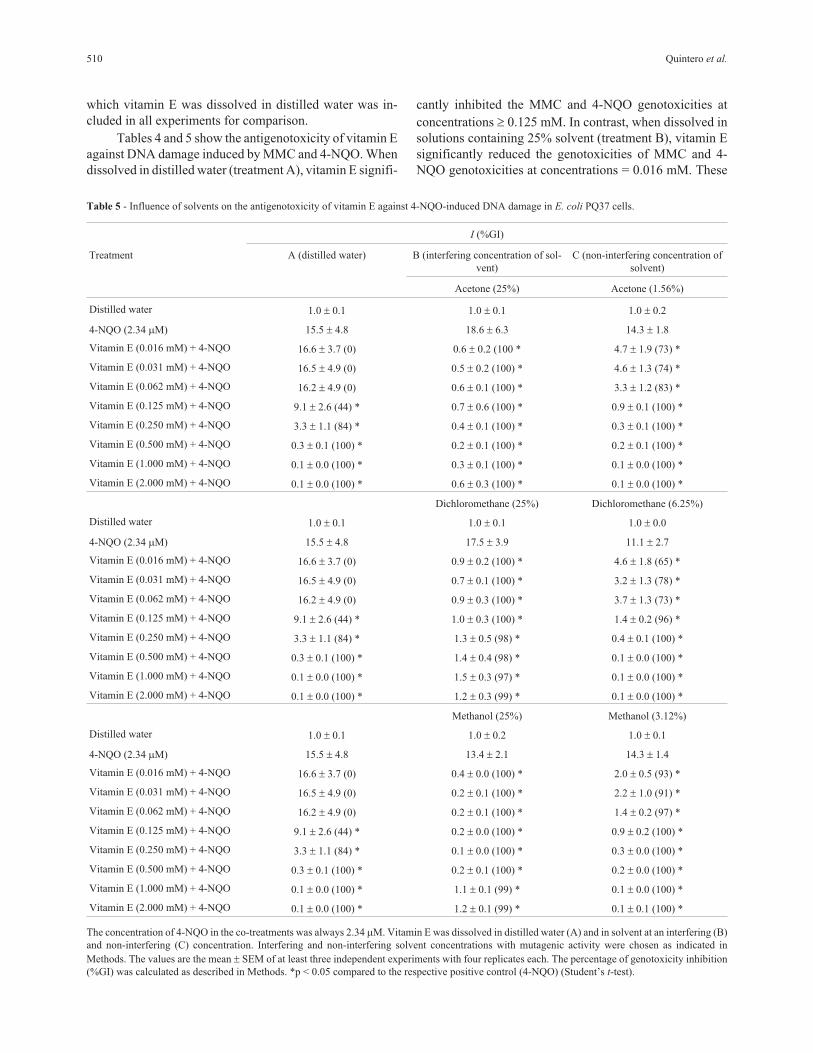
Influence of organic solvents on vitamin E
antigenotoxicity
We hypothesized that the solvent concentrations that
interfered with the genotoxic activities of MMC- and
4-NQO could increase the antigenotoxicity of vitamin E
against these mutagens, resulting in overestimates of
antigenotoxicity. To assess this hypothesis, we assayed the
acetone, dichloromethane and methanol concentrations
that did and did not interfere with the genotoxic activities of
MMC and 4-NQO (Tables 4 and 5). A control assay in
Organic solvents and the SOS chromotest 509
Table 4 - Influence of solvents on vitamin E antigenotoxicity against MMC-induced DNA damage in E. coli PQ37 cells.
I (%GI)
Treatment A (distilled water) B (interfering concentration of solvent) C (non-interfering concentration of solvent)
Acetone (25%) Acetone (0.78%)
Distilled water 1.0 0.1 1.0 0.2 1.0 0.1
MMC (0.187 �M) 6.5 1.6 4.9 1.1 6.5 1.9
Vitamin E (0.016 mM) + MMC 7.7 1.9 (0) 0.5 0.2 (100)* 6.6 1.4 (0.0)
Vitamin E (0.031 mM) + MMC 6.8 2.1 (0) 0.4 0.1 (100)* 5.9 1.1 (11)
Vitamin E (0.062 mM) + MMC 5.5 0.9 (17) 0.3 0.1 (100)* 3.7 0.7 (51)*
Vitamin E (0.125 mM) + MMC 2.6 0.8 (70)* 0.3 0.0 (100)* 2.8 0.6 (67)*
Vitamin E (0.250 mM) + MMC 0.9 0.1 (100)* 0.2 0.0 (100)* 0.6 0.2 (100)*
Vitamin E (0.500 mM) + MMC 0.2 0.0 (100)* 0.2 0.1 (100)* 0.2 0.1 (100)*
Vitamin E (1.000 mM) + MMC 0.2 0.0 (100)* 0.4 0.2 (100)* 0.1 0.0 (100)*
Vitamin E (2.000 mM) + MMC 0.1 0.0 (100)* 0.3 0.1 (100)* 0.1 0.0 (100)*
Dichloromethane (25%) Dichloromethane (3.12%)
Distilled water 1.0 0.1 0.9 0.2 1.0 0.1
MMC (0.187 �M) 6.5 1.6 5.7 1.1 4.1 1.0
Vitamin E (0.016 mM) + MMC 7.7 1.9 (0) 0.6 0.1 (100)* 1.2 0.3 (94)*
Vitamin E (0.031 mM) + MMC 6.8 2.1 (0) 0.5 0.1 (100)* 0.6 0.1 (100)*
Vitamin E (0.062 mM) + MMC 5.5 0.9 (17) 0.7 0.1 (100)* 0.9 0.2 (100)*
Vitamin E (0.125 mM) + MMC 2.6 0.8 (70)* 0.7 0.1 (100)* 0.7 0.3 (100)*
Vitamin E (0.250 mM) + MMC 0.9 0.1 (100)* 0.8 0.2 (100)* 0.5 0.1 (100)*
Vitamin E (0.500 mM) + MMC 0.2 0.0 (100)* 0.7 0.1 (100)* 0.3 0.0 (100)*
Vitamin E (1.000 mM) + MMC 0.2 0.0 (100)* 0.8 0.1 (100)* 0.3 0.0 (100)*
Vitamin E (2.000 mM) + MMC 0.1 0.0 (100)* 0.9 0.2 (100)* 0.3 0.1 (100)*
Methanol (25%) Methanol (3.12%)
Distilled water 1.0 0.1 1.0 0.1 1.0 0.2
MMC (0.187 �M) 6.5 1.6 5.2 1.8 8.3 2.4
Vitamin E (0.016 mM) + MMC 7.7 1.9 (0) 1.1 0.0 (98)* 0.6 0.1 (100)*
Vitamin E (0.031 mM) + MMC 6.8 2.1 (0) 1.1 0.2 (98)* 0.6 0.3 (100)*
Vitamin E (0.062 mM) + MMC 5.5 0.9 (17) 0.8 0.1 (100)* 0.4 0.1 (100)*
Vitamin E (0.125 mM) + MMC 2.6 0.8 (70)* 0.4 0.0 (100)* 0.3 0.1 (100)*
Vitamin E (0.250 mM) + MMC 0.9 0.1 (100)* 0.3 0.1 (100)* 0.5 0.3 (100)*
Vitamin E (0.500 mM) + MMC 0.2 0.0 (100)* 0.2 0.0 (100)* 0.2 0.1 (100)*
Vitamin E (1.000 mM) + MMC 0.2 0.0 (100)* 0.2 0.0 (100)* 0.2 0.0 (100)*
Vitamin E (2.000 mM) + MMC 0.1 0.0 (100)* 0.2 0.0 (100)* 0.1 0.0 (100)*
The concentration of MMM in the co-treatments was always 0.187 �M. Vitamin E was dissolved in distilled water (A) and in solvent at an interfering (B)
and non-interfering (C) concentration. Interfering and non-interfering solvent concentrations with mutagenic activity were chosen as indicated in
Methods. The values are the mean SEM of at least three independent experiments with four replicates each. The percentage of genotoxicity inhibition
(%GI) was calculated as described in Methods. * p < 0.05 compared to the respective positive control (MMC) (Student’s t-test).
which vitamin E was dissolved in distilled water was in-
cluded in all experiments for comparison.
Tables 4 and 5 show the antigenotoxicity of vitamin E
against DNA damage induced by MMC and 4-NQO. When
dissolved in distilled water (treatment A), vitamin E signifi-
cantly inhibited the MMC and 4-NQO genotoxicities at
concentrations � 0.125 mM. In contrast, when dissolved in
solutions containing 25% solvent (treatment B), vitamin E
significantly reduced the genotoxicities of MMC and 4-
NQO genotoxicities at concentrations = 0.016 mM. These
510 Quintero et al.
Table 5 - Influence of solvents on the antigenotoxicity of vitamin E against 4-NQO-induced DNA damage in E. coli PQ37 cells.
I (%GI)
Treatment A (distilled water) B (interfering concentration of sol-
vent)
C (non-interfering concentration of
solvent)
Acetone (25%) Acetone (1.56%)
Distilled water 1.0 0.1 1.0 0.1 1.0 0.2
4-NQO (2.34 �M) 15.5 4.8 18.6 6.3 14.3 1.8
Vitamin E (0.016 mM) + 4-NQO 16.6 3.7 (0) 0.6 0.2 (100 * 4.7 1.9 (73) *
Vitamin E (0.031 mM) + 4-NQO 16.5 4.9 (0) 0.5 0.2 (100) * 4.6 1.3 (74) *
Vitamin E (0.062 mM) + 4-NQO 16.2 4.9 (0) 0.6 0.1 (100) * 3.3 1.2 (83) *
Vitamin E (0.125 mM) + 4-NQO 9.1 2.6 (44) * 0.7 0.6 (100) * 0.9 0.1 (100) *
Vitamin E (0.250 mM) + 4-NQO 3.3 1.1 (84) * 0.4 0.1 (100) * 0.3 0.1 (100) *
Vitamin E (0.500 mM) + 4-NQO 0.3 0.1 (100) * 0.2 0.1 (100) * 0.2 0.1 (100) *
Vitamin E (1.000 mM) + 4-NQO 0.1 0.0 (100) * 0.3 0.1 (100) * 0.1 0.0 (100) *
Vitamin E (2.000 mM) + 4-NQO 0.1 0.0 (100) * 0.6 0.3 (100) * 0.1 0.0 (100) *
Dichloromethane (25%) Dichloromethane (6.25%)
Distilled water 1.0 0.1 1.0 0.1 1.0 0.0
4-NQO (2.34 �M) 15.5 4.8 17.5 3.9 11.1 2.7
Vitamin E (0.016 mM) + 4-NQO 16.6 3.7 (0) 0.9 0.2 (100) * 4.6 1.8 (65) *
Vitamin E (0.031 mM) + 4-NQO 16.5 4.9 (0) 0.7 0.1 (100) * 3.2 1.3 (78) *
Vitamin E (0.062 mM) + 4-NQO 16.2 4.9 (0) 0.9 0.3 (100) * 3.7 1.3 (73) *
Vitamin E (0.125 mM) + 4-NQO 9.1 2.6 (44) * 1.0 0.3 (100) * 1.4 0.2 (96) *
Vitamin E (0.250 mM) + 4-NQO 3.3 1.1 (84) * 1.3 0.5 (98) * 0.4 0.1 (100) *
Vitamin E (0.500 mM) + 4-NQO 0.3 0.1 (100) * 1.4 0.4 (98) * 0.1 0.0 (100) *
Vitamin E (1.000 mM) + 4-NQO 0.1 0.0 (100) * 1.5 0.3 (97) * 0.1 0.0 (100) *
Vitamin E (2.000 mM) + 4-NQO 0.1 0.0 (100) * 1.2 0.3 (99) * 0.1 0.0 (100) *
Methanol (25%) Methanol (3.12%)
Distilled water 1.0 0.1 1.0 0.2 1.0 0.1
4-NQO (2.34 �M) 15.5 4.8 13.4 2.1 14.3 1.4
Vitamin E (0.016 mM) + 4-NQO 16.6 3.7 (0) 0.4 0.0 (100) * 2.0 0.5 (93) *
Vitamin E (0.031 mM) + 4-NQO 16.5 4.9 (0) 0.2 0.1 (100) * 2.2 1.0 (91) *
Vitamin E (0.062 mM) + 4-NQO 16.2 4.9 (0) 0.2 0.1 (100) * 1.4 0.2 (97) *
Vitamin E (0.125 mM) + 4-NQO 9.1 2.6 (44) * 0.2 0.0 (100) * 0.9 0.2 (100) *
Vitamin E (0.250 mM) + 4-NQO 3.3 1.1 (84) * 0.1 0.0 (100) * 0.3 0.0 (100) *
Vitamin E (0.500 mM) + 4-NQO 0.3 0.1 (100) * 0.2 0.1 (100) * 0.2 0.0 (100) *
Vitamin E (1.000 mM) + 4-NQO 0.1 0.0 (100) * 1.1 0.1 (99) * 0.1 0.0 (100) *
Vitamin E (2.000 mM) + 4-NQO 0.1 0.0 (100) * 1.2 0.1 (99) * 0.1 0.1 (100) *
The concentration of 4-NQO in the co-treatments was always 2.34 �M. Vitamin E was dissolved in distilled water (A) and in solvent at an interfering (B)
and non-interfering (C) concentration. Interfering and non-interfering solvent concentrations with mutagenic activity were chosen as indicated in
Methods. The values are the mean SEM of at least three independent experiments with four replicates each. The percentage of genotoxicity inhibition
(%GI) was calculated as described in Methods. *p < 0.05 compared to the respective positive control (4-NQO) (Student’s t-test).
findings support the hypothesis that organic solvents can
enhance antigenotoxic activity, possibly through an addi-
tive effect. Surprisingly, the antigenotoxic effect of vitamin
E against MMC and 4-NQO (treatment C) was also en-
hanced when the vitamin was dissolved in a non-interfering
concentration of solvent. Only the co-treatment with
MMC/vitamin E dissolved in 0.78% acetone showed simi-
lar antigenotoxicity to that of treatment A, although there
was a significant reduction in the MMC genotoxicity at a
lower vitamin concentration (0.062 mM). These results in-
dicate that low, non-interfering concentrations of solvent
can also influence the antigenotoxicity of vitamin E.
Discussion
In this study, we examined some of the factors that
can influence the results of the SOS chromotest that is fre-
quently used to screen for antimutagenic activity. Spe-
cifically, we investigated (1) the toxicity and genotoxicity
of solvents, (2) the ability of solvents to interfere with mu-
tagenesis, and (3) the contribution of organic solvents to
overestimates of the antimutagenic activity of vitamin E
(through additive or synergistic effects of the solvents).
Based on previous studies with the SOS chromotest
(Von der Hude et al., 1988; Quillardet and Hofnung, 1993;
Mersch-Sundermann et al., 1994), the solvents studied here
were not genotoxic. However, some solvents such as car-
bon tetrachloride, DMSO, ethanol and ether were toxic to
E. coli at high concentrations, as indicated by the inhibition
of protein synthesis. In the standard procedure for the SOS
chromotest (Quillardet and Hofnung, 1985) the maximum
volume of solvent (DMSO or ethanol) used is 20 �L/assay
(i.e., for 600 �L of cell suspension), which corresponds to a
final solvent concentration of 3.3% in the reaction mixture.
Since there is very little information on the influence of sol-
vents in the SOS chromotest, the data reported in Table 2
can be of practical use in choosing solvents compatible
with the SOS chromotest when screening natural antige-
notoxic compounds.
The ability of solvents to interfere with chemical mu-
tagenesis has been documented, as indicated in the Intro-
duction. For example, solvents such as DMSO can interfere
with the genotoxicity of cisplatin in the SOS chromotest
(Gebel and Koenig, 1999). As shown here, several solvents
(including some not studied before) significantly reduced
the genotoxicities of MMC and 4-NQO at high concentra-
tions; this interference may limit the usefulness of the SOS
Chromotest during antimutagenesis screening if the test
substances are soluble only in organic solvents. Under
these conditions, the assay can yield overestimates of anti-
genotoxicity for the tested compound. Acetone, dichloro-
methane and methanol increased the antigenotoxicity of
vitamin E when the solvents were tested at a final concen-
tration of 25%, which interfered with mutagenic activity.
Surprisingly, these solvents also increased vitamin E anti-
genotoxicity at a concentration that did not interfere with
mutagen activity, which suggested a synergistic interaction
between solvents and vitamin E under these conditions.
Donnelly et al. (1998) have previously shown that this
synergic and/or additive interaction is characteristic of
complex mixtures that contain mutagens.
DMSO is a cytoprotective compound frequently used
as a solvent in mutagenic and anti-mutagenic studies be-
cause it has been considered to be non-genotoxic (Yu and
Quinn, 1994). Indeed, several studies have used DMSO as a
solvent to study the antimutagenic properties of biological
extracts and/or natural compounds in the SOS chromotest
(Table 6). However, DMSO can act as a free radical scav-
enger and can penetrate cell membranes to affect cell mem-
brane stability (Yu and Quinn, 1998) and this in turn can
interfere with the activity of antimutagens. As shown by
Gebel and Koenig (1999), DMSO can also interfere with
the activity of different mutagens in the SOS chromotest.
Based on these findings, a critical evaluation of the anti-
genotoxic potential of the antimutagens indicated in Ta-
ble 6 is required.
We have previously proposed the SOS chromotest as
a potentially useful assay for primary screening of medici-
nal plant extracts for antigenotoxic activity against �-rays
(Fuentes et al., 2006a). With this test, we have observed
that the use of DMSO as solvent resulted in overestimates
of the antimutagenic activity of essential oils when com-
pared with distilled water (unpublished data). The additive
and synergistic effect of solvents on vitamin E antige-
notoxicity shown here supports the use of distilled water as
the ideal solvent for antimutagen screening with the SOS
chromotest. When substances soluble only in organic sol-
vents are tested it is necessary to establish adequate final
solvent concentrations to avoid interference with the test
results. Solvents such as acetone, dichloromethane and
methanol that were neither toxic nor genotoxic to E. coli
PQ37 cells appear to be compatible with the SOS chro-
motest and are potentially useful for studying antimuta-
gens. However, their ability to interfere with the assayed
mutagens needs to be evaluated in each case.
Based on the results described here, we conclude that:
(1) the solvents studied were not genotoxic in E. coli PQ37
cells, although some of them (carbon tetrachloride, DMSO,
ethanol and ether) inhibited protein synthesis, depending
on their concentration, (2) organic solvents can reduce the
genotoxicity of MMC and 4-NQO in the SOS chromotest in
a concentration-dependent manner; water is the best sol-
vent when screening for antigenotoxicity in this assay, and
(3) a solvent concentration that interferes directly with
mutagen activity can significantly increase the antigeno-
toxicity of vitamin E, possibly through additive and syner-
gistic effects; this interference can result in overestimates
of antigenotoxicity. Overall, these findings indicate the
need to carefully determine the appropriate concentration
Organic solvents and the SOS chromotest 511

512 Quintero et al.
Table 6 - Antimutagenicity of different compounds and biological extracts detected with the SOS Chromotest.
Species Mutagen Reference
Natural antioxidants
Ascorbic acid†- NF, FZ Gajewska et al. (1990)
UVR, 4-NQO Sato et al. (1991)
Butyl-hydroxyanisole‡- B[a]P Potenberg et al. (1988)
Butyl-hydroxytoluene‡- B[a]P Potenberg et al. (1988)
5-chloro-uracyl†- UVR, MNNG Sato et al. (1991)
Dodecyl-gallate‡- B[a]P Potenberg et al. (1988)
Ethoxyquin‡- B[a]P Potenberg et al. (1988)
5-fluoro-uracyl†- UVR, 4-NQO Sato et al. (1991)
MNNG Sato et al. (1991)
Gallic acid esters†- H2O2 Nakayama et al., 1993
Glutathione†- 4-NQO, MNNG Sato et al. (1991)
Lignin derivates†- 4-NQO, H2O2 Mikulasova and Kosikova (2003)
Octyl-gallate‡- B[a]P Potenberg et al. (1988)
Propyl-gallate‡- B[a]P Potenberg et al. (1988)
Sodium selenite†- NF Gajewska et al. (1990)
Vitamin E - NDEA Aiub et al. (2009)
Plants extract or compounds
AE†, FF†, CE‡, EAE‡, ME‡, PEE‡ Acacia salicina B[a]P, NFA Bouhlel et al. (2007)
Norbixin† Bixa orellana UVR Junior et al. (2005)
Turmeric†, curcumin† Curcuma longa 4-NQO Polasa et al. (1997)
AE† Cymbopogon citratus �-radiation Fuentes et al. (2006b)
AE†, EO† Cyperus rotundus AFB1, NFA Kilani et al. (2005)
MCF‡, stigmasterol‡, � -sitosterol Gleditsia sinensis 4-NQO, MNNG Lim et al. (2005)
EO†, citral†, carvone†, limonene† Lippia alba BLM López et al. (2011)
EO†, thymol†, carvacrol† Lippia origanoides BLM Vicuña et al. (2010)
AE‡, FF‡, HE‡, CE‡, EAE‡, ME‡, EO‡ Myrthus communis AFB1, NFA Hayder et al. (2004)
Myricetin-3-o-galactoside‡, Myricetin-3-o-rhamnoside‡ Myrthus communis AFB1, NFA Hayder et al. (2008)
AE† Phyllanthus orbicularis �-radiation Fuentes et al. (2006b)
AE†, TT† Pinus caribeae �-radiation Fuentes et al. (2006a)
AE†, Pinus caribeae �-radiation Fuentes et al. (2006b)
Gallic acid‡, 1,2,3,4,6-pentagalloyl glucose‡ Pistacia lentiscus AFB1, NFA Abdelwahed et al. (2007)
EO† Pituranthus chloranthus NFA, H2O2 Neffati et al. (2009)
AE‡, emodin‡ Polygonum cuspidatum 1-NP Su et al. (1995)
FF†, EAE‡, ME‡ Rhamnus alaternus AFB1, NFA Ammar et al. (2007)
AE‡, CF‡, EAF‡, BF‡, PEE‡, CE‡ Rhamnus alaternus AFB1, NFA Ammar et al. (2008)
Emodin derivatives‡ Rumex acetosa 4-NQO, MNNG Lee et al. (2005)
Microbial extracts
Probiotic preparation† Bacillus sp. 4-NQO Caldini et al. (2002)
Probiotic preparation† Lactobacillus sp. 4-NQO, MNNG Caldini et al. (2008)
Probiotic preparation† Lactobacillus salivarius 4-NQO Fang et al. (2008)
Probiotic preparation† Saccharomyces boulardii 4-NQO, FU, NA Toma et al. (2005)
Animal extracts
BF‡, 5�-cholest-7-en-3�-ol‡ Asterina pectinifera (starfish) 4-NQO, MNNG Han et al. (2000)
Extract definitions: AE - aqueous extract, CE - chloroform extract, EAE - ethyl acetate extract, EE - ethanol extract, EO -essential oils, HE - hexane ex-
tract, ME - methanol extracts, PEE - petroleum ether extract. Fraction definitions: BF - butanolic fraction, CF - chloroform fraction, EAF - ethyl acetate
fraction, FF - flavonoid fractions, MCF - methylene chloride fraction, TT - tannin fraction. The extracts or fractions were assayed using water (†) or
dimethyl sulfoxide (‡) as the diluent. Mutagen definitions: AFB1 - aflatoxin B1, B[a]P - benzo[amine]pyrene, BLM - bleomycin, FU - furazolidone, FZ -
furazolidone, H2O2 - hydrogen peroxide, MNNG - N-methyl-N-nitro-N-nitrosoguanidine, NA - nalidixic acid, NDEA - N-nitrosodiethylamine, NF -
nitrofurasone, NFA - nifuroxazide, NPD - 4-nitro-o-phenylenediamine, 1-NP - 1-nitropyrene, 4-NQO - 4-nitroquinoline-1-oxide, SA - sodium azide,
�-radiation - gamma radiation, UVR - ultraviolet radiation.

of solvent to use when screening for antigenotoxicity in the
SOS chromotest.
Acknowledgments
The authors thank Dr. Montserrat Llagostera Casal
(Universidad Autónoma de Barcelona) for providing E.
coli strain PQ37 and Dr. Rodrigo Torres (Universidad In-
dustrial de Santander) for letting us use some equipment.
This work was supported by the Vice-Rectory for Science
Research at Universidad Industrial de Santander (UIS,
grant no. 5154) and by the Colombian Institute of Science
and Technology (COLCIENCIAS, grant no. RC-245-
2011).
References
Abdelwahed A, Bouhlel I, Skandrani I, Valenti K, Kadri M,
Guiraud P, Steiman R, Mariotte A-M, Ghedira K, Laporte F,
et al. (2007) Study of antimutagenic and antioxidant activi-
ties of gallic acid and 1,2,3,4,6-pentagalloylglucose from
Pistacia lentiscus. Confirmation by microarray expression
profiling. Chem Biol Interact 165:1-13.
Aiub CAF, Ribero-Pinto LF and Felzenszwalb I (2009) DNA-
repair genes and vitamin E in the prevention of N-nitroso-
diethylamine mutagenicity. Cell Biol Toxicol 25:393-402.
Ammar RB, Bouhlel I, Valenti K, Sghaier MB, Kilani S, Mariotte
A-M, Dijoux-Franca M-G, Laporte F, Ghedira K and Che-
kir-Ghedira L (2007) Transcriptional response of genes in-
volved in cell defense system in human cells stressed by
H2O2 and pre-treated with (Tunisian) Rhamnus alaternus
extracts: Combination with polyphenolic compounds and
classic in vitro assays. Chem Biol Interact 168:171-183.
Ammar RB, Sghaier MB, Boubaker J, Bhouri W, Naffeti A,
Skandrani I, Bouhlel I, Kilani S, Ghedira K and Chekir-
Ghedira L (2008) Antioxidant activity and inhibition of
aflotoxin B1-, nifuroxazide-, and sodium azide-induced mu-
tagenicity by extracts from Rhamnus alaternus L. Chem
Biol Interact 174:1-10.
Bouhlel I, Mansour HB, Limem I, Sghaier MB, Mahmoud A,
Chibani JB, Ghedira K and Chekir-Ghedira L (2007)
Screening of antimutagenicity via antioxidant activity in dif-
ferent extracts from the leaves of Acacia salicina from the
center of Tunisia. Environ Toxicol Pharmacol 23:56-63.
Caldini G, Trotta F and Cenci G (2002) Inhibition of 4-nitro-
quinoline-1-oxide genotoxicity by Bacillus strains. Res Mi-
crobiol 153:165-171.
Caldini G, Trotta F, Corsetti A and Cenci G (2008) Evidence for
in vitro anti-genotoxicity of cheese non-starter lactobacilli.
Antonie van Leeuwenhoek 93:51-59.
De Flora S (1998) Mechanisms of inhibitors of mutagenesis and
carcinogenesis. Mutat Res 402:151-158.
De Flora S and Ferguson LR (2005) Overview of mechanisms of
cancer chemopreventive agents. Mutat Res 591:8-15.
De Flora S and Ramel C (1988) Mechanisms of inhibitors of mu-
tagenesis and carcinogenesis, classification and overview.
Mutat Res 202:285-306.
De Flora S, Bronzetti G and Sobels FH (1992a) Assessment of
antimutagenicity and anticarcinogenicity. Mutat Res
267:153-155.
De Flora S, Camoirano A, D’Agostino F and Balansky R (1992b)
Modulation of the mutagenic response in prokaryotes. Mutat
Res 267:183-192.
DeMarini DM, Lawrence BK, Brooks HG and Houk VS (1991)
Compatibility of organic solvents with the microscreen pro-
phage-induction assay: Solvent-mutagen interactions.
Mutat Res 263:107-113.
Donnelly KC, Claxton LD, Huebner HJ and Capizzi JL (1998)
Mutagenic interactions of model chemical mixures. Che-
mosphere 37:1253-1261.
Fang W, Lu J, Ai-Ping L, Xing-Hua G and Fa-Zheng F (2008)
Analysis of antigenotoxicity of Lactobacillus salivarius by
high performance liquid chromatography. Chin J Anal
Chem 36:740-744.
Fronza G, Madzak C, Campomenosi P, Inga A, Lannone R,
Abbondandolo A and Sarasin A (1994) Mutation spectrum
of 4-nitroquinoline-1-oxide-damaged single-stranded shut-
tle vector DNA transfected into monkey cells. Mutat Res
308:117-125.
Fuentes JL, Alonso A, Cuétara E, Vernhe M, Alvarez N, Sán-
chez-Lamar A and Llagostera M (2006a) Usefulness of SOS
chromotest in the study of medicinal plant as radiopro-
tectors. Int J Radiat Biol 82:323-329.
Fuentes JL, Vernhe M, Cuetara EB, Sánchez-Lamar A, Santana
JL and Llagostera M (2006b) Tannins from barks of Pinus
caribeae Morelet protect Escherichia coli cells against DNA
damage induced by �-rays. Fitoterapia 77:116-120.
Gajewska J, Szczypka M, Tudek B and Szymczyk T (1990)
Studies on the effect of ascorbic acid and selenium on the
genotoxicity of nitrofurans: Nitrofurazone and furazolidone.
Mutat Res 232:191-197.
Gebel T and Koenig A (1999) Impact of dimethyl sulfoxide and
examples of combined genotoxicity in the SOS chromotest.
Mutat Res 444:405-411.
Han Y-H, Ham J-H, Lee N-J, Park C-H, Shin Y-H and Lee D-U
(2000) Antimutagenic activity of 5�-cholest-7-en-3�-ol, a
new component from the starfish Asterina pectinifera. Biol
Pharm Bull 23:1247-1249.
Hayder N, Abdelwahed A, Kilani S, Ammar RB, Mahmoud A,
Ghedira K and Chekir-Ghedira L (2004) Anti-genotoxic and
free-radical scavenging activities of extracts from (Tuni-
sian) Myrtus communis. Mutat Res 564:89-95.
Hayder N, Bouhlel I, Skandrani I, Kadri M, Steiman R, Guiraud P,
Mariotte A-M, Ghedira K, Dijoux-Franca M-G and Chekir-
Ghedira L (2008) In vitro antioxidant and antigenotoxic po-
tentials of myricetin-3-o-galactoside and myricetin-3-o-
rhamnoside from Myrtus communis: Modulation of expres-
sion of genes involved in cell defense system using cDNA
microarray. Toxicol in Vitro 22:567-581.
Hermann M, Bedouelles H and Hofnung M (1978) Influence of
solvents in the Salmonella/microsome assay. Mutat Res
53:198-199.
Junior ACTS, Asad LMBO, Oliveira EB, Kovary K, Asad NR and
Felzenszwalb I (2005) Antigenotoxic and antimutagenic po-
tential of an annatto pigment (norbixin) against oxidative
stress. Genet Mol Res 4:94-99.
Kada T and Shimoi K (1987) Desmutagens and bio-antimutagens
- Their modes of action. BioEssays 7:113-116.
Kilani S, Bouhlel I, Ben R, Abdelwahed A, Hayder N, Mahmoud
A, Ghedira K and Chekir-Ghedira L (2005) Evaluation of
the antimutagenic and antiradical potentials of extracts from
Organic solvents and the SOS chromotest 513

the tubers of (Tunisian) Cyperus rotundus. Toxicol Environ
Chem 87:415-425.
Kuroda Y and Inoue T (1988) Antimutagenesis by factors affect-
ing DNA repair in bacteria. Mutat Res 202:387-391.
Lee N-J, Choi J-H, Koo B-S, Ryu S-Y, Han Y-H, Lee S-I and Lee
D-U (2005) Antimutagenicity and cytotoxicity of the con-
stituents from the aerial parts of Rumex acetosa. Biol Pharm
Bull 28:2158-2161.
Lim J-C, Park JH, Budesinsky M, Kasal A, Han Y-H, Koo B-S,
Lee S-I and Lee D-U (2005) Antimutagenic constituents
from the thorns of Gleditsia sinensis. Chem Pharm Bull
53:561-564.
López MA, Stashenko EE and Fuentes JL (2011) Chemical com-
position and antigenotoxic properties of the Lippia alba es-
sential oils. Genet Mol Biol 34:479-488.
Maron D, Katzenellenbogen BJ and Ames BN (1981) Compatibil-
ity of organic solvents with the Salmonella/microsome test.
Mutat Res 88:343-350.
Mersch-Sundermann V, Schneider U, Klopman G and Rosen-
kranz HS (1994) SOS induction in Escherichia coli and Sal-
monella mutagenicity: A comparison using 330 compounds.
Mutagenesis 9:205-224.
Mikulasova M and Kosikova B (2003) Modulation of muta-
genicity of various mutagens by lignin derivates. Mutat Res
535:171-180.
Nakayama T, Hiramitsu M, Osawa T and Kawakishi S (1993) The
protective role of gallic acid esters in bacterial cytotoxicity
and SOS responses induced by hydrogen peroxide. Mutat
Res 303:29-34.
Neffati A, Bouhlel I, Sghaier MB, Boubaker J, Limem I, Kilani S,
Skandrani I, Bhouri W, Le Dauphin J, Barillier D, et al.
(2009) Antigenotoxic and antioxidant activities of Pituran-
tus chloranthus essential oils. Environ Toxicol Pharmacol
27:187-194.
Odin AP (1997) Vitamins as antimutagens: Advantages and some
possible mechanisms of antimutagenic action. Mutat Res
386:39-67.
Polasa K, Nadamuni AN and Krishnaswamy R (1997) Inhibition
of SOS response in E. coli PQ37 by heated turmeric and
curcumin. Mutat Res 379:S206.
Potenberg J, Von der Hude W, Bauszus M, Basler A and Kahl R
(1988) Enhancement and inhibition of benzo [a]pyrene-
induced SOS function in E. coli by synthetic antioxidants.
Mutat Res 207:7-11.
Quillardet P and Hofnung M (1985) The SOS chromotest, a
colorimetric bacterial assay for genotoxins: Procedures.
Mutat Res 147:65-78.
Quillardet P and Hofnung M (1993) The SOS chromotest: A re-
view. Mutat Res 297:235-279.
Quillardet P, Huisman O, D’ari R and Hofnung M (1982) SOS
chromotest, a direct assay of induction of an SOS function in
Escherichia coli K-12 to measure genotoxicity. Proc Natl
Acad Sci USA 79:5971-5975.
Sato T, Chikazawa K, Yamamori H, Ose Y, Nagase H and Kito H
(1991) Evaluation of the SOS chromotest for the detection
of antimutagens. Environ Mol Mutagen 17:258-263.
Su HY, Cherng S-H, Chen C-C and Lee H (1995) Emodin inhibits
the mutagenicity and DNA adducts induced by 1-nitro-
pyrene. Mutat Res 329:205-212.
Toma MM, Raipulis J, Kalnina I and Rutkis R (2005) Does
probiotic yeast act as antigenotoxin? Food Technol Biotech
43:301-305.
Tomasz M and Palom Y (1997) The mitomycin bioreductive
antitumor agents: Cross-linking and alkylation of DNA as
the molecular basis of their activity. Pharmacol Ther 76:73-
87.
Uno Y and Morita M (1993) Mutagenic activity of some activity
of some platinum and palladium complexes. Mutat Res
298:269-275.
Vicuña GC, Stashenko EE and Fuentes JL (2010) Chemical com-
position of the Lippia origanoides essential oils and their
antigenotoxicity against bleomycin-induced DNA damage.
Fitoterapia 81:343-349.
Von der Hude W, Behm C, Gürtler R and Basler A (1988) Evalua-
tion of the SOS chromotest. Mutat Res 203:81-94.
Wattenberg LW (1985) Chemoprevention of cancer. Cancer Res
45:1-8.
Yahagi T, Nagao M, Seino Y, Matsushima T, Sugimura T and
Okada M (1977) Mutagenicities of N-nitrosamines on Sal-
monella. Mutat Res 48:121-129.
Yu ZW and Quinn PJ (1994) Dimethyl sulphoxide: A review of its
applications in cell biology. Bioscience Rep 14:259-281.
Yu ZW and Quinn PJ (1998) The modulation of membrane struc-
ture and stability by dimethyl sulphoxide (review). Mol
Membr Biol 15:59-68.
Zeiger E (2007) What is needed for an acceptable antimuta-
genicity manuscript? Mutat Res 626:1-3.
Associate Editor: Daisy M.F. Salvadori
License information: This is an open-access article distributed under the terms of theCreative Commons Attribution License, which permits unrestricted use, distribution, andreproduction in any medium, provided the original work is properly cited.
514 Quintero et al.